Quick filters:
Liver lobules Stock Photos and Images
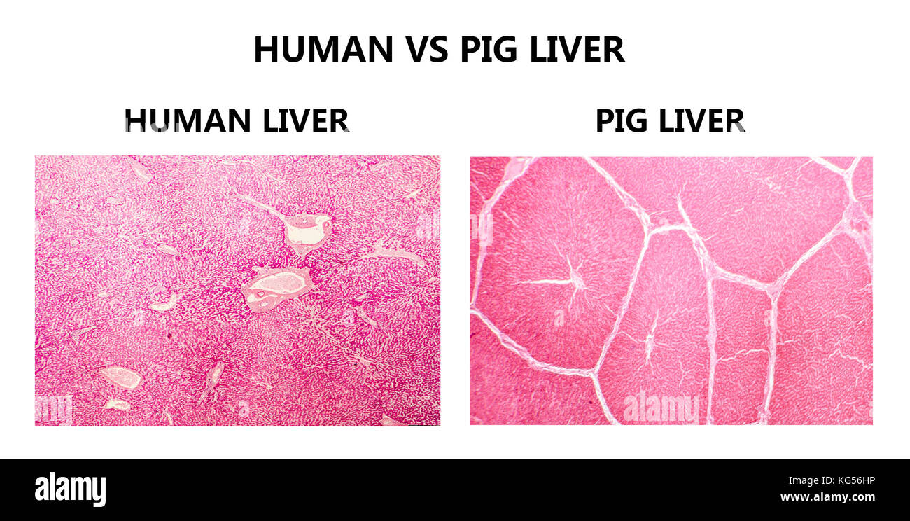 Human and pig livers. Light micrograph showing tissue from a healthy human liver (left) and a pig liver (right). The pig liver has well demarcated lobules with thick interlobal connective tissue. Stock Photohttps://www.alamy.com/image-license-details/?v=1https://www.alamy.com/stock-image-human-and-pig-livers-light-micrograph-showing-tissue-from-a-healthy-164842770.html
RFKG56HP–Human and pig livers. Light micrograph showing tissue from a healthy human liver (left) and a pig liver (right). The pig liver has well demarcated lobules with thick interlobal connective tissue.
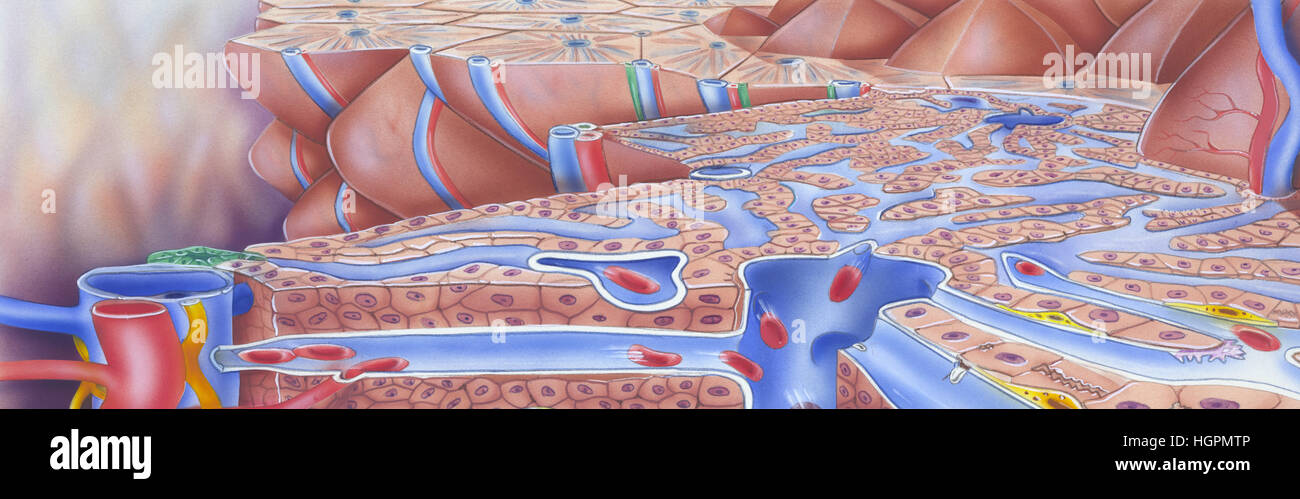 Liver - Cut Away View to show internal structures including: liver lobules, kupffers cells, fenestrations, lymphatic vessel, hepatic artery Stock Photohttps://www.alamy.com/image-license-details/?v=1https://www.alamy.com/stock-photo-liver-cut-away-view-to-show-internal-structures-including-liver-lobules-130806390.html
RFHGPMTP–Liver - Cut Away View to show internal structures including: liver lobules, kupffers cells, fenestrations, lymphatic vessel, hepatic artery
 The structure of the liver and the arrangement of liver lobules around the sublobular branches of the hepatic vein in the upper image. Section of port Stock Vectorhttps://www.alamy.com/image-license-details/?v=1https://www.alamy.com/the-structure-of-the-liver-and-the-arrangement-of-liver-lobules-around-the-sublobular-branches-of-the-hepatic-vein-in-the-upper-image-section-of-port-image359326233.html
RF2BTGM7N–The structure of the liver and the arrangement of liver lobules around the sublobular branches of the hepatic vein in the upper image. Section of port
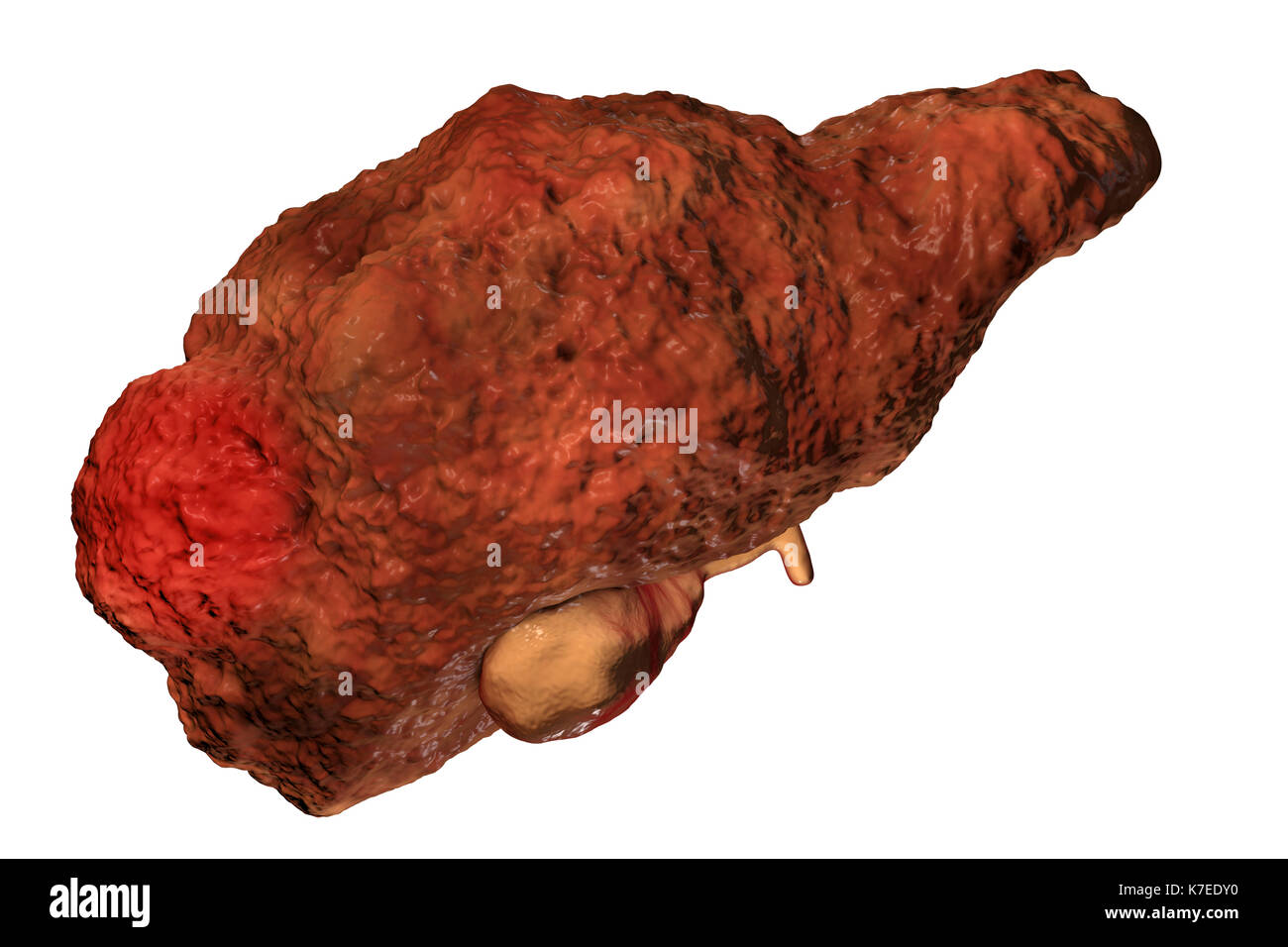 Cirrhotic liver with hepatocellular carcinoma, computer artwork. One of the causes of hepatocellular carcinoma is viral infection, particularly with hepatitis C viruses (HCV). Hepatitis C is a common cause of chronic hepatitis which progresses to liver cirrhosis. Viruses cause cell death (necrosis) in the hepatic lobules, which leads to scarring. This causes the liver surface to appear rough and lumpy instead of the usual smooth healthy appearance. The disease is irreversible and may lead to liver carcinoma. Stock Photohttps://www.alamy.com/image-license-details/?v=1https://www.alamy.com/cirrhotic-liver-with-hepatocellular-carcinoma-computer-artwork-one-image159514180.html
RFK7EDY0–Cirrhotic liver with hepatocellular carcinoma, computer artwork. One of the causes of hepatocellular carcinoma is viral infection, particularly with hepatitis C viruses (HCV). Hepatitis C is a common cause of chronic hepatitis which progresses to liver cirrhosis. Viruses cause cell death (necrosis) in the hepatic lobules, which leads to scarring. This causes the liver surface to appear rough and lumpy instead of the usual smooth healthy appearance. The disease is irreversible and may lead to liver carcinoma.
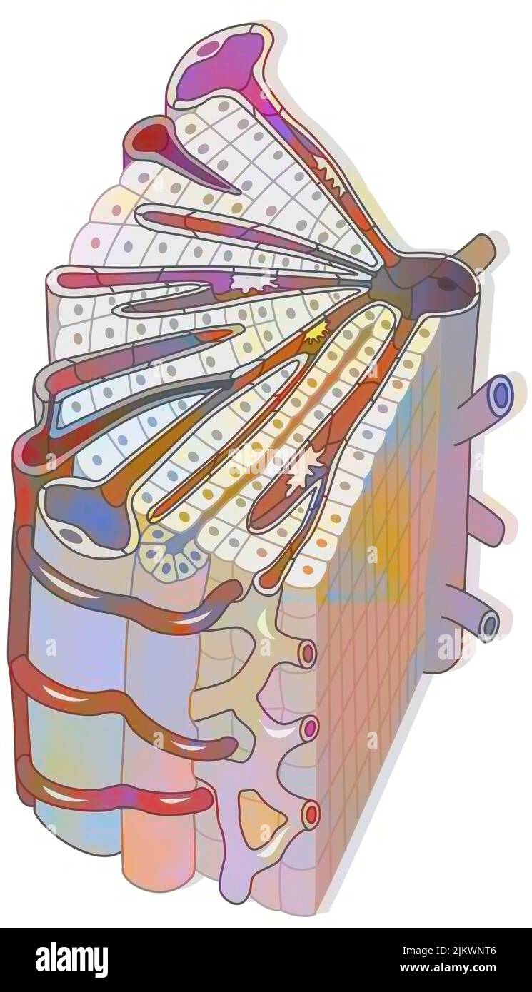 Hepatic lobule consisting of hepatocytes, a central vein, centrilobular vein. Stock Photohttps://www.alamy.com/image-license-details/?v=1https://www.alamy.com/hepatic-lobule-consisting-of-hepatocytes-a-central-vein-centrilobular-vein-image476924342.html
RF2JKWNT6–Hepatic lobule consisting of hepatocytes, a central vein, centrilobular vein.
 Vegetable and liver salad. Stock Photohttps://www.alamy.com/image-license-details/?v=1https://www.alamy.com/vegetable-and-liver-salad-image402534709.html
RF2EAW13H–Vegetable and liver salad.
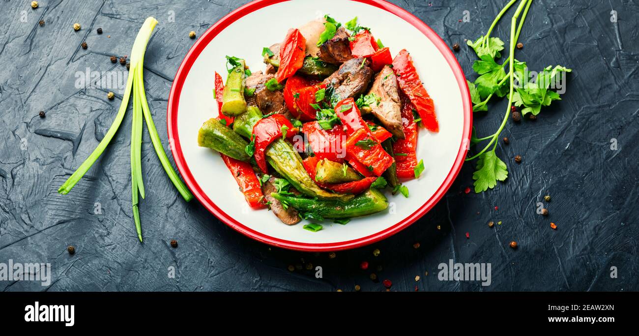 Vegetable and liver salad. Stock Photohttps://www.alamy.com/image-license-details/?v=1https://www.alamy.com/vegetable-and-liver-salad-image402536141.html
RF2EAW2XN–Vegetable and liver salad.
 This illustration represents Lobules of the Liver, vintage line drawing or engraving illustration. Stock Vectorhttps://www.alamy.com/image-license-details/?v=1https://www.alamy.com/this-illustration-represents-lobules-of-the-liver-vintage-line-drawing-or-engraving-illustration-image348636731.html
RF2B75NKR–This illustration represents Lobules of the Liver, vintage line drawing or engraving illustration.
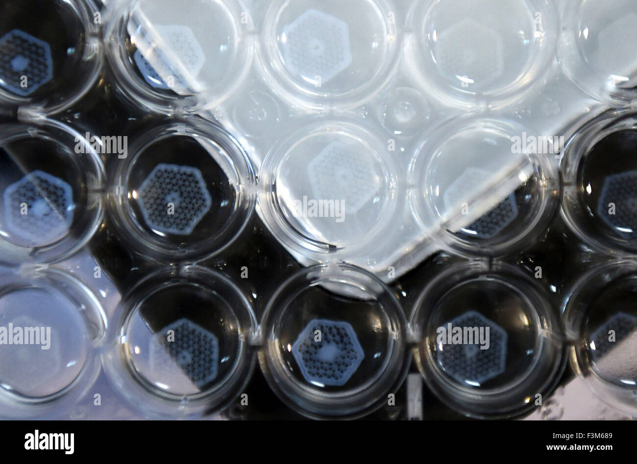 Hangzhou. 9th Oct, 2015. Photo taken on Oct. 9, 2015 shows hepatic lobules printed on the Regenovo 3D bio-print Work Station in Hangzhou, capital of east China's Zhejiang Province. A Chinese team announced Friday that it has used a specialized 3D printer to produce tiny sections of the liver that could eventually be used to form a full artificial version of the organ. © Han Chuanhao/Xinhua/Alamy Live News Stock Photohttps://www.alamy.com/image-license-details/?v=1https://www.alamy.com/stock-photo-hangzhou-9th-oct-2015-photo-taken-on-oct-9-2015-shows-hepatic-lobules-88339785.html
RMF3M689–Hangzhou. 9th Oct, 2015. Photo taken on Oct. 9, 2015 shows hepatic lobules printed on the Regenovo 3D bio-print Work Station in Hangzhou, capital of east China's Zhejiang Province. A Chinese team announced Friday that it has used a specialized 3D printer to produce tiny sections of the liver that could eventually be used to form a full artificial version of the organ. © Han Chuanhao/Xinhua/Alamy Live News
 Warm salad with chicken liver and vegetables.Chicken liver salad Stock Photohttps://www.alamy.com/image-license-details/?v=1https://www.alamy.com/warm-salad-with-chicken-liver-and-vegetableschicken-liver-salad-image433225091.html
RF2G4R30K–Warm salad with chicken liver and vegetables.Chicken liver salad
 (151009) -- HANGZHOU, Oct. 9, 2015 -- Photo taken on Oct. 9, 2015 shows hepatic lobules printed on the Regenovo 3D bio-print Work Station in Hangzhou, capital of east China s Zhejiang Province. A Chinese team announced Friday that it has used a specialized 3D printer to produce tiny sections of the liver that could eventually be used to form a full artificial version of the organ. )(mcg) CHINA-HANGZHOU-3D-PRINT-LIVER (CN) HanxChuanhao PUBLICATIONxNOTxINxCHN 151009 Hangzhou OCT 9 2015 Photo Taken ON OCT 9 2015 Shows hepatic printed ON The Regenovo 3D Bio Print Work Station in Hangzhou Capita Stock Photohttps://www.alamy.com/image-license-details/?v=1https://www.alamy.com/151009-hangzhou-oct-9-2015-photo-taken-on-oct-9-2015-shows-hepatic-lobules-printed-on-the-regenovo-3d-bio-print-work-station-in-hangzhou-capital-of-east-china-s-zhejiang-province-a-chinese-team-announced-friday-that-it-has-used-a-specialized-3d-printer-to-produce-tiny-sections-of-the-liver-that-could-eventually-be-used-to-form-a-full-artificial-version-of-the-organ-mcg-china-hangzhou-3d-print-liver-cn-hanxchuanhao-publicationxnotxinxchn-151009-hangzhou-oct-9-2015-photo-taken-on-oct-9-2015-shows-hepatic-printed-on-the-regenovo-3d-bio-print-work-station-in-hangzhou-capita-image563820611.html
RM2RN86XB–(151009) -- HANGZHOU, Oct. 9, 2015 -- Photo taken on Oct. 9, 2015 shows hepatic lobules printed on the Regenovo 3D bio-print Work Station in Hangzhou, capital of east China s Zhejiang Province. A Chinese team announced Friday that it has used a specialized 3D printer to produce tiny sections of the liver that could eventually be used to form a full artificial version of the organ. )(mcg) CHINA-HANGZHOU-3D-PRINT-LIVER (CN) HanxChuanhao PUBLICATIONxNOTxINxCHN 151009 Hangzhou OCT 9 2015 Photo Taken ON OCT 9 2015 Shows hepatic printed ON The Regenovo 3D Bio Print Work Station in Hangzhou Capita
 Warm salad with chicken liver and vegetables.Chicken liver salad Stock Photohttps://www.alamy.com/image-license-details/?v=1https://www.alamy.com/warm-salad-with-chicken-liver-and-vegetableschicken-liver-salad-image394975058.html
RF2DXGJM2–Warm salad with chicken liver and vegetables.Chicken liver salad
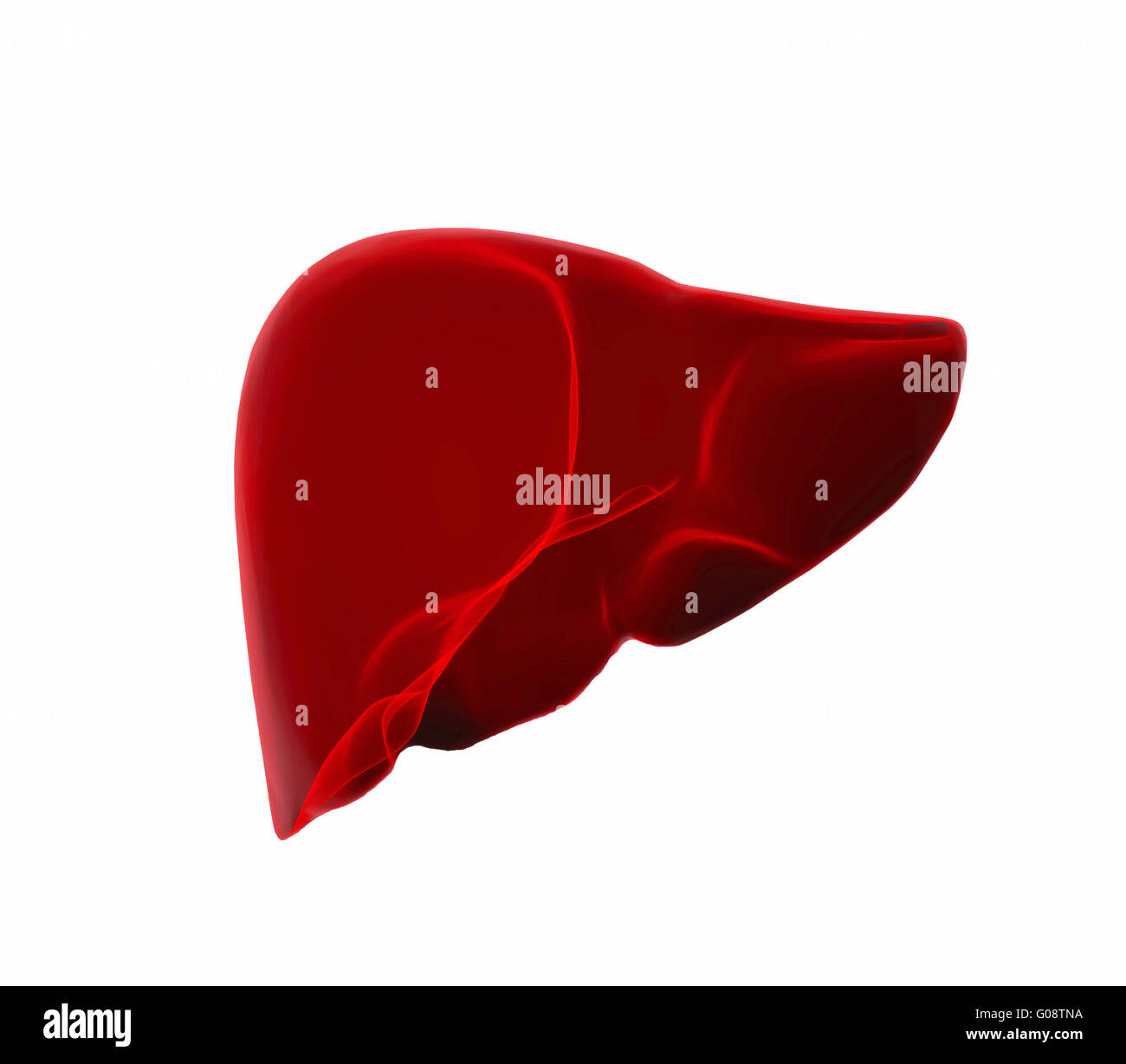 human liver in x-ray view Stock Photohttps://www.alamy.com/image-license-details/?v=1https://www.alamy.com/stock-photo-human-liver-in-x-ray-view-103457238.html
RMG08TNA–human liver in x-ray view
 Front view of the liver within a male figure. Stock Photohttps://www.alamy.com/image-license-details/?v=1https://www.alamy.com/stock-photo-front-view-of-the-liver-within-a-male-figure-52077164.html
RMD0M8XM–Front view of the liver within a male figure.
![. Cunningham's Text-book of anatomy. Anatomy. 1198 THE DIGESTIVE SYSTEM. Stkucture of the Liver. The liver is invested by an outer tunica serosa described in connexion with the peritoneum. Within this is a thin capsula fibrosa [Glissonii] (O.T. G-lisson's capsule) Intralobular capillary plexus Intralobular capillary plexus. Central vein Central vein Sublobular vein Fig 940.—Liver of a Pig injected from the Hepatic Vein by T. A. Carter. (From a specimen left in the Anatomical Department of Edinburgh University by Sir William Turner.) Liver lobules of delicate fibrous tissue, which is most evi Stock Photo . Cunningham's Text-book of anatomy. Anatomy. 1198 THE DIGESTIVE SYSTEM. Stkucture of the Liver. The liver is invested by an outer tunica serosa described in connexion with the peritoneum. Within this is a thin capsula fibrosa [Glissonii] (O.T. G-lisson's capsule) Intralobular capillary plexus Intralobular capillary plexus. Central vein Central vein Sublobular vein Fig 940.—Liver of a Pig injected from the Hepatic Vein by T. A. Carter. (From a specimen left in the Anatomical Department of Edinburgh University by Sir William Turner.) Liver lobules of delicate fibrous tissue, which is most evi Stock Photo](https://c8.alamy.com/comp/RD5HR6/cunninghams-text-book-of-anatomy-anatomy-1198-the-digestive-system-stkucture-of-the-liver-the-liver-is-invested-by-an-outer-tunica-serosa-described-in-connexion-with-the-peritoneum-within-this-is-a-thin-capsula-fibrosa-glissonii-ot-g-lissons-capsule-intralobular-capillary-plexus-intralobular-capillary-plexus-central-vein-central-vein-sublobular-vein-fig-940liver-of-a-pig-injected-from-the-hepatic-vein-by-t-a-carter-from-a-specimen-left-in-the-anatomical-department-of-edinburgh-university-by-sir-william-turner-liver-lobules-of-delicate-fibrous-tissue-which-is-most-evi-RD5HR6.jpg) . Cunningham's Text-book of anatomy. Anatomy. 1198 THE DIGESTIVE SYSTEM. Stkucture of the Liver. The liver is invested by an outer tunica serosa described in connexion with the peritoneum. Within this is a thin capsula fibrosa [Glissonii] (O.T. G-lisson's capsule) Intralobular capillary plexus Intralobular capillary plexus. Central vein Central vein Sublobular vein Fig 940.—Liver of a Pig injected from the Hepatic Vein by T. A. Carter. (From a specimen left in the Anatomical Department of Edinburgh University by Sir William Turner.) Liver lobules of delicate fibrous tissue, which is most evi Stock Photohttps://www.alamy.com/image-license-details/?v=1https://www.alamy.com/cunninghams-text-book-of-anatomy-anatomy-1198-the-digestive-system-stkucture-of-the-liver-the-liver-is-invested-by-an-outer-tunica-serosa-described-in-connexion-with-the-peritoneum-within-this-is-a-thin-capsula-fibrosa-glissonii-ot-g-lissons-capsule-intralobular-capillary-plexus-intralobular-capillary-plexus-central-vein-central-vein-sublobular-vein-fig-940liver-of-a-pig-injected-from-the-hepatic-vein-by-t-a-carter-from-a-specimen-left-in-the-anatomical-department-of-edinburgh-university-by-sir-william-turner-liver-lobules-of-delicate-fibrous-tissue-which-is-most-evi-image231849050.html
. Cunningham's Text-book of anatomy. Anatomy. 1198 THE DIGESTIVE SYSTEM. Stkucture of the Liver. The liver is invested by an outer tunica serosa described in connexion with the peritoneum. Within this is a thin capsula fibrosa [Glissonii] (O.T. G-lisson's capsule) Intralobular capillary plexus Intralobular capillary plexus. Central vein Central vein Sublobular vein Fig 940.—Liver of a Pig injected from the Hepatic Vein by T. A. Carter. (From a specimen left in the Anatomical Department of Edinburgh University by Sir William Turner.) Liver lobules of delicate fibrous tissue, which is most evi Stock Photohttps://www.alamy.com/image-license-details/?v=1https://www.alamy.com/cunninghams-text-book-of-anatomy-anatomy-1198-the-digestive-system-stkucture-of-the-liver-the-liver-is-invested-by-an-outer-tunica-serosa-described-in-connexion-with-the-peritoneum-within-this-is-a-thin-capsula-fibrosa-glissonii-ot-g-lissons-capsule-intralobular-capillary-plexus-intralobular-capillary-plexus-central-vein-central-vein-sublobular-vein-fig-940liver-of-a-pig-injected-from-the-hepatic-vein-by-t-a-carter-from-a-specimen-left-in-the-anatomical-department-of-edinburgh-university-by-sir-william-turner-liver-lobules-of-delicate-fibrous-tissue-which-is-most-evi-image231849050.htmlRMRD5HR6–. Cunningham's Text-book of anatomy. Anatomy. 1198 THE DIGESTIVE SYSTEM. Stkucture of the Liver. The liver is invested by an outer tunica serosa described in connexion with the peritoneum. Within this is a thin capsula fibrosa [Glissonii] (O.T. G-lisson's capsule) Intralobular capillary plexus Intralobular capillary plexus. Central vein Central vein Sublobular vein Fig 940.—Liver of a Pig injected from the Hepatic Vein by T. A. Carter. (From a specimen left in the Anatomical Department of Edinburgh University by Sir William Turner.) Liver lobules of delicate fibrous tissue, which is most evi
 Fish anatomy. Pike (Esox lucius). The hepatic parenchyma is divided into lobules and covered with a fibrous membrane. Digestive, metabolic and protect Stock Photohttps://www.alamy.com/image-license-details/?v=1https://www.alamy.com/fish-anatomy-pike-esox-lucius-the-hepatic-parenchyma-is-divided-into-lobules-and-covered-with-a-fibrous-membrane-digestive-metabolic-and-protect-image594312514.html
RF2WEW7JX–Fish anatomy. Pike (Esox lucius). The hepatic parenchyma is divided into lobules and covered with a fibrous membrane. Digestive, metabolic and protect
 Human Liver Radiology Exam Stock Photohttps://www.alamy.com/image-license-details/?v=1https://www.alamy.com/human-liver-radiology-exam-image274342041.html
RMWX9A21–Human Liver Radiology Exam
![Infographic of the structure and function of the liver, the pancreas and the gallbladder. [QuarkXPress (.qxp); 6259x4015]. Stock Photo Infographic of the structure and function of the liver, the pancreas and the gallbladder. [QuarkXPress (.qxp); 6259x4015]. Stock Photo](https://c8.alamy.com/comp/2NECAY8/infographic-of-the-structure-and-function-of-the-liver-the-pancreas-and-the-gallbladder-quarkxpress-qxp-6259x4015-2NECAY8.jpg) Infographic of the structure and function of the liver, the pancreas and the gallbladder. [QuarkXPress (.qxp); 6259x4015]. Stock Photohttps://www.alamy.com/image-license-details/?v=1https://www.alamy.com/infographic-of-the-structure-and-function-of-the-liver-the-pancreas-and-the-gallbladder-quarkxpress-qxp-6259x4015-image525188252.html
Infographic of the structure and function of the liver, the pancreas and the gallbladder. [QuarkXPress (.qxp); 6259x4015]. Stock Photohttps://www.alamy.com/image-license-details/?v=1https://www.alamy.com/infographic-of-the-structure-and-function-of-the-liver-the-pancreas-and-the-gallbladder-quarkxpress-qxp-6259x4015-image525188252.htmlRM2NECAY8–Infographic of the structure and function of the liver, the pancreas and the gallbladder. [QuarkXPress (.qxp); 6259x4015].
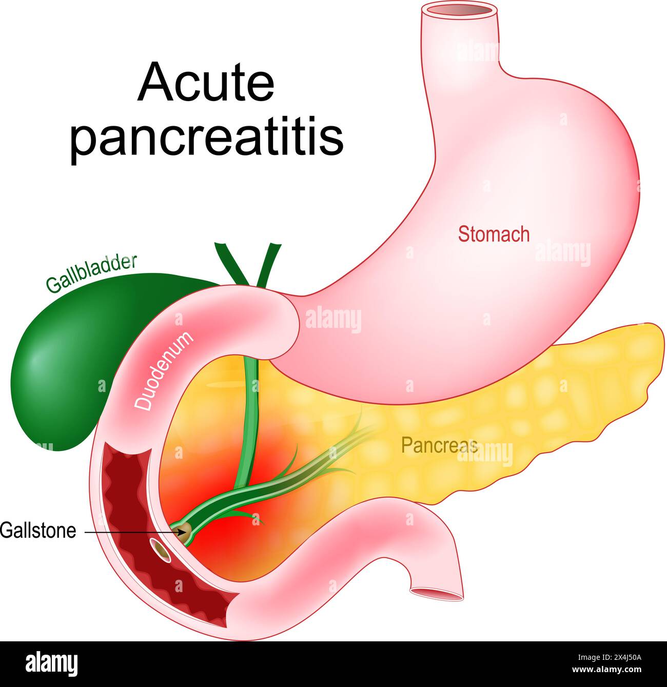 Acute pancreatitis. Pancreas inflammation. Realistic image of abdominal organs Gallbladder, Duodenum, Stomach, and Pancreas. Close-up of a Gallstone t Stock Vectorhttps://www.alamy.com/image-license-details/?v=1https://www.alamy.com/acute-pancreatitis-pancreas-inflammation-realistic-image-of-abdominal-organs-gallbladder-duodenum-stomach-and-pancreas-close-up-of-a-gallstone-t-image605220570.html
RF2X4J50A–Acute pancreatitis. Pancreas inflammation. Realistic image of abdominal organs Gallbladder, Duodenum, Stomach, and Pancreas. Close-up of a Gallstone t
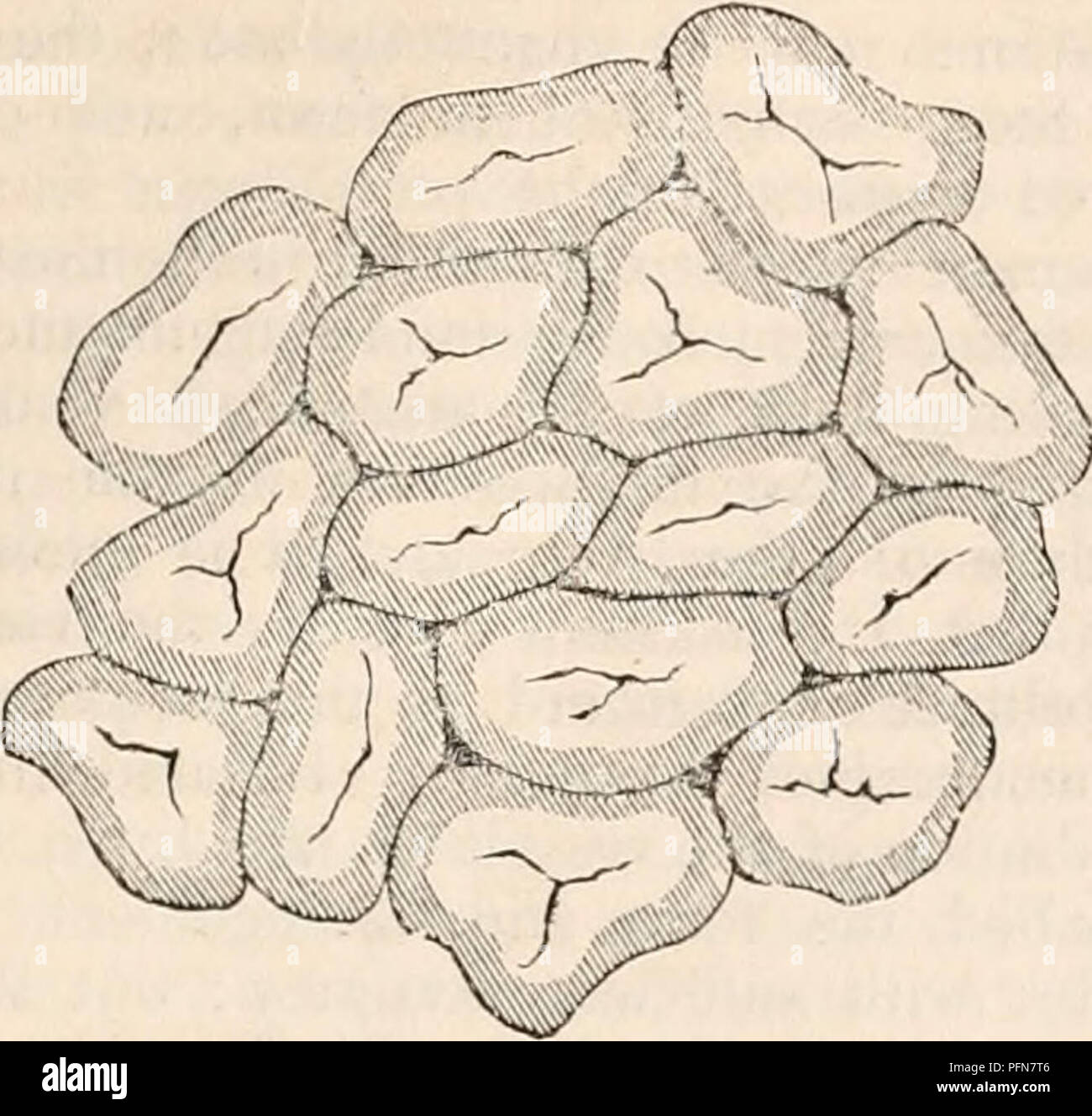 . The cyclopædia of anatomy and physiology. Anatomy; Physiology; Zoology. ABNORMAL ANATOMY OF THE LIVER. Fig. 44.. Lobules in a state of portal venous congestion, as seen on the surface of the. liver. The congested part oc- cupies the margins of the lobules, the uncongested portion their centres. After Kiernan. dullary and occupying the centres of the lo- bules." The causes of congestion are all such as tend to interfere with the circulation in the liver or with the general circulation ; for in- stance, impediment to the circulation of the blood through the capillaries of the lungs, disea Stock Photohttps://www.alamy.com/image-license-details/?v=1https://www.alamy.com/the-cyclopdia-of-anatomy-and-physiology-anatomy-physiology-zoology-abnormal-anatomy-of-the-liver-fig-44-lobules-in-a-state-of-portal-venous-congestion-as-seen-on-the-surface-of-the-liver-the-congested-part-oc-cupies-the-margins-of-the-lobules-the-uncongested-portion-their-centres-after-kiernan-dullary-and-occupying-the-centres-of-the-lo-bulesquot-the-causes-of-congestion-are-all-such-as-tend-to-interfere-with-the-circulation-in-the-liver-or-with-the-general-circulation-for-in-stance-impediment-to-the-circulation-of-the-blood-through-the-capillaries-of-the-lungs-disea-image216211414.html
RMPFN7T6–. The cyclopædia of anatomy and physiology. Anatomy; Physiology; Zoology. ABNORMAL ANATOMY OF THE LIVER. Fig. 44.. Lobules in a state of portal venous congestion, as seen on the surface of the. liver. The congested part oc- cupies the margins of the lobules, the uncongested portion their centres. After Kiernan. dullary and occupying the centres of the lo- bules." The causes of congestion are all such as tend to interfere with the circulation in the liver or with the general circulation ; for in- stance, impediment to the circulation of the blood through the capillaries of the lungs, disea
 Archive image from page 180 of The cyclopædia of anatomy and. The cyclopædia of anatomy and physiology cyclopdiaofana03todd Year: 1847 NORMAL ANATOMY OF THE LIVER. 167 omentum, and which accompanies them along the portal canals and interlobular fissures to their ultimate distribution in the substance of the lobules. It forms for each of the lobules a distinct capsule which invests it on all sides with the exception of its base, and is then ex- panded over the whole of the exterior of the organ, constituting the proper capsule of the liver. Glisson's capsule serves to maintain the portal vein Stock Photohttps://www.alamy.com/image-license-details/?v=1https://www.alamy.com/archive-image-from-page-180-of-the-cyclopdia-of-anatomy-and-the-cyclopdia-of-anatomy-and-physiology-cyclopdiaofana03todd-year-1847-normal-anatomy-of-the-liver-167-omentum-and-which-accompanies-them-along-the-portal-canals-and-interlobular-fissures-to-their-ultimate-distribution-in-the-substance-of-the-lobules-it-forms-for-each-of-the-lobules-a-distinct-capsule-which-invests-it-on-all-sides-with-the-exception-of-its-base-and-is-then-ex-panded-over-the-whole-of-the-exterior-of-the-organ-constituting-the-proper-capsule-of-the-liver-glissons-capsule-serves-to-maintain-the-portal-vein-image259469208.html
RMW23RHC–Archive image from page 180 of The cyclopædia of anatomy and. The cyclopædia of anatomy and physiology cyclopdiaofana03todd Year: 1847 NORMAL ANATOMY OF THE LIVER. 167 omentum, and which accompanies them along the portal canals and interlobular fissures to their ultimate distribution in the substance of the lobules. It forms for each of the lobules a distinct capsule which invests it on all sides with the exception of its base, and is then ex- panded over the whole of the exterior of the organ, constituting the proper capsule of the liver. Glisson's capsule serves to maintain the portal vein
 . Elementary physiology . Fig. 83.—Diagrammatic representation of two hepatic lobules. The left-hand lobule is represented with the "intralobular vein cut across ; in the right- hand one the section takes the course of the intralobular vein. /, znUrohusir branches of the portal vein; k, i7ttraohiar branches of the hepatic veins; s, sub- lobular vein; c, capillaries of the lobules. The arrows indicate the direction of the course of the blood. The liver-cells are only represented in one part of each lobule. Stock Photohttps://www.alamy.com/image-license-details/?v=1https://www.alamy.com/elementary-physiology-fig-83diagrammatic-representation-of-two-hepatic-lobules-the-left-hand-lobule-is-represented-with-the-quotintralobular-vein-cut-across-in-the-right-hand-one-the-section-takes-the-course-of-the-intralobular-vein-znurohusir-branches-of-the-portal-vein-k-i7ttraohiar-branches-of-the-hepatic-veins-s-sub-lobular-vein-c-capillaries-of-the-lobules-the-arrows-indicate-the-direction-of-the-course-of-the-blood-the-liver-cells-are-only-represented-in-one-part-of-each-lobule-image178403507.html
RMMA6YDR–. Elementary physiology . Fig. 83.—Diagrammatic representation of two hepatic lobules. The left-hand lobule is represented with the "intralobular vein cut across ; in the right- hand one the section takes the course of the intralobular vein. /, znUrohusir branches of the portal vein; k, i7ttraohiar branches of the hepatic veins; s, sub- lobular vein; c, capillaries of the lobules. The arrows indicate the direction of the course of the blood. The liver-cells are only represented in one part of each lobule.
 Computer illustration showing a cirrhotic liver and a close-up of hepatitis C viruses. Hepatitis C is a common cause of chronic hepatitis which progresses to liver cirrhosis. Viruses cause cell death (necrosis) in the hepatic lobules, which leads to scarring. This causes the liver surface to appear rough and lumpy instead of the usual smooth healthy appearance. The disease is irreversible and may lead to liver carcinoma. Stock Photohttps://www.alamy.com/image-license-details/?v=1https://www.alamy.com/computer-illustration-showing-a-cirrhotic-liver-and-a-close-up-of-image159514159.html
RFK7EDX7–Computer illustration showing a cirrhotic liver and a close-up of hepatitis C viruses. Hepatitis C is a common cause of chronic hepatitis which progresses to liver cirrhosis. Viruses cause cell death (necrosis) in the hepatic lobules, which leads to scarring. This causes the liver surface to appear rough and lumpy instead of the usual smooth healthy appearance. The disease is irreversible and may lead to liver carcinoma.
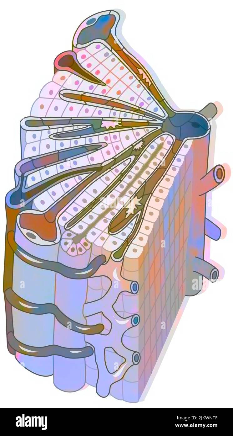 Hepatic lobule consisting of hepatocytes, a central vein, centrilobular vein. Stock Photohttps://www.alamy.com/image-license-details/?v=1https://www.alamy.com/hepatic-lobule-consisting-of-hepatocytes-a-central-vein-centrilobular-vein-image476924351.html
RF2JKWNTF–Hepatic lobule consisting of hepatocytes, a central vein, centrilobular vein.
 Vegetable and liver salad. Stock Photohttps://www.alamy.com/image-license-details/?v=1https://www.alamy.com/vegetable-and-liver-salad-image402534697.html
RF2EAW135–Vegetable and liver salad.
 Vegetable and liver salad. Stock Photohttps://www.alamy.com/image-license-details/?v=1https://www.alamy.com/vegetable-and-liver-salad-image402537171.html
RF2EAW47F–Vegetable and liver salad.
 This illustration represents Hepatic Lobules of the Liver, vintage line drawing or engraving illustration. Stock Vectorhttps://www.alamy.com/image-license-details/?v=1https://www.alamy.com/this-illustration-represents-hepatic-lobules-of-the-liver-vintage-line-drawing-or-engraving-illustration-image348655154.html
RF2B76H5P–This illustration represents Hepatic Lobules of the Liver, vintage line drawing or engraving illustration.
 Hepatitis c, hcv Stock Photohttps://www.alamy.com/image-license-details/?v=1https://www.alamy.com/hepatitis-c-hcv-image338279345.html
RM2AJ9XN5–Hepatitis c, hcv
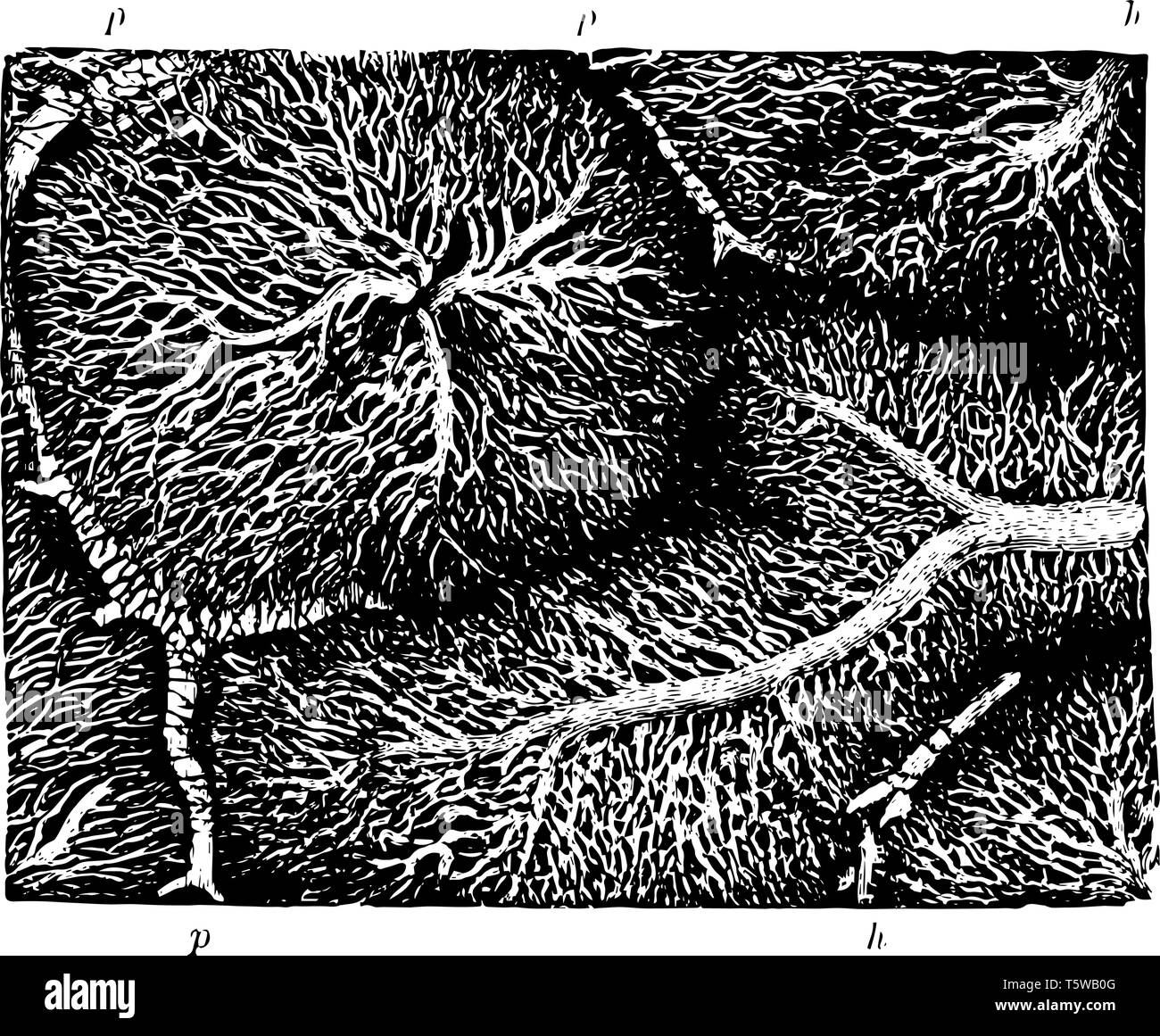 This illustration represents Capillary Network in the Lobules of a Rabbit Liver vintage line drawing or engraving illustration. Stock Vectorhttps://www.alamy.com/image-license-details/?v=1https://www.alamy.com/this-illustration-represents-capillary-network-in-the-lobules-of-a-rabbit-liver-vintage-line-drawing-or-engraving-illustration-image244575872.html
RFT5WB0G–This illustration represents Capillary Network in the Lobules of a Rabbit Liver vintage line drawing or engraving illustration.
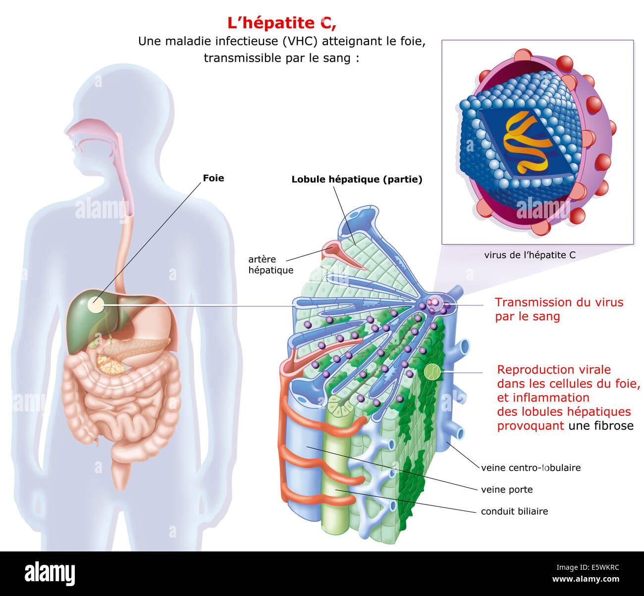 Hepatitis c drawing Stock Photohttps://www.alamy.com/image-license-details/?v=1https://www.alamy.com/stock-photo-hepatitis-c-drawing-72479104.html
RME5WKRC–Hepatitis c drawing
 Warm salad with chicken liver and vegetables.Chicken liver salad Stock Photohttps://www.alamy.com/image-license-details/?v=1https://www.alamy.com/warm-salad-with-chicken-liver-and-vegetableschicken-liver-salad-image395454569.html
RF2DYAE9D–Warm salad with chicken liver and vegetables.Chicken liver salad
 Warm salad with chicken liver and vegetables.Chicken liver salad Stock Photohttps://www.alamy.com/image-license-details/?v=1https://www.alamy.com/warm-salad-with-chicken-liver-and-vegetableschicken-liver-salad-image447007811.html
RF2GY6Y0K–Warm salad with chicken liver and vegetables.Chicken liver salad
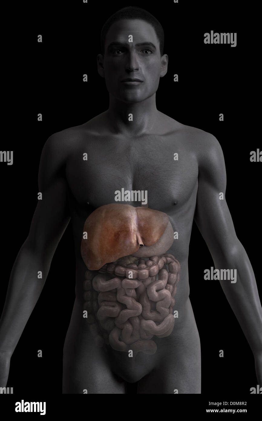 Front view of the liver within a male figure. Stock Photohttps://www.alamy.com/image-license-details/?v=1https://www.alamy.com/stock-photo-front-view-of-the-liver-within-a-male-figure-52077062.html
RMD0M8R2–Front view of the liver within a male figure.
 Salad with liver,bell pepper,okra and herbs.Chicken liver salad Stock Photohttps://www.alamy.com/image-license-details/?v=1https://www.alamy.com/salad-with-liverbell-pepperokra-and-herbschicken-liver-salad-image447007818.html
RF2GY6Y0X–Salad with liver,bell pepper,okra and herbs.Chicken liver salad
 Fish anatomy. Pike (Esox lucius). The hepatic parenchyma is divided into lobules and covered with a fibrous membrane. Digestive, metabolic and protect Stock Photohttps://www.alamy.com/image-license-details/?v=1https://www.alamy.com/fish-anatomy-pike-esox-lucius-the-hepatic-parenchyma-is-divided-into-lobules-and-covered-with-a-fibrous-membrane-digestive-metabolic-and-protect-image607392804.html
RF2X853M4–Fish anatomy. Pike (Esox lucius). The hepatic parenchyma is divided into lobules and covered with a fibrous membrane. Digestive, metabolic and protect
 Salad with liver,bell pepper,okra and herbs.Chicken liver salad Stock Photohttps://www.alamy.com/image-license-details/?v=1https://www.alamy.com/salad-with-liverbell-pepperokra-and-herbschicken-liver-salad-image447007813.html
RF2GY6Y0N–Salad with liver,bell pepper,okra and herbs.Chicken liver salad
 Textbook of normal histology: including an account of the development of the tissues and of the organs . he tubes inthe formation of thecords of cells. The fibrous tissueenveloping the exteriorof the liver is prolongedinto the interior of theorgan through thetransverse fissure, incompany with theblood-vessels and thebile - ducts. The de-marcation of the indi-vidual lobules depends upon the development of this interlobularconnective tissue, known as the capsule of Glisson; when welldeveloped, as in the liver of the hog, the lobules are defined withgreat distinctness, being completely surrounded Stock Photohttps://www.alamy.com/image-license-details/?v=1https://www.alamy.com/textbook-of-normal-histology-including-an-account-of-the-development-of-the-tissues-and-of-the-organs-he-tubes-inthe-formation-of-thecords-of-cells-the-fibrous-tissueenveloping-the-exteriorof-the-liver-is-prolongedinto-the-interior-of-theorgan-through-thetransverse-fissure-incompany-with-theblood-vessels-and-thebile-ducts-the-de-marcation-of-the-indi-vidual-lobules-depends-upon-the-development-of-this-interlobularconnective-tissue-known-as-the-capsule-of-glisson-when-welldeveloped-as-in-the-liver-of-the-hog-the-lobules-are-defined-withgreat-distinctness-being-completely-surrounded-image338958999.html
RM2AKCWJF–Textbook of normal histology: including an account of the development of the tissues and of the organs . he tubes inthe formation of thecords of cells. The fibrous tissueenveloping the exteriorof the liver is prolongedinto the interior of theorgan through thetransverse fissure, incompany with theblood-vessels and thebile - ducts. The de-marcation of the indi-vidual lobules depends upon the development of this interlobularconnective tissue, known as the capsule of Glisson; when welldeveloped, as in the liver of the hog, the lobules are defined withgreat distinctness, being completely surrounded
 Salad with liver,bell pepper,okra and herbs.Healthy salad with liver Stock Photohttps://www.alamy.com/image-license-details/?v=1https://www.alamy.com/salad-with-liverbell-pepperokra-and-herbshealthy-salad-with-liver-image447007816.html
RF2GY6Y0T–Salad with liver,bell pepper,okra and herbs.Healthy salad with liver
 . Cunningham's Text-book of anatomy. Anatomy. Central vein Central vein Sublobular vein Fig 940.—Liver of a Pig injected from the Hepatic Vein by T. A. Carter. (From a specimen left in the Anatomical Department of Edinburgh University by Sir William Turner.) Liver lobules of delicate fibrous tissue, which is most evident where the serous coat is absent. In the neighbourhood of the porta hepatis it is particularly abundant, and here it surrounds the vessels entering the porta, and accompanies them through the portal canals in the liver substance. This coat is continuous with the fine areolar Stock Photohttps://www.alamy.com/image-license-details/?v=1https://www.alamy.com/cunninghams-text-book-of-anatomy-anatomy-central-vein-central-vein-sublobular-vein-fig-940liver-of-a-pig-injected-from-the-hepatic-vein-by-t-a-carter-from-a-specimen-left-in-the-anatomical-department-of-edinburgh-university-by-sir-william-turner-liver-lobules-of-delicate-fibrous-tissue-which-is-most-evident-where-the-serous-coat-is-absent-in-the-neighbourhood-of-the-porta-hepatis-it-is-particularly-abundant-and-here-it-surrounds-the-vessels-entering-the-porta-and-accompanies-them-through-the-portal-canals-in-the-liver-substance-this-coat-is-continuous-with-the-fine-areolar-image216340157.html
RMPFY425–. Cunningham's Text-book of anatomy. Anatomy. Central vein Central vein Sublobular vein Fig 940.—Liver of a Pig injected from the Hepatic Vein by T. A. Carter. (From a specimen left in the Anatomical Department of Edinburgh University by Sir William Turner.) Liver lobules of delicate fibrous tissue, which is most evident where the serous coat is absent. In the neighbourhood of the porta hepatis it is particularly abundant, and here it surrounds the vessels entering the porta, and accompanies them through the portal canals in the liver substance. This coat is continuous with the fine areolar
 Archive image from page 178 of The cyclopædia of anatomy and. The cyclopædia of anatomy and physiology cyclopdiaofana03todd Year: 1847 NORMAL ANATOMY OF THE LIVER. 165 called Glisson's capsule, of the ramifications of tlie portal vein, hepatic duct, hepatic artery, hepatic veins, lymphatics and nerves. For an accurate knowledge of these different structures, anatomy is indebted to the labours of Mr. Kienian, to whose paper on ' The Anatomy and Physiology of the Liver,' con- tained in the Philosophical Transactions for 1833, I shall have constant occasion to refer. The small bodies (lobules, Stock Photohttps://www.alamy.com/image-license-details/?v=1https://www.alamy.com/archive-image-from-page-178-of-the-cyclopdia-of-anatomy-and-the-cyclopdia-of-anatomy-and-physiology-cyclopdiaofana03todd-year-1847-normal-anatomy-of-the-liver-165-called-glissons-capsule-of-the-ramifications-of-tlie-portal-vein-hepatic-duct-hepatic-artery-hepatic-veins-lymphatics-and-nerves-for-an-accurate-knowledge-of-these-different-structures-anatomy-is-indebted-to-the-labours-of-mr-kienian-to-whose-paper-on-the-anatomy-and-physiology-of-the-liver-con-tained-in-the-philosophical-transactions-for-1833-i-shall-have-constant-occasion-to-refer-the-small-bodies-lobules-image259468519.html
RMW23PMR–Archive image from page 178 of The cyclopædia of anatomy and. The cyclopædia of anatomy and physiology cyclopdiaofana03todd Year: 1847 NORMAL ANATOMY OF THE LIVER. 165 called Glisson's capsule, of the ramifications of tlie portal vein, hepatic duct, hepatic artery, hepatic veins, lymphatics and nerves. For an accurate knowledge of these different structures, anatomy is indebted to the labours of Mr. Kienian, to whose paper on ' The Anatomy and Physiology of the Liver,' con- tained in the Philosophical Transactions for 1833, I shall have constant occasion to refer. The small bodies (lobules,
 . Elementary physiology . Fig. 70.—Section of the pancreas of the dog. (Klein.) d, termination of a duct in the tubular alveolus, a. salivary gland and of the pancreas of the dog. In the salivary glands the ducts are more numerous than in the pancreas. The alveoli, or acini, are also much shorter and more rounded in the salivary glands than in the pancreas, where they form long columns of cells. The bile duct, which carries the bile from the liver to the duodenum, arises in a similar branching fashion from the lobules of the liver. But the secretion of bile is not the main function of the live Stock Photohttps://www.alamy.com/image-license-details/?v=1https://www.alamy.com/elementary-physiology-fig-70section-of-the-pancreas-of-the-dog-klein-d-termination-of-a-duct-in-the-tubular-alveolus-a-salivary-gland-and-of-the-pancreas-of-the-dog-in-the-salivary-glands-the-ducts-are-more-numerous-than-in-the-pancreas-the-alveoli-or-acini-are-also-much-shorter-and-more-rounded-in-the-salivary-glands-than-in-the-pancreas-where-they-form-long-columns-of-cells-the-bile-duct-which-carries-the-bile-from-the-liver-to-the-duodenum-arises-in-a-similar-branching-fashion-from-the-lobules-of-the-liver-but-the-secretion-of-bile-is-not-the-main-function-of-the-live-image178403514.html
RMMA6YE2–. Elementary physiology . Fig. 70.—Section of the pancreas of the dog. (Klein.) d, termination of a duct in the tubular alveolus, a. salivary gland and of the pancreas of the dog. In the salivary glands the ducts are more numerous than in the pancreas. The alveoli, or acini, are also much shorter and more rounded in the salivary glands than in the pancreas, where they form long columns of cells. The bile duct, which carries the bile from the liver to the duodenum, arises in a similar branching fashion from the lobules of the liver. But the secretion of bile is not the main function of the live
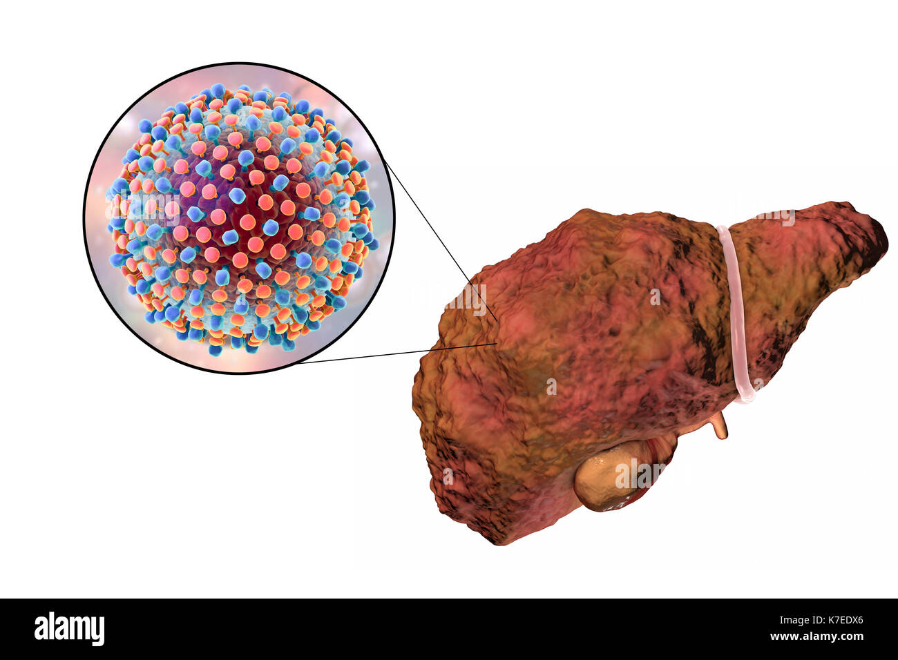 Computer illustration showing a cirrhotic liver and a close-up of hepatitis C viruses. Hepatitis C is a common cause of chronic hepatitis which progresses to liver cirrhosis. Viruses cause cell death (necrosis) in the hepatic lobules, which leads to scarring. This causes the liver surface to appear rough and lumpy instead of the usual smooth healthy appearance. The disease is irreversible and may lead to liver carcinoma. Stock Photohttps://www.alamy.com/image-license-details/?v=1https://www.alamy.com/computer-illustration-showing-a-cirrhotic-liver-and-a-close-up-of-image159514158.html
RFK7EDX6–Computer illustration showing a cirrhotic liver and a close-up of hepatitis C viruses. Hepatitis C is a common cause of chronic hepatitis which progresses to liver cirrhosis. Viruses cause cell death (necrosis) in the hepatic lobules, which leads to scarring. This causes the liver surface to appear rough and lumpy instead of the usual smooth healthy appearance. The disease is irreversible and may lead to liver carcinoma.
 Vegetable and liver salad. Stock Photohttps://www.alamy.com/image-license-details/?v=1https://www.alamy.com/vegetable-and-liver-salad-image402534704.html
RF2EAW13C–Vegetable and liver salad.
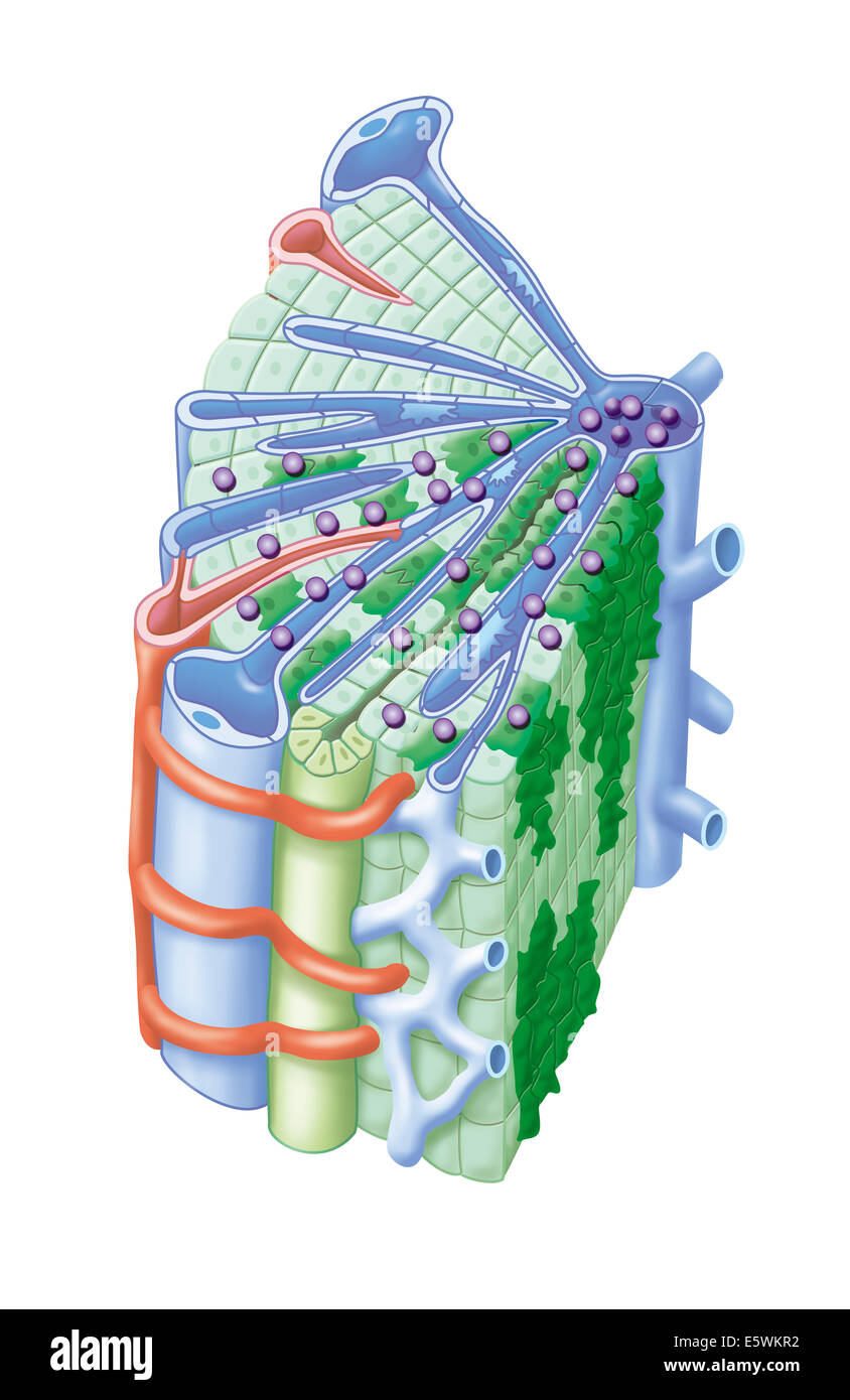 Hepatitis c drawing Stock Photohttps://www.alamy.com/image-license-details/?v=1https://www.alamy.com/stock-photo-hepatitis-c-drawing-72479094.html
RME5WKR2–Hepatitis c drawing
 Warm salad with chicken liver and vegetables.Chicken liver salad Stock Photohttps://www.alamy.com/image-license-details/?v=1https://www.alamy.com/warm-salad-with-chicken-liver-and-vegetableschicken-liver-salad-image394974271.html
RF2DXGHKY–Warm salad with chicken liver and vegetables.Chicken liver salad
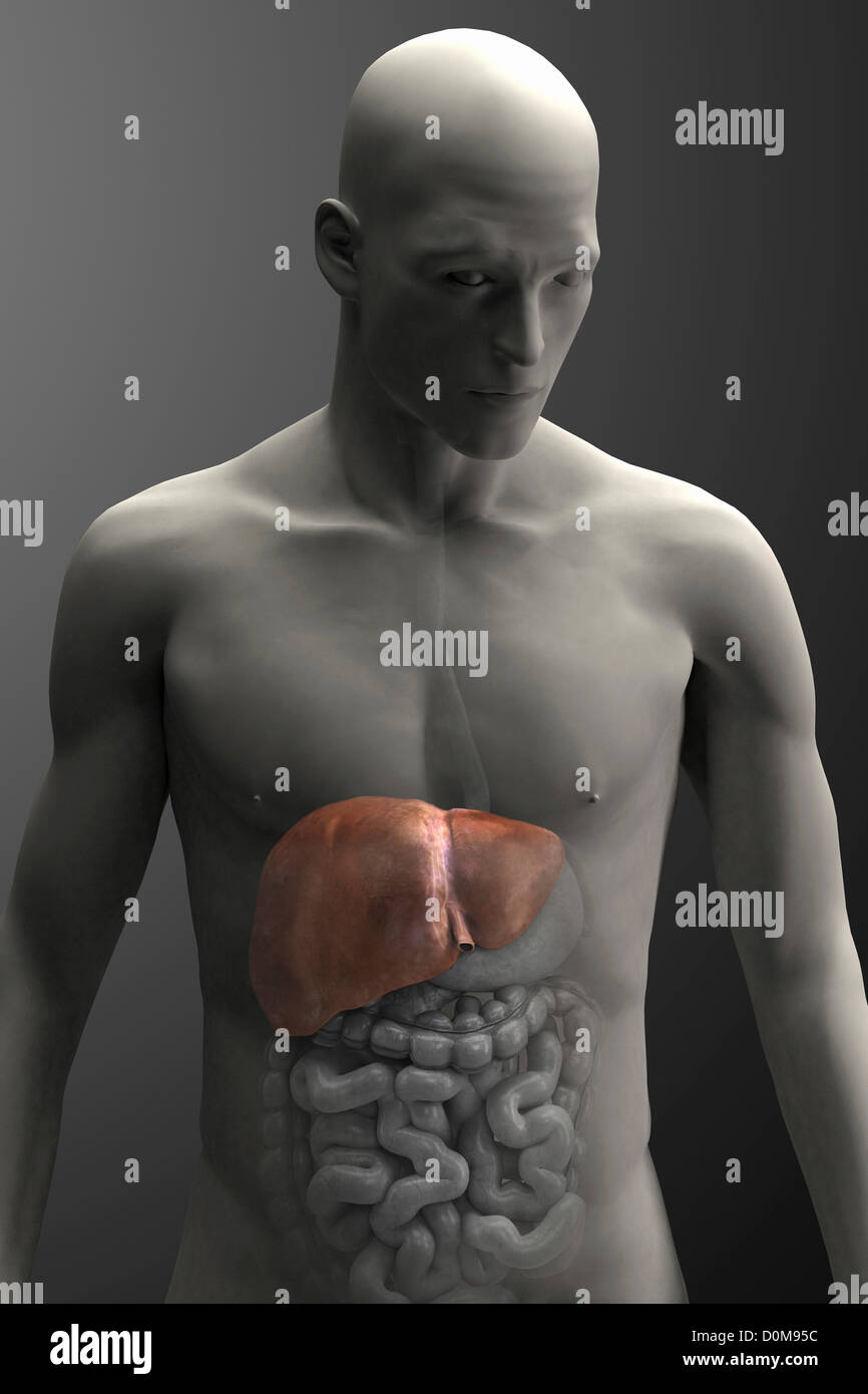 Human liver highlighted within a male figure (front view). Stock Photohttps://www.alamy.com/image-license-details/?v=1https://www.alamy.com/stock-photo-human-liver-highlighted-within-a-male-figure-front-view-52077352.html
RMD0M95C–Human liver highlighted within a male figure (front view).
 Annals of surgery . glands are irregular in shape and size, and are linedwith a varying number of ill-formed columnar cells. The fibrouscoat is thickened, and shows but little stroma between the glands.In some places the lumen of the glands is entirely filled witliepithelial cells. Figure 2.—Primary carcinoma of the gall-bladder. CaseIII.—This section includes the gall-bladder wall and a portionof the adjacent liver-tissue. The pathologic findings are de-scribed by Dr. John E. Hays as follows: The liver lobules showfatty degeneration and passive congestion. The adventitiouscoat of the blood-ve Stock Photohttps://www.alamy.com/image-license-details/?v=1https://www.alamy.com/annals-of-surgery-glands-are-irregular-in-shape-and-size-and-are-linedwith-a-varying-number-of-ill-formed-columnar-cells-the-fibrouscoat-is-thickened-and-shows-but-little-stroma-between-the-glandsin-some-places-the-lumen-of-the-glands-is-entirely-filled-witliepithelial-cells-figure-2primary-carcinoma-of-the-gall-bladder-caseiiithis-section-includes-the-gall-bladder-wall-and-a-portionof-the-adjacent-liver-tissue-the-pathologic-findings-are-de-scribed-by-dr-john-e-hays-as-follows-the-liver-lobules-showfatty-degeneration-and-passive-congestion-the-adventitiouscoat-of-the-blood-ve-image338275557.html
RM2AJ9NWW–Annals of surgery . glands are irregular in shape and size, and are linedwith a varying number of ill-formed columnar cells. The fibrouscoat is thickened, and shows but little stroma between the glands.In some places the lumen of the glands is entirely filled witliepithelial cells. Figure 2.—Primary carcinoma of the gall-bladder. CaseIII.—This section includes the gall-bladder wall and a portionof the adjacent liver-tissue. The pathologic findings are de-scribed by Dr. John E. Hays as follows: The liver lobules showfatty degeneration and passive congestion. The adventitiouscoat of the blood-ve
![. Cunningham's Text-book of anatomy. Anatomy. 1198 THE DIGESTIVE SYSTEM. Stkucture of the Liver. The liver is invested by an outer tunica serosa described in connexion with the peritoneum. Within this is a thin capsula fibrosa [Glissonii] (O.T. G-lisson's capsule) Intralobular capillary plexus Intralobular capillary plexus. Central vein Central vein Sublobular vein Fig 940.—Liver of a Pig injected from the Hepatic Vein by T. A. Carter. (From a specimen left in the Anatomical Department of Edinburgh University by Sir William Turner.) Liver lobules of delicate fibrous tissue, which is most evi Stock Photo . Cunningham's Text-book of anatomy. Anatomy. 1198 THE DIGESTIVE SYSTEM. Stkucture of the Liver. The liver is invested by an outer tunica serosa described in connexion with the peritoneum. Within this is a thin capsula fibrosa [Glissonii] (O.T. G-lisson's capsule) Intralobular capillary plexus Intralobular capillary plexus. Central vein Central vein Sublobular vein Fig 940.—Liver of a Pig injected from the Hepatic Vein by T. A. Carter. (From a specimen left in the Anatomical Department of Edinburgh University by Sir William Turner.) Liver lobules of delicate fibrous tissue, which is most evi Stock Photo](https://c8.alamy.com/comp/PFY427/cunninghams-text-book-of-anatomy-anatomy-1198-the-digestive-system-stkucture-of-the-liver-the-liver-is-invested-by-an-outer-tunica-serosa-described-in-connexion-with-the-peritoneum-within-this-is-a-thin-capsula-fibrosa-glissonii-ot-g-lissons-capsule-intralobular-capillary-plexus-intralobular-capillary-plexus-central-vein-central-vein-sublobular-vein-fig-940liver-of-a-pig-injected-from-the-hepatic-vein-by-t-a-carter-from-a-specimen-left-in-the-anatomical-department-of-edinburgh-university-by-sir-william-turner-liver-lobules-of-delicate-fibrous-tissue-which-is-most-evi-PFY427.jpg) . Cunningham's Text-book of anatomy. Anatomy. 1198 THE DIGESTIVE SYSTEM. Stkucture of the Liver. The liver is invested by an outer tunica serosa described in connexion with the peritoneum. Within this is a thin capsula fibrosa [Glissonii] (O.T. G-lisson's capsule) Intralobular capillary plexus Intralobular capillary plexus. Central vein Central vein Sublobular vein Fig 940.—Liver of a Pig injected from the Hepatic Vein by T. A. Carter. (From a specimen left in the Anatomical Department of Edinburgh University by Sir William Turner.) Liver lobules of delicate fibrous tissue, which is most evi Stock Photohttps://www.alamy.com/image-license-details/?v=1https://www.alamy.com/cunninghams-text-book-of-anatomy-anatomy-1198-the-digestive-system-stkucture-of-the-liver-the-liver-is-invested-by-an-outer-tunica-serosa-described-in-connexion-with-the-peritoneum-within-this-is-a-thin-capsula-fibrosa-glissonii-ot-g-lissons-capsule-intralobular-capillary-plexus-intralobular-capillary-plexus-central-vein-central-vein-sublobular-vein-fig-940liver-of-a-pig-injected-from-the-hepatic-vein-by-t-a-carter-from-a-specimen-left-in-the-anatomical-department-of-edinburgh-university-by-sir-william-turner-liver-lobules-of-delicate-fibrous-tissue-which-is-most-evi-image216340159.html
. Cunningham's Text-book of anatomy. Anatomy. 1198 THE DIGESTIVE SYSTEM. Stkucture of the Liver. The liver is invested by an outer tunica serosa described in connexion with the peritoneum. Within this is a thin capsula fibrosa [Glissonii] (O.T. G-lisson's capsule) Intralobular capillary plexus Intralobular capillary plexus. Central vein Central vein Sublobular vein Fig 940.—Liver of a Pig injected from the Hepatic Vein by T. A. Carter. (From a specimen left in the Anatomical Department of Edinburgh University by Sir William Turner.) Liver lobules of delicate fibrous tissue, which is most evi Stock Photohttps://www.alamy.com/image-license-details/?v=1https://www.alamy.com/cunninghams-text-book-of-anatomy-anatomy-1198-the-digestive-system-stkucture-of-the-liver-the-liver-is-invested-by-an-outer-tunica-serosa-described-in-connexion-with-the-peritoneum-within-this-is-a-thin-capsula-fibrosa-glissonii-ot-g-lissons-capsule-intralobular-capillary-plexus-intralobular-capillary-plexus-central-vein-central-vein-sublobular-vein-fig-940liver-of-a-pig-injected-from-the-hepatic-vein-by-t-a-carter-from-a-specimen-left-in-the-anatomical-department-of-edinburgh-university-by-sir-william-turner-liver-lobules-of-delicate-fibrous-tissue-which-is-most-evi-image216340159.htmlRMPFY427–. Cunningham's Text-book of anatomy. Anatomy. 1198 THE DIGESTIVE SYSTEM. Stkucture of the Liver. The liver is invested by an outer tunica serosa described in connexion with the peritoneum. Within this is a thin capsula fibrosa [Glissonii] (O.T. G-lisson's capsule) Intralobular capillary plexus Intralobular capillary plexus. Central vein Central vein Sublobular vein Fig 940.—Liver of a Pig injected from the Hepatic Vein by T. A. Carter. (From a specimen left in the Anatomical Department of Edinburgh University by Sir William Turner.) Liver lobules of delicate fibrous tissue, which is most evi
 Archive image from page 1232 of Cunningham's Text-book of anatomy (1914). Cunningham's Text-book of anatomy cunninghamstextb00cunn Year: 1914 ( VESSELS OF THE LIVER. 1199 number of thorns growing out on all sides from the sublobular twigs of the tree). On each of these little central veins there is impaled, as it were, a lobule. These little conical lobules, with their central veins running through them, are so numerous and so closely packed together, that they give rise to the practically solid liver tissue. The lobules are surrounded by the vense interlobulares, branches of the portal vein, Stock Photohttps://www.alamy.com/image-license-details/?v=1https://www.alamy.com/archive-image-from-page-1232-of-cunninghams-text-book-of-anatomy-1914-cunninghams-text-book-of-anatomy-cunninghamstextb00cunn-year-1914-vessels-of-the-liver-1199-number-of-thorns-growing-out-on-all-sides-from-the-sublobular-twigs-of-the-tree-on-each-of-these-little-central-veins-there-is-impaled-as-it-were-a-lobule-these-little-conical-lobules-with-their-central-veins-running-through-them-are-so-numerous-and-so-closely-packed-together-that-they-give-rise-to-the-practically-solid-liver-tissue-the-lobules-are-surrounded-by-the-vense-interlobulares-branches-of-the-portal-vein-image264068615.html
RMW9HA5Y–Archive image from page 1232 of Cunningham's Text-book of anatomy (1914). Cunningham's Text-book of anatomy cunninghamstextb00cunn Year: 1914 ( VESSELS OF THE LIVER. 1199 number of thorns growing out on all sides from the sublobular twigs of the tree). On each of these little central veins there is impaled, as it were, a lobule. These little conical lobules, with their central veins running through them, are so numerous and so closely packed together, that they give rise to the practically solid liver tissue. The lobules are surrounded by the vense interlobulares, branches of the portal vein,
 . Elementary physiology . Fir 8a —Section of a portion of liver passing longitudinally through a considerable hepatic vein, from the pig (after Kiernan). (About 5 diameters.) H heoatic venous trunk, against which the sides of the lobules are applied ; h, h, h ' three sublobular hepatic veins, on which the bases of the lobules rest and through coats of which they are seen as polygonal figures ; /, mouth of the nitralobular veins, opening into the sublobular veins ; z', intralobular veins shown passing up the centre Sf some divided lobules; c, c, walls of the hepatic venous canal, with the polyg Stock Photohttps://www.alamy.com/image-license-details/?v=1https://www.alamy.com/elementary-physiology-fir-8a-section-of-a-portion-of-liver-passing-longitudinally-through-a-considerable-hepatic-vein-from-the-pig-after-kiernan-about-5-diameters-h-heoatic-venous-trunk-against-which-the-sides-of-the-lobules-are-applied-h-h-h-three-sublobular-hepatic-veins-on-which-the-bases-of-the-lobules-rest-and-through-coats-of-which-they-are-seen-as-polygonal-figures-mouth-of-the-nitralobular-veins-opening-into-the-sublobular-veins-z-intralobular-veins-shown-passing-up-the-centre-sf-some-divided-lobules-c-c-walls-of-the-hepatic-venous-canal-with-the-polyg-image178403503.html
RMMA6YDK–. Elementary physiology . Fir 8a —Section of a portion of liver passing longitudinally through a considerable hepatic vein, from the pig (after Kiernan). (About 5 diameters.) H heoatic venous trunk, against which the sides of the lobules are applied ; h, h, h ' three sublobular hepatic veins, on which the bases of the lobules rest and through coats of which they are seen as polygonal figures ; /, mouth of the nitralobular veins, opening into the sublobular veins ; z', intralobular veins shown passing up the centre Sf some divided lobules; c, c, walls of the hepatic venous canal, with the polyg
 Computer illustration showing a cirrhotic liver and a close-up of hepatitis C viruses. Hepatitis C is a common cause of chronic hepatitis which progresses to liver cirrhosis. Viruses cause cell death (necrosis) in the hepatic lobules, which leads to scarring. This causes the liver surface to appear rough and lumpy instead of the usual smooth healthy appearance. The disease is irreversible and may lead to liver carcinoma. Stock Photohttps://www.alamy.com/image-license-details/?v=1https://www.alamy.com/computer-illustration-showing-a-cirrhotic-liver-and-a-close-up-of-image159514157.html
RFK7EDX5–Computer illustration showing a cirrhotic liver and a close-up of hepatitis C viruses. Hepatitis C is a common cause of chronic hepatitis which progresses to liver cirrhosis. Viruses cause cell death (necrosis) in the hepatic lobules, which leads to scarring. This causes the liver surface to appear rough and lumpy instead of the usual smooth healthy appearance. The disease is irreversible and may lead to liver carcinoma.
 Vegetable and liver salad. Stock Photohttps://www.alamy.com/image-license-details/?v=1https://www.alamy.com/vegetable-and-liver-salad-image402534707.html
RF2EAW13F–Vegetable and liver salad.
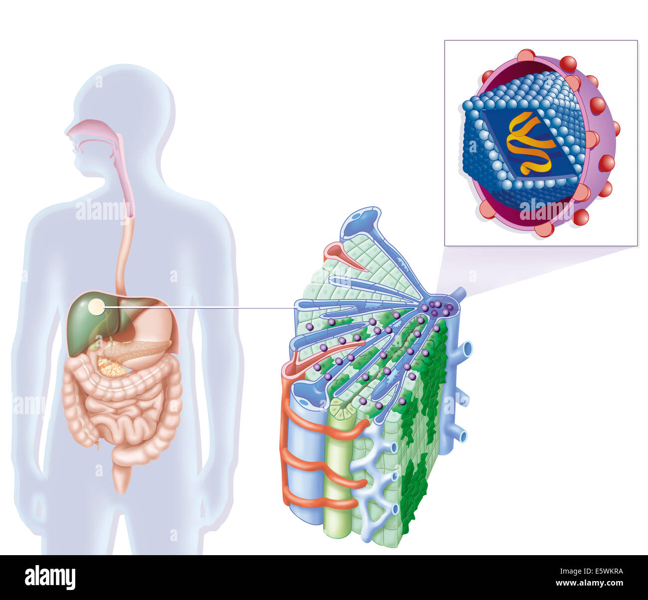 Hepatitis c drawing Stock Photohttps://www.alamy.com/image-license-details/?v=1https://www.alamy.com/stock-photo-hepatitis-c-drawing-72479102.html
RME5WKRA–Hepatitis c drawing
 Warm salad with chicken liver and vegetables.Chicken liver salad Stock Photohttps://www.alamy.com/image-license-details/?v=1https://www.alamy.com/warm-salad-with-chicken-liver-and-vegetableschicken-liver-salad-image396440888.html
RF2E0YCB4–Warm salad with chicken liver and vegetables.Chicken liver salad
 Vue histologique des lobules hépatiques sain avec, à droite, des lobules hépatiques infectés (plus clairs) présentant une inflammation et une fibrose. Chaque lobule, grossièrement hexagonal, comporte en son centre une veinule centrolobulaire et une triade portale à chaque angle (branche de veine porte, branche d'artère hépatique, canal biliaire. Des sinusoïdes veineux (bleu) séparent les hépatocytes en lames hépatocytaires en se dirigeant vers la veine centrale du lobule. Stock Photohttps://www.alamy.com/image-license-details/?v=1https://www.alamy.com/vue-histologique-des-lobules-hpatiques-sain-avec-droite-des-lobules-hpatiques-infects-plus-clairs-prsentant-une-inflammation-et-une-fibrose-chaque-lobule-grossirement-hexagonal-comporte-en-son-centre-une-veinule-centrolobulaire-et-une-triade-portale-chaque-angle-branche-de-veine-porte-branche-dartre-hpatique-canal-biliaire-des-sinusodes-veineux-bleu-sparent-les-hpatocytes-en-lames-hpatocytaires-en-se-dirigeant-vers-la-veine-centrale-du-lobule-image338279348.html
RM2AJ9XN8–Vue histologique des lobules hépatiques sain avec, à droite, des lobules hépatiques infectés (plus clairs) présentant une inflammation et une fibrose. Chaque lobule, grossièrement hexagonal, comporte en son centre une veinule centrolobulaire et une triade portale à chaque angle (branche de veine porte, branche d'artère hépatique, canal biliaire. Des sinusoïdes veineux (bleu) séparent les hépatocytes en lames hépatocytaires en se dirigeant vers la veine centrale du lobule.
 Close-up of the liver which is faded to show the position of the gallbladder relative to the stomach and intestines. Stock Photohttps://www.alamy.com/image-license-details/?v=1https://www.alamy.com/stock-photo-close-up-of-the-liver-which-is-faded-to-show-the-position-of-the-gallbladder-52076834.html
RMD0M8EX–Close-up of the liver which is faded to show the position of the gallbladder relative to the stomach and intestines.
 Influence of vegetables greened with copper salts on the nutrition and health of man . en, heart muscle, lungs,and spinal cord. Section of large intestme includes a worm-nodule in submucoustissue. Sinusoids of liver lobules barely indicated. Cells staincompactly. In kidney sections, groups of necrotic epithelial cells incross section of tubules. Some small endothehal-cell foci also presentin interstitial tissue. In spleen a moderate increase in polynuclearleucocytes. Alveoli in section of lungs filled with a finely granularreddish stained material. In these animals the microscopic examination, Stock Photohttps://www.alamy.com/image-license-details/?v=1https://www.alamy.com/influence-of-vegetables-greened-with-copper-salts-on-the-nutrition-and-health-of-man-en-heart-muscle-lungsand-spinal-cord-section-of-large-intestme-includes-a-worm-nodule-in-submucoustissue-sinusoids-of-liver-lobules-barely-indicated-cells-staincompactly-in-kidney-sections-groups-of-necrotic-epithelial-cells-incross-section-of-tubules-some-small-endothehal-cell-foci-also-presentin-interstitial-tissue-in-spleen-a-moderate-increase-in-polynuclearleucocytes-alveoli-in-section-of-lungs-filled-with-a-finely-granularreddish-stained-material-in-these-animals-the-microscopic-examination-image340302563.html
RM2ANJ3AY–Influence of vegetables greened with copper salts on the nutrition and health of man . en, heart muscle, lungs,and spinal cord. Section of large intestme includes a worm-nodule in submucoustissue. Sinusoids of liver lobules barely indicated. Cells staincompactly. In kidney sections, groups of necrotic epithelial cells incross section of tubules. Some small endothehal-cell foci also presentin interstitial tissue. In spleen a moderate increase in polynuclearleucocytes. Alveoli in section of lungs filled with a finely granularreddish stained material. In these animals the microscopic examination,
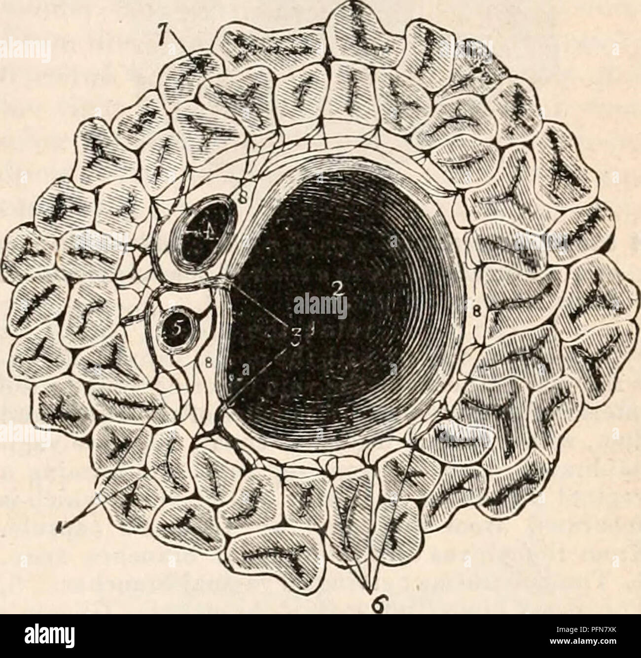 . The cyclopædia of anatomy and physiology. Anatomy; Physiology; Zoology. NORMAL ANATOMY OF THE LIVER. 167 omentum, and which accompanies them along the portal canals and interlobular fissures to their ultimate distribution in the substance of the lobules. It forms for each of the lobules a distinct capsule which invests it on all sides with the exception of its base, and is then ex- panded over the whole of the exterior of the organ, constituting the proper capsule of the liver. Glisson's capsule serves to maintain the portal vein, hepatic artery, and hepatic ducts in connection with each oth Stock Photohttps://www.alamy.com/image-license-details/?v=1https://www.alamy.com/the-cyclopdia-of-anatomy-and-physiology-anatomy-physiology-zoology-normal-anatomy-of-the-liver-167-omentum-and-which-accompanies-them-along-the-portal-canals-and-interlobular-fissures-to-their-ultimate-distribution-in-the-substance-of-the-lobules-it-forms-for-each-of-the-lobules-a-distinct-capsule-which-invests-it-on-all-sides-with-the-exception-of-its-base-and-is-then-ex-panded-over-the-whole-of-the-exterior-of-the-organ-constituting-the-proper-capsule-of-the-liver-glissons-capsule-serves-to-maintain-the-portal-vein-hepatic-artery-and-hepatic-ducts-in-connection-with-each-oth-image216211483.html
RMPFN7XK–. The cyclopædia of anatomy and physiology. Anatomy; Physiology; Zoology. NORMAL ANATOMY OF THE LIVER. 167 omentum, and which accompanies them along the portal canals and interlobular fissures to their ultimate distribution in the substance of the lobules. It forms for each of the lobules a distinct capsule which invests it on all sides with the exception of its base, and is then ex- panded over the whole of the exterior of the organ, constituting the proper capsule of the liver. Glisson's capsule serves to maintain the portal vein, hepatic artery, and hepatic ducts in connection with each oth
 Archive image from page 182 of The cyclopædia of anatomy and. The cyclopædia of anatomy and physiology cyclopdiaofana03todd Year: 1847 NORMAL ANATOMY OF THE LIVER. 169 Fig. 39. Two lobules, in which the portal venous plexus is seen. After Kiernan. a a, Interlobular veins. The appearance of venous circles formed by these veins is that which is afforded by a common lens ; when examined with a higher power the interlobular fissure is seen to be filled by a vascular plexus, b, The lobular venous plexus. The circular and ovoid spaces seen be- tween the branches of the plexuses are occupied by Stock Photohttps://www.alamy.com/image-license-details/?v=1https://www.alamy.com/archive-image-from-page-182-of-the-cyclopdia-of-anatomy-and-the-cyclopdia-of-anatomy-and-physiology-cyclopdiaofana03todd-year-1847-normal-anatomy-of-the-liver-169-fig-39-two-lobules-in-which-the-portal-venous-plexus-is-seen-after-kiernan-a-a-interlobular-veins-the-appearance-of-venous-circles-formed-by-these-veins-is-that-which-is-afforded-by-a-common-lens-when-examined-with-a-higher-power-the-interlobular-fissure-is-seen-to-be-filled-by-a-vascular-plexus-b-the-lobular-venous-plexus-the-circular-and-ovoid-spaces-seen-be-tween-the-branches-of-the-plexuses-are-occupied-by-image259469799.html
RMW23TAF–Archive image from page 182 of The cyclopædia of anatomy and. The cyclopædia of anatomy and physiology cyclopdiaofana03todd Year: 1847 NORMAL ANATOMY OF THE LIVER. 169 Fig. 39. Two lobules, in which the portal venous plexus is seen. After Kiernan. a a, Interlobular veins. The appearance of venous circles formed by these veins is that which is afforded by a common lens ; when examined with a higher power the interlobular fissure is seen to be filled by a vascular plexus, b, The lobular venous plexus. The circular and ovoid spaces seen be- tween the branches of the plexuses are occupied by
 Vegetable and liver salad. Stock Photohttps://www.alamy.com/image-license-details/?v=1https://www.alamy.com/vegetable-and-liver-salad-image402534713.html
RF2EAW13N–Vegetable and liver salad.
 Warm salad with chicken liver and vegetables.Chicken liver salad Stock Photohttps://www.alamy.com/image-license-details/?v=1https://www.alamy.com/warm-salad-with-chicken-liver-and-vegetableschicken-liver-salad-image394973626.html
RF2DXGGTX–Warm salad with chicken liver and vegetables.Chicken liver salad
 Hepatitis c, hcv Stock Photohttps://www.alamy.com/image-license-details/?v=1https://www.alamy.com/hepatitis-c-hcv-image338279371.html
RM2AJ9XP3–Hepatitis c, hcv
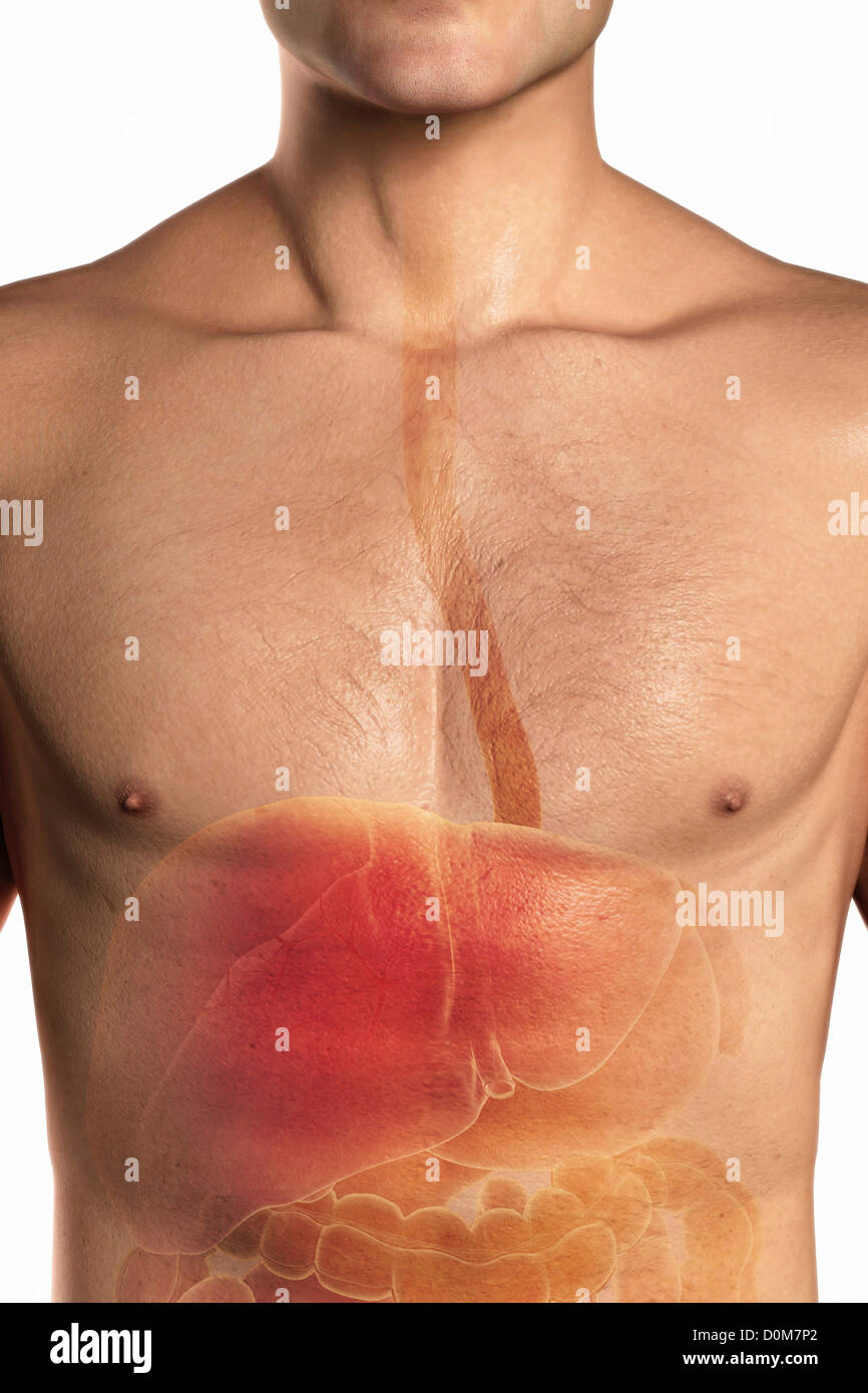 Close-up view of the upper digestive tract and liver within a stylized male skin. Stock Photohttps://www.alamy.com/image-license-details/?v=1https://www.alamy.com/stock-photo-close-up-view-of-the-upper-digestive-tract-and-liver-within-a-stylized-52076250.html
RMD0M7P2–Close-up view of the upper digestive tract and liver within a stylized male skin.
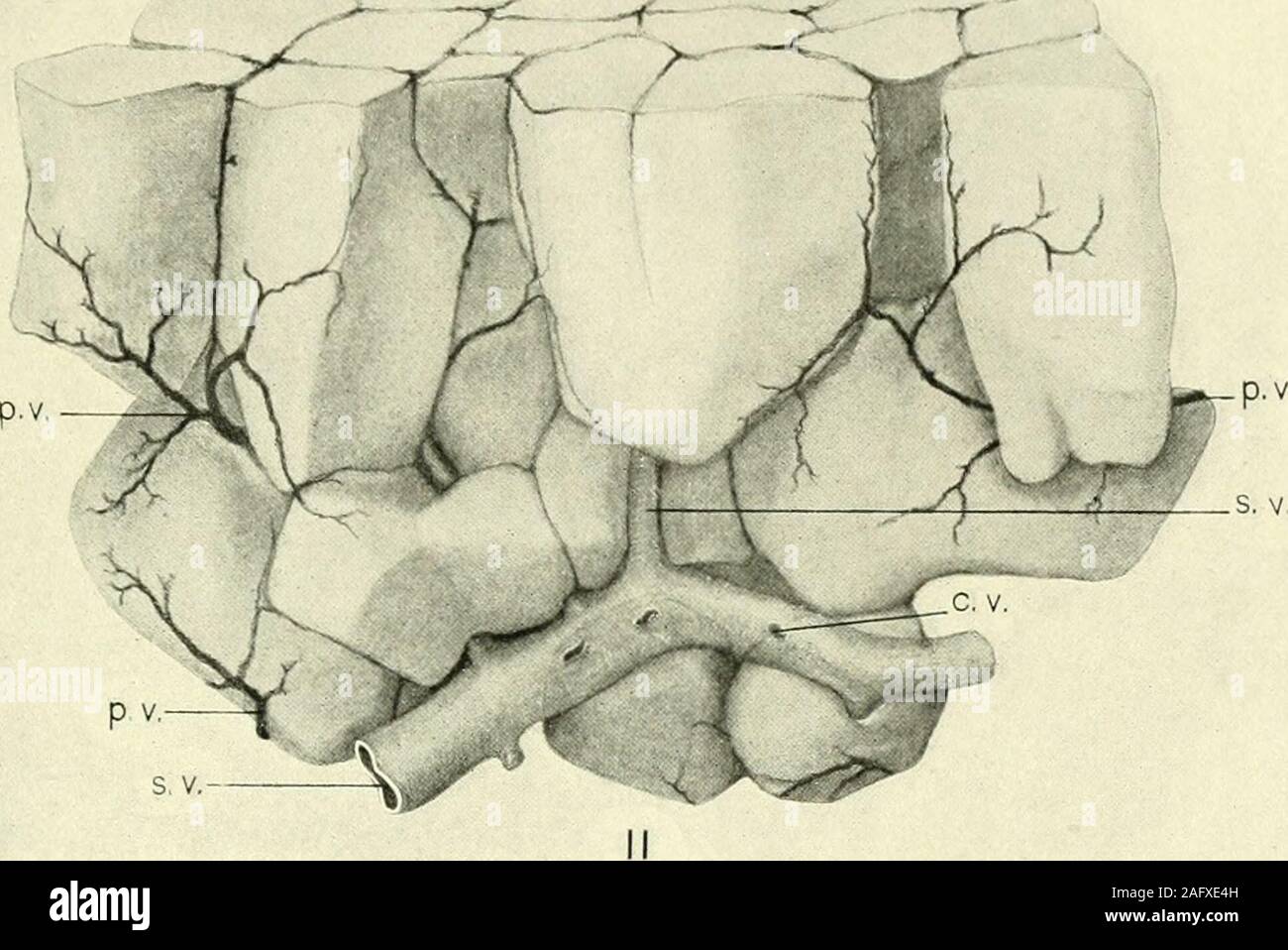 . The American journal of anatomy. 281 PLATE 2 EXPLANATIO>f OF FIGURES Dissections of liver lobules to show their arrangement. 11 A group of sui-face lobules. Bile ducts and branches of the hepatic artery-have been omitted. 12 A group of lobules situated deep in the substance of the liver. On theleft is seen a large portal canal with bile duct, hepatic artery, and portal vein.The branches of these vessels were worked out as far as possible. Undoubtedlysome of them were torn away in lifting off the lobules in dissecting, so that allthe branches ramifying over the surfaces of the lobules are Stock Photohttps://www.alamy.com/image-license-details/?v=1https://www.alamy.com/the-american-journal-of-anatomy-281-plate-2-explanatiogtf-of-figures-dissections-of-liver-lobules-to-show-their-arrangement-11-a-group-of-sui-face-lobules-bile-ducts-and-branches-of-the-hepatic-artery-have-been-omitted-12-a-group-of-lobules-situated-deep-in-the-substance-of-the-liver-on-theleft-is-seen-a-large-portal-canal-with-bile-duct-hepatic-artery-and-portal-veinthe-branches-of-these-vessels-were-worked-out-as-far-as-possible-undoubtedlysome-of-them-were-torn-away-in-lifting-off-the-lobules-in-dissecting-so-that-allthe-branches-ramifying-over-the-surfaces-of-the-lobules-are-image336798689.html
RM2AFXE4H–. The American journal of anatomy. 281 PLATE 2 EXPLANATIO>f OF FIGURES Dissections of liver lobules to show their arrangement. 11 A group of sui-face lobules. Bile ducts and branches of the hepatic artery-have been omitted. 12 A group of lobules situated deep in the substance of the liver. On theleft is seen a large portal canal with bile duct, hepatic artery, and portal vein.The branches of these vessels were worked out as far as possible. Undoubtedlysome of them were torn away in lifting off the lobules in dissecting, so that allthe branches ramifying over the surfaces of the lobules are
 . The cyclopædia of anatomy and physiology. Anatomy; Physiology; Zoology. Lobules in a state of portal venous congestion, as seen on the surface of the. liver. The congested part oc- cupies the margins of the lobules, the uncongested portion their centres. After Kiernan. dullary and occupying the centres of the lo- bules." The causes of congestion are all such as tend to interfere with the circulation in the liver or with the general circulation ; for in- stance, impediment to the circulation of the blood through the capillaries of the lungs, diseases of the valves of the heart, aneurism, Stock Photohttps://www.alamy.com/image-license-details/?v=1https://www.alamy.com/the-cyclopdia-of-anatomy-and-physiology-anatomy-physiology-zoology-lobules-in-a-state-of-portal-venous-congestion-as-seen-on-the-surface-of-the-liver-the-congested-part-oc-cupies-the-margins-of-the-lobules-the-uncongested-portion-their-centres-after-kiernan-dullary-and-occupying-the-centres-of-the-lo-bulesquot-the-causes-of-congestion-are-all-such-as-tend-to-interfere-with-the-circulation-in-the-liver-or-with-the-general-circulation-for-in-stance-impediment-to-the-circulation-of-the-blood-through-the-capillaries-of-the-lungs-diseases-of-the-valves-of-the-heart-aneurism-image216211408.html
RMPFN7T0–. The cyclopædia of anatomy and physiology. Anatomy; Physiology; Zoology. Lobules in a state of portal venous congestion, as seen on the surface of the. liver. The congested part oc- cupies the margins of the lobules, the uncongested portion their centres. After Kiernan. dullary and occupying the centres of the lo- bules." The causes of congestion are all such as tend to interfere with the circulation in the liver or with the general circulation ; for in- stance, impediment to the circulation of the blood through the capillaries of the lungs, diseases of the valves of the heart, aneurism,
 Archive image from page 181 of The cyclopædia of anatomy and. The cyclopædia of anatomy and physiology cyclopdiaofana03todd Year: 1847 168 NORMAL ANATOMY OF THE LIVER. and hepatic artery, and the interlobular spaces are supplied solely with branches which are derived fiom its ramifications. But in the smaller por- tal canals (fig. 37, Jig. 38) the capsule of Ghs- son, upon which the plexus chiefly depends, is Fig. 37. Fig. 38. A transverse section of a small portal canal and its ves- sels. The lobules are in a state of general congestion. From Kier nan's paper. No. 1, The portal vein ; th Stock Photohttps://www.alamy.com/image-license-details/?v=1https://www.alamy.com/archive-image-from-page-181-of-the-cyclopdia-of-anatomy-and-the-cyclopdia-of-anatomy-and-physiology-cyclopdiaofana03todd-year-1847-168-normal-anatomy-of-the-liver-and-hepatic-artery-and-the-interlobular-spaces-are-supplied-solely-with-branches-which-are-derived-fiom-its-ramifications-but-in-the-smaller-por-tal-canals-fig-37-jig-38-the-capsule-of-ghs-son-upon-which-the-plexus-chiefly-depends-is-fig-37-fig-38-a-transverse-section-of-a-small-portal-canal-and-its-ves-sels-the-lobules-are-in-a-state-of-general-congestion-from-kier-nans-paper-no-1-the-portal-vein-th-image259469449.html
RMW23RX1–Archive image from page 181 of The cyclopædia of anatomy and. The cyclopædia of anatomy and physiology cyclopdiaofana03todd Year: 1847 168 NORMAL ANATOMY OF THE LIVER. and hepatic artery, and the interlobular spaces are supplied solely with branches which are derived fiom its ramifications. But in the smaller por- tal canals (fig. 37, Jig. 38) the capsule of Ghs- son, upon which the plexus chiefly depends, is Fig. 37. Fig. 38. A transverse section of a small portal canal and its ves- sels. The lobules are in a state of general congestion. From Kier nan's paper. No. 1, The portal vein ; th
 Vegetable and liver salad. Stock Photohttps://www.alamy.com/image-license-details/?v=1https://www.alamy.com/vegetable-and-liver-salad-image402534708.html
RF2EAW13G–Vegetable and liver salad.
 Warm salad with chicken liver and vegetables.Chicken liver salad Stock Photohttps://www.alamy.com/image-license-details/?v=1https://www.alamy.com/warm-salad-with-chicken-liver-and-vegetableschicken-liver-salad-image396440939.html
RF2E0YCCY–Warm salad with chicken liver and vegetables.Chicken liver salad
 Hepatitis c, hcv Stock Photohttps://www.alamy.com/image-license-details/?v=1https://www.alamy.com/hepatitis-c-hcv-image338279373.html
RM2AJ9XP5–Hepatitis c, hcv
 Three-quarter close up liver which is faded show position gallbladder relative stomach intestines. Stock Photohttps://www.alamy.com/image-license-details/?v=1https://www.alamy.com/stock-photo-three-quarter-close-up-liver-which-is-faded-show-position-gallbladder-52076327.html
RMD0M7TR–Three-quarter close up liver which is faded show position gallbladder relative stomach intestines.
 Manual of pathology : including bacteriology, the technic of postmortems, and methods of pathologic research . U ?^A. /?>.. ^m .^4 Fig. 369.—Syphilitic Cirrhosis of the Liver.—{Schmaus.) X 40 diameters.a. Liver capsule, b, b. b. Newly formed connective tissue, which is grouped in a band-like area as at g. c.Branch of portal vein. /, /, /. Liver lobules, e. Deep fissure produced in the liver surface by the tractionexerted by the fibrous bundles, g. often quite similar, and a microscopic or bacteriologic examination maybe necessary to differentiate the lesions. Miliary tuberculosis of thelive Stock Photohttps://www.alamy.com/image-license-details/?v=1https://www.alamy.com/manual-of-pathology-including-bacteriology-the-technic-of-postmortems-and-methods-of-pathologic-research-u-a-gt-m-4-fig-369syphilitic-cirrhosis-of-the-liverschmaus-x-40-diametersa-liver-capsule-b-b-b-newly-formed-connective-tissue-which-is-grouped-in-a-band-like-area-as-at-g-cbranch-of-portal-vein-liver-lobules-e-deep-fissure-produced-in-the-liver-surface-by-the-tractionexerted-by-the-fibrous-bundles-g-often-quite-similar-and-a-microscopic-or-bacteriologic-examination-maybe-necessary-to-differentiate-the-lesions-miliary-tuberculosis-of-thelive-image338404205.html
RM2AJFJ0D–Manual of pathology : including bacteriology, the technic of postmortems, and methods of pathologic research . U ?^A. /?>.. ^m .^4 Fig. 369.—Syphilitic Cirrhosis of the Liver.—{Schmaus.) X 40 diameters.a. Liver capsule, b, b. b. Newly formed connective tissue, which is grouped in a band-like area as at g. c.Branch of portal vein. /, /, /. Liver lobules, e. Deep fissure produced in the liver surface by the tractionexerted by the fibrous bundles, g. often quite similar, and a microscopic or bacteriologic examination maybe necessary to differentiate the lesions. Miliary tuberculosis of thelive
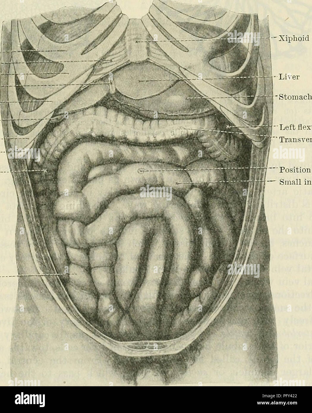 . Cunningham's Text-book of anatomy. Anatomy. VESSELS OF THE LIVER. 1199 number of thorns growing out on all sides from the sublobular twigs of the tree). On each of these little central veins there is impaled, as it were, a lobule. These little conical lobules, with their central veins running through them, are so numerous and so closely packed together, that they give rise to the practically solid liver tissue. The lobules are surrounded by the vense interlobulares, branches of the portal vein, from which numerous twigs enter the lobule on all sides, and converging, join the central vein (Fi Stock Photohttps://www.alamy.com/image-license-details/?v=1https://www.alamy.com/cunninghams-text-book-of-anatomy-anatomy-vessels-of-the-liver-1199-number-of-thorns-growing-out-on-all-sides-from-the-sublobular-twigs-of-the-tree-on-each-of-these-little-central-veins-there-is-impaled-as-it-were-a-lobule-these-little-conical-lobules-with-their-central-veins-running-through-them-are-so-numerous-and-so-closely-packed-together-that-they-give-rise-to-the-practically-solid-liver-tissue-the-lobules-are-surrounded-by-the-vense-interlobulares-branches-of-the-portal-vein-from-which-numerous-twigs-enter-the-lobule-on-all-sides-and-converging-join-the-central-vein-fi-image216340154.html
RMPFY422–. Cunningham's Text-book of anatomy. Anatomy. VESSELS OF THE LIVER. 1199 number of thorns growing out on all sides from the sublobular twigs of the tree). On each of these little central veins there is impaled, as it were, a lobule. These little conical lobules, with their central veins running through them, are so numerous and so closely packed together, that they give rise to the practically solid liver tissue. The lobules are surrounded by the vense interlobulares, branches of the portal vein, from which numerous twigs enter the lobule on all sides, and converging, join the central vein (Fi
 Vegetable and liver salad. Stock Photohttps://www.alamy.com/image-license-details/?v=1https://www.alamy.com/vegetable-and-liver-salad-image402536128.html
RF2EAW2X8–Vegetable and liver salad.
 Salad with liver,bell pepper,okra and herbs.Chicken liver salad Stock Photohttps://www.alamy.com/image-license-details/?v=1https://www.alamy.com/salad-with-liverbell-pepperokra-and-herbschicken-liver-salad-image395454571.html
RF2DYAE9F–Salad with liver,bell pepper,okra and herbs.Chicken liver salad
 Hepatitis c, hcv Stock Photohttps://www.alamy.com/image-license-details/?v=1https://www.alamy.com/hepatitis-c-hcv-image338279369.html
RM2AJ9XP1–Hepatitis c, hcv
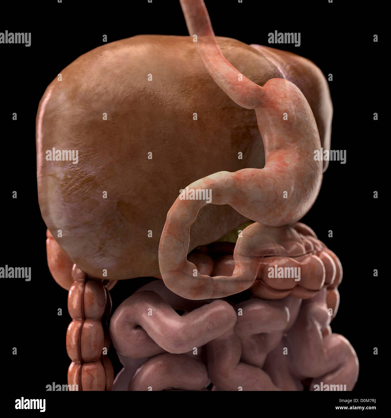 Three-quarter close up liver which is faded show position stomach relative spleen gallbladder intestines. Stock Photohttps://www.alamy.com/image-license-details/?v=1https://www.alamy.com/stock-photo-three-quarter-close-up-liver-which-is-faded-show-position-stomach-52076294.html
RMD0M7RJ–Three-quarter close up liver which is faded show position stomach relative spleen gallbladder intestines.
 . A laboratory manual and text-book of embryology. Embryology. i86 THE ENTODERMAL CANAL AND ITS DERIVATIVES which bear the same relation to each other and to new lobules as did the primary branches to the first lobule. This process is repeated until thousands of liver lobules are developed. Until the 20 mm. stage the portal vein alone supplies the liver. The hepatic artery from the cceliac axis comes into relation first with the hepatic duct and gall-bladder. Later, it grows into the connective tissue about the larger bile ducts and branches of the portal vein, and also supplies the capsule of Stock Photohttps://www.alamy.com/image-license-details/?v=1https://www.alamy.com/a-laboratory-manual-and-text-book-of-embryology-embryology-i86-the-entodermal-canal-and-its-derivatives-which-bear-the-same-relation-to-each-other-and-to-new-lobules-as-did-the-primary-branches-to-the-first-lobule-this-process-is-repeated-until-thousands-of-liver-lobules-are-developed-until-the-20-mm-stage-the-portal-vein-alone-supplies-the-liver-the-hepatic-artery-from-the-cceliac-axis-comes-into-relation-first-with-the-hepatic-duct-and-gall-bladder-later-it-grows-into-the-connective-tissue-about-the-larger-bile-ducts-and-branches-of-the-portal-vein-and-also-supplies-the-capsule-of-image232344965.html
RMRE06AD–. A laboratory manual and text-book of embryology. Embryology. i86 THE ENTODERMAL CANAL AND ITS DERIVATIVES which bear the same relation to each other and to new lobules as did the primary branches to the first lobule. This process is repeated until thousands of liver lobules are developed. Until the 20 mm. stage the portal vein alone supplies the liver. The hepatic artery from the cceliac axis comes into relation first with the hepatic duct and gall-bladder. Later, it grows into the connective tissue about the larger bile ducts and branches of the portal vein, and also supplies the capsule of
 . The development of the human body : a manual of human embryology. Embryology; Embryo, Non-Mammalian. 3io THE LIVER and thence extend throughout the neighboring cylinders, anastomos- ing with capillaries developing in relation to neighboring portal branches. As the extension so proceeds the older capillaries con- tinue to enlarge and later become transformed into bile-ducts (Fig. 191, C), the cells of the cylinders in which these capillaries were situated becoming converted into the epithelial lining of the ducts. The lobules, which form so characteristic a feature of the adult liver, are lat Stock Photohttps://www.alamy.com/image-license-details/?v=1https://www.alamy.com/the-development-of-the-human-body-a-manual-of-human-embryology-embryology-embryo-non-mammalian-3io-the-liver-and-thence-extend-throughout-the-neighboring-cylinders-anastomos-ing-with-capillaries-developing-in-relation-to-neighboring-portal-branches-as-the-extension-so-proceeds-the-older-capillaries-con-tinue-to-enlarge-and-later-become-transformed-into-bile-ducts-fig-191-c-the-cells-of-the-cylinders-in-which-these-capillaries-were-situated-becoming-converted-into-the-epithelial-lining-of-the-ducts-the-lobules-which-form-so-characteristic-a-feature-of-the-adult-liver-are-lat-image215957751.html
RMPF9M8R–. The development of the human body : a manual of human embryology. Embryology; Embryo, Non-Mammalian. 3io THE LIVER and thence extend throughout the neighboring cylinders, anastomos- ing with capillaries developing in relation to neighboring portal branches. As the extension so proceeds the older capillaries con- tinue to enlarge and later become transformed into bile-ducts (Fig. 191, C), the cells of the cylinders in which these capillaries were situated becoming converted into the epithelial lining of the ducts. The lobules, which form so characteristic a feature of the adult liver, are lat
 Vegetable and liver salad. Stock Photohttps://www.alamy.com/image-license-details/?v=1https://www.alamy.com/vegetable-and-liver-salad-image402536133.html
RF2EAW2XD–Vegetable and liver salad.
 Salad with liver,bell pepper,okra and herbs.Chicken liver salad Stock Photohttps://www.alamy.com/image-license-details/?v=1https://www.alamy.com/salad-with-liverbell-pepperokra-and-herbschicken-liver-salad-image433225045.html
RF2G4R2Y1–Salad with liver,bell pepper,okra and herbs.Chicken liver salad
 Hepatitis c, hcv Stock Photohttps://www.alamy.com/image-license-details/?v=1https://www.alamy.com/hepatitis-c-hcv-image338279370.html
RM2AJ9XP2–Hepatitis c, hcv
 A view male figure African ethnicity liver other organs digestive system present within abdomen. liver is showing signs Stock Photohttps://www.alamy.com/image-license-details/?v=1https://www.alamy.com/stock-photo-a-view-male-figure-african-ethnicity-liver-other-organs-digestive-52092170.html
RMD0N02J–A view male figure African ethnicity liver other organs digestive system present within abdomen. liver is showing signs
 . Cunningham's Text-book of anatomy. Anatomy. Central vein Central vein Sublobular vein Fig 940.—Liver of a Pig injected from the Hepatic Vein by T. A. Carter. (From a specimen left in the Anatomical Department of Edinburgh University by Sir William Turner.) Liver lobules of delicate fibrous tissue, which is most evident where the serous coat is absent. In the neighbourhood of the porta hepatis it is particularly abundant, and here it surrounds the vessels entering the porta, and accompanies them through the portal canals in the liver substance. This coat is continuous with the fine areolar Stock Photohttps://www.alamy.com/image-license-details/?v=1https://www.alamy.com/cunninghams-text-book-of-anatomy-anatomy-central-vein-central-vein-sublobular-vein-fig-940liver-of-a-pig-injected-from-the-hepatic-vein-by-t-a-carter-from-a-specimen-left-in-the-anatomical-department-of-edinburgh-university-by-sir-william-turner-liver-lobules-of-delicate-fibrous-tissue-which-is-most-evident-where-the-serous-coat-is-absent-in-the-neighbourhood-of-the-porta-hepatis-it-is-particularly-abundant-and-here-it-surrounds-the-vessels-entering-the-porta-and-accompanies-them-through-the-portal-canals-in-the-liver-substance-this-coat-is-continuous-with-the-fine-areolar-image231849044.html
RMRD5HR0–. Cunningham's Text-book of anatomy. Anatomy. Central vein Central vein Sublobular vein Fig 940.—Liver of a Pig injected from the Hepatic Vein by T. A. Carter. (From a specimen left in the Anatomical Department of Edinburgh University by Sir William Turner.) Liver lobules of delicate fibrous tissue, which is most evident where the serous coat is absent. In the neighbourhood of the porta hepatis it is particularly abundant, and here it surrounds the vessels entering the porta, and accompanies them through the portal canals in the liver substance. This coat is continuous with the fine areolar
 . The cyclopædia of anatomy and physiology. Anatomy; Physiology; Zoology. CEPHALOPODA. 537 for it is divided into four lobes, and these are connected by a fifth portion, which passes transversely below the fundus of the crop. All these larger divisions are subdivided into numerous lobules of an angular form, which vary in size from three to five lines. These lobules are immediately invested by a very delicate capsule, and are more loosely sur- rounded by a peritoneal covering common to this gland and the crop. The liver is supplied by large branches which are given off from the aorta, (r,fg. 2 Stock Photohttps://www.alamy.com/image-license-details/?v=1https://www.alamy.com/the-cyclopdia-of-anatomy-and-physiology-anatomy-physiology-zoology-cephalopoda-537-for-it-is-divided-into-four-lobes-and-these-are-connected-by-a-fifth-portion-which-passes-transversely-below-the-fundus-of-the-crop-all-these-larger-divisions-are-subdivided-into-numerous-lobules-of-an-angular-form-which-vary-in-size-from-three-to-five-lines-these-lobules-are-immediately-invested-by-a-very-delicate-capsule-and-are-more-loosely-sur-rounded-by-a-peritoneal-covering-common-to-this-gland-and-the-crop-the-liver-is-supplied-by-large-branches-which-are-given-off-from-the-aorta-rfg-2-image216190879.html
RMPFM9JR–. The cyclopædia of anatomy and physiology. Anatomy; Physiology; Zoology. CEPHALOPODA. 537 for it is divided into four lobes, and these are connected by a fifth portion, which passes transversely below the fundus of the crop. All these larger divisions are subdivided into numerous lobules of an angular form, which vary in size from three to five lines. These lobules are immediately invested by a very delicate capsule, and are more loosely sur- rounded by a peritoneal covering common to this gland and the crop. The liver is supplied by large branches which are given off from the aorta, (r,fg. 2
 Vegetable and liver salad. Stock Photohttps://www.alamy.com/image-license-details/?v=1https://www.alamy.com/vegetable-and-liver-salad-image402537180.html
RF2EAW47T–Vegetable and liver salad.
 Fried chicken liver and vegetable salad.Chicken liver salad Stock Photohttps://www.alamy.com/image-license-details/?v=1https://www.alamy.com/fried-chicken-liver-and-vegetable-saladchicken-liver-salad-image395572253.html
RF2DYFTCD–Fried chicken liver and vegetable salad.Chicken liver salad
 Hepatitis c, hcv Stock Photohttps://www.alamy.com/image-license-details/?v=1https://www.alamy.com/hepatitis-c-hcv-image338279364.html
RM2AJ9XNT–Hepatitis c, hcv
 Salad with liver,bell pepper,okra and herbs.Chicken liver salad Stock Photohttps://www.alamy.com/image-license-details/?v=1https://www.alamy.com/salad-with-liverbell-pepperokra-and-herbschicken-liver-salad-image433225077.html
RF2G4R305–Salad with liver,bell pepper,okra and herbs.Chicken liver salad
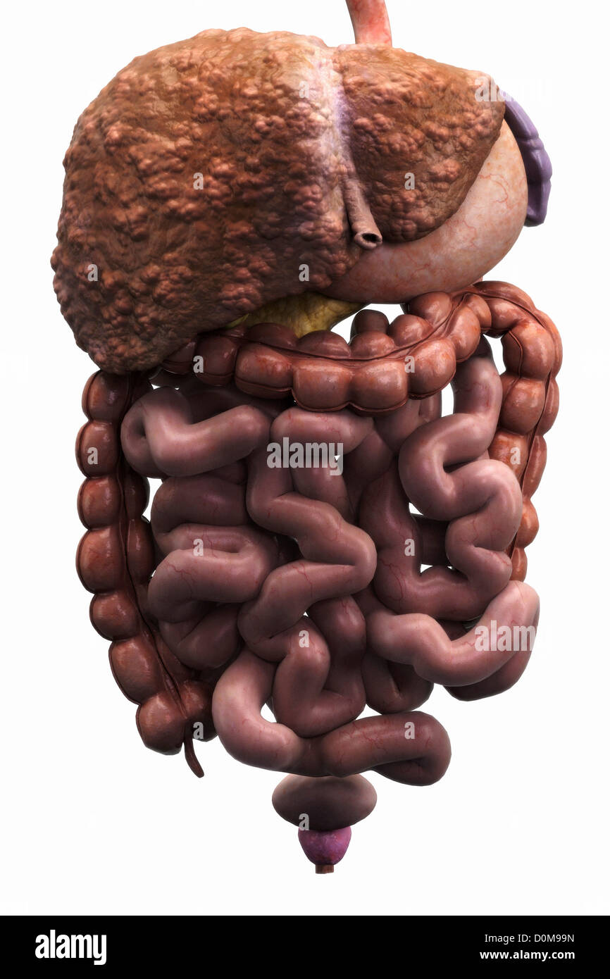 Front view organs digestive system. liver is displaying regenerative nodules which is symptomatic liver cirrhosis. Stock Photohttps://www.alamy.com/image-license-details/?v=1https://www.alamy.com/stock-photo-front-view-organs-digestive-system-liver-is-displaying-regenerative-52077473.html
RMD0M99N–Front view organs digestive system. liver is displaying regenerative nodules which is symptomatic liver cirrhosis.
 Salad with liver,bell pepper,okra and herbs.Chicken liver salad Stock Photohttps://www.alamy.com/image-license-details/?v=1https://www.alamy.com/salad-with-liverbell-pepperokra-and-herbschicken-liver-salad-image394974665.html
RF2DXGJ61–Salad with liver,bell pepper,okra and herbs.Chicken liver salad
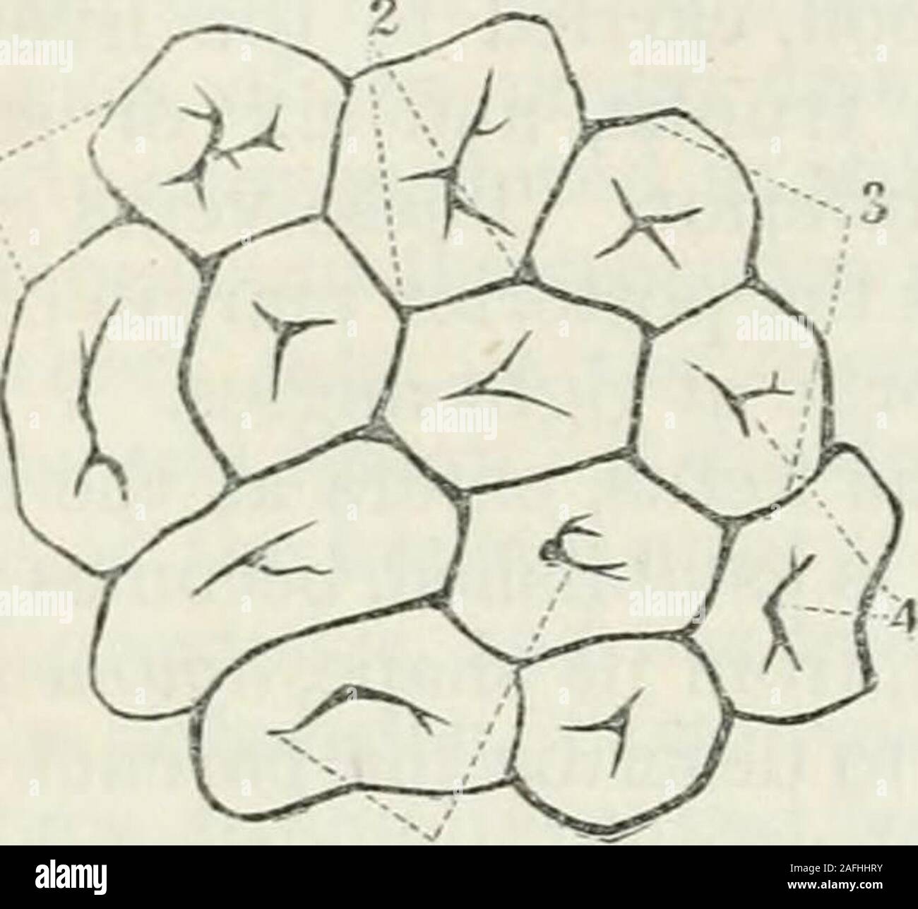 . Human physiology. Connexion of Lobules of Liver with He-patic Vein. 1. Hepatic vein. 2, 2, 2. Lobules, each con-taining an intralobular or hepatic twig. )30 SECRETION their substance and are supposed to subdivirle into minute branches,which by anastomoses with each other form the reticulated plexus de-picted in Fig. 160, called by Mr. Kiernan the lobular biliary plexus. Fi!?. 158. Fig. 159.. Stock Photohttps://www.alamy.com/image-license-details/?v=1https://www.alamy.com/human-physiology-connexion-of-lobules-of-liver-with-he-patic-vein-1-hepatic-vein-2-2-2-lobules-each-con-taining-an-intralobular-or-hepatic-twig-30-secretion-their-substance-and-are-supposed-to-subdivirle-into-minute-brancheswhich-by-anastomoses-with-each-other-form-the-reticulated-plexus-de-picted-in-fig-160-called-by-mr-kiernan-the-lobular-biliary-plexus-fi!-158-fig-159-image336604015.html
RM2AFHHRY–. Human physiology. Connexion of Lobules of Liver with He-patic Vein. 1. Hepatic vein. 2, 2, 2. Lobules, each con-taining an intralobular or hepatic twig. )30 SECRETION their substance and are supposed to subdivirle into minute branches,which by anastomoses with each other form the reticulated plexus de-picted in Fig. 160, called by Mr. Kiernan the lobular biliary plexus. Fi!?. 158. Fig. 159..
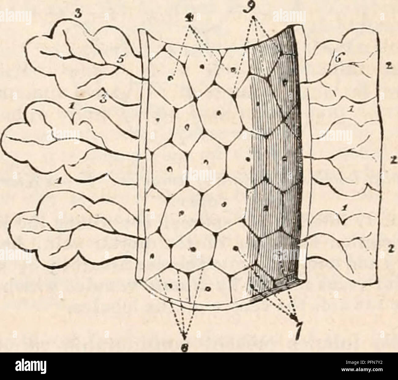 . The cyclopædia of anatomy and physiology. Anatomy; Physiology; Zoology. NORMAL ANATOMY OF THE LIVER. 165 called Glisson's capsule, of the ramifications of tlie portal vein, hepatic duct, hepatic artery, hepatic veins, lymphatics and nerves. For an accurate knowledge of these different structures, anatomy is indebted to the labours of Mr. Kienian, to whose paper on " The Anatomy and Physiology of the Liver," con- tained in the Philosophical Transactions for 1833, I shall have constant occasion to refer. The small bodies (lobules, acini, corpuscula, glandular grains, granulations) of Stock Photohttps://www.alamy.com/image-license-details/?v=1https://www.alamy.com/the-cyclopdia-of-anatomy-and-physiology-anatomy-physiology-zoology-normal-anatomy-of-the-liver-165-called-glissons-capsule-of-the-ramifications-of-tlie-portal-vein-hepatic-duct-hepatic-artery-hepatic-veins-lymphatics-and-nerves-for-an-accurate-knowledge-of-these-different-structures-anatomy-is-indebted-to-the-labours-of-mr-kienian-to-whose-paper-on-quot-the-anatomy-and-physiology-of-the-liverquot-con-tained-in-the-philosophical-transactions-for-1833-i-shall-have-constant-occasion-to-refer-the-small-bodies-lobules-acini-corpuscula-glandular-grains-granulations-of-image216211494.html
RMPFN7Y2–. The cyclopædia of anatomy and physiology. Anatomy; Physiology; Zoology. NORMAL ANATOMY OF THE LIVER. 165 called Glisson's capsule, of the ramifications of tlie portal vein, hepatic duct, hepatic artery, hepatic veins, lymphatics and nerves. For an accurate knowledge of these different structures, anatomy is indebted to the labours of Mr. Kienian, to whose paper on " The Anatomy and Physiology of the Liver," con- tained in the Philosophical Transactions for 1833, I shall have constant occasion to refer. The small bodies (lobules, acini, corpuscula, glandular grains, granulations) of
 Warm salad with chicken liver and vegetables.Chicken liver salad Stock Photohttps://www.alamy.com/image-license-details/?v=1https://www.alamy.com/warm-salad-with-chicken-liver-and-vegetableschicken-liver-salad-image443740003.html
RF2GNX2W7–Warm salad with chicken liver and vegetables.Chicken liver salad
 Hepatitis c, hcv Stock Photohttps://www.alamy.com/image-license-details/?v=1https://www.alamy.com/hepatitis-c-hcv-image338279372.html
RM2AJ9XP4–Hepatitis c, hcv
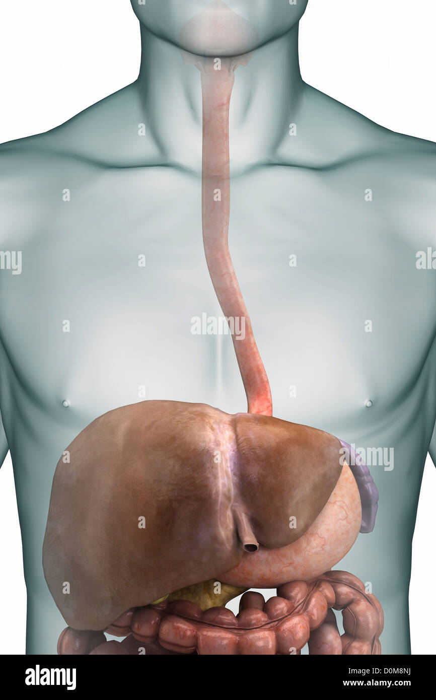 Front view of a close-up of the upper digestive tract focusing on the esophagus and liver. Stock Photohttps://www.alamy.com/image-license-details/?v=1https://www.alamy.com/stock-photo-front-view-of-a-close-up-of-the-upper-digestive-tract-focusing-on-52077022.html
RMD0M8NJ–Front view of a close-up of the upper digestive tract focusing on the esophagus and liver.
 Salad with liver,bell pepper,okra and herbs.Chicken liver salad Stock Photohttps://www.alamy.com/image-license-details/?v=1https://www.alamy.com/salad-with-liverbell-pepperokra-and-herbschicken-liver-salad-image395454589.html
RF2DYAEA5–Salad with liver,bell pepper,okra and herbs.Chicken liver salad
 A system of human anatomy, general and special . artery and duct. The in-terlobular portion occupies the interlobular fissures and spaces, andthe lobular portion forms the supporting tissue to the substance of thelobules. The Portal vein, entering the liver at the transverse fissure, ramifiesthrough its structure in canals, which resemble, by their surfaces, theexternal superficies of the liver, and are formed by the capsular sur-faces of the lobules. These are the portal canals, and contain, be-sides the portal vein with its ramifications, the artery and duct withtheir branches. In the larger Stock Photohttps://www.alamy.com/image-license-details/?v=1https://www.alamy.com/a-system-of-human-anatomy-general-and-special-artery-and-duct-the-in-terlobular-portion-occupies-the-interlobular-fissures-and-spaces-andthe-lobular-portion-forms-the-supporting-tissue-to-the-substance-of-thelobules-the-portal-vein-entering-the-liver-at-the-transverse-fissure-ramifiesthrough-its-structure-in-canals-which-resemble-by-their-surfaces-theexternal-superficies-of-the-liver-and-are-formed-by-the-capsular-sur-faces-of-the-lobules-these-are-the-portal-canals-and-contain-be-sides-the-portal-vein-with-its-ramifications-the-artery-and-duct-withtheir-branches-in-the-larger-image342702734.html
RM2AWFCRA–A system of human anatomy, general and special . artery and duct. The in-terlobular portion occupies the interlobular fissures and spaces, andthe lobular portion forms the supporting tissue to the substance of thelobules. The Portal vein, entering the liver at the transverse fissure, ramifiesthrough its structure in canals, which resemble, by their surfaces, theexternal superficies of the liver, and are formed by the capsular sur-faces of the lobules. These are the portal canals, and contain, be-sides the portal vein with its ramifications, the artery and duct withtheir branches. In the larger
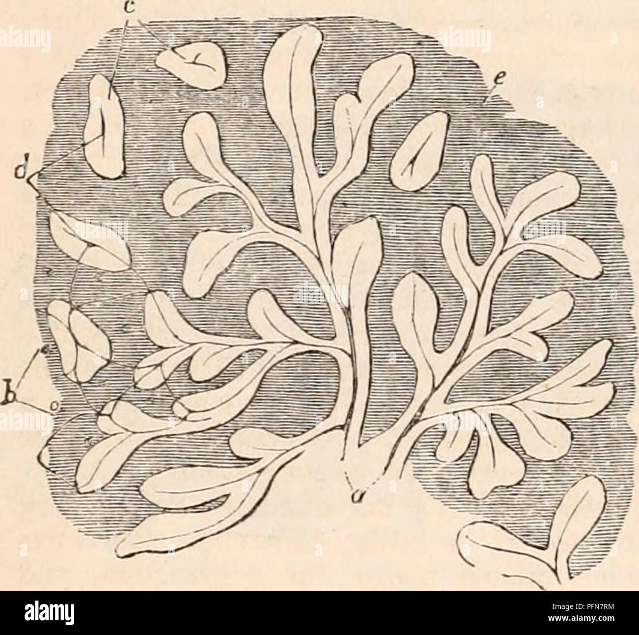 . The cyclopædia of anatomy and physiology. Anatomy; Physiology; Zoology. 186 ABNORMAL ANATOMY OF THE LIVER. formes acini, in figuris ramosis et foliatis varie dispositis." Now the truth is, that this section is in the second stage of hepatic venous con- gestion, and the " figuris ramosis et foliatis" are simply the uncongested portions of the lobules, of a lighter colour than the rest, and presenting the foliated and ramous appearance which is common to this form of congestion. The " fines ductuum biliferorum elongati seu cylindriformes acini" are obviously imaginary. Stock Photohttps://www.alamy.com/image-license-details/?v=1https://www.alamy.com/the-cyclopdia-of-anatomy-and-physiology-anatomy-physiology-zoology-186-abnormal-anatomy-of-the-liver-formes-acini-in-figuris-ramosis-et-foliatis-varie-dispositisquot-now-the-truth-is-that-this-section-is-in-the-second-stage-of-hepatic-venous-con-gestion-and-the-quot-figuris-ramosis-et-foliatisquot-are-simply-the-uncongested-portions-of-the-lobules-of-a-lighter-colour-than-the-rest-and-presenting-the-foliated-and-ramous-appearance-which-is-common-to-this-form-of-congestion-the-quot-fines-ductuum-biliferorum-elongati-seu-cylindriformes-aciniquot-are-obviously-imaginary-image216211400.html
RMPFN7RM–. The cyclopædia of anatomy and physiology. Anatomy; Physiology; Zoology. 186 ABNORMAL ANATOMY OF THE LIVER. formes acini, in figuris ramosis et foliatis varie dispositis." Now the truth is, that this section is in the second stage of hepatic venous con- gestion, and the " figuris ramosis et foliatis" are simply the uncongested portions of the lobules, of a lighter colour than the rest, and presenting the foliated and ramous appearance which is common to this form of congestion. The " fines ductuum biliferorum elongati seu cylindriformes acini" are obviously imaginary.
 Warm salad with chicken liver and vegetables.Chicken liver salad Stock Photohttps://www.alamy.com/image-license-details/?v=1https://www.alamy.com/warm-salad-with-chicken-liver-and-vegetableschicken-liver-salad-image443739979.html
RF2GNX2TB–Warm salad with chicken liver and vegetables.Chicken liver salad
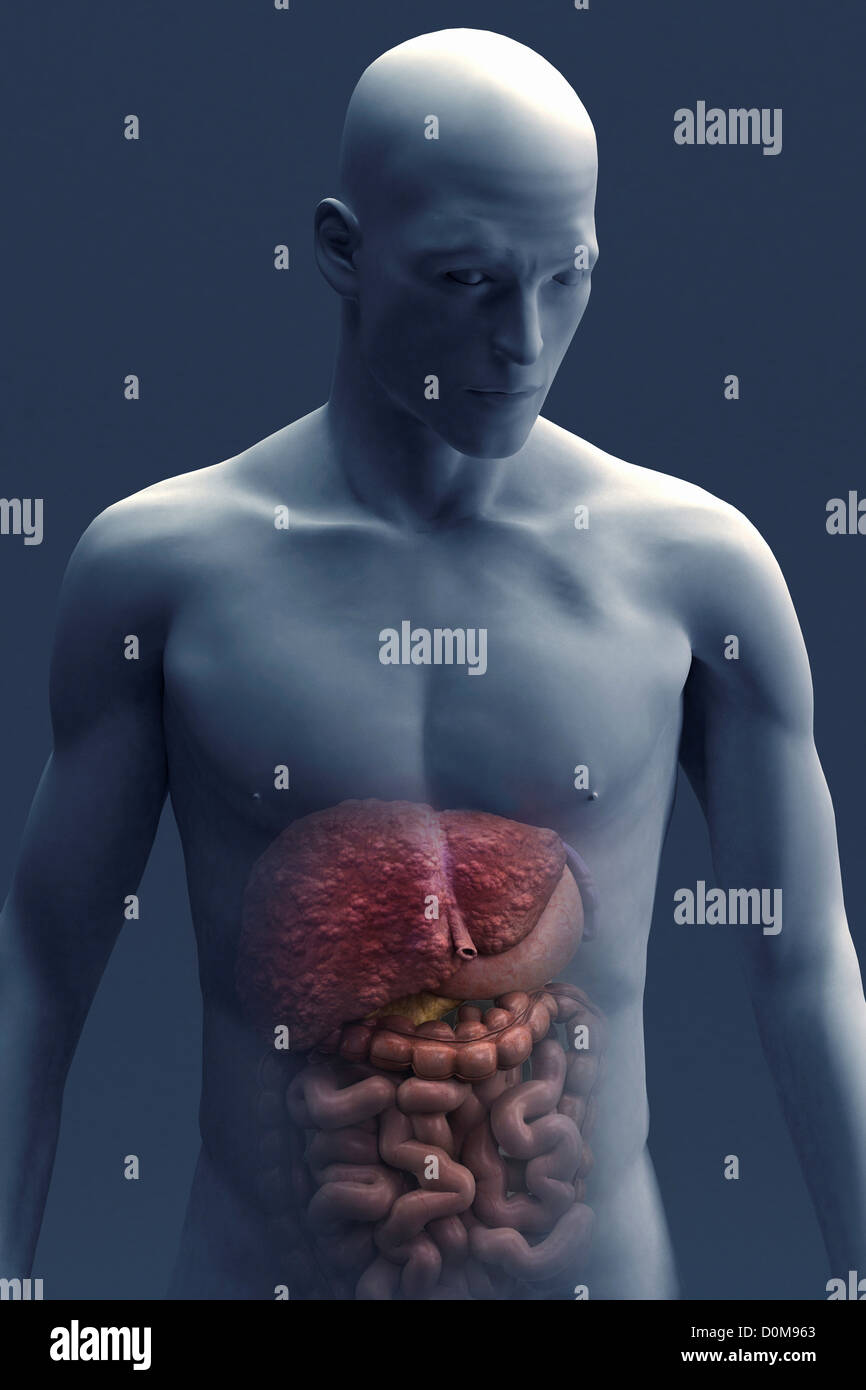 Front view organs digestive system within male figure. liver is displaying regenerative nodules which is symptomatic liver Stock Photohttps://www.alamy.com/image-license-details/?v=1https://www.alamy.com/stock-photo-front-view-organs-digestive-system-within-male-figure-liver-is-displaying-52077371.html
RMD0M963–Front view organs digestive system within male figure. liver is displaying regenerative nodules which is symptomatic liver