Quick filters:
Longitudinal fissure Stock Photos and Images
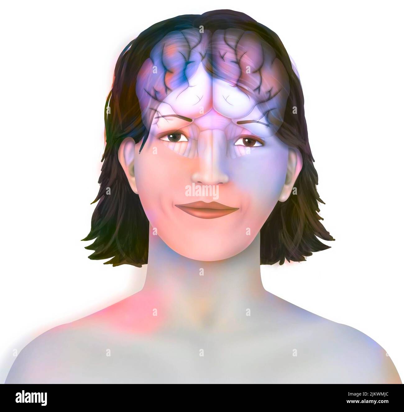 Brain: cerebral hemispheres with longitudinal fissure, cerebellum and brainstem). Stock Photohttps://www.alamy.com/image-license-details/?v=1https://www.alamy.com/brain-cerebral-hemispheres-with-longitudinal-fissure-cerebellum-and-brainstem-image476923396.html
RF2JKWMJC–Brain: cerebral hemispheres with longitudinal fissure, cerebellum and brainstem).
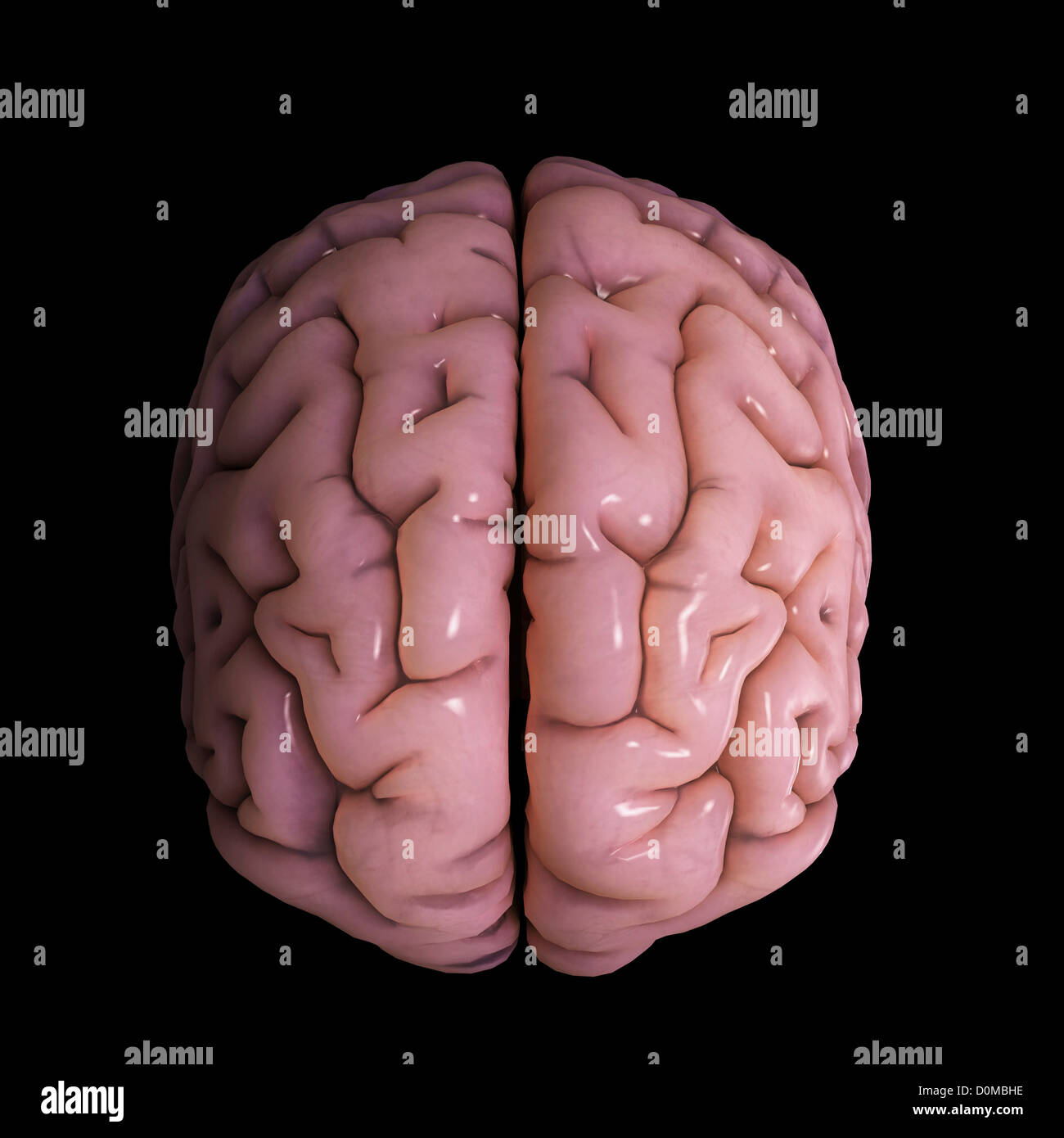 A stylized model human brain showing cerebral cortex medial longitudinal fissure which separates two hemispheres. Stock Photohttps://www.alamy.com/image-license-details/?v=1https://www.alamy.com/stock-photo-a-stylized-model-human-brain-showing-cerebral-cortex-medial-longitudinal-52079258.html
RMD0MBHE–A stylized model human brain showing cerebral cortex medial longitudinal fissure which separates two hemispheres.
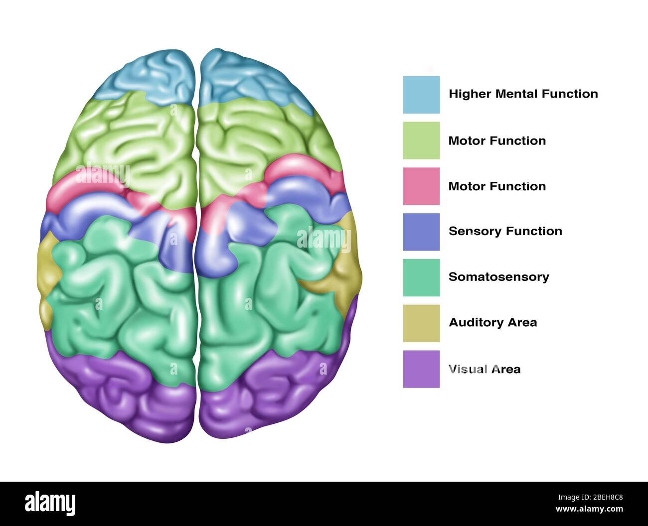 Brain Function, Illustration Stock Photohttps://www.alamy.com/image-license-details/?v=1https://www.alamy.com/brain-function-illustration-image353192344.html
RF2BEH8C8–Brain Function, Illustration
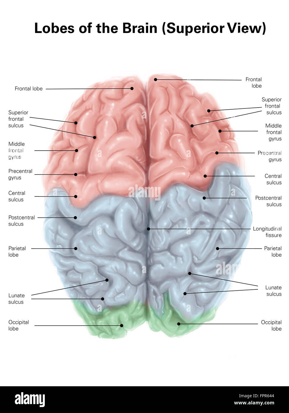 Superior view of human brain with colored lobes and labels. Stock Photohttps://www.alamy.com/image-license-details/?v=1https://www.alamy.com/stock-photo-superior-view-of-human-brain-with-colored-lobes-and-labels-100083988.html
RFFPR644–Superior view of human brain with colored lobes and labels.
 Scots Pine bole (Pinus sylvestris) marked by a big longitudinal frost crack. Pin sylvestre marqué par une importante gélivure Stock Photohttps://www.alamy.com/image-license-details/?v=1https://www.alamy.com/stock-photo-scots-pine-bole-pinus-sylvestris-marked-by-a-big-longitudinal-frost-34497942.html
RMC03EDA–Scots Pine bole (Pinus sylvestris) marked by a big longitudinal frost crack. Pin sylvestre marqué par une importante gélivure
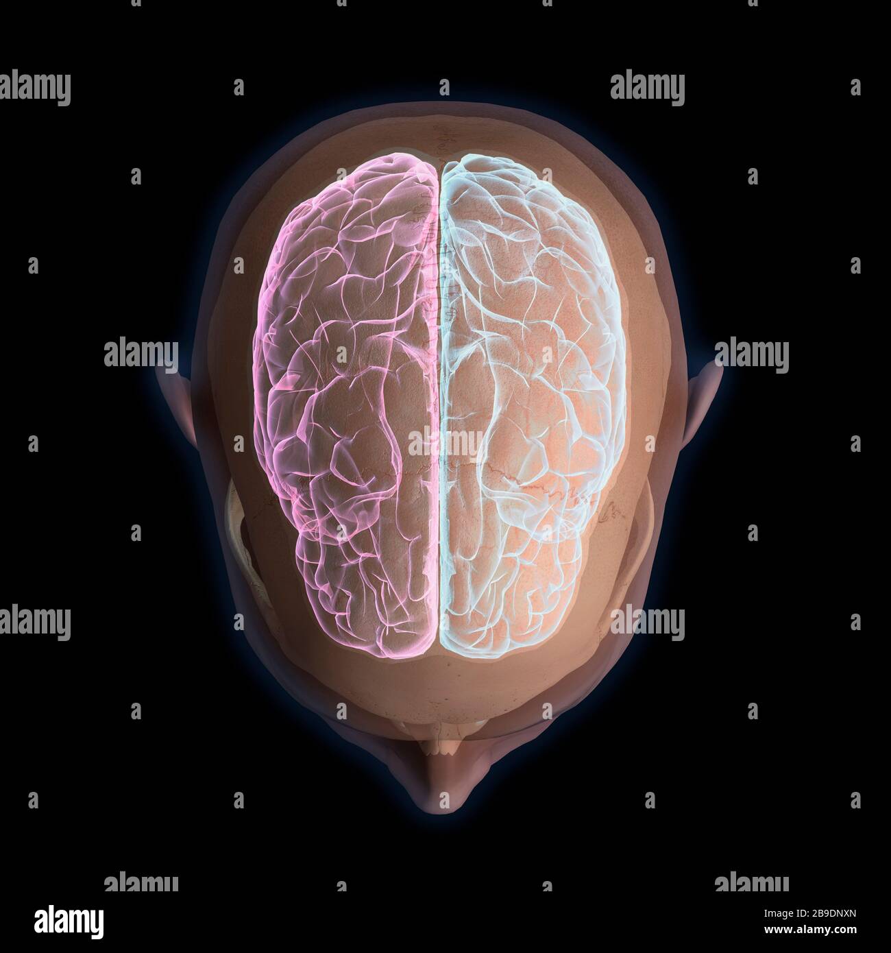 3D rendering of top view of human head and brain on black background. Stock Photohttps://www.alamy.com/image-license-details/?v=1https://www.alamy.com/3d-rendering-of-top-view-of-human-head-and-brain-on-black-background-image350041853.html
RF2B9DNXN–3D rendering of top view of human head and brain on black background.
 massive ancient twisted bark of mature old sweet chestnut tree starting to show signs of decay Stock Photohttps://www.alamy.com/image-license-details/?v=1https://www.alamy.com/stock-photo-massive-ancient-twisted-bark-of-mature-old-sweet-chestnut-tree-starting-79738798.html
RMEHMBJ6–massive ancient twisted bark of mature old sweet chestnut tree starting to show signs of decay
 VERTICAL AERIAL VIEW. Crevasses on the surface of the Bossons Glacier in October. Chamonix-Mont Blanc, Haute-Savoie, Auvergne-Rhône-Alpes, France. Stock Photohttps://www.alamy.com/image-license-details/?v=1https://www.alamy.com/vertical-aerial-view-crevasses-on-the-surface-of-the-bossons-glacier-in-october-chamonix-mont-blanc-haute-savoie-auvergne-rhne-alpes-france-image450091333.html
RM2H47C2D–VERTICAL AERIAL VIEW. Crevasses on the surface of the Bossons Glacier in October. Chamonix-Mont Blanc, Haute-Savoie, Auvergne-Rhône-Alpes, France.
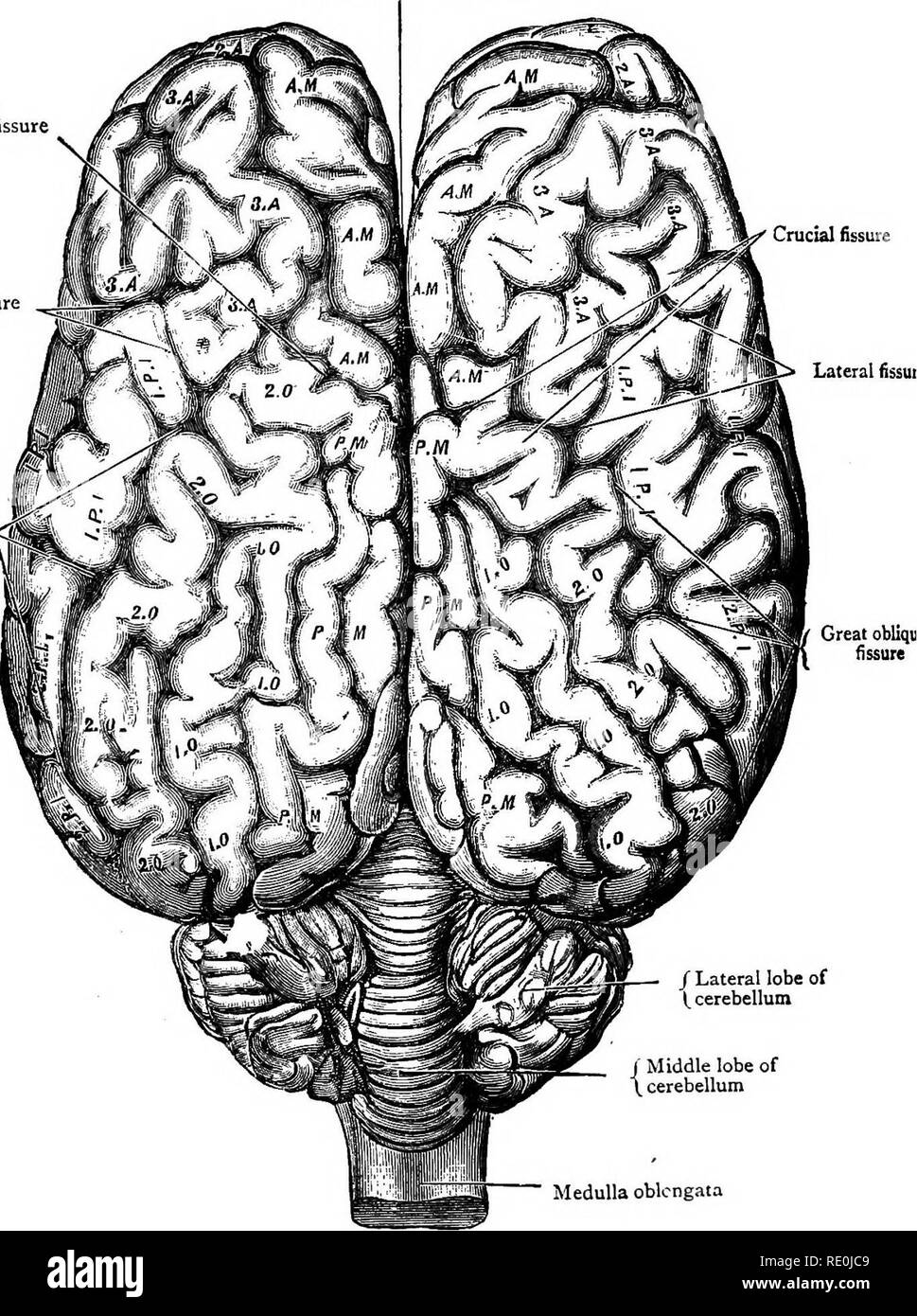 . Diseases & disorders of the horse, a treatise on equine medicine and surgery, being a contribution to the science of comparative pathology. Horses. Great longitudinal fissure between hemispheres of cerebrum â Crucial fissure Lateral fissure Crucial fissure Lateral lissui (Great oblique ' . fissure I. J Lateral lobe of I, cerebellum r Middle lobe of cerebellum Medulla oblongata BRAINâSuperior Aspect.. Please note that these images are extracted from scanned page images that may have been digitally enhanced for readability - coloration and appearance of these illustrations may not perf Stock Photohttps://www.alamy.com/image-license-details/?v=1https://www.alamy.com/diseases-amp-disorders-of-the-horse-a-treatise-on-equine-medicine-and-surgery-being-a-contribution-to-the-science-of-comparative-pathology-horses-great-longitudinal-fissure-between-hemispheres-of-cerebrum-crucial-fissure-lateral-fissure-crucial-fissure-lateral-lissui-great-oblique-fissure-i-j-lateral-lobe-of-i-cerebellum-r-middle-lobe-of-cerebellum-medulla-oblongata-brainsuperior-aspect-please-note-that-these-images-are-extracted-from-scanned-page-images-that-may-have-been-digitally-enhanced-for-readability-coloration-and-appearance-of-these-illustrations-may-not-perf-image232354425.html
RMRE0JC9–. Diseases & disorders of the horse, a treatise on equine medicine and surgery, being a contribution to the science of comparative pathology. Horses. Great longitudinal fissure between hemispheres of cerebrum â Crucial fissure Lateral fissure Crucial fissure Lateral lissui (Great oblique ' . fissure I. J Lateral lobe of I, cerebellum r Middle lobe of cerebellum Medulla oblongata BRAINâSuperior Aspect.. Please note that these images are extracted from scanned page images that may have been digitally enhanced for readability - coloration and appearance of these illustrations may not perf
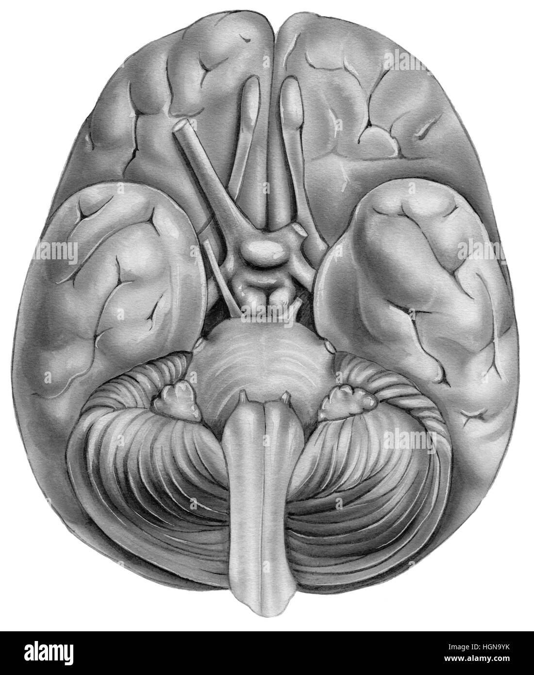 Human Brain - Bottom View. Shown are the temporal lobe,olfactory bulb,frontal lobe, pons,medulla region,cerebellum,pyramid,chiasma,optic nerve,olfacto Stock Photohttps://www.alamy.com/image-license-details/?v=1https://www.alamy.com/stock-photo-human-brain-bottom-view-shown-are-the-temporal-lobeolfactory-bulbfrontal-130775895.html
RFHGN9YK–Human Brain - Bottom View. Shown are the temporal lobe,olfactory bulb,frontal lobe, pons,medulla region,cerebellum,pyramid,chiasma,optic nerve,olfacto
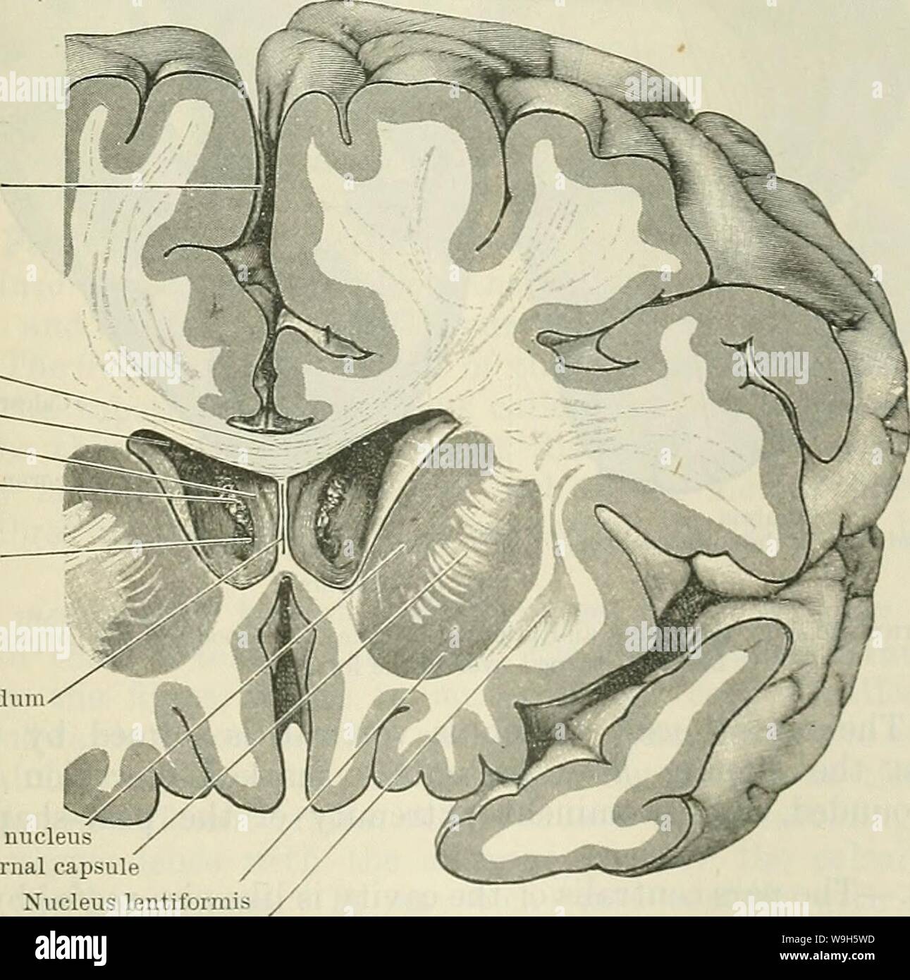 Archive image from page 666 of Cunningham's Text-book of anatomy (1914). Cunningham's Text-book of anatomy cunninghamstextb00cunn Year: 1914 ( Longitudinal fissure Corpus callosum Lateral ventricle Column of fornix Chorioid plexus Foramen inter- ventrieulare Septum pellucidum Caudate nucleus Internal capsule Nucleus lentiformis Claustrum Fig. 562.—Frontal Section through the Cerebbal Hemispheres so as to cut through the anterior horns of the lateral ventricles, through which the central part of the ventricles, the columns of the fornix, and the interventricular foramina can be seen. ticulu Stock Photohttps://www.alamy.com/image-license-details/?v=1https://www.alamy.com/archive-image-from-page-666-of-cunninghams-text-book-of-anatomy-1914-cunninghams-text-book-of-anatomy-cunninghamstextb00cunn-year-1914-longitudinal-fissure-corpus-callosum-lateral-ventricle-column-of-fornix-chorioid-plexus-foramen-inter-ventrieulare-septum-pellucidum-caudate-nucleus-internal-capsule-nucleus-lentiformis-claustrum-fig-562frontal-section-through-the-cerebbal-hemispheres-so-as-to-cut-through-the-anterior-horns-of-the-lateral-ventricles-through-which-the-central-part-of-the-ventricles-the-columns-of-the-fornix-and-the-interventricular-foramina-can-be-seen-ticulu-image264065241.html
RMW9H5WD–Archive image from page 666 of Cunningham's Text-book of anatomy (1914). Cunningham's Text-book of anatomy cunninghamstextb00cunn Year: 1914 ( Longitudinal fissure Corpus callosum Lateral ventricle Column of fornix Chorioid plexus Foramen inter- ventrieulare Septum pellucidum Caudate nucleus Internal capsule Nucleus lentiformis Claustrum Fig. 562.—Frontal Section through the Cerebbal Hemispheres so as to cut through the anterior horns of the lateral ventricles, through which the central part of the ventricles, the columns of the fornix, and the interventricular foramina can be seen. ticulu
 old and worn wooden background painted with blue paint for the background image Stock Photohttps://www.alamy.com/image-license-details/?v=1https://www.alamy.com/stock-photo-old-and-worn-wooden-background-painted-with-blue-paint-for-the-background-73366303.html
RFE7A3D3–old and worn wooden background painted with blue paint for the background image
 The cerebral hemispheres are separated by a deep groove, the longitudinal cerebral fissure . Stock Photohttps://www.alamy.com/image-license-details/?v=1https://www.alamy.com/the-cerebral-hemispheres-are-separated-by-a-deep-groove-the-longitudinal-cerebral-fissure-image596591997.html
RF2WJH351–The cerebral hemispheres are separated by a deep groove, the longitudinal cerebral fissure .
 Plan and sections, Brookfield gold district, Queens Co., Nova Scotia. Oriented with north to the upper right. Relief shown by spot heights. Includes cross-section diagrams: 'Longitudinal section in the plane of the Libbey fissure vein'--'Vertical transverse section.' Includes legend... Brookfield gold district. Brookfield gold district, Queens County North Brookfield Stock Photohttps://www.alamy.com/image-license-details/?v=1https://www.alamy.com/plan-and-sections-brookfield-gold-district-queens-co-nova-scotia-oriented-with-north-to-the-upper-right-relief-shown-by-spot-heights-includes-cross-section-diagrams-longitudinal-section-in-the-plane-of-the-libbey-fissure-vein-vertical-transverse-section-includes-legend-brookfield-gold-district-brookfield-gold-district-queens-county-north-brookfield-image502715322.html
RM2M5TJEJ–Plan and sections, Brookfield gold district, Queens Co., Nova Scotia. Oriented with north to the upper right. Relief shown by spot heights. Includes cross-section diagrams: 'Longitudinal section in the plane of the Libbey fissure vein'--'Vertical transverse section.' Includes legend... Brookfield gold district. Brookfield gold district, Queens County North Brookfield
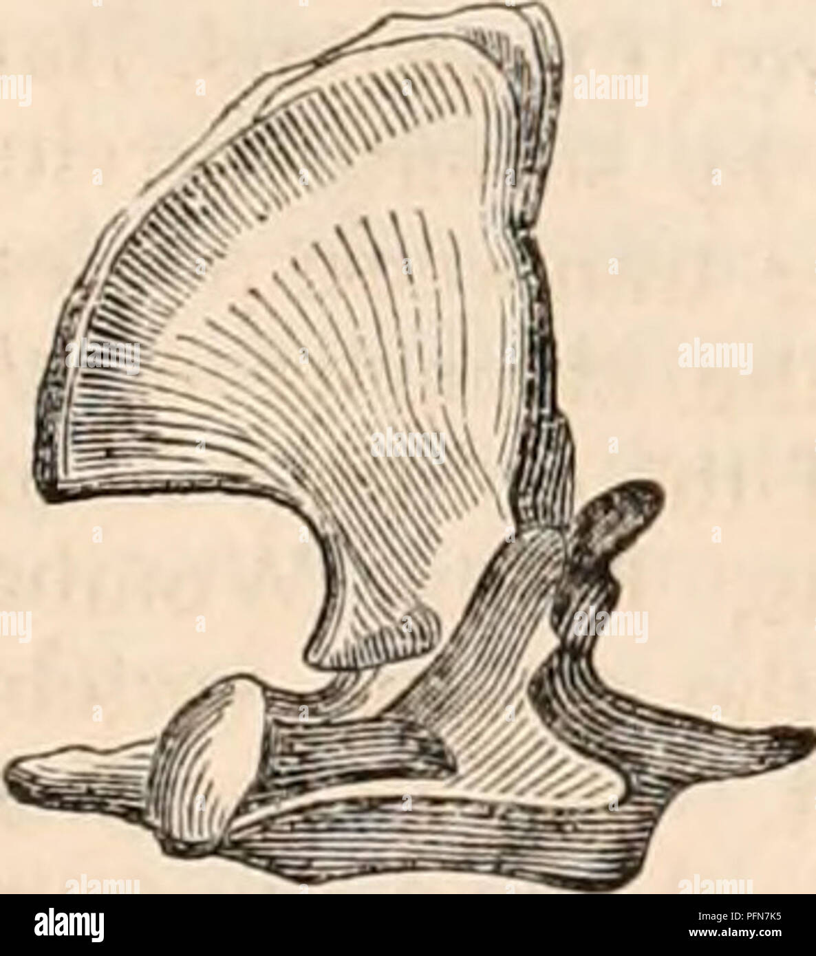 . The cyclopædia of anatomy and physiology. Anatomy; Physiology; Zoology. Atlas, axis, and third cervical vertebra, Koala. and Potoroos, the atlas is completed below by an extension of ossification from the neurapophyses into the cartilaginous nucleus representing the body, and the ring of the vertebra is for a long time interrupted by a longitudinal fissure in the middle line, the breadth of which diminishes with age. This fissure is represented in figures of the atlas of a Potoroo and Kangaroo, given by Pander and D'Alton, ( Beutelthiere,Jig. c, plates iii. &vii.); but in some of the ske Stock Photohttps://www.alamy.com/image-license-details/?v=1https://www.alamy.com/the-cyclopdia-of-anatomy-and-physiology-anatomy-physiology-zoology-atlas-axis-and-third-cervical-vertebra-koala-and-potoroos-the-atlas-is-completed-below-by-an-extension-of-ossification-from-the-neurapophyses-into-the-cartilaginous-nucleus-representing-the-body-and-the-ring-of-the-vertebra-is-for-a-long-time-interrupted-by-a-longitudinal-fissure-in-the-middle-line-the-breadth-of-which-diminishes-with-age-this-fissure-is-represented-in-figures-of-the-atlas-of-a-potoroo-and-kangaroo-given-by-pander-and-dalton-beutelthierejig-c-plates-iii-ampvii-but-in-some-of-the-ske-image216211273.html
RMPFN7K5–. The cyclopædia of anatomy and physiology. Anatomy; Physiology; Zoology. Atlas, axis, and third cervical vertebra, Koala. and Potoroos, the atlas is completed below by an extension of ossification from the neurapophyses into the cartilaginous nucleus representing the body, and the ring of the vertebra is for a long time interrupted by a longitudinal fissure in the middle line, the breadth of which diminishes with age. This fissure is represented in figures of the atlas of a Potoroo and Kangaroo, given by Pander and D'Alton, ( Beutelthiere,Jig. c, plates iii. &vii.); but in some of the ske
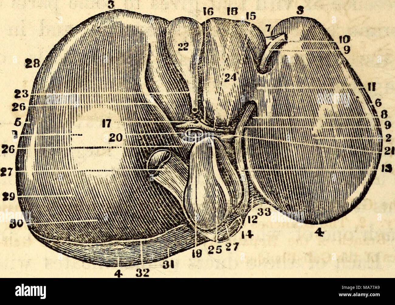 . Elementary anatomy and physiology : for colleges, academies, and other schools . The Inferior or Concave Surface of the Liver, showing its Subdivisions into Lobes. I, Center of the Light Lobe. 2, Center of the Left Lobe. 3, Its Anterior, Inferior, or Thin Margin. 4, Its Posterior, Thick or Diaphragmatic Portion. 5, The Light Extrem- ity. 6, The Left Extremity. 7, The Notch on the Anterior Margin. 8, The Umbilical or Longitudinal Fissure. 9, The Hound Ligament or remains of the Umbilical Vein. 10, The Portion of the Suspensory Ligament in connection with the Round Ligament. II, Pons Ilepatis, Stock Photohttps://www.alamy.com/image-license-details/?v=1https://www.alamy.com/elementary-anatomy-and-physiology-for-colleges-academies-and-other-schools-the-inferior-or-concave-surface-of-the-liver-showing-its-subdivisions-into-lobes-i-center-of-the-light-lobe-2-center-of-the-left-lobe-3-its-anterior-inferior-or-thin-margin-4-its-posterior-thick-or-diaphragmatic-portion-5-the-light-extrem-ity-6-the-left-extremity-7-the-notch-on-the-anterior-margin-8-the-umbilical-or-longitudinal-fissure-9-the-hound-ligament-or-remains-of-the-umbilical-vein-10-the-portion-of-the-suspensory-ligament-in-connection-with-the-round-ligament-ii-pons-ilepatis-image178409681.html
RMMA77A9–. Elementary anatomy and physiology : for colleges, academies, and other schools . The Inferior or Concave Surface of the Liver, showing its Subdivisions into Lobes. I, Center of the Light Lobe. 2, Center of the Left Lobe. 3, Its Anterior, Inferior, or Thin Margin. 4, Its Posterior, Thick or Diaphragmatic Portion. 5, The Light Extrem- ity. 6, The Left Extremity. 7, The Notch on the Anterior Margin. 8, The Umbilical or Longitudinal Fissure. 9, The Hound Ligament or remains of the Umbilical Vein. 10, The Portion of the Suspensory Ligament in connection with the Round Ligament. II, Pons Ilepatis,
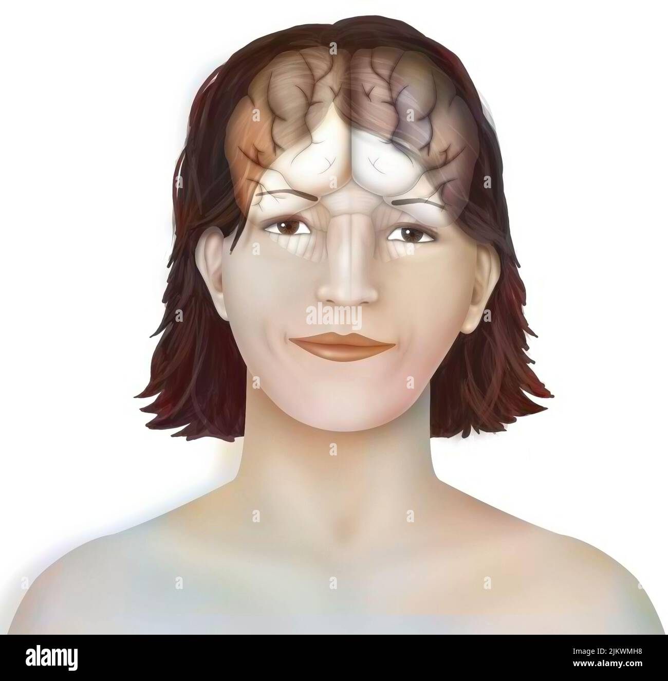 Brain: cerebral hemispheres with longitudinal fissure, cerebellum and brainstem). Stock Photohttps://www.alamy.com/image-license-details/?v=1https://www.alamy.com/brain-cerebral-hemispheres-with-longitudinal-fissure-cerebellum-and-brainstem-image476923364.html
RF2JKWMH8–Brain: cerebral hemispheres with longitudinal fissure, cerebellum and brainstem).
 Part of a human brain, showing the medial longitudinal fissure that separates the two hemispheres. Stock Photohttps://www.alamy.com/image-license-details/?v=1https://www.alamy.com/stock-photo-part-of-a-human-brain-showing-the-medial-longitudinal-fissure-that-52079165.html
RMD0MBE5–Part of a human brain, showing the medial longitudinal fissure that separates the two hemispheres.
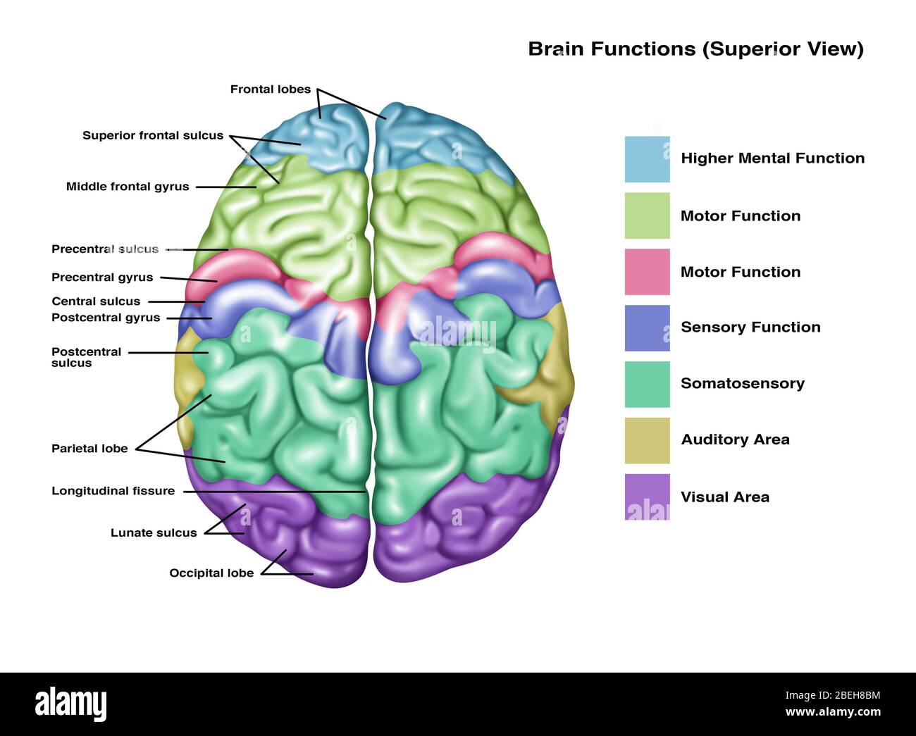 Anatomy & Functions of Brain, Illustration Stock Photohttps://www.alamy.com/image-license-details/?v=1https://www.alamy.com/anatomy-functions-of-brain-illustration-image353192328.html
RF2BEH8BM–Anatomy & Functions of Brain, Illustration
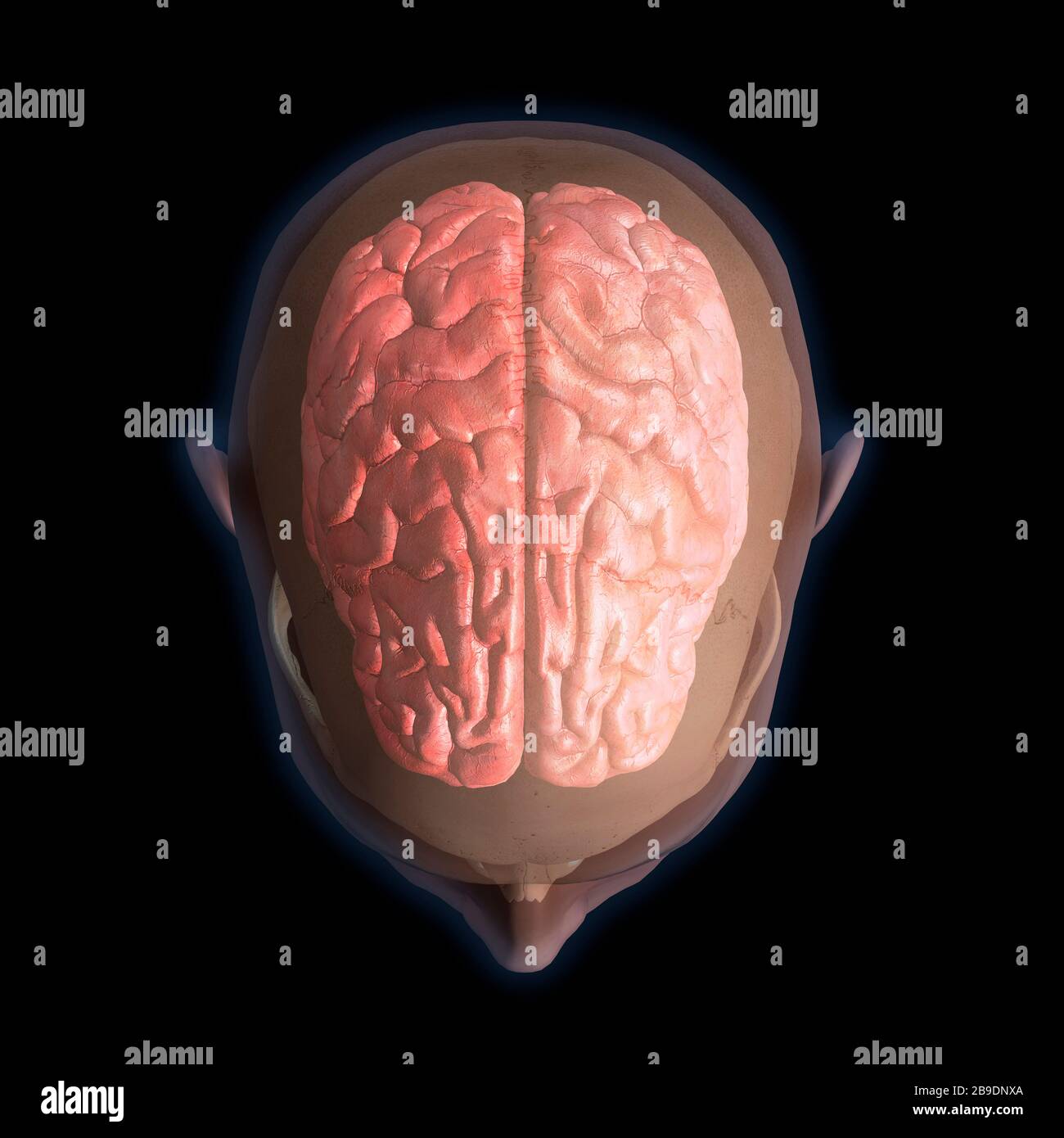 3D rendering of top view of human head and brain on black background. Stock Photohttps://www.alamy.com/image-license-details/?v=1https://www.alamy.com/3d-rendering-of-top-view-of-human-head-and-brain-on-black-background-image350041842.html
RF2B9DNXA–3D rendering of top view of human head and brain on black background.
 massive ancient twisted bark of mature old sweet chestnut tree starting to show signs of decay Stock Photohttps://www.alamy.com/image-license-details/?v=1https://www.alamy.com/stock-photo-massive-ancient-twisted-bark-of-mature-old-sweet-chestnut-tree-starting-79738062.html
RMEHMAKX–massive ancient twisted bark of mature old sweet chestnut tree starting to show signs of decay
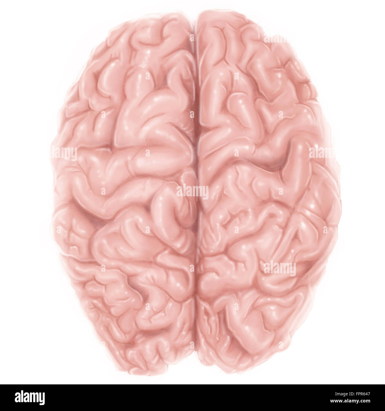 Superior view of human brain. Stock Photohttps://www.alamy.com/image-license-details/?v=1https://www.alamy.com/stock-photo-superior-view-of-human-brain-100083991.html
RFFPR647–Superior view of human brain.
 . Anthropoid apes. Apes. 194 ANTHEOPOID APES. the cerebrum, always divided from each other by a deep longitudinal fissure, overlap the cerebellum as far as a minute posterior segment. In this respect I find the brain of the gorilla a little behind the other anthropoids. Up to this time, I have only observed the projection of the cerebellum through the cerebrum in the case of an orang * (see also Fig. 56). Eetzius asserts that the cerebellum of Lapps is incompletely covered, while the covering is. Fig. 59.—Brain of orang, seen from above (Duncan, from a specimen in the Museum of Koyal College o Stock Photohttps://www.alamy.com/image-license-details/?v=1https://www.alamy.com/anthropoid-apes-apes-194-antheopoid-apes-the-cerebrum-always-divided-from-each-other-by-a-deep-longitudinal-fissure-overlap-the-cerebellum-as-far-as-a-minute-posterior-segment-in-this-respect-i-find-the-brain-of-the-gorilla-a-little-behind-the-other-anthropoids-up-to-this-time-i-have-only-observed-the-projection-of-the-cerebellum-through-the-cerebrum-in-the-case-of-an-orang-see-also-fig-56-eetzius-asserts-that-the-cerebellum-of-lapps-is-incompletely-covered-while-the-covering-is-fig-59brain-of-orang-seen-from-above-duncan-from-a-specimen-in-the-museum-of-koyal-college-o-image232125306.html
RMRDJ65E–. Anthropoid apes. Apes. 194 ANTHEOPOID APES. the cerebrum, always divided from each other by a deep longitudinal fissure, overlap the cerebellum as far as a minute posterior segment. In this respect I find the brain of the gorilla a little behind the other anthropoids. Up to this time, I have only observed the projection of the cerebellum through the cerebrum in the case of an orang * (see also Fig. 56). Eetzius asserts that the cerebellum of Lapps is incompletely covered, while the covering is. Fig. 59.—Brain of orang, seen from above (Duncan, from a specimen in the Museum of Koyal College o
 Archive image from page 673 of Cunningham's Text-book of anatomy (1914). Cunningham's Text-book of anatomy cunninghamstextb00cunn Year: 1914 ( Longitudinal fissure Corpus callosum Fornix Caudate nucleus Vena terminalis Tela chorioidea ventrieuli tertii â Thalamus â Third ventricle â Chorioid plexus Internal capsule Interventricular foramen '-''Column of fornix Anterior commissure Optic tract Infundibulum â Optic chiasma Optic nerve Substantia perforata anterior Olfactory peduncle Fig 569.âFrontal Section through the Cerebrum so as to cut through the three divisions of the lentiforra nucleus ; Stock Photohttps://www.alamy.com/image-license-details/?v=1https://www.alamy.com/archive-image-from-page-673-of-cunninghams-text-book-of-anatomy-1914-cunninghams-text-book-of-anatomy-cunninghamstextb00cunn-year-1914-longitudinal-fissure-corpus-callosum-fornix-caudate-nucleus-vena-terminalis-tela-chorioidea-ventrieuli-tertii-thalamus-third-ventricle-chorioid-plexus-internal-capsule-interventricular-foramen-column-of-fornix-anterior-commissure-optic-tract-infundibulum-optic-chiasma-optic-nerve-substantia-perforata-anterior-olfactory-peduncle-fig-569frontal-section-through-the-cerebrum-so-as-to-cut-through-the-three-divisions-of-the-lentiforra-nucleus-image264065391.html
RMW9H62R–Archive image from page 673 of Cunningham's Text-book of anatomy (1914). Cunningham's Text-book of anatomy cunninghamstextb00cunn Year: 1914 ( Longitudinal fissure Corpus callosum Fornix Caudate nucleus Vena terminalis Tela chorioidea ventrieuli tertii â Thalamus â Third ventricle â Chorioid plexus Internal capsule Interventricular foramen '-''Column of fornix Anterior commissure Optic tract Infundibulum â Optic chiasma Optic nerve Substantia perforata anterior Olfactory peduncle Fig 569.âFrontal Section through the Cerebrum so as to cut through the three divisions of the lentiforra nucleus ;
 The cerebral hemispheres are separated by a deep groove, the longitudinal cerebral fissure . Stock Photohttps://www.alamy.com/image-license-details/?v=1https://www.alamy.com/the-cerebral-hemispheres-are-separated-by-a-deep-groove-the-longitudinal-cerebral-fissure-image596583532.html
RF2WJGMAM–The cerebral hemispheres are separated by a deep groove, the longitudinal cerebral fissure .
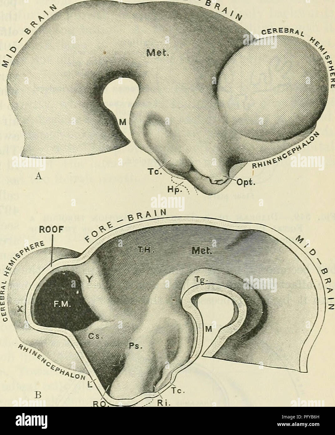 . Cunningham's Text-book of anatomy. Anatomy. CEEEBRAL HEMISPHERES. 621 FORE the vicinity of the parts which he subjacent to the parietal tuberosities of the cranium. The massive rounded character of the anterior or frontal end of each cerebral hemisphere constitutes a leading human characteristic ; but the posterior or occipital end is narrow and pointed, and is directed somewhat downwards. The two cerebral hemispheres are separated from each other by a deep median cleft, termed the longitudinal fissure. The cerebral hemisphere is formed from a small area of the extreme anterior end of the Stock Photohttps://www.alamy.com/image-license-details/?v=1https://www.alamy.com/cunninghams-text-book-of-anatomy-anatomy-ceeebral-hemispheres-621-fore-the-vicinity-of-the-parts-which-he-subjacent-to-the-parietal-tuberosities-of-the-cranium-the-massive-rounded-character-of-the-anterior-or-frontal-end-of-each-cerebral-hemisphere-constitutes-a-leading-human-characteristic-but-the-posterior-or-occipital-end-is-narrow-and-pointed-and-is-directed-somewhat-downwards-the-two-cerebral-hemispheres-are-separated-from-each-other-by-a-deep-median-cleft-termed-the-longitudinal-fissure-the-cerebral-hemisphere-is-formed-from-a-small-area-of-the-extreme-anterior-end-of-the-image216345769.html
RMPFYB6H–. Cunningham's Text-book of anatomy. Anatomy. CEEEBRAL HEMISPHERES. 621 FORE the vicinity of the parts which he subjacent to the parietal tuberosities of the cranium. The massive rounded character of the anterior or frontal end of each cerebral hemisphere constitutes a leading human characteristic ; but the posterior or occipital end is narrow and pointed, and is directed somewhat downwards. The two cerebral hemispheres are separated from each other by a deep median cleft, termed the longitudinal fissure. The cerebral hemisphere is formed from a small area of the extreme anterior end of the
 . It is scarcely ever advisable to perform neurotomy on a young horse. Some time ago, we performed the operation on a four-year-old colt, on which all other methods had been tried. The animal has since been perfectly sound, having made a good recovery. Our readers must bear in mind that the treatment of navicular disease is at best mainly unsatisfactory. It is always well to dispose of animals so affected, when an opportunity offers itself. SANDCRACK. BY a sandcr?c"u we understand a longitudinal fissure of greater or less extent in the horiy fibres of any part of the wall of the hoof, com Stock Photohttps://www.alamy.com/image-license-details/?v=1https://www.alamy.com/it-is-scarcely-ever-advisable-to-perform-neurotomy-on-a-young-horse-some-time-ago-we-performed-the-operation-on-a-four-year-old-colt-on-which-all-other-methods-had-been-tried-the-animal-has-since-been-perfectly-sound-having-made-a-good-recovery-our-readers-must-bear-in-mind-that-the-treatment-of-navicular-disease-is-at-best-mainly-unsatisfactory-it-is-always-well-to-dispose-of-animals-so-affected-when-an-opportunity-offers-itself-sandcrack-by-a-sandcrcquotu-we-understand-a-longitudinal-fissure-of-greater-or-less-extent-in-the-horiy-fibres-of-any-part-of-the-wall-of-the-hoof-com-image180026468.html
RMMCTWGM–. It is scarcely ever advisable to perform neurotomy on a young horse. Some time ago, we performed the operation on a four-year-old colt, on which all other methods had been tried. The animal has since been perfectly sound, having made a good recovery. Our readers must bear in mind that the treatment of navicular disease is at best mainly unsatisfactory. It is always well to dispose of animals so affected, when an opportunity offers itself. SANDCRACK. BY a sandcr?c"u we understand a longitudinal fissure of greater or less extent in the horiy fibres of any part of the wall of the hoof, com
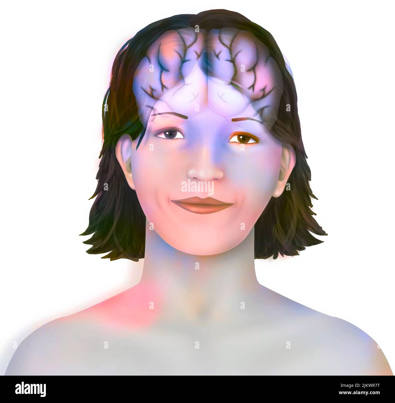 Brain (right and left cerebral hemispheres) in a woman's face. Stock Photohttps://www.alamy.com/image-license-details/?v=1https://www.alamy.com/brain-right-and-left-cerebral-hemispheres-in-a-womans-face-image476925452.html
RF2JKWR7T–Brain (right and left cerebral hemispheres) in a woman's face.
 Part of a human brain, showing the medial longitudinal fissure that separates the two hemispheres. Stock Photohttps://www.alamy.com/image-license-details/?v=1https://www.alamy.com/stock-photo-part-of-a-human-brain-showing-the-medial-longitudinal-fissure-that-52079257.html
RMD0MBHD–Part of a human brain, showing the medial longitudinal fissure that separates the two hemispheres.
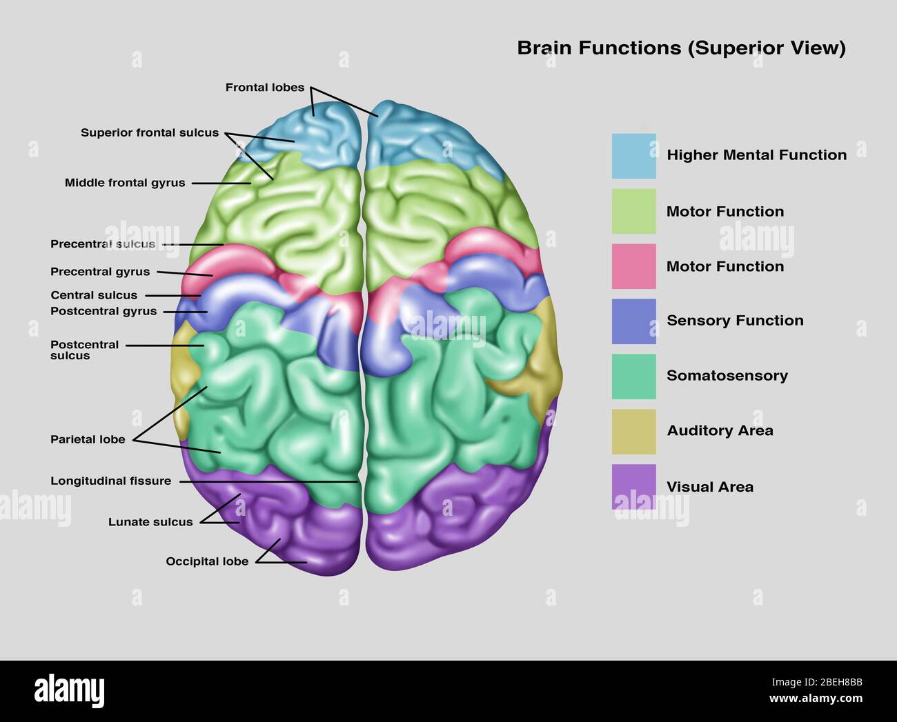 Anatomy & Functions of Brain, Illustration Stock Photohttps://www.alamy.com/image-license-details/?v=1https://www.alamy.com/anatomy-functions-of-brain-illustration-image353192319.html
RF2BEH8BB–Anatomy & Functions of Brain, Illustration
 massive ancient twisted bark of mature old sweet chestnut tree starting to show signs of decay Stock Photohttps://www.alamy.com/image-license-details/?v=1https://www.alamy.com/stock-photo-massive-ancient-twisted-bark-of-mature-old-sweet-chestnut-tree-starting-79738774.html
RMEHMBHA–massive ancient twisted bark of mature old sweet chestnut tree starting to show signs of decay
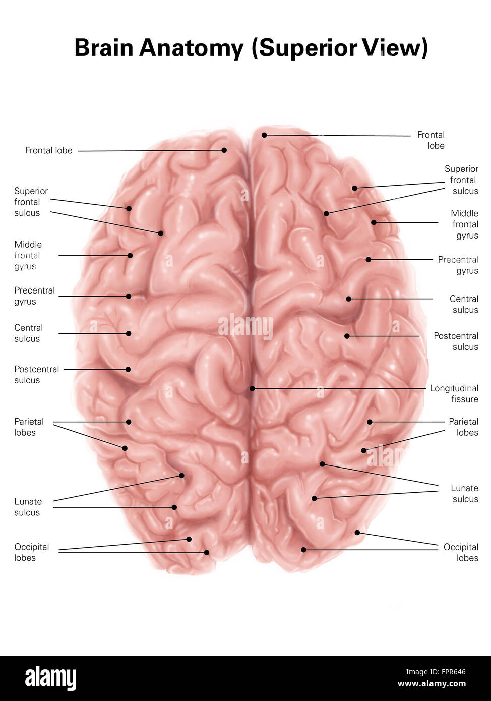 Human brain anatomy, superior view. Stock Photohttps://www.alamy.com/image-license-details/?v=1https://www.alamy.com/stock-photo-human-brain-anatomy-superior-view-100083990.html
RFFPR646–Human brain anatomy, superior view.
 . The anatomy of the horse, a dissection guide. Horses. PLATE XXXin Great longitudinal fissure between hemispheres of cerebrum Infimdibulum^ Tuber cinereun. Pons rarinl Great <»lili4ue j^ y figure Ponfa Varul: Trapezium ^ iJ' ^ * I^teiallnhe W ^^ of cerebellum i ^ ' Tnf pvraniul . StPmiwa -byW StA.K Johniror EdinWgJr.fe-LonaDn ^ BKAIN—iNFEBiou Aspect. Please note that these images are extracted from scanned page images that may have been digitally enhanced for readability - coloration and appearance of these illustrations may not perfectly resemble the original work.. McFadyean, John, S Stock Photohttps://www.alamy.com/image-license-details/?v=1https://www.alamy.com/the-anatomy-of-the-horse-a-dissection-guide-horses-plate-xxxin-great-longitudinal-fissure-between-hemispheres-of-cerebrum-infimdibulum-tuber-cinereun-pons-rarinl-great-ltlili4ue-j-y-figure-ponfa-varul-trapezium-ij-iteiallnhe-w-of-cerebellum-i-tnf-pvraniul-stpmiwa-byw-stak-johniror-edinwgjrfe-lonadn-bkaininfebiou-aspect-please-note-that-these-images-are-extracted-from-scanned-page-images-that-may-have-been-digitally-enhanced-for-readability-coloration-and-appearance-of-these-illustrations-may-not-perfectly-resemble-the-original-work-mcfadyean-john-s-image232429078.html
RMRE41JE–. The anatomy of the horse, a dissection guide. Horses. PLATE XXXin Great longitudinal fissure between hemispheres of cerebrum Infimdibulum^ Tuber cinereun. Pons rarinl Great <»lili4ue j^ y figure Ponfa Varul: Trapezium ^ iJ' ^ * I^teiallnhe W ^^ of cerebellum i ^ ' Tnf pvraniul . StPmiwa -byW StA.K Johniror EdinWgJr.fe-LonaDn ^ BKAIN—iNFEBiou Aspect. Please note that these images are extracted from scanned page images that may have been digitally enhanced for readability - coloration and appearance of these illustrations may not perfectly resemble the original work.. McFadyean, John, S
 Archive image from page 353 of Cuvier's animal kingdom arranged. Cuvier's animal kingdom : arranged according to its organization cuviersanimalkin00cuvi Year: 1840 342 MOLLUSCA. fissure ; from the exterior surface being marked witii a longitudinal gutter on one side, or with two or several gutters towards the summit ; or as that surface is smooth and without gutters. Some fossils, very much hke the Belemnites, but without a cavity, and even with a protruding basis, form the genus Actinocamax of Miller. It is upon similar conjectures that the classification of the Ammonites, Brug., or Sna Stock Photohttps://www.alamy.com/image-license-details/?v=1https://www.alamy.com/archive-image-from-page-353-of-cuviers-animal-kingdom-arranged-cuviers-animal-kingdom-arranged-according-to-its-organization-cuviersanimalkin00cuvi-year-1840-342-mollusca-fissure-from-the-exterior-surface-being-marked-witii-a-longitudinal-gutter-on-one-side-or-with-two-or-several-gutters-towards-the-summit-or-as-that-surface-is-smooth-and-without-gutters-some-fossils-very-much-hke-the-belemnites-but-without-a-cavity-and-even-with-a-protruding-basis-form-the-genus-actinocamax-of-miller-it-is-upon-similar-conjectures-that-the-classification-of-the-ammonites-brug-or-sna-image259563216.html
RMW283ET–Archive image from page 353 of Cuvier's animal kingdom arranged. Cuvier's animal kingdom : arranged according to its organization cuviersanimalkin00cuvi Year: 1840 342 MOLLUSCA. fissure ; from the exterior surface being marked witii a longitudinal gutter on one side, or with two or several gutters towards the summit ; or as that surface is smooth and without gutters. Some fossils, very much hke the Belemnites, but without a cavity, and even with a protruding basis, form the genus Actinocamax of Miller. It is upon similar conjectures that the classification of the Ammonites, Brug., or Sna
 The cerebral hemispheres are separated by a deep groove, the longitudinal cerebral fissure . Stock Photohttps://www.alamy.com/image-license-details/?v=1https://www.alamy.com/the-cerebral-hemispheres-are-separated-by-a-deep-groove-the-longitudinal-cerebral-fissure-image596591508.html
RF2WJH2FG–The cerebral hemispheres are separated by a deep groove, the longitudinal cerebral fissure .
 . The cyclopædia of anatomy and physiology. Anatomy; Physiology; Zoology. PTEROPODA. 175 either the tentacle (fig. 110. 10 and 11,*), or the orifice (fig. 110. 12, /), through which it is protruded : the two flat surfaces are separated from each other when the cowls are closed by a longitudinal fissure (p), the margins of which form two prominent lips (o o). The lateral tentacles (£) are cylindrical, smooth, and terminated by rounded extremi- ties. They are hollow, and in their interior, three longitudinal bands of muscle and a nerve of considerable size are distinguishable, so that they can b Stock Photohttps://www.alamy.com/image-license-details/?v=1https://www.alamy.com/the-cyclopdia-of-anatomy-and-physiology-anatomy-physiology-zoology-pteropoda-175-either-the-tentacle-fig-110-10-and-11-or-the-orifice-fig-110-12-through-which-it-is-protruded-the-two-flat-surfaces-are-separated-from-each-other-when-the-cowls-are-closed-by-a-longitudinal-fissure-p-the-margins-of-which-form-two-prominent-lips-o-o-the-lateral-tentacles-are-cylindrical-smooth-and-terminated-by-rounded-extremi-ties-they-are-hollow-and-in-their-interior-three-longitudinal-bands-of-muscle-and-a-nerve-of-considerable-size-are-distinguishable-so-that-they-can-b-image216211032.html
RMPFN7AG–. The cyclopædia of anatomy and physiology. Anatomy; Physiology; Zoology. PTEROPODA. 175 either the tentacle (fig. 110. 10 and 11,*), or the orifice (fig. 110. 12, /), through which it is protruded : the two flat surfaces are separated from each other when the cowls are closed by a longitudinal fissure (p), the margins of which form two prominent lips (o o). The lateral tentacles (£) are cylindrical, smooth, and terminated by rounded extremi- ties. They are hollow, and in their interior, three longitudinal bands of muscle and a nerve of considerable size are distinguishable, so that they can b
 . Elementary physiology . iiliiiiiiiliiil^v^ Fig. 81.—The liver of a young subject, sketched from below and behind. R.L., right lobe ; L.L., left lobe ; L.S., lobe of Spigelius; L.C., caudate lobe ; L.Q., quadrate lobe;/, portal fissure; ii./., umbilical fissure; g:.hL, gall-bladder; v.c.i-, vena cava inferior ; i-g., impressions on the under surface of the left lobe corresponding to the stomach ; C, position of the cardia of the stomach ; X, surface of the liver uncovered by peritoneum. Posteriorly there is a transverse fissure at right angles to the longitudinal fissure at which the vessels Stock Photohttps://www.alamy.com/image-license-details/?v=1https://www.alamy.com/elementary-physiology-iiliiiiiiiliiilv-fig-81the-liver-of-a-young-subject-sketched-from-below-and-behind-rl-right-lobe-ll-left-lobe-ls-lobe-of-spigelius-lc-caudate-lobe-lq-quadrate-lobe-portal-fissure-ii-umbilical-fissure-ghl-gall-bladder-vci-vena-cava-inferior-i-g-impressions-on-the-under-surface-of-the-left-lobe-corresponding-to-the-stomach-c-position-of-the-cardia-of-the-stomach-x-surface-of-the-liver-uncovered-by-peritoneum-posteriorly-there-is-a-transverse-fissure-at-right-angles-to-the-longitudinal-fissure-at-which-the-vessels-image178403504.html
RMMA6YDM–. Elementary physiology . iiliiiiiiiliiil^v^ Fig. 81.—The liver of a young subject, sketched from below and behind. R.L., right lobe ; L.L., left lobe ; L.S., lobe of Spigelius; L.C., caudate lobe ; L.Q., quadrate lobe;/, portal fissure; ii./., umbilical fissure; g:.hL, gall-bladder; v.c.i-, vena cava inferior ; i-g., impressions on the under surface of the left lobe corresponding to the stomach ; C, position of the cardia of the stomach ; X, surface of the liver uncovered by peritoneum. Posteriorly there is a transverse fissure at right angles to the longitudinal fissure at which the vessels
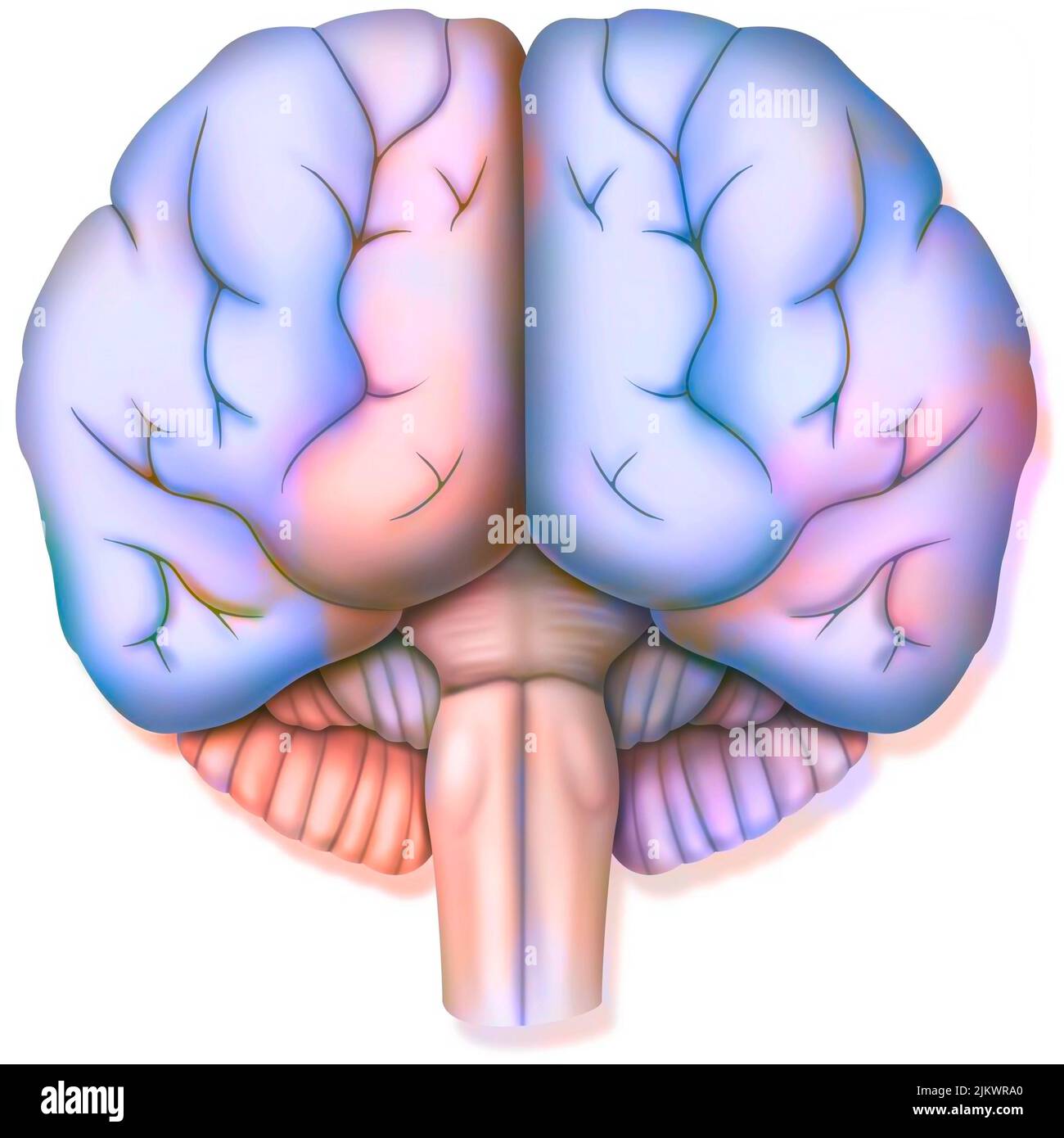 Brain, with the two cerebral hemispheres, the cerebellum and the brainstem. Stock Photohttps://www.alamy.com/image-license-details/?v=1https://www.alamy.com/brain-with-the-two-cerebral-hemispheres-the-cerebellum-and-the-brainstem-image476925512.html
RF2JKWRA0–Brain, with the two cerebral hemispheres, the cerebellum and the brainstem.
 Part of a human brain, showing the brain stem. Stock Photohttps://www.alamy.com/image-license-details/?v=1https://www.alamy.com/stock-photo-part-of-a-human-brain-showing-the-brain-stem-52079246.html
RMD0MBH2–Part of a human brain, showing the brain stem.
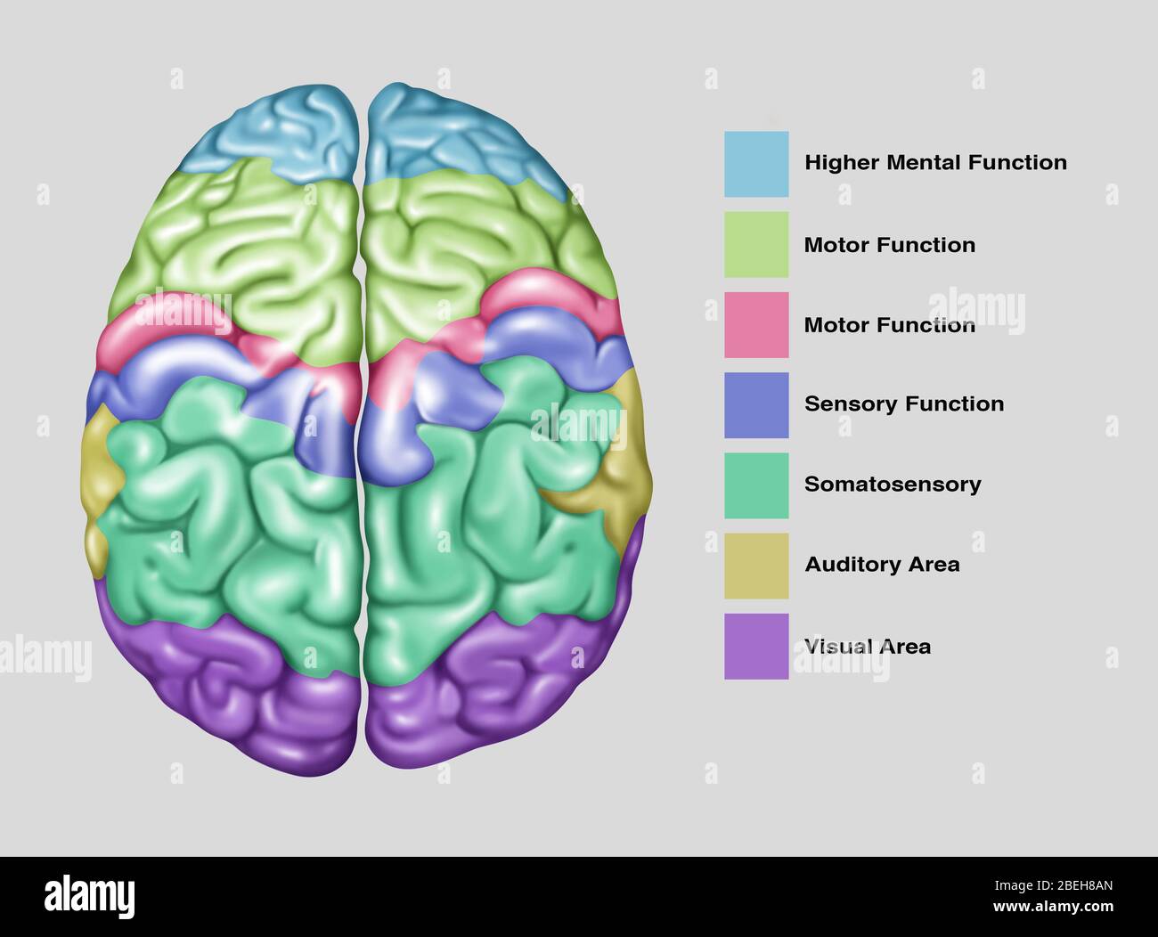 Brain Function, Illustration Stock Photohttps://www.alamy.com/image-license-details/?v=1https://www.alamy.com/brain-function-illustration-image353192301.html
RF2BEH8AN–Brain Function, Illustration
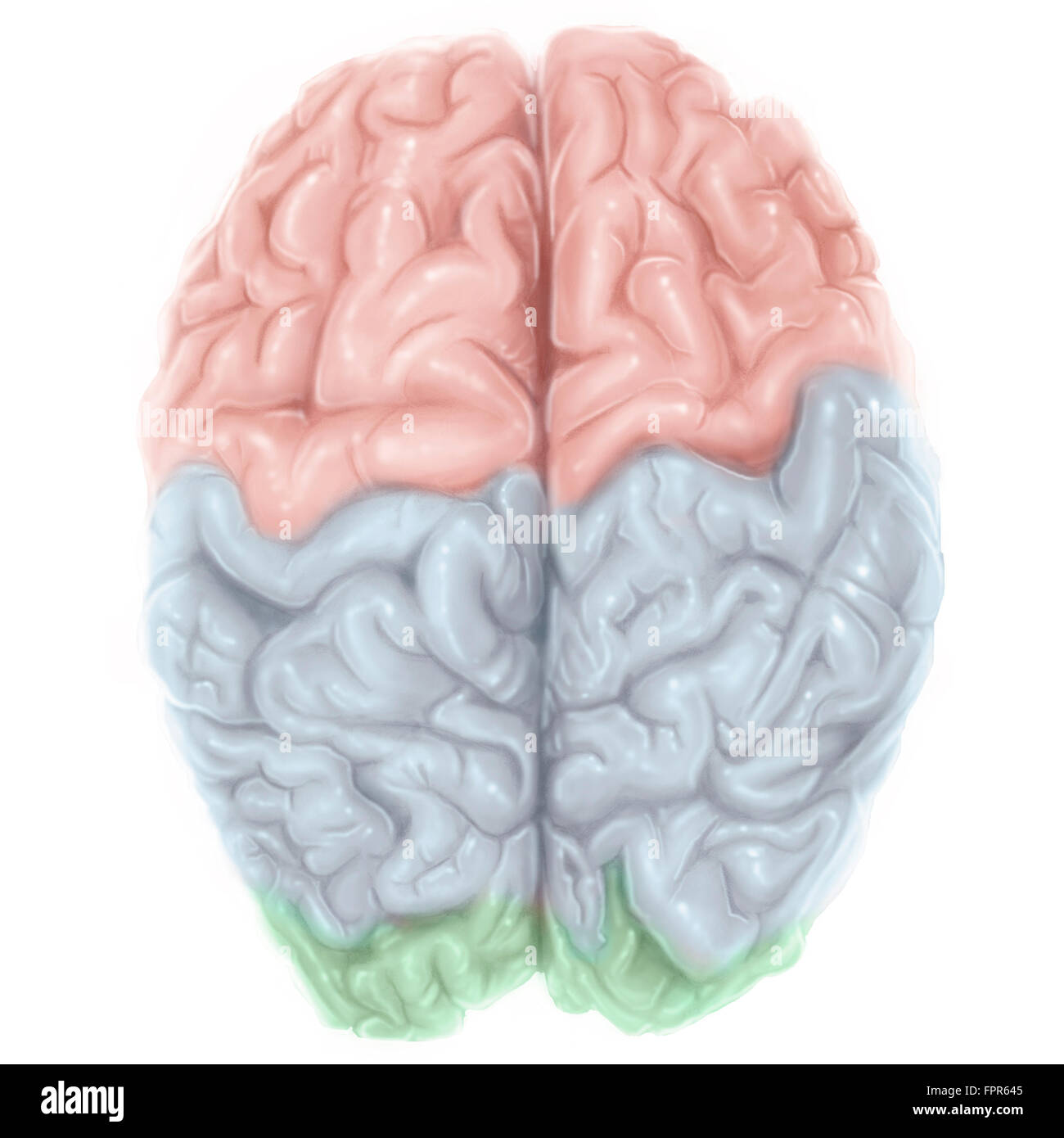 Superior view of human brain with colored lobes. Stock Photohttps://www.alamy.com/image-license-details/?v=1https://www.alamy.com/stock-photo-superior-view-of-human-brain-with-colored-lobes-100083989.html
RFFPR645–Superior view of human brain with colored lobes.
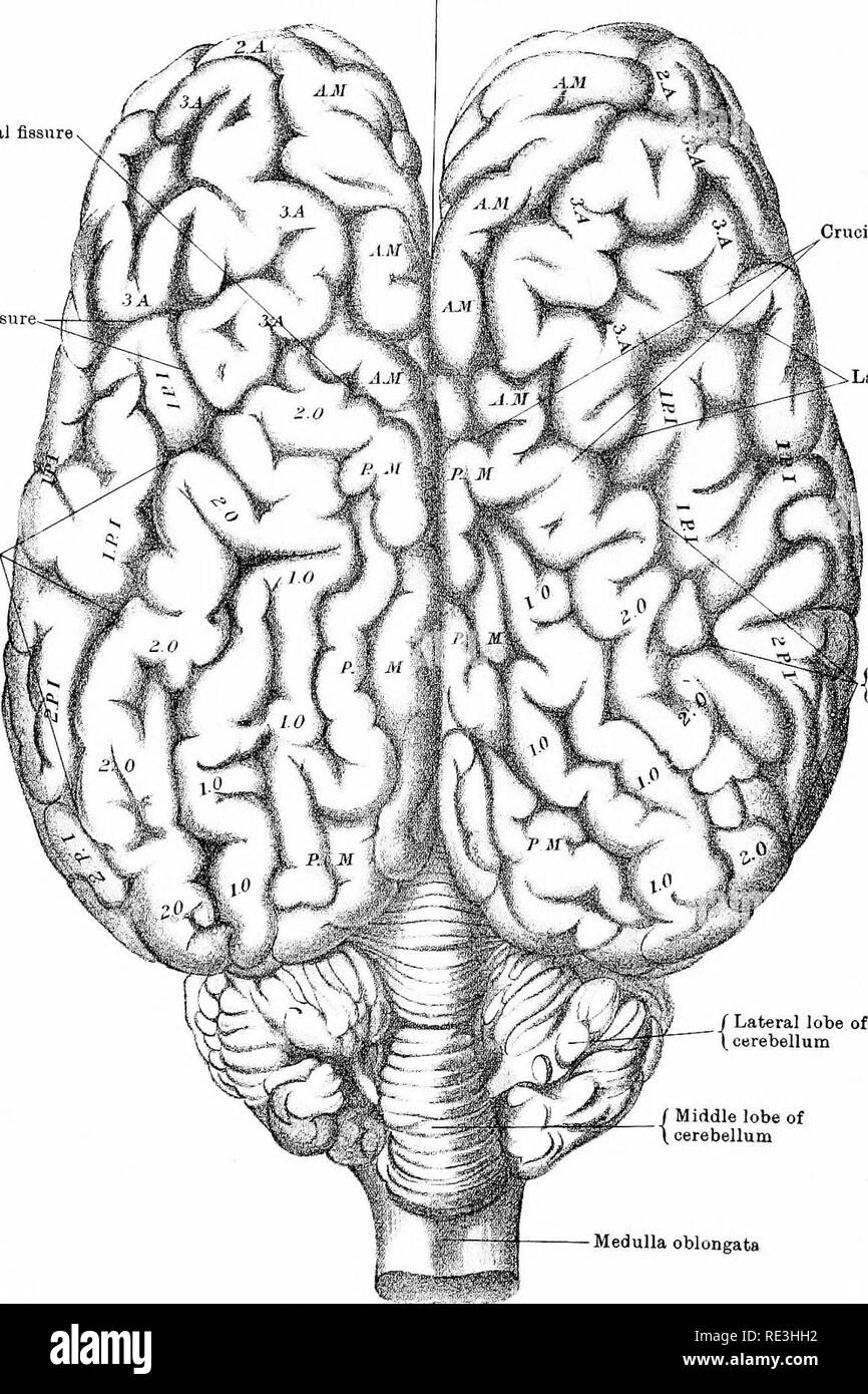 . The anatomy of the horse, a dissection guide. Horses. PLATE XXXIV Great longitudinal fissure between hemispheres of cerebrum Crucial fissure V ^ Lateral fis&ure- Crucial fiasure Lateral fissure . Great oblique fissure. J Great obliqj| ^ / Lateral lobe of oe- erebellum Middle lobe of cerebellum Medulla oblongata DrawTL ALil±i,o;^raplLed "byW A A E JolmjrttJii Lumtdd Edmlrur^ i lonim BRAIN—Superior Aspect. Please note that these images are extracted from scanned page images that may have been digitally enhanced for readability - coloration and appearance of these illustrations m Stock Photohttps://www.alamy.com/image-license-details/?v=1https://www.alamy.com/the-anatomy-of-the-horse-a-dissection-guide-horses-plate-xxxiv-great-longitudinal-fissure-between-hemispheres-of-cerebrum-crucial-fissure-v-lateral-fisampure-crucial-fiasure-lateral-fissure-great-oblique-fissure-j-great-obliqj-lateral-lobe-of-oe-erebellum-middle-lobe-of-cerebellum-medulla-oblongata-drawtl-alilioraplled-quotbyw-a-a-e-jolmjrttjii-lumtdd-edmlrur-i-lonim-brainsuperior-aspect-please-note-that-these-images-are-extracted-from-scanned-page-images-that-may-have-been-digitally-enhanced-for-readability-coloration-and-appearance-of-these-illustrations-m-image232419630.html
RMRE3HH2–. The anatomy of the horse, a dissection guide. Horses. PLATE XXXIV Great longitudinal fissure between hemispheres of cerebrum Crucial fissure V ^ Lateral fis&ure- Crucial fiasure Lateral fissure . Great oblique fissure. J Great obliqj| ^ / Lateral lobe of oe- erebellum Middle lobe of cerebellum Medulla oblongata DrawTL ALil±i,o;^raplLed "byW A A E JolmjrttJii Lumtdd Edmlrur^ i lonim BRAIN—Superior Aspect. Please note that these images are extracted from scanned page images that may have been digitally enhanced for readability - coloration and appearance of these illustrations m
 Archive image from page 353 of Cuvier's animal kingdom arranged. Cuvier's animal kingdom : arranged according to its organization cuviersanimalkin00cuvi Year: 1840 fissure ; from the exterior surface being marked witii a longitudinal gutter on one side, or with two or several gutters towards the summit ; or as that surface is smooth and without gutters. Some fossils, very much hke the Belemnites, but without a cavity, and even with a protruding basis, form the genus Actinocamax of Miller. It is upon similar conjectures that the classification of the Ammonites, Brug., or Snake-stones,— Is fo Stock Photohttps://www.alamy.com/image-license-details/?v=1https://www.alamy.com/archive-image-from-page-353-of-cuviers-animal-kingdom-arranged-cuviers-animal-kingdom-arranged-according-to-its-organization-cuviersanimalkin00cuvi-year-1840-fissure-from-the-exterior-surface-being-marked-witii-a-longitudinal-gutter-on-one-side-or-with-two-or-several-gutters-towards-the-summit-or-as-that-surface-is-smooth-and-without-gutters-some-fossils-very-much-hke-the-belemnites-but-without-a-cavity-and-even-with-a-protruding-basis-form-the-genus-actinocamax-of-miller-it-is-upon-similar-conjectures-that-the-classification-of-the-ammonites-brug-or-snake-stones-is-fo-image259563187.html
RMW283DR–Archive image from page 353 of Cuvier's animal kingdom arranged. Cuvier's animal kingdom : arranged according to its organization cuviersanimalkin00cuvi Year: 1840 fissure ; from the exterior surface being marked witii a longitudinal gutter on one side, or with two or several gutters towards the summit ; or as that surface is smooth and without gutters. Some fossils, very much hke the Belemnites, but without a cavity, and even with a protruding basis, form the genus Actinocamax of Miller. It is upon similar conjectures that the classification of the Ammonites, Brug., or Snake-stones,— Is fo
 The cerebral hemispheres are separated by a deep groove, the longitudinal cerebral fissure . Stock Photohttps://www.alamy.com/image-license-details/?v=1https://www.alamy.com/the-cerebral-hemispheres-are-separated-by-a-deep-groove-the-longitudinal-cerebral-fissure-image596591758.html
RF2WJH2TE–The cerebral hemispheres are separated by a deep groove, the longitudinal cerebral fissure .
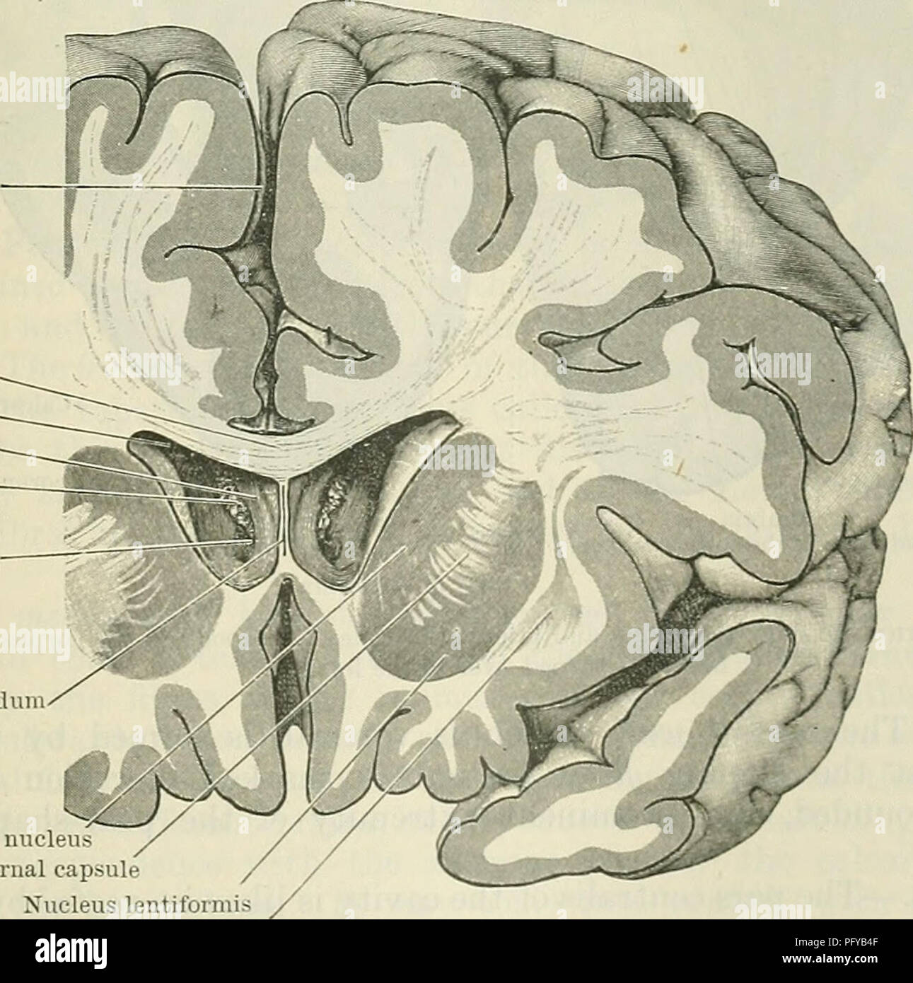 . Cunningham's Text-book of anatomy. Anatomy. Longitudinal fissure Corpus callosum Lateral ventricle Column of fornix Chorioid plexus Foramen inter- ventrieulare Septum pellucidum. Caudate nucleus Internal capsule Nucleus lentiformis Claustrum Fig. 562.—Frontal Section through the Cerebbal Hemispheres so as to cut through the anterior horns of the lateral ventricles, through which the central part of the ventricles, the columns of the fornix, and the interventricular foramina can be seen. ticulum gated which or elon- pouch, grows. Please note that these images are extracted from scanned page i Stock Photohttps://www.alamy.com/image-license-details/?v=1https://www.alamy.com/cunninghams-text-book-of-anatomy-anatomy-longitudinal-fissure-corpus-callosum-lateral-ventricle-column-of-fornix-chorioid-plexus-foramen-inter-ventrieulare-septum-pellucidum-caudate-nucleus-internal-capsule-nucleus-lentiformis-claustrum-fig-562frontal-section-through-the-cerebbal-hemispheres-so-as-to-cut-through-the-anterior-horns-of-the-lateral-ventricles-through-which-the-central-part-of-the-ventricles-the-columns-of-the-fornix-and-the-interventricular-foramina-can-be-seen-ticulum-gated-which-or-elon-pouch-grows-please-note-that-these-images-are-extracted-from-scanned-page-i-image216345711.html
RMPFYB4F–. Cunningham's Text-book of anatomy. Anatomy. Longitudinal fissure Corpus callosum Lateral ventricle Column of fornix Chorioid plexus Foramen inter- ventrieulare Septum pellucidum. Caudate nucleus Internal capsule Nucleus lentiformis Claustrum Fig. 562.—Frontal Section through the Cerebbal Hemispheres so as to cut through the anterior horns of the lateral ventricles, through which the central part of the ventricles, the columns of the fornix, and the interventricular foramina can be seen. ticulum gated which or elon- pouch, grows. Please note that these images are extracted from scanned page i
 . Fig. 234.—A few leaves enlarged from Fig. 233. The leaf to left hand bears pycnidia on red spots on the upper surface of the leaf; the remaining leaves bear aecidia on raised portions of their surface. Several aecidia still further enlarged show the peridia dehiscing by longitudinal slits, (v. Tubeuf del.) tissues occur in the year-rings as already described for G. davariaeforme, viz. thickened twisted tracheids, loosely connected together and with fissure-like pits; medullary rays more numerous and broader; the limits of the year-ring difficult to distinguish; and a yellow pigment deposited Stock Photohttps://www.alamy.com/image-license-details/?v=1https://www.alamy.com/fig-234a-few-leaves-enlarged-from-fig-233-the-leaf-to-left-hand-bears-pycnidia-on-red-spots-on-the-upper-surface-of-the-leaf-the-remaining-leaves-bear-aecidia-on-raised-portions-of-their-surface-several-aecidia-still-further-enlarged-show-the-peridia-dehiscing-by-longitudinal-slits-v-tubeuf-del-tissues-occur-in-the-year-rings-as-already-described-for-g-davariaeforme-viz-thickened-twisted-tracheids-loosely-connected-together-and-with-fissure-like-pits-medullary-rays-more-numerous-and-broader-the-limits-of-the-year-ring-difficult-to-distinguish-and-a-yellow-pigment-deposited-image179901739.html
RMMCK6E3–. Fig. 234.—A few leaves enlarged from Fig. 233. The leaf to left hand bears pycnidia on red spots on the upper surface of the leaf; the remaining leaves bear aecidia on raised portions of their surface. Several aecidia still further enlarged show the peridia dehiscing by longitudinal slits, (v. Tubeuf del.) tissues occur in the year-rings as already described for G. davariaeforme, viz. thickened twisted tracheids, loosely connected together and with fissure-like pits; medullary rays more numerous and broader; the limits of the year-ring difficult to distinguish; and a yellow pigment deposited
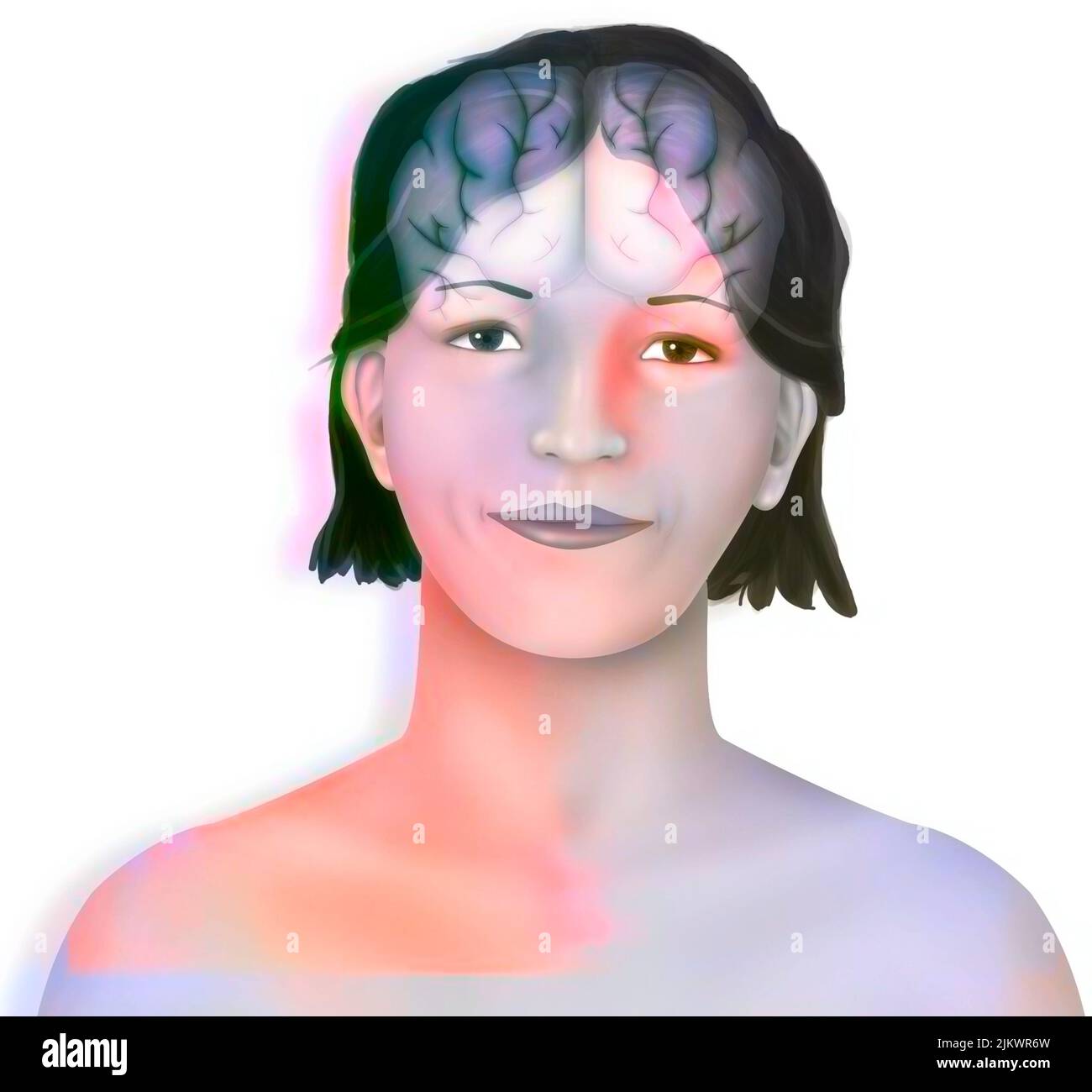 Brain (right and left cerebral hemispheres) in a woman's face. Stock Photohttps://www.alamy.com/image-license-details/?v=1https://www.alamy.com/brain-right-and-left-cerebral-hemispheres-in-a-womans-face-image476925425.html
RF2JKWR6W–Brain (right and left cerebral hemispheres) in a woman's face.
 Superior view of an anatomical model of the brain and its blood supply. Stock Photohttps://www.alamy.com/image-license-details/?v=1https://www.alamy.com/stock-photo-superior-view-of-an-anatomical-model-of-the-brain-and-its-blood-supply-52097475.html
RMD0N6T3–Superior view of an anatomical model of the brain and its blood supply.
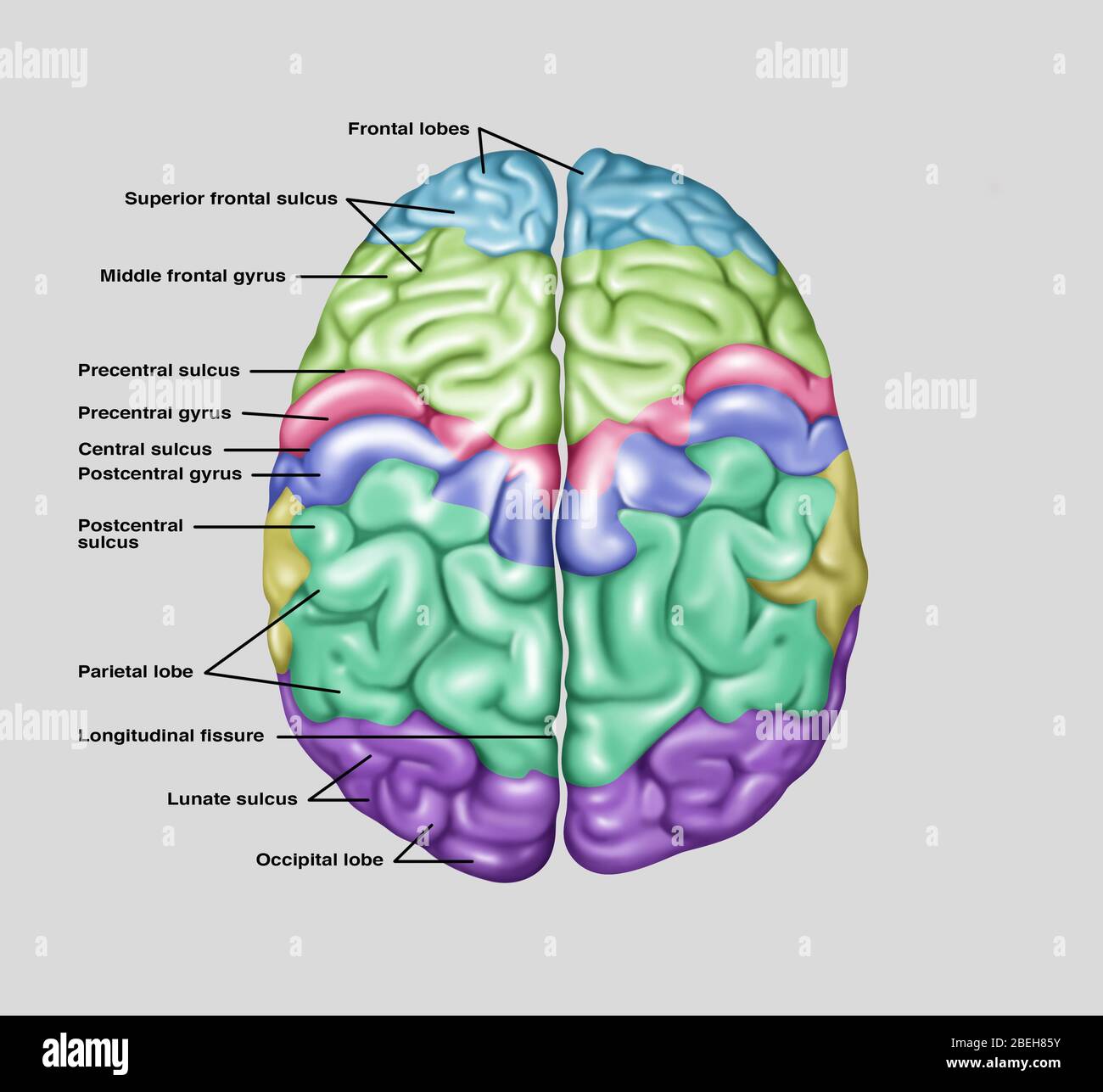 Top View of Normal Brain, Illustration Stock Photohttps://www.alamy.com/image-license-details/?v=1https://www.alamy.com/top-view-of-normal-brain-illustration-image353192167.html
RF2BEH85Y–Top View of Normal Brain, Illustration
 Illustration showing anatomy of a normal brain in a superior (top) view. Noted from top to bottom on the left side are the following: frontal lobes, superior frontal sulcus, middle frontal gyrus, precentral sulcus, precentral gyrus, central sulcus, postce Stock Photohttps://www.alamy.com/image-license-details/?v=1https://www.alamy.com/stock-photo-illustration-showing-anatomy-of-a-normal-brain-in-a-superior-top-view-103992549.html
RMG157FH–Illustration showing anatomy of a normal brain in a superior (top) view. Noted from top to bottom on the left side are the following: frontal lobes, superior frontal sulcus, middle frontal gyrus, precentral sulcus, precentral gyrus, central sulcus, postce
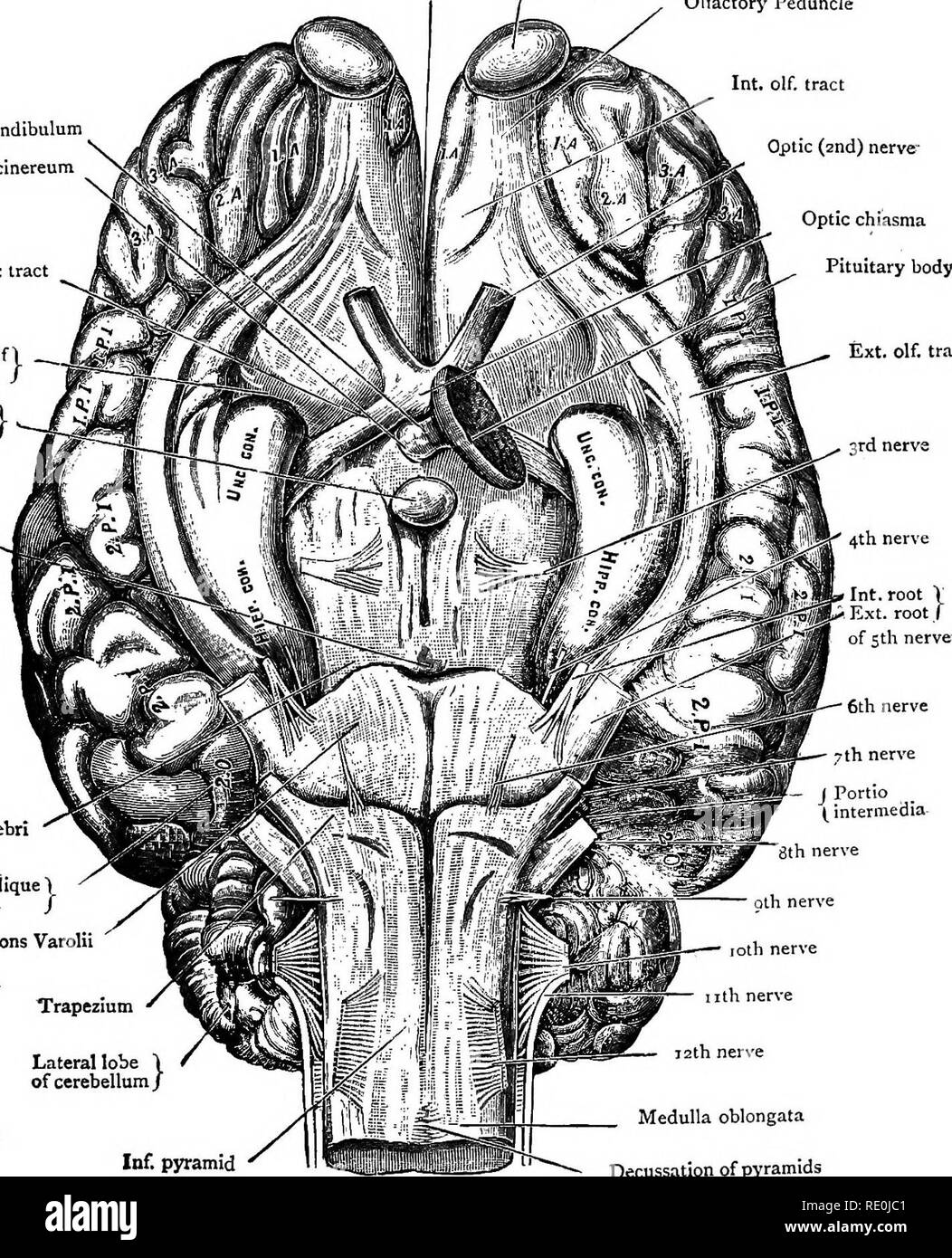 . Diseases & disorders of the horse, a treatise on equine medicine and surgery, being a contribution to the science of comparative pathology. Horses. 93 Great longitudinal fissure between hemispheres of cerebrum Olfactory bulb Infundibulum Tuber cinereum Optic tract Fissure of 1 Sylvius / Corpus albicans / â Pons Tarini/ Olfactory Peduncle Int olf tract Optic (2nd) nerve- Optic chiasma Pituitary body fixt. olf. tract 3rd nerve. Crus cerebri Great oblique fissure- / Pons Varolii U'rapezmm Lateral lobe of cerebellum/ Inf. pyramid Medulla oblongata Decussation of pyramids BRAINâ^Infer Stock Photohttps://www.alamy.com/image-license-details/?v=1https://www.alamy.com/diseases-amp-disorders-of-the-horse-a-treatise-on-equine-medicine-and-surgery-being-a-contribution-to-the-science-of-comparative-pathology-horses-93-great-longitudinal-fissure-between-hemispheres-of-cerebrum-olfactory-bulb-infundibulum-tuber-cinereum-optic-tract-fissure-of-1-sylvius-corpus-albicans-pons-tarini-olfactory-peduncle-int-olf-tract-optic-2nd-nerve-optic-chiasma-pituitary-body-fixt-olf-tract-3rd-nerve-crus-cerebri-great-oblique-fissure-pons-varolii-urapezmm-lateral-lobe-of-cerebellum-inf-pyramid-medulla-oblongata-decussation-of-pyramids-braininfer-image232354417.html
RMRE0JC1–. Diseases & disorders of the horse, a treatise on equine medicine and surgery, being a contribution to the science of comparative pathology. Horses. 93 Great longitudinal fissure between hemispheres of cerebrum Olfactory bulb Infundibulum Tuber cinereum Optic tract Fissure of 1 Sylvius / Corpus albicans / â Pons Tarini/ Olfactory Peduncle Int olf tract Optic (2nd) nerve- Optic chiasma Pituitary body fixt. olf. tract 3rd nerve. Crus cerebri Great oblique fissure- / Pons Varolii U'rapezmm Lateral lobe of cerebellum/ Inf. pyramid Medulla oblongata Decussation of pyramids BRAINâ^Infer
 Diseases & disorders of the Diseases & disorders of the horse : a treatise on equine medicine and surgery diseasesdisorder00gres Year: 1886 93 Great longitudinal fissure between hemispheres of cerebrum Olfactory bull) Infundibulum Tuber cinereun 'J. I'l Optic tract / L W / Fissure Fissure of 1 —Jj'W # K, Sylvius / rir#—-' Stock Photohttps://www.alamy.com/image-license-details/?v=1https://www.alamy.com/diseases-disorders-of-the-diseases-disorders-of-the-horse-a-treatise-on-equine-medicine-and-surgery-diseasesdisorder00gres-year-1886-93-great-longitudinal-fissure-between-hemispheres-of-cerebrum-olfactory-bull-infundibulum-tuber-cinereun-j-il-optic-tract-l-w-fissure-fissure-of-1-jjw-k-sylvius-rir-image241944033.html
RMT1HE29–Diseases & disorders of the Diseases & disorders of the horse : a treatise on equine medicine and surgery diseasesdisorder00gres Year: 1886 93 Great longitudinal fissure between hemispheres of cerebrum Olfactory bull) Infundibulum Tuber cinereun 'J. I'l Optic tract / L W / Fissure Fissure of 1 —Jj'W # K, Sylvius / rir#—-'
 The cerebral hemispheres are separated by a deep groove, the longitudinal cerebral fissure . Stock Photohttps://www.alamy.com/image-license-details/?v=1https://www.alamy.com/the-cerebral-hemispheres-are-separated-by-a-deep-groove-the-longitudinal-cerebral-fissure-image596588712.html
RF2WJGXYM–The cerebral hemispheres are separated by a deep groove, the longitudinal cerebral fissure .
 . On the anatomy of vertebrates. Vertebrates; Anatomy, Comparative; 1866. 0-20 ANATOMY OF VERTEBRATES. 428 largest at one end, and impressed by a slight longitudinal fissure. The rudiments of the neural axis are first recognisable in the two parallel longitudinal elevations (' primitive trace' or ' laminaj dorsales,' ib. m, n) bordering the fissure. Beneath these is at the same time forming the notochordal rudiment of the vertebral column, ib. c. The albuminous principle is concentrated in m, the gelatinous one in c: this chemical differentiation does not aifect n. The polyhedral cells extend Stock Photohttps://www.alamy.com/image-license-details/?v=1https://www.alamy.com/on-the-anatomy-of-vertebrates-vertebrates-anatomy-comparative-1866-0-20-anatomy-of-vertebrates-428-largest-at-one-end-and-impressed-by-a-slight-longitudinal-fissure-the-rudiments-of-the-neural-axis-are-first-recognisable-in-the-two-parallel-longitudinal-elevations-primitive-trace-or-laminaj-dorsales-ib-m-n-bordering-the-fissure-beneath-these-is-at-the-same-time-forming-the-notochordal-rudiment-of-the-vertebral-column-ib-c-the-albuminous-principle-is-concentrated-in-m-the-gelatinous-one-in-c-this-chemical-differentiation-does-not-aifect-n-the-polyhedral-cells-extend-image216398853.html
RMPG1PXD–. On the anatomy of vertebrates. Vertebrates; Anatomy, Comparative; 1866. 0-20 ANATOMY OF VERTEBRATES. 428 largest at one end, and impressed by a slight longitudinal fissure. The rudiments of the neural axis are first recognisable in the two parallel longitudinal elevations (' primitive trace' or ' laminaj dorsales,' ib. m, n) bordering the fissure. Beneath these is at the same time forming the notochordal rudiment of the vertebral column, ib. c. The albuminous principle is concentrated in m, the gelatinous one in c: this chemical differentiation does not aifect n. The polyhedral cells extend
 . Fig. 234.—A few leaves enlarged from Fig. 233. The leaf to left hand bears pycnidia on red spots on the upper surface of the leaf; the remaining le;ives bear aecidia on raised portions of their surface. Several aecidia still furtlier enlarged show the peridia dehiscing by longitudinal slits, (v. Tubcuf del.) tissues occur in the year-rings as already described for G. clavariaeforme, viz. thickened twisted tracheids, loosely connected together and with fissure-like pits; medullary rays more numerous and broader; the limits of the year-ring difficult to distinguish; and a yellow pigment deposi Stock Photohttps://www.alamy.com/image-license-details/?v=1https://www.alamy.com/fig-234a-few-leaves-enlarged-from-fig-233-the-leaf-to-left-hand-bears-pycnidia-on-red-spots-on-the-upper-surface-of-the-leaf-the-remaining-leives-bear-aecidia-on-raised-portions-of-their-surface-several-aecidia-still-furtlier-enlarged-show-the-peridia-dehiscing-by-longitudinal-slits-v-tubcuf-del-tissues-occur-in-the-year-rings-as-already-described-for-g-clavariaeforme-viz-thickened-twisted-tracheids-loosely-connected-together-and-with-fissure-like-pits-medullary-rays-more-numerous-and-broader-the-limits-of-the-year-ring-difficult-to-distinguish-and-a-yellow-pigment-deposi-image179901413.html
RMMCK62D–. Fig. 234.—A few leaves enlarged from Fig. 233. The leaf to left hand bears pycnidia on red spots on the upper surface of the leaf; the remaining le;ives bear aecidia on raised portions of their surface. Several aecidia still furtlier enlarged show the peridia dehiscing by longitudinal slits, (v. Tubcuf del.) tissues occur in the year-rings as already described for G. clavariaeforme, viz. thickened twisted tracheids, loosely connected together and with fissure-like pits; medullary rays more numerous and broader; the limits of the year-ring difficult to distinguish; and a yellow pigment deposi
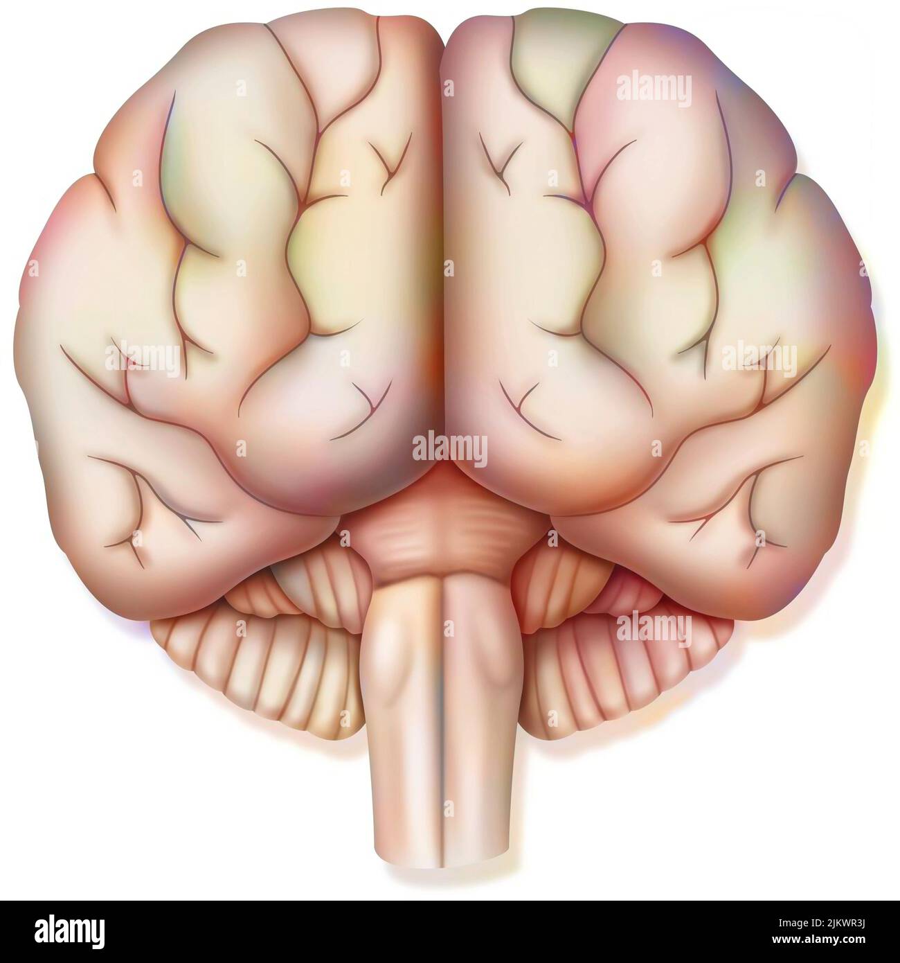 Brain, with the two cerebral hemispheres, the cerebellum and the brainstem. Stock Photohttps://www.alamy.com/image-license-details/?v=1https://www.alamy.com/brain-with-the-two-cerebral-hemispheres-the-cerebellum-and-the-brainstem-image476925334.html
RF2JKWR3J–Brain, with the two cerebral hemispheres, the cerebellum and the brainstem.
 A stylized close up view of the front of the head. The brain is visible within the skull. Stock Photohttps://www.alamy.com/image-license-details/?v=1https://www.alamy.com/stock-photo-a-stylized-close-up-view-of-the-front-of-the-head-the-brain-is-visible-52095178.html
RMD0N3X2–A stylized close up view of the front of the head. The brain is visible within the skull.
 . The anatomy of the horse, a dissection guide. Horses. PLATE XXXJII Great longitudinal fissure between hemispheres of cerebrum Olfactory bulb Olfactory Peduncle Infundibulum Tuber cinereum Optic tract Fissure of Sylvius / Int o!f. tract Optic (2nd) nerve Optic chiasma - Pituitary body Ext olf. tract Corpus albicans /. Crus cerebi Great obliqu' fissure j 6th nerve neri?e Portio intermedia 8th nerve Ponb ai(jlii-^ Trapeziiiin Ldteial lobt Inf. pyramid. 10th nerve — 11th nerve 12th nerve Medulla oblongata Decussation of pyramids DrsBra. A litLo^ajilied "byW 4 AJE Jaliiifltcoo..Lmiil Stock Photohttps://www.alamy.com/image-license-details/?v=1https://www.alamy.com/the-anatomy-of-the-horse-a-dissection-guide-horses-plate-xxxjii-great-longitudinal-fissure-between-hemispheres-of-cerebrum-olfactory-bulb-olfactory-peduncle-infundibulum-tuber-cinereum-optic-tract-fissure-of-sylvius-int-o!f-tract-optic-2nd-nerve-optic-chiasma-pituitary-body-ext-olf-tract-corpus-albicans-crus-cerebi-great-obliqu-fissure-j-6th-nerve-nerie-portio-intermedia-8th-nerve-ponb-aijlii-trapeziiiin-ldteial-lobt-inf-pyramid-10th-nerve-11th-nerve-12th-nerve-medulla-oblongata-decussation-of-pyramids-drsbra-a-litloajilied-quotbyw-4-aje-jaliiifltcoolmiil-image232419634.html
RMRE3HH6–. The anatomy of the horse, a dissection guide. Horses. PLATE XXXJII Great longitudinal fissure between hemispheres of cerebrum Olfactory bulb Olfactory Peduncle Infundibulum Tuber cinereum Optic tract Fissure of Sylvius / Int o!f. tract Optic (2nd) nerve Optic chiasma - Pituitary body Ext olf. tract Corpus albicans /. Crus cerebi Great obliqu' fissure j 6th nerve neri?e Portio intermedia 8th nerve Ponb ai(jlii-^ Trapeziiiin Ldteial lobt Inf. pyramid. 10th nerve — 11th nerve 12th nerve Medulla oblongata Decussation of pyramids DrsBra. A litLo^ajilied "byW 4 AJE Jaliiifltcoo..Lmiil
 Diseases & disorders of the Diseases & disorders of the horse : a treatise on equine medicine and surgery diseasesdisorder00gres Year: 1886 92 Great longitudinal fissure between hemispheres of cerebrum Crucial fissure i.ateral fissure Ireat oblique fissure Crucial fissure Lateral fissure Great oblique fissure / Lateral lobe of I cerebellum Middle lobe of t cerebellum Medulla oblongata BRAIN—Superior Aspect. Stock Photohttps://www.alamy.com/image-license-details/?v=1https://www.alamy.com/diseases-disorders-of-the-diseases-disorders-of-the-horse-a-treatise-on-equine-medicine-and-surgery-diseasesdisorder00gres-year-1886-92-great-longitudinal-fissure-between-hemispheres-of-cerebrum-crucial-fissure-iateral-fissure-ireat-oblique-fissure-crucial-fissure-lateral-fissure-great-oblique-fissure-lateral-lobe-of-i-cerebellum-middle-lobe-of-t-cerebellum-medulla-oblongata-brainsuperior-aspect-image241943955.html
RMT1HDYF–Diseases & disorders of the Diseases & disorders of the horse : a treatise on equine medicine and surgery diseasesdisorder00gres Year: 1886 92 Great longitudinal fissure between hemispheres of cerebrum Crucial fissure i.ateral fissure Ireat oblique fissure Crucial fissure Lateral fissure Great oblique fissure / Lateral lobe of I cerebellum Middle lobe of t cerebellum Medulla oblongata BRAIN—Superior Aspect.
 The cerebral hemispheres are separated by a deep groove, the longitudinal cerebral fissure . Stock Photohttps://www.alamy.com/image-license-details/?v=1https://www.alamy.com/the-cerebral-hemispheres-are-separated-by-a-deep-groove-the-longitudinal-cerebral-fissure-image596590647.html
RF2WJH1CR–The cerebral hemispheres are separated by a deep groove, the longitudinal cerebral fissure .
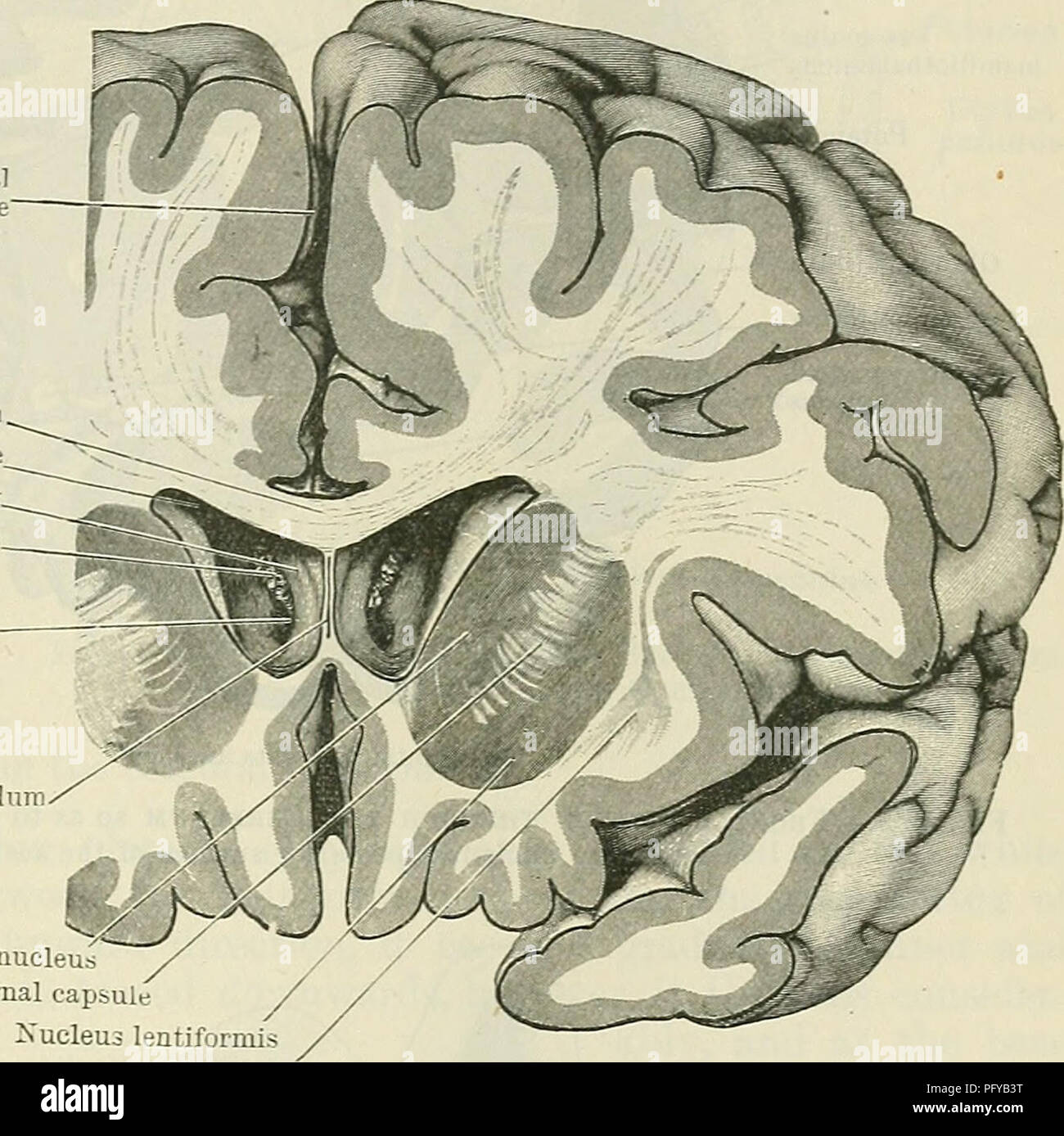 . Cunningham's Text-book of anatomy. Anatomy. BASAL GANGLIA OF THE CEEEBEAL HEMISPHERE. 639 Longitudinal fissure Column of fornix. Chorioid plexus Foramen inter- ventriculare Septum pellucidum external and internal medullary laminas, are now evident, which traverse its sub- stance in a vertical direction and divide it into three masses. The lateral, basal, and larger mass is termed the putamen ; the two medial portions together constitute the globus pallidus. The putamen forms much the largest part of the lentiform nucleus. It is darker in colour than the globus pallidus, and in this respect r Stock Photohttps://www.alamy.com/image-license-details/?v=1https://www.alamy.com/cunninghams-text-book-of-anatomy-anatomy-basal-ganglia-of-the-ceeebeal-hemisphere-639-longitudinal-fissure-column-of-fornix-chorioid-plexus-foramen-inter-ventriculare-septum-pellucidum-external-and-internal-medullary-laminas-are-now-evident-which-traverse-its-sub-stance-in-a-vertical-direction-and-divide-it-into-three-masses-the-lateral-basal-and-larger-mass-is-termed-the-putamen-the-two-medial-portions-together-constitute-the-globus-pallidus-the-putamen-forms-much-the-largest-part-of-the-lentiform-nucleus-it-is-darker-in-colour-than-the-globus-pallidus-and-in-this-respect-r-image216345692.html
RMPFYB3T–. Cunningham's Text-book of anatomy. Anatomy. BASAL GANGLIA OF THE CEEEBEAL HEMISPHERE. 639 Longitudinal fissure Column of fornix. Chorioid plexus Foramen inter- ventriculare Septum pellucidum external and internal medullary laminas, are now evident, which traverse its sub- stance in a vertical direction and divide it into three masses. The lateral, basal, and larger mass is termed the putamen ; the two medial portions together constitute the globus pallidus. The putamen forms much the largest part of the lentiform nucleus. It is darker in colour than the globus pallidus, and in this respect r
![. Fir,. 234.—A few leaves enlarged from Fig. 233. Tlie leaf t« left han.l bears pycnidia on red ti>otti on the upper surface of the Iciif; the remaining Icjives boar aecidia on raised portions of their surface. Several aecidia still further enlarged show the peridia dehiscing by longitudinal slits, (v. Tul>euf del.) ti.ssues occur in tlie year-rings as already described fur (;. dacariarfonne, viz. thickened twisted tracheid.s, locsely connected together and with fissure-like ])its; medullary rays more numerous and Ijroader; the hmits of the year-ring diflicult to distinguisli: and a yel Stock Photo . Fir,. 234.—A few leaves enlarged from Fig. 233. Tlie leaf t« left han.l bears pycnidia on red ti>otti on the upper surface of the Iciif; the remaining Icjives boar aecidia on raised portions of their surface. Several aecidia still further enlarged show the peridia dehiscing by longitudinal slits, (v. Tul>euf del.) ti.ssues occur in tlie year-rings as already described fur (;. dacariarfonne, viz. thickened twisted tracheid.s, locsely connected together and with fissure-like ])its; medullary rays more numerous and Ijroader; the hmits of the year-ring diflicult to distinguisli: and a yel Stock Photo](https://c8.alamy.com/comp/MCK593/fir-234a-few-leaves-enlarged-from-fig-233-tlie-leaf-t-left-hanl-bears-pycnidia-on-red-tigtotti-on-the-upper-surface-of-the-iciif-the-remaining-icjives-boar-aecidia-on-raised-portions-of-their-surface-several-aecidia-still-further-enlarged-show-the-peridia-dehiscing-by-longitudinal-slits-v-tulgteuf-del-tissues-occur-in-tlie-year-rings-as-already-described-fur-dacariarfonne-viz-thickened-twisted-tracheids-locsely-connected-together-and-with-fissure-like-its-medullary-rays-more-numerous-and-ijroader-the-hmits-of-the-year-ring-diflicult-to-distinguisli-and-a-yel-MCK593.jpg) . Fir,. 234.—A few leaves enlarged from Fig. 233. Tlie leaf t« left han.l bears pycnidia on red ti>otti on the upper surface of the Iciif; the remaining Icjives boar aecidia on raised portions of their surface. Several aecidia still further enlarged show the peridia dehiscing by longitudinal slits, (v. Tul>euf del.) ti.ssues occur in tlie year-rings as already described fur (;. dacariarfonne, viz. thickened twisted tracheid.s, locsely connected together and with fissure-like ])its; medullary rays more numerous and Ijroader; the hmits of the year-ring diflicult to distinguisli: and a yel Stock Photohttps://www.alamy.com/image-license-details/?v=1https://www.alamy.com/fir-234a-few-leaves-enlarged-from-fig-233-tlie-leaf-t-left-hanl-bears-pycnidia-on-red-tigtotti-on-the-upper-surface-of-the-iciif-the-remaining-icjives-boar-aecidia-on-raised-portions-of-their-surface-several-aecidia-still-further-enlarged-show-the-peridia-dehiscing-by-longitudinal-slits-v-tulgteuf-del-tissues-occur-in-tlie-year-rings-as-already-described-fur-dacariarfonne-viz-thickened-twisted-tracheids-locsely-connected-together-and-with-fissure-like-its-medullary-rays-more-numerous-and-ijroader-the-hmits-of-the-year-ring-diflicult-to-distinguisli-and-a-yel-image179900815.html
. Fir,. 234.—A few leaves enlarged from Fig. 233. Tlie leaf t« left han.l bears pycnidia on red ti>otti on the upper surface of the Iciif; the remaining Icjives boar aecidia on raised portions of their surface. Several aecidia still further enlarged show the peridia dehiscing by longitudinal slits, (v. Tul>euf del.) ti.ssues occur in tlie year-rings as already described fur (;. dacariarfonne, viz. thickened twisted tracheid.s, locsely connected together and with fissure-like ])its; medullary rays more numerous and Ijroader; the hmits of the year-ring diflicult to distinguisli: and a yel Stock Photohttps://www.alamy.com/image-license-details/?v=1https://www.alamy.com/fir-234a-few-leaves-enlarged-from-fig-233-tlie-leaf-t-left-hanl-bears-pycnidia-on-red-tigtotti-on-the-upper-surface-of-the-iciif-the-remaining-icjives-boar-aecidia-on-raised-portions-of-their-surface-several-aecidia-still-further-enlarged-show-the-peridia-dehiscing-by-longitudinal-slits-v-tulgteuf-del-tissues-occur-in-tlie-year-rings-as-already-described-fur-dacariarfonne-viz-thickened-twisted-tracheids-locsely-connected-together-and-with-fissure-like-its-medullary-rays-more-numerous-and-ijroader-the-hmits-of-the-year-ring-diflicult-to-distinguisli-and-a-yel-image179900815.htmlRMMCK593–. Fir,. 234.—A few leaves enlarged from Fig. 233. Tlie leaf t« left han.l bears pycnidia on red ti>otti on the upper surface of the Iciif; the remaining Icjives boar aecidia on raised portions of their surface. Several aecidia still further enlarged show the peridia dehiscing by longitudinal slits, (v. Tul>euf del.) ti.ssues occur in tlie year-rings as already described fur (;. dacariarfonne, viz. thickened twisted tracheid.s, locsely connected together and with fissure-like ])its; medullary rays more numerous and Ijroader; the hmits of the year-ring diflicult to distinguisli: and a yel
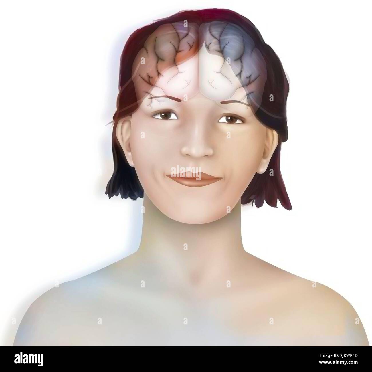 Brain (right and left cerebral hemispheres) in a woman's face. Stock Photohttps://www.alamy.com/image-license-details/?v=1https://www.alamy.com/brain-right-and-left-cerebral-hemispheres-in-a-womans-face-image476925357.html
RF2JKWR4D–Brain (right and left cerebral hemispheres) in a woman's face.
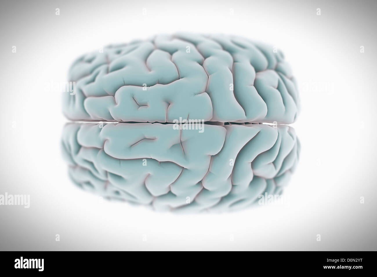 Stylized anatomical model of the human brain views from a superior perspective. Stock Photohttps://www.alamy.com/image-license-details/?v=1https://www.alamy.com/stock-photo-stylized-anatomical-model-of-the-human-brain-views-from-a-superior-52094444.html
RMD0N2YT–Stylized anatomical model of the human brain views from a superior perspective.
 Carpenter's principles of human physiology . is called the parietooccipital fissure. Onthe opposed flat surfaces of the hemispheres the principal convolutions seen are the marginalconvolutions, which form the margin of the great longitudinal fissure and the gyrus fornicatus,which encircles the corpus callosum, commencing anteriorly near the anterior perforated space,and terminating at the point of the temporo-sphenoidal lobe. By some anatomists these twogreat convolutions have been subdivided. 513. The Cortical substance or grey matter of the Hemispheres essentiallyconsists of that vesicular n Stock Photohttps://www.alamy.com/image-license-details/?v=1https://www.alamy.com/carpenters-principles-of-human-physiology-is-called-the-parietooccipital-fissure-onthe-opposed-flat-surfaces-of-the-hemispheres-the-principal-convolutions-seen-are-the-marginalconvolutions-which-form-the-margin-of-the-great-longitudinal-fissure-and-the-gyrus-fornicatuswhich-encircles-the-corpus-callosum-commencing-anteriorly-near-the-anterior-perforated-spaceand-terminating-at-the-point-of-the-temporo-sphenoidal-lobe-by-some-anatomists-these-twogreat-convolutions-have-been-subdivided-513-the-cortical-substance-or-grey-matter-of-the-hemispheres-essentiallyconsists-of-that-vesicular-n-image340002984.html
RM2AN4D7M–Carpenter's principles of human physiology . is called the parietooccipital fissure. Onthe opposed flat surfaces of the hemispheres the principal convolutions seen are the marginalconvolutions, which form the margin of the great longitudinal fissure and the gyrus fornicatus,which encircles the corpus callosum, commencing anteriorly near the anterior perforated space,and terminating at the point of the temporo-sphenoidal lobe. By some anatomists these twogreat convolutions have been subdivided. 513. The Cortical substance or grey matter of the Hemispheres essentiallyconsists of that vesicular n
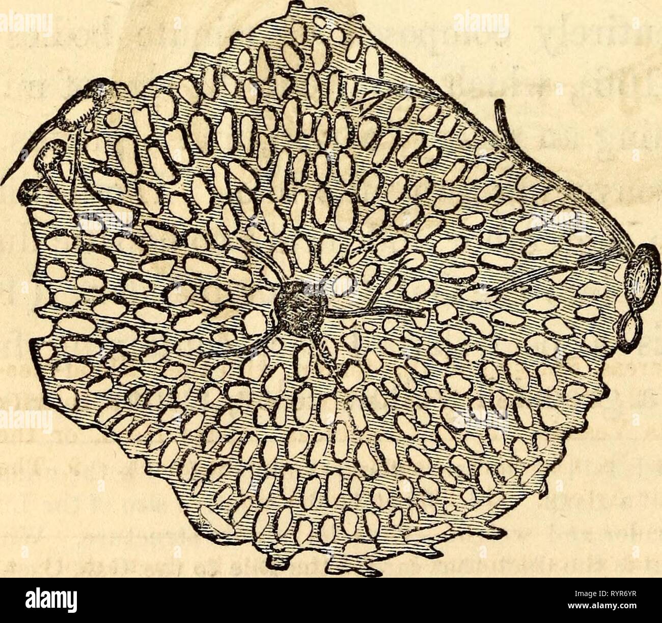 Elementary anatomy and physiology Elementary anatomy and physiology : for colleges, academies, and other schools . elementaryanato00hitc Year: 1869 The Inferior or Concave Surface of the Liver, showing its Subdivisions into Lobes. I, Center of the Light Lobe. 2, Center of the Left Lobe. 3, Its Anterior, Inferior, or Thin Margin. 4, Its Posterior, Thick or Diaphragmatic Portion. 5, The Light Extrem- ity. 6, The Left Extremity. 7, The Notch on the Anterior Margin. 8, The Umbilical or Longitudinal Fissure. 9, The Hound Ligament or remains of the Umbilical Vein. 10, The Portion of the Suspensory Stock Photohttps://www.alamy.com/image-license-details/?v=1https://www.alamy.com/elementary-anatomy-and-physiology-elementary-anatomy-and-physiology-for-colleges-academies-and-other-schools-elementaryanato00hitc-year-1869-the-inferior-or-concave-surface-of-the-liver-showing-its-subdivisions-into-lobes-i-center-of-the-light-lobe-2-center-of-the-left-lobe-3-its-anterior-inferior-or-thin-margin-4-its-posterior-thick-or-diaphragmatic-portion-5-the-light-extrem-ity-6-the-left-extremity-7-the-notch-on-the-anterior-margin-8-the-umbilical-or-longitudinal-fissure-9-the-hound-ligament-or-remains-of-the-umbilical-vein-10-the-portion-of-the-suspensory-image240840875.html
RMRYR6YR–Elementary anatomy and physiology Elementary anatomy and physiology : for colleges, academies, and other schools . elementaryanato00hitc Year: 1869 The Inferior or Concave Surface of the Liver, showing its Subdivisions into Lobes. I, Center of the Light Lobe. 2, Center of the Left Lobe. 3, Its Anterior, Inferior, or Thin Margin. 4, Its Posterior, Thick or Diaphragmatic Portion. 5, The Light Extrem- ity. 6, The Left Extremity. 7, The Notch on the Anterior Margin. 8, The Umbilical or Longitudinal Fissure. 9, The Hound Ligament or remains of the Umbilical Vein. 10, The Portion of the Suspensory
 The cerebral hemispheres are separated by a deep groove, the longitudinal cerebral fissure . Stock Photohttps://www.alamy.com/image-license-details/?v=1https://www.alamy.com/the-cerebral-hemispheres-are-separated-by-a-deep-groove-the-longitudinal-cerebral-fissure-image596592815.html
RF2WJH467–The cerebral hemispheres are separated by a deep groove, the longitudinal cerebral fissure .
 . On the anatomy of vertebrates. Vertebrates; Anatomy, Comparative; 1866. LARYNX 01'' REPTILES. 527 sending off lateral branches : it then abrnptly terminates in a dilated elongated passage, similar to those in which the side- branches open. These passages correspond with the primary divisions of the pulmonary cavity in the Turtle, and the air passes from them Ijy numerous round apertures into the smaller subdivisions forming the cellular structure of the lung.' § 9 5. Larynx of Reptiles. — In perennibranchial and tailed Batrucliia the glottis is a simple longitudinal fissure, fig. 346, &, Stock Photohttps://www.alamy.com/image-license-details/?v=1https://www.alamy.com/on-the-anatomy-of-vertebrates-vertebrates-anatomy-comparative-1866-larynx-01-reptiles-527-sending-off-lateral-branches-it-then-abrnptly-terminates-in-a-dilated-elongated-passage-similar-to-those-in-which-the-side-branches-open-these-passages-correspond-with-the-primary-divisions-of-the-pulmonary-cavity-in-the-turtle-and-the-air-passes-from-them-ijy-numerous-round-apertures-into-the-smaller-subdivisions-forming-the-cellular-structure-of-the-lung-9-5-larynx-of-reptiles-in-perennibranchial-and-tailed-batrucliia-the-glottis-is-a-simple-longitudinal-fissure-fig-346-amp-image216416661.html
RMPG2HJD–. On the anatomy of vertebrates. Vertebrates; Anatomy, Comparative; 1866. LARYNX 01'' REPTILES. 527 sending off lateral branches : it then abrnptly terminates in a dilated elongated passage, similar to those in which the side- branches open. These passages correspond with the primary divisions of the pulmonary cavity in the Turtle, and the air passes from them Ijy numerous round apertures into the smaller subdivisions forming the cellular structure of the lung.' § 9 5. Larynx of Reptiles. — In perennibranchial and tailed Batrucliia the glottis is a simple longitudinal fissure, fig. 346, &,
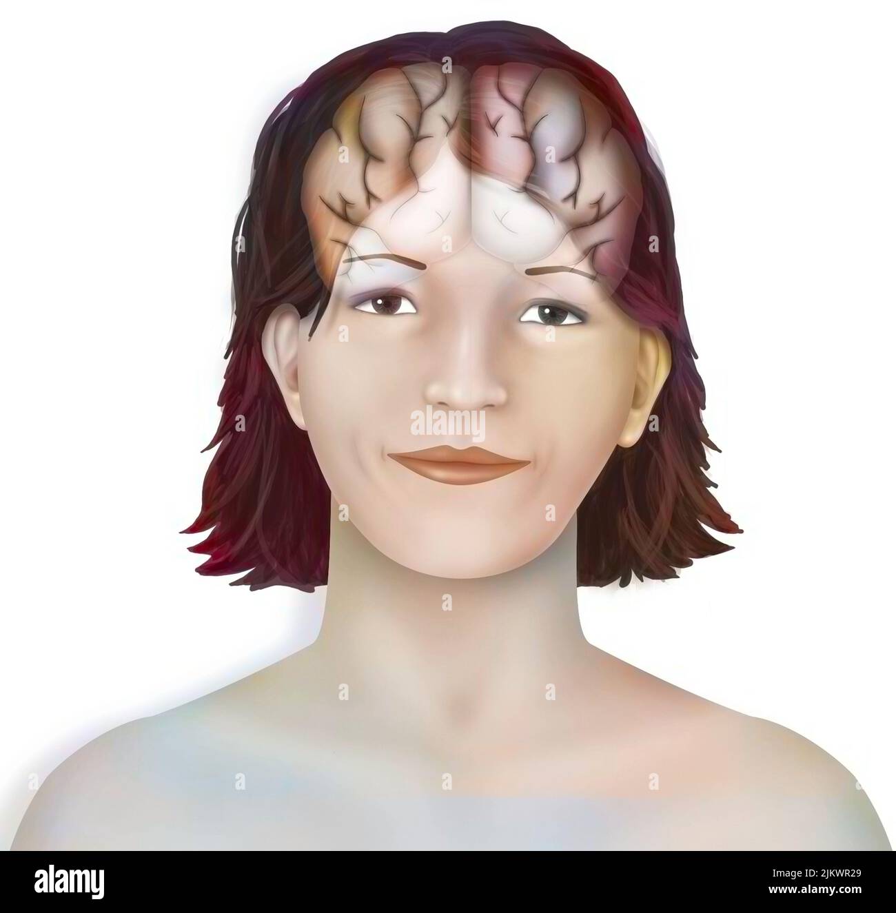 Brain (right and left cerebral hemispheres) in a woman's face. Stock Photohttps://www.alamy.com/image-license-details/?v=1https://www.alamy.com/brain-right-and-left-cerebral-hemispheres-in-a-womans-face-image476925297.html
RF2JKWR29–Brain (right and left cerebral hemispheres) in a woman's face.
 Superior view of an anatomical model of the brain and its blood supply. Stock Photohttps://www.alamy.com/image-license-details/?v=1https://www.alamy.com/stock-photo-superior-view-of-an-anatomical-model-of-the-brain-and-its-blood-supply-52096506.html
RMD0N5HE–Superior view of an anatomical model of the brain and its blood supply.
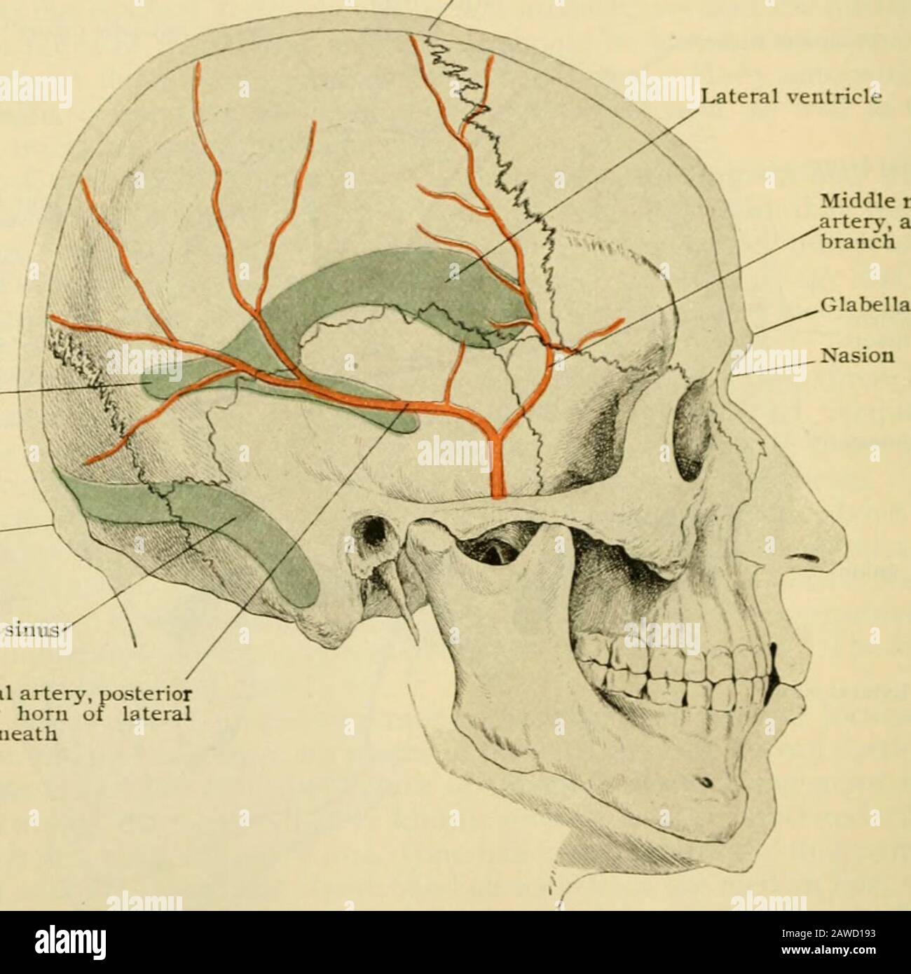 Human anatomy, including structure and development and practical considerations . lel to the longitudinal fissure to within i8mm. (^ ;?4 in.; of the fissure of Rolando. Tlie inferior frontal sulcus is represented,appro.ximately, by the anterior end of the temporal ridge. In the parietal lobe the most imjjortant sulcus is the intraparietal. It beginsnear the horizontal limb of the fissure of Sylvius, and passes upward and backwardabout midway between the fissure of Rolando and the parietal eminence. It thenturns backward, running about midway to the longitudinal fissure and the centreof the par Stock Photohttps://www.alamy.com/image-license-details/?v=1https://www.alamy.com/human-anatomy-including-structure-and-development-and-practical-considerations-lel-to-the-longitudinal-fissure-to-within-i8mm-4-in-of-the-fissure-of-rolando-tlie-inferior-frontal-sulcus-is-representedapproximately-by-the-anterior-end-of-the-temporal-ridge-in-the-parietal-lobe-the-most-imjjortant-sulcus-is-the-intraparietal-it-beginsnear-the-horizontal-limb-of-the-fissure-of-sylvius-and-passes-upward-and-backwardabout-midway-between-the-fissure-of-rolando-and-the-parietal-eminence-it-thenturns-backward-running-about-midway-to-the-longitudinal-fissure-and-the-centreof-the-par-image342649807.html
RM2AWD193–Human anatomy, including structure and development and practical considerations . lel to the longitudinal fissure to within i8mm. (^ ;?4 in.; of the fissure of Rolando. Tlie inferior frontal sulcus is represented,appro.ximately, by the anterior end of the temporal ridge. In the parietal lobe the most imjjortant sulcus is the intraparietal. It beginsnear the horizontal limb of the fissure of Sylvius, and passes upward and backwardabout midway between the fissure of Rolando and the parietal eminence. It thenturns backward, running about midway to the longitudinal fissure and the centreof the par
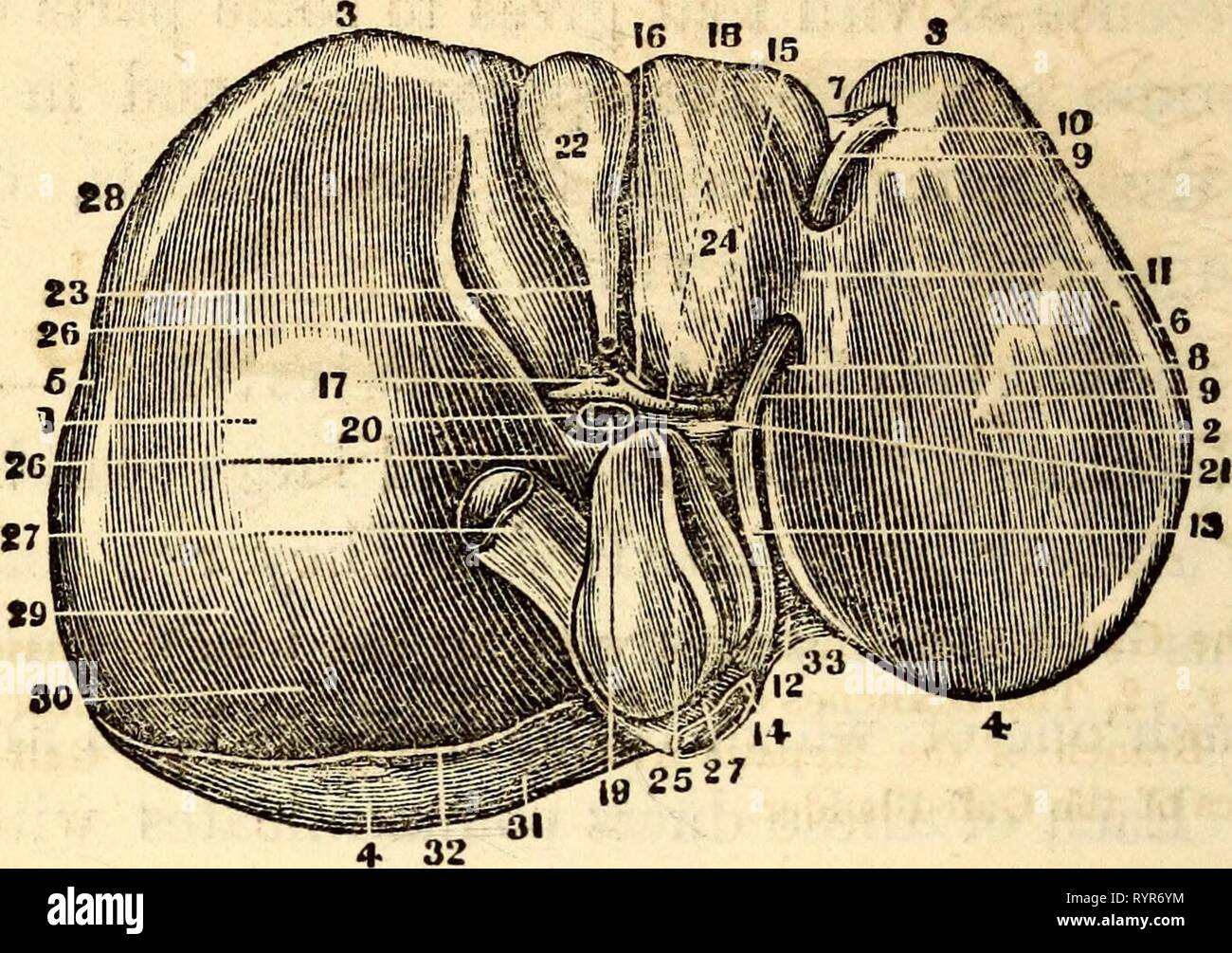 Elementary anatomy and physiology Elementary anatomy and physiology : for colleges, academies, and other schools . elementaryanato00hitc Year: 1869 166 HITCHCOCK'S ANATOMY Fig. 178. The Inferior or Concave Surface of the Liver, showing its Subdivisions into Lobes. I, Center of the Light Lobe. 2, Center of the Left Lobe. 3, Its Anterior, Inferior, or Thin Margin. 4, Its Posterior, Thick or Diaphragmatic Portion. 5, The Light Extrem- ity. 6, The Left Extremity. 7, The Notch on the Anterior Margin. 8, The Umbilical or Longitudinal Fissure. 9, The Hound Ligament or remains of the Umbilical Ve Stock Photohttps://www.alamy.com/image-license-details/?v=1https://www.alamy.com/elementary-anatomy-and-physiology-elementary-anatomy-and-physiology-for-colleges-academies-and-other-schools-elementaryanato00hitc-year-1869-166-hitchcocks-anatomy-fig-178-the-inferior-or-concave-surface-of-the-liver-showing-its-subdivisions-into-lobes-i-center-of-the-light-lobe-2-center-of-the-left-lobe-3-its-anterior-inferior-or-thin-margin-4-its-posterior-thick-or-diaphragmatic-portion-5-the-light-extrem-ity-6-the-left-extremity-7-the-notch-on-the-anterior-margin-8-the-umbilical-or-longitudinal-fissure-9-the-hound-ligament-or-remains-of-the-umbilical-ve-image240840872.html
RMRYR6YM–Elementary anatomy and physiology Elementary anatomy and physiology : for colleges, academies, and other schools . elementaryanato00hitc Year: 1869 166 HITCHCOCK'S ANATOMY Fig. 178. The Inferior or Concave Surface of the Liver, showing its Subdivisions into Lobes. I, Center of the Light Lobe. 2, Center of the Left Lobe. 3, Its Anterior, Inferior, or Thin Margin. 4, Its Posterior, Thick or Diaphragmatic Portion. 5, The Light Extrem- ity. 6, The Left Extremity. 7, The Notch on the Anterior Margin. 8, The Umbilical or Longitudinal Fissure. 9, The Hound Ligament or remains of the Umbilical Ve
 The cerebral hemispheres are separated by a deep groove, the longitudinal cerebral fissure . Stock Photohttps://www.alamy.com/image-license-details/?v=1https://www.alamy.com/the-cerebral-hemispheres-are-separated-by-a-deep-groove-the-longitudinal-cerebral-fissure-image596592818.html
RF2WJH46A–The cerebral hemispheres are separated by a deep groove, the longitudinal cerebral fissure .
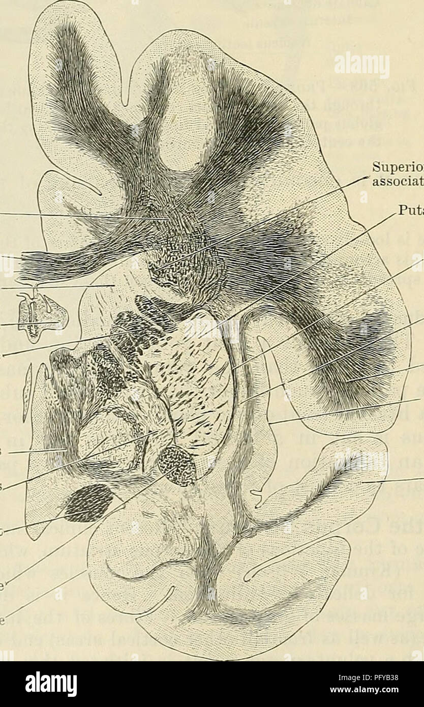 . Cunningham's Text-book of anatomy. Anatomy. Longitudinal fissure Corpus callosum Fornix Caudate nucleus Vena terminalis Tela chorioidea ventrieuli tertii â Thalamus â Third ventricle â Chorioid plexus Internal capsule Interventricular foramen ^"-""Column of fornix Anterior commissure Optic tract Infundibulum â Optic chiasma Optic nerve Substantia perforata anterior Olfactory peduncle Fig 569.âFrontal Section through the Cerebrum so as to cut through the three divisions of the lentiforra nucleus ; posterior surface of the section shown here. Intersection of corona radiata and c Stock Photohttps://www.alamy.com/image-license-details/?v=1https://www.alamy.com/cunninghams-text-book-of-anatomy-anatomy-longitudinal-fissure-corpus-callosum-fornix-caudate-nucleus-vena-terminalis-tela-chorioidea-ventrieuli-tertii-thalamus-third-ventricle-chorioid-plexus-internal-capsule-interventricular-foramen-quot-quotquotcolumn-of-fornix-anterior-commissure-optic-tract-infundibulum-optic-chiasma-optic-nerve-substantia-perforata-anterior-olfactory-peduncle-fig-569frontal-section-through-the-cerebrum-so-as-to-cut-through-the-three-divisions-of-the-lentiforra-nucleus-posterior-surface-of-the-section-shown-here-intersection-of-corona-radiata-and-c-image216345676.html
RMPFYB38–. Cunningham's Text-book of anatomy. Anatomy. Longitudinal fissure Corpus callosum Fornix Caudate nucleus Vena terminalis Tela chorioidea ventrieuli tertii â Thalamus â Third ventricle â Chorioid plexus Internal capsule Interventricular foramen ^"-""Column of fornix Anterior commissure Optic tract Infundibulum â Optic chiasma Optic nerve Substantia perforata anterior Olfactory peduncle Fig 569.âFrontal Section through the Cerebrum so as to cut through the three divisions of the lentiforra nucleus ; posterior surface of the section shown here. Intersection of corona radiata and c
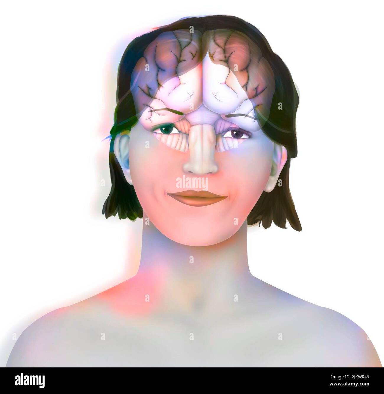 Brain (right and left cerebral hemispheres, cerebellum and brainstem) in a woman's face. Stock Photohttps://www.alamy.com/image-license-details/?v=1https://www.alamy.com/brain-right-and-left-cerebral-hemispheres-cerebellum-and-brainstem-in-a-womans-face-image476925353.html
RF2JKWR49–Brain (right and left cerebral hemispheres, cerebellum and brainstem) in a woman's face.
 Superior view of an anatomical model of the brain and its blood supply. Stock Photohttps://www.alamy.com/image-license-details/?v=1https://www.alamy.com/stock-photo-superior-view-of-an-anatomical-model-of-the-brain-and-its-blood-supply-52094004.html
RMD0N2C4–Superior view of an anatomical model of the brain and its blood supply.
 . Human physiology. le. Thefornix or inferior longitudinalcommissure of Mr. Solly, whoseoffice is to connect the anteriorand j)osterior parts of the samehemisphere, as the transversecommissures do those of theopposite hemisphere, is placedhorizontally below the last.The band of fibres which runsin each hemisphere above thecorpus callosum, on the edge ofthe longitudinal fissure, is theswperior longitudinal commissureof Mr. Solly. Its use is sup-posed to resemble that ascribedto the inferior longitudinal com-missure. The fornix is of atriangular shape; and consti-tutes the upper paries of anothe Stock Photohttps://www.alamy.com/image-license-details/?v=1https://www.alamy.com/human-physiology-le-thefornix-or-inferior-longitudinalcommissure-of-mr-solly-whoseoffice-is-to-connect-the-anteriorand-josterior-parts-of-the-samehemisphere-as-the-transversecommissures-do-those-of-theopposite-hemisphere-is-placedhorizontally-below-the-lastthe-band-of-fibres-which-runsin-each-hemisphere-above-thecorpus-callosum-on-the-edge-ofthe-longitudinal-fissure-is-theswperior-longitudinal-commissureof-mr-solly-its-use-is-sup-posed-to-resemble-that-ascribedto-the-inferior-longitudinal-com-missure-the-fornix-is-of-atriangular-shape-and-consti-tutes-the-upper-paries-of-anothe-image336600772.html
RM2AFHDM4–. Human physiology. le. Thefornix or inferior longitudinalcommissure of Mr. Solly, whoseoffice is to connect the anteriorand j)osterior parts of the samehemisphere, as the transversecommissures do those of theopposite hemisphere, is placedhorizontally below the last.The band of fibres which runsin each hemisphere above thecorpus callosum, on the edge ofthe longitudinal fissure, is theswperior longitudinal commissureof Mr. Solly. Its use is sup-posed to resemble that ascribedto the inferior longitudinal com-missure. The fornix is of atriangular shape; and consti-tutes the upper paries of anothe
 Elements of histology (1898) Elements of histology elementsofhistol00klei Year: 1898 p.m. Cty.o. a.c. Fig. 129.—Cross-section of the central part of the Spinal Cord from tlie Lumbar region of an Adult, showing the central canal, its lining epithe- lium surrounded by neuroglia, forming the central grey nucleus, (After Schdfer.) /.a., Anterior median fissure; p.m.c, posterior white column ; ff.c, anterior white comniissure. fibrils extend chiefly in a longitudinal direction, in the gi'ey matter they extend uniformly in all directions, and in the septa between the columns Stock Photohttps://www.alamy.com/image-license-details/?v=1https://www.alamy.com/elements-of-histology-1898-elements-of-histology-elementsofhistol00klei-year-1898-pm-ctyo-ac-fig-129cross-section-of-the-central-part-of-the-spinal-cord-from-tlie-lumbar-region-of-an-adult-showing-the-central-canal-its-lining-epithe-lium-surrounded-by-neuroglia-forming-the-central-grey-nucleus-after-schdfer-a-anterior-median-fissure-pmc-posterior-white-column-ffc-anterior-white-comniissure-fibrils-extend-chiefly-in-a-longitudinal-direction-in-the-giey-matter-they-extend-uniformly-in-all-directions-and-in-the-septa-between-the-columns-image239593361.html
RMRWPBNN–Elements of histology (1898) Elements of histology elementsofhistol00klei Year: 1898 p.m. Cty.o. a.c. Fig. 129.—Cross-section of the central part of the Spinal Cord from tlie Lumbar region of an Adult, showing the central canal, its lining epithe- lium surrounded by neuroglia, forming the central grey nucleus, (After Schdfer.) /.a., Anterior median fissure; p.m.c, posterior white column ; ff.c, anterior white comniissure. fibrils extend chiefly in a longitudinal direction, in the gi'ey matter they extend uniformly in all directions, and in the septa between the columns
 The cerebral hemispheres are separated by a deep groove, the longitudinal cerebral fissure . Stock Photohttps://www.alamy.com/image-license-details/?v=1https://www.alamy.com/the-cerebral-hemispheres-are-separated-by-a-deep-groove-the-longitudinal-cerebral-fissure-image596592270.html
RF2WJH3EP–The cerebral hemispheres are separated by a deep groove, the longitudinal cerebral fissure .
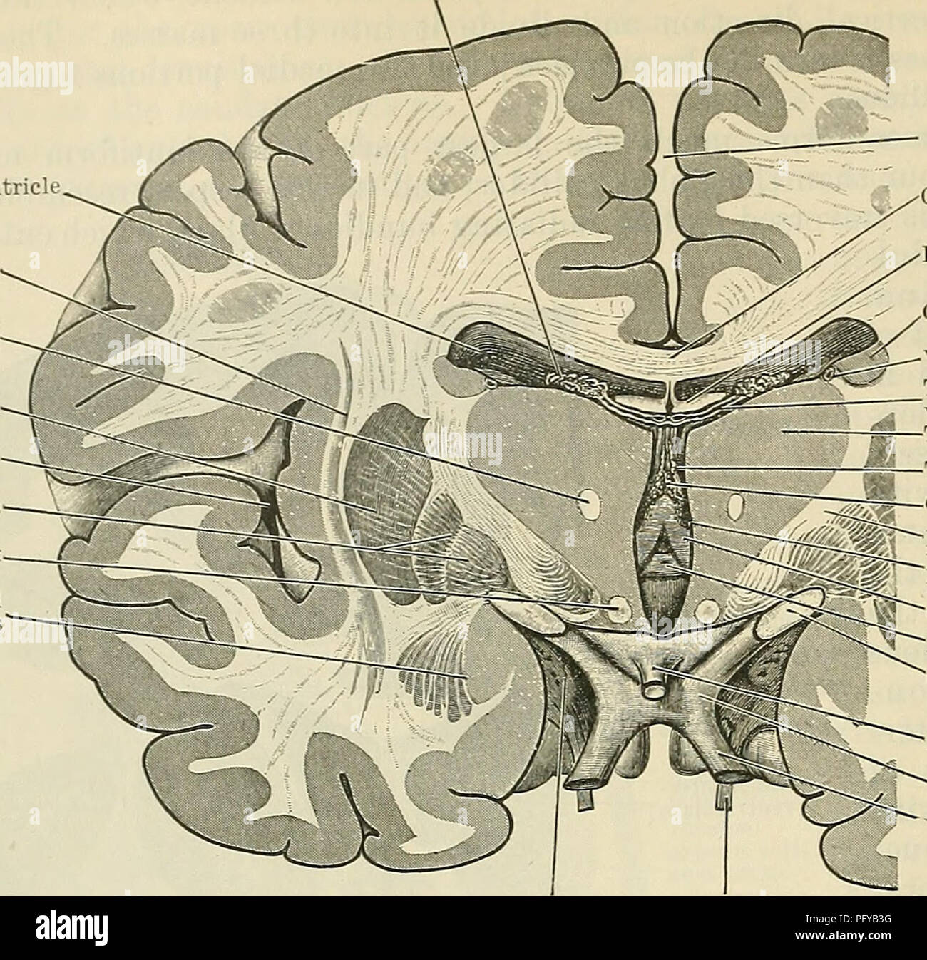 . Cunningham's Text-book of anatomy. Anatomy. 640 THE NEEVOUS SYSTEM. Chorioid plexus Lateral ventricle Claustrum Fasciculus mamillothalamicus Putanien Insula Globus pallidus Column of fornix Amygdaloid nucleus. Longitudinal fissure Corpus callosum Fornix Caudate nucleus Vena terminalis Tela chorioidea ventrieuli tertii â Thalamus â Third ventricle â Chorioid plexus Internal capsule Interventricular foramen ^"-""Column of fornix Anterior commissure Optic tract Infundibulum â Optic chiasma Optic nerve Substantia perforata anterior Olfactory peduncle Fig 569.âFrontal Section throu Stock Photohttps://www.alamy.com/image-license-details/?v=1https://www.alamy.com/cunninghams-text-book-of-anatomy-anatomy-640-the-neevous-system-chorioid-plexus-lateral-ventricle-claustrum-fasciculus-mamillothalamicus-putanien-insula-globus-pallidus-column-of-fornix-amygdaloid-nucleus-longitudinal-fissure-corpus-callosum-fornix-caudate-nucleus-vena-terminalis-tela-chorioidea-ventrieuli-tertii-thalamus-third-ventricle-chorioid-plexus-internal-capsule-interventricular-foramen-quot-quotquotcolumn-of-fornix-anterior-commissure-optic-tract-infundibulum-optic-chiasma-optic-nerve-substantia-perforata-anterior-olfactory-peduncle-fig-569frontal-section-throu-image216345684.html
RMPFYB3G–. Cunningham's Text-book of anatomy. Anatomy. 640 THE NEEVOUS SYSTEM. Chorioid plexus Lateral ventricle Claustrum Fasciculus mamillothalamicus Putanien Insula Globus pallidus Column of fornix Amygdaloid nucleus. Longitudinal fissure Corpus callosum Fornix Caudate nucleus Vena terminalis Tela chorioidea ventrieuli tertii â Thalamus â Third ventricle â Chorioid plexus Internal capsule Interventricular foramen ^"-""Column of fornix Anterior commissure Optic tract Infundibulum â Optic chiasma Optic nerve Substantia perforata anterior Olfactory peduncle Fig 569.âFrontal Section throu
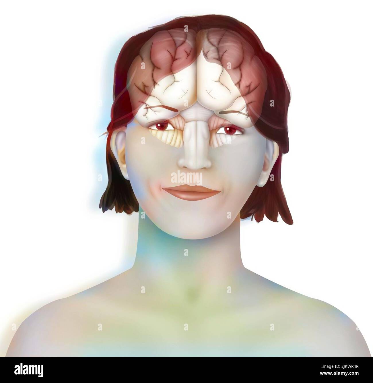 Brain (right and left cerebral hemispheres, cerebellum and brainstem) in a woman's face. Stock Photohttps://www.alamy.com/image-license-details/?v=1https://www.alamy.com/brain-right-and-left-cerebral-hemispheres-cerebellum-and-brainstem-in-a-womans-face-image476925367.html
RF2JKWR4R–Brain (right and left cerebral hemispheres, cerebellum and brainstem) in a woman's face.
 A stylized close up view of the front of the head. The brain is visible within the skull. Stock Photohttps://www.alamy.com/image-license-details/?v=1https://www.alamy.com/stock-photo-a-stylized-close-up-view-of-the-front-of-the-head-the-brain-is-visible-52094321.html
RMD0N2RD–A stylized close up view of the front of the head. The brain is visible within the skull.
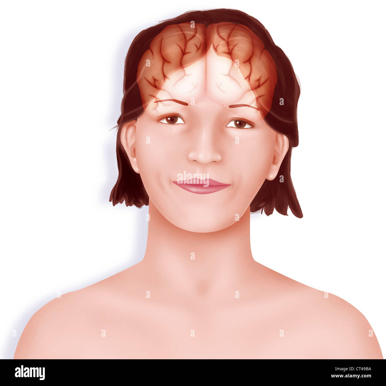 BRAIN, DRAWING Stock Photohttps://www.alamy.com/image-license-details/?v=1https://www.alamy.com/stock-photo-brain-drawing-49267662.html
RMCT49BA–BRAIN, DRAWING
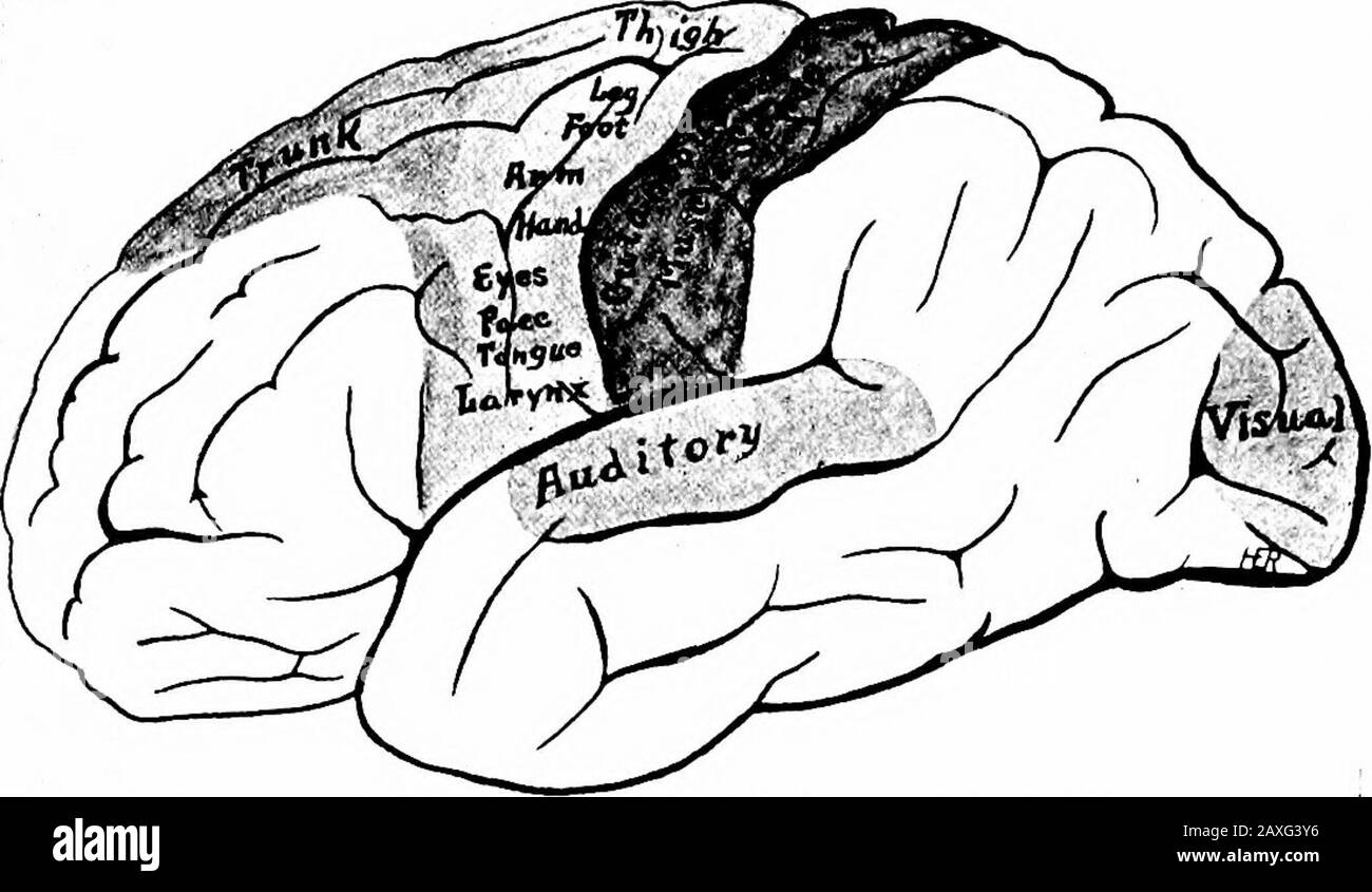 A manual of anatomy . l longitudinal fissure that liessubcallosal and the thin walls of the septum constitute the medialwalls of the hemispheres of this area where nerve tissues fail todevelop in any great quantity. The hippocampus is an elevation in the lateral ventricle and is inrelation with the fimbria as above described. It constitutes a partof the rhinencephalon. Within each hemicerebrum is an extensive and irregular cavitycalled the lateral ventricle {ventriculus lateralis). Each consists ofa body, frontal, occipital and temporal extensions, or horns. Eachventricle is in connection with Stock Photohttps://www.alamy.com/image-license-details/?v=1https://www.alamy.com/a-manual-of-anatomy-l-longitudinal-fissure-that-liessubcallosal-and-the-thin-walls-of-the-septum-constitute-the-medialwalls-of-the-hemispheres-of-this-area-where-nerve-tissues-fail-todevelop-in-any-great-quantity-the-hippocampus-is-an-elevation-in-the-lateral-ventricle-and-is-inrelation-with-the-fimbria-as-above-described-it-constitutes-a-partof-the-rhinencephalon-within-each-hemicerebrum-is-an-extensive-and-irregular-cavitycalled-the-lateral-ventricle-ventriculus-lateralis-each-consists-ofa-body-frontal-occipital-and-temporal-extensions-or-horns-eachventricle-is-in-connection-with-image343332394.html
RM2AXG3Y6–A manual of anatomy . l longitudinal fissure that liessubcallosal and the thin walls of the septum constitute the medialwalls of the hemispheres of this area where nerve tissues fail todevelop in any great quantity. The hippocampus is an elevation in the lateral ventricle and is inrelation with the fimbria as above described. It constitutes a partof the rhinencephalon. Within each hemicerebrum is an extensive and irregular cavitycalled the lateral ventricle {ventriculus lateralis). Each consists ofa body, frontal, occipital and temporal extensions, or horns. Eachventricle is in connection with
 The cerebral hemispheres are separated by a deep groove, the longitudinal cerebral fissure . Stock Photohttps://www.alamy.com/image-license-details/?v=1https://www.alamy.com/the-cerebral-hemispheres-are-separated-by-a-deep-groove-the-longitudinal-cerebral-fissure-image596591746.html
RF2WJH2T2–The cerebral hemispheres are separated by a deep groove, the longitudinal cerebral fissure .
 . The anatomy of the domestic fowl . Domestic animals; Veterinary medicine; Poultry. Fig. 75.—The brain of a hen. Photograph. A. Upper surface of the brain. 1, Medulla oblongata. 2, Calamus scrip- torius. 3, Cerebellum. 4, Optic lobes. S- Transverse fissure. 6, Longitudinal fissure. 7, Upper surface of the left cerebral lobe. 8, Upper surface of the right cerebral lobe. 9, Lateral pillar of the cerebellum.. B. The posterior surface of the eyeball. 10, The sectioned surface of the optic nerve. C. The inferior stirface of the brain. 11, Corneo-scleral juncture. 12, The cornea. 13, The sclera. 14 Stock Photohttps://www.alamy.com/image-license-details/?v=1https://www.alamy.com/the-anatomy-of-the-domestic-fowl-domestic-animals-veterinary-medicine-poultry-fig-75the-brain-of-a-hen-photograph-a-upper-surface-of-the-brain-1-medulla-oblongata-2-calamus-scrip-torius-3-cerebellum-4-optic-lobes-s-transverse-fissure-6-longitudinal-fissure-7-upper-surface-of-the-left-cerebral-lobe-8-upper-surface-of-the-right-cerebral-lobe-9-lateral-pillar-of-the-cerebellum-b-the-posterior-surface-of-the-eyeball-10-the-sectioned-surface-of-the-optic-nerve-c-the-inferior-stirface-of-the-brain-11-corneo-scleral-juncture-12-the-cornea-13-the-sclera-14-image216371344.html
RMPG0FT0–. The anatomy of the domestic fowl . Domestic animals; Veterinary medicine; Poultry. Fig. 75.—The brain of a hen. Photograph. A. Upper surface of the brain. 1, Medulla oblongata. 2, Calamus scrip- torius. 3, Cerebellum. 4, Optic lobes. S- Transverse fissure. 6, Longitudinal fissure. 7, Upper surface of the left cerebral lobe. 8, Upper surface of the right cerebral lobe. 9, Lateral pillar of the cerebellum.. B. The posterior surface of the eyeball. 10, The sectioned surface of the optic nerve. C. The inferior stirface of the brain. 11, Corneo-scleral juncture. 12, The cornea. 13, The sclera. 14
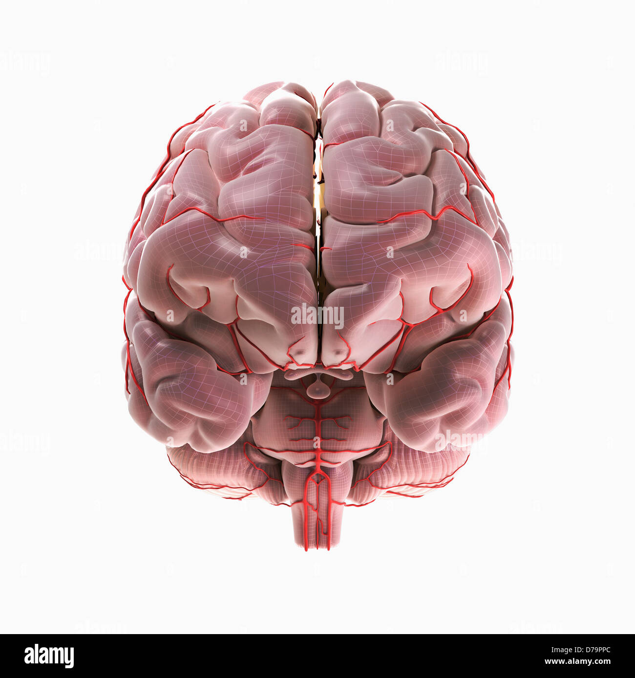 Brain Blood Supply Stock Photohttps://www.alamy.com/image-license-details/?v=1https://www.alamy.com/stock-photo-brain-blood-supply-56149140.html
RMD79PPC–Brain Blood Supply
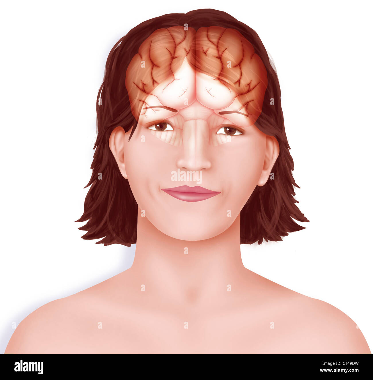 BRAIN, DRAWING Stock Photohttps://www.alamy.com/image-license-details/?v=1https://www.alamy.com/stock-photo-brain-drawing-49267733.html
RMCT49DW–BRAIN, DRAWING
 The principles and practice of medicine, designed for the use of practitioners and students of medicine . on is excitable, andstimulation by weak electrical currents produces muscular movements inthe opposite half of the body. The centres presiding over the differentgroups of muscles may be thus classified : {a) Centres for the trunk. These have been shown by Schafer to besituated in the marginal gyrus, just within the longitudinal fissure, theregion sometimes spoken of as the paracental lobule. (h) Centres for the lower limbs. These are situated at the upper partof the Rolandic region, close Stock Photohttps://www.alamy.com/image-license-details/?v=1https://www.alamy.com/the-principles-and-practice-of-medicine-designed-for-the-use-of-practitioners-and-students-of-medicine-on-is-excitable-andstimulation-by-weak-electrical-currents-produces-muscular-movements-inthe-opposite-half-of-the-body-the-centres-presiding-over-the-differentgroups-of-muscles-may-be-thus-classified-a-centres-for-the-trunk-these-have-been-shown-by-schafer-to-besituated-in-the-marginal-gyrus-just-within-the-longitudinal-fissure-theregion-sometimes-spoken-of-as-the-paracental-lobule-h-centres-for-the-lower-limbs-these-are-situated-at-the-upper-partof-the-rolandic-region-close-image339974466.html
RM2AN34W6–The principles and practice of medicine, designed for the use of practitioners and students of medicine . on is excitable, andstimulation by weak electrical currents produces muscular movements inthe opposite half of the body. The centres presiding over the differentgroups of muscles may be thus classified : {a) Centres for the trunk. These have been shown by Schafer to besituated in the marginal gyrus, just within the longitudinal fissure, theregion sometimes spoken of as the paracental lobule. (h) Centres for the lower limbs. These are situated at the upper partof the Rolandic region, close
 The cerebral hemispheres are separated by a deep groove, the longitudinal cerebral fissure . Stock Photohttps://www.alamy.com/image-license-details/?v=1https://www.alamy.com/the-cerebral-hemispheres-are-separated-by-a-deep-groove-the-longitudinal-cerebral-fissure-image596587441.html
RF2WJGWA9–The cerebral hemispheres are separated by a deep groove, the longitudinal cerebral fissure .
 . The development of the chick : an introduction to embryology. Embryology; Chickens -- Embryos. ll' £.K :â â %. Fig. 146. â Transverse section through the cervical swelling of the spinal cord of a 12-clay chick. (After v. Kupffer.) C, Central canal, d. H., Dorsal horn of the gray matter. Ep., Ependyma. N. H., Nucleus of Hoffmann, s. d., Dorsal fissure. s. v., Ventral fissure."- v. H., Ventral horn of the gray matter. The development of the so-called dorsal and ventral fissures is essentially different. The entire ventral longitudinal fissure of the cord owes its origin to growth of the Stock Photohttps://www.alamy.com/image-license-details/?v=1https://www.alamy.com/the-development-of-the-chick-an-introduction-to-embryology-embryology-chickens-embryos-ll-k-fig-146-transverse-section-through-the-cervical-swelling-of-the-spinal-cord-of-a-12-clay-chick-after-v-kupffer-c-central-canal-d-h-dorsal-horn-of-the-gray-matter-ep-ependyma-n-h-nucleus-of-hoffmann-s-d-dorsal-fissure-s-v-ventral-fissurequot-v-h-ventral-horn-of-the-gray-matter-the-development-of-the-so-called-dorsal-and-ventral-fissures-is-essentially-different-the-entire-ventral-longitudinal-fissure-of-the-cord-owes-its-origin-to-growth-of-the-image215969816.html
RMPFA7KM–. The development of the chick : an introduction to embryology. Embryology; Chickens -- Embryos. ll' £.K :â â %. Fig. 146. â Transverse section through the cervical swelling of the spinal cord of a 12-clay chick. (After v. Kupffer.) C, Central canal, d. H., Dorsal horn of the gray matter. Ep., Ependyma. N. H., Nucleus of Hoffmann, s. d., Dorsal fissure. s. v., Ventral fissure."- v. H., Ventral horn of the gray matter. The development of the so-called dorsal and ventral fissures is essentially different. The entire ventral longitudinal fissure of the cord owes its origin to growth of the
 Brain Blood Supply Stock Photohttps://www.alamy.com/image-license-details/?v=1https://www.alamy.com/stock-photo-brain-blood-supply-56149320.html
RMD79R0T–Brain Blood Supply
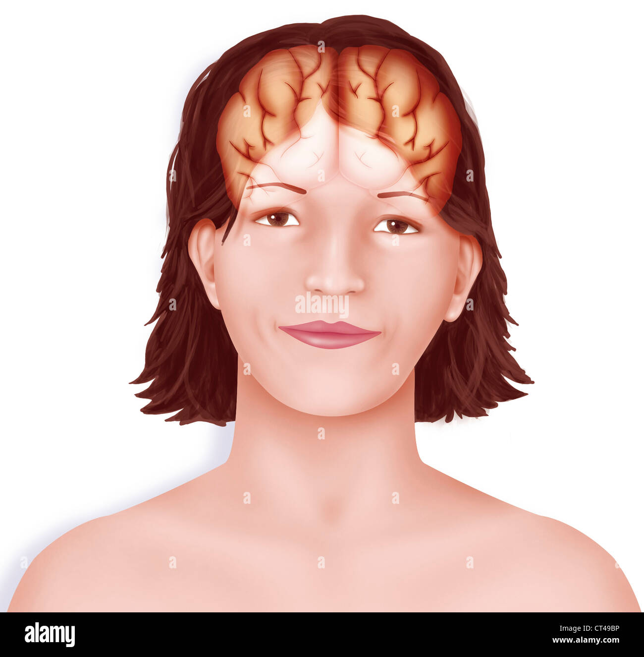 BRAIN, DRAWING Stock Photohttps://www.alamy.com/image-license-details/?v=1https://www.alamy.com/stock-photo-brain-drawing-49267674.html
RMCT49BP–BRAIN, DRAWING
 Surgical anatomy : a treatise on human anatomy in its application to the practice of medicine and surgery . perforated space and the mainportion of the fissure of Sylvius. The apex is directed forward, and is formed bythe curving of the convolutions in i:)assing from the convex to the orbital surface.The sides are formed by the longitudinal fissure and the lower border of the hemi-sphere. On this surface are two secondary fissures, the olfactory and the orbital. The olfactory fissure runs parallel with the longitudinal fissure and a shortdistance external to it. It lodges the olfactory tract a Stock Photohttps://www.alamy.com/image-license-details/?v=1https://www.alamy.com/surgical-anatomy-a-treatise-on-human-anatomy-in-its-application-to-the-practice-of-medicine-and-surgery-perforated-space-and-the-mainportion-of-the-fissure-of-sylvius-the-apex-is-directed-forward-and-is-formed-bythe-curving-of-the-convolutions-in-iassing-from-the-convex-to-the-orbital-surfacethe-sides-are-formed-by-the-longitudinal-fissure-and-the-lower-border-of-the-hemi-sphere-on-this-surface-are-two-secondary-fissures-the-olfactory-and-the-orbital-the-olfactory-fissure-runs-parallel-with-the-longitudinal-fissure-and-a-shortdistance-external-to-it-it-lodges-the-olfactory-tract-a-image338918372.html
RM2AKB1RG–Surgical anatomy : a treatise on human anatomy in its application to the practice of medicine and surgery . perforated space and the mainportion of the fissure of Sylvius. The apex is directed forward, and is formed bythe curving of the convolutions in i:)assing from the convex to the orbital surface.The sides are formed by the longitudinal fissure and the lower border of the hemi-sphere. On this surface are two secondary fissures, the olfactory and the orbital. The olfactory fissure runs parallel with the longitudinal fissure and a shortdistance external to it. It lodges the olfactory tract a
 The cerebral hemispheres are separated by a deep groove, the longitudinal cerebral fissure . Stock Photohttps://www.alamy.com/image-license-details/?v=1https://www.alamy.com/the-cerebral-hemispheres-are-separated-by-a-deep-groove-the-longitudinal-cerebral-fissure-image596585467.html
RF2WJGPRR–The cerebral hemispheres are separated by a deep groove, the longitudinal cerebral fissure .
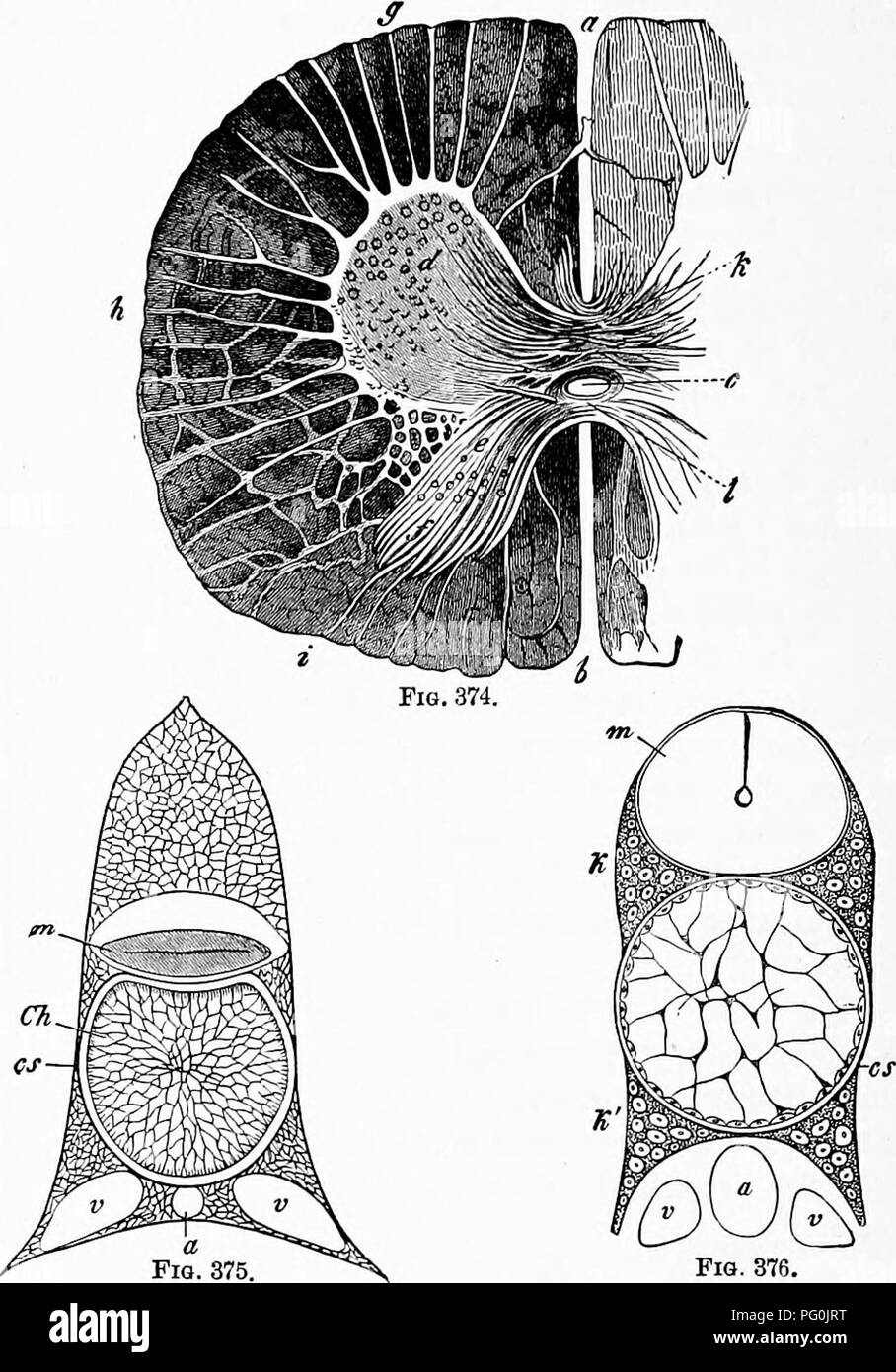 . Zoology : for students and general readers . Zoology. 394 ZOOLOGY. segmented. In all Vertebrates above the lamprey, the verte- bral column grows around the notochord, which finally. Fig. 376. Fi?. 374 —Transveree section through the spinal cord of a calf, a, anterior, &, poirterior longitudinal fissure ; c, central canal; d, anterior, e, posterior cornua; /, substantia gclatinosa; (/, anterior column of the white isube^tance ; A, lateral, i, pos- terior column ; k^ transverse commist-ures.—After Gegenbaur. Fig. 375.—SectioD throu^i^h the vertebral column of Ammoccetes (lamprey). Ch, no- Stock Photohttps://www.alamy.com/image-license-details/?v=1https://www.alamy.com/zoology-for-students-and-general-readers-zoology-394-zoology-segmented-in-all-vertebrates-above-the-lamprey-the-verte-bral-column-grows-around-the-notochord-which-finally-fig-376-fi-374-transveree-section-through-the-spinal-cord-of-a-calf-a-anterior-amp-poirterior-longitudinal-fissure-c-central-canal-d-anterior-e-posterior-cornua-substantia-gclatinosa-anterior-column-of-the-white-isubetance-a-lateral-i-pos-terior-column-k-transverse-commist-uresafter-gegenbaur-fig-375sectiod-throuih-the-vertebral-column-of-ammoccetes-lamprey-ch-no-image216373692.html
RMPG0JRT–. Zoology : for students and general readers . Zoology. 394 ZOOLOGY. segmented. In all Vertebrates above the lamprey, the verte- bral column grows around the notochord, which finally. Fig. 376. Fi?. 374 —Transveree section through the spinal cord of a calf, a, anterior, &, poirterior longitudinal fissure ; c, central canal; d, anterior, e, posterior cornua; /, substantia gclatinosa; (/, anterior column of the white isube^tance ; A, lateral, i, pos- terior column ; k^ transverse commist-ures.—After Gegenbaur. Fig. 375.—SectioD throu^i^h the vertebral column of Ammoccetes (lamprey). Ch, no-
 A stylized close up view of the front of the head. The brain is visible within the skull. Stock Photohttps://www.alamy.com/image-license-details/?v=1https://www.alamy.com/stock-photo-a-stylized-close-up-view-of-the-front-of-the-head-the-brain-is-visible-52099214.html
RMD0N926–A stylized close up view of the front of the head. The brain is visible within the skull.
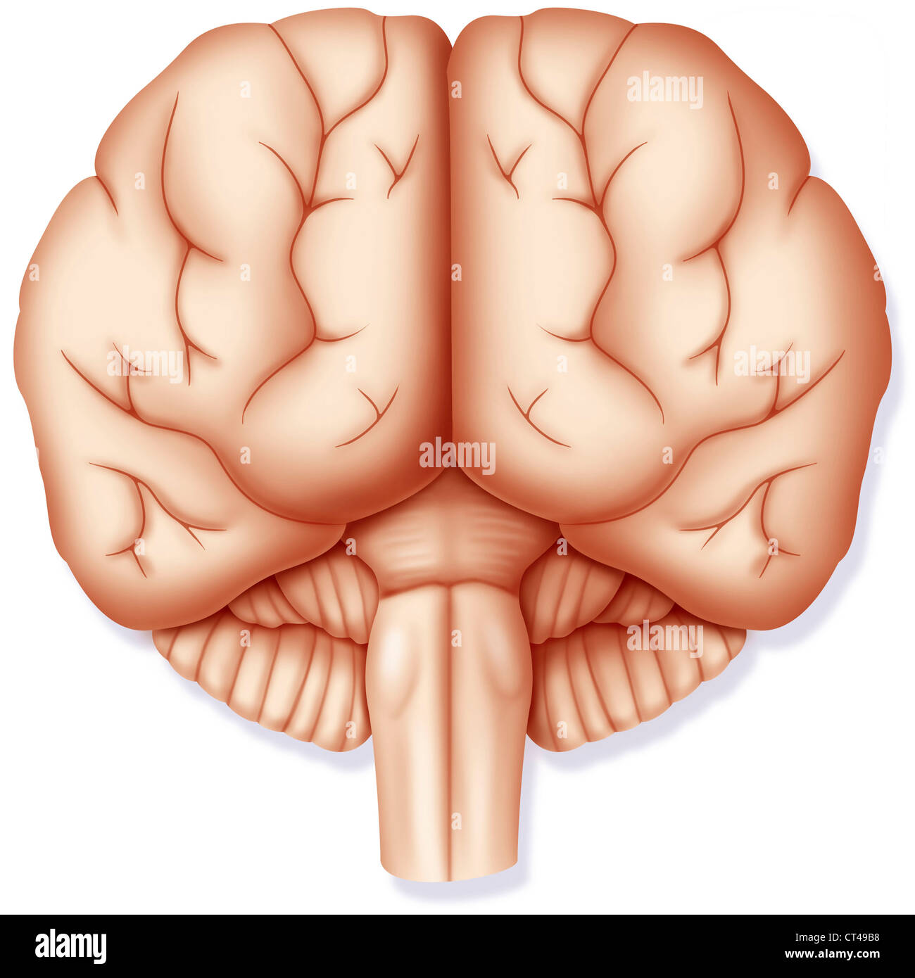 BRAIN, DRAWING Stock Photohttps://www.alamy.com/image-license-details/?v=1https://www.alamy.com/stock-photo-brain-drawing-49267660.html
RMCT49B8–BRAIN, DRAWING
 The encyclopdia britannica; a dictionary of arts, sciences, literature and general information . skeletal muscles.The movements occur in the half of the body of the side crossedfrom that of the hemisphere excited. The motor representa-tion, as it is termed, is in the cortex better described as arepresentation of definite actions than of particular muscles.The actions represented in the top part of the gyrus, namelynext the great longitudinal fissure, move the leg; those in thelowest part of the gyrus belong to the tongue and mouth. Thetopical distribution along the length of the gyrus may be d Stock Photohttps://www.alamy.com/image-license-details/?v=1https://www.alamy.com/the-encyclopdia-britannica-a-dictionary-of-arts-sciences-literature-and-general-information-skeletal-musclesthe-movements-occur-in-the-half-of-the-body-of-the-side-crossedfrom-that-of-the-hemisphere-excited-the-motor-representa-tion-as-it-is-termed-is-in-the-cortex-better-described-as-arepresentation-of-definite-actions-than-of-particular-musclesthe-actions-represented-in-the-top-part-of-the-gyrus-namelynext-the-great-longitudinal-fissure-move-the-leg-those-in-thelowest-part-of-the-gyrus-belong-to-the-tongue-and-mouth-thetopical-distribution-along-the-length-of-the-gyrus-may-be-d-image340226415.html
RM2ANEJ7B–The encyclopdia britannica; a dictionary of arts, sciences, literature and general information . skeletal muscles.The movements occur in the half of the body of the side crossedfrom that of the hemisphere excited. The motor representa-tion, as it is termed, is in the cortex better described as arepresentation of definite actions than of particular muscles.The actions represented in the top part of the gyrus, namelynext the great longitudinal fissure, move the leg; those in thelowest part of the gyrus belong to the tongue and mouth. Thetopical distribution along the length of the gyrus may be d
 The cerebral hemispheres are separated by a deep groove, the longitudinal cerebral fissure . Stock Photohttps://www.alamy.com/image-license-details/?v=1https://www.alamy.com/the-cerebral-hemispheres-are-separated-by-a-deep-groove-the-longitudinal-cerebral-fissure-image596590208.html
RF2WJH0W4–The cerebral hemispheres are separated by a deep groove, the longitudinal cerebral fissure .