Quick filters:
Lower arm bone Stock Photos and Images
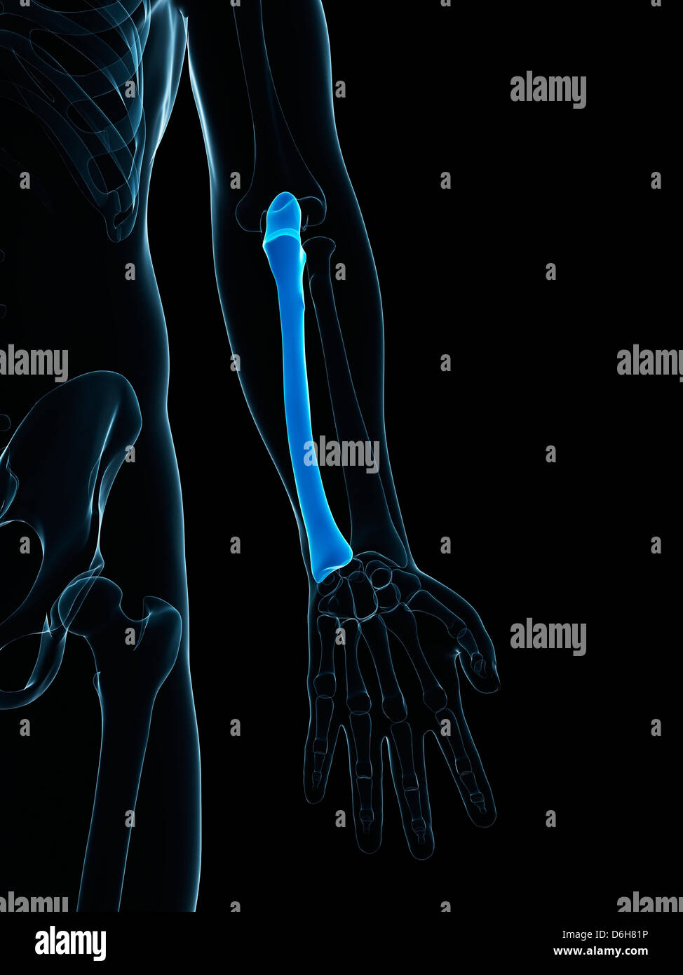 Lower arm bone, artwork Stock Photohttps://www.alamy.com/image-license-details/?v=1https://www.alamy.com/stock-photo-lower-arm-bone-artwork-55698546.html
RFD6H81P–Lower arm bone, artwork
 medically accurate illustration of the lower arm bones Stock Photohttps://www.alamy.com/image-license-details/?v=1https://www.alamy.com/stock-photo-medically-accurate-illustration-of-the-lower-arm-bones-89767382.html
RFF6175X–medically accurate illustration of the lower arm bones
 Upper image : Fracture ulnar and radius (Forearm bone) , Lower image : It was operated and internal fixed with plate and screw Stock Photohttps://www.alamy.com/image-license-details/?v=1https://www.alamy.com/stock-photo-upper-image-fracture-ulnar-and-radius-forearm-bone-lower-image-it-77405520.html
RFEDX3ET–Upper image : Fracture ulnar and radius (Forearm bone) , Lower image : It was operated and internal fixed with plate and screw
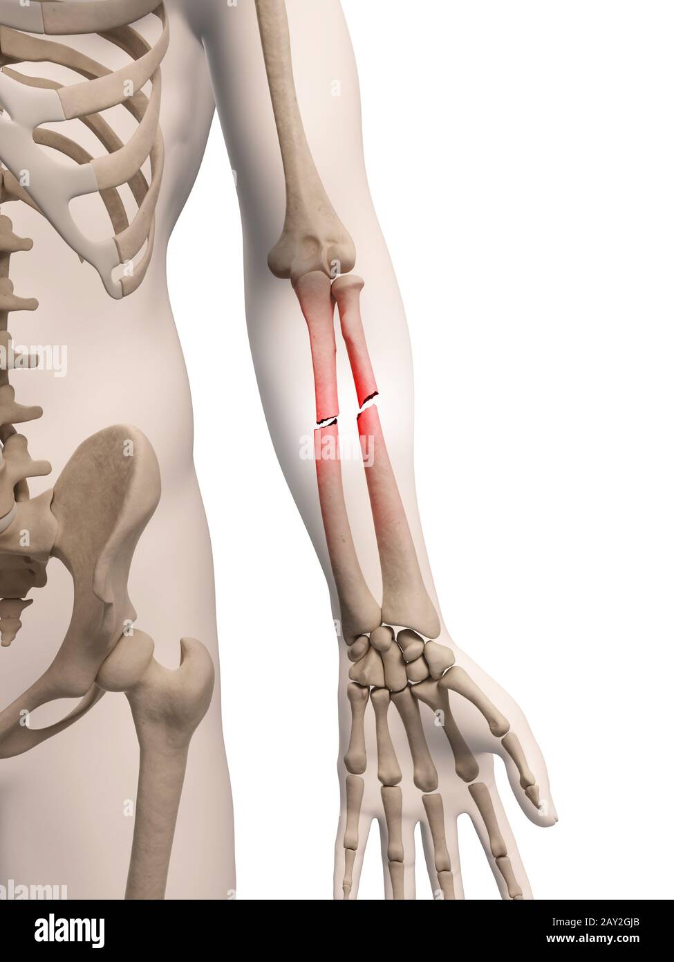 medical illustration of arm bone Stock Photohttps://www.alamy.com/image-license-details/?v=1https://www.alamy.com/medical-illustration-of-arm-bone-image343649667.html
RM2AY2GJB–medical illustration of arm bone
 Hand and lower arm in skeleton form viewed from above Stock Photohttps://www.alamy.com/image-license-details/?v=1https://www.alamy.com/hand-and-lower-arm-in-skeleton-form-viewed-from-above-image491950177.html
RM2KGA7D5–Hand and lower arm in skeleton form viewed from above
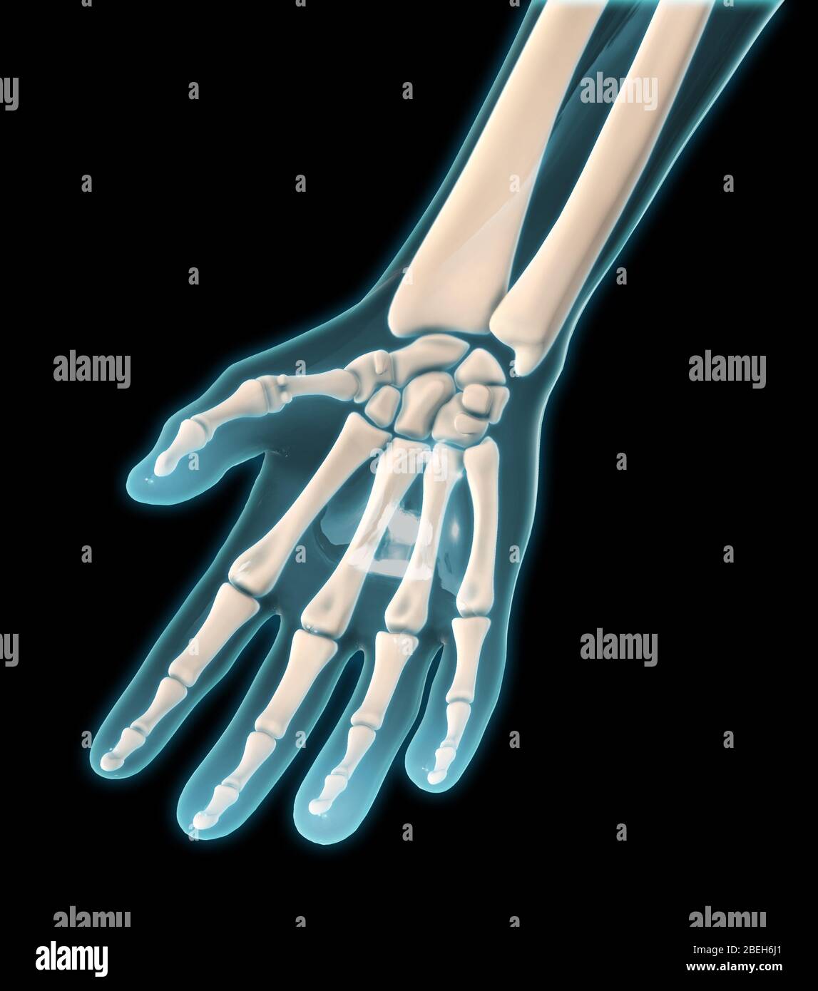 A palmar view of the bones of the right hand. Stock Photohttps://www.alamy.com/image-license-details/?v=1https://www.alamy.com/a-palmar-view-of-the-bones-of-the-right-hand-image353190937.html
RM2BEH6J1–A palmar view of the bones of the right hand.
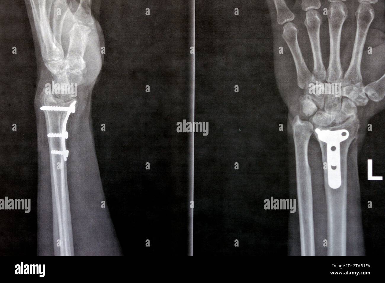 Plain x ray showing a recent fissure fracture at the lower part of a left radius bone, also showing a previous internal fixation of the wrist join wit Stock Photohttps://www.alamy.com/image-license-details/?v=1https://www.alamy.com/plain-x-ray-showing-a-recent-fissure-fracture-at-the-lower-part-of-a-left-radius-bone-also-showing-a-previous-internal-fixation-of-the-wrist-join-wit-image574331390.html
RF2TAB1FA–Plain x ray showing a recent fissure fracture at the lower part of a left radius bone, also showing a previous internal fixation of the wrist join wit
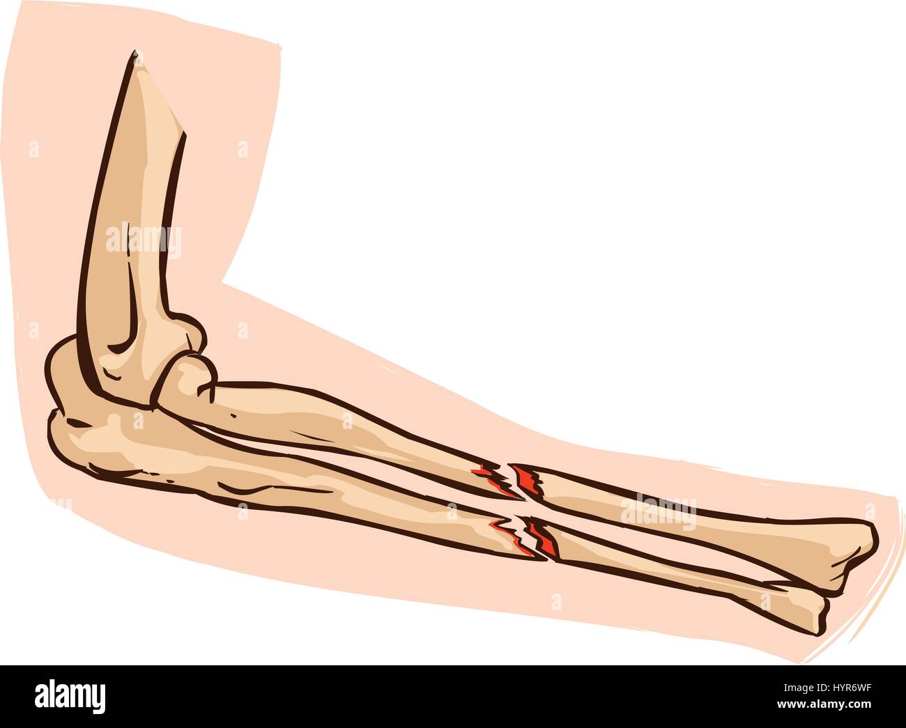 vector illustration of a medical illustration of arm bone Stock Vectorhttps://www.alamy.com/image-license-details/?v=1https://www.alamy.com/stock-photo-vector-illustration-of-a-medical-illustration-of-arm-bone-137578603.html
RFHYR6WF–vector illustration of a medical illustration of arm bone
 Rear view shot of a man with a lower backpain isolated on white background Stock Photohttps://www.alamy.com/image-license-details/?v=1https://www.alamy.com/rear-view-shot-of-a-man-with-a-lower-backpain-isolated-on-white-background-image567821580.html
RF2RYPE64–Rear view shot of a man with a lower backpain isolated on white background
 Woman from Middle Belt, Nigeria. Upper and lower lip and ear lobe have bone or ivory inserted discs and face is scarified with family tribal recogniti Stock Photohttps://www.alamy.com/image-license-details/?v=1https://www.alamy.com/stock-image-woman-from-middle-belt-nigeria-upper-and-lower-lip-and-ear-lobe-have-160221624.html
RMK8JM8T–Woman from Middle Belt, Nigeria. Upper and lower lip and ear lobe have bone or ivory inserted discs and face is scarified with family tribal recogniti
 Rear view shot of an elderly man holding his back with visible spine bone at a gym Stock Photohttps://www.alamy.com/image-license-details/?v=1https://www.alamy.com/rear-view-shot-of-an-elderly-man-holding-his-back-with-visible-spine-bone-at-a-gym-image568780078.html
RF2T1A4P6–Rear view shot of an elderly man holding his back with visible spine bone at a gym
 Back pain woman getting after wake up. Sick person with Backache in spine. Unrecognizable woman in bedroom have lumbar, lower back pain after sleeping Stock Photohttps://www.alamy.com/image-license-details/?v=1https://www.alamy.com/back-pain-woman-getting-after-wake-up-sick-person-with-backache-in-spine-unrecognizable-woman-in-bedroom-have-lumbar-lower-back-pain-after-sleeping-image434744822.html
RF2G789CP–Back pain woman getting after wake up. Sick person with Backache in spine. Unrecognizable woman in bedroom have lumbar, lower back pain after sleeping
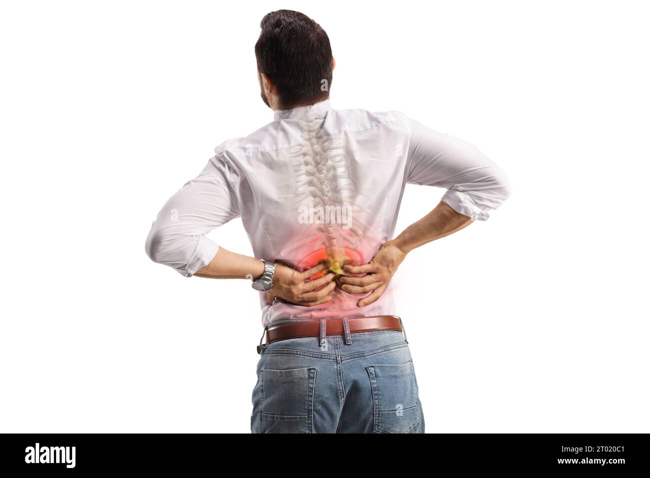 Rear view shot of a man holding lower back inflamed zone with visible spine bone isolated on white background Stock Photohttps://www.alamy.com/image-license-details/?v=1https://www.alamy.com/rear-view-shot-of-a-man-holding-lower-back-inflamed-zone-with-visible-spine-bone-isolated-on-white-background-image567986385.html
RF2T020C1–Rear view shot of a man holding lower back inflamed zone with visible spine bone isolated on white background
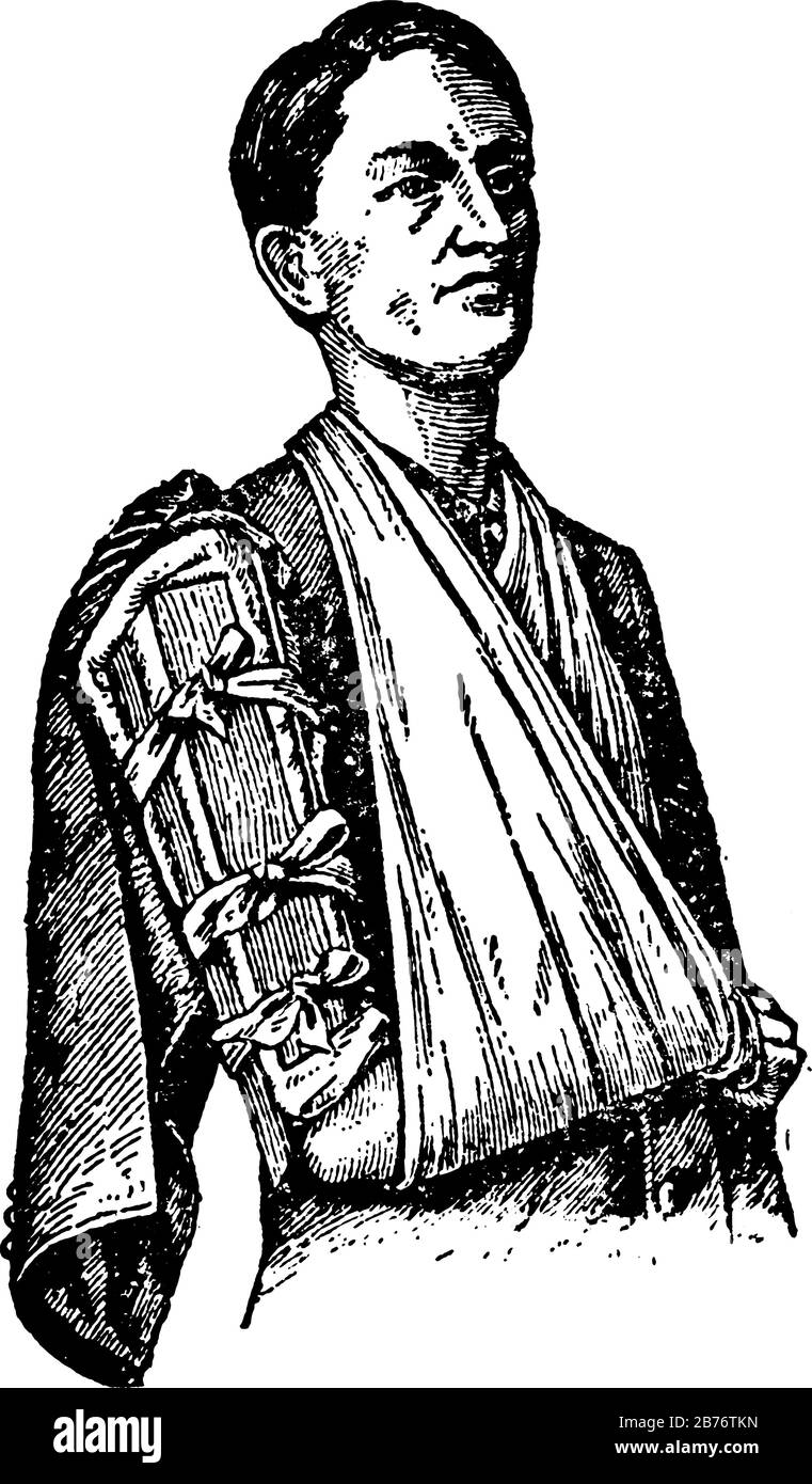 Two splints applied, one in front, the other behind, if the lower part of the bone is broke; or to the inner and outer sides, vintage line drawing or Stock Vectorhttps://www.alamy.com/image-license-details/?v=1https://www.alamy.com/two-splints-applied-one-in-front-the-other-behind-if-the-lower-part-of-the-bone-is-broke-or-to-the-inner-and-outer-sides-vintage-line-drawing-or-image348661033.html
RF2B76TKN–Two splints applied, one in front, the other behind, if the lower part of the bone is broke; or to the inner and outer sides, vintage line drawing or
 vector illustration of a who broke his arm falling Stock Vectorhttps://www.alamy.com/image-license-details/?v=1https://www.alamy.com/stock-photo-vector-illustration-of-a-who-broke-his-arm-falling-137611356.html
RFHYTMK8–vector illustration of a who broke his arm falling
 Human skeleton art work vector illustration Stock Vectorhttps://www.alamy.com/image-license-details/?v=1https://www.alamy.com/human-skeleton-art-work-vector-illustration-image453498739.html
RF2H9PJ7F–Human skeleton art work vector illustration
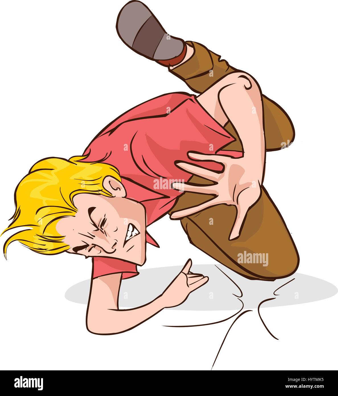 vector illustration of a who broke his arm falling Stock Vectorhttps://www.alamy.com/image-license-details/?v=1https://www.alamy.com/stock-photo-vector-illustration-of-a-who-broke-his-arm-falling-137611353.html
RFHYTMK5–vector illustration of a who broke his arm falling
 Female scratching her arm on white background with copy space. Medical, healthcare for advertising concept. Stock Photohttps://www.alamy.com/image-license-details/?v=1https://www.alamy.com/female-scratching-her-arm-on-white-background-with-copy-space-medical-healthcare-for-advertising-concept-image357629425.html
RF2BNRBYD–Female scratching her arm on white background with copy space. Medical, healthcare for advertising concept.
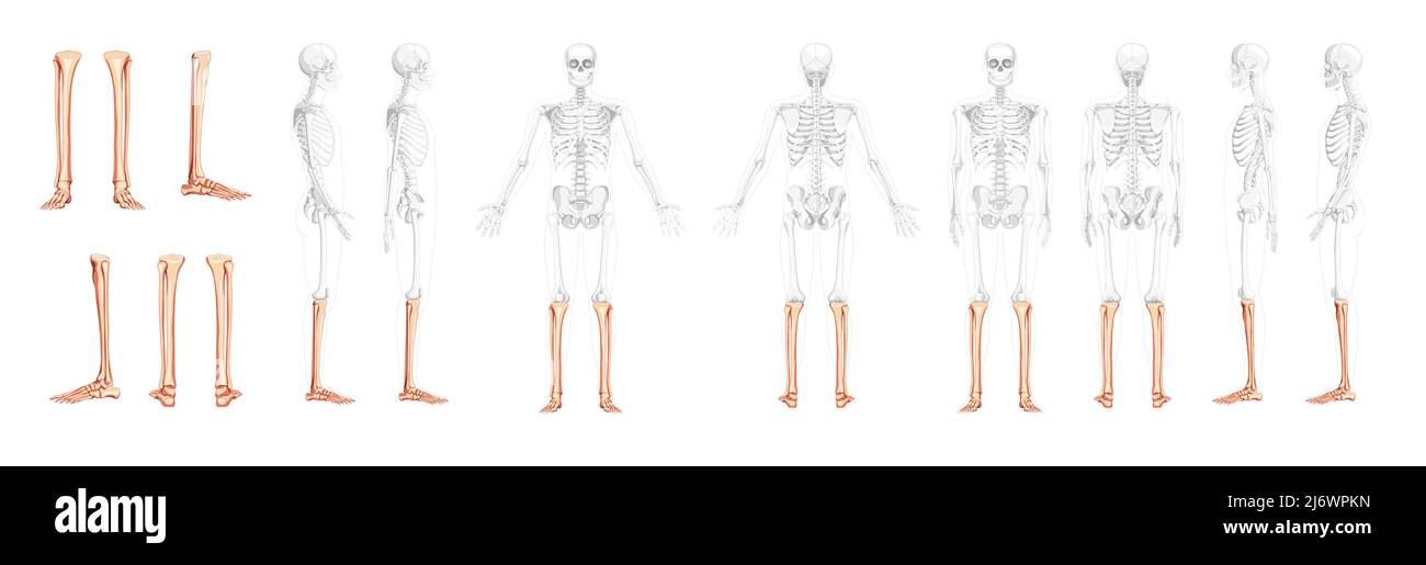 Set of Skeleton leg tibia, Foot, ankle Human front back side view with partly transparent bones position. 3D realistic flat natural color Vector illustration of anatomy isolated on white background Stock Vectorhttps://www.alamy.com/image-license-details/?v=1https://www.alamy.com/set-of-skeleton-leg-tibia-foot-ankle-human-front-back-side-view-with-partly-transparent-bones-position-3d-realistic-flat-natural-color-vector-illustration-of-anatomy-isolated-on-white-background-image468934473.html
RF2J6WPKN–Set of Skeleton leg tibia, Foot, ankle Human front back side view with partly transparent bones position. 3D realistic flat natural color Vector illustration of anatomy isolated on white background
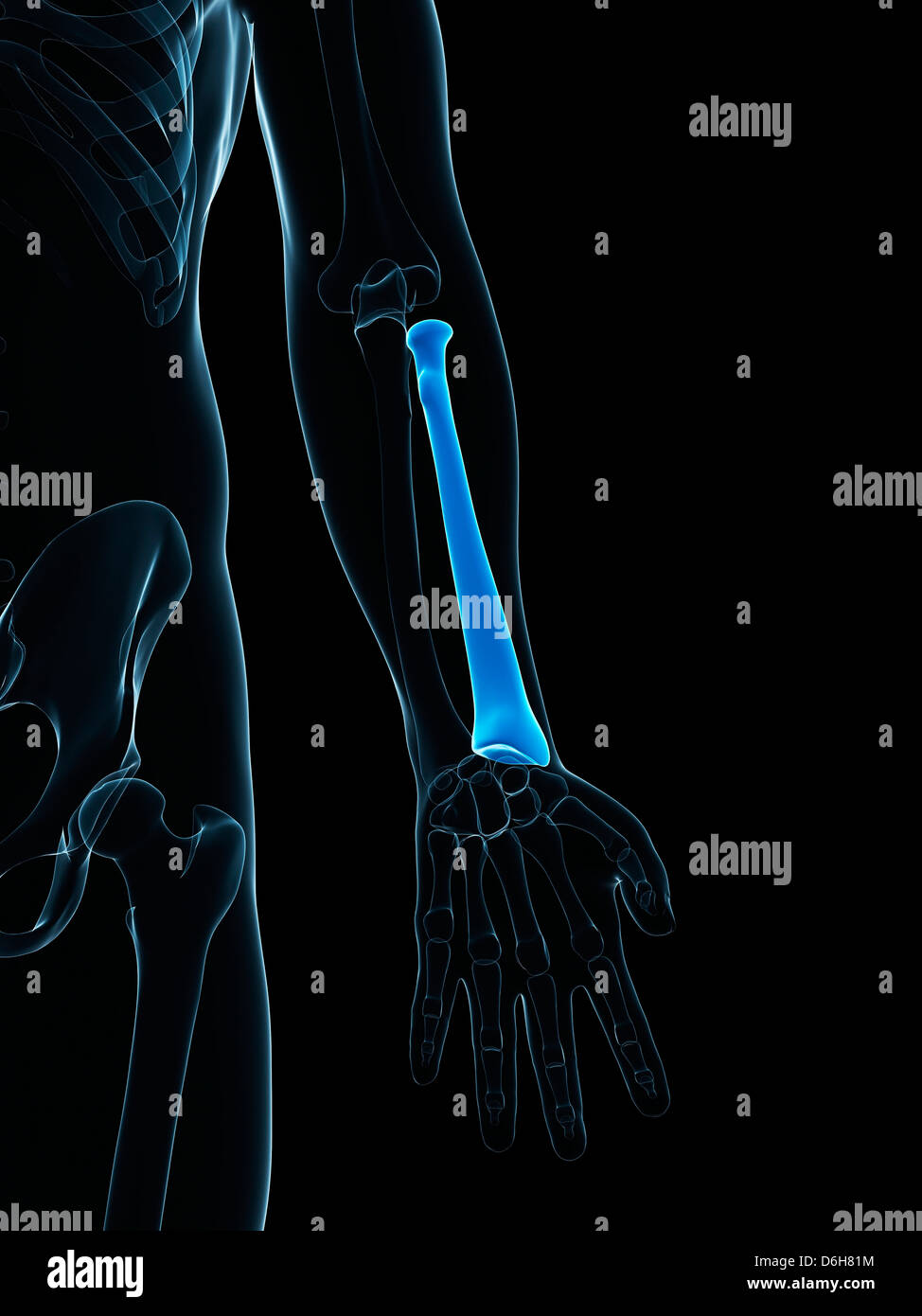 Lower arm bone, artwork Stock Photohttps://www.alamy.com/image-license-details/?v=1https://www.alamy.com/stock-photo-lower-arm-bone-artwork-55698544.html
RFD6H81M–Lower arm bone, artwork
 medically accurate illustration of the lower arm bones Stock Photohttps://www.alamy.com/image-license-details/?v=1https://www.alamy.com/stock-photo-medically-accurate-illustration-of-the-lower-arm-bones-89767388.html
RFF61764–medically accurate illustration of the lower arm bones
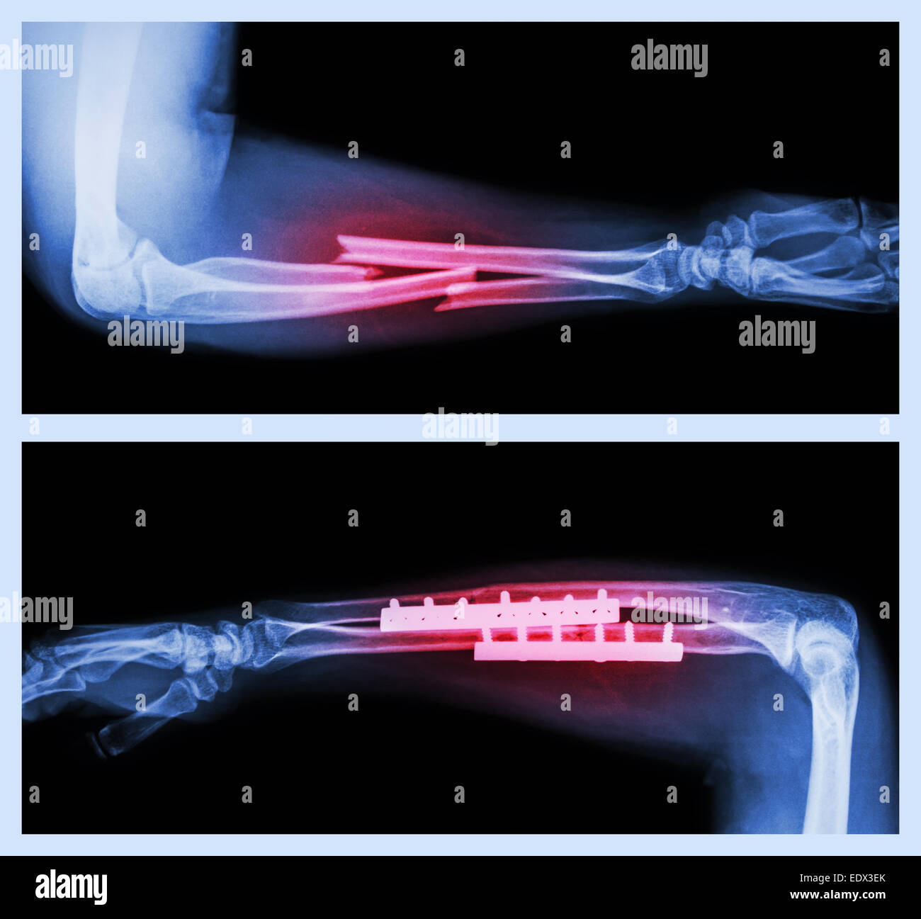 Upper image : Fracture ulnar and radius (Forearm bone) , Lower image : It was operated and internal fixed with plate and screw Stock Photohttps://www.alamy.com/image-license-details/?v=1https://www.alamy.com/stock-photo-upper-image-fracture-ulnar-and-radius-forearm-bone-lower-image-it-77405515.html
RFEDX3EK–Upper image : Fracture ulnar and radius (Forearm bone) , Lower image : It was operated and internal fixed with plate and screw
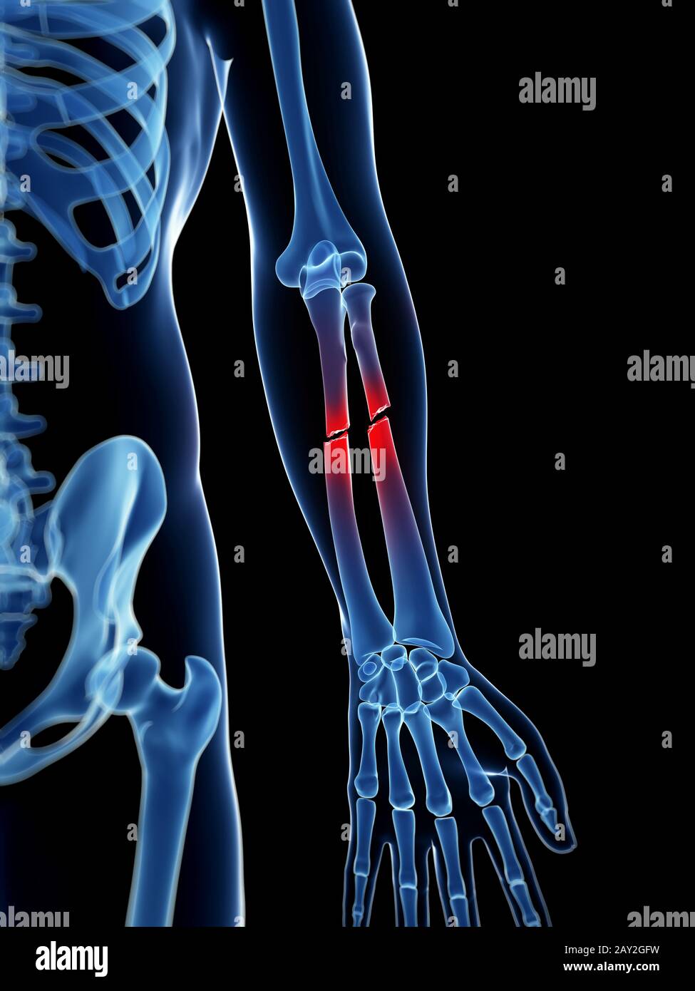 medical illustration of a broken lower arm Stock Photohttps://www.alamy.com/image-license-details/?v=1https://www.alamy.com/medical-illustration-of-a-broken-lower-arm-image343649597.html
RM2AY2GFW–medical illustration of a broken lower arm
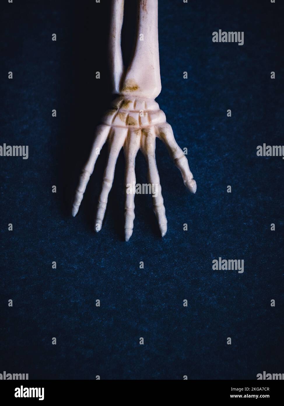 View from above of hand and lower arm in skeleton form Stock Photohttps://www.alamy.com/image-license-details/?v=1https://www.alamy.com/view-from-above-of-hand-and-lower-arm-in-skeleton-form-image491950167.html
RM2KGA7CR–View from above of hand and lower arm in skeleton form
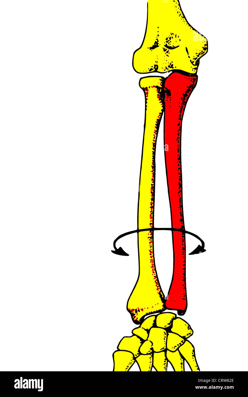 Forearm bone Stock Photohttps://www.alamy.com/image-license-details/?v=1https://www.alamy.com/stock-photo-forearm-bone-49112966.html
RFCRW82E–Forearm bone
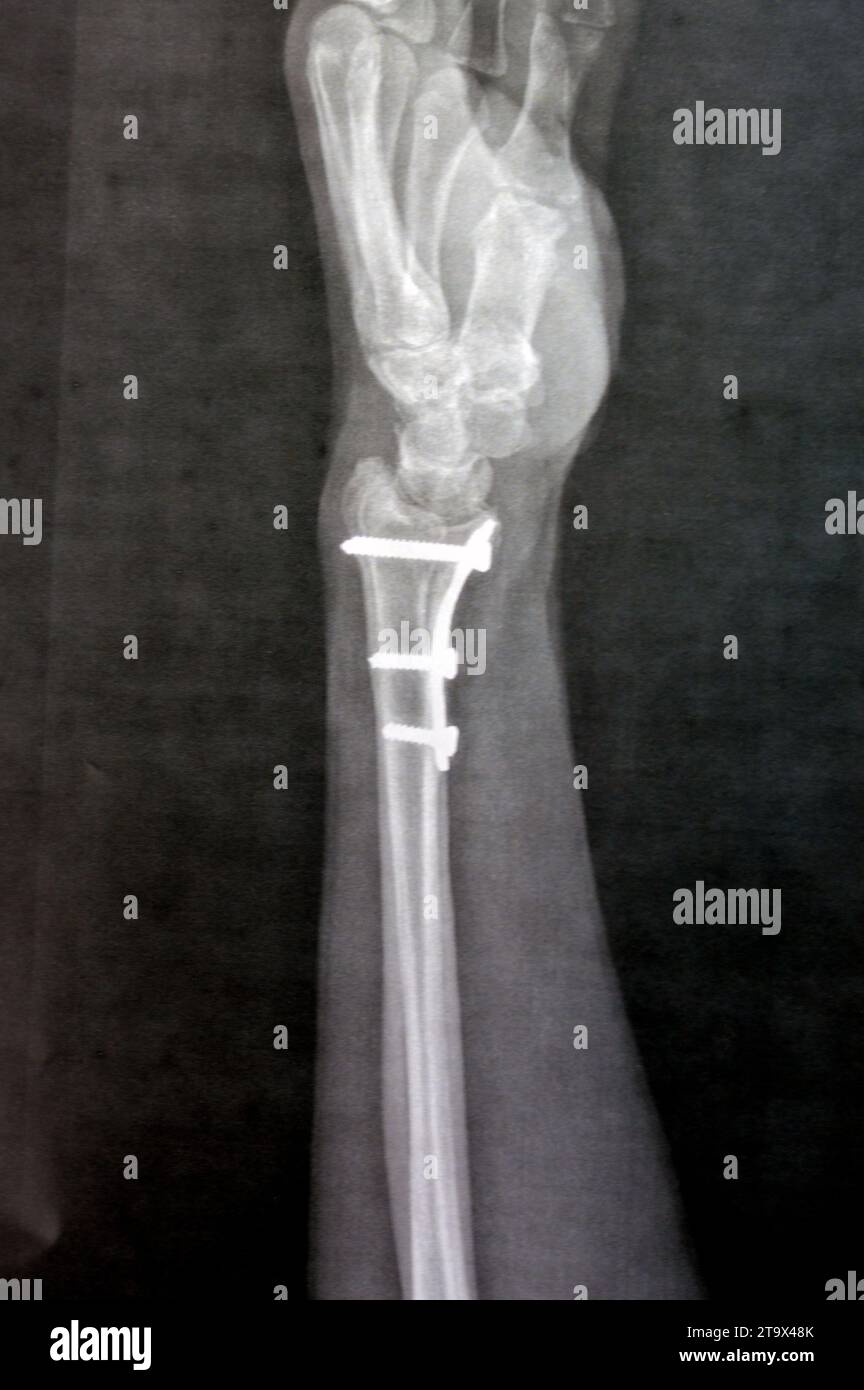 Plain x ray showing a recent fissure fracture at the lower part of a left radius bone, also showing a previous internal fixation of the wrist join wit Stock Photohttps://www.alamy.com/image-license-details/?v=1https://www.alamy.com/plain-x-ray-showing-a-recent-fissure-fracture-at-the-lower-part-of-a-left-radius-bone-also-showing-a-previous-internal-fixation-of-the-wrist-join-wit-image574048179.html
RF2T9X48K–Plain x ray showing a recent fissure fracture at the lower part of a left radius bone, also showing a previous internal fixation of the wrist join wit
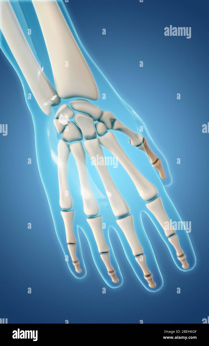 Bones of the Hand, Dorsal View Stock Photohttps://www.alamy.com/image-license-details/?v=1https://www.alamy.com/bones-of-the-hand-dorsal-view-image353190895.html
RM2BEH6GF–Bones of the Hand, Dorsal View
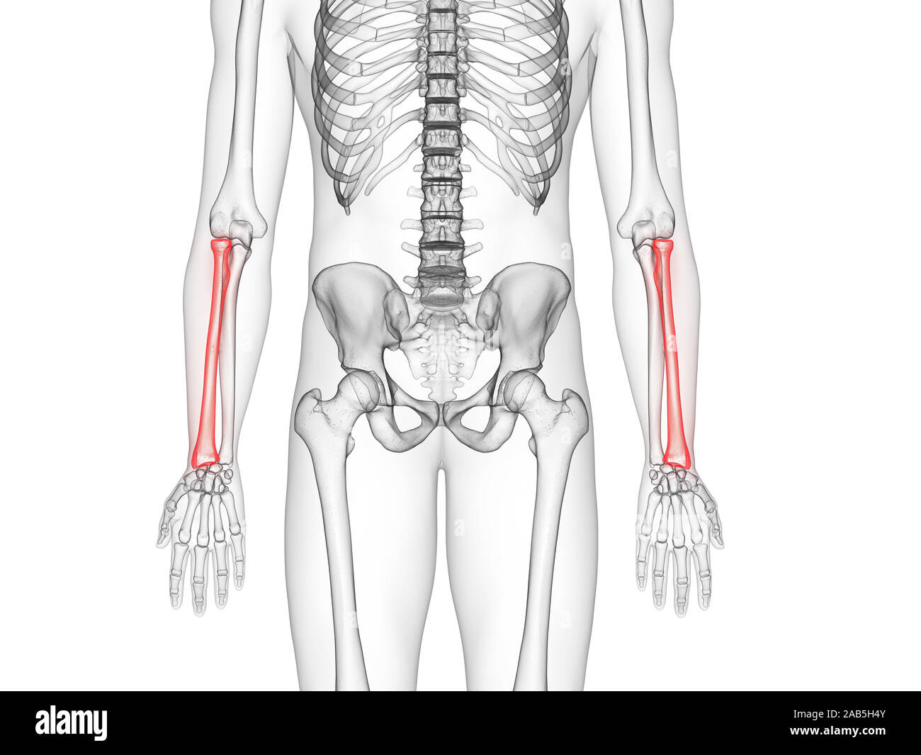 3d rendered medically accurate illustration of the radius bone Stock Photohttps://www.alamy.com/image-license-details/?v=1https://www.alamy.com/3d-rendered-medically-accurate-illustration-of-the-radius-bone-image333881435.html
RF2AB5H4Y–3d rendered medically accurate illustration of the radius bone
 Bones of the lower arm and hand. Stock Photohttps://www.alamy.com/image-license-details/?v=1https://www.alamy.com/stock-photo-bones-of-the-lower-arm-and-hand-52088899.html
RMD0MRWR–Bones of the lower arm and hand.
 Right arm cover, Arm cover, iron, blank, consists of an articulated shoulder plate, upper arm pipe, an elbow cap and a lower arm pipe. At the top of the shoulder plate is an iron buckle, below which is an articulated strip that is riveted to the furnace arm pipe, the edges are flanged high and cabled. The upper arm pipe consists of two parts that are connected to each other at two points, a cabled bone in the middle. The elbow cover has two parts and has flanged cable edges. The forearm pipe consists of two parts connected to each other with two hinges through which the pieces can be opened Stock Photohttps://www.alamy.com/image-license-details/?v=1https://www.alamy.com/right-arm-cover-arm-cover-iron-blank-consists-of-an-articulated-shoulder-plate-upper-arm-pipe-an-elbow-cap-and-a-lower-arm-pipe-at-the-top-of-the-shoulder-plate-is-an-iron-buckle-below-which-is-an-articulated-strip-that-is-riveted-to-the-furnace-arm-pipe-the-edges-are-flanged-high-and-cabled-the-upper-arm-pipe-consists-of-two-parts-that-are-connected-to-each-other-at-two-points-a-cabled-bone-in-the-middle-the-elbow-cover-has-two-parts-and-has-flanged-cable-edges-the-forearm-pipe-consists-of-two-parts-connected-to-each-other-with-two-hinges-through-which-the-pieces-can-be-opened-image261383159.html
RMW570TR–Right arm cover, Arm cover, iron, blank, consists of an articulated shoulder plate, upper arm pipe, an elbow cap and a lower arm pipe. At the top of the shoulder plate is an iron buckle, below which is an articulated strip that is riveted to the furnace arm pipe, the edges are flanged high and cabled. The upper arm pipe consists of two parts that are connected to each other at two points, a cabled bone in the middle. The elbow cover has two parts and has flanged cable edges. The forearm pipe consists of two parts connected to each other with two hinges through which the pieces can be opened
 Back pain woman getting after wake up. Sick person with lower back pain. Unrecognizable woman in bedroom have Lower back pain after sleeping Stock Photohttps://www.alamy.com/image-license-details/?v=1https://www.alamy.com/back-pain-woman-getting-after-wake-up-sick-person-with-lower-back-pain-unrecognizable-woman-in-bedroom-have-lower-back-pain-after-sleeping-image415171458.html
RF2F3CKC2–Back pain woman getting after wake up. Sick person with lower back pain. Unrecognizable woman in bedroom have Lower back pain after sleeping
 Rear view shot of a man holding lower back inflamed zone isolated on white background Stock Photohttps://www.alamy.com/image-license-details/?v=1https://www.alamy.com/rear-view-shot-of-a-man-holding-lower-back-inflamed-zone-isolated-on-white-background-image567902699.html
RF2RYX5K7–Rear view shot of a man holding lower back inflamed zone isolated on white background
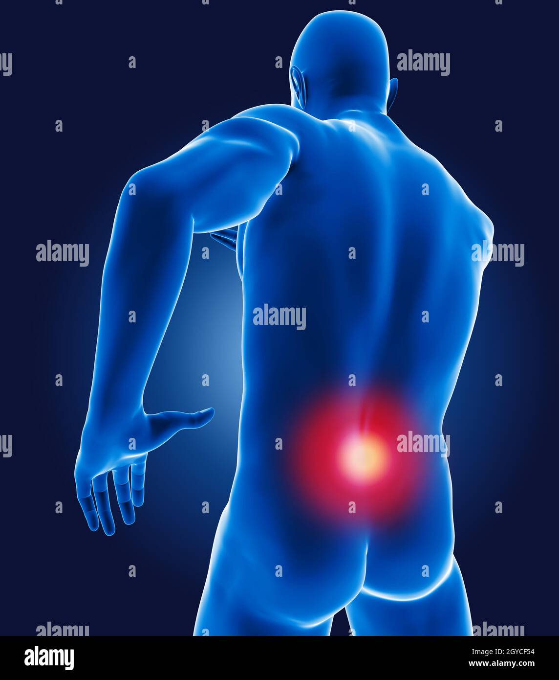 3D medical man with lower back highlighted Stock Photohttps://www.alamy.com/image-license-details/?v=1https://www.alamy.com/3d-medical-man-with-lower-back-highlighted-image447130240.html
RF2GYCF54–3D medical man with lower back highlighted
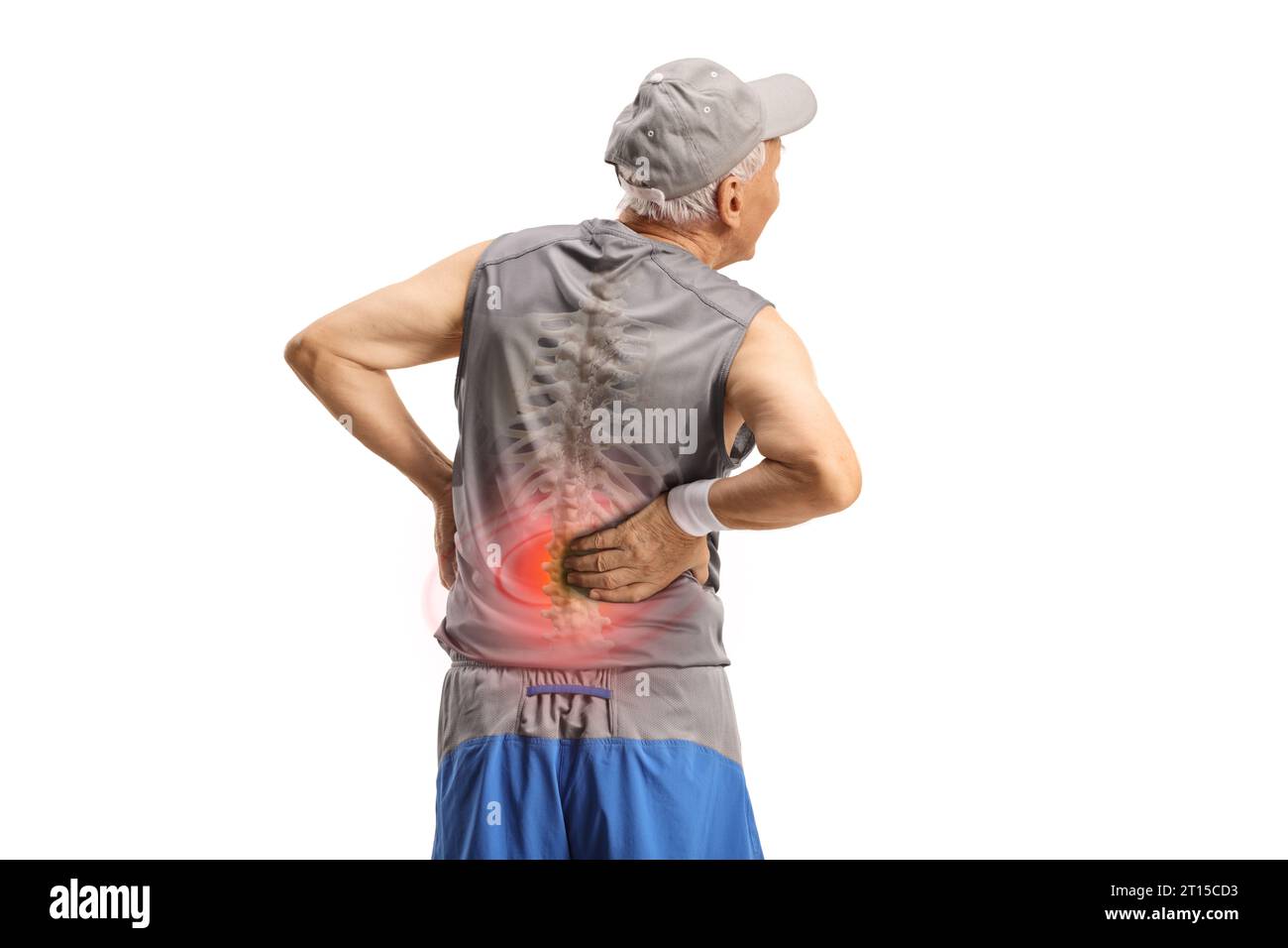 Rear view shot of an elderly man in sportswear holding his back with visible spine bone isolated on white background Stock Photohttps://www.alamy.com/image-license-details/?v=1https://www.alamy.com/rear-view-shot-of-an-elderly-man-in-sportswear-holding-his-back-with-visible-spine-bone-isolated-on-white-background-image568676335.html
RF2T15CD3–Rear view shot of an elderly man in sportswear holding his back with visible spine bone isolated on white background
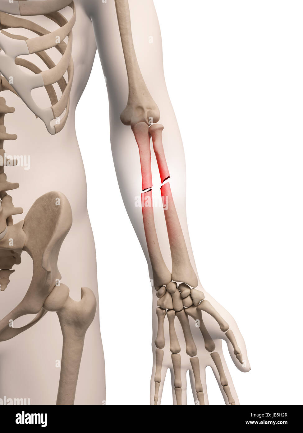 medical illustration of arm bone Stock Photohttps://www.alamy.com/image-license-details/?v=1https://www.alamy.com/stock-photo-medical-illustration-of-arm-bone-144567327.html
RFJB5H2R–medical illustration of arm bone
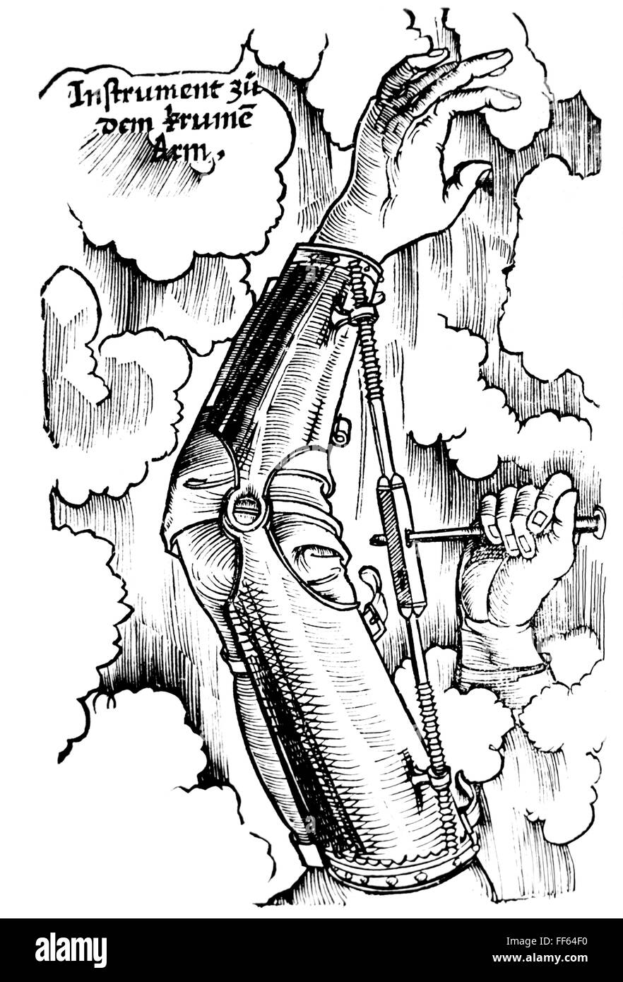 medicine, instruments / equipment, adjustable arm cuff, woodcut, 16th century, 16th century, graphic, graphics, anatomy, hand, hands, arm, arms, bone, cuff, cuffs, metal, metals, fixate, fixating, adjustable, shift, shifting, screw, screws, part of the body, parts of the body, the lower limbs, immobilize a limb, medicine, medicines, instrument, instruments, devices, device, woodcut, woodcuts, historic, historical, people, Additional-Rights-Clearences-Not Available Stock Photohttps://www.alamy.com/image-license-details/?v=1https://www.alamy.com/stock-photo-medicine-instruments-equipment-adjustable-arm-cuff-woodcut-16th-century-95406948.html
RMFF64F0–medicine, instruments / equipment, adjustable arm cuff, woodcut, 16th century, 16th century, graphic, graphics, anatomy, hand, hands, arm, arms, bone, cuff, cuffs, metal, metals, fixate, fixating, adjustable, shift, shifting, screw, screws, part of the body, parts of the body, the lower limbs, immobilize a limb, medicine, medicines, instrument, instruments, devices, device, woodcut, woodcuts, historic, historical, people, Additional-Rights-Clearences-Not Available
 Male scratching his arm on white background with copy space. Medical, healthcare for advertising concept. Stock Photohttps://www.alamy.com/image-license-details/?v=1https://www.alamy.com/male-scratching-his-arm-on-white-background-with-copy-space-medical-healthcare-for-advertising-concept-image357629281.html
RF2BNRBP9–Male scratching his arm on white background with copy space. Medical, healthcare for advertising concept.
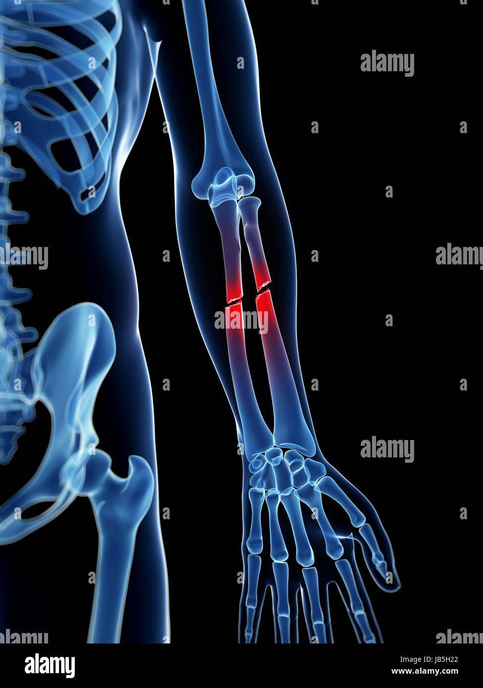 medical illustration of a broken lower arm Stock Photohttps://www.alamy.com/image-license-details/?v=1https://www.alamy.com/stock-photo-medical-illustration-of-a-broken-lower-arm-144567306.html
RFJB5H22–medical illustration of a broken lower arm
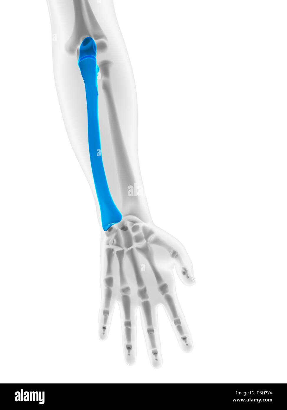 Lower arm bone, artwork Stock Photohttps://www.alamy.com/image-license-details/?v=1https://www.alamy.com/stock-photo-lower-arm-bone-artwork-55698478.html
RFD6H7YA–Lower arm bone, artwork
 Composite image of articular pain. Stock Photohttps://www.alamy.com/image-license-details/?v=1https://www.alamy.com/stock-photo-composite-image-of-articular-pain-79767212.html
RMEHNKW0–Composite image of articular pain.
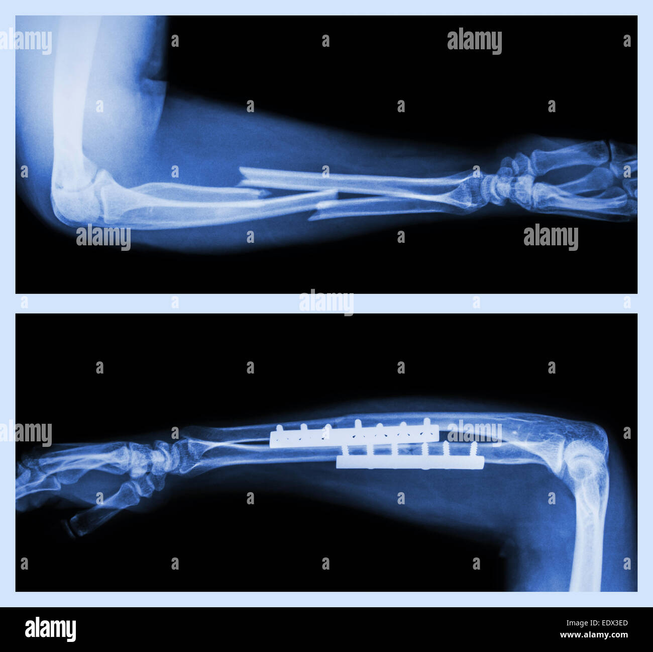 Upper image : Fracture ulnar and radius (Forearm bone) , Lower image : It was operated and internal fixed with plate and screw Stock Photohttps://www.alamy.com/image-license-details/?v=1https://www.alamy.com/stock-photo-upper-image-fracture-ulnar-and-radius-forearm-bone-lower-image-it-77405509.html
RFEDX3ED–Upper image : Fracture ulnar and radius (Forearm bone) , Lower image : It was operated and internal fixed with plate and screw
 Coat of arms with skull on a gravestone Bad Pyrmont cemetery Stock Photohttps://www.alamy.com/image-license-details/?v=1https://www.alamy.com/coat-of-arms-with-skull-on-a-gravestone-bad-pyrmont-cemetery-image619587895.html
RM2Y00JK3–Coat of arms with skull on a gravestone Bad Pyrmont cemetery
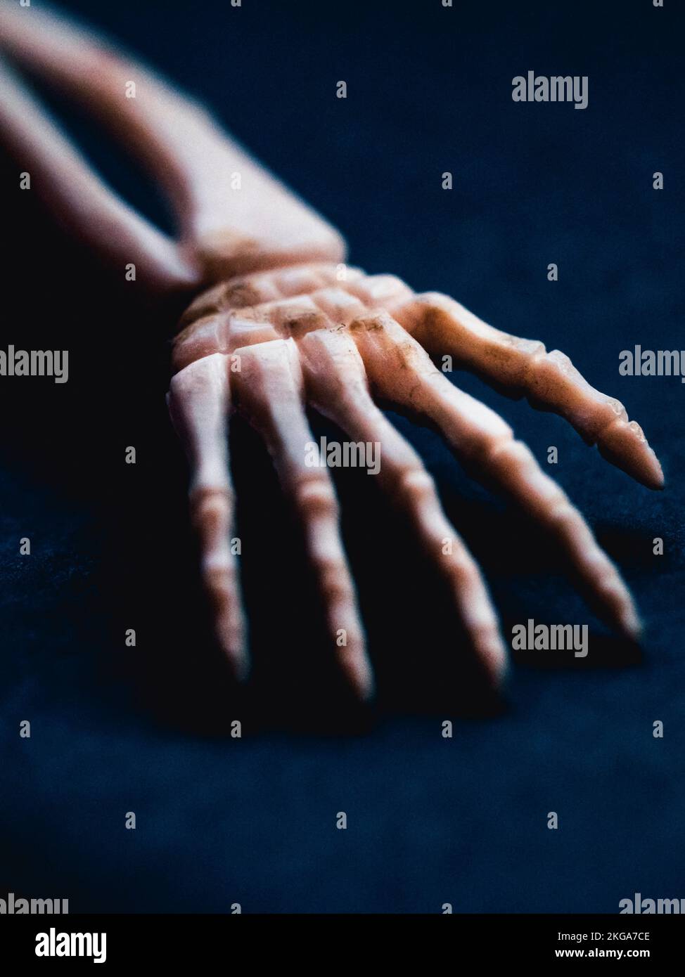 Close up of bones of hand and lower arm Stock Photohttps://www.alamy.com/image-license-details/?v=1https://www.alamy.com/close-up-of-bones-of-hand-and-lower-arm-image491950158.html
RM2KGA7CE–Close up of bones of hand and lower arm
 Skull front view with a lower jaw between two pair wings and ribbons. Vintage label isolated on white Stock Vectorhttps://www.alamy.com/image-license-details/?v=1https://www.alamy.com/skull-front-view-with-a-lower-jaw-between-two-pair-wings-and-ribbons-vintage-label-isolated-on-white-image333417455.html
RF2AACDA7–Skull front view with a lower jaw between two pair wings and ribbons. Vintage label isolated on white
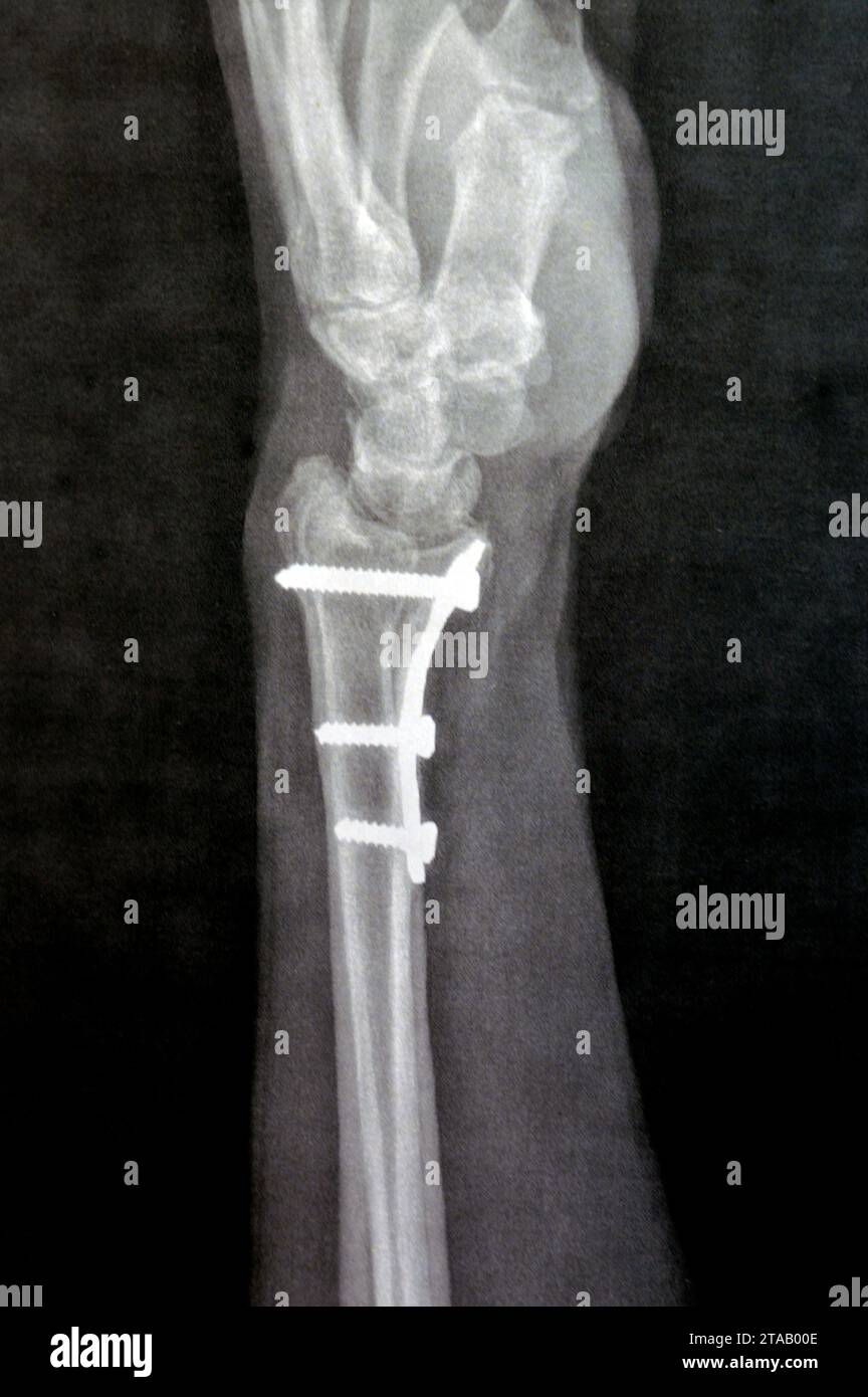 Plain x ray showing a recent fissure fracture at the lower part of a left radius bone, also showing a previous internal fixation of the wrist join wit Stock Photohttps://www.alamy.com/image-license-details/?v=1https://www.alamy.com/plain-x-ray-showing-a-recent-fissure-fracture-at-the-lower-part-of-a-left-radius-bone-also-showing-a-previous-internal-fixation-of-the-wrist-join-wit-image574330190.html
RF2TAB00E–Plain x ray showing a recent fissure fracture at the lower part of a left radius bone, also showing a previous internal fixation of the wrist join wit
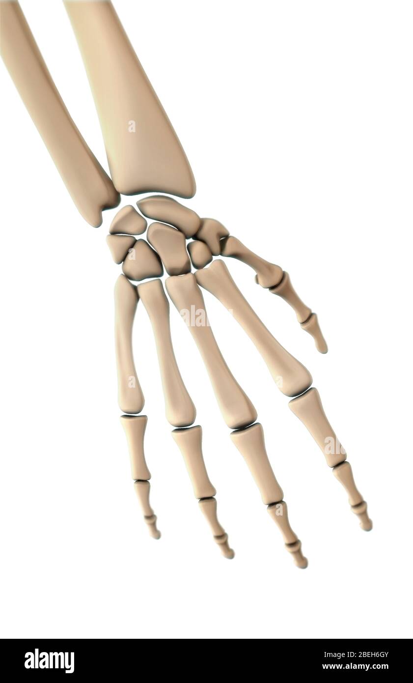 Bones of the Hand, Dorsal View Stock Photohttps://www.alamy.com/image-license-details/?v=1https://www.alamy.com/bones-of-the-hand-dorsal-view-image353190907.html
RM2BEH6GY–Bones of the Hand, Dorsal View
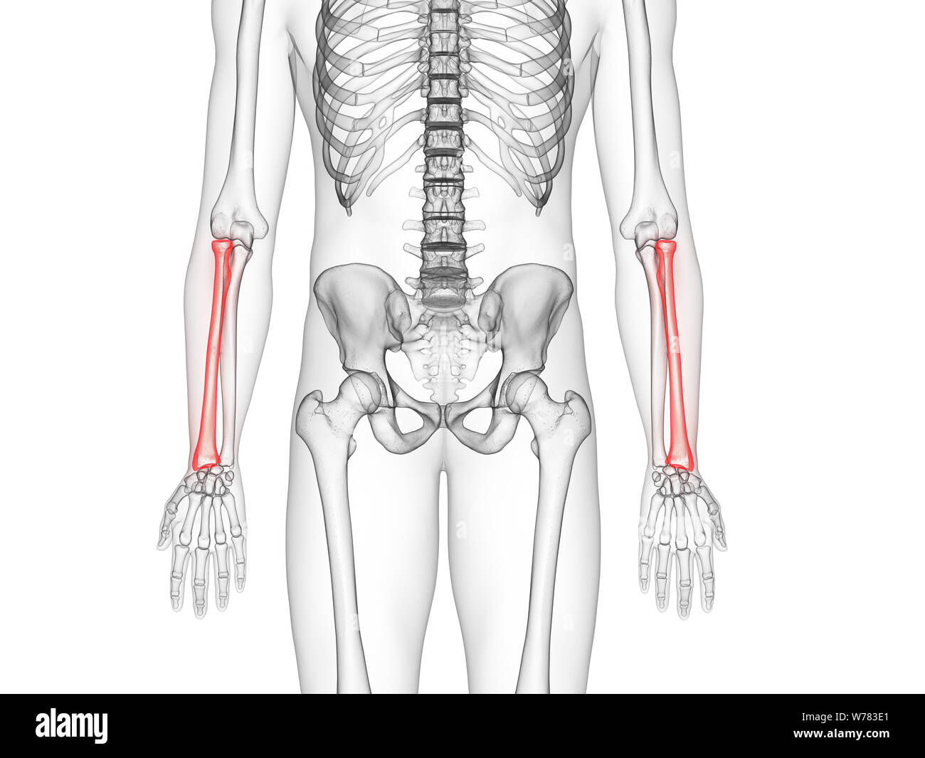 3d rendered medically accurate illustration of the radius bone Stock Photohttps://www.alamy.com/image-license-details/?v=1https://www.alamy.com/3d-rendered-medically-accurate-illustration-of-the-radius-bone-image262636473.html
RFW783E1–3d rendered medically accurate illustration of the radius bone
 Bones of the lower arm and hand. Stock Photohttps://www.alamy.com/image-license-details/?v=1https://www.alamy.com/stock-photo-bones-of-the-lower-arm-and-hand-52089067.html
RMD0MT3R–Bones of the lower arm and hand.
 Kuras breastplate, Breastplate, iron, blank, arm and neck cut-outs as well as lower and rear edges of the belly collar, high-quality cabled flange. At the front of the breastplate a etched weapon in which a helmet with a feather is surrounded by a weapon trophy. The central bone of the breastplate ends in a bulge. A belly collar is riveted to the breastplate which consists of two parts. A double decorative line running parallel to the top at the top. The inside of the breastplate is painted black., anonymous, Europe, 1499 - 1699, geheel, etching, l 11.3 cm × w 35.6 cm × h 49 cm Stock Photohttps://www.alamy.com/image-license-details/?v=1https://www.alamy.com/kuras-breastplate-breastplate-iron-blank-arm-and-neck-cut-outs-as-well-as-lower-and-rear-edges-of-the-belly-collar-high-quality-cabled-flange-at-the-front-of-the-breastplate-a-etched-weapon-in-which-a-helmet-with-a-feather-is-surrounded-by-a-weapon-trophy-the-central-bone-of-the-breastplate-ends-in-a-bulge-a-belly-collar-is-riveted-to-the-breastplate-which-consists-of-two-parts-a-double-decorative-line-running-parallel-to-the-top-at-the-top-the-inside-of-the-breastplate-is-painted-black-anonymous-europe-1499-1699-geheel-etching-l-113-cm-w-356-cm-h-49-cm-image261383156.html
RMW570TM–Kuras breastplate, Breastplate, iron, blank, arm and neck cut-outs as well as lower and rear edges of the belly collar, high-quality cabled flange. At the front of the breastplate a etched weapon in which a helmet with a feather is surrounded by a weapon trophy. The central bone of the breastplate ends in a bulge. A belly collar is riveted to the breastplate which consists of two parts. A double decorative line running parallel to the top at the top. The inside of the breastplate is painted black., anonymous, Europe, 1499 - 1699, geheel, etching, l 11.3 cm × w 35.6 cm × h 49 cm
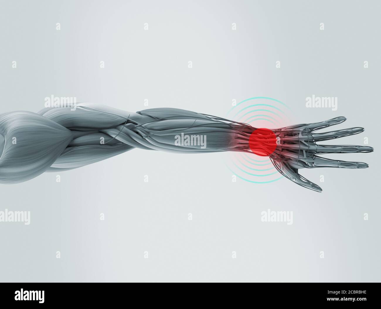 Human anatomy xray-like illustration of arm and hand. Stock Photohttps://www.alamy.com/image-license-details/?v=1https://www.alamy.com/human-anatomy-xray-like-illustration-of-arm-and-hand-image368692954.html
RF2CBRBHE–Human anatomy xray-like illustration of arm and hand.
 Albert Garcia, 47th Operations Support Squadron radar approach controller and retired master sergeant, poses for a photo at a rehabilitation center in San Antonio, July 17, 2015. Garcia was the victim of a hit-and-run on State Road 277 North, and suffered breaks to multiple bones including his pelvis into two pieces; his upper left leg; his lower left leg; his left arm in three places, cutting to the bone; both wrists and his spine; and multiple cuts and scrapes. (Photo provided by the Garcia family)(Released) Stock Photohttps://www.alamy.com/image-license-details/?v=1https://www.alamy.com/albert-garcia-47th-operations-support-squadron-radar-approach-controller-and-retired-master-sergeant-poses-for-a-photo-at-a-rehabilitation-center-in-san-antonio-july-17-2015-garcia-was-the-victim-of-a-hit-and-run-on-state-road-277-north-and-suffered-breaks-to-multiple-bones-including-his-pelvis-into-two-pieces-his-upper-left-leg-his-lower-left-leg-his-left-arm-in-three-places-cutting-to-the-bone-both-wrists-and-his-spine-and-multiple-cuts-and-scrapes-photo-provided-by-the-garcia-familyreleased-image215314723.html
RMPE8C3F–Albert Garcia, 47th Operations Support Squadron radar approach controller and retired master sergeant, poses for a photo at a rehabilitation center in San Antonio, July 17, 2015. Garcia was the victim of a hit-and-run on State Road 277 North, and suffered breaks to multiple bones including his pelvis into two pieces; his upper left leg; his lower left leg; his left arm in three places, cutting to the bone; both wrists and his spine; and multiple cuts and scrapes. (Photo provided by the Garcia family)(Released)
 arm splint, Injured female hand with lower body. Hand bandage with elastic fabric Stock Photohttps://www.alamy.com/image-license-details/?v=1https://www.alamy.com/arm-splint-injured-female-hand-with-lower-body-hand-bandage-with-elastic-fabric-image255305071.html
RFTRA467–arm splint, Injured female hand with lower body. Hand bandage with elastic fabric
 Rear view shot of an elderly man in sportswear holding his back isolated on white background Stock Photohttps://www.alamy.com/image-license-details/?v=1https://www.alamy.com/rear-view-shot-of-an-elderly-man-in-sportswear-holding-his-back-isolated-on-white-background-image568510561.html
RF2T0WW0H–Rear view shot of an elderly man in sportswear holding his back isolated on white background
 Encephala and spinal cord, brain, longitudinal section of the head, cerebellum, vintage engraved illustration. La Vie dans la nature, 1890. Stock Vectorhttps://www.alamy.com/image-license-details/?v=1https://www.alamy.com/stock-photo-encephala-and-spinal-cord-brain-longitudinal-section-of-the-head-cerebellum-84306734.html
RFEW4E2P–Encephala and spinal cord, brain, longitudinal section of the head, cerebellum, vintage engraved illustration. La Vie dans la nature, 1890.
 Rear view shot of a man with painful back standing in a living room Stock Photohttps://www.alamy.com/image-license-details/?v=1https://www.alamy.com/rear-view-shot-of-a-man-with-painful-back-standing-in-a-living-room-image567986394.html
RF2T020CA–Rear view shot of a man with painful back standing in a living room
 Female touching acute lower back pain on white background with copy space. Medical, healthcare for advertising concept. Stock Photohttps://www.alamy.com/image-license-details/?v=1https://www.alamy.com/female-touching-acute-lower-back-pain-on-white-background-with-copy-space-medical-healthcare-for-advertising-concept-image357628972.html
RF2BNRBB8–Female touching acute lower back pain on white background with copy space. Medical, healthcare for advertising concept.
 Young woman out jogging suffers a muscle injury standing holding her elbow while grimacing in pain on a rural road, close up upp Stock Photohttps://www.alamy.com/image-license-details/?v=1https://www.alamy.com/stock-photo-young-woman-out-jogging-suffers-a-muscle-injury-standing-holding-her-113875345.html
RMGH7D4H–Young woman out jogging suffers a muscle injury standing holding her elbow while grimacing in pain on a rural road, close up upp
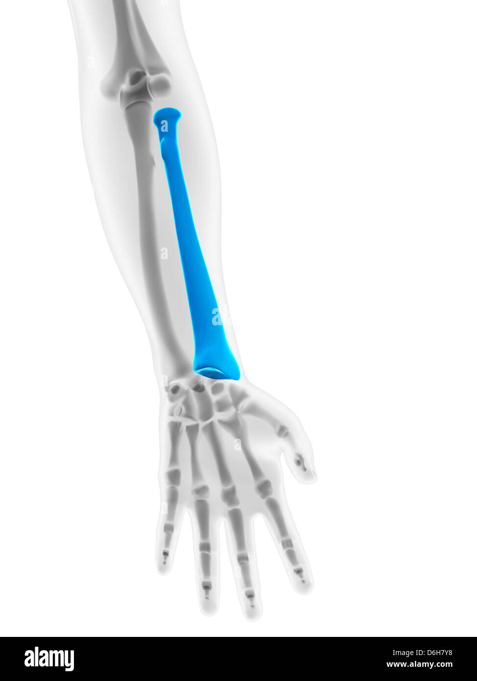 Lower arm bone, artwork Stock Photohttps://www.alamy.com/image-license-details/?v=1https://www.alamy.com/stock-photo-lower-arm-bone-artwork-55698476.html
RFD6H7Y8–Lower arm bone, artwork
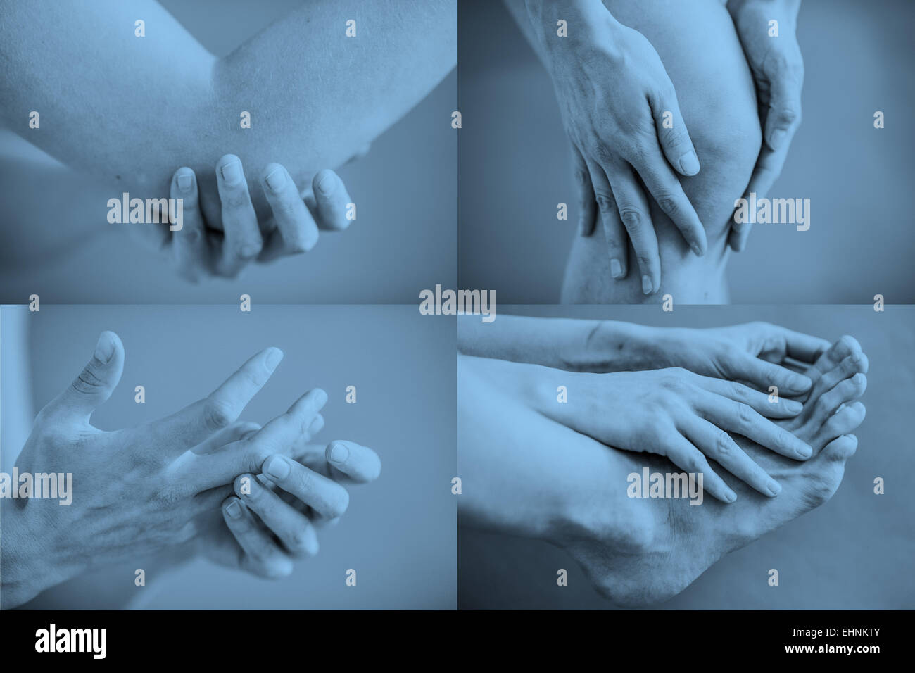 Composite image of articular pain. Stock Photohttps://www.alamy.com/image-license-details/?v=1https://www.alamy.com/stock-photo-composite-image-of-articular-pain-79767211.html
RMEHNKTY–Composite image of articular pain.
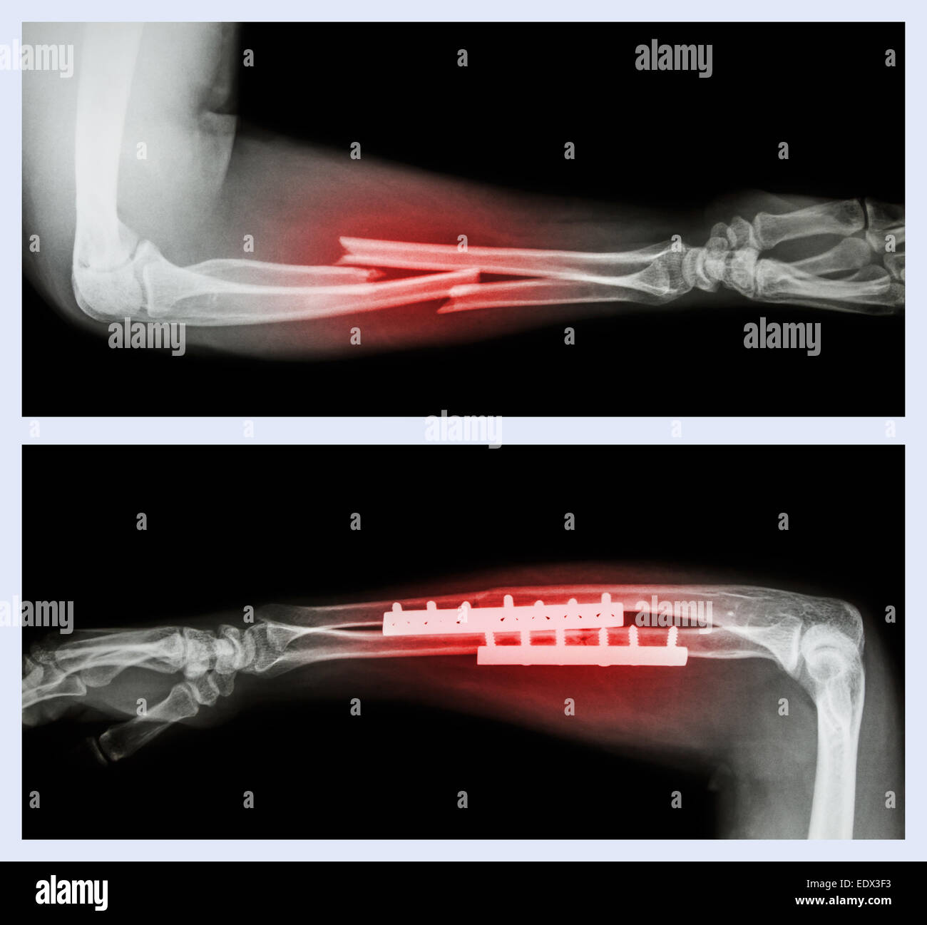 Upper image : Fracture ulnar and radius (Forearm bone) , Lower image : It was operated and internal fixed with plate and screw Stock Photohttps://www.alamy.com/image-license-details/?v=1https://www.alamy.com/stock-photo-upper-image-fracture-ulnar-and-radius-forearm-bone-lower-image-it-77405527.html
RFEDX3F3–Upper image : Fracture ulnar and radius (Forearm bone) , Lower image : It was operated and internal fixed with plate and screw
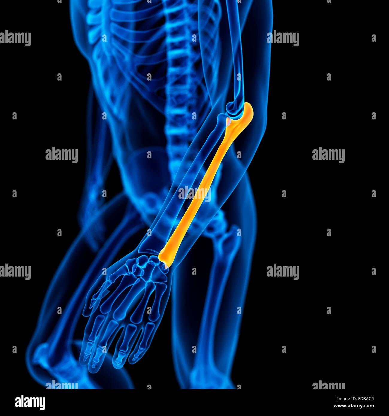 Human ulna (arm) bone, illustration. Stock Photohttps://www.alamy.com/image-license-details/?v=1https://www.alamy.com/stock-photo-human-ulna-arm-bone-illustration-94292039.html
RFFDBACR–Human ulna (arm) bone, illustration.
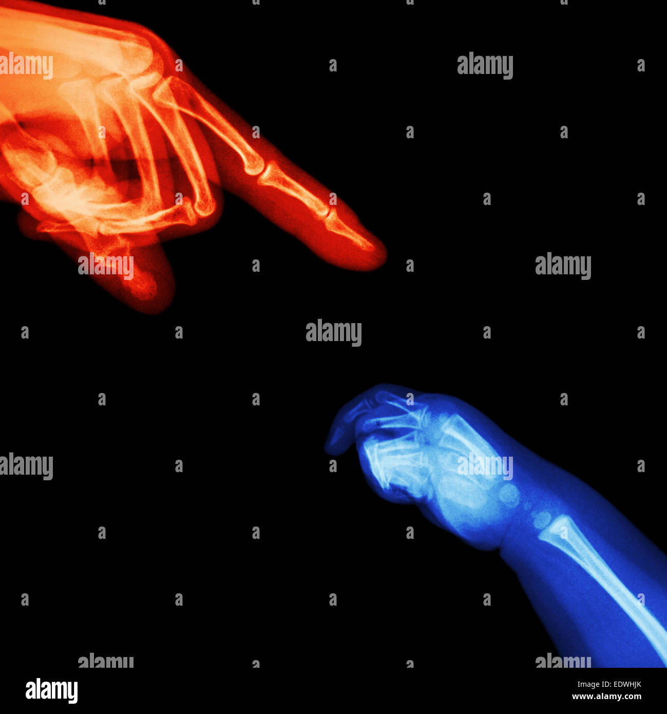 X-ray adult's hand point finger at upper side and baby's hand at lower side Stock Photohttps://www.alamy.com/image-license-details/?v=1https://www.alamy.com/stock-photo-x-ray-adults-hand-point-finger-at-upper-side-and-babys-hand-at-lower-77394651.html
RFEDWHJK–X-ray adult's hand point finger at upper side and baby's hand at lower side
 Close up of right hand and lower arm skeleton against a dark background Stock Photohttps://www.alamy.com/image-license-details/?v=1https://www.alamy.com/close-up-of-right-hand-and-lower-arm-skeleton-against-a-dark-background-image437408221.html
RM2GBHJJ5–Close up of right hand and lower arm skeleton against a dark background
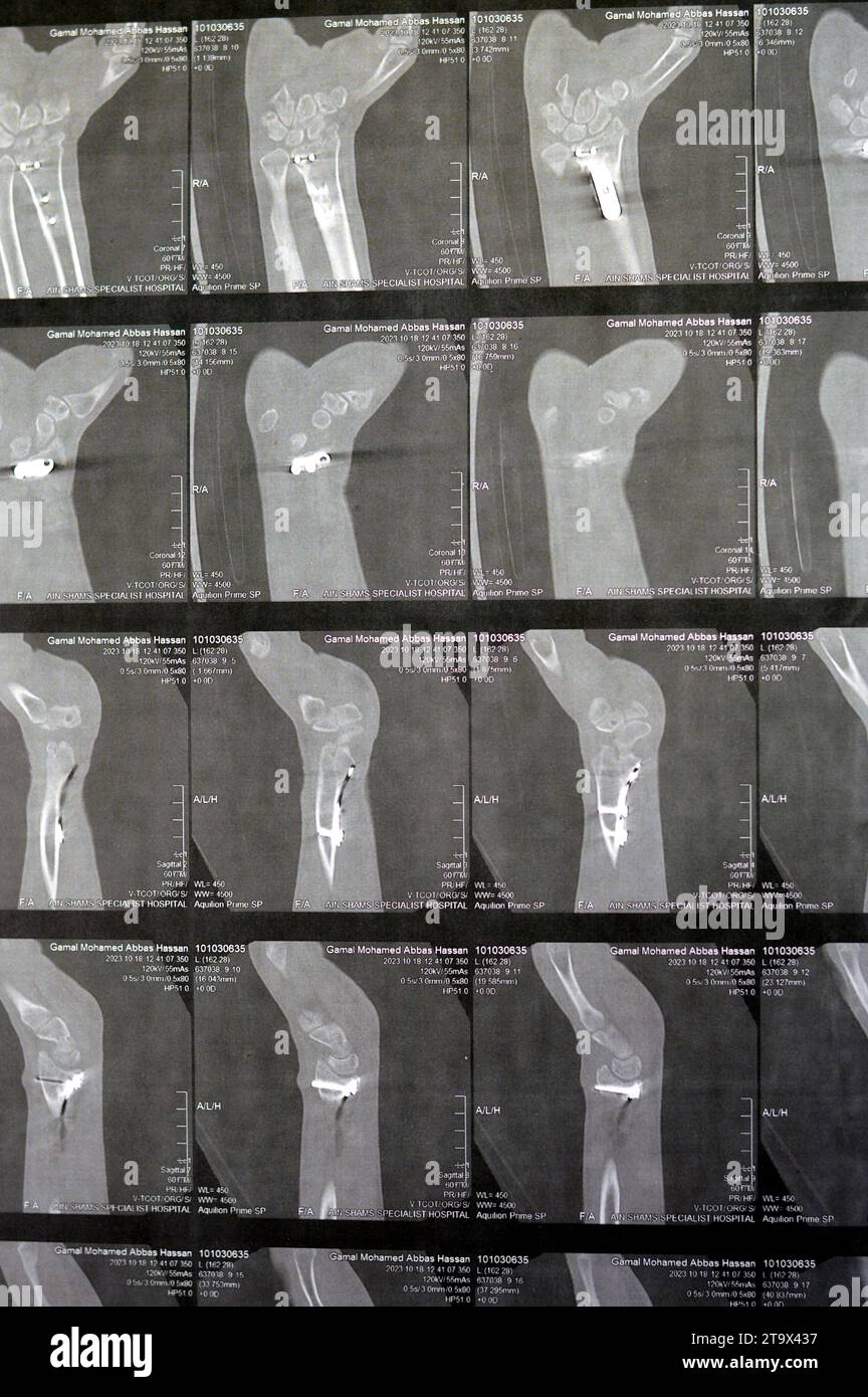 Cairo, Egypt, October 18 2023: CT scan left wrist joint showing a recent fissure fracture at the lower part of a left radius bone, a previous internal Stock Photohttps://www.alamy.com/image-license-details/?v=1https://www.alamy.com/cairo-egypt-october-18-2023-ct-scan-left-wrist-joint-showing-a-recent-fissure-fracture-at-the-lower-part-of-a-left-radius-bone-a-previous-internal-image574048027.html
RF2T9X437–Cairo, Egypt, October 18 2023: CT scan left wrist joint showing a recent fissure fracture at the lower part of a left radius bone, a previous internal
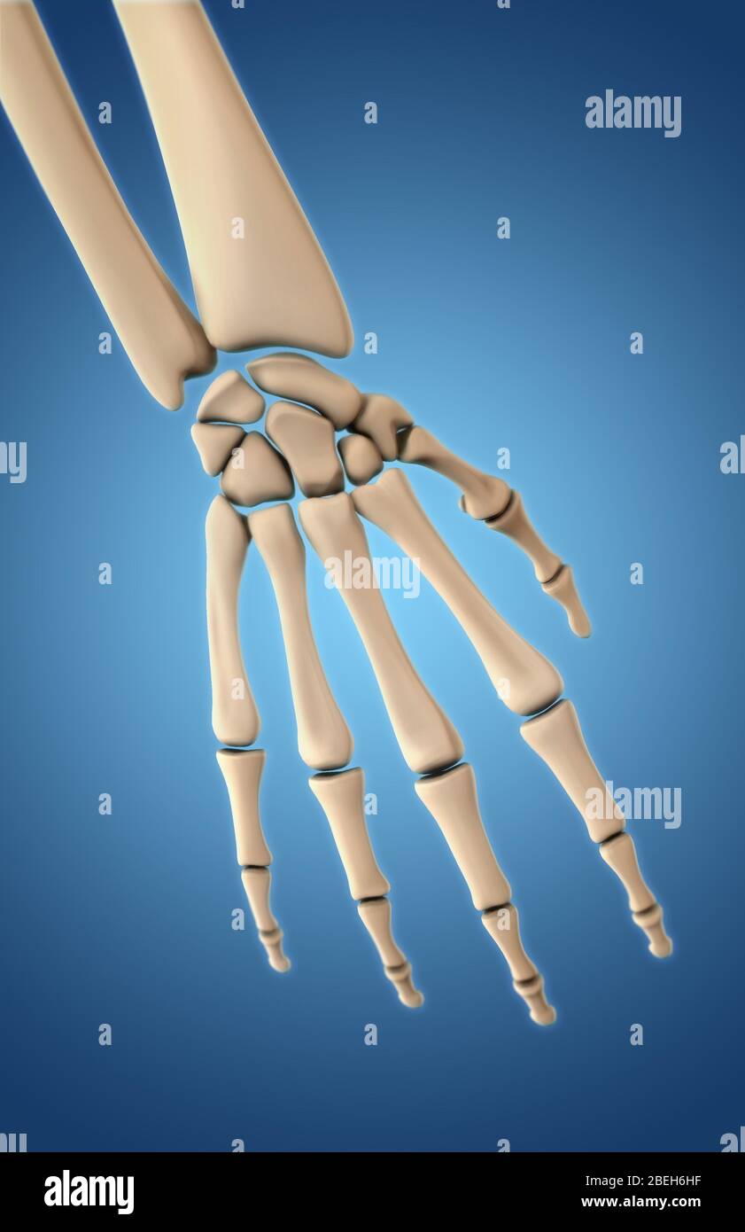 Bones of the Hand, Dorsal View Stock Photohttps://www.alamy.com/image-license-details/?v=1https://www.alamy.com/bones-of-the-hand-dorsal-view-image353190923.html
RM2BEH6HF–Bones of the Hand, Dorsal View
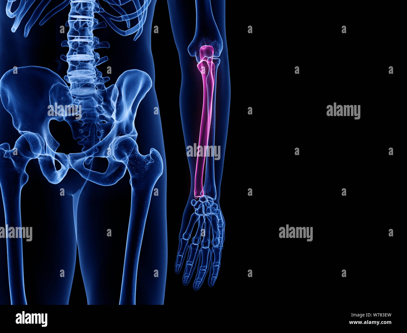 3d rendered medically accurate illustration of the ulna bone Stock Photohttps://www.alamy.com/image-license-details/?v=1https://www.alamy.com/3d-rendered-medically-accurate-illustration-of-the-ulna-bone-image262636497.html
RFW783EW–3d rendered medically accurate illustration of the ulna bone
 Skull front view without a lower jaw between two pair wings and ribbons. Vintage label isolated on white Stock Vectorhttps://www.alamy.com/image-license-details/?v=1https://www.alamy.com/skull-front-view-without-a-lower-jaw-between-two-pair-wings-and-ribbons-vintage-label-isolated-on-white-image333417646.html
RF2AACDH2–Skull front view without a lower jaw between two pair wings and ribbons. Vintage label isolated on white
 Overtired senior gray-haired man outside office building, businessman in business suit, holding hand behind back, having severe back pain, long sedentary work Stock Photohttps://www.alamy.com/image-license-details/?v=1https://www.alamy.com/overtired-senior-gray-haired-man-outside-office-building-businessman-in-business-suit-holding-hand-behind-back-having-severe-back-pain-long-sedentary-work-image477830891.html
RF2JNB24Y–Overtired senior gray-haired man outside office building, businessman in business suit, holding hand behind back, having severe back pain, long sedentary work
 Human anatomy xray-like illustration of arm and hand. Stock Photohttps://www.alamy.com/image-license-details/?v=1https://www.alamy.com/human-anatomy-xray-like-illustration-of-arm-and-hand-image368692949.html
RF2CBRBH9–Human anatomy xray-like illustration of arm and hand.
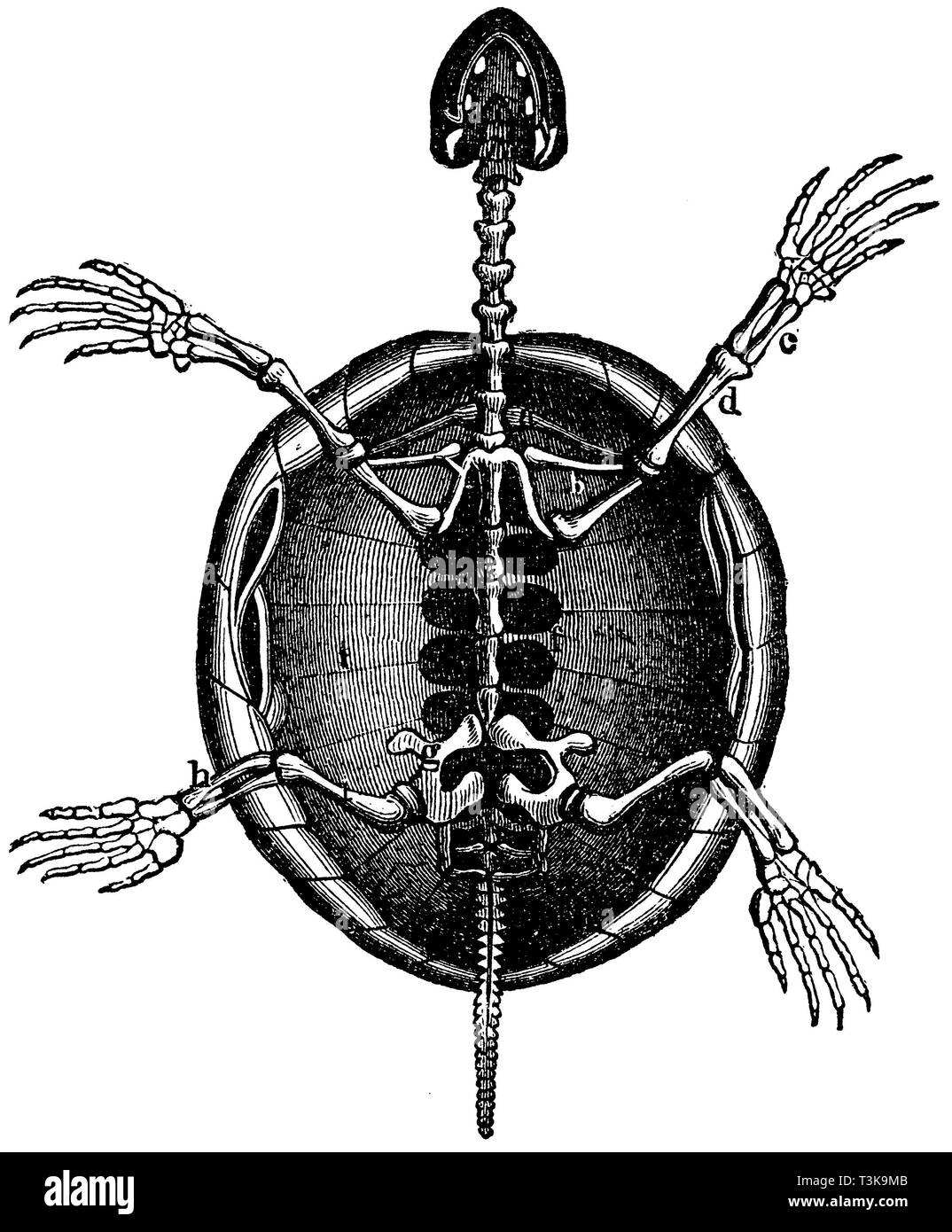 Skeleton of a turtle. a) Shoulder blade, b) Collarbone, c) Forearm, d) Upper arm, e) Dorsal vertebrae, f) Protrusions of vertebrae, g) Pelvic bone, h) Lower leg, i) Thigh, anonym 1877 Stock Photohttps://www.alamy.com/image-license-details/?v=1https://www.alamy.com/skeleton-of-a-turtle-a-shoulder-blade-b-collarbone-c-forearm-d-upper-arm-e-dorsal-vertebrae-f-protrusions-of-vertebrae-g-pelvic-bone-h-lower-leg-i-thigh-anonym-1877-image243213835.html
RMT3K9MB–Skeleton of a turtle. a) Shoulder blade, b) Collarbone, c) Forearm, d) Upper arm, e) Dorsal vertebrae, f) Protrusions of vertebrae, g) Pelvic bone, h) Lower leg, i) Thigh, anonym 1877
 arm splint, Injured female hand with lower body. Hand bandage with elastic fabric Stock Photohttps://www.alamy.com/image-license-details/?v=1https://www.alamy.com/arm-splint-injured-female-hand-with-lower-body-hand-bandage-with-elastic-fabric-image255304690.html
RFTRA3MJ–arm splint, Injured female hand with lower body. Hand bandage with elastic fabric
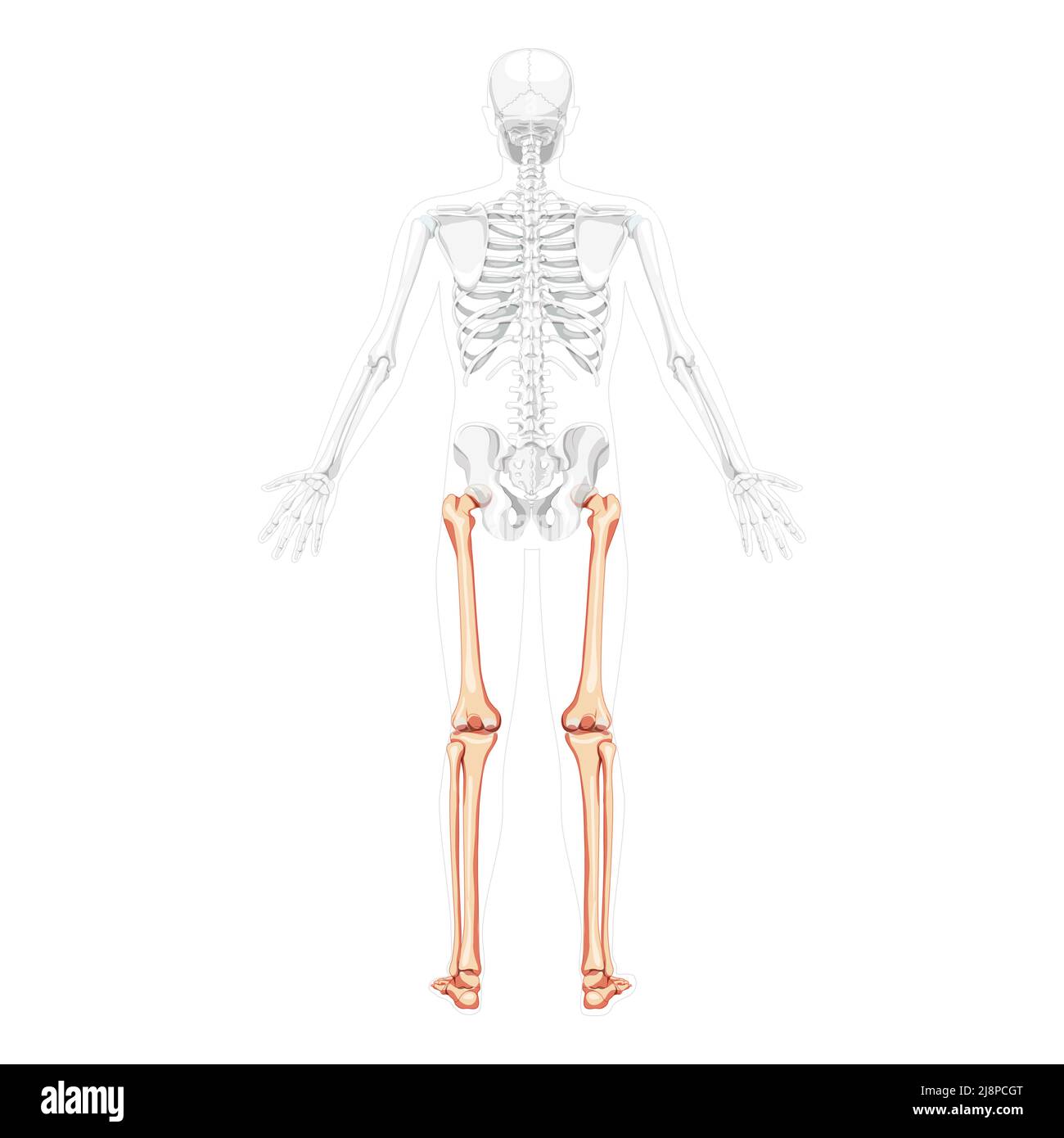 Skeleton Thighs and legs lower limb Human back view with two arm poses with partly transparent bones position. Patella, fibula, foot realistic flat concept Vector illustration of anatomy isolated Stock Vectorhttps://www.alamy.com/image-license-details/?v=1https://www.alamy.com/skeleton-thighs-and-legs-lower-limb-human-back-view-with-two-arm-poses-with-partly-transparent-bones-position-patella-fibula-foot-realistic-flat-concept-vector-illustration-of-anatomy-isolated-image470090008.html
RF2J8PCGT–Skeleton Thighs and legs lower limb Human back view with two arm poses with partly transparent bones position. Patella, fibula, foot realistic flat concept Vector illustration of anatomy isolated
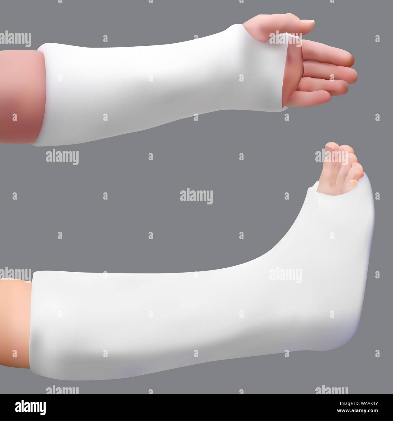 Plastered leg and arm. Treatment of a broken leg and broken arm. Medicine and health. Isolated realistic object. Vector Stock Vectorhttps://www.alamy.com/image-license-details/?v=1https://www.alamy.com/plastered-leg-and-arm-treatment-of-a-broken-leg-and-broken-arm-medicine-and-health-isolated-realistic-object-vector-image264536551.html
RFWAAK1Y–Plastered leg and arm. Treatment of a broken leg and broken arm. Medicine and health. Isolated realistic object. Vector
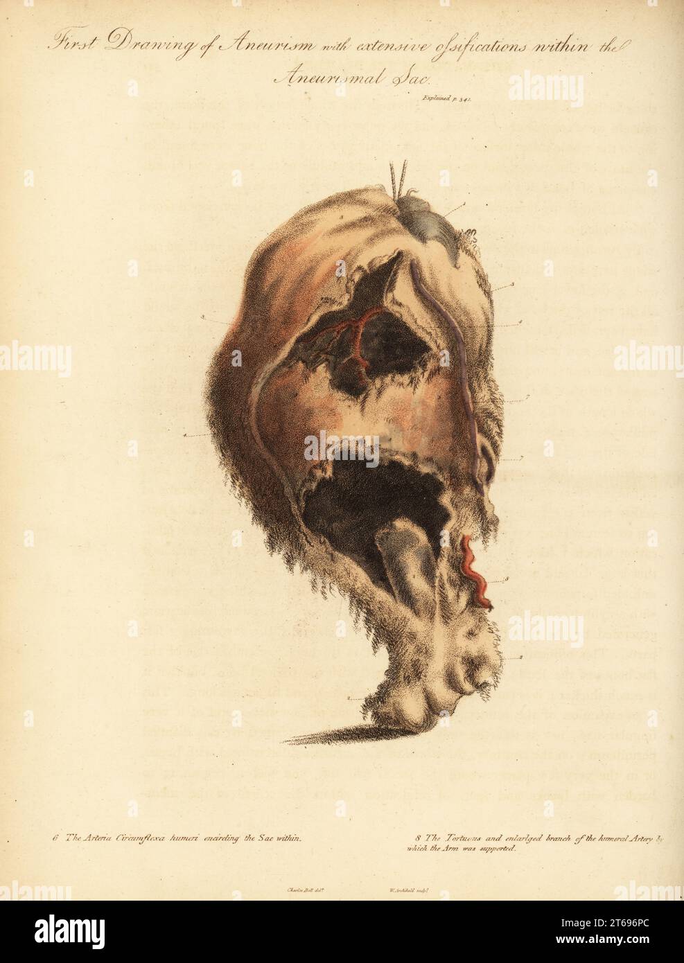 Aneurysmal tumor from the broken arm of a woman knocked down by a horse and cart, 1815. First drawing of aneurism and extensive ossifications with the aneurismal sac. Head of the humerus 1, lower part of the bone 2, arteria circumflexa humeri encircling the sac within 6, and the tortuous and enlarged branch of the humeral artery. Handcoloured copperplate engraving by William Archibald after an illustration by John Bell from his own Principles of Surgery, as they Relate to Wounds, Ulcers and Fistulas, Longman, Hurst, Rees, Orme and Brown, London, 1815. Stock Photohttps://www.alamy.com/image-license-details/?v=1https://www.alamy.com/aneurysmal-tumor-from-the-broken-arm-of-a-woman-knocked-down-by-a-horse-and-cart-1815-first-drawing-of-aneurism-and-extensive-ossifications-with-the-aneurismal-sac-head-of-the-humerus-1-lower-part-of-the-bone-2-arteria-circumflexa-humeri-encircling-the-sac-within-6-and-the-tortuous-and-enlarged-branch-of-the-humeral-artery-handcoloured-copperplate-engraving-by-william-archibald-after-an-illustration-by-john-bell-from-his-own-principles-of-surgery-as-they-relate-to-wounds-ulcers-and-fistulas-longman-hurst-rees-orme-and-brown-london-1815-image571832980.html
RM2T696PC–Aneurysmal tumor from the broken arm of a woman knocked down by a horse and cart, 1815. First drawing of aneurism and extensive ossifications with the aneurismal sac. Head of the humerus 1, lower part of the bone 2, arteria circumflexa humeri encircling the sac within 6, and the tortuous and enlarged branch of the humeral artery. Handcoloured copperplate engraving by William Archibald after an illustration by John Bell from his own Principles of Surgery, as they Relate to Wounds, Ulcers and Fistulas, Longman, Hurst, Rees, Orme and Brown, London, 1815.
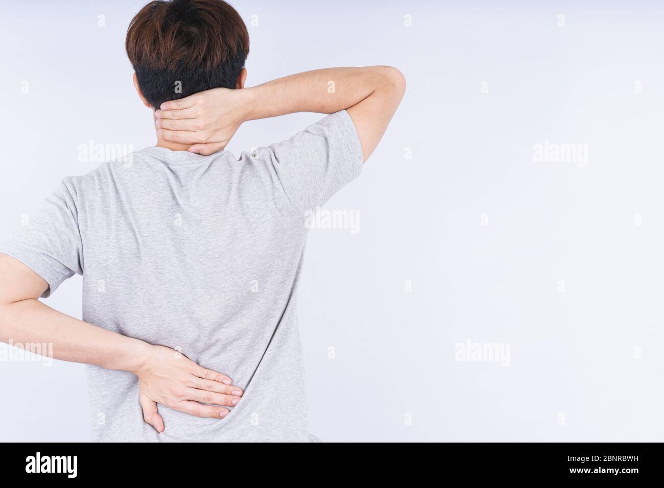 Male touching acute neck and lower back pain on white background with copy space. Medical, healthcare for advertising concept. Stock Photohttps://www.alamy.com/image-license-details/?v=1https://www.alamy.com/male-touching-acute-neck-and-lower-back-pain-on-white-background-with-copy-space-medical-healthcare-for-advertising-concept-image357629373.html
RF2BNRBWH–Male touching acute neck and lower back pain on white background with copy space. Medical, healthcare for advertising concept.
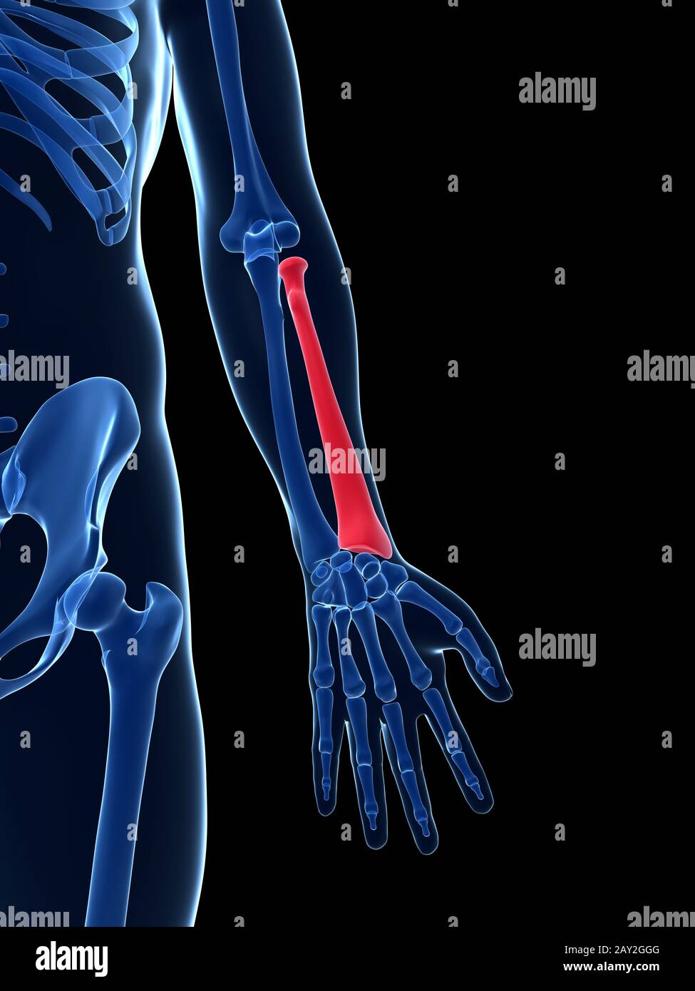 3d rendered illustration - radius Stock Photohttps://www.alamy.com/image-license-details/?v=1https://www.alamy.com/3d-rendered-illustration-radius-image343649616.html
RM2AY2GGG–3d rendered illustration - radius
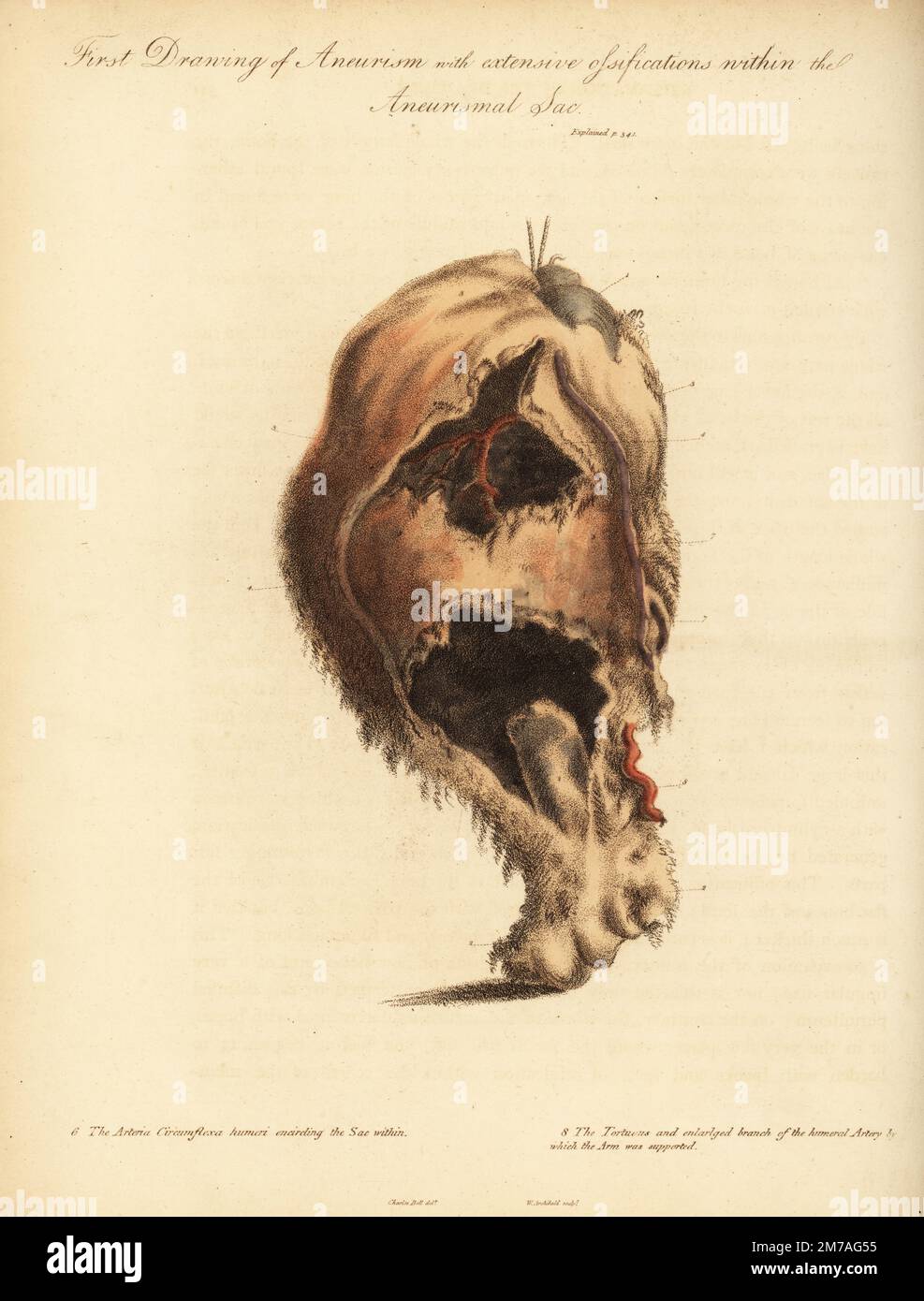 Aneurysmal tumor from the broken arm of a woman knocked down by a horse and cart, 1815. First drawing of aneurism and extensive ossifications with the aneurismal sac. Head of the humerus 1, lower part of the bone 2, arteria circumflexa humeri encircling the sac within 6, and the tortuous and enlarged branch of the humeral artery. Handcoloured copperplate engraving by William Archibald after an illustration by John Bell from his own Principles of Surgery, as they Relate to Wounds, Ulcers and Fistulas, Longman, Hurst, Rees, Orme and Brown, London, 1815. Stock Photohttps://www.alamy.com/image-license-details/?v=1https://www.alamy.com/aneurysmal-tumor-from-the-broken-arm-of-a-woman-knocked-down-by-a-horse-and-cart-1815-first-drawing-of-aneurism-and-extensive-ossifications-with-the-aneurismal-sac-head-of-the-humerus-1-lower-part-of-the-bone-2-arteria-circumflexa-humeri-encircling-the-sac-within-6-and-the-tortuous-and-enlarged-branch-of-the-humeral-artery-handcoloured-copperplate-engraving-by-william-archibald-after-an-illustration-by-john-bell-from-his-own-principles-of-surgery-as-they-relate-to-wounds-ulcers-and-fistulas-longman-hurst-rees-orme-and-brown-london-1815-image503635473.html
RM2M7AG55–Aneurysmal tumor from the broken arm of a woman knocked down by a horse and cart, 1815. First drawing of aneurism and extensive ossifications with the aneurismal sac. Head of the humerus 1, lower part of the bone 2, arteria circumflexa humeri encircling the sac within 6, and the tortuous and enlarged branch of the humeral artery. Handcoloured copperplate engraving by William Archibald after an illustration by John Bell from his own Principles of Surgery, as they Relate to Wounds, Ulcers and Fistulas, Longman, Hurst, Rees, Orme and Brown, London, 1815.
 Physical therapist checks the patient wrist by pressing the wrist bone in clinic room. Stock Photohttps://www.alamy.com/image-license-details/?v=1https://www.alamy.com/physical-therapist-checks-the-patient-wrist-by-pressing-the-wrist-bone-in-clinic-room-image470275997.html
RF2J92WR9–Physical therapist checks the patient wrist by pressing the wrist bone in clinic room.
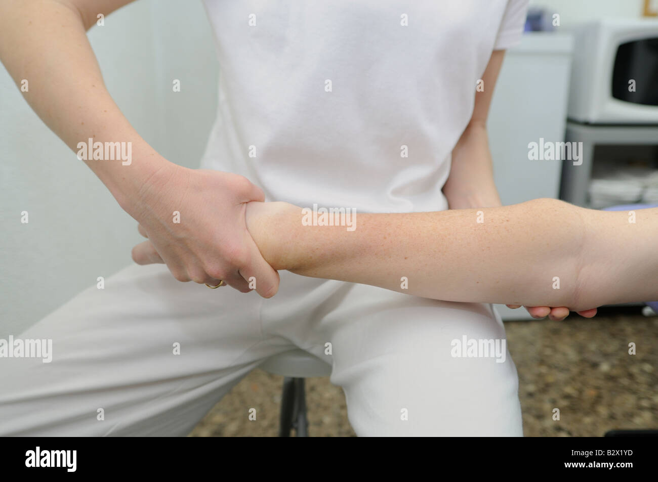 Osteopathic manipulation of lower extremity of radius bone. Osteopathic medicine. Osteopathic therapy. Stock Photohttps://www.alamy.com/image-license-details/?v=1https://www.alamy.com/stock-photo-osteopathic-manipulation-of-lower-extremity-of-radius-bone-osteopathic-19011985.html
RMB2X1YD–Osteopathic manipulation of lower extremity of radius bone. Osteopathic medicine. Osteopathic therapy.
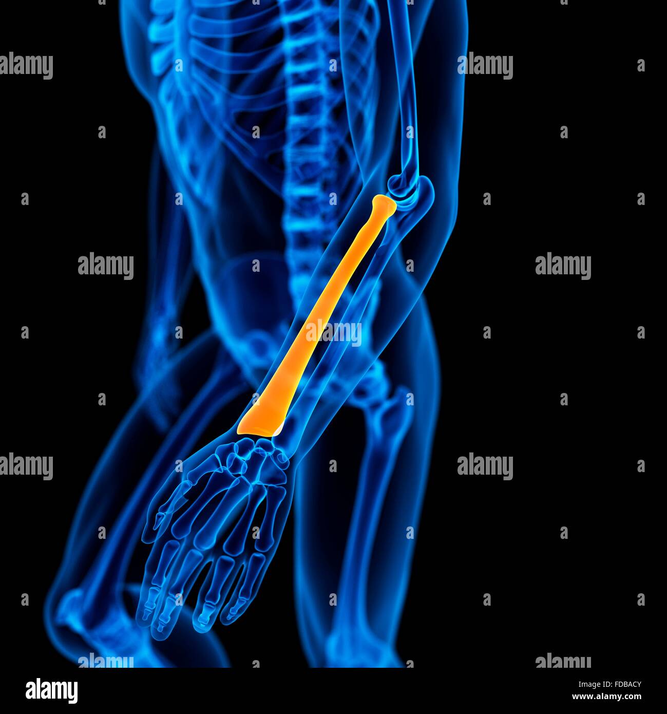 Human radius (arm) bone, illustration. Stock Photohttps://www.alamy.com/image-license-details/?v=1https://www.alamy.com/stock-photo-human-radius-arm-bone-illustration-94292043.html
RFFDBACY–Human radius (arm) bone, illustration.
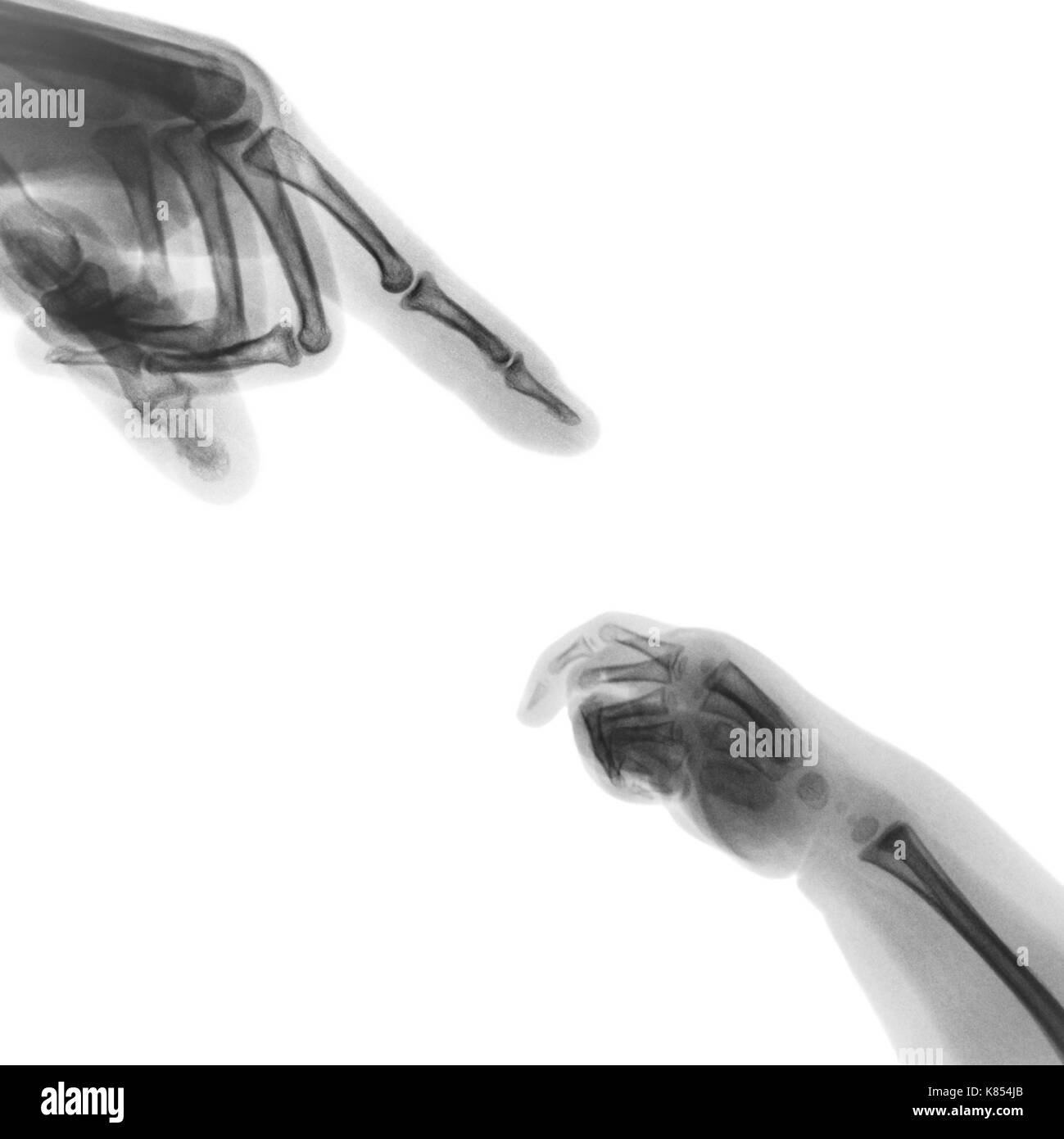 X-ray adult's hand point finger at upper side and baby's hand at lower side . Stock Photohttps://www.alamy.com/image-license-details/?v=1https://www.alamy.com/x-ray-adults-hand-point-finger-at-upper-side-and-babys-hand-at-lower-image159923971.html
RFK854JB–X-ray adult's hand point finger at upper side and baby's hand at lower side .
 leg bone human part image Stock Vectorhttps://www.alamy.com/image-license-details/?v=1https://www.alamy.com/stock-photo-leg-bone-human-part-image-140111303.html
RFJ3XHB3–leg bone human part image
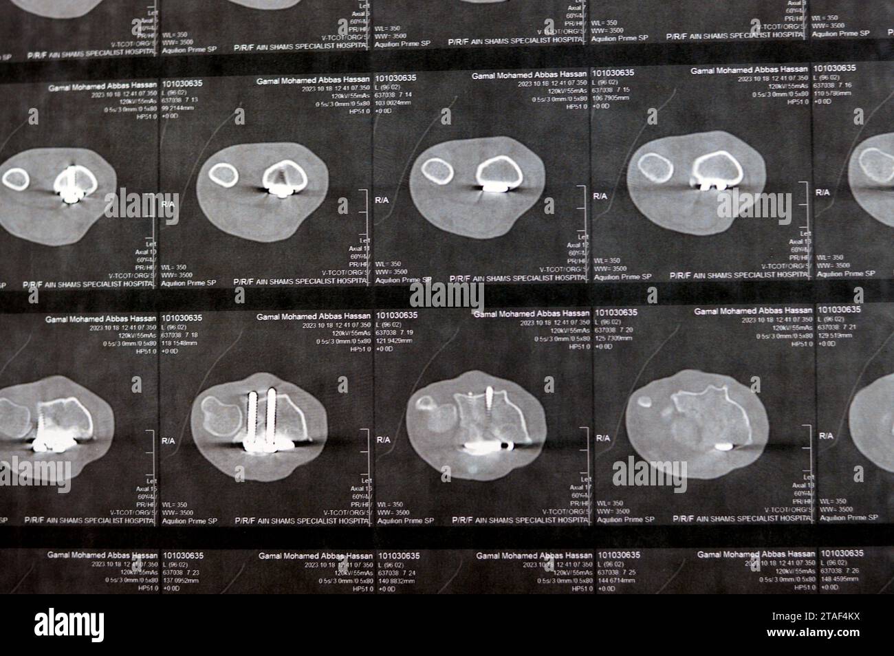 Cairo, Egypt, October 18 2023: CT scan left wrist joint showing a recent fissure fracture at the lower part of a left radius bone, a previous internal Stock Photohttps://www.alamy.com/image-license-details/?v=1https://www.alamy.com/cairo-egypt-october-18-2023-ct-scan-left-wrist-joint-showing-a-recent-fissure-fracture-at-the-lower-part-of-a-left-radius-bone-a-previous-internal-image574421678.html
RF2TAF4KX–Cairo, Egypt, October 18 2023: CT scan left wrist joint showing a recent fissure fracture at the lower part of a left radius bone, a previous internal
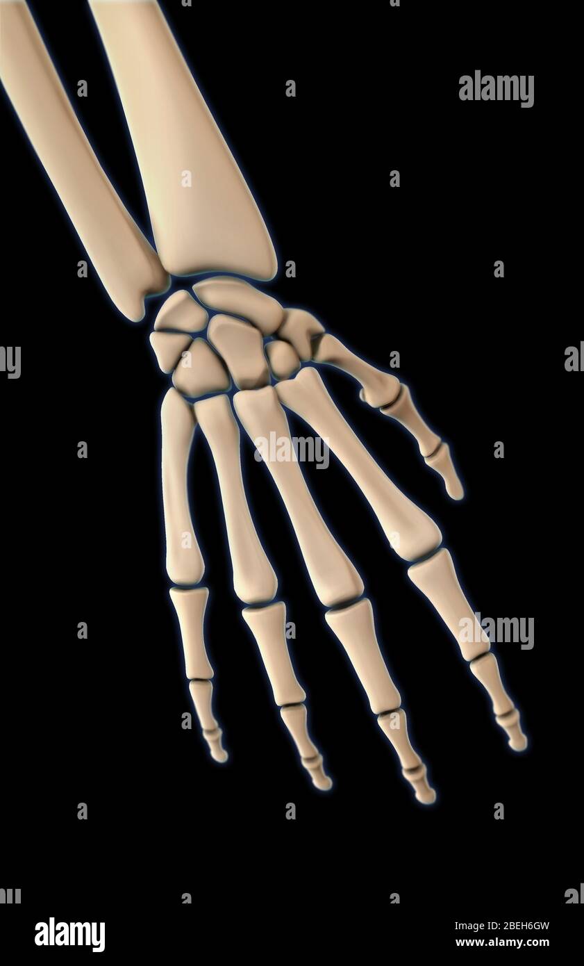 Bones of the Hand, Dorsal View Stock Photohttps://www.alamy.com/image-license-details/?v=1https://www.alamy.com/bones-of-the-hand-dorsal-view-image353190905.html
RM2BEH6GW–Bones of the Hand, Dorsal View
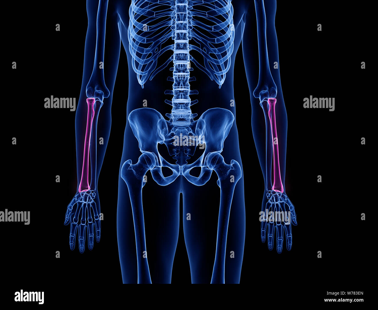 3d rendered medically accurate illustration of the radius bone Stock Photohttps://www.alamy.com/image-license-details/?v=1https://www.alamy.com/3d-rendered-medically-accurate-illustration-of-the-radius-bone-image262636493.html
RFW783EN–3d rendered medically accurate illustration of the radius bone
 Skull front view with a lower jaw between two pair wings and line pattern. Vintage label isolated on white Stock Vectorhttps://www.alamy.com/image-license-details/?v=1https://www.alamy.com/skull-front-view-with-a-lower-jaw-between-two-pair-wings-and-line-pattern-vintage-label-isolated-on-white-image333417349.html
RF2AACD6D–Skull front view with a lower jaw between two pair wings and line pattern. Vintage label isolated on white
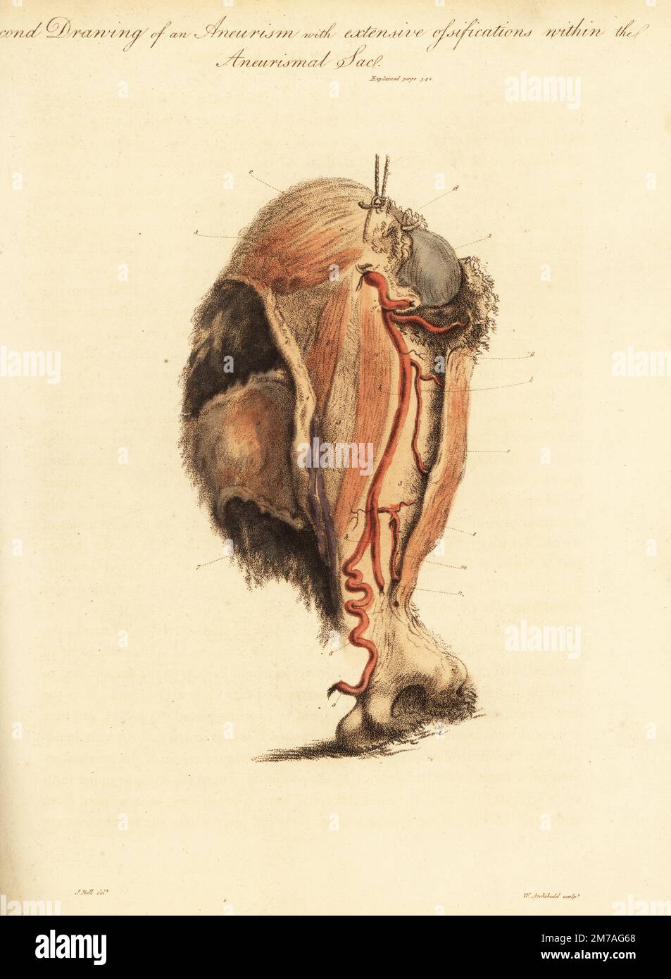 Aneurysmal tumor from the broken arm of a woman knocked down by a horse and cart, 1815. Second drawing of aneurism and extensive ossifications with the aneurismal sac. Humerus bone upper 1 and lower 2, sac 3, coraco brachialis a, biceps b, and deltoid c muscles, arteries f-m and basilic vein 7 stretched over the sac. Handcoloured copperplate engraving by William Archibald after an illustration by John Bell from his own Principles of Surgery, as they Relate to Wounds, Ulcers and Fistulas, Longman, Hurst, Rees, Orme and Brown, London, 1815. Stock Photohttps://www.alamy.com/image-license-details/?v=1https://www.alamy.com/aneurysmal-tumor-from-the-broken-arm-of-a-woman-knocked-down-by-a-horse-and-cart-1815-second-drawing-of-aneurism-and-extensive-ossifications-with-the-aneurismal-sac-humerus-bone-upper-1-and-lower-2-sac-3-coraco-brachialis-a-biceps-b-and-deltoid-c-muscles-arteries-f-m-and-basilic-vein-7-stretched-over-the-sac-handcoloured-copperplate-engraving-by-william-archibald-after-an-illustration-by-john-bell-from-his-own-principles-of-surgery-as-they-relate-to-wounds-ulcers-and-fistulas-longman-hurst-rees-orme-and-brown-london-1815-image503635504.html
RM2M7AG68–Aneurysmal tumor from the broken arm of a woman knocked down by a horse and cart, 1815. Second drawing of aneurism and extensive ossifications with the aneurismal sac. Humerus bone upper 1 and lower 2, sac 3, coraco brachialis a, biceps b, and deltoid c muscles, arteries f-m and basilic vein 7 stretched over the sac. Handcoloured copperplate engraving by William Archibald after an illustration by John Bell from his own Principles of Surgery, as they Relate to Wounds, Ulcers and Fistulas, Longman, Hurst, Rees, Orme and Brown, London, 1815.
 Skull front view without a lower jaw in the center of floral wreath with vintage weapon. Set of vintage banners on white Stock Vectorhttps://www.alamy.com/image-license-details/?v=1https://www.alamy.com/skull-front-view-without-a-lower-jaw-in-the-center-of-floral-wreath-with-vintage-weapon-set-of-vintage-banners-on-white-image333419983.html
RF2AACGGF–Skull front view without a lower jaw in the center of floral wreath with vintage weapon. Set of vintage banners on white
 Human anatomy xray-like illustration of arm and hand. Stock Photohttps://www.alamy.com/image-license-details/?v=1https://www.alamy.com/human-anatomy-xray-like-illustration-of-arm-and-hand-image368692953.html
RF2CBRBHD–Human anatomy xray-like illustration of arm and hand.
 Colored X-ray of a compound fracture of the radius (upper bone) and ulna (lower bone) forearm bones. Stock Photohttps://www.alamy.com/image-license-details/?v=1https://www.alamy.com/stock-photo-colored-x-ray-of-a-compound-fracture-of-the-radius-upper-bone-and-72416082.html
RME5PRCJ–Colored X-ray of a compound fracture of the radius (upper bone) and ulna (lower bone) forearm bones.
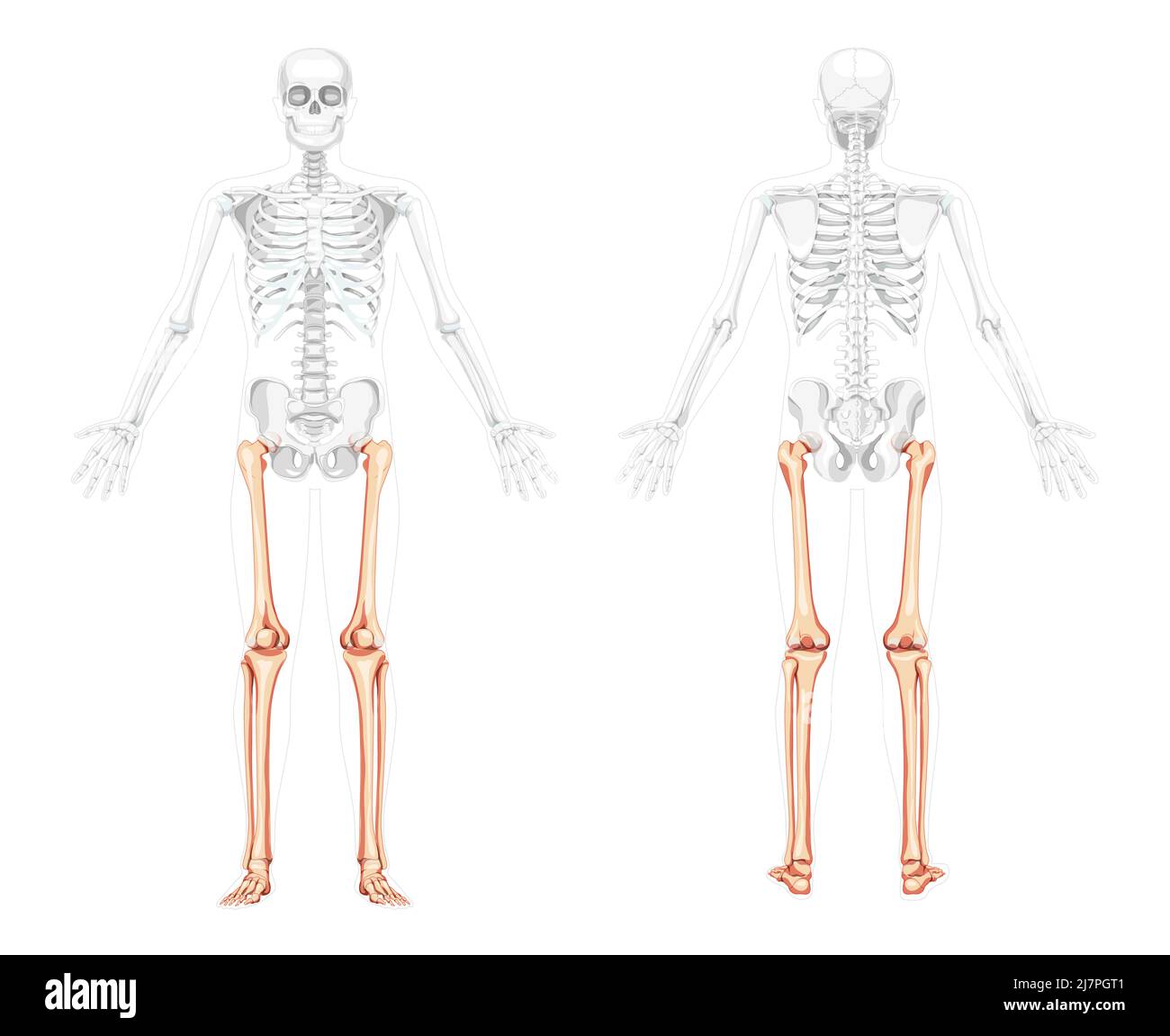 Skeleton Thighs and legs lower limb Human front back view with two arm poses with partly transparent bones. Fibula, tibia, foot realistic flat Vector illustration anatomy isolated on white background Stock Vectorhttps://www.alamy.com/image-license-details/?v=1https://www.alamy.com/skeleton-thighs-and-legs-lower-limb-human-front-back-view-with-two-arm-poses-with-partly-transparent-bones-fibula-tibia-foot-realistic-flat-vector-illustration-anatomy-isolated-on-white-background-image469478689.html
RF2J7PGT1–Skeleton Thighs and legs lower limb Human front back view with two arm poses with partly transparent bones. Fibula, tibia, foot realistic flat Vector illustration anatomy isolated on white background
 Set of objects of traumatology medicine. Treatment of musculoskeletal system. Gypsum tire, crutch, X-ray, wheelchair, cane different in design, walker Stock Vectorhttps://www.alamy.com/image-license-details/?v=1https://www.alamy.com/set-of-objects-of-traumatology-medicine-treatment-of-musculoskeletal-system-gypsum-tire-crutch-x-ray-wheelchair-cane-different-in-design-walker-image263592734.html
RFW8RK66–Set of objects of traumatology medicine. Treatment of musculoskeletal system. Gypsum tire, crutch, X-ray, wheelchair, cane different in design, walker
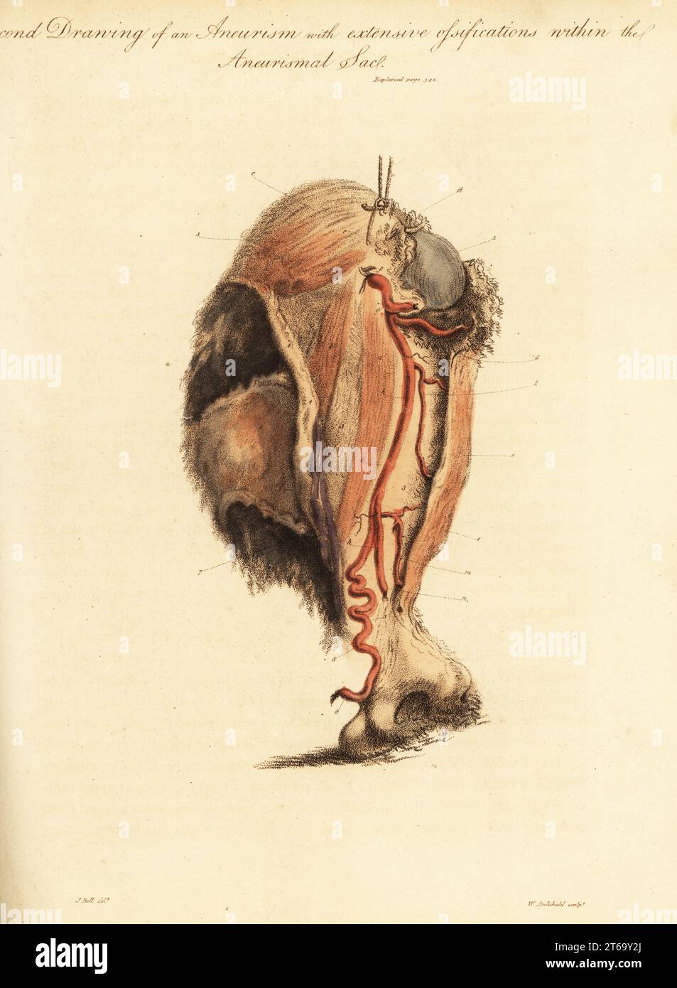 Aneurysmal tumor from the broken arm of a woman knocked down by a horse and cart, 1815. Second drawing of aneurism and extensive ossifications with the aneurismal sac. Humerus bone upper 1 and lower 2, sac 3, coraco brachialis a, biceps b, and deltoid c muscles, arteries f-m and basilic vein 7 stretched over the sac. Handcoloured copperplate engraving by William Archibald after an illustration by John Bell from his own Principles of Surgery, as they Relate to Wounds, Ulcers and Fistulas, Longman, Hurst, Rees, Orme and Brown, London, 1815. Stock Photohttps://www.alamy.com/image-license-details/?v=1https://www.alamy.com/aneurysmal-tumor-from-the-broken-arm-of-a-woman-knocked-down-by-a-horse-and-cart-1815-second-drawing-of-aneurism-and-extensive-ossifications-with-the-aneurismal-sac-humerus-bone-upper-1-and-lower-2-sac-3-coraco-brachialis-a-biceps-b-and-deltoid-c-muscles-arteries-f-m-and-basilic-vein-7-stretched-over-the-sac-handcoloured-copperplate-engraving-by-william-archibald-after-an-illustration-by-john-bell-from-his-own-principles-of-surgery-as-they-relate-to-wounds-ulcers-and-fistulas-longman-hurst-rees-orme-and-brown-london-1815-image571848890.html
RM2T69Y2J–Aneurysmal tumor from the broken arm of a woman knocked down by a horse and cart, 1815. Second drawing of aneurism and extensive ossifications with the aneurismal sac. Humerus bone upper 1 and lower 2, sac 3, coraco brachialis a, biceps b, and deltoid c muscles, arteries f-m and basilic vein 7 stretched over the sac. Handcoloured copperplate engraving by William Archibald after an illustration by John Bell from his own Principles of Surgery, as they Relate to Wounds, Ulcers and Fistulas, Longman, Hurst, Rees, Orme and Brown, London, 1815.
 Female touching acute neck and lower back pain on white background with copy space. Medical, healthcare for advertising concept. Stock Photohttps://www.alamy.com/image-license-details/?v=1https://www.alamy.com/female-touching-acute-neck-and-lower-back-pain-on-white-background-with-copy-space-medical-healthcare-for-advertising-concept-image357628965.html
RF2BNRBB1–Female touching acute neck and lower back pain on white background with copy space. Medical, healthcare for advertising concept.
 Skull front view with a lower jaw between two pair wings and ribbons. Vintage label on blackboard Stock Vectorhttps://www.alamy.com/image-license-details/?v=1https://www.alamy.com/skull-front-view-with-a-lower-jaw-between-two-pair-wings-and-ribbons-vintage-label-on-blackboard-image333415878.html
RF2AACB9X–Skull front view with a lower jaw between two pair wings and ribbons. Vintage label on blackboard
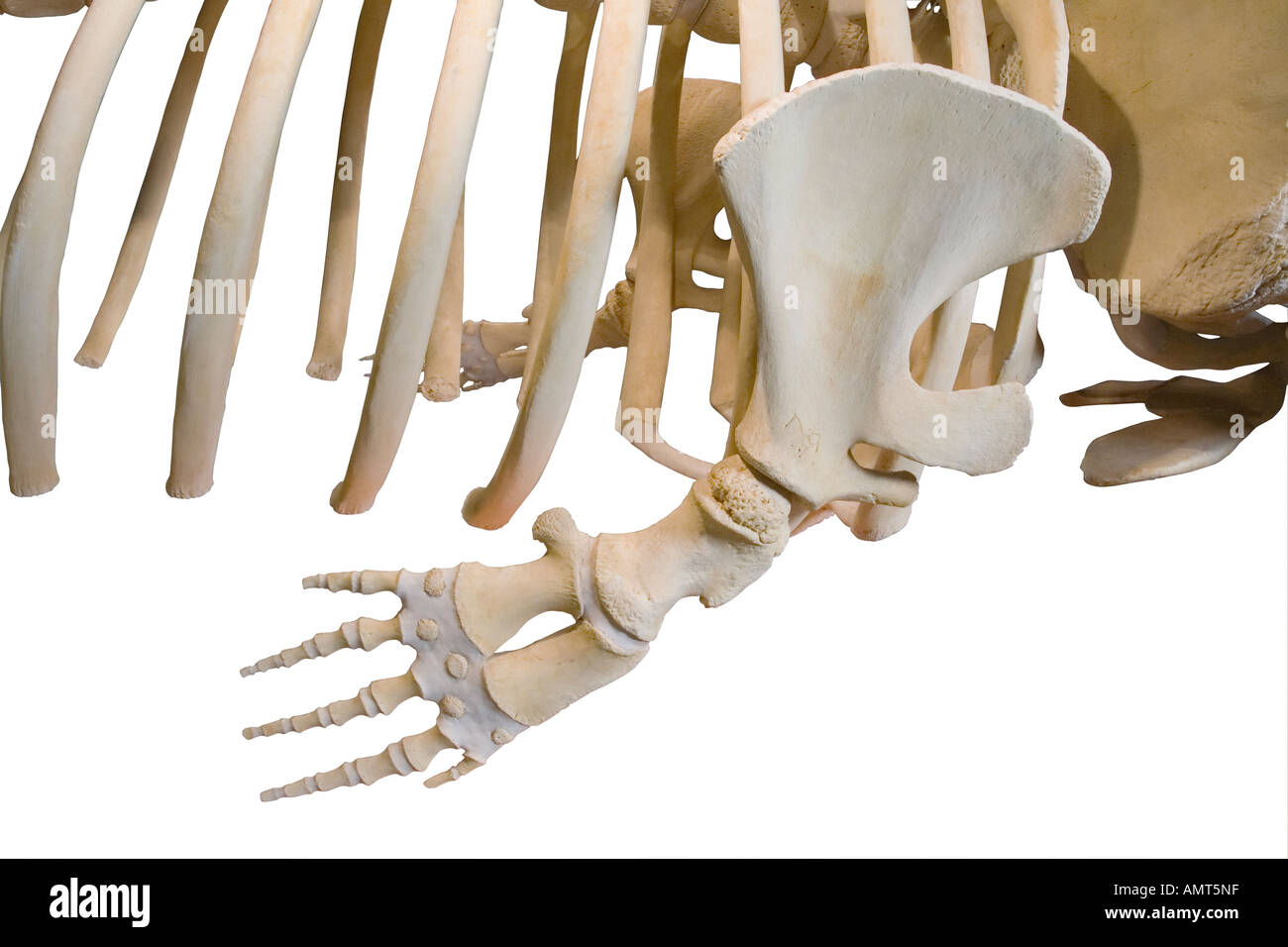 Sperm whale skeleton lower flipper closeup Stock Photohttps://www.alamy.com/image-license-details/?v=1https://www.alamy.com/sperm-whale-skeleton-lower-flipper-closeup-image8760350.html
RMAMT5NF–Sperm whale skeleton lower flipper closeup
 Physical therapist checks the patient wrist by pressing the wrist bone in clinic room. Stock Photohttps://www.alamy.com/image-license-details/?v=1https://www.alamy.com/physical-therapist-checks-the-patient-wrist-by-pressing-the-wrist-bone-in-clinic-room-image467259093.html
RF2J45DMN–Physical therapist checks the patient wrist by pressing the wrist bone in clinic room.
 Skull front view without a lower jaw in center of line pattern. Heraldic vintage label on blackboard Stock Vectorhttps://www.alamy.com/image-license-details/?v=1https://www.alamy.com/skull-front-view-without-a-lower-jaw-in-center-of-line-pattern-heraldic-vintage-label-on-blackboard-image333415805.html
RF2AACB79–Skull front view without a lower jaw in center of line pattern. Heraldic vintage label on blackboard
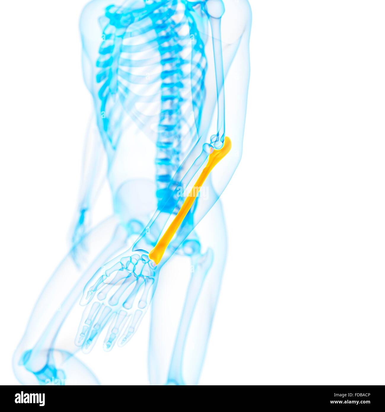 Human ulna (arm) bone, illustration. Stock Photohttps://www.alamy.com/image-license-details/?v=1https://www.alamy.com/stock-photo-human-ulna-arm-bone-illustration-94292038.html
RFFDBACP–Human ulna (arm) bone, illustration.
 X-ray adult's hand point finger at upper side and baby's hand at lower side . Stock Photohttps://www.alamy.com/image-license-details/?v=1https://www.alamy.com/x-ray-adults-hand-point-finger-at-upper-side-and-babys-hand-at-lower-image159923969.html
RFK854J9–X-ray adult's hand point finger at upper side and baby's hand at lower side .