Quick filters:
Lower leg bones Stock Photos and Images
 medical accurate illustration of the lower leg bones Stock Photohttps://www.alamy.com/image-license-details/?v=1https://www.alamy.com/stock-photo-medical-accurate-illustration-of-the-lower-leg-bones-89742503.html
RFF603DB–medical accurate illustration of the lower leg bones
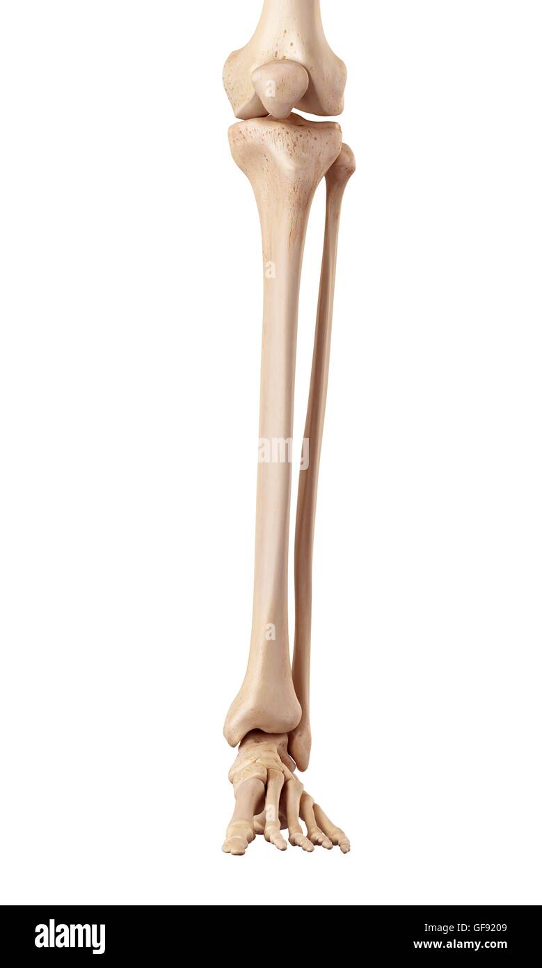 Human bones of the lower leg, illustration. Stock Photohttps://www.alamy.com/image-license-details/?v=1https://www.alamy.com/stock-photo-human-bones-of-the-lower-leg-illustration-112681193.html
RFGF9209–Human bones of the lower leg, illustration.
 The Invention of Drawing (recto), Sketch of Lower Leg Bones of Human Skeleton (verso); Joseph-Benoît Suvée, Belgian Stock Photohttps://www.alamy.com/image-license-details/?v=1https://www.alamy.com/stock-photo-the-invention-of-drawing-recto-sketch-of-lower-leg-bones-of-human-77453850.html
RMEE094X–The Invention of Drawing (recto), Sketch of Lower Leg Bones of Human Skeleton (verso); Joseph-Benoît Suvée, Belgian
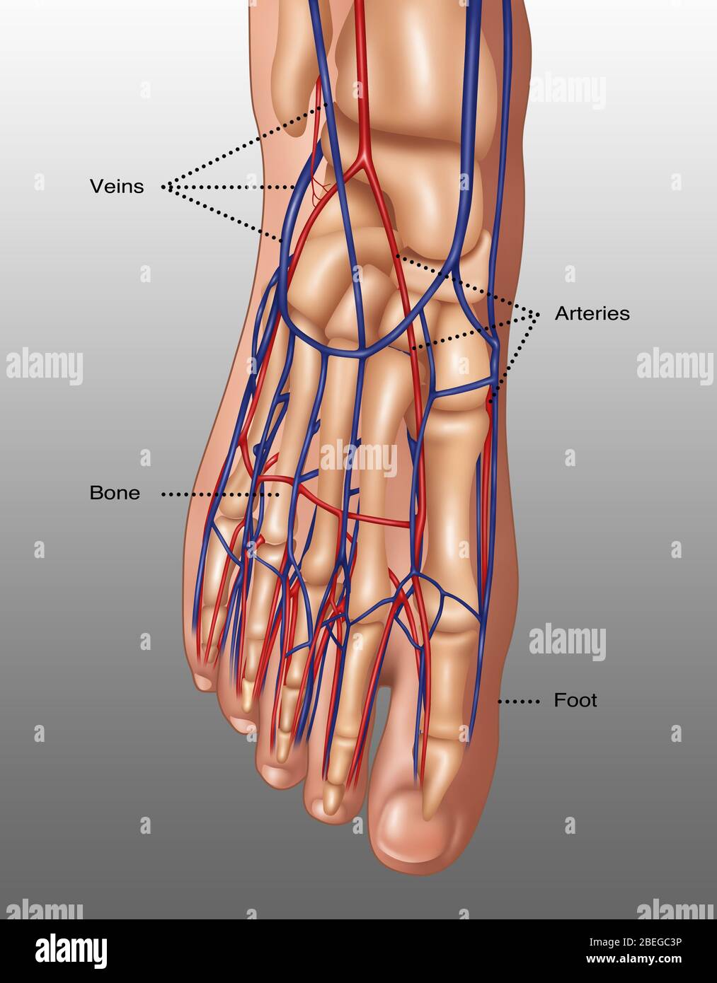 Illustration the anatomy of the foot including the skin, bones, arteries (red), and veins (blue). The toes are made up of the phalanx bones (phalanges), two for the big toe (lower right) and three for the others. Metatarsal bones in the mid-foot link the phalanges to the tarsal bones. The tarsal bones include the cuneiform bones, cuboid and heel bone. These bones articulate with the lower leg bones, the fibula and tibia (shin bone), to form the ankle. Stock Photohttps://www.alamy.com/image-license-details/?v=1https://www.alamy.com/illustration-the-anatomy-of-the-foot-including-the-skin-bones-arteries-red-and-veins-blue-the-toes-are-made-up-of-the-phalanx-bones-phalanges-two-for-the-big-toe-lower-right-and-three-for-the-others-metatarsal-bones-in-the-mid-foot-link-the-phalanges-to-the-tarsal-bones-the-tarsal-bones-include-the-cuneiform-bones-cuboid-and-heel-bone-these-bones-articulate-with-the-lower-leg-bones-the-fibula-and-tibia-shin-bone-to-form-the-ankle-image353173290.html
RM2BEGC3P–Illustration the anatomy of the foot including the skin, bones, arteries (red), and veins (blue). The toes are made up of the phalanx bones (phalanges), two for the big toe (lower right) and three for the others. Metatarsal bones in the mid-foot link the phalanges to the tarsal bones. The tarsal bones include the cuneiform bones, cuboid and heel bone. These bones articulate with the lower leg bones, the fibula and tibia (shin bone), to form the ankle.
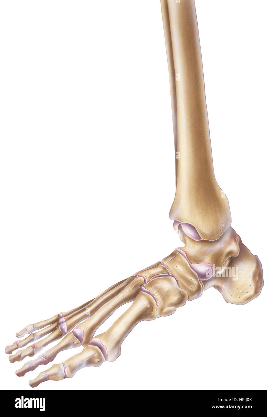 A normal human foot and ankle showing the bones and joints. Stock Photohttps://www.alamy.com/image-license-details/?v=1https://www.alamy.com/stock-photo-a-normal-human-foot-and-ankle-showing-the-bones-and-joints-134404275.html
RFHPJJ0K–A normal human foot and ankle showing the bones and joints.
 Bones from the lower part of the leg, which are the tibia and the fibula. Stock Photohttps://www.alamy.com/image-license-details/?v=1https://www.alamy.com/bones-from-the-lower-part-of-the-leg-which-are-the-tibia-and-the-fibula-image156173036.html
RMK2288C–Bones from the lower part of the leg, which are the tibia and the fibula.
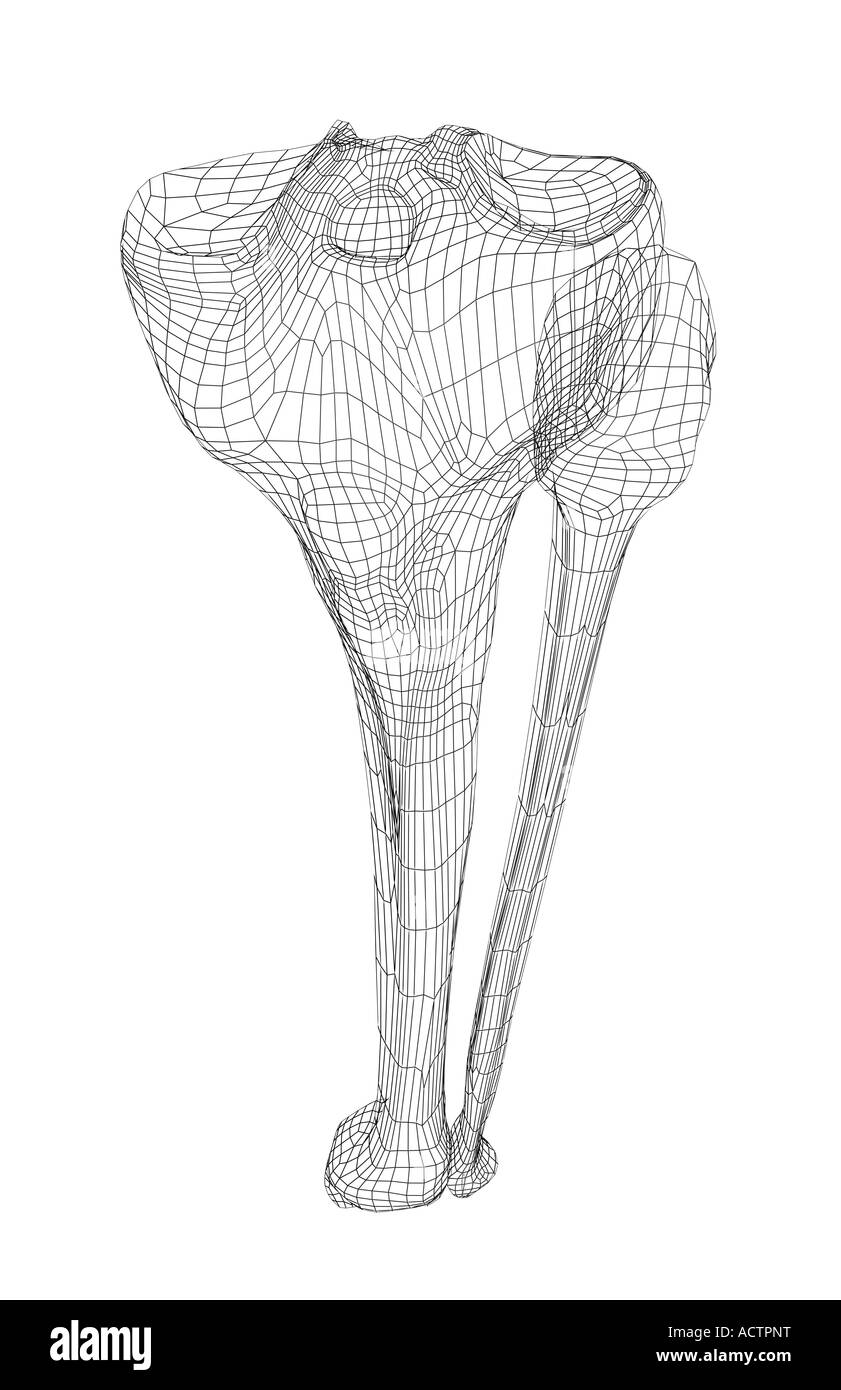 Lower leg bones Stock Photohttps://www.alamy.com/image-license-details/?v=1https://www.alamy.com/stock-photo-lower-leg-bones-13229027.html
RFACTPNT–Lower leg bones
 Front view of the bones of the left lower leg. Stock Photohttps://www.alamy.com/image-license-details/?v=1https://www.alamy.com/stock-photo-front-view-of-the-bones-of-the-left-lower-leg-52076815.html
RMD0M8E7–Front view of the bones of the left lower leg.
 Anatomy of the nerves of the lower limb (leg). Stock Photohttps://www.alamy.com/image-license-details/?v=1https://www.alamy.com/anatomy-of-the-nerves-of-the-lower-limb-leg-image476924332.html
RF2JKWNRT–Anatomy of the nerves of the lower limb (leg).
 Stacked skulls, upper and lower leg bones in an ossuary of a Catholic church Stock Photohttps://www.alamy.com/image-license-details/?v=1https://www.alamy.com/stacked-skulls-upper-and-lower-leg-bones-in-an-ossuary-of-a-catholic-church-image573796710.html
RF2T9EKFJ–Stacked skulls, upper and lower leg bones in an ossuary of a Catholic church
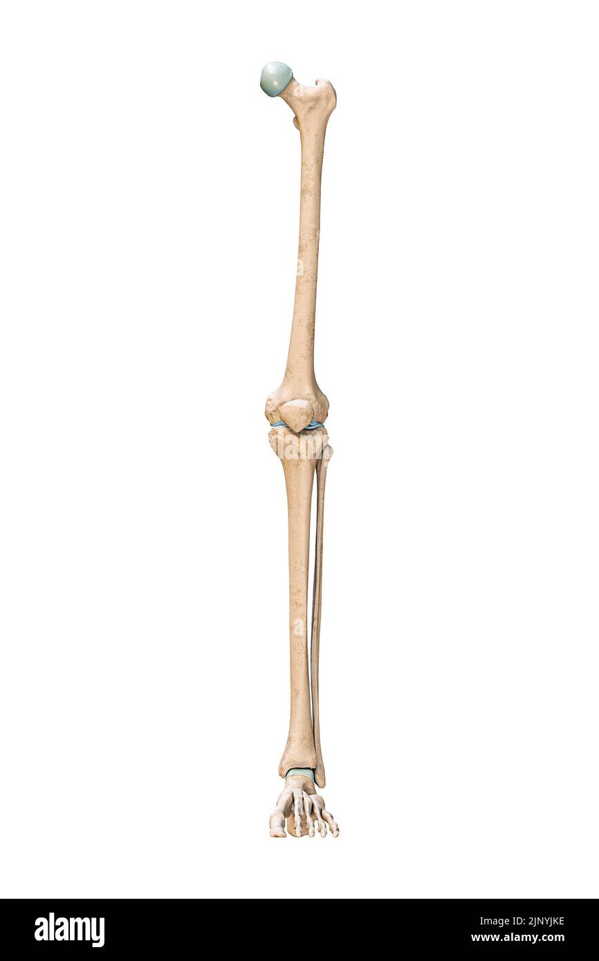 Accurate anterior or front view of the leg or lower limb bones of the human skeletal system isolated on white background 3D rendering illustration. An Stock Photohttps://www.alamy.com/image-license-details/?v=1https://www.alamy.com/accurate-anterior-or-front-view-of-the-leg-or-lower-limb-bones-of-the-human-skeletal-system-isolated-on-white-background-3d-rendering-illustration-an-image478195074.html
RF2JNYJKE–Accurate anterior or front view of the leg or lower limb bones of the human skeletal system isolated on white background 3D rendering illustration. An
 210120-N-DA693-1408 SAN DIEGO (Jan. 20, 2021) Surgeons assigned to Naval Medical Center San Diego (NMCSD) observe plastic surgeons harvest a fibula during an immediate jaw reconstruction with 3D-printed teeth in one of NMCSD’s operating rooms Jan. 20. The procedure was performed by teams from the hospital’s Oral and Maxillofacial Surgery, Plastic Surgery and Otolaryngology Departments. The surgery comprised of not only cancer removal, but the reconstruction of a jaw using a section of the patient’s fibula, the smaller of the two lower leg bones. The COVID-19 pandemic has changed the way many Stock Photohttps://www.alamy.com/image-license-details/?v=1https://www.alamy.com/210120-n-da693-1408-san-diego-jan-20-2021-surgeons-assigned-to-naval-medical-center-san-diego-nmcsd-observe-plastic-surgeons-harvest-a-fibula-during-an-immediate-jaw-reconstruction-with-3d-printed-teeth-in-one-of-nmcsds-operating-rooms-jan-20-the-procedure-was-performed-by-teams-from-the-hospitals-oral-and-maxillofacial-surgery-plastic-surgery-and-otolaryngology-departments-the-surgery-comprised-of-not-only-cancer-removal-but-the-reconstruction-of-a-jaw-using-a-section-of-the-patients-fibula-the-smaller-of-the-two-lower-leg-bones-the-covid-19-pandemic-has-changed-the-way-many-image447732784.html
RM2H0BYMG–210120-N-DA693-1408 SAN DIEGO (Jan. 20, 2021) Surgeons assigned to Naval Medical Center San Diego (NMCSD) observe plastic surgeons harvest a fibula during an immediate jaw reconstruction with 3D-printed teeth in one of NMCSD’s operating rooms Jan. 20. The procedure was performed by teams from the hospital’s Oral and Maxillofacial Surgery, Plastic Surgery and Otolaryngology Departments. The surgery comprised of not only cancer removal, but the reconstruction of a jaw using a section of the patient’s fibula, the smaller of the two lower leg bones. The COVID-19 pandemic has changed the way many
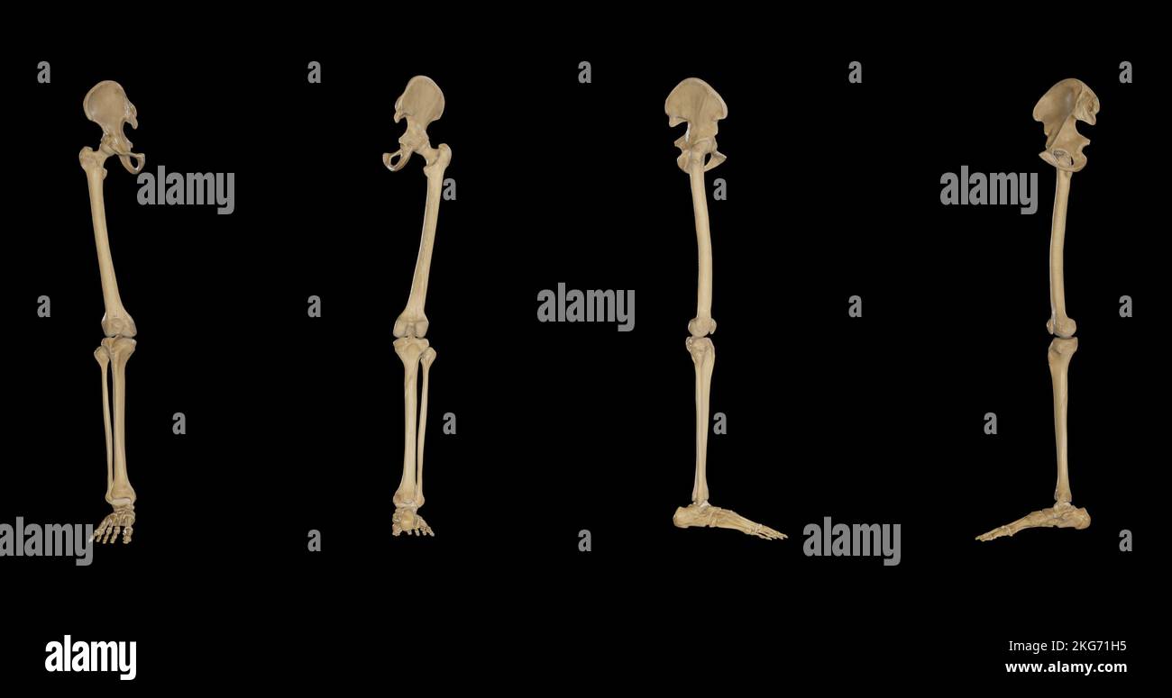 Bones of Lower Limb-Multiple Views Stock Photohttps://www.alamy.com/image-license-details/?v=1https://www.alamy.com/bones-of-lower-limb-multiple-views-image491879729.html
RF2KG71H5–Bones of Lower Limb-Multiple Views
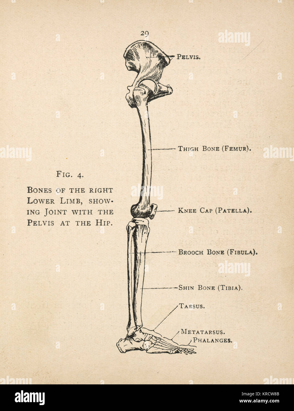 Diagram of the bones of the right leg, showing the joint with the pelvis at the hip. Date: 1908 Stock Photohttps://www.alamy.com/image-license-details/?v=1https://www.alamy.com/stock-image-diagram-of-the-bones-of-the-right-leg-showing-the-joint-with-the-pelvis-169313659.html
RMKRCW8B–Diagram of the bones of the right leg, showing the joint with the pelvis at the hip. Date: 1908
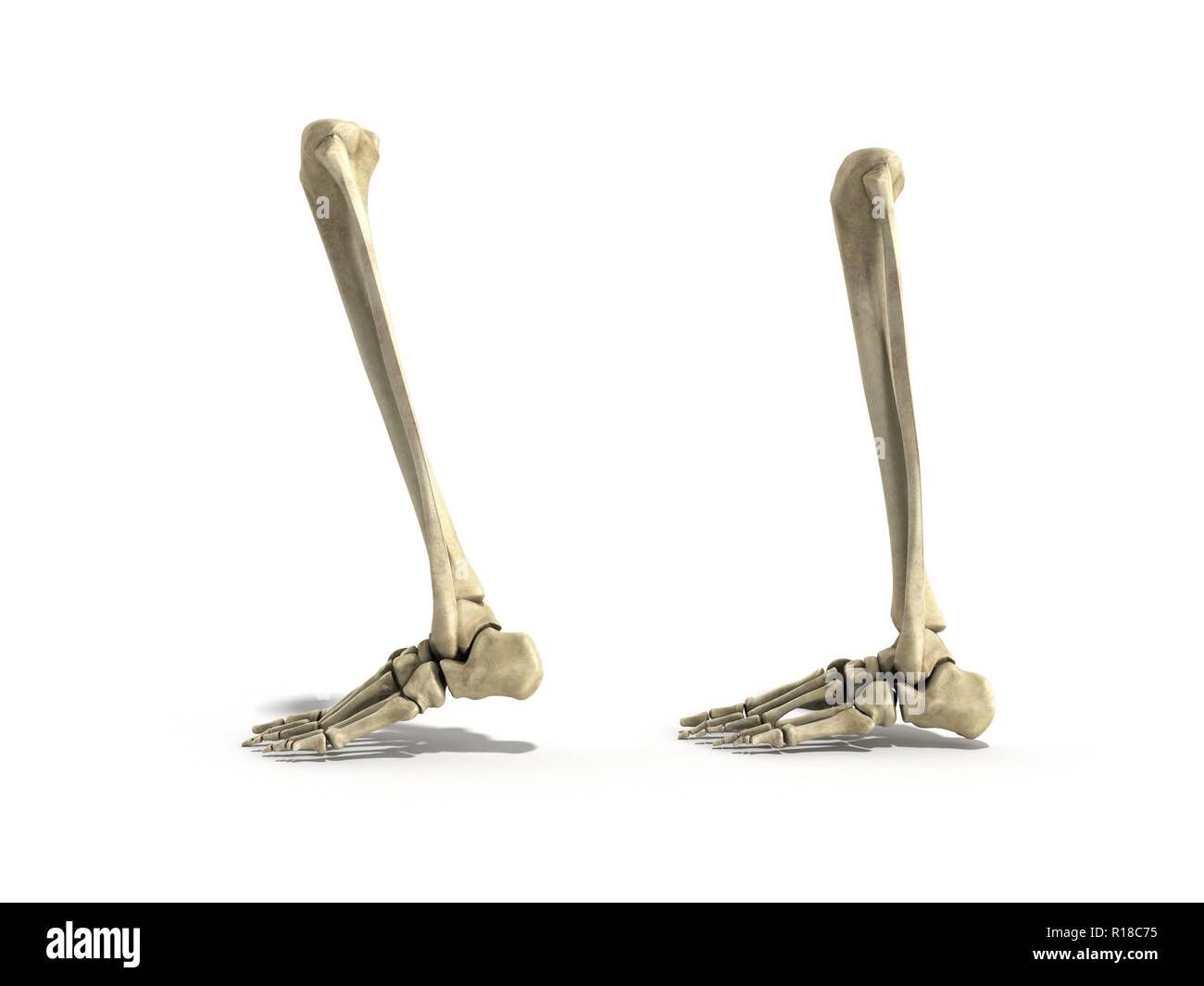 medical accurate illustration of the lower leg bones 3d render Stock Photohttps://www.alamy.com/image-license-details/?v=1https://www.alamy.com/medical-accurate-illustration-of-the-lower-leg-bones-3d-render-image224534665.html
RFR18C75–medical accurate illustration of the lower leg bones 3d render
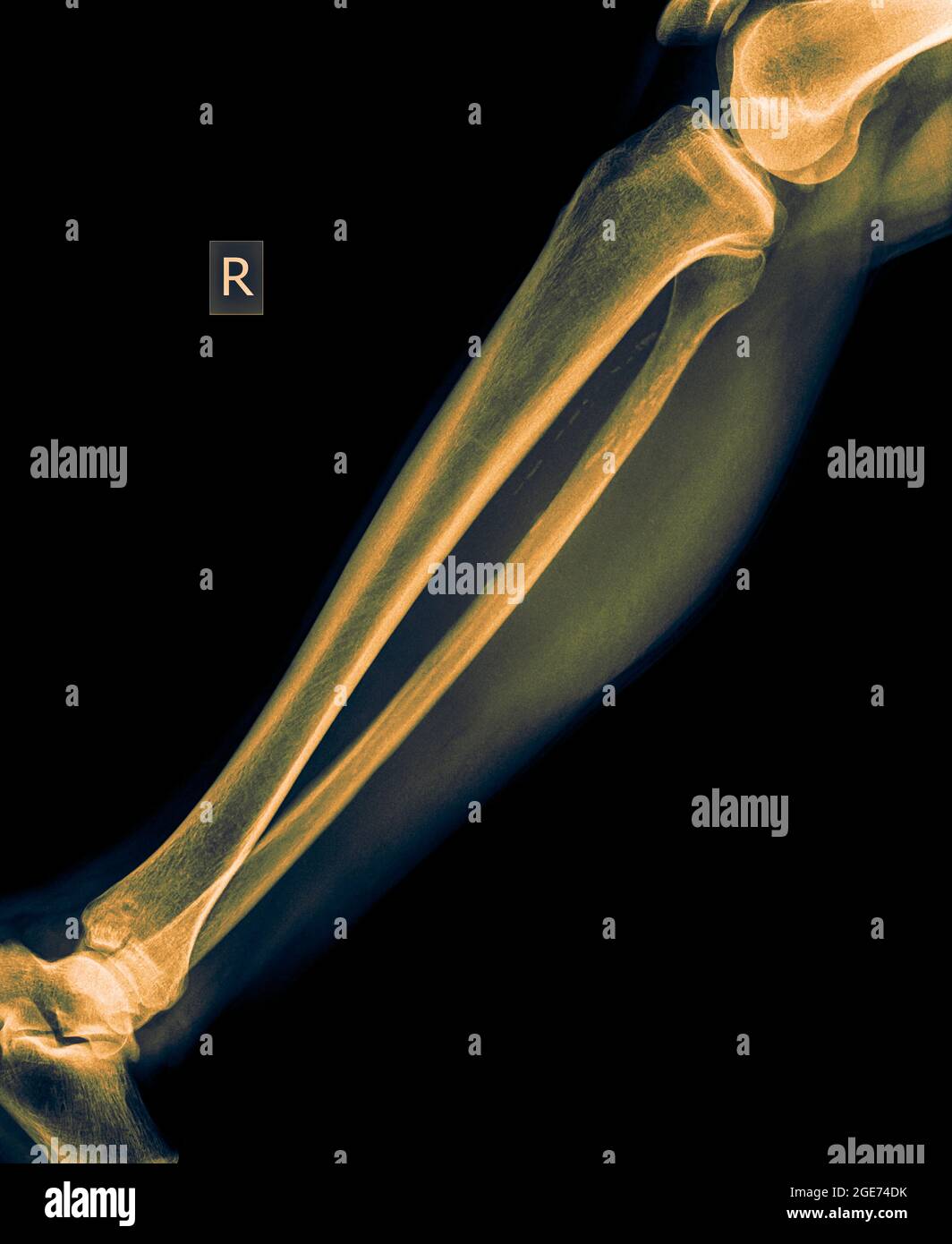 X-ray of the lower leg 50 year old male with a fractured tibia front view Stock Photohttps://www.alamy.com/image-license-details/?v=1https://www.alamy.com/x-ray-of-the-lower-leg-50-year-old-male-with-a-fractured-tibia-front-view-image439021567.html
RM2GE74DK–X-ray of the lower leg 50 year old male with a fractured tibia front view
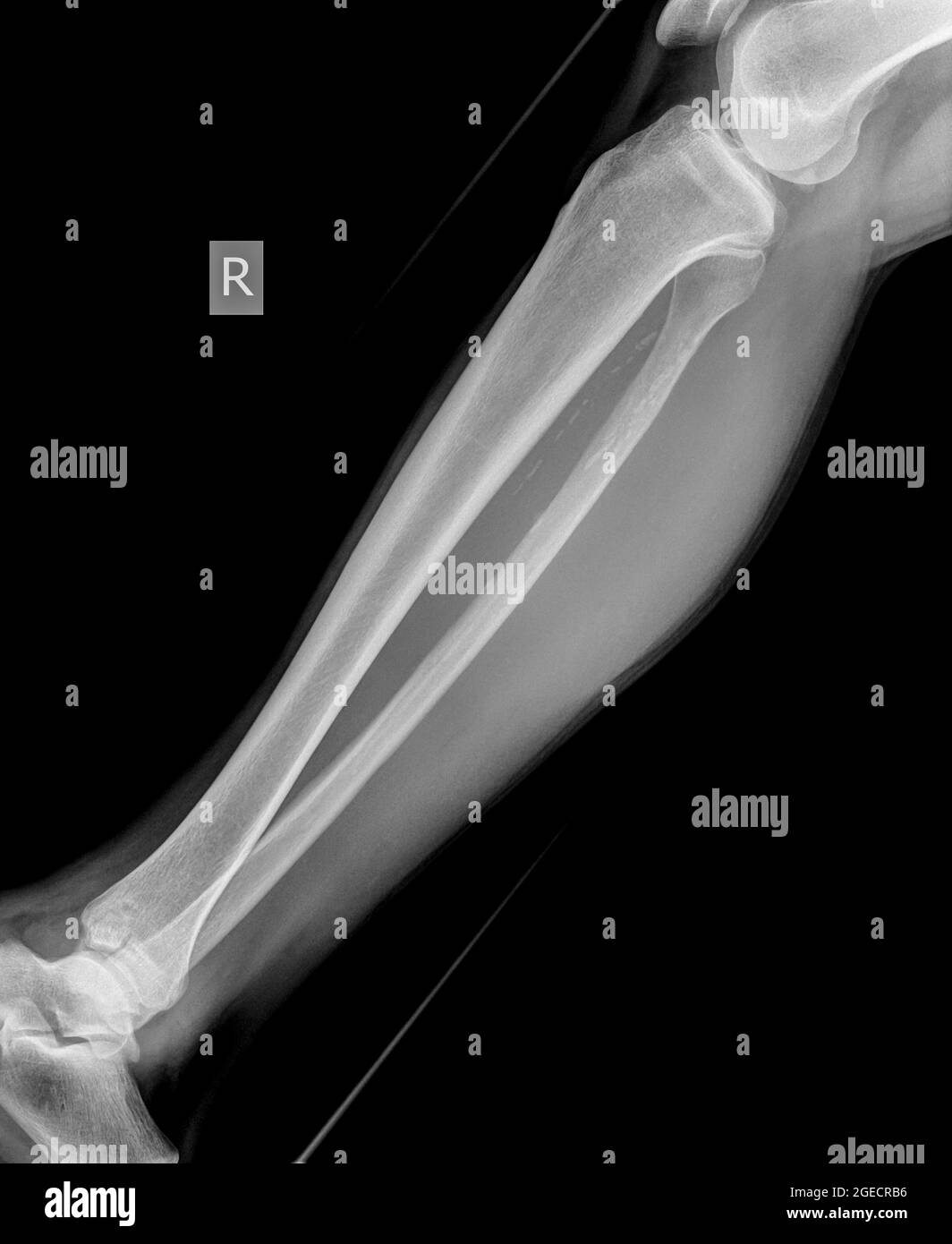 X-ray of the lower leg 50 year old male with a fractured tibia front view Stock Photohttps://www.alamy.com/image-license-details/?v=1https://www.alamy.com/x-ray-of-the-lower-leg-50-year-old-male-with-a-fractured-tibia-front-view-image439146154.html
RM2GECRB6–X-ray of the lower leg 50 year old male with a fractured tibia front view
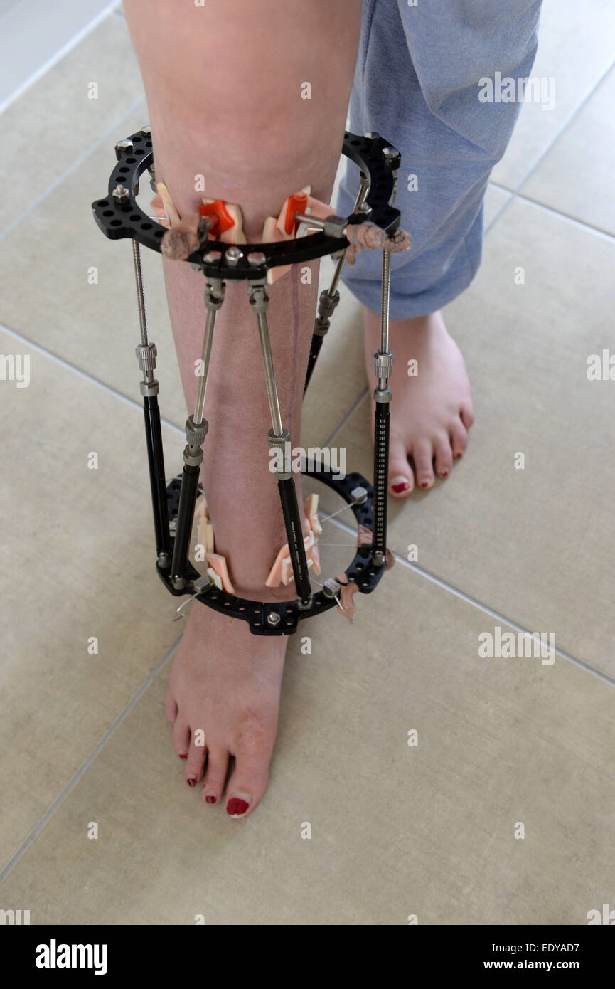 External fixator fitted to a leg to help hold it in the correct position while the bones grow. Stock Photohttps://www.alamy.com/image-license-details/?v=1https://www.alamy.com/stock-photo-external-fixator-fitted-to-a-leg-to-help-hold-it-in-the-correct-position-77432915.html
RFEDYAD7–External fixator fitted to a leg to help hold it in the correct position while the bones grow.
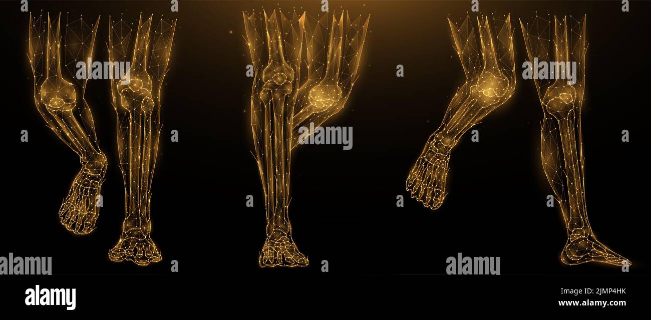 Polygonal vector illustration of anatomy of human legs. Low poly art lower limbs on a dark background. The flesh and bones of th Stock Photohttps://www.alamy.com/image-license-details/?v=1https://www.alamy.com/polygonal-vector-illustration-of-anatomy-of-human-legs-low-poly-art-lower-limbs-on-a-dark-background-the-flesh-and-bones-of-th-image477459631.html
RF2JMP4HK–Polygonal vector illustration of anatomy of human legs. Low poly art lower limbs on a dark background. The flesh and bones of th
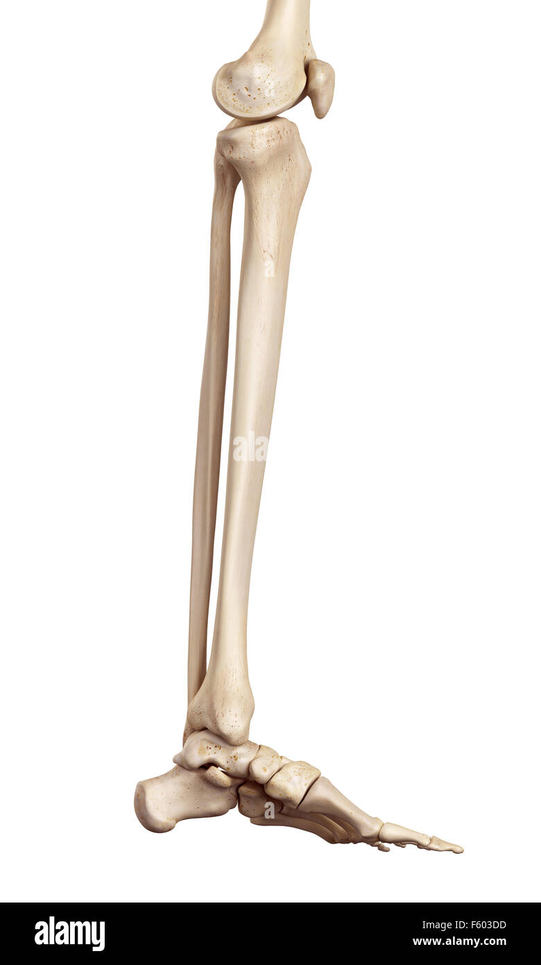 medical accurate illustration of the lower leg bones Stock Photohttps://www.alamy.com/image-license-details/?v=1https://www.alamy.com/stock-photo-medical-accurate-illustration-of-the-lower-leg-bones-89742505.html
RFF603DD–medical accurate illustration of the lower leg bones
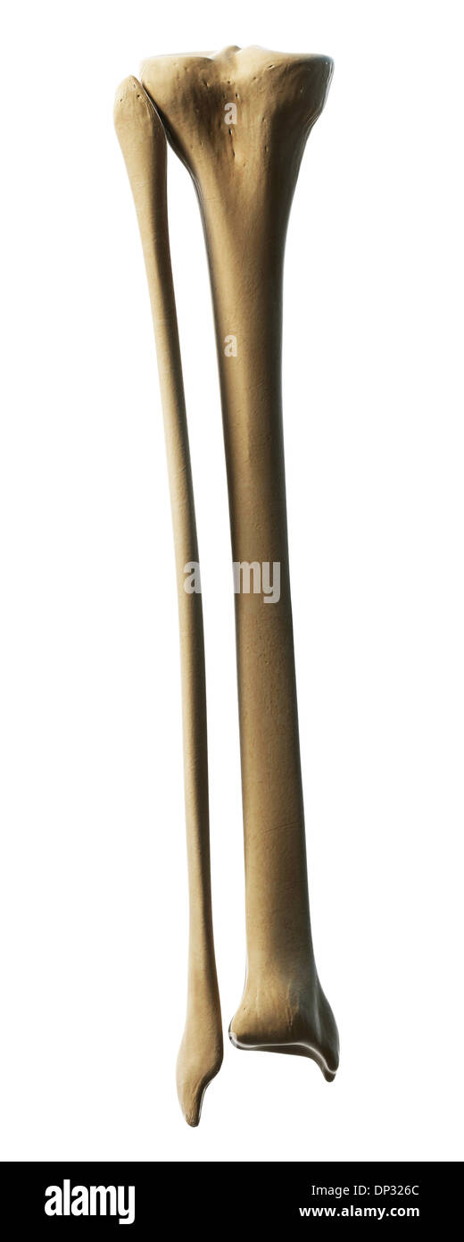 Lower leg bones, artwork Stock Photohttps://www.alamy.com/image-license-details/?v=1https://www.alamy.com/lower-leg-bones-artwork-image65221140.html
RFDP326C–Lower leg bones, artwork
 The Invention of Drawing, recto, Sketch of Lower Leg Bones of Human Skeleton, verso, Joseph-Benoît Suvée, Belgian, 1743 - 1807 Stock Photohttps://www.alamy.com/image-license-details/?v=1https://www.alamy.com/the-invention-of-drawing-recto-sketch-of-lower-leg-bones-of-human-skeleton-verso-joseph-benot-suve-belgian-1743-1807-image220634103.html
RMPPXN1B–The Invention of Drawing, recto, Sketch of Lower Leg Bones of Human Skeleton, verso, Joseph-Benoît Suvée, Belgian, 1743 - 1807
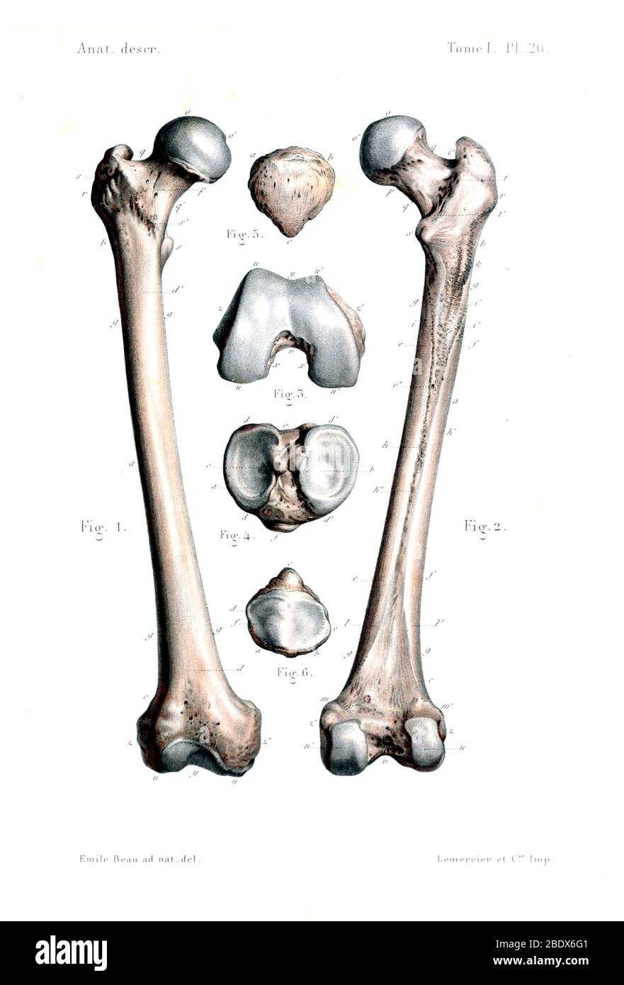 Human Leg Bones, 1844 Stock Photohttps://www.alamy.com/image-license-details/?v=1https://www.alamy.com/human-leg-bones-1844-image352773793.html
RM2BDX6G1–Human Leg Bones, 1844
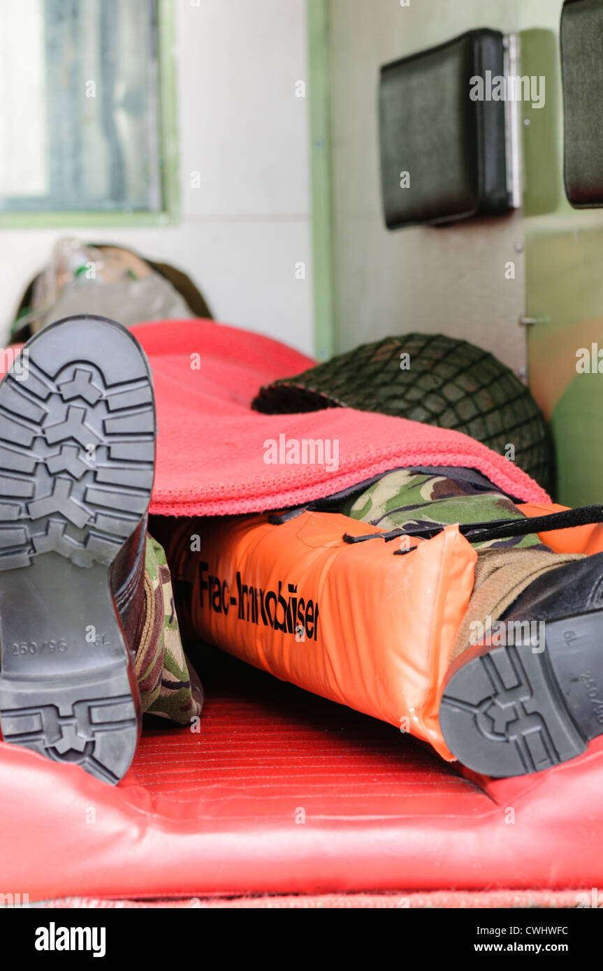 Model of a soldier lying in the back of a military ambulance with a leg fracture immobiliser to stabilise a broken lower leg. Stock Photohttps://www.alamy.com/image-license-details/?v=1https://www.alamy.com/stock-photo-model-of-a-soldier-lying-in-the-back-of-a-military-ambulance-with-50180352.html
RMCWHWFC–Model of a soldier lying in the back of a military ambulance with a leg fracture immobiliser to stabilise a broken lower leg.
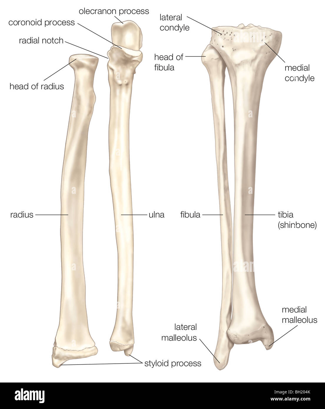 Bones of the forearm and lower leg Stock Photohttps://www.alamy.com/image-license-details/?v=1https://www.alamy.com/stock-photo-bones-of-the-forearm-and-lower-leg-27703555.html
RMBH204K–Bones of the forearm and lower leg
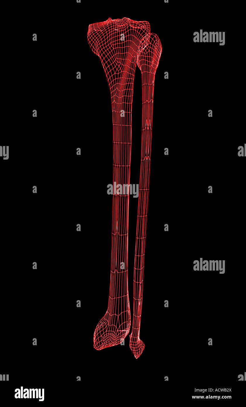 The lower leg Stock Photohttps://www.alamy.com/image-license-details/?v=1https://www.alamy.com/stock-photo-the-lower-leg-13234513.html
RFACWB2X–The lower leg
 The bones of the the lower leg and foot. Shown are the tibia; femur; patella; fibula, medial malleolus, lateral malleolus; metatarsals; bones; lower l Stock Photohttps://www.alamy.com/image-license-details/?v=1https://www.alamy.com/stock-photo-the-bones-of-the-the-lower-leg-and-foot-shown-are-the-tibia-femur-130806376.html
RFHGPMT8–The bones of the the lower leg and foot. Shown are the tibia; femur; patella; fibula, medial malleolus, lateral malleolus; metatarsals; bones; lower l
 Anatomy of the nerves of the lower limb (leg). Stock Photohttps://www.alamy.com/image-license-details/?v=1https://www.alamy.com/anatomy-of-the-nerves-of-the-lower-limb-leg-image476924461.html
RF2JKWP0D–Anatomy of the nerves of the lower limb (leg).
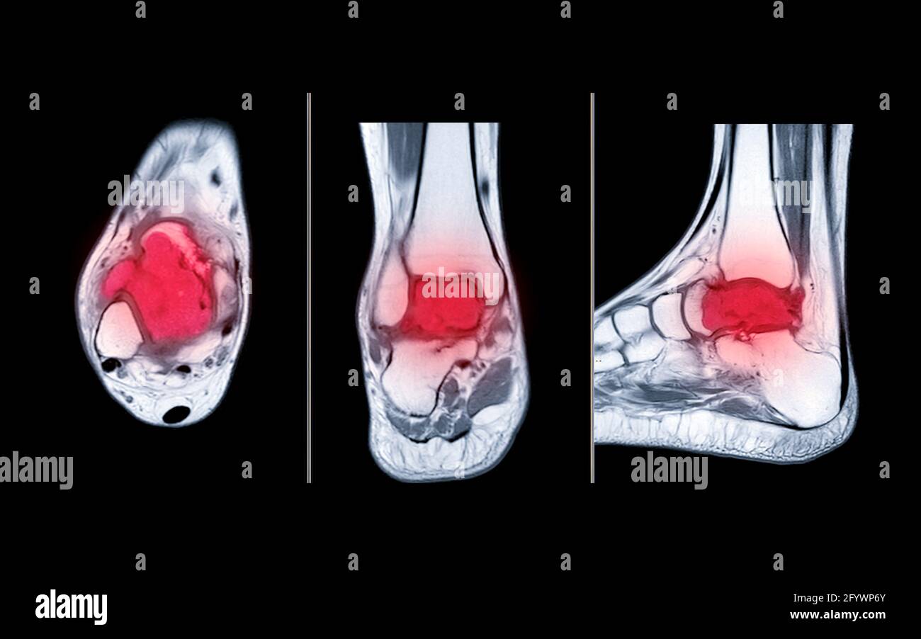 Compare of MRI ankle axial, coronal and sagittal PDW view showing bone metastasis to the talus. Stock Photohttps://www.alamy.com/image-license-details/?v=1https://www.alamy.com/compare-of-mri-ankle-axial-coronal-and-sagittal-pdw-view-showing-bone-metastasis-to-the-talus-image430210787.html
RF2FYWP6Y–Compare of MRI ankle axial, coronal and sagittal PDW view showing bone metastasis to the talus.
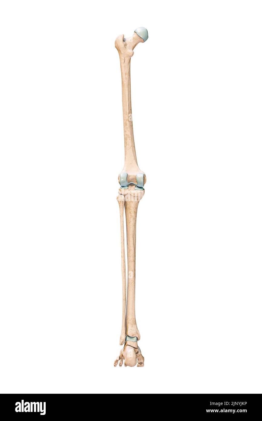 Accurate posterior or rear view of the leg or lower limb bones of the human skeletal system isolated on white background 3D rendering illustration. An Stock Photohttps://www.alamy.com/image-license-details/?v=1https://www.alamy.com/accurate-posterior-or-rear-view-of-the-leg-or-lower-limb-bones-of-the-human-skeletal-system-isolated-on-white-background-3d-rendering-illustration-an-image478195082.html
RF2JNYJKP–Accurate posterior or rear view of the leg or lower limb bones of the human skeletal system isolated on white background 3D rendering illustration. An
 210120-N-DA693-1268 SAN DIEGO (Jan. 20, 2021) A detailed photo of surgeons’ hands during an immediate jaw reconstruction with 3D-printed teeth in one of Naval Medical Center San Diego’s (NMCSD) operating rooms Jan. 20. The procedure was performed by teams from the hospital’s Oral and Maxillofacial Surgery, Plastic Surgery and Otolaryngology Departments. The surgery comprised of not only cancer removal, but the reconstruction of a jaw using a section of the patient’s fibula, the smaller of the two lower leg bones. The COVID-19 pandemic has changed the way many facets of healthcare are conducte Stock Photohttps://www.alamy.com/image-license-details/?v=1https://www.alamy.com/210120-n-da693-1268-san-diego-jan-20-2021-a-detailed-photo-of-surgeons-hands-during-an-immediate-jaw-reconstruction-with-3d-printed-teeth-in-one-of-naval-medical-center-san-diegos-nmcsd-operating-rooms-jan-20-the-procedure-was-performed-by-teams-from-the-hospitals-oral-and-maxillofacial-surgery-plastic-surgery-and-otolaryngology-departments-the-surgery-comprised-of-not-only-cancer-removal-but-the-reconstruction-of-a-jaw-using-a-section-of-the-patients-fibula-the-smaller-of-the-two-lower-leg-bones-the-covid-19-pandemic-has-changed-the-way-many-facets-of-healthcare-are-conducte-image447732819.html
RM2H0BYNR–210120-N-DA693-1268 SAN DIEGO (Jan. 20, 2021) A detailed photo of surgeons’ hands during an immediate jaw reconstruction with 3D-printed teeth in one of Naval Medical Center San Diego’s (NMCSD) operating rooms Jan. 20. The procedure was performed by teams from the hospital’s Oral and Maxillofacial Surgery, Plastic Surgery and Otolaryngology Departments. The surgery comprised of not only cancer removal, but the reconstruction of a jaw using a section of the patient’s fibula, the smaller of the two lower leg bones. The COVID-19 pandemic has changed the way many facets of healthcare are conducte
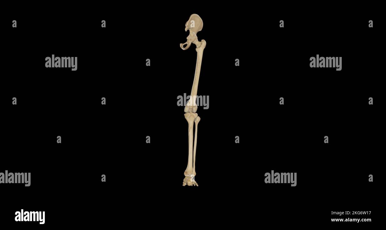 Bones of Right Lower Limb - Posterior View Stock Photohttps://www.alamy.com/image-license-details/?v=1https://www.alamy.com/bones-of-right-lower-limb-posterior-view-image491876147.html
RF2KG6W17–Bones of Right Lower Limb - Posterior View
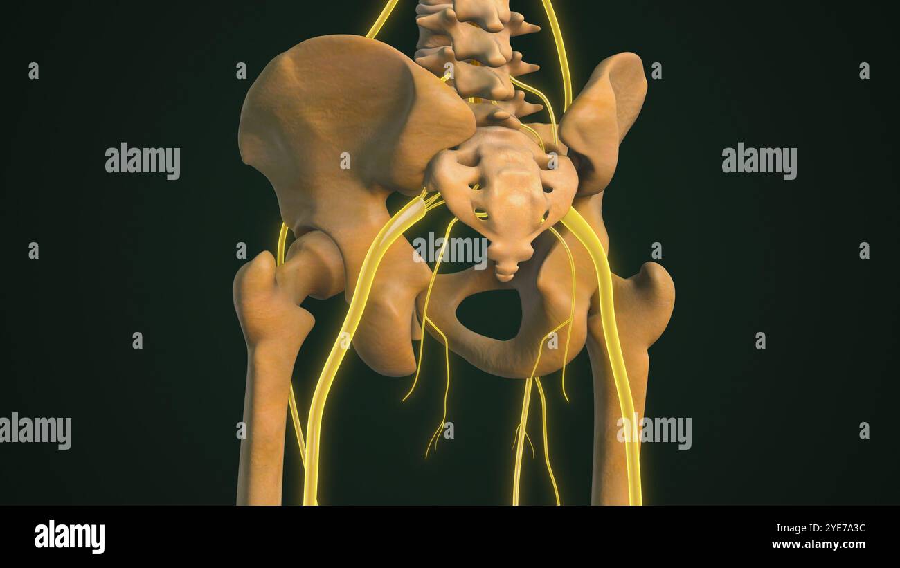 Highlighted Sciatica Nerve with Bone Structure Stock Photohttps://www.alamy.com/image-license-details/?v=1https://www.alamy.com/highlighted-sciatica-nerve-with-bone-structure-image628340032.html
RF2YE7A3C–Highlighted Sciatica Nerve with Bone Structure
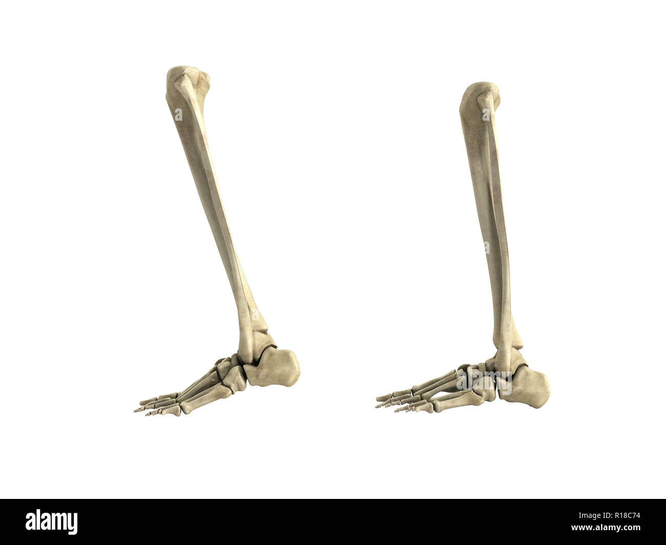 medical accurate illustration of the lower leg bones 3d render no shadow Stock Photohttps://www.alamy.com/image-license-details/?v=1https://www.alamy.com/medical-accurate-illustration-of-the-lower-leg-bones-3d-render-no-shadow-image224534664.html
RFR18C74–medical accurate illustration of the lower leg bones 3d render no shadow
 Physiotherapist crossing the leg of his patient Stock Photohttps://www.alamy.com/image-license-details/?v=1https://www.alamy.com/stock-photo-physiotherapist-crossing-the-leg-of-his-patient-49470606.html
RFCTDG7A–Physiotherapist crossing the leg of his patient
 The Tibia and Fibula are long bones of the lower leg, tibia is the only weight bearing bone, vintage line drawing or engraving illustration. Stock Vectorhttps://www.alamy.com/image-license-details/?v=1https://www.alamy.com/the-tibia-and-fibula-are-long-bones-of-the-lower-leg-tibia-is-the-only-weight-bearing-bone-vintage-line-drawing-or-engraving-illustration-image359330411.html
RF2BTGWGY–The Tibia and Fibula are long bones of the lower leg, tibia is the only weight bearing bone, vintage line drawing or engraving illustration.
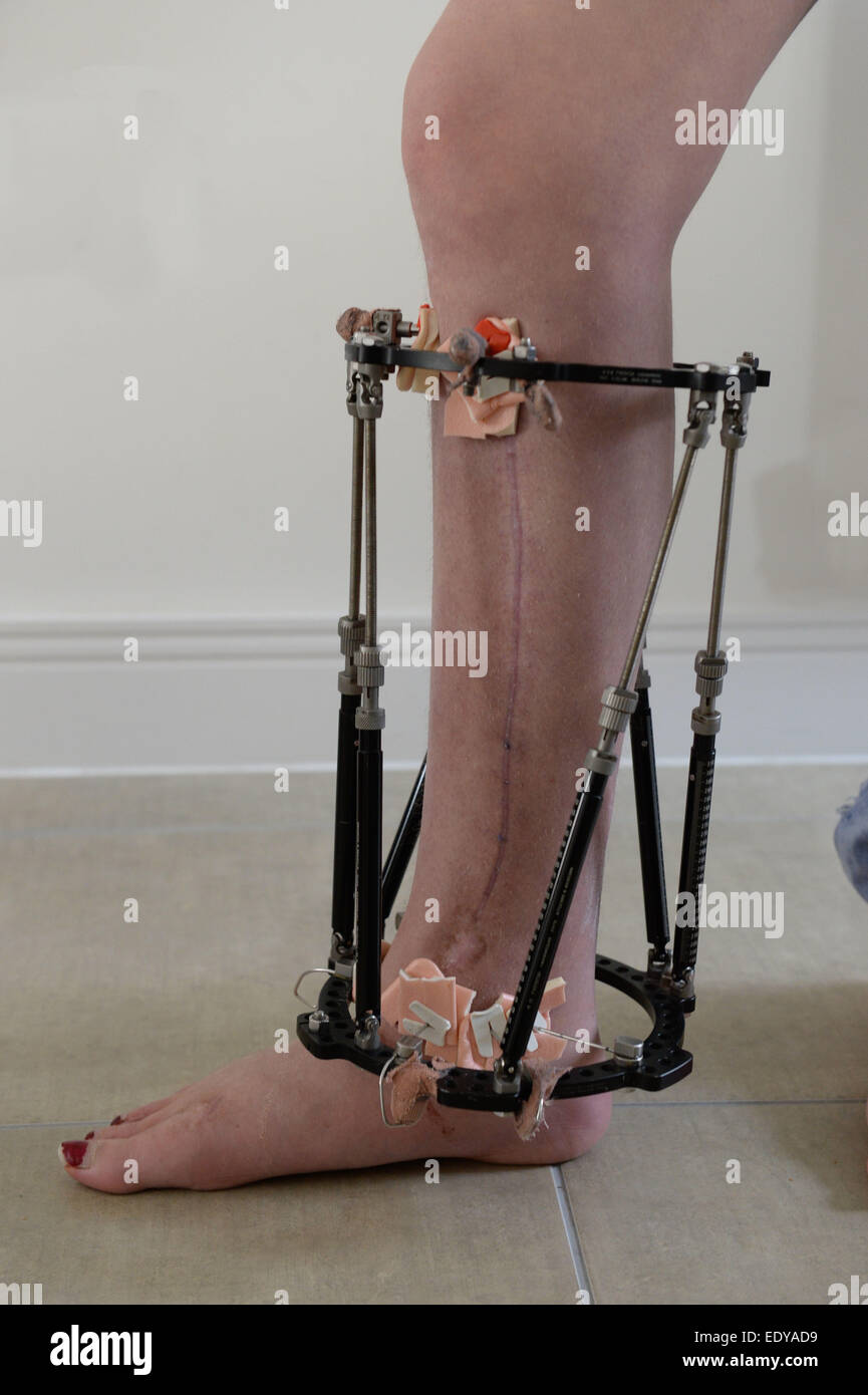 External fixator fitted to a leg to help hold it in the correct position while the bones grow. Stock Photohttps://www.alamy.com/image-license-details/?v=1https://www.alamy.com/stock-photo-external-fixator-fitted-to-a-leg-to-help-hold-it-in-the-correct-position-77432917.html
RFEDYAD9–External fixator fitted to a leg to help hold it in the correct position while the bones grow.
 Skeletons in a row, bones Stock Photohttps://www.alamy.com/image-license-details/?v=1https://www.alamy.com/stock-photo-skeletons-in-a-row-bones-86911217.html
RFF1B441–Skeletons in a row, bones
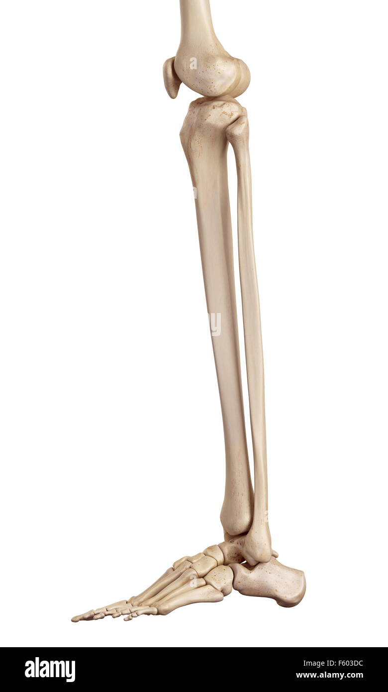 medical accurate illustration of the lower leg bones Stock Photohttps://www.alamy.com/image-license-details/?v=1https://www.alamy.com/stock-photo-medical-accurate-illustration-of-the-lower-leg-bones-89742504.html
RFF603DC–medical accurate illustration of the lower leg bones
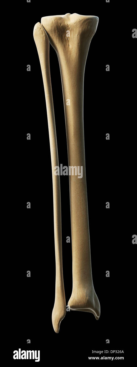 Lower leg bones, artwork Stock Photohttps://www.alamy.com/image-license-details/?v=1https://www.alamy.com/lower-leg-bones-artwork-image65221138.html
RFDP326A–Lower leg bones, artwork
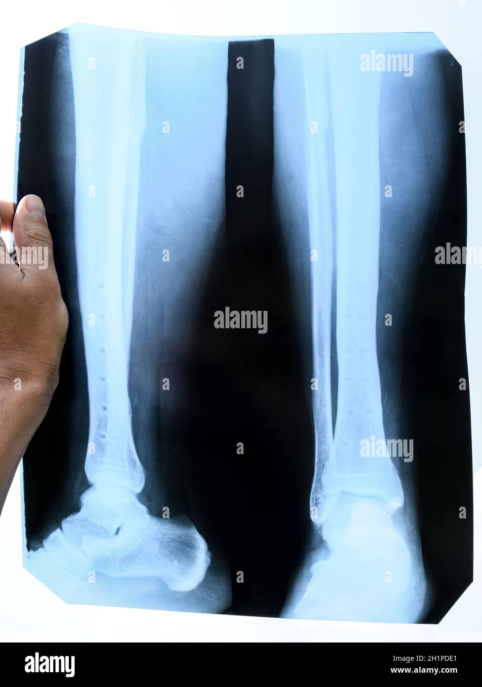 Fused bones of the lower leg after removing the steel bonding plate, x-ray of the leg Stock Photohttps://www.alamy.com/image-license-details/?v=1https://www.alamy.com/fused-bones-of-the-lower-leg-after-removing-the-steel-bonding-plate-x-ray-of-the-leg-image448577753.html
RM2H1PDE1–Fused bones of the lower leg after removing the steel bonding plate, x-ray of the leg
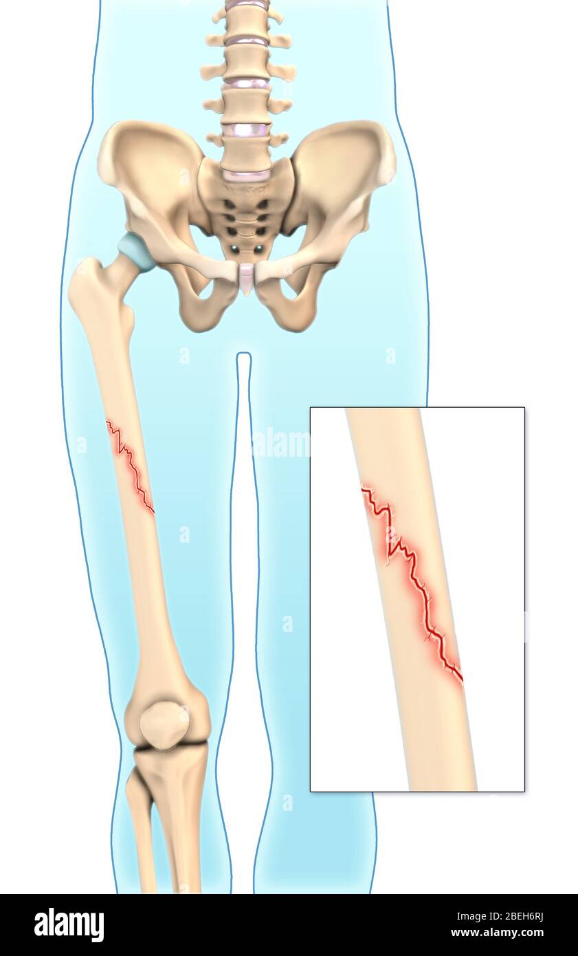 An illustration of an oblique fracture in which a break is diagonal to the bone's long axis. Stock Photohttps://www.alamy.com/image-license-details/?v=1https://www.alamy.com/an-illustration-of-an-oblique-fracture-in-which-a-break-is-diagonal-to-the-bones-long-axis-image353191094.html
RM2BEH6RJ–An illustration of an oblique fracture in which a break is diagonal to the bone's long axis.
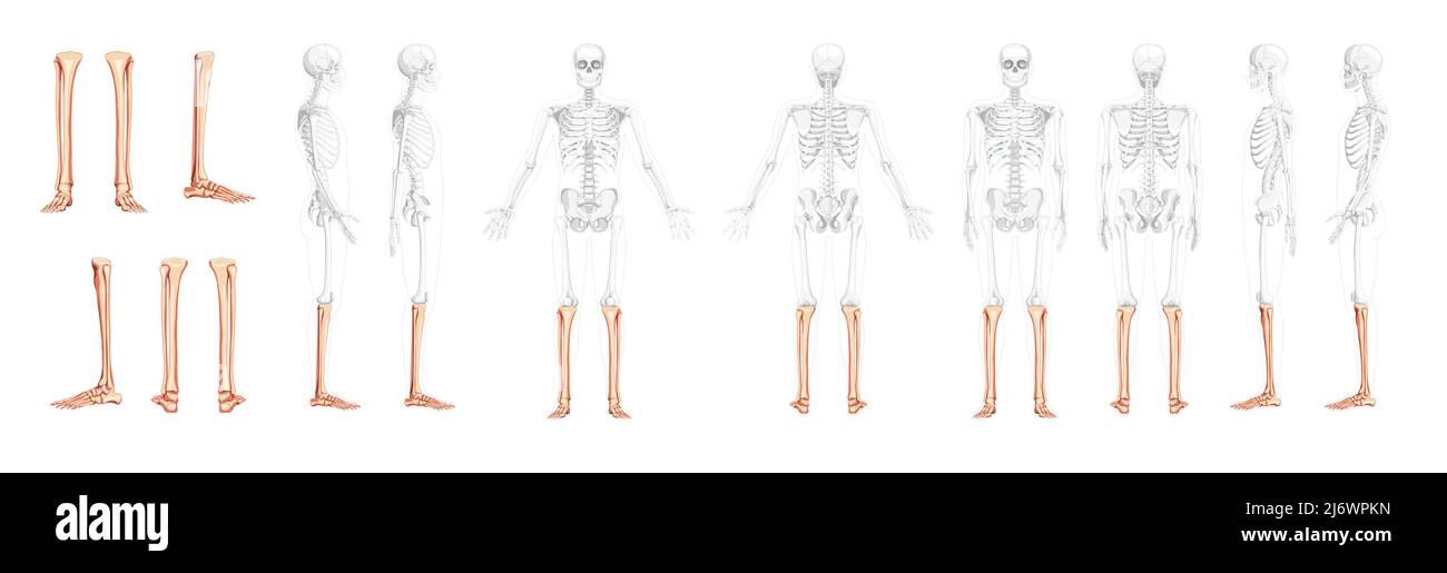 Set of Skeleton leg tibia, Foot, ankle Human front back side view with partly transparent bones position. 3D realistic flat natural color Vector illustration of anatomy isolated on white background Stock Vectorhttps://www.alamy.com/image-license-details/?v=1https://www.alamy.com/set-of-skeleton-leg-tibia-foot-ankle-human-front-back-side-view-with-partly-transparent-bones-position-3d-realistic-flat-natural-color-vector-illustration-of-anatomy-isolated-on-white-background-image468934473.html
RF2J6WPKN–Set of Skeleton leg tibia, Foot, ankle Human front back side view with partly transparent bones position. 3D realistic flat natural color Vector illustration of anatomy isolated on white background
 Amputation of a leg, 16th century, Amputation eines Beines im 16. Jahrhundert Stock Photohttps://www.alamy.com/image-license-details/?v=1https://www.alamy.com/stock-photo-amputation-of-a-leg-16th-century-amputation-eines-beines-im-16-jahrhundert-76638635.html
RMECK5A3–Amputation of a leg, 16th century, Amputation eines Beines im 16. Jahrhundert
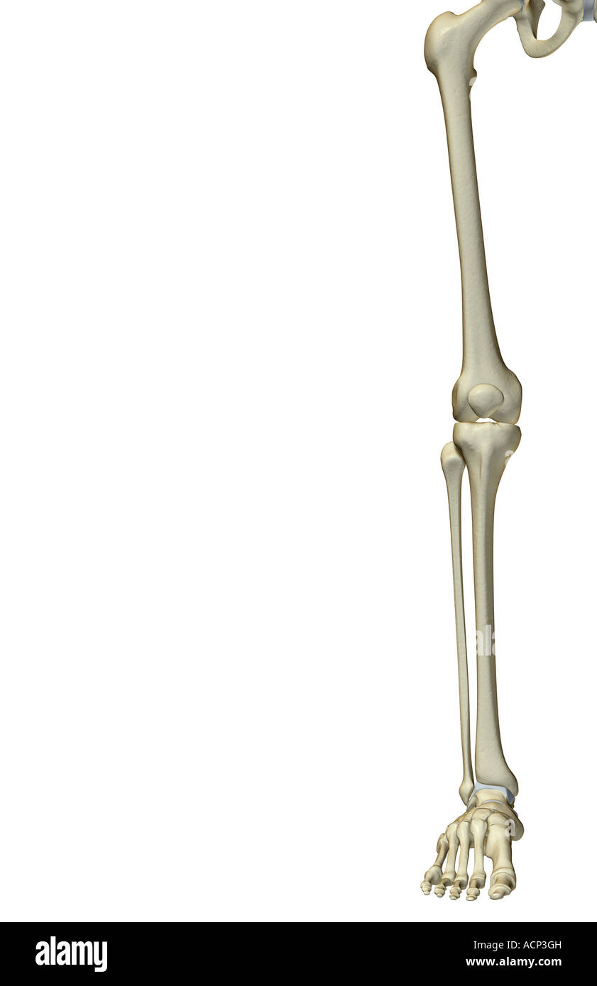 The bones of the lower limb Stock Photohttps://www.alamy.com/image-license-details/?v=1https://www.alamy.com/stock-photo-the-bones-of-the-lower-limb-13203760.html
RFACP3GH–The bones of the lower limb
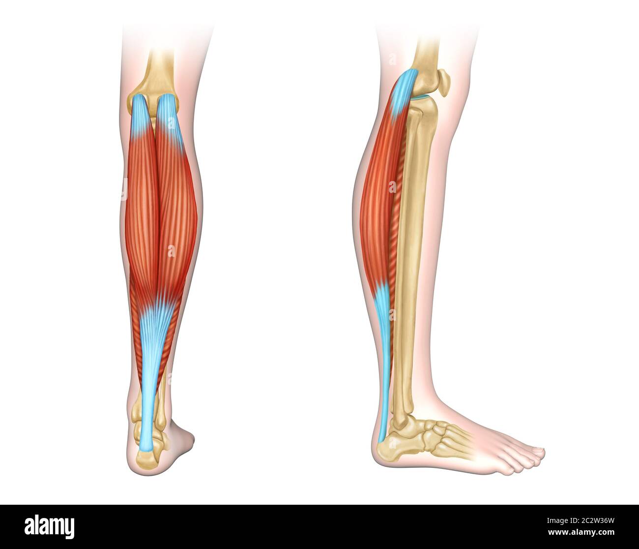 Back and side view of human calf muscles. Digital illustration. Stock Photohttps://www.alamy.com/image-license-details/?v=1https://www.alamy.com/back-and-side-view-of-human-calf-muscles-digital-illustration-image363198385.html
RF2C2W36W–Back and side view of human calf muscles. Digital illustration.
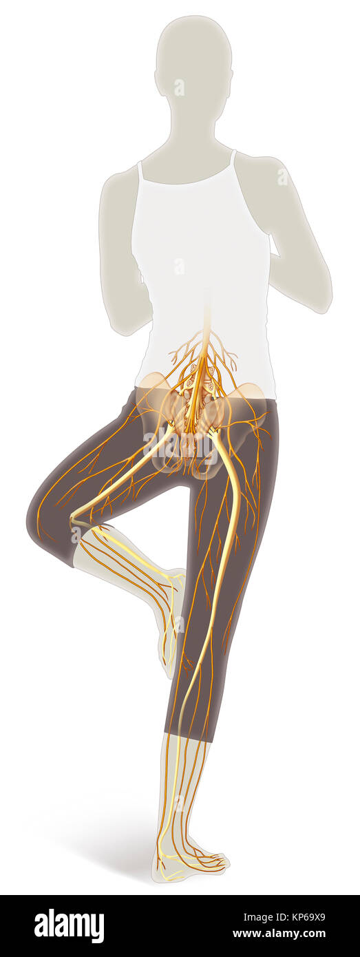 LOWER LIMB NERVE, ILLUSTRATION Stock Photohttps://www.alamy.com/image-license-details/?v=1https://www.alamy.com/stock-image-lower-limb-nerve-illustration-168555249.html
RMKP69X9–LOWER LIMB NERVE, ILLUSTRATION
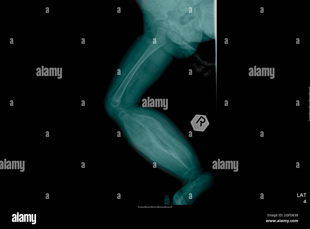 X-ray of an 11 month old infant suffering from Infantile cortical hyperostosis (Caffey's disease) Stock Photohttps://www.alamy.com/image-license-details/?v=1https://www.alamy.com/x-ray-of-an-11-month-old-infant-suffering-from-infantile-cortical-hyperostosis-caffeys-disease-image439770780.html
RM2GFD838–X-ray of an 11 month old infant suffering from Infantile cortical hyperostosis (Caffey's disease)
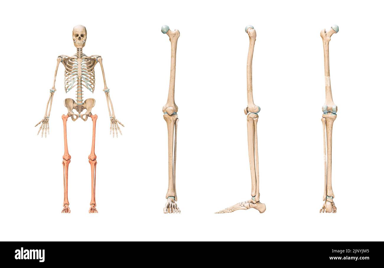 Accurate leg or lower limb bones of the human skeletal system or skeleton isolated on white background 3D rendering illustration. Anterior, lateral an Stock Photohttps://www.alamy.com/image-license-details/?v=1https://www.alamy.com/accurate-leg-or-lower-limb-bones-of-the-human-skeletal-system-or-skeleton-isolated-on-white-background-3d-rendering-illustration-anterior-lateral-an-image478195093.html
RF2JNYJM5–Accurate leg or lower limb bones of the human skeletal system or skeleton isolated on white background 3D rendering illustration. Anterior, lateral an
 210120-N-DA693-1151 SAN DIEGO (Jan. 20, 2021) A detailed photo of surgeons’ hands performing an immediate jaw reconstruction with 3D-printed teeth in one of Naval Medical Center San Diego’s (NMCSD) operating rooms Jan. 20. The procedure was performed by teams from the hospital’s Oral and Maxillofacial Surgery, Plastic Surgery and Otolaryngology Departments. The surgery comprised of not only cancer removal, but the reconstruction of a jaw using a section of the patient’s fibula, the smaller of the two lower leg bones. The COVID-19 pandemic has changed the way many facets of healthcare are cond Stock Photohttps://www.alamy.com/image-license-details/?v=1https://www.alamy.com/210120-n-da693-1151-san-diego-jan-20-2021-a-detailed-photo-of-surgeons-hands-performing-an-immediate-jaw-reconstruction-with-3d-printed-teeth-in-one-of-naval-medical-center-san-diegos-nmcsd-operating-rooms-jan-20-the-procedure-was-performed-by-teams-from-the-hospitals-oral-and-maxillofacial-surgery-plastic-surgery-and-otolaryngology-departments-the-surgery-comprised-of-not-only-cancer-removal-but-the-reconstruction-of-a-jaw-using-a-section-of-the-patients-fibula-the-smaller-of-the-two-lower-leg-bones-the-covid-19-pandemic-has-changed-the-way-many-facets-of-healthcare-are-cond-image447732757.html
RM2H0BYKH–210120-N-DA693-1151 SAN DIEGO (Jan. 20, 2021) A detailed photo of surgeons’ hands performing an immediate jaw reconstruction with 3D-printed teeth in one of Naval Medical Center San Diego’s (NMCSD) operating rooms Jan. 20. The procedure was performed by teams from the hospital’s Oral and Maxillofacial Surgery, Plastic Surgery and Otolaryngology Departments. The surgery comprised of not only cancer removal, but the reconstruction of a jaw using a section of the patient’s fibula, the smaller of the two lower leg bones. The COVID-19 pandemic has changed the way many facets of healthcare are cond
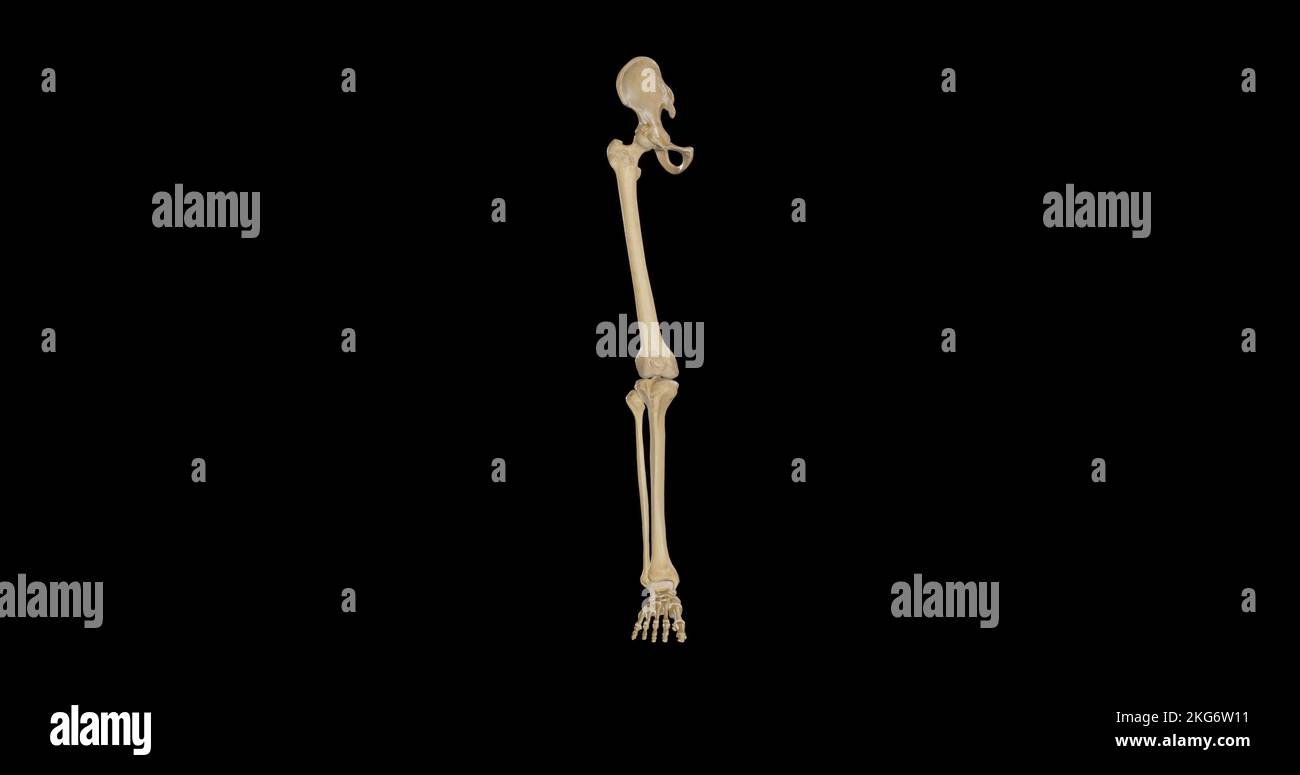 Bones of Right Lower Limb - Anterior View Stock Photohttps://www.alamy.com/image-license-details/?v=1https://www.alamy.com/bones-of-right-lower-limb-anterior-view-image491876141.html
RF2KG6W11–Bones of Right Lower Limb - Anterior View
 Highlighted Sciatica Nerve with Bones Stock Photohttps://www.alamy.com/image-license-details/?v=1https://www.alamy.com/highlighted-sciatica-nerve-with-bones-image628340058.html
RF2YE7A4A–Highlighted Sciatica Nerve with Bones
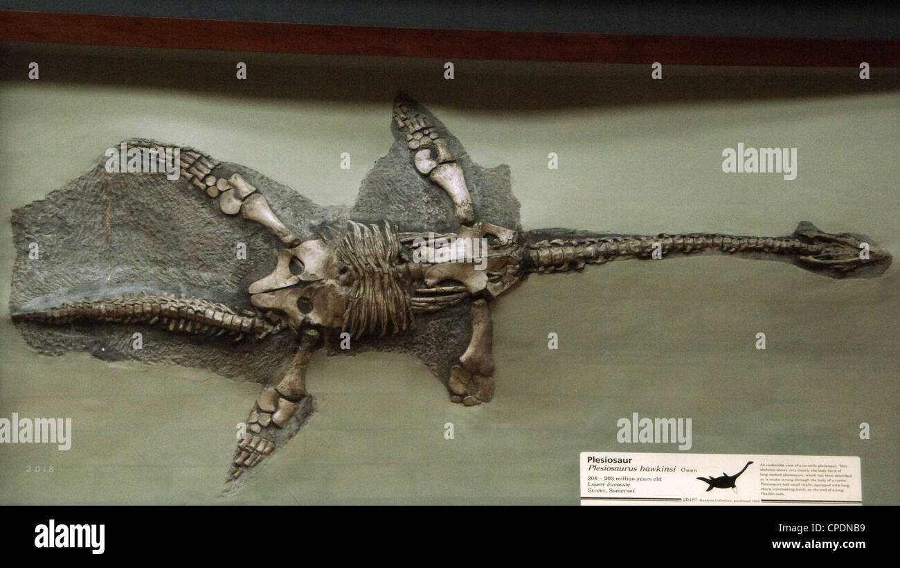 Fossil of Plesiosaur (Plesiosaurus hawkinsi). 208-203 million years old. Lower Jurassic. Street, Somerset. Stock Photohttps://www.alamy.com/image-license-details/?v=1https://www.alamy.com/stock-photo-fossil-of-plesiosaur-plesiosaurus-hawkinsi-208-203-million-years-old-48245325.html
RMCPDNB9–Fossil of Plesiosaur (Plesiosaurus hawkinsi). 208-203 million years old. Lower Jurassic. Street, Somerset.
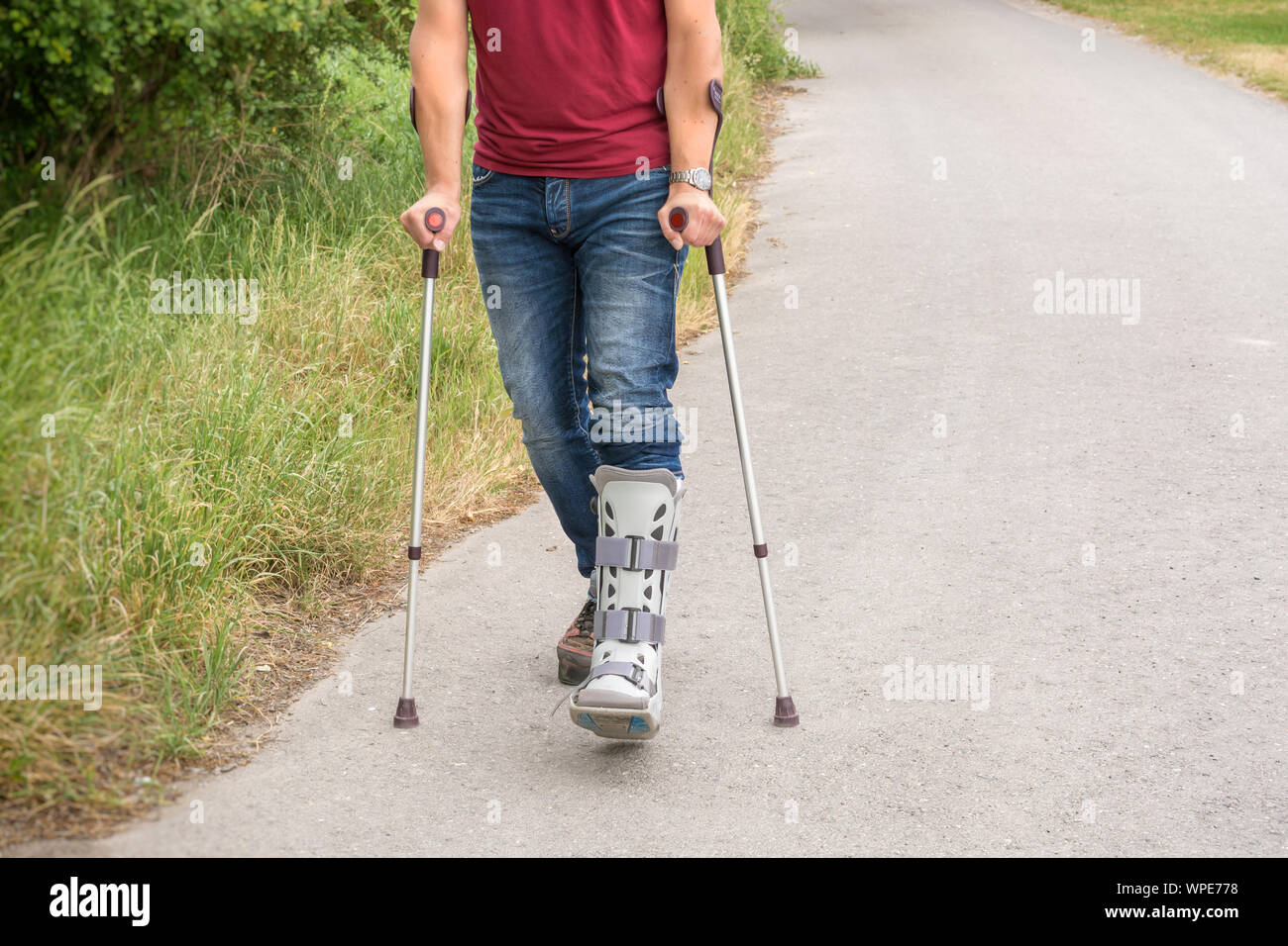 Walking exercises with crutches and an orthosis on the lower leg Stock Photohttps://www.alamy.com/image-license-details/?v=1https://www.alamy.com/walking-exercises-with-crutches-and-an-orthosis-on-the-lower-leg-image271990972.html
RFWPE778–Walking exercises with crutches and an orthosis on the lower leg
 Model of human colored spine bones on metallic background, lower part, 3D render Stock Photohttps://www.alamy.com/image-license-details/?v=1https://www.alamy.com/model-of-human-colored-spine-bones-on-metallic-background-lower-part-3d-render-image412196352.html
RF2EXH4J8–Model of human colored spine bones on metallic background, lower part, 3D render
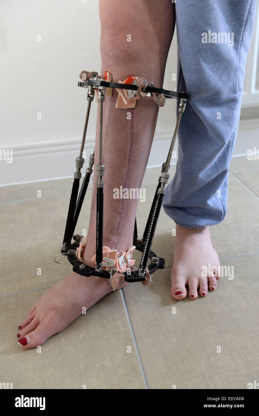 External fixator fitted to a leg to help hold it in the correct position while the bones grow. Stock Photohttps://www.alamy.com/image-license-details/?v=1https://www.alamy.com/stock-photo-external-fixator-fitted-to-a-leg-to-help-hold-it-in-the-correct-position-77432916.html
RFEDYAD8–External fixator fitted to a leg to help hold it in the correct position while the bones grow.
 Joseph-Benoît Suvée (Belgian - The Invention of Drawing (recto); Sketch of Lower Leg Bones of Human Skeleton (verso) - Google Art Project Stock Photohttps://www.alamy.com/image-license-details/?v=1https://www.alamy.com/stock-photo-joseph-benot-suve-belgian-the-invention-of-drawing-recto-sketch-of-170667092.html
RMKWJFH8–Joseph-Benoît Suvée (Belgian - The Invention of Drawing (recto); Sketch of Lower Leg Bones of Human Skeleton (verso) - Google Art Project
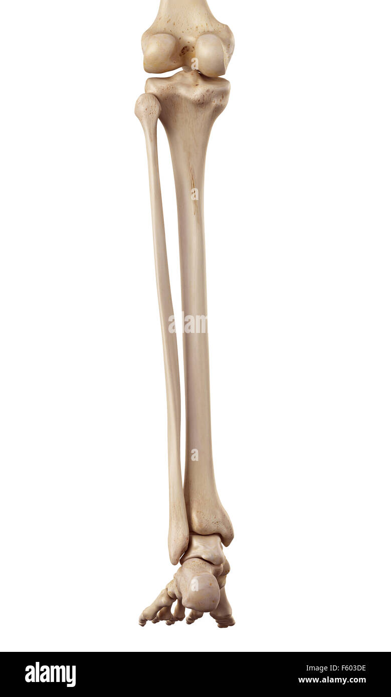 medical accurate illustration of the lower leg bones Stock Photohttps://www.alamy.com/image-license-details/?v=1https://www.alamy.com/stock-photo-medical-accurate-illustration-of-the-lower-leg-bones-89742506.html
RFF603DE–medical accurate illustration of the lower leg bones
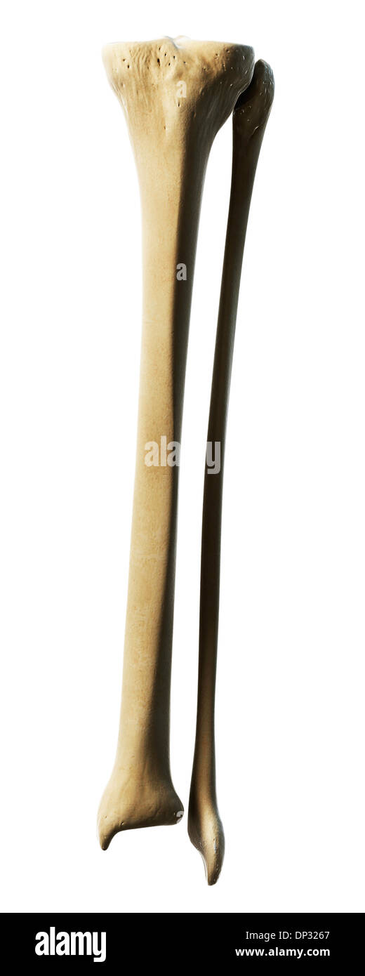 Lower leg bones, artwork Stock Photohttps://www.alamy.com/image-license-details/?v=1https://www.alamy.com/lower-leg-bones-artwork-image65221135.html
RFDP3267–Lower leg bones, artwork
 Fused bones of the lower leg after removing the steel bonding plate, x-ray of the leg Stock Photohttps://www.alamy.com/image-license-details/?v=1https://www.alamy.com/fused-bones-of-the-lower-leg-after-removing-the-steel-bonding-plate-x-ray-of-the-leg-image448577813.html
RM2H1PDG5–Fused bones of the lower leg after removing the steel bonding plate, x-ray of the leg
 Joseph-Benoît Suvée (Belgian - The Invention of Drawing (recto); Sketch of Lower Leg Bones of Human Skeleton (verso) Stock Photohttps://www.alamy.com/image-license-details/?v=1https://www.alamy.com/joseph-benot-suve-belgian-the-invention-of-drawing-recto-sketch-of-lower-leg-bones-of-human-skeleton-verso-image374357810.html
RM2CN1D5P–Joseph-Benoît Suvée (Belgian - The Invention of Drawing (recto); Sketch of Lower Leg Bones of Human Skeleton (verso)
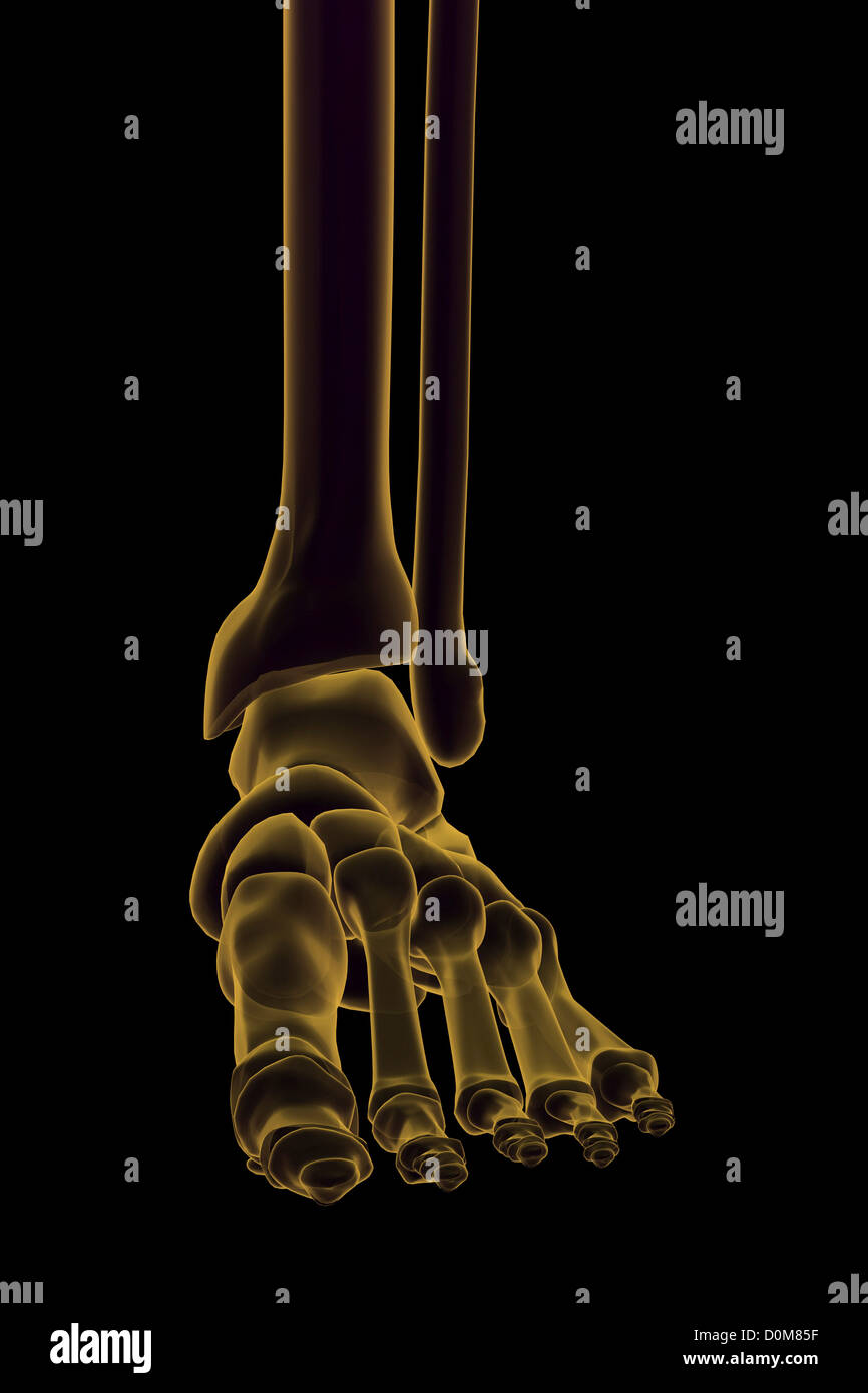 Close-up view of the bones of the lower leg and foot. Stock Photohttps://www.alamy.com/image-license-details/?v=1https://www.alamy.com/stock-photo-close-up-view-of-the-bones-of-the-lower-leg-and-foot-52076571.html
RMD0M85F–Close-up view of the bones of the lower leg and foot.
 Nurse bandages the leg. Fracture of human lower limbs. Treatment of broken bones. Impose a gypsum. Patient surgical department. The doctor's hands Stock Photohttps://www.alamy.com/image-license-details/?v=1https://www.alamy.com/nurse-bandages-the-leg-fracture-of-human-lower-limbs-treatment-of-broken-bones-impose-a-gypsum-patient-surgical-department-the-doctors-hands-image329408427.html
RM2A3WRPK–Nurse bandages the leg. Fracture of human lower limbs. Treatment of broken bones. Impose a gypsum. Patient surgical department. The doctor's hands
 The bones of the lower limb Stock Photohttps://www.alamy.com/image-license-details/?v=1https://www.alamy.com/stock-photo-the-bones-of-the-lower-limb-13165613.html
RFACJ21J–The bones of the lower limb
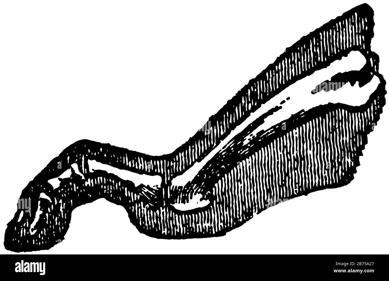 The Foot is that part of the lower extremity below the leg on which we stand and walk, vintage line drawing or engraving illustration. Stock Vectorhttps://www.alamy.com/image-license-details/?v=1https://www.alamy.com/the-foot-is-that-part-of-the-lower-extremity-below-the-leg-on-which-we-stand-and-walk-vintage-line-drawing-or-engraving-illustration-image348627615.html
RF2B75A27–The Foot is that part of the lower extremity below the leg on which we stand and walk, vintage line drawing or engraving illustration.
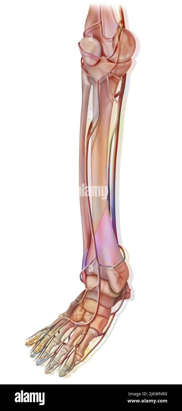 The arteries of the lower part of the lower extremity. Stock Photohttps://www.alamy.com/image-license-details/?v=1https://www.alamy.com/the-arteries-of-the-lower-part-of-the-lower-extremity-image476924308.html
RF2JKWNR0–The arteries of the lower part of the lower extremity.
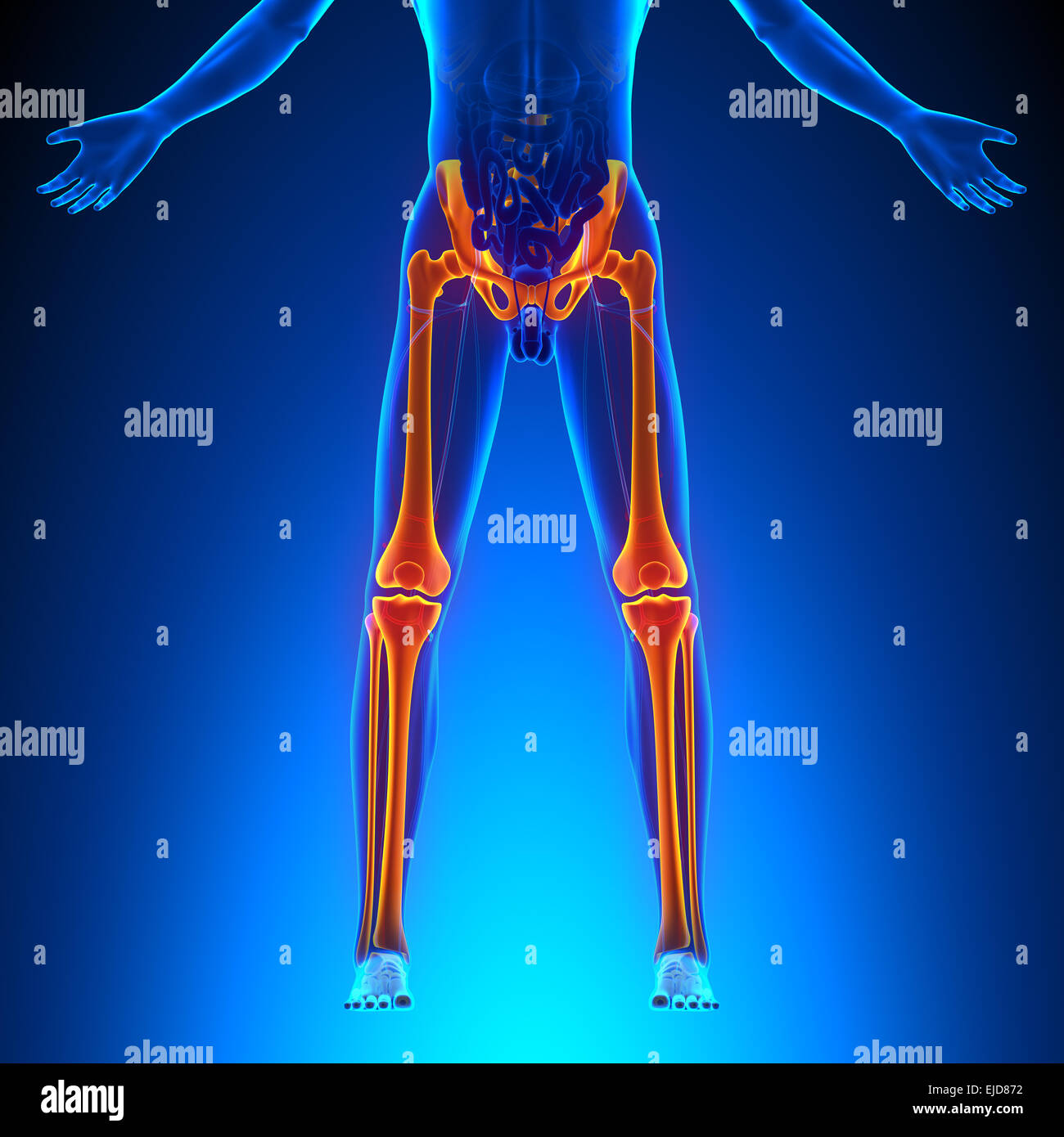 Bones Legs Anatomy Stock Photohttps://www.alamy.com/image-license-details/?v=1https://www.alamy.com/stock-photo-bones-legs-anatomy-80197126.html
RFEJD872–Bones Legs Anatomy
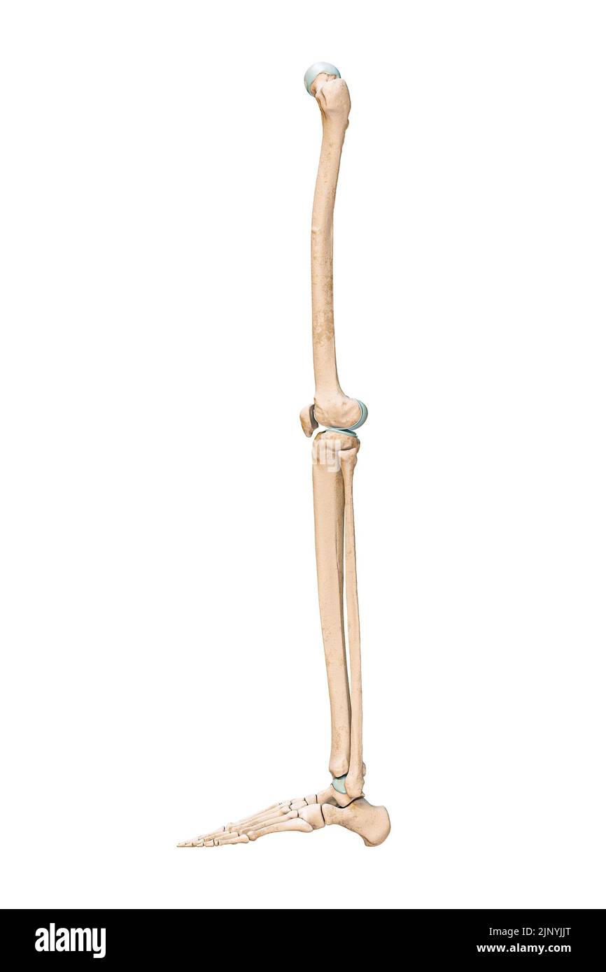 Accurate lateral or profile view of the leg or lower limb bones of the human skeletal system isolated on white background 3D rendering illustration. A Stock Photohttps://www.alamy.com/image-license-details/?v=1https://www.alamy.com/accurate-lateral-or-profile-view-of-the-leg-or-lower-limb-bones-of-the-human-skeletal-system-isolated-on-white-background-3d-rendering-illustration-a-image478195056.html
RF2JNYJJT–Accurate lateral or profile view of the leg or lower limb bones of the human skeletal system isolated on white background 3D rendering illustration. A
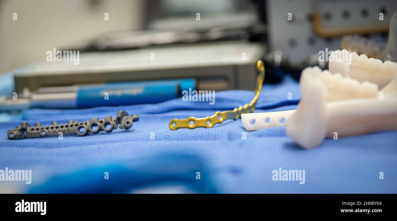 210120-N-DA693-1172 SAN DIEGO (Jan. 20, 2021) A detailed photo of a cutting guide, bone mounting hardware and a 3D-printed jaw mock up during an immediate jaw reconstruction with 3D-printed teeth in one of Naval Medical Center San Diego’s (NMCSD) operating rooms Jan. 20. The procedure was performed by teams from the hospital’s Oral and Maxillofacial Surgery, Plastic Surgery and Otolaryngology Departments. The surgery comprised of not only cancer removal, but the reconstruction of a jaw using a section of the patient’s fibula, the smaller of the two lower leg bones. The COVID-19 pandemic has c Stock Photohttps://www.alamy.com/image-license-details/?v=1https://www.alamy.com/210120-n-da693-1172-san-diego-jan-20-2021-a-detailed-photo-of-a-cutting-guide-bone-mounting-hardware-and-a-3d-printed-jaw-mock-up-during-an-immediate-jaw-reconstruction-with-3d-printed-teeth-in-one-of-naval-medical-center-san-diegos-nmcsd-operating-rooms-jan-20-the-procedure-was-performed-by-teams-from-the-hospitals-oral-and-maxillofacial-surgery-plastic-surgery-and-otolaryngology-departments-the-surgery-comprised-of-not-only-cancer-removal-but-the-reconstruction-of-a-jaw-using-a-section-of-the-patients-fibula-the-smaller-of-the-two-lower-leg-bones-the-covid-19-pandemic-has-c-image447732748.html
RM2H0BYK8–210120-N-DA693-1172 SAN DIEGO (Jan. 20, 2021) A detailed photo of a cutting guide, bone mounting hardware and a 3D-printed jaw mock up during an immediate jaw reconstruction with 3D-printed teeth in one of Naval Medical Center San Diego’s (NMCSD) operating rooms Jan. 20. The procedure was performed by teams from the hospital’s Oral and Maxillofacial Surgery, Plastic Surgery and Otolaryngology Departments. The surgery comprised of not only cancer removal, but the reconstruction of a jaw using a section of the patient’s fibula, the smaller of the two lower leg bones. The COVID-19 pandemic has c
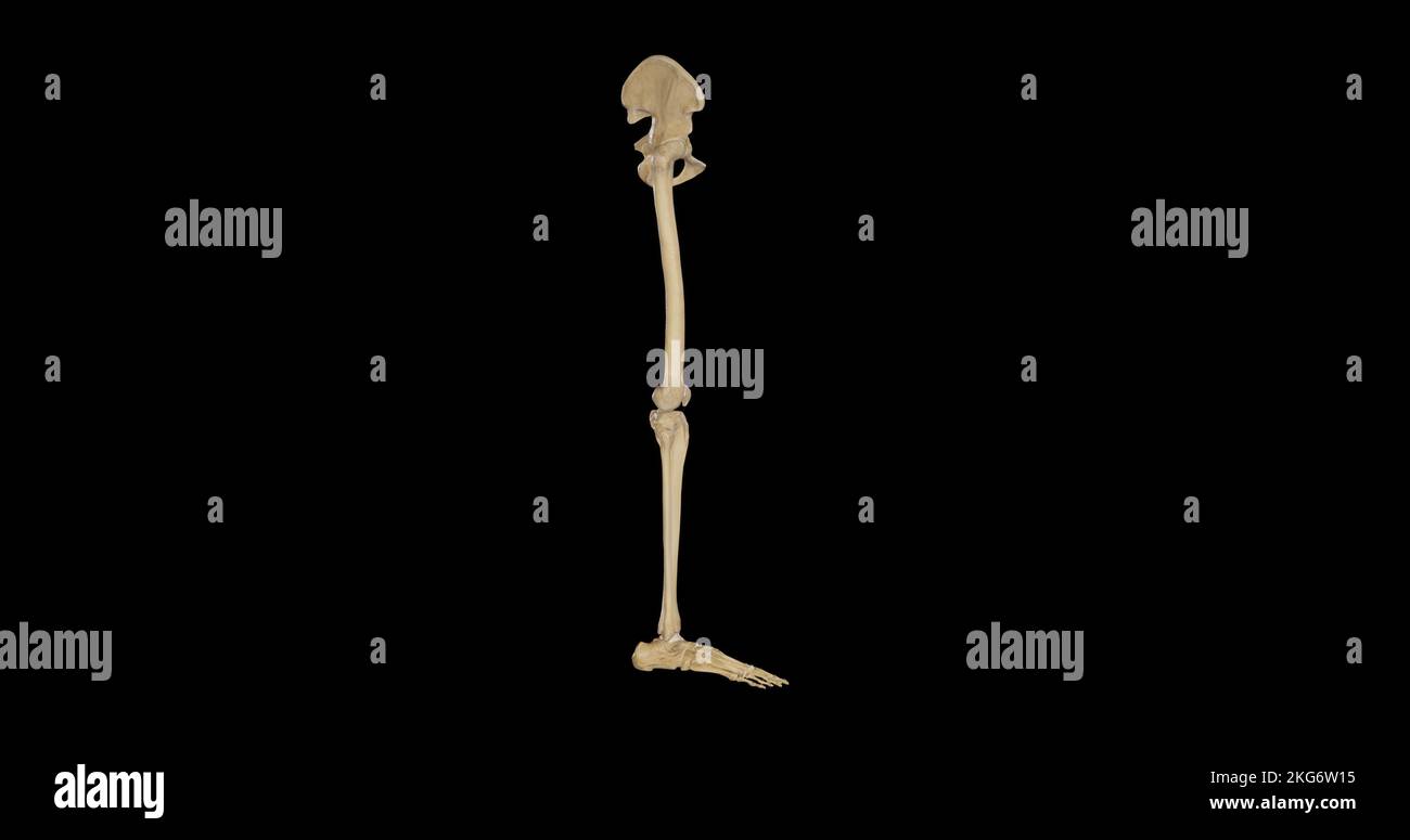 Bones of Right Lower Limb - Lateral View Stock Photohttps://www.alamy.com/image-license-details/?v=1https://www.alamy.com/bones-of-right-lower-limb-lateral-view-image491876145.html
RF2KG6W15–Bones of Right Lower Limb - Lateral View
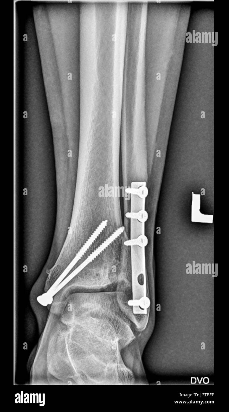 Foot medical xray, lower limb bones, broken ankle, tibia fibula with screws, fingers joints Stock Photohttps://www.alamy.com/image-license-details/?v=1https://www.alamy.com/stock-photo-foot-medical-xray-lower-limb-bones-broken-ankle-tibia-fibula-with-148053326.html
RFJGTBEP–Foot medical xray, lower limb bones, broken ankle, tibia fibula with screws, fingers joints
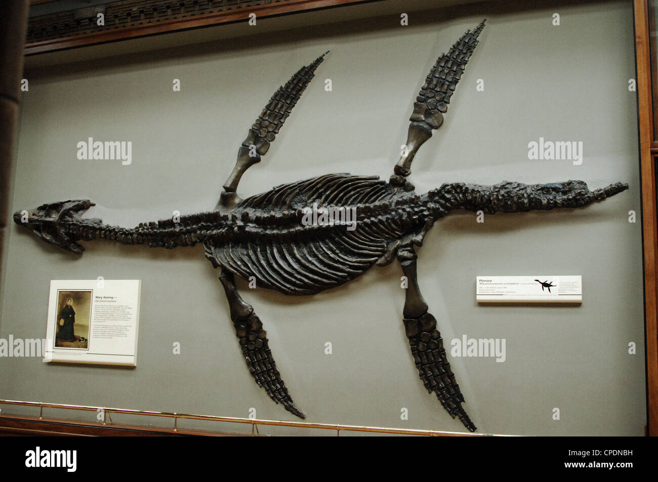 Skeleton of a Pliosaur (Rhomaleosaurus cramptoni). 187-178 million years old. Lower Jurassic. Kettleness, Yorkshire. Stock Photohttps://www.alamy.com/image-license-details/?v=1https://www.alamy.com/stock-photo-skeleton-of-a-pliosaur-rhomaleosaurus-cramptoni-187-178-million-years-48245333.html
RMCPDNBH–Skeleton of a Pliosaur (Rhomaleosaurus cramptoni). 187-178 million years old. Lower Jurassic. Kettleness, Yorkshire.
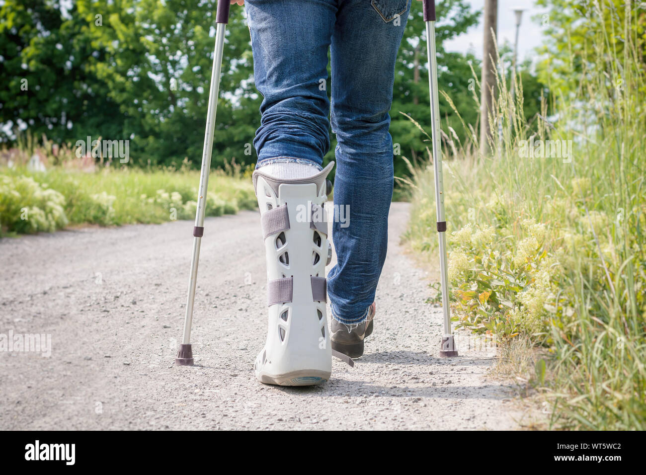 Splint in the foot area and lower leg area after rupture of the Achilles tendon Stock Photohttps://www.alamy.com/image-license-details/?v=1https://www.alamy.com/splint-in-the-foot-area-and-lower-leg-area-after-rupture-of-the-achilles-tendon-image273036962.html
RFWT5WC2–Splint in the foot area and lower leg area after rupture of the Achilles tendon
 Joseph-Benoît Suvée (Belgian - The Invention of Drawing (recto); Sketch of Lower Leg Bones of Human Skeleton (verso) - Stock Photohttps://www.alamy.com/image-license-details/?v=1https://www.alamy.com/stock-photo-joseph-benot-suve-belgian-the-invention-of-drawing-recto-sketch-of-137892605.html
RMJ09FBW–Joseph-Benoît Suvée (Belgian - The Invention of Drawing (recto); Sketch of Lower Leg Bones of Human Skeleton (verso) -
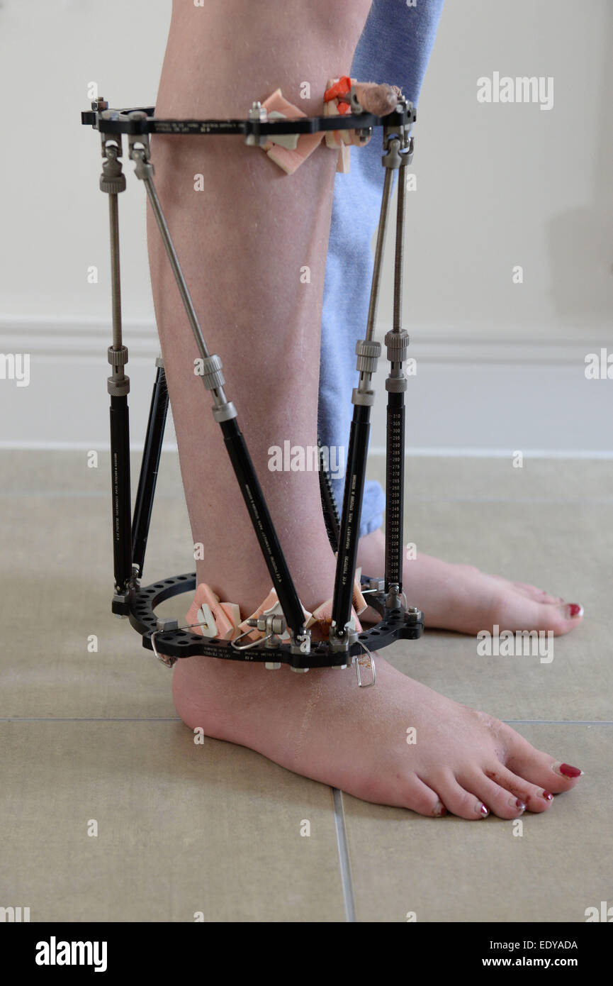 External fixator fitted to a leg to help hold it in the correct position while the bones grow. Stock Photohttps://www.alamy.com/image-license-details/?v=1https://www.alamy.com/stock-photo-external-fixator-fitted-to-a-leg-to-help-hold-it-in-the-correct-position-77432918.html
RFEDYADA–External fixator fitted to a leg to help hold it in the correct position while the bones grow.
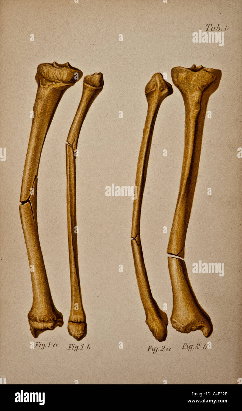 Tibia and fibula fracture circa 1902. Stock Photohttps://www.alamy.com/image-license-details/?v=1https://www.alamy.com/stock-photo-tibia-and-fibula-fracture-circa-1902-37188326.html
RFC4E22E–Tibia and fibula fracture circa 1902.
 medically accurate illustration of the lower leg bones Stock Photohttps://www.alamy.com/image-license-details/?v=1https://www.alamy.com/stock-photo-medically-accurate-illustration-of-the-lower-leg-bones-89766404.html
RFF615Y0–medically accurate illustration of the lower leg bones
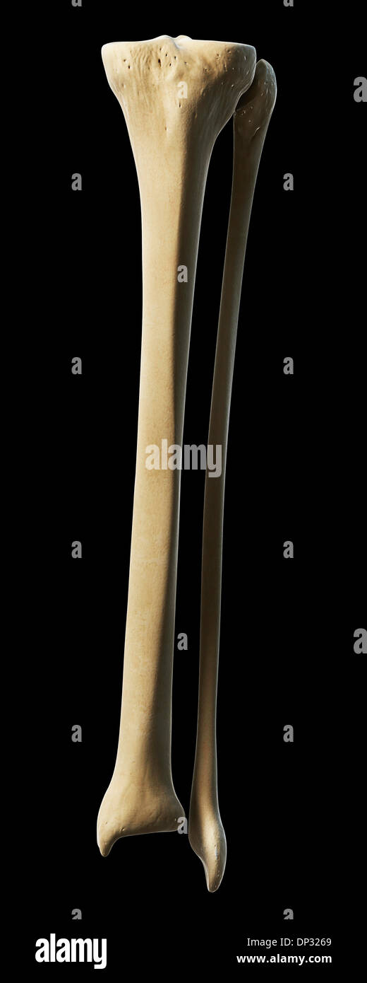 Lower leg bones, artwork Stock Photohttps://www.alamy.com/image-license-details/?v=1https://www.alamy.com/lower-leg-bones-artwork-image65221137.html
RFDP3269–Lower leg bones, artwork
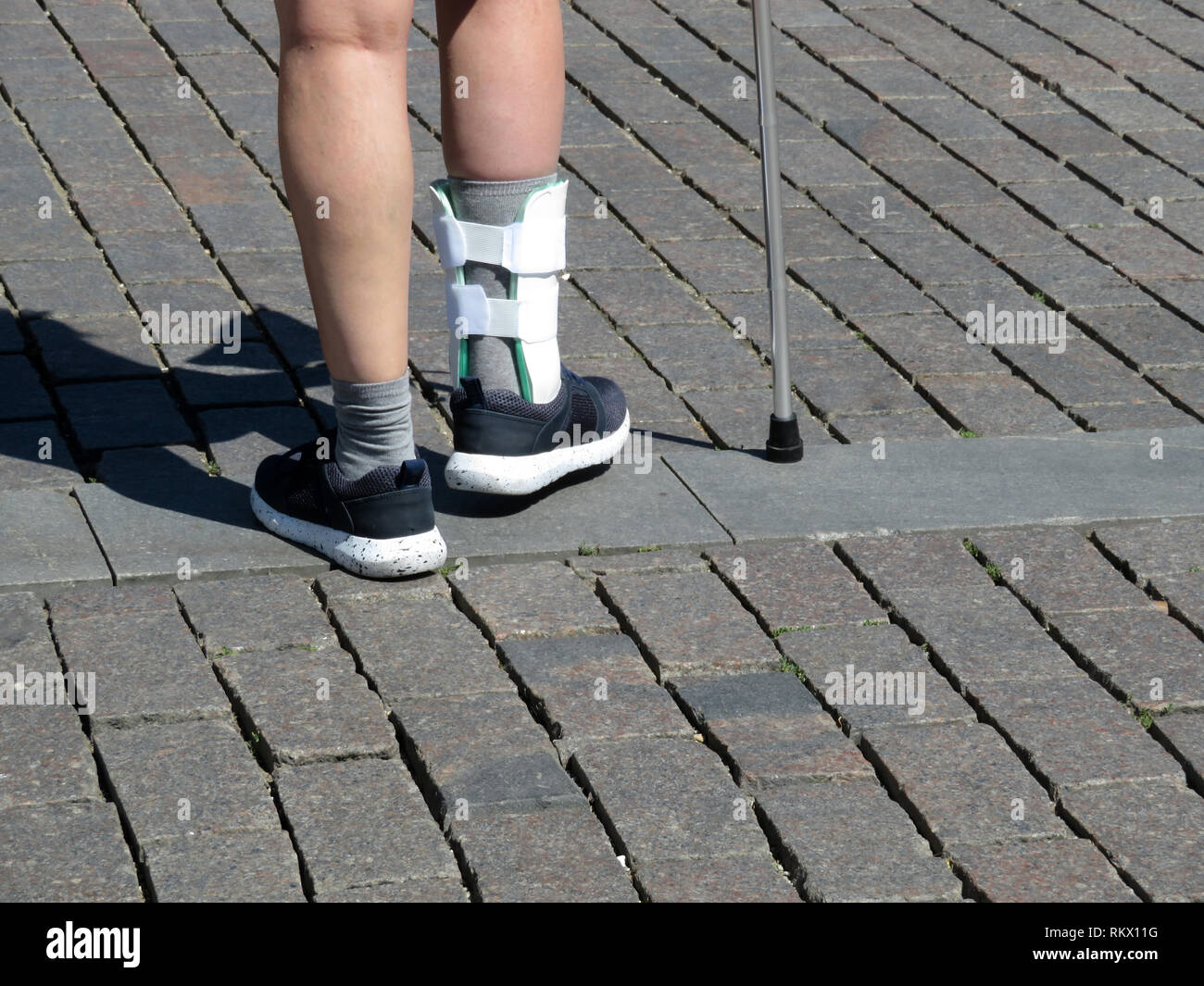 Female feet with the splint, orthosis for leg. Person with cane walking after a broken or sprain legs. Orthopedic orthosis for immobilizing foot Stock Photohttps://www.alamy.com/image-license-details/?v=1https://www.alamy.com/female-feet-with-the-splint-orthosis-for-leg-person-with-cane-walking-after-a-broken-or-sprain-legs-orthopedic-orthosis-for-immobilizing-foot-image235984828.html
RFRKX11G–Female feet with the splint, orthosis for leg. Person with cane walking after a broken or sprain legs. Orthopedic orthosis for immobilizing foot
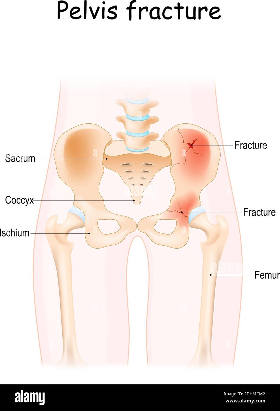 Pelvis Fracture. pelvic bones: sacrum, ilium, coccyx, pubis, ischium and femur. Vector illustration isolated on a white background Stock Vectorhttps://www.alamy.com/image-license-details/?v=1https://www.alamy.com/pelvis-fracture-pelvic-bones-sacrum-ilium-coccyx-pubis-ischium-and-femur-vector-illustration-isolated-on-a-white-background-image389526258.html
RF2DHMCM2–Pelvis Fracture. pelvic bones: sacrum, ilium, coccyx, pubis, ischium and femur. Vector illustration isolated on a white background
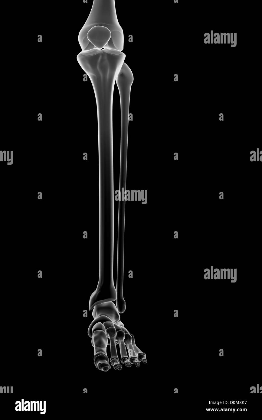 Stylized bones of the lower left leg, ankle joint and foot. Stock Photohttps://www.alamy.com/image-license-details/?v=1https://www.alamy.com/stock-photo-stylized-bones-of-the-lower-left-leg-ankle-joint-and-foot-52076955.html
RMD0M8K7–Stylized bones of the lower left leg, ankle joint and foot.
 Nurse bandages the leg. Fracture of human lower limbs. Treatment of broken bones. Impose a gypsum. Patient surgical department. The doctor's hands Stock Photohttps://www.alamy.com/image-license-details/?v=1https://www.alamy.com/nurse-bandages-the-leg-fracture-of-human-lower-limbs-treatment-of-broken-bones-impose-a-gypsum-patient-surgical-department-the-doctors-hands-image329408426.html
RM2A3WRPJ–Nurse bandages the leg. Fracture of human lower limbs. Treatment of broken bones. Impose a gypsum. Patient surgical department. The doctor's hands
 The bones of the lower limb Stock Photohttps://www.alamy.com/image-license-details/?v=1https://www.alamy.com/stock-photo-the-bones-of-the-lower-limb-13171593.html
RFACJKRP–The bones of the lower limb
 Polygonal vector illustration of anatomy of human legs. Low poly art lower limbs on a dark background. The flesh and bones of the legs anatomical art. Stock Vectorhttps://www.alamy.com/image-license-details/?v=1https://www.alamy.com/polygonal-vector-illustration-of-anatomy-of-human-legs-low-poly-art-lower-limbs-on-a-dark-background-the-flesh-and-bones-of-the-legs-anatomical-art-image468869651.html
RF2J6PT0K–Polygonal vector illustration of anatomy of human legs. Low poly art lower limbs on a dark background. The flesh and bones of the legs anatomical art.
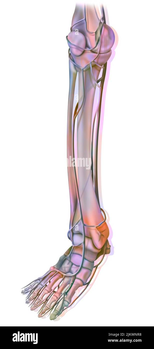 The arteries of the lower part of the lower extremity. Stock Photohttps://www.alamy.com/image-license-details/?v=1https://www.alamy.com/the-arteries-of-the-lower-part-of-the-lower-extremity-image476924316.html
RF2JKWNR8–The arteries of the lower part of the lower extremity.
 Details of an injured leg in an orthosis of a patient going to the hospital Stock Photohttps://www.alamy.com/image-license-details/?v=1https://www.alamy.com/details-of-an-injured-leg-in-an-orthosis-of-a-patient-going-to-the-hospital-image216244713.html
RFPFPP9D–Details of an injured leg in an orthosis of a patient going to the hospital
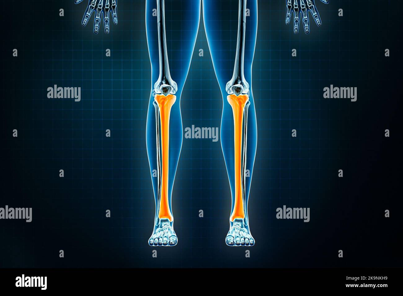 Tibia or shinbone x-ray front or anterior view. Osteology of the human skeleton, leg or lower limb bones 3D rendering illustration. Anatomy, medical, Stock Photohttps://www.alamy.com/image-license-details/?v=1https://www.alamy.com/tibia-or-shinbone-x-ray-front-or-anterior-view-osteology-of-the-human-skeleton-leg-or-lower-limb-bones-3d-rendering-illustration-anatomy-medical-image487898581.html
RF2K9NKH9–Tibia or shinbone x-ray front or anterior view. Osteology of the human skeleton, leg or lower limb bones 3D rendering illustration. Anatomy, medical,
 Senior Enlisted Advisor Tony L. Whitehead, the SEA to the Chief of the National Guard Bureau, speaks to Alaska Guardsman Senior Master Sgt. Jeremy Maddamma, a pararescueman with the Alaska Air National Guard's 212th Rescue Squadron ahead of the coin presentation at Joint Base Elmendorf - Richardson, Alaska, Feb., 1, 2023. Maddamma received a coin for his efforts in 2012 during a deployment in Afghanistan that shattered the bones in his lower left leg. He spent years in and out of a hospital, undergoing multiple surgeries and a demanding physical therapy regimen in an attempt to regain the full Stock Photohttps://www.alamy.com/image-license-details/?v=1https://www.alamy.com/senior-enlisted-advisor-tony-l-whitehead-the-sea-to-the-chief-of-the-national-guard-bureau-speaks-to-alaska-guardsman-senior-master-sgt-jeremy-maddamma-a-pararescueman-with-the-alaska-air-national-guards-212th-rescue-squadron-ahead-of-the-coin-presentation-at-joint-base-elmendorf-richardson-alaska-feb-1-2023-maddamma-received-a-coin-for-his-efforts-in-2012-during-a-deployment-in-afghanistan-that-shattered-the-bones-in-his-lower-left-leg-he-spent-years-in-and-out-of-a-hospital-undergoing-multiple-surgeries-and-a-demanding-physical-therapy-regimen-in-an-attempt-to-regain-the-full-image531901451.html
RM2NWA5MB–Senior Enlisted Advisor Tony L. Whitehead, the SEA to the Chief of the National Guard Bureau, speaks to Alaska Guardsman Senior Master Sgt. Jeremy Maddamma, a pararescueman with the Alaska Air National Guard's 212th Rescue Squadron ahead of the coin presentation at Joint Base Elmendorf - Richardson, Alaska, Feb., 1, 2023. Maddamma received a coin for his efforts in 2012 during a deployment in Afghanistan that shattered the bones in his lower left leg. He spent years in and out of a hospital, undergoing multiple surgeries and a demanding physical therapy regimen in an attempt to regain the full
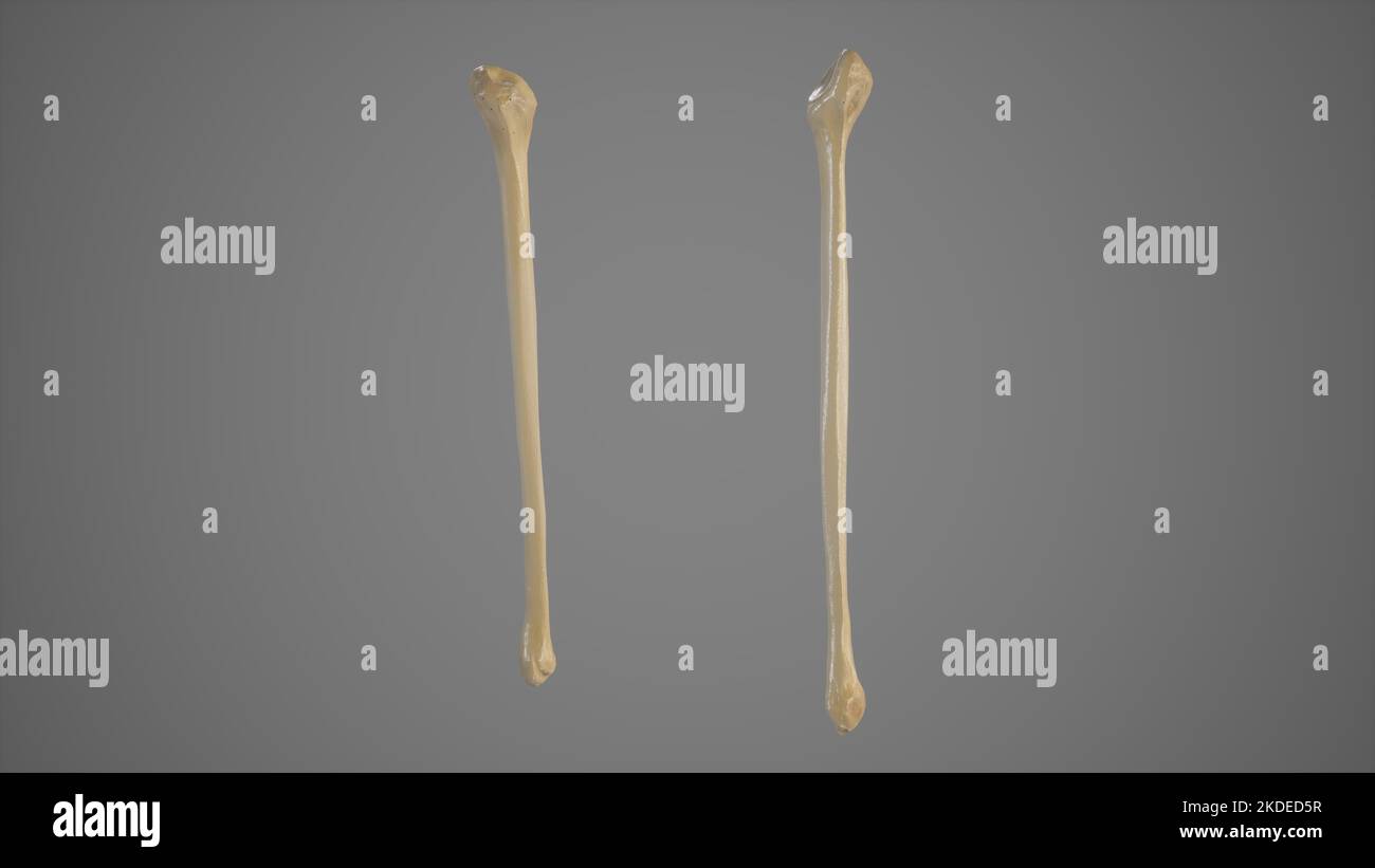 Anterior and Posterior View of Fibula Stock Photohttps://www.alamy.com/image-license-details/?v=1https://www.alamy.com/anterior-and-posterior-view-of-fibula-image490198515.html
RF2KDED5R–Anterior and Posterior View of Fibula
 Bones of the leg from the foot to the femur. Stock Photohttps://www.alamy.com/image-license-details/?v=1https://www.alamy.com/bones-of-the-leg-from-the-foot-to-the-femur-image156174230.html
RMK229R2–Bones of the leg from the foot to the femur.
 The Invention of Drawing (recto); Sketch of Lower Leg Bones of Human Skeleton (verso). Joseph-Benoît Suvée (Belgian, 1743 - 1807) Stock Photohttps://www.alamy.com/image-license-details/?v=1https://www.alamy.com/the-invention-of-drawing-recto-sketch-of-lower-leg-bones-of-human-skeleton-verso-joseph-benot-suve-belgian-1743-1807-image416591179.html
RM2F5NA8B–The Invention of Drawing (recto); Sketch of Lower Leg Bones of Human Skeleton (verso). Joseph-Benoît Suvée (Belgian, 1743 - 1807)
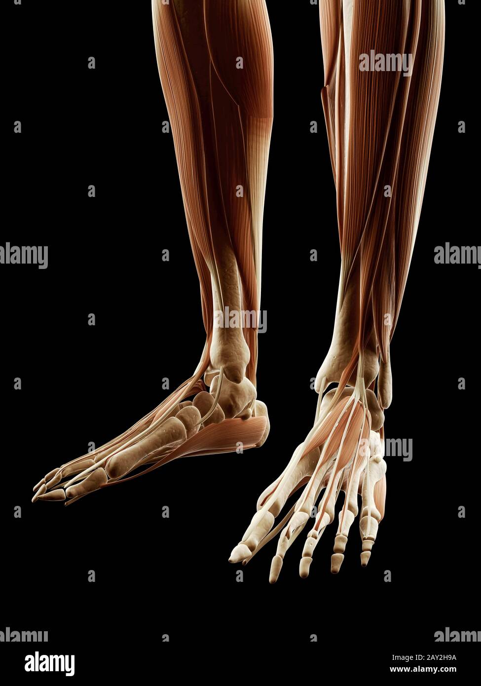 medical illustration of the leg/foot muscles Stock Photohttps://www.alamy.com/image-license-details/?v=1https://www.alamy.com/medical-illustration-of-the-legfoot-muscles-image343650198.html
RM2AY2H9A–medical illustration of the leg/foot muscles
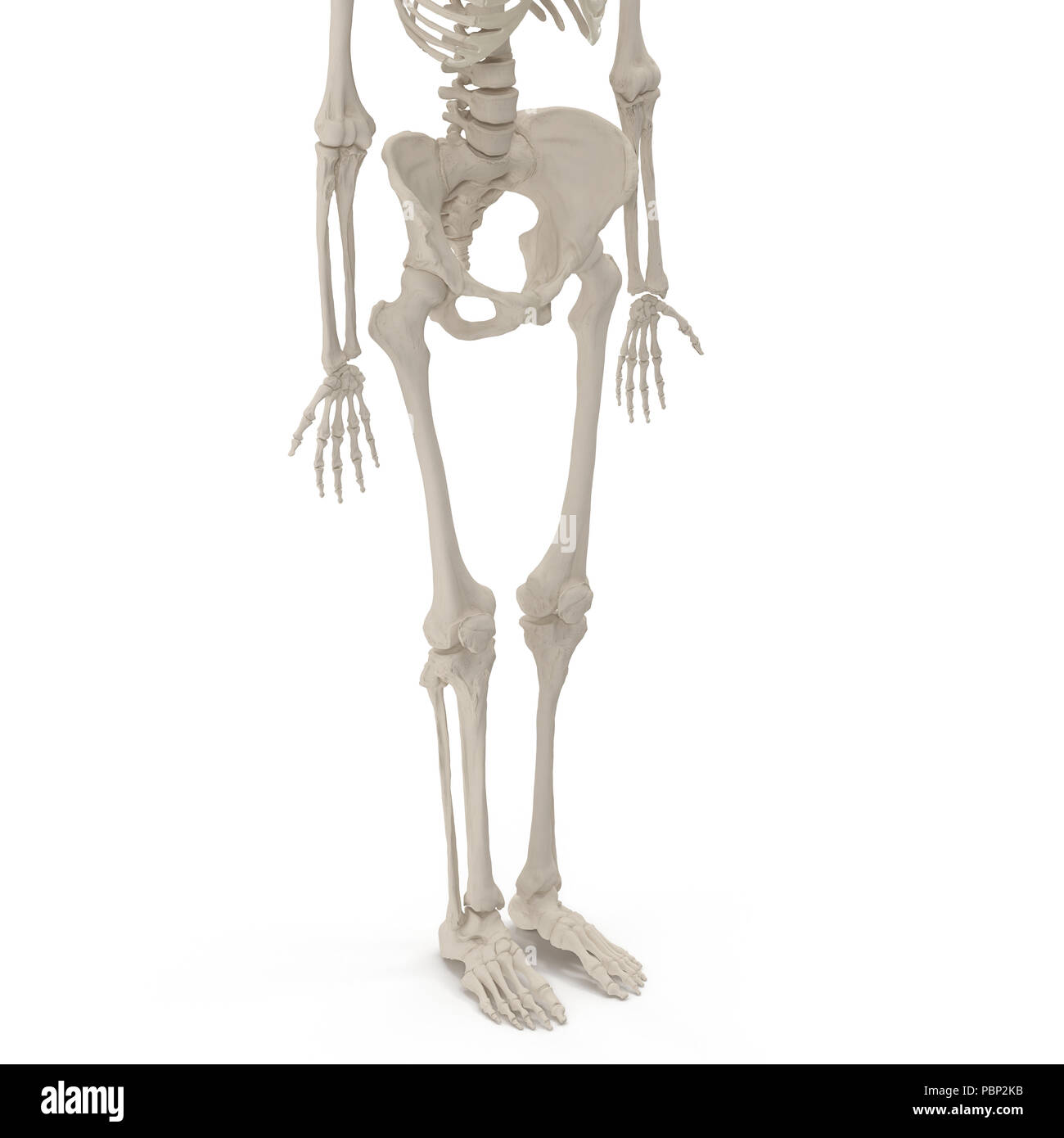 Female Lower Body Skeleton on white. 3D illustration Stock Photohttps://www.alamy.com/image-license-details/?v=1https://www.alamy.com/female-lower-body-skeleton-on-white-3d-illustration-image213770687.html
RFPBP2KB–Female Lower Body Skeleton on white. 3D illustration
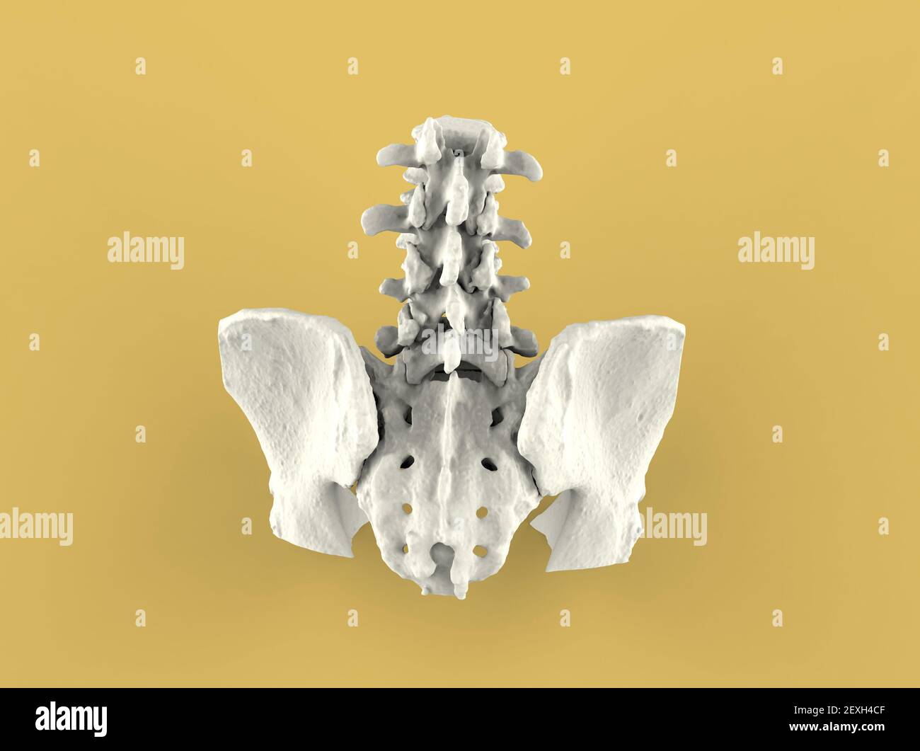 Model of human spine, lower part, 3D render Stock Photohttps://www.alamy.com/image-license-details/?v=1https://www.alamy.com/model-of-human-spine-lower-part-3d-render-image412196191.html
RF2EXH4CF–Model of human spine, lower part, 3D render
 Boiled and Baked Pork Legs with Bones Lying in Glass Stock Photohttps://www.alamy.com/image-license-details/?v=1https://www.alamy.com/boiled-and-baked-pork-legs-with-bones-lying-in-glass-image331491127.html
RF2A78M8R–Boiled and Baked Pork Legs with Bones Lying in Glass
 medically accurate illustration of the lower leg bones Stock Photohttps://www.alamy.com/image-license-details/?v=1https://www.alamy.com/stock-photo-medically-accurate-illustration-of-the-lower-leg-bones-89766405.html
RFF615Y1–medically accurate illustration of the lower leg bones
 Broken lower leg bones, computer illustration. Stock Photohttps://www.alamy.com/image-license-details/?v=1https://www.alamy.com/stock-photo-broken-lower-leg-bones-computer-illustration-92445759.html
RFFAB7E7–Broken lower leg bones, computer illustration.
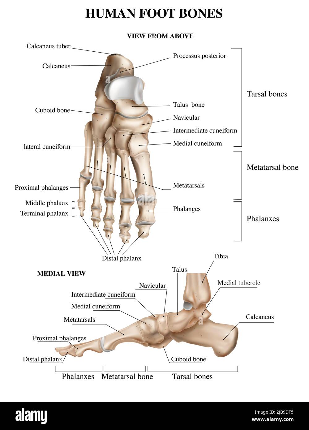 Realistic foot bones anatomy composition with front and side views of human footstep with text captions vector illustration Stock Vectorhttps://www.alamy.com/image-license-details/?v=1https://www.alamy.com/realistic-foot-bones-anatomy-composition-with-front-and-side-views-of-human-footstep-with-text-captions-vector-illustration-image471649589.html
RF2JB9DT5–Realistic foot bones anatomy composition with front and side views of human footstep with text captions vector illustration
 Biomechanics: researchers developing exoskeleton intended for people suffering from muscular weakness in the lower limbs. Fatronik Foundation, Researc Stock Photohttps://www.alamy.com/image-license-details/?v=1https://www.alamy.com/biomechanics-researchers-developing-exoskeleton-intended-for-people-suffering-from-muscular-weakness-in-the-lower-limbs-fatronik-foundation-researc-image616403319.html
RM2XPRGM7–Biomechanics: researchers developing exoskeleton intended for people suffering from muscular weakness in the lower limbs. Fatronik Foundation, Researc
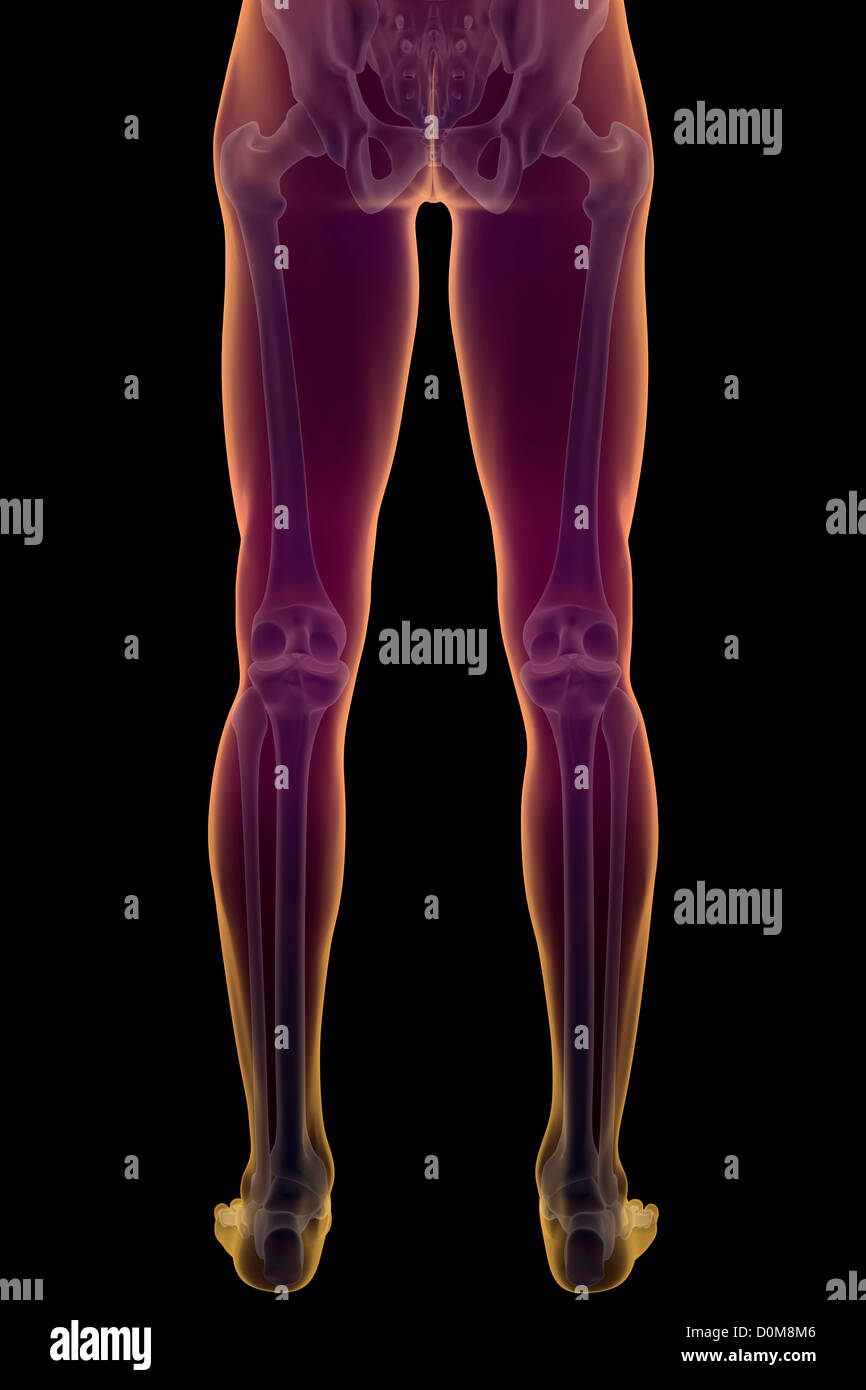 Rear view of the bones of the lower body of the male skeleton. Stock Photohttps://www.alamy.com/image-license-details/?v=1https://www.alamy.com/stock-photo-rear-view-of-the-bones-of-the-lower-body-of-the-male-skeleton-52076982.html
RMD0M8M6–Rear view of the bones of the lower body of the male skeleton.