Quick filters:
Macula retinae Stock Photos and Images
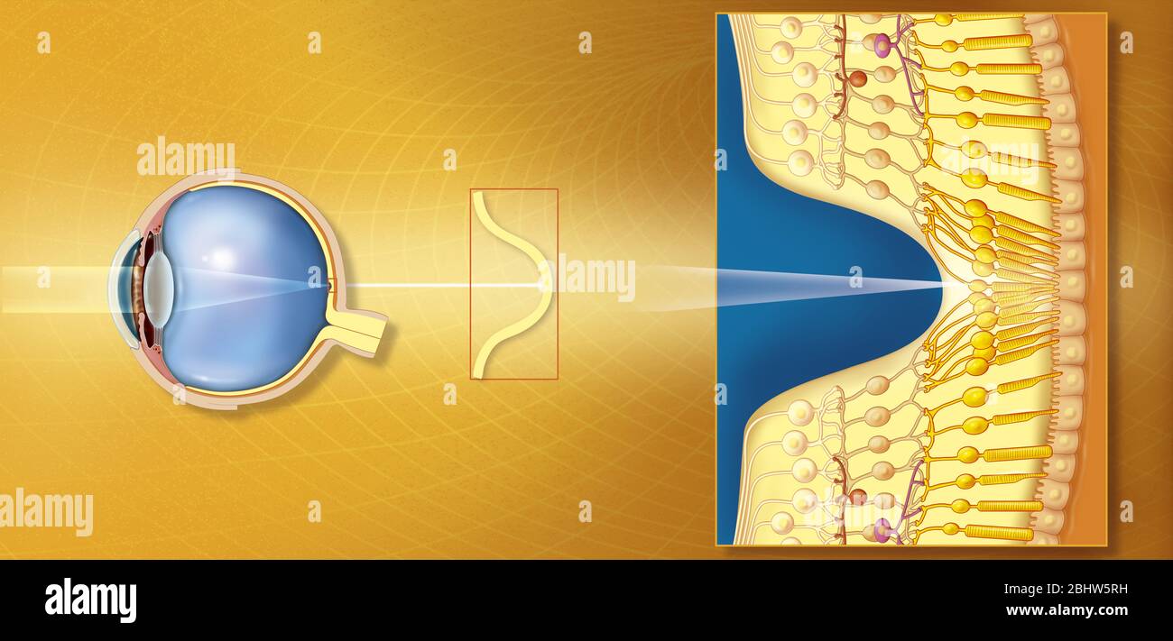 Fovea Stock Photohttps://www.alamy.com/image-license-details/?v=1https://www.alamy.com/fovea-image355209893.html
RM2BHW5RH–Fovea
 . Der Frosch; zugleich eine Einf in das praktische Studium des Wirbeltier-Krs. Frogs. — 50 buswand und wird, da sie unempfindlich gegen Licht ist, der blinde Fleck (Papilla nervi optici) genannt. Etwas dorsal von ihr liegt die Stelle des schärfsten Sehens (Area centralis retinae), welche der Macula lutea, dem gelben Fleck des Säuger-Auges entspricht. Hinter der Sehöffnung befindet sich die beinahe kugelige Linse, welche nach dem Corpus ciliare verlaufende Fasern (Zonula ciliaris s. Zinnii) in ihrer Lage halten. Den Raum zwischen der Linse und der Retina füllt der Glaskörper (Corpus vitreum) au Stock Photohttps://www.alamy.com/image-license-details/?v=1https://www.alamy.com/der-frosch-zugleich-eine-einf-in-das-praktische-studium-des-wirbeltier-krs-frogs-50-buswand-und-wird-da-sie-unempfindlich-gegen-licht-ist-der-blinde-fleck-papilla-nervi-optici-genannt-etwas-dorsal-von-ihr-liegt-die-stelle-des-schrfsten-sehens-area-centralis-retinae-welche-der-macula-lutea-dem-gelben-fleck-des-suger-auges-entspricht-hinter-der-sehffnung-befindet-sich-die-beinahe-kugelige-linse-welche-nach-dem-corpus-ciliare-verlaufende-fasern-zonula-ciliaris-s-zinnii-in-ihrer-lage-halten-den-raum-zwischen-der-linse-und-der-retina-fllt-der-glaskrper-corpus-vitreum-au-image216003867.html
RMPFBR3R–. Der Frosch; zugleich eine Einf in das praktische Studium des Wirbeltier-Krs. Frogs. — 50 buswand und wird, da sie unempfindlich gegen Licht ist, der blinde Fleck (Papilla nervi optici) genannt. Etwas dorsal von ihr liegt die Stelle des schärfsten Sehens (Area centralis retinae), welche der Macula lutea, dem gelben Fleck des Säuger-Auges entspricht. Hinter der Sehöffnung befindet sich die beinahe kugelige Linse, welche nach dem Corpus ciliare verlaufende Fasern (Zonula ciliaris s. Zinnii) in ihrer Lage halten. Den Raum zwischen der Linse und der Retina füllt der Glaskörper (Corpus vitreum) au
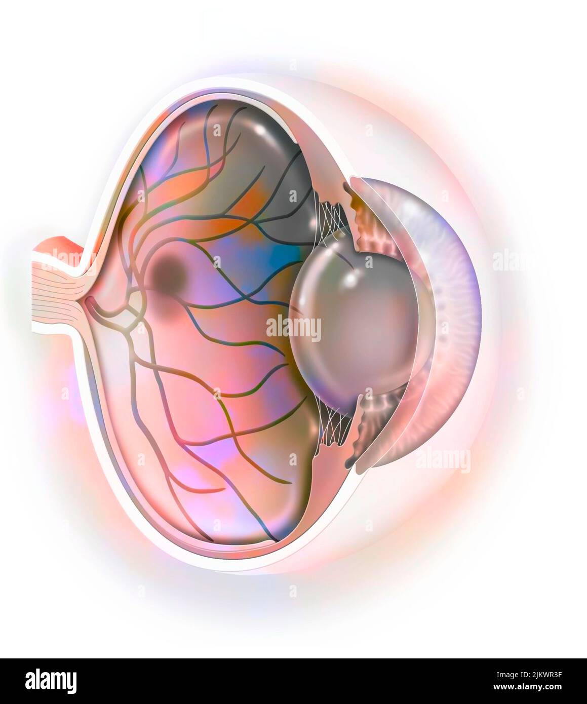 Age-related macular degeneration affecting the macula, the central area of the retina. Stock Photohttps://www.alamy.com/image-license-details/?v=1https://www.alamy.com/age-related-macular-degeneration-affecting-the-macula-the-central-area-of-the-retina-image476925331.html
RF2JKWR3F–Age-related macular degeneration affecting the macula, the central area of the retina.
 . Elements of histology. Histology. mm. -J} f-fC-, ^r ^ ;. Fig. 270.âDiagram of the Ner- Fi^ voiis Elements of the Retina. â¢271.âDiagram of the Connective- Tissue Substance of the same. « xro,.- fihrps â 1 â¢'in^'lion ceUs:4, inner molecular layer; 5. inner nuclear - ';lver:^6.omermole6u^rl^y^ riourernuclearlaver; 8. membranahmitans externa; 9, rods and cones. (Jfojc Schultze.) papiUa nervi optici, (b) the macula lutea and fovea centralis retinae, and (c) the ora serrata of the retina. (a) The papilla nervi optici, or the blind spot of. Please note that these images are extracted from scann Stock Photohttps://www.alamy.com/image-license-details/?v=1https://www.alamy.com/elements-of-histology-histology-mm-j-f-fc-r-fig-270diagram-of-the-ner-fi-voiis-elements-of-the-retina-271diagram-of-the-connective-tissue-substance-of-the-same-xro-fihrps-1-inlion-ceus4-inner-molecular-layer-5-inner-nuclear-lver6omermole6urly-riourernuclearlaver-8-membranahmitans-externa-9-rods-and-cones-jfojc-schultze-papiua-nervi-optici-b-the-macula-lutea-and-fovea-centralis-retinae-and-c-the-ora-serrata-of-the-retina-a-the-papilla-nervi-optici-or-the-blind-spot-of-please-note-that-these-images-are-extracted-from-scann-image231473352.html
RMRCGEHC–. Elements of histology. Histology. mm. -J} f-fC-, ^r ^ ;. Fig. 270.âDiagram of the Ner- Fi^ voiis Elements of the Retina. â¢271.âDiagram of the Connective- Tissue Substance of the same. « xro,.- fihrps â 1 â¢'in^'lion ceUs:4, inner molecular layer; 5. inner nuclear - ';lver:^6.omermole6u^rl^y^ riourernuclearlaver; 8. membranahmitans externa; 9, rods and cones. (Jfojc Schultze.) papiUa nervi optici, (b) the macula lutea and fovea centralis retinae, and (c) the ora serrata of the retina. (a) The papilla nervi optici, or the blind spot of. Please note that these images are extracted from scann
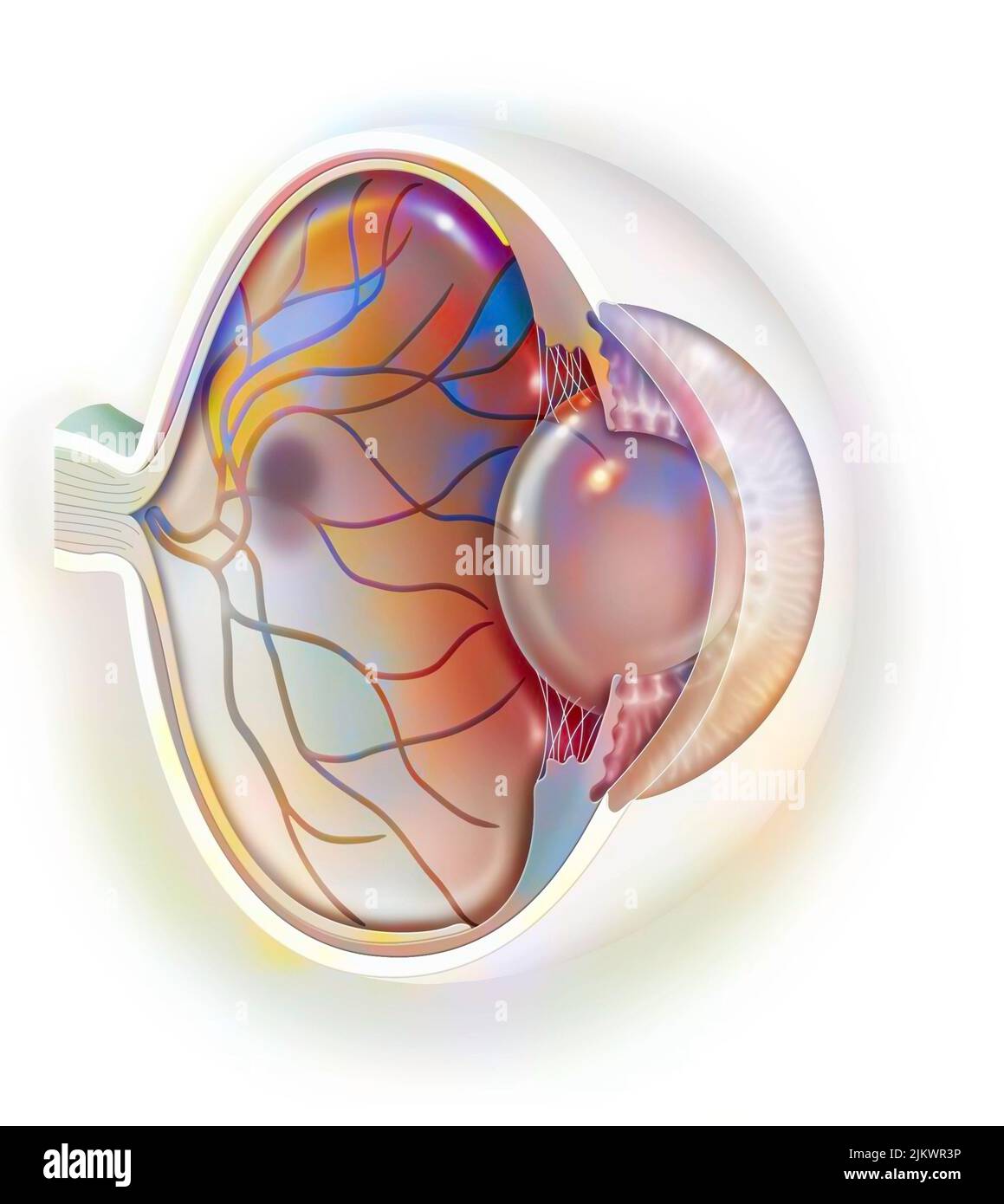 Age-related macular degeneration affecting the macula, the central area of the retina. Stock Photohttps://www.alamy.com/image-license-details/?v=1https://www.alamy.com/age-related-macular-degeneration-affecting-the-macula-the-central-area-of-the-retina-image476925338.html
RF2JKWR3P–Age-related macular degeneration affecting the macula, the central area of the retina.
![. Elements of histology. Histology. Chap. XXXIX.] The Retina. 309 435. From this arrangement is excepted—(a) the papilla nervi optici, (b) the macula lutea and fovea centralis retinae, and (c) the ora serrata of the retina. (a) The papilla nervi optici, or the blind spot of. Fig. 158.—Diagram of the Ner- Fig. 159.—Diagram of the Connective vous Elements of the Eetina. Tissue Substance of the same. 2, Nerve-fibres; 3, ganglion cells; 4, inner molecular layer; 5, inner nuclear layer; 6, outer molecular layer; 7, outer nuclear layer; 8, the membrana limitans externa; 9, the rods and cones. (Max S Stock Photo . Elements of histology. Histology. Chap. XXXIX.] The Retina. 309 435. From this arrangement is excepted—(a) the papilla nervi optici, (b) the macula lutea and fovea centralis retinae, and (c) the ora serrata of the retina. (a) The papilla nervi optici, or the blind spot of. Fig. 158.—Diagram of the Ner- Fig. 159.—Diagram of the Connective vous Elements of the Eetina. Tissue Substance of the same. 2, Nerve-fibres; 3, ganglion cells; 4, inner molecular layer; 5, inner nuclear layer; 6, outer molecular layer; 7, outer nuclear layer; 8, the membrana limitans externa; 9, the rods and cones. (Max S Stock Photo](https://c8.alamy.com/comp/RCHAEM/elements-of-histology-histology-chap-xxxix-the-retina-309-435-from-this-arrangement-is-excepteda-the-papilla-nervi-optici-b-the-macula-lutea-and-fovea-centralis-retinae-and-c-the-ora-serrata-of-the-retina-a-the-papilla-nervi-optici-or-the-blind-spot-of-fig-158diagram-of-the-ner-fig-159diagram-of-the-connective-vous-elements-of-the-eetina-tissue-substance-of-the-same-2-nerve-fibres-3-ganglion-cells-4-inner-molecular-layer-5-inner-nuclear-layer-6-outer-molecular-layer-7-outer-nuclear-layer-8-the-membrana-limitans-externa-9-the-rods-and-cones-max-s-RCHAEM.jpg) . Elements of histology. Histology. Chap. XXXIX.] The Retina. 309 435. From this arrangement is excepted—(a) the papilla nervi optici, (b) the macula lutea and fovea centralis retinae, and (c) the ora serrata of the retina. (a) The papilla nervi optici, or the blind spot of. Fig. 158.—Diagram of the Ner- Fig. 159.—Diagram of the Connective vous Elements of the Eetina. Tissue Substance of the same. 2, Nerve-fibres; 3, ganglion cells; 4, inner molecular layer; 5, inner nuclear layer; 6, outer molecular layer; 7, outer nuclear layer; 8, the membrana limitans externa; 9, the rods and cones. (Max S Stock Photohttps://www.alamy.com/image-license-details/?v=1https://www.alamy.com/elements-of-histology-histology-chap-xxxix-the-retina-309-435-from-this-arrangement-is-excepteda-the-papilla-nervi-optici-b-the-macula-lutea-and-fovea-centralis-retinae-and-c-the-ora-serrata-of-the-retina-a-the-papilla-nervi-optici-or-the-blind-spot-of-fig-158diagram-of-the-ner-fig-159diagram-of-the-connective-vous-elements-of-the-eetina-tissue-substance-of-the-same-2-nerve-fibres-3-ganglion-cells-4-inner-molecular-layer-5-inner-nuclear-layer-6-outer-molecular-layer-7-outer-nuclear-layer-8-the-membrana-limitans-externa-9-the-rods-and-cones-max-s-image231492092.html
. Elements of histology. Histology. Chap. XXXIX.] The Retina. 309 435. From this arrangement is excepted—(a) the papilla nervi optici, (b) the macula lutea and fovea centralis retinae, and (c) the ora serrata of the retina. (a) The papilla nervi optici, or the blind spot of. Fig. 158.—Diagram of the Ner- Fig. 159.—Diagram of the Connective vous Elements of the Eetina. Tissue Substance of the same. 2, Nerve-fibres; 3, ganglion cells; 4, inner molecular layer; 5, inner nuclear layer; 6, outer molecular layer; 7, outer nuclear layer; 8, the membrana limitans externa; 9, the rods and cones. (Max S Stock Photohttps://www.alamy.com/image-license-details/?v=1https://www.alamy.com/elements-of-histology-histology-chap-xxxix-the-retina-309-435-from-this-arrangement-is-excepteda-the-papilla-nervi-optici-b-the-macula-lutea-and-fovea-centralis-retinae-and-c-the-ora-serrata-of-the-retina-a-the-papilla-nervi-optici-or-the-blind-spot-of-fig-158diagram-of-the-ner-fig-159diagram-of-the-connective-vous-elements-of-the-eetina-tissue-substance-of-the-same-2-nerve-fibres-3-ganglion-cells-4-inner-molecular-layer-5-inner-nuclear-layer-6-outer-molecular-layer-7-outer-nuclear-layer-8-the-membrana-limitans-externa-9-the-rods-and-cones-max-s-image231492092.htmlRMRCHAEM–. Elements of histology. Histology. Chap. XXXIX.] The Retina. 309 435. From this arrangement is excepted—(a) the papilla nervi optici, (b) the macula lutea and fovea centralis retinae, and (c) the ora serrata of the retina. (a) The papilla nervi optici, or the blind spot of. Fig. 158.—Diagram of the Ner- Fig. 159.—Diagram of the Connective vous Elements of the Eetina. Tissue Substance of the same. 2, Nerve-fibres; 3, ganglion cells; 4, inner molecular layer; 5, inner nuclear layer; 6, outer molecular layer; 7, outer nuclear layer; 8, the membrana limitans externa; 9, the rods and cones. (Max S
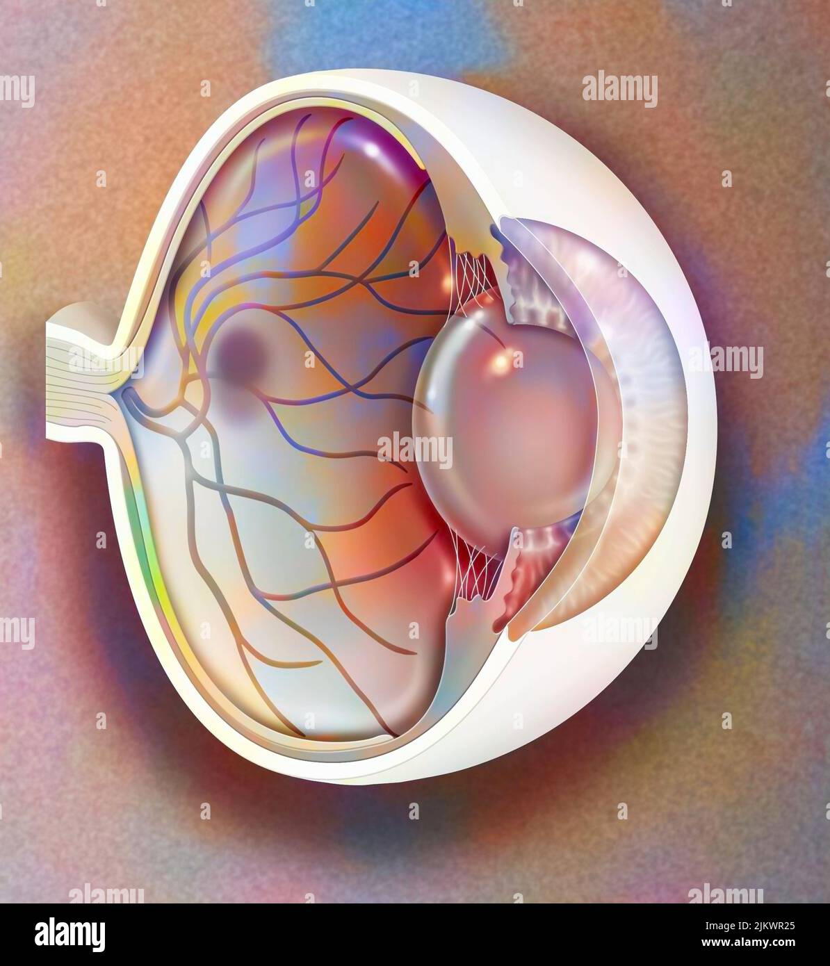 Age-related macular degeneration affecting the macula, the central area of the retina. Stock Photohttps://www.alamy.com/image-license-details/?v=1https://www.alamy.com/age-related-macular-degeneration-affecting-the-macula-the-central-area-of-the-retina-image476925293.html
RF2JKWR25–Age-related macular degeneration affecting the macula, the central area of the retina.
 . Der Frosch; zugleich eine Einf in das praktische Studium des Wirbeltier-Krs. Frogs. — 50 buswand und wird, da sie unempfindlich gegen Licht ist, der blinde Fleck (Papilla nervi optici) genannt. Etwas dorsal von ihr liegt die Stelle des schärfsten Sehens (Area centralis retinae), welche der Macula lutea, dem gelben Fleck des Säuger-Auges entspricht. Hinter der Sehöffnung befindet sich die beinahe kugelige Linse, welche nach dem Corpus ciliare verlaufende Fasern (Zonula ciliaris s. Zinnii) in ihrer Lage halten. Den Raum zwischen der Linse und der Retina füllt der Glaskörper (Corpus vitreum) au Stock Photohttps://www.alamy.com/image-license-details/?v=1https://www.alamy.com/der-frosch-zugleich-eine-einf-in-das-praktische-studium-des-wirbeltier-krs-frogs-50-buswand-und-wird-da-sie-unempfindlich-gegen-licht-ist-der-blinde-fleck-papilla-nervi-optici-genannt-etwas-dorsal-von-ihr-liegt-die-stelle-des-schrfsten-sehens-area-centralis-retinae-welche-der-macula-lutea-dem-gelben-fleck-des-suger-auges-entspricht-hinter-der-sehffnung-befindet-sich-die-beinahe-kugelige-linse-welche-nach-dem-corpus-ciliare-verlaufende-fasern-zonula-ciliaris-s-zinnii-in-ihrer-lage-halten-den-raum-zwischen-der-linse-und-der-retina-fllt-der-glaskrper-corpus-vitreum-au-image231712497.html
RMRCYBJ9–. Der Frosch; zugleich eine Einf in das praktische Studium des Wirbeltier-Krs. Frogs. — 50 buswand und wird, da sie unempfindlich gegen Licht ist, der blinde Fleck (Papilla nervi optici) genannt. Etwas dorsal von ihr liegt die Stelle des schärfsten Sehens (Area centralis retinae), welche der Macula lutea, dem gelben Fleck des Säuger-Auges entspricht. Hinter der Sehöffnung befindet sich die beinahe kugelige Linse, welche nach dem Corpus ciliare verlaufende Fasern (Zonula ciliaris s. Zinnii) in ihrer Lage halten. Den Raum zwischen der Linse und der Retina füllt der Glaskörper (Corpus vitreum) au
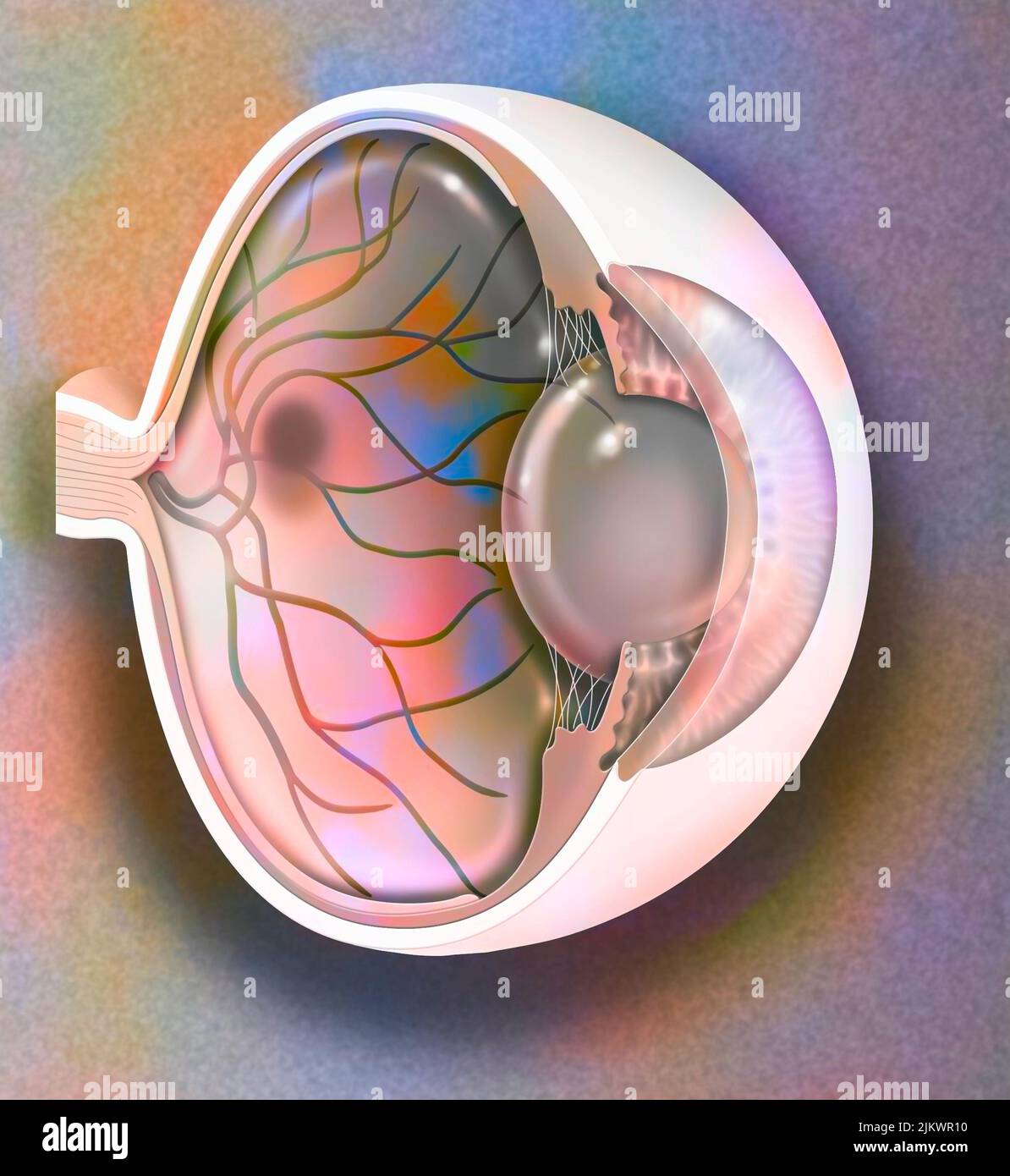 Age-related macular degeneration affecting the macula, the central area of the retina. Stock Photohttps://www.alamy.com/image-license-details/?v=1https://www.alamy.com/age-related-macular-degeneration-affecting-the-macula-the-central-area-of-the-retina-image476925260.html
RF2JKWR10–Age-related macular degeneration affecting the macula, the central area of the retina.
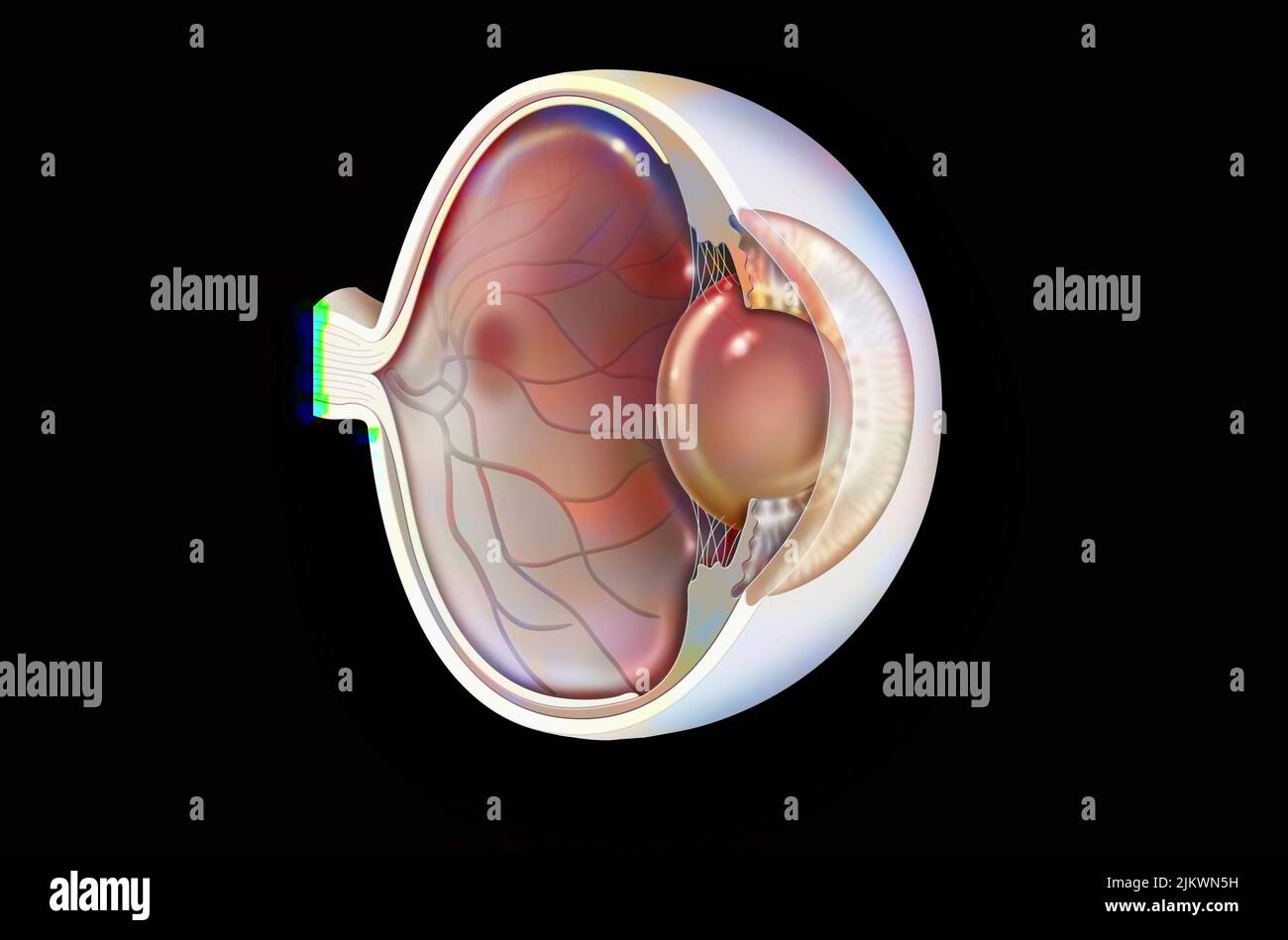 Eye: age-related macular degeneration (degenerative disease of the macula). Stock Photohttps://www.alamy.com/image-license-details/?v=1https://www.alamy.com/eye-age-related-macular-degeneration-degenerative-disease-of-the-macula-image476923821.html
RF2JKWN5H–Eye: age-related macular degeneration (degenerative disease of the macula).
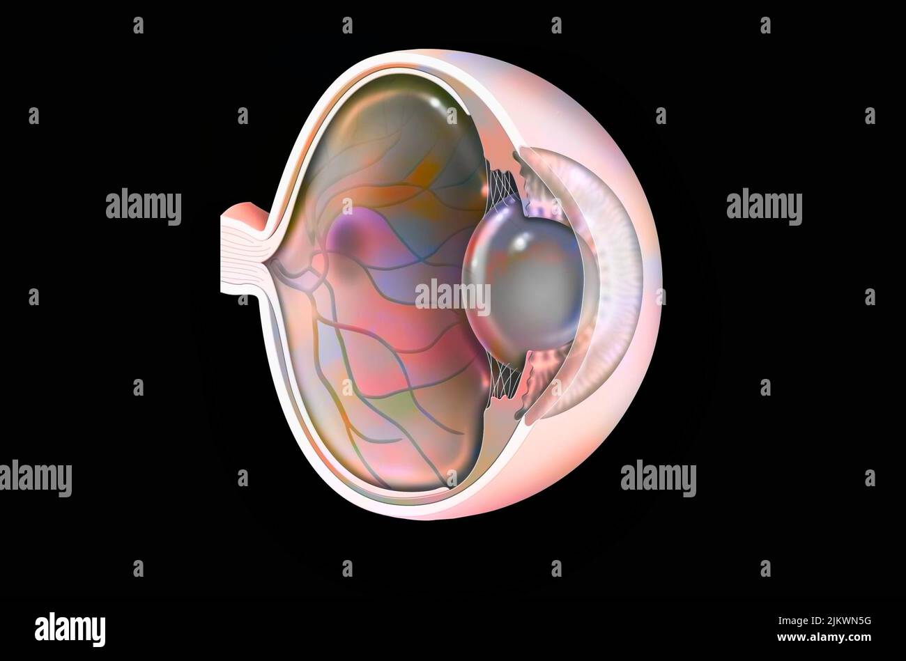 Eye: age-related macular degeneration (degenerative disease of the macula). Stock Photohttps://www.alamy.com/image-license-details/?v=1https://www.alamy.com/eye-age-related-macular-degeneration-degenerative-disease-of-the-macula-image476923820.html
RF2JKWN5G–Eye: age-related macular degeneration (degenerative disease of the macula).
 Cutaway eye: macular degeneration (opaque red spot on the macula). Stock Photohttps://www.alamy.com/image-license-details/?v=1https://www.alamy.com/cutaway-eye-macular-degeneration-opaque-red-spot-on-the-macula-image476923985.html
RF2JKWNBD–Cutaway eye: macular degeneration (opaque red spot on the macula).
 Cutaway eye: macular degeneration (opaque red spot on the macula). Stock Photohttps://www.alamy.com/image-license-details/?v=1https://www.alamy.com/cutaway-eye-macular-degeneration-opaque-red-spot-on-the-macula-image476923968.html
RF2JKWNAT–Cutaway eye: macular degeneration (opaque red spot on the macula).
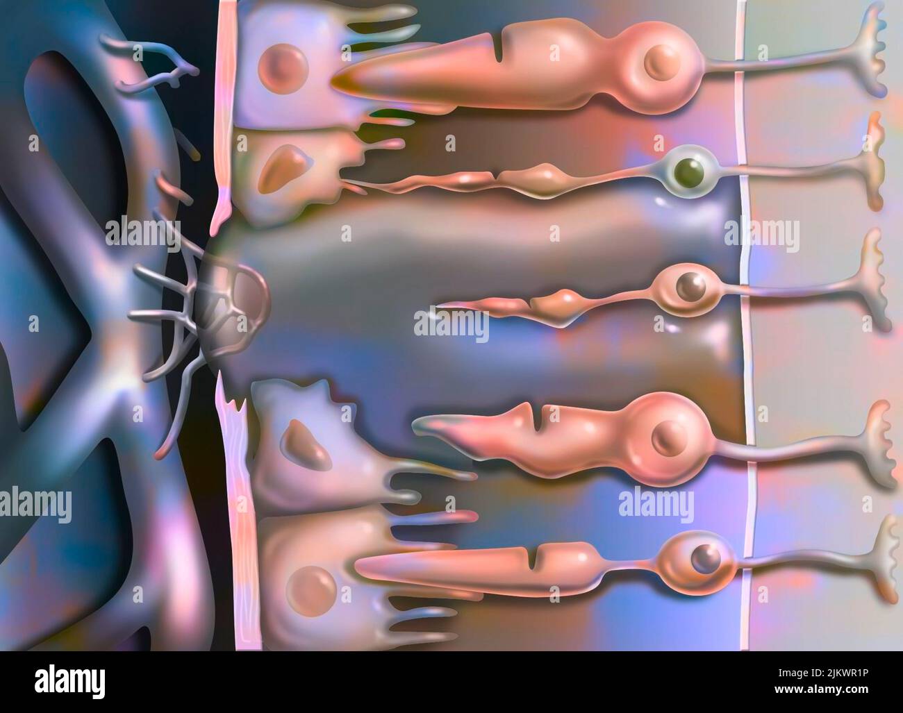 Zoom in on age-related macular degeneration affecting the macula, the central area of the retina. Stock Photohttps://www.alamy.com/image-license-details/?v=1https://www.alamy.com/zoom-in-on-age-related-macular-degeneration-affecting-the-macula-the-central-area-of-the-retina-image476925282.html
RF2JKWR1P–Zoom in on age-related macular degeneration affecting the macula, the central area of the retina.
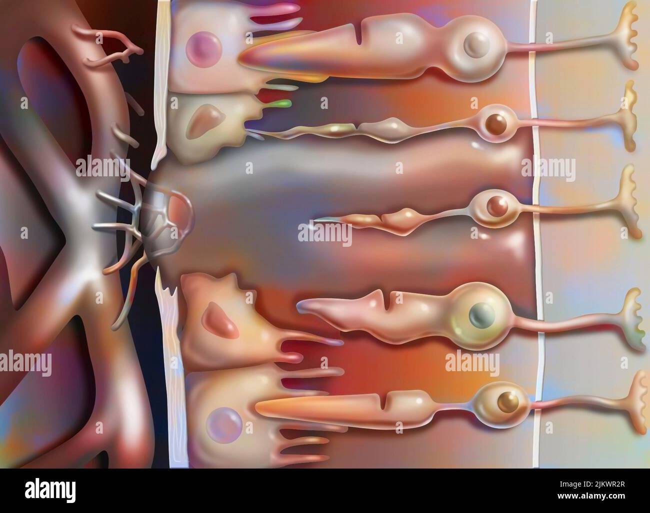 Zoom in on age-related macular degeneration affecting the macula, the central area of the retina. Stock Photohttps://www.alamy.com/image-license-details/?v=1https://www.alamy.com/zoom-in-on-age-related-macular-degeneration-affecting-the-macula-the-central-area-of-the-retina-image476925311.html
RF2JKWR2R–Zoom in on age-related macular degeneration affecting the macula, the central area of the retina.
 Eye showing the retina and the fovea. Stock Photohttps://www.alamy.com/image-license-details/?v=1https://www.alamy.com/eye-showing-the-retina-and-the-fovea-image476925436.html
RF2JKWR78–Eye showing the retina and the fovea.
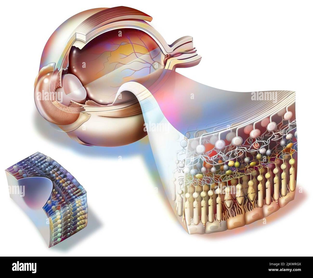 Structure of an eye with zoom on the retina and fovea. Stock Photohttps://www.alamy.com/image-license-details/?v=1https://www.alamy.com/structure-of-an-eye-with-zoom-on-the-retina-and-fovea-image476925706.html
RF2JKWRGX–Structure of an eye with zoom on the retina and fovea.
 Eye showing the retina and the fovea. Stock Photohttps://www.alamy.com/image-license-details/?v=1https://www.alamy.com/eye-showing-the-retina-and-the-fovea-image476925443.html
RF2JKWR7F–Eye showing the retina and the fovea.
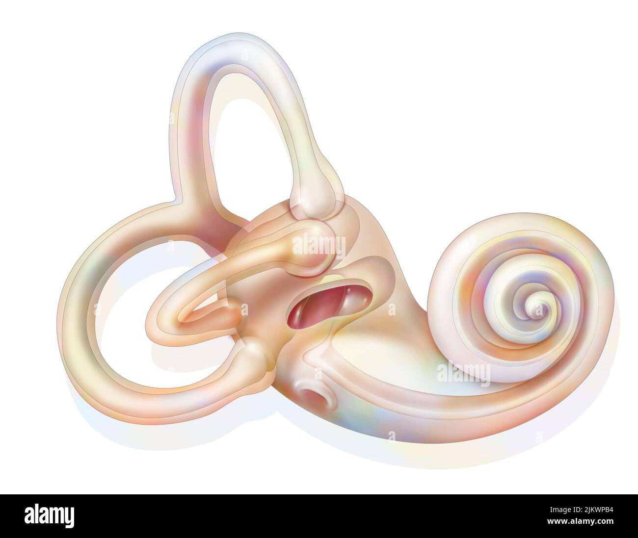 Anatomy of the inner ear showing the macule. Stock Photohttps://www.alamy.com/image-license-details/?v=1https://www.alamy.com/anatomy-of-the-inner-ear-showing-the-macule-image476924760.html
RF2JKWPB4–Anatomy of the inner ear showing the macule.
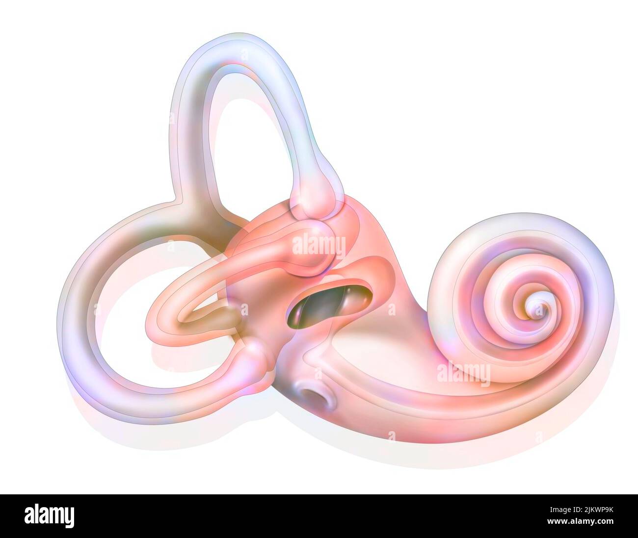 Anatomy of the inner ear showing the macule. Stock Photohttps://www.alamy.com/image-license-details/?v=1https://www.alamy.com/anatomy-of-the-inner-ear-showing-the-macule-image476924719.html
RF2JKWP9K–Anatomy of the inner ear showing the macule.
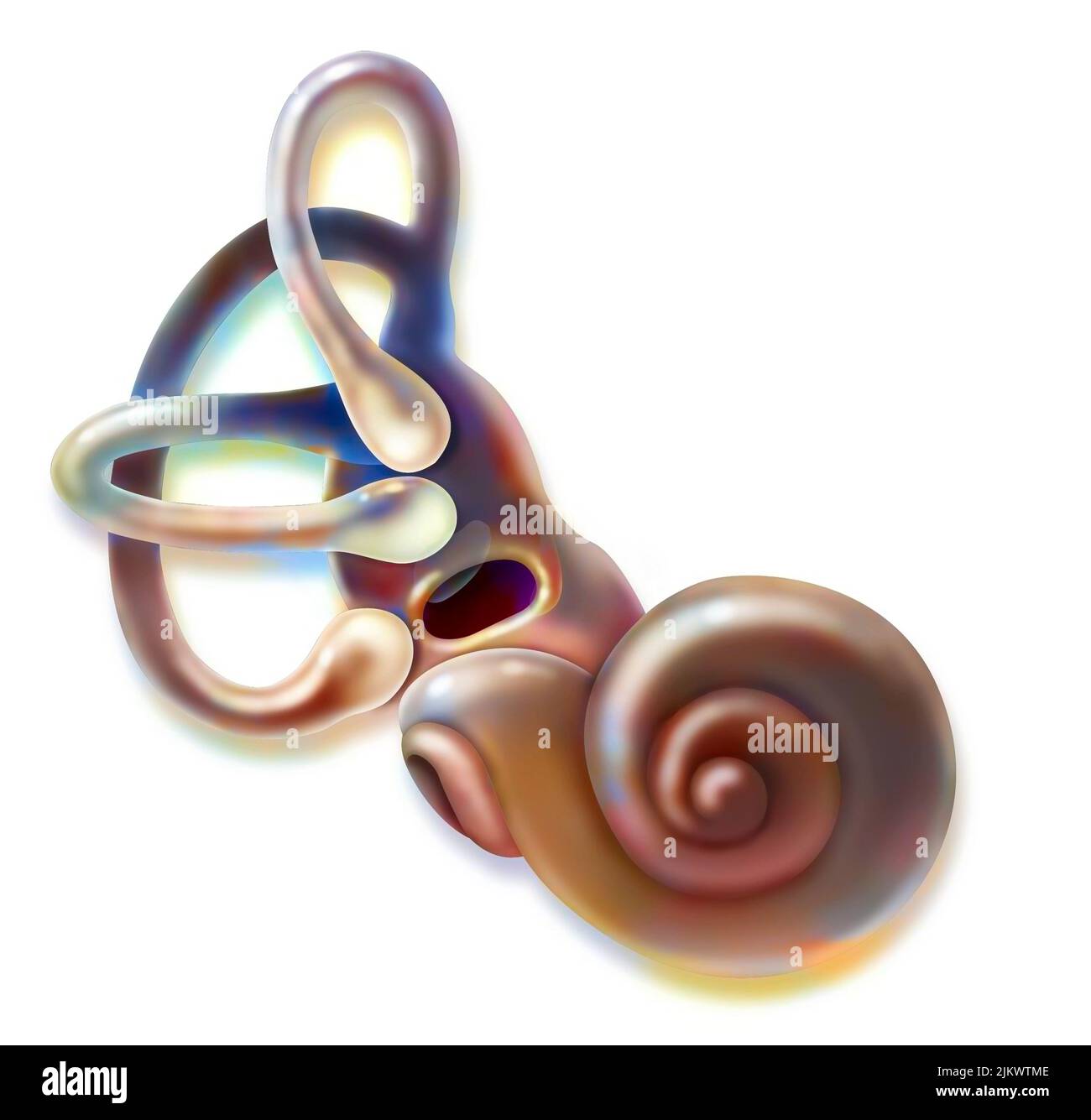 Inner ear and vestibular apparatus with semicircular canals, macule. Stock Photohttps://www.alamy.com/image-license-details/?v=1https://www.alamy.com/inner-ear-and-vestibular-apparatus-with-semicircular-canals-macule-image476926590.html
RF2JKWTME–Inner ear and vestibular apparatus with semicircular canals, macule.
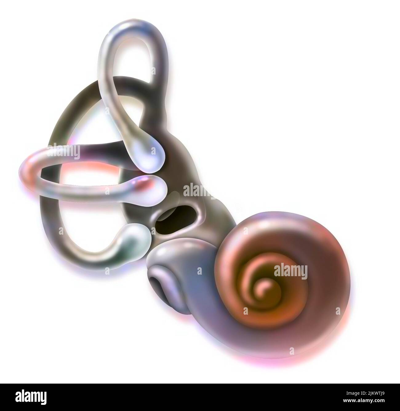 Inner ear and vestibular apparatus with semicircular canals, macule. Stock Photohttps://www.alamy.com/image-license-details/?v=1https://www.alamy.com/inner-ear-and-vestibular-apparatus-with-semicircular-canals-macule-image476926529.html
RF2JKWTJ9–Inner ear and vestibular apparatus with semicircular canals, macule.
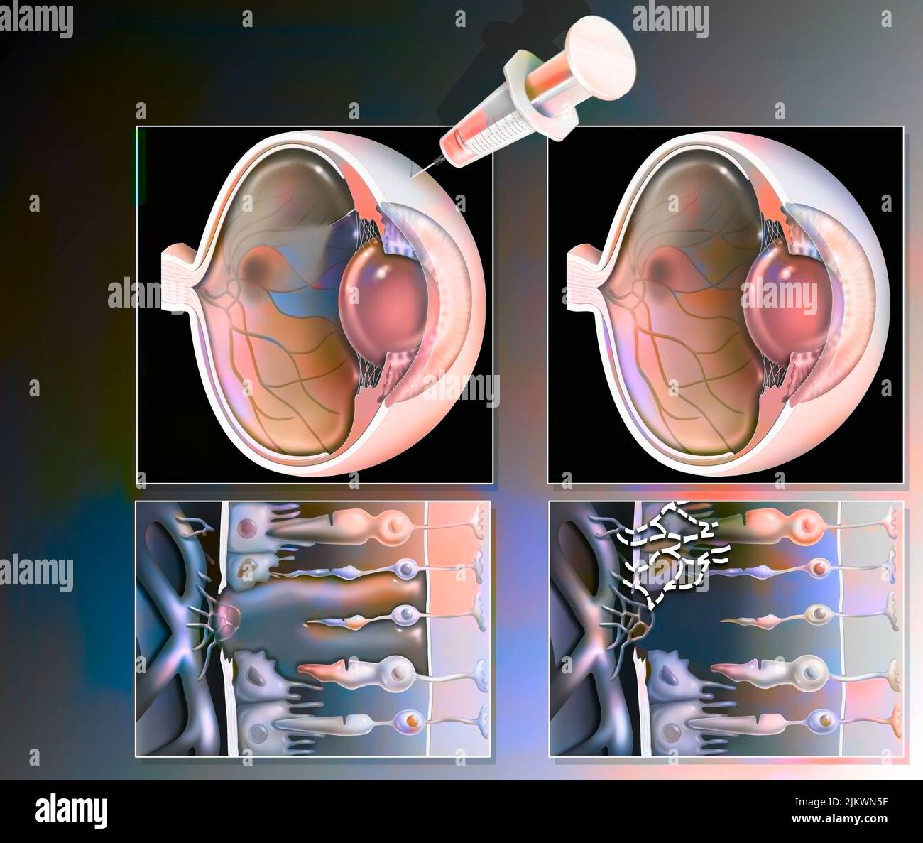 Eye: treatment of macular degeneration by RNA interference. Stock Photohttps://www.alamy.com/image-license-details/?v=1https://www.alamy.com/eye-treatment-of-macular-degeneration-by-rna-interference-image476923819.html
RF2JKWN5F–Eye: treatment of macular degeneration by RNA interference.
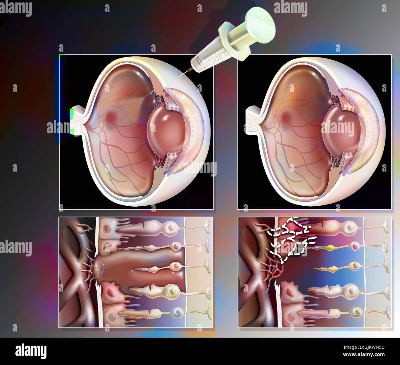 Eye: treatment of macular degeneration by RNA interference. Stock Photohttps://www.alamy.com/image-license-details/?v=1https://www.alamy.com/eye-treatment-of-macular-degeneration-by-rna-interference-image476923817.html
RF2JKWN5D–Eye: treatment of macular degeneration by RNA interference.
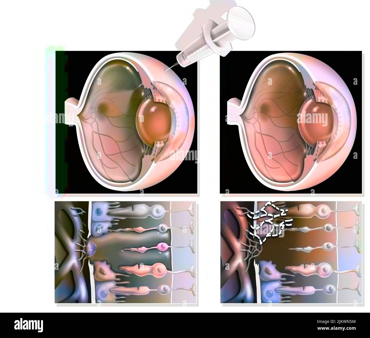 Eye: treatment of macular degeneration by RNA interference. Stock Photohttps://www.alamy.com/image-license-details/?v=1https://www.alamy.com/eye-treatment-of-macular-degeneration-by-rna-interference-image476923824.html
RF2JKWN5M–Eye: treatment of macular degeneration by RNA interference.
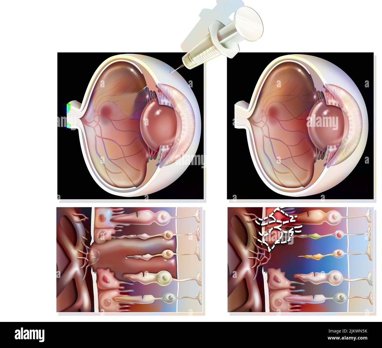 Eye: treatment of macular degeneration by RNA interference. Stock Photohttps://www.alamy.com/image-license-details/?v=1https://www.alamy.com/eye-treatment-of-macular-degeneration-by-rna-interference-image476923823.html
RF2JKWN5K–Eye: treatment of macular degeneration by RNA interference.
 Eye: treatment of macular degeneration by RNA interference. Stock Photohttps://www.alamy.com/image-license-details/?v=1https://www.alamy.com/eye-treatment-of-macular-degeneration-by-rna-interference-image476923932.html
RF2JKWN9G–Eye: treatment of macular degeneration by RNA interference.
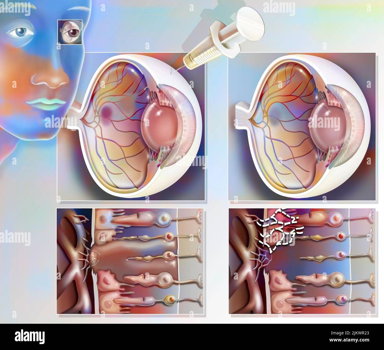 Treatment of macular degeneration by injection of RNA interference. Stock Photohttps://www.alamy.com/image-license-details/?v=1https://www.alamy.com/treatment-of-macular-degeneration-by-injection-of-rna-interference-image476925291.html
RF2JKWR23–Treatment of macular degeneration by injection of RNA interference.
 Eye: treatment of macular degeneration by RNA interference. Stock Photohttps://www.alamy.com/image-license-details/?v=1https://www.alamy.com/eye-treatment-of-macular-degeneration-by-rna-interference-image476923929.html
RF2JKWN9D–Eye: treatment of macular degeneration by RNA interference.
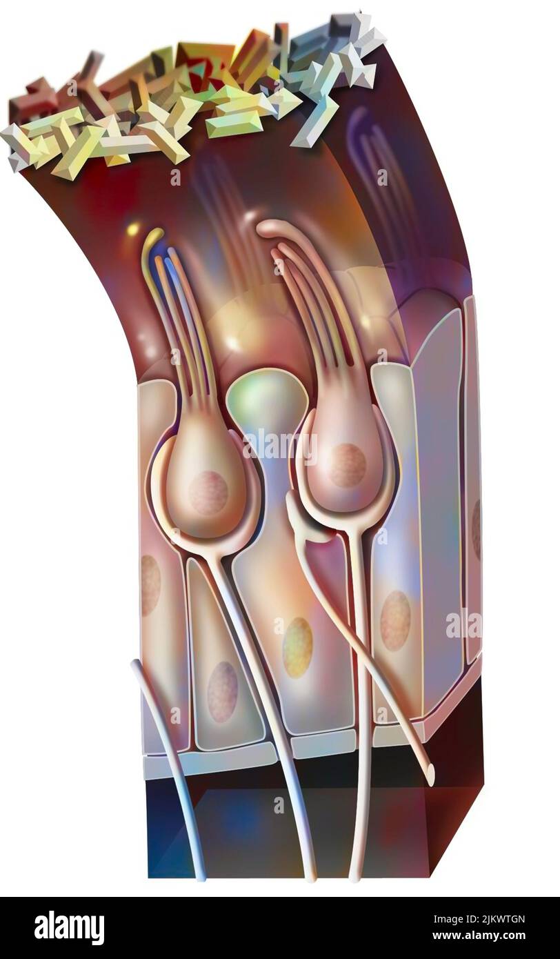 Functioning of the macule: organ of static balance (position of the head). Stock Photohttps://www.alamy.com/image-license-details/?v=1https://www.alamy.com/functioning-of-the-macule-organ-of-static-balance-position-of-the-head-image476926485.html
RF2JKWTGN–Functioning of the macule: organ of static balance (position of the head).
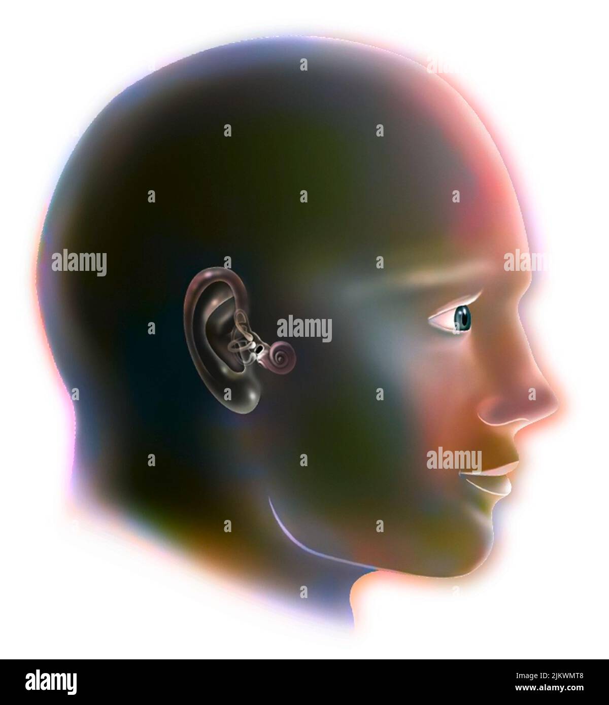 Inner ear (organ of hearing) and vestibular system. Stock Photohttps://www.alamy.com/image-license-details/?v=1https://www.alamy.com/inner-ear-organ-of-hearing-and-vestibular-system-image476923560.html
RF2JKWMT8–Inner ear (organ of hearing) and vestibular system.
 Eye: treatment of macular degeneration by RNA interference. Stock Photohttps://www.alamy.com/image-license-details/?v=1https://www.alamy.com/eye-treatment-of-macular-degeneration-by-rna-interference-image476923979.html
RF2JKWNB7–Eye: treatment of macular degeneration by RNA interference.
 Anatomy of the macule showing the cells (ciliates, supports). Stock Photohttps://www.alamy.com/image-license-details/?v=1https://www.alamy.com/anatomy-of-the-macule-showing-the-cells-ciliates-supports-image476926525.html
RF2JKWTJ5–Anatomy of the macule showing the cells (ciliates, supports).
 Treatment of macular degeneration by injection of RNA interference. Stock Photohttps://www.alamy.com/image-license-details/?v=1https://www.alamy.com/treatment-of-macular-degeneration-by-injection-of-rna-interference-image476925315.html
RF2JKWR2Y–Treatment of macular degeneration by injection of RNA interference.
 Treatment of macular degeneration by injection of RNA interference. Stock Photohttps://www.alamy.com/image-license-details/?v=1https://www.alamy.com/treatment-of-macular-degeneration-by-injection-of-rna-interference-image476925306.html
RF2JKWR2J–Treatment of macular degeneration by injection of RNA interference.
 Treatment of macular degeneration by injection of RNA interference. Stock Photohttps://www.alamy.com/image-license-details/?v=1https://www.alamy.com/treatment-of-macular-degeneration-by-injection-of-rna-interference-image476925391.html
RF2JKWR5K–Treatment of macular degeneration by injection of RNA interference.
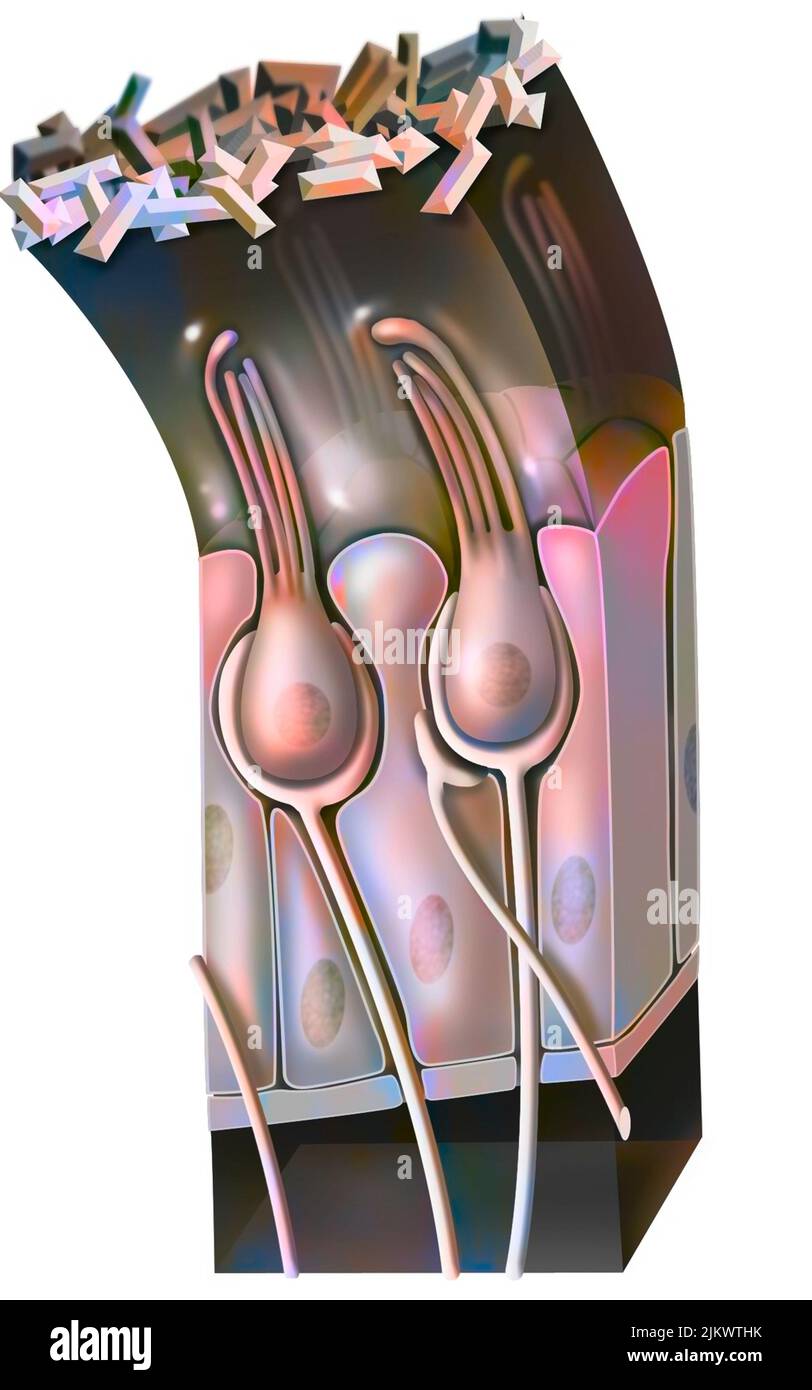 Functioning of the macule: organ of static balance (position of the head). Stock Photohttps://www.alamy.com/image-license-details/?v=1https://www.alamy.com/functioning-of-the-macule-organ-of-static-balance-position-of-the-head-image476926511.html
RF2JKWTHK–Functioning of the macule: organ of static balance (position of the head).
 Functioning of the macule: organ of static balance (position of the head). Stock Photohttps://www.alamy.com/image-license-details/?v=1https://www.alamy.com/functioning-of-the-macule-organ-of-static-balance-position-of-the-head-image476926488.html
RF2JKWTGT–Functioning of the macule: organ of static balance (position of the head).
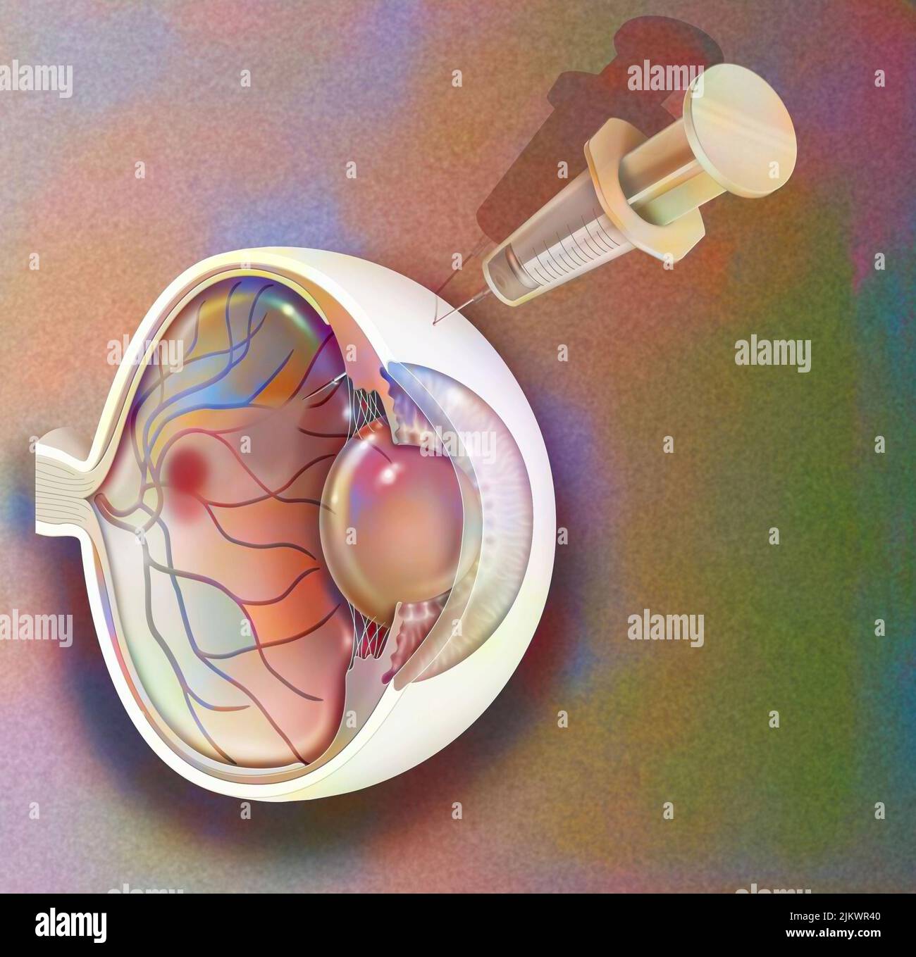 Treatment of macular degeneration by injection of RNA interference. Stock Photohttps://www.alamy.com/image-license-details/?v=1https://www.alamy.com/treatment-of-macular-degeneration-by-injection-of-rna-interference-image476925344.html
RF2JKWR40–Treatment of macular degeneration by injection of RNA interference.
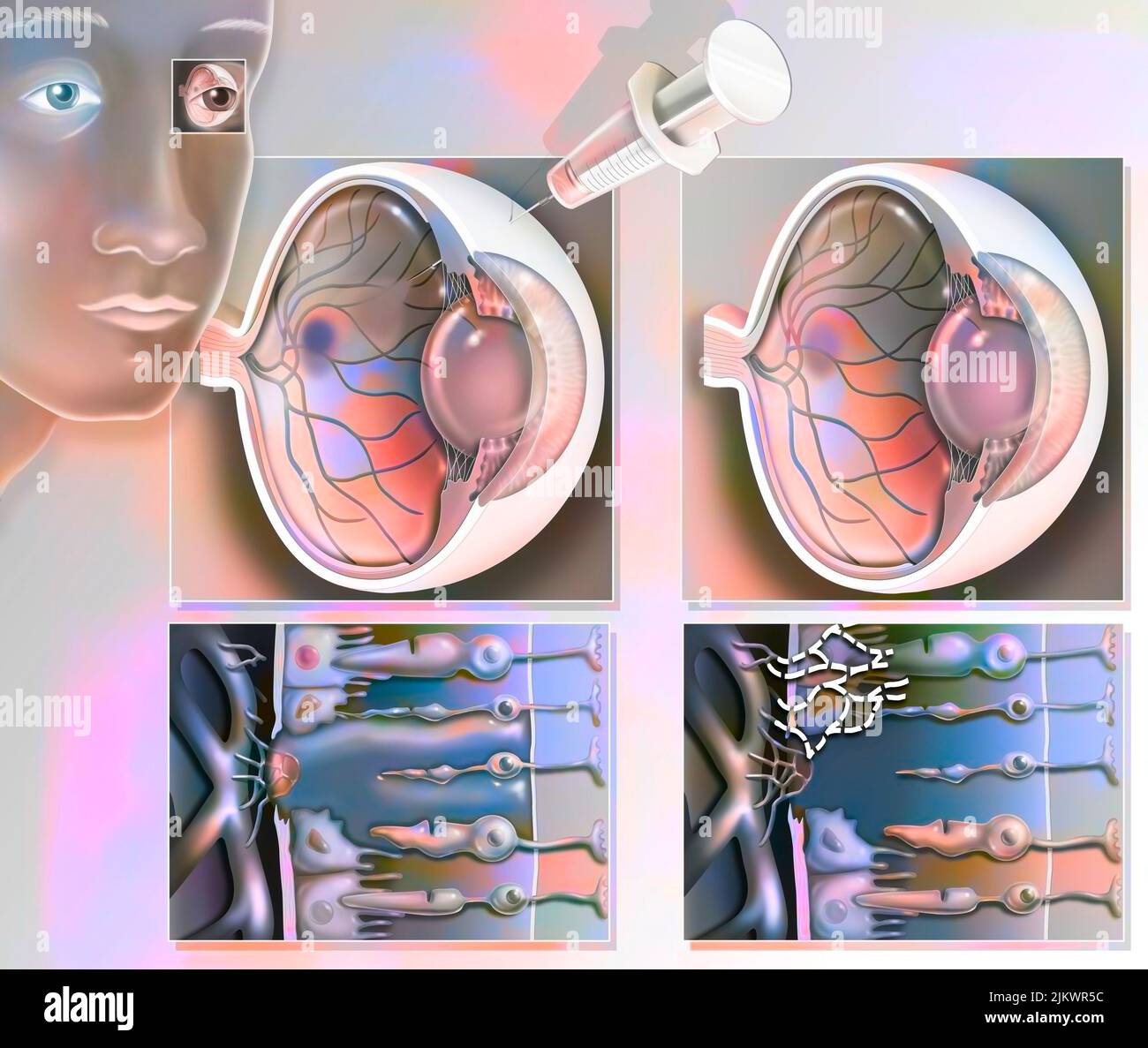 Treatment of macular degeneration by injection of RNA interference. Stock Photohttps://www.alamy.com/image-license-details/?v=1https://www.alamy.com/treatment-of-macular-degeneration-by-injection-of-rna-interference-image476925384.html
RF2JKWR5C–Treatment of macular degeneration by injection of RNA interference.
 Anatomy of the macule showing the cells (ciliates, supports). Stock Photohttps://www.alamy.com/image-license-details/?v=1https://www.alamy.com/anatomy-of-the-macule-showing-the-cells-ciliates-supports-image476926482.html
RF2JKWTGJ–Anatomy of the macule showing the cells (ciliates, supports).
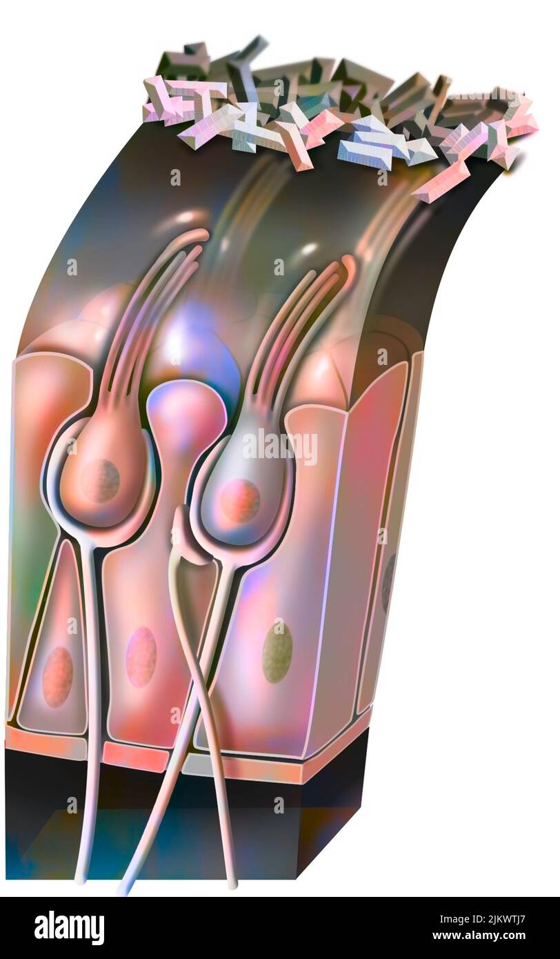 Functioning of the macule: organ of static balance (position of the head). Stock Photohttps://www.alamy.com/image-license-details/?v=1https://www.alamy.com/functioning-of-the-macule-organ-of-static-balance-position-of-the-head-image476926527.html
RF2JKWTJ7–Functioning of the macule: organ of static balance (position of the head).
 Treatment of macular degeneration by injection of RNA interference. Stock Photohttps://www.alamy.com/image-license-details/?v=1https://www.alamy.com/treatment-of-macular-degeneration-by-injection-of-rna-interference-image476925304.html
RF2JKWR2G–Treatment of macular degeneration by injection of RNA interference.
 Treatment of macular degeneration by injection of RNA interference. Stock Photohttps://www.alamy.com/image-license-details/?v=1https://www.alamy.com/treatment-of-macular-degeneration-by-injection-of-rna-interference-image476925366.html
RF2JKWR4P–Treatment of macular degeneration by injection of RNA interference.
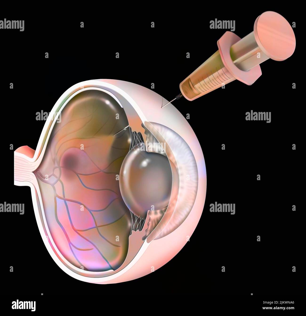 Eye: treatment of macular degeneration by RNA interference. Stock Photohttps://www.alamy.com/image-license-details/?v=1https://www.alamy.com/eye-treatment-of-macular-degeneration-by-rna-interference-image476923950.html
RF2JKWNA6–Eye: treatment of macular degeneration by RNA interference.
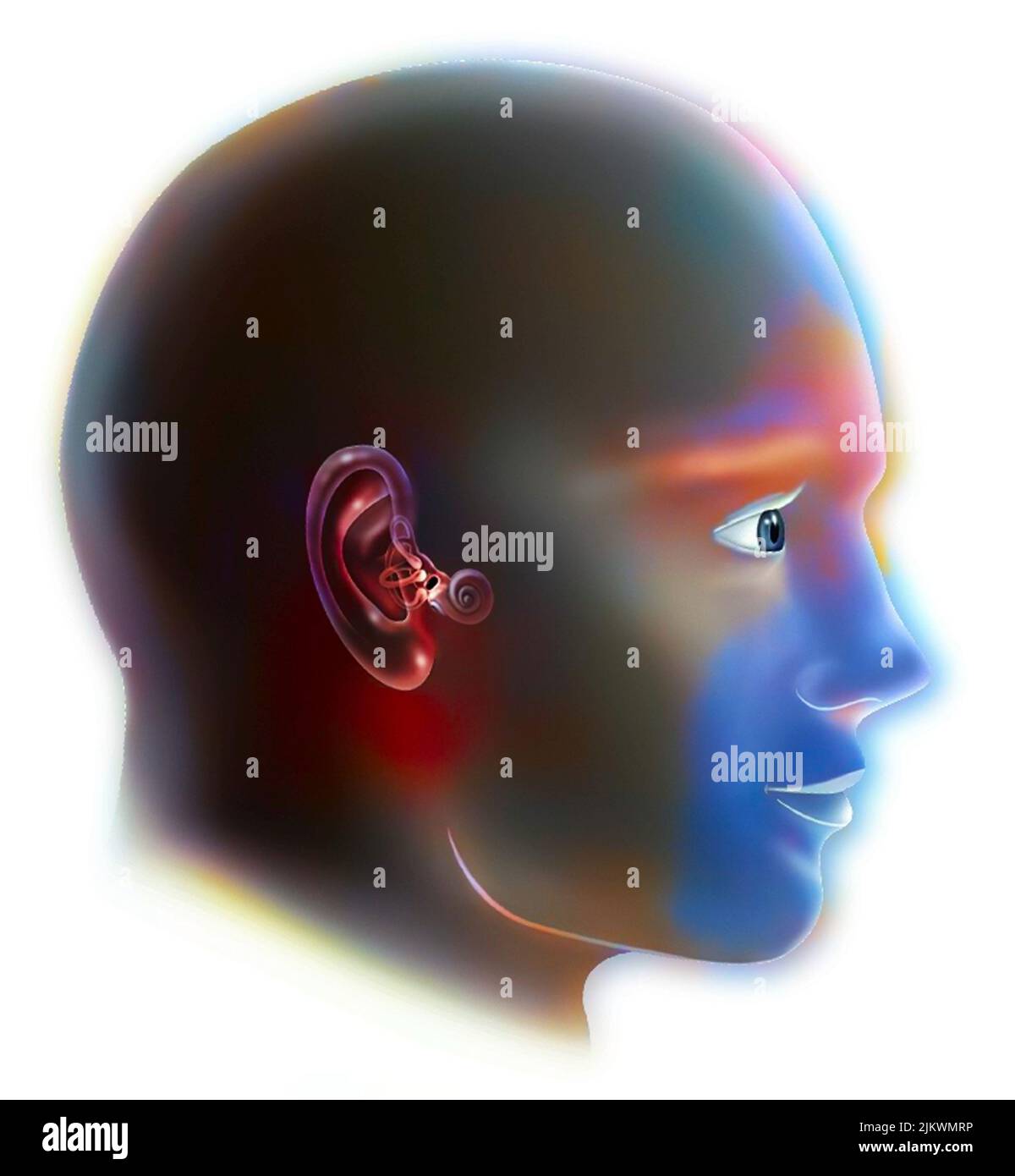 Inner ear (organ of hearing) and vestibular system. Stock Photohttps://www.alamy.com/image-license-details/?v=1https://www.alamy.com/inner-ear-organ-of-hearing-and-vestibular-system-image476923546.html
RF2JKWMRP–Inner ear (organ of hearing) and vestibular system.
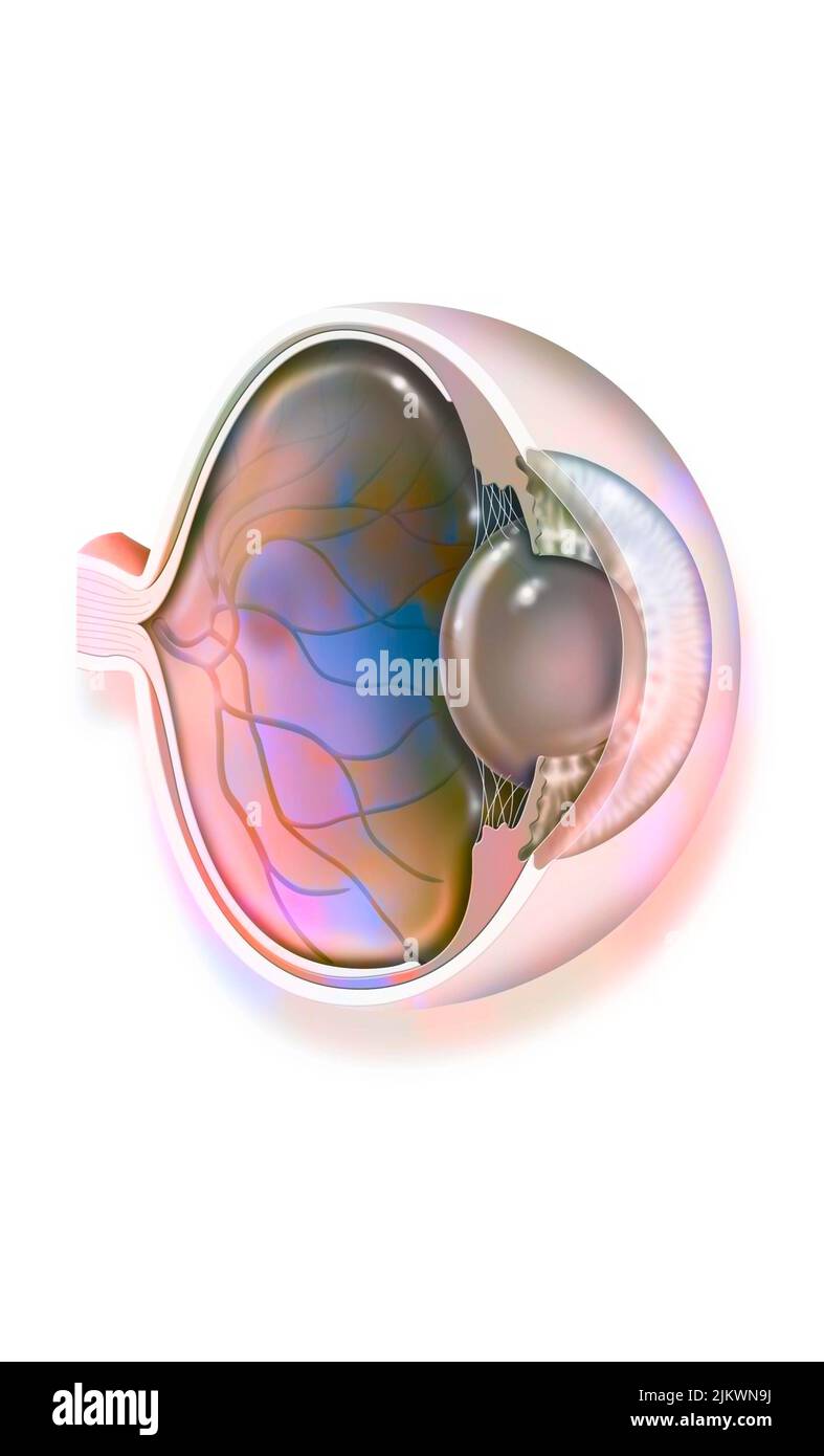 Anatomy of the eye with lens, retinal veins and arteries. Stock Photohttps://www.alamy.com/image-license-details/?v=1https://www.alamy.com/anatomy-of-the-eye-with-lens-retinal-veins-and-arteries-image476923934.html
RF2JKWN9J–Anatomy of the eye with lens, retinal veins and arteries.
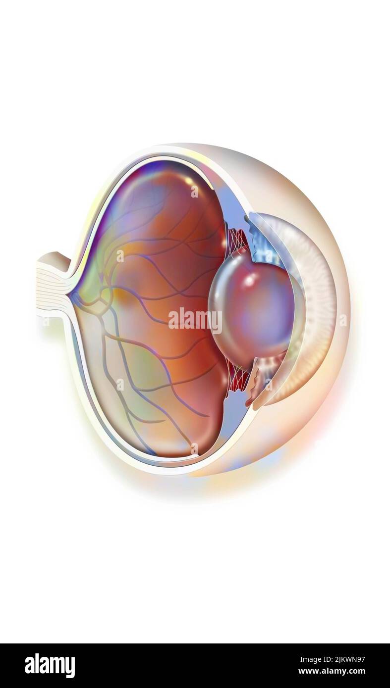 Anatomy of the eye with lens, retinal veins and arteries. Stock Photohttps://www.alamy.com/image-license-details/?v=1https://www.alamy.com/anatomy-of-the-eye-with-lens-retinal-veins-and-arteries-image476923923.html
RF2JKWN97–Anatomy of the eye with lens, retinal veins and arteries.
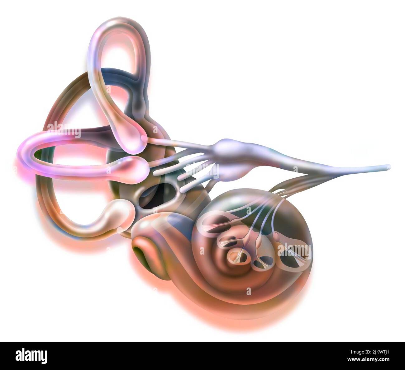 Inner ear and vestibular apparatus with semicircular canals, macule. Stock Photohttps://www.alamy.com/image-license-details/?v=1https://www.alamy.com/inner-ear-and-vestibular-apparatus-with-semicircular-canals-macule-image476926521.html
RF2JKWTJ1–Inner ear and vestibular apparatus with semicircular canals, macule.
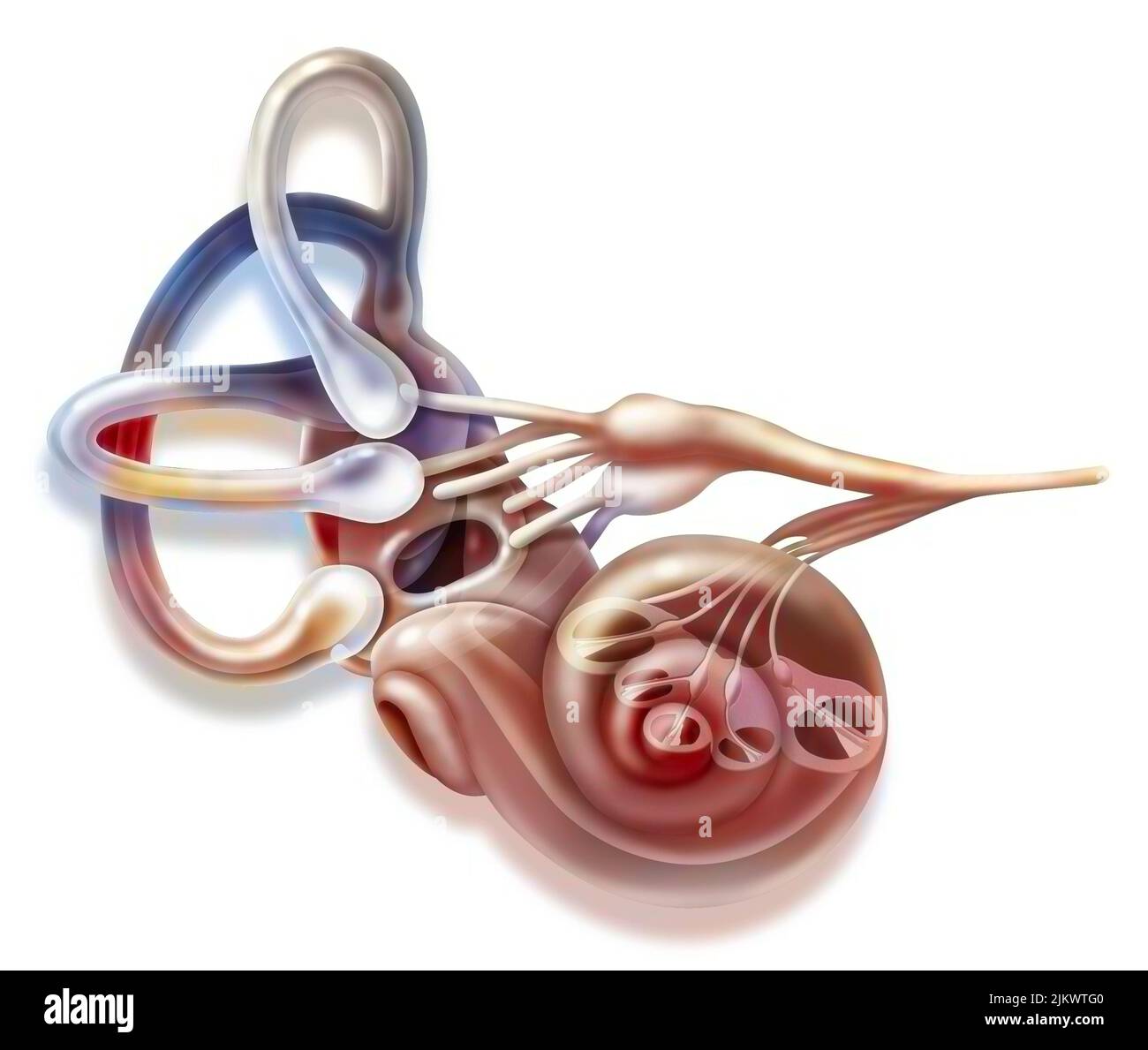 Inner ear and vestibular apparatus with semicircular canals, macule. Stock Photohttps://www.alamy.com/image-license-details/?v=1https://www.alamy.com/inner-ear-and-vestibular-apparatus-with-semicircular-canals-macule-image476926464.html
RF2JKWTG0–Inner ear and vestibular apparatus with semicircular canals, macule.
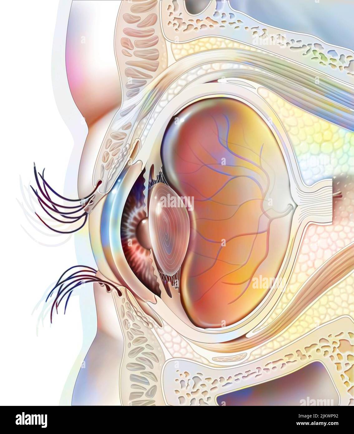 Eye anatomy and sectional eyelids with lens, retina. Stock Photohttps://www.alamy.com/image-license-details/?v=1https://www.alamy.com/eye-anatomy-and-sectional-eyelids-with-lens-retina-image476924702.html
RF2JKWP92–Eye anatomy and sectional eyelids with lens, retina.
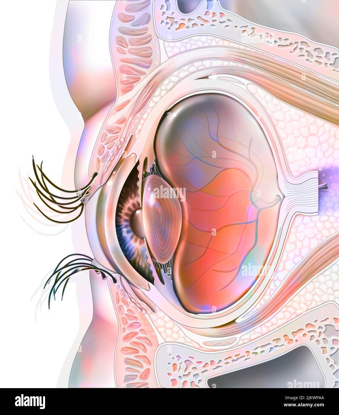 Eye anatomy and sectional eyelids with lens, retina. Stock Photohttps://www.alamy.com/image-license-details/?v=1https://www.alamy.com/eye-anatomy-and-sectional-eyelids-with-lens-retina-image476924738.html
RF2JKWPAA–Eye anatomy and sectional eyelids with lens, retina.
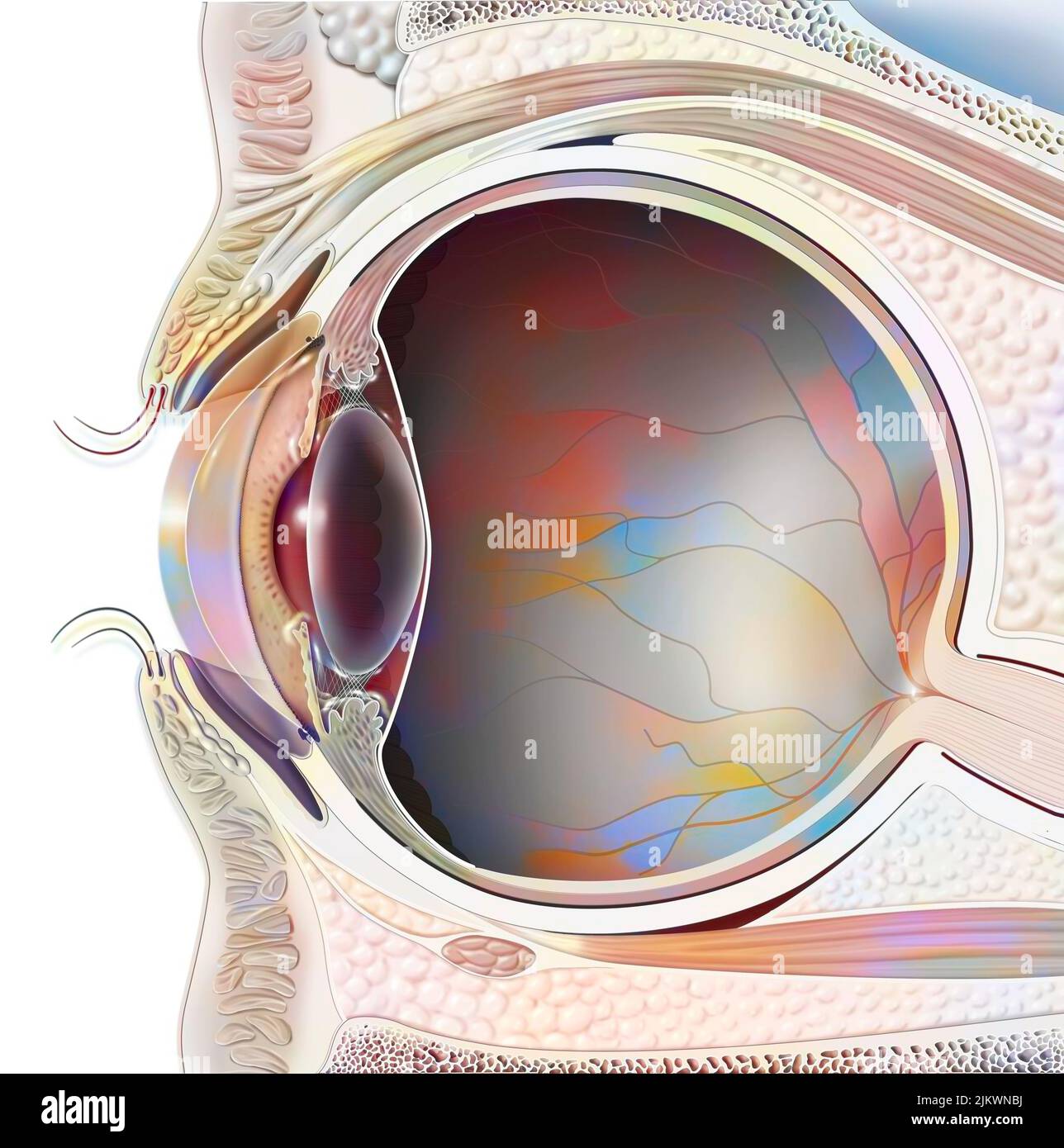 Anatomy of an eye in section showing lens, retina. Stock Photohttps://www.alamy.com/image-license-details/?v=1https://www.alamy.com/anatomy-of-an-eye-in-section-showing-lens-retina-image476923990.html
RF2JKWNBJ–Anatomy of an eye in section showing lens, retina.
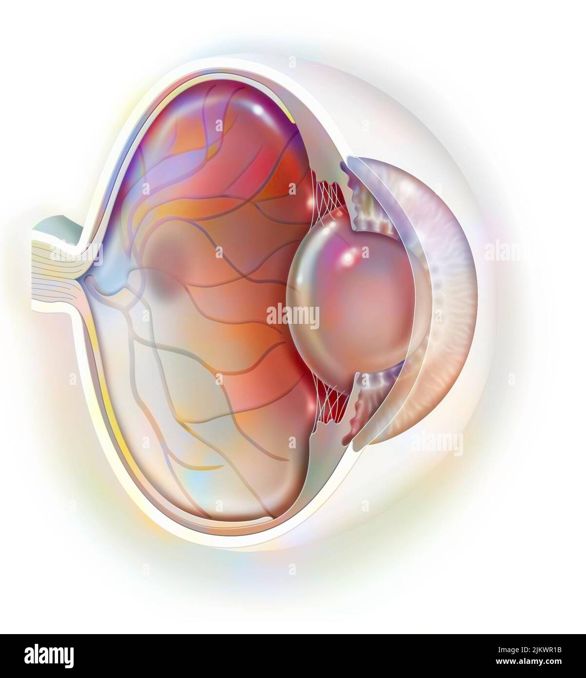 Sagittal view of the eye anatomy showing lens, retina, cornea, iris, choroid. Stock Photohttps://www.alamy.com/image-license-details/?v=1https://www.alamy.com/sagittal-view-of-the-eye-anatomy-showing-lens-retina-cornea-iris-choroid-image476925271.html
RF2JKWR1B–Sagittal view of the eye anatomy showing lens, retina, cornea, iris, choroid.
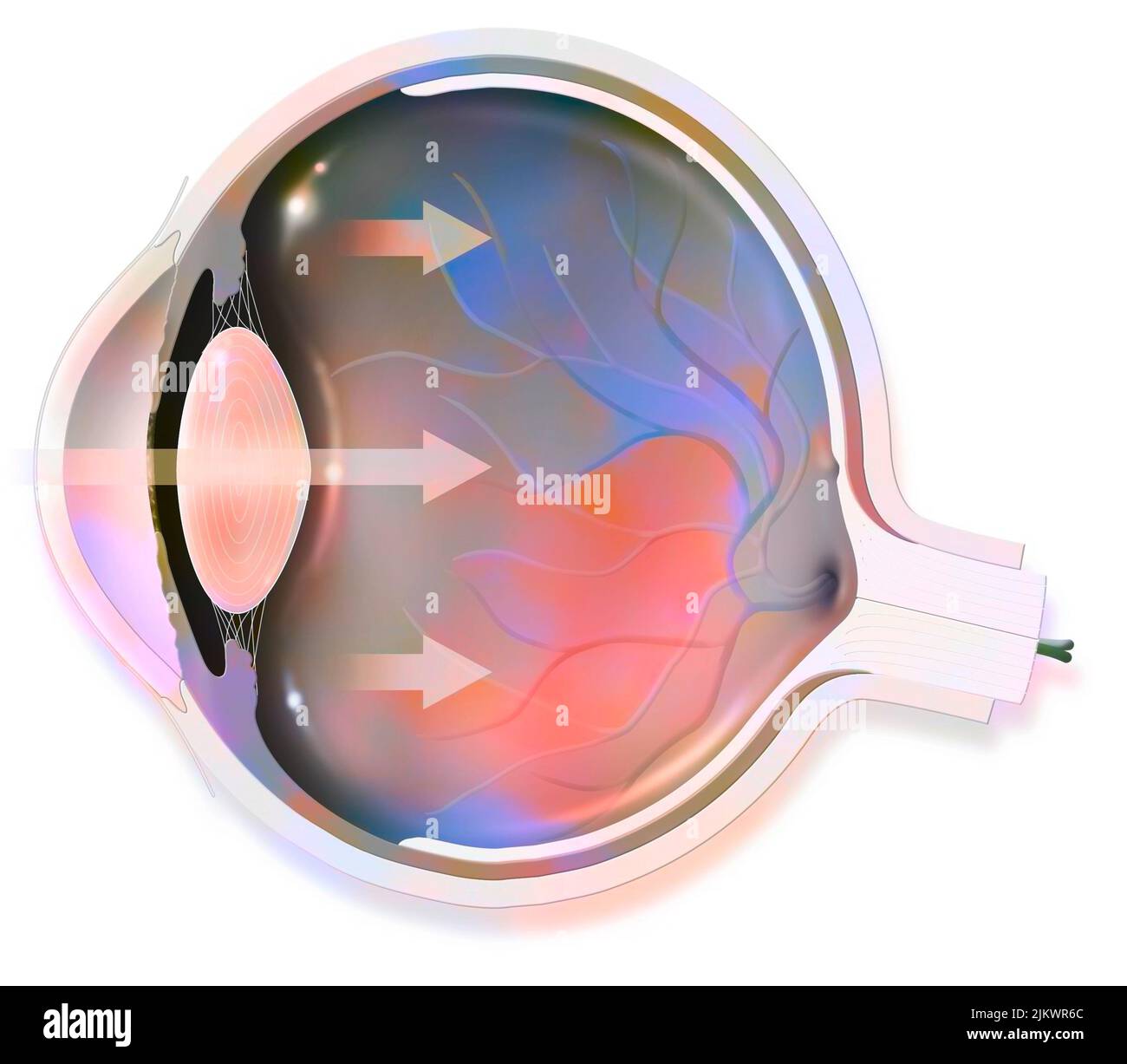 Anatomy of the eye whose arrows represent light and revealing the lens, retina, cornea, iris, choroid. Stock Photohttps://www.alamy.com/image-license-details/?v=1https://www.alamy.com/anatomy-of-the-eye-whose-arrows-represent-light-and-revealing-the-lens-retina-cornea-iris-choroid-image476925412.html
RF2JKWR6C–Anatomy of the eye whose arrows represent light and revealing the lens, retina, cornea, iris, choroid.
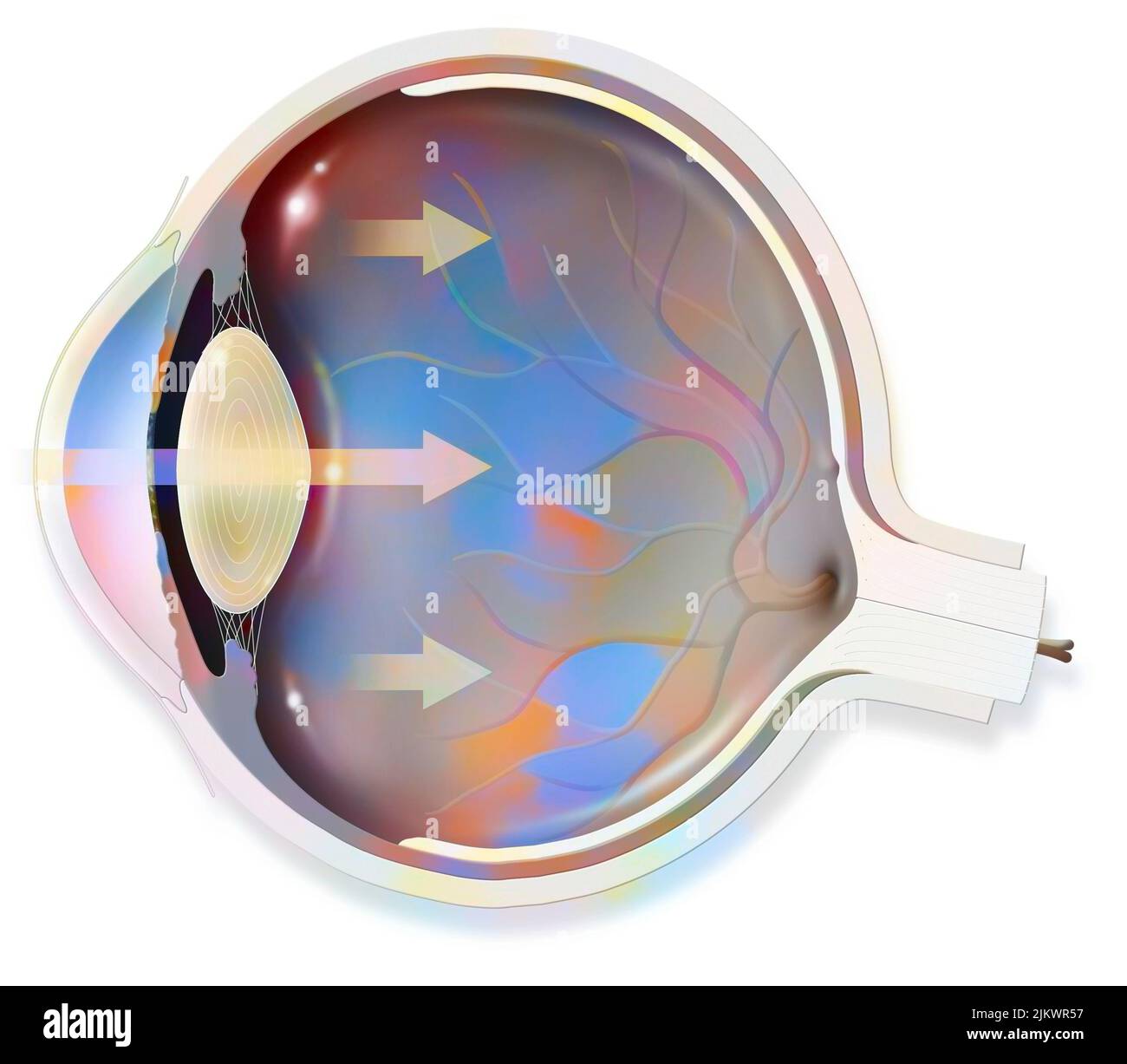 Anatomy of the eye whose arrows represent light and revealing the lens, retina, cornea, iris, choroid. Stock Photohttps://www.alamy.com/image-license-details/?v=1https://www.alamy.com/anatomy-of-the-eye-whose-arrows-represent-light-and-revealing-the-lens-retina-cornea-iris-choroid-image476925379.html
RF2JKWR57–Anatomy of the eye whose arrows represent light and revealing the lens, retina, cornea, iris, choroid.
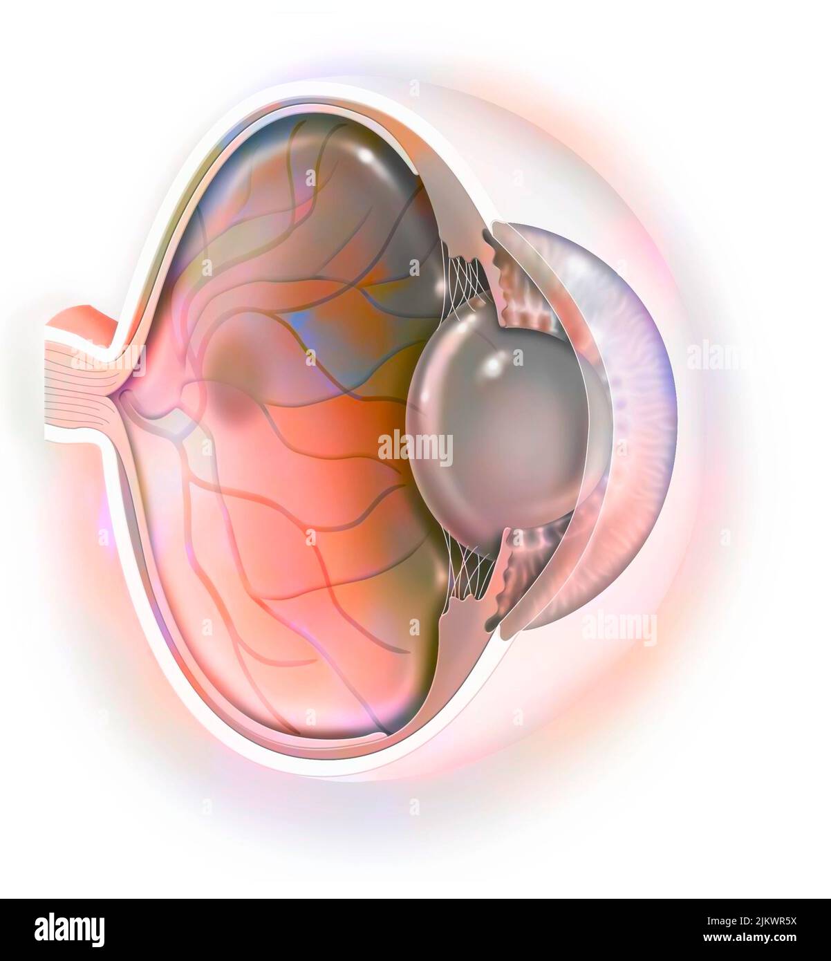 Sagittal view of the eye anatomy showing lens, retina, cornea, iris, choroid. Stock Photohttps://www.alamy.com/image-license-details/?v=1https://www.alamy.com/sagittal-view-of-the-eye-anatomy-showing-lens-retina-cornea-iris-choroid-image476925398.html
RF2JKWR5X–Sagittal view of the eye anatomy showing lens, retina, cornea, iris, choroid.
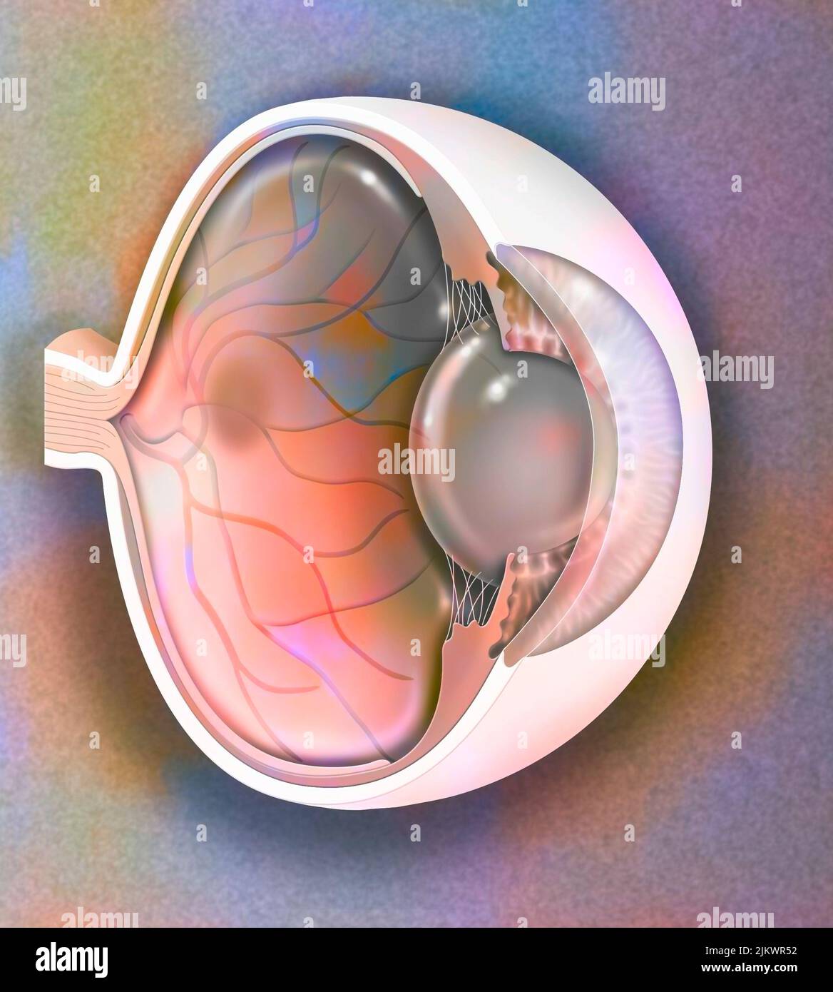 Sagittal view of the eye anatomy showing lens, retina, cornea, iris, choroid. Stock Photohttps://www.alamy.com/image-license-details/?v=1https://www.alamy.com/sagittal-view-of-the-eye-anatomy-showing-lens-retina-cornea-iris-choroid-image476925374.html
RF2JKWR52–Sagittal view of the eye anatomy showing lens, retina, cornea, iris, choroid.
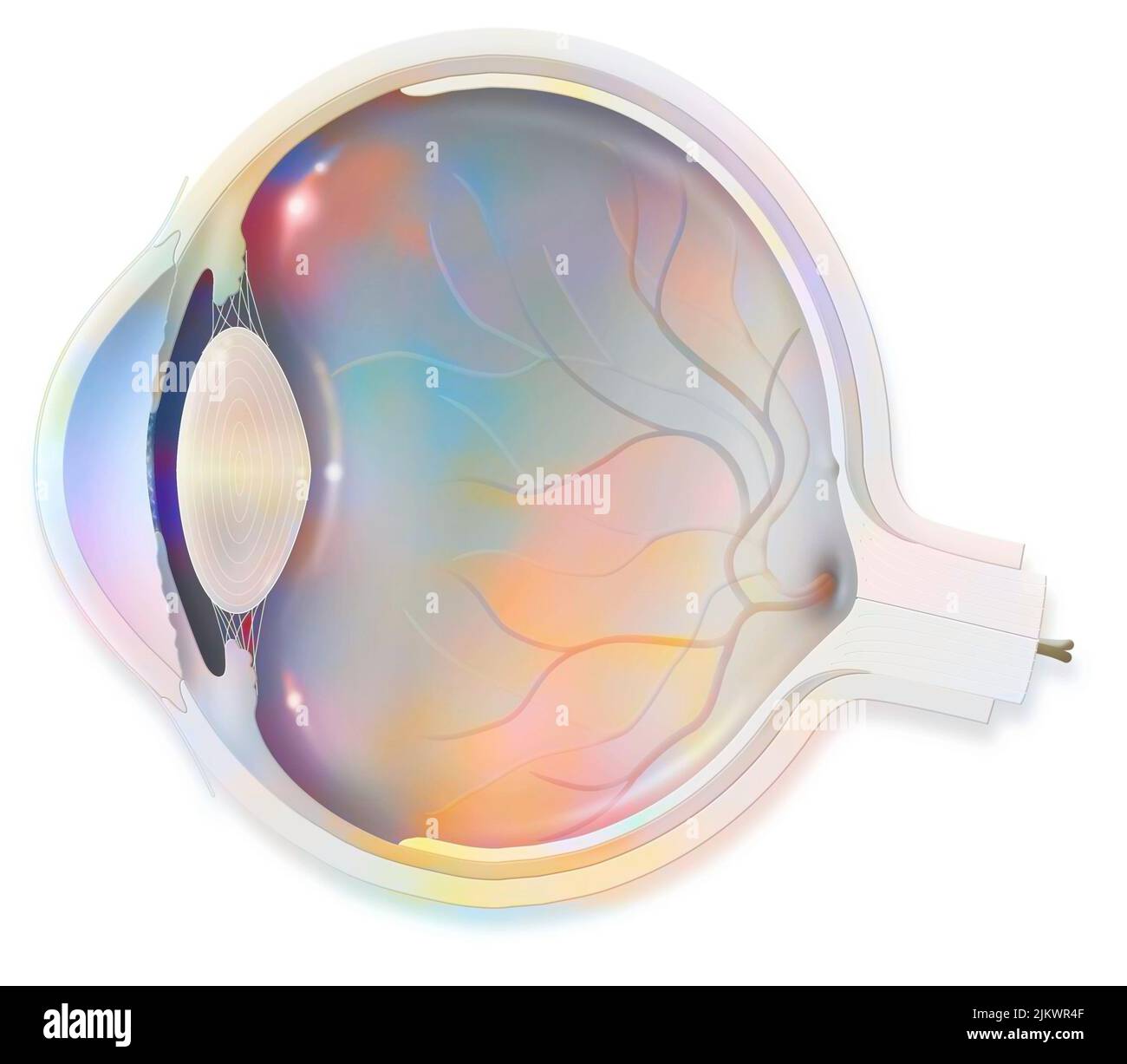 Anatomy section of the eye showing lens, retina, cornea, iris, choroid. Stock Photohttps://www.alamy.com/image-license-details/?v=1https://www.alamy.com/anatomy-section-of-the-eye-showing-lens-retina-cornea-iris-choroid-image476925359.html
RF2JKWR4F–Anatomy section of the eye showing lens, retina, cornea, iris, choroid.
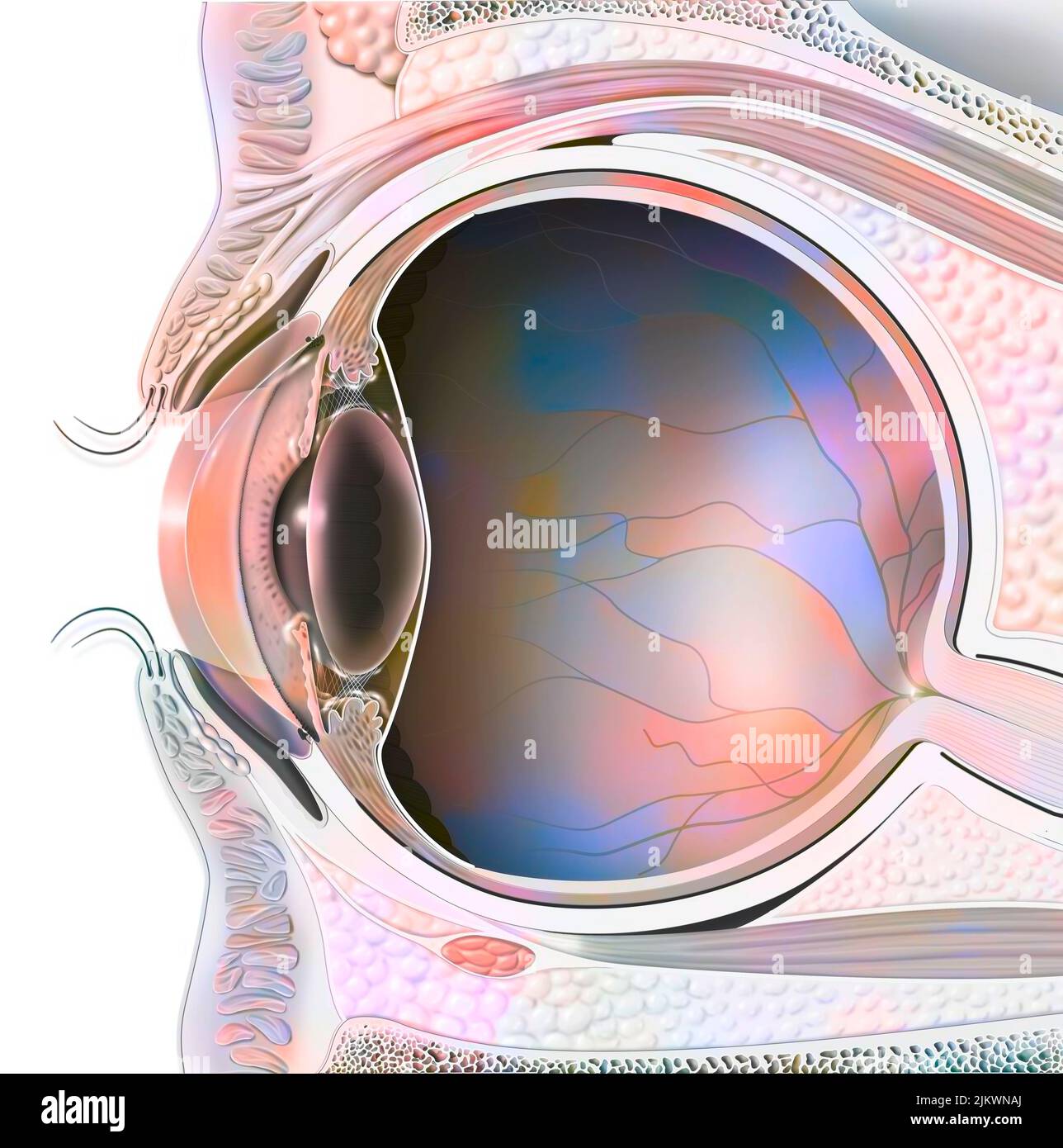 Anatomy of an eye in section showing lens, retina. Stock Photohttps://www.alamy.com/image-license-details/?v=1https://www.alamy.com/anatomy-of-an-eye-in-section-showing-lens-retina-image476923962.html
RF2JKWNAJ–Anatomy of an eye in section showing lens, retina.
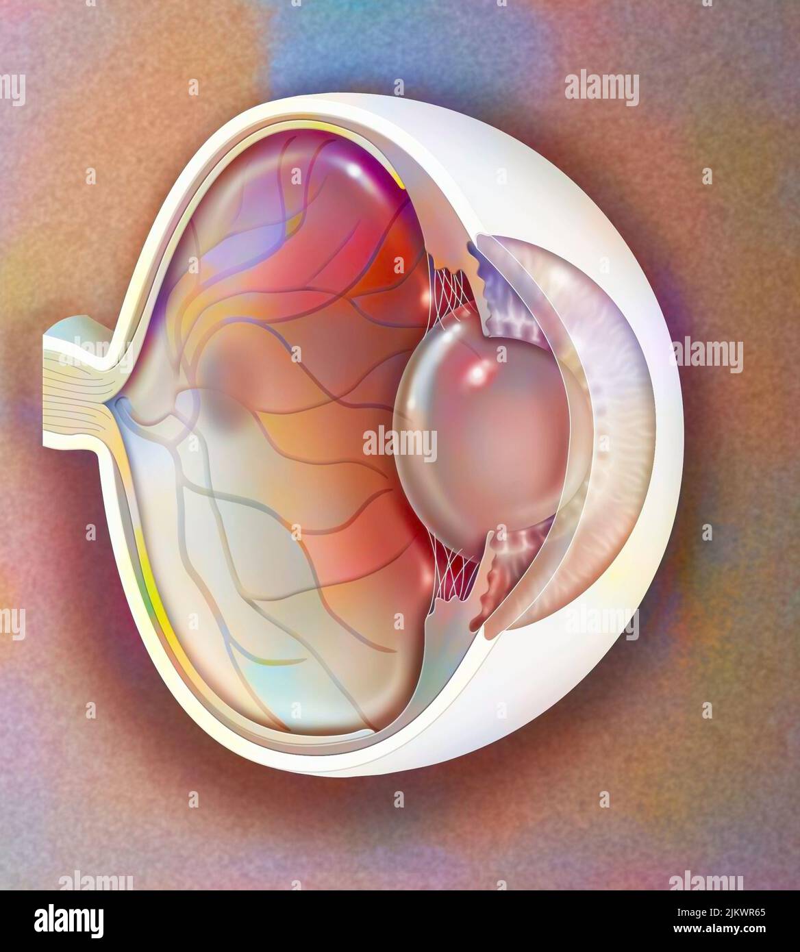 Sagittal view of the eye anatomy showing lens, retina, cornea, iris, choroid. Stock Photohttps://www.alamy.com/image-license-details/?v=1https://www.alamy.com/sagittal-view-of-the-eye-anatomy-showing-lens-retina-cornea-iris-choroid-image476925405.html
RF2JKWR65–Sagittal view of the eye anatomy showing lens, retina, cornea, iris, choroid.
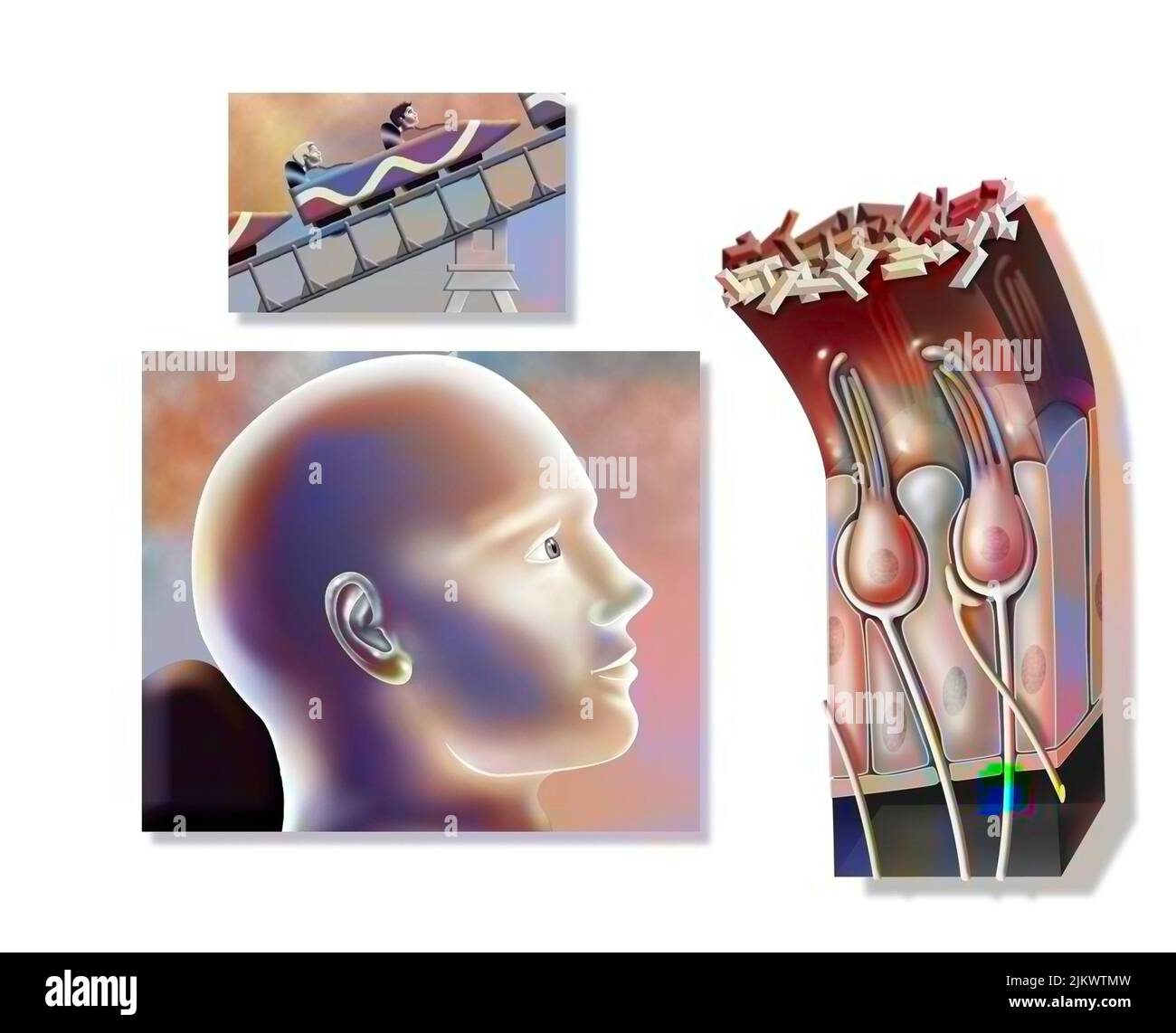 Functioning of the macule: organ of static balance (position of the head). Stock Photohttps://www.alamy.com/image-license-details/?v=1https://www.alamy.com/functioning-of-the-macule-organ-of-static-balance-position-of-the-head-image476926601.html
RF2JKWTMW–Functioning of the macule: organ of static balance (position of the head).
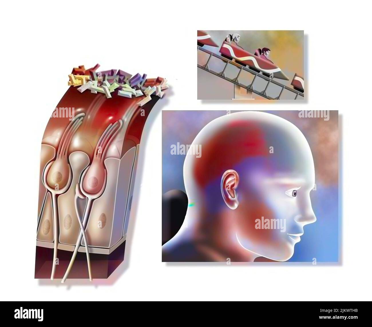 Functioning of the macule: organ of static balance (position of the head). Stock Photohttps://www.alamy.com/image-license-details/?v=1https://www.alamy.com/functioning-of-the-macule-organ-of-static-balance-position-of-the-head-image476926503.html
RF2JKWTHB–Functioning of the macule: organ of static balance (position of the head).
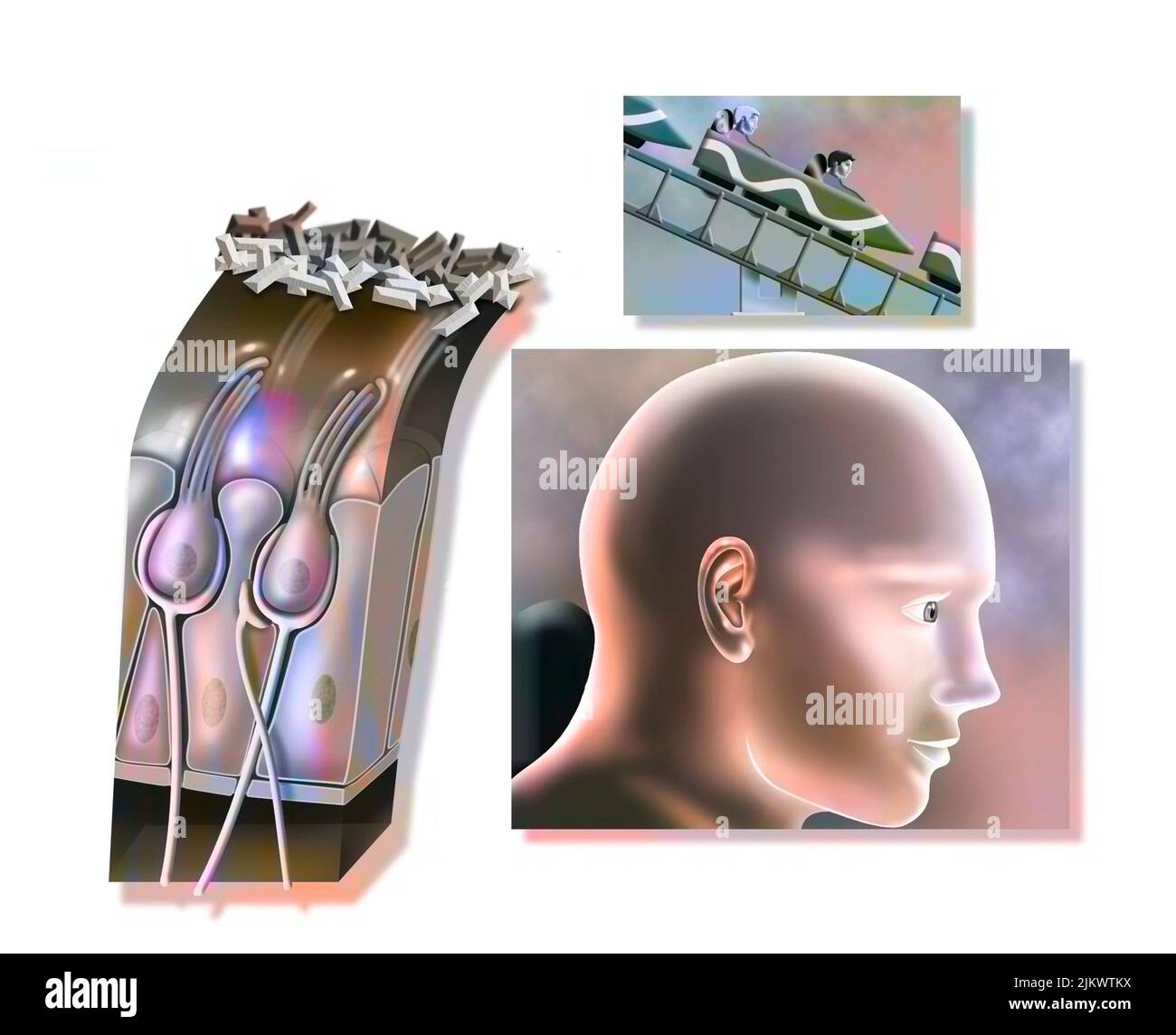 Functioning of the macule: organ of static balance (position of the head). Stock Photohttps://www.alamy.com/image-license-details/?v=1https://www.alamy.com/functioning-of-the-macule-organ-of-static-balance-position-of-the-head-image476926574.html
RF2JKWTKX–Functioning of the macule: organ of static balance (position of the head).
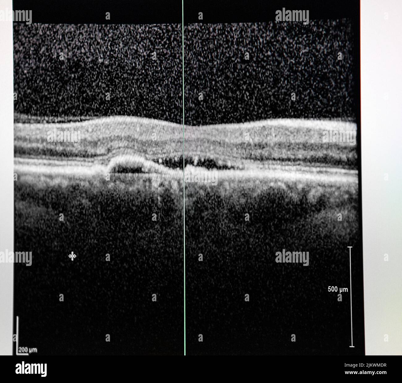 Optical coherence tomography (OCT) of a patient with macular edema. Stock Photohttps://www.alamy.com/image-license-details/?v=1https://www.alamy.com/optical-coherence-tomography-oct-of-a-patient-with-macular-edema-image476923267.html
RF2JKWMDR–Optical coherence tomography (OCT) of a patient with macular edema.
 Optical coherence tomography (OCT) showing the beginnings of macular degeneration. Stock Photohttps://www.alamy.com/image-license-details/?v=1https://www.alamy.com/optical-coherence-tomography-oct-showing-the-beginnings-of-macular-degeneration-image476923018.html
RF2JKWM4X–Optical coherence tomography (OCT) showing the beginnings of macular degeneration.
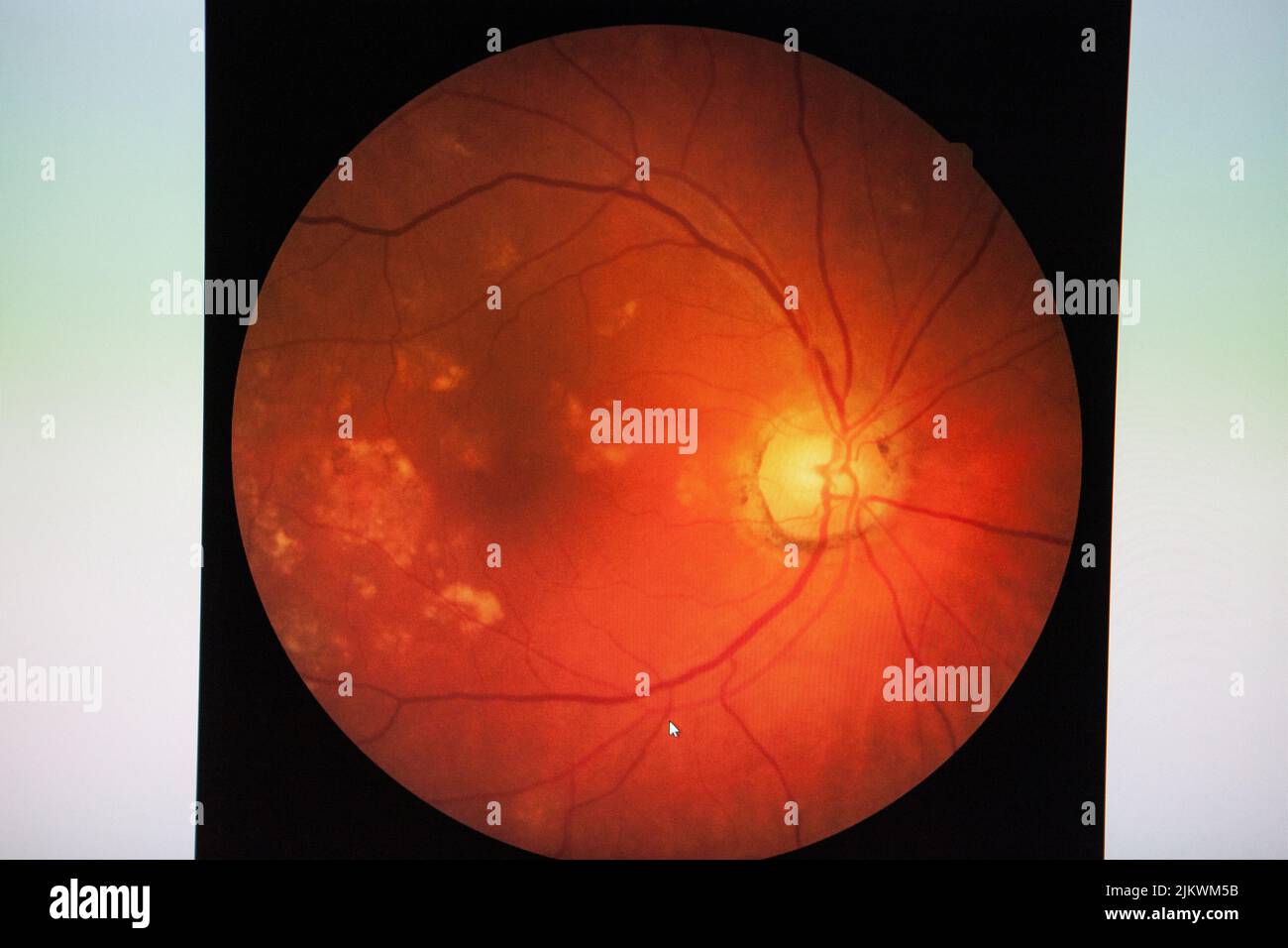 Fundus: routine examination established by an ophthalmoscope to check the health of the eye. Stock Photohttps://www.alamy.com/image-license-details/?v=1https://www.alamy.com/fundus-routine-examination-established-by-an-ophthalmoscope-to-check-the-health-of-the-eye-image476923031.html
RF2JKWM5B–Fundus: routine examination established by an ophthalmoscope to check the health of the eye.
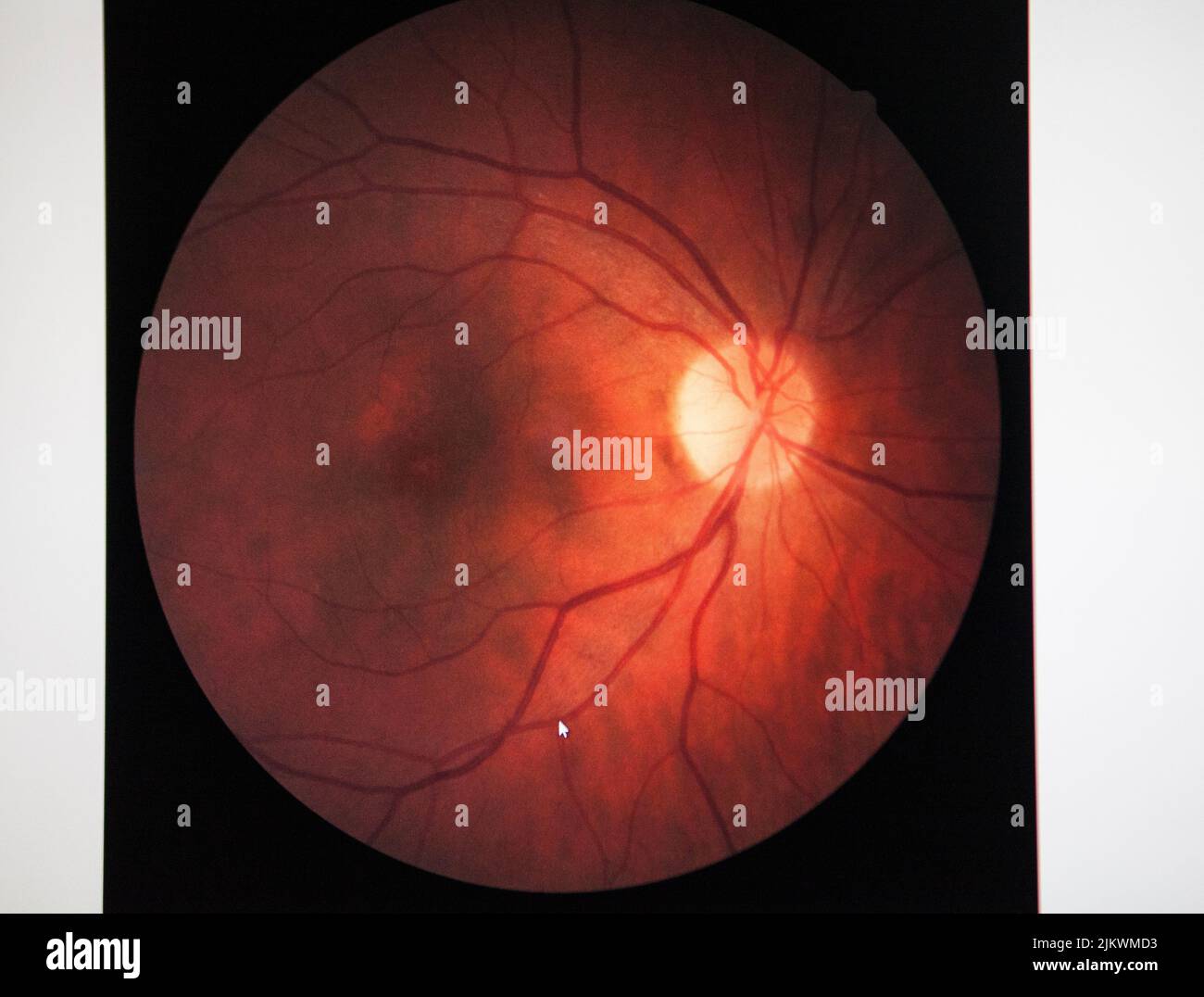 Fundus of a patient with macular edema. Stock Photohttps://www.alamy.com/image-license-details/?v=1https://www.alamy.com/fundus-of-a-patient-with-macular-edema-image476923247.html
RF2JKWMD3–Fundus of a patient with macular edema.
 Retina Stock Photohttps://www.alamy.com/image-license-details/?v=1https://www.alamy.com/retina-image338279358.html
RM2AJ9XNJ–Retina
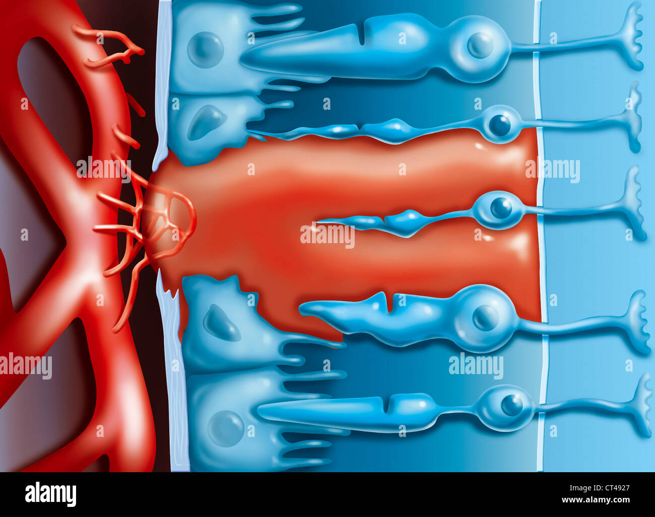 AMD, ILLUSTRATION Stock Photohttps://www.alamy.com/image-license-details/?v=1https://www.alamy.com/stock-photo-amd-illustration-49267407.html
RMCT4927–AMD, ILLUSTRATION
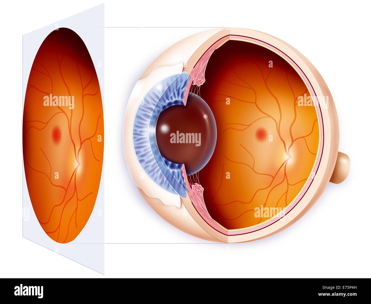 Eye, drawing Stock Photohttps://www.alamy.com/image-license-details/?v=1https://www.alamy.com/stock-photo-eye-drawing-73271201.html
RME75P4H–Eye, drawing
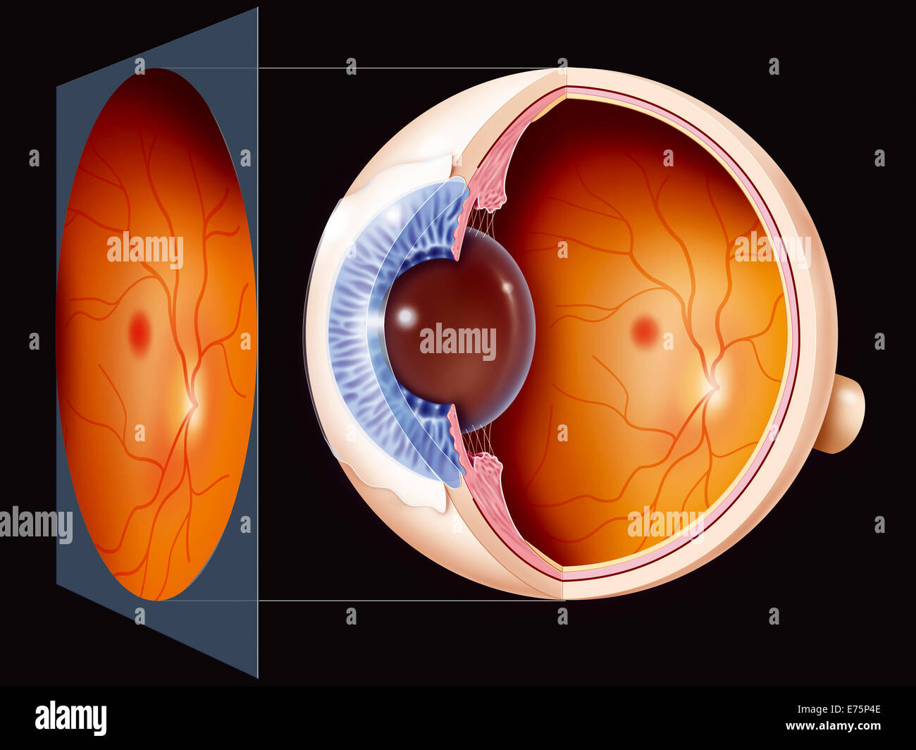 Eye, drawing Stock Photohttps://www.alamy.com/image-license-details/?v=1https://www.alamy.com/stock-photo-eye-drawing-73271198.html
RME75P4E–Eye, drawing
 AMD TREATMENT, ILLUSTRATION Stock Photohttps://www.alamy.com/image-license-details/?v=1https://www.alamy.com/stock-photo-amd-treatment-illustration-49267488.html
RMCT4954–AMD TREATMENT, ILLUSTRATION
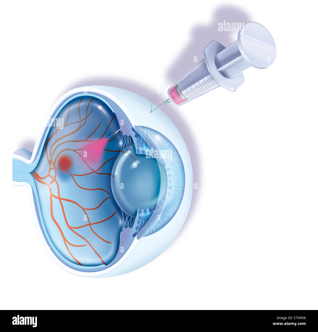 AMD TREATMENT, ILLUSTRATION Stock Photohttps://www.alamy.com/image-license-details/?v=1https://www.alamy.com/stock-photo-amd-treatment-illustration-49267490.html
RMCT4956–AMD TREATMENT, ILLUSTRATION
 AMD, ILLUSTRATION Stock Photohttps://www.alamy.com/image-license-details/?v=1https://www.alamy.com/stock-photo-amd-illustration-49267495.html
RMCT495B–AMD, ILLUSTRATION
 AMD TREATMENT, ILLUSTRATION Stock Photohttps://www.alamy.com/image-license-details/?v=1https://www.alamy.com/stock-photo-amd-treatment-illustration-49267477.html
RMCT494N–AMD TREATMENT, ILLUSTRATION
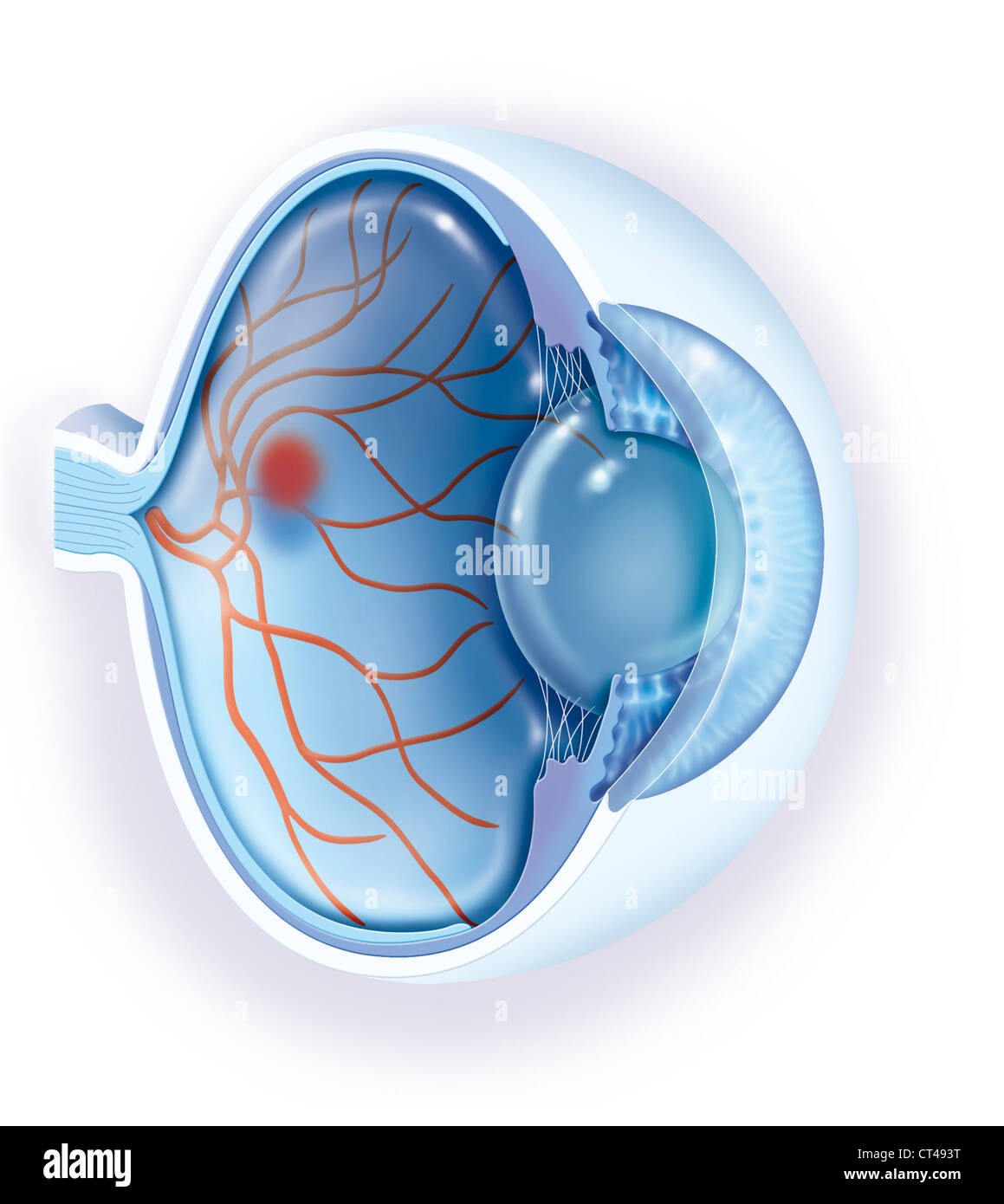 AMD, ILLUSTRATION Stock Photohttps://www.alamy.com/image-license-details/?v=1https://www.alamy.com/stock-photo-amd-illustration-49267452.html
RMCT493T–AMD, ILLUSTRATION
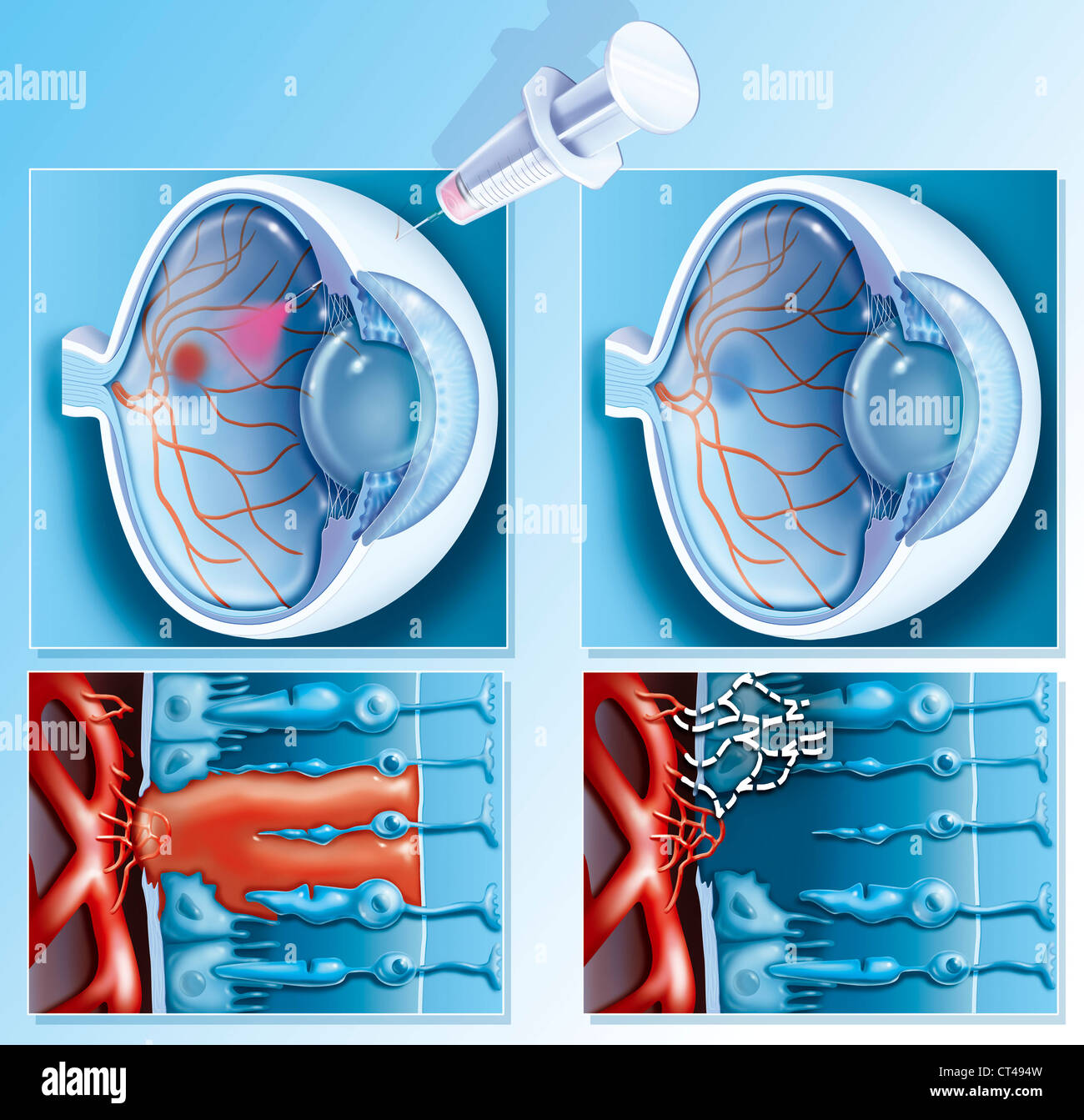 AMD TREATMENT, ILLUSTRATION Stock Photohttps://www.alamy.com/image-license-details/?v=1https://www.alamy.com/stock-photo-amd-treatment-illustration-49267481.html
RMCT494W–AMD TREATMENT, ILLUSTRATION
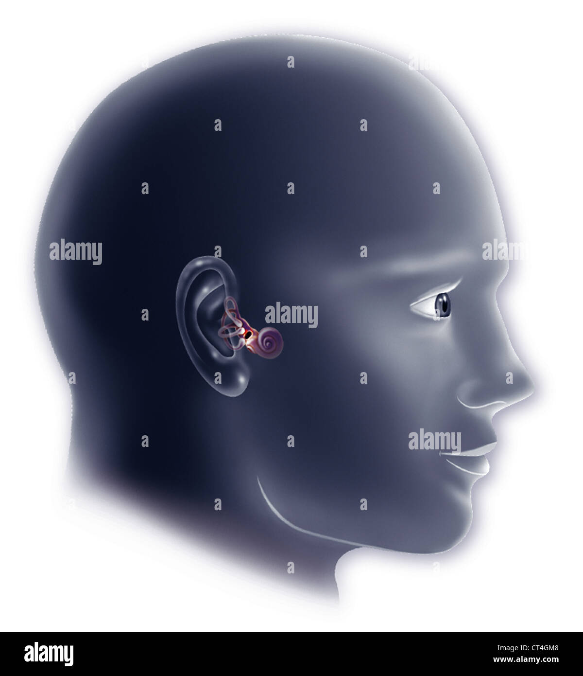 INTERNAL EAR, DRAWING Stock Photohttps://www.alamy.com/image-license-details/?v=1https://www.alamy.com/stock-photo-internal-ear-drawing-49273400.html
RMCT4GM8–INTERNAL EAR, DRAWING
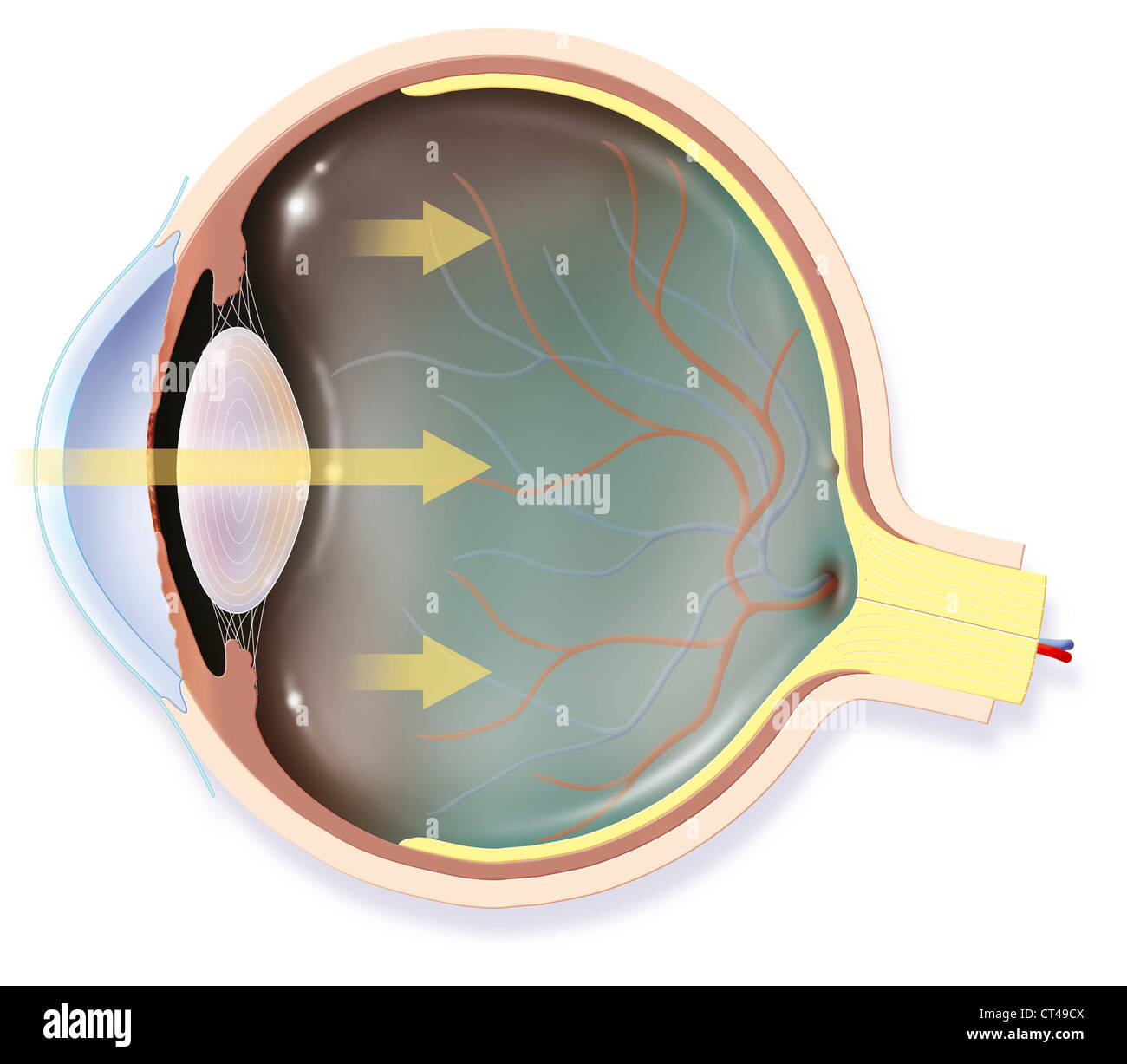 EYE, DRAWING Stock Photohttps://www.alamy.com/image-license-details/?v=1https://www.alamy.com/stock-photo-eye-drawing-49267706.html
RMCT49CX–EYE, DRAWING
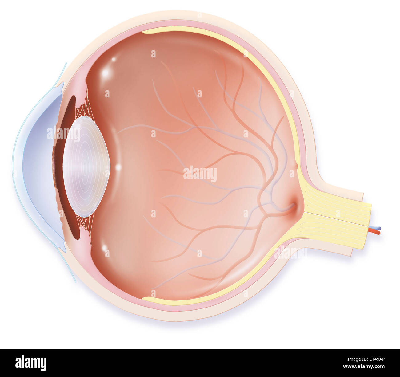 EYE, DRAWING Stock Photohttps://www.alamy.com/image-license-details/?v=1https://www.alamy.com/stock-photo-eye-drawing-49267646.html
RMCT49AP–EYE, DRAWING
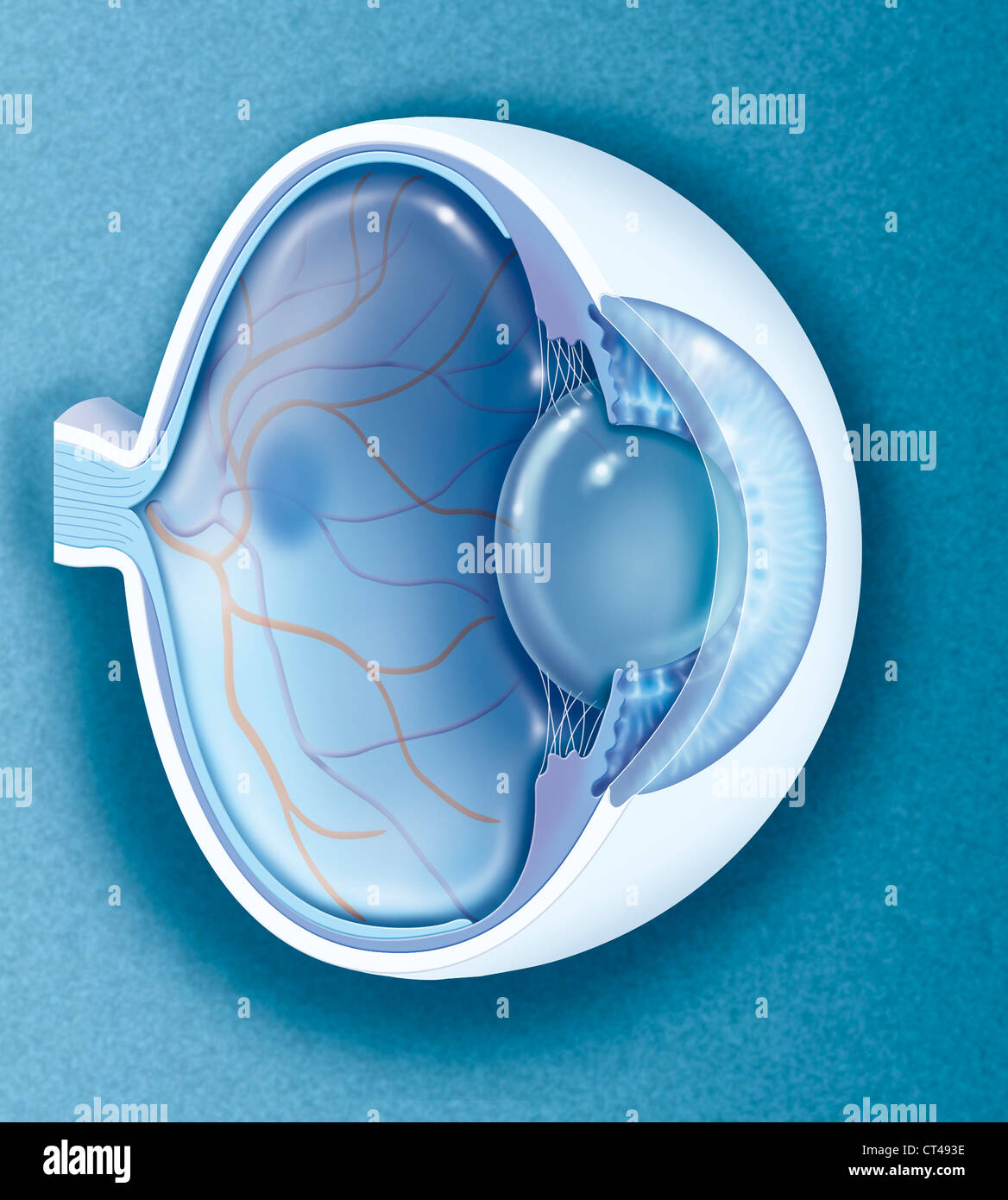 EYE, DRAWING Stock Photohttps://www.alamy.com/image-license-details/?v=1https://www.alamy.com/stock-photo-eye-drawing-49267442.html
RMCT493E–EYE, DRAWING
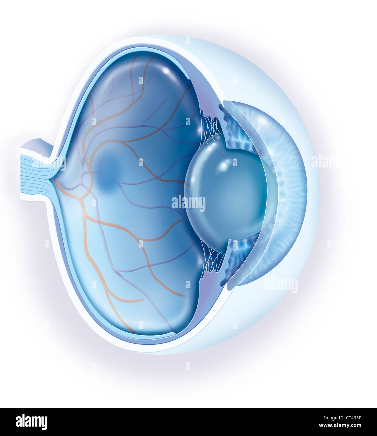 EYE, DRAWING Stock Photohttps://www.alamy.com/image-license-details/?v=1https://www.alamy.com/stock-photo-eye-drawing-49267450.html
RMCT493P–EYE, DRAWING
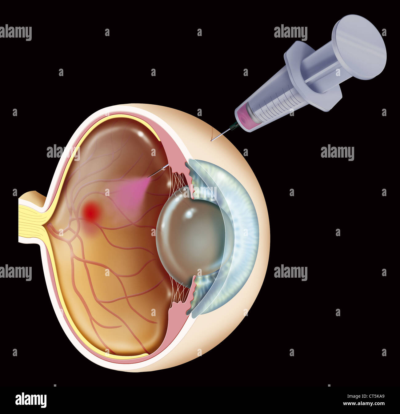 AMD TREATMENT, ILLUSTRATION Stock Photohttps://www.alamy.com/image-license-details/?v=1https://www.alamy.com/stock-photo-amd-treatment-illustration-49297425.html
RMCT5KA9–AMD TREATMENT, ILLUSTRATION
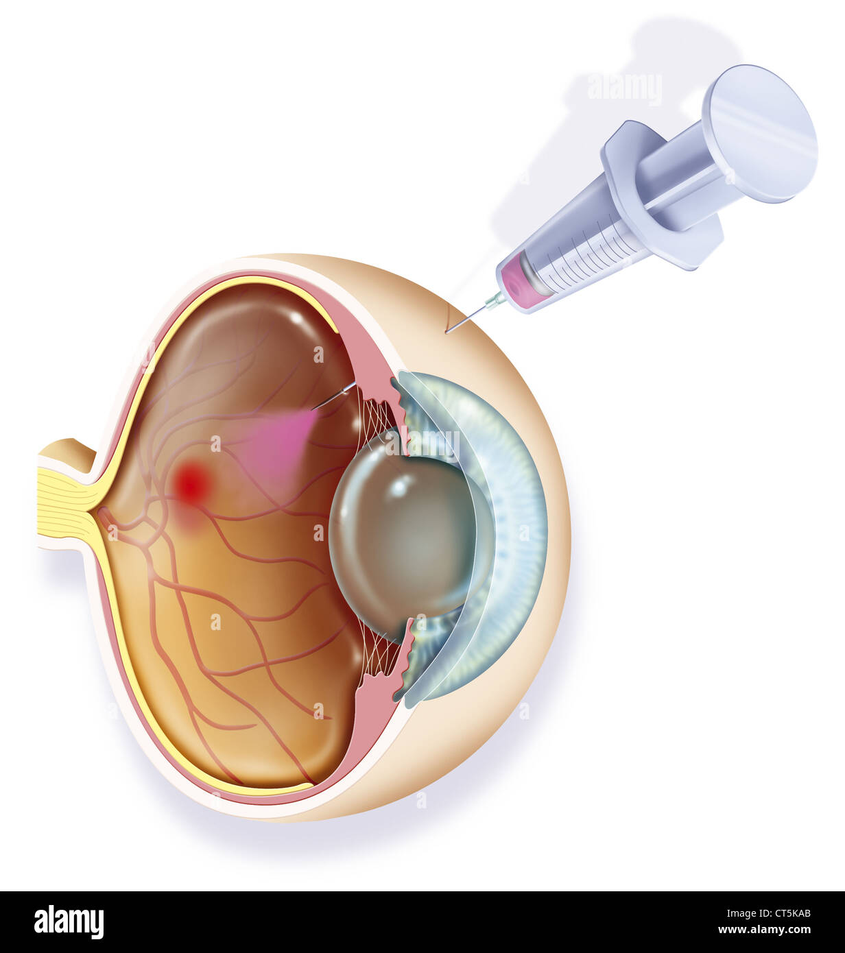 AMD TREATMENT, ILLUSTRATION Stock Photohttps://www.alamy.com/image-license-details/?v=1https://www.alamy.com/stock-photo-amd-treatment-illustration-49297427.html
RMCT5KAB–AMD TREATMENT, ILLUSTRATION
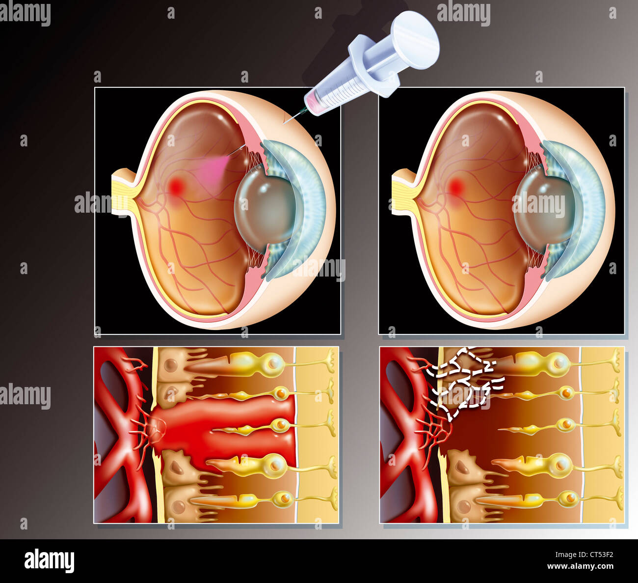 AMD TREATMENT, ILLUSTRATION Stock Photohttps://www.alamy.com/image-license-details/?v=1https://www.alamy.com/stock-photo-amd-treatment-illustration-49285014.html
RMCT53F2–AMD TREATMENT, ILLUSTRATION
 AMD TREATMENT, ILLUSTRATION Stock Photohttps://www.alamy.com/image-license-details/?v=1https://www.alamy.com/stock-photo-amd-treatment-illustration-49285020.html
RMCT53F8–AMD TREATMENT, ILLUSTRATION
 EYE, DRAWING Stock Photohttps://www.alamy.com/image-license-details/?v=1https://www.alamy.com/stock-photo-eye-drawing-75741306.html
RMEB68PJ–EYE, DRAWING
 EYE DISORDER, DRAWING Stock Photohttps://www.alamy.com/image-license-details/?v=1https://www.alamy.com/stock-photo-eye-disorder-drawing-75741302.html
RMEB68PE–EYE DISORDER, DRAWING
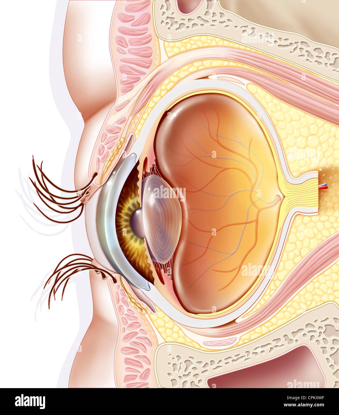 EYE, DRAWING Stock Photohttps://www.alamy.com/image-license-details/?v=1https://www.alamy.com/stock-photo-eye-drawing-48381355.html
RMCPKXWF–EYE, DRAWING
 AMD, ILLUSTRATION Stock Photohttps://www.alamy.com/image-license-details/?v=1https://www.alamy.com/stock-photo-amd-illustration-49285011.html
RMCT53EY–AMD, ILLUSTRATION
 EYE SURGERY, DRAWING Stock Photohttps://www.alamy.com/image-license-details/?v=1https://www.alamy.com/stock-photo-eye-surgery-drawing-54678990.html
RMD4XRH2–EYE SURGERY, DRAWING
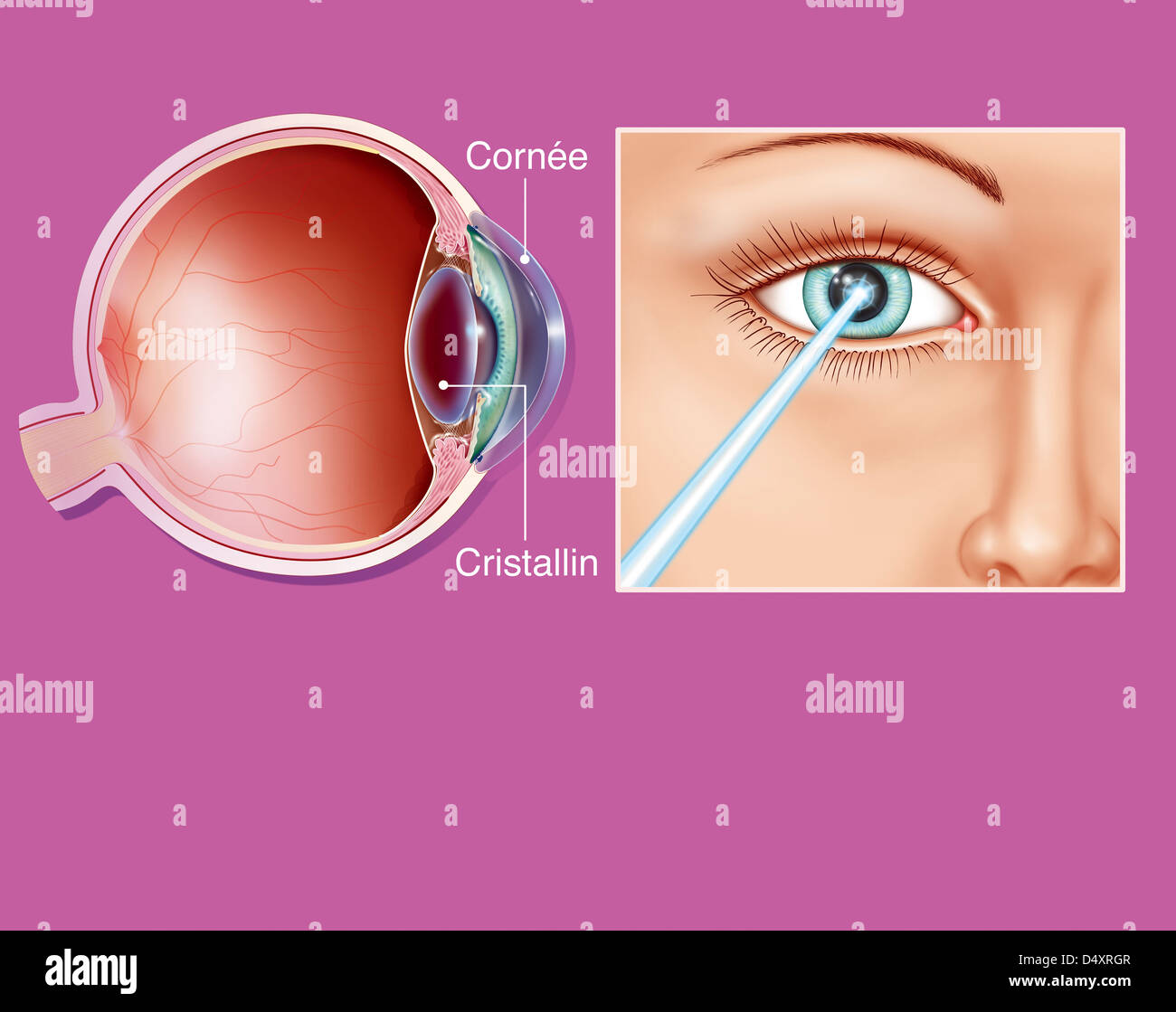 EYE SURGERY, DRAWING Stock Photohttps://www.alamy.com/image-license-details/?v=1https://www.alamy.com/stock-photo-eye-surgery-drawing-54678983.html
RMD4XRGR–EYE SURGERY, DRAWING
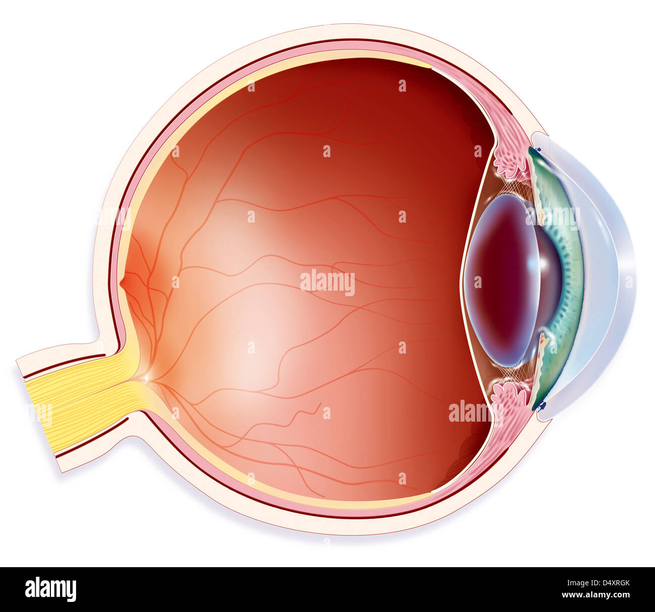 ANATOMY, EYE Stock Photohttps://www.alamy.com/image-license-details/?v=1https://www.alamy.com/stock-photo-anatomy-eye-54678979.html
RMD4XRGK–ANATOMY, EYE
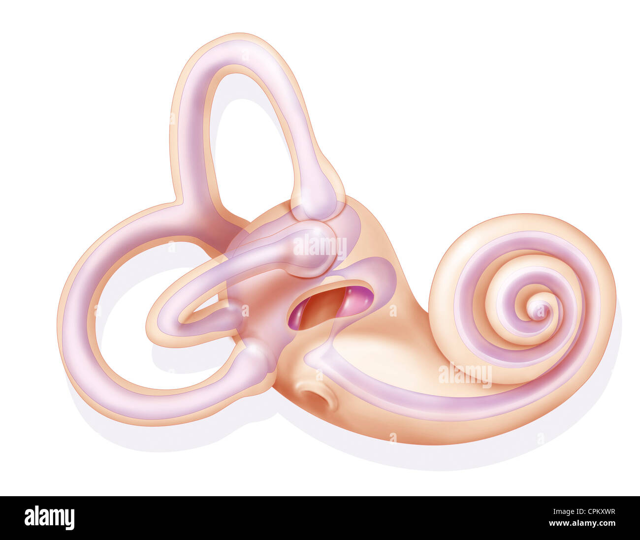 INTERNAL EAR, DRAWING Stock Photohttps://www.alamy.com/image-license-details/?v=1https://www.alamy.com/stock-photo-internal-ear-drawing-48381363.html
RMCPKXWR–INTERNAL EAR, DRAWING
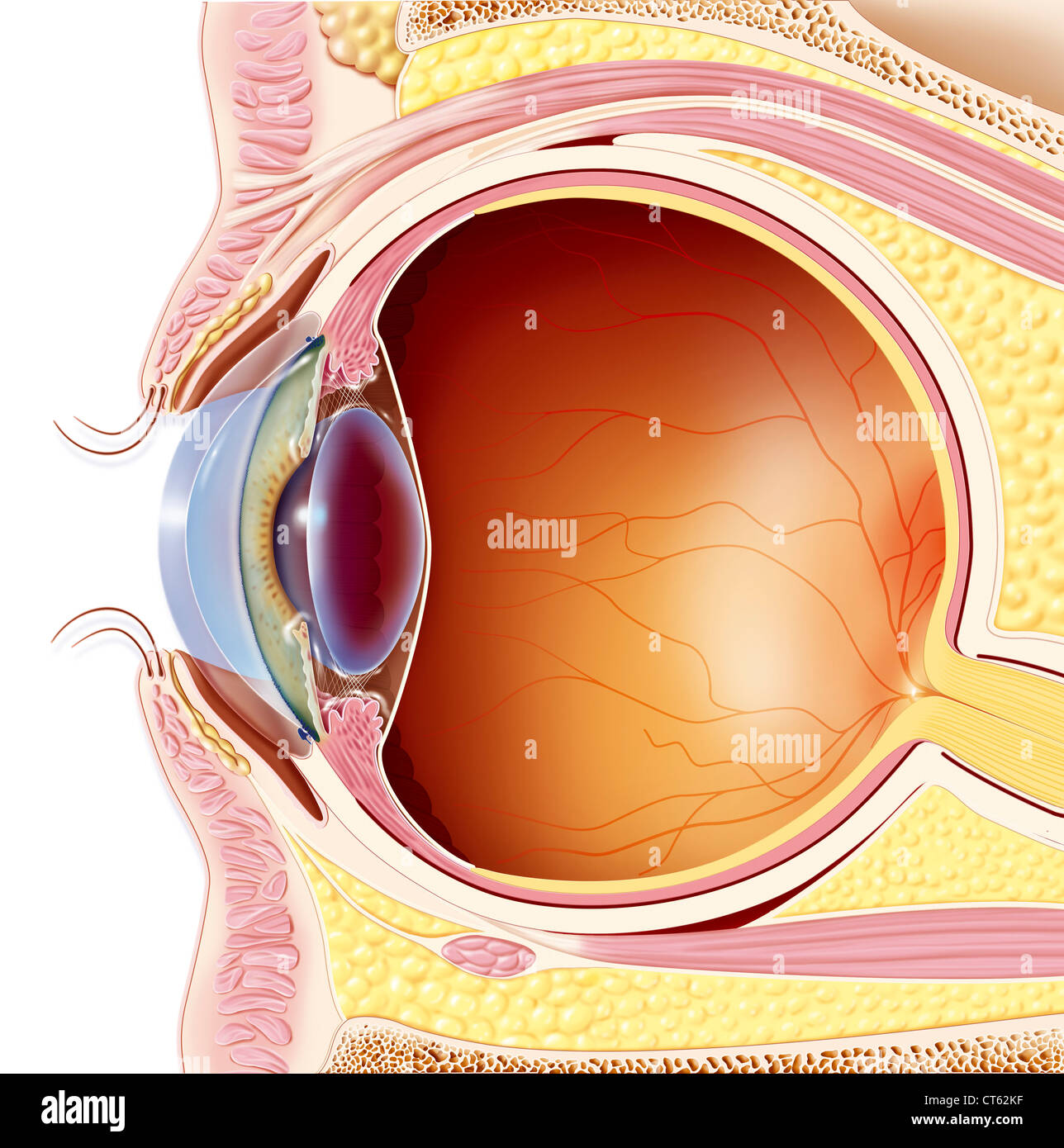 EYE, DRAWING Stock Photohttps://www.alamy.com/image-license-details/?v=1https://www.alamy.com/stock-photo-eye-drawing-49306307.html
RMCT62KF–EYE, DRAWING
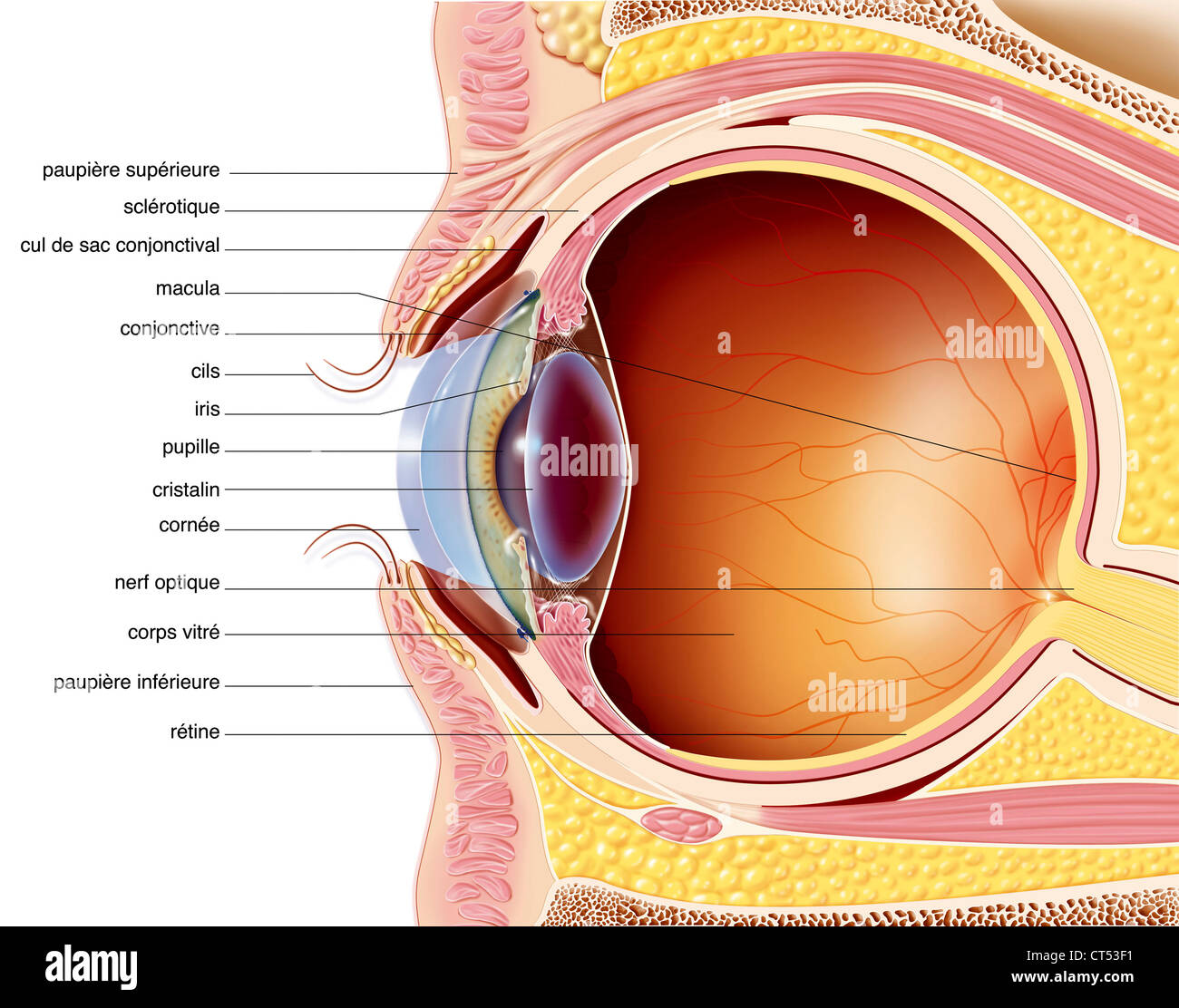 EYE, DRAWING Stock Photohttps://www.alamy.com/image-license-details/?v=1https://www.alamy.com/stock-photo-eye-drawing-49285013.html
RMCT53F1–EYE, DRAWING
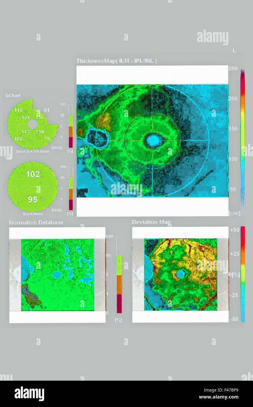
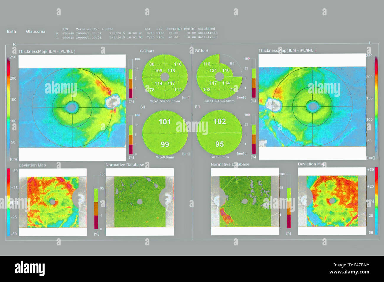
 OPTICAL COHERENCE TOMOGRAPHY Stock Photohttps://www.alamy.com/image-license-details/?v=1https://www.alamy.com/stock-photo-optical-coherence-tomography-88673375.html
RMF47BP7–OPTICAL COHERENCE TOMOGRAPHY
