Quick filters:
Malpighian tubule Stock Photos and Images
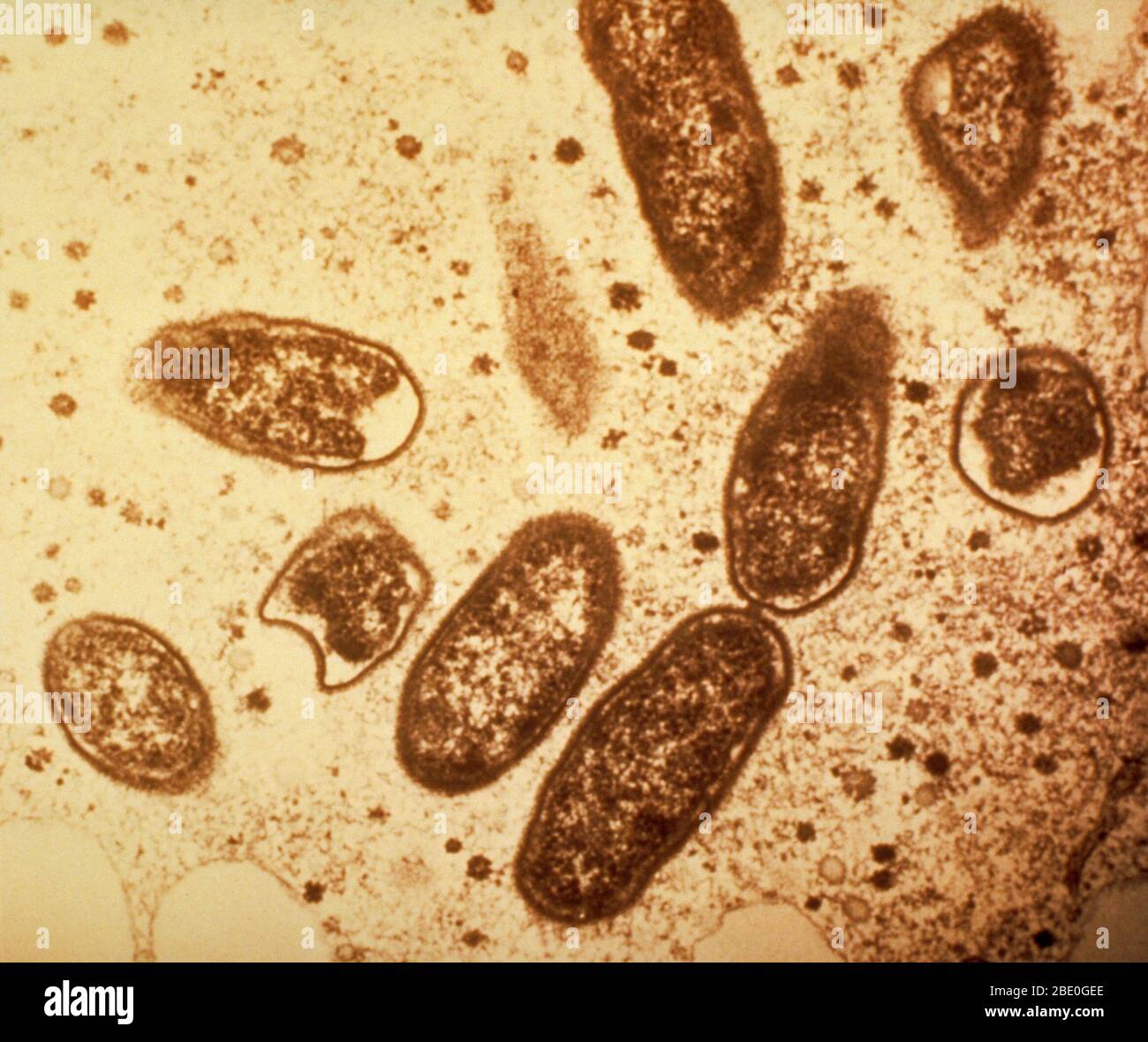 Color enhanced Transmission Electron Micrograph (TEM) of Rickettsia rickettsii in a Malpighian tubule cell of a wood tick (Dermacentor andersoni). Rickettsia rickettsii is a bacteria that causes Rocky Mountain spotted fever in humans. The bacteria is transmitted to humans by wood ticks. Symptoms include fever, headache, muscle pain, and rash. If it is not treated early with antibiotics, the disease can be fatal. Magnification: unknown. Stock Photohttps://www.alamy.com/image-license-details/?v=1https://www.alamy.com/color-enhanced-transmission-electron-micrograph-tem-of-rickettsia-rickettsii-in-a-malpighian-tubule-cell-of-a-wood-tick-dermacentor-andersoni-rickettsia-rickettsii-is-a-bacteria-that-causes-rocky-mountain-spotted-fever-in-humans-the-bacteria-is-transmitted-to-humans-by-wood-ticks-symptoms-include-fever-headache-muscle-pain-and-rash-if-it-is-not-treated-early-with-antibiotics-the-disease-can-be-fatal-magnification-unknown-image352825494.html
RM2BE0GEE–Color enhanced Transmission Electron Micrograph (TEM) of Rickettsia rickettsii in a Malpighian tubule cell of a wood tick (Dermacentor andersoni). Rickettsia rickettsii is a bacteria that causes Rocky Mountain spotted fever in humans. The bacteria is transmitted to humans by wood ticks. Symptoms include fever, headache, muscle pain, and rash. If it is not treated early with antibiotics, the disease can be fatal. Magnification: unknown.
 Malpighian body, vintage engraving. Old engraved illustration of Malpighian body structure with its functioning parts and their Stock Vectorhttps://www.alamy.com/image-license-details/?v=1https://www.alamy.com/stock-photo-malpighian-body-vintage-engraving-old-engraved-illustration-of-malpighian-84431090.html
RFEWA4M2–Malpighian body, vintage engraving. Old engraved illustration of Malpighian body structure with its functioning parts and their
 The house-fly, Musca domestica Linn: its structure, habits, development, relation to disease and control . imaginal disc.f.c. Adipose tissue cell. l.tr. Lateral tracheal trunk. m.t. Malpighian tubule cut rather longitudinally. oe. Oesophagus. jir.d. Prothoracic imaginal disc. v.ms. Ventral mesothoracic imaginal disc. IMAGINAL THORACIC DISCS 149 the jinterior vnd otthr gani^lion ;ui(l slope oblnjudy toiwards; thedistal end of each is attached to the body-wall on the ventral sidebetween segments three and four. These discs develop into theprothoracic legs, and probably also into the much-reduced Stock Photohttps://www.alamy.com/image-license-details/?v=1https://www.alamy.com/the-house-fly-musca-domestica-linn-its-structure-habits-development-relation-to-disease-and-control-imaginal-discfc-adipose-tissue-cell-ltr-lateral-tracheal-trunk-mt-malpighian-tubule-cut-rather-longitudinally-oe-oesophagus-jird-prothoracic-imaginal-disc-vms-ventral-mesothoracic-imaginal-disc-imaginal-thoracic-discs-149-the-jinterior-vnd-otthr-ganilion-uil-slope-oblnjudy-toiwards-thedistal-end-of-each-is-attached-to-the-body-wall-on-the-ventral-sidebetween-segments-three-and-four-these-discs-develop-into-theprothoracic-legs-and-probably-also-into-the-much-reduced-image342786663.html
RM2AWK7TR–The house-fly, Musca domestica Linn: its structure, habits, development, relation to disease and control . imaginal disc.f.c. Adipose tissue cell. l.tr. Lateral tracheal trunk. m.t. Malpighian tubule cut rather longitudinally. oe. Oesophagus. jir.d. Prothoracic imaginal disc. v.ms. Ventral mesothoracic imaginal disc. IMAGINAL THORACIC DISCS 149 the jinterior vnd otthr gani^lion ;ui(l slope oblnjudy toiwards; thedistal end of each is attached to the body-wall on the ventral sidebetween segments three and four. These discs develop into theprothoracic legs, and probably also into the much-reduced
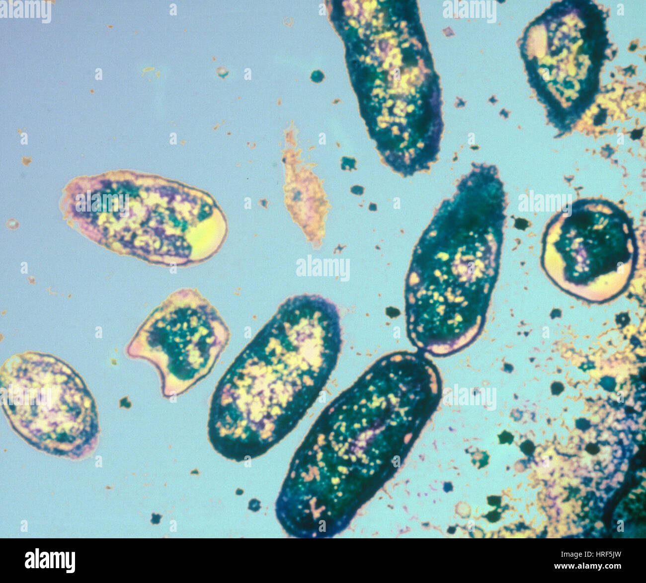 Rickettsia rickettsii, TEM Stock Photohttps://www.alamy.com/image-license-details/?v=1https://www.alamy.com/stock-photo-rickettsia-rickettsii-tem-134943393.html
RMHRF5JW–Rickettsia rickettsii, TEM
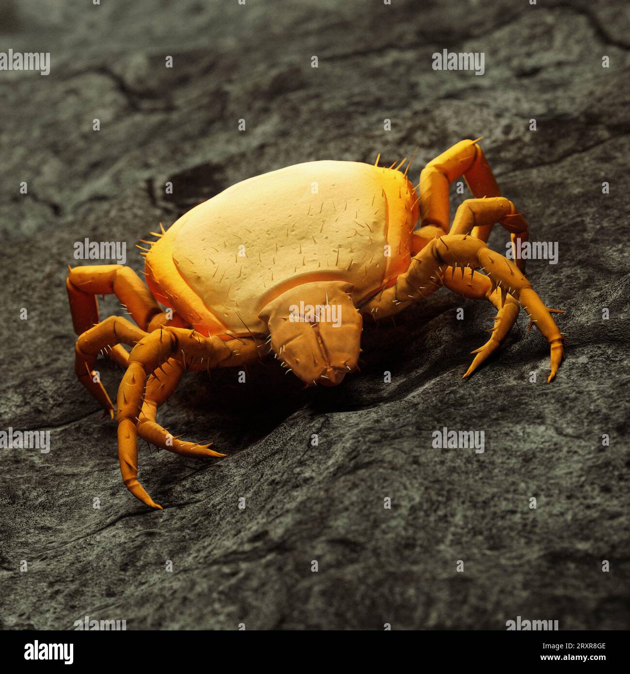 3D illustration of a detailed tick: SEM Electron Microscope Replica in orange colour Stock Photohttps://www.alamy.com/image-license-details/?v=1https://www.alamy.com/3d-illustration-of-a-detailed-tick-sem-electron-microscope-replica-in-orange-colour-image567224462.html
RF2RXR8GE–3D illustration of a detailed tick: SEM Electron Microscope Replica in orange colour
 . Beekeeping; a discussion of the life of the honeybee and of the production of honey. Bees; Honey. The Life Processes of the Individual 145. Fig. 77.—Histological details of alimentary canal of worker: A, cross- section of ventriculus; B, section of ventriciilus wall; C, section of Malpighian tubule; D, cross-section of small intestine; E, section of ventriculiis wall, showing formation of enzjTne cells; F, section of anterior end of rectum, showing rectal glands (RGl); G, slightly oblique section of posterior end of ventriculus, showing openings of Malpighian tubules. larval food is regurgit Stock Photohttps://www.alamy.com/image-license-details/?v=1https://www.alamy.com/beekeeping-a-discussion-of-the-life-of-the-honeybee-and-of-the-production-of-honey-bees-honey-the-life-processes-of-the-individual-145-fig-77histological-details-of-alimentary-canal-of-worker-a-cross-section-of-ventriculus-b-section-of-ventriciilus-wall-c-section-of-malpighian-tubule-d-cross-section-of-small-intestine-e-section-of-ventriculiis-wall-showing-formation-of-enzjtne-cells-f-section-of-anterior-end-of-rectum-showing-rectal-glands-rgl-g-slightly-oblique-section-of-posterior-end-of-ventriculus-showing-openings-of-malpighian-tubules-larval-food-is-regurgit-image216361606.html
RMPG03C6–. Beekeeping; a discussion of the life of the honeybee and of the production of honey. Bees; Honey. The Life Processes of the Individual 145. Fig. 77.—Histological details of alimentary canal of worker: A, cross- section of ventriculus; B, section of ventriciilus wall; C, section of Malpighian tubule; D, cross-section of small intestine; E, section of ventriculiis wall, showing formation of enzjTne cells; F, section of anterior end of rectum, showing rectal glands (RGl); G, slightly oblique section of posterior end of ventriculus, showing openings of Malpighian tubules. larval food is regurgit
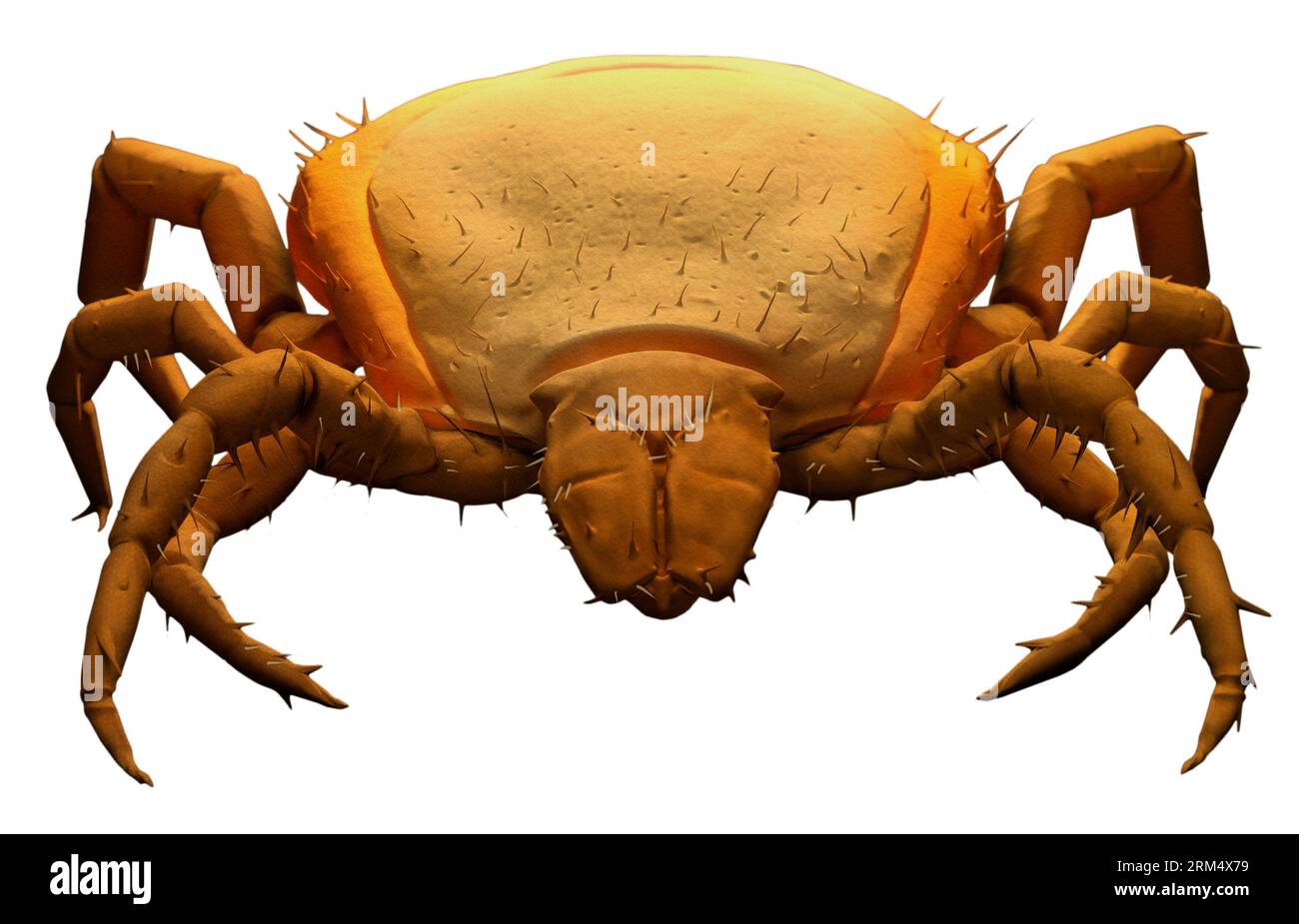 3D illustration of a detailed tick: SEM Electron Microscope CGI Replica in digital orange colour Stock Photohttps://www.alamy.com/image-license-details/?v=1https://www.alamy.com/3d-illustration-of-a-detailed-tick-sem-electron-microscope-cgi-replica-in-digital-orange-colour-image563133293.html
RF2RM4X79–3D illustration of a detailed tick: SEM Electron Microscope CGI Replica in digital orange colour
 Blood circulation kidney drawing Stock Photohttps://www.alamy.com/image-license-details/?v=1https://www.alamy.com/blood-circulation-kidney-drawing-image67545414.html
RMDWTXT6–Blood circulation kidney drawing
 Diagram of blood supply to kidney, vintage engraved illustration. Stock Photohttps://www.alamy.com/image-license-details/?v=1https://www.alamy.com/diagram-of-blood-supply-to-kidney-vintage-engraved-illustration-image363167893.html
RF2C2RM9W–Diagram of blood supply to kidney, vintage engraved illustration.
 Elementary text-book of zoology, general Elementary text-book of zoology, general part and special part: protozoa to insecta elementarytextbo00clau Year: 1892 Fig. 72.—Ciliated funnel and Malpighian body from the anterior part of the kidney of Proteus (after Spengel). Nr, kidney tubule; Tr, ciliated funnel; Mk, Malpig- hian body. Stock Photohttps://www.alamy.com/image-license-details/?v=1https://www.alamy.com/elementary-text-book-of-zoology-general-elementary-text-book-of-zoology-general-part-and-special-part-protozoa-to-insecta-elementarytextbo00clau-year-1892-fig-72ciliated-funnel-and-malpighian-body-from-the-anterior-part-of-the-kidney-of-proteus-after-spengel-nr-kidney-tubule-tr-ciliated-funnel-mk-malpig-hian-body-image239578511.html
RMRWNMRB–Elementary text-book of zoology, general Elementary text-book of zoology, general part and special part: protozoa to insecta elementarytextbo00clau Year: 1892 Fig. 72.—Ciliated funnel and Malpighian body from the anterior part of the kidney of Proteus (after Spengel). Nr, kidney tubule; Tr, ciliated funnel; Mk, Malpig- hian body.
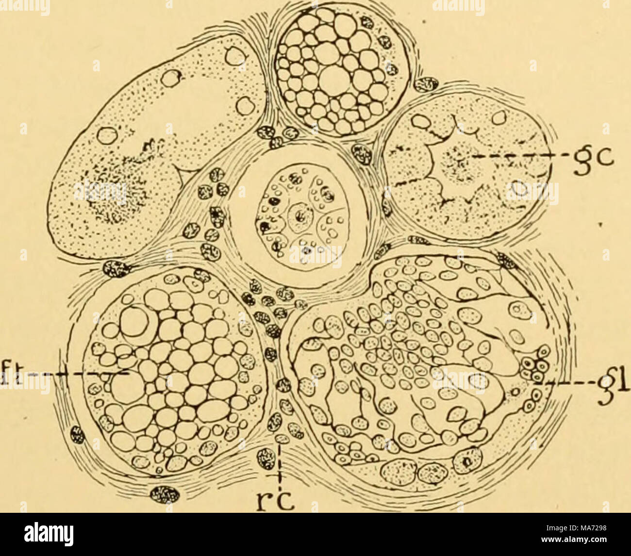 . The effects of inanition and malnutrition upon growth and structure . FlG. 99.—Portion of a section of the kidney from a young fowl on aqueous inanition (dry diet), ft, uriniferous tubules containing epithelial cells with "fatty degeneration;" gc, urinif- erous tubule containing a granular cylinder; gl, an abnormal renal (Malpighian) corpuscle, showing capsule and glomerulus (glomerulitis?); re, stroma, with small round cell infiltration. X400. (Pernice and Scagliosi '95a.) Many uriniferous tubules were also filled with blood, and some contained hyalin cylinders. In many glomeruli Stock Photohttps://www.alamy.com/image-license-details/?v=1https://www.alamy.com/the-effects-of-inanition-and-malnutrition-upon-growth-and-structure-flg-99portion-of-a-section-of-the-kidney-from-a-young-fowl-on-aqueous-inanition-dry-diet-ft-uriniferous-tubules-containing-epithelial-cells-with-quotfatty-degenerationquot-gc-urinif-erous-tubule-containing-a-granular-cylinder-gl-an-abnormal-renal-malpighian-corpuscle-showing-capsule-and-glomerulus-glomerulitis-re-stroma-with-small-round-cell-infiltration-x400-pernice-and-scagliosi-95a-many-uriniferous-tubules-were-also-filled-with-blood-and-some-contained-hyalin-cylinders-in-many-glomeruli-image178405732.html
RMMA7298–. The effects of inanition and malnutrition upon growth and structure . FlG. 99.—Portion of a section of the kidney from a young fowl on aqueous inanition (dry diet), ft, uriniferous tubules containing epithelial cells with "fatty degeneration;" gc, urinif- erous tubule containing a granular cylinder; gl, an abnormal renal (Malpighian) corpuscle, showing capsule and glomerulus (glomerulitis?); re, stroma, with small round cell infiltration. X400. (Pernice and Scagliosi '95a.) Many uriniferous tubules were also filled with blood, and some contained hyalin cylinders. In many glomeruli
 Forest entomology . cerebral ganglion; ch, mesenteron;e, proctodeum; g, genital aperture; h, heart; k, crop ; m, mouth; n, ventral ganglion;sp, salivary gland; u, malpighian tubule ; a, ovary. section of an insect, shows the relative position of the more importantanatomical points from a side view. ^The nervous system of an insect resembles that of most otheranimals, inasmuch as the terminal seat of the nerve-centres is thebrain, Avhence proceed other nerve-centres and nerves all over the body. The nerves receive impres-sions from the outside world, whichreact on the organism, and thus stim-ul Stock Photohttps://www.alamy.com/image-license-details/?v=1https://www.alamy.com/forest-entomology-cerebral-ganglion-ch-mesenterone-proctodeum-g-genital-aperture-h-heart-k-crop-m-mouth-n-ventral-ganglionsp-salivary-gland-u-malpighian-tubule-a-ovary-section-of-an-insect-shows-the-relative-position-of-the-more-importantanatomical-points-from-a-side-view-the-nervous-system-of-an-insect-resembles-that-of-most-otheranimals-inasmuch-as-the-terminal-seat-of-the-nerve-centres-is-thebrain-avhence-proceed-other-nerve-centres-and-nerves-all-over-the-body-the-nerves-receive-impres-sions-from-the-outside-world-whichreact-on-the-organism-and-thus-stim-ul-image340232969.html
RM2ANEXHD–Forest entomology . cerebral ganglion; ch, mesenteron;e, proctodeum; g, genital aperture; h, heart; k, crop ; m, mouth; n, ventral ganglion;sp, salivary gland; u, malpighian tubule ; a, ovary. section of an insect, shows the relative position of the more importantanatomical points from a side view. ^The nervous system of an insect resembles that of most otheranimals, inasmuch as the terminal seat of the nerve-centres is thebrain, Avhence proceed other nerve-centres and nerves all over the body. The nerves receive impres-sions from the outside world, whichreact on the organism, and thus stim-ul
 . The anatomy of the domestic fowl . Domestic animals; Veterinary medicine; Poultry. THE TJRO-GENITAL SYSTEM 171 lobules. The renal artery (Fig. 53, No. 5,, 3) breaking up into arterioles in the. kidney, and finally reaching the cortical portion of the lobules, form capillary plexuses in the shape of minute spheres, which are the glomerules (Fig. 53, No. C, 3 and B, 5). Around each glomerule there is formed a capsule called Boisrman's capsule, which is the beginning of the uriniferous tubule. This entire mass is called the Malpighian body, or renal corpuscle. This capsule then ex- tends as the Stock Photohttps://www.alamy.com/image-license-details/?v=1https://www.alamy.com/the-anatomy-of-the-domestic-fowl-domestic-animals-veterinary-medicine-poultry-the-tjro-genital-system-171-lobules-the-renal-artery-fig-53-no-5-3-breaking-up-into-arterioles-in-the-kidney-and-finally-reaching-the-cortical-portion-of-the-lobules-form-capillary-plexuses-in-the-shape-of-minute-spheres-which-are-the-glomerules-fig-53-no-c-3-and-b-5-around-each-glomerule-there-is-formed-a-capsule-called-boisrmans-capsule-which-is-the-beginning-of-the-uriniferous-tubule-this-entire-mass-is-called-the-malpighian-body-or-renal-corpuscle-this-capsule-then-ex-tends-as-the-image216389186.html
RMPG1AH6–. The anatomy of the domestic fowl . Domestic animals; Veterinary medicine; Poultry. THE TJRO-GENITAL SYSTEM 171 lobules. The renal artery (Fig. 53, No. 5,, 3) breaking up into arterioles in the. kidney, and finally reaching the cortical portion of the lobules, form capillary plexuses in the shape of minute spheres, which are the glomerules (Fig. 53, No. C, 3 and B, 5). Around each glomerule there is formed a capsule called Boisrman's capsule, which is the beginning of the uriniferous tubule. This entire mass is called the Malpighian body, or renal corpuscle. This capsule then ex- tends as the
 Blood circulation kidney drawing Stock Photohttps://www.alamy.com/image-license-details/?v=1https://www.alamy.com/blood-circulation-kidney-drawing-image67545421.html
RMDWTXTD–Blood circulation kidney drawing
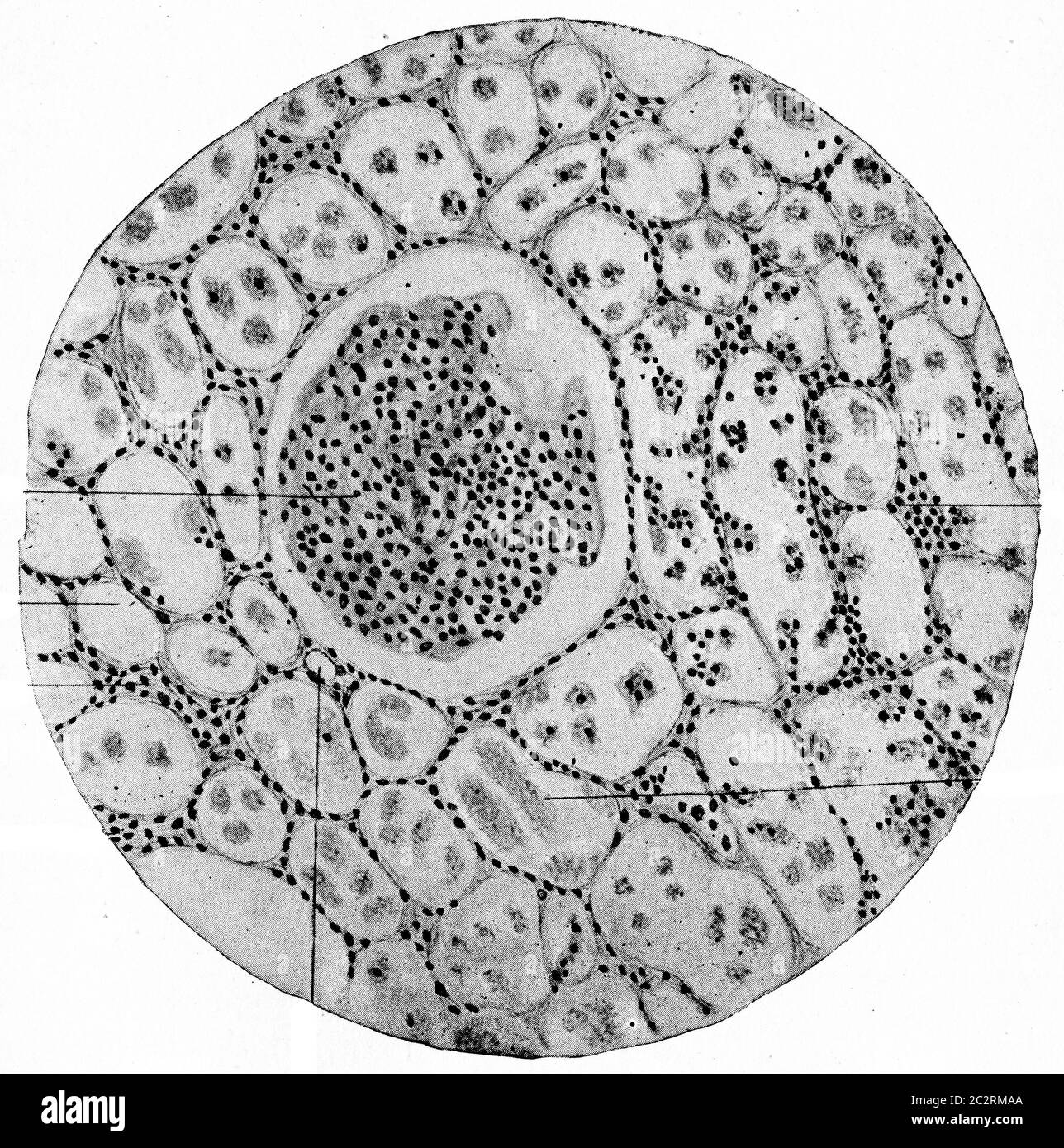 Chronic parenchymatous nephritis, vintage engraved illustration. Stock Photohttps://www.alamy.com/image-license-details/?v=1https://www.alamy.com/chronic-parenchymatous-nephritis-vintage-engraved-illustration-image363167906.html
RF2C2RMAA–Chronic parenchymatous nephritis, vintage engraved illustration.
 Quarterly journal of microscopical science . oc. I. Fig. 4.—Crypt of stomach of mature larva. Duboscq, iron haema-toxvlin. Zeiss, ob. 12 horn. imm.. oc. 1. Fig. 5.—Oblique section of Malpighian tubule of mature larva. In theprotoplasm of cells conspicuous differentiation of layers and copiousaccumulation of pigment grains [pg). Duboscq, (una- Polychr.-Methylenblau. Zeiss, ob. „ horn. imm.. oc. Fig. 6.—Same, in a larva of first stage. Duboscq. if xyliri. Zci--. ob. /,, horn. imm.. oc. 0. Fig. 7.—Hair rht) of chaetoid type from back of larva. Und< r th ? hairi; the usual hypodermic cell (hp) Stock Photohttps://www.alamy.com/image-license-details/?v=1https://www.alamy.com/quarterly-journal-of-microscopical-science-oc-i-fig-4crypt-of-stomach-of-mature-larva-duboscq-iron-haema-toxvlin-zeiss-ob-12-horn-imm-oc-1-fig-5oblique-section-of-malpighian-tubule-of-mature-larva-in-theprotoplasm-of-cells-conspicuous-differentiation-of-layers-and-copiousaccumulation-of-pigment-grains-pg-duboscq-una-polychr-methylenblau-zeiss-ob-horn-imm-oc-fig-6same-in-a-larva-of-first-stage-duboscq-if-xyliri-zci-ob-horn-imm-oc-0-fig-7hair-rht-of-chaetoid-type-from-back-of-larva-undlt-r-th-hairi-the-usual-hypodermic-cell-hp-image340019267.html
RM2AN5617–Quarterly journal of microscopical science . oc. I. Fig. 4.—Crypt of stomach of mature larva. Duboscq, iron haema-toxvlin. Zeiss, ob. 12 horn. imm.. oc. 1. Fig. 5.—Oblique section of Malpighian tubule of mature larva. In theprotoplasm of cells conspicuous differentiation of layers and copiousaccumulation of pigment grains [pg). Duboscq, (una- Polychr.-Methylenblau. Zeiss, ob. „ horn. imm.. oc. Fig. 6.—Same, in a larva of first stage. Duboscq. if xyliri. Zci--. ob. /,, horn. imm.. oc. 0. Fig. 7.—Hair rht) of chaetoid type from back of larva. Und< r th ? hairi; the usual hypodermic cell (hp)
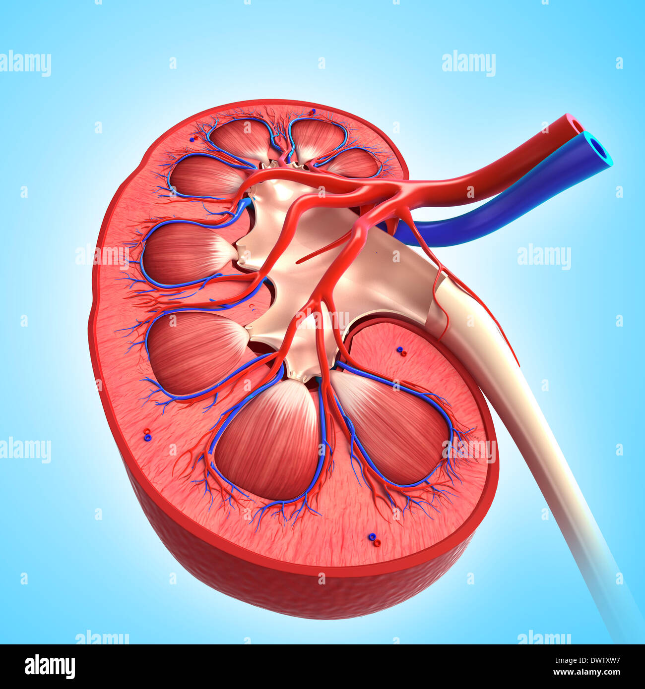 Blood circulation kidney drawing Stock Photohttps://www.alamy.com/image-license-details/?v=1https://www.alamy.com/blood-circulation-kidney-drawing-image67545443.html
RMDWTXW7–Blood circulation kidney drawing
 . The biology of the Protozoa. Protozoa; Protozoa. 234 BIOLOGY OF THE PROTOZOA. Fig 115—Gamete formation and fertilization in Ophryocystis mesnili. A, two individuals attached by processes to ciliated cells of a Malpighian tubule of Tenehrio viollitor; B, union of gamonts in pseudoconjugation; C, D, E, probable meiotic divisions of nuclei of the two gamonts; G to K, formation of two gametes and their union in fertilization; L to A^, metagamic divisions resulting in eight sporozoites m the single sporoblast. (After Leger.). Please note that these images are extracted from scanned page images th Stock Photohttps://www.alamy.com/image-license-details/?v=1https://www.alamy.com/the-biology-of-the-protozoa-protozoa-protozoa-234-biology-of-the-protozoa-fig-115gamete-formation-and-fertilization-in-ophryocystis-mesnili-a-two-individuals-attached-by-processes-to-ciliated-cells-of-a-malpighian-tubule-of-tenehrio-viollitor-b-union-of-gamonts-in-pseudoconjugation-c-d-e-probable-meiotic-divisions-of-nuclei-of-the-two-gamonts-g-to-k-formation-of-two-gametes-and-their-union-in-fertilization-l-to-a-metagamic-divisions-resulting-in-eight-sporozoites-m-the-single-sporoblast-after-leger-please-note-that-these-images-are-extracted-from-scanned-page-images-th-image234603086.html
RMRHK2HJ–. The biology of the Protozoa. Protozoa; Protozoa. 234 BIOLOGY OF THE PROTOZOA. Fig 115—Gamete formation and fertilization in Ophryocystis mesnili. A, two individuals attached by processes to ciliated cells of a Malpighian tubule of Tenehrio viollitor; B, union of gamonts in pseudoconjugation; C, D, E, probable meiotic divisions of nuclei of the two gamonts; G to K, formation of two gametes and their union in fertilization; L to A^, metagamic divisions resulting in eight sporozoites m the single sporoblast. (After Leger.). Please note that these images are extracted from scanned page images th
 Blood circulation kidney drawing Stock Photohttps://www.alamy.com/image-license-details/?v=1https://www.alamy.com/blood-circulation-kidney-drawing-image67545418.html
RMDWTXTA–Blood circulation kidney drawing
 . African Ixodoidea. l. Ticks of the Sudan... Ticks -- Sudan. Central ganglion Coxal organ Accessory gland Filter — ,chamber,< r Oviduct Malpighian tubule -V .Gene's orgarvx?^'^"'^ Oesophagus -V^. Salivary gland Stomach and diverticula Rectal ampulla 56 Figure 56 9 CiRNITHODCROS MOUBATA mTEENAL CEGMIS /After Burgdorfer (1951)> with permission of the editor of ACTA TROPICA 7 PLATE XIX - 157-. Please note that these images are extracted from scanned page images that may have been digitally enhanced for readability - coloration and appearance of these illustrations may not perfectly Stock Photohttps://www.alamy.com/image-license-details/?v=1https://www.alamy.com/african-ixodoidea-l-ticks-of-the-sudan-ticks-sudan-central-ganglion-coxal-organ-accessory-gland-filter-chamberlt-r-oviduct-malpighian-tubule-v-genes-orgarvxquot-oesophagus-v-salivary-gland-stomach-and-diverticula-rectal-ampulla-56-figure-56-9-cirnithodcros-moubata-mteenal-cegmis-after-burgdorfer-1951gt-with-permission-of-the-editor-of-acta-tropica-7-plate-xix-157-please-note-that-these-images-are-extracted-from-scanned-page-images-that-may-have-been-digitally-enhanced-for-readability-coloration-and-appearance-of-these-illustrations-may-not-perfectly-image237913419.html
RMRR1TYR–. African Ixodoidea. l. Ticks of the Sudan... Ticks -- Sudan. Central ganglion Coxal organ Accessory gland Filter — ,chamber,< r Oviduct Malpighian tubule -V .Gene's orgarvx?^'^"'^ Oesophagus -V^. Salivary gland Stomach and diverticula Rectal ampulla 56 Figure 56 9 CiRNITHODCROS MOUBATA mTEENAL CEGMIS /After Burgdorfer (1951)> with permission of the editor of ACTA TROPICA 7 PLATE XIX - 157-. Please note that these images are extracted from scanned page images that may have been digitally enhanced for readability - coloration and appearance of these illustrations may not perfectly
 Blood circulation kidney drawing Stock Photohttps://www.alamy.com/image-license-details/?v=1https://www.alamy.com/blood-circulation-kidney-drawing-image67545424.html
RMDWTXTG–Blood circulation kidney drawing
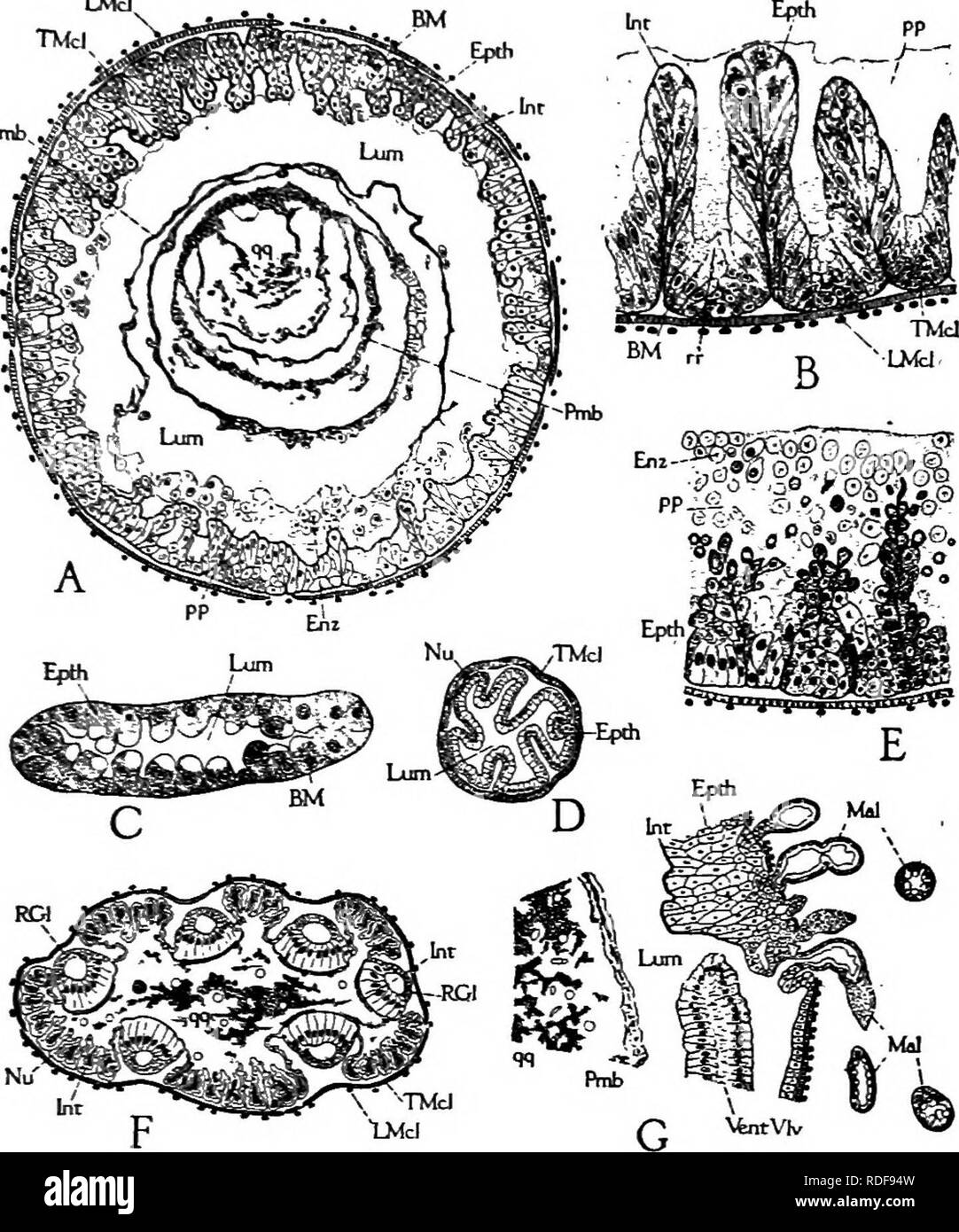 . Beekeeping; a discussion of the life of the honeybee and of the production of honey. Bees; Honey. The Life Processes of the Individual 145. Fig. 77.—Histological details of alimentary canal of worker: A, cross- section of ventriculus; B, section of ventriciilus wall; C, section of Malpighian tubule; D, cross-section of small intestine; E, section of ventriculiis wall, showing formation of enzjTne cells; F, section of anterior end of rectum, showing rectal glands (RGl); G, slightly oblique section of posterior end of ventriculus, showing openings of Malpighian tubules. larval food is regurgit Stock Photohttps://www.alamy.com/image-license-details/?v=1https://www.alamy.com/beekeeping-a-discussion-of-the-life-of-the-honeybee-and-of-the-production-of-honey-bees-honey-the-life-processes-of-the-individual-145-fig-77histological-details-of-alimentary-canal-of-worker-a-cross-section-of-ventriculus-b-section-of-ventriciilus-wall-c-section-of-malpighian-tubule-d-cross-section-of-small-intestine-e-section-of-ventriculiis-wall-showing-formation-of-enzjtne-cells-f-section-of-anterior-end-of-rectum-showing-rectal-glands-rgl-g-slightly-oblique-section-of-posterior-end-of-ventriculus-showing-openings-of-malpighian-tubules-larval-food-is-regurgit-image232061785.html
RMRDF94W–. Beekeeping; a discussion of the life of the honeybee and of the production of honey. Bees; Honey. The Life Processes of the Individual 145. Fig. 77.—Histological details of alimentary canal of worker: A, cross- section of ventriculus; B, section of ventriciilus wall; C, section of Malpighian tubule; D, cross-section of small intestine; E, section of ventriculiis wall, showing formation of enzjTne cells; F, section of anterior end of rectum, showing rectal glands (RGl); G, slightly oblique section of posterior end of ventriculus, showing openings of Malpighian tubules. larval food is regurgit
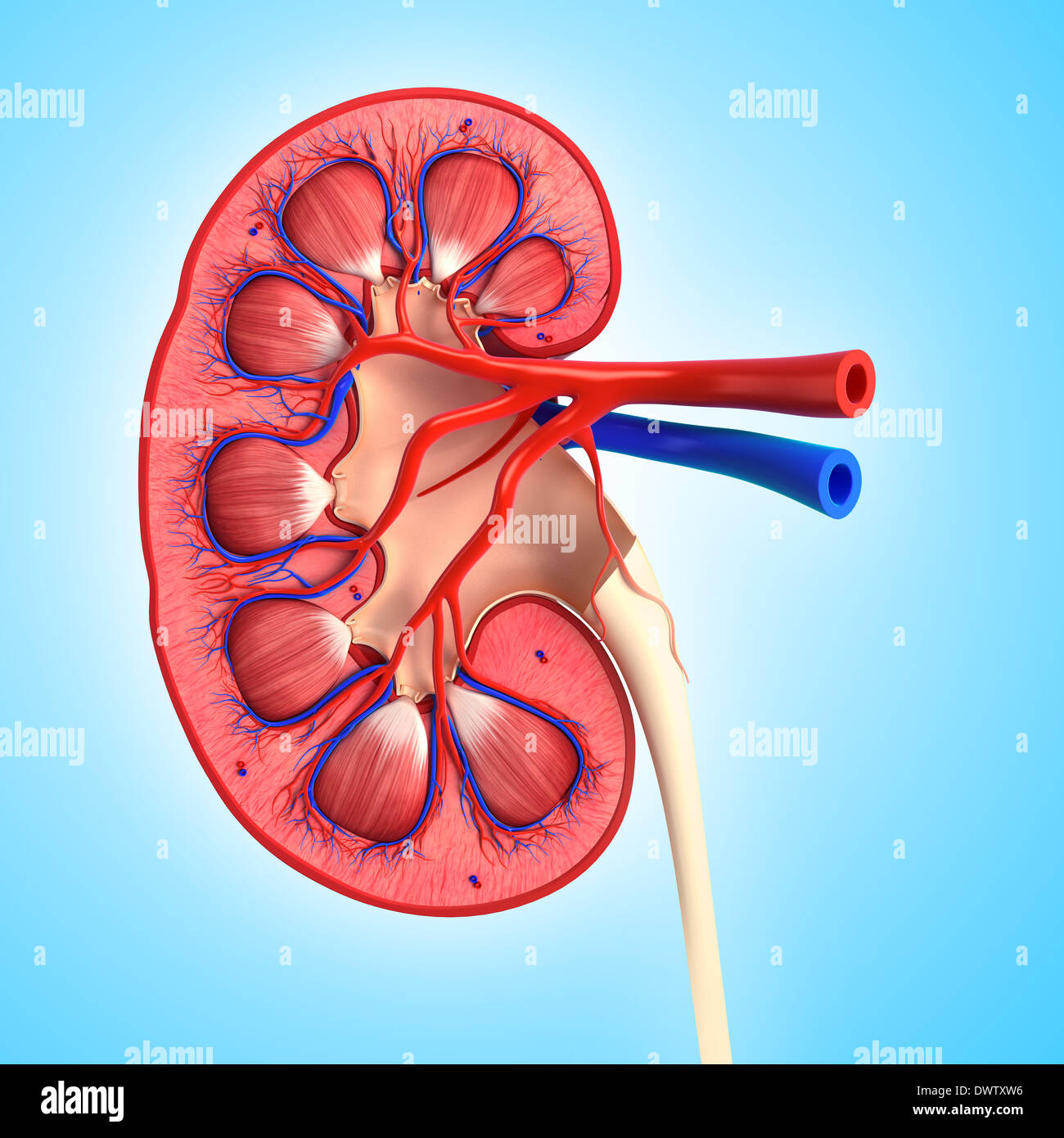 Blood circulation kidney drawing Stock Photohttps://www.alamy.com/image-license-details/?v=1https://www.alamy.com/blood-circulation-kidney-drawing-image67545442.html
RMDWTXW6–Blood circulation kidney drawing
 . Beekeeping; a discussion of the life of the honeybee and of the production of honey. Bee culture; Honey. The Life Processes of the Individual 145 LMcl Epth Pmb. Fig. 77.—Histological details of alimentary canal of worker: A, cross- section of ventriculus; B, section of ventriculus wall; C, section of Malpighian tubule ; D, cross-section of small intestine ; E, section of ventriculus wall, showing formation of enzyme cells; F, section of anterior end of rectum, showing rectal glands (RGl) ; G, slightly oblique section of posterior end of ventriculus, showing openings of Malpighian tubules. la Stock Photohttps://www.alamy.com/image-license-details/?v=1https://www.alamy.com/beekeeping-a-discussion-of-the-life-of-the-honeybee-and-of-the-production-of-honey-bee-culture-honey-the-life-processes-of-the-individual-145-lmcl-epth-pmb-fig-77histological-details-of-alimentary-canal-of-worker-a-cross-section-of-ventriculus-b-section-of-ventriculus-wall-c-section-of-malpighian-tubule-d-cross-section-of-small-intestine-e-section-of-ventriculus-wall-showing-formation-of-enzyme-cells-f-section-of-anterior-end-of-rectum-showing-rectal-glands-rgl-g-slightly-oblique-section-of-posterior-end-of-ventriculus-showing-openings-of-malpighian-tubules-la-image235166139.html
RMRJGMPK–. Beekeeping; a discussion of the life of the honeybee and of the production of honey. Bee culture; Honey. The Life Processes of the Individual 145 LMcl Epth Pmb. Fig. 77.—Histological details of alimentary canal of worker: A, cross- section of ventriculus; B, section of ventriculus wall; C, section of Malpighian tubule ; D, cross-section of small intestine ; E, section of ventriculus wall, showing formation of enzyme cells; F, section of anterior end of rectum, showing rectal glands (RGl) ; G, slightly oblique section of posterior end of ventriculus, showing openings of Malpighian tubules. la
 Blood circulation kidney drawing Stock Photohttps://www.alamy.com/image-license-details/?v=1https://www.alamy.com/blood-circulation-kidney-drawing-image67545423.html
RMDWTXTF–Blood circulation kidney drawing
 . The biology of the Protozoa. Protozoa; Protozoa. Fig. 180.—Ophryocystis mesnili, gamete formation and fertilization. A, Two individuals attached by processes to epithelial cells of a Malpighian tubule of Tene- brio mollitor; B, union of gamonts in pseudoconjugation; C, D, E, divisions (probably meiotic) of nuclei of the two gamonts; G to K, formation of two gametes and their union in fertilization; L to N, metagamic divisions resulting in eight sporozoites in the single sporoblast. (After Leger.). Please note that these images are extracted from scanned page images that may have been digital Stock Photohttps://www.alamy.com/image-license-details/?v=1https://www.alamy.com/the-biology-of-the-protozoa-protozoa-protozoa-fig-180ophryocystis-mesnili-gamete-formation-and-fertilization-a-two-individuals-attached-by-processes-to-epithelial-cells-of-a-malpighian-tubule-of-tene-brio-mollitor-b-union-of-gamonts-in-pseudoconjugation-c-d-e-divisions-probably-meiotic-of-nuclei-of-the-two-gamonts-g-to-k-formation-of-two-gametes-and-their-union-in-fertilization-l-to-n-metagamic-divisions-resulting-in-eight-sporozoites-in-the-single-sporoblast-after-leger-please-note-that-these-images-are-extracted-from-scanned-page-images-that-may-have-been-digital-image234601367.html
RMRHK0C7–. The biology of the Protozoa. Protozoa; Protozoa. Fig. 180.—Ophryocystis mesnili, gamete formation and fertilization. A, Two individuals attached by processes to epithelial cells of a Malpighian tubule of Tene- brio mollitor; B, union of gamonts in pseudoconjugation; C, D, E, divisions (probably meiotic) of nuclei of the two gamonts; G to K, formation of two gametes and their union in fertilization; L to N, metagamic divisions resulting in eight sporozoites in the single sporoblast. (After Leger.). Please note that these images are extracted from scanned page images that may have been digital
 . The biology of the protozoa. Protozoa; Protozoa. REPRODUCTION 281. K L M Fig. 120.—Gamete formation and fertilization in Ophryocystis mesnili. A, two individuals attached by processes to ciliated cells of a Malpighian tubule of Tenebrio mollitor; B, union of gamonts in pseudoconjligation; C, D, E, probable meiotie divisions of nuclei of the two gamonts; G to K, formation of two gametes and their union in fertilization; L to N, metagamic divisions resulting in eight sporozoites in the single sporoblast. (After Leger.). Please note that these images are extracted from scanned page images that Stock Photohttps://www.alamy.com/image-license-details/?v=1https://www.alamy.com/the-biology-of-the-protozoa-protozoa-protozoa-reproduction-281-k-l-m-fig-120gamete-formation-and-fertilization-in-ophryocystis-mesnili-a-two-individuals-attached-by-processes-to-ciliated-cells-of-a-malpighian-tubule-of-tenebrio-mollitor-b-union-of-gamonts-in-pseudoconjligation-c-d-e-probable-meiotie-divisions-of-nuclei-of-the-two-gamonts-g-to-k-formation-of-two-gametes-and-their-union-in-fertilization-l-to-n-metagamic-divisions-resulting-in-eight-sporozoites-in-the-single-sporoblast-after-leger-please-note-that-these-images-are-extracted-from-scanned-page-images-that-image234602899.html
RMRHK2AY–. The biology of the protozoa. Protozoa; Protozoa. REPRODUCTION 281. K L M Fig. 120.—Gamete formation and fertilization in Ophryocystis mesnili. A, two individuals attached by processes to ciliated cells of a Malpighian tubule of Tenebrio mollitor; B, union of gamonts in pseudoconjligation; C, D, E, probable meiotie divisions of nuclei of the two gamonts; G to K, formation of two gametes and their union in fertilization; L to N, metagamic divisions resulting in eight sporozoites in the single sporoblast. (After Leger.). Please note that these images are extracted from scanned page images that
 . Bulletin of the Museum of Comparative Zoology at Harvard College. Zoology. EISNER: ANT PROVENTRICL'LUS 44:{ sal. gl Mssjea*. Fig. 1. Digestive tract of the ant Myrmica rubra (simplified, with some cephalic structures omitted; after Janet). Abbreviations: ant. int., anterior intestine; b. c, buccal cavity; cr., crop; inf. chb., infrabuccal chamber; m. <j., midgut; m. t., malpighian tubule; oes., oesophagus; pny., pharynx; pv., proventrieulus; rec, rectum; sal. dct., salivary duct; sal. ffl., salivary gland. Fig. 2. Proventrieulus of Apis (after Snodgrass, relabelled). Abbrevia- tions: bl., Stock Photohttps://www.alamy.com/image-license-details/?v=1https://www.alamy.com/bulletin-of-the-museum-of-comparative-zoology-at-harvard-college-zoology-eisner-ant-proventricllus-44-sal-gl-mssjea-fig-1-digestive-tract-of-the-ant-myrmica-rubra-simplified-with-some-cephalic-structures-omitted-after-janet-abbreviations-ant-int-anterior-intestine-b-c-buccal-cavity-cr-crop-inf-chb-infrabuccal-chamber-m-ltj-midgut-m-t-malpighian-tubule-oes-oesophagus-pny-pharynx-pv-proventrieulus-rec-rectum-sal-dct-salivary-duct-sal-ffl-salivary-gland-fig-2-proventrieulus-of-apis-after-snodgrass-relabelled-abbrevia-tions-bl-image233863031.html
RMRGDAK3–. Bulletin of the Museum of Comparative Zoology at Harvard College. Zoology. EISNER: ANT PROVENTRICL'LUS 44:{ sal. gl Mssjea*. Fig. 1. Digestive tract of the ant Myrmica rubra (simplified, with some cephalic structures omitted; after Janet). Abbreviations: ant. int., anterior intestine; b. c, buccal cavity; cr., crop; inf. chb., infrabuccal chamber; m. <j., midgut; m. t., malpighian tubule; oes., oesophagus; pny., pharynx; pv., proventrieulus; rec, rectum; sal. dct., salivary duct; sal. ffl., salivary gland. Fig. 2. Proventrieulus of Apis (after Snodgrass, relabelled). Abbrevia- tions: bl.,
 . Annals. Entomology. Fig. 1. Ephedriis incompletus Prov. a, Egg; b, Reproductive system; 1, Alkaline gland; 2, Ovaries; 3 Poison glands; 4, Receptacle. Fig. 2. Aphidius phorodontis Ash. a, Second stage larva; 1, Stomach; 2, Nervous system; 3, Rudimentary intestine; 4, CEsophagus; 5, Mandible; b, Fourth stage larva; 1, Stomach; 2, Silk gland; 3, Nervous system; 4, Malpighian tubule; 5, Oesophagus; 6, Mouth; 7, Anus; 8, Silk gland ori- fice; 9, Gonad; 10, Intestine; 11, Blind extremity of the stomach. The female organs of Aphidius phorodontis Ash. and Ephedrus incompletus Prov. were examined an Stock Photohttps://www.alamy.com/image-license-details/?v=1https://www.alamy.com/annals-entomology-fig-1-ephedriis-incompletus-prov-a-egg-b-reproductive-system-1-alkaline-gland-2-ovaries-3-poison-glands-4-receptacle-fig-2-aphidius-phorodontis-ash-a-second-stage-larva-1-stomach-2-nervous-system-3-rudimentary-intestine-4-cesophagus-5-mandible-b-fourth-stage-larva-1-stomach-2-silk-gland-3-nervous-system-4-malpighian-tubule-5-oesophagus-6-mouth-7-anus-8-silk-gland-ori-fice-9-gonad-10-intestine-11-blind-extremity-of-the-stomach-the-female-organs-of-aphidius-phorodontis-ash-and-ephedrus-incompletus-prov-were-examined-an-image236519097.html
RMRMPAEH–. Annals. Entomology. Fig. 1. Ephedriis incompletus Prov. a, Egg; b, Reproductive system; 1, Alkaline gland; 2, Ovaries; 3 Poison glands; 4, Receptacle. Fig. 2. Aphidius phorodontis Ash. a, Second stage larva; 1, Stomach; 2, Nervous system; 3, Rudimentary intestine; 4, CEsophagus; 5, Mandible; b, Fourth stage larva; 1, Stomach; 2, Silk gland; 3, Nervous system; 4, Malpighian tubule; 5, Oesophagus; 6, Mouth; 7, Anus; 8, Silk gland ori- fice; 9, Gonad; 10, Intestine; 11, Blind extremity of the stomach. The female organs of Aphidius phorodontis Ash. and Ephedrus incompletus Prov. were examined an
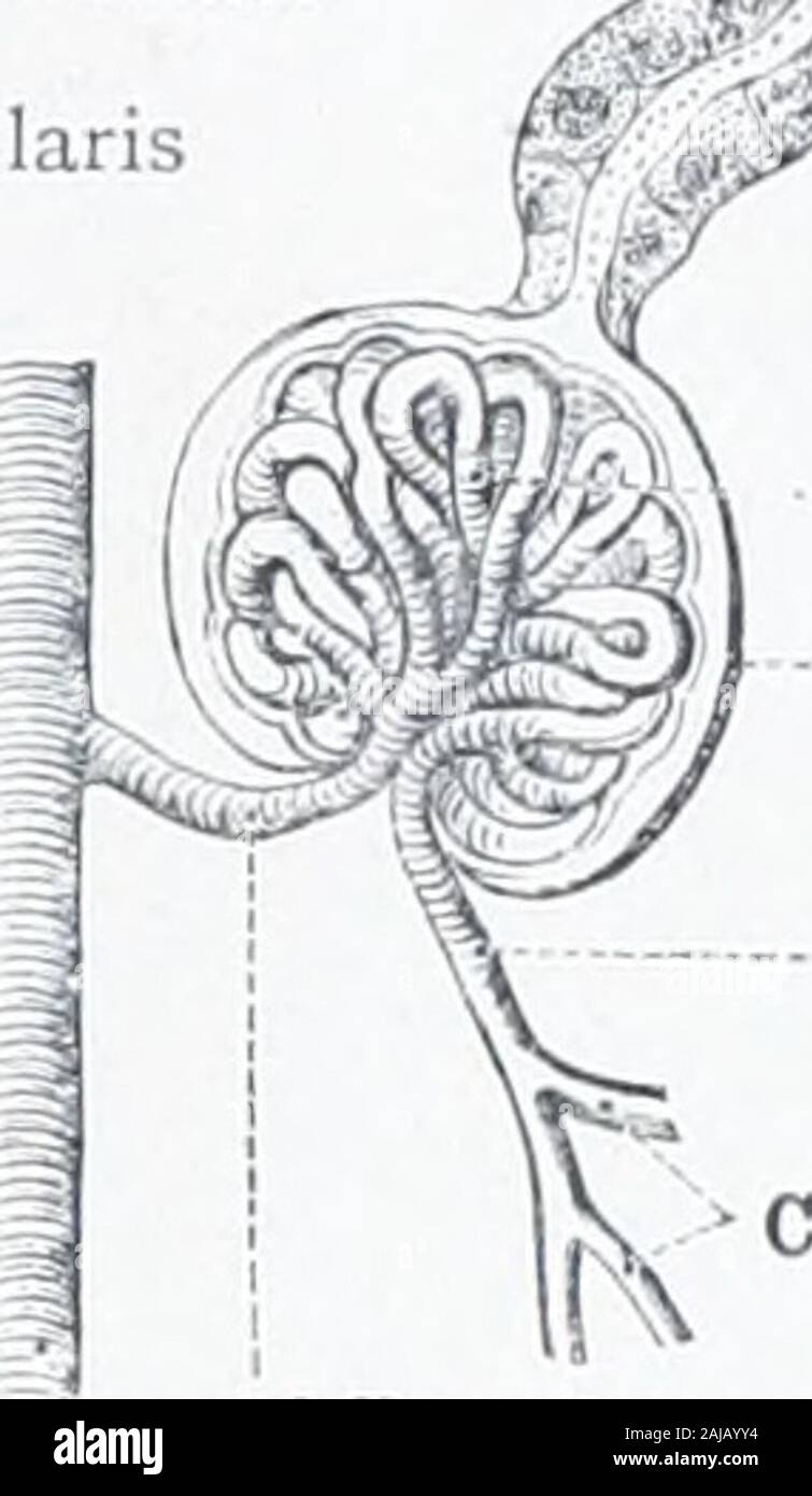 An atlas of human anatomy for students and physicians . Tubuli renalcs contorti -- ^Malpighian corpuscles ^ (.<.ir[uiscula renis (Malpighii) Interlobular or radiateartery/ ^ A. interlobularis. I First convoluted tubule I Tiibnlus renalis contortus -Glomerulus Capsule of theglomerulus (Bowmanscapsule)Capsula glomeruli-- Efferent vessel ofthe glomerulusVas tllercnsCapillary vessels Straight tubules Tubuli renalesrecti Afferent vessel of the glomerulus Vas afferens Interlobular or radiate arteryA. interlobularis Fig. 833.—Part of a Sfxtion through the CortexOF the Kidney in the direction of t Stock Photohttps://www.alamy.com/image-license-details/?v=1https://www.alamy.com/an-atlas-of-human-anatomy-for-students-and-physicians-tubuli-renalcs-contorti-malpighian-corpuscles-ltir-uiscula-renis-malpighii-interlobular-or-radiateartery-a-interlobularis-i-first-convoluted-tubule-i-tiibnlus-renalis-contortus-glomerulus-capsule-of-theglomerulus-bowmanscapsulecapsula-glomeruli-efferent-vessel-ofthe-glomerulusvas-tllercnscapillary-vessels-straight-tubules-tubuli-renalesrecti-afferent-vessel-of-the-glomerulus-vas-afferens-interlobular-or-radiate-arterya-interlobularis-fig-833part-of-a-sfxtion-through-the-cortexof-the-kidney-in-the-direction-of-t-image338302248.html
RM2AJAYY4–An atlas of human anatomy for students and physicians . Tubuli renalcs contorti -- ^Malpighian corpuscles ^ (.<.ir[uiscula renis (Malpighii) Interlobular or radiateartery/ ^ A. interlobularis. I First convoluted tubule I Tiibnlus renalis contortus -Glomerulus Capsule of theglomerulus (Bowmanscapsule)Capsula glomeruli-- Efferent vessel ofthe glomerulusVas tllercnsCapillary vessels Straight tubules Tubuli renalesrecti Afferent vessel of the glomerulus Vas afferens Interlobular or radiate arteryA. interlobularis Fig. 833.—Part of a Sfxtion through the CortexOF the Kidney in the direction of t
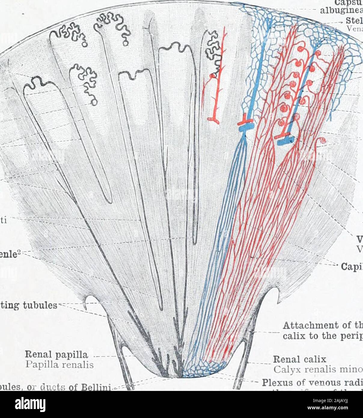 An atlas of human anatomy for students and physicians . I First convoluted tubule I Tiibnlus renalis contortus -Glomerulus Capsule of theglomerulus (Bowmanscapsule)Capsula glomeruli-- Efferent vessel ofthe glomerulusVas tllercnsCapillary vessels Straight tubules Tubuli renalesrecti Afferent vessel of the glomerulus Vas afferens Interlobular or radiate arteryA. interlobularis Fig. 833.—Part of a Sfxtion through the CortexOF the Kidney in the direction of the StraightTubules. Fig. 834.—CoRPUscuLUM Renis (Malpighji),Malpighian Corpuscle of the Kidney. Dl.AGRAMM.ATIC. Second convoluted tubule < Stock Photohttps://www.alamy.com/image-license-details/?v=1https://www.alamy.com/an-atlas-of-human-anatomy-for-students-and-physicians-i-first-convoluted-tubule-i-tiibnlus-renalis-contortus-glomerulus-capsule-of-theglomerulus-bowmanscapsulecapsula-glomeruli-efferent-vessel-ofthe-glomerulusvas-tllercnscapillary-vessels-straight-tubules-tubuli-renalesrecti-afferent-vessel-of-the-glomerulus-vas-afferens-interlobular-or-radiate-arterya-interlobularis-fig-833part-of-a-sfxtion-through-the-cortexof-the-kidney-in-the-direction-of-the-straighttubules-fig-834corpusculum-renis-malpighjimalpighian-corpuscle-of-the-kidney-dlagrammatic-second-convoluted-tubule-lt-image338302010.html
RM2AJAYJJ–An atlas of human anatomy for students and physicians . I First convoluted tubule I Tiibnlus renalis contortus -Glomerulus Capsule of theglomerulus (Bowmanscapsule)Capsula glomeruli-- Efferent vessel ofthe glomerulusVas tllercnsCapillary vessels Straight tubules Tubuli renalesrecti Afferent vessel of the glomerulus Vas afferens Interlobular or radiate arteryA. interlobularis Fig. 833.—Part of a Sfxtion through the CortexOF the Kidney in the direction of the StraightTubules. Fig. 834.—CoRPUscuLUM Renis (Malpighji),Malpighian Corpuscle of the Kidney. Dl.AGRAMM.ATIC. Second convoluted tubule <
 The physiology and hygiene of the house in which we live . the kidney theyare known as the uriniferous tubules, and if we follow up oneof these tubules from the kidney, from its opening on the surface of a pyramid, we shallfind that it terminates in a dila-tation not unlike those found inthe sweat-glands. The dilatationin the urinary tubule is knownas the Malpighian (from its dis-coverer, Malpighi) capsule (seefigure b), which incloses within ita network of capillaries (glomer-ulus) given off from the branchesof the renal artery. Through thistuft of capillaries the liquid ref-use of the blood Stock Photohttps://www.alamy.com/image-license-details/?v=1https://www.alamy.com/the-physiology-and-hygiene-of-the-house-in-which-we-live-the-kidney-theyare-known-as-the-uriniferous-tubules-and-if-we-follow-up-oneof-these-tubules-from-the-kidney-from-its-opening-on-the-surface-of-a-pyramid-we-shallfind-that-it-terminates-in-a-dila-tation-not-unlike-those-found-inthe-sweat-glands-the-dilatationin-the-urinary-tubule-is-knownas-the-malpighian-from-its-dis-coverer-malpighi-capsule-seefigure-b-which-incloses-within-ita-network-of-capillaries-glomer-ulus-given-off-from-the-branchesof-the-renal-artery-through-thistuft-of-capillaries-the-liquid-ref-use-of-the-blood-image339995148.html
RM2AN437T–The physiology and hygiene of the house in which we live . the kidney theyare known as the uriniferous tubules, and if we follow up oneof these tubules from the kidney, from its opening on the surface of a pyramid, we shallfind that it terminates in a dila-tation not unlike those found inthe sweat-glands. The dilatationin the urinary tubule is knownas the Malpighian (from its dis-coverer, Malpighi) capsule (seefigure b), which incloses within ita network of capillaries (glomer-ulus) given off from the branchesof the renal artery. Through thistuft of capillaries the liquid ref-use of the blood
 A treatise on zoology . te, or itmay from the first be fused with the lateral plate (Teleost). Inthe typical fully developed organ each tubule resembles amesonephric tubule, and consists of a segmental ciliated funnelopening into the coelom, the coelomostome (outer funnel, orprimary nephrostome of the communicating canal). This leads bya narrow canal (Ei-giinzungskanal) to the renal chamber or capsule(Bowmans capsule of the Malpighian body, the urocoele). Intothis small chamber opens a funnel (inner funnel, or urostome)leading into the main renal canal. The renal capsule and itscanal arise as Stock Photohttps://www.alamy.com/image-license-details/?v=1https://www.alamy.com/a-treatise-on-zoology-te-or-itmay-from-the-first-be-fused-with-the-lateral-plate-teleost-inthe-typical-fully-developed-organ-each-tubule-resembles-amesonephric-tubule-and-consists-of-a-segmental-ciliated-funnelopening-into-the-coelom-the-coelomostome-outer-funnel-orprimary-nephrostome-of-the-communicating-canal-this-leads-bya-narrow-canal-ei-giinzungskanal-to-the-renal-chamber-or-capsulebowmans-capsule-of-the-malpighian-body-the-urocoele-intothis-small-chamber-opens-a-funnel-inner-funnel-or-urostomeleading-into-the-main-renal-canal-the-renal-capsule-and-itscanal-arise-as-image338348565.html
RM2AJD319–A treatise on zoology . te, or itmay from the first be fused with the lateral plate (Teleost). Inthe typical fully developed organ each tubule resembles amesonephric tubule, and consists of a segmental ciliated funnelopening into the coelom, the coelomostome (outer funnel, orprimary nephrostome of the communicating canal). This leads bya narrow canal (Ei-giinzungskanal) to the renal chamber or capsule(Bowmans capsule of the Malpighian body, the urocoele). Intothis small chamber opens a funnel (inner funnel, or urostome)leading into the main renal canal. The renal capsule and itscanal arise as
 Quain's elements of anatomy . s coim^nence in the labyiinth of thecortical substance by spherical dilatations enclosing like a capsule thevascular Malpighian tufts to be afterwards described. Emerging from this dilatation (fig. 566, 1), which is known as thecapside, by a narrow neck (2), the tubule is at first convoluted and wide{first convoluted tudicle), but on approaching the medullary ray itbecomes nearly straight with a slight tendency to a spiral (spiral tubuleof Schachowa, 4). At the junction of cortex and medulla the spiraltube rapidly narrows and passes straight down through the bound Stock Photohttps://www.alamy.com/image-license-details/?v=1https://www.alamy.com/quains-elements-of-anatomy-s-coimnence-in-the-labyiinth-of-thecortical-substance-by-spherical-dilatations-enclosing-like-a-capsule-thevascular-malpighian-tufts-to-be-afterwards-described-emerging-from-this-dilatation-fig-566-1-which-is-known-as-thecapside-by-a-narrow-neck-2-the-tubule-is-at-first-convoluted-and-widefirst-convoluted-tudicle-but-on-approaching-the-medullary-ray-itbecomes-nearly-straight-with-a-slight-tendency-to-a-spiral-spiral-tubuleof-schachowa-4-at-the-junction-of-cortex-and-medulla-the-spiraltube-rapidly-narrows-and-passes-straight-down-through-the-bound-image340306474.html
RM2ANJ8AJ–Quain's elements of anatomy . s coim^nence in the labyiinth of thecortical substance by spherical dilatations enclosing like a capsule thevascular Malpighian tufts to be afterwards described. Emerging from this dilatation (fig. 566, 1), which is known as thecapside, by a narrow neck (2), the tubule is at first convoluted and wide{first convoluted tudicle), but on approaching the medullary ray itbecomes nearly straight with a slight tendency to a spiral (spiral tubuleof Schachowa, 4). At the junction of cortex and medulla the spiraltube rapidly narrows and passes straight down through the bound
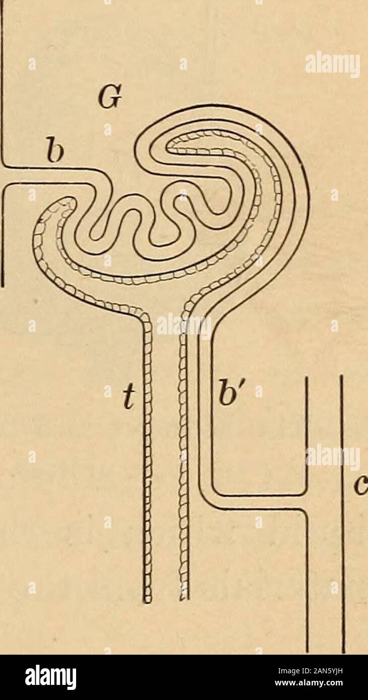
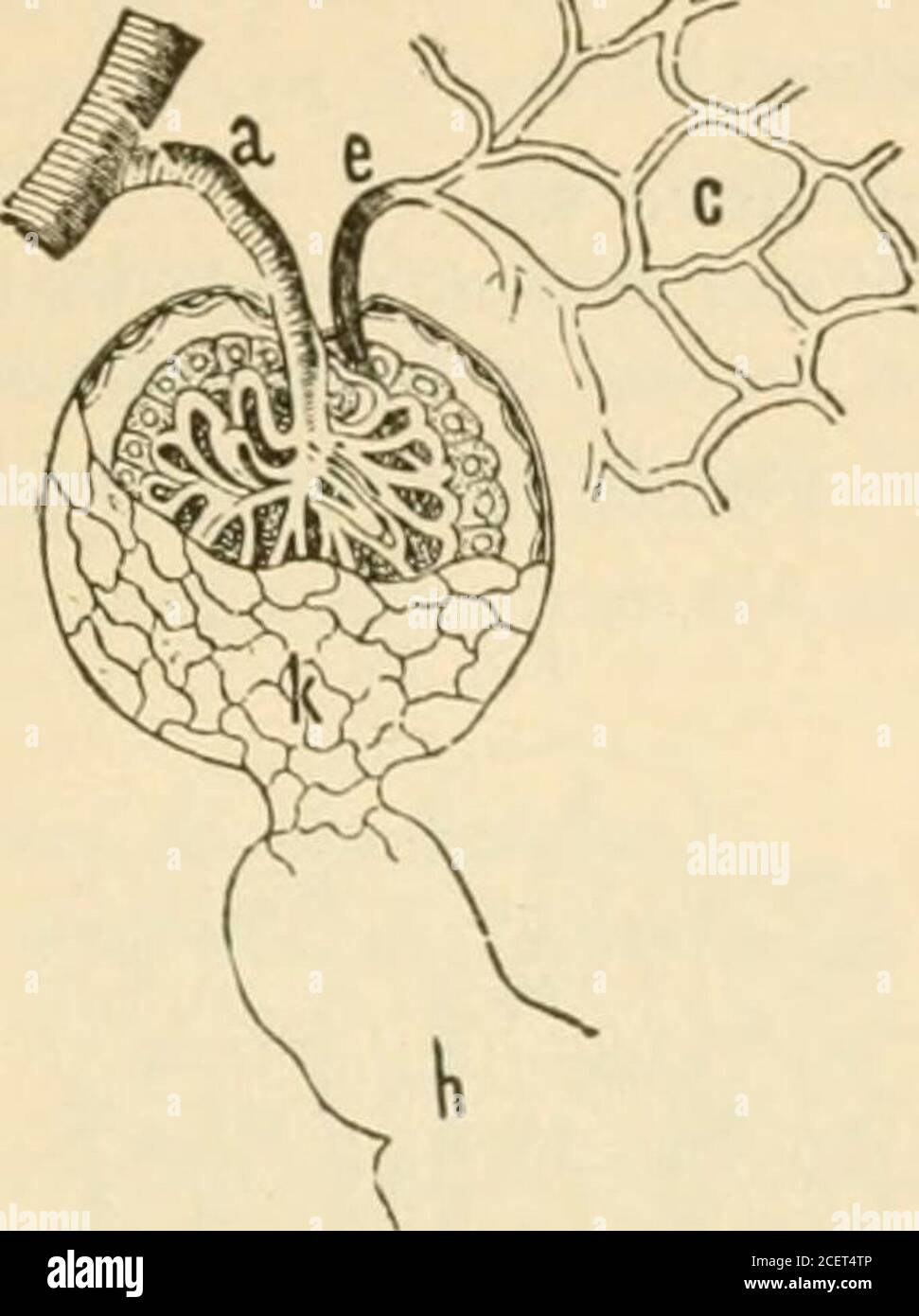
 Anatomy, physiology and hygiene for high schools . a glo-merulus and tubule, a, artery briuging blood topart; b, capillary briuging bloodto glomerulus; h, vessel continu-ing with blood to tubule; c, vein;t, tubule; G, Malpighian capsuleand glomemlus. 200 PHYSIOLOGY AND HYGIENE epithelial cells which lines the closed end of the tubule sur-rounding the glomerulus. Through these thin layers of cells the waste substances pass intothe tubules. Below the capsules the bloodcapillaries run in a networkabout the tubules, and fromthese lower capillaries the ureais separated into the tubules,just as is t Stock Photohttps://www.alamy.com/image-license-details/?v=1https://www.alamy.com/anatomy-physiology-and-hygiene-for-high-schools-a-glo-merulus-and-tubule-a-artery-briuging-blood-topart-b-capillary-briuging-bloodto-glomerulus-h-vessel-continu-ing-with-blood-to-tubule-c-veint-tubule-g-malpighian-capsuleand-glomemlus-200-physiology-and-hygiene-epithelial-cells-which-lines-the-closed-end-of-the-tubule-sur-rounding-the-glomerulus-through-these-thin-layers-of-cells-the-waste-substances-pass-intothe-tubules-below-the-capsules-the-bloodcapillaries-run-in-a-networkabout-the-tubules-and-fromthese-lower-capillaries-the-ureais-separated-into-the-tubulesjust-as-is-t-image340035940.html
RM2AN5Y8M–Anatomy, physiology and hygiene for high schools . a glo-merulus and tubule, a, artery briuging blood topart; b, capillary briuging bloodto glomerulus; h, vessel continu-ing with blood to tubule; c, vein;t, tubule; G, Malpighian capsuleand glomemlus. 200 PHYSIOLOGY AND HYGIENE epithelial cells which lines the closed end of the tubule sur-rounding the glomerulus. Through these thin layers of cells the waste substances pass intothe tubules. Below the capsules the bloodcapillaries run in a networkabout the tubules, and fromthese lower capillaries the ureais separated into the tubules,just as is t
 Anatomy, physiology and hygiene for high schools . he tubes and ramify into fine branches in the cortex.These branches end in fine clusters of capillaries known asglomeruli. From each of these clusters a vein issues. Eachcluster of capillaries, or glomerulus(Latin glomus, a ball of cotton ),is suiTounded by the closed, dilatedend of a tuhule ( small tube ). Thewhole structure, the cluster of ves-sels projecting into the closed endof a tubule, is known as a Malpighian(after Malpighi, who first describedit) capsule. The wall of a tubule consists ofa single layer of epithelial cells,mostly cubica Stock Photohttps://www.alamy.com/image-license-details/?v=1https://www.alamy.com/anatomy-physiology-and-hygiene-for-high-schools-he-tubes-and-ramify-into-fine-branches-in-the-cortexthese-branches-end-in-fine-clusters-of-capillaries-known-asglomeruli-from-each-of-these-clusters-a-vein-issues-eachcluster-of-capillaries-or-glomeruluslatin-glomus-a-ball-of-cotton-is-suitounded-by-the-closed-dilatedend-of-a-tuhule-small-tube-thewhole-structure-the-cluster-of-ves-sels-projecting-into-the-closed-endof-a-tubule-is-known-as-a-malpighianafter-malpighi-who-first-describedit-capsule-the-wall-of-a-tubule-consists-ofa-single-layer-of-epithelial-cellsmostly-cubica-image340036217.html
RM2AN5YJH–Anatomy, physiology and hygiene for high schools . he tubes and ramify into fine branches in the cortex.These branches end in fine clusters of capillaries known asglomeruli. From each of these clusters a vein issues. Eachcluster of capillaries, or glomerulus(Latin glomus, a ball of cotton ),is suiTounded by the closed, dilatedend of a tuhule ( small tube ). Thewhole structure, the cluster of ves-sels projecting into the closed endof a tubule, is known as a Malpighian(after Malpighi, who first describedit) capsule. The wall of a tubule consists ofa single layer of epithelial cells,mostly cubica
 Practical pathology; a manual for students and practitioners . a. , J: .? -J fl.-v. Fig. I20.—Section of kidney. Chronic Interstitial nephritis.Stained with alum hivmalein and picro-eiythiosin. ( x 50.) a. Thickened and laminated lihious capsule,a.t. Atrophied tubules containing colloid casts.AI. Fibroid Malpighian body. e.a. Artery with thickened walls, endarteritis obliterans.c.c. Dilated convoluted tubule containing colloid cast.f.c.i. Pibro-cellular tissue in which are numerous atrophied, andsome dilated, tubules.c.t. Dil.ated dollecling tubules containing hyaline casts. bodies nearer the Stock Photohttps://www.alamy.com/image-license-details/?v=1https://www.alamy.com/practical-pathology-a-manual-for-students-and-practitioners-a-j-j-fl-v-fig-i20section-of-kidney-chronic-interstitial-nephritisstained-with-alum-hivmalein-and-picro-eiythiosin-x-50-a-thickened-and-laminated-lihious-capsuleat-atrophied-tubules-containing-colloid-castsai-fibroid-malpighian-body-ea-artery-with-thickened-walls-endarteritis-obliteranscc-dilated-convoluted-tubule-containing-colloid-castfci-pibro-cellular-tissue-in-which-are-numerous-atrophied-andsome-dilated-tubulesct-dilated-dollecling-tubules-containing-hyaline-casts-bodies-nearer-the-image342990856.html
RM2AX0G9C–Practical pathology; a manual for students and practitioners . a. , J: .? -J fl.-v. Fig. I20.—Section of kidney. Chronic Interstitial nephritis.Stained with alum hivmalein and picro-eiythiosin. ( x 50.) a. Thickened and laminated lihious capsule,a.t. Atrophied tubules containing colloid casts.AI. Fibroid Malpighian body. e.a. Artery with thickened walls, endarteritis obliterans.c.c. Dilated convoluted tubule containing colloid cast.f.c.i. Pibro-cellular tissue in which are numerous atrophied, andsome dilated, tubules.c.t. Dil.ated dollecling tubules containing hyaline casts. bodies nearer the
 . On disorders of digestion, their consequences and treatment . capsule surrounding the Malpighian tuft, often known as Bowmanscapsule, the tubule passes off. At first it forms a constrictedportion or neck, then it becomes dilated and very tortuous, andruns more or less transversely towards the adjacent medullary ray.It is known at this point by the name of the proximal convolutedtubule. It now runs downwards and is called the spiral tubule ofSchachowa. It next becomes greatly constricted, and runs in astraight course down through the boundary layer, into the papillarylayer, and here, bending Stock Photohttps://www.alamy.com/image-license-details/?v=1https://www.alamy.com/on-disorders-of-digestion-their-consequences-and-treatment-capsule-surrounding-the-malpighian-tuft-often-known-as-bowmanscapsule-the-tubule-passes-off-at-first-it-forms-a-constrictedportion-or-neck-then-it-becomes-dilated-and-very-tortuous-andruns-more-or-less-transversely-towards-the-adjacent-medullary-rayit-is-known-at-this-point-by-the-name-of-the-proximal-convolutedtubule-it-now-runs-downwards-and-is-called-the-spiral-tubule-ofschachowa-it-next-becomes-greatly-constricted-and-runs-in-astraight-course-down-through-the-boundary-layer-into-the-papillarylayer-and-here-bending-image371633185.html
RM2CGH9WN–. On disorders of digestion, their consequences and treatment . capsule surrounding the Malpighian tuft, often known as Bowmanscapsule, the tubule passes off. At first it forms a constrictedportion or neck, then it becomes dilated and very tortuous, andruns more or less transversely towards the adjacent medullary ray.It is known at this point by the name of the proximal convolutedtubule. It now runs downwards and is called the spiral tubule ofSchachowa. It next becomes greatly constricted, and runs in astraight course down through the boundary layer, into the papillarylayer, and here, bending
 . Text-book of anatomy and physiology for nurses. Inner wall.6. Glomerulus. 7. Neck of tubule (Stohr). to form a collecting tubewhich opens into the sinus. Malpighian corpuscles and convoluted tubes occupy most of theportion of the kidney near the surface, forming the cortex (or corticalportion). The straight or collecting tubes aregrouped together into pyramids, pointingtoward the interior and forming the medullaryportion. The apex of each pyramid projectsinto the sinus, presenting the openings ofseveral collecting tubes (Fig. 155). The cells which line this system of tubesdo the work of excr Stock Photohttps://www.alamy.com/image-license-details/?v=1https://www.alamy.com/text-book-of-anatomy-and-physiology-for-nurses-inner-wall6-glomerulus-7-neck-of-tubule-stohr-to-form-a-collecting-tubewhich-opens-into-the-sinus-malpighian-corpuscles-and-convoluted-tubes-occupy-most-of-theportion-of-the-kidney-near-the-surface-forming-the-cortex-or-corticalportion-the-straight-or-collecting-tubes-aregrouped-together-into-pyramids-pointingtoward-the-interior-and-forming-the-medullaryportion-the-apex-of-each-pyramid-projectsinto-the-sinus-presenting-the-openings-ofseveral-collecting-tubes-fig-155-the-cells-which-line-this-system-of-tubesdo-the-work-of-excr-image370333030.html
RM2CEE3FJ–. Text-book of anatomy and physiology for nurses. Inner wall.6. Glomerulus. 7. Neck of tubule (Stohr). to form a collecting tubewhich opens into the sinus. Malpighian corpuscles and convoluted tubes occupy most of theportion of the kidney near the surface, forming the cortex (or corticalportion). The straight or collecting tubes aregrouped together into pyramids, pointingtoward the interior and forming the medullaryportion. The apex of each pyramid projectsinto the sinus, presenting the openings ofseveral collecting tubes (Fig. 155). The cells which line this system of tubesdo the work of excr
![. The development of the chick; an introduction to embryology . st advanced vesiclein this region possesses a hollow sprout extending laterally to theWolffian duct with which it is in close contact; this is the ])ri-mordium of the tubular part of the mesonephric tubule (cf. Fig.114 A and B). In more anterior somites it is found that suchsprouts have fused with the wall of the duct in such a maimer thatthe lumen of the tubule now conmiunicates with that of the duct. Simultaneously the median ]X)rtion of the original vesiclehas been transformed into a small Malpighian corpuscle in thefollowing m Stock Photo . The development of the chick; an introduction to embryology . st advanced vesiclein this region possesses a hollow sprout extending laterally to theWolffian duct with which it is in close contact; this is the ])ri-mordium of the tubular part of the mesonephric tubule (cf. Fig.114 A and B). In more anterior somites it is found that suchsprouts have fused with the wall of the duct in such a maimer thatthe lumen of the tubule now conmiunicates with that of the duct. Simultaneously the median ]X)rtion of the original vesiclehas been transformed into a small Malpighian corpuscle in thefollowing m Stock Photo](https://c8.alamy.com/comp/2CPNCJ9/the-development-of-the-chick-an-introduction-to-embryology-st-advanced-vesiclein-this-region-possesses-a-hollow-sprout-extending-laterally-to-thewolffian-duct-with-which-it-is-in-close-contact-this-is-the-ri-mordium-of-the-tubular-part-of-the-mesonephric-tubule-cf-fig114-a-and-b-in-more-anterior-somites-it-is-found-that-suchsprouts-have-fused-with-the-wall-of-the-duct-in-such-a-maimer-thatthe-lumen-of-the-tubule-now-conmiunicates-with-that-of-the-duct-simultaneously-the-median-xrtion-of-the-original-vesiclehas-been-transformed-into-a-small-malpighian-corpuscle-in-thefollowing-m-2CPNCJ9.jpg) . The development of the chick; an introduction to embryology . st advanced vesiclein this region possesses a hollow sprout extending laterally to theWolffian duct with which it is in close contact; this is the ])ri-mordium of the tubular part of the mesonephric tubule (cf. Fig.114 A and B). In more anterior somites it is found that suchsprouts have fused with the wall of the duct in such a maimer thatthe lumen of the tubule now conmiunicates with that of the duct. Simultaneously the median ]X)rtion of the original vesiclehas been transformed into a small Malpighian corpuscle in thefollowing m Stock Photohttps://www.alamy.com/image-license-details/?v=1https://www.alamy.com/the-development-of-the-chick-an-introduction-to-embryology-st-advanced-vesiclein-this-region-possesses-a-hollow-sprout-extending-laterally-to-thewolffian-duct-with-which-it-is-in-close-contact-this-is-the-ri-mordium-of-the-tubular-part-of-the-mesonephric-tubule-cf-fig114-a-and-b-in-more-anterior-somites-it-is-found-that-suchsprouts-have-fused-with-the-wall-of-the-duct-in-such-a-maimer-thatthe-lumen-of-the-tubule-now-conmiunicates-with-that-of-the-duct-simultaneously-the-median-xrtion-of-the-original-vesiclehas-been-transformed-into-a-small-malpighian-corpuscle-in-thefollowing-m-image375411073.html
. The development of the chick; an introduction to embryology . st advanced vesiclein this region possesses a hollow sprout extending laterally to theWolffian duct with which it is in close contact; this is the ])ri-mordium of the tubular part of the mesonephric tubule (cf. Fig.114 A and B). In more anterior somites it is found that suchsprouts have fused with the wall of the duct in such a maimer thatthe lumen of the tubule now conmiunicates with that of the duct. Simultaneously the median ]X)rtion of the original vesiclehas been transformed into a small Malpighian corpuscle in thefollowing m Stock Photohttps://www.alamy.com/image-license-details/?v=1https://www.alamy.com/the-development-of-the-chick-an-introduction-to-embryology-st-advanced-vesiclein-this-region-possesses-a-hollow-sprout-extending-laterally-to-thewolffian-duct-with-which-it-is-in-close-contact-this-is-the-ri-mordium-of-the-tubular-part-of-the-mesonephric-tubule-cf-fig114-a-and-b-in-more-anterior-somites-it-is-found-that-suchsprouts-have-fused-with-the-wall-of-the-duct-in-such-a-maimer-thatthe-lumen-of-the-tubule-now-conmiunicates-with-that-of-the-duct-simultaneously-the-median-xrtion-of-the-original-vesiclehas-been-transformed-into-a-small-malpighian-corpuscle-in-thefollowing-m-image375411073.htmlRM2CPNCJ9–. The development of the chick; an introduction to embryology . st advanced vesiclein this region possesses a hollow sprout extending laterally to theWolffian duct with which it is in close contact; this is the ])ri-mordium of the tubular part of the mesonephric tubule (cf. Fig.114 A and B). In more anterior somites it is found that suchsprouts have fused with the wall of the duct in such a maimer thatthe lumen of the tubule now conmiunicates with that of the duct. Simultaneously the median ]X)rtion of the original vesiclehas been transformed into a small Malpighian corpuscle in thefollowing m
 . Essentials of physiology, arranged in the form of questions and answers, prepared especially for students of medicine. red.The uriniferous tubule, however, is given off from the Malpighianbody on the opposite side from that at which the artery entersand the efferent vessel leaves. The capsule of Bowman, or the beginning of the uriniferoustubule, may be considered as a sac, into which is secreted the liquidby the Malpighian tuft. Is the efferent vessel called a vein ?The efferent vessel after leaving the Malpighian body forms a 86 ESSENTIALS OF HUMAN PHYSIOLOGY, second capillary network, twis Stock Photohttps://www.alamy.com/image-license-details/?v=1https://www.alamy.com/essentials-of-physiology-arranged-in-the-form-of-questions-and-answers-prepared-especially-for-students-of-medicine-redthe-uriniferous-tubule-however-is-given-off-from-the-malpighianbody-on-the-opposite-side-from-that-at-which-the-artery-entersand-the-efferent-vessel-leaves-the-capsule-of-bowman-or-the-beginning-of-the-uriniferoustubule-may-be-considered-as-a-sac-into-which-is-secreted-the-liquidby-the-malpighian-tuft-is-the-efferent-vessel-called-a-vein-the-efferent-vessel-after-leaving-the-malpighian-body-forms-a-86-essentials-of-human-physiology-second-capillary-network-twis-image370553542.html
RM2CET4R2–. Essentials of physiology, arranged in the form of questions and answers, prepared especially for students of medicine. red.The uriniferous tubule, however, is given off from the Malpighianbody on the opposite side from that at which the artery entersand the efferent vessel leaves. The capsule of Bowman, or the beginning of the uriniferoustubule, may be considered as a sac, into which is secreted the liquidby the Malpighian tuft. Is the efferent vessel called a vein ?The efferent vessel after leaving the Malpighian body forms a 86 ESSENTIALS OF HUMAN PHYSIOLOGY, second capillary network, twis
 . Text-book of normal histology: including an account of the development of the tissues and of the organs. Portions of the various divisions of the uriniferous tubules drawn from sections of human kidney:A, Malpighian body ; jr, squamous epithelium lining the capsule and reflected over the glomerulus ;y, z, afferent and eflFerent vessels of the tuft ; e, nuclei of capillaries; «, constricted neck markingpassage of capsule into convoluted tubule ; B, proximal convoluted tubule; C, irregular tubule ; £>and F, spiral tubules ; E, ascending limb of Henles loop ; G, straight collecting tubule. t Stock Photohttps://www.alamy.com/image-license-details/?v=1https://www.alamy.com/text-book-of-normal-histology-including-an-account-of-the-development-of-the-tissues-and-of-the-organs-portions-of-the-various-divisions-of-the-uriniferous-tubules-drawn-from-sections-of-human-kidneya-malpighian-body-jr-squamous-epithelium-lining-the-capsule-and-reflected-over-the-glomerulus-y-z-afferent-and-eflferent-vessels-of-the-tuft-e-nuclei-of-capillaries-constricted-neck-markingpassage-of-capsule-into-convoluted-tubule-b-proximal-convoluted-tubule-c-irregular-tubule-gtand-f-spiral-tubules-e-ascending-limb-of-henles-loop-g-straight-collecting-tubule-t-image370381977.html
RM2CEG9YN–. Text-book of normal histology: including an account of the development of the tissues and of the organs. Portions of the various divisions of the uriniferous tubules drawn from sections of human kidney:A, Malpighian body ; jr, squamous epithelium lining the capsule and reflected over the glomerulus ;y, z, afferent and eflFerent vessels of the tuft ; e, nuclei of capillaries; «, constricted neck markingpassage of capsule into convoluted tubule ; B, proximal convoluted tubule; C, irregular tubule ; £>and F, spiral tubules ; E, ascending limb of Henles loop ; G, straight collecting tubule. t
 . On disorders of digestion, their consequences and treatment . Bundles of venules. — Bundles of arterioles. Venous plexus around theapices of the pyi-amids. ^ Fig. 35.—Diagram of the blood-vessels in the kidney (after Ludwig). or knots known by the name of Malpighian tufts or glomeruli.The branch going to the tuft is called the afferent artery (m, Fig.36), and the branch going from it, the efferent artery (ve, Fig. 36). 302 STRUCTURE OF THE KIDNEY The efferent artery, almost immediately after its exit, breadsup into a capillary mesh-work (Figs. 80 and 32), spreading aroundand among the tubule Stock Photohttps://www.alamy.com/image-license-details/?v=1https://www.alamy.com/on-disorders-of-digestion-their-consequences-and-treatment-bundles-of-venules-bundles-of-arterioles-venous-plexus-around-theapices-of-the-pyi-amids-fig-35diagram-of-the-blood-vessels-in-the-kidney-after-ludwig-or-knots-known-by-the-name-of-malpighian-tufts-or-glomerulithe-branch-going-to-the-tuft-is-called-the-afferent-artery-m-fig36-and-the-branch-going-from-it-the-efferent-artery-ve-fig-36-302-structure-of-the-kidney-the-efferent-artery-almost-immediately-after-its-exit-breadsup-into-a-capillary-mesh-work-figs-80-and-32-spreading-aroundand-among-the-tubule-image371633536.html
RM2CGHAA8–. On disorders of digestion, their consequences and treatment . Bundles of venules. — Bundles of arterioles. Venous plexus around theapices of the pyi-amids. ^ Fig. 35.—Diagram of the blood-vessels in the kidney (after Ludwig). or knots known by the name of Malpighian tufts or glomeruli.The branch going to the tuft is called the afferent artery (m, Fig.36), and the branch going from it, the efferent artery (ve, Fig. 36). 302 STRUCTURE OF THE KIDNEY The efferent artery, almost immediately after its exit, breadsup into a capillary mesh-work (Figs. 80 and 32), spreading aroundand among the tubule
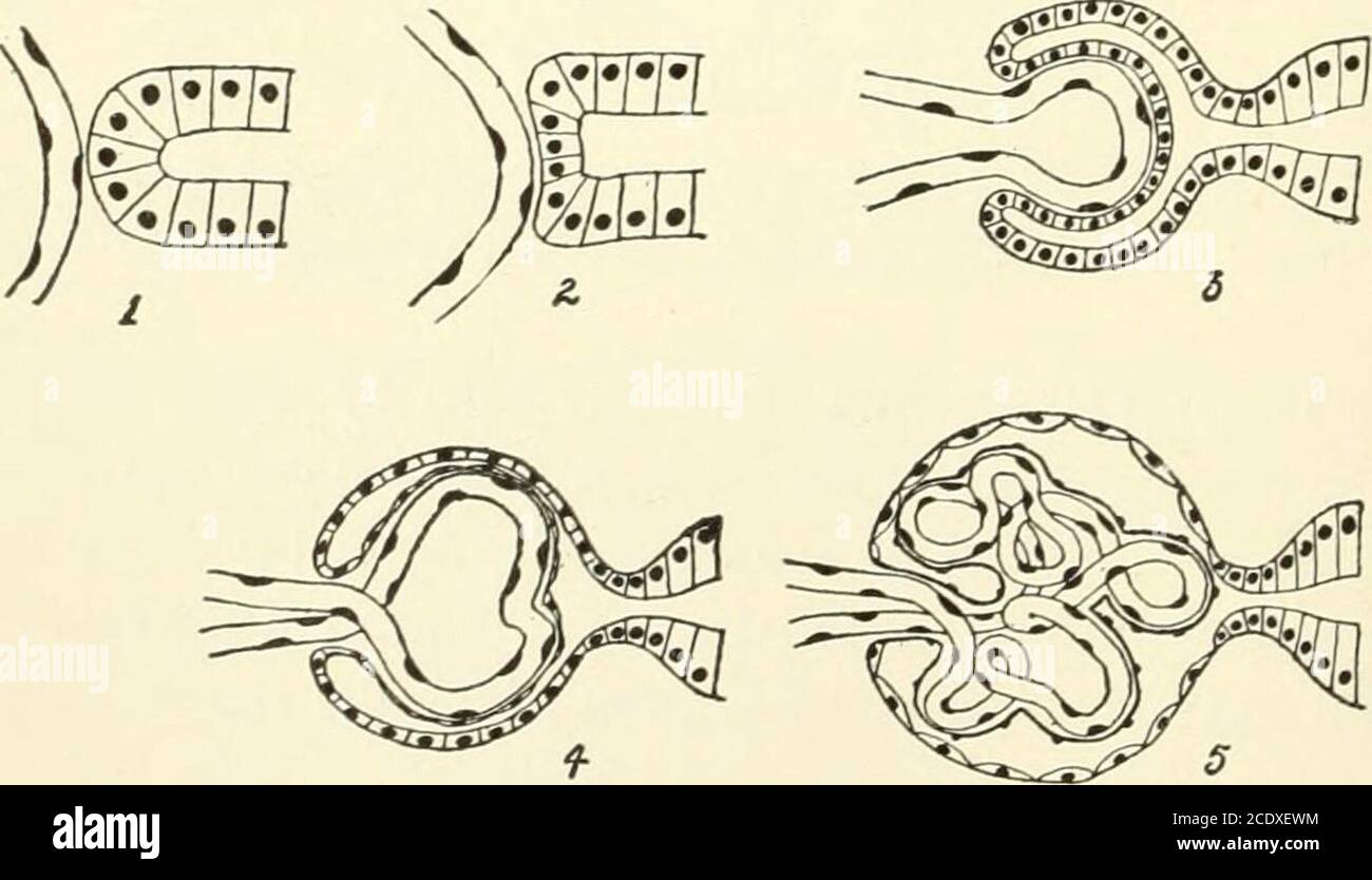 . Kirkes' handbook of physiology . ui. r i 29°-—Malpighian Capsule and Tuft of Capillaries, Injected through the Renal Arterywitn colored Lrelatin. a, Glomerular vessels; b, capsule; c, anterior capsule; d, glomerular artery;e, efferent veins; /. epithelium of tubes. (Cadiat.). Fig. 291.—Diagrams Illustrating Stages in the Development of the Malpighian Capsule. In 1and 2 the developing blood-vessel is approaching the blind end of the capsule. In 3 the tubule isbeginning to invagmate and enclose the capillary. In 4 and 5 later stages are shown. The cellsforming the two layers of the capsule gro Stock Photohttps://www.alamy.com/image-license-details/?v=1https://www.alamy.com/kirkes-handbook-of-physiology-ui-r-i-29-malpighian-capsule-and-tuft-of-capillaries-injected-through-the-renal-arterywitn-colored-lrelatin-a-glomerular-vessels-b-capsule-c-anterior-capsule-d-glomerular-arterye-efferent-veins-epithelium-of-tubes-cadiat-fig-291diagrams-illustrating-stages-in-the-development-of-the-malpighian-capsule-in-1and-2-the-developing-blood-vessel-is-approaching-the-blind-end-of-the-capsule-in-3-the-tubule-isbeginning-to-invagmate-and-enclose-the-capillary-in-4-and-5-later-stages-are-shown-the-cellsforming-the-two-layers-of-the-capsule-gro-image369990704.html
RM2CDXEWM–. Kirkes' handbook of physiology . ui. r i 29°-—Malpighian Capsule and Tuft of Capillaries, Injected through the Renal Arterywitn colored Lrelatin. a, Glomerular vessels; b, capsule; c, anterior capsule; d, glomerular artery;e, efferent veins; /. epithelium of tubes. (Cadiat.). Fig. 291.—Diagrams Illustrating Stages in the Development of the Malpighian Capsule. In 1and 2 the developing blood-vessel is approaching the blind end of the capsule. In 3 the tubule isbeginning to invagmate and enclose the capillary. In 4 and 5 later stages are shown. The cellsforming the two layers of the capsule gro
 . On disorders of digestion, their consequences and treatment . Henles loop, descending iand ascending limbs. ) Straight part of collecting tube.Intermediary segment or distalconvoluted tubule.CORTEX.IiTegular tubule. Proximal convoluted tubule. Wavy part of ascending Umb.Constriction or neck.Spiral tubule. Malpighian tuft surrounded ^y£owmans capsule. Spir.al part of ascending limb ofUeules loop. BORDER LAYER. Henles loop, first thick portionof ascending limb. Collecting tube, Large collecting tube, orduct of BeUiuL Henles-loop,PAPILLARY LAYEE. Fig. 38.—Diagram of the course of the uriniferou Stock Photohttps://www.alamy.com/image-license-details/?v=1https://www.alamy.com/on-disorders-of-digestion-their-consequences-and-treatment-henles-loop-descending-iand-ascending-limbs-straight-part-of-collecting-tubeintermediary-segment-or-distalconvoluted-tubulecortexiitegular-tubule-proximal-convoluted-tubule-wavy-part-of-ascending-umbconstriction-or-neckspiral-tubule-malpighian-tuft-surrounded-yowmans-capsule-spiral-part-of-ascending-limb-ofueules-loop-border-layer-henles-loop-first-thick-portionof-ascending-limb-collecting-tube-large-collecting-tube-orduct-of-beuiul-henles-looppapillary-layee-fig-38diagram-of-the-course-of-the-uriniferou-image371632921.html
RM2CGH9G9–. On disorders of digestion, their consequences and treatment . Henles loop, descending iand ascending limbs. ) Straight part of collecting tube.Intermediary segment or distalconvoluted tubule.CORTEX.IiTegular tubule. Proximal convoluted tubule. Wavy part of ascending Umb.Constriction or neck.Spiral tubule. Malpighian tuft surrounded ^y£owmans capsule. Spir.al part of ascending limb ofUeules loop. BORDER LAYER. Henles loop, first thick portionof ascending limb. Collecting tube, Large collecting tube, orduct of BeUiuL Henles-loop,PAPILLARY LAYEE. Fig. 38.—Diagram of the course of the uriniferou
![. On the anatomy of vertebrates [electronic resource] . Tubuli uriniferi of corticaland medullary parts of kid-ney. CCLXXXVI. 481 The veins from the centre ofdip into the renal substance,unite, and ultimately emerge at the hilus anterior to or ventrad of the artery ; but, in afew Mammals, they unite in an arborescentdisposition (Felis, Hymia) or form a network(Pkoca) upon the surface of the kidney; in all, the venous trunk,fig. 418, k, terminates in the postcaval, ib. v.The uriniferous tubule commences in Mam-mals, as in lower Vertebrates (vol. ii. p. 538,fig. 356), from the malpighian corpusc Stock Photo . On the anatomy of vertebrates [electronic resource] . Tubuli uriniferi of corticaland medullary parts of kid-ney. CCLXXXVI. 481 The veins from the centre ofdip into the renal substance,unite, and ultimately emerge at the hilus anterior to or ventrad of the artery ; but, in afew Mammals, they unite in an arborescentdisposition (Felis, Hymia) or form a network(Pkoca) upon the surface of the kidney; in all, the venous trunk,fig. 418, k, terminates in the postcaval, ib. v.The uriniferous tubule commences in Mam-mals, as in lower Vertebrates (vol. ii. p. 538,fig. 356), from the malpighian corpusc Stock Photo](https://c8.alamy.com/comp/2CP6FH5/on-the-anatomy-of-vertebrates-electronic-resource-tubuli-uriniferi-of-corticaland-medullary-parts-of-kid-ney-cclxxxvi-481-the-veins-from-the-centre-ofdip-into-the-renal-substanceunite-and-ultimately-emerge-at-the-hilus-anterior-to-or-ventrad-of-the-artery-but-in-afew-mammals-they-unite-in-an-arborescentdisposition-felis-hymia-or-form-a-networkpkoca-upon-the-surface-of-the-kidney-in-all-the-venous-trunkfig-418-k-terminates-in-the-postcaval-ib-vthe-uriniferous-tubule-commences-in-mam-mals-as-in-lower-vertebrates-vol-ii-p-538fig-356-from-the-malpighian-corpusc-2CP6FH5.jpg) . On the anatomy of vertebrates [electronic resource] . Tubuli uriniferi of corticaland medullary parts of kid-ney. CCLXXXVI. 481 The veins from the centre ofdip into the renal substance,unite, and ultimately emerge at the hilus anterior to or ventrad of the artery ; but, in afew Mammals, they unite in an arborescentdisposition (Felis, Hymia) or form a network(Pkoca) upon the surface of the kidney; in all, the venous trunk,fig. 418, k, terminates in the postcaval, ib. v.The uriniferous tubule commences in Mam-mals, as in lower Vertebrates (vol. ii. p. 538,fig. 356), from the malpighian corpusc Stock Photohttps://www.alamy.com/image-license-details/?v=1https://www.alamy.com/on-the-anatomy-of-vertebrates-electronic-resource-tubuli-uriniferi-of-corticaland-medullary-parts-of-kid-ney-cclxxxvi-481-the-veins-from-the-centre-ofdip-into-the-renal-substanceunite-and-ultimately-emerge-at-the-hilus-anterior-to-or-ventrad-of-the-artery-but-in-afew-mammals-they-unite-in-an-arborescentdisposition-felis-hymia-or-form-a-networkpkoca-upon-the-surface-of-the-kidney-in-all-the-venous-trunkfig-418-k-terminates-in-the-postcaval-ib-vthe-uriniferous-tubule-commences-in-mam-mals-as-in-lower-vertebrates-vol-ii-p-538fig-356-from-the-malpighian-corpusc-image375084113.html
. On the anatomy of vertebrates [electronic resource] . Tubuli uriniferi of corticaland medullary parts of kid-ney. CCLXXXVI. 481 The veins from the centre ofdip into the renal substance,unite, and ultimately emerge at the hilus anterior to or ventrad of the artery ; but, in afew Mammals, they unite in an arborescentdisposition (Felis, Hymia) or form a network(Pkoca) upon the surface of the kidney; in all, the venous trunk,fig. 418, k, terminates in the postcaval, ib. v.The uriniferous tubule commences in Mam-mals, as in lower Vertebrates (vol. ii. p. 538,fig. 356), from the malpighian corpusc Stock Photohttps://www.alamy.com/image-license-details/?v=1https://www.alamy.com/on-the-anatomy-of-vertebrates-electronic-resource-tubuli-uriniferi-of-corticaland-medullary-parts-of-kid-ney-cclxxxvi-481-the-veins-from-the-centre-ofdip-into-the-renal-substanceunite-and-ultimately-emerge-at-the-hilus-anterior-to-or-ventrad-of-the-artery-but-in-afew-mammals-they-unite-in-an-arborescentdisposition-felis-hymia-or-form-a-networkpkoca-upon-the-surface-of-the-kidney-in-all-the-venous-trunkfig-418-k-terminates-in-the-postcaval-ib-vthe-uriniferous-tubule-commences-in-mam-mals-as-in-lower-vertebrates-vol-ii-p-538fig-356-from-the-malpighian-corpusc-image375084113.htmlRM2CP6FH5–. On the anatomy of vertebrates [electronic resource] . Tubuli uriniferi of corticaland medullary parts of kid-ney. CCLXXXVI. 481 The veins from the centre ofdip into the renal substance,unite, and ultimately emerge at the hilus anterior to or ventrad of the artery ; but, in afew Mammals, they unite in an arborescentdisposition (Felis, Hymia) or form a network(Pkoca) upon the surface of the kidney; in all, the venous trunk,fig. 418, k, terminates in the postcaval, ib. v.The uriniferous tubule commences in Mam-mals, as in lower Vertebrates (vol. ii. p. 538,fig. 356), from the malpighian corpusc
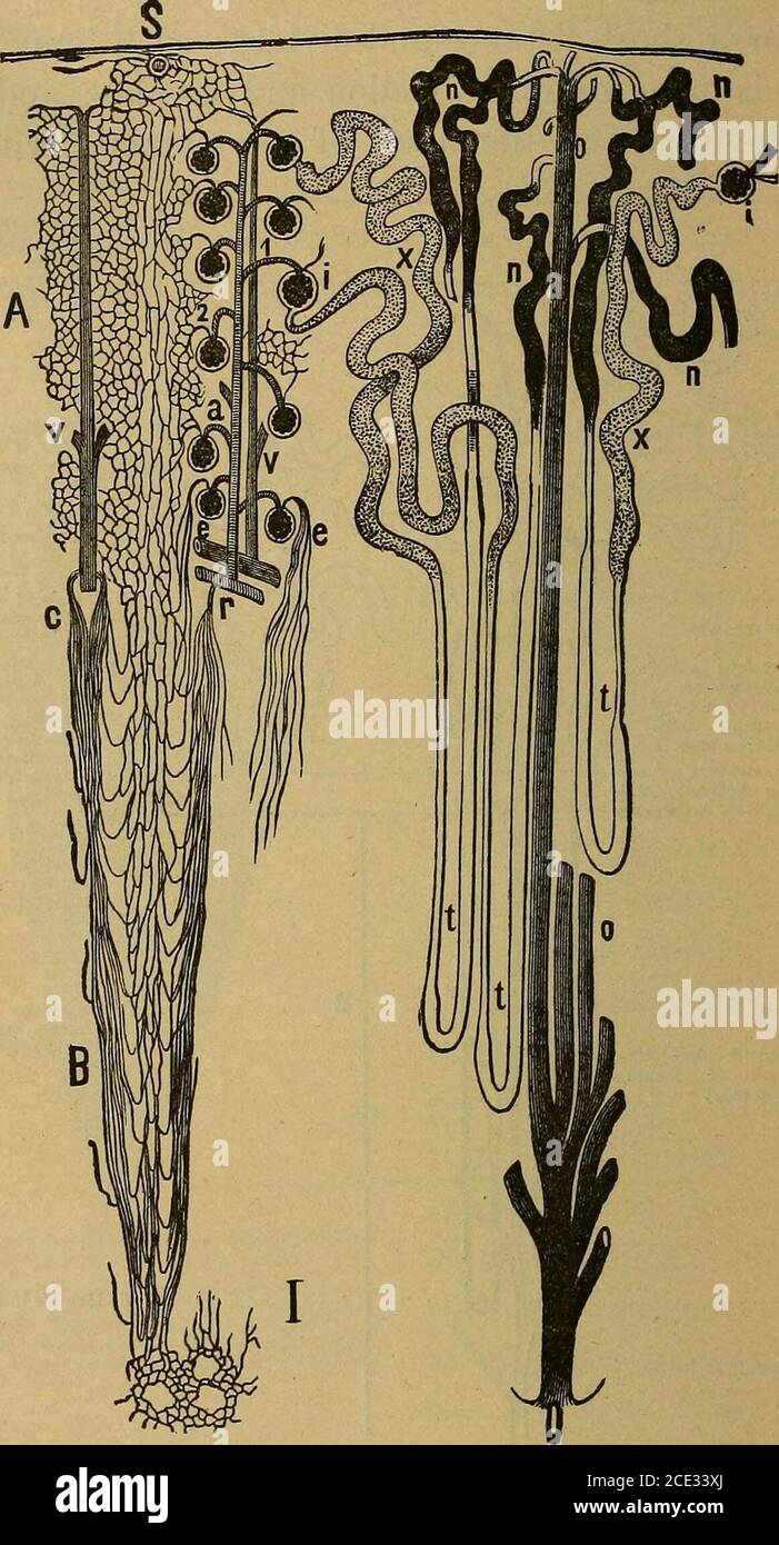
 . Essentials of physiology, arranged in the form of questions and answers, prepared especially for students of medicine. les. (Tyson and Brunton, after Klein and NobleSmith.) 1. Malpighian tuft surrounded by Bowmans capsule. 2. Constriction, or neck.3. Proximal convoluted tubule. 4. Spinal tubule. 5. Descending limb of Henles loop.6. Henles loop. 7 and 8. Ascending limb of Henles loop. 9. Wavy part of ascendinglimb of Henles loop. 10. Irregular tubule. 11. Distal convoluted tubule. 12. Firstpart of collecting tube. 13 and 14. Straight part of collecting tube. 15. Excretoryducts of Bellini. of Stock Photohttps://www.alamy.com/image-license-details/?v=1https://www.alamy.com/essentials-of-physiology-arranged-in-the-form-of-questions-and-answers-prepared-especially-for-students-of-medicine-les-tyson-and-brunton-after-klein-and-noblesmith-1-malpighian-tuft-surrounded-by-bowmans-capsule-2-constriction-or-neck3-proximal-convoluted-tubule-4-spinal-tubule-5-descending-limb-of-henles-loop6-henles-loop-7-and-8-ascending-limb-of-henles-loop-9-wavy-part-of-ascendinglimb-of-henles-loop-10-irregular-tubule-11-distal-convoluted-tubule-12-firstpart-of-collecting-tube-13-and-14-straight-part-of-collecting-tube-15-excretoryducts-of-bellini-of-image370553590.html
RM2CET4TP–. Essentials of physiology, arranged in the form of questions and answers, prepared especially for students of medicine. les. (Tyson and Brunton, after Klein and NobleSmith.) 1. Malpighian tuft surrounded by Bowmans capsule. 2. Constriction, or neck.3. Proximal convoluted tubule. 4. Spinal tubule. 5. Descending limb of Henles loop.6. Henles loop. 7 and 8. Ascending limb of Henles loop. 9. Wavy part of ascendinglimb of Henles loop. 10. Irregular tubule. 11. Distal convoluted tubule. 12. Firstpart of collecting tube. 13 and 14. Straight part of collecting tube. 15. Excretoryducts of Bellini. of
 . Kirkes' handbook of physiology . Fig. 288.—From a Vertical Section through the Kidney of a Dog, the Capsule of which is Sup-posed to be on the Right, a, The capillaries of the Malpighian capsule, the glomerulus, are arrangedin lobules; n, neck of capsule; c, convoluted tubes cut in various directions; b, irregular tubule:d, e, and f are straight tubes running toward capsules forming a so-called medullary ray; d, collect-ing tube; e, spiral tube; /, narrow section of ascending limb. X 380. (Klein and Noble Smith.)J mately supply the glomerulus. The small afferent artery, figures 287, a,290, d Stock Photohttps://www.alamy.com/image-license-details/?v=1https://www.alamy.com/kirkes-handbook-of-physiology-fig-288from-a-vertical-section-through-the-kidney-of-a-dog-the-capsule-of-which-is-sup-posed-to-be-on-the-right-a-the-capillaries-of-the-malpighian-capsule-the-glomerulus-are-arrangedin-lobules-n-neck-of-capsule-c-convoluted-tubes-cut-in-various-directions-b-irregular-tubuled-e-and-f-are-straight-tubes-running-toward-capsules-forming-a-so-called-medullary-ray-d-collect-ing-tube-e-spiral-tube-narrow-section-of-ascending-limb-x-380-klein-and-noble-smithj-mately-supply-the-glomerulus-the-small-afferent-artery-figures-287-a290-d-image369990824.html
RM2CDXF20–. Kirkes' handbook of physiology . Fig. 288.—From a Vertical Section through the Kidney of a Dog, the Capsule of which is Sup-posed to be on the Right, a, The capillaries of the Malpighian capsule, the glomerulus, are arrangedin lobules; n, neck of capsule; c, convoluted tubes cut in various directions; b, irregular tubule:d, e, and f are straight tubes running toward capsules forming a so-called medullary ray; d, collect-ing tube; e, spiral tube; /, narrow section of ascending limb. X 380. (Klein and Noble Smith.)J mately supply the glomerulus. The small afferent artery, figures 287, a,290, d
 . A treatise on practical anatomy: for students of anatomy and surgery . 7 and 8. Ascendinglimb of Henlesloop tube. Subcapsular layer with-out Mai pighian cor-puscles. 12. First part of col-lecting tube.11. Distal convolutedtubule.A> A. CORTE.X. 10. Irregular tubule. 3. Proximal convo-luted tubule. 9. Wavy part of as-cending limb. 2. Constriction or neck. 4. Spiral tubule. 1. Malpighian tuftsurrounded by Bowmans capsule. 8. Spiral part of as-cending limb ofHenles loop. B. Boundary Zo>t:.5. Descending limb ofHenles loop tube. 6. Henles loop. 15. Tubule of Bellini lO C. Papillary Zone. Fig Stock Photohttps://www.alamy.com/image-license-details/?v=1https://www.alamy.com/a-treatise-on-practical-anatomy-for-students-of-anatomy-and-surgery-7-and-8-ascendinglimb-of-henlesloop-tube-subcapsular-layer-with-out-mai-pighian-cor-puscles-12-first-part-of-col-lecting-tube11-distal-convolutedtubuleagt-a-cortex-10-irregular-tubule-3-proximal-convo-luted-tubule-9-wavy-part-of-as-cending-limb-2-constriction-or-neck-4-spiral-tubule-1-malpighian-tuftsurrounded-by-bowmans-capsule-8-spiral-part-of-as-cending-limb-ofhenles-loop-b-boundary-zogtt5-descending-limb-ofhenles-loop-tube-6-henles-loop-15-tubule-of-bellini-lo-c-papillary-zone-fig-image370091866.html
RM2CE33XJ–. A treatise on practical anatomy: for students of anatomy and surgery . 7 and 8. Ascendinglimb of Henlesloop tube. Subcapsular layer with-out Mai pighian cor-puscles. 12. First part of col-lecting tube.11. Distal convolutedtubule.A> A. CORTE.X. 10. Irregular tubule. 3. Proximal convo-luted tubule. 9. Wavy part of as-cending limb. 2. Constriction or neck. 4. Spiral tubule. 1. Malpighian tuftsurrounded by Bowmans capsule. 8. Spiral part of as-cending limb ofHenles loop. B. Boundary Zo>t:.5. Descending limb ofHenles loop tube. 6. Henles loop. 15. Tubule of Bellini lO C. Papillary Zone. Fig
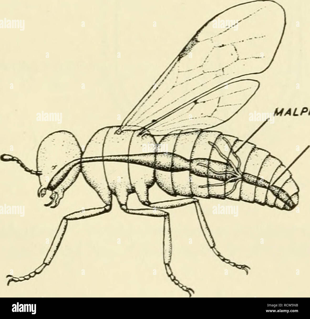 . Elements of biology, with special reference to their rôle in the lives of animals. Biology; Zoology. GREEN GLAND Fi« 127.—The green gland, excretory organs of a lobster. antennae abstract the non-gaseous wastes from the circulation and expel them. In insects a series of fine malpighian tubules are found in the body cavity (Fig. 128). These tubes open into the intestine,. MALPIGHIAN TUBULE INTESTINE Fig. 128.—Diagram representing the Malpighian tubules, the excretory organs of an insect, and their position. (After Kiihn: Grimdriss der allgemeinen Zoologie. Georg Theime, Leipzig.) into which t Stock Photohttps://www.alamy.com/image-license-details/?v=1https://www.alamy.com/elements-of-biology-with-special-reference-to-their-rle-in-the-lives-of-animals-biology-zoology-green-gland-fi-127the-green-gland-excretory-organs-of-a-lobster-antennae-abstract-the-non-gaseous-wastes-from-the-circulation-and-expel-them-in-insects-a-series-of-fine-malpighian-tubules-are-found-in-the-body-cavity-fig-128-these-tubes-open-into-the-intestine-malpighian-tubule-intestine-fig-128diagram-representing-the-malpighian-tubules-the-excretory-organs-of-an-insect-and-their-position-after-kiihn-grimdriss-der-allgemeinen-zoologie-georg-theime-leipzig-into-which-t-image231663975.html
RMRCW5NB–. Elements of biology, with special reference to their rôle in the lives of animals. Biology; Zoology. GREEN GLAND Fi« 127.—The green gland, excretory organs of a lobster. antennae abstract the non-gaseous wastes from the circulation and expel them. In insects a series of fine malpighian tubules are found in the body cavity (Fig. 128). These tubes open into the intestine,. MALPIGHIAN TUBULE INTESTINE Fig. 128.—Diagram representing the Malpighian tubules, the excretory organs of an insect, and their position. (After Kiihn: Grimdriss der allgemeinen Zoologie. Georg Theime, Leipzig.) into which t
 . The comparative anatomy of the domesticated animals. Veterinary anatomy. 572 URIXARY APPARATUS. thin and elastic, the internal face of which is lined by simple epithelium that readily alters -, the cells are polygonal in certain points, polyhedral in others, and transparent or granular. The uriniferous tube has not everywhere the same direction or diameter. Taking it at its termination on the crest of the pelvis, and following it to its origin in the Malpighian body, it is found that the tubule is at firet single, straight, and voluminous, but that during its com'se across the medullary sub- Stock Photohttps://www.alamy.com/image-license-details/?v=1https://www.alamy.com/the-comparative-anatomy-of-the-domesticated-animals-veterinary-anatomy-572-urixary-apparatus-thin-and-elastic-the-internal-face-of-which-is-lined-by-simple-epithelium-that-readily-alters-the-cells-are-polygonal-in-certain-points-polyhedral-in-others-and-transparent-or-granular-the-uriniferous-tube-has-not-everywhere-the-same-direction-or-diameter-taking-it-at-its-termination-on-the-crest-of-the-pelvis-and-following-it-to-its-origin-in-the-malpighian-body-it-is-found-that-the-tubule-is-at-firet-single-straight-and-voluminous-but-that-during-its-comse-across-the-medullary-sub-image232679323.html
RMREFCRR–. The comparative anatomy of the domesticated animals. Veterinary anatomy. 572 URIXARY APPARATUS. thin and elastic, the internal face of which is lined by simple epithelium that readily alters -, the cells are polygonal in certain points, polyhedral in others, and transparent or granular. The uriniferous tube has not everywhere the same direction or diameter. Taking it at its termination on the crest of the pelvis, and following it to its origin in the Malpighian body, it is found that the tubule is at firet single, straight, and voluminous, but that during its com'se across the medullary sub-
 . The comparative anatomy of the domesticated animals. Horses; Veterinary anatomy. 572 URINARY APPARATUS. thin and elastic, the internal face of which is lined by simple epithelium that readily alters ; the cells are polygonal in certain points, polyhedral in others, and transparent or granular. The uriniferous tube has not everywhere the same dii-ection or diameter. Taking it at its termination on the crest of the pelvis, and following it to its origin in the Malpighian body, it is found that the tubule is at first single, straight, and voluminous, but that during its course across the medull Stock Photohttps://www.alamy.com/image-license-details/?v=1https://www.alamy.com/the-comparative-anatomy-of-the-domesticated-animals-horses-veterinary-anatomy-572-urinary-apparatus-thin-and-elastic-the-internal-face-of-which-is-lined-by-simple-epithelium-that-readily-alters-the-cells-are-polygonal-in-certain-points-polyhedral-in-others-and-transparent-or-granular-the-uriniferous-tube-has-not-everywhere-the-same-dii-ection-or-diameter-taking-it-at-its-termination-on-the-crest-of-the-pelvis-and-following-it-to-its-origin-in-the-malpighian-body-it-is-found-that-the-tubule-is-at-first-single-straight-and-voluminous-but-that-during-its-course-across-the-medull-image232678923.html
RMREFC9F–. The comparative anatomy of the domesticated animals. Horses; Veterinary anatomy. 572 URINARY APPARATUS. thin and elastic, the internal face of which is lined by simple epithelium that readily alters ; the cells are polygonal in certain points, polyhedral in others, and transparent or granular. The uriniferous tube has not everywhere the same dii-ection or diameter. Taking it at its termination on the crest of the pelvis, and following it to its origin in the Malpighian body, it is found that the tubule is at first single, straight, and voluminous, but that during its course across the medull
 . The comparative anatomy of the domesticated animals. Veterinary anatomy. SECnON OF THE CORTICAL SUBSTANCE OF THE KIDNET A, A, Tubuli urinifcri divided transTersely, showing the spheroidal epithelium in â their interior; B, Malpighian capsule; a. Its afferent branch of the renal artery; b. Its glomerulus of capillaries; c, c, Secreting plexus formed by its efferent vessels; d, d, Fibrous stroma. origin in the Malpighian body, it is found that the tubule is at first single, straight, and voluminous, but that during its course across the medullary substance it divides into three or four tubes, Stock Photohttps://www.alamy.com/image-license-details/?v=1https://www.alamy.com/the-comparative-anatomy-of-the-domesticated-animals-veterinary-anatomy-secnon-of-the-cortical-substance-of-the-kidnet-a-a-tubuli-urinifcri-divided-transtersely-showing-the-spheroidal-epithelium-in-their-interior-b-malpighian-capsule-a-its-afferent-branch-of-the-renal-artery-b-its-glomerulus-of-capillaries-c-c-secreting-plexus-formed-by-its-efferent-vessels-d-d-fibrous-stroma-origin-in-the-malpighian-body-it-is-found-that-the-tubule-is-at-first-single-straight-and-voluminous-but-that-during-its-course-across-the-medullary-substance-it-divides-into-three-or-four-tubes-image232452436.html
RMRE53CM–. The comparative anatomy of the domesticated animals. Veterinary anatomy. SECnON OF THE CORTICAL SUBSTANCE OF THE KIDNET A, A, Tubuli urinifcri divided transTersely, showing the spheroidal epithelium in â their interior; B, Malpighian capsule; a. Its afferent branch of the renal artery; b. Its glomerulus of capillaries; c, c, Secreting plexus formed by its efferent vessels; d, d, Fibrous stroma. origin in the Malpighian body, it is found that the tubule is at first single, straight, and voluminous, but that during its course across the medullary substance it divides into three or four tubes,
 . The comparative anatomy of the domesticated animals. Veterinary anatomy. SECTION OF THE CORTICAL SUBSTANCE OF THE KIDNEY A, A, Tubuli uriniferi divided transversely, showing the spheroidal epithelium in their interior; B, Malpighian capsule; a, Its afterent branch of the renal artery; b, Its glomerulus of capillaries; c, c, Secreting plexus formed by its efferent vessels; d, d, Fibrous stroma. Fis. 250. origin in the Malpighian body, it is found that the tubule is at first single, straight, and voluminous, but that during its course across the medullary substance it divides into three or fou Stock Photohttps://www.alamy.com/image-license-details/?v=1https://www.alamy.com/the-comparative-anatomy-of-the-domesticated-animals-veterinary-anatomy-section-of-the-cortical-substance-of-the-kidney-a-a-tubuli-uriniferi-divided-transversely-showing-the-spheroidal-epithelium-in-their-interior-b-malpighian-capsule-a-its-afterent-branch-of-the-renal-artery-b-its-glomerulus-of-capillaries-c-c-secreting-plexus-formed-by-its-efferent-vessels-d-d-fibrous-stroma-fis-250-origin-in-the-malpighian-body-it-is-found-that-the-tubule-is-at-first-single-straight-and-voluminous-but-that-during-its-course-across-the-medullary-substance-it-divides-into-three-or-fou-image232668075.html
RMREEXE3–. The comparative anatomy of the domesticated animals. Veterinary anatomy. SECTION OF THE CORTICAL SUBSTANCE OF THE KIDNEY A, A, Tubuli uriniferi divided transversely, showing the spheroidal epithelium in their interior; B, Malpighian capsule; a, Its afterent branch of the renal artery; b, Its glomerulus of capillaries; c, c, Secreting plexus formed by its efferent vessels; d, d, Fibrous stroma. Fis. 250. origin in the Malpighian body, it is found that the tubule is at first single, straight, and voluminous, but that during its course across the medullary substance it divides into three or fou
 . Comparative anatomy of vertebrates. Anatomy, Comparative; Vertebrates -- Anatomy. UROGENITAL SYSTEM. 309 stome; the cilia, which may continue for some distance along the inside of the tubule, serving to create a current which carries the ccelomic fluid into the tubule and thence outward. Farther along the tubule expands into a Malpighian or renal corpuscle (fig. 315). This consists of a vesicle (Bowman's capsule), one side of which. FIG. 314.—Diagram of conventionalized excretory tubule. a, ascending limb of Henle's loop; b, Bowman's capsule of Malpighian body; c1—c2, first and second con- v Stock Photohttps://www.alamy.com/image-license-details/?v=1https://www.alamy.com/comparative-anatomy-of-vertebrates-anatomy-comparative-vertebrates-anatomy-urogenital-system-309-stome-the-cilia-which-may-continue-for-some-distance-along-the-inside-of-the-tubule-serving-to-create-a-current-which-carries-the-ccelomic-fluid-into-the-tubule-and-thence-outward-farther-along-the-tubule-expands-into-a-malpighian-or-renal-corpuscle-fig-315-this-consists-of-a-vesicle-bowmans-capsule-one-side-of-which-fig-314diagram-of-conventionalized-excretory-tubule-a-ascending-limb-of-henles-loop-b-bowmans-capsule-of-malpighian-body-c1c2-first-and-second-con-v-image232681853.html
RMREFG25–. Comparative anatomy of vertebrates. Anatomy, Comparative; Vertebrates -- Anatomy. UROGENITAL SYSTEM. 309 stome; the cilia, which may continue for some distance along the inside of the tubule, serving to create a current which carries the ccelomic fluid into the tubule and thence outward. Farther along the tubule expands into a Malpighian or renal corpuscle (fig. 315). This consists of a vesicle (Bowman's capsule), one side of which. FIG. 314.—Diagram of conventionalized excretory tubule. a, ascending limb of Henle's loop; b, Bowman's capsule of Malpighian body; c1—c2, first and second con- v
 . The anatomy of the domestic fowl . Domestic animals; Veterinary medicine; Poultry. THE TJRO-GENITAL SYSTEM 171 lobules. The renal artery (Fig. 53, No. 5,, 3) breaking up into arterioles in the. kidney, and finally reaching the cortical portion of the lobules, form capillary plexuses in the shape of minute spheres, which are the glomerules (Fig. 53, No. C, 3 and B, 5). Around each glomerule there is formed a capsule called Boisrman's capsule, which is the beginning of the uriniferous tubule. This entire mass is called the Malpighian body, or renal corpuscle. This capsule then ex- tends as the Stock Photohttps://www.alamy.com/image-license-details/?v=1https://www.alamy.com/the-anatomy-of-the-domestic-fowl-domestic-animals-veterinary-medicine-poultry-the-tjro-genital-system-171-lobules-the-renal-artery-fig-53-no-5-3-breaking-up-into-arterioles-in-the-kidney-and-finally-reaching-the-cortical-portion-of-the-lobules-form-capillary-plexuses-in-the-shape-of-minute-spheres-which-are-the-glomerules-fig-53-no-c-3-and-b-5-around-each-glomerule-there-is-formed-a-capsule-called-boisrmans-capsule-which-is-the-beginning-of-the-uriniferous-tubule-this-entire-mass-is-called-the-malpighian-body-or-renal-corpuscle-this-capsule-then-ex-tends-as-the-image231978472.html
RMRDBEWC–. The anatomy of the domestic fowl . Domestic animals; Veterinary medicine; Poultry. THE TJRO-GENITAL SYSTEM 171 lobules. The renal artery (Fig. 53, No. 5,, 3) breaking up into arterioles in the. kidney, and finally reaching the cortical portion of the lobules, form capillary plexuses in the shape of minute spheres, which are the glomerules (Fig. 53, No. C, 3 and B, 5). Around each glomerule there is formed a capsule called Boisrman's capsule, which is the beginning of the uriniferous tubule. This entire mass is called the Malpighian body, or renal corpuscle. This capsule then ex- tends as the
 . Text-book of embryology. Embryology. IV PEONEPHEOS 227 the splanchnocoele, the chamber containing the glomerulus appearing to be a side branch (Fig. 124, C), or on the other hand the pro- nephric chamber appears to form the dilated end of the tubule while the peritoneal canal appears to form a side branch (Fig. 124, D). In connexion with what has been said it is important'that the student should get clear, in his mind from the beginning (1) that the cavity into which the glomerulus projects (known as the cavity of the Malpighian body in the more highly evolved types of kidney) is simply a mo Stock Photohttps://www.alamy.com/image-license-details/?v=1https://www.alamy.com/text-book-of-embryology-embryology-iv-peonepheos-227-the-splanchnocoele-the-chamber-containing-the-glomerulus-appearing-to-be-a-side-branch-fig-124-c-or-on-the-other-hand-the-pro-nephric-chamber-appears-to-form-the-dilated-end-of-the-tubule-while-the-peritoneal-canal-appears-to-form-a-side-branch-fig-124-d-in-connexion-with-what-has-been-said-it-is-importantthat-the-student-should-get-clear-in-his-mind-from-the-beginning-1-that-the-cavity-into-which-the-glomerulus-projects-known-as-the-cavity-of-the-malpighian-body-in-the-more-highly-evolved-types-of-kidney-is-simply-a-mo-image232141049.html
RMRDJX7N–. Text-book of embryology. Embryology. IV PEONEPHEOS 227 the splanchnocoele, the chamber containing the glomerulus appearing to be a side branch (Fig. 124, C), or on the other hand the pro- nephric chamber appears to form the dilated end of the tubule while the peritoneal canal appears to form a side branch (Fig. 124, D). In connexion with what has been said it is important'that the student should get clear, in his mind from the beginning (1) that the cavity into which the glomerulus projects (known as the cavity of the Malpighian body in the more highly evolved types of kidney) is simply a mo
 . Comparative embryology of the vertebrates; with 2057 drawings and photos. grouped as 380 illus. Vertebrates -- Embryology; Comparative embryology. MESONEPHRIC RENAL UNITS ^7 ARISING BY CONDENSATION OF GROUPS OF CELLS WITHIN 15 NEPHROGENIC CORD - PRONEPHRIC IMESONEPHRIC) DUCT VESTIGIAL PRONEPHRIC TUBULE SECRETORY PORTION OF TUBULE. MALPIGHIAN BODY COELOM NEPHROSTOMAL / SECRETING CANAL MESONEPHRIC TUBULE DUCT Fig. 344. Regions of kidney origin within the vertebrate group; types of renal units formed. (A) The regions in the body where the diflferent types of vertebrate kidneys arise. The pronep Stock Photohttps://www.alamy.com/image-license-details/?v=1https://www.alamy.com/comparative-embryology-of-the-vertebrates-with-2057-drawings-and-photos-grouped-as-380-illus-vertebrates-embryology-comparative-embryology-mesonephric-renal-units-7-arising-by-condensation-of-groups-of-cells-within-15-nephrogenic-cord-pronephric-imesonephric-duct-vestigial-pronephric-tubule-secretory-portion-of-tubule-malpighian-body-coelom-nephrostomal-secreting-canal-mesonephric-tubule-duct-fig-344-regions-of-kidney-origin-within-the-vertebrate-group-types-of-renal-units-formed-a-the-regions-in-the-body-where-the-diflferent-types-of-vertebrate-kidneys-arise-the-pronep-image232674231.html
RMREF69Y–. Comparative embryology of the vertebrates; with 2057 drawings and photos. grouped as 380 illus. Vertebrates -- Embryology; Comparative embryology. MESONEPHRIC RENAL UNITS ^7 ARISING BY CONDENSATION OF GROUPS OF CELLS WITHIN 15 NEPHROGENIC CORD - PRONEPHRIC IMESONEPHRIC) DUCT VESTIGIAL PRONEPHRIC TUBULE SECRETORY PORTION OF TUBULE. MALPIGHIAN BODY COELOM NEPHROSTOMAL / SECRETING CANAL MESONEPHRIC TUBULE DUCT Fig. 344. Regions of kidney origin within the vertebrate group; types of renal units formed. (A) The regions in the body where the diflferent types of vertebrate kidneys arise. The pronep
 . Comparative anatomy. Anatomy, Comparative. 98 COMPARATIVE ANATOMY formed longitudinal duct which, as the pronephros degenerates, then serves at least in part as the mesonephric or "Wolffian duct. In Anamnia each mesonephric tubule has a ciliated nephrostome opening into the coelom (Fig. 72). Near the nephrostome the tubule gives rise to a cup- shaped expansion (Bowman's capsule). The hollow of the cup is occa- sioned by ingrowth of a dense network of fine blood vessels, the glomerulus. The capsule and glomerulus together constitute a renal (or Malpighian) body or corpuscle. The part of Stock Photohttps://www.alamy.com/image-license-details/?v=1https://www.alamy.com/comparative-anatomy-anatomy-comparative-98-comparative-anatomy-formed-longitudinal-duct-which-as-the-pronephros-degenerates-then-serves-at-least-in-part-as-the-mesonephric-or-quotwolffian-duct-in-anamnia-each-mesonephric-tubule-has-a-ciliated-nephrostome-opening-into-the-coelom-fig-72-near-the-nephrostome-the-tubule-gives-rise-to-a-cup-shaped-expansion-bowmans-capsule-the-hollow-of-the-cup-is-occa-sioned-by-ingrowth-of-a-dense-network-of-fine-blood-vessels-the-glomerulus-the-capsule-and-glomerulus-together-constitute-a-renal-or-malpighian-body-or-corpuscle-the-part-of-image232666374.html
RMREET9A–. Comparative anatomy. Anatomy, Comparative. 98 COMPARATIVE ANATOMY formed longitudinal duct which, as the pronephros degenerates, then serves at least in part as the mesonephric or "Wolffian duct. In Anamnia each mesonephric tubule has a ciliated nephrostome opening into the coelom (Fig. 72). Near the nephrostome the tubule gives rise to a cup- shaped expansion (Bowman's capsule). The hollow of the cup is occa- sioned by ingrowth of a dense network of fine blood vessels, the glomerulus. The capsule and glomerulus together constitute a renal (or Malpighian) body or corpuscle. The part of
 . Comparative anatomy of vertebrates. Anatomy, Comparative; Vertebrates -- Anatomy. FIG. 314.—Diagram of conventionalized excretory tubule. a, ascending limb of Henle's loop; b, Bowman's capsule of Malpighian body; c1—c2, first and second con- voluted tubules; ct, collecting tubule; d, descending limb of Henle's loop; g, glomerulus of Malpighian body; with artery and vein; h, Henle's loop; n, nephrostome opening into ccelom; x, entrance of other tubules into collecting duct. projects into the other, nearly filling the cavity. This inturned portion is the glomerulus. It consists of a network of Stock Photohttps://www.alamy.com/image-license-details/?v=1https://www.alamy.com/comparative-anatomy-of-vertebrates-anatomy-comparative-vertebrates-anatomy-fig-314diagram-of-conventionalized-excretory-tubule-a-ascending-limb-of-henles-loop-b-bowmans-capsule-of-malpighian-body-c1c2-first-and-second-con-voluted-tubules-ct-collecting-tubule-d-descending-limb-of-henles-loop-g-glomerulus-of-malpighian-body-with-artery-and-vein-h-henles-loop-n-nephrostome-opening-into-ccelom-x-entrance-of-other-tubules-into-collecting-duct-projects-into-the-other-nearly-filling-the-cavity-this-inturned-portion-is-the-glomerulus-it-consists-of-a-network-of-image232681843.html
RMREFG1R–. Comparative anatomy of vertebrates. Anatomy, Comparative; Vertebrates -- Anatomy. FIG. 314.—Diagram of conventionalized excretory tubule. a, ascending limb of Henle's loop; b, Bowman's capsule of Malpighian body; c1—c2, first and second con- voluted tubules; ct, collecting tubule; d, descending limb of Henle's loop; g, glomerulus of Malpighian body; with artery and vein; h, Henle's loop; n, nephrostome opening into ccelom; x, entrance of other tubules into collecting duct. projects into the other, nearly filling the cavity. This inturned portion is the glomerulus. It consists of a network of
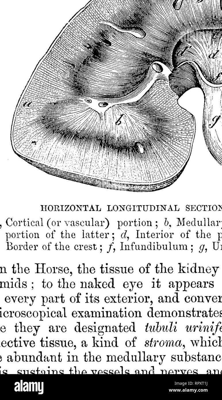 . The comparative anatomy of the domesticated animals. Veterinary anatomy. SECTION OP THE CORTICAL SUBSTANCE OF THE KIDNEY A, A, Tubuli nriniferi divided transversely, showing the spheroidal epithelium in their interior; B, Malpighian capsule; a, Its afferent branch of the renal artery; bf Its glomerulus of capillaries; c, c, Secreting plexus formed by its efferent vessels; d, d, Fibrous stroma. Fig. 250.. DIAGRAM OP THE COURSE OP THE URINIFEROUS TUBULE. a, Orifice of tubule at pelvic crest; 6, Recurrent branches which form loops, c, in the medullary portion of the kidney, and terminate in the Stock Photohttps://www.alamy.com/image-license-details/?v=1https://www.alamy.com/the-comparative-anatomy-of-the-domesticated-animals-veterinary-anatomy-section-op-the-cortical-substance-of-the-kidney-a-a-tubuli-nriniferi-divided-transversely-showing-the-spheroidal-epithelium-in-their-interior-b-malpighian-capsule-a-its-afferent-branch-of-the-renal-artery-bf-its-glomerulus-of-capillaries-c-c-secreting-plexus-formed-by-its-efferent-vessels-d-d-fibrous-stroma-fig-250-diagram-op-the-course-op-the-uriniferous-tubule-a-orifice-of-tubule-at-pelvic-crest-6-recurrent-branches-which-form-loops-c-in-the-medullary-portion-of-the-kidney-and-terminate-in-the-image237846830.html
RMRPXT1J–. The comparative anatomy of the domesticated animals. Veterinary anatomy. SECTION OP THE CORTICAL SUBSTANCE OF THE KIDNEY A, A, Tubuli nriniferi divided transversely, showing the spheroidal epithelium in their interior; B, Malpighian capsule; a, Its afferent branch of the renal artery; bf Its glomerulus of capillaries; c, c, Secreting plexus formed by its efferent vessels; d, d, Fibrous stroma. Fig. 250.. DIAGRAM OP THE COURSE OP THE URINIFEROUS TUBULE. a, Orifice of tubule at pelvic crest; 6, Recurrent branches which form loops, c, in the medullary portion of the kidney, and terminate in the
 . The comparative anatomy of the domesticated animals. Veterinary anatomy. DISTRIBUTION OF THE RENAL VESSELS IN THE horse's KIDNEY. a, Branch of reaal artery; a/, afferent vessel; m, m, malpighian tufts; ef, cf, efferent vessels tubes ; vascular plexus surrounding the st, straight tube ; ct, convoluted tube. DIAGRAM OF THE COURSE OF THE URINI- FEROUS TUBULE. a, Orifice of tubule at pelvic crest; 5, re- current bj'anches which form loops, c, in the medullary portion of the kidney, and terminate in the Malpighian capsules in the cortical portion. capillaries. In the medullary substance, there ar Stock Photohttps://www.alamy.com/image-license-details/?v=1https://www.alamy.com/the-comparative-anatomy-of-the-domesticated-animals-veterinary-anatomy-distribution-of-the-renal-vessels-in-the-horses-kidney-a-branch-of-reaal-artery-a-afferent-vessel-m-m-malpighian-tufts-ef-cf-efferent-vessels-tubes-vascular-plexus-surrounding-the-st-straight-tube-ct-convoluted-tube-diagram-of-the-course-of-the-urini-ferous-tubule-a-orifice-of-tubule-at-pelvic-crest-5-re-current-bjanches-which-form-loops-c-in-the-medullary-portion-of-the-kidney-and-terminate-in-the-malpighian-capsules-in-the-cortical-portion-capillaries-in-the-medullary-substance-there-ar-image232679304.html
RMREFCR4–. The comparative anatomy of the domesticated animals. Veterinary anatomy. DISTRIBUTION OF THE RENAL VESSELS IN THE horse's KIDNEY. a, Branch of reaal artery; a/, afferent vessel; m, m, malpighian tufts; ef, cf, efferent vessels tubes ; vascular plexus surrounding the st, straight tube ; ct, convoluted tube. DIAGRAM OF THE COURSE OF THE URINI- FEROUS TUBULE. a, Orifice of tubule at pelvic crest; 5, re- current bj'anches which form loops, c, in the medullary portion of the kidney, and terminate in the Malpighian capsules in the cortical portion. capillaries. In the medullary substance, there ar
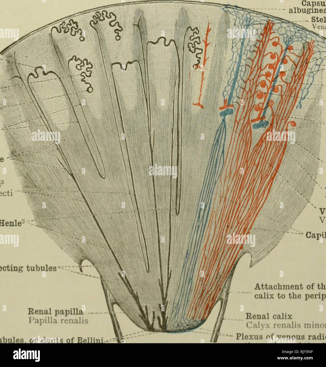 . An atlas of human anatomy for students and physicians. Anatomy. First convoluted tubule Tubulus renalis contortus -Glomerulus Capsule of the ""glomerulus (Bowman's capsule) Capsula glomeruli EflFerent vessel of the glomerulus Vas efferens Capillary vessels f/';r-"'-;=-Straight tubules ,. Tubuli renales 'â recti ,^ Afferent vessel of the glomerulus Vas af^erens Interlobular or radiate artery A. interlobularis Fig. 833.âPart of a Sf.ction through the Cortex OF THE Kidney in the direction of the Straight Tubules. Fig. 834.âCorpusculum Renis (Malpighii), Malpighian Corpuscle of Stock Photohttps://www.alamy.com/image-license-details/?v=1https://www.alamy.com/an-atlas-of-human-anatomy-for-students-and-physicians-anatomy-first-convoluted-tubule-tubulus-renalis-contortus-glomerulus-capsule-of-the-quotquotglomerulus-bowmans-capsule-capsula-glomeruli-eflferent-vessel-of-the-glomerulus-vas-efferens-capillary-vessels-fr-quot-=-straight-tubules-tubuli-renales-recti-afferent-vessel-of-the-glomerulus-vas-aferens-interlobular-or-radiate-artery-a-interlobularis-fig-833part-of-a-sfction-through-the-cortex-of-the-kidney-in-the-direction-of-the-straight-tubules-fig-834corpusculum-renis-malpighii-malpighian-corpuscle-of-image235395819.html
RMRJY5NF–. An atlas of human anatomy for students and physicians. Anatomy. First convoluted tubule Tubulus renalis contortus -Glomerulus Capsule of the ""glomerulus (Bowman's capsule) Capsula glomeruli EflFerent vessel of the glomerulus Vas efferens Capillary vessels f/';r-"'-;=-Straight tubules ,. Tubuli renales 'â recti ,^ Afferent vessel of the glomerulus Vas af^erens Interlobular or radiate artery A. interlobularis Fig. 833.âPart of a Sf.ction through the Cortex OF THE Kidney in the direction of the Straight Tubules. Fig. 834.âCorpusculum Renis (Malpighii), Malpighian Corpuscle of
 . Comparative embryology of the vertebrates; with 2057 drawings and photos. grouped as 380 illus. Vertebrates -- Embryology; Comparative embryology. GLOMERULU BOWMAN'S CAPSULE LOOP OF HENLE LUNG BUD URACHUS BLADDER PHALLUS STOMACH UROGENITAL SINUS. aL SEPTUM Vf CLOACA RECTUM SONEPHRIC KIDNEY EPHRIC DUCT VISCERAL ROOT PHRIC DUCT INTESTIN UMBILICAL „.,,^ ARTERY METANEPHRIC DUCT ARCHED COLLECTING TUBULE IMARY OR STRAIGHT OLLECTING TUBULE DISTAL ONVOLUTED TUBULE EFFERENT BLOOD VESSEL AFFERENT BLOOD VESSEL RENAL CORPUSCLE (MALPIGHIAN BODY) LOOD CAPILLARIES ASCENDING LIMB OF HENLE'S LOOP STRAIGHT CO Stock Photohttps://www.alamy.com/image-license-details/?v=1https://www.alamy.com/comparative-embryology-of-the-vertebrates-with-2057-drawings-and-photos-grouped-as-380-illus-vertebrates-embryology-comparative-embryology-glomerulu-bowmans-capsule-loop-of-henle-lung-bud-urachus-bladder-phallus-stomach-urogenital-sinus-al-septum-vf-cloaca-rectum-sonephric-kidney-ephric-duct-visceral-root-phric-duct-intestin-umbilical-artery-metanephric-duct-arched-collecting-tubule-imary-or-straight-ollecting-tubule-distal-onvoluted-tubule-efferent-blood-vessel-afferent-blood-vessel-renal-corpuscle-malpighian-body-lood-capillaries-ascending-limb-of-henles-loop-straight-co-image232665462.html
RMREER4P–. Comparative embryology of the vertebrates; with 2057 drawings and photos. grouped as 380 illus. Vertebrates -- Embryology; Comparative embryology. GLOMERULU BOWMAN'S CAPSULE LOOP OF HENLE LUNG BUD URACHUS BLADDER PHALLUS STOMACH UROGENITAL SINUS. aL SEPTUM Vf CLOACA RECTUM SONEPHRIC KIDNEY EPHRIC DUCT VISCERAL ROOT PHRIC DUCT INTESTIN UMBILICAL „.,,^ ARTERY METANEPHRIC DUCT ARCHED COLLECTING TUBULE IMARY OR STRAIGHT OLLECTING TUBULE DISTAL ONVOLUTED TUBULE EFFERENT BLOOD VESSEL AFFERENT BLOOD VESSEL RENAL CORPUSCLE (MALPIGHIAN BODY) LOOD CAPILLARIES ASCENDING LIMB OF HENLE'S LOOP STRAIGHT CO
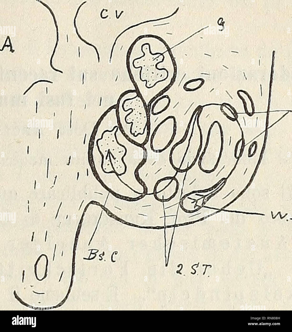 . Anatomischer Anzeiger. Anatomy, Comparative. / S!7? Fig. 5. Rabbits embryo 11 days old. From the Wolffian duct the tubule bends inwards and then makes a sharp bend forwards. On the convexity of this bend the future glomerulus forms (G), and moulded on the bend the Bowman's capsule is also seen (Bs. C). Fig. 6. Eabbits embryo 14 days old. The Wolffian duct opens into narrow col- lecting tubules (1 ST), these into larger tubules (2 ST) and these again into Malpighian bodies. The three Malpighian bodies represented in figure all open in this way into Wolffian duct. be seen, and corresponding wi Stock Photohttps://www.alamy.com/image-license-details/?v=1https://www.alamy.com/anatomischer-anzeiger-anatomy-comparative-s!7-fig-5-rabbits-embryo-11-days-old-from-the-wolffian-duct-the-tubule-bends-inwards-and-then-makes-a-sharp-bend-forwards-on-the-convexity-of-this-bend-the-future-glomerulus-forms-g-and-moulded-on-the-bend-the-bowmans-capsule-is-also-seen-bs-c-fig-6-eabbits-embryo-14-days-old-the-wolffian-duct-opens-into-narrow-col-lecting-tubules-1-st-these-into-larger-tubules-2-st-and-these-again-into-malpighian-bodies-the-three-malpighian-bodies-represented-in-figure-all-open-in-this-way-into-wolffian-duct-be-seen-and-corresponding-wi-image236818417.html
RMRN808H–. Anatomischer Anzeiger. Anatomy, Comparative. / S!7? Fig. 5. Rabbits embryo 11 days old. From the Wolffian duct the tubule bends inwards and then makes a sharp bend forwards. On the convexity of this bend the future glomerulus forms (G), and moulded on the bend the Bowman's capsule is also seen (Bs. C). Fig. 6. Eabbits embryo 14 days old. The Wolffian duct opens into narrow col- lecting tubules (1 ST), these into larger tubules (2 ST) and these again into Malpighian bodies. The three Malpighian bodies represented in figure all open in this way into Wolffian duct. be seen, and corresponding wi
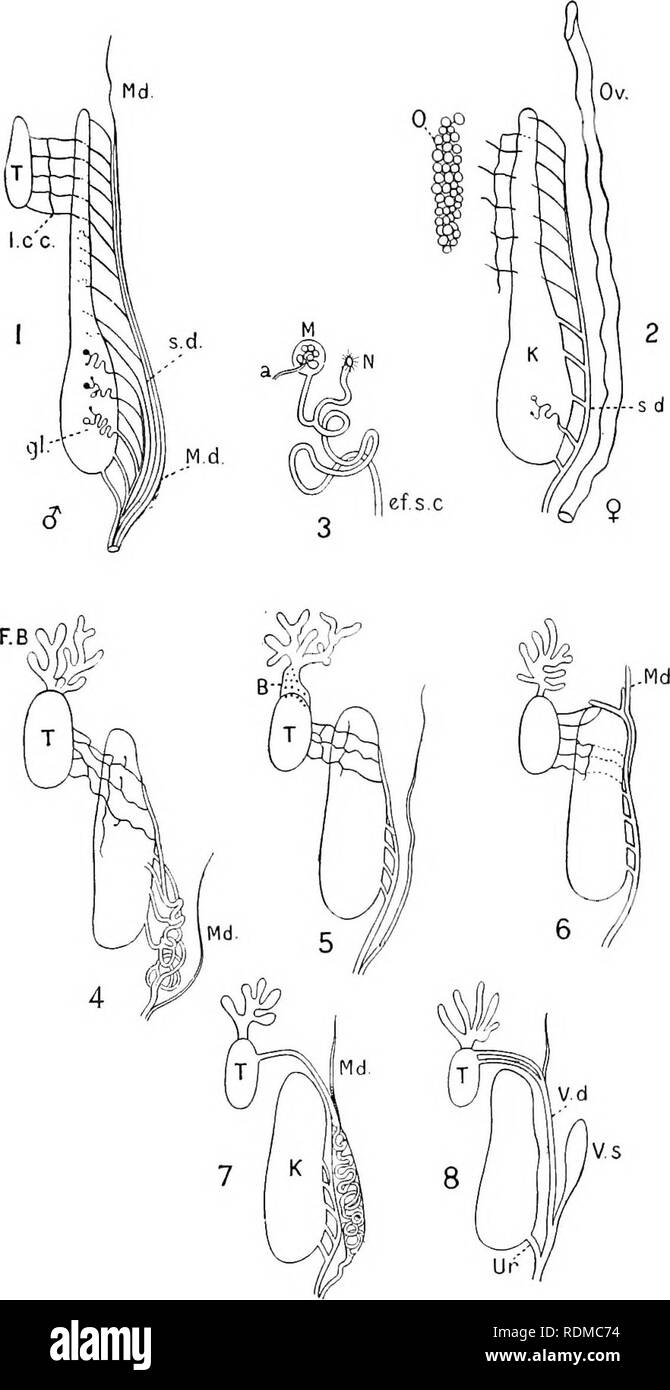 . The Cambridge natural history. Zoology. URINO-GENITAL ORGANS 49. Fig. 7.—Diagrammatic representation of .modifications of the urino-genital ducts. 1, 2, Male and female Newt; 3, a tubule of the kidney ; 4, male Rana; 5, male iJ?(/b/ 6, male iJom&ma^o/* / T, Tii2i& Discoglossus ; ^,rta&Alytes. a, Artery entering, and producing a coil in, the Malpighian body, il; B, Bidder's organ ; ef.sx, efferent segmental canal; F.B, fat-body; gl, glomerulus; K, Iddney ; l.cr, longitudinal collecting canal; J/, Malpighian body; Md, Miillerian duct ; N, nephrostome ; 0, ovary ; Ov, oviduct; s. Stock Photohttps://www.alamy.com/image-license-details/?v=1https://www.alamy.com/the-cambridge-natural-history-zoology-urino-genital-organs-49-fig-7diagrammatic-representation-of-modifications-of-the-urino-genital-ducts-1-2-male-and-female-newt-3-a-tubule-of-the-kidney-4-male-rana-5-male-ijb-6-male-ijomampmao-t-tii2iamp-discoglossus-rtaampalytes-a-artery-entering-and-producing-a-coil-in-the-malpighian-body-il-b-bidders-organ-efsx-efferent-segmental-canal-fb-fat-body-gl-glomerulus-k-iddney-lcr-longitudinal-collecting-canal-j-malpighian-body-md-miillerian-duct-n-nephrostome-0-ovary-ov-oviduct-s-image232173960.html
RMRDMC74–. The Cambridge natural history. Zoology. URINO-GENITAL ORGANS 49. Fig. 7.—Diagrammatic representation of .modifications of the urino-genital ducts. 1, 2, Male and female Newt; 3, a tubule of the kidney ; 4, male Rana; 5, male iJ?(/b/ 6, male iJom&ma^o/* / T, Tii2i& Discoglossus ; ^,rta&Alytes. a, Artery entering, and producing a coil in, the Malpighian body, il; B, Bidder's organ ; ef.sx, efferent segmental canal; F.B, fat-body; gl, glomerulus; K, Iddney ; l.cr, longitudinal collecting canal; J/, Malpighian body; Md, Miillerian duct ; N, nephrostome ; 0, ovary ; Ov, oviduct; s.
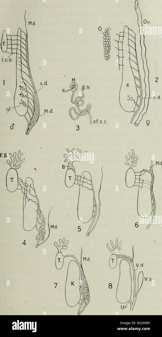 . The Cambridge natural history. Zoology. URINO-GENITAL ORGANS 49. Fig. 7.—Diagrammatic representation of modifications of the urino-genital ducts. 1, 2, Male and female Newt; 3, a tubule of the kidney ; 4, male Rana; 5, male Bufo; 6, male Bombiaator ; 7, male iJiacof/lossiis : 8, male Alytes. a, Ai-tery entering, and producing a coil in, the Malpighiau body, M; B, Bidder's organ ; 1'f.s.c, efferent segmental canal ; F.B, fat-body ; gl, glomerulus : K, kidney ; l.c.c. longitudinal collecting canal ; M, Malpighian body ; Md, Miillerian duct ; N, nephrostome ; 0, ovary ; Or, oviduct ; s.d, segme Stock Photohttps://www.alamy.com/image-license-details/?v=1https://www.alamy.com/the-cambridge-natural-history-zoology-urino-genital-organs-49-fig-7diagrammatic-representation-of-modifications-of-the-urino-genital-ducts-1-2-male-and-female-newt-3-a-tubule-of-the-kidney-4-male-rana-5-male-bufo-6-male-bombiaator-7-male-ijiacoflossiis-8-male-alytes-a-ai-tery-entering-and-producing-a-coil-in-the-malpighiau-body-m-b-bidders-organ-1fsc-efferent-segmental-canal-fb-fat-body-gl-glomerulus-k-kidney-lcc-longitudinal-collecting-canal-m-malpighian-body-md-miillerian-duct-n-nephrostome-0-ovary-or-oviduct-sd-segme-image233648714.html
RMRG3H8X–. The Cambridge natural history. Zoology. URINO-GENITAL ORGANS 49. Fig. 7.—Diagrammatic representation of modifications of the urino-genital ducts. 1, 2, Male and female Newt; 3, a tubule of the kidney ; 4, male Rana; 5, male Bufo; 6, male Bombiaator ; 7, male iJiacof/lossiis : 8, male Alytes. a, Ai-tery entering, and producing a coil in, the Malpighiau body, M; B, Bidder's organ ; 1'f.s.c, efferent segmental canal ; F.B, fat-body ; gl, glomerulus : K, kidney ; l.c.c. longitudinal collecting canal ; M, Malpighian body ; Md, Miillerian duct ; N, nephrostome ; 0, ovary ; Or, oviduct ; s.d, segme
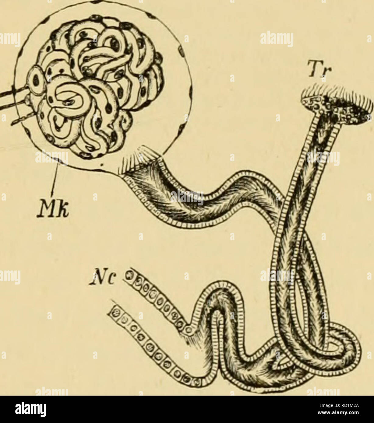 . Elementary text-book of zoology, general part and special part: protozoa to insecta. Animals. Fig. 72.—Ciliated funnel and Malpighian body from the anterior part of the kidney of Proteus (after Spengel). Nr, kidney tubule; Tr, ciliated funnel; Mk, Malpig- hian body.. Please note that these images are extracted from scanned page images that may have been digitally enhanced for readability - coloration and appearance of these illustrations may not perfectly resemble the original work.. Claus, Carl, 1835-1899; Metcalf Collection (North Carolina State University). NCRS. London, Swan Sonnenschein Stock Photohttps://www.alamy.com/image-license-details/?v=1https://www.alamy.com/elementary-text-book-of-zoology-general-part-and-special-part-protozoa-to-insecta-animals-fig-72ciliated-funnel-and-malpighian-body-from-the-anterior-part-of-the-kidney-of-proteus-after-spengel-nr-kidney-tubule-tr-ciliated-funnel-mk-malpig-hian-body-please-note-that-these-images-are-extracted-from-scanned-page-images-that-may-have-been-digitally-enhanced-for-readability-coloration-and-appearance-of-these-illustrations-may-not-perfectly-resemble-the-original-work-claus-carl-1835-1899-metcalf-collection-north-carolina-state-university-ncrs-london-swan-sonnenschein-image231763010.html
RMRD1M2A–. Elementary text-book of zoology, general part and special part: protozoa to insecta. Animals. Fig. 72.—Ciliated funnel and Malpighian body from the anterior part of the kidney of Proteus (after Spengel). Nr, kidney tubule; Tr, ciliated funnel; Mk, Malpig- hian body.. Please note that these images are extracted from scanned page images that may have been digitally enhanced for readability - coloration and appearance of these illustrations may not perfectly resemble the original work.. Claus, Carl, 1835-1899; Metcalf Collection (North Carolina State University). NCRS. London, Swan Sonnenschein
 . Elements of the comparative anatomy of vertebrates. Anatomy, Comparative; Vertebrates -- Anatomy. GENERATIVE ORGANS. 315. -ES FIG. 246.—DIAGRAM OF A PORTION OF THE MALE GENERATIVE APPARATUS OF THE GYMNOPHIONA. Ho, Ho. testis ; Sg, collecting duct of testis ; K, K, testicular capsules ; Q, Q, transverse canals connecting the collecting duct with the longitudinal canal (L, L) ; Ql, Q1, second series of transverse canals ; M, M, Malpighian capsules ; N, N, kidney ; ST, nephrostome ; S, convoluted portion of urinary tubule ; Hti, urinogenital duct.. Please note that these images are extracted fr Stock Photohttps://www.alamy.com/image-license-details/?v=1https://www.alamy.com/elements-of-the-comparative-anatomy-of-vertebrates-anatomy-comparative-vertebrates-anatomy-generative-organs-315-es-fig-246diagram-of-a-portion-of-the-male-generative-apparatus-of-the-gymnophiona-ho-ho-testis-sg-collecting-duct-of-testis-k-k-testicular-capsules-q-q-transverse-canals-connecting-the-collecting-duct-with-the-longitudinal-canal-l-l-ql-q1-second-series-of-transverse-canals-m-m-malpighian-capsules-n-n-kidney-st-nephrostome-s-convoluted-portion-of-urinary-tubule-hti-urinogenital-duct-please-note-that-these-images-are-extracted-fr-image231602968.html
RMRCPBXG–. Elements of the comparative anatomy of vertebrates. Anatomy, Comparative; Vertebrates -- Anatomy. GENERATIVE ORGANS. 315. -ES FIG. 246.—DIAGRAM OF A PORTION OF THE MALE GENERATIVE APPARATUS OF THE GYMNOPHIONA. Ho, Ho. testis ; Sg, collecting duct of testis ; K, K, testicular capsules ; Q, Q, transverse canals connecting the collecting duct with the longitudinal canal (L, L) ; Ql, Q1, second series of transverse canals ; M, M, Malpighian capsules ; N, N, kidney ; ST, nephrostome ; S, convoluted portion of urinary tubule ; Hti, urinogenital duct.. Please note that these images are extracted fr
 . Elements of histology. Histology. Fig. 135.—From a Vertical Section through, the Kidney of Dog, showing part of the Labyrinth and the adjoining Medullary Ray. a, The capsule of Bowman; the capillaries of the glomerulus are arranged in lobules; n, neck of capsule; b, irregular tubule; c, proximal convoluted tubules; d, a collecting tube ; e, part of the spiral tubule ; /, portion of the ascending limb of Henle's loop-tube; d, e, / form the medullary ray. (Atlas.) mouse—they already have begun in the Malpighian corpuscle. The outer part of the cell protoplasm—i.e.,. Please note that these imag Stock Photohttps://www.alamy.com/image-license-details/?v=1https://www.alamy.com/elements-of-histology-histology-fig-135from-a-vertical-section-through-the-kidney-of-dog-showing-part-of-the-labyrinth-and-the-adjoining-medullary-ray-a-the-capsule-of-bowman-the-capillaries-of-the-glomerulus-are-arranged-in-lobules-n-neck-of-capsule-b-irregular-tubule-c-proximal-convoluted-tubules-d-a-collecting-tube-e-part-of-the-spiral-tubule-portion-of-the-ascending-limb-of-henles-loop-tube-d-e-form-the-medullary-ray-atlas-mousethey-already-have-begun-in-the-malpighian-corpuscle-the-outer-part-of-the-cell-protoplasmie-please-note-that-these-imag-image231492137.html
RMRCHAG9–. Elements of histology. Histology. Fig. 135.—From a Vertical Section through, the Kidney of Dog, showing part of the Labyrinth and the adjoining Medullary Ray. a, The capsule of Bowman; the capillaries of the glomerulus are arranged in lobules; n, neck of capsule; b, irregular tubule; c, proximal convoluted tubules; d, a collecting tube ; e, part of the spiral tubule ; /, portion of the ascending limb of Henle's loop-tube; d, e, / form the medullary ray. (Atlas.) mouse—they already have begun in the Malpighian corpuscle. The outer part of the cell protoplasm—i.e.,. Please note that these imag
 . Elements of the comparative anatomy of vertebrates. Anatomy, Comparative; Vertebrates -- Anatomy. -ES FIG. 246.—DIAGRAM OF A PORTION OF THE MALE GENERATIVE APPARATUS OF THE GYMNOPHIONA. Ho, Ho. testis ; Sg, collecting duct of testis ; K, K, testicular capsules ; Q, Q, transverse canals connecting the collecting duct with the longitudinal canal (L, L) ; Ql, Q1, second series of transverse canals ; M, M, Malpighian capsules ; N, N, kidney ; ST, nephrostome ; S, convoluted portion of urinary tubule ; Hti, urinogenital duct.. Ur FIG. 247.—TESTIS AND ANTERIOR END OF KIDNEY OF Eana, csculcnta. (Se Stock Photohttps://www.alamy.com/image-license-details/?v=1https://www.alamy.com/elements-of-the-comparative-anatomy-of-vertebrates-anatomy-comparative-vertebrates-anatomy-es-fig-246diagram-of-a-portion-of-the-male-generative-apparatus-of-the-gymnophiona-ho-ho-testis-sg-collecting-duct-of-testis-k-k-testicular-capsules-q-q-transverse-canals-connecting-the-collecting-duct-with-the-longitudinal-canal-l-l-ql-q1-second-series-of-transverse-canals-m-m-malpighian-capsules-n-n-kidney-st-nephrostome-s-convoluted-portion-of-urinary-tubule-hti-urinogenital-duct-ur-fig-247testis-and-anterior-end-of-kidney-of-eana-csculcnta-se-image231602966.html
RMRCPBXE–. Elements of the comparative anatomy of vertebrates. Anatomy, Comparative; Vertebrates -- Anatomy. -ES FIG. 246.—DIAGRAM OF A PORTION OF THE MALE GENERATIVE APPARATUS OF THE GYMNOPHIONA. Ho, Ho. testis ; Sg, collecting duct of testis ; K, K, testicular capsules ; Q, Q, transverse canals connecting the collecting duct with the longitudinal canal (L, L) ; Ql, Q1, second series of transverse canals ; M, M, Malpighian capsules ; N, N, kidney ; ST, nephrostome ; S, convoluted portion of urinary tubule ; Hti, urinogenital duct.. Ur FIG. 247.—TESTIS AND ANTERIOR END OF KIDNEY OF Eana, csculcnta. (Se