Quick filters:
Mature follicle of ovary Stock Photos and Images
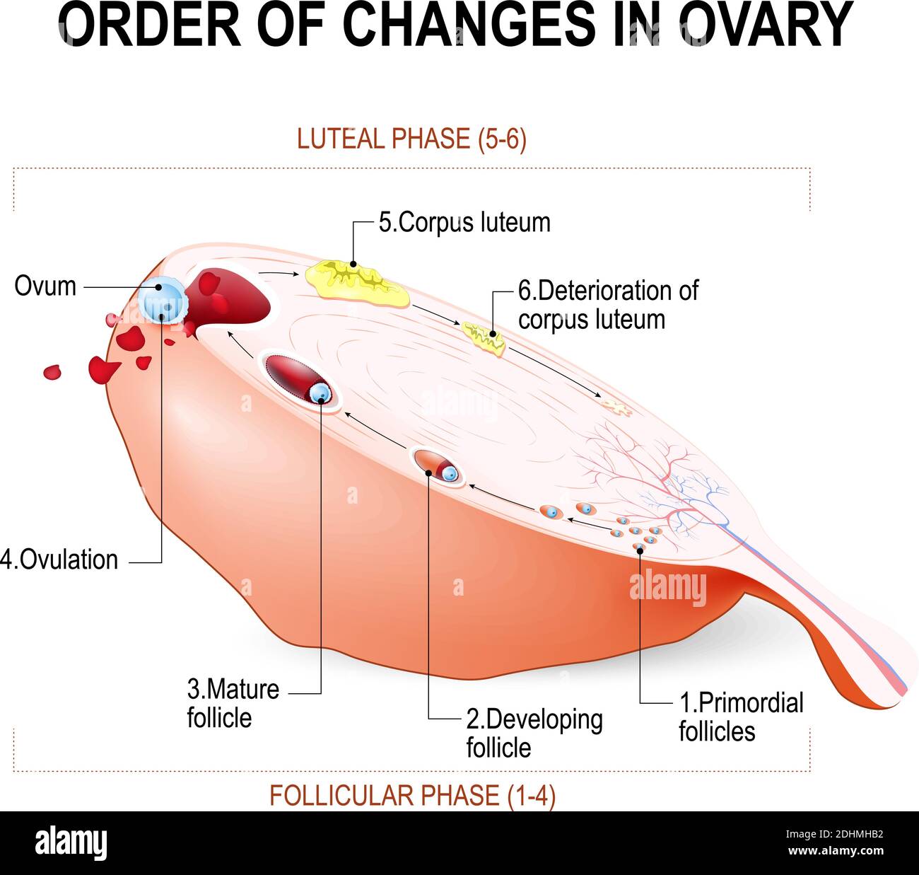 Order of changes in ovary: from Developing follicle to Ovulation and Corpus luteum. Menstruation. Education Chart of Biology. vector Diagram. Stock Vectorhttps://www.alamy.com/image-license-details/?v=1https://www.alamy.com/order-of-changes-in-ovary-from-developing-follicle-to-ovulation-and-corpus-luteum-menstruation-education-chart-of-biology-vector-diagram-image389529926.html
RF2DHMHB2–Order of changes in ovary: from Developing follicle to Ovulation and Corpus luteum. Menstruation. Education Chart of Biology. vector Diagram.
 Ovulation, drawing Stock Photohttps://www.alamy.com/image-license-details/?v=1https://www.alamy.com/ovulation-drawing-image407105479.html
RM2EJ975B–Ovulation, drawing
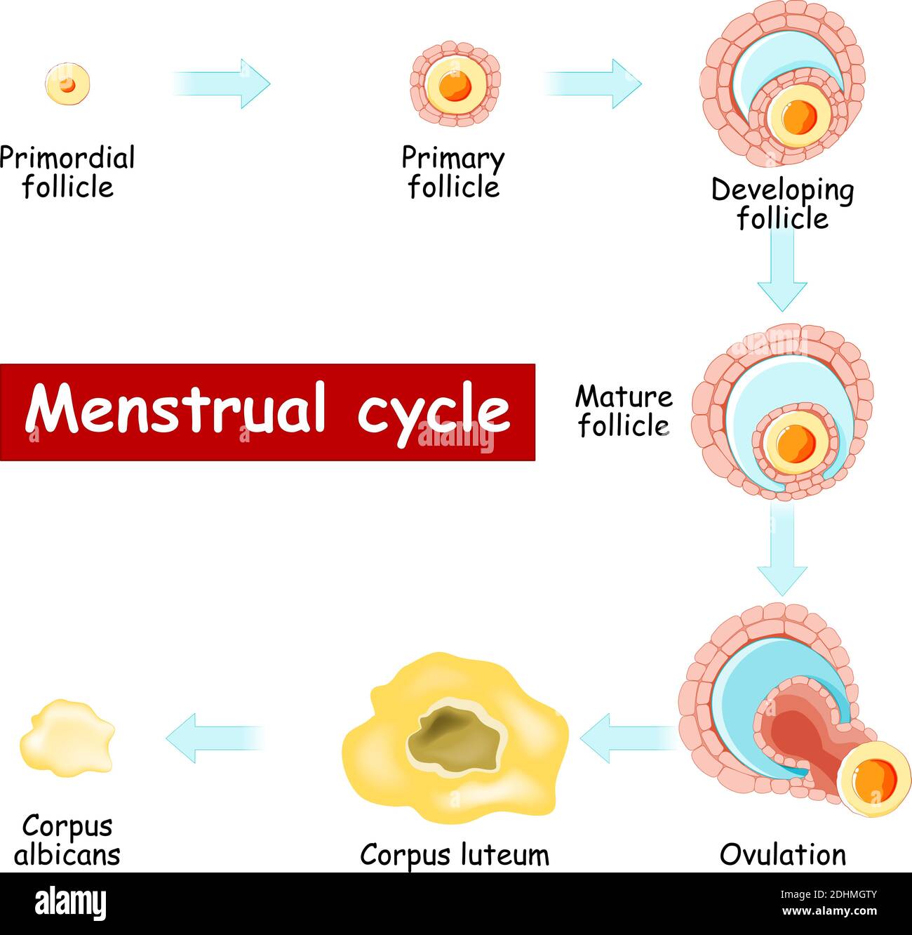 Menstrual cycle. Changes in ovary: from Developing follicle to Ovulation and Corpus luteum. Chart of Biology. vector Diagram. maturation of a follicle Stock Vectorhttps://www.alamy.com/image-license-details/?v=1https://www.alamy.com/menstrual-cycle-changes-in-ovary-from-developing-follicle-to-ovulation-and-corpus-luteum-chart-of-biology-vector-diagram-maturation-of-a-follicle-image389529531.html
RF2DHMGTY–Menstrual cycle. Changes in ovary: from Developing follicle to Ovulation and Corpus luteum. Chart of Biology. vector Diagram. maturation of a follicle
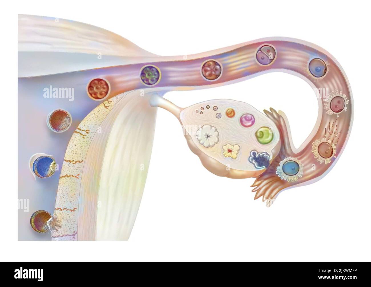 Female genitalia: ovarian cycle, ovulation, fertilization, embryo segmentation, implantation. Stock Photohttps://www.alamy.com/image-license-details/?v=1https://www.alamy.com/female-genitalia-ovarian-cycle-ovulation-fertilization-embryo-segmentation-implantation-image476923322.html
RF2JKWMFP–Female genitalia: ovarian cycle, ovulation, fertilization, embryo segmentation, implantation.
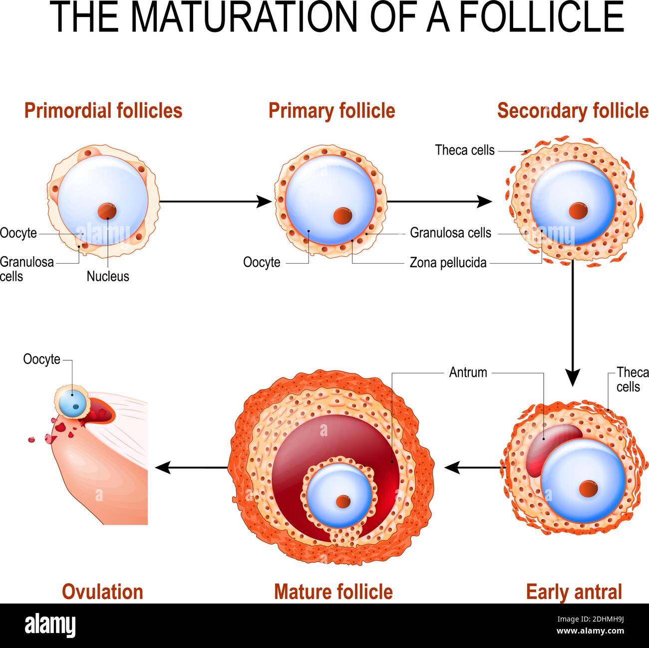 maturation of a follicle. Diagram of folliculogenesis. Stock Vectorhttps://www.alamy.com/image-license-details/?v=1https://www.alamy.com/maturation-of-a-follicle-diagram-of-folliculogenesis-image389529886.html
RF2DHMH9J–maturation of a follicle. Diagram of folliculogenesis.
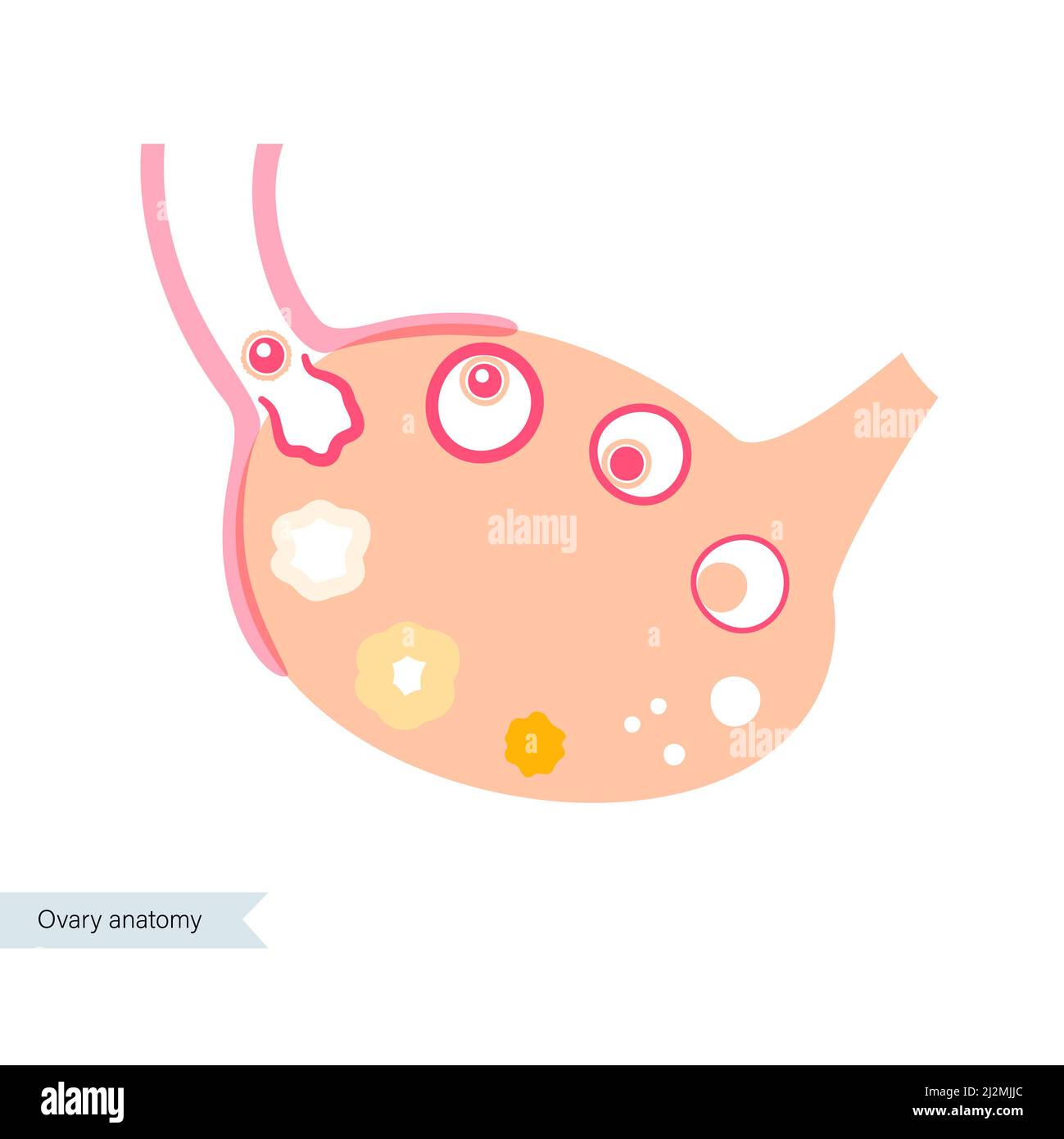 Menstrual cycle, illustration Stock Photohttps://www.alamy.com/image-license-details/?v=1https://www.alamy.com/menstrual-cycle-illustration-image466362916.html
RF2J2MJJC–Menstrual cycle, illustration
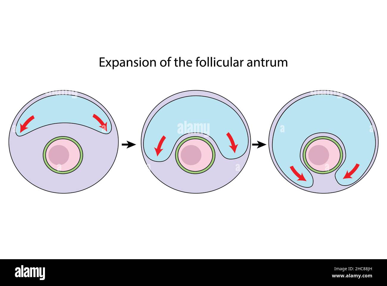 Expansion of the follicular antrum diagram, ovulation (unlabeled) Stock Photohttps://www.alamy.com/image-license-details/?v=1https://www.alamy.com/expansion-of-the-follicular-antrum-diagram-ovulation-unlabeled-image455027849.html
RF2HC88JH–Expansion of the follicular antrum diagram, ovulation (unlabeled)
 The ovary Stock Photohttps://www.alamy.com/image-license-details/?v=1https://www.alamy.com/stock-photo-the-ovary-13184410.html
RFACM1YR–The ovary
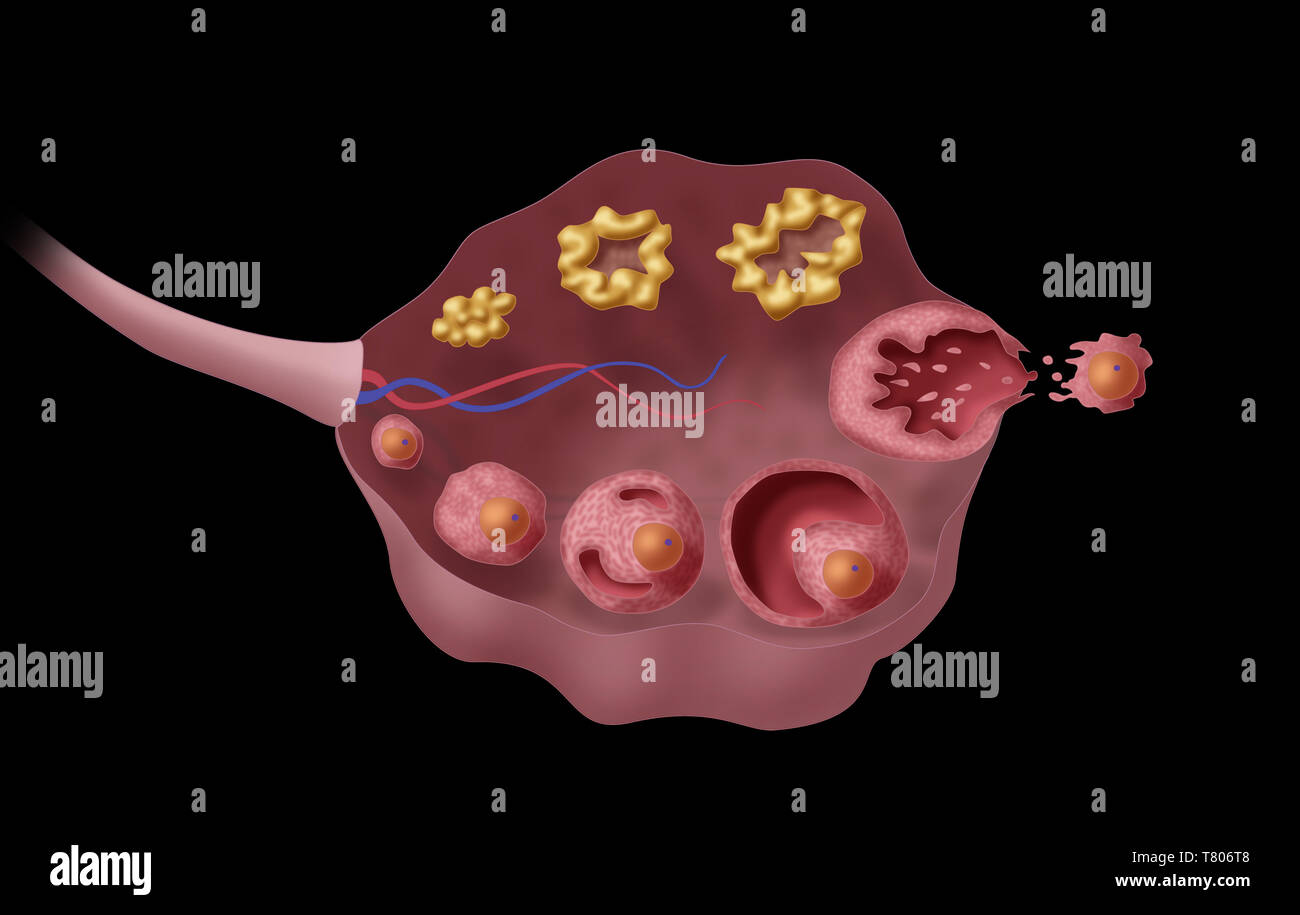 Ovarian Follicles, Illustration Stock Photohttps://www.alamy.com/image-license-details/?v=1https://www.alamy.com/ovarian-follicles-illustration-image245867784.html
RMT806T8–Ovarian Follicles, Illustration
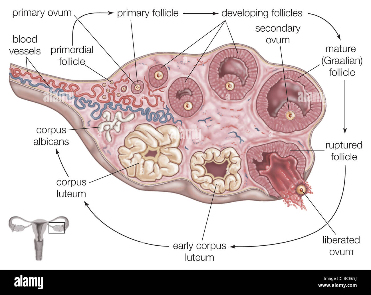 The steps of ovulation: a primordial follicle grows and matures, before being released by the ovary into the fallopian tube. Stock Photohttps://www.alamy.com/image-license-details/?v=1https://www.alamy.com/stock-photo-the-steps-of-ovulation-a-primordial-follicle-grows-and-matures-before-24898542.html
RMBCE69J–The steps of ovulation: a primordial follicle grows and matures, before being released by the ovary into the fallopian tube.
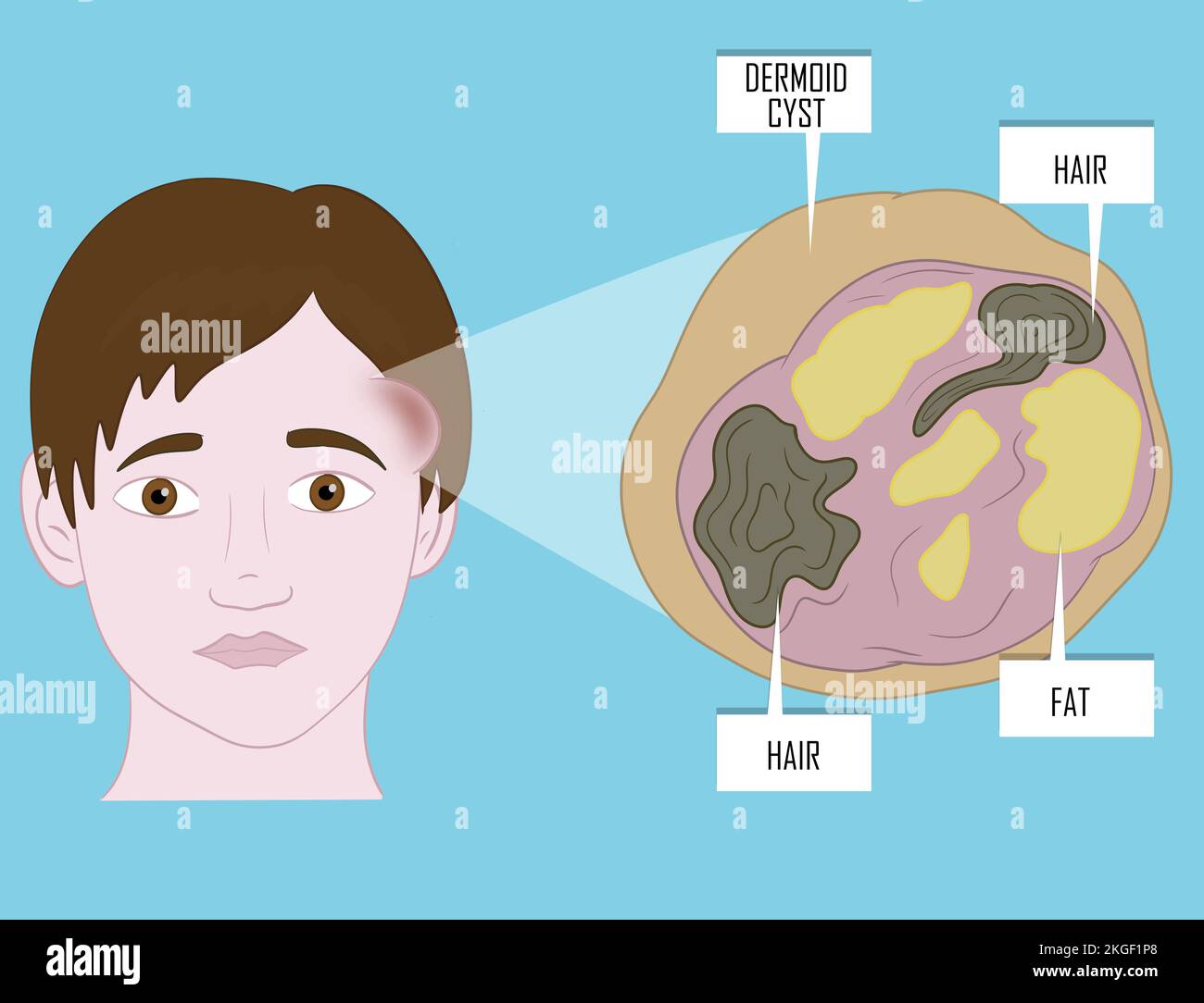 Dermoid cysts symptoms on young patient face. Illustration. Stock Photohttps://www.alamy.com/image-license-details/?v=1https://www.alamy.com/dermoid-cysts-symptoms-on-young-patient-face-illustration-image492055488.html
RF2KGF1P8–Dermoid cysts symptoms on young patient face. Illustration.
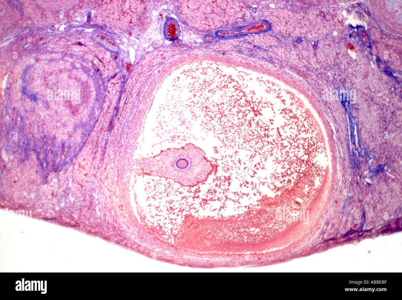 Mature Graafian follicle Stained section Stock Photohttps://www.alamy.com/image-license-details/?v=1https://www.alamy.com/mature-graafian-follicle-stained-section-image769727.html
RMABBEBF–Mature Graafian follicle Stained section
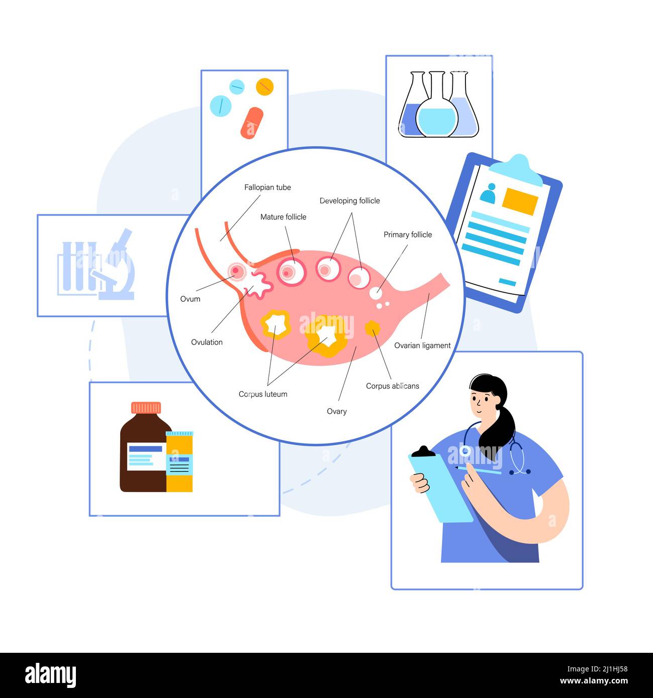 Female fertility, conceptual illustration Stock Photohttps://www.alamy.com/image-license-details/?v=1https://www.alamy.com/female-fertility-conceptual-illustration-image465682036.html
RF2J1HJ58–Female fertility, conceptual illustration
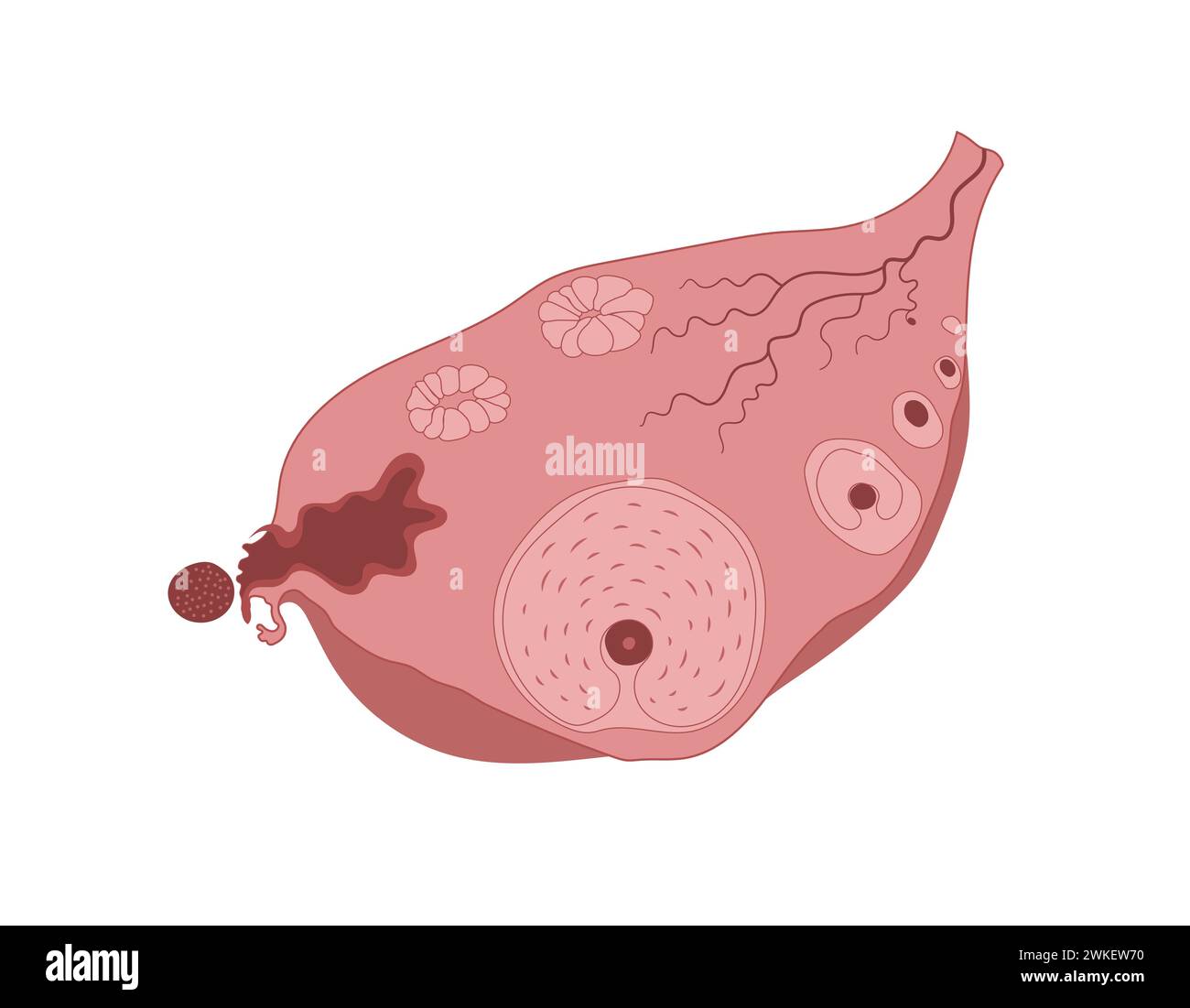 Ovulation steps infographic diagram, ovarian cycle, anatomy of female egg cell development. Stock Vectorhttps://www.alamy.com/image-license-details/?v=1https://www.alamy.com/ovulation-steps-infographic-diagram-ovarian-cycle-anatomy-of-female-egg-cell-development-image597158100.html
RF2WKEW70–Ovulation steps infographic diagram, ovarian cycle, anatomy of female egg cell development.
 Manual of pathological anatomy . ar epithelium. It is worth noting, that the sizeof the mature follicle is ten to twelve millimetres == three-eighthsof an inch in diameter. Simple cysts of this kind are met with inthe foetal ovary, and subsequently at all ages. As the dilatationincreases the ovum perishes, but has been still demonstrated incysts, the size of a bean or of a cherry, by Eokitansky andEitchie. Several cysts are often found in the same ovary, butnaturally the largest are those which occur singly. The largercysts are lined by a simple pavement-epithelium. Their con-tents are either Stock Photohttps://www.alamy.com/image-license-details/?v=1https://www.alamy.com/manual-of-pathological-anatomy-ar-epithelium-it-is-worth-noting-that-the-sizeof-the-mature-follicle-is-ten-to-twelve-millimetres-==-three-eighthsof-an-inch-in-diameter-simple-cysts-of-this-kind-are-met-with-inthe-foetal-ovary-and-subsequently-at-all-ages-as-the-dilatationincreases-the-ovum-perishes-but-has-been-still-demonstrated-incysts-the-size-of-a-bean-or-of-a-cherry-by-eokitansky-andeitchie-several-cysts-are-often-found-in-the-same-ovary-butnaturally-the-largest-are-those-which-occur-singly-the-largercysts-are-lined-by-a-simple-pavement-epithelium-their-con-tents-are-either-image339443471.html
RM2AM6YH3–Manual of pathological anatomy . ar epithelium. It is worth noting, that the sizeof the mature follicle is ten to twelve millimetres == three-eighthsof an inch in diameter. Simple cysts of this kind are met with inthe foetal ovary, and subsequently at all ages. As the dilatationincreases the ovum perishes, but has been still demonstrated incysts, the size of a bean or of a cherry, by Eokitansky andEitchie. Several cysts are often found in the same ovary, butnaturally the largest are those which occur singly. The largercysts are lined by a simple pavement-epithelium. Their con-tents are either
 . Text-fig. 5. Morphological variation in corpora lutea. See text for explanation. In the humpback material there were 35 females which were found to have just ovulated, the blood- stained hole (from 4 to 13 mm. in diameter) being immediately obvious on the surface of the ovary. Immediately after ovulation the follicle was collapsed and the wall wrinkled. The size range at this stage was 2-2-6-0 cm. (median 3-7 cm.) and the smallest recently formed corpora lutea are about 4 cm. in diameter (Chittleborough, 1954, fig. 4), suggesting that the size of the original mature follicle at ovulation was Stock Photohttps://www.alamy.com/image-license-details/?v=1https://www.alamy.com/text-fig-5-morphological-variation-in-corpora-lutea-see-text-for-explanation-in-the-humpback-material-there-were-35-females-which-were-found-to-have-just-ovulated-the-blood-stained-hole-from-4-to-13-mm-in-diameter-being-immediately-obvious-on-the-surface-of-the-ovary-immediately-after-ovulation-the-follicle-was-collapsed-and-the-wall-wrinkled-the-size-range-at-this-stage-was-2-2-6-0-cm-median-3-7-cm-and-the-smallest-recently-formed-corpora-lutea-are-about-4-cm-in-diameter-chittleborough-1954-fig-4-suggesting-that-the-size-of-the-original-mature-follicle-at-ovulation-was-image179949891.html
RMMCNBWR–. Text-fig. 5. Morphological variation in corpora lutea. See text for explanation. In the humpback material there were 35 females which were found to have just ovulated, the blood- stained hole (from 4 to 13 mm. in diameter) being immediately obvious on the surface of the ovary. Immediately after ovulation the follicle was collapsed and the wall wrinkled. The size range at this stage was 2-2-6-0 cm. (median 3-7 cm.) and the smallest recently formed corpora lutea are about 4 cm. in diameter (Chittleborough, 1954, fig. 4), suggesting that the size of the original mature follicle at ovulation was
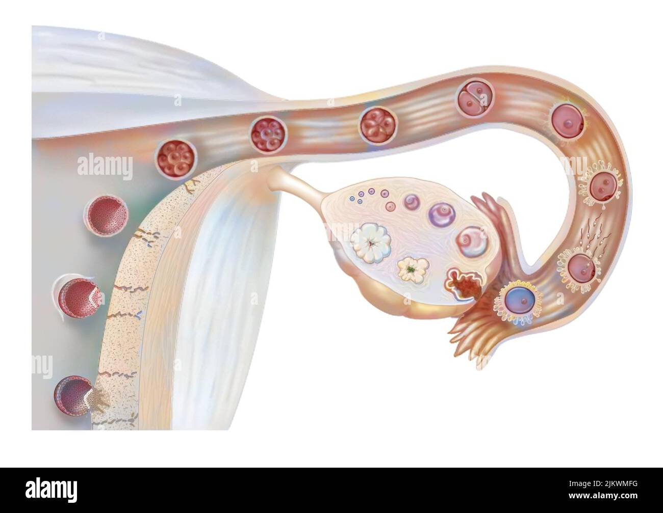 Female genitalia: ovarian cycle, ovulation, fertilization, embryo segmentation, implantation. Stock Photohttps://www.alamy.com/image-license-details/?v=1https://www.alamy.com/female-genitalia-ovarian-cycle-ovulation-fertilization-embryo-segmentation-implantation-image476923316.html
RF2JKWMFG–Female genitalia: ovarian cycle, ovulation, fertilization, embryo segmentation, implantation.
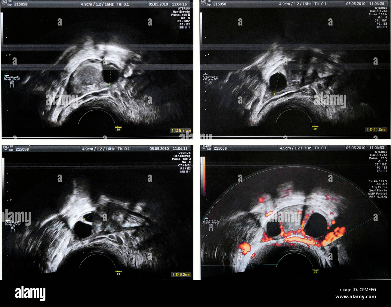 OVARY, SONOGRAPHY Stock Photohttps://www.alamy.com/image-license-details/?v=1https://www.alamy.com/stock-photo-ovary-sonography-48393620.html
RMCPMEFG–OVARY, SONOGRAPHY
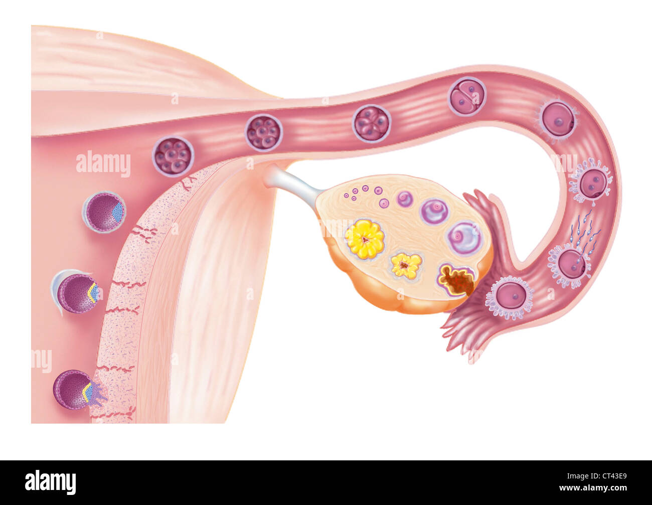 Fertilization, drawing Stock Photohttps://www.alamy.com/image-license-details/?v=1https://www.alamy.com/stock-photo-fertilization-drawing-49263041.html
RMCT43E9–Fertilization, drawing
 The ovary Stock Photohttps://www.alamy.com/image-license-details/?v=1https://www.alamy.com/stock-photo-the-ovary-13186119.html
RFACM72G–The ovary
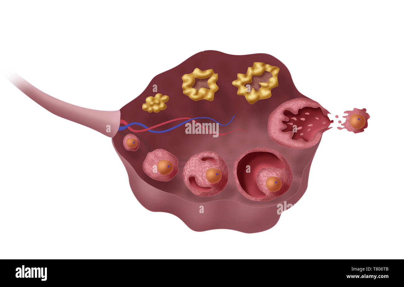 Ovarian Follicles, Illustration Stock Photohttps://www.alamy.com/image-license-details/?v=1https://www.alamy.com/ovarian-follicles-illustration-image245867787.html
RMT806TB–Ovarian Follicles, Illustration
 Ovarian cycle, drawing Stock Photohttps://www.alamy.com/image-license-details/?v=1https://www.alamy.com/ovarian-cycle-drawing-image67527503.html
RMDWT40F–Ovarian cycle, drawing
 Physiology : a manual for students and practitioners . Section of the Ovary (after Schron): 1, outer covering; 1, attached border; 2, centralstroma; 3, peripheral stroma; 4, blood-vessels ; 5, Graafian follicles in their earlieststa^e ; 6, 7, 8, more advanced follicles ; !), an almost mature follicle ; 9, follicle fromwhich the ovum has esciped ; 10, corpus luteum. Whence is the ovum derived? It is a very highly developed cell, wliich is derived from thegerminal epithelium covering the ovary. In the development ofthe ovary this epithelium dips into the surface of the organ, and a 176 EMBRYOLOG Stock Photohttps://www.alamy.com/image-license-details/?v=1https://www.alamy.com/physiology-a-manual-for-students-and-practitioners-section-of-the-ovary-after-schron-1-outer-covering-1-attached-border-2-centralstroma-3-peripheral-stroma-4-blood-vessels-5-graafian-follicles-in-their-earlieststae-6-7-8-more-advanced-follicles-!-an-almost-mature-follicle-9-follicle-fromwhich-the-ovum-has-esciped-10-corpus-luteum-whence-is-the-ovum-derived-it-is-a-very-highly-developed-cell-wliich-is-derived-from-thegerminal-epithelium-covering-the-ovary-in-the-development-ofthe-ovary-this-epithelium-dips-into-the-surface-of-the-organ-and-a-176-embryolog-image342759707.html
RM2AWJ1E3–Physiology : a manual for students and practitioners . Section of the Ovary (after Schron): 1, outer covering; 1, attached border; 2, centralstroma; 3, peripheral stroma; 4, blood-vessels ; 5, Graafian follicles in their earlieststa^e ; 6, 7, 8, more advanced follicles ; !), an almost mature follicle ; 9, follicle fromwhich the ovum has esciped ; 10, corpus luteum. Whence is the ovum derived? It is a very highly developed cell, wliich is derived from thegerminal epithelium covering the ovary. In the development ofthe ovary this epithelium dips into the surface of the organ, and a 176 EMBRYOLOG
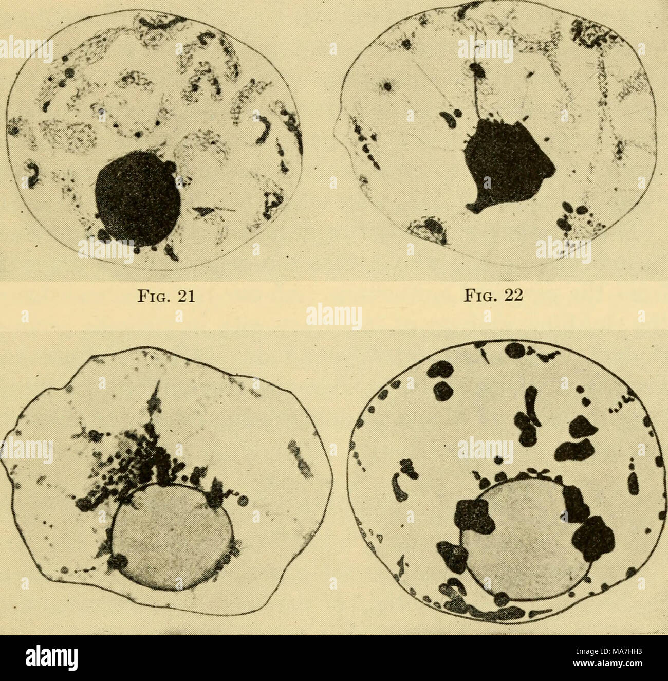 . The eggs of mammals . Fig. 23 Fig. 24 Plate II. (From the Journal of Morphology) Figs. 17-22. Nuclei of ova from ovary of rat 20 days post partum. 17, Deuto- broch nucleus. 18, Beginning of the formation of clumps shown in next figure. 19, Modified pachynema. 20, Later stage showing characters of diplonema. 21, Nu- cleus toward the end of the growth period. 22, Final stage in twenty-day rat. Fig. 23. Nucleus in mature follicle from adult rat. Fig. 24. Nucleus from ripe follicle from adult rat. 13 Stock Photohttps://www.alamy.com/image-license-details/?v=1https://www.alamy.com/the-eggs-of-mammals-fig-23-fig-24-plate-ii-from-the-journal-of-morphology-figs-17-22-nuclei-of-ova-from-ovary-of-rat-20-days-post-partum-17-deuto-broch-nucleus-18-beginning-of-the-formation-of-clumps-shown-in-next-figure-19-modified-pachynema-20-later-stage-showing-characters-of-diplonema-21-nu-cleus-toward-the-end-of-the-growth-period-22-final-stage-in-twenty-day-rat-fig-23-nucleus-in-mature-follicle-from-adult-rat-fig-24-nucleus-from-ripe-follicle-from-adult-rat-13-image178417711.html
RMMA7HH3–. The eggs of mammals . Fig. 23 Fig. 24 Plate II. (From the Journal of Morphology) Figs. 17-22. Nuclei of ova from ovary of rat 20 days post partum. 17, Deuto- broch nucleus. 18, Beginning of the formation of clumps shown in next figure. 19, Modified pachynema. 20, Later stage showing characters of diplonema. 21, Nu- cleus toward the end of the growth period. 22, Final stage in twenty-day rat. Fig. 23. Nucleus in mature follicle from adult rat. Fig. 24. Nucleus from ripe follicle from adult rat. 13
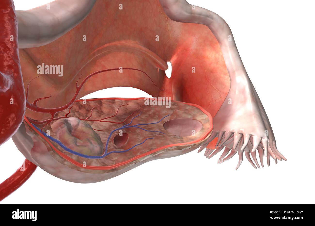 The female reproductive system Stock Photohttps://www.alamy.com/image-license-details/?v=1https://www.alamy.com/stock-photo-the-female-reproductive-system-13235064.html
RFACWCMW–The female reproductive system
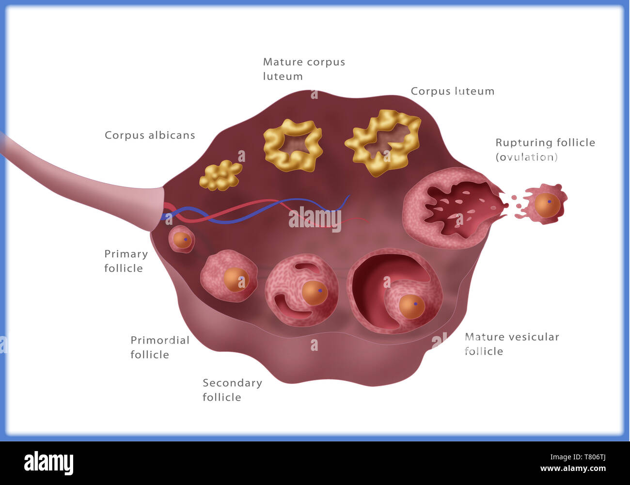 Ovarian Follicles, Illustration Stock Photohttps://www.alamy.com/image-license-details/?v=1https://www.alamy.com/ovarian-follicles-illustration-image245867794.html
RMT806TJ–Ovarian Follicles, Illustration
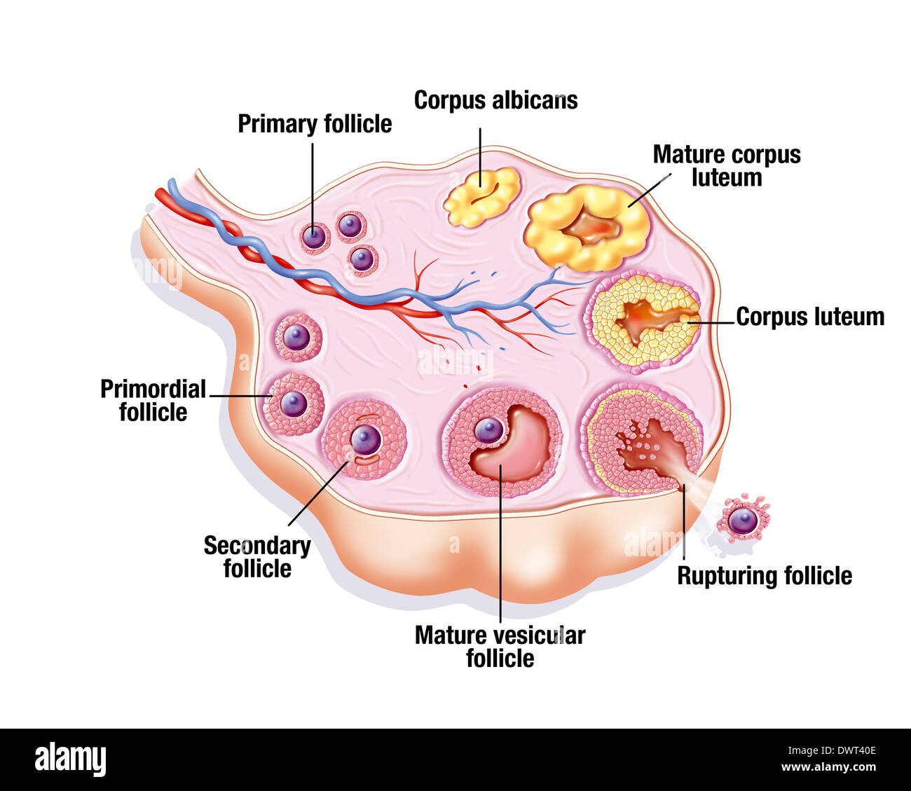 Ovarian cycle, drawing Stock Photohttps://www.alamy.com/image-license-details/?v=1https://www.alamy.com/ovarian-cycle-drawing-image67527502.html
RMDWT40E–Ovarian cycle, drawing
 OVARIAN CYCLE, DRAWING Stock Photohttps://www.alamy.com/image-license-details/?v=1https://www.alamy.com/stock-photo-ovarian-cycle-drawing-49285615.html
RMCT548F–OVARIAN CYCLE, DRAWING
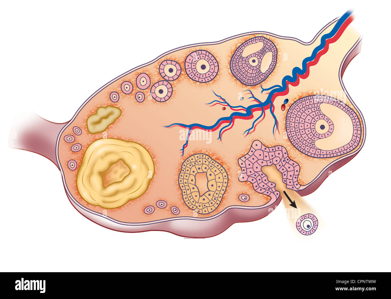 OVARIAN CYCLE, DRAWING Stock Photohttps://www.alamy.com/image-license-details/?v=1https://www.alamy.com/stock-photo-ovarian-cycle-drawing-48423701.html
RMCPNTWW–OVARIAN CYCLE, DRAWING
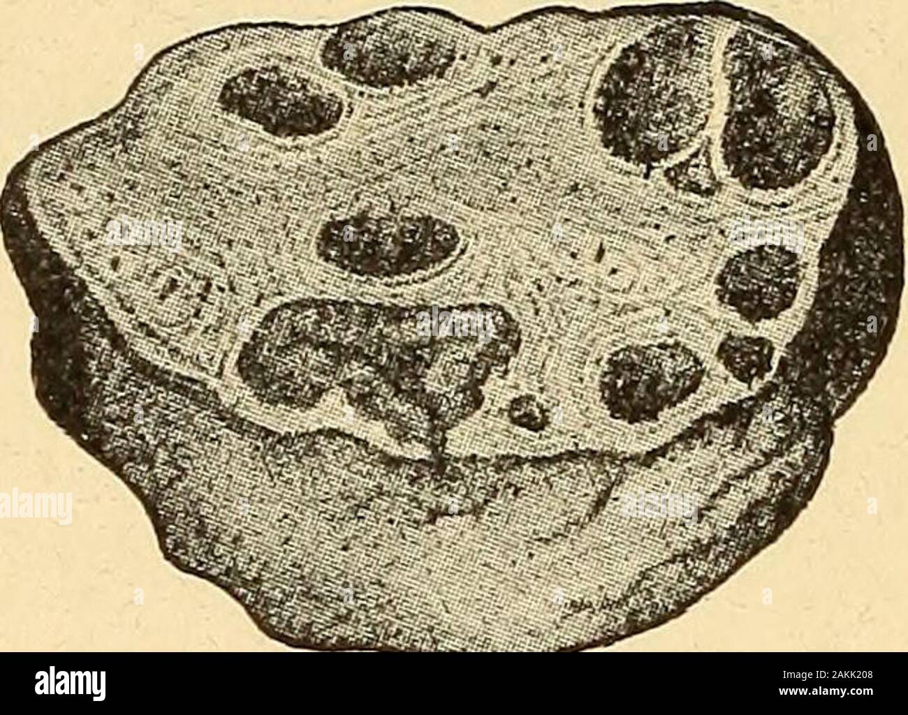 A nurse's handbook of obstetrics, for use in training-schools . Fig. 15.—Longitudinal section through ovary of a woman twenty-two days after the lastmenstruation. (Leopold.) m.f., mature Graafian follicle; pr., most prominent point offollicle, where the rupture may be expected. like a small pimple. The Graafian follicle then becomes thinnedat one point, where it soon bursts and allows the ovum to escapeinto the Fallopian tube (Fig. 16). 37 38 A NURSES HANDBOOK OF OBSTETRICS. Once within the Fallopian tube, the ovum makes its way intothe uterus, and, if unimpregnated by the male element, it los Stock Photohttps://www.alamy.com/image-license-details/?v=1https://www.alamy.com/a-nurses-handbook-of-obstetrics-for-use-in-training-schools-fig-15longitudinal-section-through-ovary-of-a-woman-twenty-two-days-after-the-lastmenstruation-leopold-mf-mature-graafian-follicle-pr-most-prominent-point-offollicle-where-the-rupture-may-be-expected-like-a-small-pimple-the-graafian-follicle-then-becomes-thinnedat-one-point-where-it-soon-bursts-and-allows-the-ovum-to-escapeinto-the-fallopian-tube-fig-16-37-38-a-nurses-handbook-of-obstetrics-once-within-the-fallopian-tube-the-ovum-makes-its-way-intothe-uterus-and-if-unimpregnated-by-the-male-element-it-los-image339094120.html
RM2AKK208–A nurse's handbook of obstetrics, for use in training-schools . Fig. 15.—Longitudinal section through ovary of a woman twenty-two days after the lastmenstruation. (Leopold.) m.f., mature Graafian follicle; pr., most prominent point offollicle, where the rupture may be expected. like a small pimple. The Graafian follicle then becomes thinnedat one point, where it soon bursts and allows the ovum to escapeinto the Fallopian tube (Fig. 16). 37 38 A NURSES HANDBOOK OF OBSTETRICS. Once within the Fallopian tube, the ovum makes its way intothe uterus, and, if unimpregnated by the male element, it los
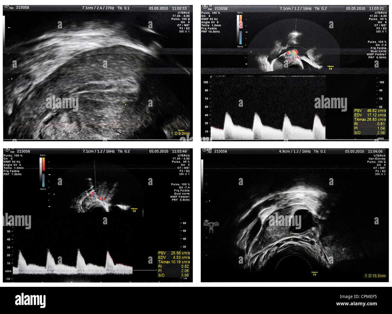 UTERUS SONOGRAPHY Stock Photohttps://www.alamy.com/image-license-details/?v=1https://www.alamy.com/stock-photo-uterus-sonography-48393609.html
RMCPMEF5–UTERUS SONOGRAPHY
![. The eggs of mammals . -».FA Fig. 5. Section through the ovary of mature rat showing the iobed condition. YF, young fol- licle; F, follicle; FA, fatty tissue. (From the Quarterly Review of Biology.) occurred in the sixty animals over forty days of age. She noted that in females under forty days of age the ovary is relatively smooth and compact and not very heavily em- bedded in fat (Figure 4), whereas in older animals the ovary is Iobed and surrounded by a la]:ger amount of fat (Figure 5). She accordingly ovariectomized a second set of animals con- sisting of eighty-five females under forty d Stock Photo . The eggs of mammals . -».FA Fig. 5. Section through the ovary of mature rat showing the iobed condition. YF, young fol- licle; F, follicle; FA, fatty tissue. (From the Quarterly Review of Biology.) occurred in the sixty animals over forty days of age. She noted that in females under forty days of age the ovary is relatively smooth and compact and not very heavily em- bedded in fat (Figure 4), whereas in older animals the ovary is Iobed and surrounded by a la]:ger amount of fat (Figure 5). She accordingly ovariectomized a second set of animals con- sisting of eighty-five females under forty d Stock Photo](https://c8.alamy.com/comp/MA7HGX/the-eggs-of-mammals-fa-fig-5-section-through-the-ovary-of-mature-rat-showing-the-iobed-condition-yf-young-fol-licle-f-follicle-fa-fatty-tissue-from-the-quarterly-review-of-biology-occurred-in-the-sixty-animals-over-forty-days-of-age-she-noted-that-in-females-under-forty-days-of-age-the-ovary-is-relatively-smooth-and-compact-and-not-very-heavily-em-bedded-in-fat-figure-4-whereas-in-older-animals-the-ovary-is-iobed-and-surrounded-by-a-la-ger-amount-of-fat-figure-5-she-accordingly-ovariectomized-a-second-set-of-animals-con-sisting-of-eighty-five-females-under-forty-d-MA7HGX.jpg) . The eggs of mammals . -».FA Fig. 5. Section through the ovary of mature rat showing the iobed condition. YF, young fol- licle; F, follicle; FA, fatty tissue. (From the Quarterly Review of Biology.) occurred in the sixty animals over forty days of age. She noted that in females under forty days of age the ovary is relatively smooth and compact and not very heavily em- bedded in fat (Figure 4), whereas in older animals the ovary is Iobed and surrounded by a la]:ger amount of fat (Figure 5). She accordingly ovariectomized a second set of animals con- sisting of eighty-five females under forty d Stock Photohttps://www.alamy.com/image-license-details/?v=1https://www.alamy.com/the-eggs-of-mammals-fa-fig-5-section-through-the-ovary-of-mature-rat-showing-the-iobed-condition-yf-young-fol-licle-f-follicle-fa-fatty-tissue-from-the-quarterly-review-of-biology-occurred-in-the-sixty-animals-over-forty-days-of-age-she-noted-that-in-females-under-forty-days-of-age-the-ovary-is-relatively-smooth-and-compact-and-not-very-heavily-em-bedded-in-fat-figure-4-whereas-in-older-animals-the-ovary-is-iobed-and-surrounded-by-a-la-ger-amount-of-fat-figure-5-she-accordingly-ovariectomized-a-second-set-of-animals-con-sisting-of-eighty-five-females-under-forty-d-image178417706.html
. The eggs of mammals . -».FA Fig. 5. Section through the ovary of mature rat showing the iobed condition. YF, young fol- licle; F, follicle; FA, fatty tissue. (From the Quarterly Review of Biology.) occurred in the sixty animals over forty days of age. She noted that in females under forty days of age the ovary is relatively smooth and compact and not very heavily em- bedded in fat (Figure 4), whereas in older animals the ovary is Iobed and surrounded by a la]:ger amount of fat (Figure 5). She accordingly ovariectomized a second set of animals con- sisting of eighty-five females under forty d Stock Photohttps://www.alamy.com/image-license-details/?v=1https://www.alamy.com/the-eggs-of-mammals-fa-fig-5-section-through-the-ovary-of-mature-rat-showing-the-iobed-condition-yf-young-fol-licle-f-follicle-fa-fatty-tissue-from-the-quarterly-review-of-biology-occurred-in-the-sixty-animals-over-forty-days-of-age-she-noted-that-in-females-under-forty-days-of-age-the-ovary-is-relatively-smooth-and-compact-and-not-very-heavily-em-bedded-in-fat-figure-4-whereas-in-older-animals-the-ovary-is-iobed-and-surrounded-by-a-la-ger-amount-of-fat-figure-5-she-accordingly-ovariectomized-a-second-set-of-animals-con-sisting-of-eighty-five-females-under-forty-d-image178417706.htmlRMMA7HGX–. The eggs of mammals . -».FA Fig. 5. Section through the ovary of mature rat showing the iobed condition. YF, young fol- licle; F, follicle; FA, fatty tissue. (From the Quarterly Review of Biology.) occurred in the sixty animals over forty days of age. She noted that in females under forty days of age the ovary is relatively smooth and compact and not very heavily em- bedded in fat (Figure 4), whereas in older animals the ovary is Iobed and surrounded by a la]:ger amount of fat (Figure 5). She accordingly ovariectomized a second set of animals con- sisting of eighty-five females under forty d
 IVF Stock Photohttps://www.alamy.com/image-license-details/?v=1https://www.alamy.com/stock-photo-ivf-48414396.html
RMCPND1G–IVF
 IVF Stock Photohttps://www.alamy.com/image-license-details/?v=1https://www.alamy.com/stock-photo-ivf-48407879.html
RMCPN4MR–IVF
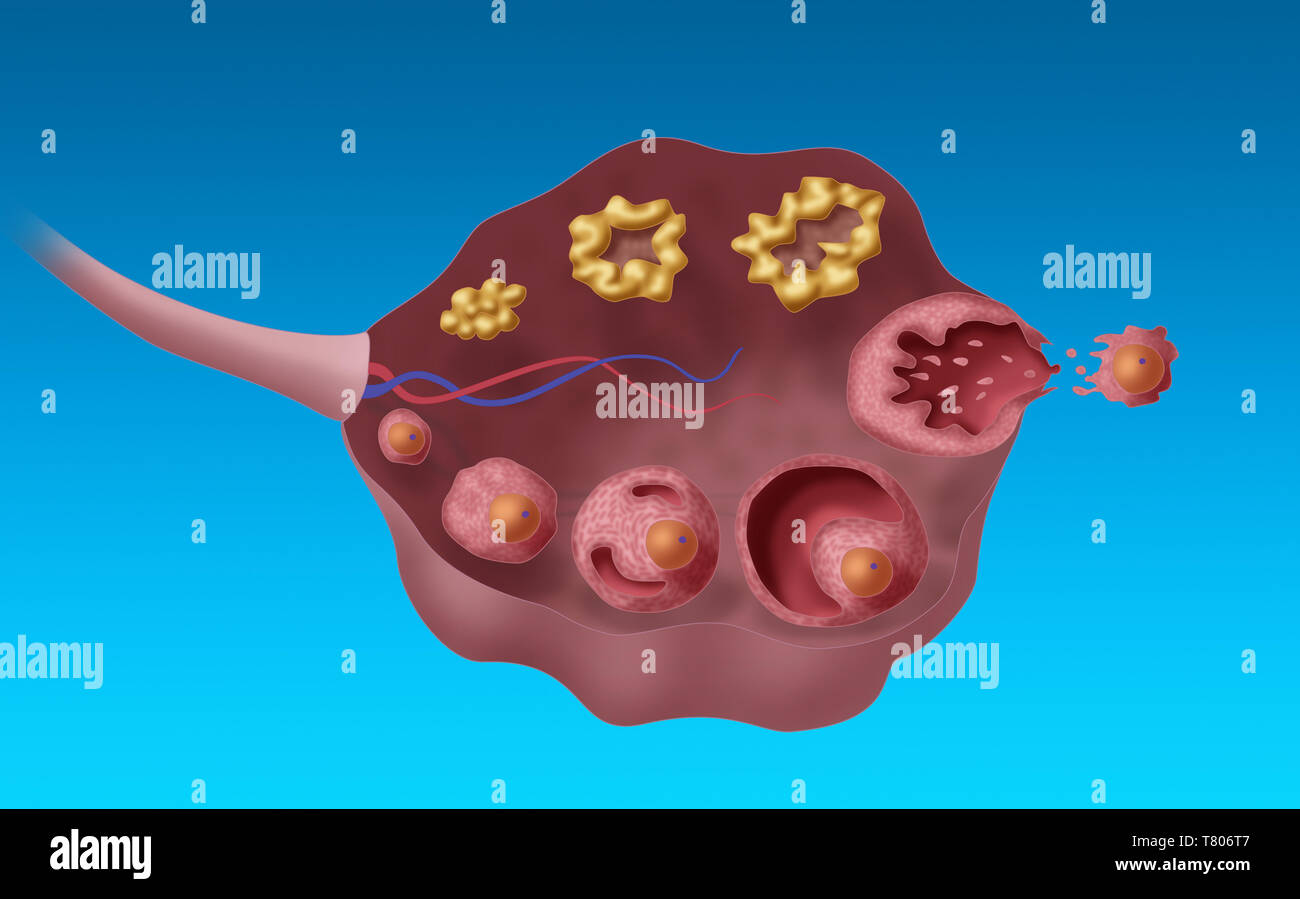 Ovarian Follicles, Illustration Stock Photohttps://www.alamy.com/image-license-details/?v=1https://www.alamy.com/ovarian-follicles-illustration-image245867783.html
RMT806T7–Ovarian Follicles, Illustration
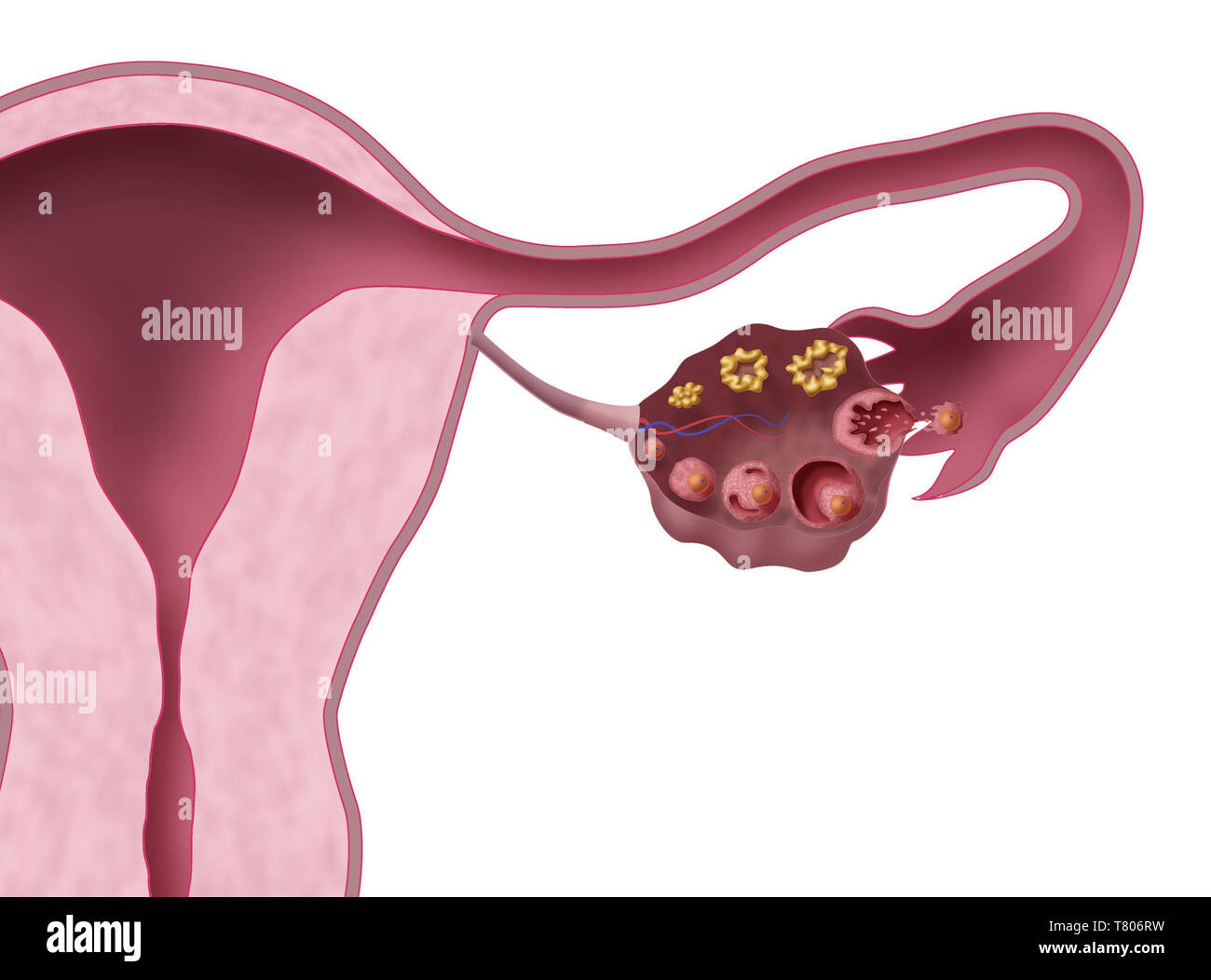 Ovarian Follicles, Illustration Stock Photohttps://www.alamy.com/image-license-details/?v=1https://www.alamy.com/ovarian-follicles-illustration-image245867773.html
RMT806RW–Ovarian Follicles, Illustration
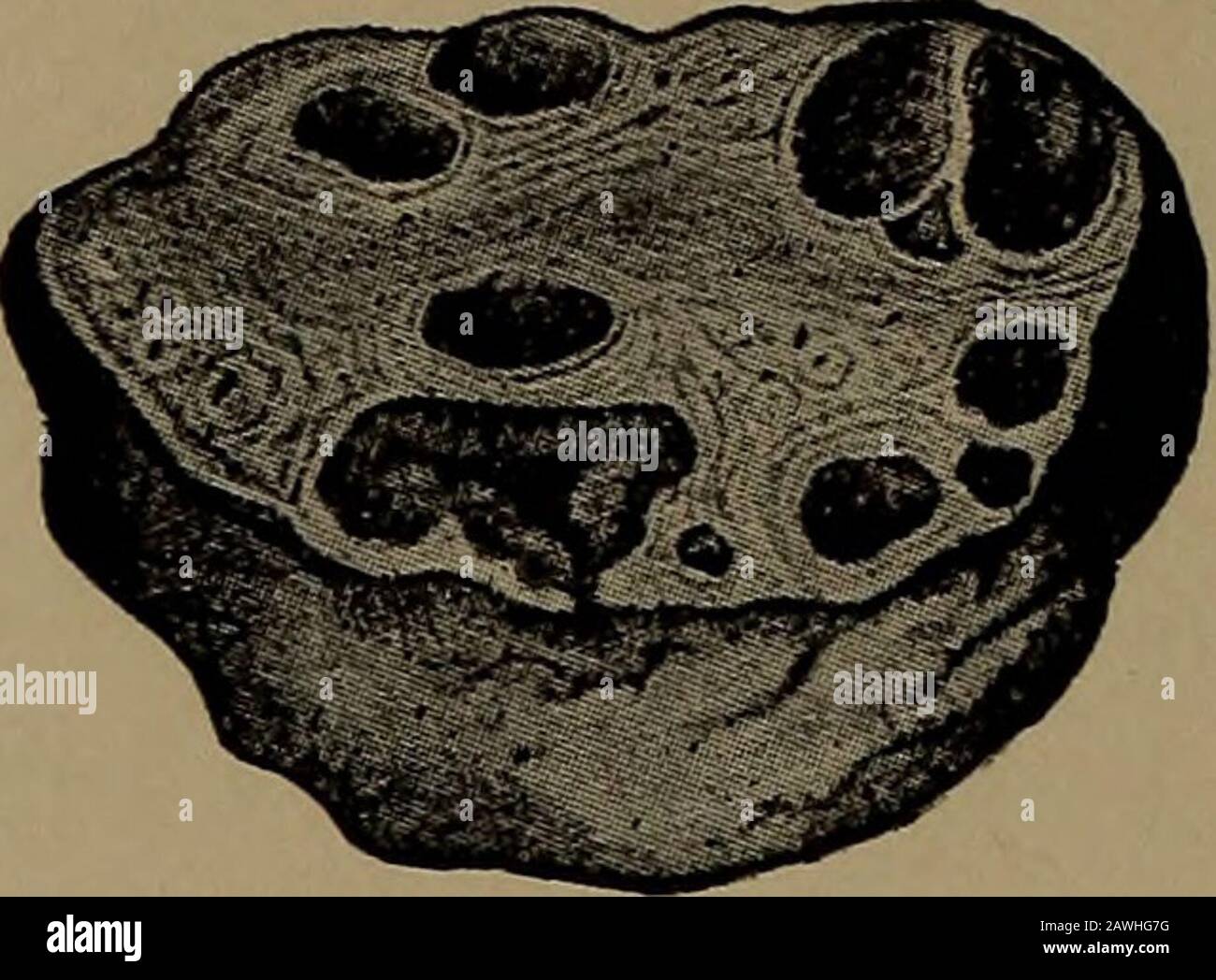 A nurse's handbook of obstetrics, for use in training-schools . Fig. 15.—Longitudinal section through ovary of a woman twenty-two days after the lastmenstruation. (Leopold.) m.f., mature Graafian follicle; pr., most prominent point offollicle, where the rupture may be expected. like a small pimple. The Graafian follicle then becomes thinnedat one point, where it soon bursts and allows the ovum to escapeinto the Fallopian tube (Fig. 16). 37 38 A NURSES HANDBOOK OF OBSTETRICS. Once within the Fallopian tube, the ovum makes its way intothe uterus, and, if unimpregnated by the male element, it los Stock Photohttps://www.alamy.com/image-license-details/?v=1https://www.alamy.com/a-nurses-handbook-of-obstetrics-for-use-in-training-schools-fig-15longitudinal-section-through-ovary-of-a-woman-twenty-two-days-after-the-lastmenstruation-leopold-mf-mature-graafian-follicle-pr-most-prominent-point-offollicle-where-the-rupture-may-be-expected-like-a-small-pimple-the-graafian-follicle-then-becomes-thinnedat-one-point-where-it-soon-bursts-and-allows-the-ovum-to-escapeinto-the-fallopian-tube-fig-16-37-38-a-nurses-handbook-of-obstetrics-once-within-the-fallopian-tube-the-ovum-makes-its-way-intothe-uterus-and-if-unimpregnated-by-the-male-element-it-los-image342749332.html
RM2AWHG7G–A nurse's handbook of obstetrics, for use in training-schools . Fig. 15.—Longitudinal section through ovary of a woman twenty-two days after the lastmenstruation. (Leopold.) m.f., mature Graafian follicle; pr., most prominent point offollicle, where the rupture may be expected. like a small pimple. The Graafian follicle then becomes thinnedat one point, where it soon bursts and allows the ovum to escapeinto the Fallopian tube (Fig. 16). 37 38 A NURSES HANDBOOK OF OBSTETRICS. Once within the Fallopian tube, the ovum makes its way intothe uterus, and, if unimpregnated by the male element, it los
 IVF Stock Photohttps://www.alamy.com/image-license-details/?v=1https://www.alamy.com/stock-photo-ivf-48407897.html
RMCPN4ND–IVF
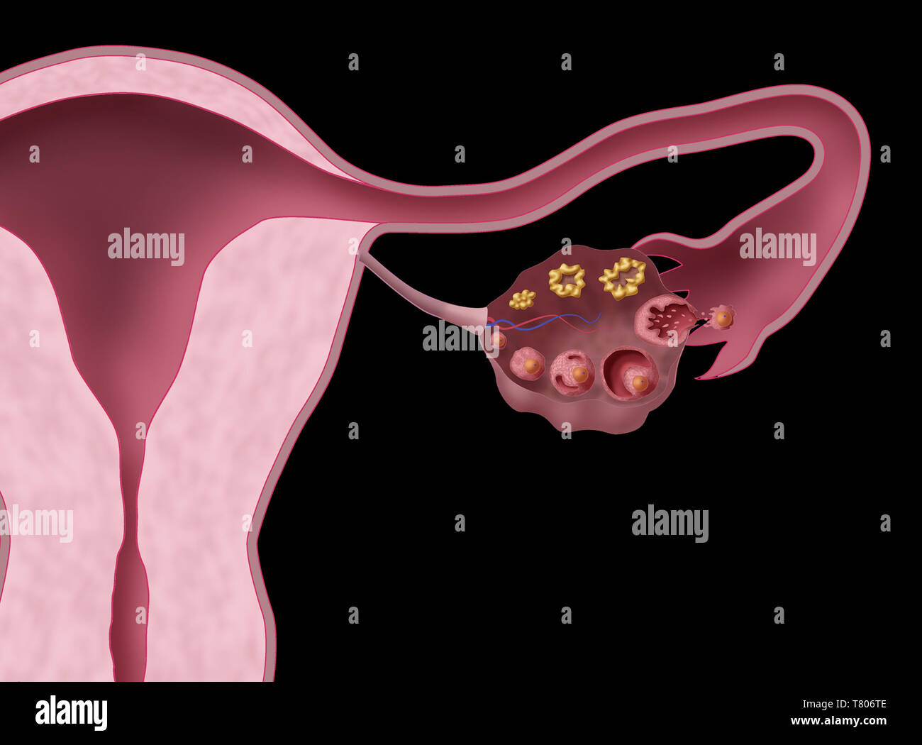 Ovarian Follicles, Illustration Stock Photohttps://www.alamy.com/image-license-details/?v=1https://www.alamy.com/ovarian-follicles-illustration-image245867790.html
RMT806TE–Ovarian Follicles, Illustration
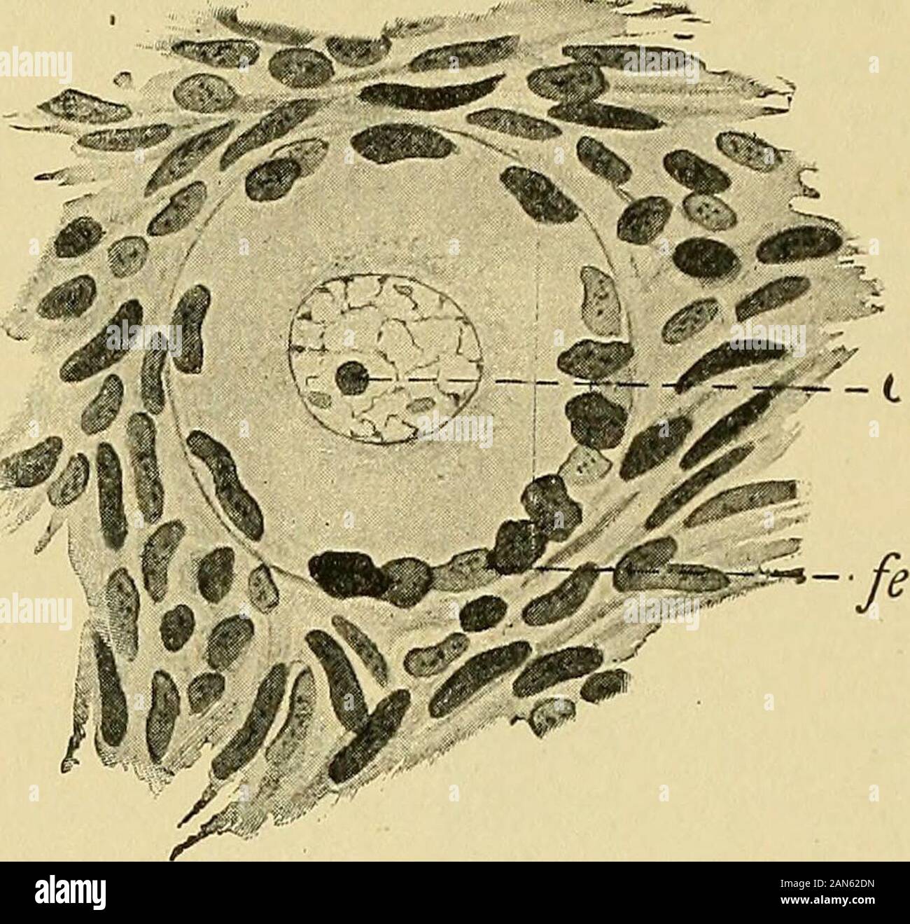 Pathology and treatment of diseases of women . n a slow growth sets in again until the occur-rence of the next premenstrual congestion. This process of menstruation, however, is not a self-dependent one. 16 DISEASES OF WOMEN It is dependent on the much more important occurrence of ovulation which takes place in the ovaries. We understand by ovulation the development of a primary follicle to a mature follicle and the expulsionof the ripe ovum from the ovary(Fig. 11). In the transformation of a pri-mary follicle (Fig. 11) to a maturefollicle the follicular epithelium be-comes many-layered and fo Stock Photohttps://www.alamy.com/image-license-details/?v=1https://www.alamy.com/pathology-and-treatment-of-diseases-of-women-n-a-slow-growth-sets-in-again-until-the-occur-rence-of-the-next-premenstrual-congestion-this-process-of-menstruation-however-is-not-a-self-dependent-one-16-diseases-of-women-it-is-dependent-on-the-much-more-important-occurrence-of-ovulation-which-takes-place-in-the-ovaries-we-understand-by-ovulation-the-development-of-a-primary-follicle-to-a-mature-follicle-and-the-expulsionof-the-ripe-ovum-from-the-ovaryfig-11-in-the-transformation-of-a-pri-mary-follicle-fig-11-to-a-maturefollicle-the-follicular-epithelium-be-comes-many-layered-and-fo-image340038433.html
RM2AN62DN–Pathology and treatment of diseases of women . n a slow growth sets in again until the occur-rence of the next premenstrual congestion. This process of menstruation, however, is not a self-dependent one. 16 DISEASES OF WOMEN It is dependent on the much more important occurrence of ovulation which takes place in the ovaries. We understand by ovulation the development of a primary follicle to a mature follicle and the expulsionof the ripe ovum from the ovary(Fig. 11). In the transformation of a pri-mary follicle (Fig. 11) to a maturefollicle the follicular epithelium be-comes many-layered and fo
 IVF Stock Photohttps://www.alamy.com/image-license-details/?v=1https://www.alamy.com/stock-photo-ivf-48407834.html
RMCPN4K6–IVF
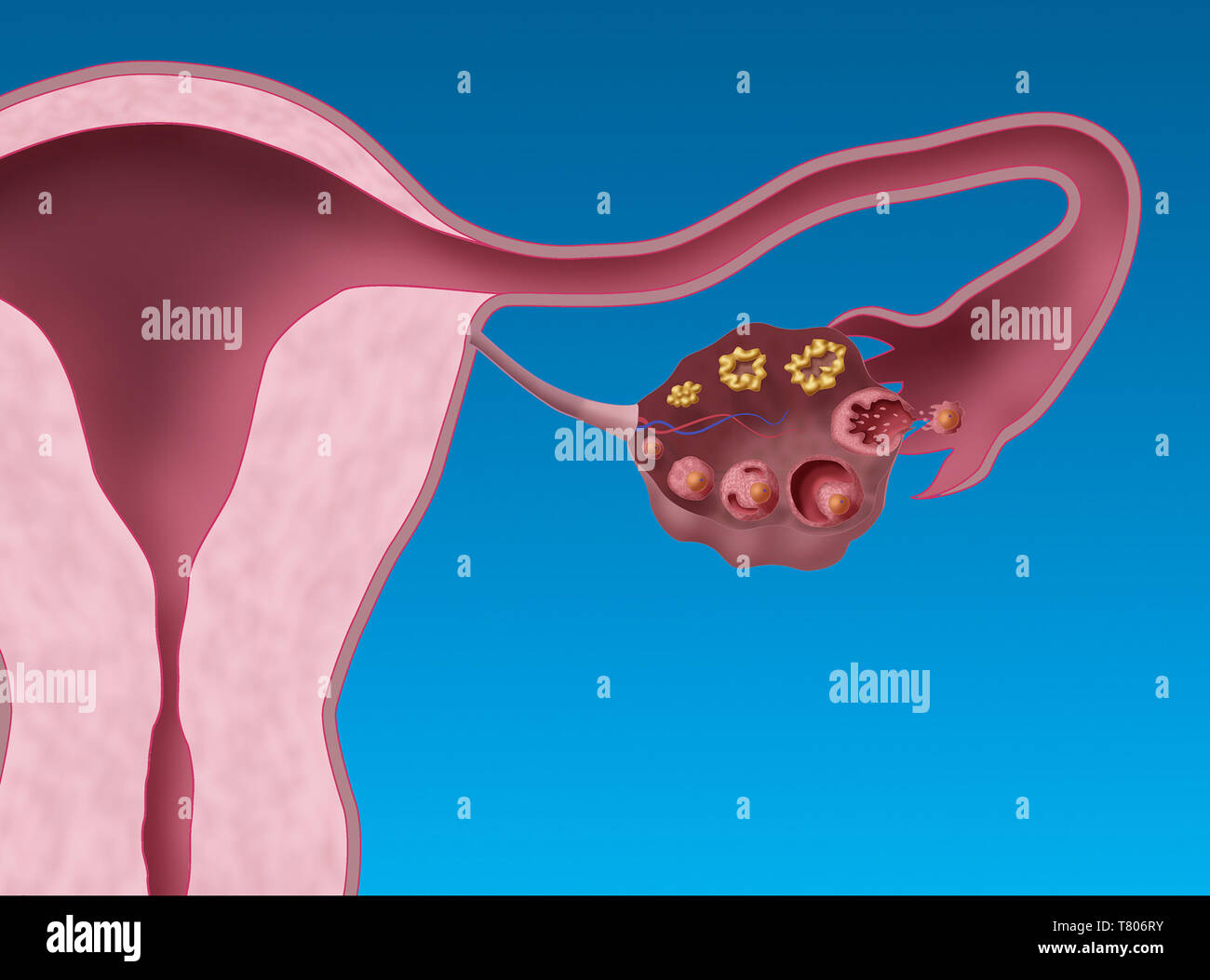 Ovarian Follicles, Illustration Stock Photohttps://www.alamy.com/image-license-details/?v=1https://www.alamy.com/ovarian-follicles-illustration-image245867775.html
RMT806RY–Ovarian Follicles, Illustration
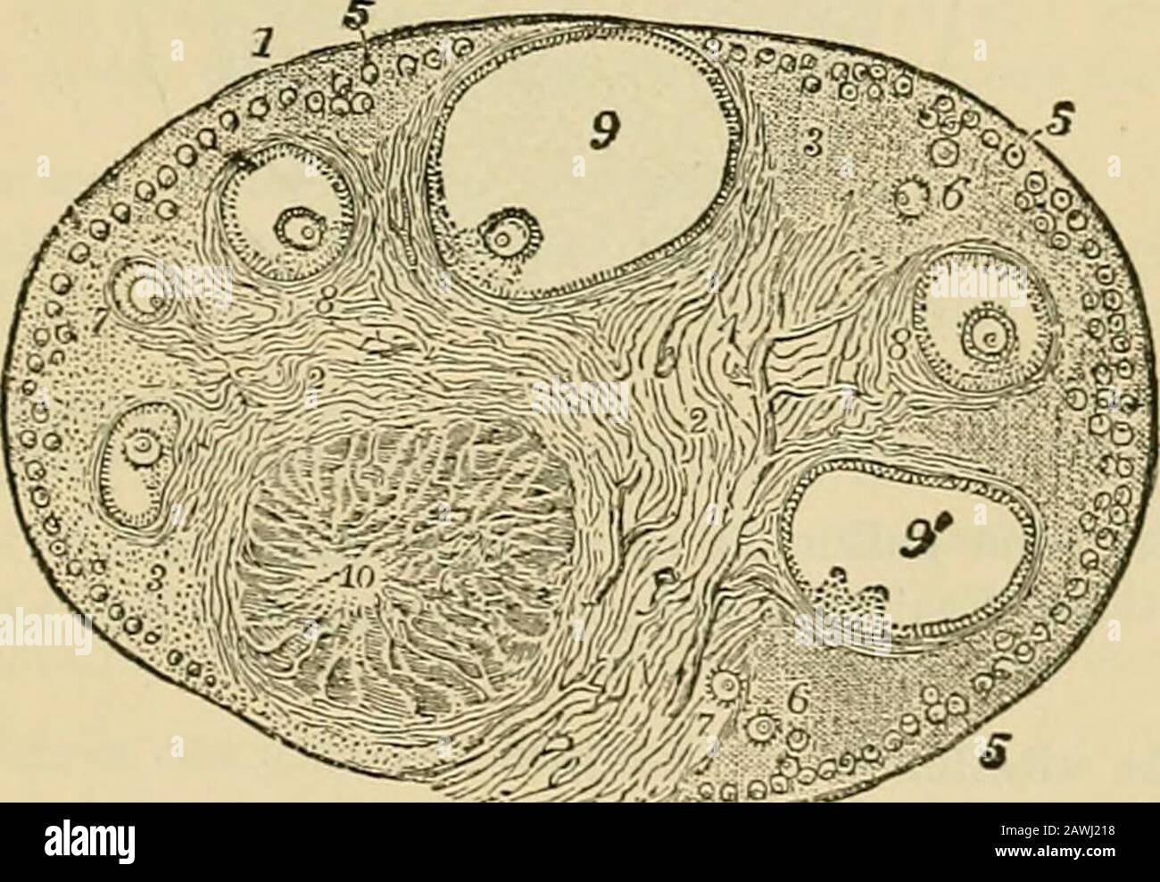 Physiology : a manual for students and practitioners . Human Ovum, rui)fured byPressure, showing tlie vitelluspartially expelled, the gernii-native vesicle, with its germi-uative spot, at a, and thesmooth fracture of the vitel-line membrane.. Section of the Ovary (after Schron): 1, outer covering; 1, attached border; 2, centralstroma; 3, peripheral stroma; 4, blood-vessels ; 5, Graafian follicles in their earlieststa^e ; 6, 7, 8, more advanced follicles ; !), an almost mature follicle ; 9, follicle fromwhich the ovum has esciped ; 10, corpus luteum. Whence is the ovum derived? It is a very hig Stock Photohttps://www.alamy.com/image-license-details/?v=1https://www.alamy.com/physiology-a-manual-for-students-and-practitioners-human-ovum-ruifured-bypressure-showing-tlie-vitelluspartially-expelled-the-gernii-native-vesicle-with-its-germi-uative-spot-at-a-and-thesmooth-fracture-of-the-vitel-line-membrane-section-of-the-ovary-after-schron-1-outer-covering-1-attached-border-2-centralstroma-3-peripheral-stroma-4-blood-vessels-5-graafian-follicles-in-their-earlieststae-6-7-8-more-advanced-follicles-!-an-almost-mature-follicle-9-follicle-fromwhich-the-ovum-has-esciped-10-corpus-luteum-whence-is-the-ovum-derived-it-is-a-very-hig-image342760132.html
RM2AWJ218–Physiology : a manual for students and practitioners . Human Ovum, rui)fured byPressure, showing tlie vitelluspartially expelled, the gernii-native vesicle, with its germi-uative spot, at a, and thesmooth fracture of the vitel-line membrane.. Section of the Ovary (after Schron): 1, outer covering; 1, attached border; 2, centralstroma; 3, peripheral stroma; 4, blood-vessels ; 5, Graafian follicles in their earlieststa^e ; 6, 7, 8, more advanced follicles ; !), an almost mature follicle ; 9, follicle fromwhich the ovum has esciped ; 10, corpus luteum. Whence is the ovum derived? It is a very hig
 IVF Stock Photohttps://www.alamy.com/image-license-details/?v=1https://www.alamy.com/stock-photo-ivf-48407888.html
RMCPN4N4–IVF
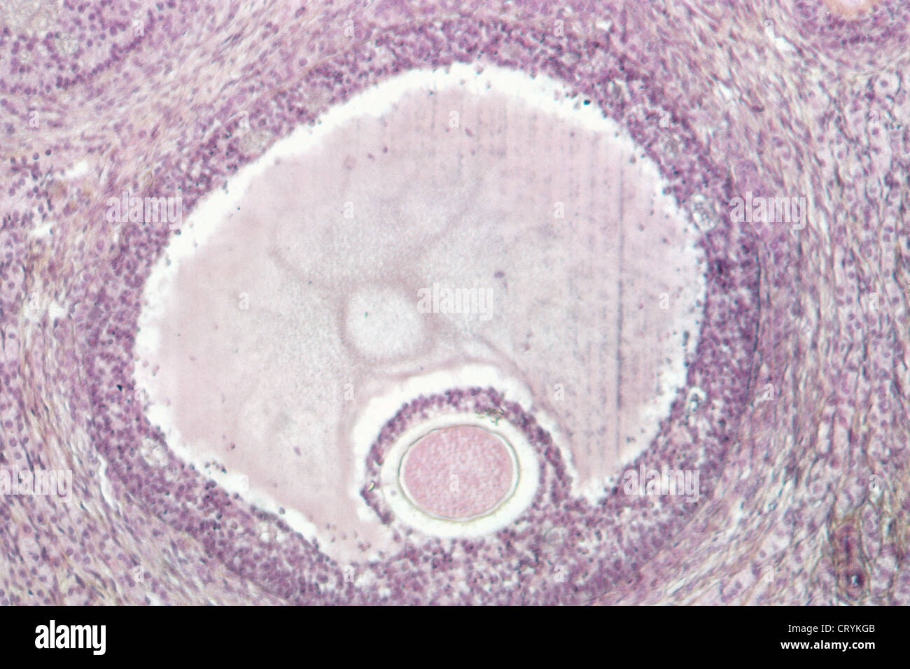 OVARY,HISTOLOGY Stock Photohttps://www.alamy.com/image-license-details/?v=1https://www.alamy.com/stock-photo-ovaryhistology-49165883.html
RMCRYKGB–OVARY,HISTOLOGY
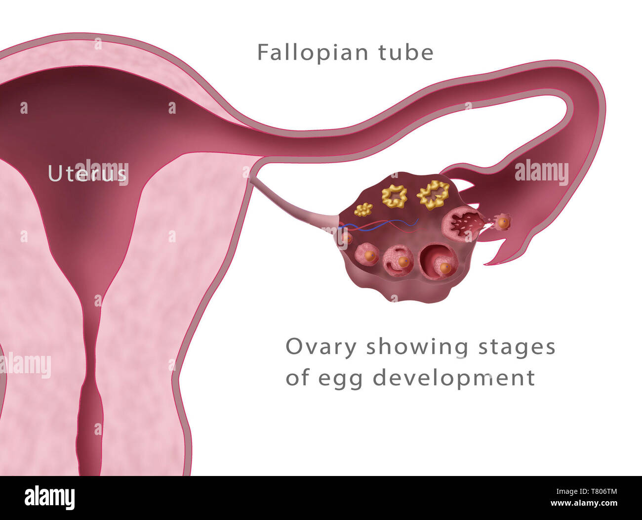 Ovarian Follicles, Menstruation, Illustration Stock Photohttps://www.alamy.com/image-license-details/?v=1https://www.alamy.com/ovarian-follicles-menstruation-illustration-image245867796.html
RMT806TM–Ovarian Follicles, Menstruation, Illustration
 A nurse's handbook of obstetrics . Fig. 15.—Longitudinal section through ovary of a woman twenty-two days after the lastmenstruation. (Leopold.) m.f., mature Graafian follicle; pr., most prominent point offollicle, where the rupture may be expected. like a small pimple. The Graafian follicle then becomes thinnedat one point, where it soon bursts and allows the ovum to escapeinto the Fallopian tube (Fig. 16). 43 44 A NURSES HANDBOOK OF OBSTETRICS. Once within the Fallopian tube, the ovum makes its way intothe uterus, and, if unimpregnated by the male element, it losesits vitality in a few days Stock Photohttps://www.alamy.com/image-license-details/?v=1https://www.alamy.com/a-nurses-handbook-of-obstetrics-fig-15longitudinal-section-through-ovary-of-a-woman-twenty-two-days-after-the-lastmenstruation-leopold-mf-mature-graafian-follicle-pr-most-prominent-point-offollicle-where-the-rupture-may-be-expected-like-a-small-pimple-the-graafian-follicle-then-becomes-thinnedat-one-point-where-it-soon-bursts-and-allows-the-ovum-to-escapeinto-the-fallopian-tube-fig-16-43-44-a-nurses-handbook-of-obstetrics-once-within-the-fallopian-tube-the-ovum-makes-its-way-intothe-uterus-and-if-unimpregnated-by-the-male-element-it-losesits-vitality-in-a-few-days-image339093672.html
RM2AKK1C8–A nurse's handbook of obstetrics . Fig. 15.—Longitudinal section through ovary of a woman twenty-two days after the lastmenstruation. (Leopold.) m.f., mature Graafian follicle; pr., most prominent point offollicle, where the rupture may be expected. like a small pimple. The Graafian follicle then becomes thinnedat one point, where it soon bursts and allows the ovum to escapeinto the Fallopian tube (Fig. 16). 43 44 A NURSES HANDBOOK OF OBSTETRICS. Once within the Fallopian tube, the ovum makes its way intothe uterus, and, if unimpregnated by the male element, it losesits vitality in a few days
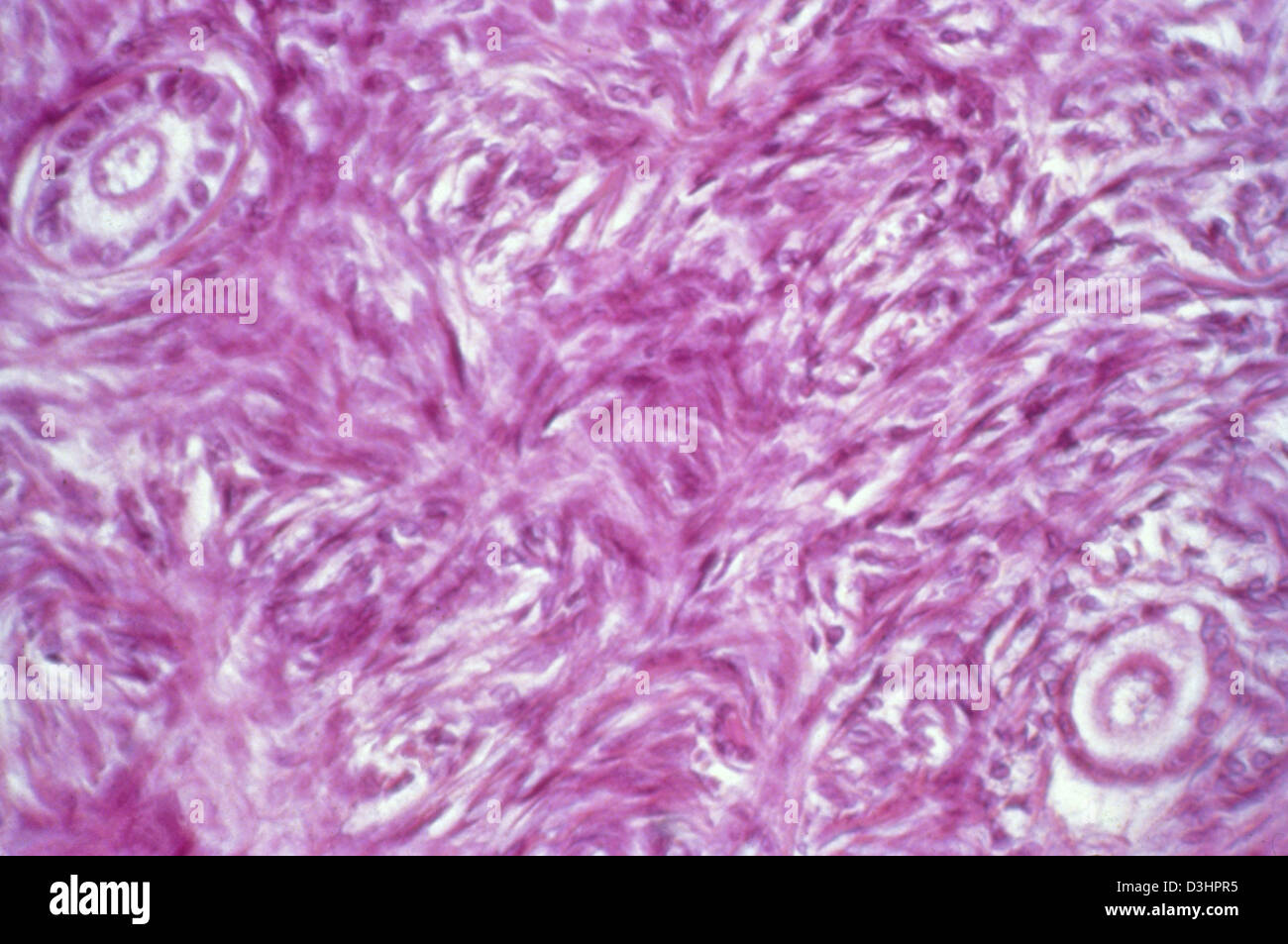 OVARY,HISTOLOGY Stock Photohttps://www.alamy.com/image-license-details/?v=1https://www.alamy.com/stock-photo-ovaryhistology-53866153.html
RFD3HPR5–OVARY,HISTOLOGY
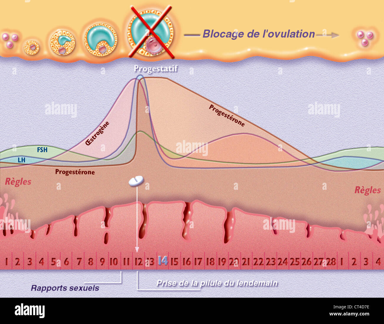 MORNING-AFTER PILL Stock Photohttps://www.alamy.com/image-license-details/?v=1https://www.alamy.com/stock-photo-morning-after-pill-49270690.html
RMCT4D7E–MORNING-AFTER PILL
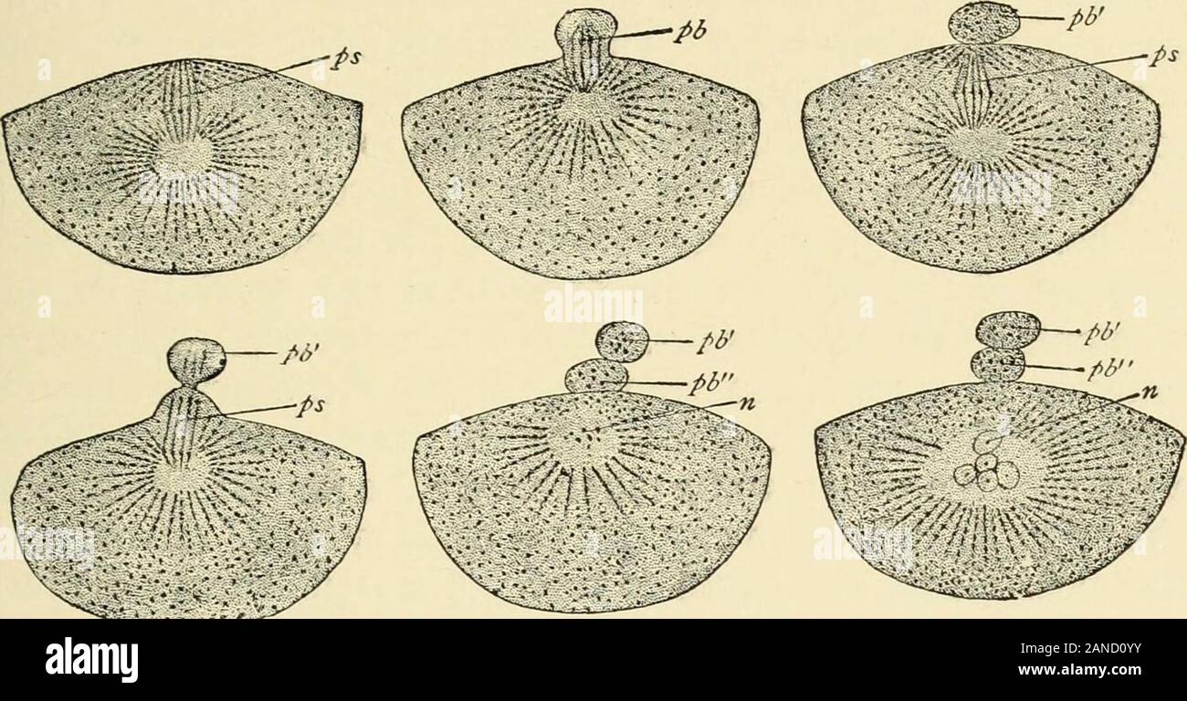 A textbook of obstetrics . Si ifp K Fig- 53-—Section through part of amammalian ovary : KE, Germinal epitheli-um ; PS, an egg-cord ; i U, primitive ova ;G, investing cells; A*, germinal vesicle ;S, follicular cavity arising in one of theolder follicles ; Lf, follicular cavity, moreenlarged ; AY, nearly mature ovum, whichhas developed around it the zona pellu-cida, Mp; Mg, membrana granulosa; E>,Discus proligerus; So, ovarian stroma;Tf, capsule of follicle ; g,g, blood-vessels;u, immature Graafian follicle (after Wie-dersheim). OVULATION. 61 The Discharge of the Ovum from the Ovary and its Stock Photohttps://www.alamy.com/image-license-details/?v=1https://www.alamy.com/a-textbook-of-obstetrics-si-ifp-k-fig-53-section-through-part-of-amammalian-ovary-ke-germinal-epitheli-um-ps-an-egg-cord-i-u-primitive-ova-g-investing-cells-a-germinal-vesicle-s-follicular-cavity-arising-in-one-of-theolder-follicles-lf-follicular-cavity-moreenlarged-ay-nearly-mature-ovum-whichhas-developed-around-it-the-zona-pellu-cida-mp-mg-membrana-granulosa-egtdiscus-proligerus-so-ovarian-stromatf-capsule-of-follicle-gg-blood-vesselsu-immature-graafian-follicle-after-wie-dersheim-ovulation-61-the-discharge-of-the-ovum-from-the-ovary-and-its-image340190927.html
RM2AND0YY–A textbook of obstetrics . Si ifp K Fig- 53-—Section through part of amammalian ovary : KE, Germinal epitheli-um ; PS, an egg-cord ; i U, primitive ova ;G, investing cells; A*, germinal vesicle ;S, follicular cavity arising in one of theolder follicles ; Lf, follicular cavity, moreenlarged ; AY, nearly mature ovum, whichhas developed around it the zona pellu-cida, Mp; Mg, membrana granulosa; E>,Discus proligerus; So, ovarian stroma;Tf, capsule of follicle ; g,g, blood-vessels;u, immature Graafian follicle (after Wie-dersheim). OVULATION. 61 The Discharge of the Ovum from the Ovary and its
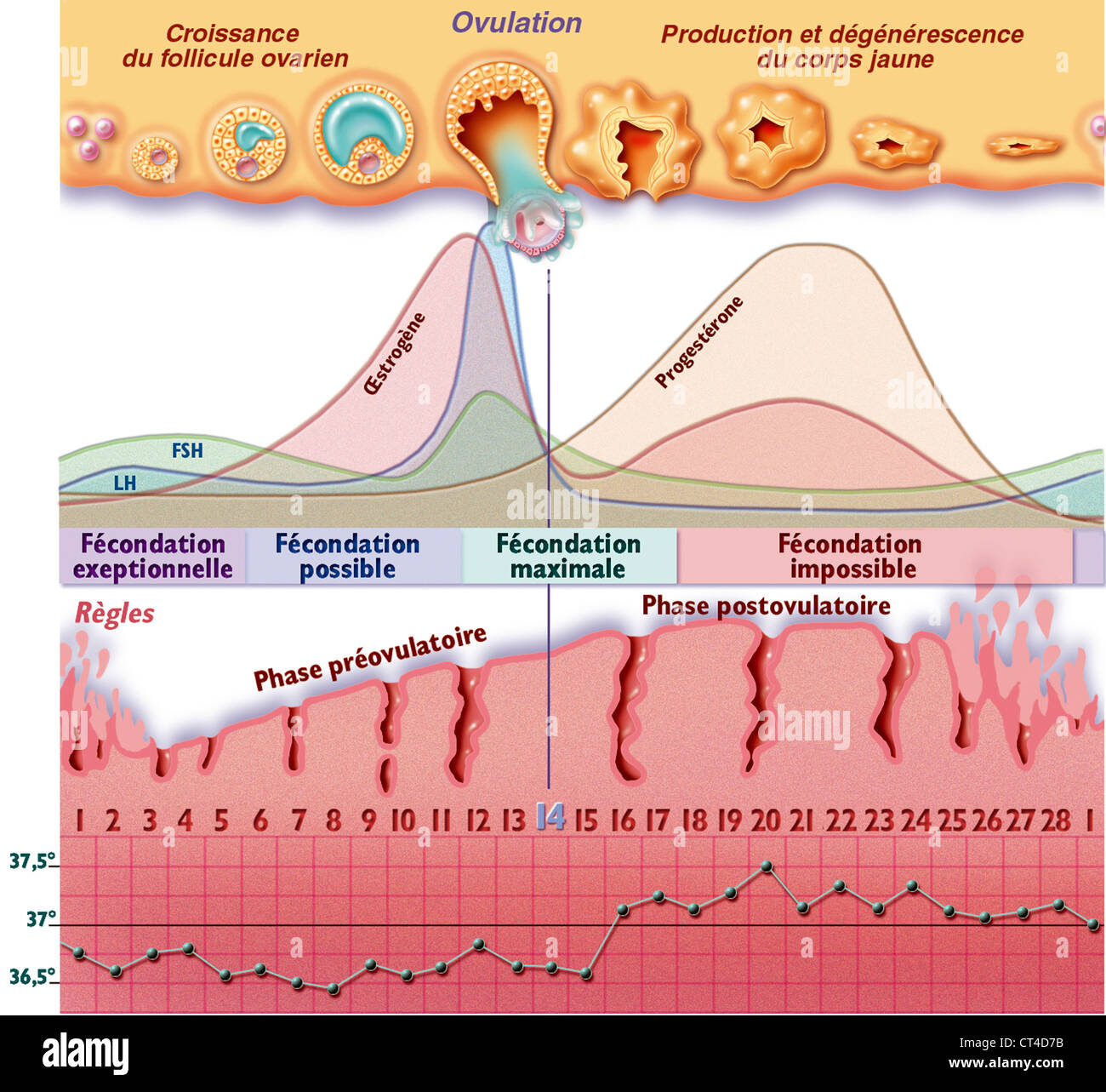 MENSTRUAL CYCLE, DRAWING Stock Photohttps://www.alamy.com/image-license-details/?v=1https://www.alamy.com/stock-photo-menstrual-cycle-drawing-49270687.html
RMCT4D7B–MENSTRUAL CYCLE, DRAWING
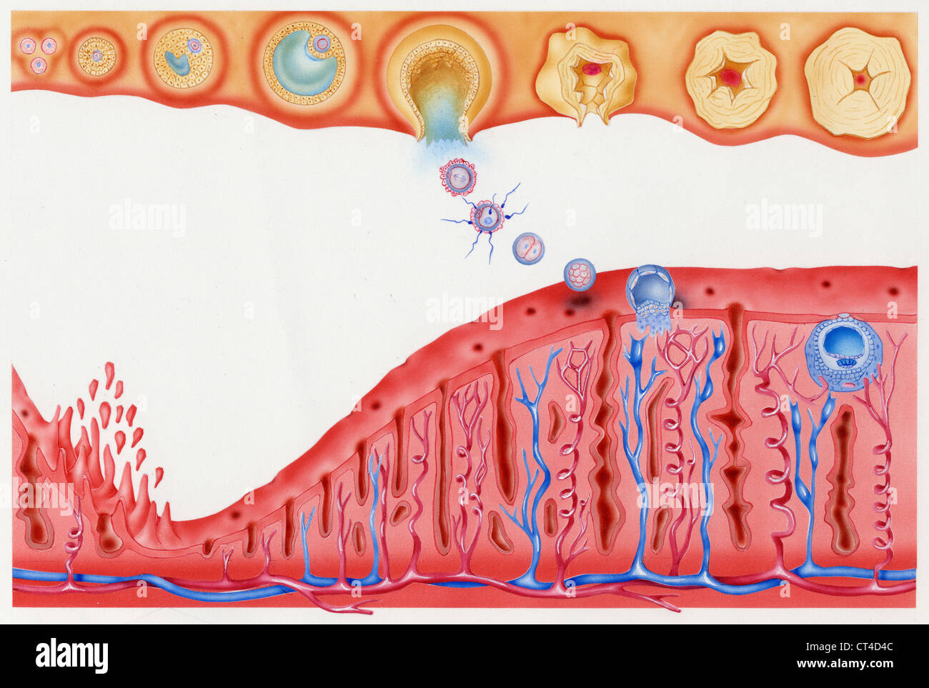 MENSTRUAL CYCLE, DRAWING Stock Photohttps://www.alamy.com/image-license-details/?v=1https://www.alamy.com/stock-photo-menstrual-cycle-drawing-49270604.html
RMCT4D4C–MENSTRUAL CYCLE, DRAWING
![Carpenter's principles of human physiology . pi- ] • thelium of a second ovum in the same follicle; g, fibrous persistent cell of a peculiar epithelial tunic; h, tunica propria folliculi; i, epithelium of the fnrTnatinn Wlipn matnrp a rvrimnr- foliicle (membrana granulosa). iormation. vv nen mature a prnnor- dial ovum has a diameter of 200^u (and* Principles of Biology, vol. ii. part vi. pp. 419-440.t For the literature of the Histology of the Ovaries up to 1871, see Art. Ovary and Par-ovarium, by W. Waldeyer, in Striekers Hum. and Comp. Histology, Syd. Soc. transl.,1872, vol. ii. p. 164. Sinc Stock Photo Carpenter's principles of human physiology . pi- ] • thelium of a second ovum in the same follicle; g, fibrous persistent cell of a peculiar epithelial tunic; h, tunica propria folliculi; i, epithelium of the fnrTnatinn Wlipn matnrp a rvrimnr- foliicle (membrana granulosa). iormation. vv nen mature a prnnor- dial ovum has a diameter of 200^u (and* Principles of Biology, vol. ii. part vi. pp. 419-440.t For the literature of the Histology of the Ovaries up to 1871, see Art. Ovary and Par-ovarium, by W. Waldeyer, in Striekers Hum. and Comp. Histology, Syd. Soc. transl.,1872, vol. ii. p. 164. Sinc Stock Photo](https://c8.alamy.com/comp/2AN3M5F/carpenters-principles-of-human-physiology-pi-thelium-of-a-second-ovum-in-the-same-follicle-g-fibrous-persistent-cell-of-a-peculiar-epithelial-tunic-h-tunica-propria-folliculi-i-epithelium-of-the-fnrtnatinn-wlipn-matnrp-a-rvrimnr-foliicle-membrana-granulosa-iormation-vv-nen-mature-a-prnnor-dial-ovum-has-a-diameter-of-200u-and-principles-of-biology-vol-ii-part-vi-pp-419-440t-for-the-literature-of-the-histology-of-the-ovaries-up-to-1871-see-art-ovary-and-par-ovarium-by-w-waldeyer-in-striekers-hum-and-comp-histology-syd-soc-transl1872-vol-ii-p-164-sinc-2AN3M5F.jpg) Carpenter's principles of human physiology . pi- ] • thelium of a second ovum in the same follicle; g, fibrous persistent cell of a peculiar epithelial tunic; h, tunica propria folliculi; i, epithelium of the fnrTnatinn Wlipn matnrp a rvrimnr- foliicle (membrana granulosa). iormation. vv nen mature a prnnor- dial ovum has a diameter of 200^u (and* Principles of Biology, vol. ii. part vi. pp. 419-440.t For the literature of the Histology of the Ovaries up to 1871, see Art. Ovary and Par-ovarium, by W. Waldeyer, in Striekers Hum. and Comp. Histology, Syd. Soc. transl.,1872, vol. ii. p. 164. Sinc Stock Photohttps://www.alamy.com/image-license-details/?v=1https://www.alamy.com/carpenters-principles-of-human-physiology-pi-thelium-of-a-second-ovum-in-the-same-follicle-g-fibrous-persistent-cell-of-a-peculiar-epithelial-tunic-h-tunica-propria-folliculi-i-epithelium-of-the-fnrtnatinn-wlipn-matnrp-a-rvrimnr-foliicle-membrana-granulosa-iormation-vv-nen-mature-a-prnnor-dial-ovum-has-a-diameter-of-200u-and-principles-of-biology-vol-ii-part-vi-pp-419-440t-for-the-literature-of-the-histology-of-the-ovaries-up-to-1871-see-art-ovary-and-par-ovarium-by-w-waldeyer-in-striekers-hum-and-comp-histology-syd-soc-transl1872-vol-ii-p-164-sinc-image339986459.html
Carpenter's principles of human physiology . pi- ] • thelium of a second ovum in the same follicle; g, fibrous persistent cell of a peculiar epithelial tunic; h, tunica propria folliculi; i, epithelium of the fnrTnatinn Wlipn matnrp a rvrimnr- foliicle (membrana granulosa). iormation. vv nen mature a prnnor- dial ovum has a diameter of 200^u (and* Principles of Biology, vol. ii. part vi. pp. 419-440.t For the literature of the Histology of the Ovaries up to 1871, see Art. Ovary and Par-ovarium, by W. Waldeyer, in Striekers Hum. and Comp. Histology, Syd. Soc. transl.,1872, vol. ii. p. 164. Sinc Stock Photohttps://www.alamy.com/image-license-details/?v=1https://www.alamy.com/carpenters-principles-of-human-physiology-pi-thelium-of-a-second-ovum-in-the-same-follicle-g-fibrous-persistent-cell-of-a-peculiar-epithelial-tunic-h-tunica-propria-folliculi-i-epithelium-of-the-fnrtnatinn-wlipn-matnrp-a-rvrimnr-foliicle-membrana-granulosa-iormation-vv-nen-mature-a-prnnor-dial-ovum-has-a-diameter-of-200u-and-principles-of-biology-vol-ii-part-vi-pp-419-440t-for-the-literature-of-the-histology-of-the-ovaries-up-to-1871-see-art-ovary-and-par-ovarium-by-w-waldeyer-in-striekers-hum-and-comp-histology-syd-soc-transl1872-vol-ii-p-164-sinc-image339986459.htmlRM2AN3M5F–Carpenter's principles of human physiology . pi- ] • thelium of a second ovum in the same follicle; g, fibrous persistent cell of a peculiar epithelial tunic; h, tunica propria folliculi; i, epithelium of the fnrTnatinn Wlipn matnrp a rvrimnr- foliicle (membrana granulosa). iormation. vv nen mature a prnnor- dial ovum has a diameter of 200^u (and* Principles of Biology, vol. ii. part vi. pp. 419-440.t For the literature of the Histology of the Ovaries up to 1871, see Art. Ovary and Par-ovarium, by W. Waldeyer, in Striekers Hum. and Comp. Histology, Syd. Soc. transl.,1872, vol. ii. p. 164. Sinc
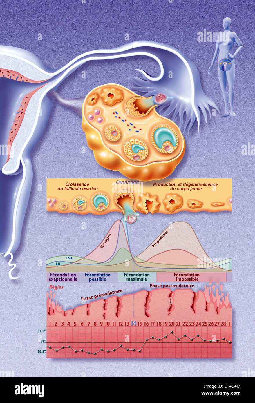 OVULATION, DRAWING Stock Photohttps://www.alamy.com/image-license-details/?v=1https://www.alamy.com/stock-photo-ovulation-drawing-49270612.html
RMCT4D4M–OVULATION, DRAWING
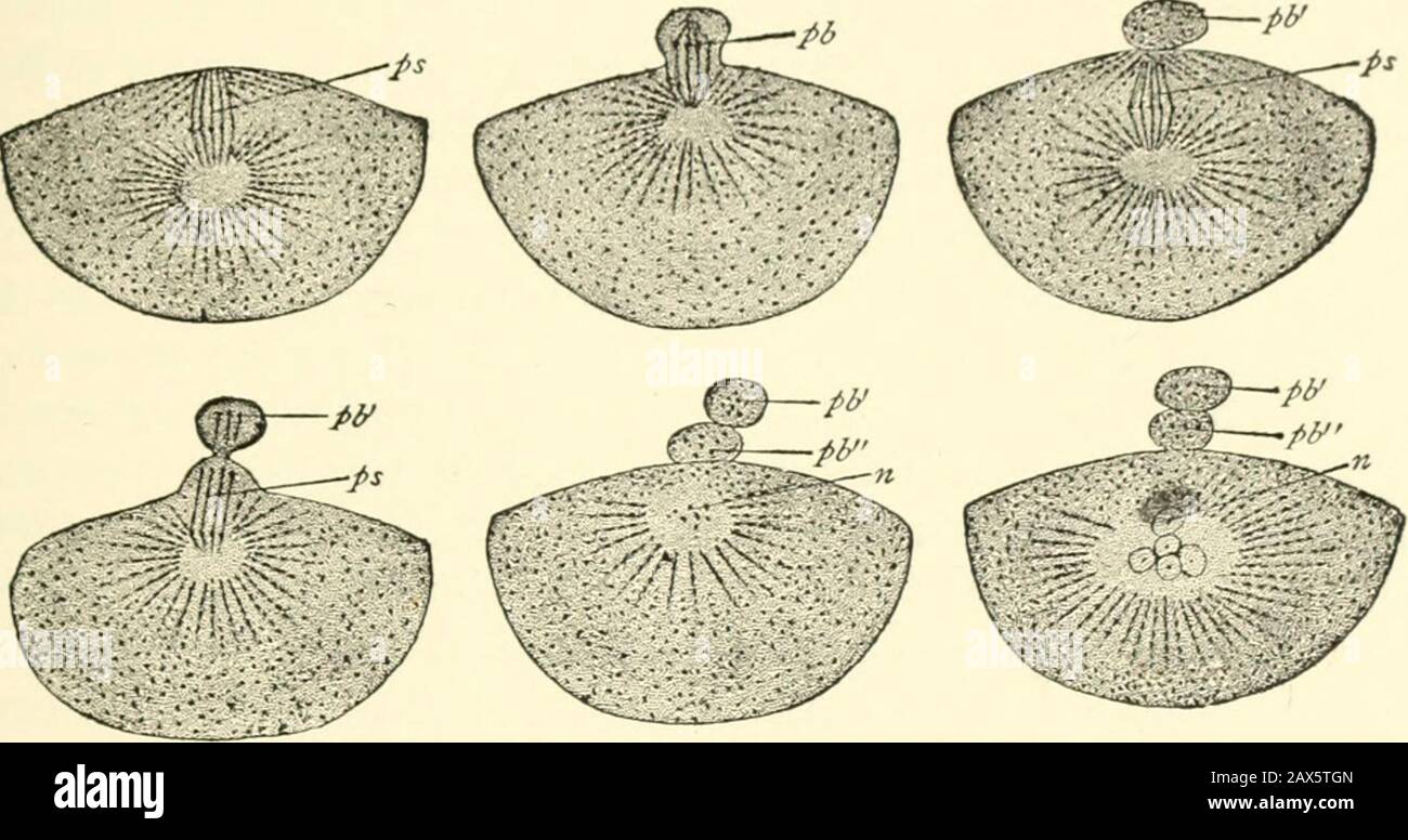 A textbook of obstetrics . Ei Afp K Fig- 53-—Section through part of amammalian ovary: A/Y, Germinal epitheli-um ; PS, an egg-cord; I, U, primitive ova;G, investing cells; A, germinal vesicle;.V, follicular cavity arising in one of theolder follicles; Lf, follicular cavity, moreenlarged ; Ei, nearly mature ovum, whichhas developed around it the zona pellucida, J/// Mg, membrana granulosa; D,I >i-< us proligerus; So, ovarian stroma;Tf, capsule ol follicle; g,gt blood vessels;u, immature Graafian follicle (alter Wiidersheim |. OVULATION, 6l The Discharge of the Ovum from the Ovary and its Stock Photohttps://www.alamy.com/image-license-details/?v=1https://www.alamy.com/a-textbook-of-obstetrics-ei-afp-k-fig-53-section-through-part-of-amammalian-ovary-ay-germinal-epitheli-um-ps-an-egg-cord-i-u-primitive-ovag-investing-cells-a-germinal-vesiclev-follicular-cavity-arising-in-one-of-theolder-follicles-lf-follicular-cavity-moreenlarged-ei-nearly-mature-ovum-whichhas-developed-around-it-the-zona-pellucida-j-mg-membrana-granulosa-di-gti-lt-us-proligerus-so-ovarian-stromatf-capsule-ol-follicle-ggt-blood-vesselsu-immature-graafian-follicle-alter-wiidersheim-ovulation-6l-the-discharge-of-the-ovum-from-the-ovary-and-its-image343107093.html
RM2AX5TGN–A textbook of obstetrics . Ei Afp K Fig- 53-—Section through part of amammalian ovary: A/Y, Germinal epitheli-um ; PS, an egg-cord; I, U, primitive ova;G, investing cells; A, germinal vesicle;.V, follicular cavity arising in one of theolder follicles; Lf, follicular cavity, moreenlarged ; Ei, nearly mature ovum, whichhas developed around it the zona pellucida, J/// Mg, membrana granulosa; D,I >i-< us proligerus; So, ovarian stroma;Tf, capsule ol follicle; g,gt blood vessels;u, immature Graafian follicle (alter Wiidersheim |. OVULATION, 6l The Discharge of the Ovum from the Ovary and its
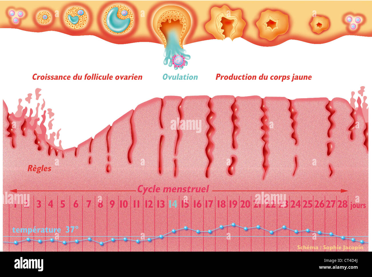 MENSTRUAL CYCLE, DRAWING Stock Photohttps://www.alamy.com/image-license-details/?v=1https://www.alamy.com/stock-photo-menstrual-cycle-drawing-49270610.html
RMCT4D4J–MENSTRUAL CYCLE, DRAWING
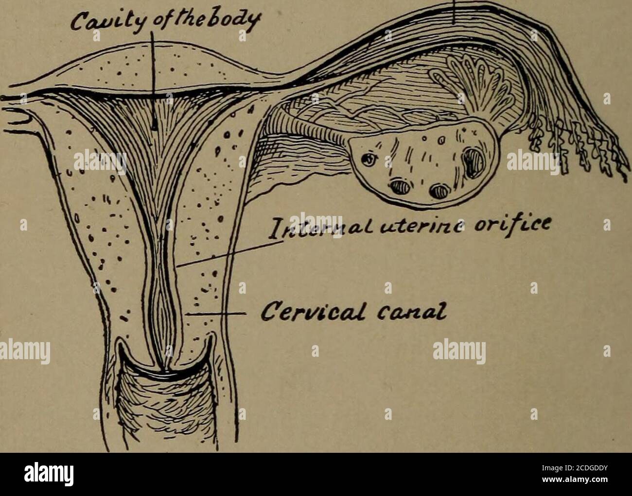 . A reference hand-book of gynecology for nurses . External uterine orifice Cowity ofthe$Oi Co-*-*- 0/ FcUlofita.H,tlcb*. Figs. 7 and 8.—The uterus, ovary, and Fallopian tube. the dilated infundibulum, a trumpet-shaped orificesurrounded by finger-like processes called fimbriae.The oviduct contains a canal lined with mucousmembrane. ANATOMY 19 The ovaries, two in number, are almond-shapedbodies attached to the posterior surface of thebroad ligaments. They contain innumerable ova,each one surrounded by a more or less well-developed cellular envelop, which is called aGraafian follicle when mature Stock Photohttps://www.alamy.com/image-license-details/?v=1https://www.alamy.com/a-reference-hand-book-of-gynecology-for-nurses-external-uterine-orifice-cowity-oftheoi-co-0-fculofitahtlcb-figs-7-and-8the-uterus-ovary-and-fallopian-tube-the-dilated-infundibulum-a-trumpet-shaped-orificesurrounded-by-finger-like-processes-called-fimbriaethe-oviduct-contains-a-canal-lined-with-mucousmembrane-anatomy-19-the-ovaries-two-in-number-are-almond-shapedbodies-attached-to-the-posterior-surface-of-thebroad-ligaments-they-contain-innumerable-ovaeach-one-surrounded-by-a-more-or-less-well-developed-cellular-envelop-which-is-called-agraafian-follicle-when-mature-image369770071.html
RM2CDGDDY–. A reference hand-book of gynecology for nurses . External uterine orifice Cowity ofthe$Oi Co-*-*- 0/ FcUlofita.H,tlcb*. Figs. 7 and 8.—The uterus, ovary, and Fallopian tube. the dilated infundibulum, a trumpet-shaped orificesurrounded by finger-like processes called fimbriae.The oviduct contains a canal lined with mucousmembrane. ANATOMY 19 The ovaries, two in number, are almond-shapedbodies attached to the posterior surface of thebroad ligaments. They contain innumerable ova,each one surrounded by a more or less well-developed cellular envelop, which is called aGraafian follicle when mature
 MENSTRUAL CYCLE, DRAWING Stock Photohttps://www.alamy.com/image-license-details/?v=1https://www.alamy.com/stock-photo-menstrual-cycle-drawing-49270598.html
RMCT4D46–MENSTRUAL CYCLE, DRAWING
 . Anatomy, descriptive and surgical. of minute vesicles of uniform size about ■Y^-q- of an inch in diameter. These arethe Graafian vesicles in their earliestcondition, and the layer where they are foundlayer. has been termed the corticalThey are especially numerous Fig. 662. ■ Zcne TcTitmcwn Section of the Ovary (after Schron): 1, outer covering;1, attached horder; 2, central stroma; 3, peripheralstroma; 4, blood-vessels; 5, Graafian follicles in theirearliest stage; 6,7, 8, more advanced follicles; 9, analmost mature follicle ; 9, follicle from which the ovumhas escaped; 10, corpus luteum.. S Stock Photohttps://www.alamy.com/image-license-details/?v=1https://www.alamy.com/anatomy-descriptive-and-surgical-of-minute-vesicles-of-uniform-size-about-y-q-of-an-inch-in-diameter-these-arethe-graafian-vesicles-in-their-earliestcondition-and-the-layer-where-they-are-foundlayer-has-been-termed-the-corticalthey-are-especially-numerous-fig-662-zcne-tctitmcwn-section-of-the-ovary-after-schron-1-outer-covering1-attached-horder-2-central-stroma-3-peripheralstroma-4-blood-vessels-5-graafian-follicles-in-theirearliest-stage-67-8-more-advanced-follicles-9-analmost-mature-follicle-9-follicle-from-which-the-ovumhas-escaped-10-corpus-luteum-s-image370501290.html
RM2CENP4X–. Anatomy, descriptive and surgical. of minute vesicles of uniform size about ■Y^-q- of an inch in diameter. These arethe Graafian vesicles in their earliestcondition, and the layer where they are foundlayer. has been termed the corticalThey are especially numerous Fig. 662. ■ Zcne TcTitmcwn Section of the Ovary (after Schron): 1, outer covering;1, attached horder; 2, central stroma; 3, peripheralstroma; 4, blood-vessels; 5, Graafian follicles in theirearliest stage; 6,7, 8, more advanced follicles; 9, analmost mature follicle ; 9, follicle from which the ovumhas escaped; 10, corpus luteum.. S
 MENSTRUAL CYCLE, DRAWING Stock Photohttps://www.alamy.com/image-license-details/?v=1https://www.alamy.com/stock-photo-menstrual-cycle-drawing-49270587.html
RMCT4D3R–MENSTRUAL CYCLE, DRAWING
 . An American text-book of obstetrics. For practitioners and students. Among the immature follicles are others in Fig. 52.—Section of human ovary, includingcortex: a, germinal epithelium of free surface; 6,tunica albuginea; c, peripheral stroma contain-ing immature Graafian follicles (d); e, well-ad-vanced follicle from whose wall membrana granu-losa has partially separated ; /, cavity of liquor fol-liculi; g, ovum surrounded by cell-mass consti-tuting discus proligerus (Piersol).. Fig. 53.—Ovary with mature Graafian follicle aboutready to burst (Ribemont-Dessaignes). various stages of more ad Stock Photohttps://www.alamy.com/image-license-details/?v=1https://www.alamy.com/an-american-text-book-of-obstetrics-for-practitioners-and-students-among-the-immature-follicles-are-others-in-fig-52section-of-human-ovary-includingcortex-a-germinal-epithelium-of-free-surface-6tunica-albuginea-c-peripheral-stroma-contain-ing-immature-graafian-follicles-d-e-well-ad-vanced-follicle-from-whose-wall-membrana-granu-losa-has-partially-separated-cavity-of-liquor-fol-liculi-g-ovum-surrounded-by-cell-mass-consti-tuting-discus-proligerus-piersol-fig-53ovary-with-mature-graafian-follicle-aboutready-to-burst-ribemont-dessaignes-various-stages-of-more-ad-image370602182.html
RM2CEXAT6–. An American text-book of obstetrics. For practitioners and students. Among the immature follicles are others in Fig. 52.—Section of human ovary, includingcortex: a, germinal epithelium of free surface; 6,tunica albuginea; c, peripheral stroma contain-ing immature Graafian follicles (d); e, well-ad-vanced follicle from whose wall membrana granu-losa has partially separated ; /, cavity of liquor fol-liculi; g, ovum surrounded by cell-mass consti-tuting discus proligerus (Piersol).. Fig. 53.—Ovary with mature Graafian follicle aboutready to burst (Ribemont-Dessaignes). various stages of more ad
 MENSTRUAL CYCLE, DRAWING Stock Photohttps://www.alamy.com/image-license-details/?v=1https://www.alamy.com/stock-photo-menstrual-cycle-drawing-49270591.html
RMCT4D3Y–MENSTRUAL CYCLE, DRAWING
 GYNECOLOGY, ILLUSTRATION Stock Photohttps://www.alamy.com/image-license-details/?v=1https://www.alamy.com/stock-photo-gynecology-illustration-53864721.html
RMD3HN01–GYNECOLOGY, ILLUSTRATION
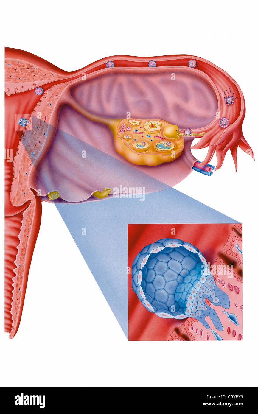 FERTILIZATION, DRAWING Stock Photohttps://www.alamy.com/image-license-details/?v=1https://www.alamy.com/stock-photo-fertilization-drawing-49159889.html
RMCRYBX9–FERTILIZATION, DRAWING
 OVARIAN CYCLE, DRAWING Stock Photohttps://www.alamy.com/image-license-details/?v=1https://www.alamy.com/stock-photo-ovarian-cycle-drawing-49267444.html
RMCT493G–OVARIAN CYCLE, DRAWING
 . Outlines of zoology. Zoology. 666 MAMMALIA. is formed containing fluid, and into this cavity the follicle cells immediately surrounding the ovum project, forming what is called the discusproligerus. (See Fig. 198, p. 467.) When mature, the ovum protrudes on the surface of the ovary, and is liberated by the bursting of the Graafian fol- licle. Some blood, which fills up the empty follicle, degener- ates into what is called the corpus luteum. The spermatozoa are formed from germinal epithelium in the testes. The primitive male cells or spermatogonia give rise by division to daughter cells or s Stock Photohttps://www.alamy.com/image-license-details/?v=1https://www.alamy.com/outlines-of-zoology-zoology-666-mammalia-is-formed-containing-fluid-and-into-this-cavity-the-follicle-cells-immediately-surrounding-the-ovum-project-forming-what-is-called-the-discusproligerus-see-fig-198-p-467-when-mature-the-ovum-protrudes-on-the-surface-of-the-ovary-and-is-liberated-by-the-bursting-of-the-graafian-fol-licle-some-blood-which-fills-up-the-empty-follicle-degener-ates-into-what-is-called-the-corpus-luteum-the-spermatozoa-are-formed-from-germinal-epithelium-in-the-testes-the-primitive-male-cells-or-spermatogonia-give-rise-by-division-to-daughter-cells-or-s-image232213061.html
RMRDP63H–. Outlines of zoology. Zoology. 666 MAMMALIA. is formed containing fluid, and into this cavity the follicle cells immediately surrounding the ovum project, forming what is called the discusproligerus. (See Fig. 198, p. 467.) When mature, the ovum protrudes on the surface of the ovary, and is liberated by the bursting of the Graafian fol- licle. Some blood, which fills up the empty follicle, degener- ates into what is called the corpus luteum. The spermatozoa are formed from germinal epithelium in the testes. The primitive male cells or spermatogonia give rise by division to daughter cells or s
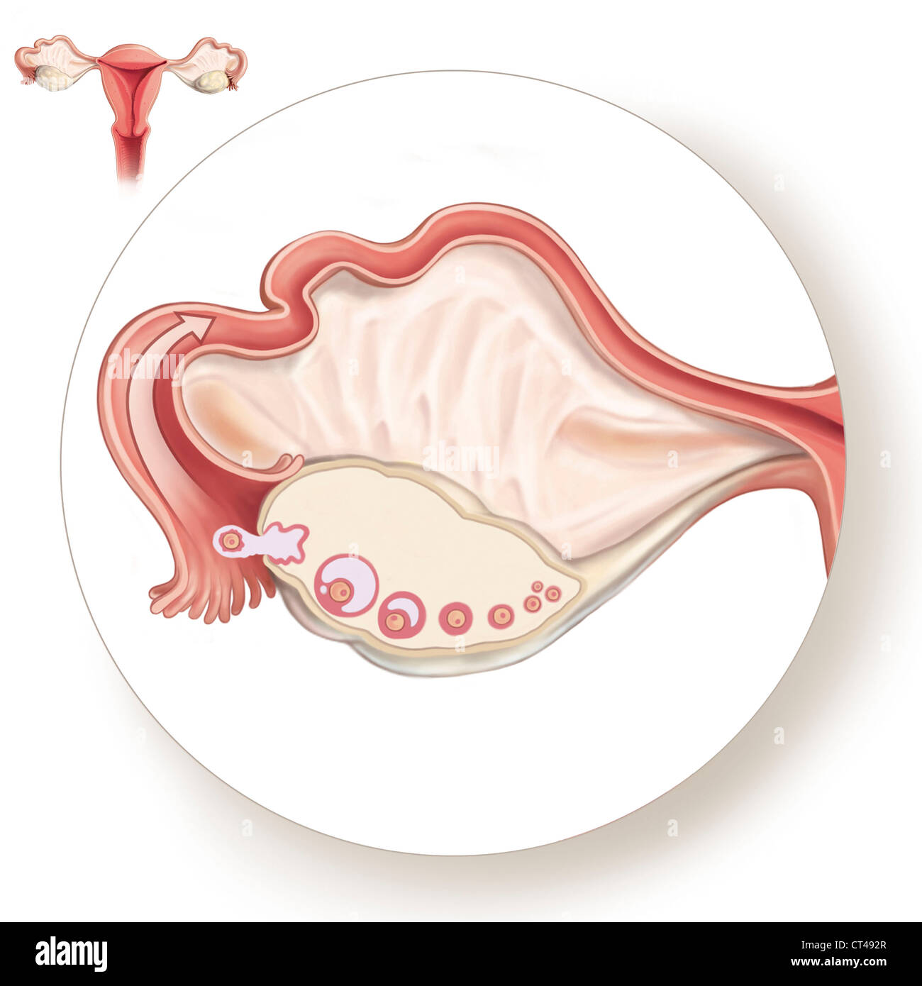 OVARIAN CYCLE, DRAWING Stock Photohttps://www.alamy.com/image-license-details/?v=1https://www.alamy.com/stock-photo-ovarian-cycle-drawing-49267423.html
RMCT492R–OVARIAN CYCLE, DRAWING
 . Chordate morphology. Morphology (Animals); Chordata. licles deeper in the stroma. When ripe, the follicles rupture into the body cavity. The ovary is sparsely spotted with follicles of which the smaller ones are near the free margin. As the eggs become larger they hang down into the body cavity below the level of the free margin (Figure 9-28). The eggs are shelled while still in the follicle; a small micropile, opening through the shell, allows for fertilization. Not all of the large follicles ob- served develop to mature eggs; some develop at the expense of others which atrophy. The mature Stock Photohttps://www.alamy.com/image-license-details/?v=1https://www.alamy.com/chordate-morphology-morphology-animals-chordata-licles-deeper-in-the-stroma-when-ripe-the-follicles-rupture-into-the-body-cavity-the-ovary-is-sparsely-spotted-with-follicles-of-which-the-smaller-ones-are-near-the-free-margin-as-the-eggs-become-larger-they-hang-down-into-the-body-cavity-below-the-level-of-the-free-margin-figure-9-28-the-eggs-are-shelled-while-still-in-the-follicle-a-small-micropile-opening-through-the-shell-allows-for-fertilization-not-all-of-the-large-follicles-ob-served-develop-to-mature-eggs-some-develop-at-the-expense-of-others-which-atrophy-the-mature-image234937887.html
RMRJ69JR–. Chordate morphology. Morphology (Animals); Chordata. licles deeper in the stroma. When ripe, the follicles rupture into the body cavity. The ovary is sparsely spotted with follicles of which the smaller ones are near the free margin. As the eggs become larger they hang down into the body cavity below the level of the free margin (Figure 9-28). The eggs are shelled while still in the follicle; a small micropile, opening through the shell, allows for fertilization. Not all of the large follicles ob- served develop to mature eggs; some develop at the expense of others which atrophy. The mature
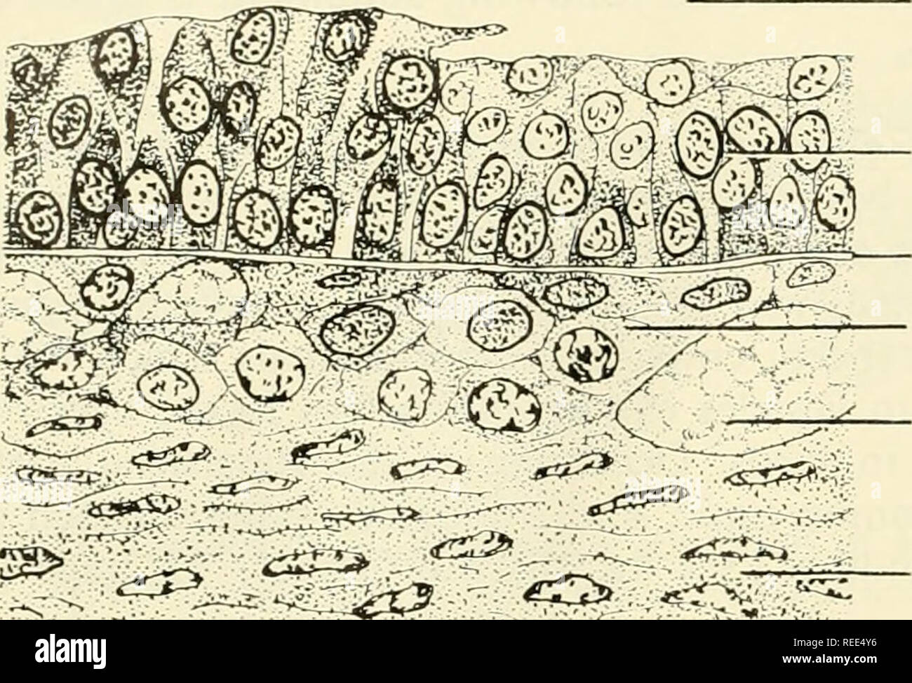 . Comparative embryology of the vertebrates; with 2057 drawings and photos. grouped as 380 illus. Vertebrates -- Embryology; Comparative embryology. 72 THE VERTEBRATE OVARY AND ITS RELATIONSHIP TO REPRODUCTION. CAVITY OF FOLLICLE GRANULOSA CELLS BASEMENT MEMBRANE THECA INTERNA CAPILLARY THECA EXTERN A Fig. 44. Cellular wall of the mature Graafian follicle in the opossum. layers of epithelial or granulosa cells surrounding the egg. It may now be regarded as a secondary Graafian follicle (fig. 42A, B). When a stage is reached where the granulosa cells form a layer five to seven or more cells in Stock Photohttps://www.alamy.com/image-license-details/?v=1https://www.alamy.com/comparative-embryology-of-the-vertebrates-with-2057-drawings-and-photos-grouped-as-380-illus-vertebrates-embryology-comparative-embryology-72-the-vertebrate-ovary-and-its-relationship-to-reproduction-cavity-of-follicle-granulosa-cells-basement-membrane-theca-interna-capillary-theca-extern-a-fig-44-cellular-wall-of-the-mature-graafian-follicle-in-the-opossum-layers-of-epithelial-or-granulosa-cells-surrounding-the-egg-it-may-now-be-regarded-as-a-secondary-graafian-follicle-fig-42a-b-when-a-stage-is-reached-where-the-granulosa-cells-form-a-layer-five-to-seven-or-more-cells-in-image232651194.html
RMREE4Y6–. Comparative embryology of the vertebrates; with 2057 drawings and photos. grouped as 380 illus. Vertebrates -- Embryology; Comparative embryology. 72 THE VERTEBRATE OVARY AND ITS RELATIONSHIP TO REPRODUCTION. CAVITY OF FOLLICLE GRANULOSA CELLS BASEMENT MEMBRANE THECA INTERNA CAPILLARY THECA EXTERN A Fig. 44. Cellular wall of the mature Graafian follicle in the opossum. layers of epithelial or granulosa cells surrounding the egg. It may now be regarded as a secondary Graafian follicle (fig. 42A, B). When a stage is reached where the granulosa cells form a layer five to seven or more cells in
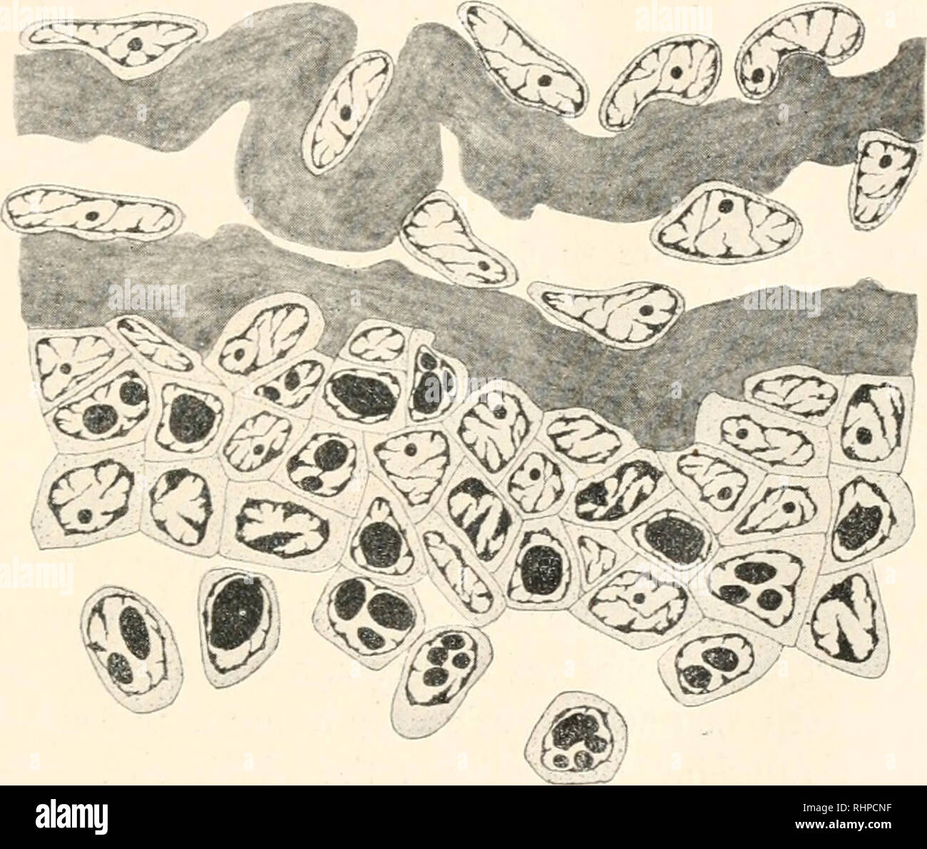 . The Biological bulletin. Biology; Zoology; Biology; Marine Biology. THE FOLLICLE SACS OF THE AMPHIBIAN OVARY. 251 the mature ovarian egg is in contact with the outer membrane of the wall of the ovary, I have never found the opening of an empty follicle sac to be more than 0.38 mm., the average being 0.20 mm. This shows that the edges of the opening tend to close in as soon as the egg has passed out of the ovary. On examining a longitudinal section of an empty follicle sac (Fig. 3), one finds that the two layers of which its walls are composed are united at frequent intervals, and that, on th Stock Photohttps://www.alamy.com/image-license-details/?v=1https://www.alamy.com/the-biological-bulletin-biology-zoology-biology-marine-biology-the-follicle-sacs-of-the-amphibian-ovary-251-the-mature-ovarian-egg-is-in-contact-with-the-outer-membrane-of-the-wall-of-the-ovary-i-have-never-found-the-opening-of-an-empty-follicle-sac-to-be-more-than-038-mm-the-average-being-020-mm-this-shows-that-the-edges-of-the-opening-tend-to-close-in-as-soon-as-the-egg-has-passed-out-of-the-ovary-on-examining-a-longitudinal-section-of-an-empty-follicle-sac-fig-3-one-finds-that-the-two-layers-of-which-its-walls-are-composed-are-united-at-frequent-intervals-and-that-on-th-image234676891.html
RMRHPCNF–. The Biological bulletin. Biology; Zoology; Biology; Marine Biology. THE FOLLICLE SACS OF THE AMPHIBIAN OVARY. 251 the mature ovarian egg is in contact with the outer membrane of the wall of the ovary, I have never found the opening of an empty follicle sac to be more than 0.38 mm., the average being 0.20 mm. This shows that the edges of the opening tend to close in as soon as the egg has passed out of the ovary. On examining a longitudinal section of an empty follicle sac (Fig. 3), one finds that the two layers of which its walls are composed are united at frequent intervals, and that, on th
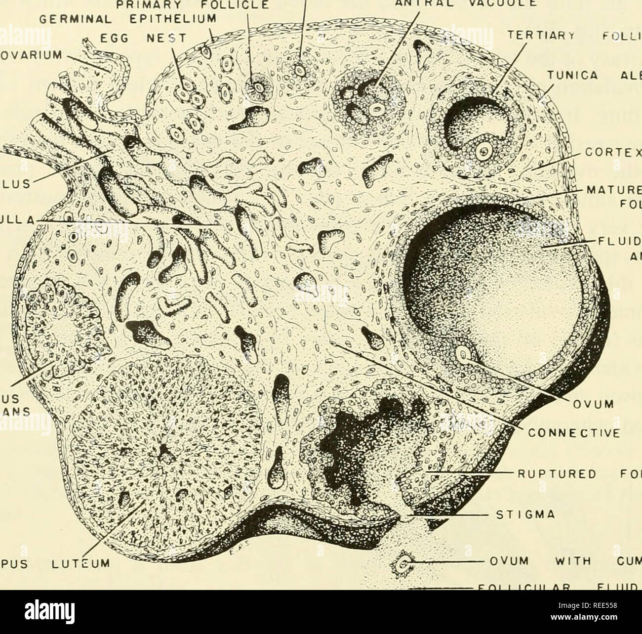 . Comparative embryology of the vertebrates; with 2057 drawings and photos. grouped as 380 illus. Vertebrates -- Embryology; Comparative embryology. 60 THE VERTEBRATE OVARY AND ITS RELATIONSHIP TO REPRODUC1 ION SECONDARY FOLLICLE PRIMARY FOLLICLE GERMINAL EPITHELIUM wyEGG NEST MESOVARIUM- MEDULLA "^ (i " ANTRAL VACUOLE TERTIARY FOLLICLE TUNICA ALBUGINEA MATURE FOLLICLE CORP ALBICANS. FLUID- FILLED ANTRUM CORPUS LUTEUM OVUM CONNE CT VE TISSUE RUPTURED FOLLICLE OVUM WITH CUMULUS CELLS FOLLICULAR FLUID Fig. 30. Schematic three-dimensional representation of the cyclic changes which occur Stock Photohttps://www.alamy.com/image-license-details/?v=1https://www.alamy.com/comparative-embryology-of-the-vertebrates-with-2057-drawings-and-photos-grouped-as-380-illus-vertebrates-embryology-comparative-embryology-60-the-vertebrate-ovary-and-its-relationship-to-reproduc1-ion-secondary-follicle-primary-follicle-germinal-epithelium-wyegg-nest-mesovarium-medulla-quot-i-quot-antral-vacuole-tertiary-follicle-tunica-albuginea-mature-follicle-corp-albicans-fluid-filled-antrum-corpus-luteum-ovum-conne-ct-ve-tissue-ruptured-follicle-ovum-with-cumulus-cells-follicular-fluid-fig-30-schematic-three-dimensional-representation-of-the-cyclic-changes-which-occur-image232651364.html
RMREE558–. Comparative embryology of the vertebrates; with 2057 drawings and photos. grouped as 380 illus. Vertebrates -- Embryology; Comparative embryology. 60 THE VERTEBRATE OVARY AND ITS RELATIONSHIP TO REPRODUC1 ION SECONDARY FOLLICLE PRIMARY FOLLICLE GERMINAL EPITHELIUM wyEGG NEST MESOVARIUM- MEDULLA "^ (i " ANTRAL VACUOLE TERTIARY FOLLICLE TUNICA ALBUGINEA MATURE FOLLICLE CORP ALBICANS. FLUID- FILLED ANTRUM CORPUS LUTEUM OVUM CONNE CT VE TISSUE RUPTURED FOLLICLE OVUM WITH CUMULUS CELLS FOLLICULAR FLUID Fig. 30. Schematic three-dimensional representation of the cyclic changes which occur
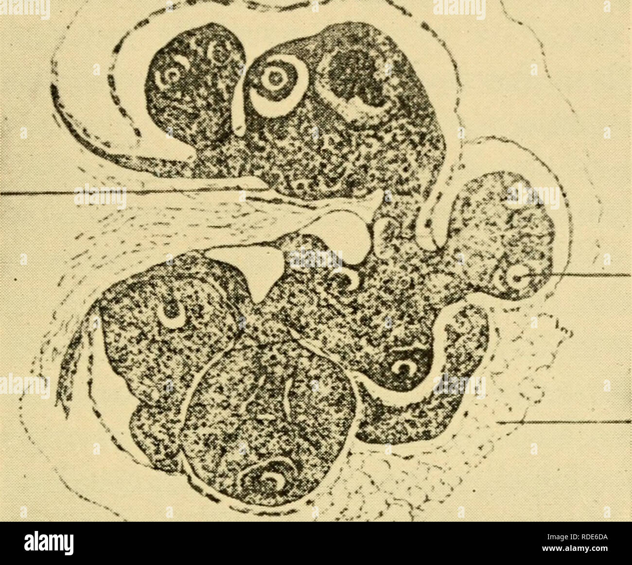 . The eggs of mammals. Ovum; Embryology -- Mammals; Mammals -- physiology; Ovum. THE ORIGIN OF THE DEFINITIVE OVA 17 a series of double ovariectomies in the rat, has demonstrated the presumable source of regenerated tissue in animals with apparently completely extirpated ovaries. In an initial series of 105 double ovariectomies she found germ cells at the ovarian site in eight cases, and observed that all eight YF. -».FA Fig. 5. Section through the ovary of mature rat showing the iobed condition. YF, young fol- licle; F, follicle; FA, fatty tissue. (From the Quarterly Review of Biology.) occur Stock Photohttps://www.alamy.com/image-license-details/?v=1https://www.alamy.com/the-eggs-of-mammals-ovum-embryology-mammals-mammals-physiology-ovum-the-origin-of-the-definitive-ova-17-a-series-of-double-ovariectomies-in-the-rat-has-demonstrated-the-presumable-source-of-regenerated-tissue-in-animals-with-apparently-completely-extirpated-ovaries-in-an-initial-series-of-105-double-ovariectomies-she-found-germ-cells-at-the-ovarian-site-in-eight-cases-and-observed-that-all-eight-yf-fa-fig-5-section-through-the-ovary-of-mature-rat-showing-the-iobed-condition-yf-young-fol-licle-f-follicle-fa-fatty-tissue-from-the-quarterly-review-of-biology-occur-image232037718.html
RMRDE6DA–. The eggs of mammals. Ovum; Embryology -- Mammals; Mammals -- physiology; Ovum. THE ORIGIN OF THE DEFINITIVE OVA 17 a series of double ovariectomies in the rat, has demonstrated the presumable source of regenerated tissue in animals with apparently completely extirpated ovaries. In an initial series of 105 double ovariectomies she found germ cells at the ovarian site in eight cases, and observed that all eight YF. -».FA Fig. 5. Section through the ovary of mature rat showing the iobed condition. YF, young fol- licle; F, follicle; FA, fatty tissue. (From the Quarterly Review of Biology.) occur
 . The Biological bulletin. Biology; Zoology; Biology; Marine Biology. B A v. FIG. 9. A, mature ovary, showing a single ripe ovum and the residual cells of the follicle; /, indifferent residual cells; og, ovogonia; ocs, young ovocytes in synap- tic phase; oc, residual ovocyte; several pycnotic residual cells (re) are free in the lumen. B, C, spireme stages in young ovocytes. (Fig. 9). The genesis of the ripe ovum, measuring about .05 mm. in diameter has been so fully described that further discussion here is unnecessary. The spawning reactions of both male and female have been fully investigate Stock Photohttps://www.alamy.com/image-license-details/?v=1https://www.alamy.com/the-biological-bulletin-biology-zoology-biology-marine-biology-b-a-v-fig-9-a-mature-ovary-showing-a-single-ripe-ovum-and-the-residual-cells-of-the-follicle-indifferent-residual-cells-og-ovogonia-ocs-young-ovocytes-in-synap-tic-phase-oc-residual-ovocyte-several-pycnotic-residual-cells-re-are-free-in-the-lumen-b-c-spireme-stages-in-young-ovocytes-fig-9-the-genesis-of-the-ripe-ovum-measuring-about-05-mm-in-diameter-has-been-so-fully-described-that-further-discussion-here-is-unnecessary-the-spawning-reactions-of-both-male-and-female-have-been-fully-investigate-image234674445.html
RMRHP9J5–. The Biological bulletin. Biology; Zoology; Biology; Marine Biology. B A v. FIG. 9. A, mature ovary, showing a single ripe ovum and the residual cells of the follicle; /, indifferent residual cells; og, ovogonia; ocs, young ovocytes in synap- tic phase; oc, residual ovocyte; several pycnotic residual cells (re) are free in the lumen. B, C, spireme stages in young ovocytes. (Fig. 9). The genesis of the ripe ovum, measuring about .05 mm. in diameter has been so fully described that further discussion here is unnecessary. The spawning reactions of both male and female have been fully investigate
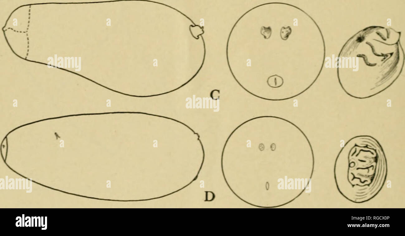 . Bulletin of the U.S. Department of Agriculture. Agriculture; Agriculture. Fig. 14.—Dexiid and tachinid puparia, showing their distinguishing characters: A, Daia centralis; B, Prostna sibenta; C, Ochromeigenia ormioides; D, CmUta cinerta tion of individuals was the same in each ovary. An average of 11 mature eggs was found in the gravid females'examined. A single egg was always present in the ovarian sac, one in one or both of the ovarian tubes, and a varying total in the follicles. Each follicle normally contains one fuliy developed egg, one slightly more *han half size, and a series of buds Stock Photohttps://www.alamy.com/image-license-details/?v=1https://www.alamy.com/bulletin-of-the-us-department-of-agriculture-agriculture-agriculture-fig-14dexiid-and-tachinid-puparia-showing-their-distinguishing-characters-a-daia-centralis-b-prostna-sibenta-c-ochromeigenia-ormioides-d-cmuta-cinerta-tion-of-individuals-was-the-same-in-each-ovary-an-average-of-11-mature-eggs-was-found-in-the-gravid-femalesexamined-a-single-egg-was-always-present-in-the-ovarian-sac-one-in-one-or-both-of-the-ovarian-tubes-and-a-varying-total-in-the-follicles-each-follicle-normally-contains-one-fuliy-developed-egg-one-slightly-more-han-half-size-and-a-series-of-buds-image233853110.html
RMRGCX0P–. Bulletin of the U.S. Department of Agriculture. Agriculture; Agriculture. Fig. 14.—Dexiid and tachinid puparia, showing their distinguishing characters: A, Daia centralis; B, Prostna sibenta; C, Ochromeigenia ormioides; D, CmUta cinerta tion of individuals was the same in each ovary. An average of 11 mature eggs was found in the gravid females'examined. A single egg was always present in the ovarian sac, one in one or both of the ovarian tubes, and a varying total in the follicles. Each follicle normally contains one fuliy developed egg, one slightly more *han half size, and a series of buds
 . A laboratory manual and text-book of embryology. Embryology. Fig. 3.âSection of human ovary, including cortex; a, germinal epithel- ium of free surface; 6, tunica albugi- nea; c, peripheral stroma containing immature Graafian follicles (d); e, well-advanced follicle from whose wall membrana granulosa has partially separated; /, cavity of liquor folli- culi; g, ovum surrounded by cell-mass constituting cumulus oophorus (Pier- sol). M â Ktfm 1M,. Fig. 4.âSection of well-developed Graafian follicle Fig. 5.âOvary with mature Graafian from human embryo (von Herff); the enclosed ovum follicle abou Stock Photohttps://www.alamy.com/image-license-details/?v=1https://www.alamy.com/a-laboratory-manual-and-text-book-of-embryology-embryology-fig-3section-of-human-ovary-including-cortex-a-germinal-epithel-ium-of-free-surface-6-tunica-albugi-nea-c-peripheral-stroma-containing-immature-graafian-follicles-d-e-well-advanced-follicle-from-whose-wall-membrana-granulosa-has-partially-separated-cavity-of-liquor-folli-culi-g-ovum-surrounded-by-cell-mass-constituting-cumulus-oophorus-pier-sol-m-ktfm-1m-fig-4section-of-well-developed-graafian-follicle-fig-5ovary-with-mature-graafian-from-human-embryo-von-herff-the-enclosed-ovum-follicle-abou-image232351229.html
RMRE0EA5–. A laboratory manual and text-book of embryology. Embryology. Fig. 3.âSection of human ovary, including cortex; a, germinal epithel- ium of free surface; 6, tunica albugi- nea; c, peripheral stroma containing immature Graafian follicles (d); e, well-advanced follicle from whose wall membrana granulosa has partially separated; /, cavity of liquor folli- culi; g, ovum surrounded by cell-mass constituting cumulus oophorus (Pier- sol). M â Ktfm 1M,. Fig. 4.âSection of well-developed Graafian follicle Fig. 5.âOvary with mature Graafian from human embryo (von Herff); the enclosed ovum follicle abou
 . The Biological bulletin. Biology; Zoology; Biology; Marine Biology. Figure 7. Origin of the egg envelope and morphological changes that occur after egg-laying. (A) Surface layers investing a fertilized, mature egg (eg) in the ovary. El a' and Elb': two sublayers of the envelope, i.e.. vitelline membrane (vm). flc: follicle cell, y: yolk. (B) The egg envelope 5-10 min after egg-laying. Electron- dense materials seem to be secreted by the embryos (small arrows). (C) The egg envelope about 10 min after egg-laying. Sublayer Elb' begins to fuse to Ela'. The envelope i.s adhesive, but plasticity i Stock Photohttps://www.alamy.com/image-license-details/?v=1https://www.alamy.com/the-biological-bulletin-biology-zoology-biology-marine-biology-figure-7-origin-of-the-egg-envelope-and-morphological-changes-that-occur-after-egg-laying-a-surface-layers-investing-a-fertilized-mature-egg-eg-in-the-ovary-el-a-and-elb-two-sublayers-of-the-envelope-ie-vitelline-membrane-vm-flc-follicle-cell-y-yolk-b-the-egg-envelope-5-10-min-after-egg-laying-electron-dense-materials-seem-to-be-secreted-by-the-embryos-small-arrows-c-the-egg-envelope-about-10-min-after-egg-laying-sublayer-elb-begins-to-fuse-to-ela-the-envelope-is-adhesive-but-plasticity-i-image234614918.html
RMRHKHM6–. The Biological bulletin. Biology; Zoology; Biology; Marine Biology. Figure 7. Origin of the egg envelope and morphological changes that occur after egg-laying. (A) Surface layers investing a fertilized, mature egg (eg) in the ovary. El a' and Elb': two sublayers of the envelope, i.e.. vitelline membrane (vm). flc: follicle cell, y: yolk. (B) The egg envelope 5-10 min after egg-laying. Electron- dense materials seem to be secreted by the embryos (small arrows). (C) The egg envelope about 10 min after egg-laying. Sublayer Elb' begins to fuse to Ela'. The envelope i.s adhesive, but plasticity i
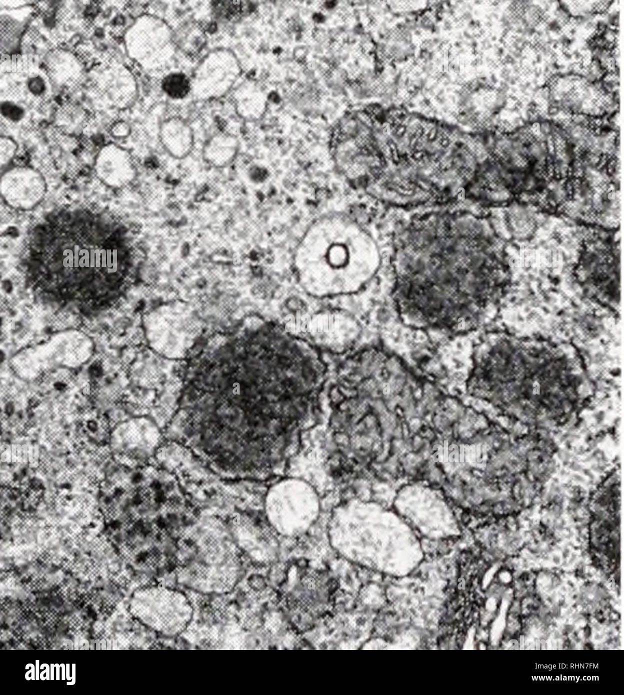 . The Biological bulletin. Biology; Zoology; Biology; Marine Biology. ^;fv^ P?." ':^ ^ "— f-'.?f ^ >-"'!. -*•;• ,?*. r r'"l'-;• -, FIGURE 4. Ovary from an immature female collected in mid-August. Premolt condition is unknown. Many small oocytes are apparent. FIGURE 5. Oocyte from mature female brooding young. The oocyte (O) is surrounded by forming egg coat (EC) and follicle cells (FC). FIGURE 6. Mature oocytes from bright orange ovary of female at the end of the breeding season. Follicle cells (FC) still surround the oocyte and egg coat (EC) has only partially formed. F Stock Photohttps://www.alamy.com/image-license-details/?v=1https://www.alamy.com/the-biological-bulletin-biology-zoology-biology-marine-biology-fv-pquot-quot-f-f-gt-quot!-r-rquotl-figure-4-ovary-from-an-immature-female-collected-in-mid-august-premolt-condition-is-unknown-many-small-oocytes-are-apparent-figure-5-oocyte-from-mature-female-brooding-young-the-oocyte-o-is-surrounded-by-forming-egg-coat-ec-and-follicle-cells-fc-figure-6-mature-oocytes-from-bright-orange-ovary-of-female-at-the-end-of-the-breeding-season-follicle-cells-fc-still-surround-the-oocyte-and-egg-coat-ec-has-only-partially-formed-f-image234650856.html
RMRHN7FM–. The Biological bulletin. Biology; Zoology; Biology; Marine Biology. ^;fv^ P?." ':^ ^ "— f-'.?f ^ >-"'!. -*•;• ,?*. r r'"l'-;• -, FIGURE 4. Ovary from an immature female collected in mid-August. Premolt condition is unknown. Many small oocytes are apparent. FIGURE 5. Oocyte from mature female brooding young. The oocyte (O) is surrounded by forming egg coat (EC) and follicle cells (FC). FIGURE 6. Mature oocytes from bright orange ovary of female at the end of the breeding season. Follicle cells (FC) still surround the oocyte and egg coat (EC) has only partially formed. F
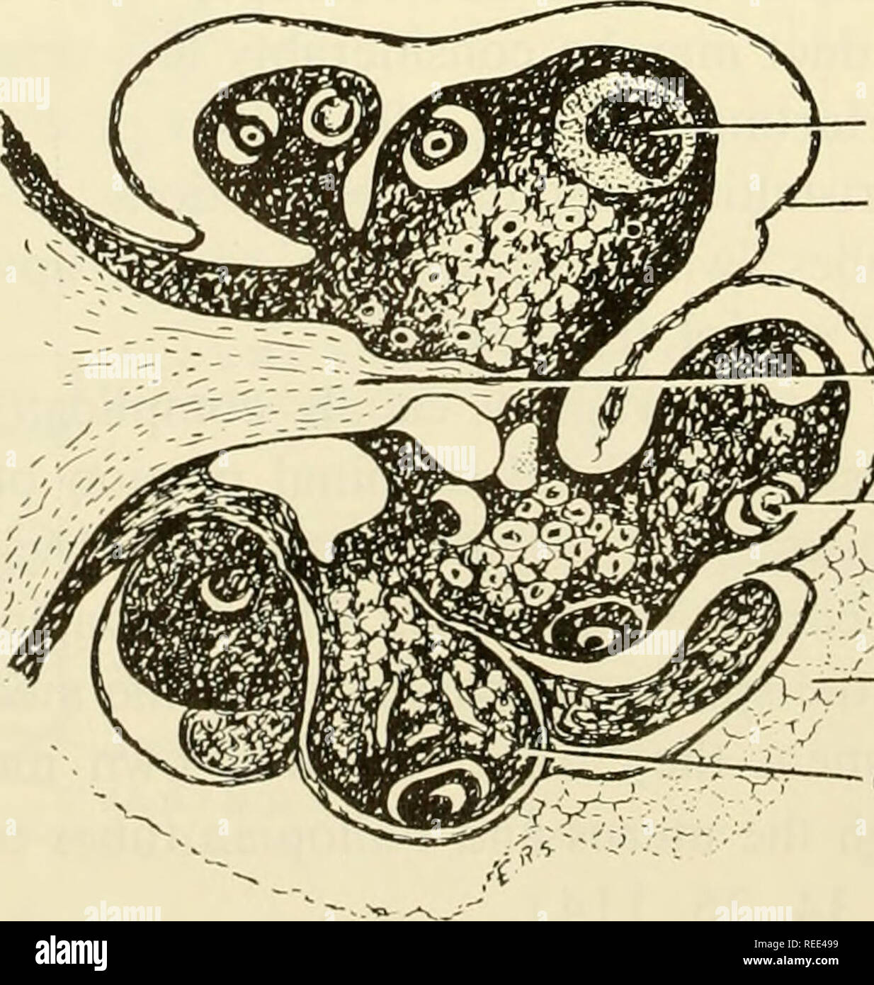 . Comparative embryology of the vertebrates; with 2057 drawings and photos. grouped as 380 illus. Vertebrates -- Embryology; Comparative embryology. SUSPENSORY LIGAMENT FA LL 0 PI A N TUBE HNFUNDI BULUM U TE R US OVARY LIGAMENT Fig. 111. Dorsal view of anterior end of uterine horn of the common opossum, Didelphys virginiana, showing relation of ovary to infundibulum.. CORPUS LUTEUM CAPSULE i—H I L U S ) /— FOLLICLE ^FAT T Y TISSUE OVARIAN LOBE Fig. 112. Section through ovary of mature rat, showing lobed condition and ovarian capsule. (Adapted from Heys, Quart. Rev. Biol., VI.) 194. Please note Stock Photohttps://www.alamy.com/image-license-details/?v=1https://www.alamy.com/comparative-embryology-of-the-vertebrates-with-2057-drawings-and-photos-grouped-as-380-illus-vertebrates-embryology-comparative-embryology-suspensory-ligament-fa-ll-0-pi-a-n-tube-hnfundi-bulum-u-te-r-us-ovary-ligament-fig-111-dorsal-view-of-anterior-end-of-uterine-horn-of-the-common-opossum-didelphys-virginiana-showing-relation-of-ovary-to-infundibulum-corpus-luteum-capsule-ih-i-l-u-s-follicle-fat-t-y-tissue-ovarian-lobe-fig-112-section-through-ovary-of-mature-rat-showing-lobed-condition-and-ovarian-capsule-adapted-from-heys-quart-rev-biol-vi-194-please-note-image232650693.html
RMREE499–. Comparative embryology of the vertebrates; with 2057 drawings and photos. grouped as 380 illus. Vertebrates -- Embryology; Comparative embryology. SUSPENSORY LIGAMENT FA LL 0 PI A N TUBE HNFUNDI BULUM U TE R US OVARY LIGAMENT Fig. 111. Dorsal view of anterior end of uterine horn of the common opossum, Didelphys virginiana, showing relation of ovary to infundibulum.. CORPUS LUTEUM CAPSULE i—H I L U S ) /— FOLLICLE ^FAT T Y TISSUE OVARIAN LOBE Fig. 112. Section through ovary of mature rat, showing lobed condition and ovarian capsule. (Adapted from Heys, Quart. Rev. Biol., VI.) 194. Please note
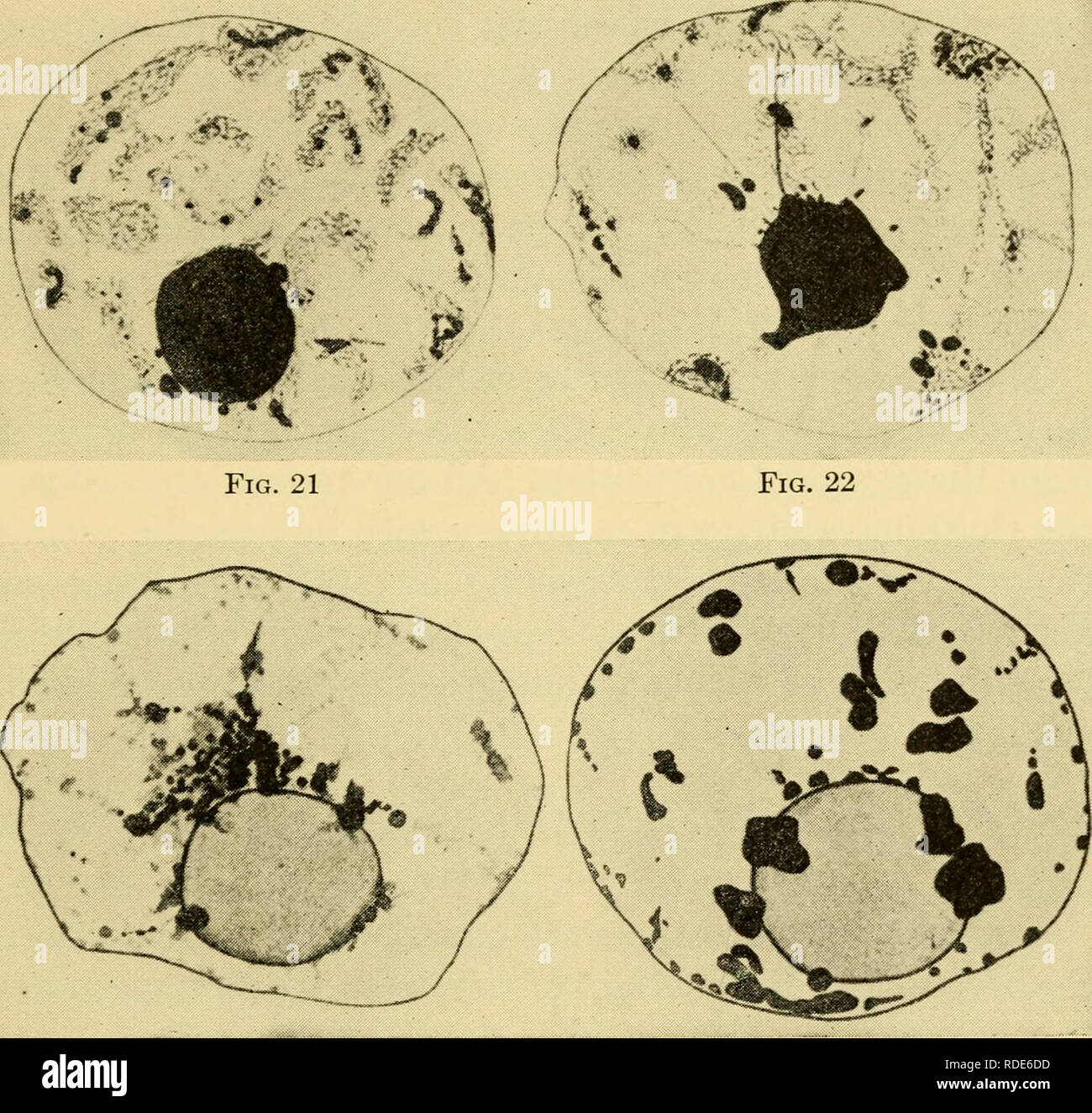 . The eggs of mammals. Ovum; Embryology -- Mammals; Mammals -- physiology; Ovum. Fig. 17 Fig. 18 Fig. 19 Fig. 20. Fig. 23 Fig. 24 Plate II. (From the Journal of Morphology) Figs. 17-22. Nuclei of ova from ovary of rat 20 days post partum. 17, Deuto- broch nucleus. 18, Beginning of the formation of clumps shown in next figure. 19, Modified pachynema. 20, Later stage showing characters of diplonema. 21, Nu- cleus toward the end of the growth period. 22, Final stage in twenty-day rat. Fig. 23. Nucleus in mature follicle from adult rat. Fig. 24. Nucleus from ripe follicle from adult rat. 13. Pleas Stock Photohttps://www.alamy.com/image-license-details/?v=1https://www.alamy.com/the-eggs-of-mammals-ovum-embryology-mammals-mammals-physiology-ovum-fig-17-fig-18-fig-19-fig-20-fig-23-fig-24-plate-ii-from-the-journal-of-morphology-figs-17-22-nuclei-of-ova-from-ovary-of-rat-20-days-post-partum-17-deuto-broch-nucleus-18-beginning-of-the-formation-of-clumps-shown-in-next-figure-19-modified-pachynema-20-later-stage-showing-characters-of-diplonema-21-nu-cleus-toward-the-end-of-the-growth-period-22-final-stage-in-twenty-day-rat-fig-23-nucleus-in-mature-follicle-from-adult-rat-fig-24-nucleus-from-ripe-follicle-from-adult-rat-13-pleas-image232037721.html
RMRDE6DD–. The eggs of mammals. Ovum; Embryology -- Mammals; Mammals -- physiology; Ovum. Fig. 17 Fig. 18 Fig. 19 Fig. 20. Fig. 23 Fig. 24 Plate II. (From the Journal of Morphology) Figs. 17-22. Nuclei of ova from ovary of rat 20 days post partum. 17, Deuto- broch nucleus. 18, Beginning of the formation of clumps shown in next figure. 19, Modified pachynema. 20, Later stage showing characters of diplonema. 21, Nu- cleus toward the end of the growth period. 22, Final stage in twenty-day rat. Fig. 23. Nucleus in mature follicle from adult rat. Fig. 24. Nucleus from ripe follicle from adult rat. 13. Pleas