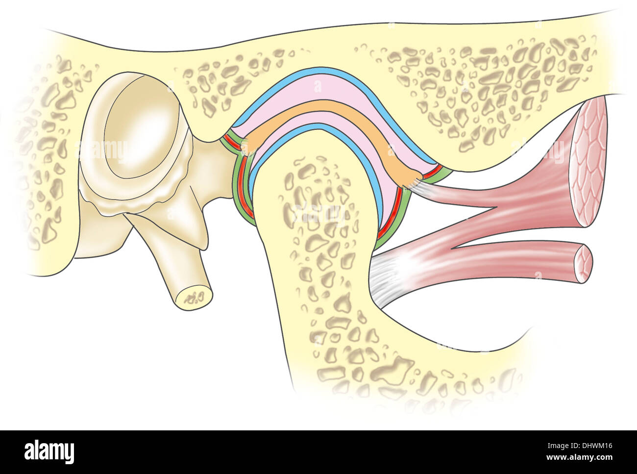
Maxillary jaw Cut Out Stock Images
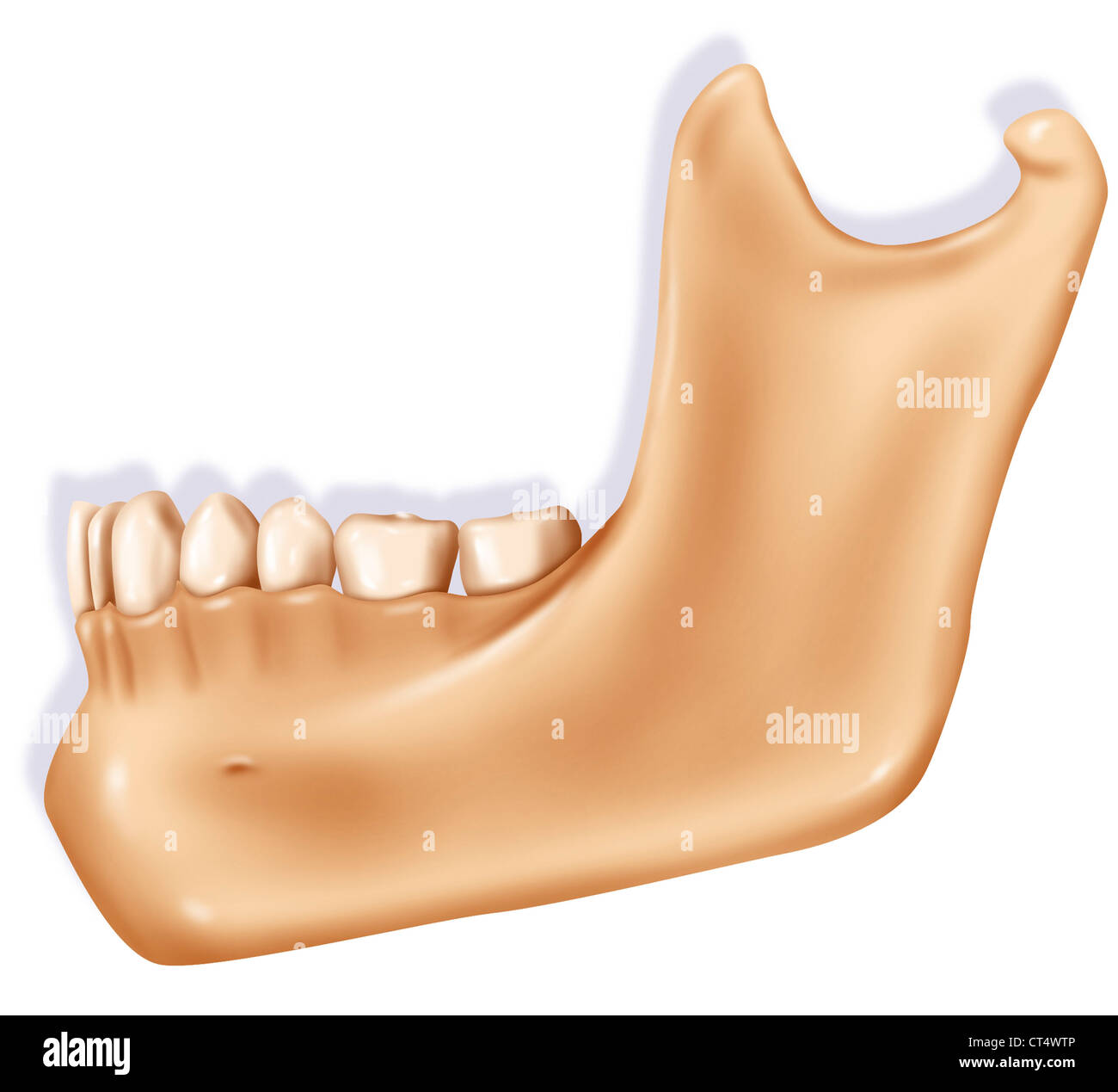 JAW, ILLUSTRATION Stock Photohttps://www.alamy.com/image-license-details/?v=1https://www.alamy.com/stock-photo-jaw-illustration-49280582.html
RMCT4WTP–JAW, ILLUSTRATION
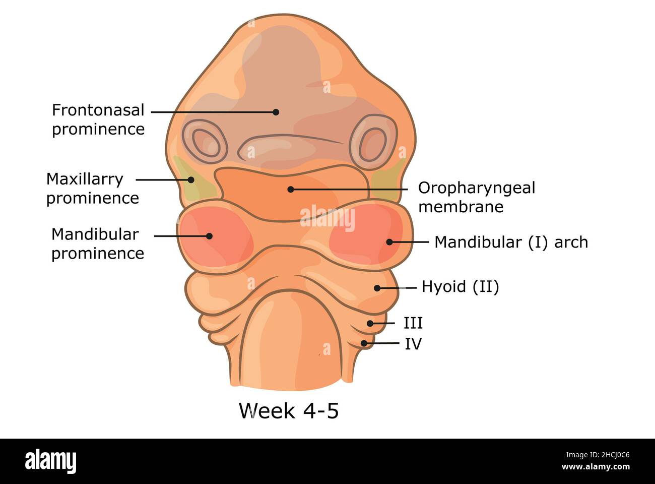 Development of external structures of human face week 4 - 5. Stock Photohttps://www.alamy.com/image-license-details/?v=1https://www.alamy.com/development-of-external-structures-of-human-face-week-4-5-image455240918.html
RF2HCJ0C6–Development of external structures of human face week 4 - 5.
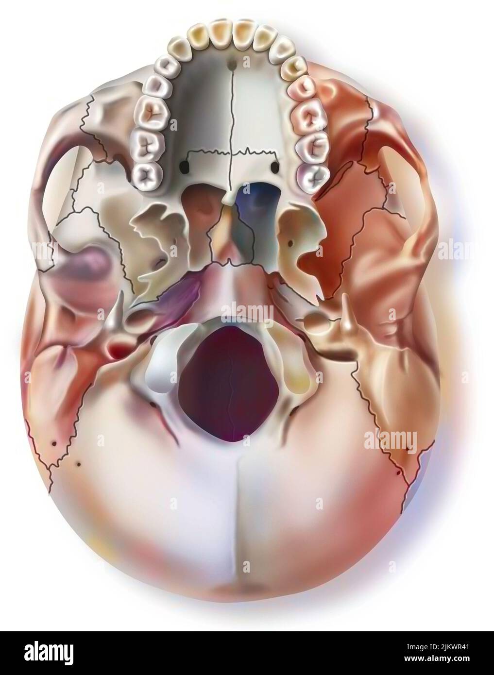 Inferior view of the human skull with the upper jaw. Stock Photohttps://www.alamy.com/image-license-details/?v=1https://www.alamy.com/inferior-view-of-the-human-skull-with-the-upper-jaw-image476925345.html
RF2JKWR41–Inferior view of the human skull with the upper jaw.
 Mesial bite profile before and after orthodontic treatment. Human with malocclusion, lower jaw extended forward, bite correction by braces. Vector Stock Vectorhttps://www.alamy.com/image-license-details/?v=1https://www.alamy.com/mesial-bite-profile-before-and-after-orthodontic-treatment-human-with-malocclusion-lower-jaw-extended-forward-bite-correction-by-braces-vector-image366914184.html
RF2C8XAP0–Mesial bite profile before and after orthodontic treatment. Human with malocclusion, lower jaw extended forward, bite correction by braces. Vector
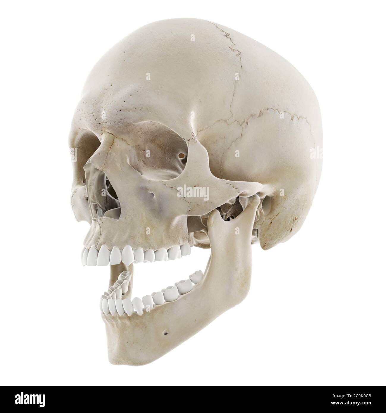 Skull with open jaw, illustration. Stock Photohttps://www.alamy.com/image-license-details/?v=1https://www.alamy.com/skull-with-open-jaw-illustration-image367367067.html
RF2C9K0CB–Skull with open jaw, illustration.
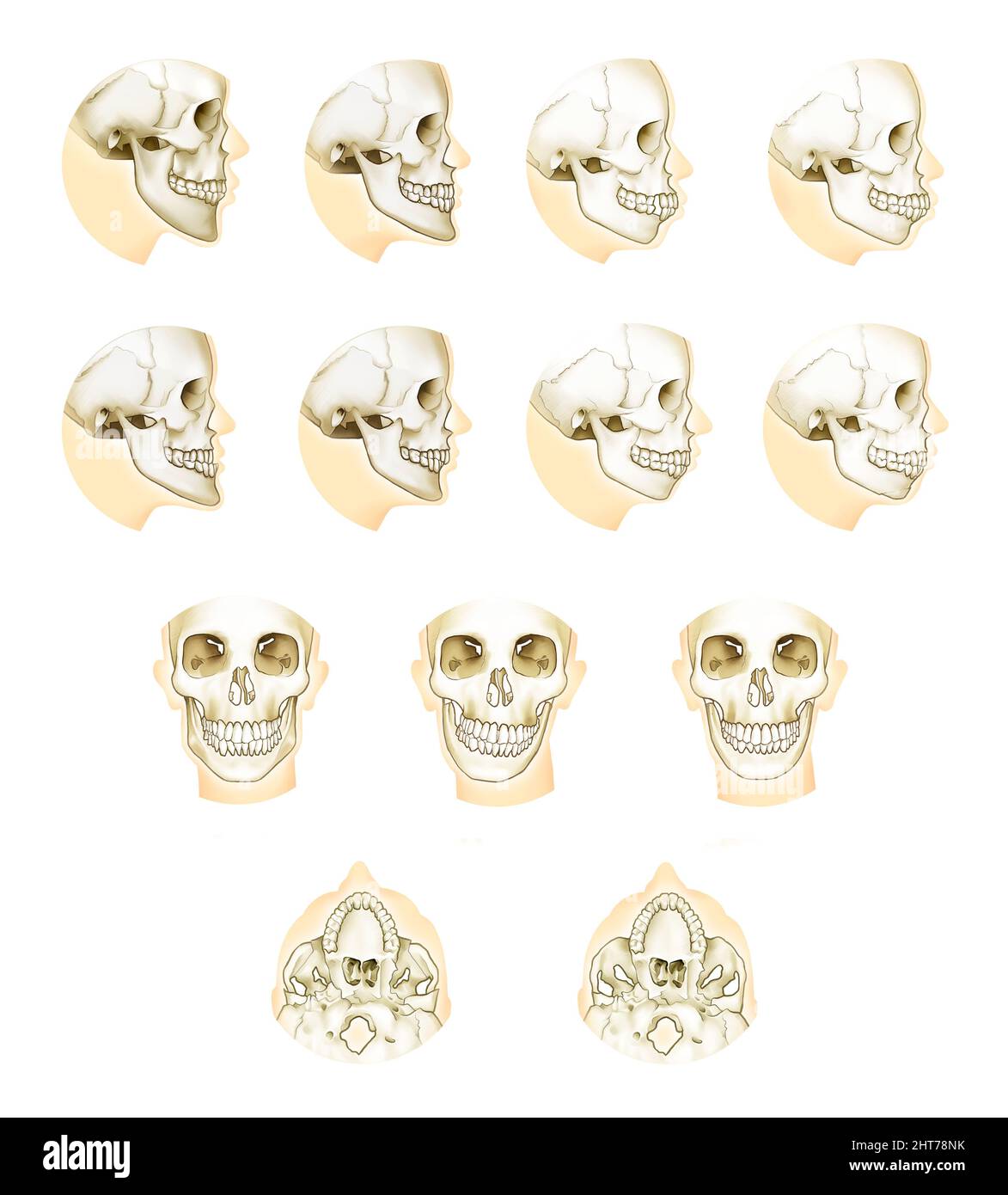 Realistic illustration of orthodontic double jaw surgery anatomy Stock Photohttps://www.alamy.com/image-license-details/?v=1https://www.alamy.com/realistic-illustration-of-orthodontic-double-jaw-surgery-anatomy-image462381855.html
RF2HT78NK–Realistic illustration of orthodontic double jaw surgery anatomy
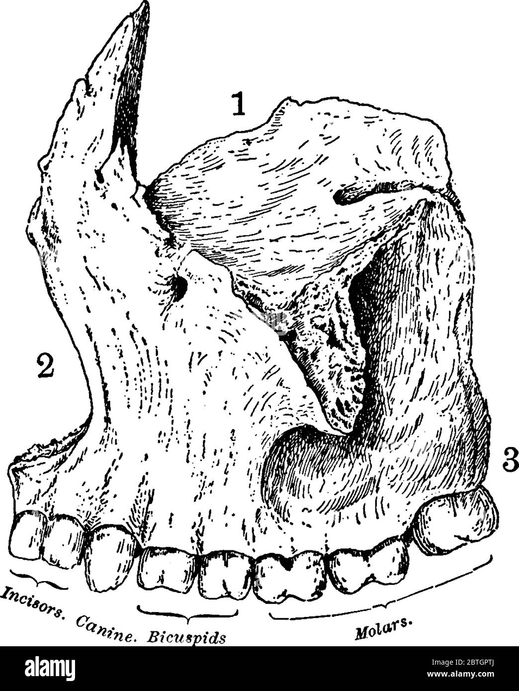 Superior maxillary bone together with its fellow on the opposite side, forms the whole of the upper jaw, with its parts labelled, vintage line drawing Stock Vectorhttps://www.alamy.com/image-license-details/?v=1https://www.alamy.com/superior-maxillary-bone-together-with-its-fellow-on-the-opposite-side-forms-the-whole-of-the-upper-jaw-with-its-parts-labelled-vintage-line-drawing-image359328274.html
RF2BTGPTJ–Superior maxillary bone together with its fellow on the opposite side, forms the whole of the upper jaw, with its parts labelled, vintage line drawing
 Dental roots in maxillary sinus. Medical illustration in flat style. Stock Vectorhttps://www.alamy.com/image-license-details/?v=1https://www.alamy.com/dental-roots-in-maxillary-sinus-medical-illustration-in-flat-style-image502009683.html
RF2M4MED7–Dental roots in maxillary sinus. Medical illustration in flat style.
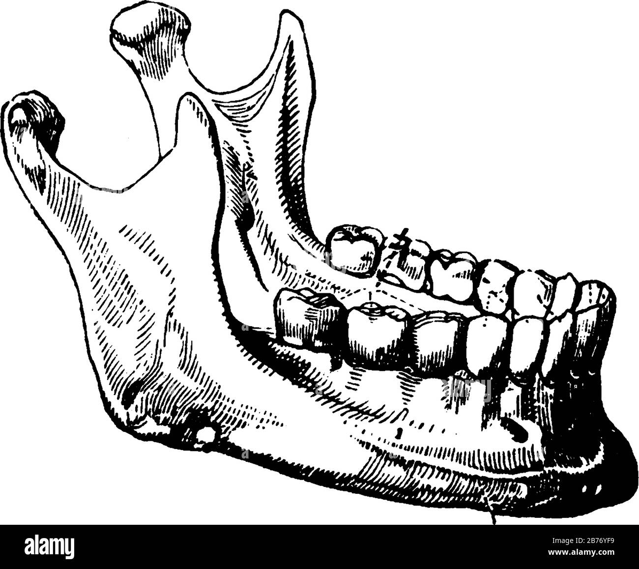 The term, mandibular, is given to teeth in the lower jaw. Adult mouth has 32 teeth, the middlemost four teeth on the lower jaws, vintage line drawing Stock Vectorhttps://www.alamy.com/image-license-details/?v=1https://www.alamy.com/the-term-mandibular-is-given-to-teeth-in-the-lower-jaw-adult-mouth-has-32-teeth-the-middlemost-four-teeth-on-the-lower-jaws-vintage-line-drawing-image348663261.html
RF2B76YF9–The term, mandibular, is given to teeth in the lower jaw. Adult mouth has 32 teeth, the middlemost four teeth on the lower jaws, vintage line drawing
 3d rendered medically accurate illustration of the skull with open jaw Stock Photohttps://www.alamy.com/image-license-details/?v=1https://www.alamy.com/3d-rendered-medically-accurate-illustration-of-the-skull-with-open-jaw-image334071384.html
RF2ABE7CT–3d rendered medically accurate illustration of the skull with open jaw
 3d render of jaw with lidocaine and syringe over white background. Dental anesthesia concept. Lidocaine is an organic chemical compound used as a loc Stock Photohttps://www.alamy.com/image-license-details/?v=1https://www.alamy.com/3d-render-of-jaw-with-lidocaine-and-syringe-over-white-background-dental-anesthesia-concept-lidocaine-is-an-organic-chemical-compound-used-as-a-loc-image231482462.html
RFRCGX6P–3d render of jaw with lidocaine and syringe over white background. Dental anesthesia concept. Lidocaine is an organic chemical compound used as a loc
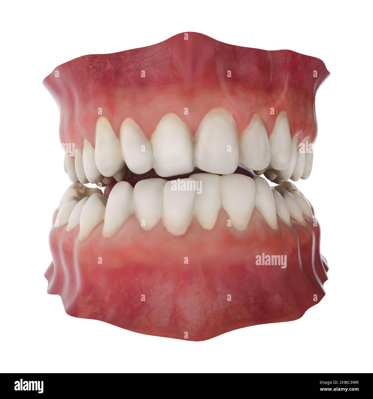 Jaw with abnormal teeth position. Orthodontic treatment concept. Realistic 3D illustration Stock Photohttps://www.alamy.com/image-license-details/?v=1https://www.alamy.com/jaw-with-abnormal-teeth-position-orthodontic-treatment-concept-realistic-3d-illustration-image454497283.html
RF2HBC3WR–Jaw with abnormal teeth position. Orthodontic treatment concept. Realistic 3D illustration
 Teeth mould dentist plaster 3D impression denture dentures lower jaw Cutout cut out white background isolated copy space Orthodo Stock Photohttps://www.alamy.com/image-license-details/?v=1https://www.alamy.com/stock-photo-teeth-mould-dentist-plaster-3d-impression-denture-dentures-lower-jaw-92299427.html
RMFA4GT3–Teeth mould dentist plaster 3D impression denture dentures lower jaw Cutout cut out white background isolated copy space Orthodo
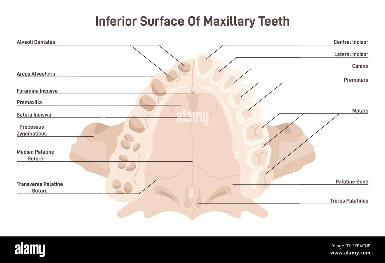 Maxillary anatomy. Inferior surface of upper jaw skeletal structure with teeth. Dentistry infographic. Flat vector illustration Stock Vectorhttps://www.alamy.com/image-license-details/?v=1https://www.alamy.com/maxillary-anatomy-inferior-surface-of-upper-jaw-skeletal-structure-with-teeth-dentistry-infographic-flat-vector-illustration-image609353514.html
RF2XBACHE–Maxillary anatomy. Inferior surface of upper jaw skeletal structure with teeth. Dentistry infographic. Flat vector illustration
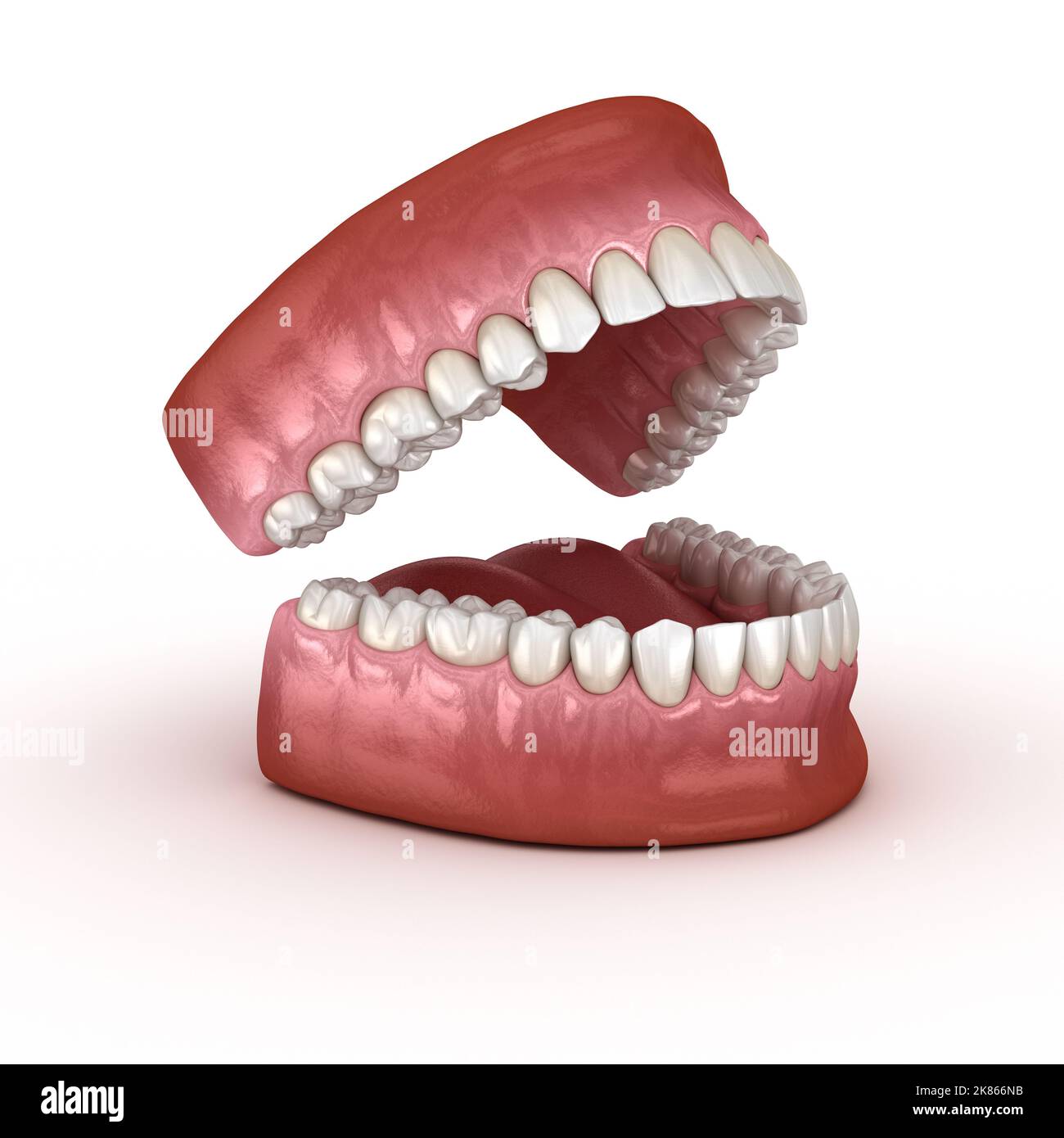 Dental anatomy - Opened Dentures. Medically accurate dental 3D illustration Stock Photohttps://www.alamy.com/image-license-details/?v=1https://www.alamy.com/dental-anatomy-opened-dentures-medically-accurate-dental-3d-illustration-image486944567.html
RF2K866NB–Dental anatomy - Opened Dentures. Medically accurate dental 3D illustration
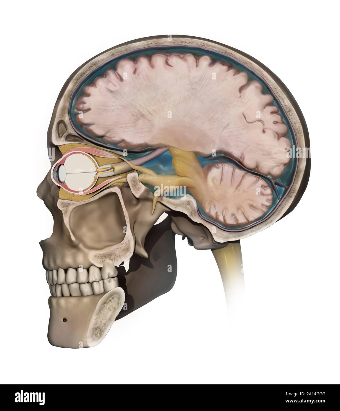 Medical illustration depicting the anatomy of a sagittal section of the human cranium. Stock Photohttps://www.alamy.com/image-license-details/?v=1https://www.alamy.com/medical-illustration-depicting-the-anatomy-of-a-sagittal-section-of-the-human-cranium-image327712464.html
RF2A14GGG–Medical illustration depicting the anatomy of a sagittal section of the human cranium.
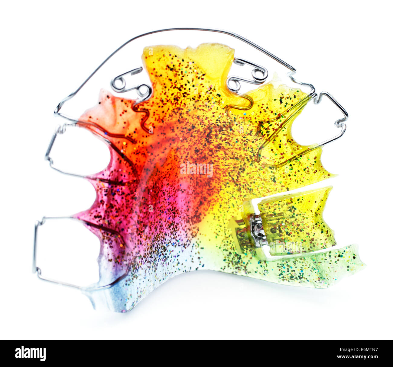 Multicolored and glittered upper orthodontic appliance with two z-springs, 3 clasps, labial bow and an expansion screw Stock Photohttps://www.alamy.com/image-license-details/?v=1https://www.alamy.com/stock-photo-multicolored-and-glittered-upper-orthodontic-appliance-with-two-z-72987859.html
RFE6MTN7–Multicolored and glittered upper orthodontic appliance with two z-springs, 3 clasps, labial bow and an expansion screw
 ..The fishes of Illinois . very band at base; ventrals and pectorals a smoky greenishgray. Head small, slender, subconic, its length 3.6 to 4 in body, itsgreatest depth less than in /. anguilla, 4.9 to 5 .2 in body; interorbital spaceflat or slightly convex; occipital region and shoulders gently rounded andcovered with thin, close-fitting skin; mouth more nearly terminal than inanguilla, the upper jaw only slightly longer than the lower; lips somewhatthicker than in preceding species; maxillary barbels long and slender,reaching past gill-opening; eye oval, lying above median axis of body andne Stock Photohttps://www.alamy.com/image-license-details/?v=1https://www.alamy.com/the-fishes-of-illinois-very-band-at-base-ventrals-and-pectorals-a-smoky-greenishgray-head-small-slender-subconic-its-length-36-to-4-in-body-itsgreatest-depth-less-than-in-anguilla-49-to-5-2-in-body-interorbital-spaceflat-or-slightly-convex-occipital-region-and-shoulders-gently-rounded-andcovered-with-thin-close-fitting-skin-mouth-more-nearly-terminal-than-inanguilla-the-upper-jaw-only-slightly-longer-than-the-lower-lips-somewhatthicker-than-in-preceding-species-maxillary-barbels-long-and-slenderreaching-past-gill-opening-eye-oval-lying-above-median-axis-of-body-andne-image339961595.html
RM2AN2GDF–..The fishes of Illinois . very band at base; ventrals and pectorals a smoky greenishgray. Head small, slender, subconic, its length 3.6 to 4 in body, itsgreatest depth less than in /. anguilla, 4.9 to 5 .2 in body; interorbital spaceflat or slightly convex; occipital region and shoulders gently rounded andcovered with thin, close-fitting skin; mouth more nearly terminal than inanguilla, the upper jaw only slightly longer than the lower; lips somewhatthicker than in preceding species; maxillary barbels long and slender,reaching past gill-opening; eye oval, lying above median axis of body andne
RF2DGAEBY–incicor icon, black vector sign with editable strokes, concept illustration
 Dentures on a white background. Acrylic denture on white background. Full denture close-up. Full removable plastic denture of the jaws. Isolate on whi Stock Photohttps://www.alamy.com/image-license-details/?v=1https://www.alamy.com/dentures-on-a-white-background-acrylic-denture-on-white-background-full-denture-close-up-full-removable-plastic-denture-of-the-jaws-isolate-on-whi-image459129785.html
RF2HJY4M9–Dentures on a white background. Acrylic denture on white background. Full denture close-up. Full removable plastic denture of the jaws. Isolate on whi
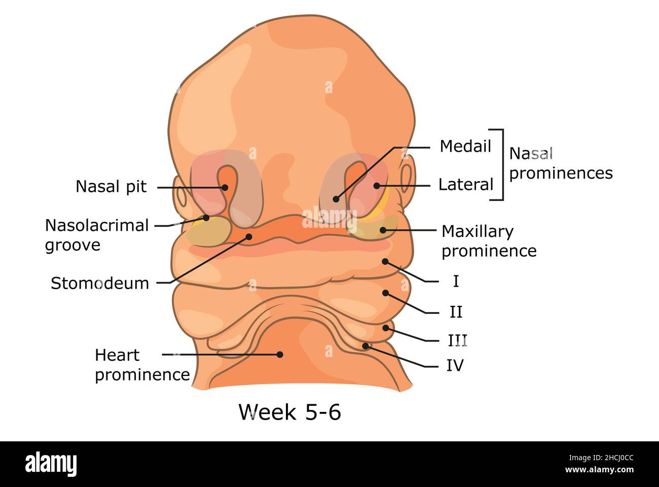 Development of external structures of human face week 5 - 6. Stock Photohttps://www.alamy.com/image-license-details/?v=1https://www.alamy.com/development-of-external-structures-of-human-face-week-5-6-image455240924.html
RF2HCJ0CC–Development of external structures of human face week 5 - 6.
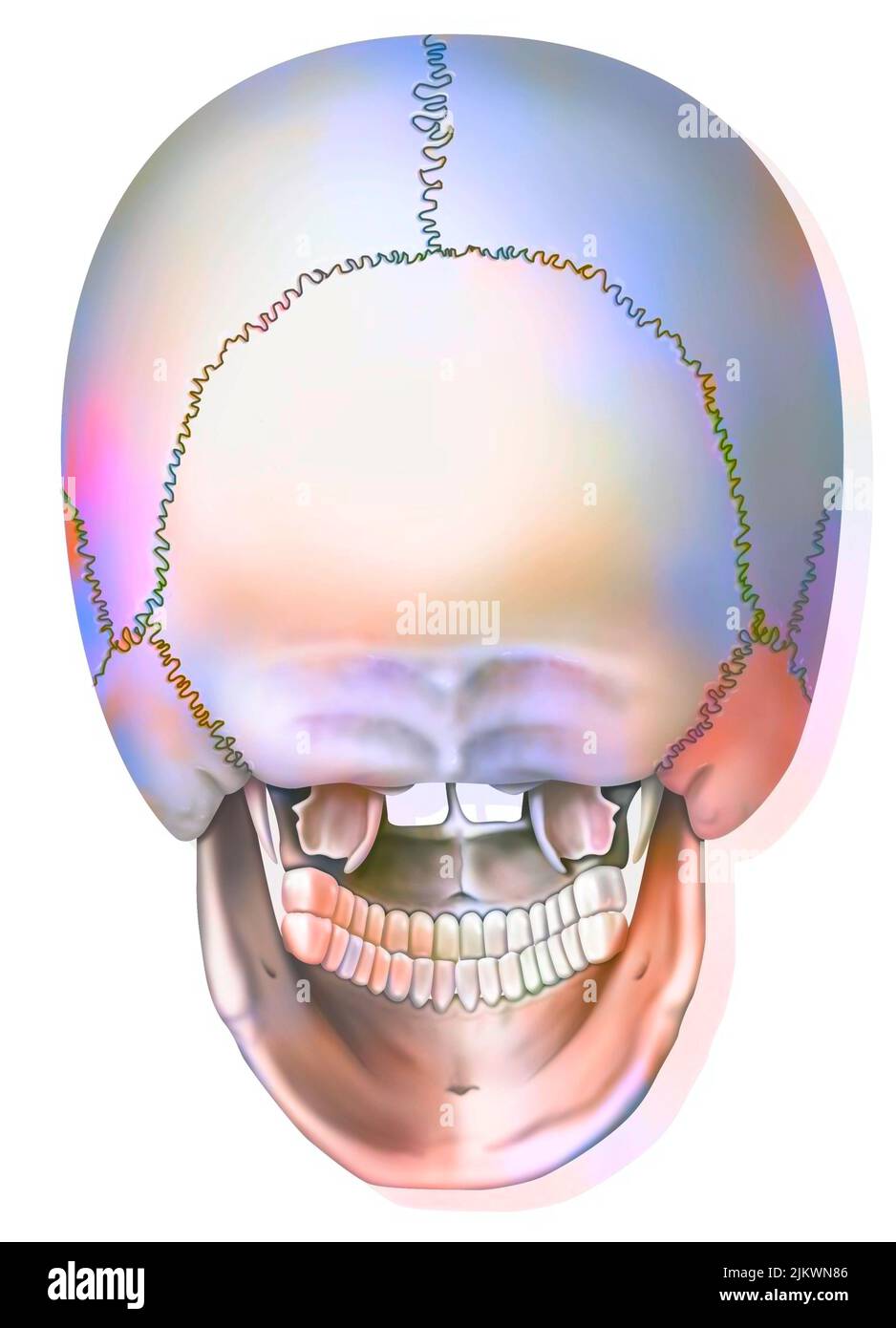 Posterior view of the bones of the skull and jaw. Stock Photohttps://www.alamy.com/image-license-details/?v=1https://www.alamy.com/posterior-view-of-the-bones-of-the-skull-and-jaw-image476923894.html
RF2JKWN86–Posterior view of the bones of the skull and jaw.
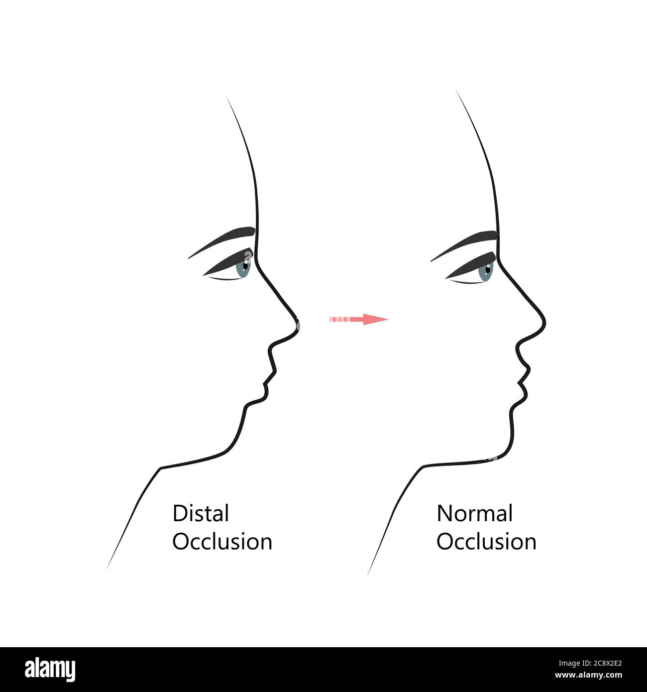 Distal bite profile before and after orthodontic treatment. Human with malocclusion, lower jaw pushing back, bite correction by braces. Vector Stock Vectorhttps://www.alamy.com/image-license-details/?v=1https://www.alamy.com/distal-bite-profile-before-and-after-orthodontic-treatment-human-with-malocclusion-lower-jaw-pushing-back-bite-correction-by-braces-vector-image366907690.html
RF2C8X2E2–Distal bite profile before and after orthodontic treatment. Human with malocclusion, lower jaw pushing back, bite correction by braces. Vector
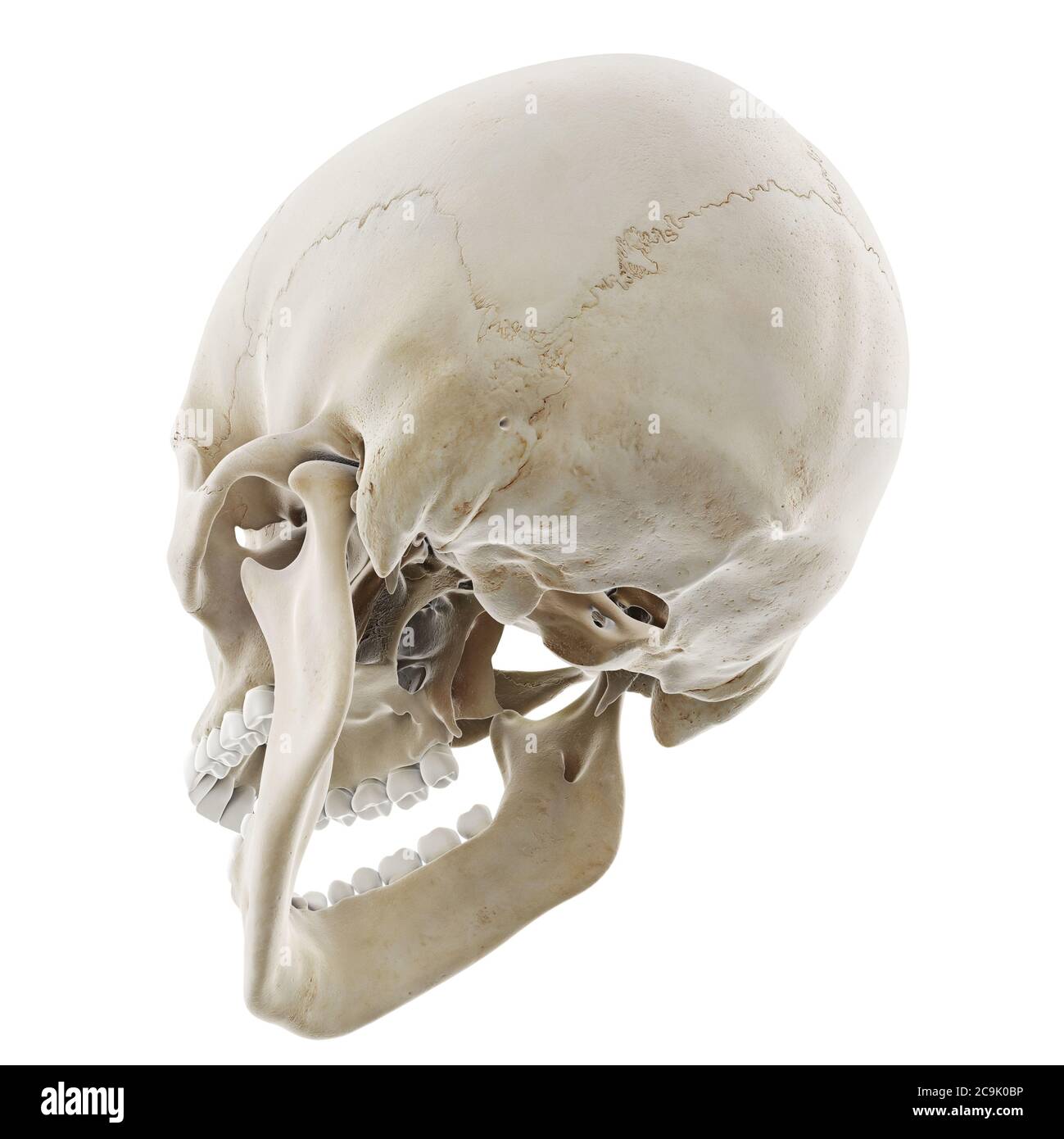 Skull with open jaw, illustration. Stock Photohttps://www.alamy.com/image-license-details/?v=1https://www.alamy.com/skull-with-open-jaw-illustration-image367367050.html
RF2C9K0BP–Skull with open jaw, illustration.
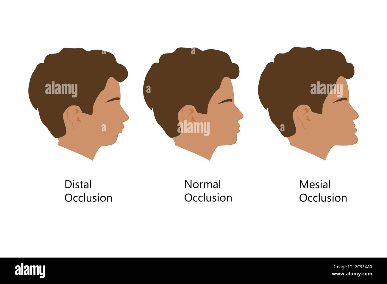 Guy with Distal, Normal, and Mesial bite profile, vector illustration. Overbite or underbite before and after orthodontic treatment. Human with Stock Vectorhttps://www.alamy.com/image-license-details/?v=1https://www.alamy.com/guy-with-distal-normal-and-mesial-bite-profile-vector-illustration-overbite-or-underbite-before-and-after-orthodontic-treatment-human-with-image367036152.html
RF2C93XA0–Guy with Distal, Normal, and Mesial bite profile, vector illustration. Overbite or underbite before and after orthodontic treatment. Human with
 . The families and genera of bats . Bats; Bats. 66 BULLETIN 51, UNITED STATES NATIONAL MUSEUM.. from canines by narrow spaces, their crowns bifid or notched, rather distinctly marked off from shafts, sometimes by an evident con- striction.0 Canines small and weak, not peculiar in form, without secondary cusps or distinct ridges, the anterior surface without trace of longitudinal furrows. Except that the mandibular canines are smaller, they are almost identical in appearance with those of the upper jaw. No small maxillary premolar or molar (pm2, m2). The remaining maxil- lary teeth (pms, pm4, a Stock Photohttps://www.alamy.com/image-license-details/?v=1https://www.alamy.com/the-families-and-genera-of-bats-bats-bats-66-bulletin-51-united-states-national-museum-from-canines-by-narrow-spaces-their-crowns-bifid-or-notched-rather-distinctly-marked-off-from-shafts-sometimes-by-an-evident-con-striction0-canines-small-and-weak-not-peculiar-in-form-without-secondary-cusps-or-distinct-ridges-the-anterior-surface-without-trace-of-longitudinal-furrows-except-that-the-mandibular-canines-are-smaller-they-are-almost-identical-in-appearance-with-those-of-the-upper-jaw-no-small-maxillary-premolar-or-molar-pm2-m2-the-remaining-maxil-lary-teeth-pms-pm4-a-image216355969.html
RMPFYT6W–. The families and genera of bats . Bats; Bats. 66 BULLETIN 51, UNITED STATES NATIONAL MUSEUM.. from canines by narrow spaces, their crowns bifid or notched, rather distinctly marked off from shafts, sometimes by an evident con- striction.0 Canines small and weak, not peculiar in form, without secondary cusps or distinct ridges, the anterior surface without trace of longitudinal furrows. Except that the mandibular canines are smaller, they are almost identical in appearance with those of the upper jaw. No small maxillary premolar or molar (pm2, m2). The remaining maxil- lary teeth (pms, pm4, a
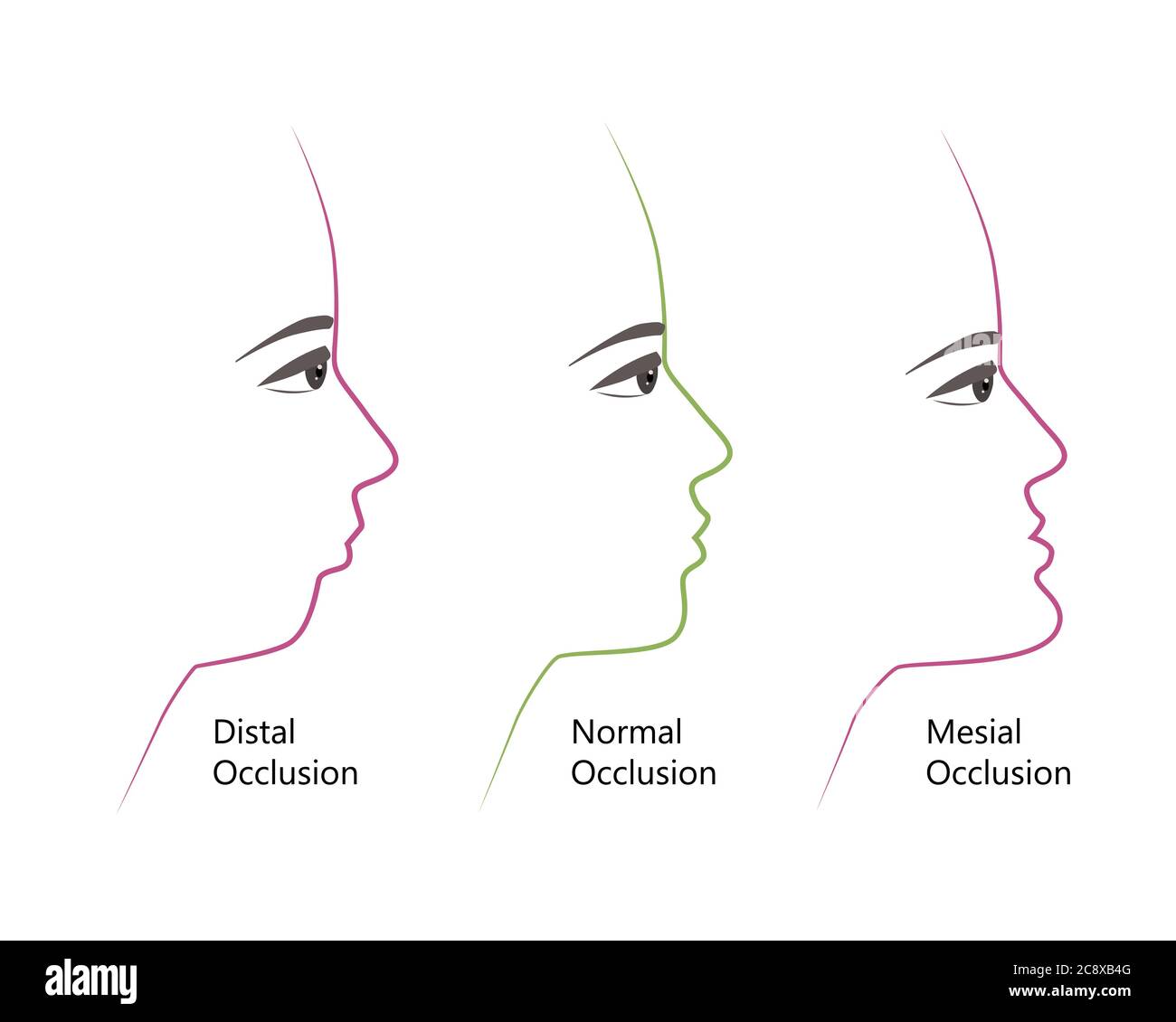 distal, Normal, and Mesial bite profile, vector illustration. Overbite or underbite before and after orthodontic treatment. Human with malocclusion Stock Vectorhttps://www.alamy.com/image-license-details/?v=1https://www.alamy.com/distal-normal-and-mesial-bite-profile-vector-illustration-overbite-or-underbite-before-and-after-orthodontic-treatment-human-with-malocclusion-image366914480.html
RF2C8XB4G–distal, Normal, and Mesial bite profile, vector illustration. Overbite or underbite before and after orthodontic treatment. Human with malocclusion
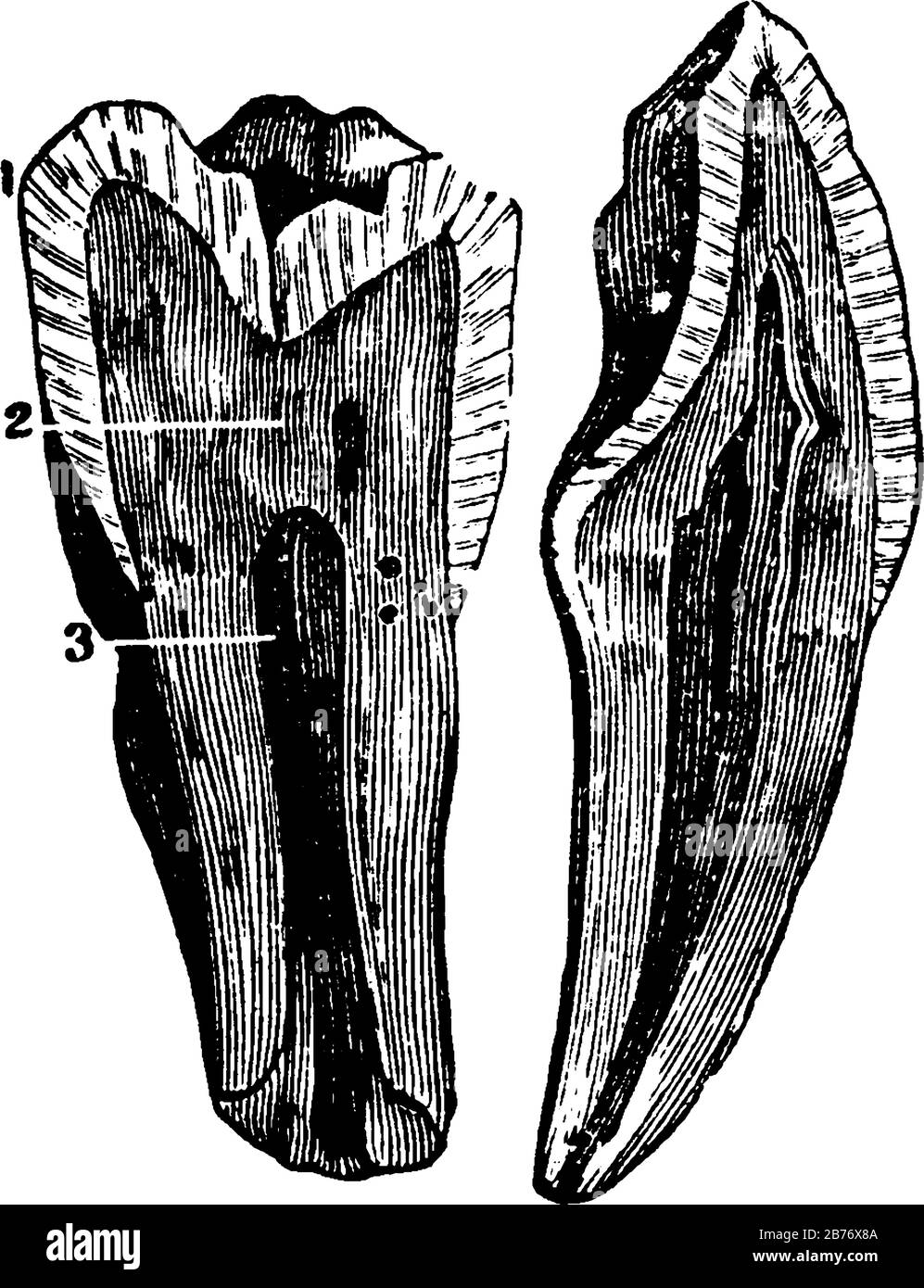 The internal view of a tooth cut through from the top or crown to the tips of the root, with the parts labelled, 1, enamel; 2, dentine; 3, pulp, vinta Stock Vectorhttps://www.alamy.com/image-license-details/?v=1https://www.alamy.com/the-internal-view-of-a-tooth-cut-through-from-the-top-or-crown-to-the-tips-of-the-root-with-the-parts-labelled-1-enamel-2-dentine-3-pulp-vinta-image348662282.html
RF2B76X8A–The internal view of a tooth cut through from the top or crown to the tips of the root, with the parts labelled, 1, enamel; 2, dentine; 3, pulp, vinta
 3d rendered medically accurate illustration of the skull with open jaw Stock Photohttps://www.alamy.com/image-license-details/?v=1https://www.alamy.com/3d-rendered-medically-accurate-illustration-of-the-skull-with-open-jaw-image334050055.html
RF2ABD873–3d rendered medically accurate illustration of the skull with open jaw
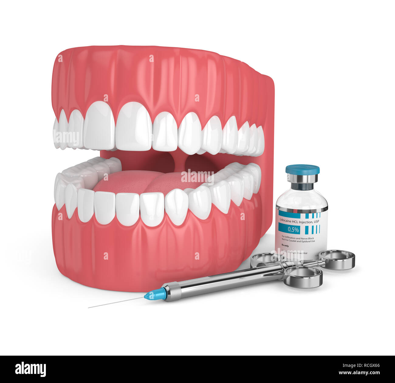 3d render of jaw with lidocaine and syringe over white background. Dental anesthesia concept. Lidocaine is an organic chemical compound used as a loc Stock Photohttps://www.alamy.com/image-license-details/?v=1https://www.alamy.com/3d-render-of-jaw-with-lidocaine-and-syringe-over-white-background-dental-anesthesia-concept-lidocaine-is-an-organic-chemical-compound-used-as-a-loc-image231482446.html
RFRCGX66–3d render of jaw with lidocaine and syringe over white background. Dental anesthesia concept. Lidocaine is an organic chemical compound used as a loc
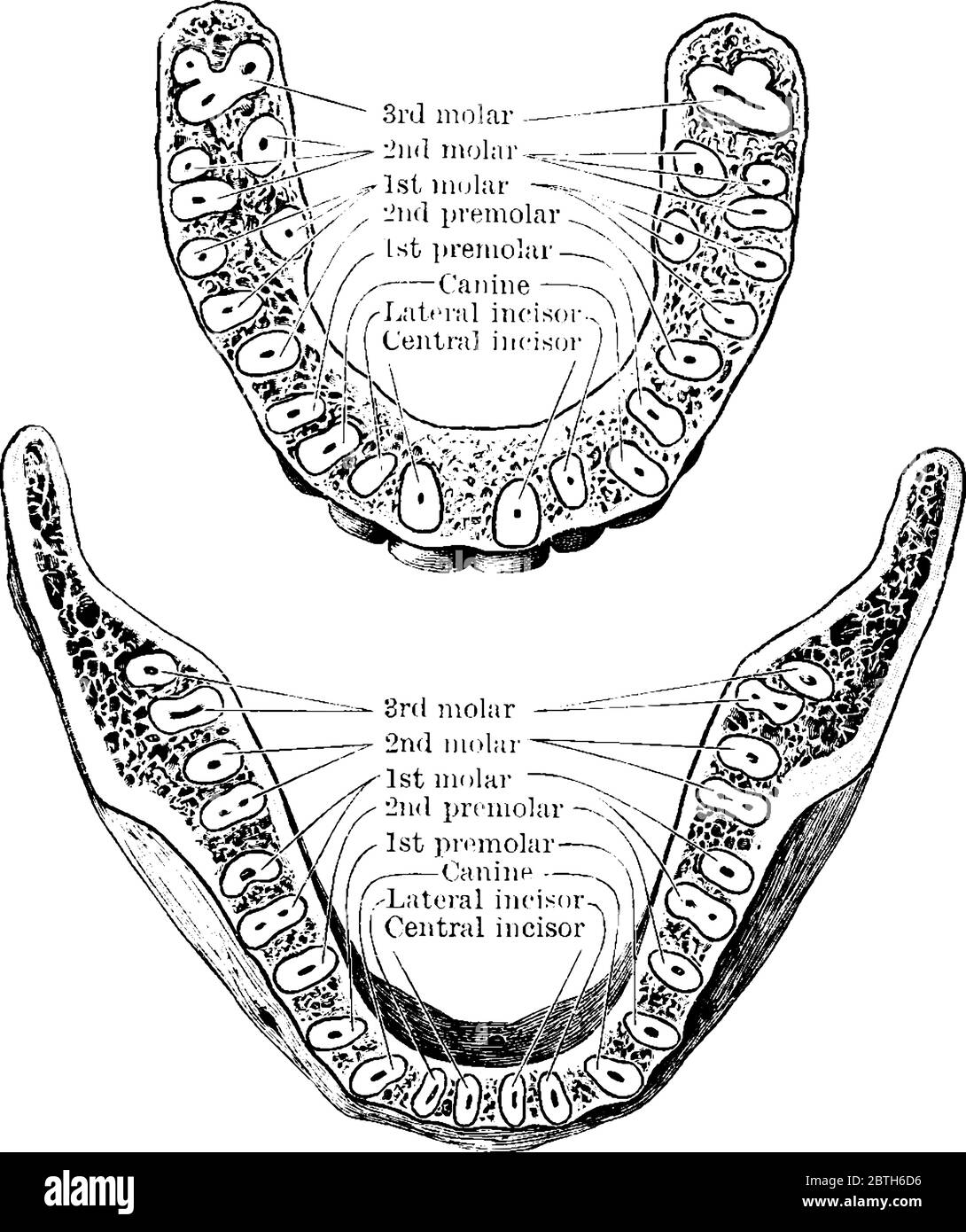 The horizontal section through both the upper and lower jaws, showing the roots of the teeth, vintage line drawing or engraving illustration. Stock Vectorhttps://www.alamy.com/image-license-details/?v=1https://www.alamy.com/the-horizontal-section-through-both-the-upper-and-lower-jaws-showing-the-roots-of-the-teeth-vintage-line-drawing-or-engraving-illustration-image359337362.html
RF2BTH6D6–The horizontal section through both the upper and lower jaws, showing the roots of the teeth, vintage line drawing or engraving illustration.
 Teeth mould dentist plaster impression 3D denture dentures lower jaw Cutout cut out white background isolated copy space Stock Photohttps://www.alamy.com/image-license-details/?v=1https://www.alamy.com/stock-photo-teeth-mould-dentist-plaster-impression-3d-denture-dentures-lower-jaw-92299428.html
RMFA4GT4–Teeth mould dentist plaster impression 3D denture dentures lower jaw Cutout cut out white background isolated copy space
 Shows the relation of the upper to the lower teeth, when the mouth is closed, vintage line drawing or engraving illustration. Stock Vectorhttps://www.alamy.com/image-license-details/?v=1https://www.alamy.com/shows-the-relation-of-the-upper-to-the-lower-teeth-when-the-mouth-is-closed-vintage-line-drawing-or-engraving-illustration-image359338242.html
RF2BTH7GJ–Shows the relation of the upper to the lower teeth, when the mouth is closed, vintage line drawing or engraving illustration.
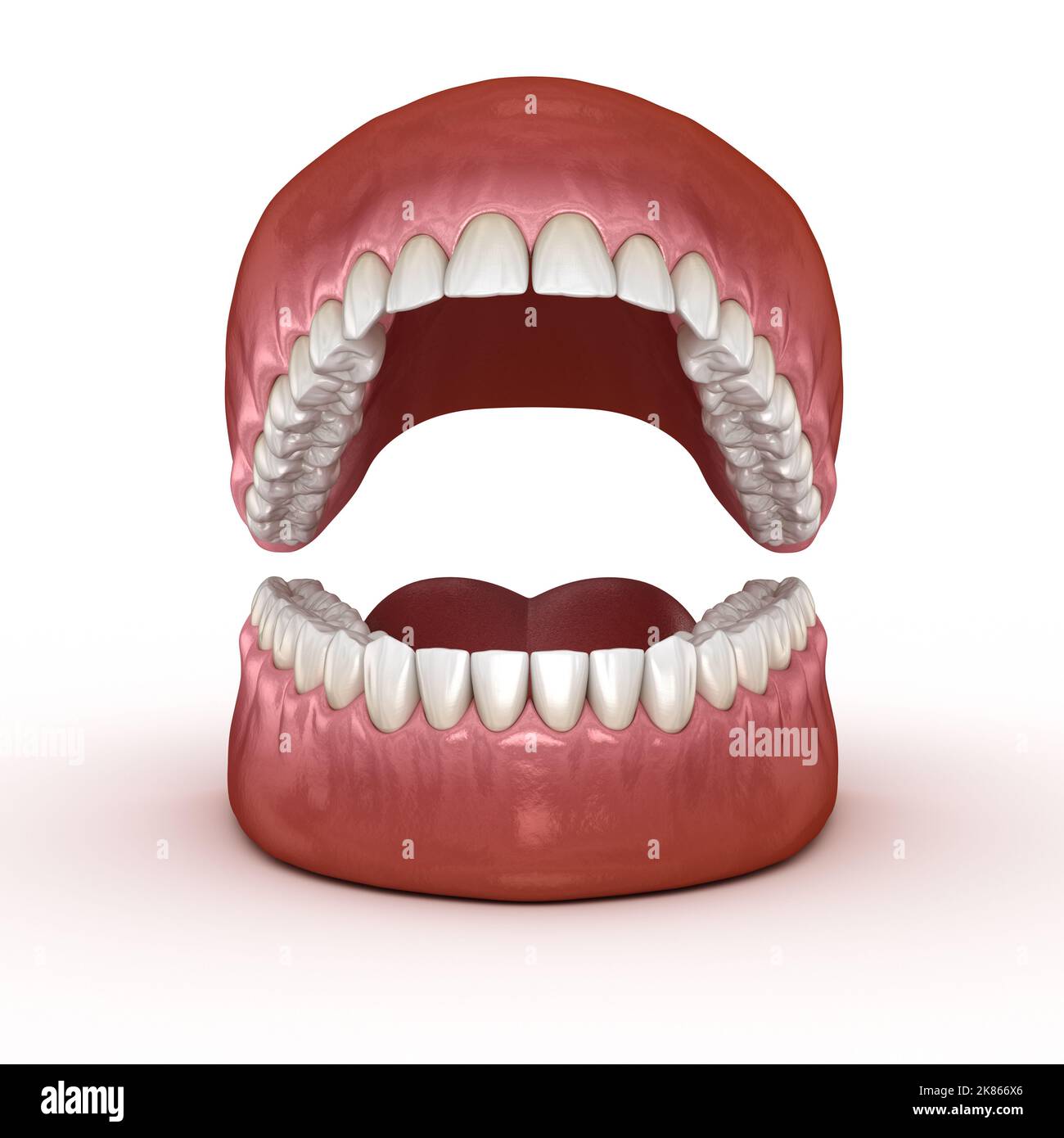 Dental anatomy - Opened Dentures. Medically accurate dental 3D illustration Stock Photohttps://www.alamy.com/image-license-details/?v=1https://www.alamy.com/dental-anatomy-opened-dentures-medically-accurate-dental-3d-illustration-image486944702.html
RF2K866X6–Dental anatomy - Opened Dentures. Medically accurate dental 3D illustration
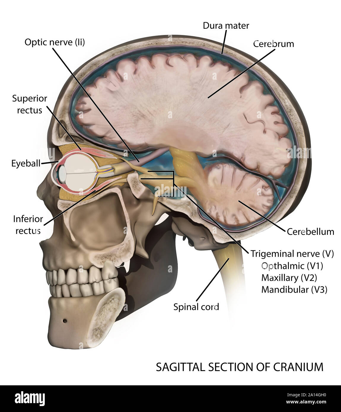 Medical illustration depicting the anatomy of a sagittal section of the human cranium. Stock Photohttps://www.alamy.com/image-license-details/?v=1https://www.alamy.com/medical-illustration-depicting-the-anatomy-of-a-sagittal-section-of-the-human-cranium-image327712476.html
RF2A14GH0–Medical illustration depicting the anatomy of a sagittal section of the human cranium.
 The largest and strongest bone in the face and serves for the reception of the lower teeth, with its parts labelled, vintage line drawing or engraving Stock Vectorhttps://www.alamy.com/image-license-details/?v=1https://www.alamy.com/the-largest-and-strongest-bone-in-the-face-and-serves-for-the-reception-of-the-lower-teeth-with-its-parts-labelled-vintage-line-drawing-or-engraving-image359331713.html
RF2BTGY7D–The largest and strongest bone in the face and serves for the reception of the lower teeth, with its parts labelled, vintage line drawing or engraving
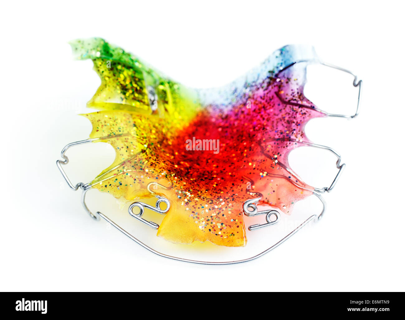 Multicolored and glittered upper orthodontic appliance with two z-springs, 3 clasps, labial bow and an expansion screw Stock Photohttps://www.alamy.com/image-license-details/?v=1https://www.alamy.com/stock-photo-multicolored-and-glittered-upper-orthodontic-appliance-with-two-z-72987861.html
RFE6MTN9–Multicolored and glittered upper orthodontic appliance with two z-springs, 3 clasps, labial bow and an expansion screw
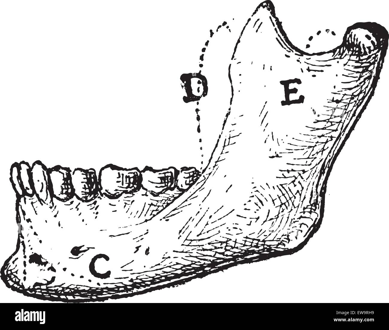 Human Mandible, showing (A) Mental Protuberance, (C) Triangularis, (D) Coronoid Process, and (E) Masseter, vintage engraved illustration. Dictionary of Words and Things - Larive and Fleury - 1895 Stock Vectorhttps://www.alamy.com/image-license-details/?v=1https://www.alamy.com/stock-photo-human-mandible-showing-a-mental-protuberance-c-triangularis-d-coronoid-84423957.html
RFEW9RH9–Human Mandible, showing (A) Mental Protuberance, (C) Triangularis, (D) Coronoid Process, and (E) Masseter, vintage engraved illustration. Dictionary of Words and Things - Larive and Fleury - 1895
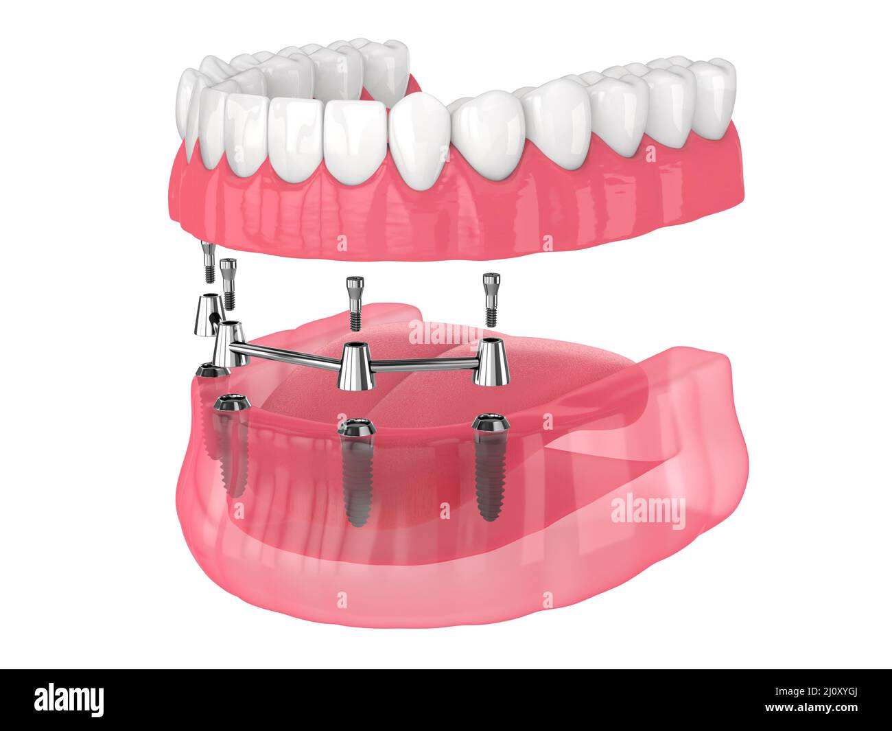 Removable overdenture installation on bar clip attachment, supported by implants over white background Stock Photohttps://www.alamy.com/image-license-details/?v=1https://www.alamy.com/removable-overdenture-installation-on-bar-clip-attachment-supported-by-implants-over-white-background-image465272322.html
RF2J0XYGJ–Removable overdenture installation on bar clip attachment, supported by implants over white background
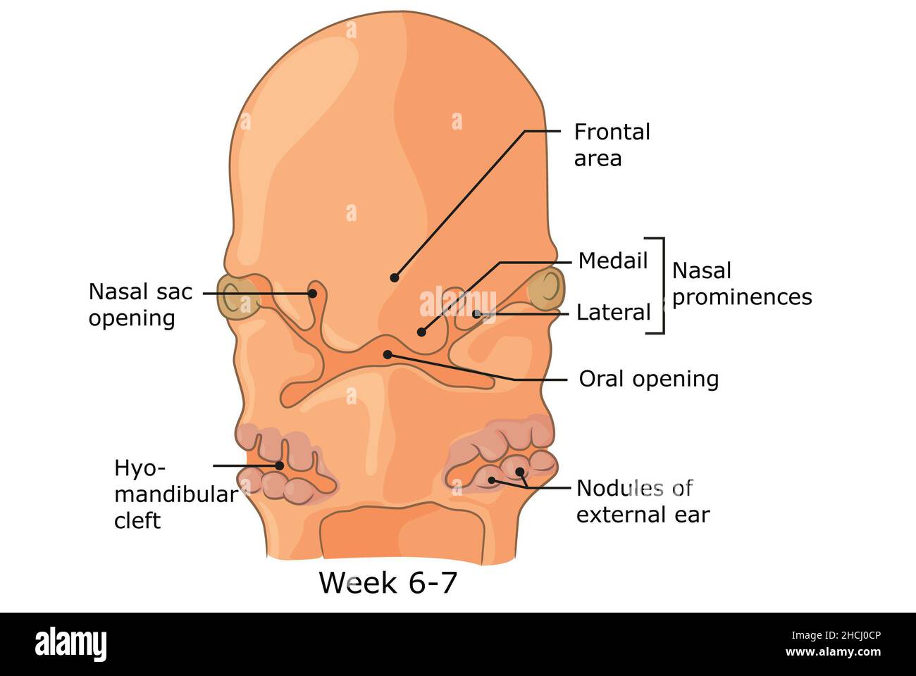 Development of external structures of human face week 6 - 7. Stock Photohttps://www.alamy.com/image-license-details/?v=1https://www.alamy.com/development-of-external-structures-of-human-face-week-6-7-image455240934.html
RF2HCJ0CP–Development of external structures of human face week 6 - 7.
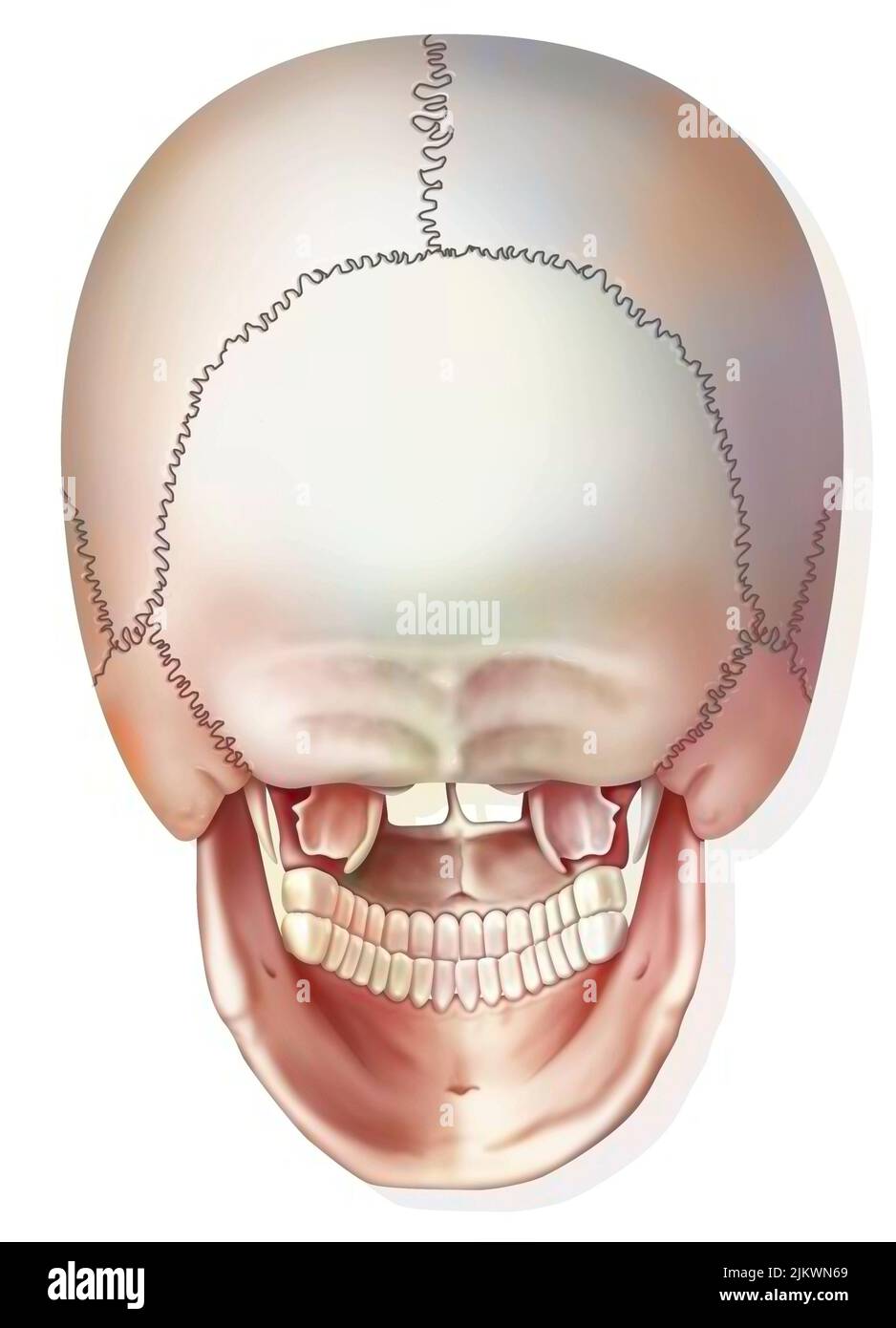 Posterior view of the bones of the skull and jaw. Stock Photohttps://www.alamy.com/image-license-details/?v=1https://www.alamy.com/posterior-view-of-the-bones-of-the-skull-and-jaw-image476923841.html
RF2JKWN69–Posterior view of the bones of the skull and jaw.
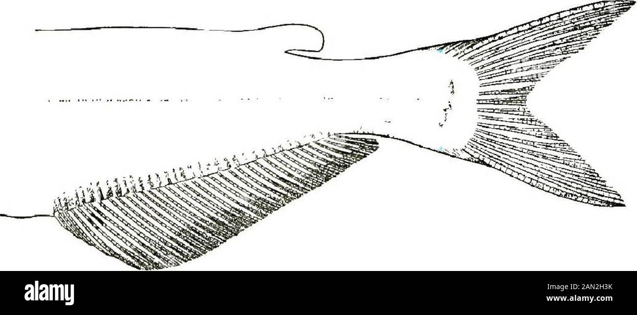 ..The fishes of Illinois . in the next species, its length 4 to 4.4,its greatest depth 5 .2 to 5 .6 in body; top of head and nape prominentlyconvex, the back subcarinate in front of dorsal, the skin thin and fittedclosely over the bones; mouth small, inferior, the lower jaw wholly•included; lips thin; maxillary barbels reaching past gill-opening; evesmall, oval, lying on the median axis of the body and nearer lower thanupper surface of head; diameter of orbit 7 .2 to 7 . 8 in head. Dorsal finhigh, nearer snout than adipose, its distance from snout 3 to 3.5 inlength; the spine rather longer and Stock Photohttps://www.alamy.com/image-license-details/?v=1https://www.alamy.com/the-fishes-of-illinois-in-the-next-species-its-length-4-to-44its-greatest-depth-5-2-to-5-6-in-body-top-of-head-and-nape-prominentlyconvex-the-back-subcarinate-in-front-of-dorsal-the-skin-thin-and-fittedclosely-over-the-bones-mouth-small-inferior-the-lower-jaw-whollyincluded-lips-thin-maxillary-barbels-reaching-past-gill-opening-evesmall-oval-lying-on-the-median-axis-of-the-body-and-nearer-lower-thanupper-surface-of-head-diameter-of-orbit-7-2-to-7-8-in-head-dorsal-finhigh-nearer-snout-than-adipose-its-distance-from-snout-3-to-35-inlength-the-spine-rather-longer-and-image339962103.html
RM2AN2H3K–..The fishes of Illinois . in the next species, its length 4 to 4.4,its greatest depth 5 .2 to 5 .6 in body; top of head and nape prominentlyconvex, the back subcarinate in front of dorsal, the skin thin and fittedclosely over the bones; mouth small, inferior, the lower jaw wholly•included; lips thin; maxillary barbels reaching past gill-opening; evesmall, oval, lying on the median axis of the body and nearer lower thanupper surface of head; diameter of orbit 7 .2 to 7 . 8 in head. Dorsal finhigh, nearer snout than adipose, its distance from snout 3 to 3.5 inlength; the spine rather longer and
 Skull with open jaw, illustration. Stock Photohttps://www.alamy.com/image-license-details/?v=1https://www.alamy.com/skull-with-open-jaw-illustration-image367367003.html
RF2C9K0A3–Skull with open jaw, illustration.
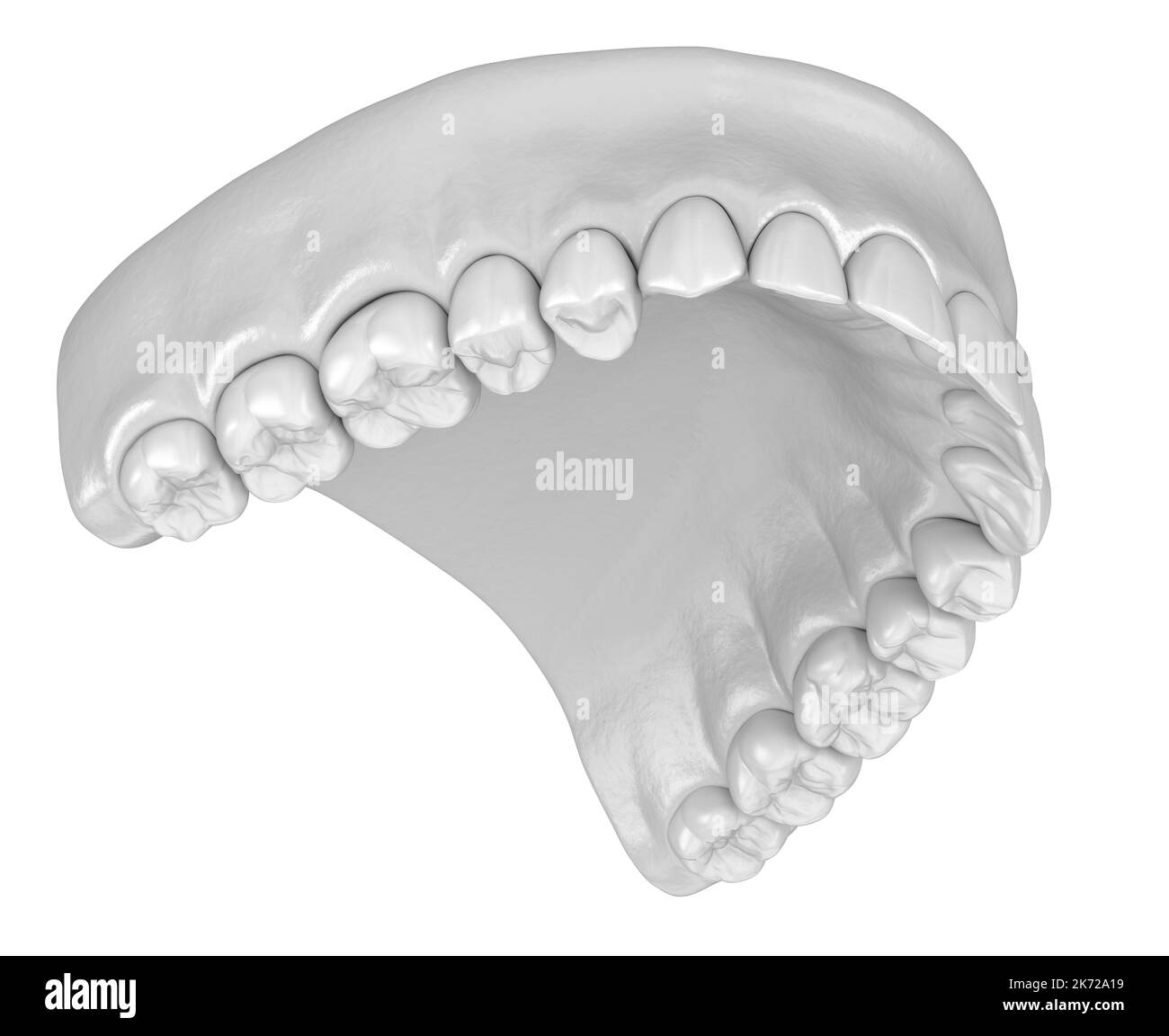 Human gum and teeth in white style. Medically accurate tooth 3D illustration Stock Photohttps://www.alamy.com/image-license-details/?v=1https://www.alamy.com/human-gum-and-teeth-in-white-style-medically-accurate-tooth-3d-illustration-image486244677.html
RF2K72A19–Human gum and teeth in white style. Medically accurate tooth 3D illustration
 . The families and genera of bats . Bats; Bats. THE f amilieS and genera 6f bats. 71 and well developed, without secondary cusps or prominent cingula, the maxillary canines with deep longitudinal groove on anterior sur- face. Cheek teeth small, without distinct contrasts of size or form, the two anterior in each jaw {pm 2, pm 3, pm 2, pm 8) with the crowns compressed and elevated into distinct though blunt cusp, the others {pm4, m1, m2, pm 4, m u m 2, m 8) with narrow nearly flat orowns bounded by indistinct ridges, of which that on the outer side is the less developed. Anterior premolar, both Stock Photohttps://www.alamy.com/image-license-details/?v=1https://www.alamy.com/the-families-and-genera-of-bats-bats-bats-the-f-amilies-and-genera-6f-bats-71-and-well-developed-without-secondary-cusps-or-prominent-cingula-the-maxillary-canines-with-deep-longitudinal-groove-on-anterior-sur-face-cheek-teeth-small-without-distinct-contrasts-of-size-or-form-the-two-anterior-in-each-jaw-pm-2-pm-3-pm-2-pm-8-with-the-crowns-compressed-and-elevated-into-distinct-though-blunt-cusp-the-others-pm4-m1-m2-pm-4-m-u-m-2-m-8-with-narrow-nearly-flat-orowns-bounded-by-indistinct-ridges-of-which-that-on-the-outer-side-is-the-less-developed-anterior-premolar-both-image216355952.html
RMPFYT68–. The families and genera of bats . Bats; Bats. THE f amilieS and genera 6f bats. 71 and well developed, without secondary cusps or prominent cingula, the maxillary canines with deep longitudinal groove on anterior sur- face. Cheek teeth small, without distinct contrasts of size or form, the two anterior in each jaw {pm 2, pm 3, pm 2, pm 8) with the crowns compressed and elevated into distinct though blunt cusp, the others {pm4, m1, m2, pm 4, m u m 2, m 8) with narrow nearly flat orowns bounded by indistinct ridges, of which that on the outer side is the less developed. Anterior premolar, both
 3d rendered medically accurate illustration of the skull with open jaw Stock Photohttps://www.alamy.com/image-license-details/?v=1https://www.alamy.com/3d-rendered-medically-accurate-illustration-of-the-skull-with-open-jaw-image334050053.html
RF2ABD871–3d rendered medically accurate illustration of the skull with open jaw
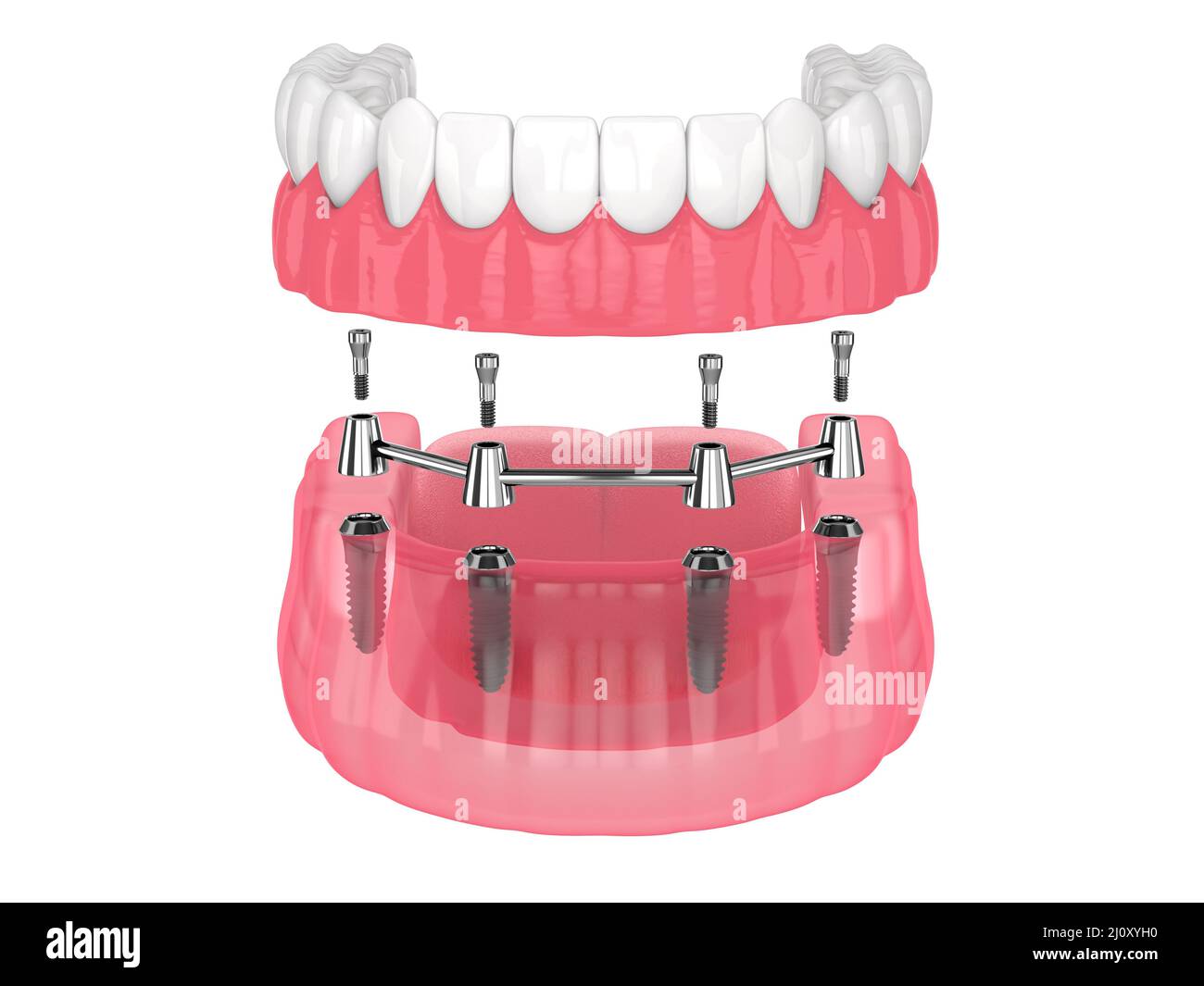 Removable overdenture installation on bar clip attachment, supported by implants over white background Stock Photohttps://www.alamy.com/image-license-details/?v=1https://www.alamy.com/removable-overdenture-installation-on-bar-clip-attachment-supported-by-implants-over-white-background-image465272332.html
RF2J0XYH0–Removable overdenture installation on bar clip attachment, supported by implants over white background
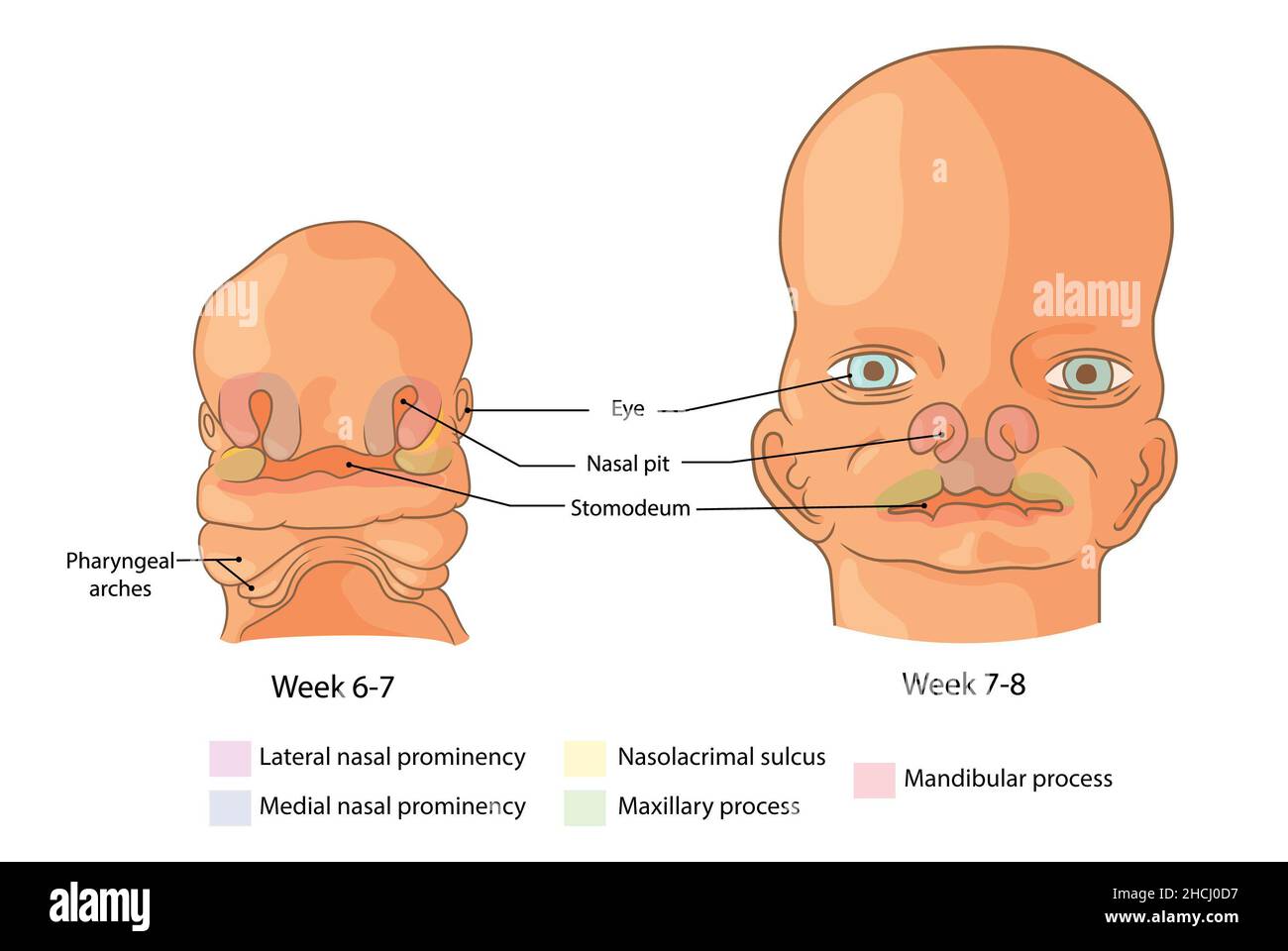 Embryological development of the face weeks 6-8 Stock Photohttps://www.alamy.com/image-license-details/?v=1https://www.alamy.com/embryological-development-of-the-face-weeks-6-8-image455240947.html
RF2HCJ0D7–Embryological development of the face weeks 6-8
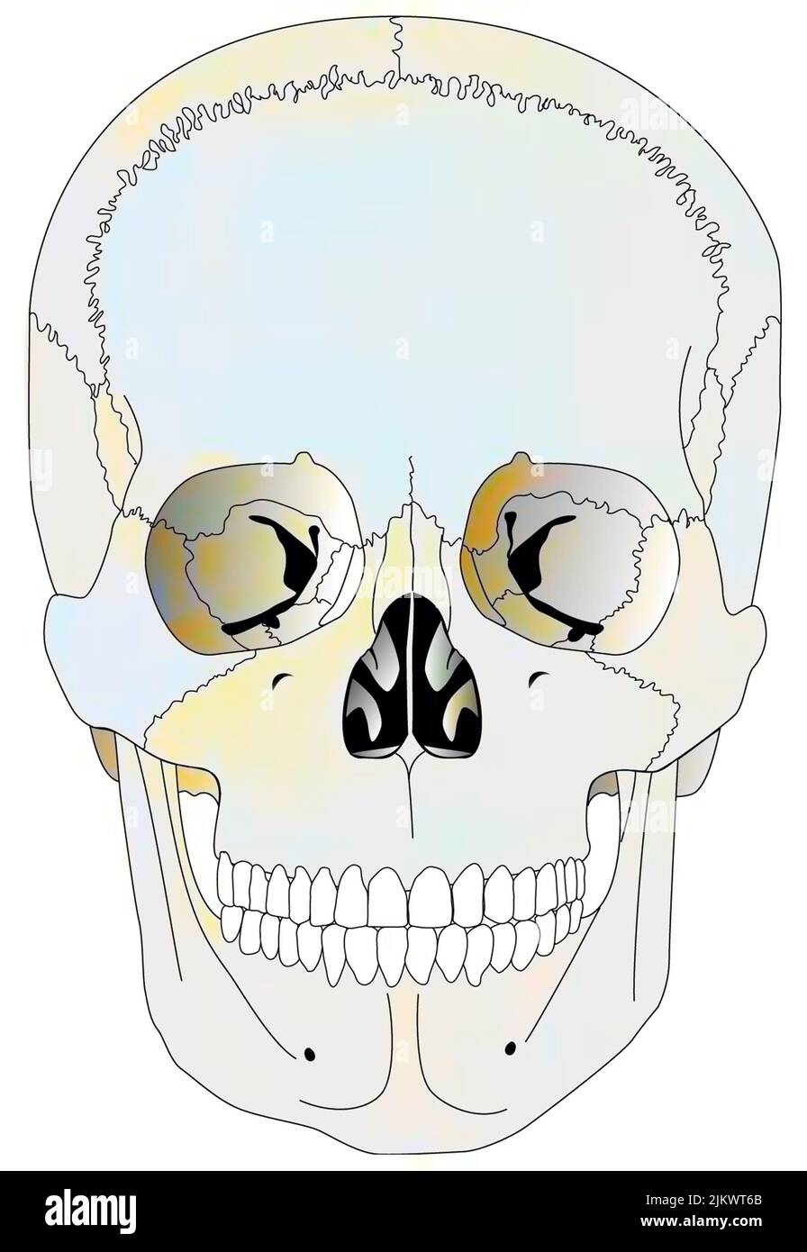 Cranial correspondences of the skeleton and viscera in adults. Stock Photohttps://www.alamy.com/image-license-details/?v=1https://www.alamy.com/cranial-correspondences-of-the-skeleton-and-viscera-in-adults-image476926195.html
RF2JKWT6B–Cranial correspondences of the skeleton and viscera in adults.
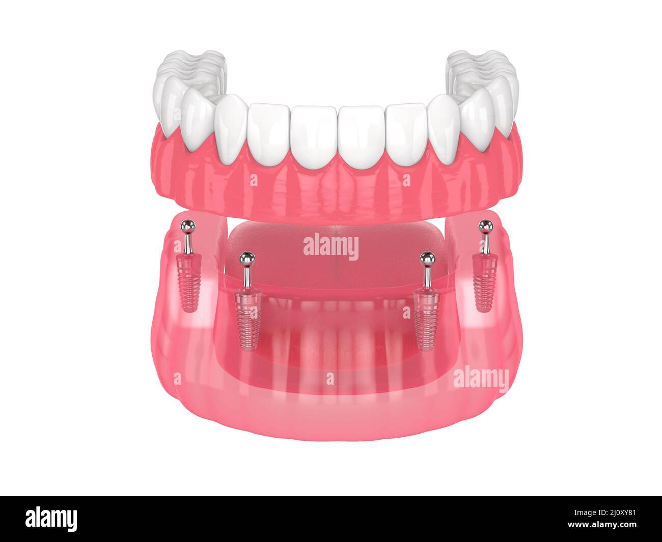 All-on-4 removable, implants supported, overdenture installation over white background Stock Photohttps://www.alamy.com/image-license-details/?v=1https://www.alamy.com/all-on-4-removable-implants-supported-overdenture-installation-over-white-background-image465272081.html
RF2J0XY81–All-on-4 removable, implants supported, overdenture installation over white background
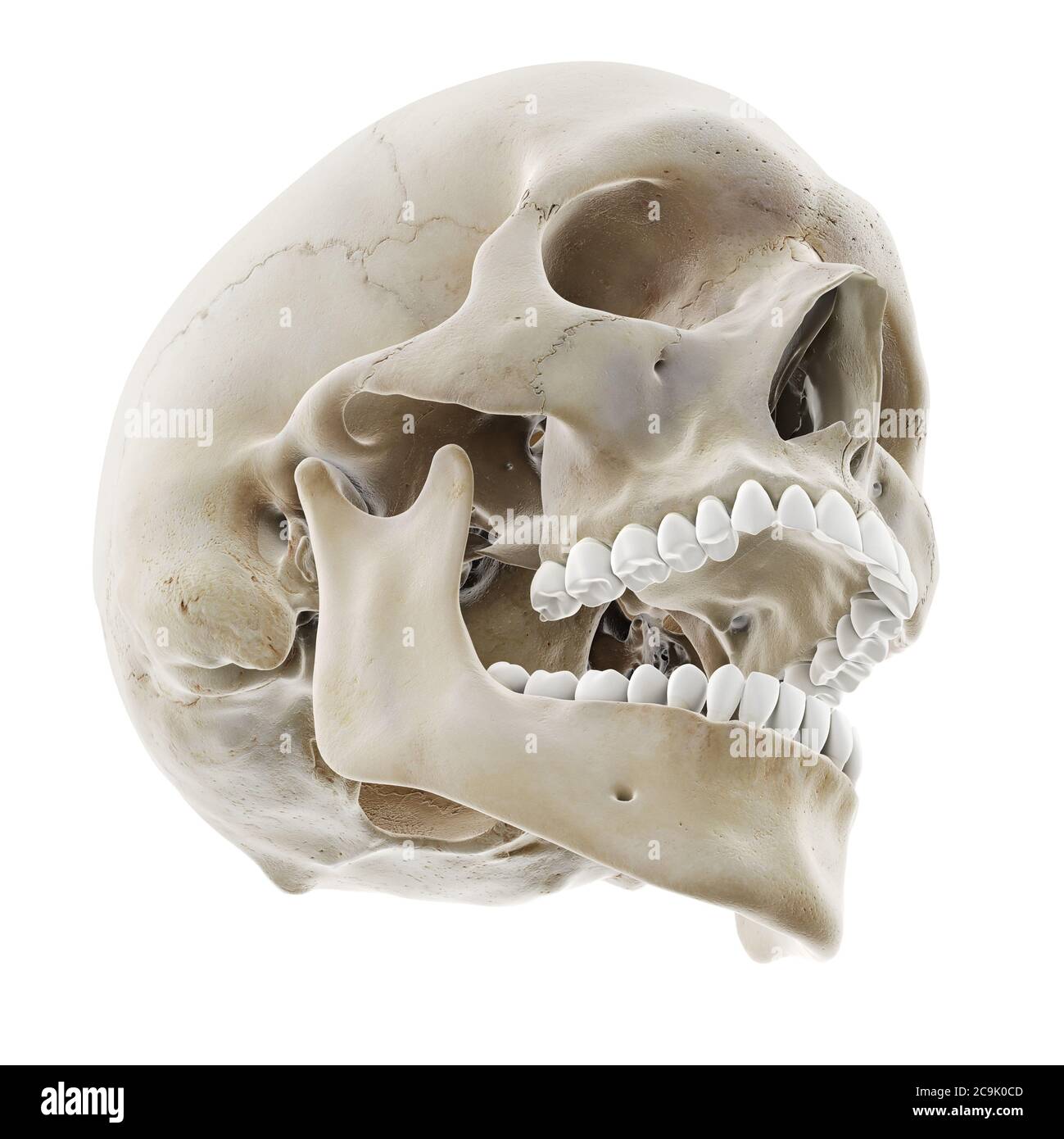 Skull with open jaw, illustration. Stock Photohttps://www.alamy.com/image-license-details/?v=1https://www.alamy.com/skull-with-open-jaw-illustration-image367367069.html
RF2C9K0CD–Skull with open jaw, illustration.
 First lesson in zoology : adapted for use in schools . Fi». 96o.—1. The common garden spider (Epeira): a, leg; b, maxillary palpus;c, poison-jaws; e, spinnerets. 2. Front view of head with the eight simpleeyes and the poison-jaws. 3. End o( a jaw: a, outlet of the poison-canal. 7.Palpus of female; 8, of male spider. 6. Spines and claws at end of a leg.4. Spinnerets, highly magnified. 5. A single silk-tube.—After Emerton.. Fig. 97o.—Structure of a centipede. A, Lithobius americantts, natural size.B, under side of head and first two body-segments and legs, enlarged: ant,antenna; 1. jaws; 2. firs Stock Photohttps://www.alamy.com/image-license-details/?v=1https://www.alamy.com/first-lesson-in-zoology-adapted-for-use-in-schools-fi-96o1-the-common-garden-spider-epeira-a-leg-b-maxillary-palpusc-poison-jaws-e-spinnerets-2-front-view-of-head-with-the-eight-simpleeyes-and-the-poison-jaws-3-end-o-a-jaw-a-outlet-of-the-poison-canal-7palpus-of-female-8-of-male-spider-6-spines-and-claws-at-end-of-a-leg4-spinnerets-highly-magnified-5-a-single-silk-tubeafter-emerton-fig-97ostructure-of-a-centipede-a-lithobius-americantts-natural-sizeb-under-side-of-head-and-first-two-body-segments-and-legs-enlarged-antantenna-1-jaws-2-firs-image339246241.html
RM2AKX015–First lesson in zoology : adapted for use in schools . Fi». 96o.—1. The common garden spider (Epeira): a, leg; b, maxillary palpus;c, poison-jaws; e, spinnerets. 2. Front view of head with the eight simpleeyes and the poison-jaws. 3. End o( a jaw: a, outlet of the poison-canal. 7.Palpus of female; 8, of male spider. 6. Spines and claws at end of a leg.4. Spinnerets, highly magnified. 5. A single silk-tube.—After Emerton.. Fig. 97o.—Structure of a centipede. A, Lithobius americantts, natural size.B, under side of head and first two body-segments and legs, enlarged: ant,antenna; 1. jaws; 2. firs
 3d rendered medically accurate illustration of the skull with open jaw Stock Photohttps://www.alamy.com/image-license-details/?v=1https://www.alamy.com/3d-rendered-medically-accurate-illustration-of-the-skull-with-open-jaw-image334050122.html
RF2ABD89E–3d rendered medically accurate illustration of the skull with open jaw
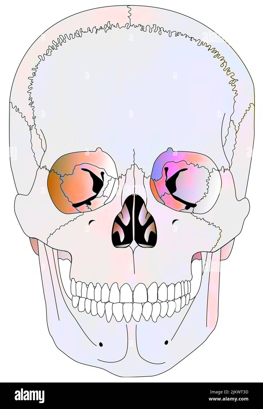 Cranial correspondences of the skeleton and viscera in adults. Stock Photohttps://www.alamy.com/image-license-details/?v=1https://www.alamy.com/cranial-correspondences-of-the-skeleton-and-viscera-in-adults-image476926113.html
RF2JKWT3D–Cranial correspondences of the skeleton and viscera in adults.
 All-on-4 removable, implants supported, overdenture installation over white background Stock Photohttps://www.alamy.com/image-license-details/?v=1https://www.alamy.com/all-on-4-removable-implants-supported-overdenture-installation-over-white-background-image465272078.html
RF2J0XY7X–All-on-4 removable, implants supported, overdenture installation over white background
 Skull with open jaw, illustration. Stock Photohttps://www.alamy.com/image-license-details/?v=1https://www.alamy.com/skull-with-open-jaw-illustration-image367367054.html
RF2C9K0BX–Skull with open jaw, illustration.
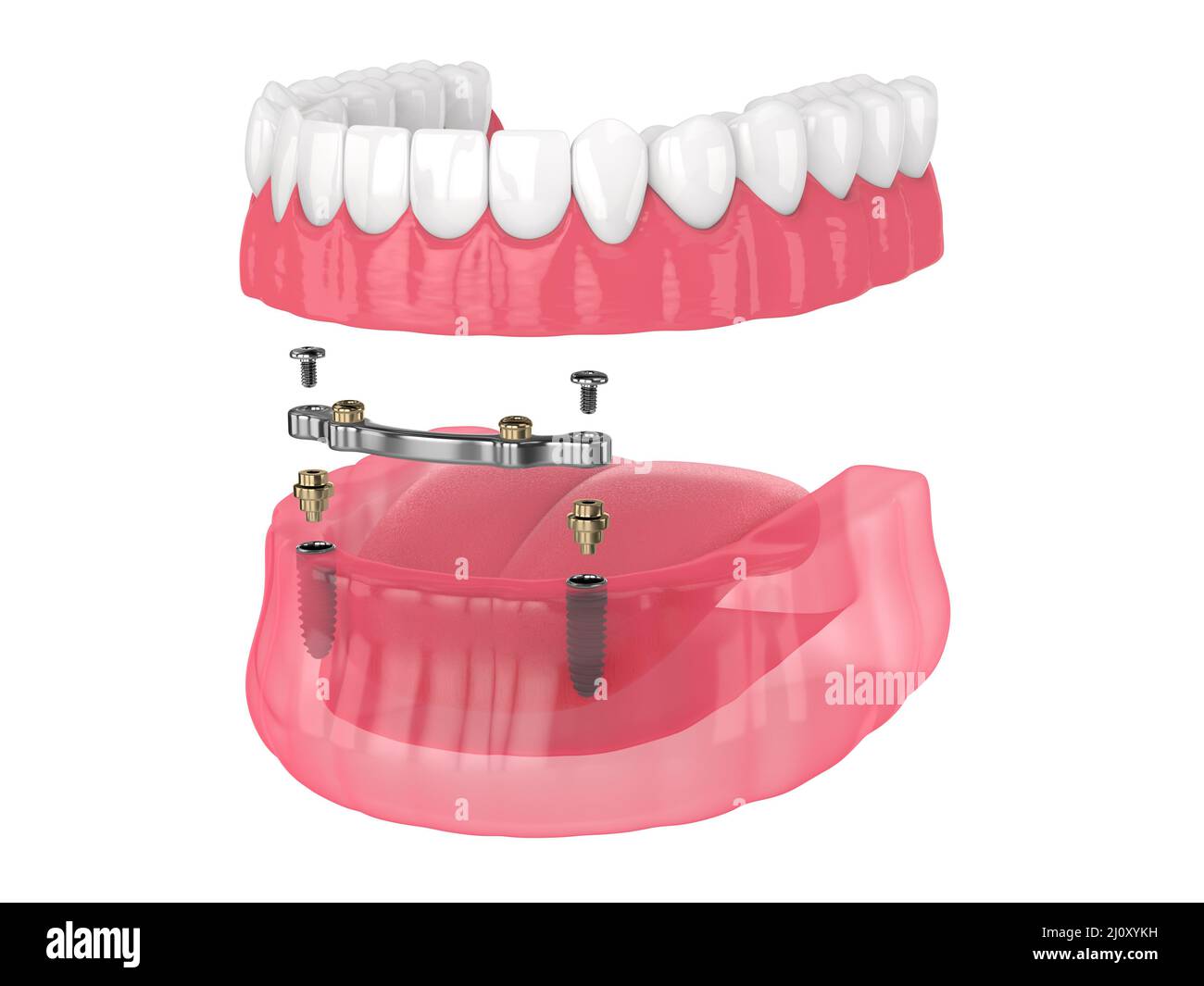 Bar retained removable overdenture installation supported by implants over white backgroud Stock Photohttps://www.alamy.com/image-license-details/?v=1https://www.alamy.com/bar-retained-removable-overdenture-installation-supported-by-implants-over-white-backgroud-image465272405.html
RF2J0XYKH–Bar retained removable overdenture installation supported by implants over white backgroud
 . Human physiology. pearance of white hairs. They are attached tothe sphenoid bone in front, the parietal bones above, and the occipital bonebehind. They also send out processes which unite with the cheek-bones, form-ing bony arches. 22 ELEMENTARY PHYSIOLOGY The bones of the face are as follows Two superior maxillary.Two palatal.Two nasal.Two lachrymal. Two inferior turbinated. One vomer. Two malar. One inferior maxillary. The superior maxillary bones (Lat. maxilla, a jaw) form the upper jaw,and the greater portion of the palate or roof of the mouth. In them are fixedthe upper set of teeth. Be Stock Photohttps://www.alamy.com/image-license-details/?v=1https://www.alamy.com/human-physiology-pearance-of-white-hairs-they-are-attached-tothe-sphenoid-bone-in-front-the-parietal-bones-above-and-the-occipital-bonebehind-they-also-send-out-processes-which-unite-with-the-cheek-bones-form-ing-bony-arches-22-elementary-physiology-the-bones-of-the-face-are-as-follows-two-superior-maxillarytwo-palataltwo-nasaltwo-lachrymal-two-inferior-turbinated-one-vomer-two-malar-one-inferior-maxillary-the-superior-maxillary-bones-lat-maxilla-a-jaw-form-the-upper-jawand-the-greater-portion-of-the-palate-or-roof-of-the-mouth-in-them-are-fixedthe-upper-set-of-teeth-be-image370649900.html
RM2CF0FMC–. Human physiology. pearance of white hairs. They are attached tothe sphenoid bone in front, the parietal bones above, and the occipital bonebehind. They also send out processes which unite with the cheek-bones, form-ing bony arches. 22 ELEMENTARY PHYSIOLOGY The bones of the face are as follows Two superior maxillary.Two palatal.Two nasal.Two lachrymal. Two inferior turbinated. One vomer. Two malar. One inferior maxillary. The superior maxillary bones (Lat. maxilla, a jaw) form the upper jaw,and the greater portion of the palate or roof of the mouth. In them are fixedthe upper set of teeth. Be
 3d rendered medically accurate illustration of the skull with open jaw Stock Photohttps://www.alamy.com/image-license-details/?v=1https://www.alamy.com/3d-rendered-medically-accurate-illustration-of-the-skull-with-open-jaw-image334071399.html
RF2ABE7DB–3d rendered medically accurate illustration of the skull with open jaw
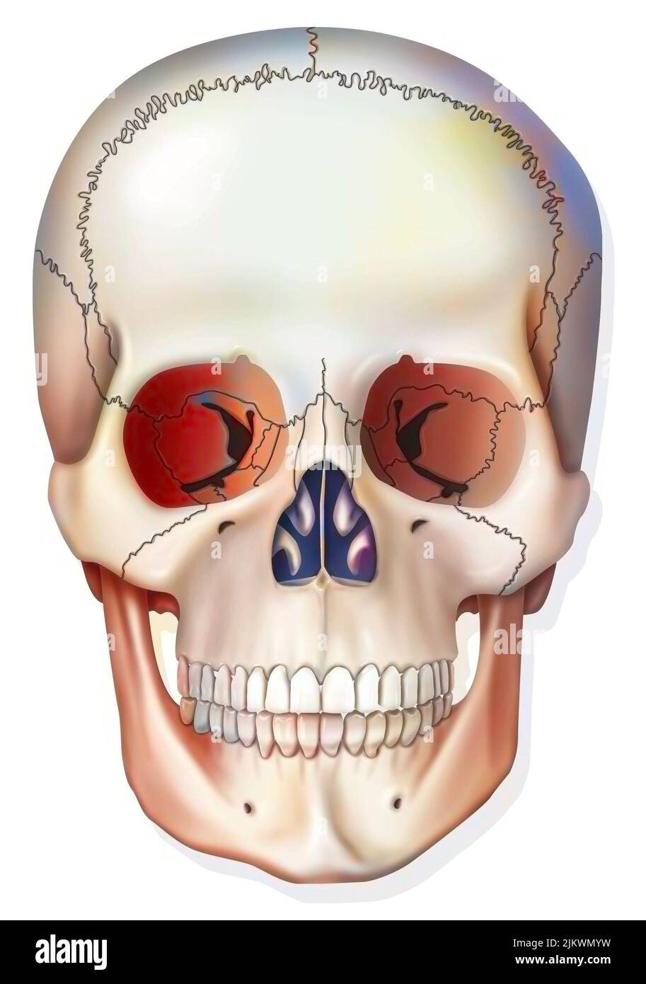 Bone system: human skull with jawbone, eye sockets. Stock Photohttps://www.alamy.com/image-license-details/?v=1https://www.alamy.com/bone-system-human-skull-with-jawbone-eye-sockets-image476923661.html
RF2JKWMYW–Bone system: human skull with jawbone, eye sockets.
 Skull with open jaw, illustration. Stock Photohttps://www.alamy.com/image-license-details/?v=1https://www.alamy.com/skull-with-open-jaw-illustration-image367367078.html
RF2C9K0CP–Skull with open jaw, illustration.
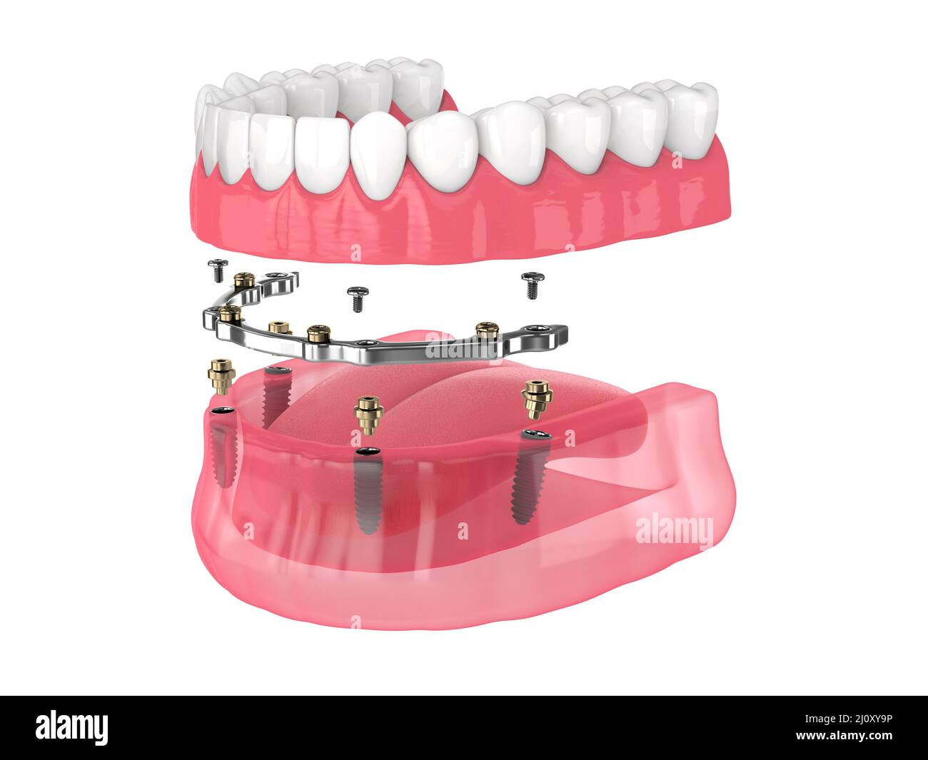 Bar retained removable overdenture installation supported by implants over white backgroud Stock Photohttps://www.alamy.com/image-license-details/?v=1https://www.alamy.com/bar-retained-removable-overdenture-installation-supported-by-implants-over-white-backgroud-image465272130.html
RF2J0XY9P–Bar retained removable overdenture installation supported by implants over white backgroud
 . Fishes. Fishes. CHAPTER XXIX SALMOPERC^ AND OTHER TRANSITIONAL GROUPS UBORDER Salmopercae, the Trout-perches: Percopsidae. —More ancient than the Hemibranchii, and still more distinctly in the line of transition from soft-rayed to spiny-rayed fishes, is the small suborder of Salmopercas. This is characterized by the presence of the adipose fin of the salmon,. Fig. 360.—Sand-roller, Pecropsis gutlatus Agassiz. Okoboji Lake, la. in connection with the mouth, scales, and fin-spines of a perch. The premaxillary forms the entire edge of the upper jaw, the maxillary being without teeth. The air-bl Stock Photohttps://www.alamy.com/image-license-details/?v=1https://www.alamy.com/fishes-fishes-chapter-xxix-salmoperc-and-other-transitional-groups-uborder-salmopercae-the-trout-perches-percopsidae-more-ancient-than-the-hemibranchii-and-still-more-distinctly-in-the-line-of-transition-from-soft-rayed-to-spiny-rayed-fishes-is-the-small-suborder-of-salmopercas-this-is-characterized-by-the-presence-of-the-adipose-fin-of-the-salmon-fig-360sand-roller-pecropsis-gutlatus-agassiz-okoboji-lake-la-in-connection-with-the-mouth-scales-and-fin-spines-of-a-perch-the-premaxillary-forms-the-entire-edge-of-the-upper-jaw-the-maxillary-being-without-teeth-the-air-bl-image232217894.html
RMRDPC86–. Fishes. Fishes. CHAPTER XXIX SALMOPERC^ AND OTHER TRANSITIONAL GROUPS UBORDER Salmopercae, the Trout-perches: Percopsidae. —More ancient than the Hemibranchii, and still more distinctly in the line of transition from soft-rayed to spiny-rayed fishes, is the small suborder of Salmopercas. This is characterized by the presence of the adipose fin of the salmon,. Fig. 360.—Sand-roller, Pecropsis gutlatus Agassiz. Okoboji Lake, la. in connection with the mouth, scales, and fin-spines of a perch. The premaxillary forms the entire edge of the upper jaw, the maxillary being without teeth. The air-bl
 3d rendered medically accurate illustration of the skull with open jaw Stock Photohttps://www.alamy.com/image-license-details/?v=1https://www.alamy.com/3d-rendered-medically-accurate-illustration-of-the-skull-with-open-jaw-image334071379.html
RF2ABE7CK–3d rendered medically accurate illustration of the skull with open jaw
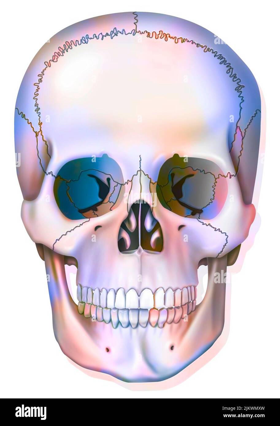 Bone system: human skull with jawbone, eye sockets. Stock Photohttps://www.alamy.com/image-license-details/?v=1https://www.alamy.com/bone-system-human-skull-with-jawbone-eye-sockets-image476923633.html
RF2JKWMXW–Bone system: human skull with jawbone, eye sockets.
 Skull with open jaw, illustration. Stock Photohttps://www.alamy.com/image-license-details/?v=1https://www.alamy.com/skull-with-open-jaw-illustration-image367367064.html
RF2C9K0C8–Skull with open jaw, illustration.
 Bar retained removable overdenture installation supported by implants over white backgroud Stock Photohttps://www.alamy.com/image-license-details/?v=1https://www.alamy.com/bar-retained-removable-overdenture-installation-supported-by-implants-over-white-backgroud-image465272144.html
RF2J0XYA8–Bar retained removable overdenture installation supported by implants over white backgroud
 . The physiology of domestic animals ... Physiology, Comparative; Veterinary physiology. Fig. 86.—Head of Carnivora—Dog. (Btclard.) Fig. 87.—Inferior Maxillary Bone of Carnivora—Polar Bear. (B6clard.) c, profile view of articular condyle of lower jaw (condyle of right side); ct, front view of same condyle. and forward motion of the lower jaw, which is, therefore, the character- istic motion of rodents. Second.—In the ruminants the jaws are long and feeble, the canine and upper incisor teeth are absent, while the molars are compound teeth with a flat crown, with the enamel arranged in anteropos Stock Photohttps://www.alamy.com/image-license-details/?v=1https://www.alamy.com/the-physiology-of-domestic-animals-physiology-comparative-veterinary-physiology-fig-86head-of-carnivoradog-btclard-fig-87inferior-maxillary-bone-of-carnivorapolar-bear-b6clard-c-profile-view-of-articular-condyle-of-lower-jaw-condyle-of-right-side-ct-front-view-of-same-condyle-and-forward-motion-of-the-lower-jaw-which-is-therefore-the-character-istic-motion-of-rodents-secondin-the-ruminants-the-jaws-are-long-and-feeble-the-canine-and-upper-incisor-teeth-are-absent-while-the-molars-are-compound-teeth-with-a-flat-crown-with-the-enamel-arranged-in-anteropos-image232426698.html
RMRE3XHE–. The physiology of domestic animals ... Physiology, Comparative; Veterinary physiology. Fig. 86.—Head of Carnivora—Dog. (Btclard.) Fig. 87.—Inferior Maxillary Bone of Carnivora—Polar Bear. (B6clard.) c, profile view of articular condyle of lower jaw (condyle of right side); ct, front view of same condyle. and forward motion of the lower jaw, which is, therefore, the character- istic motion of rodents. Second.—In the ruminants the jaws are long and feeble, the canine and upper incisor teeth are absent, while the molars are compound teeth with a flat crown, with the enamel arranged in anteropos
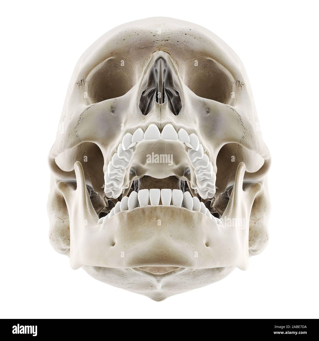 3d rendered medically accurate illustration of the skull with open jaw Stock Photohttps://www.alamy.com/image-license-details/?v=1https://www.alamy.com/3d-rendered-medically-accurate-illustration-of-the-skull-with-open-jaw-image334071398.html
RF2ABE7DA–3d rendered medically accurate illustration of the skull with open jaw
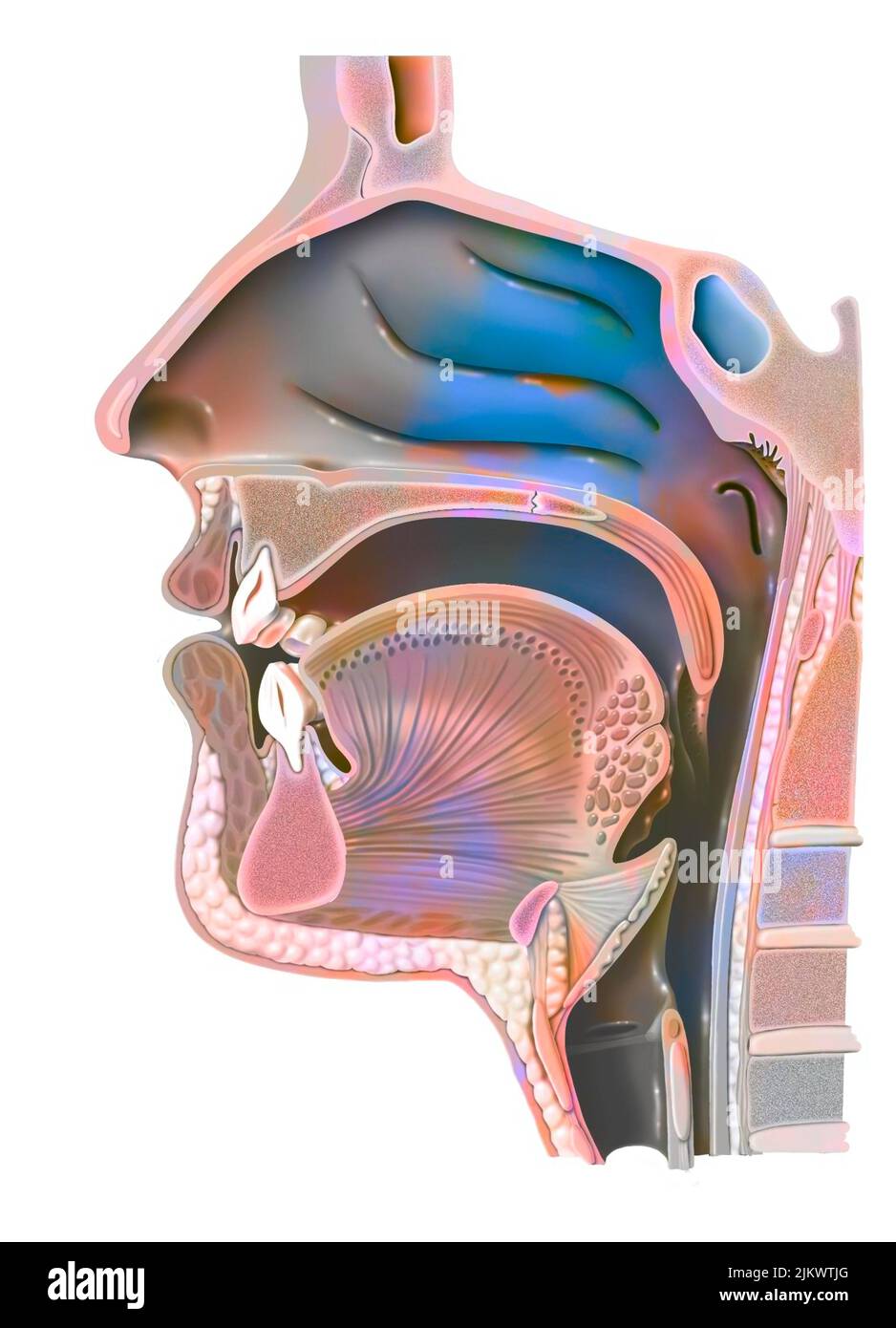 Anatomy of nasopharynx with nasal cavity, oral cavity. Stock Photohttps://www.alamy.com/image-license-details/?v=1https://www.alamy.com/anatomy-of-nasopharynx-with-nasal-cavity-oral-cavity-image476926536.html
RF2JKWTJG–Anatomy of nasopharynx with nasal cavity, oral cavity.
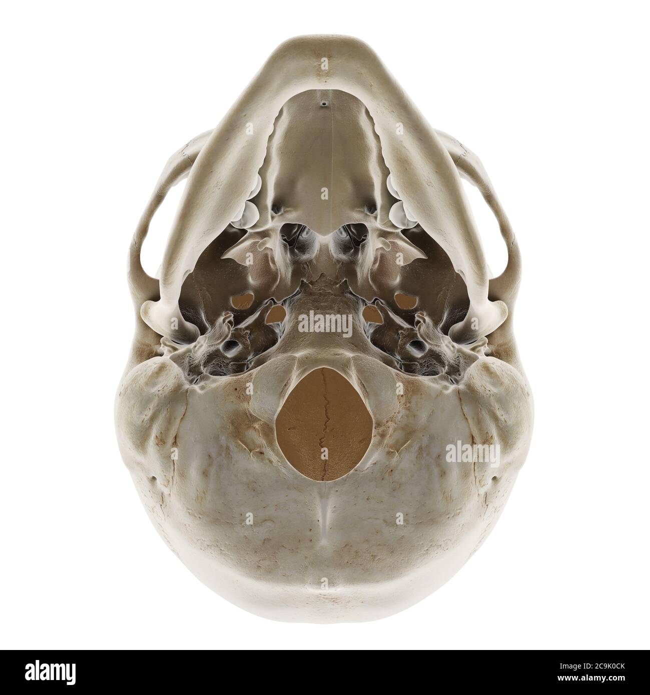 Human skull, illustration. Stock Photohttps://www.alamy.com/image-license-details/?v=1https://www.alamy.com/human-skull-illustration-image367367075.html
RF2C9K0CK–Human skull, illustration.
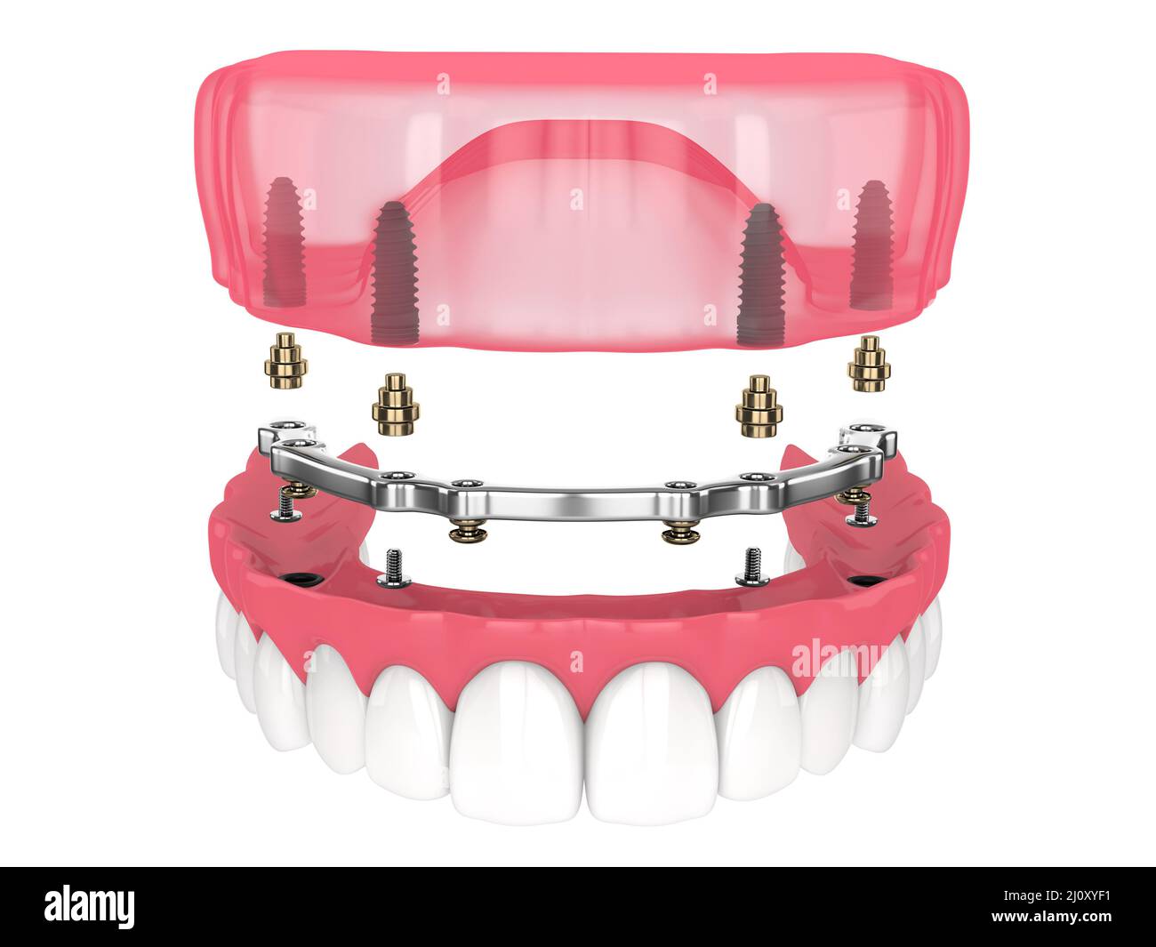 Bar retained removable overdenture installation supported by implants over white backgroud Stock Photohttps://www.alamy.com/image-license-details/?v=1https://www.alamy.com/bar-retained-removable-overdenture-installation-supported-by-implants-over-white-backgroud-image465272277.html
RF2J0XYF1–Bar retained removable overdenture installation supported by implants over white backgroud
 . The physiology of the domestic animals; a text-book for veterinary and medical students and practitioners. Physiology, Comparative; Domestic animals. Fig. 86.—Head of Carnivora—Dog. (Biclard.) Fig. 87.—Inferior Maxillary Bone of Carnivora—Polak Beak. (B&clard.) c, profile view of articular condyle of lower jaw (condyle of right side); d, front view of same condyle. and forward motion of the lower jaw, which is, therefore, the character- istic motion of rodents. Second.—In the ruminants the jaws are long and feeble, the canine and upper incisor teeth are absent, while the molars are compo Stock Photohttps://www.alamy.com/image-license-details/?v=1https://www.alamy.com/the-physiology-of-the-domestic-animals-a-text-book-for-veterinary-and-medical-students-and-practitioners-physiology-comparative-domestic-animals-fig-86head-of-carnivoradog-biclard-fig-87inferior-maxillary-bone-of-carnivorapolak-beak-bampclard-c-profile-view-of-articular-condyle-of-lower-jaw-condyle-of-right-side-d-front-view-of-same-condyle-and-forward-motion-of-the-lower-jaw-which-is-therefore-the-character-istic-motion-of-rodents-secondin-the-ruminants-the-jaws-are-long-and-feeble-the-canine-and-upper-incisor-teeth-are-absent-while-the-molars-are-compo-image232343530.html
RMRE04F6–. The physiology of the domestic animals; a text-book for veterinary and medical students and practitioners. Physiology, Comparative; Domestic animals. Fig. 86.—Head of Carnivora—Dog. (Biclard.) Fig. 87.—Inferior Maxillary Bone of Carnivora—Polak Beak. (B&clard.) c, profile view of articular condyle of lower jaw (condyle of right side); d, front view of same condyle. and forward motion of the lower jaw, which is, therefore, the character- istic motion of rodents. Second.—In the ruminants the jaws are long and feeble, the canine and upper incisor teeth are absent, while the molars are compo
 3d rendered medically accurate illustration of the skull with open jaw Stock Photohttps://www.alamy.com/image-license-details/?v=1https://www.alamy.com/3d-rendered-medically-accurate-illustration-of-the-skull-with-open-jaw-image334050084.html
RF2ABD884–3d rendered medically accurate illustration of the skull with open jaw
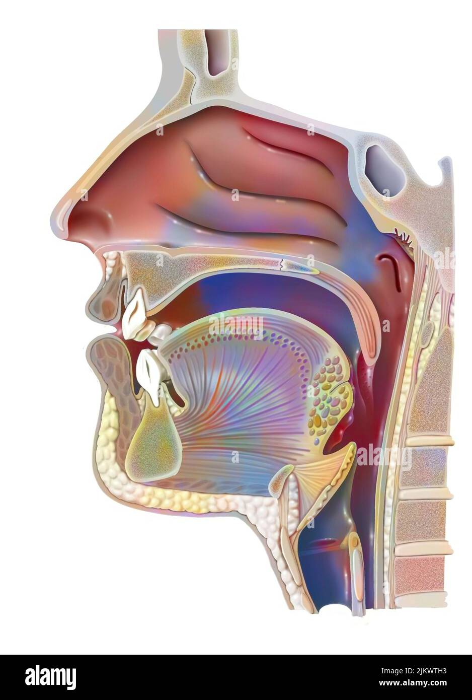 Anatomy of nasopharynx with nasal cavity, oral cavity. Stock Photohttps://www.alamy.com/image-license-details/?v=1https://www.alamy.com/anatomy-of-nasopharynx-with-nasal-cavity-oral-cavity-image476926495.html
RF2JKWTH3–Anatomy of nasopharynx with nasal cavity, oral cavity.
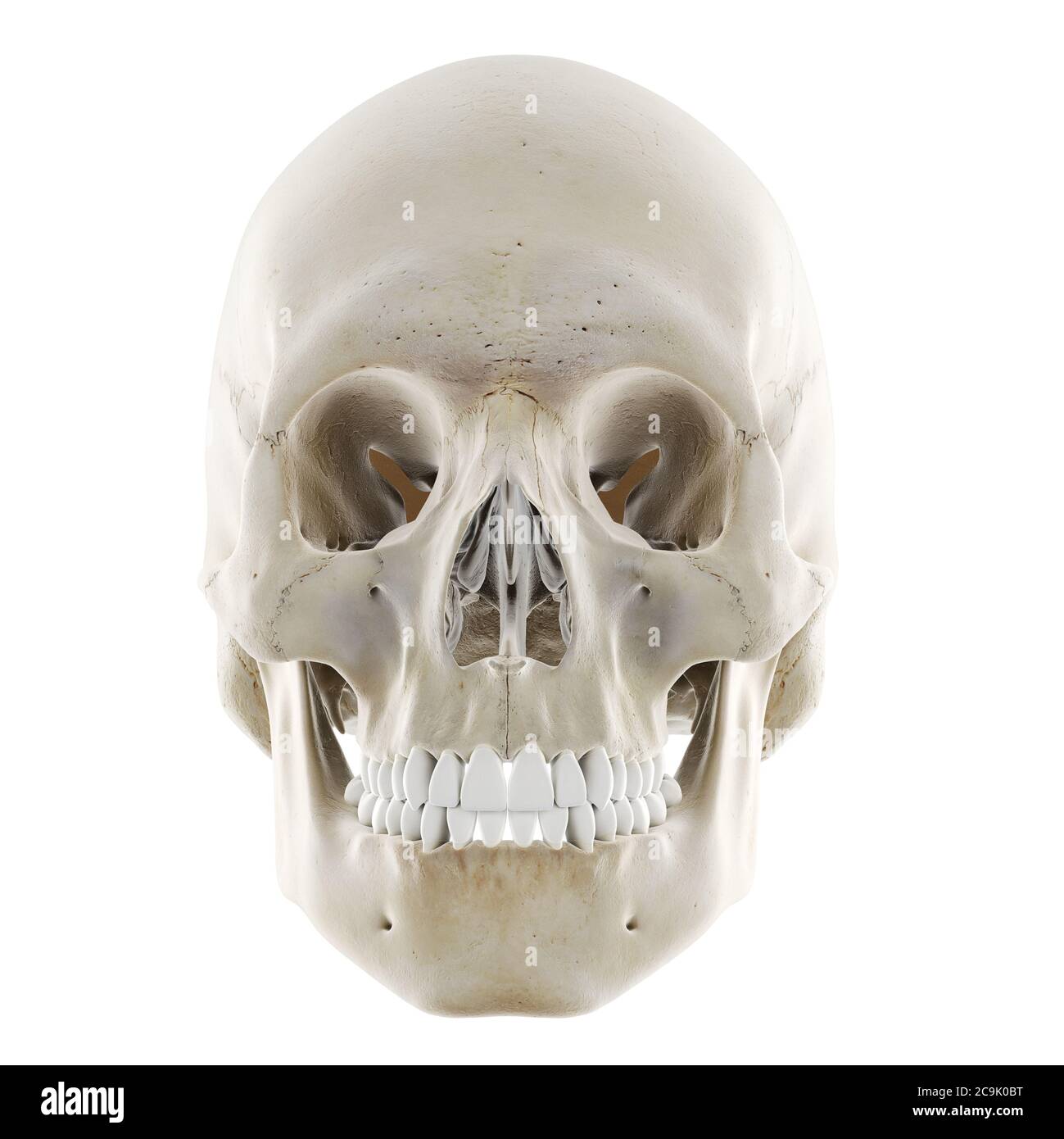 Human skull, illustration. Stock Photohttps://www.alamy.com/image-license-details/?v=1https://www.alamy.com/human-skull-illustration-image367367052.html
RF2C9K0BT–Human skull, illustration.
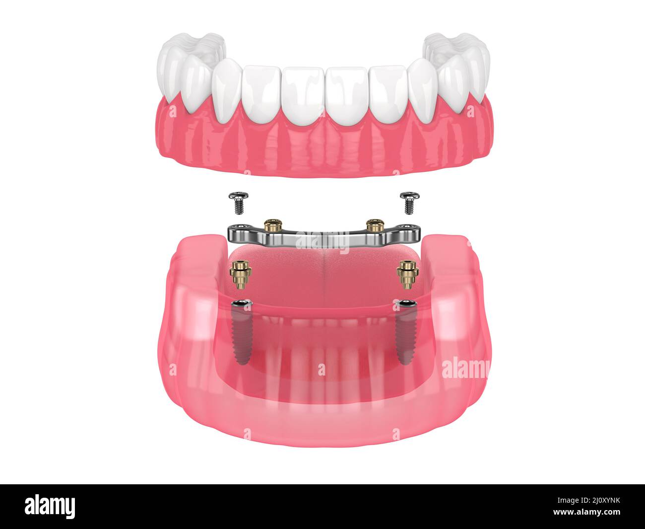 Bar retained removable overdenture installation supported by implants over white backgroud Stock Photohttps://www.alamy.com/image-license-details/?v=1https://www.alamy.com/bar-retained-removable-overdenture-installation-supported-by-implants-over-white-backgroud-image465272463.html
RF2J0XYNK–Bar retained removable overdenture installation supported by implants over white backgroud
 . The physiology of the domestic animals; a text-book for veterinary and medical students and practitioners. Physiology, Comparative; Domestic animals. Fig. 86.—Head of Carnivora—Dog. (Biclard.) Fig. 87.—Inferior Maxillary Bone of Carnivora—Polak Beak. (B&clard.) c, profile view of articular condyle of lower jaw (condyle of right side); d, front view of same condyle. and forward motion of the lower jaw, which is, therefore, the character- istic motion of rodents. Second.—In the ruminants the jaws are long and feeble, the canine and upper incisor teeth are absent, while the molars are compo Stock Photohttps://www.alamy.com/image-license-details/?v=1https://www.alamy.com/the-physiology-of-the-domestic-animals-a-text-book-for-veterinary-and-medical-students-and-practitioners-physiology-comparative-domestic-animals-fig-86head-of-carnivoradog-biclard-fig-87inferior-maxillary-bone-of-carnivorapolak-beak-bampclard-c-profile-view-of-articular-condyle-of-lower-jaw-condyle-of-right-side-d-front-view-of-same-condyle-and-forward-motion-of-the-lower-jaw-which-is-therefore-the-character-istic-motion-of-rodents-secondin-the-ruminants-the-jaws-are-long-and-feeble-the-canine-and-upper-incisor-teeth-are-absent-while-the-molars-are-compo-image232343526.html
RMRE04F2–. The physiology of the domestic animals; a text-book for veterinary and medical students and practitioners. Physiology, Comparative; Domestic animals. Fig. 86.—Head of Carnivora—Dog. (Biclard.) Fig. 87.—Inferior Maxillary Bone of Carnivora—Polak Beak. (B&clard.) c, profile view of articular condyle of lower jaw (condyle of right side); d, front view of same condyle. and forward motion of the lower jaw, which is, therefore, the character- istic motion of rodents. Second.—In the ruminants the jaws are long and feeble, the canine and upper incisor teeth are absent, while the molars are compo
 3d rendered medically accurate illustration of the skull with open jaw Stock Photohttps://www.alamy.com/image-license-details/?v=1https://www.alamy.com/3d-rendered-medically-accurate-illustration-of-the-skull-with-open-jaw-image334071428.html
RF2ABE7EC–3d rendered medically accurate illustration of the skull with open jaw
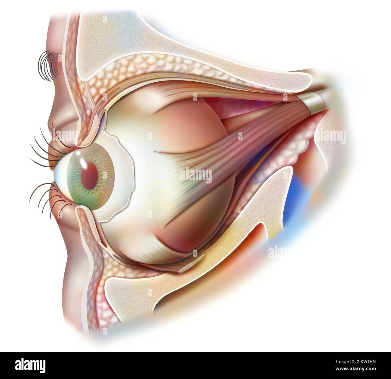 Anatomy of the eye and eyelid (viewed from 3/4) with iris, pupil. Stock Photohttps://www.alamy.com/image-license-details/?v=1https://www.alamy.com/anatomy-of-the-eye-and-eyelid-viewed-from-34-with-iris-pupil-image476926513.html
RF2JKWTHN–Anatomy of the eye and eyelid (viewed from 3/4) with iris, pupil.
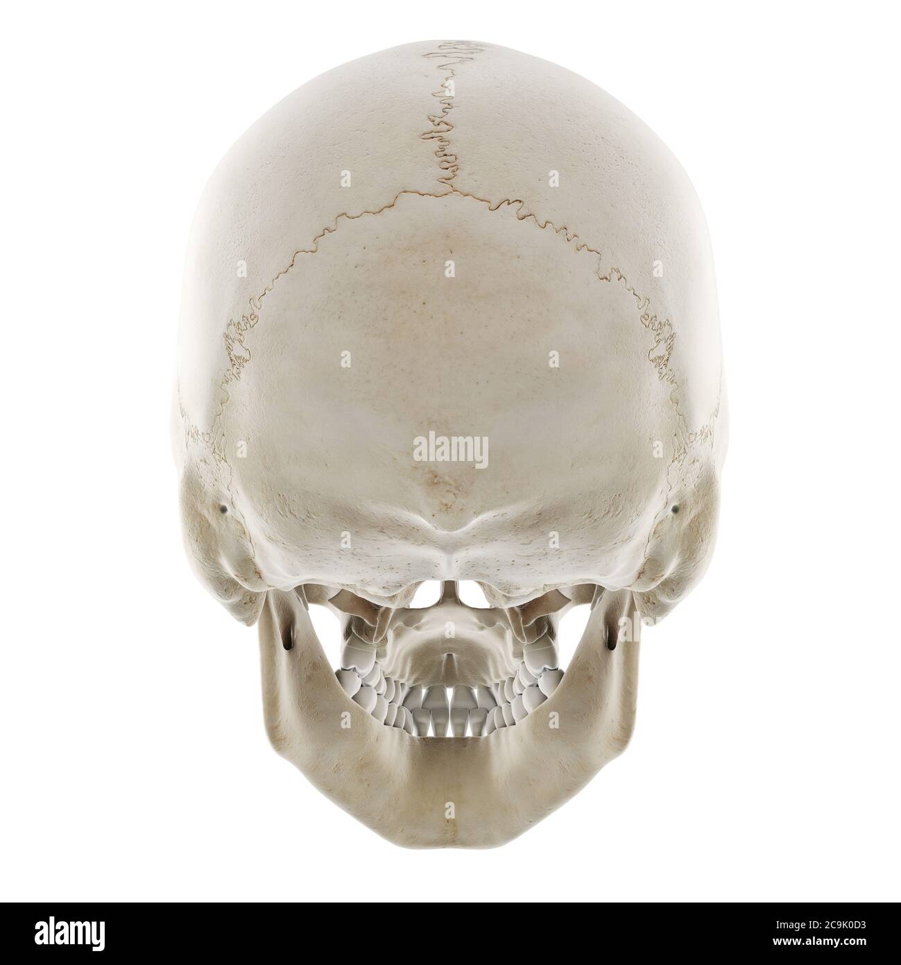 Human skull, illustration. Stock Photohttps://www.alamy.com/image-license-details/?v=1https://www.alamy.com/human-skull-illustration-image367367087.html
RF2C9K0D3–Human skull, illustration.
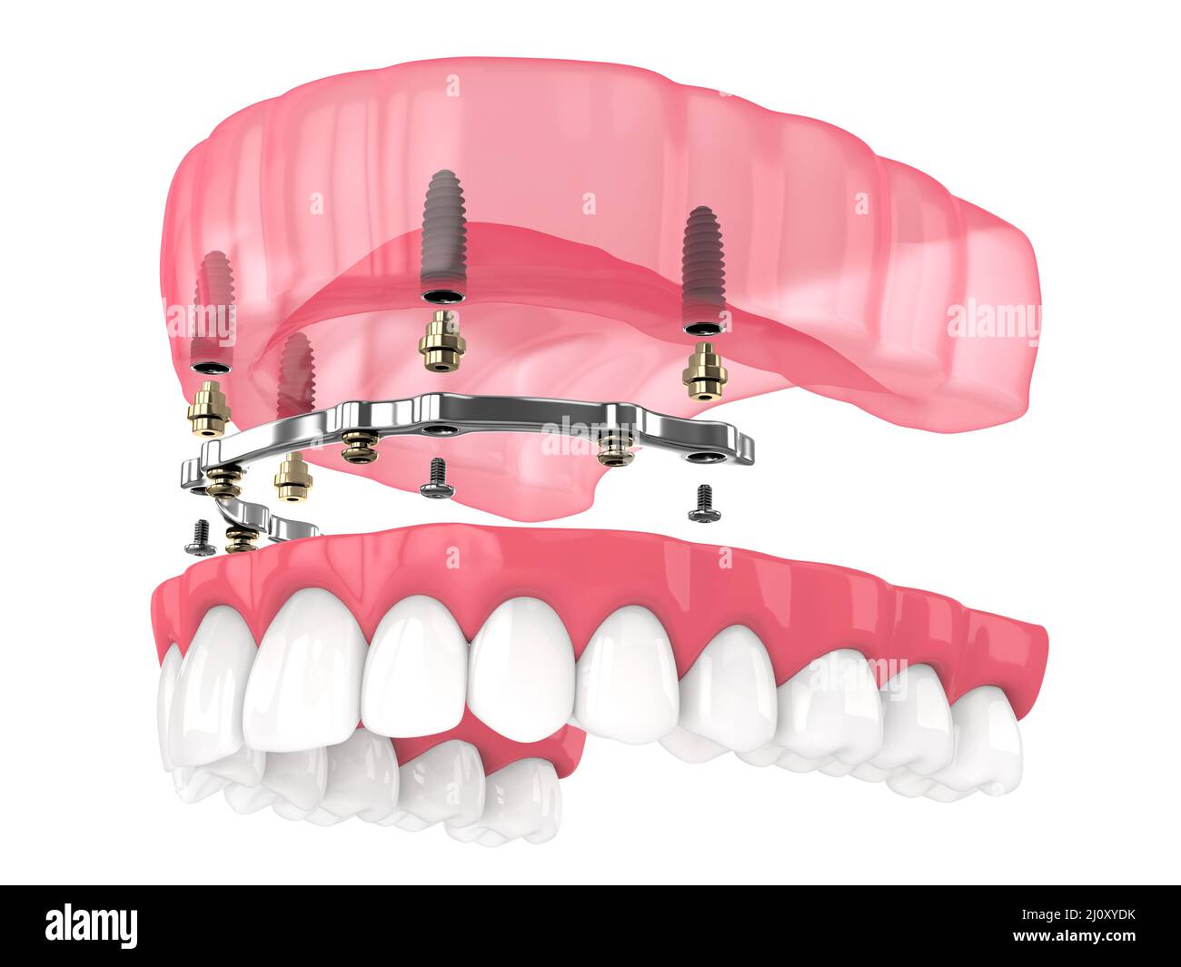 Bar retained removable overdenture installation supported by implants over white backgroud Stock Photohttps://www.alamy.com/image-license-details/?v=1https://www.alamy.com/bar-retained-removable-overdenture-installation-supported-by-implants-over-white-backgroud-image465272239.html
RF2J0XYDK–Bar retained removable overdenture installation supported by implants over white backgroud
 . The physiology of domestic animals ... Physiology, Comparative; Veterinary physiology. Fig. 86.—Head of Carnivora—Dog. (Btclard.) Fig. 87.—Inferior Maxillary Bone of Carnivora—Polar Bear. (B6clard.) c, profile view of articular condyle of lower jaw (condyle of right side); ct, front view of same condyle. and forward motion of the lower jaw, which is, therefore, the character- istic motion of rodents. Second.—In the ruminants the jaws are long and feeble, the canine and upper incisor teeth are absent, while the molars are compound teeth with a flat crown, with the enamel arranged in anteropos Stock Photohttps://www.alamy.com/image-license-details/?v=1https://www.alamy.com/the-physiology-of-domestic-animals-physiology-comparative-veterinary-physiology-fig-86head-of-carnivoradog-btclard-fig-87inferior-maxillary-bone-of-carnivorapolar-bear-b6clard-c-profile-view-of-articular-condyle-of-lower-jaw-condyle-of-right-side-ct-front-view-of-same-condyle-and-forward-motion-of-the-lower-jaw-which-is-therefore-the-character-istic-motion-of-rodents-secondin-the-ruminants-the-jaws-are-long-and-feeble-the-canine-and-upper-incisor-teeth-are-absent-while-the-molars-are-compound-teeth-with-a-flat-crown-with-the-enamel-arranged-in-anteropos-image232426694.html
RMRE3XHA–. The physiology of domestic animals ... Physiology, Comparative; Veterinary physiology. Fig. 86.—Head of Carnivora—Dog. (Btclard.) Fig. 87.—Inferior Maxillary Bone of Carnivora—Polar Bear. (B6clard.) c, profile view of articular condyle of lower jaw (condyle of right side); ct, front view of same condyle. and forward motion of the lower jaw, which is, therefore, the character- istic motion of rodents. Second.—In the ruminants the jaws are long and feeble, the canine and upper incisor teeth are absent, while the molars are compound teeth with a flat crown, with the enamel arranged in anteropos
 3d rendered medically accurate illustration of the skull with open jaw Stock Photohttps://www.alamy.com/image-license-details/?v=1https://www.alamy.com/3d-rendered-medically-accurate-illustration-of-the-skull-with-open-jaw-image334071392.html
RF2ABE7D4–3d rendered medically accurate illustration of the skull with open jaw
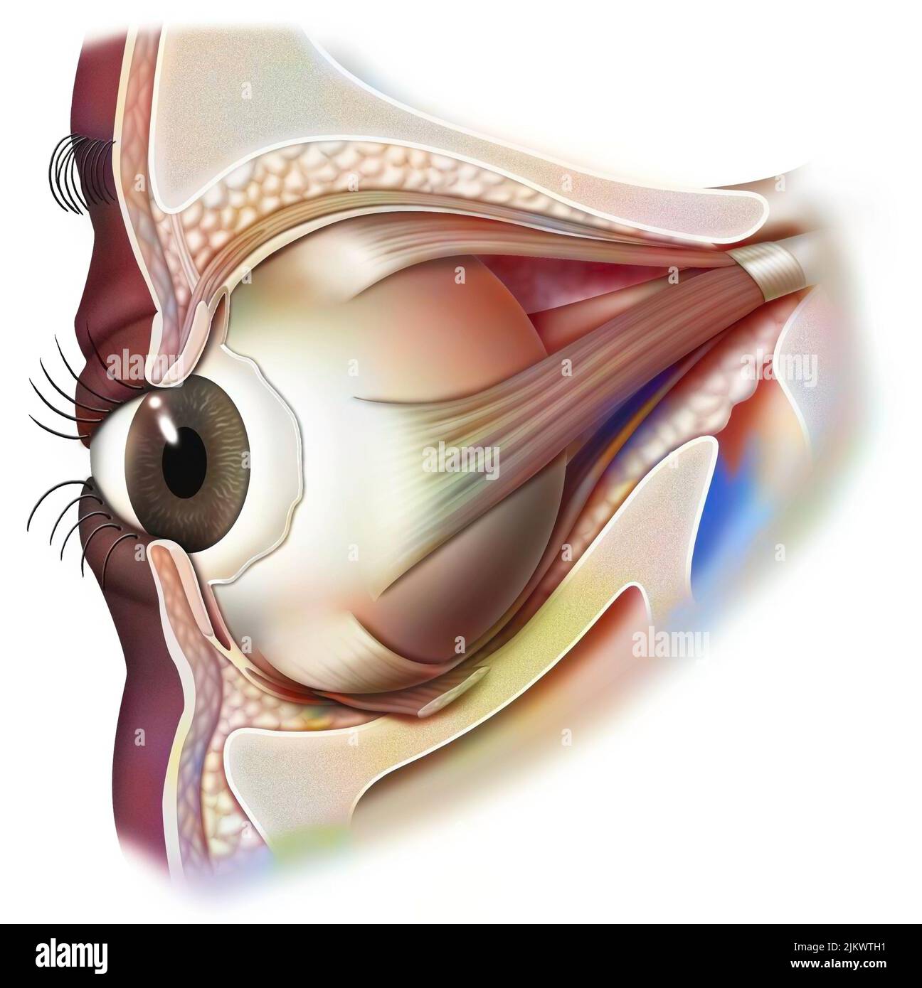 Anatomy of the eye and eyelid (viewed from 3/4) with iris, pupil. Stock Photohttps://www.alamy.com/image-license-details/?v=1https://www.alamy.com/anatomy-of-the-eye-and-eyelid-viewed-from-34-with-iris-pupil-image476926493.html
RF2JKWTH1–Anatomy of the eye and eyelid (viewed from 3/4) with iris, pupil.
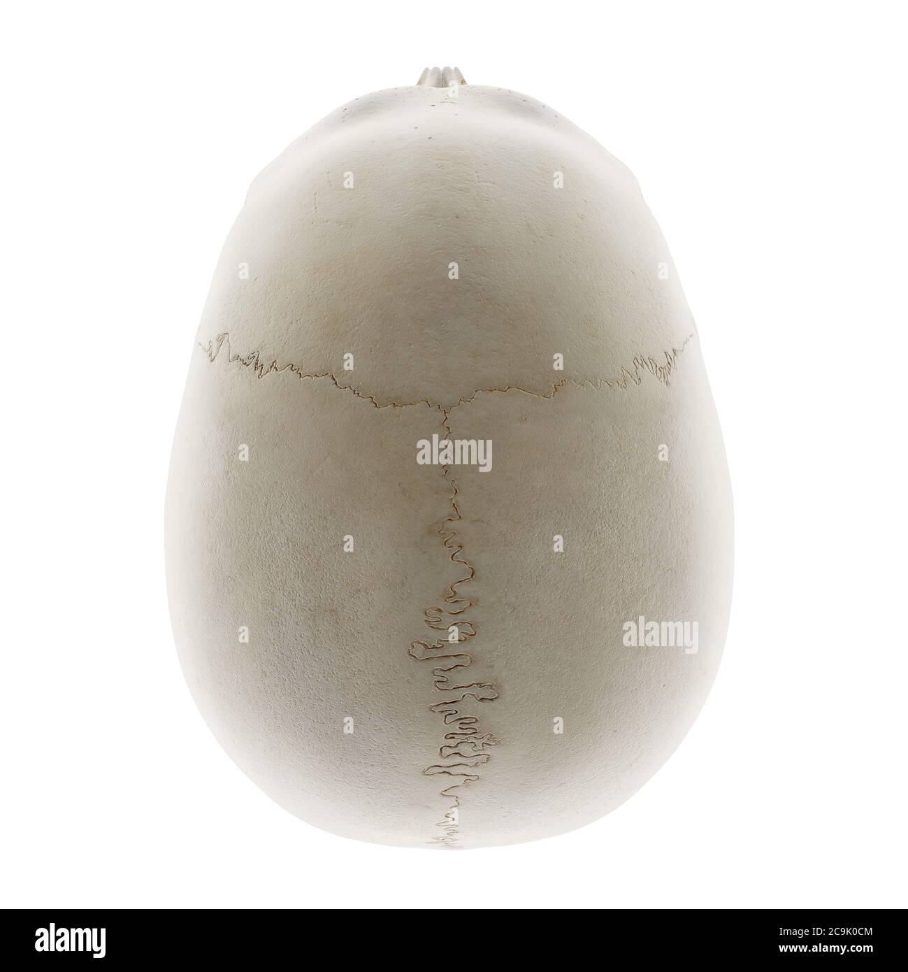 Human skull, illustration. Stock Photohttps://www.alamy.com/image-license-details/?v=1https://www.alamy.com/human-skull-illustration-image367367076.html
RF2C9K0CM–Human skull, illustration.
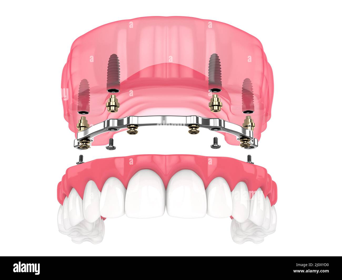 Bar retained removable overdenture installation supported by implants over white backgroud Stock Photohttps://www.alamy.com/image-license-details/?v=1https://www.alamy.com/bar-retained-removable-overdenture-installation-supported-by-implants-over-white-backgroud-image465272220.html
RF2J0XYD0–Bar retained removable overdenture installation supported by implants over white backgroud
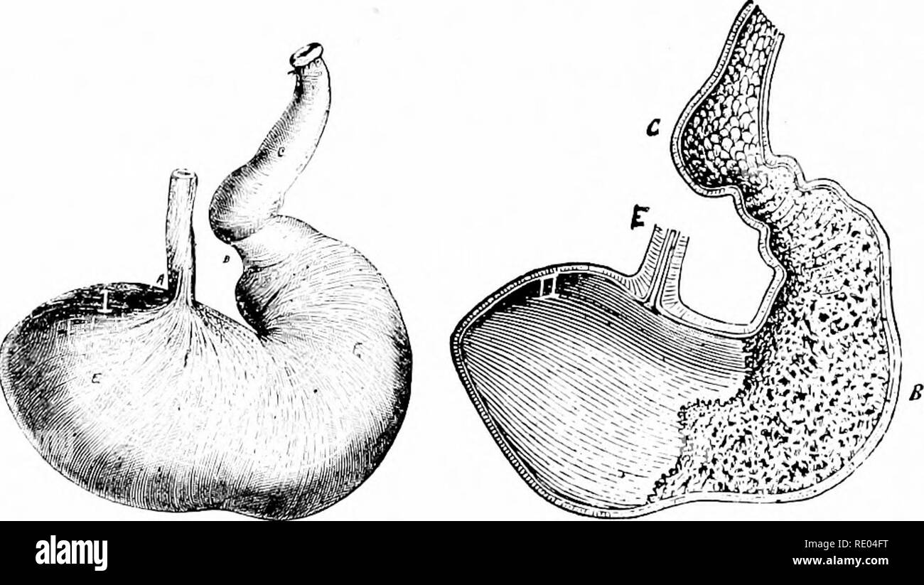 . Veterinary studies for agricultural students. Veterinary medicine. 4D ANATOMY. maxillary, one sub-lingual, and two molar. They secrete saliva which helps to change insoluble and useless starch into a soluble and useful sugar. It also assists in swallowing by so moisten- ing the food that it passes easily along. This is especially im- portant for animals like the horse, cow and sheep that live upon a dry and more or less bulky food. The horse needs on an average about 85 pounds and the cow 120 pounds every 24 hours. The parotid is located behind the lower jaw and below the base of the ear. It Stock Photohttps://www.alamy.com/image-license-details/?v=1https://www.alamy.com/veterinary-studies-for-agricultural-students-veterinary-medicine-4d-anatomy-maxillary-one-sub-lingual-and-two-molar-they-secrete-saliva-which-helps-to-change-insoluble-and-useless-starch-into-a-soluble-and-useful-sugar-it-also-assists-in-swallowing-by-so-moisten-ing-the-food-that-it-passes-easily-along-this-is-especially-im-portant-for-animals-like-the-horse-cow-and-sheep-that-live-upon-a-dry-and-more-or-less-bulky-food-the-horse-needs-on-an-average-about-85-pounds-and-the-cow-120-pounds-every-24-hours-the-parotid-is-located-behind-the-lower-jaw-and-below-the-base-of-the-ear-it-image232343548.html
RMRE04FT–. Veterinary studies for agricultural students. Veterinary medicine. 4D ANATOMY. maxillary, one sub-lingual, and two molar. They secrete saliva which helps to change insoluble and useless starch into a soluble and useful sugar. It also assists in swallowing by so moisten- ing the food that it passes easily along. This is especially im- portant for animals like the horse, cow and sheep that live upon a dry and more or less bulky food. The horse needs on an average about 85 pounds and the cow 120 pounds every 24 hours. The parotid is located behind the lower jaw and below the base of the ear. It
 3d rendered medically accurate illustration of the skull with open jaw Stock Photohttps://www.alamy.com/image-license-details/?v=1https://www.alamy.com/3d-rendered-medically-accurate-illustration-of-the-skull-with-open-jaw-image334071395.html
RF2ABE7D7–3d rendered medically accurate illustration of the skull with open jaw
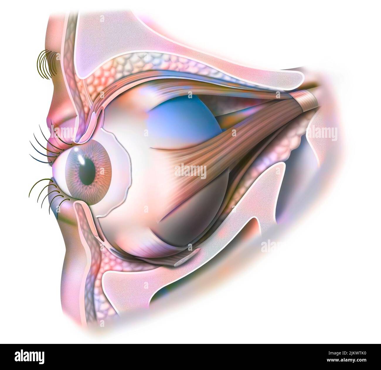 Anatomy of the eye and eyelid (viewed from 3/4) with iris, pupil. Stock Photohttps://www.alamy.com/image-license-details/?v=1https://www.alamy.com/anatomy-of-the-eye-and-eyelid-viewed-from-34-with-iris-pupil-image476926548.html
RF2JKWTK0–Anatomy of the eye and eyelid (viewed from 3/4) with iris, pupil.
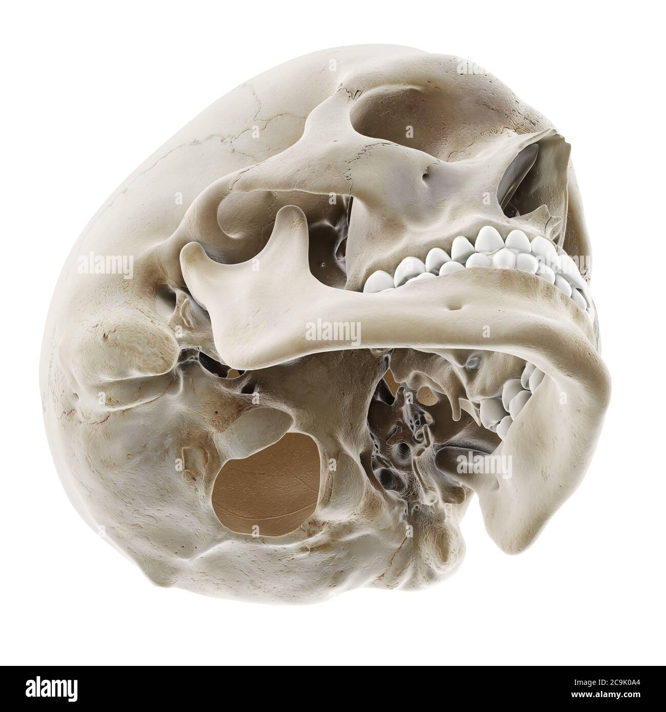 Human skull, illustration. Stock Photohttps://www.alamy.com/image-license-details/?v=1https://www.alamy.com/human-skull-illustration-image367367004.html
RF2C9K0A4–Human skull, illustration.
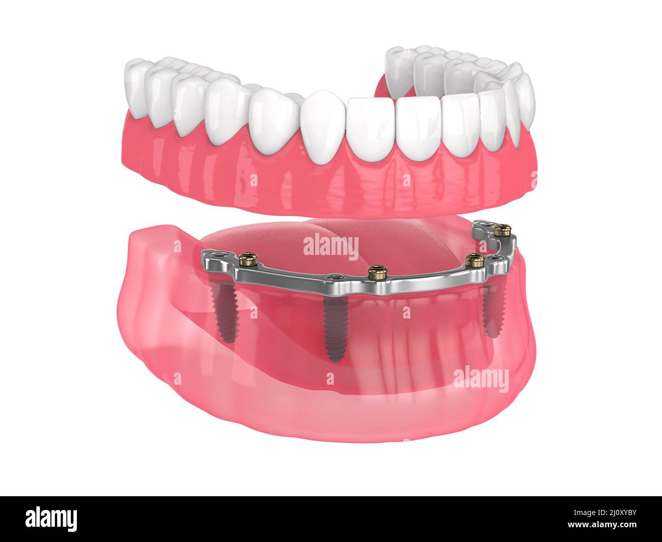 Bar retained removable overdenture installation supported by implants over white backgroud Stock Photohttps://www.alamy.com/image-license-details/?v=1https://www.alamy.com/bar-retained-removable-overdenture-installation-supported-by-implants-over-white-backgroud-image465272191.html
RF2J0XYBY–Bar retained removable overdenture installation supported by implants over white backgroud
 . The bird, its form and function. Birds. The Skull 107 large rounded portion taking up most of the skull proper is, of course, the box of bone which protects the brain. On each side, a large cavity shows where the eyes are placed, and if we compare this skull with that of a cat or dog or with that of a human being, we will see what great importance eyes must be to a bird; the cavities for them are so much larger than in other animals. Back 3raii2i Cdse. Fig. 8.3.—Skull of Fowl, showing orbit, brain-case, ear, lower jaw, premaxillary (Pmx.), maxillary (Mx.), vomer (Vo.), lacrymal (Lc), jugal ( Stock Photohttps://www.alamy.com/image-license-details/?v=1https://www.alamy.com/the-bird-its-form-and-function-birds-the-skull-107-large-rounded-portion-taking-up-most-of-the-skull-proper-is-of-course-the-box-of-bone-which-protects-the-brain-on-each-side-a-large-cavity-shows-where-the-eyes-are-placed-and-if-we-compare-this-skull-with-that-of-a-cat-or-dog-or-with-that-of-a-human-being-we-will-see-what-great-importance-eyes-must-be-to-a-bird-the-cavities-for-them-are-so-much-larger-than-in-other-animals-back-3raii2i-cdse-fig-83skull-of-fowl-showing-orbit-brain-case-ear-lower-jaw-premaxillary-pmx-maxillary-mx-vomer-vo-lacrymal-lc-jugal-image232386396.html
RMRE2364–. The bird, its form and function. Birds. The Skull 107 large rounded portion taking up most of the skull proper is, of course, the box of bone which protects the brain. On each side, a large cavity shows where the eyes are placed, and if we compare this skull with that of a cat or dog or with that of a human being, we will see what great importance eyes must be to a bird; the cavities for them are so much larger than in other animals. Back 3raii2i Cdse. Fig. 8.3.—Skull of Fowl, showing orbit, brain-case, ear, lower jaw, premaxillary (Pmx.), maxillary (Mx.), vomer (Vo.), lacrymal (Lc), jugal (
 3d rendered medically accurate illustration of the skull with open jaw Stock Photohttps://www.alamy.com/image-license-details/?v=1https://www.alamy.com/3d-rendered-medically-accurate-illustration-of-the-skull-with-open-jaw-image334050066.html
RF2ABD87E–3d rendered medically accurate illustration of the skull with open jaw
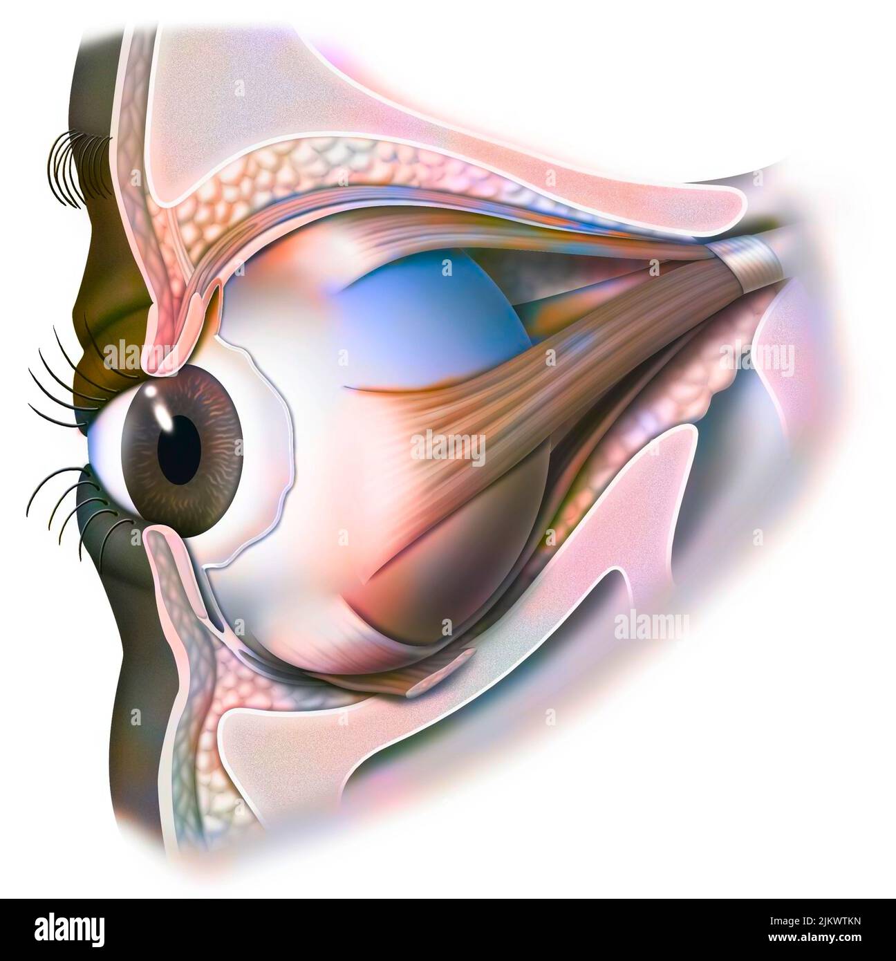 Anatomy of the eye and eyelid (viewed from 3/4) with iris, pupil. Stock Photohttps://www.alamy.com/image-license-details/?v=1https://www.alamy.com/anatomy-of-the-eye-and-eyelid-viewed-from-34-with-iris-pupil-image476926569.html
RF2JKWTKN–Anatomy of the eye and eyelid (viewed from 3/4) with iris, pupil.
 Human skull, illustration. Stock Photohttps://www.alamy.com/image-license-details/?v=1https://www.alamy.com/human-skull-illustration-image367367080.html
RF2C9K0CT–Human skull, illustration.
 . Fishes. Fishes. Fig. 293.—White Chub, Notropis Itudsonius (Chnton). Kilpatrick I^ake, Minn. Notropis. This includes the smaller and weaker species, from two to seven inches in length, characterized by the loss, mostly through degeneration, of special peculiarities of mouth, fins, and teeth. These have no barbels and never more than four teeth. Fig. 294.—Silver-jaw Minnow, Ericymha buccala Cope. Defiance, Ohio. in the main row. Few, if any, Asiatic species have so small a number, and in most of these the maxillary still retains its rudimentary barbel. But one American genus (Ortliodon) has mo Stock Photohttps://www.alamy.com/image-license-details/?v=1https://www.alamy.com/fishes-fishes-fig-293white-chub-notropis-itudsonius-chnton-kilpatrick-iake-minn-notropis-this-includes-the-smaller-and-weaker-species-from-two-to-seven-inches-in-length-characterized-by-the-loss-mostly-through-degeneration-of-special-peculiarities-of-mouth-fins-and-teeth-these-have-no-barbels-and-never-more-than-four-teeth-fig-294silver-jaw-minnow-ericymha-buccala-cope-defiance-ohio-in-the-main-row-few-if-any-asiatic-species-have-so-small-a-number-and-in-most-of-these-the-maxillary-still-retains-its-rudimentary-barbel-but-one-american-genus-ortliodon-has-mo-image232218357.html
RMRDPCTN–. Fishes. Fishes. Fig. 293.—White Chub, Notropis Itudsonius (Chnton). Kilpatrick I^ake, Minn. Notropis. This includes the smaller and weaker species, from two to seven inches in length, characterized by the loss, mostly through degeneration, of special peculiarities of mouth, fins, and teeth. These have no barbels and never more than four teeth. Fig. 294.—Silver-jaw Minnow, Ericymha buccala Cope. Defiance, Ohio. in the main row. Few, if any, Asiatic species have so small a number, and in most of these the maxillary still retains its rudimentary barbel. But one American genus (Ortliodon) has mo
 3d rendered medically accurate illustration of the skull with open jaw Stock Photohttps://www.alamy.com/image-license-details/?v=1https://www.alamy.com/3d-rendered-medically-accurate-illustration-of-the-skull-with-open-jaw-image334050109.html
RF2ABD891–3d rendered medically accurate illustration of the skull with open jaw
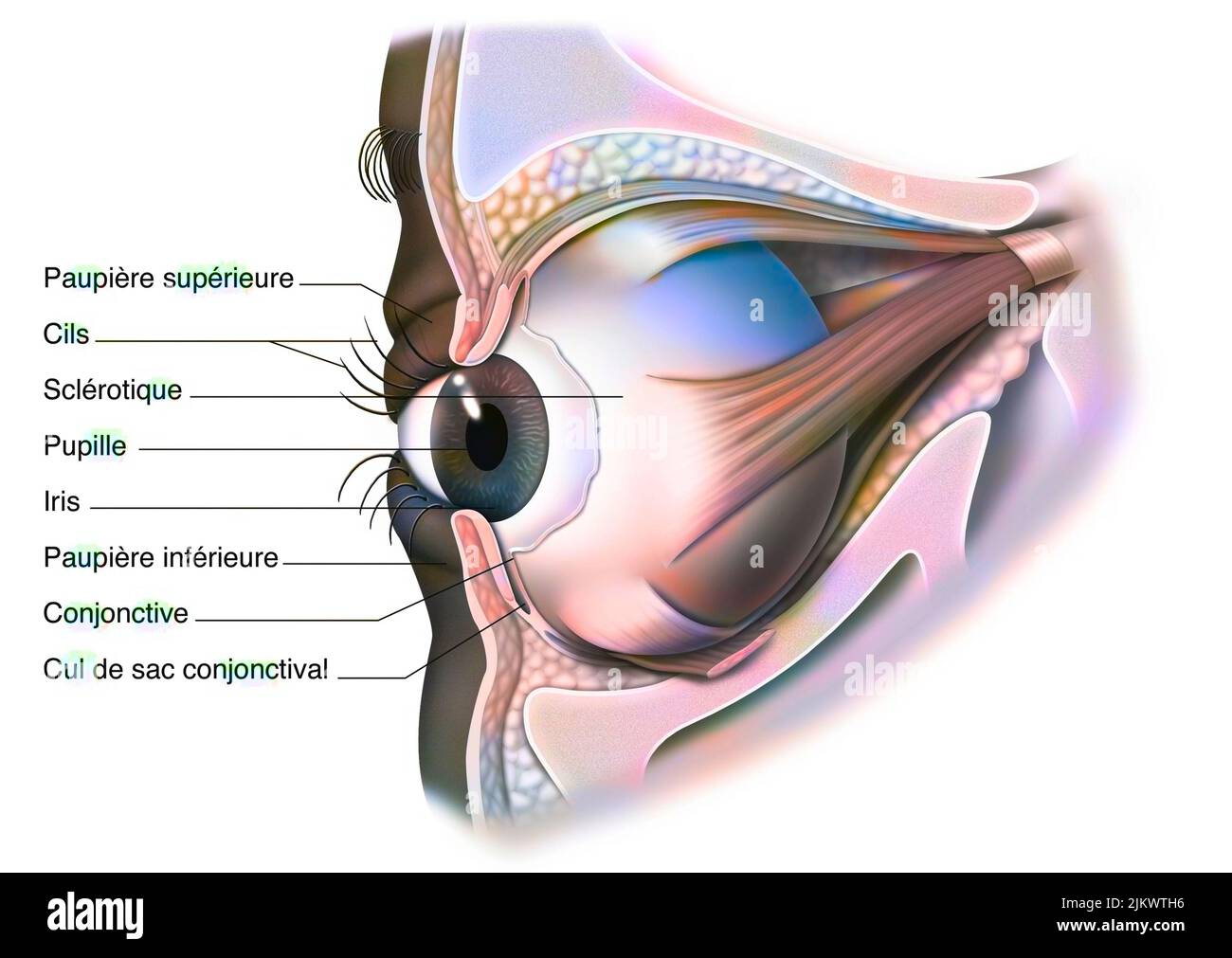 Anatomy of the eye and eyelid (viewed from 3/4) with iris, pupil. Stock Photohttps://www.alamy.com/image-license-details/?v=1https://www.alamy.com/anatomy-of-the-eye-and-eyelid-viewed-from-34-with-iris-pupil-image476926498.html
RF2JKWTH6–Anatomy of the eye and eyelid (viewed from 3/4) with iris, pupil.
