Maxillary surgery Black & White Stock Photos
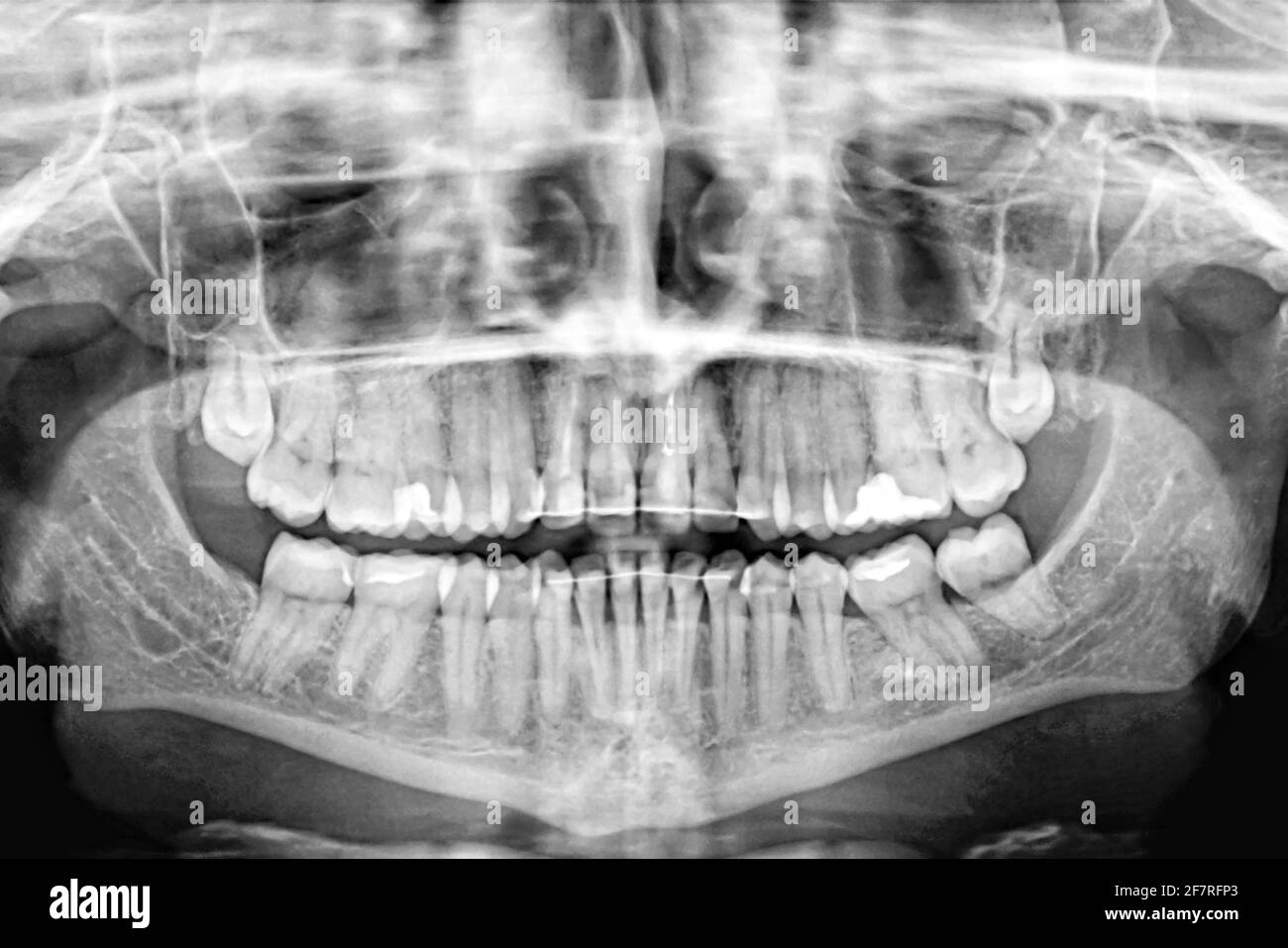 Panoramic X-ray scan of humans teeth.Examination and treatment. Dental care.Banner. Stock Photohttps://www.alamy.com/image-license-details/?v=1https://www.alamy.com/panoramic-x-ray-scan-of-humans-teethexamination-and-treatment-dental-carebanner-image417868699.html
RF2F7RFP3–Panoramic X-ray scan of humans teeth.Examination and treatment. Dental care.Banner.
 Hands of a dentist with a tool during a surgical procedure Stock Photohttps://www.alamy.com/image-license-details/?v=1https://www.alamy.com/hands-of-a-dentist-with-a-tool-during-a-surgical-procedure-image331598638.html
RF2A7DHCE–Hands of a dentist with a tool during a surgical procedure
 Dental Tooth Live life to the Maxillary Digital EPS File Stock Vectorhttps://www.alamy.com/image-license-details/?v=1https://www.alamy.com/dental-tooth-live-life-to-the-maxillary-digital-eps-file-image637492470.html
RF2S14846–Dental Tooth Live life to the Maxillary Digital EPS File
 . Manual of operative veterinary surgery. Veterinary surgery. i^ig^. ! ! I'M. 259.—Another Splint for Fracture of the Maxillary.. Please note that these images are extracted from scanned page images that may have been digitally enhanced for readability - coloration and appearance of these illustrations may not perfectly resemble the original work.. Liautard, Alexandre Franc?ois Augustin, 1835-. New York, Sabiston & Murray Stock Photohttps://www.alamy.com/image-license-details/?v=1https://www.alamy.com/manual-of-operative-veterinary-surgery-veterinary-surgery-iig-!-!-im-259another-splint-for-fracture-of-the-maxillary-please-note-that-these-images-are-extracted-from-scanned-page-images-that-may-have-been-digitally-enhanced-for-readability-coloration-and-appearance-of-these-illustrations-may-not-perfectly-resemble-the-original-work-liautard-alexandre-francois-augustin-1835-new-york-sabiston-amp-murray-image232339591.html
RMRDYYEF–. Manual of operative veterinary surgery. Veterinary surgery. i^ig^. ! ! I'M. 259.—Another Splint for Fracture of the Maxillary.. Please note that these images are extracted from scanned page images that may have been digitally enhanced for readability - coloration and appearance of these illustrations may not perfectly resemble the original work.. Liautard, Alexandre Franc?ois Augustin, 1835-. New York, Sabiston & Murray
 Maxillary human gum and teeth. Medically accurate tooth 3D illustration Stock Photohttps://www.alamy.com/image-license-details/?v=1https://www.alamy.com/maxillary-human-gum-and-teeth-medically-accurate-tooth-3d-illustration-image328500778.html
RF2A2CE2J–Maxillary human gum and teeth. Medically accurate tooth 3D illustration
RF2DGAEBY–incicor icon, black vector sign with editable strokes, concept illustration
 . Manual of operative veterinary surgery. Veterinary surgery. FEACTUEES. 233. Fig. 257.—Splint for Fracture of the Lower Maxillary. crepitation, and frequently paralysis of the under lip. But al- though the aspect of an animal suffering with a complete and often compound and comminuted fracture of the submaxiUa pre- sents at times a frightful spectacle, the prognosis of the case is comparatively simple, and recovery usually only a question of time. The severity of the lesion corresponds in degree with that of the violence to which it is due, the degree of simpUcity or the amount of compHcation Stock Photohttps://www.alamy.com/image-license-details/?v=1https://www.alamy.com/manual-of-operative-veterinary-surgery-veterinary-surgery-feactuees-233-fig-257splint-for-fracture-of-the-lower-maxillary-crepitation-and-frequently-paralysis-of-the-under-lip-but-al-though-the-aspect-of-an-animal-suffering-with-a-complete-and-often-compound-and-comminuted-fracture-of-the-submaxiua-pre-sents-at-times-a-frightful-spectacle-the-prognosis-of-the-case-is-comparatively-simple-and-recovery-usually-only-a-question-of-time-the-severity-of-the-lesion-corresponds-in-degree-with-that-of-the-violence-to-which-it-is-due-the-degree-of-simpucity-or-the-amount-of-comphcation-image232339601.html
RMRDYYEW–. Manual of operative veterinary surgery. Veterinary surgery. FEACTUEES. 233. Fig. 257.—Splint for Fracture of the Lower Maxillary. crepitation, and frequently paralysis of the under lip. But al- though the aspect of an animal suffering with a complete and often compound and comminuted fracture of the submaxiUa pre- sents at times a frightful spectacle, the prognosis of the case is comparatively simple, and recovery usually only a question of time. The severity of the lesion corresponds in degree with that of the violence to which it is due, the degree of simpUcity or the amount of compHcation
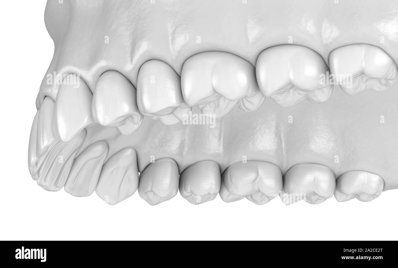 Maxillary human gum and teeth. Medically accurate tooth 3D illustration Stock Photohttps://www.alamy.com/image-license-details/?v=1https://www.alamy.com/maxillary-human-gum-and-teeth-medically-accurate-tooth-3d-illustration-image328500784.html
RF2A2CE2T–Maxillary human gum and teeth. Medically accurate tooth 3D illustration
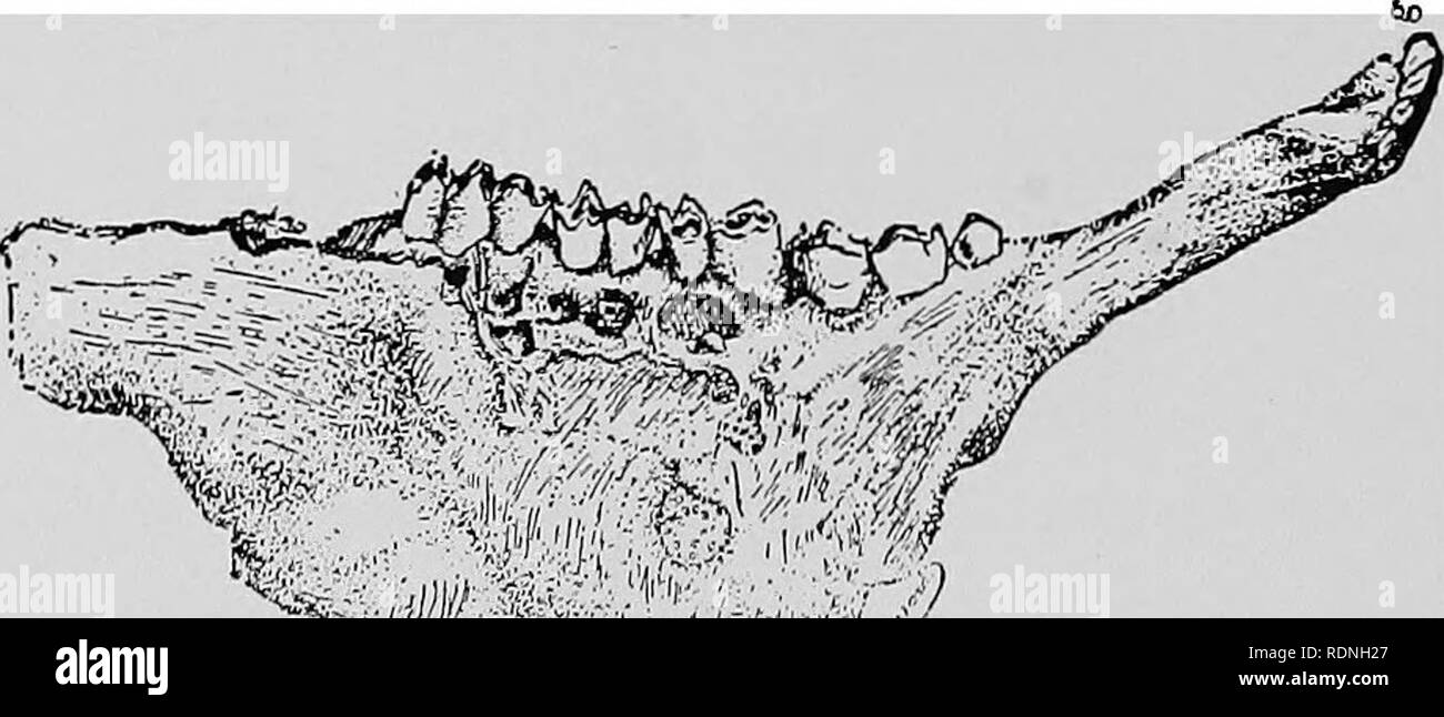 . Veterinary surgery ... Veterinary surgery; Veterinary pathology; Horses; Teeth; Domestic animals. FlG. 148B. Actinomycostic Superior Maxillary in the Ox. ing of the teeth. In the pharynx the disease is manifested by tumefaction of the parotid region and by dysphagia and dyspnoea. Actinomycosis of the lungs and other internal structures is diagnosed only at the autopsy. Symptoms of organic disorder may lead a diagnostician to suspect the ex-. ^v I '* '/ Fig. 148c. Actinomycostic Inferior Maxillary in the Ox. istence of the disease in districts where actinomycosis is prevalent, but a positi Stock Photohttps://www.alamy.com/image-license-details/?v=1https://www.alamy.com/veterinary-surgery-veterinary-surgery-veterinary-pathology-horses-teeth-domestic-animals-flg-148b-actinomycostic-superior-maxillary-in-the-ox-ing-of-the-teeth-in-the-pharynx-the-disease-is-manifested-by-tumefaction-of-the-parotid-region-and-by-dysphagia-and-dyspnoea-actinomycosis-of-the-lungs-and-other-internal-structures-is-diagnosed-only-at-the-autopsy-symptoms-of-organic-disorder-may-lead-a-diagnostician-to-suspect-the-ex-v-i-fig-148c-actinomycostic-inferior-maxillary-in-the-ox-istence-of-the-disease-in-districts-where-actinomycosis-is-prevalent-but-a-positi-image232199695.html
RMRDNH27–. Veterinary surgery ... Veterinary surgery; Veterinary pathology; Horses; Teeth; Domestic animals. FlG. 148B. Actinomycostic Superior Maxillary in the Ox. ing of the teeth. In the pharynx the disease is manifested by tumefaction of the parotid region and by dysphagia and dyspnoea. Actinomycosis of the lungs and other internal structures is diagnosed only at the autopsy. Symptoms of organic disorder may lead a diagnostician to suspect the ex-. ^v I '* '/ Fig. 148c. Actinomycostic Inferior Maxillary in the Ox. istence of the disease in districts where actinomycosis is prevalent, but a positi
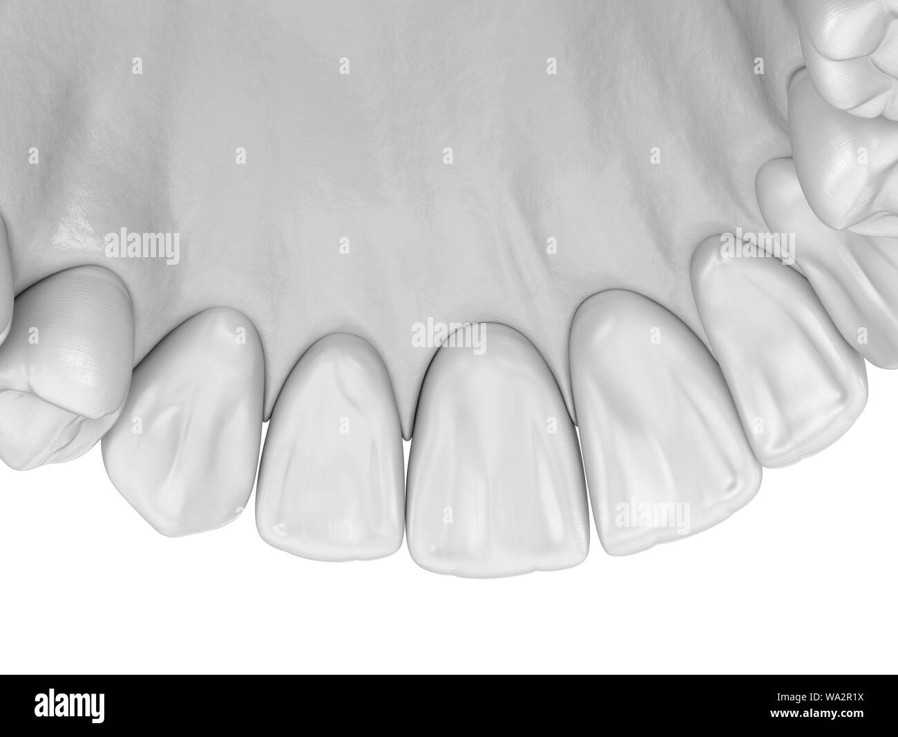 Maxillary human gum and teeth. Medically accurate tooth 3D illustration Stock Photohttps://www.alamy.com/image-license-details/?v=1https://www.alamy.com/maxillary-human-gum-and-teeth-medically-accurate-tooth-3d-illustration-image264364070.html
RFWA2R1X–Maxillary human gum and teeth. Medically accurate tooth 3D illustration
 . Manual of operative veterinary surgery. Veterinary surgery. 520 OPEBATIONS ON THE CIRCULATOEY SYSTEM. time without aid, but if it is desirable to continue it, the parts may be fomented with warm water, or covered with a warm poultice. (e) Bleeding at the PaZa«e.—Bleeding in this region of the mouth is done by a division of the capiUary network which rests between the mucous membrane and the fibrous coat which lines the bones forming the palate. The bones represented by the inferior face of the palatine pro- cess of the great maxillary bone, and the posterior face of the short process of the Stock Photohttps://www.alamy.com/image-license-details/?v=1https://www.alamy.com/manual-of-operative-veterinary-surgery-veterinary-surgery-520-opebations-on-the-circulatoey-system-time-without-aid-but-if-it-is-desirable-to-continue-it-the-parts-may-be-fomented-with-warm-water-or-covered-with-a-warm-poultice-e-bleeding-at-the-pazaebleeding-in-this-region-of-the-mouth-is-done-by-a-division-of-the-capiuary-network-which-rests-between-the-mucous-membrane-and-the-fibrous-coat-which-lines-the-bones-forming-the-palate-the-bones-represented-by-the-inferior-face-of-the-palatine-pro-cess-of-the-great-maxillary-bone-and-the-posterior-face-of-the-short-process-of-the-image232317795.html
RMRDXYM3–. Manual of operative veterinary surgery. Veterinary surgery. 520 OPEBATIONS ON THE CIRCULATOEY SYSTEM. time without aid, but if it is desirable to continue it, the parts may be fomented with warm water, or covered with a warm poultice. (e) Bleeding at the PaZa«e.—Bleeding in this region of the mouth is done by a division of the capiUary network which rests between the mucous membrane and the fibrous coat which lines the bones forming the palate. The bones represented by the inferior face of the palatine pro- cess of the great maxillary bone, and the posterior face of the short process of the
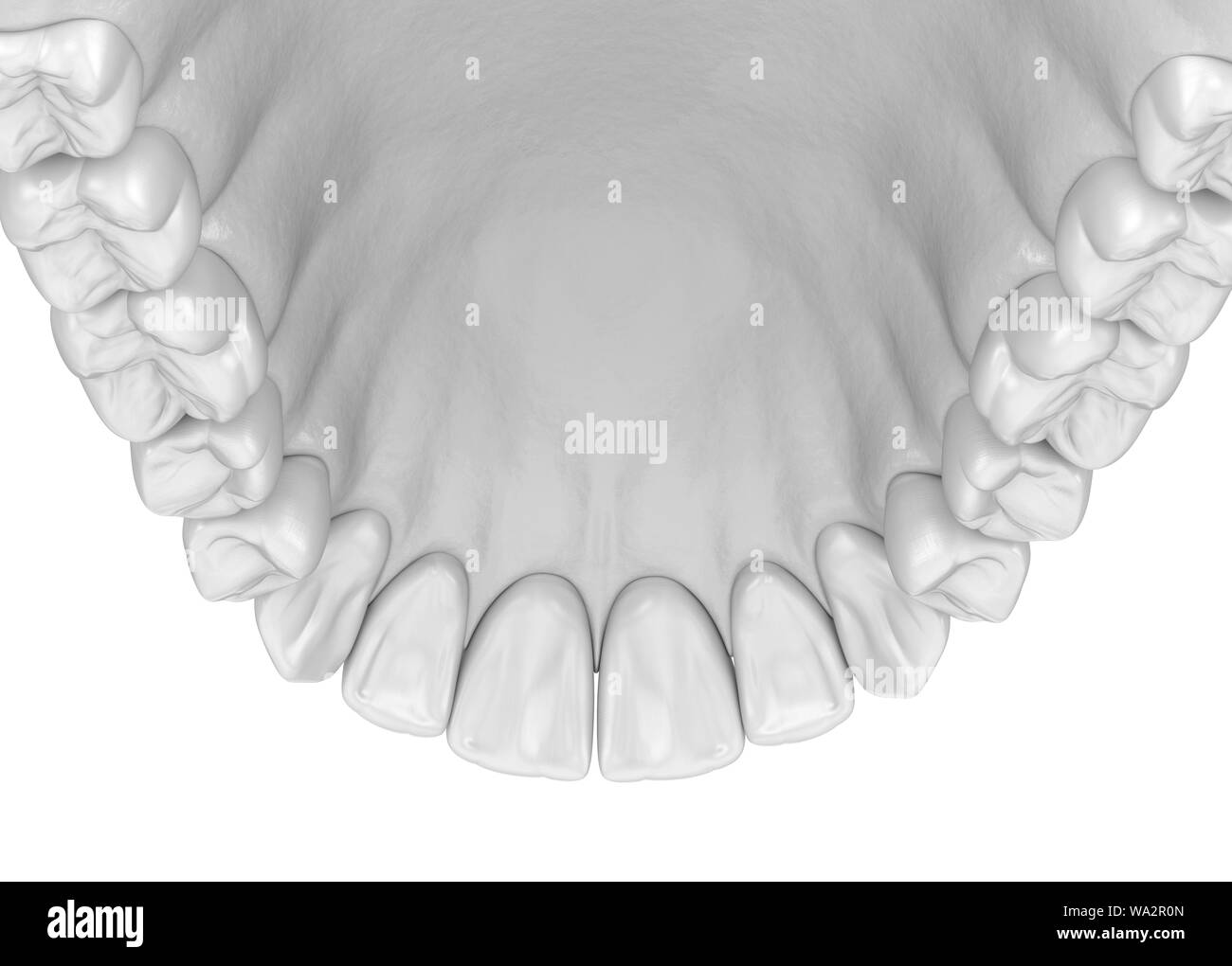 Maxillary human gum and teeth. Medically accurate tooth 3D illustration Stock Photohttps://www.alamy.com/image-license-details/?v=1https://www.alamy.com/maxillary-human-gum-and-teeth-medically-accurate-tooth-3d-illustration-image264364037.html
RFWA2R0N–Maxillary human gum and teeth. Medically accurate tooth 3D illustration
 . Veterinary surgery ... Veterinary surgery; Veterinary pathology; Horses; Teeth; Domestic animals. 26 ANIMAL DENTISTRY.. THE NERVES. The nerves are sensory and tactile, and are derived from the superior and the inferior maxillary branches of the tri-. Please note that these images are extracted from scanned page images that may have been digitally enhanced for readability - coloration and appearance of these illustrations may not perfectly resemble the original work.. Merillat, Louis A. (Louis Adolph), 1868-; Cade?ac, Ce?listin, 1858-; Le Blanc, Paul, 1872-; Carougeau, C. Chicago, A. Eger Stock Photohttps://www.alamy.com/image-license-details/?v=1https://www.alamy.com/veterinary-surgery-veterinary-surgery-veterinary-pathology-horses-teeth-domestic-animals-26-animal-dentistry-the-nerves-the-nerves-are-sensory-and-tactile-and-are-derived-from-the-superior-and-the-inferior-maxillary-branches-of-the-tri-please-note-that-these-images-are-extracted-from-scanned-page-images-that-may-have-been-digitally-enhanced-for-readability-coloration-and-appearance-of-these-illustrations-may-not-perfectly-resemble-the-original-work-merillat-louis-a-louis-adolph-1868-cadeac-celistin-1858-le-blanc-paul-1872-carougeau-c-chicago-a-eger-image232230944.html
RMRDR0X8–. Veterinary surgery ... Veterinary surgery; Veterinary pathology; Horses; Teeth; Domestic animals. 26 ANIMAL DENTISTRY.. THE NERVES. The nerves are sensory and tactile, and are derived from the superior and the inferior maxillary branches of the tri-. Please note that these images are extracted from scanned page images that may have been digitally enhanced for readability - coloration and appearance of these illustrations may not perfectly resemble the original work.. Merillat, Louis A. (Louis Adolph), 1868-; Cade?ac, Ce?listin, 1858-; Le Blanc, Paul, 1872-; Carougeau, C. Chicago, A. Eger
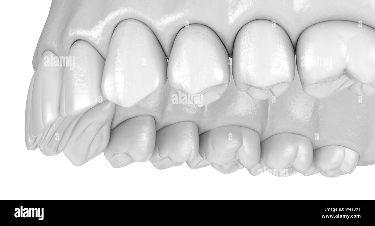 Maxillary human gum and teeth. Medically accurate tooth 3D illustration Stock Photohttps://www.alamy.com/image-license-details/?v=1https://www.alamy.com/maxillary-human-gum-and-teeth-medically-accurate-tooth-3d-illustration-image260639200.html
RFW413XT–Maxillary human gum and teeth. Medically accurate tooth 3D illustration
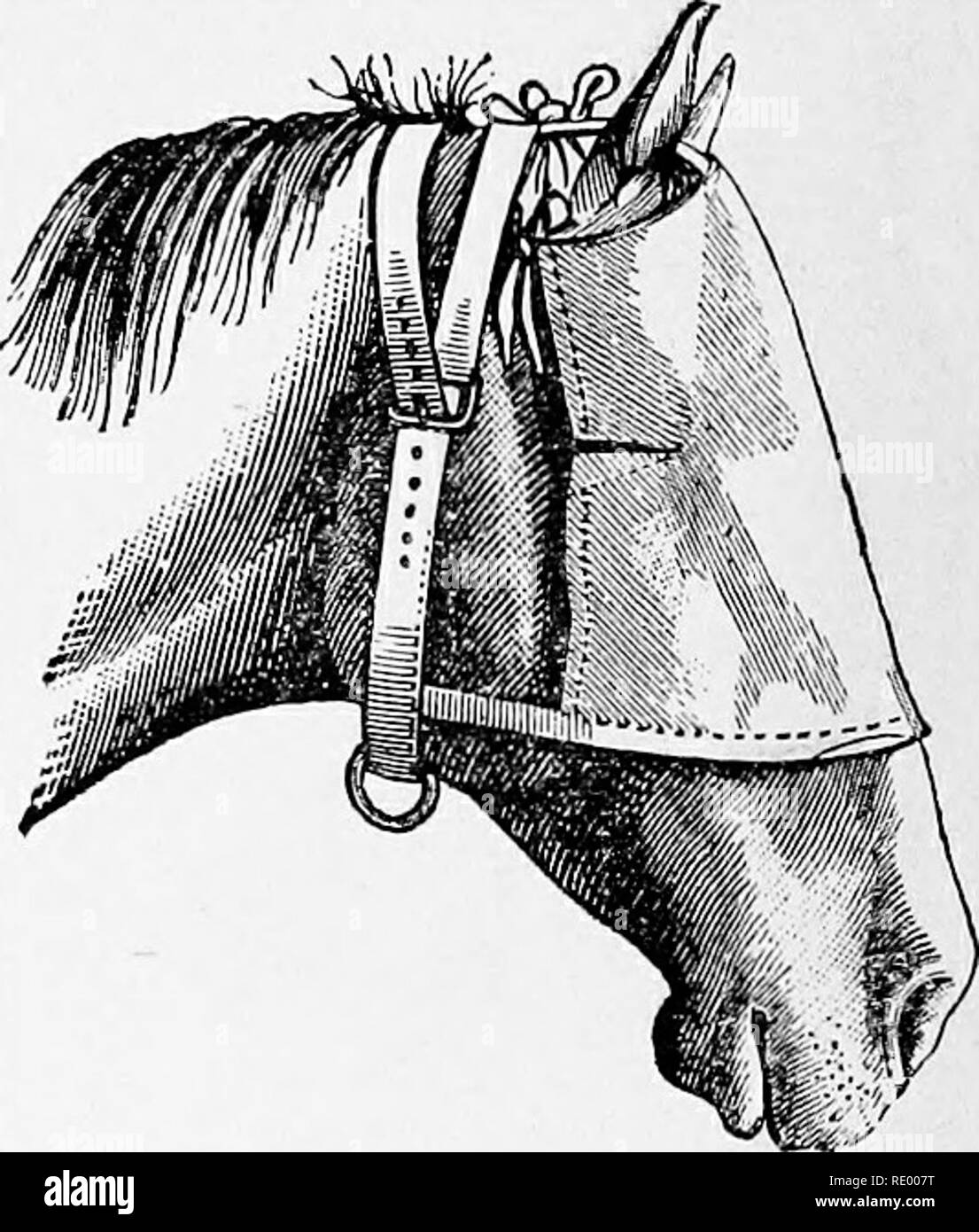 . Manual of operative veterinary surgery. Veterinary surgery. Fig. 111.—Monocular Band (full view). Fig. 112.—Monocular Band (side view). of the bandage, facilitate its adaptation to the surfaces it is to cover (Figs. 113, 114). 5th. Bandagefor the Maxillary Region.—This is of triangular shape, and is formed either of sheepskin or of two layers of cloth, between which a pad of oakum is sewed. It is furnished with four straps. When applied, the base of the triangle is turned backward, and the apex rests in the angle of the maxillary space. The two upper straps, attached at each angle of the bas Stock Photohttps://www.alamy.com/image-license-details/?v=1https://www.alamy.com/manual-of-operative-veterinary-surgery-veterinary-surgery-fig-111monocular-band-full-view-fig-112monocular-band-side-view-of-the-bandage-facilitate-its-adaptation-to-the-surfaces-it-is-to-cover-figs-113-114-5th-bandagefor-the-maxillary-regionthis-is-of-triangular-shape-and-is-formed-either-of-sheepskin-or-of-two-layers-of-cloth-between-which-a-pad-of-oakum-is-sewed-it-is-furnished-with-four-straps-when-applied-the-base-of-the-triangle-is-turned-backward-and-the-apex-rests-in-the-angle-of-the-maxillary-space-the-two-upper-straps-attached-at-each-angle-of-the-bas-image232340188.html
RMRE007T–. Manual of operative veterinary surgery. Veterinary surgery. Fig. 111.—Monocular Band (full view). Fig. 112.—Monocular Band (side view). of the bandage, facilitate its adaptation to the surfaces it is to cover (Figs. 113, 114). 5th. Bandagefor the Maxillary Region.—This is of triangular shape, and is formed either of sheepskin or of two layers of cloth, between which a pad of oakum is sewed. It is furnished with four straps. When applied, the base of the triangle is turned backward, and the apex rests in the angle of the maxillary space. The two upper straps, attached at each angle of the bas
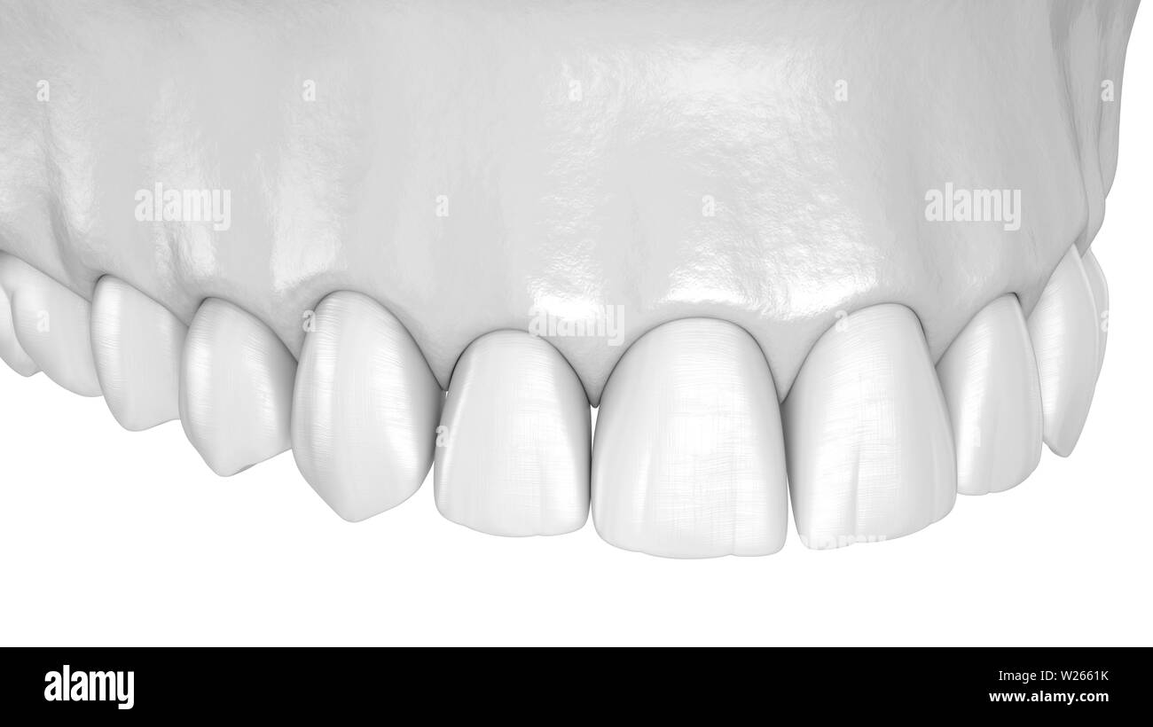 Maxillary human gum and teeth. Medically accurate tooth 3D illustration Stock Photohttps://www.alamy.com/image-license-details/?v=1https://www.alamy.com/maxillary-human-gum-and-teeth-medically-accurate-tooth-3d-illustration-image259521295.html
RFW2661K–Maxillary human gum and teeth. Medically accurate tooth 3D illustration
 . Surgical and obstetrical operations. Veterinary surgery. -5M FIG. 3. Trephining the Facial Sinuses. F, highest point at which an opening may be made into the frontal sinus without wounding the cranium and brain ; N, opening into nasal sinus ; SM, opening into superior maxillary sinus ; IM, open- ing into external portion of inferior maxillary sinus; IM', opening into the median portion of the inferior maxillary sinus.. Please note that these images are extracted from scanned page images that may have been digitally enhanced for readability - coloration and appearance of these illustrations m Stock Photohttps://www.alamy.com/image-license-details/?v=1https://www.alamy.com/surgical-and-obstetrical-operations-veterinary-surgery-5m-fig-3-trephining-the-facial-sinuses-f-highest-point-at-which-an-opening-may-be-made-into-the-frontal-sinus-without-wounding-the-cranium-and-brain-n-opening-into-nasal-sinus-sm-opening-into-superior-maxillary-sinus-im-open-ing-into-external-portion-of-inferior-maxillary-sinus-im-opening-into-the-median-portion-of-the-inferior-maxillary-sinus-please-note-that-these-images-are-extracted-from-scanned-page-images-that-may-have-been-digitally-enhanced-for-readability-coloration-and-appearance-of-these-illustrations-m-image232340815.html
RMRE0127–. Surgical and obstetrical operations. Veterinary surgery. -5M FIG. 3. Trephining the Facial Sinuses. F, highest point at which an opening may be made into the frontal sinus without wounding the cranium and brain ; N, opening into nasal sinus ; SM, opening into superior maxillary sinus ; IM, open- ing into external portion of inferior maxillary sinus; IM', opening into the median portion of the inferior maxillary sinus.. Please note that these images are extracted from scanned page images that may have been digitally enhanced for readability - coloration and appearance of these illustrations m
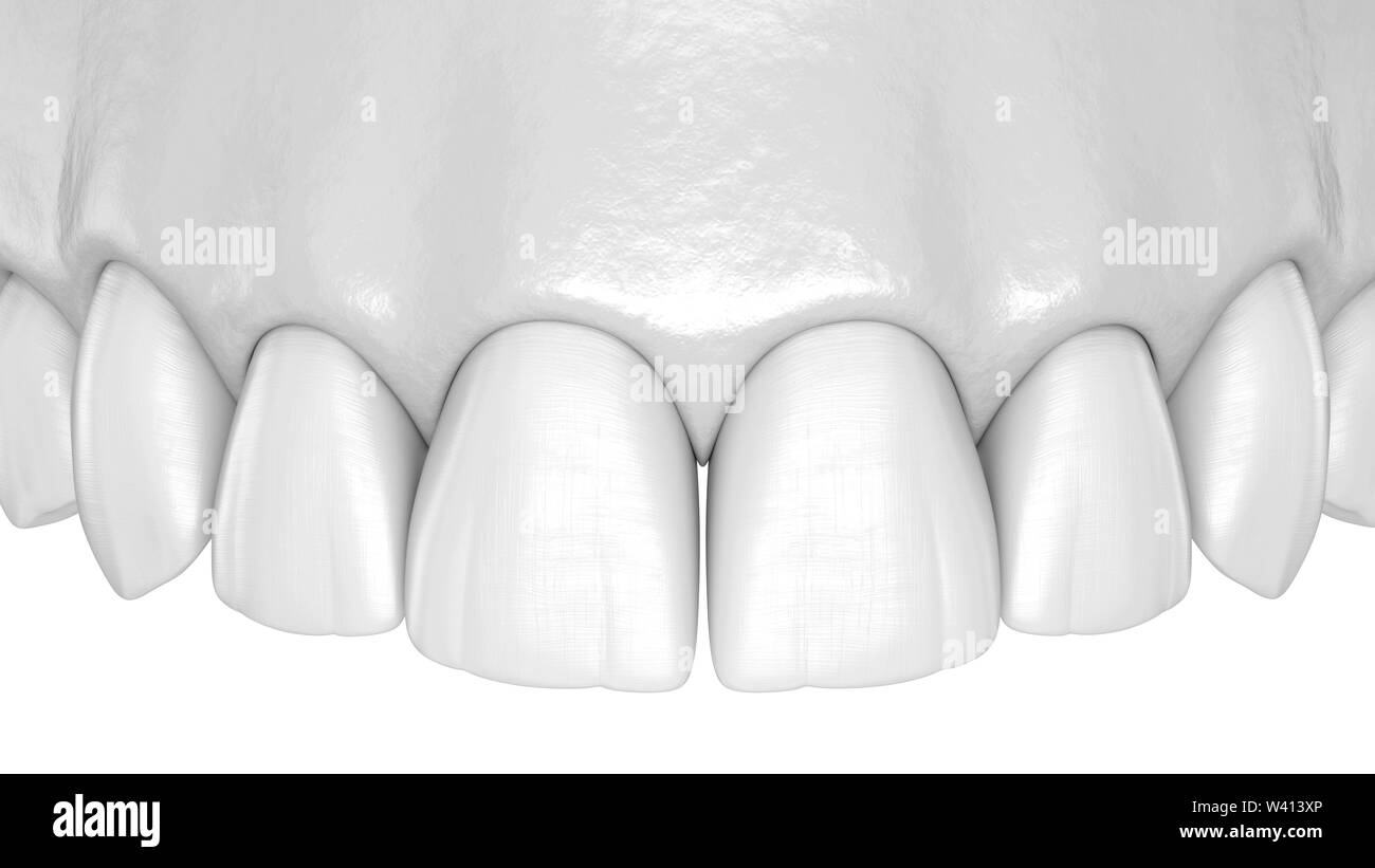 Maxillary human gum and teeth. Medically accurate tooth 3D illustration Stock Photohttps://www.alamy.com/image-license-details/?v=1https://www.alamy.com/maxillary-human-gum-and-teeth-medically-accurate-tooth-3d-illustration-image260639198.html
RFW413XP–Maxillary human gum and teeth. Medically accurate tooth 3D illustration
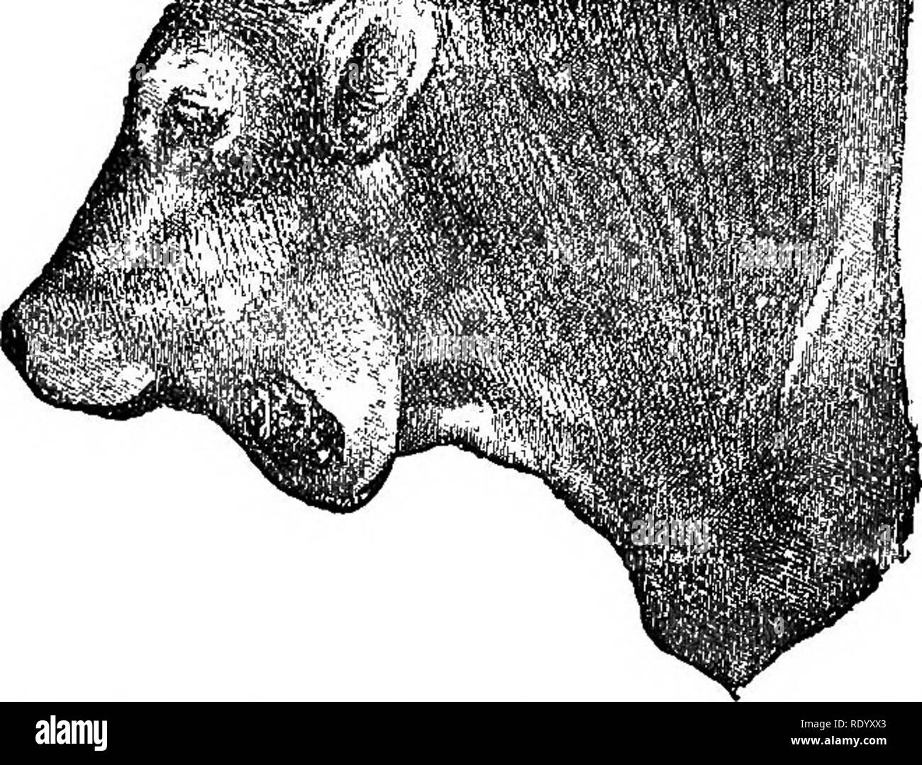 . A treatise on surgical therapeutics of domestic animals. Veterinary surgery; Therapeutics, Surgical. ACTINOMYCOSIS. 201 ). Fig. 48. Actinomycosis of the Lower Maxillary. they may heal, as in tuberculosis, without leaving apparent marks. Ac- tinomycites ordinarily spread slowly, little by little, through the blood ves- sels, and apparently most often through the veins. All tissues and all organs may be attacked, and if, ordinarily, secondary tum- ors develop in the lungs, they are also observed in the liver, kidneys, spleen, serous membranes, lympha- tic glands, and even the encephalon. Local Stock Photohttps://www.alamy.com/image-license-details/?v=1https://www.alamy.com/a-treatise-on-surgical-therapeutics-of-domestic-animals-veterinary-surgery-therapeutics-surgical-actinomycosis-201-fig-48-actinomycosis-of-the-lower-maxillary-they-may-heal-as-in-tuberculosis-without-leaving-apparent-marks-ac-tinomycites-ordinarily-spread-slowly-little-by-little-through-the-blood-ves-sels-and-apparently-most-often-through-the-veins-all-tissues-and-all-organs-may-be-attacked-and-if-ordinarily-secondary-tum-ors-develop-in-the-lungs-they-are-also-observed-in-the-liver-kidneys-spleen-serous-membranes-lympha-tic-glands-and-even-the-encephalon-local-image232339131.html
RMRDYXX3–. A treatise on surgical therapeutics of domestic animals. Veterinary surgery; Therapeutics, Surgical. ACTINOMYCOSIS. 201 ). Fig. 48. Actinomycosis of the Lower Maxillary. they may heal, as in tuberculosis, without leaving apparent marks. Ac- tinomycites ordinarily spread slowly, little by little, through the blood ves- sels, and apparently most often through the veins. All tissues and all organs may be attacked, and if, ordinarily, secondary tum- ors develop in the lungs, they are also observed in the liver, kidneys, spleen, serous membranes, lympha- tic glands, and even the encephalon. Local
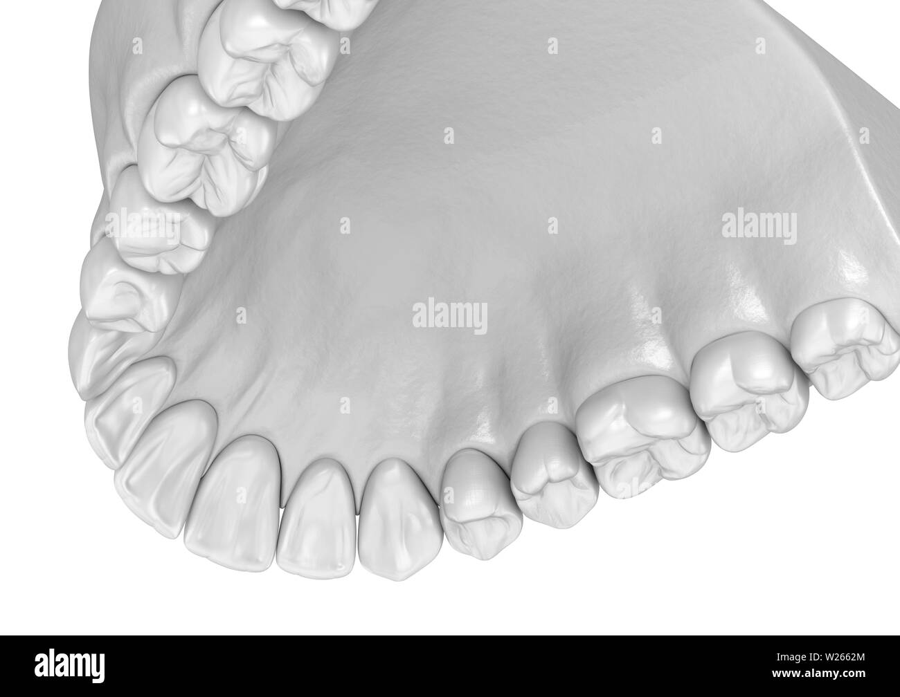 Maxillary human gum and teeth. Medically accurate tooth 3D illustration Stock Photohttps://www.alamy.com/image-license-details/?v=1https://www.alamy.com/maxillary-human-gum-and-teeth-medically-accurate-tooth-3d-illustration-image259521324.html
RFW2662M–Maxillary human gum and teeth. Medically accurate tooth 3D illustration
 . Veterinary surgery ... Veterinary surgery; Veterinary pathology; Horses; Teeth; Domestic animals. ANIMAL DENTISTRY. 169 Restraint:—Lateral recumbent position, under chloro- form anaesthesia or local cocainization, preferably the former. Indications:—This operation should be adopted univer- sally for the removal of the first, second and third superior molars. Applied to the fourth or fifth superior molars a de- formity of the face results from the removal of the extremity of the maxillary spine, and besides it has no advantages. Fig. 120. Chisel. Fig. 121. Saw. Fig. I2IA. Mouth Gag. The Most Stock Photohttps://www.alamy.com/image-license-details/?v=1https://www.alamy.com/veterinary-surgery-veterinary-surgery-veterinary-pathology-horses-teeth-domestic-animals-animal-dentistry-169-restraintlateral-recumbent-position-under-chloro-form-anaesthesia-or-local-cocainization-preferably-the-former-indicationsthis-operation-should-be-adopted-univer-sally-for-the-removal-of-the-first-second-and-third-superior-molars-applied-to-the-fourth-or-fifth-superior-molars-a-de-formity-of-the-face-results-from-the-removal-of-the-extremity-of-the-maxillary-spine-and-besides-it-has-no-advantages-fig-120-chisel-fig-121-saw-fig-i2ia-mouth-gag-the-most-image232199893.html
RMRDNH99–. Veterinary surgery ... Veterinary surgery; Veterinary pathology; Horses; Teeth; Domestic animals. ANIMAL DENTISTRY. 169 Restraint:—Lateral recumbent position, under chloro- form anaesthesia or local cocainization, preferably the former. Indications:—This operation should be adopted univer- sally for the removal of the first, second and third superior molars. Applied to the fourth or fifth superior molars a de- formity of the face results from the removal of the extremity of the maxillary spine, and besides it has no advantages. Fig. 120. Chisel. Fig. 121. Saw. Fig. I2IA. Mouth Gag. The Most
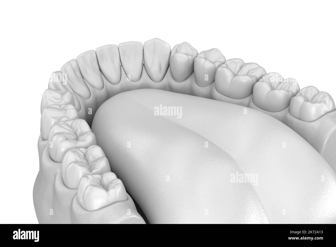 Human gum and teeth in white style. Medically accurate tooth 3D illustration Stock Photohttps://www.alamy.com/image-license-details/?v=1https://www.alamy.com/human-gum-and-teeth-in-white-style-medically-accurate-tooth-3d-illustration-image486244671.html
RF2K72A13–Human gum and teeth in white style. Medically accurate tooth 3D illustration
 . Manual of operative veterinary surgery. Veterinary surgery. Fia. 451.—Lancet to Bleed at tne Palate, If, on the contrary, an artery has been divided and the flow of blood becomes sufficiently abundant and continuous to become alarming, it becomes necessary to employ hemostatic means. These may be a small sponge compressed or moistened with cold water or an astringent solution; or, if necessary, a pad of oakum can be applied and secured with a bandage passed through the mouth and around the maxillary bone, and tied on the face. It can also be accompUshed by means of a peculiar bit, represente Stock Photohttps://www.alamy.com/image-license-details/?v=1https://www.alamy.com/manual-of-operative-veterinary-surgery-veterinary-surgery-fia-451lancet-to-bleed-at-tne-palate-if-on-the-contrary-an-artery-has-been-divided-and-the-flow-of-blood-becomes-sufficiently-abundant-and-continuous-to-become-alarming-it-becomes-necessary-to-employ-hemostatic-means-these-may-be-a-small-sponge-compressed-or-moistened-with-cold-water-or-an-astringent-solution-or-if-necessary-a-pad-of-oakum-can-be-applied-and-secured-with-a-bandage-passed-through-the-mouth-and-around-the-maxillary-bone-and-tied-on-the-face-it-can-also-be-accompushed-by-means-of-a-peculiar-bit-represente-image232317785.html
RMRDXYKN–. Manual of operative veterinary surgery. Veterinary surgery. Fia. 451.—Lancet to Bleed at tne Palate, If, on the contrary, an artery has been divided and the flow of blood becomes sufficiently abundant and continuous to become alarming, it becomes necessary to employ hemostatic means. These may be a small sponge compressed or moistened with cold water or an astringent solution; or, if necessary, a pad of oakum can be applied and secured with a bandage passed through the mouth and around the maxillary bone, and tied on the face. It can also be accompUshed by means of a peculiar bit, represente
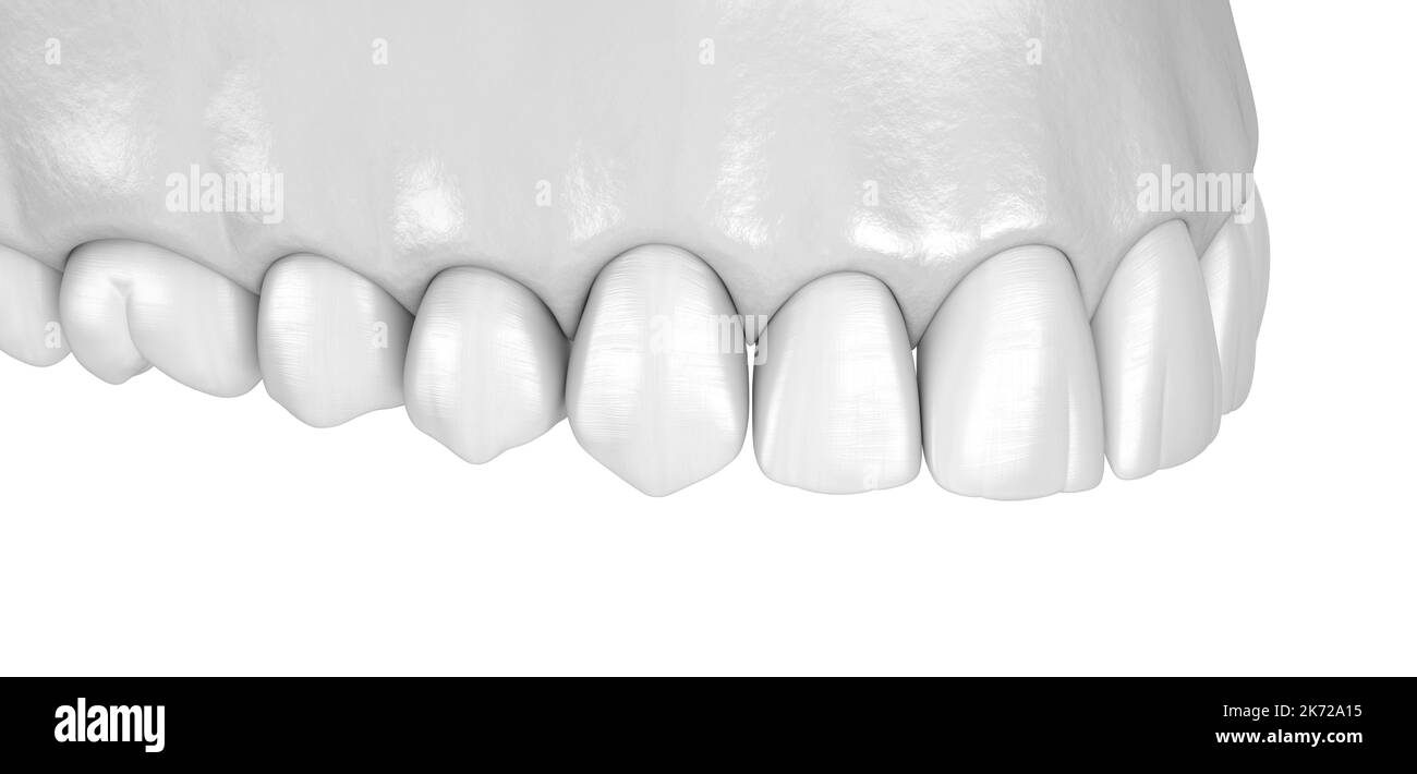 Human gum and teeth in white style. Medically accurate tooth 3D illustration Stock Photohttps://www.alamy.com/image-license-details/?v=1https://www.alamy.com/human-gum-and-teeth-in-white-style-medically-accurate-tooth-3d-illustration-image486244673.html
RF2K72A15–Human gum and teeth in white style. Medically accurate tooth 3D illustration
 . A treatise on surgical therapeutics of domestic animals. Veterinary surgery; Therapeutics, Surgical. RACHITISM 485 the lower maxillary had lost their consistency; the superiors were swollen. Mastication was impossible, in a three-year-old mare seen by Benjamin, and Redon. The pathogeny of rachitism is yet obscure. L: Lafosse accuses bad liygienic conditions, damp habitations, those badly kept, exposed to the north where sunlight never goes. Feeding with fodder poor in calcareous substances has been incriminated. Guerin, RolofE, Voit, Chossat, Milne, Edwards, have produced it artificially in Stock Photohttps://www.alamy.com/image-license-details/?v=1https://www.alamy.com/a-treatise-on-surgical-therapeutics-of-domestic-animals-veterinary-surgery-therapeutics-surgical-rachitism-485-the-lower-maxillary-had-lost-their-consistency-the-superiors-were-swollen-mastication-was-impossible-in-a-three-year-old-mare-seen-by-benjamin-and-redon-the-pathogeny-of-rachitism-is-yet-obscure-l-lafosse-accuses-bad-liygienic-conditions-damp-habitations-those-badly-kept-exposed-to-the-north-where-sunlight-never-goes-feeding-with-fodder-poor-in-calcareous-substances-has-been-incriminated-guerin-rolofe-voit-chossat-milne-edwards-have-produced-it-artificially-in-image232277357.html
RMRDW43W–. A treatise on surgical therapeutics of domestic animals. Veterinary surgery; Therapeutics, Surgical. RACHITISM 485 the lower maxillary had lost their consistency; the superiors were swollen. Mastication was impossible, in a three-year-old mare seen by Benjamin, and Redon. The pathogeny of rachitism is yet obscure. L: Lafosse accuses bad liygienic conditions, damp habitations, those badly kept, exposed to the north where sunlight never goes. Feeding with fodder poor in calcareous substances has been incriminated. Guerin, RolofE, Voit, Chossat, Milne, Edwards, have produced it artificially in
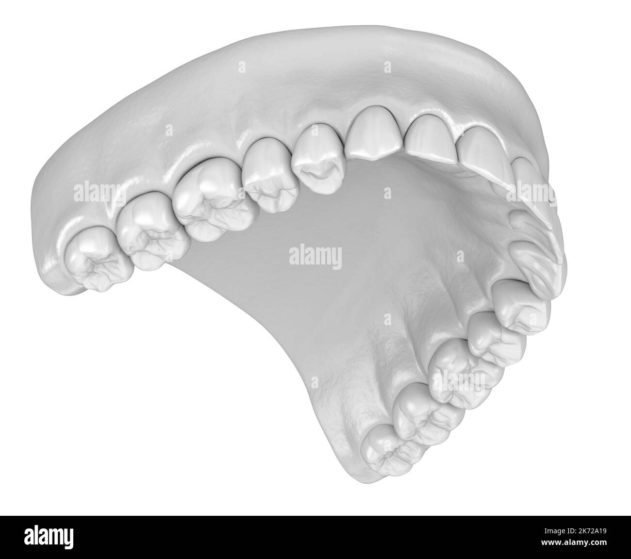 Human gum and teeth in white style. Medically accurate tooth 3D illustration Stock Photohttps://www.alamy.com/image-license-details/?v=1https://www.alamy.com/human-gum-and-teeth-in-white-style-medically-accurate-tooth-3d-illustration-image486244677.html
RF2K72A19–Human gum and teeth in white style. Medically accurate tooth 3D illustration
 . Veterinary surgery ... Veterinary surgery; Veterinary pathology; Horses; Teeth; Domestic animals. 220 ANIMAL DENTISTRY. appearance of a more or less diffused tumefaction of the in- ferior maxilla (lumpy jaw) or by rounded exostoses. As the soft structures become involved more and more by spread- ing of the diseased process, the head may assume an un- sightly appearance from the enormity of the swelling. The condition is further complicated by dental disorders—loosen-. FlG. 148B. Actinomycostic Superior Maxillary in the Ox. ing of the teeth. In the pharynx the disease is manifested by tumefac Stock Photohttps://www.alamy.com/image-license-details/?v=1https://www.alamy.com/veterinary-surgery-veterinary-surgery-veterinary-pathology-horses-teeth-domestic-animals-220-animal-dentistry-appearance-of-a-more-or-less-diffused-tumefaction-of-the-in-ferior-maxilla-lumpy-jaw-or-by-rounded-exostoses-as-the-soft-structures-become-involved-more-and-more-by-spread-ing-of-the-diseased-process-the-head-may-assume-an-un-sightly-appearance-from-the-enormity-of-the-swelling-the-condition-is-further-complicated-by-dental-disordersloosen-flg-148b-actinomycostic-superior-maxillary-in-the-ox-ing-of-the-teeth-in-the-pharynx-the-disease-is-manifested-by-tumefac-image232199699.html
RMRDNH2B–. Veterinary surgery ... Veterinary surgery; Veterinary pathology; Horses; Teeth; Domestic animals. 220 ANIMAL DENTISTRY. appearance of a more or less diffused tumefaction of the in- ferior maxilla (lumpy jaw) or by rounded exostoses. As the soft structures become involved more and more by spread- ing of the diseased process, the head may assume an un- sightly appearance from the enormity of the swelling. The condition is further complicated by dental disorders—loosen-. FlG. 148B. Actinomycostic Superior Maxillary in the Ox. ing of the teeth. In the pharynx the disease is manifested by tumefac
 . Surgical and obstetrical operations. Veterinary surgery. FIG. 5. Trephining of the Facial Sinuses. Oblique lateral view of the face with the sinuses exposed. SM, superior maxillary sinus; IM', median portion of inferior maxillary sinus; NC, nerve conduit of su^jerior maxillary division of trifacial nerve; IM, lateral portion of inferior maxillary sinus; F, frontal sinus; FE, fenestrum of communication between thefrontat and superior maxillary sinuses; SI', artificial opening through the superior turbinated bone at the lowestpartof the frontal sinus establishingafree communication with the na Stock Photohttps://www.alamy.com/image-license-details/?v=1https://www.alamy.com/surgical-and-obstetrical-operations-veterinary-surgery-fig-5-trephining-of-the-facial-sinuses-oblique-lateral-view-of-the-face-with-the-sinuses-exposed-sm-superior-maxillary-sinus-im-median-portion-of-inferior-maxillary-sinus-nc-nerve-conduit-of-sujerior-maxillary-division-of-trifacial-nerve-im-lateral-portion-of-inferior-maxillary-sinus-f-frontal-sinus-fe-fenestrum-of-communication-between-thefrontat-and-superior-maxillary-sinuses-si-artificial-opening-through-the-superior-turbinated-bone-at-the-lowestpartof-the-frontal-sinus-establishingafree-communication-with-the-na-image232340802.html
RMRE011P–. Surgical and obstetrical operations. Veterinary surgery. FIG. 5. Trephining of the Facial Sinuses. Oblique lateral view of the face with the sinuses exposed. SM, superior maxillary sinus; IM', median portion of inferior maxillary sinus; NC, nerve conduit of su^jerior maxillary division of trifacial nerve; IM, lateral portion of inferior maxillary sinus; F, frontal sinus; FE, fenestrum of communication between thefrontat and superior maxillary sinuses; SI', artificial opening through the superior turbinated bone at the lowestpartof the frontal sinus establishingafree communication with the na
 . Veterinary surgery ... Veterinary surgery; Veterinary pathology; Horses; Teeth; Domestic animals. ANIMAL DENTISTRY. 45 paratus at that time is represented by a whitened ridge of mucous membrane extending around the maxillary margin. This ridge (gingival cushion) is the matrix from which the teeth develop. At the earliest period it consists chiefly of two layers of epithelial cells and basement membrane of connective tissue. The outer layer consists of loosely ar- ranged cells while the deep one is dense and lies in close relation to the jaw, being only divided from the latter by the thin con Stock Photohttps://www.alamy.com/image-license-details/?v=1https://www.alamy.com/veterinary-surgery-veterinary-surgery-veterinary-pathology-horses-teeth-domestic-animals-animal-dentistry-45-paratus-at-that-time-is-represented-by-a-whitened-ridge-of-mucous-membrane-extending-around-the-maxillary-margin-this-ridge-gingival-cushion-is-the-matrix-from-which-the-teeth-develop-at-the-earliest-period-it-consists-chiefly-of-two-layers-of-epithelial-cells-and-basement-membrane-of-connective-tissue-the-outer-layer-consists-of-loosely-ar-ranged-cells-while-the-deep-one-is-dense-and-lies-in-close-relation-to-the-jaw-being-only-divided-from-the-latter-by-the-thin-con-image232200735.html
RMRDNJBB–. Veterinary surgery ... Veterinary surgery; Veterinary pathology; Horses; Teeth; Domestic animals. ANIMAL DENTISTRY. 45 paratus at that time is represented by a whitened ridge of mucous membrane extending around the maxillary margin. This ridge (gingival cushion) is the matrix from which the teeth develop. At the earliest period it consists chiefly of two layers of epithelial cells and basement membrane of connective tissue. The outer layer consists of loosely ar- ranged cells while the deep one is dense and lies in close relation to the jaw, being only divided from the latter by the thin con
 . Veterinary surgery ... Veterinary surgery; Veterinary pathology; Horses; Teeth; Domestic animals. 180 ANIMAL DENTISTRY. Definition—A congenital deformity in which the supe- rior incisors overlap the inferior. Etiology—A congenital deformity of obscure cause. The deformity consists of a deficiency in the proper length of the inferior maxillary, or an abnormal elongation of the pre- maxilla. Symptoms—Overlapping of the inferior incisor by the superior ones vs^ith more or less elongation of the first supe- rior and sixth inferior molars. The condition becomes more. Fig. 127. -"'*''''*" Stock Photohttps://www.alamy.com/image-license-details/?v=1https://www.alamy.com/veterinary-surgery-veterinary-surgery-veterinary-pathology-horses-teeth-domestic-animals-180-animal-dentistry-definitiona-congenital-deformity-in-which-the-supe-rior-incisors-overlap-the-inferior-etiologya-congenital-deformity-of-obscure-cause-the-deformity-consists-of-a-deficiency-in-the-proper-length-of-the-inferior-maxillary-or-an-abnormal-elongation-of-the-pre-maxilla-symptomsoverlapping-of-the-inferior-incisor-by-the-superior-ones-vsith-more-or-less-elongation-of-the-first-supe-rior-and-sixth-inferior-molars-the-condition-becomes-more-fig-127-quotquot-image232199865.html
RMRDNH89–. Veterinary surgery ... Veterinary surgery; Veterinary pathology; Horses; Teeth; Domestic animals. 180 ANIMAL DENTISTRY. Definition—A congenital deformity in which the supe- rior incisors overlap the inferior. Etiology—A congenital deformity of obscure cause. The deformity consists of a deficiency in the proper length of the inferior maxillary, or an abnormal elongation of the pre- maxilla. Symptoms—Overlapping of the inferior incisor by the superior ones vs^ith more or less elongation of the first supe- rior and sixth inferior molars. The condition becomes more. Fig. 127. -"'*''''*"
 . Veterinary surgery ... Veterinary surgery; Veterinary pathology; Horses; Teeth; Domestic animals. 234 ANIMAL DENTISTRY. Restraint—Horses may be trephined in the standing po- sition with the dental halter and twitch. When teeth are to be repulsed or when any chiseling of the bone is necessary the recumbent position is essential.. y Fig. isia. Plain Circular Trephine. ist Step—Clip, shave and disinfect the space intervening between the maxillary spine and longitudinal suture of the nasal bones. 2nd Step—First open the maxillary sinus about one inch. Please note that these images are extracted Stock Photohttps://www.alamy.com/image-license-details/?v=1https://www.alamy.com/veterinary-surgery-veterinary-surgery-veterinary-pathology-horses-teeth-domestic-animals-234-animal-dentistry-restrainthorses-may-be-trephined-in-the-standing-po-sition-with-the-dental-halter-and-twitch-when-teeth-are-to-be-repulsed-or-when-any-chiseling-of-the-bone-is-necessary-the-recumbent-position-is-essential-y-fig-isia-plain-circular-trephine-ist-stepclip-shave-and-disinfect-the-space-intervening-between-the-maxillary-spine-and-longitudinal-suture-of-the-nasal-bones-2nd-stepfirst-open-the-maxillary-sinus-about-one-inch-please-note-that-these-images-are-extracted-image232199682.html
RMRDNH1P–. Veterinary surgery ... Veterinary surgery; Veterinary pathology; Horses; Teeth; Domestic animals. 234 ANIMAL DENTISTRY. Restraint—Horses may be trephined in the standing po- sition with the dental halter and twitch. When teeth are to be repulsed or when any chiseling of the bone is necessary the recumbent position is essential.. y Fig. isia. Plain Circular Trephine. ist Step—Clip, shave and disinfect the space intervening between the maxillary spine and longitudinal suture of the nasal bones. 2nd Step—First open the maxillary sinus about one inch. Please note that these images are extracted
 . Surgical and obstetrical operations. Veterinary surgery. SM. FIG. 6. Trephining of Facial Sinuses. Frontal view of right side of face with sinuses exposed. SM, superior maxillary sinus ; IM', median portion of inferior maxillary sinus ; IM, lateral portion of inferior maxillary sinus ; F, frontal sinus ; FE, communication between the frontal and superior maxillary sinuses.. Please note that these images are extracted from scanned page images that may have been digitally enhanced for readability - coloration and appearance of these illustrations may not perfectly resemble the original work.. Stock Photohttps://www.alamy.com/image-license-details/?v=1https://www.alamy.com/surgical-and-obstetrical-operations-veterinary-surgery-sm-fig-6-trephining-of-facial-sinuses-frontal-view-of-right-side-of-face-with-sinuses-exposed-sm-superior-maxillary-sinus-im-median-portion-of-inferior-maxillary-sinus-im-lateral-portion-of-inferior-maxillary-sinus-f-frontal-sinus-fe-communication-between-the-frontal-and-superior-maxillary-sinuses-please-note-that-these-images-are-extracted-from-scanned-page-images-that-may-have-been-digitally-enhanced-for-readability-coloration-and-appearance-of-these-illustrations-may-not-perfectly-resemble-the-original-work-image232340797.html
RMRE011H–. Surgical and obstetrical operations. Veterinary surgery. SM. FIG. 6. Trephining of Facial Sinuses. Frontal view of right side of face with sinuses exposed. SM, superior maxillary sinus ; IM', median portion of inferior maxillary sinus ; IM, lateral portion of inferior maxillary sinus ; F, frontal sinus ; FE, communication between the frontal and superior maxillary sinuses.. Please note that these images are extracted from scanned page images that may have been digitally enhanced for readability - coloration and appearance of these illustrations may not perfectly resemble the original work..
 . Surgical diseases and surgery of the dog. Dogs. The Head and Neck 89 bilateral through sympathy and may give rise to a symptomatic ca- tarrhal inflammation of the nasal passages through contiguity of tissue. The sympathetic hypothesis is very doubt- ful.. Symptoms and Diag- nosis. A fistula existing in the position mentioned, should be probed. The affected tooth can gener- ally be determined in this manner. The tooth may or may not be painful to percussion. Maxillary fis- tula must be carefully differentiated from Lachrymal fistula. An animal suffering from the former disease masticates with Stock Photohttps://www.alamy.com/image-license-details/?v=1https://www.alamy.com/surgical-diseases-and-surgery-of-the-dog-dogs-the-head-and-neck-89-bilateral-through-sympathy-and-may-give-rise-to-a-symptomatic-ca-tarrhal-inflammation-of-the-nasal-passages-through-contiguity-of-tissue-the-sympathetic-hypothesis-is-very-doubt-ful-symptoms-and-diag-nosis-a-fistula-existing-in-the-position-mentioned-should-be-probed-the-affected-tooth-can-gener-ally-be-determined-in-this-manner-the-tooth-may-or-may-not-be-painful-to-percussion-maxillary-fis-tula-must-be-carefully-differentiated-from-lachrymal-fistula-an-animal-suffering-from-the-former-disease-masticates-with-image232371734.html
RMRE1CEE–. Surgical diseases and surgery of the dog. Dogs. The Head and Neck 89 bilateral through sympathy and may give rise to a symptomatic ca- tarrhal inflammation of the nasal passages through contiguity of tissue. The sympathetic hypothesis is very doubt- ful.. Symptoms and Diag- nosis. A fistula existing in the position mentioned, should be probed. The affected tooth can gener- ally be determined in this manner. The tooth may or may not be painful to percussion. Maxillary fis- tula must be carefully differentiated from Lachrymal fistula. An animal suffering from the former disease masticates with
 . Surgical and obstetrical operations. Veterinary surgery. FIG. 7 Trephining of Facial Slnnses Cross section of the right half of the head of a horse at the posterior border of the last molar. F, frontal sinus ; IM, lateral portion of inferior maxillary sinus at extreme posterior or stiperior part ; IM', median portion do. ; N, nasal chamber opposite the communication between it and the superior maxillary sinus ; NF, conduit of superior maxillary branch of the trifacial nerve ; SM, superior maxillary sinus ; M^, fragment of last molar.. Please note that these images are extracted from scanned Stock Photohttps://www.alamy.com/image-license-details/?v=1https://www.alamy.com/surgical-and-obstetrical-operations-veterinary-surgery-fig-7-trephining-of-facial-slnnses-cross-section-of-the-right-half-of-the-head-of-a-horse-at-the-posterior-border-of-the-last-molar-f-frontal-sinus-im-lateral-portion-of-inferior-maxillary-sinus-at-extreme-posterior-or-stiperior-part-im-median-portion-do-n-nasal-chamber-opposite-the-communication-between-it-and-the-superior-maxillary-sinus-nf-conduit-of-superior-maxillary-branch-of-the-trifacial-nerve-sm-superior-maxillary-sinus-m-fragment-of-last-molar-please-note-that-these-images-are-extracted-from-scanned-image232340786.html
RMRE0116–. Surgical and obstetrical operations. Veterinary surgery. FIG. 7 Trephining of Facial Slnnses Cross section of the right half of the head of a horse at the posterior border of the last molar. F, frontal sinus ; IM, lateral portion of inferior maxillary sinus at extreme posterior or stiperior part ; IM', median portion do. ; N, nasal chamber opposite the communication between it and the superior maxillary sinus ; NF, conduit of superior maxillary branch of the trifacial nerve ; SM, superior maxillary sinus ; M^, fragment of last molar.. Please note that these images are extracted from scanned
 . A treatise on surgical therapeutics of domestic animals. Veterinary surgery; Therapeutics, Surgical. Fig. 48. Actinomycosis of the Lower Maxillary. they may heal, as in tuberculosis, without leaving apparent marks. Ac- tinomycites ordinarily spread slowly, little by little, through the blood ves- sels, and apparently most often through the veins. All tissues and all organs may be attacked, and if, ordinarily, secondary tum- ors develop in the lungs, they are also observed in the liver, kidneys, spleen, serous membranes, lympha- tic glands, and even the encephalon. Locally, the disease pro- g Stock Photohttps://www.alamy.com/image-license-details/?v=1https://www.alamy.com/a-treatise-on-surgical-therapeutics-of-domestic-animals-veterinary-surgery-therapeutics-surgical-fig-48-actinomycosis-of-the-lower-maxillary-they-may-heal-as-in-tuberculosis-without-leaving-apparent-marks-ac-tinomycites-ordinarily-spread-slowly-little-by-little-through-the-blood-ves-sels-and-apparently-most-often-through-the-veins-all-tissues-and-all-organs-may-be-attacked-and-if-ordinarily-secondary-tum-ors-develop-in-the-lungs-they-are-also-observed-in-the-liver-kidneys-spleen-serous-membranes-lympha-tic-glands-and-even-the-encephalon-locally-the-disease-pro-g-image232339129.html
RMRDYXX1–. A treatise on surgical therapeutics of domestic animals. Veterinary surgery; Therapeutics, Surgical. Fig. 48. Actinomycosis of the Lower Maxillary. they may heal, as in tuberculosis, without leaving apparent marks. Ac- tinomycites ordinarily spread slowly, little by little, through the blood ves- sels, and apparently most often through the veins. All tissues and all organs may be attacked, and if, ordinarily, secondary tum- ors develop in the lungs, they are also observed in the liver, kidneys, spleen, serous membranes, lympha- tic glands, and even the encephalon. Locally, the disease pro- g
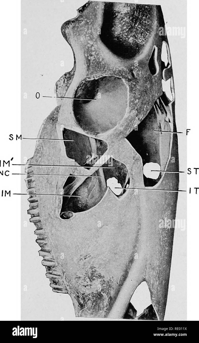 . Surgical and obstetrical operations. Veterinary surgery. NC FIG- 4. Trephining of Facial Sinuses. Right side of face, viewed laterally, showing extent and relations of the sinuses. O, orbita cavity; SM, superior maxillary sinus; IM", median portion of inferior maxillary sinus; NC, nerve conduit of superior maxillary trunk of the trifacial: IM. laierial portion of inferior maxillary sinus; F, frontal sinus; ST, opening through superior turbinated bone for the establishment of drainage from the frontal and superior maxillary sinuses into the nasal pas- â sage; IT, opening through inferior Stock Photohttps://www.alamy.com/image-license-details/?v=1https://www.alamy.com/surgical-and-obstetrical-operations-veterinary-surgery-nc-fig-4-trephining-of-facial-sinuses-right-side-of-face-viewed-laterally-showing-extent-and-relations-of-the-sinuses-o-orbita-cavity-sm-superior-maxillary-sinus-imquot-median-portion-of-inferior-maxillary-sinus-nc-nerve-conduit-of-superior-maxillary-trunk-of-the-trifacial-im-laierial-portion-of-inferior-maxillary-sinus-f-frontal-sinus-st-opening-through-superior-turbinated-bone-for-the-establishment-of-drainage-from-the-frontal-and-superior-maxillary-sinuses-into-the-nasal-pas-sage-it-opening-through-inferior-image232340806.html
RMRE011X–. Surgical and obstetrical operations. Veterinary surgery. NC FIG- 4. Trephining of Facial Sinuses. Right side of face, viewed laterally, showing extent and relations of the sinuses. O, orbita cavity; SM, superior maxillary sinus; IM", median portion of inferior maxillary sinus; NC, nerve conduit of superior maxillary trunk of the trifacial: IM. laierial portion of inferior maxillary sinus; F, frontal sinus; ST, opening through superior turbinated bone for the establishment of drainage from the frontal and superior maxillary sinuses into the nasal pas- â sage; IT, opening through inferior
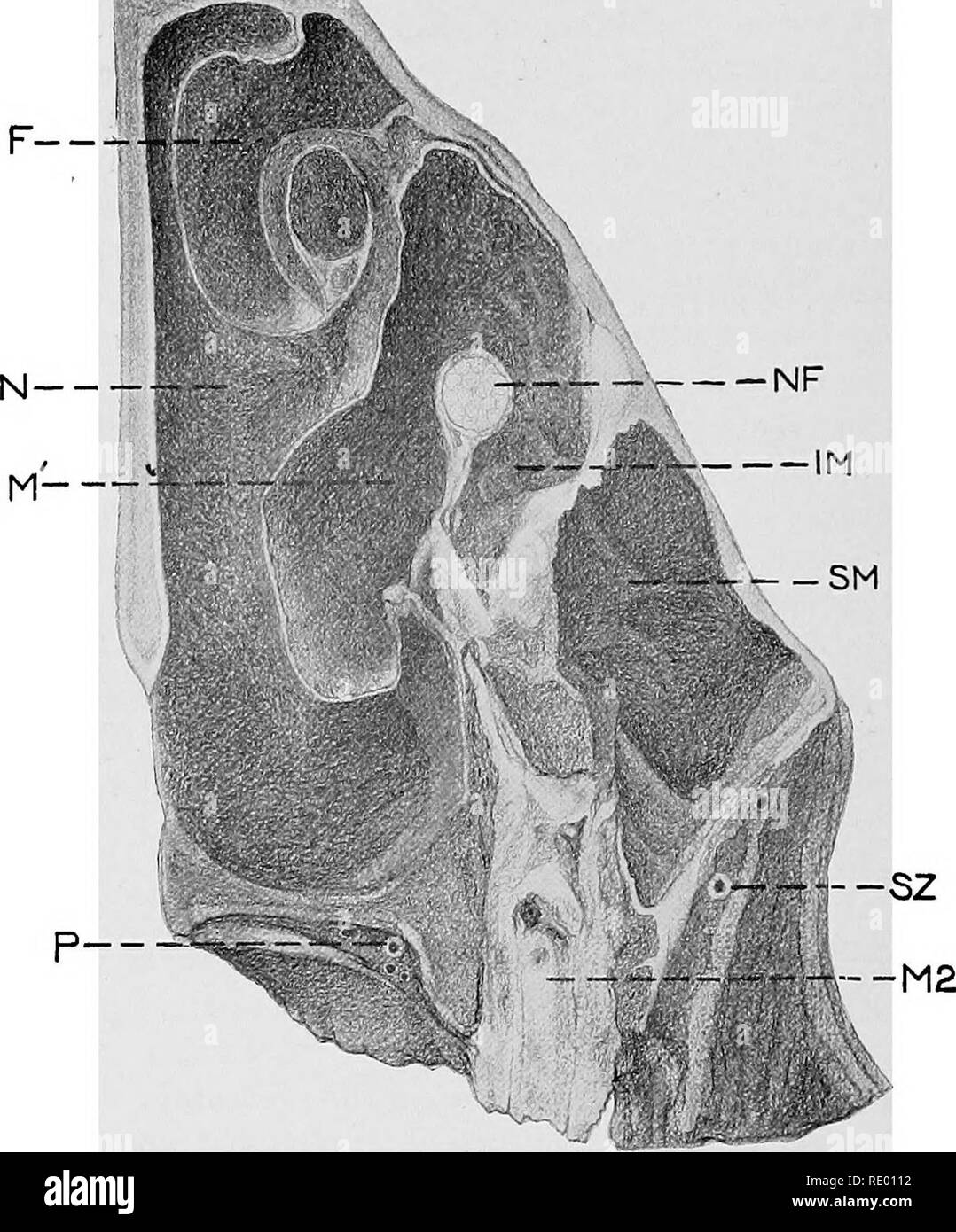 . Surgical and obstetrical operations. Veterinary surgery. FIG. 8 Trephining the Facial Sinuses Cross section of the left side of the head of an aged horse at the second molar, seen from the front. F, frontal sinus ; N. nasal sinus, opposite the communication between the nasal and maxillary sinuses ; IM, lateral portion of inferior maxillary sinus ; IM', median portion of inferior maxillary sinus; SM, superior maxillary sinus ; NF, superior maxillary division of trifacial nerve in its bony conduit; SZ, subzygomatic artery ; P, palatine artery ; M2, second molar.. Please note that these images Stock Photohttps://www.alamy.com/image-license-details/?v=1https://www.alamy.com/surgical-and-obstetrical-operations-veterinary-surgery-fig-8-trephining-the-facial-sinuses-cross-section-of-the-left-side-of-the-head-of-an-aged-horse-at-the-second-molar-seen-from-the-front-f-frontal-sinus-n-nasal-sinus-opposite-the-communication-between-the-nasal-and-maxillary-sinuses-im-lateral-portion-of-inferior-maxillary-sinus-im-median-portion-of-inferior-maxillary-sinus-sm-superior-maxillary-sinus-nf-superior-maxillary-division-of-trifacial-nerve-in-its-bony-conduit-sz-subzygomatic-artery-p-palatine-artery-m2-second-molar-please-note-that-these-images-image232340782.html
RMRE0112–. Surgical and obstetrical operations. Veterinary surgery. FIG. 8 Trephining the Facial Sinuses Cross section of the left side of the head of an aged horse at the second molar, seen from the front. F, frontal sinus ; N. nasal sinus, opposite the communication between the nasal and maxillary sinuses ; IM, lateral portion of inferior maxillary sinus ; IM', median portion of inferior maxillary sinus; SM, superior maxillary sinus ; NF, superior maxillary division of trifacial nerve in its bony conduit; SZ, subzygomatic artery ; P, palatine artery ; M2, second molar.. Please note that these images
 . Surgical and obstetrical operations. Veterinary surgery. — SM FIG. 10 Trephining of Facial Sinuses Cross section of the left side of the head anterior to the last molar, and through the widest part of the inferior maxillary sinus. M^, last superior molar ; SM, superior maxillary sinus at its antero-inferior extremity ; IM, inferior maxillary sinus, lateral portion ; IM', do. median portion ; N, nasal fossa ; S, sound lodged in lachrymal duct; NF, trifacial nerve ; F, frontal sinus.. Please note that these images are extracted from scanned page images that may have been digitally enhanced for Stock Photohttps://www.alamy.com/image-license-details/?v=1https://www.alamy.com/surgical-and-obstetrical-operations-veterinary-surgery-sm-fig-10-trephining-of-facial-sinuses-cross-section-of-the-left-side-of-the-head-anterior-to-the-last-molar-and-through-the-widest-part-of-the-inferior-maxillary-sinus-m-last-superior-molar-sm-superior-maxillary-sinus-at-its-antero-inferior-extremity-im-inferior-maxillary-sinus-lateral-portion-im-do-median-portion-n-nasal-fossa-s-sound-lodged-in-lachrymal-duct-nf-trifacial-nerve-f-frontal-sinus-please-note-that-these-images-are-extracted-from-scanned-page-images-that-may-have-been-digitally-enhanced-for-image232340773.html
RMRE010N–. Surgical and obstetrical operations. Veterinary surgery. — SM FIG. 10 Trephining of Facial Sinuses Cross section of the left side of the head anterior to the last molar, and through the widest part of the inferior maxillary sinus. M^, last superior molar ; SM, superior maxillary sinus at its antero-inferior extremity ; IM, inferior maxillary sinus, lateral portion ; IM', do. median portion ; N, nasal fossa ; S, sound lodged in lachrymal duct; NF, trifacial nerve ; F, frontal sinus.. Please note that these images are extracted from scanned page images that may have been digitally enhanced for
 . Veterinary surgery ... Veterinary surgery; Veterinary pathology; Horses; Teeth; Domestic animals. y Fig. isia. Plain Circular Trephine. ist Step—Clip, shave and disinfect the space intervening between the maxillary spine and longitudinal suture of the nasal bones. 2nd Step—First open the maxillary sinus about one inch. Fig. isib. Assorted Trephines and Tooth Drills, with Brace. from its external border and one inch from the anterior ex- tremity, but vary in the antro-posterior direction according to tooth to be repulsed. 3rd Step—Make a T-shaped incision through the skin with the base of the Stock Photohttps://www.alamy.com/image-license-details/?v=1https://www.alamy.com/veterinary-surgery-veterinary-surgery-veterinary-pathology-horses-teeth-domestic-animals-y-fig-isia-plain-circular-trephine-ist-stepclip-shave-and-disinfect-the-space-intervening-between-the-maxillary-spine-and-longitudinal-suture-of-the-nasal-bones-2nd-stepfirst-open-the-maxillary-sinus-about-one-inch-fig-isib-assorted-trephines-and-tooth-drills-with-brace-from-its-external-border-and-one-inch-from-the-anterior-ex-tremity-but-vary-in-the-antro-posterior-direction-according-to-tooth-to-be-repulsed-3rd-stepmake-a-t-shaped-incision-through-the-skin-with-the-base-of-the-image232199680.html
RMRDNH1M–. Veterinary surgery ... Veterinary surgery; Veterinary pathology; Horses; Teeth; Domestic animals. y Fig. isia. Plain Circular Trephine. ist Step—Clip, shave and disinfect the space intervening between the maxillary spine and longitudinal suture of the nasal bones. 2nd Step—First open the maxillary sinus about one inch. Fig. isib. Assorted Trephines and Tooth Drills, with Brace. from its external border and one inch from the anterior ex- tremity, but vary in the antro-posterior direction according to tooth to be repulsed. 3rd Step—Make a T-shaped incision through the skin with the base of the
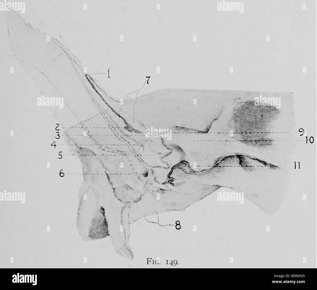 . Veterinary surgery ... Veterinary surgery; Veterinary pathology; Horses; Teeth; Domestic animals. ANIMAL DENTISTRY. 221. Fig. 149. Dental Teratoma and Conchal Fistula. 1. Mouth of the conchal fistula. 2. Bottom of the fistulous tract. 3. Gubernaculum dentis. 4. 9. Bony plates from the temporal.bone. 5. The tooth. 6. External auditory meatus. 7. Wall of the fistulous tract. 8. Petrous temporal bone. 10. Zygoma. 11. Temporo-maxillary articulation. (Williams).. Please note that these images are extracted from scanned page images that may have been digitally enhanced for readability - coloration Stock Photohttps://www.alamy.com/image-license-details/?v=1https://www.alamy.com/veterinary-surgery-veterinary-surgery-veterinary-pathology-horses-teeth-domestic-animals-animal-dentistry-221-fig-149-dental-teratoma-and-conchal-fistula-1-mouth-of-the-conchal-fistula-2-bottom-of-the-fistulous-tract-3-gubernaculum-dentis-4-9-bony-plates-from-the-temporalbone-5-the-tooth-6-external-auditory-meatus-7-wall-of-the-fistulous-tract-8-petrous-temporal-bone-10-zygoma-11-temporo-maxillary-articulation-williams-please-note-that-these-images-are-extracted-from-scanned-page-images-that-may-have-been-digitally-enhanced-for-readability-coloration-image232199693.html
RMRDNH25–. Veterinary surgery ... Veterinary surgery; Veterinary pathology; Horses; Teeth; Domestic animals. ANIMAL DENTISTRY. 221. Fig. 149. Dental Teratoma and Conchal Fistula. 1. Mouth of the conchal fistula. 2. Bottom of the fistulous tract. 3. Gubernaculum dentis. 4. 9. Bony plates from the temporal.bone. 5. The tooth. 6. External auditory meatus. 7. Wall of the fistulous tract. 8. Petrous temporal bone. 10. Zygoma. 11. Temporo-maxillary articulation. (Williams).. Please note that these images are extracted from scanned page images that may have been digitally enhanced for readability - coloration
 . Surgical and obstetrical operations. Veterinary surgery. â m FIG. 9 Trephining the Facial Sinuses Cross section obliquely downwards and backwards through the right half of the head of a two-year old colt at the first molar. F, Frontal sinus ; N, nasal passage at point of communication with the maxillary sinus, IM ; IM', median portion of inferior maxillary sinus ; SM, extreme lower end of superior maxillary sinus opened ; Mi, first molar; M2, second molar; P, palatine artery; SZ, sub- z3'gomatic artery.. Please note that these images are extracted from scanned page images that may have been Stock Photohttps://www.alamy.com/image-license-details/?v=1https://www.alamy.com/surgical-and-obstetrical-operations-veterinary-surgery-m-fig-9-trephining-the-facial-sinuses-cross-section-obliquely-downwards-and-backwards-through-the-right-half-of-the-head-of-a-two-year-old-colt-at-the-first-molar-f-frontal-sinus-n-nasal-passage-at-point-of-communication-with-the-maxillary-sinus-im-im-median-portion-of-inferior-maxillary-sinus-sm-extreme-lower-end-of-superior-maxillary-sinus-opened-mi-first-molar-m2-second-molar-p-palatine-artery-sz-sub-z3gomatic-artery-please-note-that-these-images-are-extracted-from-scanned-page-images-that-may-have-been-image232340776.html
RMRE010T–. Surgical and obstetrical operations. Veterinary surgery. â m FIG. 9 Trephining the Facial Sinuses Cross section obliquely downwards and backwards through the right half of the head of a two-year old colt at the first molar. F, Frontal sinus ; N, nasal passage at point of communication with the maxillary sinus, IM ; IM', median portion of inferior maxillary sinus ; SM, extreme lower end of superior maxillary sinus opened ; Mi, first molar; M2, second molar; P, palatine artery; SZ, sub- z3'gomatic artery.. Please note that these images are extracted from scanned page images that may have been
 . Surgical and obstetrical operations. Veterinary surgery. FIG. 17 Opening of the Guttural Pouches (Hyoyertebrotomy) According to Viborg and Chabert Head and neck of recumbent horse viewed from the side, sin, Stylo maxillaris muscle ; p, parotid gland ; /, guttural pouch ; k, larynx ; ii, sterno-maxillaris muscle; r rectus capitus anticus major muscle ; c, external carotid artery ; e, external maxillary artery ; i, internal maxillary artery ; v, external maxillary vein ; s, probe penetrating the floor of the guttural pouch ; a, wing of atlas.. Please note that these images are extracted from s Stock Photohttps://www.alamy.com/image-license-details/?v=1https://www.alamy.com/surgical-and-obstetrical-operations-veterinary-surgery-fig-17-opening-of-the-guttural-pouches-hyoyertebrotomy-according-to-viborg-and-chabert-head-and-neck-of-recumbent-horse-viewed-from-the-side-sin-stylo-maxillaris-muscle-p-parotid-gland-guttural-pouch-k-larynx-ii-sterno-maxillaris-muscle-r-rectus-capitus-anticus-major-muscle-c-external-carotid-artery-e-external-maxillary-artery-i-internal-maxillary-artery-v-external-maxillary-vein-s-probe-penetrating-the-floor-of-the-guttural-pouch-a-wing-of-atlas-please-note-that-these-images-are-extracted-from-s-image232340730.html
RMRE00Y6–. Surgical and obstetrical operations. Veterinary surgery. FIG. 17 Opening of the Guttural Pouches (Hyoyertebrotomy) According to Viborg and Chabert Head and neck of recumbent horse viewed from the side, sin, Stylo maxillaris muscle ; p, parotid gland ; /, guttural pouch ; k, larynx ; ii, sterno-maxillaris muscle; r rectus capitus anticus major muscle ; c, external carotid artery ; e, external maxillary artery ; i, internal maxillary artery ; v, external maxillary vein ; s, probe penetrating the floor of the guttural pouch ; a, wing of atlas.. Please note that these images are extracted from s
 . Veterinary surgery ... Veterinary surgery; Veterinary pathology; Horses; Teeth; Domestic animals. ANIMAL DENTISTRY. 235 4th Step—Dissect the muscle and connective tissue from the bone and arrest the hemorrhage. 5th Step—Cut a circular ring in the periosteum slightly larger than the diameter of the trephine to prevent tearing it beyond the area of the circle. 6th Step—Remove the skull plate with the three-fourths inch trephine. 7th Step—If a tooth is to be repulsed, enlarge the open-. FiG. 152. Surgical Areas of the Sinuses. A, A. Maxillary sinuses. B, B. Frontal sinuses. ing toward the media Stock Photohttps://www.alamy.com/image-license-details/?v=1https://www.alamy.com/veterinary-surgery-veterinary-surgery-veterinary-pathology-horses-teeth-domestic-animals-animal-dentistry-235-4th-stepdissect-the-muscle-and-connective-tissue-from-the-bone-and-arrest-the-hemorrhage-5th-stepcut-a-circular-ring-in-the-periosteum-slightly-larger-than-the-diameter-of-the-trephine-to-prevent-tearing-it-beyond-the-area-of-the-circle-6th-stepremove-the-skull-plate-with-the-three-fourths-inch-trephine-7th-stepif-a-tooth-is-to-be-repulsed-enlarge-the-open-fig-152-surgical-areas-of-the-sinuses-a-a-maxillary-sinuses-b-b-frontal-sinuses-ing-toward-the-media-image232199677.html
RMRDNH1H–. Veterinary surgery ... Veterinary surgery; Veterinary pathology; Horses; Teeth; Domestic animals. ANIMAL DENTISTRY. 235 4th Step—Dissect the muscle and connective tissue from the bone and arrest the hemorrhage. 5th Step—Cut a circular ring in the periosteum slightly larger than the diameter of the trephine to prevent tearing it beyond the area of the circle. 6th Step—Remove the skull plate with the three-fourths inch trephine. 7th Step—If a tooth is to be repulsed, enlarge the open-. FiG. 152. Surgical Areas of the Sinuses. A, A. Maxillary sinuses. B, B. Frontal sinuses. ing toward the media
 . Manual of operative veterinary surgery. Veterinary surgery. ^sffiy^ Fig. 117.—Bandage for tte Ears (side view). Y / Sx- V-*' > ' '4 Fig. 118.—Bandagt) lur the Ears (full view),. FIQ. 122 —Parotids Band. bands, two attached in front of the forehead, the others on the poll. This bandage is often combined with that of the maxillary region, and made in a single piece (Fig. 122). 8th. Bandage for the Superior Border of the Neck.— This bandage is a long piece of cloth placed upon the dorsal border and lateral faces of the neck, with a prolongation in front, passing. Please note that these ima Stock Photohttps://www.alamy.com/image-license-details/?v=1https://www.alamy.com/manual-of-operative-veterinary-surgery-veterinary-surgery-sffiy-fig-117bandage-for-tte-ears-side-view-y-sx-v-gt-4-fig-118bandagt-lur-the-ears-full-view-fiq-122-parotids-band-bands-two-attached-in-front-of-the-forehead-the-others-on-the-poll-this-bandage-is-often-combined-with-that-of-the-maxillary-region-and-made-in-a-single-piece-fig-122-8th-bandage-for-the-superior-border-of-the-neck-this-bandage-is-a-long-piece-of-cloth-placed-upon-the-dorsal-border-and-lateral-faces-of-the-neck-with-a-prolongation-in-front-passing-please-note-that-these-ima-image232340153.html
RMRE006H–. Manual of operative veterinary surgery. Veterinary surgery. ^sffiy^ Fig. 117.—Bandage for tte Ears (side view). Y / Sx- V-*' > ' '4 Fig. 118.—Bandagt) lur the Ears (full view),. FIQ. 122 —Parotids Band. bands, two attached in front of the forehead, the others on the poll. This bandage is often combined with that of the maxillary region, and made in a single piece (Fig. 122). 8th. Bandage for the Superior Border of the Neck.— This bandage is a long piece of cloth placed upon the dorsal border and lateral faces of the neck, with a prolongation in front, passing. Please note that these ima
 . Manual of operative veterinary surgery. Veterinary surgery. 453 Fia. 395.—Anteroposterior Section of the Head, showing the Mouth, Fances, and Nasal Cavities. 1, genio-glossuB muscle; 2, genio-hyoideus muscle; 3, the velum palali; 4, pharyn- geal cavity; 5, oesophagus; 6, guttural pouches; 7, pharyngeal opening of the Eustach- ian tube; 8, laryngeal cavity; 9, lateral ventricle of the iarynx; 10, trachea; 11, ethmoi- dal turbinated; 12, maxillary turbinated; 13, ethmoidal volutes; 14, cerebral compart- ment of the cranian cavity; 15, cerebellar compartment of the same; 16, falx cerebri; 17, t Stock Photohttps://www.alamy.com/image-license-details/?v=1https://www.alamy.com/manual-of-operative-veterinary-surgery-veterinary-surgery-453-fia-395anteroposterior-section-of-the-head-showing-the-mouth-fances-and-nasal-cavities-1-genio-glossub-muscle-2-genio-hyoideus-muscle-3-the-velum-palali-4-pharyn-geal-cavity-5-oesophagus-6-guttural-pouches-7-pharyngeal-opening-of-the-eustach-ian-tube-8-laryngeal-cavity-9-lateral-ventricle-of-the-iarynx-10-trachea-11-ethmoi-dal-turbinated-12-maxillary-turbinated-13-ethmoidal-volutes-14-cerebral-compart-ment-of-the-cranian-cavity-15-cerebellar-compartment-of-the-same-16-falx-cerebri-17-t-image232339126.html
RMRDYXWX–. Manual of operative veterinary surgery. Veterinary surgery. 453 Fia. 395.—Anteroposterior Section of the Head, showing the Mouth, Fances, and Nasal Cavities. 1, genio-glossuB muscle; 2, genio-hyoideus muscle; 3, the velum palali; 4, pharyn- geal cavity; 5, oesophagus; 6, guttural pouches; 7, pharyngeal opening of the Eustach- ian tube; 8, laryngeal cavity; 9, lateral ventricle of the iarynx; 10, trachea; 11, ethmoi- dal turbinated; 12, maxillary turbinated; 13, ethmoidal volutes; 14, cerebral compart- ment of the cranian cavity; 15, cerebellar compartment of the same; 16, falx cerebri; 17, t
 . Surgical and obstetrical operations. Veterinary surgery. FIG. 16 Trifacial Neurectomy LL Levator labii superioris proprii muscle ; lOF, infra-orbital fora- men; NF, superior maxillary division of the trifacial nerve.. Please note that these images are extracted from scanned page images that may have been digitally enhanced for readability - coloration and appearance of these illustrations may not perfectly resemble the original work.. Williams, Walter Long, 1856-1945; Frost, James Nathan, joint author; Pfeiffer, Wilhelm, 1867-. Ithaca, N. Y. , The author Stock Photohttps://www.alamy.com/image-license-details/?v=1https://www.alamy.com/surgical-and-obstetrical-operations-veterinary-surgery-fig-16-trifacial-neurectomy-ll-levator-labii-superioris-proprii-muscle-lof-infra-orbital-fora-men-nf-superior-maxillary-division-of-the-trifacial-nerve-please-note-that-these-images-are-extracted-from-scanned-page-images-that-may-have-been-digitally-enhanced-for-readability-coloration-and-appearance-of-these-illustrations-may-not-perfectly-resemble-the-original-work-williams-walter-long-1856-1945-frost-james-nathan-joint-author-pfeiffer-wilhelm-1867-ithaca-n-y-the-author-image232340735.html
RMRE00YB–. Surgical and obstetrical operations. Veterinary surgery. FIG. 16 Trifacial Neurectomy LL Levator labii superioris proprii muscle ; lOF, infra-orbital fora- men; NF, superior maxillary division of the trifacial nerve.. Please note that these images are extracted from scanned page images that may have been digitally enhanced for readability - coloration and appearance of these illustrations may not perfectly resemble the original work.. Williams, Walter Long, 1856-1945; Frost, James Nathan, joint author; Pfeiffer, Wilhelm, 1867-. Ithaca, N. Y. , The author
 . Surgical and obstetrical operations. Veterinary surgery. FIG. 14 Ligation of tlie Parotid Dnct Above, Left side of head showing general topography of operative area. Below, Detail of vessels at usual operative area. I, Parotid duct; 2, external maxillary vein ; 3, external maxillary artery ; 4, retrograde branch of external maxillary vein.. Please note that these images are extracted from scanned page images that may have been digitally enhanced for readability - coloration and appearance of these illustrations may not perfectly resemble the original work.. Williams, Walter Long, 1856-1945; Stock Photohttps://www.alamy.com/image-license-details/?v=1https://www.alamy.com/surgical-and-obstetrical-operations-veterinary-surgery-fig-14-ligation-of-tlie-parotid-dnct-above-left-side-of-head-showing-general-topography-of-operative-area-below-detail-of-vessels-at-usual-operative-area-i-parotid-duct-2-external-maxillary-vein-3-external-maxillary-artery-4-retrograde-branch-of-external-maxillary-vein-please-note-that-these-images-are-extracted-from-scanned-page-images-that-may-have-been-digitally-enhanced-for-readability-coloration-and-appearance-of-these-illustrations-may-not-perfectly-resemble-the-original-work-williams-walter-long-1856-1945-image232340747.html
RMRE00YR–. Surgical and obstetrical operations. Veterinary surgery. FIG. 14 Ligation of tlie Parotid Dnct Above, Left side of head showing general topography of operative area. Below, Detail of vessels at usual operative area. I, Parotid duct; 2, external maxillary vein ; 3, external maxillary artery ; 4, retrograde branch of external maxillary vein.. Please note that these images are extracted from scanned page images that may have been digitally enhanced for readability - coloration and appearance of these illustrations may not perfectly resemble the original work.. Williams, Walter Long, 1856-1945;