Medial collateral ligament of knee Stock Photos and Images
(236)See medial collateral ligament of knee stock video clipsQuick filters:
Medial collateral ligament of knee Stock Photos and Images
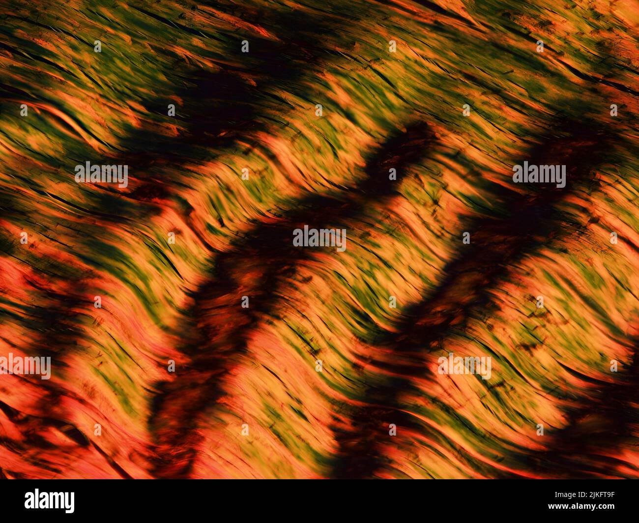 This image shows the crimp (ripple) pattern of collagen fibers in a medial collateral ligament, which is the ligament on the inside of the knee. The ligament section was magnified to 200X using polarized light microscopy. Stock Photohttps://www.alamy.com/image-license-details/?v=1https://www.alamy.com/this-image-shows-the-crimp-ripple-pattern-of-collagen-fibers-in-a-medial-collateral-ligament-which-is-the-ligament-on-the-inside-of-the-knee-the-ligament-section-was-magnified-to-200x-using-polarized-light-microscopy-image476706763.html
RM2JKFT9F–This image shows the crimp (ripple) pattern of collagen fibers in a medial collateral ligament, which is the ligament on the inside of the knee. The ligament section was magnified to 200X using polarized light microscopy.
 Medial knee ligament sprain medical vector illustration isolated on white background infographic Stock Vectorhttps://www.alamy.com/image-license-details/?v=1https://www.alamy.com/medial-knee-ligament-sprain-medical-vector-illustration-isolated-on-white-background-infographic-image331009646.html
RF2A6EP52–Medial knee ligament sprain medical vector illustration isolated on white background infographic
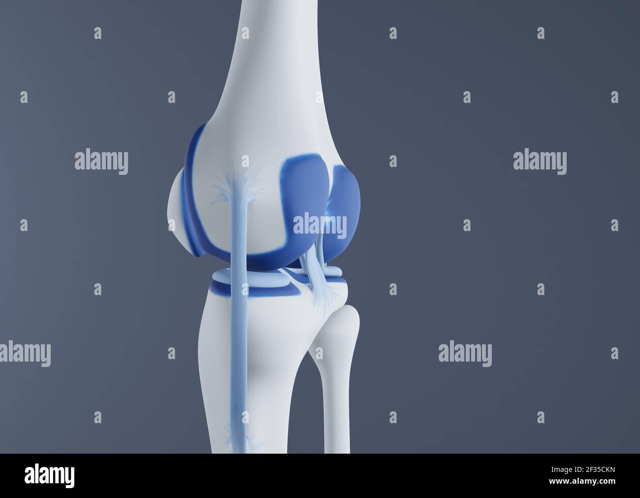 View of knee bones and ligaments. Stock Photohttps://www.alamy.com/image-license-details/?v=1https://www.alamy.com/view-of-knee-bones-and-ligaments-image415012521.html
RF2F35CKN–View of knee bones and ligaments.
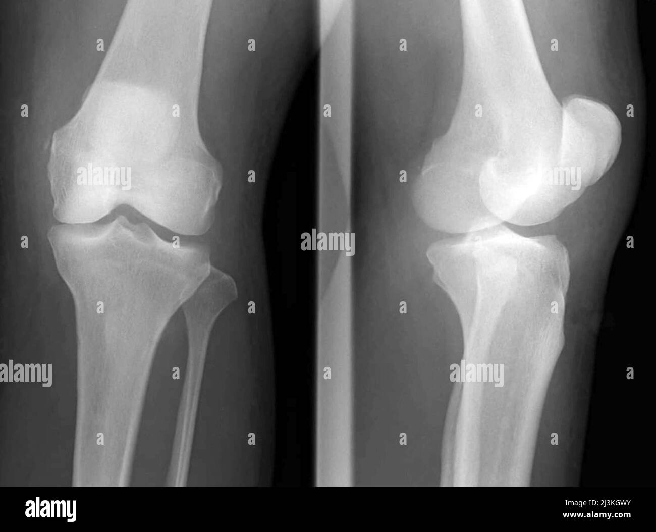 Stieda fractures, X-ray Stock Photohttps://www.alamy.com/image-license-details/?v=1https://www.alamy.com/stieda-fractures-x-ray-image466954263.html
RF2J3KGWY–Stieda fractures, X-ray
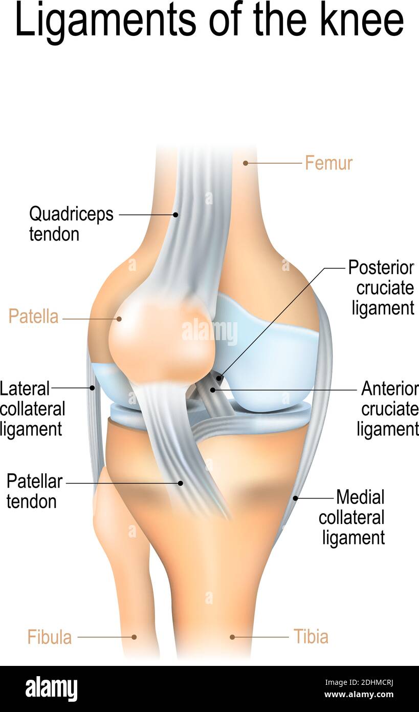 Ligaments of the knee. Anterior and Posterior cruciate ligaments, Patellar and Quadriceps, tendons, Medial and Lateral collateral ligaments Stock Vectorhttps://www.alamy.com/image-license-details/?v=1https://www.alamy.com/ligaments-of-the-knee-anterior-and-posterior-cruciate-ligaments-patellar-and-quadriceps-tendons-medial-and-lateral-collateral-ligaments-image389526358.html
RF2DHMCRJ–Ligaments of the knee. Anterior and Posterior cruciate ligaments, Patellar and Quadriceps, tendons, Medial and Lateral collateral ligaments
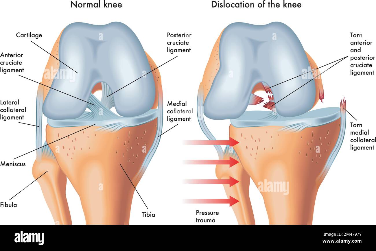 Medical illustration of symptoms of dislocated knee, with annotations. Stock Vectorhttps://www.alamy.com/image-license-details/?v=1https://www.alamy.com/medical-illustration-of-symptoms-of-dislocated-knee-with-annotations-image501720239.html
RF2M4797Y–Medical illustration of symptoms of dislocated knee, with annotations.
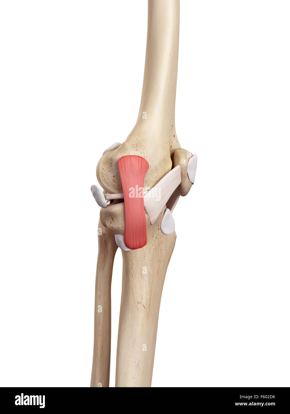 medical accurate illustration of the tibial collateral ligament Stock Photohttps://www.alamy.com/image-license-details/?v=1https://www.alamy.com/stock-photo-medical-accurate-illustration-of-the-tibial-collateral-ligament-89741714.html
RFF602D6–medical accurate illustration of the tibial collateral ligament
 Fibular collateral ligament injury. joint anatomy. Vector illustration for biological, medical, science and educational use Stock Vectorhttps://www.alamy.com/image-license-details/?v=1https://www.alamy.com/fibular-collateral-ligament-injury-joint-anatomy-vector-illustration-for-biological-medical-science-and-educational-use-image389526263.html
RF2DHMCM7–Fibular collateral ligament injury. joint anatomy. Vector illustration for biological, medical, science and educational use
 Detail of human knee showing insertion of arthroscopic instruments. Stock Photohttps://www.alamy.com/image-license-details/?v=1https://www.alamy.com/stock-photo-detail-of-human-knee-showing-insertion-of-arthroscopic-instruments-72785289.html
RME6BJAH–Detail of human knee showing insertion of arthroscopic instruments.
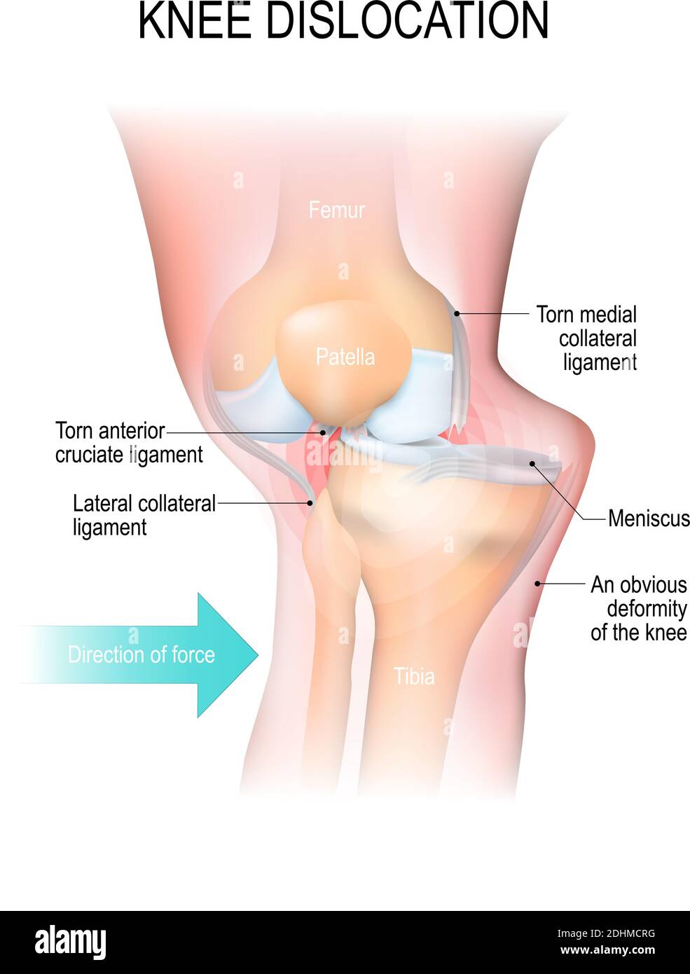 Knee dislocation. Lateral trauma to the knee, torn collateral ligaments, cruciate ligament injury and meniscus injury Stock Vectorhttps://www.alamy.com/image-license-details/?v=1https://www.alamy.com/knee-dislocation-lateral-trauma-to-the-knee-torn-collateral-ligaments-cruciate-ligament-injury-and-meniscus-injury-image389526356.html
RF2DHMCRG–Knee dislocation. Lateral trauma to the knee, torn collateral ligaments, cruciate ligament injury and meniscus injury
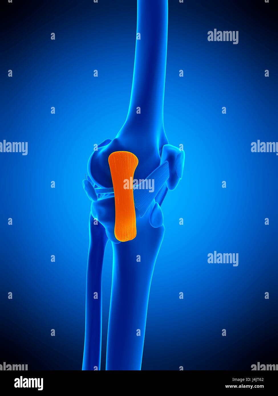 Illustration of the tibial collateral ligament. Stock Photohttps://www.alamy.com/image-license-details/?v=1https://www.alamy.com/stock-photo-illustration-of-the-tibial-collateral-ligament-140555690.html
RFJ4JT62–Illustration of the tibial collateral ligament.
 Front view of knee showing torn medial collateral ligament. Stock Photohttps://www.alamy.com/image-license-details/?v=1https://www.alamy.com/front-view-of-knee-showing-torn-medial-collateral-ligament-image431667949.html
RM2G284TD–Front view of knee showing torn medial collateral ligament.
 Knee Joint Stock Photohttps://www.alamy.com/image-license-details/?v=1https://www.alamy.com/knee-joint-image490198241.html
RF2KDECT1–Knee Joint
 Female patient holding a crutch. Stock Photohttps://www.alamy.com/image-license-details/?v=1https://www.alamy.com/stock-photo-female-patient-holding-a-crutch-14166672.html
RFAGCDAW–Female patient holding a crutch.
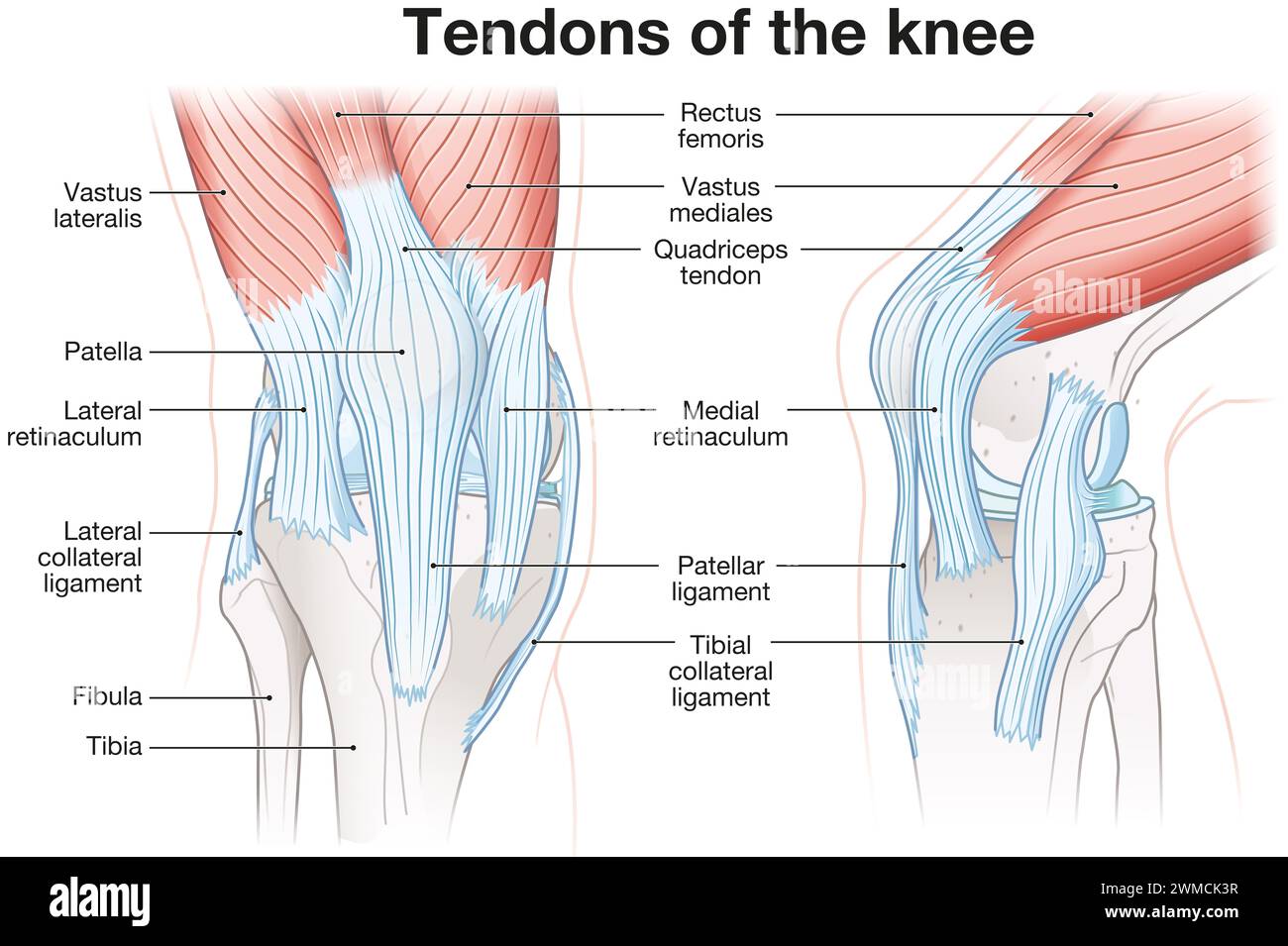 The knee joint's ligaments include the anterior cruciate ligament (ACL), posterior cruciate ligament (PCL), medial and lateral collateral ligaments. Stock Photohttps://www.alamy.com/image-license-details/?v=1https://www.alamy.com/the-knee-joints-ligaments-include-the-anterior-cruciate-ligament-acl-posterior-cruciate-ligament-pcl-medial-and-lateral-collateral-ligaments-image597724059.html
RF2WMCK3R–The knee joint's ligaments include the anterior cruciate ligament (ACL), posterior cruciate ligament (PCL), medial and lateral collateral ligaments.
 Myofascial fibrolysis instrumental hook in knee medial collateral ligament Physiotherapy medical center, Donostia, San Sebastian, Gipuzkoa, Basque Co Stock Photohttps://www.alamy.com/image-license-details/?v=1https://www.alamy.com/myofascial-fibrolysis-instrumental-hook-in-knee-medial-collateral-ligament-physiotherapy-medical-center-donostia-san-sebastian-gipuzkoa-basque-co-image604409236.html
RF2X39644–Myofascial fibrolysis instrumental hook in knee medial collateral ligament Physiotherapy medical center, Donostia, San Sebastian, Gipuzkoa, Basque Co
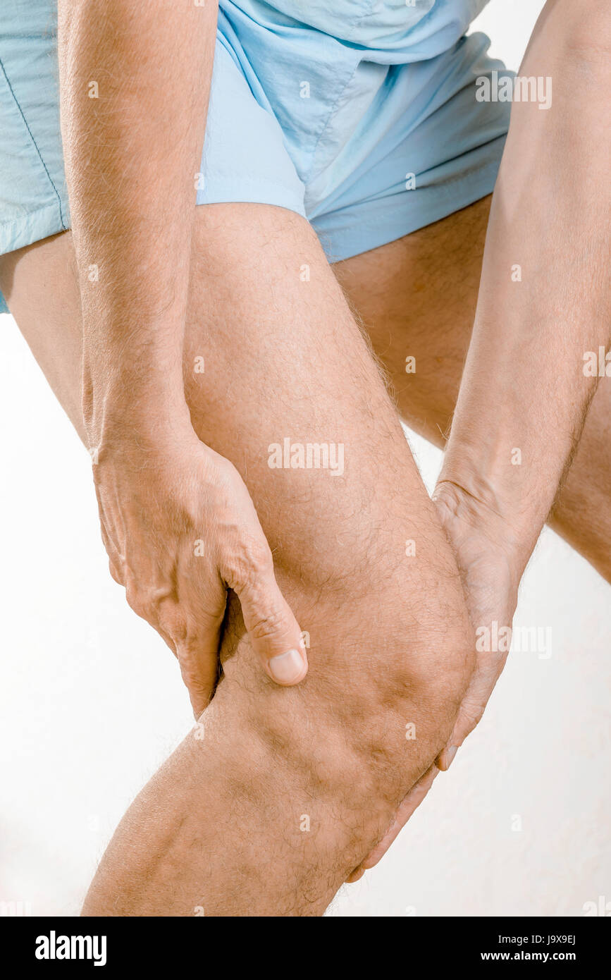 Athlete man feeling pain to the knee. It could be medial meniscus tears, injuries of the collateral lligament, bursitis or Iliotibial band syndrome. Stock Photohttps://www.alamy.com/image-license-details/?v=1https://www.alamy.com/stock-photo-athlete-man-feeling-pain-to-the-knee-it-could-be-medial-meniscus-tears-143793066.html
RFJ9X9EJ–Athlete man feeling pain to the knee. It could be medial meniscus tears, injuries of the collateral lligament, bursitis or Iliotibial band syndrome.
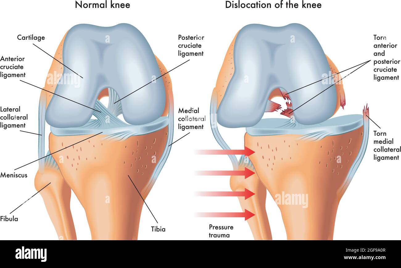 Medical illustration of symptoms of dislocated knee, with annotations. Stock Vectorhttps://www.alamy.com/image-license-details/?v=1https://www.alamy.com/medical-illustration-of-symptoms-of-dislocated-knee-with-annotations-image439684471.html
RF2GF9A0R–Medical illustration of symptoms of dislocated knee, with annotations.
 Knee ligaments, tendons, x-ray Stock Photohttps://www.alamy.com/image-license-details/?v=1https://www.alamy.com/stock-photo-knee-ligaments-tendons-x-ray-74986043.html
RFE9YWCY–Knee ligaments, tendons, x-ray
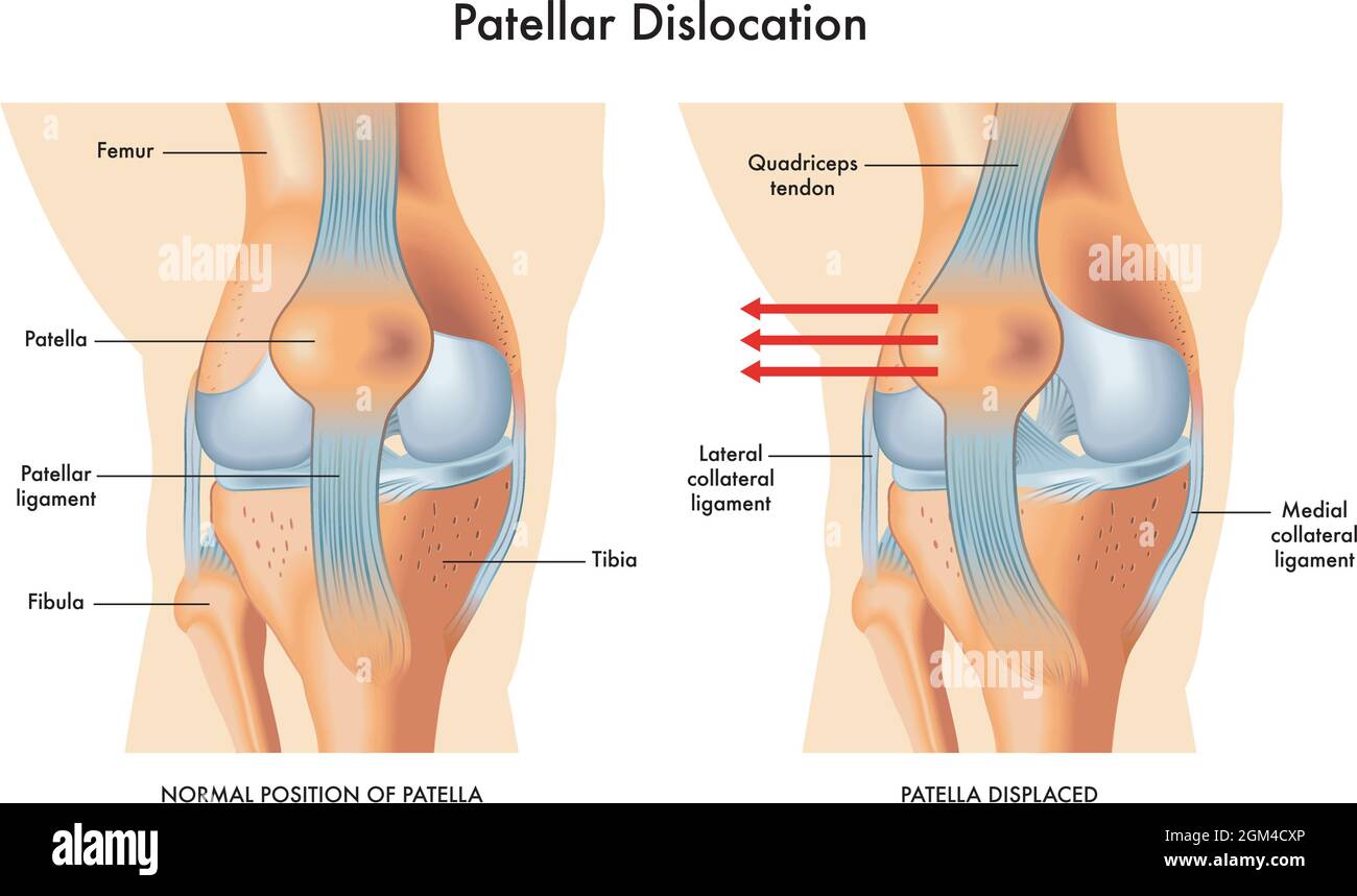 Medical illustration of symptoms of patellar dislocation, with annotations. Stock Vectorhttps://www.alamy.com/image-license-details/?v=1https://www.alamy.com/medical-illustration-of-symptoms-of-patellar-dislocation-with-annotations-image442650286.html
RF2GM4CXP–Medical illustration of symptoms of patellar dislocation, with annotations.
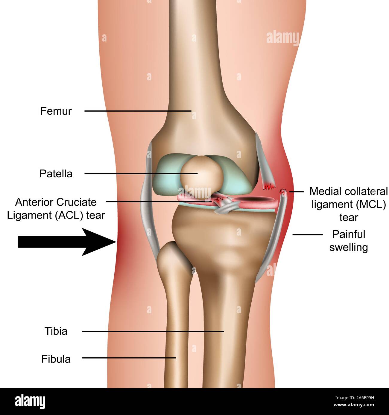 Dislocated knee, medial collateral ligament tear medical vector illustration isolated on white background Stock Vectorhttps://www.alamy.com/image-license-details/?v=1https://www.alamy.com/dislocated-knee-medial-collateral-ligament-tear-medical-vector-illustration-isolated-on-white-background-image331009773.html
RF2A6EP9H–Dislocated knee, medial collateral ligament tear medical vector illustration isolated on white background
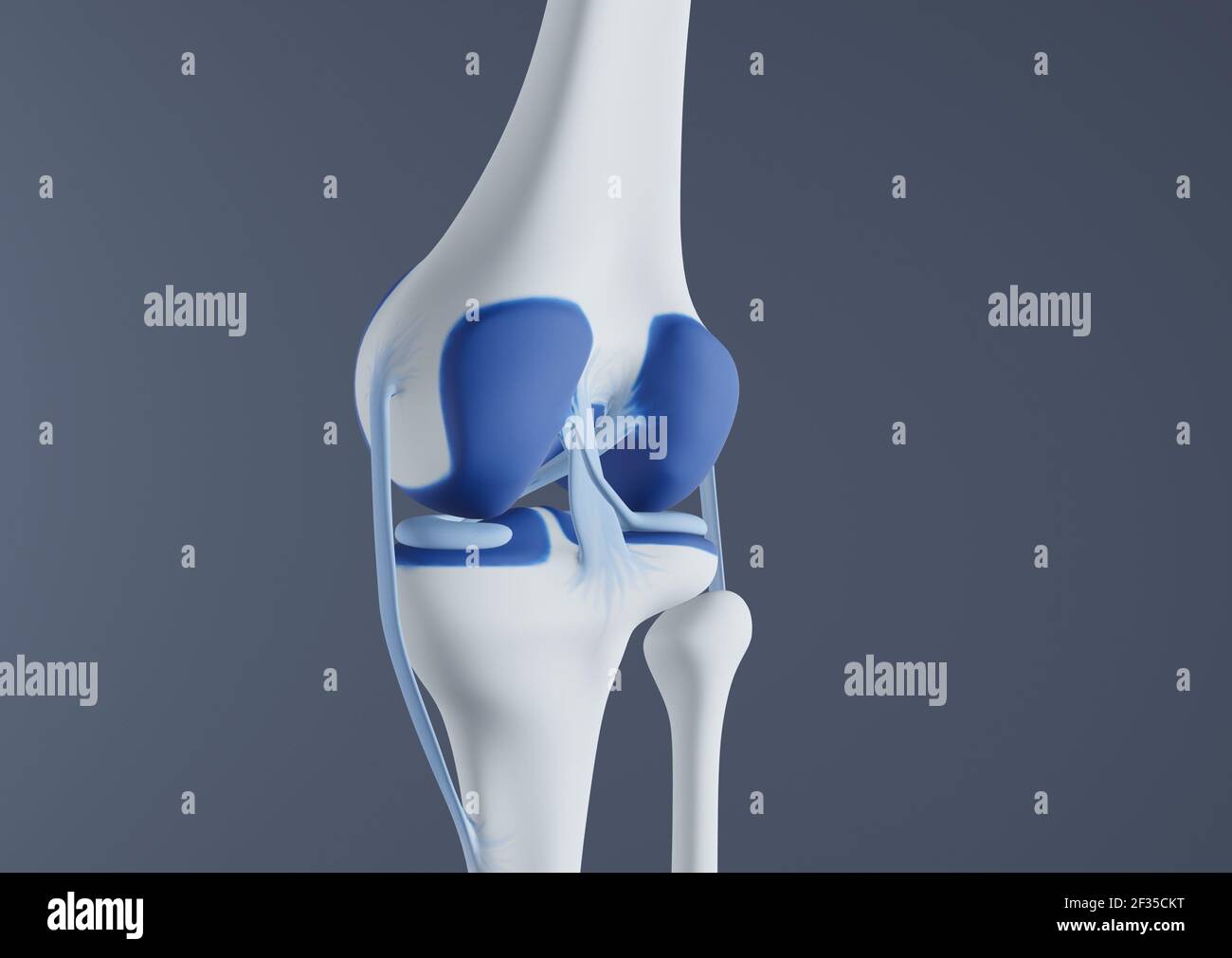 View of knee bones and ligaments. Stock Photohttps://www.alamy.com/image-license-details/?v=1https://www.alamy.com/view-of-knee-bones-and-ligaments-image415012524.html
RF2F35CKT–View of knee bones and ligaments.
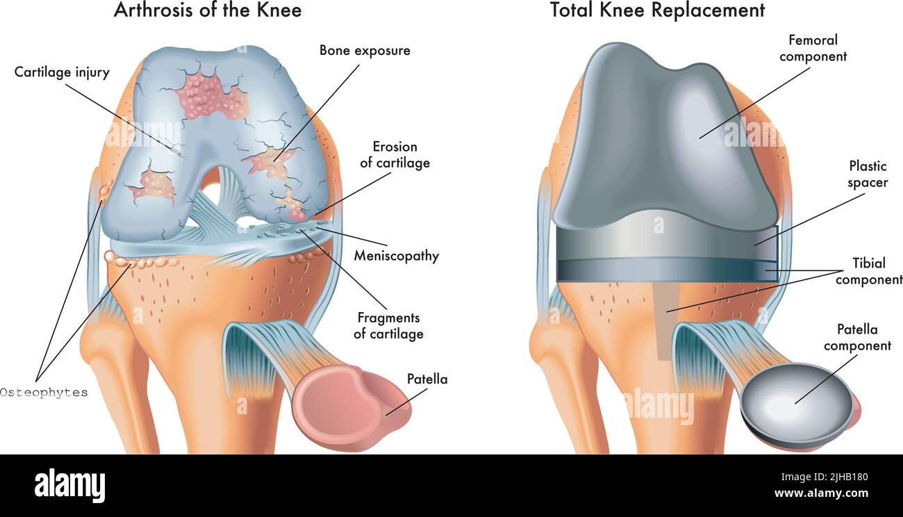 Medical illustration shows an arthrosis of the knee and total knee replacement, with annotations. Stock Vectorhttps://www.alamy.com/image-license-details/?v=1https://www.alamy.com/medical-illustration-shows-an-arthrosis-of-the-knee-and-total-knee-replacement-with-annotations-image475371568.html
RF2JHB180–Medical illustration shows an arthrosis of the knee and total knee replacement, with annotations.
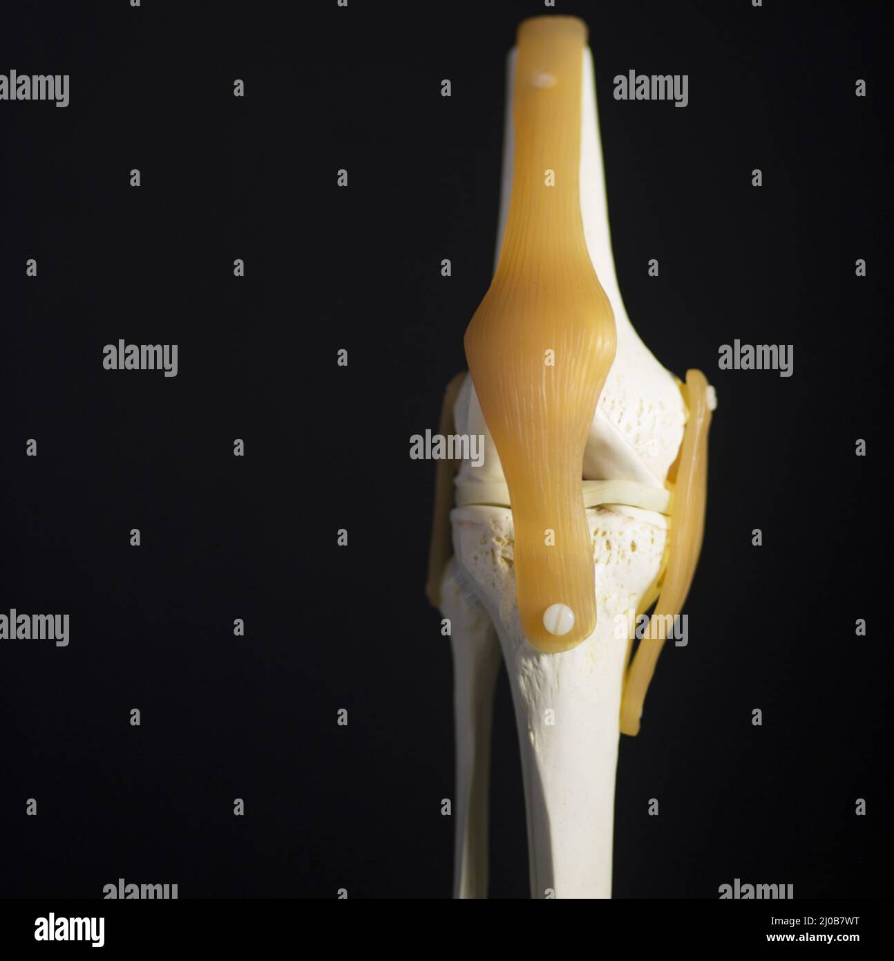 Medical knee joint meniscus plastic demonstration teaching model Stock Photohttps://www.alamy.com/image-license-details/?v=1https://www.alamy.com/medical-knee-joint-meniscus-plastic-demonstration-teaching-model-image464927620.html
RF2J0B7WT–Medical knee joint meniscus plastic demonstration teaching model
 Knee joint of human Stock Vectorhttps://www.alamy.com/image-license-details/?v=1https://www.alamy.com/knee-joint-of-human-image445967304.html
RF2GWFFRM–Knee joint of human
 The doctor winds an elastic bandage on a mock knee joint on a blue background. Knee fracture concept, cruciate ligament injury. Medial collateral liga Stock Photohttps://www.alamy.com/image-license-details/?v=1https://www.alamy.com/the-doctor-winds-an-elastic-bandage-on-a-mock-knee-joint-on-a-blue-background-knee-fracture-concept-cruciate-ligament-injury-medial-collateral-liga-image443089439.html
RF2GMTD2R–The doctor winds an elastic bandage on a mock knee joint on a blue background. Knee fracture concept, cruciate ligament injury. Medial collateral liga
 white background vector illustration of a knee Stock Vectorhttps://www.alamy.com/image-license-details/?v=1https://www.alamy.com/stock-photo-white-background-vector-illustration-of-a-knee-137578586.html
RFHYR6TX–white background vector illustration of a knee
 Knee bones, illustration Stock Photohttps://www.alamy.com/image-license-details/?v=1https://www.alamy.com/knee-bones-illustration-image560517475.html
RF2RFWNN7–Knee bones, illustration
 Knee dislocation and normal. Lateral trauma to the knee, torn collateral ligaments, cruciate ligament injury and meniscus injury. Human anatomy Stock Vectorhttps://www.alamy.com/image-license-details/?v=1https://www.alamy.com/knee-dislocation-and-normal-lateral-trauma-to-the-knee-torn-collateral-ligaments-cruciate-ligament-injury-and-meniscus-injury-human-anatomy-image389526500.html
RF2DHMD0M–Knee dislocation and normal. Lateral trauma to the knee, torn collateral ligaments, cruciate ligament injury and meniscus injury. Human anatomy
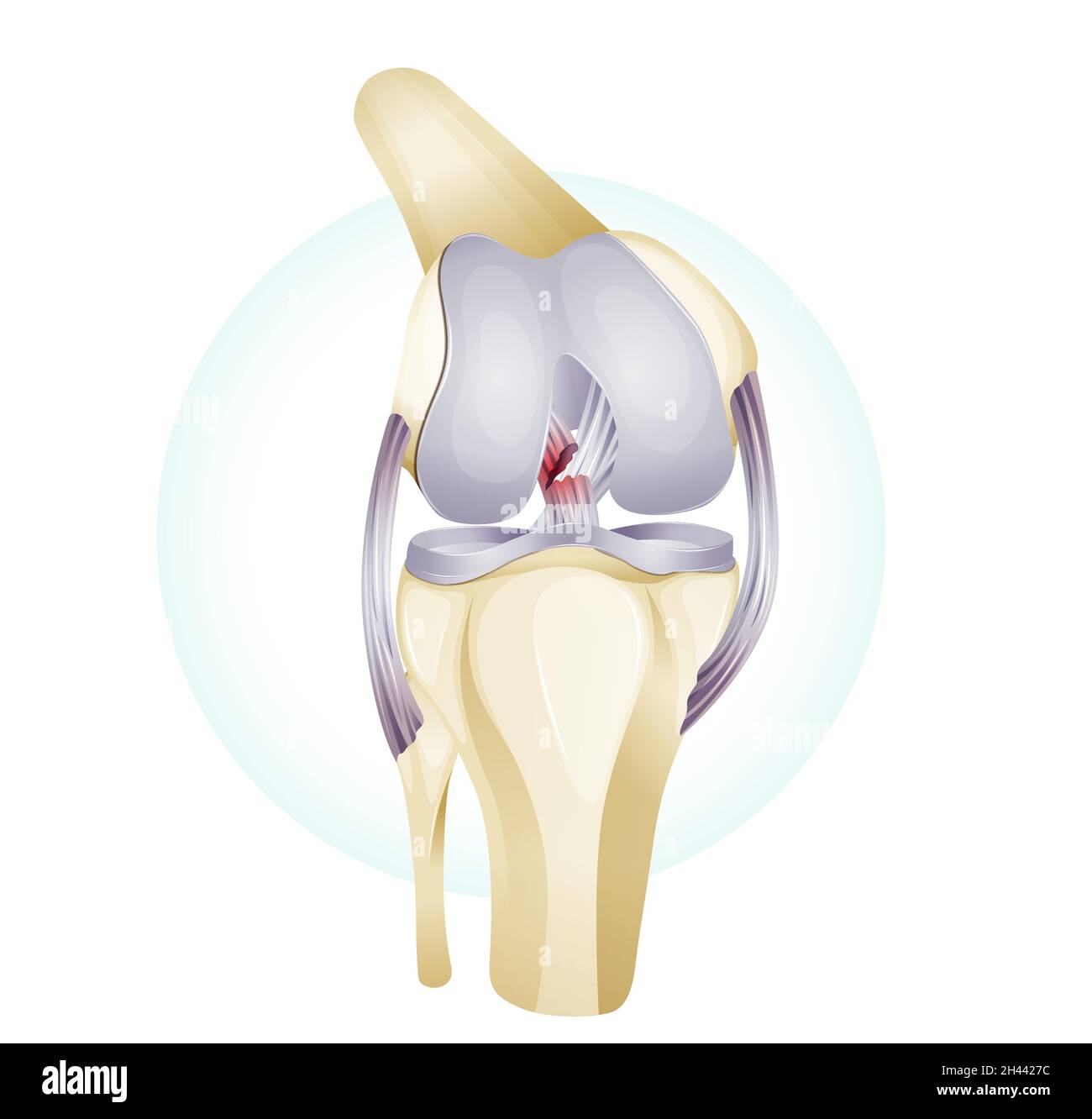 Knee Cartilage Injury - Illustration as EPS 10 File Stock Vectorhttps://www.alamy.com/image-license-details/?v=1https://www.alamy.com/knee-cartilage-injury-illustration-as-eps-10-file-image450017776.html
RF2H4427C–Knee Cartilage Injury - Illustration as EPS 10 File
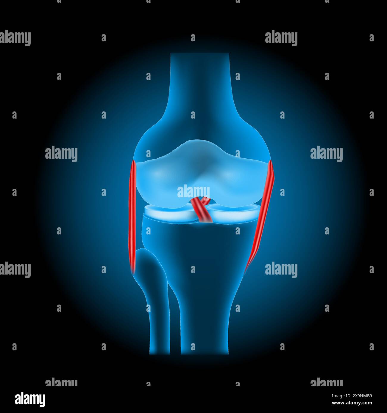 ligament injuries to the knee. Realistic transparent blue human knee joint with glowing effect on dark background. vector illustration like X-ray imag Stock Vectorhttps://www.alamy.com/image-license-details/?v=1https://www.alamy.com/ligament-injuries-to-the-knee-realistic-transparent-blue-human-knee-joint-with-glowing-effect-on-dark-background-vector-illustration-like-x-ray-imag-image608371773.html
RF2X9NMB9–ligament injuries to the knee. Realistic transparent blue human knee joint with glowing effect on dark background. vector illustration like X-ray imag
 Anterior View of Knee Joint Stock Photohttps://www.alamy.com/image-license-details/?v=1https://www.alamy.com/anterior-view-of-knee-joint-image490198448.html
RF2KDED3C–Anterior View of Knee Joint
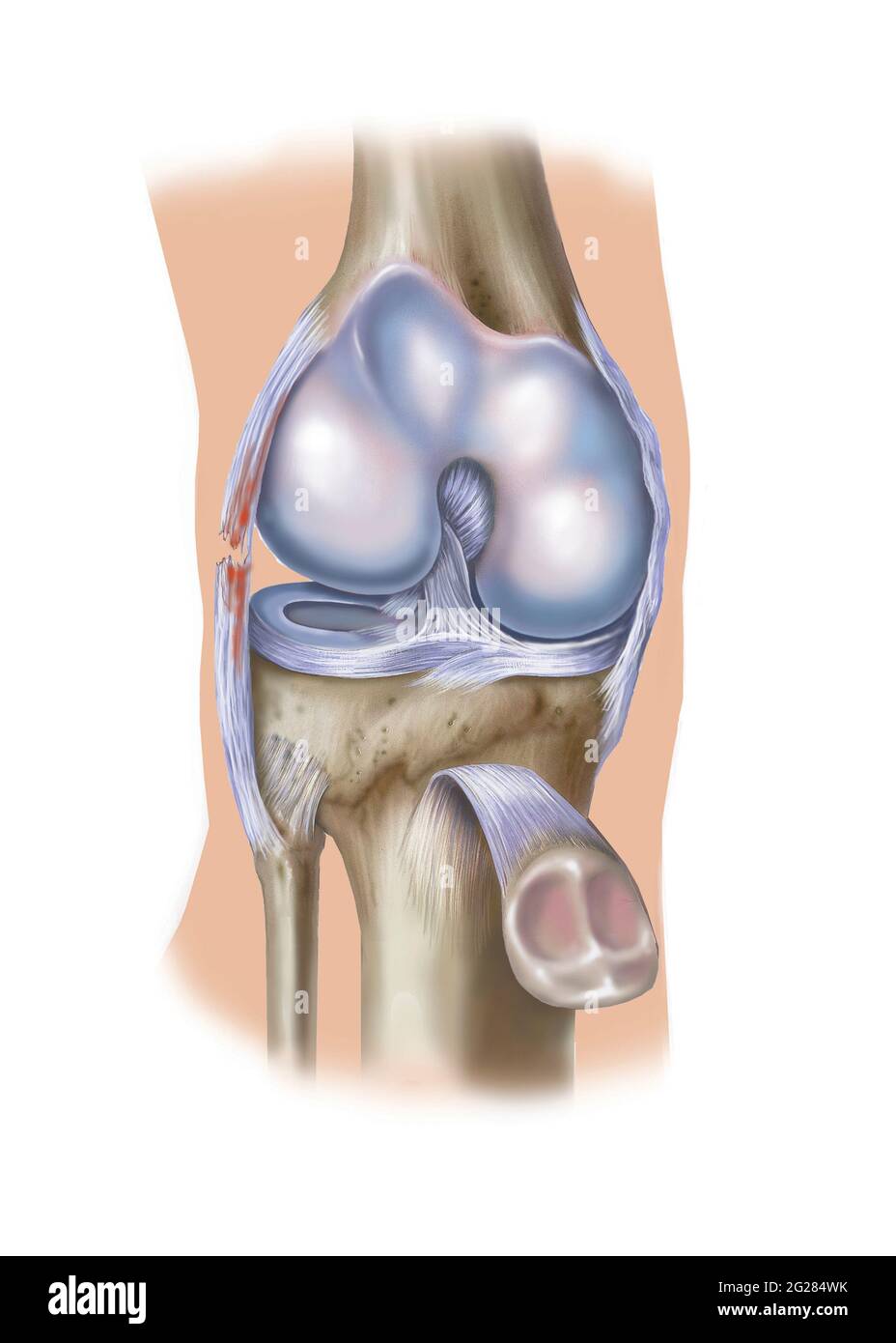 Front view of knee showing lateral collateral ligament tear. Stock Photohttps://www.alamy.com/image-license-details/?v=1https://www.alamy.com/front-view-of-knee-showing-lateral-collateral-ligament-tear-image431667983.html
RM2G284WK–Front view of knee showing lateral collateral ligament tear.
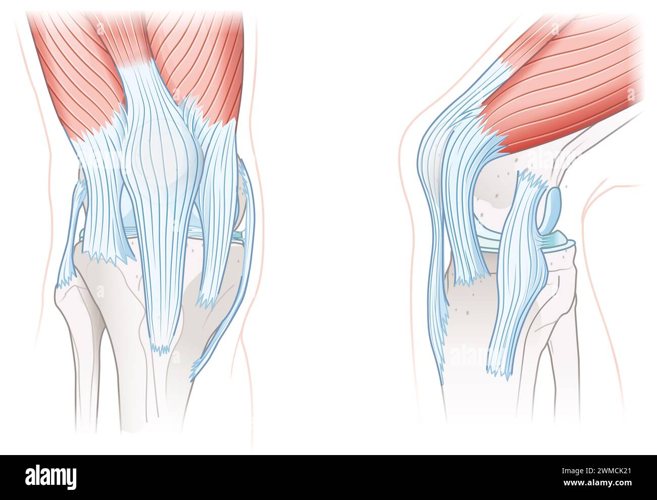 The knee joint's ligaments include the anterior cruciate ligament (ACL), posterior cruciate ligament (PCL), medial and lateral collateral ligaments. Stock Photohttps://www.alamy.com/image-license-details/?v=1https://www.alamy.com/the-knee-joints-ligaments-include-the-anterior-cruciate-ligament-acl-posterior-cruciate-ligament-pcl-medial-and-lateral-collateral-ligaments-image597724009.html
RF2WMCK21–The knee joint's ligaments include the anterior cruciate ligament (ACL), posterior cruciate ligament (PCL), medial and lateral collateral ligaments.
 Myofascial fibrolysis instrumental hook in knee medial collateral ligament Physiotherapy medical center, Donostia, San Sebastian, Gipuzkoa, Basque Co Stock Photohttps://www.alamy.com/image-license-details/?v=1https://www.alamy.com/myofascial-fibrolysis-instrumental-hook-in-knee-medial-collateral-ligament-physiotherapy-medical-center-donostia-san-sebastian-gipuzkoa-basque-co-image604409239.html
RF2X39647–Myofascial fibrolysis instrumental hook in knee medial collateral ligament Physiotherapy medical center, Donostia, San Sebastian, Gipuzkoa, Basque Co
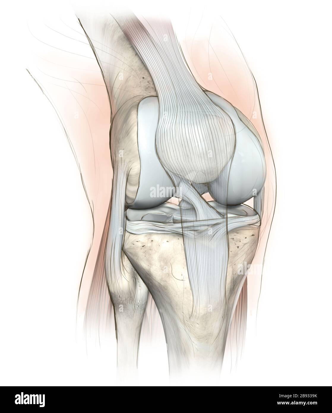 Hand drawing illustration showing human knee joint with femur, articular cartilage, meniscus, medial collateral ligament, articular cartilage, patella Stock Photohttps://www.alamy.com/image-license-details/?v=1https://www.alamy.com/hand-drawing-illustration-showing-human-knee-joint-with-femur-articular-cartilage-meniscus-medial-collateral-ligament-articular-cartilage-patella-image349807743.html
RF2B9339K–Hand drawing illustration showing human knee joint with femur, articular cartilage, meniscus, medial collateral ligament, articular cartilage, patella
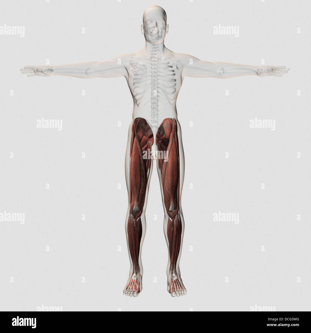 Male muscle anatomy of the human legs, anterior view. Stock Photohttps://www.alamy.com/image-license-details/?v=1https://www.alamy.com/stock-photo-male-muscle-anatomy-of-the-human-legs-anterior-view-59361136.html
RFDCG3MG–Male muscle anatomy of the human legs, anterior view.
 Knee ligaments, tendons, x-ray Stock Photohttps://www.alamy.com/image-license-details/?v=1https://www.alamy.com/stock-photo-knee-ligaments-tendons-x-ray-74986048.html
RFE9YWD4–Knee ligaments, tendons, x-ray
 Front view of knee showing anterior cruciate ligament tear. Stock Photohttps://www.alamy.com/image-license-details/?v=1https://www.alamy.com/front-view-of-knee-showing-anterior-cruciate-ligament-tear-image431667760.html
RM2G284HM–Front view of knee showing anterior cruciate ligament tear.
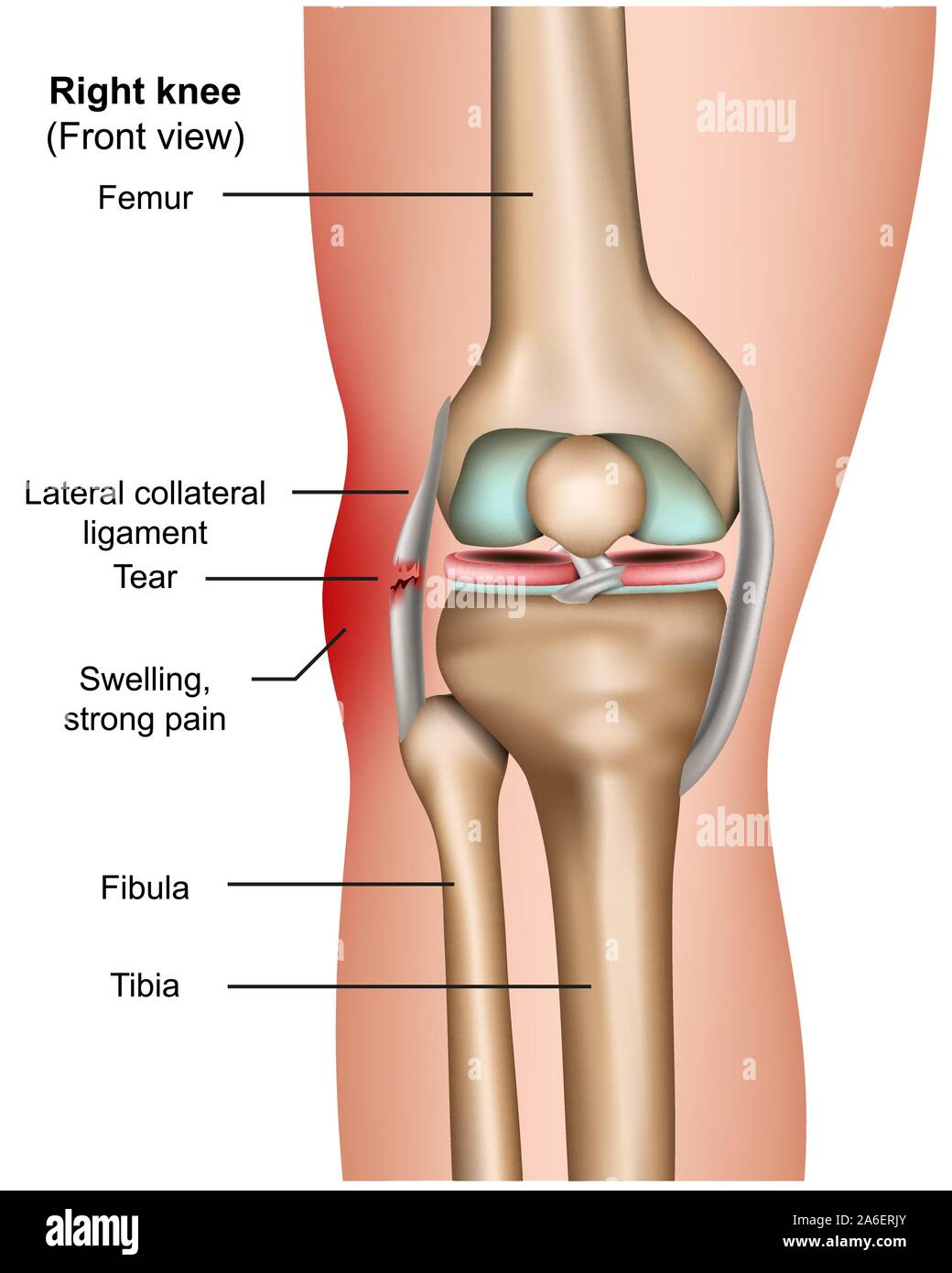 Lateral collateral tear 3d medical vector infographic isolated on white background Stock Vectorhttps://www.alamy.com/image-license-details/?v=1https://www.alamy.com/lateral-collateral-tear-3d-medical-vector-infographic-isolated-on-white-background-image331010819.html
RF2A6ERJY–Lateral collateral tear 3d medical vector infographic isolated on white background
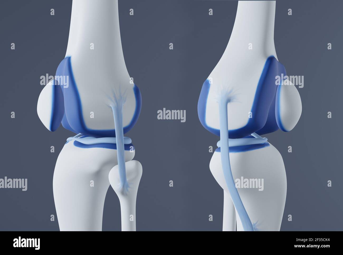 View of knee bones and ligaments. Stock Photohttps://www.alamy.com/image-license-details/?v=1https://www.alamy.com/view-of-knee-bones-and-ligaments-image415012504.html
RF2F35CK4–View of knee bones and ligaments.
 Knee and meniscus anatomy medical vector illustration isolated on white background eps 10 Stock Vectorhttps://www.alamy.com/image-license-details/?v=1https://www.alamy.com/knee-and-meniscus-anatomy-medical-vector-illustration-isolated-on-white-background-eps-10-image341381579.html
RF2ARB7K7–Knee and meniscus anatomy medical vector illustration isolated on white background eps 10
 Woman with medial collateral ligament strain on crutches Stock Photohttps://www.alamy.com/image-license-details/?v=1https://www.alamy.com/stock-photo-woman-with-medial-collateral-ligament-strain-on-crutches-14166094.html
RFAGCBJR–Woman with medial collateral ligament strain on crutches
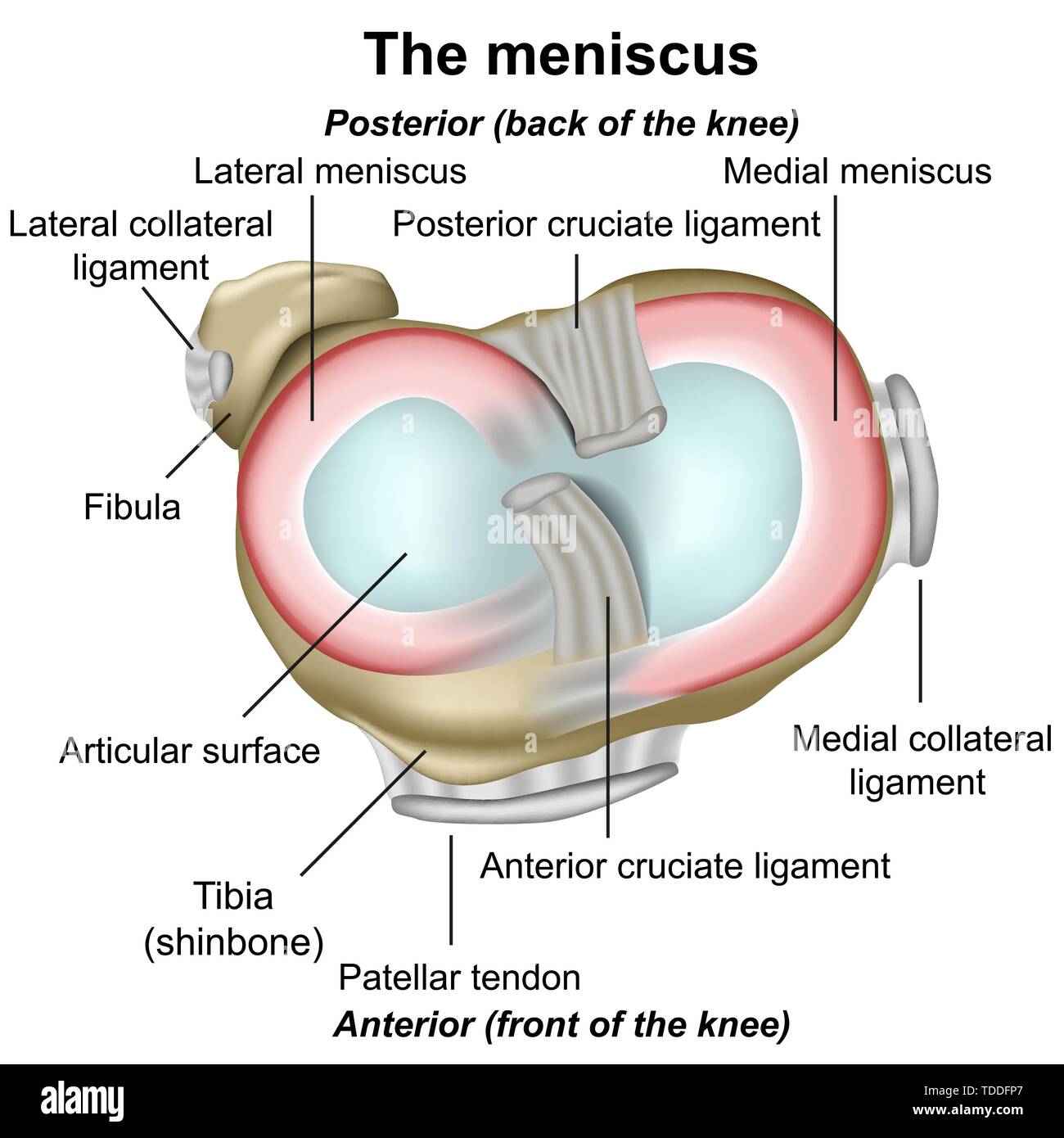 Meniscus knee anatomy medical illustration isolated on white background infographic Stock Vectorhttps://www.alamy.com/image-license-details/?v=1https://www.alamy.com/meniscus-knee-anatomy-medical-illustration-isolated-on-white-background-infographic-image249233439.html
RFTDDFP7–Meniscus knee anatomy medical illustration isolated on white background infographic
 MCL ultrasound to treat knee bursitis Physiotherapy medical center, Donostia, San Sebastian, Gipuzkoa, Basque Country, Spain Stock Photohttps://www.alamy.com/image-license-details/?v=1https://www.alamy.com/mcl-ultrasound-to-treat-knee-bursitis-physiotherapy-medical-center-donostia-san-sebastian-gipuzkoa-basque-country-spain-image604409372.html
RF2X39690–MCL ultrasound to treat knee bursitis Physiotherapy medical center, Donostia, San Sebastian, Gipuzkoa, Basque Country, Spain
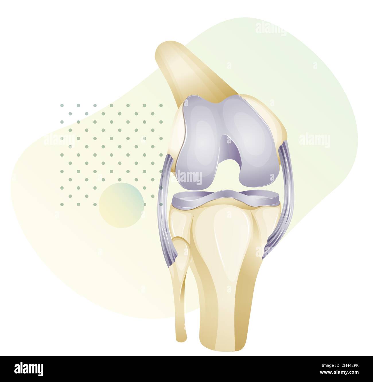 Knee Cartilage Injury - Illustration as EPS 10 File Stock Vectorhttps://www.alamy.com/image-license-details/?v=1https://www.alamy.com/knee-cartilage-injury-illustration-as-eps-10-file-image450018203.html
RF2H442PK–Knee Cartilage Injury - Illustration as EPS 10 File
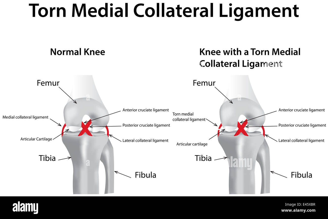 Torn Medial Collateral Ligament Stock Photohttps://www.alamy.com/image-license-details/?v=1https://www.alamy.com/stock-photo-torn-medial-collateral-ligament-71430571.html
RME45XBR–Torn Medial Collateral Ligament
 Knee Cartilage Injury - Illustration as EPS 10 File Stock Vectorhttps://www.alamy.com/image-license-details/?v=1https://www.alamy.com/knee-cartilage-injury-illustration-as-eps-10-file-image450016695.html
RF2H440TR–Knee Cartilage Injury - Illustration as EPS 10 File
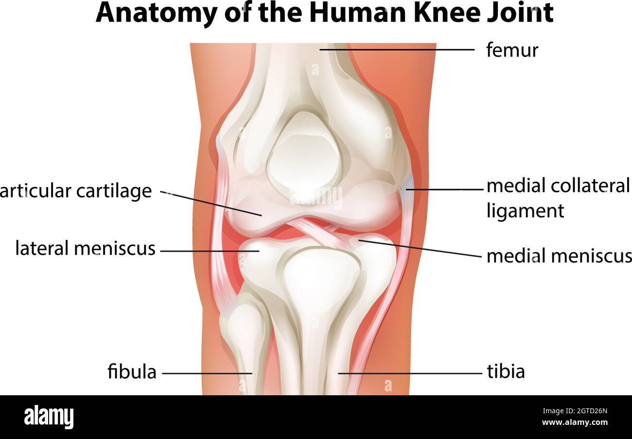 Human knee joint anatomy Stock Vectorhttps://www.alamy.com/image-license-details/?v=1https://www.alamy.com/human-knee-joint-anatomy-image445298077.html
RF2GTD26N–Human knee joint anatomy
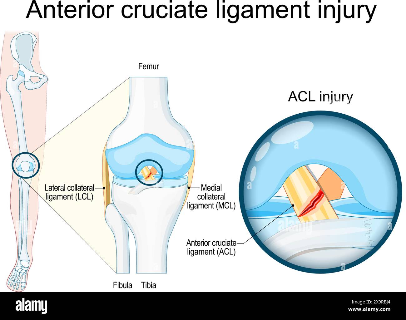 Anterior cruciate ligament injury. Close-up of a human knee joint. Knee trauma as tear or sprain of anterior cruciate ligament. Sports Injury. Vector Stock Vectorhttps://www.alamy.com/image-license-details/?v=1https://www.alamy.com/anterior-cruciate-ligament-injury-close-up-of-a-human-knee-joint-knee-trauma-as-tear-or-sprain-of-anterior-cruciate-ligament-sports-injury-vector-image608408812.html
RF2X9RBJ4–Anterior cruciate ligament injury. Close-up of a human knee joint. Knee trauma as tear or sprain of anterior cruciate ligament. Sports Injury. Vector
 Anterior and Posterior View of Knee Joint with Removed Patella Stock Photohttps://www.alamy.com/image-license-details/?v=1https://www.alamy.com/anterior-and-posterior-view-of-knee-joint-with-removed-patella-image490198506.html
RF2KDED5E–Anterior and Posterior View of Knee Joint with Removed Patella
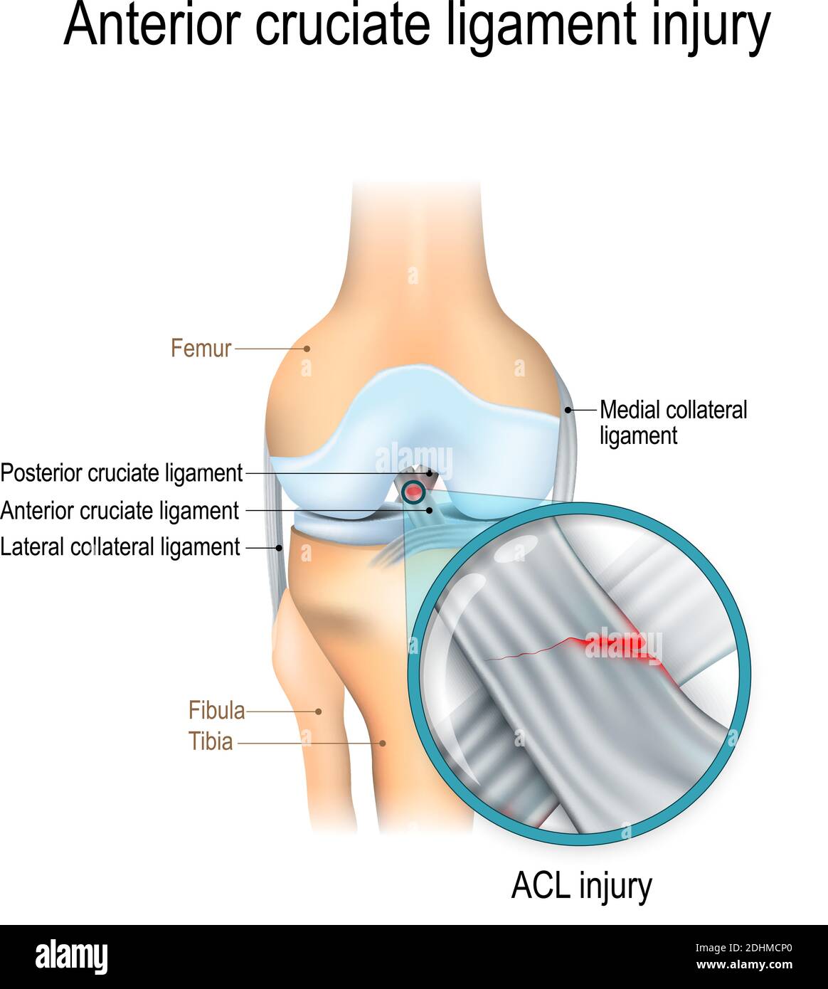 Anterior cruciate ligament injury. joint anatomy. Vector illustration for biological, medical, science and educational use Stock Vectorhttps://www.alamy.com/image-license-details/?v=1https://www.alamy.com/anterior-cruciate-ligament-injury-joint-anatomy-vector-illustration-for-biological-medical-science-and-educational-use-image389526312.html
RF2DHMCP0–Anterior cruciate ligament injury. joint anatomy. Vector illustration for biological, medical, science and educational use
 vector illustration of a Meniscus tear and surgery Stock Vectorhttps://www.alamy.com/image-license-details/?v=1https://www.alamy.com/stock-image-vector-illustration-of-a-meniscus-tear-and-surgery-166320543.html
RFKJGFFB–vector illustration of a Meniscus tear and surgery
 Medial knee injuries. joint anatomy. Vector illustration for biological, medical, science and educational use Stock Vectorhttps://www.alamy.com/image-license-details/?v=1https://www.alamy.com/medial-knee-injuries-joint-anatomy-vector-illustration-for-biological-medical-science-and-educational-use-image389526266.html
RF2DHMCMA–Medial knee injuries. joint anatomy. Vector illustration for biological, medical, science and educational use
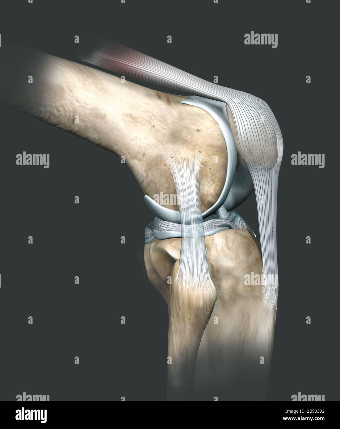 3D illustration showing human knee joint with femur, articular cartilage, meniscus, medial collateral ligament, articular cartilage, patella, kneecap, Stock Photohttps://www.alamy.com/image-license-details/?v=1https://www.alamy.com/3d-illustration-showing-human-knee-joint-with-femur-articular-cartilage-meniscus-medial-collateral-ligament-articular-cartilage-patella-kneecap-image349807726.html
RF2B93392–3D illustration showing human knee joint with femur, articular cartilage, meniscus, medial collateral ligament, articular cartilage, patella, kneecap,
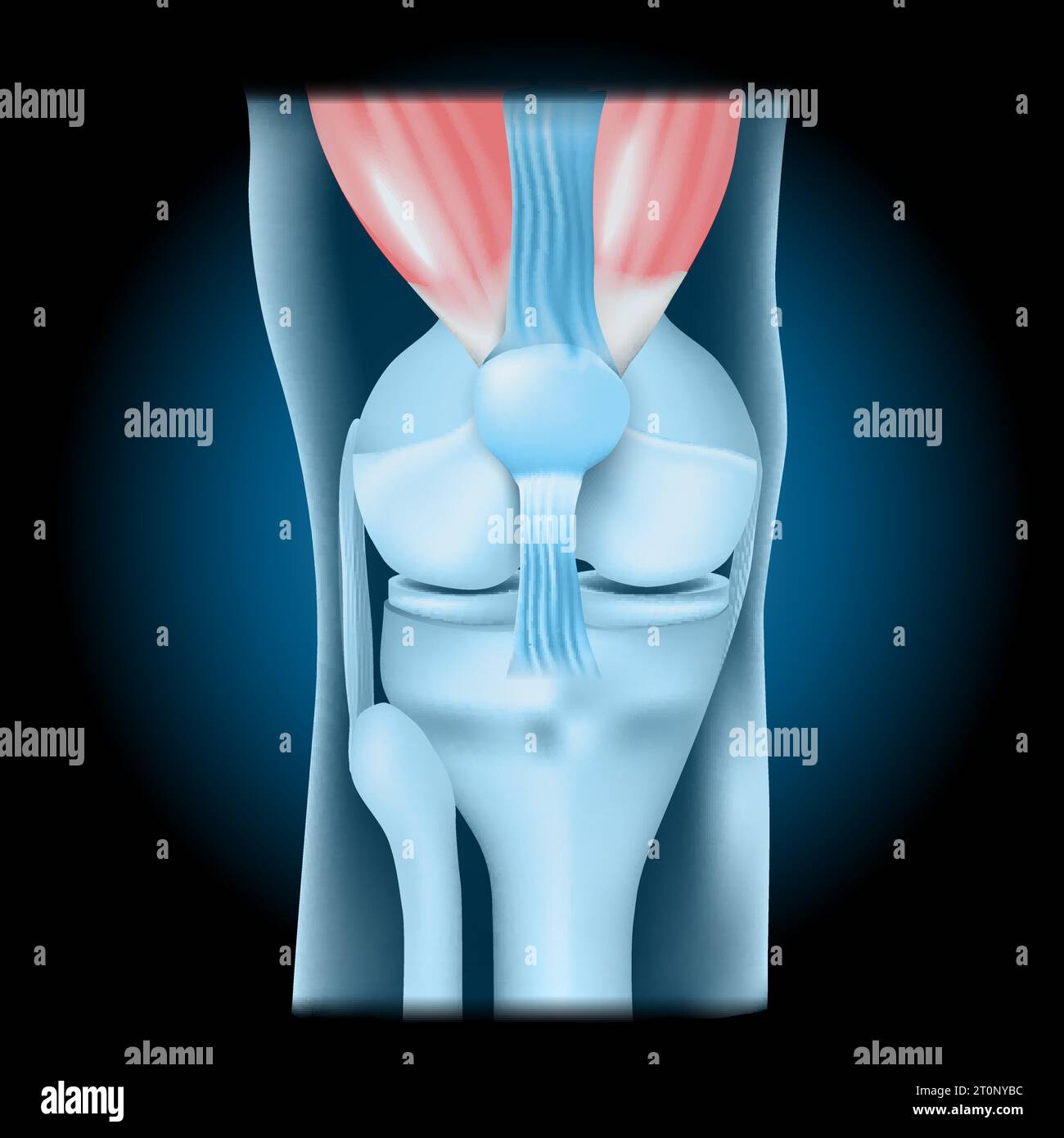 Knee joint with Quadriceps. Front view of human knee with glowing effect. Realistic transparent blue joint on dark background. vector illustration lik Stock Vectorhttps://www.alamy.com/image-license-details/?v=1https://www.alamy.com/knee-joint-with-quadriceps-front-view-of-human-knee-with-glowing-effect-realistic-transparent-blue-joint-on-dark-background-vector-illustration-lik-image568424624.html
RF2T0NYBC–Knee joint with Quadriceps. Front view of human knee with glowing effect. Realistic transparent blue joint on dark background. vector illustration lik
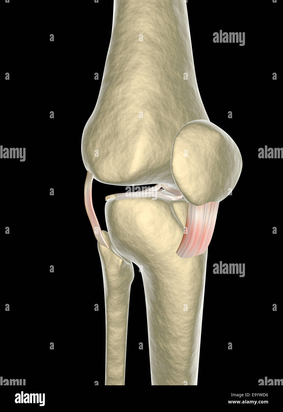 Knee ligaments, tendons, x-ray Stock Photohttps://www.alamy.com/image-license-details/?v=1https://www.alamy.com/stock-photo-knee-ligaments-tendons-x-ray-74986050.html
RFE9YWD6–Knee ligaments, tendons, x-ray
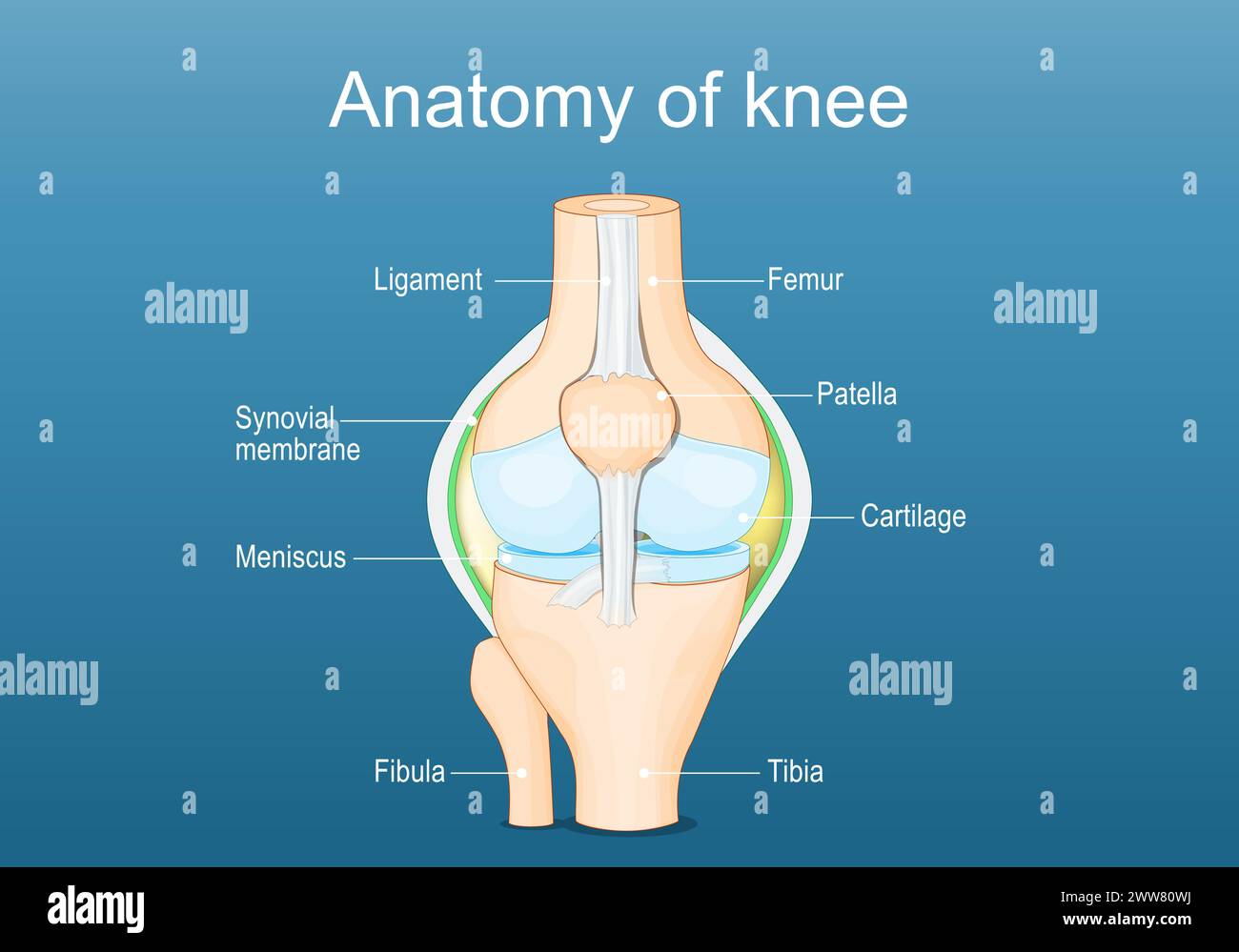 Knee joint anatomy. Labeled of all bones. Isometric Flat vector illustration Stock Vectorhttps://www.alamy.com/image-license-details/?v=1https://www.alamy.com/knee-joint-anatomy-labeled-of-all-bones-isometric-flat-vector-illustration-image600695246.html
RF2WW80WJ–Knee joint anatomy. Labeled of all bones. Isometric Flat vector illustration
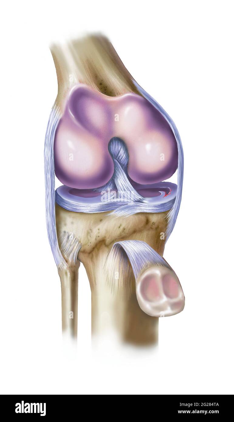 Front view of knee showing torn medial meniscus. Stock Photohttps://www.alamy.com/image-license-details/?v=1https://www.alamy.com/front-view-of-knee-showing-torn-medial-meniscus-image431667946.html
RM2G284TA–Front view of knee showing torn medial meniscus.
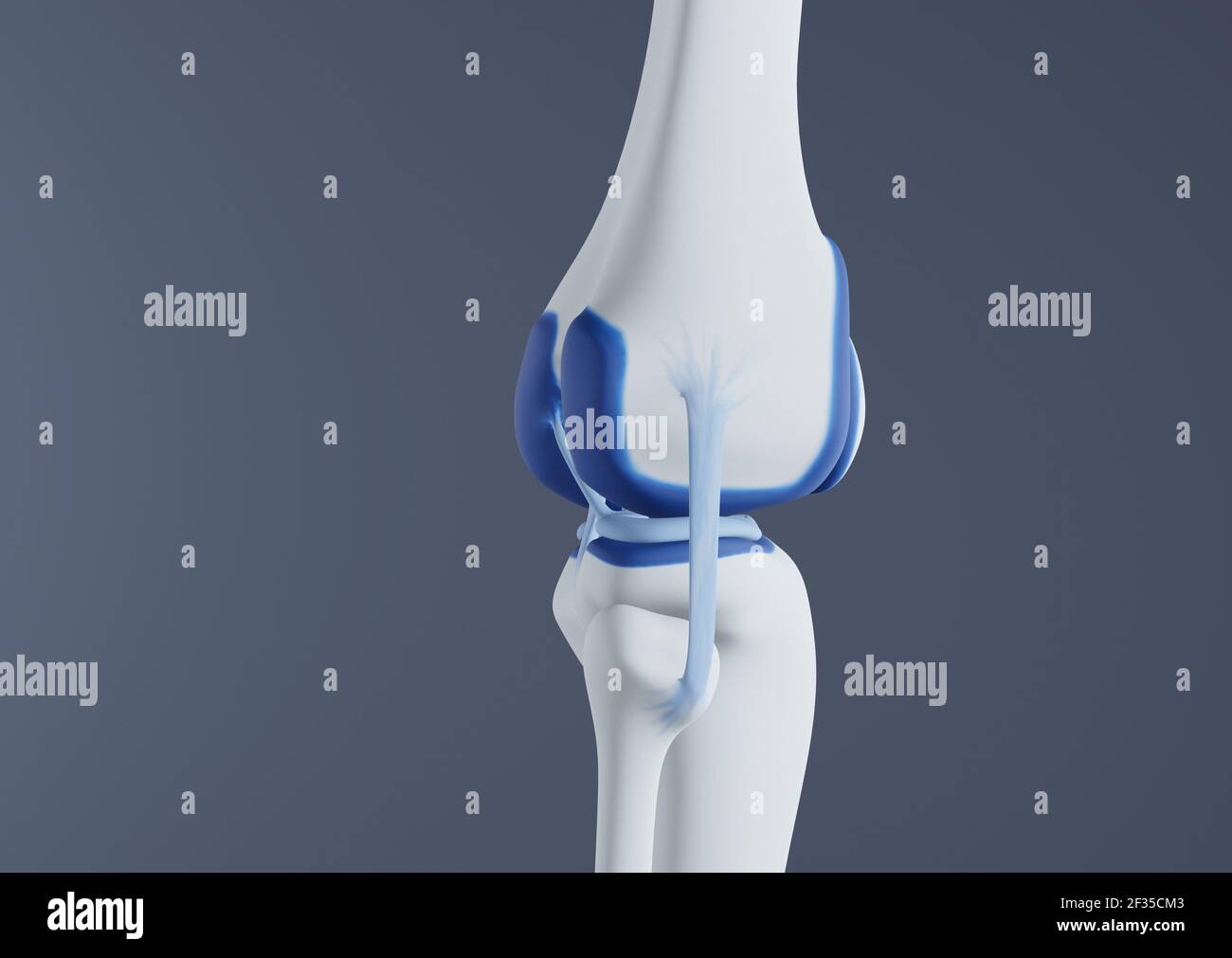 View of knee bones and ligaments. Stock Photohttps://www.alamy.com/image-license-details/?v=1https://www.alamy.com/view-of-knee-bones-and-ligaments-image415012531.html
RF2F35CM3–View of knee bones and ligaments.
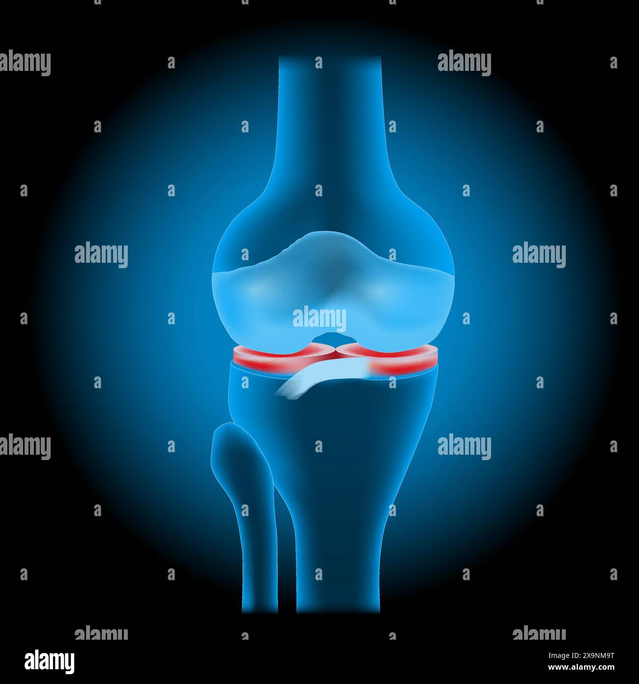 Knee meniscus injuries. Knee joint anatomy. Realistic transparent blue joint with glowing effect on dark background. vector illustration like X-ray im Stock Vectorhttps://www.alamy.com/image-license-details/?v=1https://www.alamy.com/knee-meniscus-injuries-knee-joint-anatomy-realistic-transparent-blue-joint-with-glowing-effect-on-dark-background-vector-illustration-like-x-ray-im-image608371732.html
RF2X9NM9T–Knee meniscus injuries. Knee joint anatomy. Realistic transparent blue joint with glowing effect on dark background. vector illustration like X-ray im
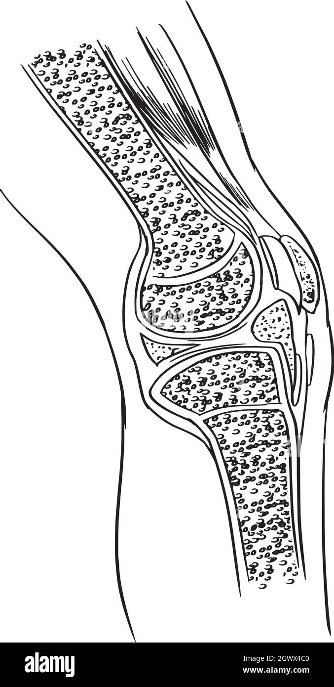 Anatomy of the knee joint Stock Vectorhttps://www.alamy.com/image-license-details/?v=1https://www.alamy.com/anatomy-of-the-knee-joint-image446199824.html
RF2GWX4C0–Anatomy of the knee joint
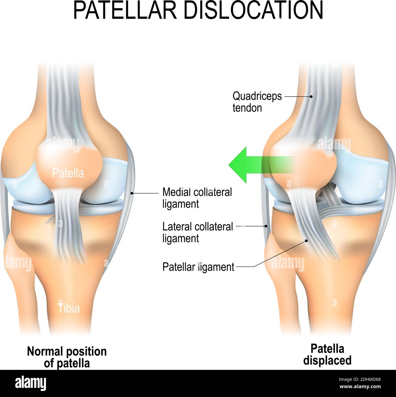 Patellar dislocation. Normal position of kneecap and Patella displaced. Anatomy of the Knee Stock Vectorhttps://www.alamy.com/image-license-details/?v=1https://www.alamy.com/patellar-dislocation-normal-position-of-kneecap-and-patella-displaced-anatomy-of-the-knee-image389526656.html
RF2DHMD68–Patellar dislocation. Normal position of kneecap and Patella displaced. Anatomy of the Knee
 Model of knee joint Stock Photohttps://www.alamy.com/image-license-details/?v=1https://www.alamy.com/stock-photo-model-of-knee-joint-120263120.html
RMGYJCRC–Model of knee joint
 tear of a meniscus is a rupturing of one or more of the fibrocartilage strips in the knee. Human Joint, and Traumatic force Stock Vectorhttps://www.alamy.com/image-license-details/?v=1https://www.alamy.com/tear-of-a-meniscus-is-a-rupturing-of-one-or-more-of-the-fibrocartilage-strips-in-the-knee-human-joint-and-traumatic-force-image389526410.html
RF2DHMCWE–tear of a meniscus is a rupturing of one or more of the fibrocartilage strips in the knee. Human Joint, and Traumatic force
 Myofascial fibrolysis instrumental hook in knee medial collateral ligament Physiotherapy medical center, Donostia, San Sebastian, Gipuzkoa, Basque Country, Spain. Stock Photohttps://www.alamy.com/image-license-details/?v=1https://www.alamy.com/myofascial-fibrolysis-instrumental-hook-in-knee-medial-collateral-ligament-physiotherapy-medical-center-donostia-san-sebastian-gipuzkoa-basque-country-spain-image603526872.html
RM2X1W0K4–Myofascial fibrolysis instrumental hook in knee medial collateral ligament Physiotherapy medical center, Donostia, San Sebastian, Gipuzkoa, Basque Country, Spain.
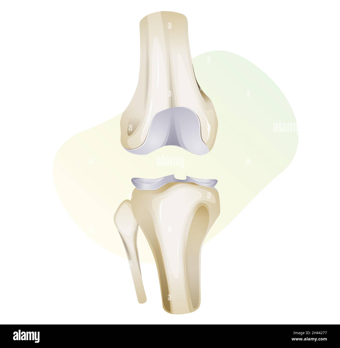 Knee Cartilage Injury - Illustration as EPS 10 File Stock Vectorhttps://www.alamy.com/image-license-details/?v=1https://www.alamy.com/knee-cartilage-injury-illustration-as-eps-10-file-image450017771.html
RF2H44277–Knee Cartilage Injury - Illustration as EPS 10 File
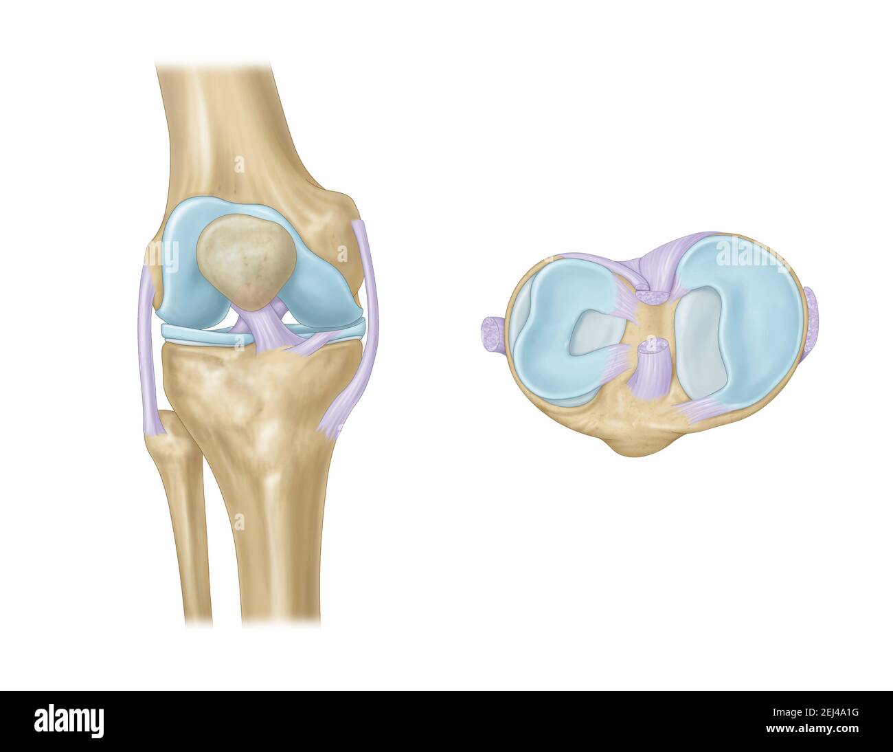 knee joint with ligaments, meniscus, articular cartilage Stock Photohttps://www.alamy.com/image-license-details/?v=1https://www.alamy.com/knee-joint-with-ligaments-meniscus-articular-cartilage-image406997964.html
RF2EJ4A1G–knee joint with ligaments, meniscus, articular cartilage
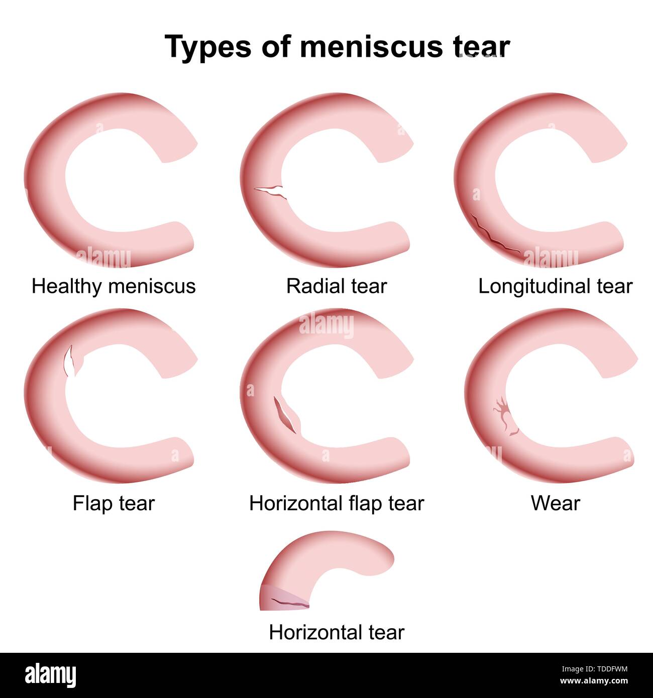 Meniscus injuries medical vector illustration isolated on white background, meniscus tear Stock Vectorhttps://www.alamy.com/image-license-details/?v=1https://www.alamy.com/meniscus-injuries-medical-vector-illustration-isolated-on-white-background-meniscus-tear-image249233536.html
RFTDDFWM–Meniscus injuries medical vector illustration isolated on white background, meniscus tear
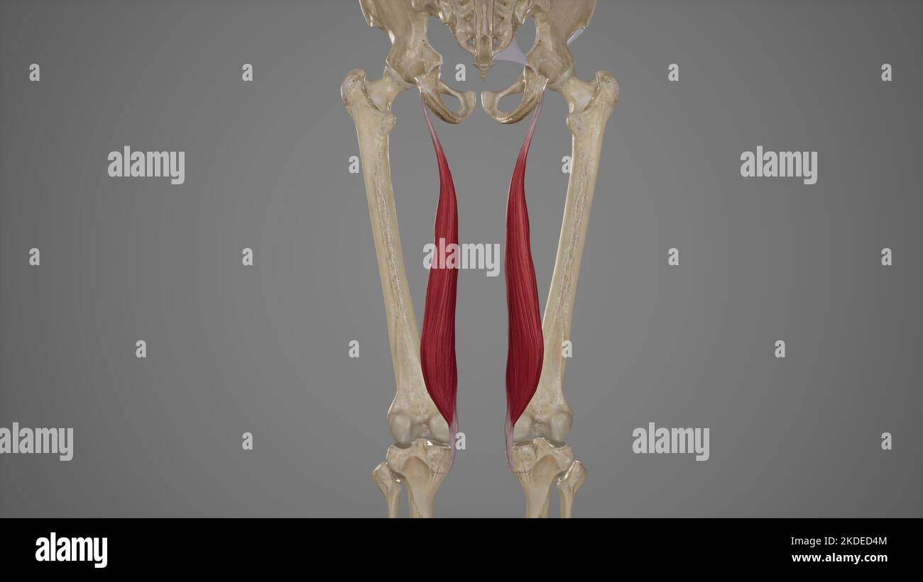 Medical Accurate Illustration of Semimembranosus Muscle Stock Photohttps://www.alamy.com/image-license-details/?v=1https://www.alamy.com/medical-accurate-illustration-of-semimembranosus-muscle-image490198484.html
RF2KDED4M–Medical Accurate Illustration of Semimembranosus Muscle
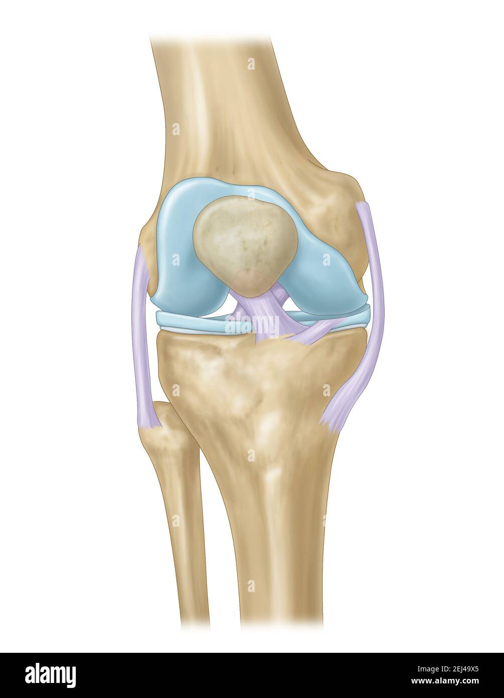 knee joint with ligaments, meniscus, articular cartilage Stock Photohttps://www.alamy.com/image-license-details/?v=1https://www.alamy.com/knee-joint-with-ligaments-meniscus-articular-cartilage-image406997869.html
RF2EJ49X5–knee joint with ligaments, meniscus, articular cartilage
 Knee sprain Stock Photohttps://www.alamy.com/image-license-details/?v=1https://www.alamy.com/stock-photo-knee-sprain-52574386.html
RFD1EY4J–Knee sprain
 knee joint with ligaments, meniscus, articular cartilage, knee osteoarthritis Stock Photohttps://www.alamy.com/image-license-details/?v=1https://www.alamy.com/knee-joint-with-ligaments-meniscus-articular-cartilage-knee-osteoarthritis-image406997973.html
RF2EJ4A1W–knee joint with ligaments, meniscus, articular cartilage, knee osteoarthritis
 3D illustration showing human knee joint with femur, articular cartilage, meniscus, medial collateral ligament, articular cartilage, patella, kneecap, Stock Photohttps://www.alamy.com/image-license-details/?v=1https://www.alamy.com/3d-illustration-showing-human-knee-joint-with-femur-articular-cartilage-meniscus-medial-collateral-ligament-articular-cartilage-patella-kneecap-image349807707.html
RF2B9338B–3D illustration showing human knee joint with femur, articular cartilage, meniscus, medial collateral ligament, articular cartilage, patella, kneecap,
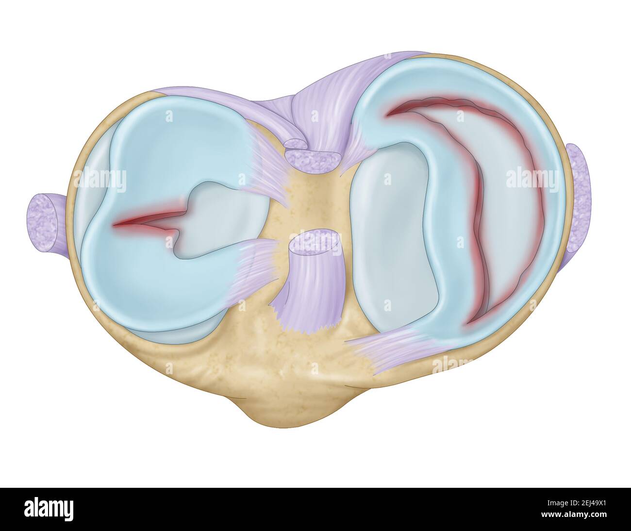 knee joint with ligaments, meniscus, articular cartilage, knee osteoarthritis Stock Photohttps://www.alamy.com/image-license-details/?v=1https://www.alamy.com/knee-joint-with-ligaments-meniscus-articular-cartilage-knee-osteoarthritis-image406997865.html
RF2EJ49X1–knee joint with ligaments, meniscus, articular cartilage, knee osteoarthritis
 Knee ligaments, tendons, x-ray Stock Photohttps://www.alamy.com/image-license-details/?v=1https://www.alamy.com/stock-photo-knee-ligaments-tendons-x-ray-74986042.html
RFE9YWCX–Knee ligaments, tendons, x-ray
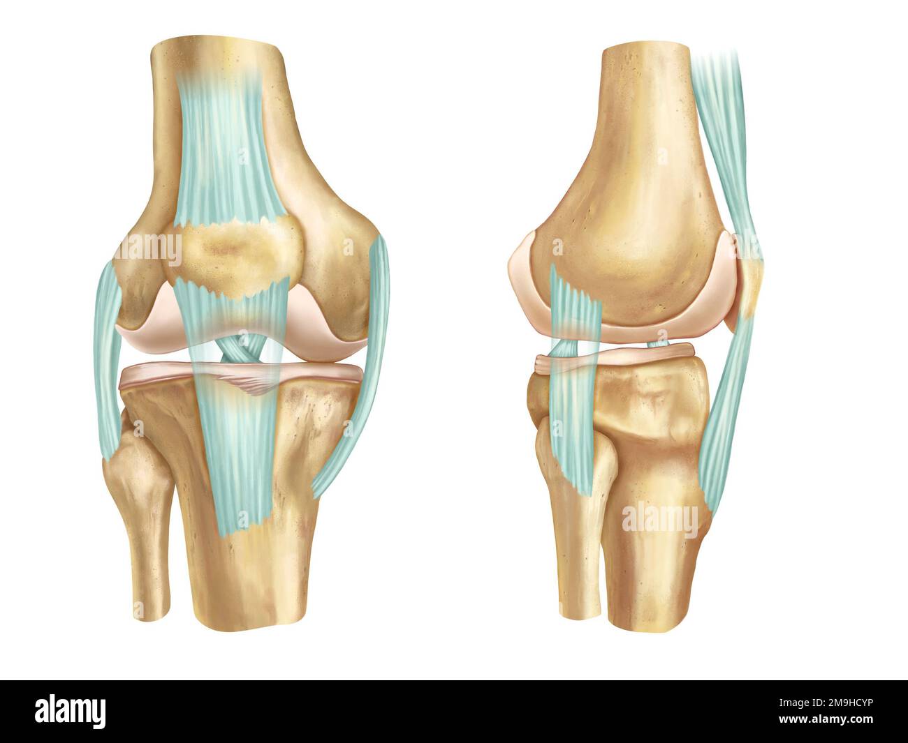 Front and side anatomical view of an human knee. Digital illustration. Stock Photohttps://www.alamy.com/image-license-details/?v=1https://www.alamy.com/front-and-side-anatomical-view-of-an-human-knee-digital-illustration-image505015946.html
RF2M9HCYP–Front and side anatomical view of an human knee. Digital illustration.
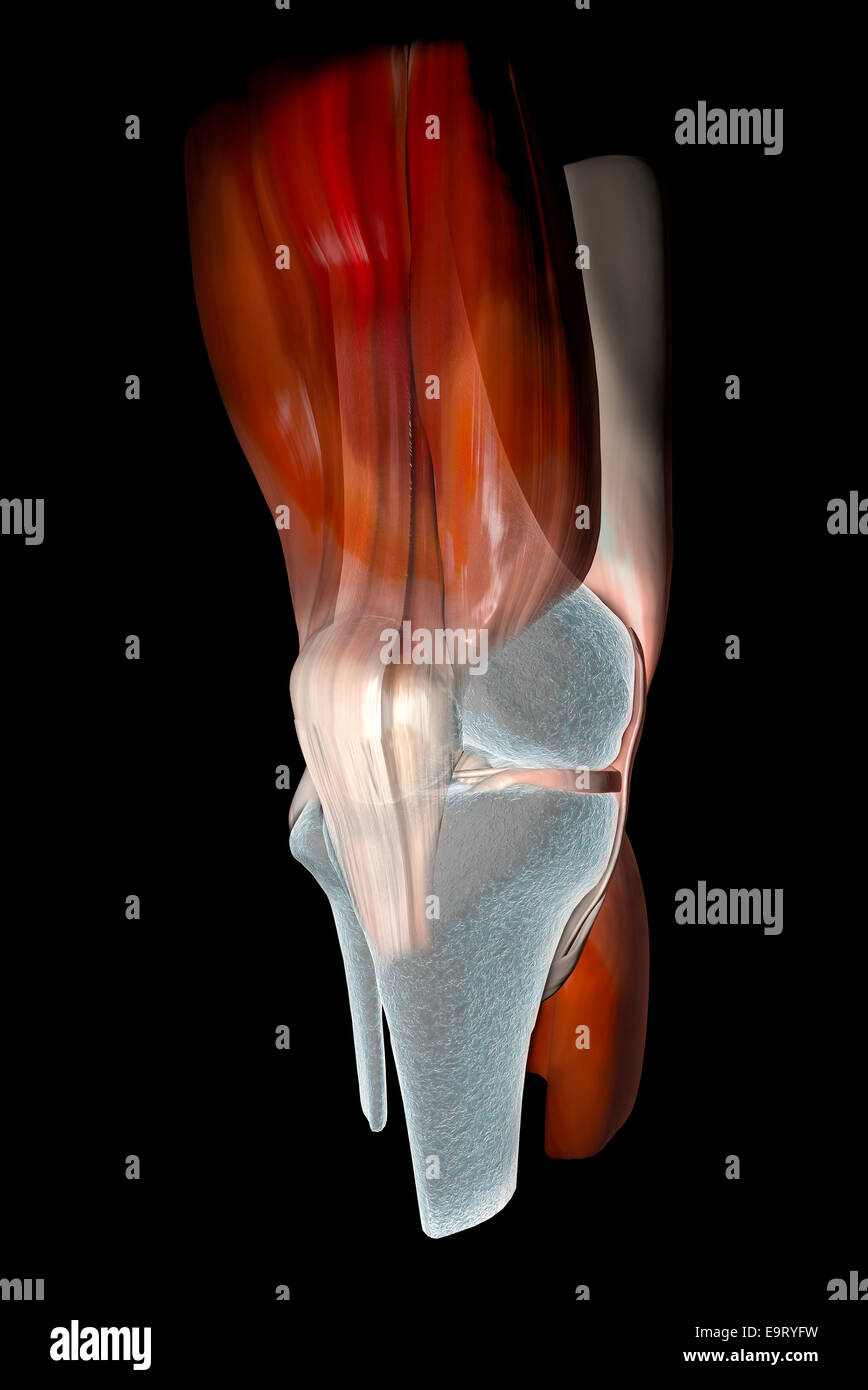 3d Knee ligaments, tendons, bones, muscles x-ray Stock Photohttps://www.alamy.com/image-license-details/?v=1https://www.alamy.com/stock-photo-3d-knee-ligaments-tendons-bones-muscles-x-ray-74899885.html
RFE9RYFW–3d Knee ligaments, tendons, bones, muscles x-ray
 View of knee bones and ligaments. Stock Photohttps://www.alamy.com/image-license-details/?v=1https://www.alamy.com/view-of-knee-bones-and-ligaments-image415012529.html
RF2F35CM1–View of knee bones and ligaments.
 Bent knee showing torn anterior cruciate ligament. Stock Photohttps://www.alamy.com/image-license-details/?v=1https://www.alamy.com/bent-knee-showing-torn-anterior-cruciate-ligament-image431667952.html
RM2G284TG–Bent knee showing torn anterior cruciate ligament.
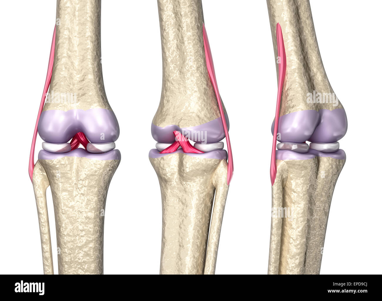 Knee joint anatomy, 3D model Stock Photohttps://www.alamy.com/image-license-details/?v=1https://www.alamy.com/stock-photo-knee-joint-anatomy-3d-model-82656690.html
RFEPD9CJ–Knee joint anatomy, 3D model
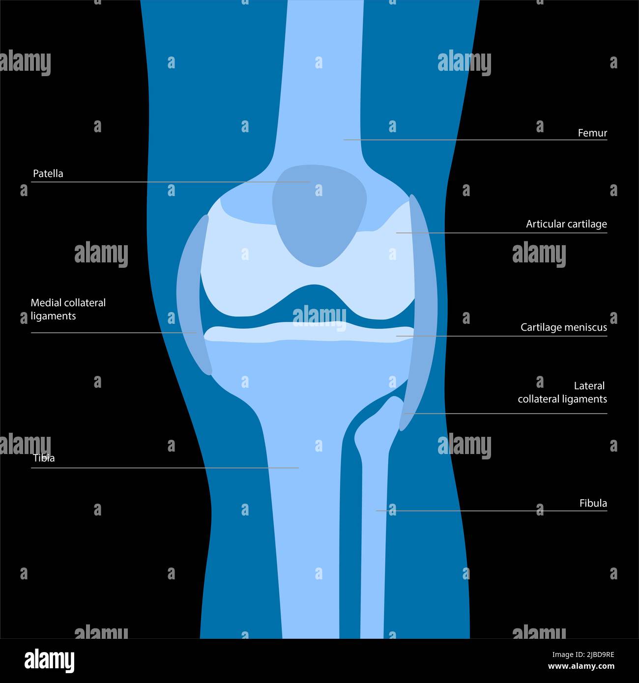 Knee anatomy, illustration Stock Photohttps://www.alamy.com/image-license-details/?v=1https://www.alamy.com/knee-anatomy-illustration-image471734242.html
RF2JBD9RE–Knee anatomy, illustration
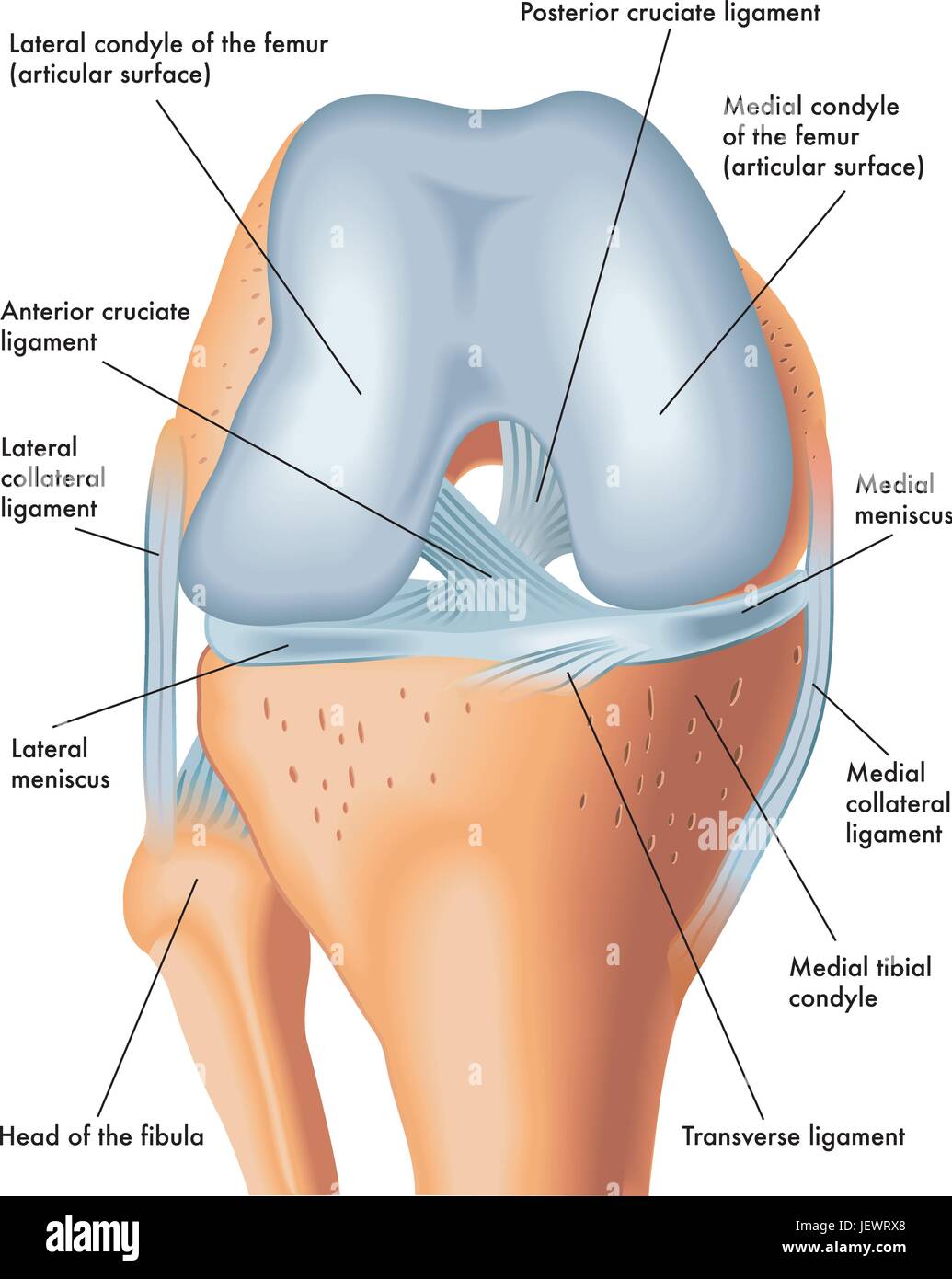 meniscus, knees, knee, legs, skeleton, leg, thigh, joints, anatomy, bones, Stock Vectorhttps://www.alamy.com/image-license-details/?v=1https://www.alamy.com/stock-photo-meniscus-knees-knee-legs-skeleton-leg-thigh-joints-anatomy-bones-146855696.html
RFJEWRX8–meniscus, knees, knee, legs, skeleton, leg, thigh, joints, anatomy, bones,
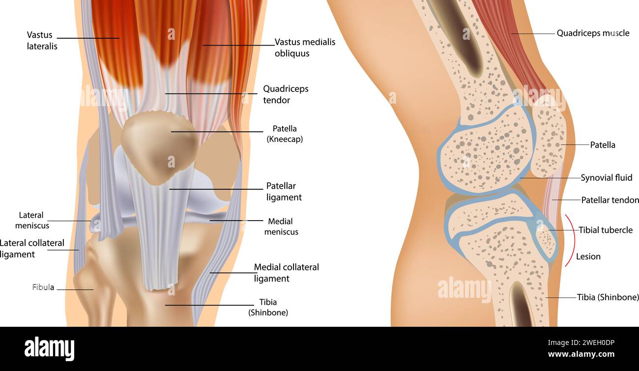 Knee anatomy including ligaments, cartilage and meniscus. Detailed Anatomy of the Knee Joint cross-section. Stock Vectorhttps://www.alamy.com/image-license-details/?v=1https://www.alamy.com/knee-anatomy-including-ligaments-cartilage-and-meniscus-detailed-anatomy-of-the-knee-joint-cross-section-image594131266.html
RF2WEH0DP–Knee anatomy including ligaments, cartilage and meniscus. Detailed Anatomy of the Knee Joint cross-section.
 Myofascial fibrolysis instrumental hook in knee medial collateral ligament Physiotherapy medical center, Donostia, San Sebastian, Gipuzkoa, Basque Country, Spain. Stock Photohttps://www.alamy.com/image-license-details/?v=1https://www.alamy.com/myofascial-fibrolysis-instrumental-hook-in-knee-medial-collateral-ligament-physiotherapy-medical-center-donostia-san-sebastian-gipuzkoa-basque-country-spain-image624981354.html
RM2Y8PA2J–Myofascial fibrolysis instrumental hook in knee medial collateral ligament Physiotherapy medical center, Donostia, San Sebastian, Gipuzkoa, Basque Country, Spain.
 Knee Cartilage Injury - Illustration as EPS 10 File Stock Vectorhttps://www.alamy.com/image-license-details/?v=1https://www.alamy.com/knee-cartilage-injury-illustration-as-eps-10-file-image450018068.html
RF2H442HT–Knee Cartilage Injury - Illustration as EPS 10 File
RF2S0DG1A–Dislocated knee line color icon. Household injuries sign for web page, mobile app, button, logo. Vector isolated button. Editable stroke.
 Knee Cartilage Anatomy - Illustration as EPS 10 File Stock Vectorhttps://www.alamy.com/image-license-details/?v=1https://www.alamy.com/knee-cartilage-anatomy-illustration-as-eps-10-file-image450018201.html
RF2H442PH–Knee Cartilage Anatomy - Illustration as EPS 10 File
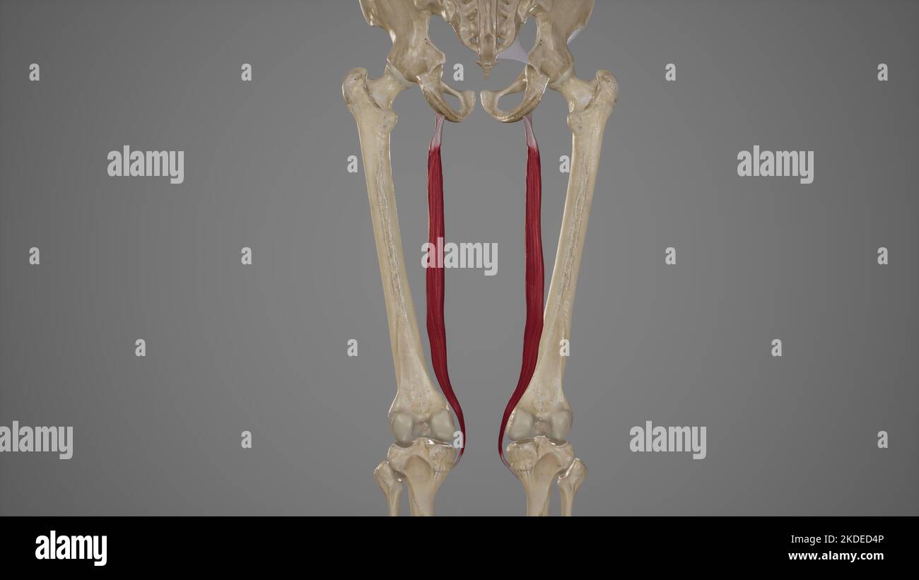 Medical Accurate Illustration of Semitendinosus Stock Photohttps://www.alamy.com/image-license-details/?v=1https://www.alamy.com/medical-accurate-illustration-of-semitendinosus-image490198486.html
RF2KDED4P–Medical Accurate Illustration of Semitendinosus
 Knee Cartilage Injury - Torn Meniscus - Stock Illustration as EPS 10 File Stock Vectorhttps://www.alamy.com/image-license-details/?v=1https://www.alamy.com/knee-cartilage-injury-torn-meniscus-stock-illustration-as-eps-10-file-image564987321.html
RF2RR5B2H–Knee Cartilage Injury - Torn Meniscus - Stock Illustration as EPS 10 File
 Menisci of the knee Stock Photohttps://www.alamy.com/image-license-details/?v=1https://www.alamy.com/stock-photo-menisci-of-the-knee-49441470.html
RFCTC72P–Menisci of the knee
RF2XWN5RA–Dislocated knee line color icon. Household injuries sign for web page, mobile app, button, logo. Vector isolated button. Editable stroke.
 Hand drawing illustration showing human knee joint with femur, articular cartilage, meniscus, medial collateral ligament, articular cartilage, patella Stock Photohttps://www.alamy.com/image-license-details/?v=1https://www.alamy.com/hand-drawing-illustration-showing-human-knee-joint-with-femur-articular-cartilage-meniscus-medial-collateral-ligament-articular-cartilage-patella-image357074154.html
RF2BMX3MA–Hand drawing illustration showing human knee joint with femur, articular cartilage, meniscus, medial collateral ligament, articular cartilage, patella
RF2XBFX7X–Dislocated knee line color icon. Household injuries sign for web page, mobile app, button, logo. Vector isolated button. Editable stroke.
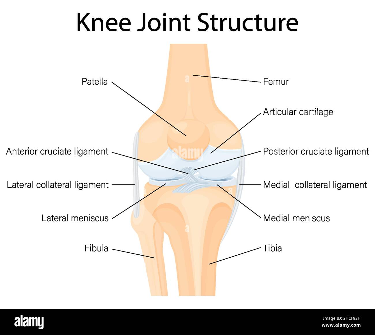 Human Knee joint anatomy. Ligaments of the knee. Anterior and Posterior cruciate ligaments, Patellar and Quadriceps, tendons, Medial and Lateral colla Stock Vectorhttps://www.alamy.com/image-license-details/?v=1https://www.alamy.com/human-knee-joint-anatomy-ligaments-of-the-knee-anterior-and-posterior-cruciate-ligaments-patellar-and-quadriceps-tendons-medial-and-lateral-colla-image455181065.html
RF2HCF82H–Human Knee joint anatomy. Ligaments of the knee. Anterior and Posterior cruciate ligaments, Patellar and Quadriceps, tendons, Medial and Lateral colla
 Moderate osteoarthritis of a human knee. Shown are cartilage erosion and cartilage fragments in the synovial fluid. Stock Photohttps://www.alamy.com/image-license-details/?v=1https://www.alamy.com/stock-photo-moderate-osteoarthritis-of-a-human-knee-shown-are-cartilage-erosion-130806337.html
RFHGPMPW–Moderate osteoarthritis of a human knee. Shown are cartilage erosion and cartilage fragments in the synovial fluid.
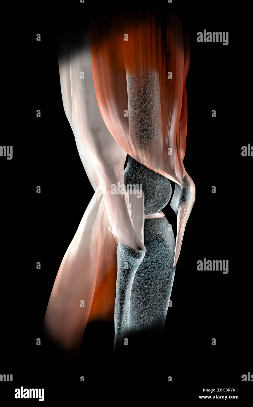 3d Knee ligaments, tendons, bones, muscles x-ray Stock Photohttps://www.alamy.com/image-license-details/?v=1https://www.alamy.com/stock-photo-3d-knee-ligaments-tendons-bones-muscles-x-ray-74899989.html
RFE9RYKH–3d Knee ligaments, tendons, bones, muscles x-ray
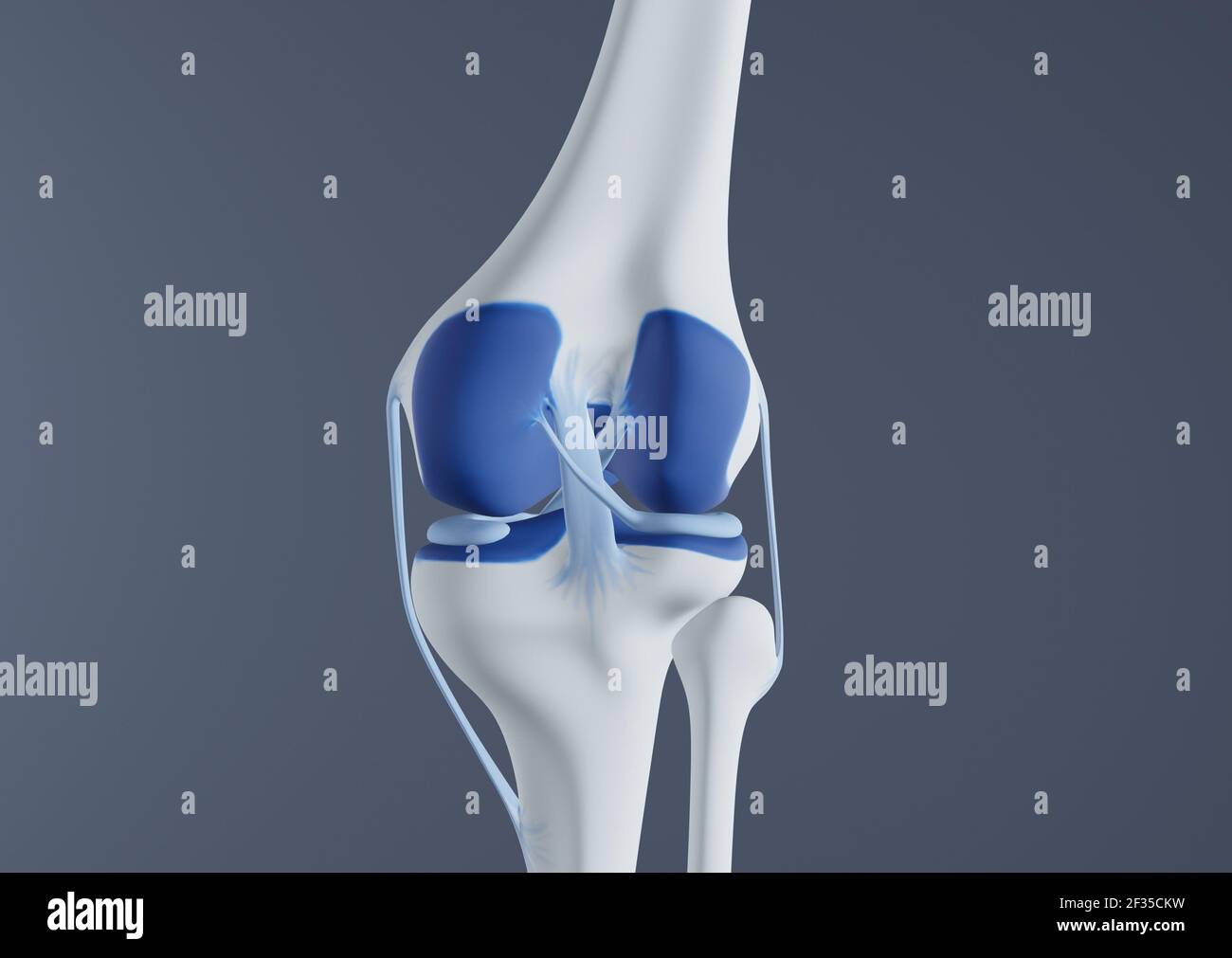 View of knee bones and ligaments. Stock Photohttps://www.alamy.com/image-license-details/?v=1https://www.alamy.com/view-of-knee-bones-and-ligaments-image415012525.html
RF2F35CKW–View of knee bones and ligaments.

