Quick filters:
Membrane proteins Stock Photos and Images
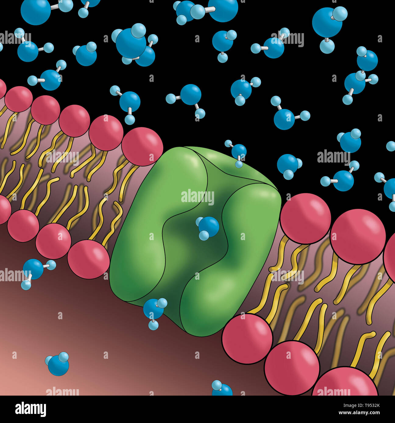 Aquaporins also called water channels, are integral membrane proteins from a larger family of major intrinsic proteins that form pores in the membrane of biological cells and allow water to flow between cells. Stock Photohttps://www.alamy.com/image-license-details/?v=1https://www.alamy.com/aquaporins-also-called-water-channels-are-integral-membrane-proteins-from-a-larger-family-of-major-intrinsic-proteins-that-form-pores-in-the-membrane-of-biological-cells-and-allow-water-to-flow-between-cells-image246589243.html
RMT9532K–Aquaporins also called water channels, are integral membrane proteins from a larger family of major intrinsic proteins that form pores in the membrane of biological cells and allow water to flow between cells.
 Covid-19 coronavirus particles, illustration. The SARS-CoV-2 coronavirus was first identified in Wuhan, China, in December 2019. It is an enveloped RNA (ribonucleic acid) virus. Within the membrane are spike proteins (large protrusions) as well as membrane proteins and envelope proteins. SARS-CoV-2 causes the respiratory infection Covid-19, which can lead to fatal pneumonia. As of March 2020, the virus has spread to many countries worldwide and has been declared a pandemic. Hundreds of thousands have been infected with tens of thousands of deaths. Stock Photohttps://www.alamy.com/image-license-details/?v=1https://www.alamy.com/covid-19-coronavirus-particles-illustration-the-sars-cov-2-coronavirus-was-first-identified-in-wuhan-china-in-december-2019-it-is-an-enveloped-rna-ribonucleic-acid-virus-within-the-membrane-are-spike-proteins-large-protrusions-as-well-as-membrane-proteins-and-envelope-proteins-sars-cov-2-causes-the-respiratory-infection-covid-19-which-can-lead-to-fatal-pneumonia-as-of-march-2020-the-virus-has-spread-to-many-countries-worldwide-and-has-been-declared-a-pandemic-hundreds-of-thousands-have-been-infected-with-tens-of-thousands-of-deaths-image352901282.html
RF2BE4156–Covid-19 coronavirus particles, illustration. The SARS-CoV-2 coronavirus was first identified in Wuhan, China, in December 2019. It is an enveloped RNA (ribonucleic acid) virus. Within the membrane are spike proteins (large protrusions) as well as membrane proteins and envelope proteins. SARS-CoV-2 causes the respiratory infection Covid-19, which can lead to fatal pneumonia. As of March 2020, the virus has spread to many countries worldwide and has been declared a pandemic. Hundreds of thousands have been infected with tens of thousands of deaths.
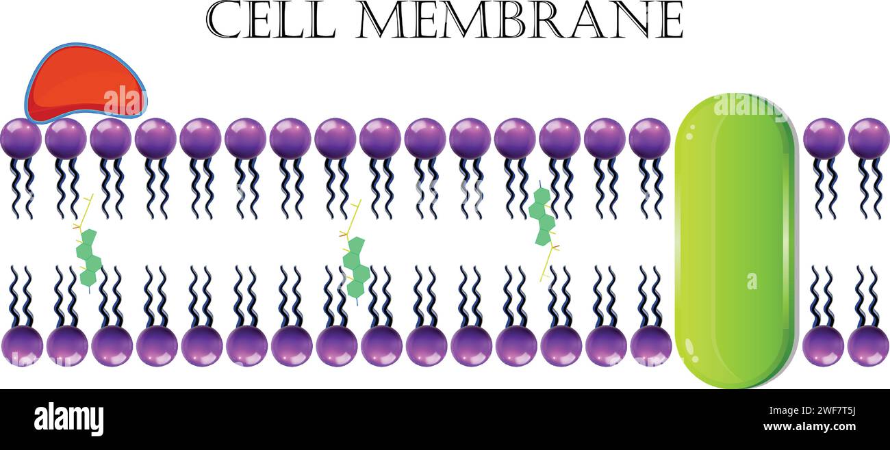 Cell Membrane Or Plasma Membrane Stock Vectorhttps://www.alamy.com/image-license-details/?v=1https://www.alamy.com/cell-membrane-or-plasma-membrane-image594544990.html
RF2WF7T5J–Cell Membrane Or Plasma Membrane
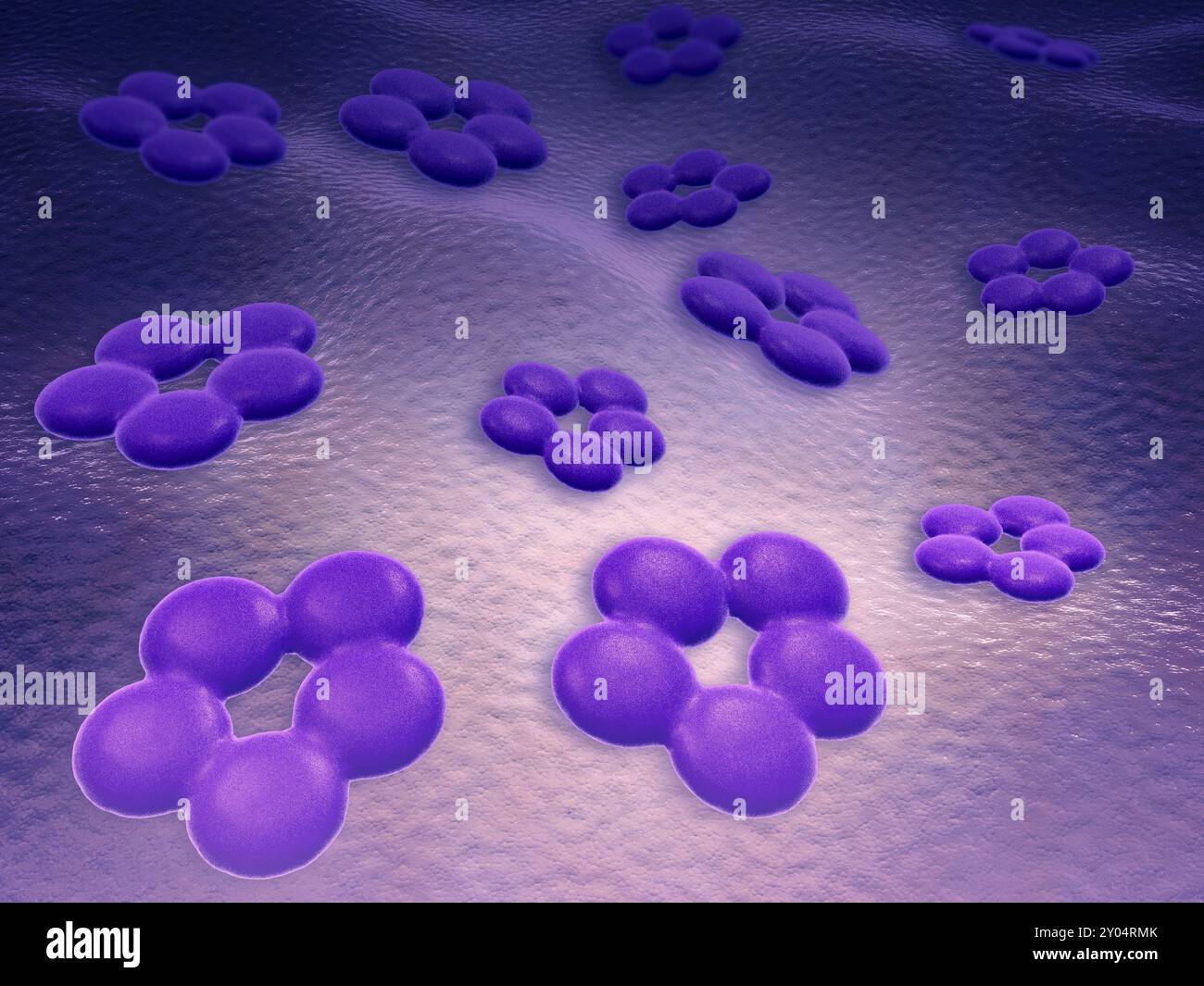 Conceptual image of cell surface receptors. Cell surface receptors are specialized integral membrane proteins that take part in communication between Stock Photohttps://www.alamy.com/image-license-details/?v=1https://www.alamy.com/conceptual-image-of-cell-surface-receptors-cell-surface-receptors-are-specialized-integral-membrane-proteins-that-take-part-in-communication-between-image619679667.html
RM2Y04RMK–Conceptual image of cell surface receptors. Cell surface receptors are specialized integral membrane proteins that take part in communication between
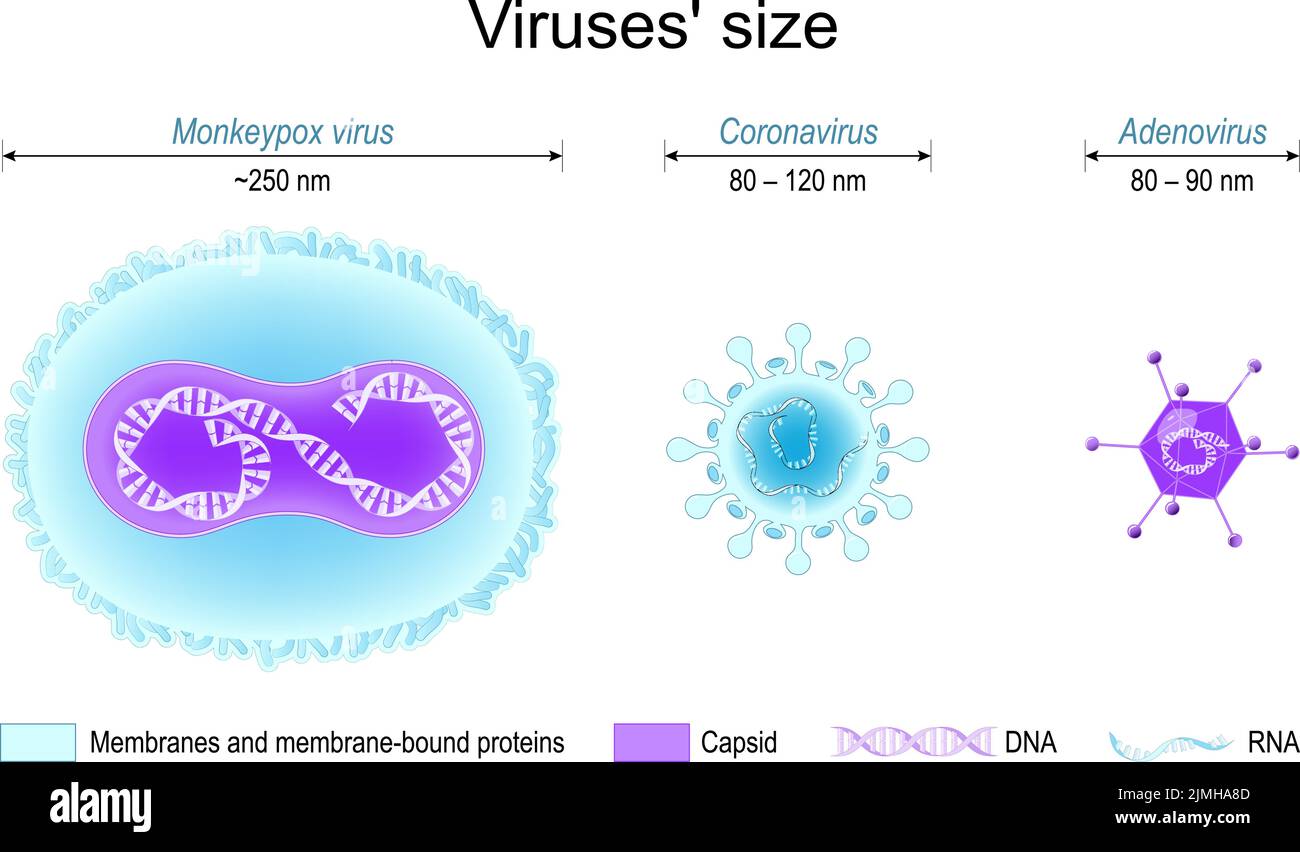 Comparison of viruses' size. monkeypox, SARS CoV-2 or coronavirus, and adenovirus. Different structure of viruses: Membrane proteins, capsids, DNA Stock Vectorhttps://www.alamy.com/image-license-details/?v=1https://www.alamy.com/comparison-of-viruses-size-monkeypox-sars-cov-2-or-coronavirus-and-adenovirus-different-structure-of-viruses-membrane-proteins-capsids-dna-image477354317.html
RF2JMHA8D–Comparison of viruses' size. monkeypox, SARS CoV-2 or coronavirus, and adenovirus. Different structure of viruses: Membrane proteins, capsids, DNA
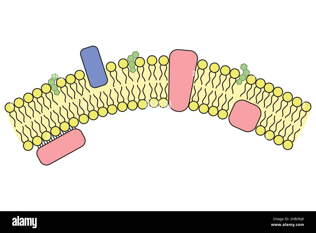 Simple illustration of cell membrane and incorporated structures Stock Photohttps://www.alamy.com/image-license-details/?v=1https://www.alamy.com/simple-illustration-of-cell-membrane-and-incorporated-structures-image454312048.html
RF2HB3KJ8–Simple illustration of cell membrane and incorporated structures
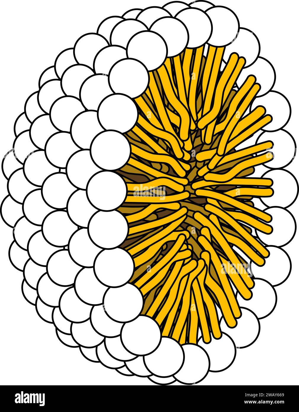 Structure of Phospholipid Molecule in Micelle .Vector illustration. Stock Vectorhttps://www.alamy.com/image-license-details/?v=1https://www.alamy.com/structure-of-phospholipid-molecule-in-micelle-vector-illustration-image591896657.html
RF2WAY669–Structure of Phospholipid Molecule in Micelle .Vector illustration.
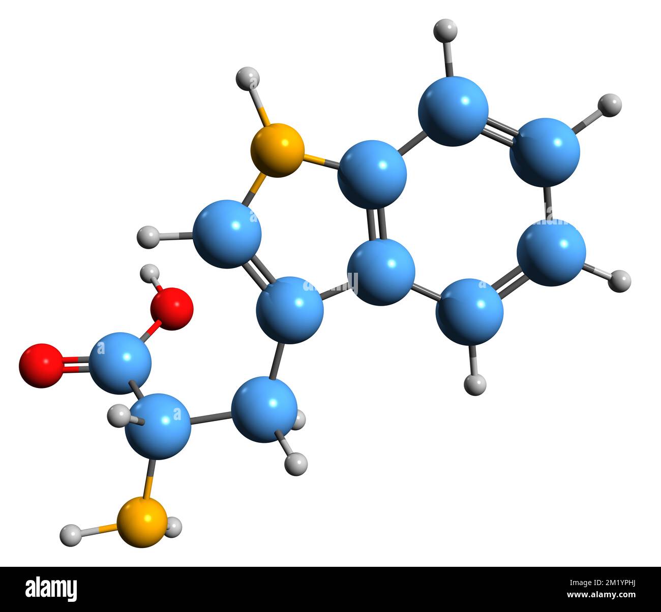 3D image of Tryptophan skeletal formula - molecular chemical structure of amino acid isolated on white background Stock Photohttps://www.alamy.com/image-license-details/?v=1https://www.alamy.com/3d-image-of-tryptophan-skeletal-formula-molecular-chemical-structure-of-amino-acid-isolated-on-white-background-image500325774.html
RF2M1YPHJ–3D image of Tryptophan skeletal formula - molecular chemical structure of amino acid isolated on white background
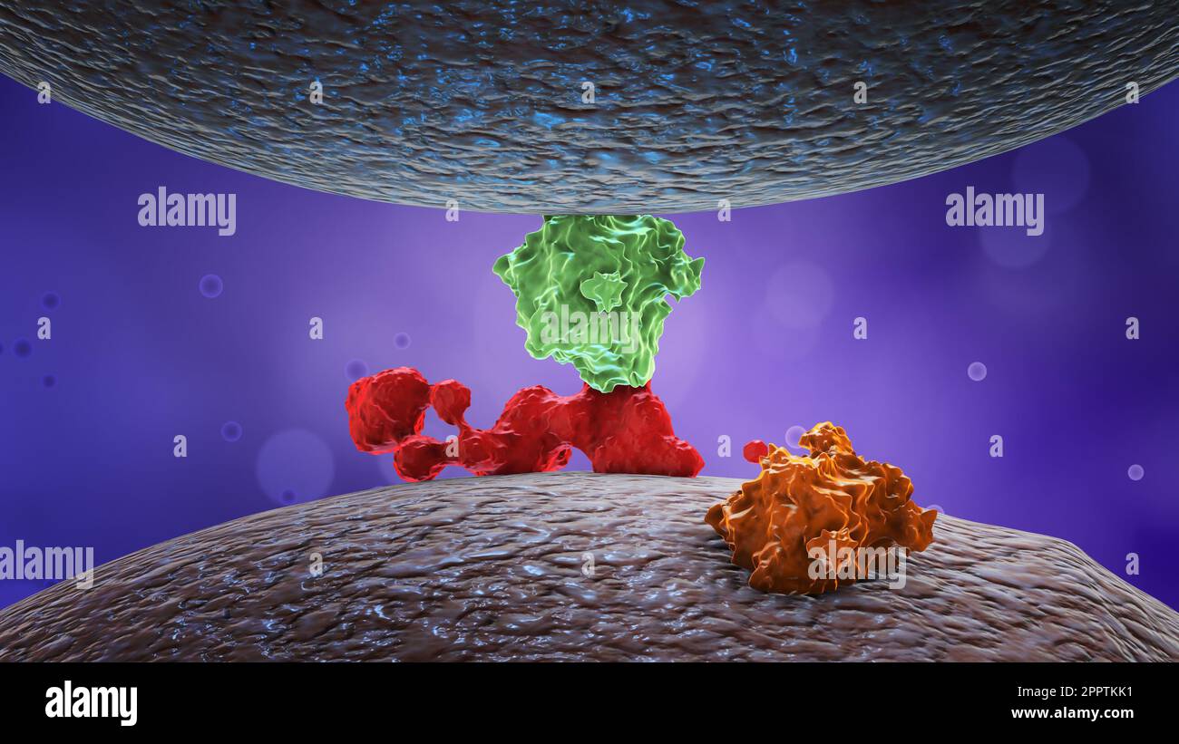 Protein or enzymes or hormones on cell membrane. Stock Photohttps://www.alamy.com/image-license-details/?v=1https://www.alamy.com/protein-or-enzymes-or-hormones-on-cell-membrane-image547586117.html
RF2PPTKK1–Protein or enzymes or hormones on cell membrane.
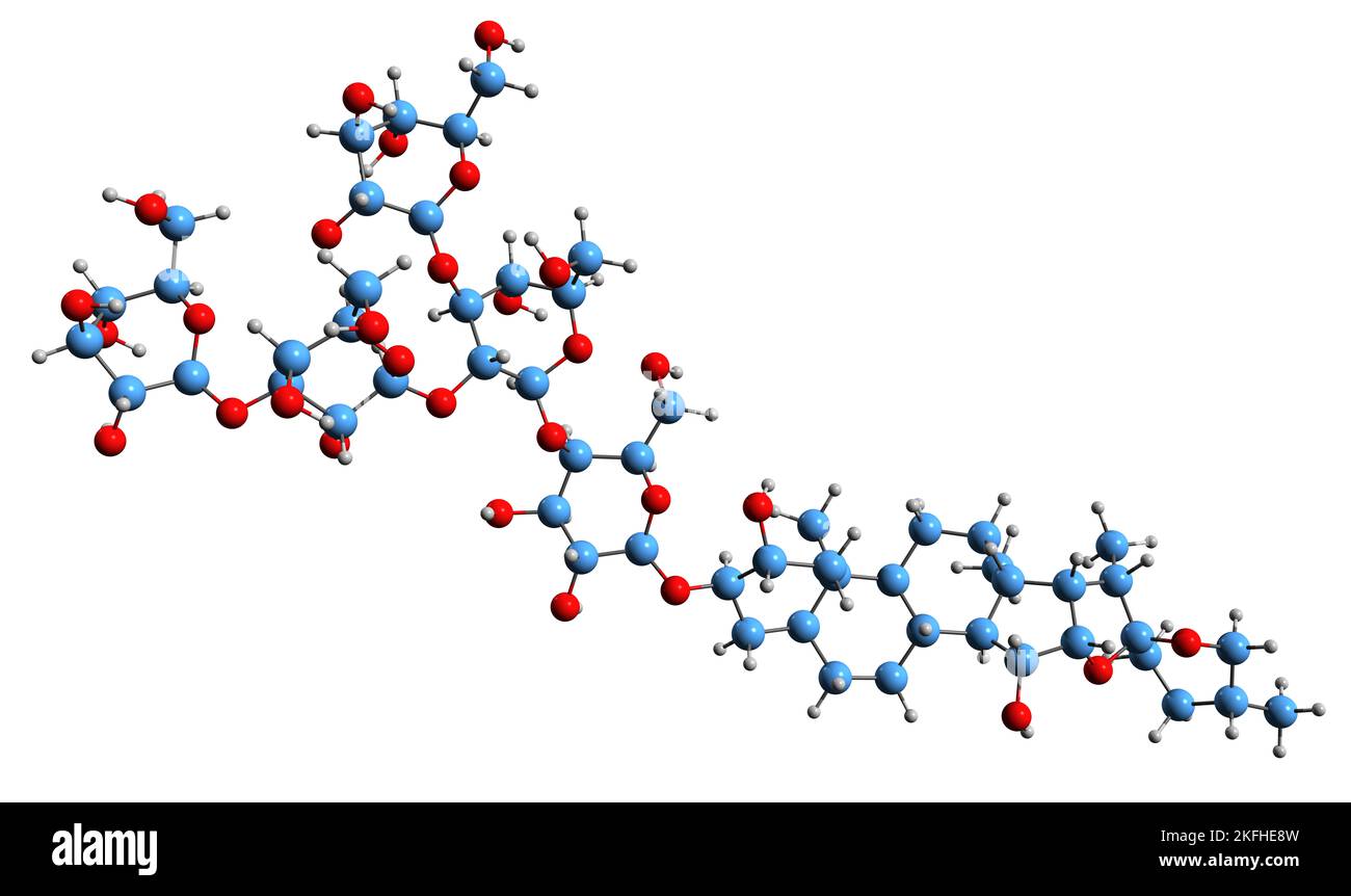 3D image of Digitonin skeletal formula - molecular chemical structure of steroidal saponin Digitin isolated on white background Stock Photohttps://www.alamy.com/image-license-details/?v=1https://www.alamy.com/3d-image-of-digitonin-skeletal-formula-molecular-chemical-structure-of-steroidal-saponin-digitin-isolated-on-white-background-image491494553.html
RF2KFHE8W–3D image of Digitonin skeletal formula - molecular chemical structure of steroidal saponin Digitin isolated on white background
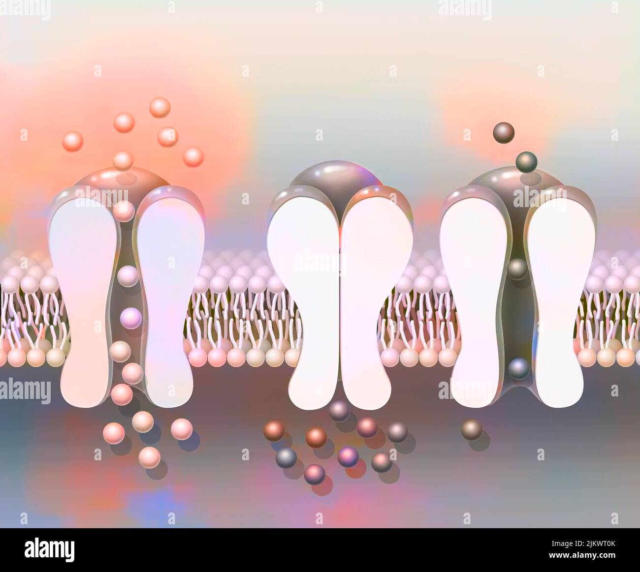 Depolarization: phospholipid membrane with NA + and K + ion channels. Stock Photohttps://www.alamy.com/image-license-details/?v=1https://www.alamy.com/depolarization-phospholipid-membrane-with-na-and-k-ion-channels-image476926035.html
RF2JKWT0K–Depolarization: phospholipid membrane with NA + and K + ion channels.
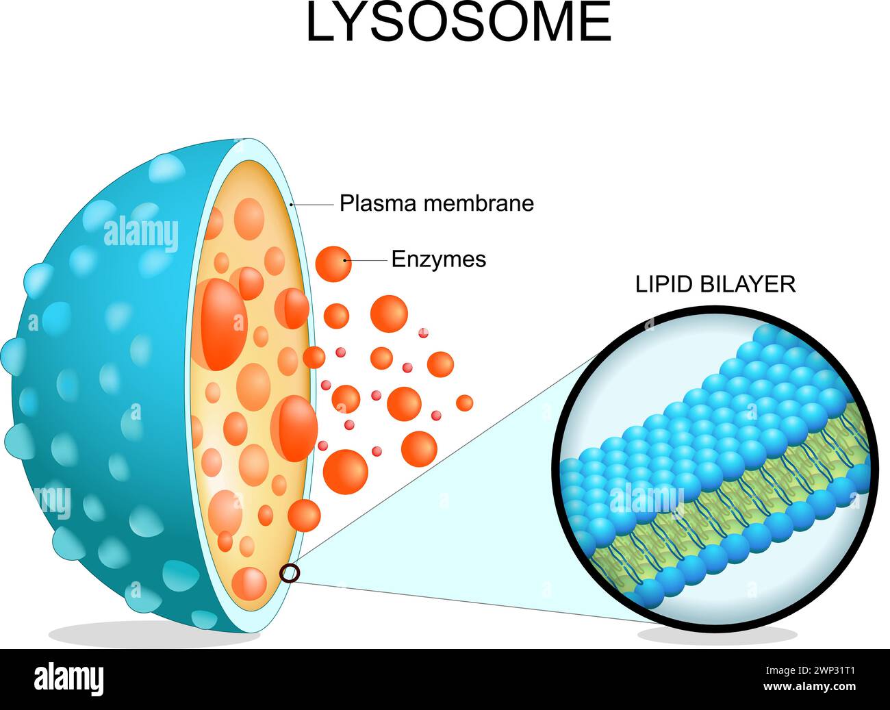 Lysosome anatomy. Cross section of a cell organelle. Close-up of a Lipid bilayer membrane, hydrolytic enzymes, transport proteins. Autophagy. Vector i Stock Vectorhttps://www.alamy.com/image-license-details/?v=1https://www.alamy.com/lysosome-anatomy-cross-section-of-a-cell-organelle-close-up-of-a-lipid-bilayer-membrane-hydrolytic-enzymes-transport-proteins-autophagy-vector-i-image598742257.html
RF2WP31T1–Lysosome anatomy. Cross section of a cell organelle. Close-up of a Lipid bilayer membrane, hydrolytic enzymes, transport proteins. Autophagy. Vector i
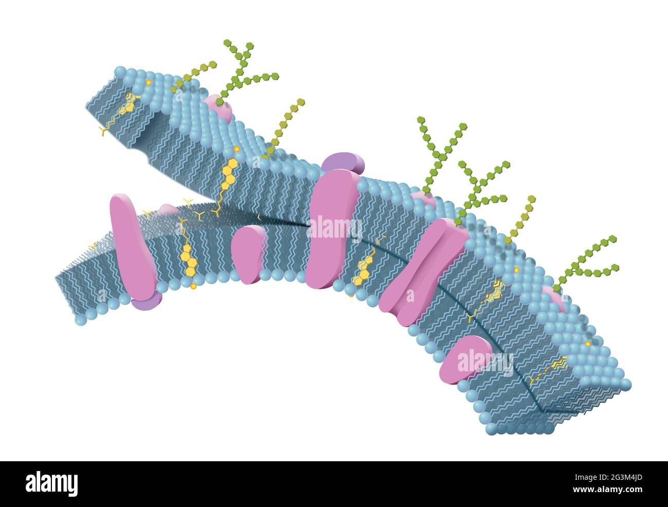 Cell membrane with phospholipids, cholesterol, intrinsic and extrinsic proteins. 3D illustration Stock Photohttps://www.alamy.com/image-license-details/?v=1https://www.alamy.com/cell-membrane-with-phospholipids-cholesterol-intrinsic-and-extrinsic-proteins-3d-illustration-image432545861.html
RF2G3M4JD–Cell membrane with phospholipids, cholesterol, intrinsic and extrinsic proteins. 3D illustration
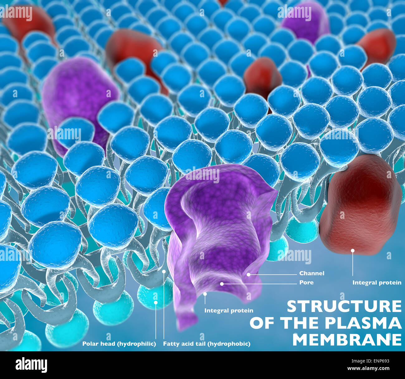 Structure of the plasma membrane of a cell Stock Photohttps://www.alamy.com/image-license-details/?v=1https://www.alamy.com/stock-photo-structure-of-the-plasma-membrane-of-a-cell-82237151.html
RMENP693–Structure of the plasma membrane of a cell
 Proteins and sugar in balance. A metaphor showing Proteins in equilibrium with Sugar, symbolizing a desired harmony between them. Stability. Harmoniou Stock Photohttps://www.alamy.com/image-license-details/?v=1https://www.alamy.com/proteins-and-sugar-in-balance-a-metaphor-showing-proteins-in-equilibrium-with-sugar-symbolizing-a-desired-harmony-between-them-stability-harmoniou-image631861970.html
RF2YKYPAX–Proteins and sugar in balance. A metaphor showing Proteins in equilibrium with Sugar, symbolizing a desired harmony between them. Stability. Harmoniou
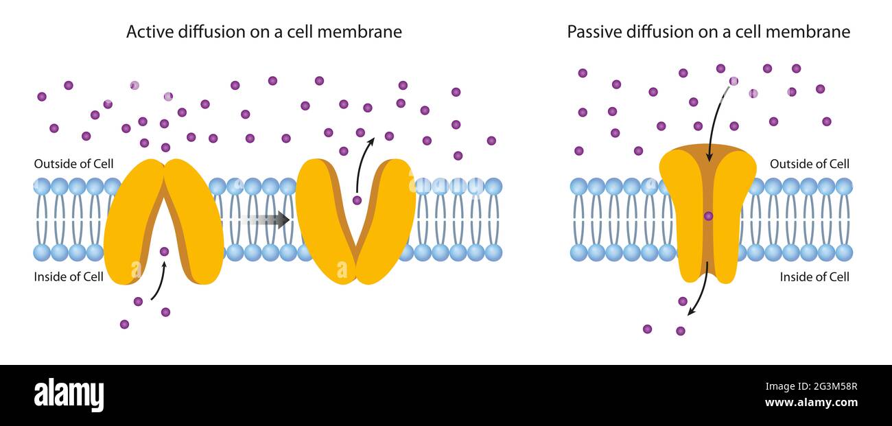 Diffusion Across the Plasma Membrane Stock Photohttps://www.alamy.com/image-license-details/?v=1https://www.alamy.com/diffusion-across-the-plasma-membrane-image432546375.html
RF2G3M58R–Diffusion Across the Plasma Membrane
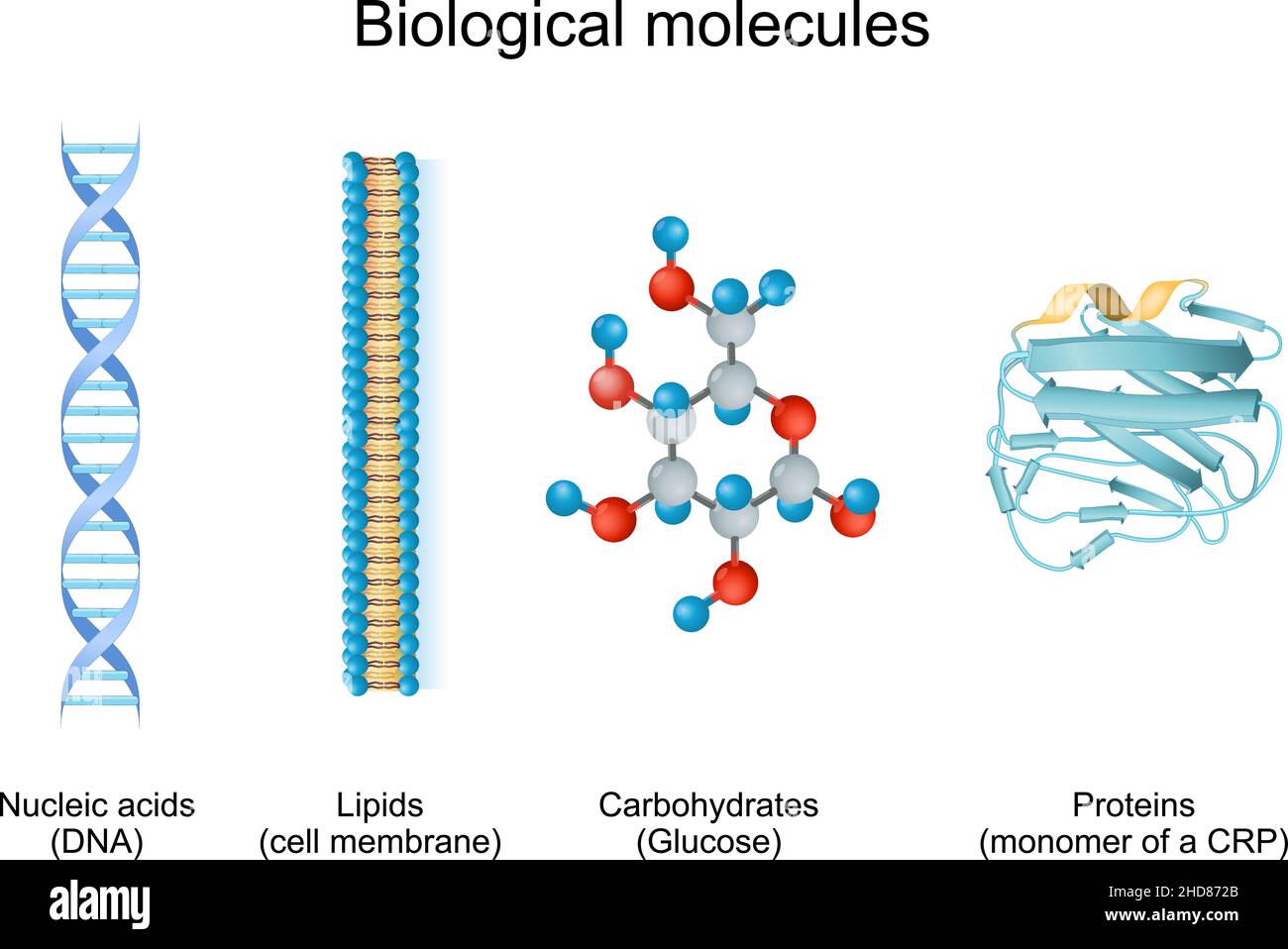 biomolecule is molecules present in live organisms. Types of biological molecule: Carbohydrates, Lipids, Nucleic acids and Proteins Stock Vectorhttps://www.alamy.com/image-license-details/?v=1https://www.alamy.com/biomolecule-is-molecules-present-in-live-organisms-types-of-biological-molecule-carbohydrates-lipids-nucleic-acids-and-proteins-image455641267.html
RF2HD872B–biomolecule is molecules present in live organisms. Types of biological molecule: Carbohydrates, Lipids, Nucleic acids and Proteins
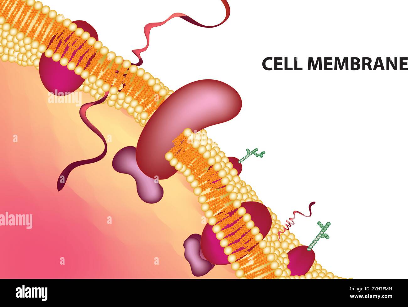 Detailed Structure of Plasma Membrane, Vector Illustration on White Background, Cell Biology Diagram, Scientific Artwork Stock Vectorhttps://www.alamy.com/image-license-details/?v=1https://www.alamy.com/detailed-structure-of-plasma-membrane-vector-illustration-on-white-background-cell-biology-diagram-scientific-artwork-image630188405.html
RF2YH7FMN–Detailed Structure of Plasma Membrane, Vector Illustration on White Background, Cell Biology Diagram, Scientific Artwork
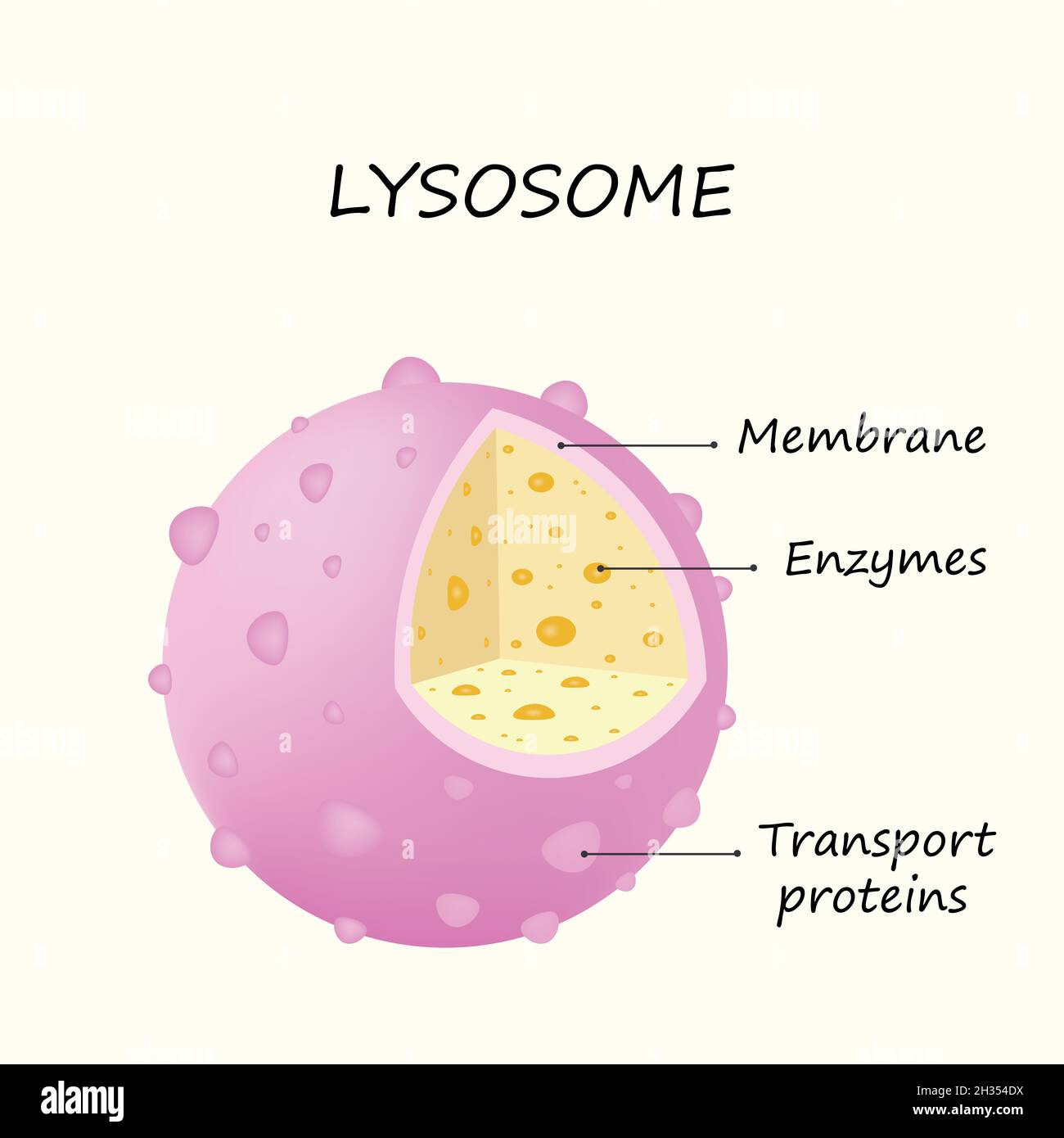 Anatomy of the Lysosome: Hydrolytic enzymes, Membrane and transport proteins colorful illustration Stock Photohttps://www.alamy.com/image-license-details/?v=1https://www.alamy.com/anatomy-of-the-lysosome-hydrolytic-enzymes-membrane-and-transport-proteins-colorful-illustration-image449426822.html
RF2H354DX–Anatomy of the Lysosome: Hydrolytic enzymes, Membrane and transport proteins colorful illustration
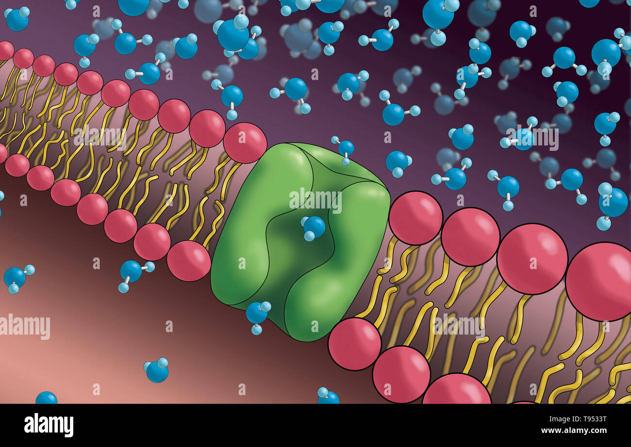 Aquaporins also called water channels, are integral membrane proteins from a larger family of major intrinsic proteins that form pores in the membrane of biological cells and allow water to flow between cells. Stock Photohttps://www.alamy.com/image-license-details/?v=1https://www.alamy.com/aquaporins-also-called-water-channels-are-integral-membrane-proteins-from-a-larger-family-of-major-intrinsic-proteins-that-form-pores-in-the-membrane-of-biological-cells-and-allow-water-to-flow-between-cells-image246589276.html
RMT9533T–Aquaporins also called water channels, are integral membrane proteins from a larger family of major intrinsic proteins that form pores in the membrane of biological cells and allow water to flow between cells.
 Covid-19 coronavirus particles, illustration. The SARS-CoV-2 coronavirus was first identified in Wuhan, China, in December 2019. It is an enveloped RNA (ribonucleic acid) virus. Within the membrane are spike proteins (large protrusions) as well as membrane proteins and envelope proteins. SARS-CoV-2 causes the respiratory infection Covid-19, which can lead to fatal pneumonia. As of March 2020, the virus has spread to many countries worldwide and has been declared a pandemic. Hundreds of thousands have been infected with tens of thousands of deaths. Stock Photohttps://www.alamy.com/image-license-details/?v=1https://www.alamy.com/covid-19-coronavirus-particles-illustration-the-sars-cov-2-coronavirus-was-first-identified-in-wuhan-china-in-december-2019-it-is-an-enveloped-rna-ribonucleic-acid-virus-within-the-membrane-are-spike-proteins-large-protrusions-as-well-as-membrane-proteins-and-envelope-proteins-sars-cov-2-causes-the-respiratory-infection-covid-19-which-can-lead-to-fatal-pneumonia-as-of-march-2020-the-virus-has-spread-to-many-countries-worldwide-and-has-been-declared-a-pandemic-hundreds-of-thousands-have-been-infected-with-tens-of-thousands-of-deaths-image352901281.html
RF2BE4155–Covid-19 coronavirus particles, illustration. The SARS-CoV-2 coronavirus was first identified in Wuhan, China, in December 2019. It is an enveloped RNA (ribonucleic acid) virus. Within the membrane are spike proteins (large protrusions) as well as membrane proteins and envelope proteins. SARS-CoV-2 causes the respiratory infection Covid-19, which can lead to fatal pneumonia. As of March 2020, the virus has spread to many countries worldwide and has been declared a pandemic. Hundreds of thousands have been infected with tens of thousands of deaths.
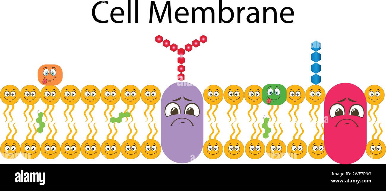 Plasma Membrane Or Cell Membrane Or Plasmalemma Stock Vectorhttps://www.alamy.com/image-license-details/?v=1https://www.alamy.com/plasma-membrane-or-cell-membrane-or-plasmalemma-image594544316.html
RF2WF7R9G–Plasma Membrane Or Cell Membrane Or Plasmalemma
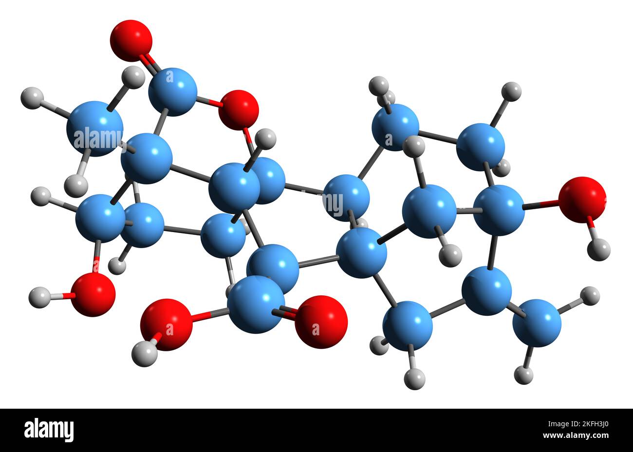 3D image of Gibberellin A1 skeletal formula - molecular chemical structure of plant hormone isolated on white background Stock Photohttps://www.alamy.com/image-license-details/?v=1https://www.alamy.com/3d-image-of-gibberellin-a1-skeletal-formula-molecular-chemical-structure-of-plant-hormone-isolated-on-white-background-image491486184.html
RF2KFH3J0–3D image of Gibberellin A1 skeletal formula - molecular chemical structure of plant hormone isolated on white background
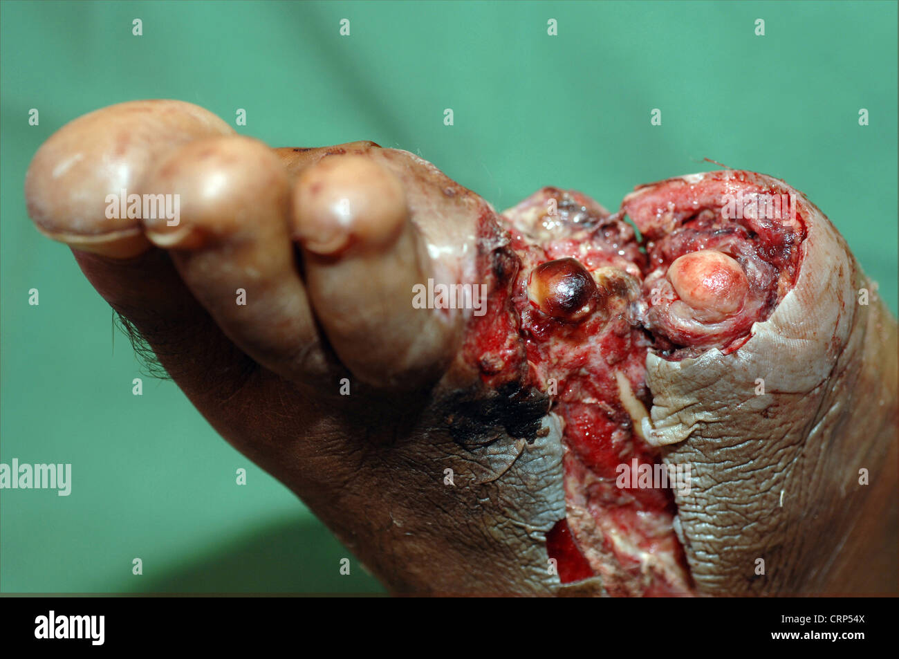 A patient with diabetes suffering with a septic foot. Unregulated hyperglycaemia (high blood glucose levels) leads to non-enzymic glycation of proteins in the walls of the blood vessels. This compromises the basement membrane of the smaller arterioles and capillaries, enabling the movement of proteins across it. As a result, the colloid pressure of the tissu Stock Photohttps://www.alamy.com/image-license-details/?v=1https://www.alamy.com/stock-photo-a-patient-with-diabetes-suffering-with-a-septic-foot-unregulated-hyperglycaemia-49044826.html
RMCRP54X–A patient with diabetes suffering with a septic foot. Unregulated hyperglycaemia (high blood glucose levels) leads to non-enzymic glycation of proteins in the walls of the blood vessels. This compromises the basement membrane of the smaller arterioles and capillaries, enabling the movement of proteins across it. As a result, the colloid pressure of the tissu
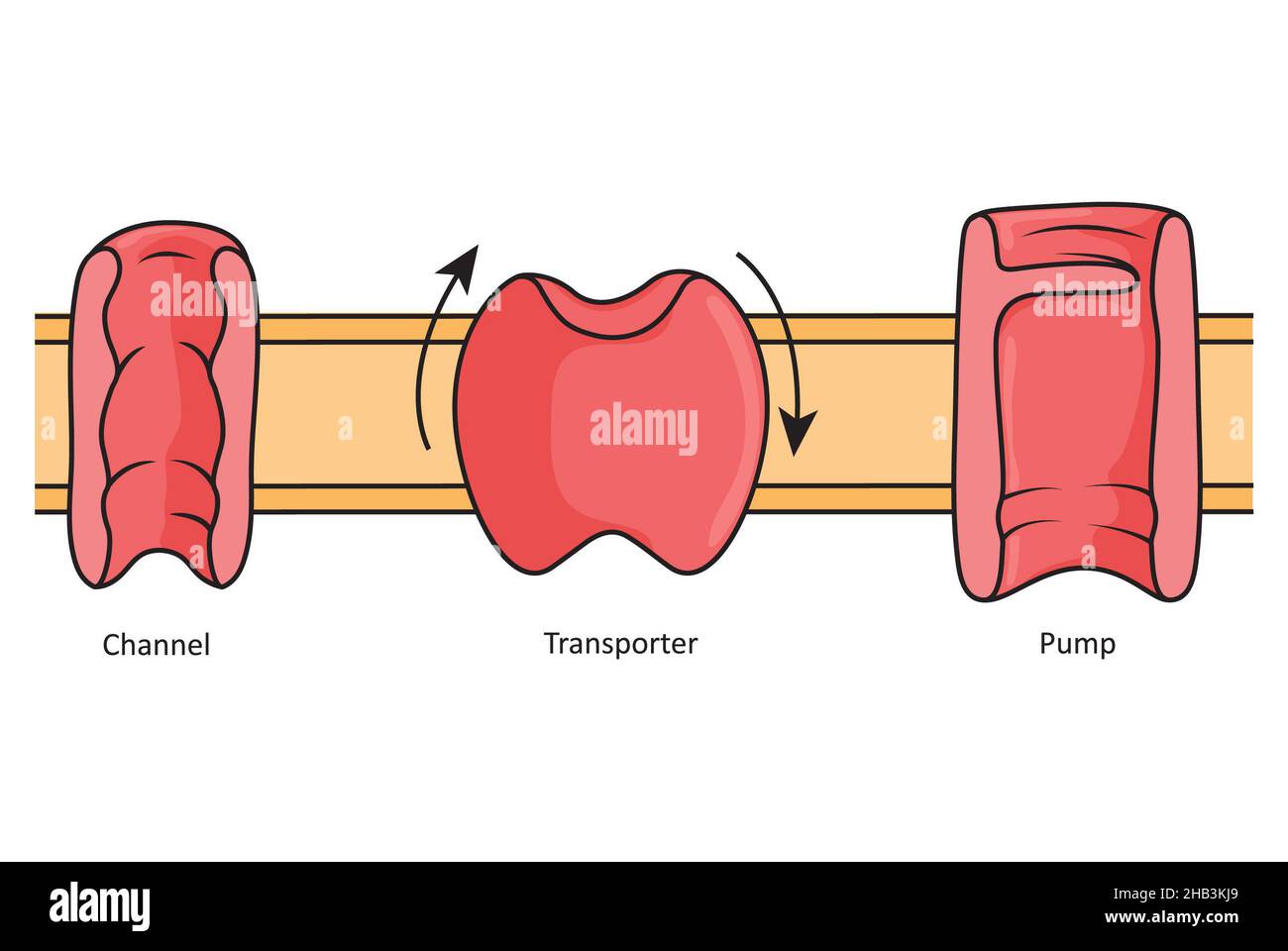 Channels, transporters and pumps, simple illustration showing different transmembrane proteins. Stock Photohttps://www.alamy.com/image-license-details/?v=1https://www.alamy.com/channels-transporters-and-pumps-simple-illustration-showing-different-transmembrane-proteins-image454312049.html
RF2HB3KJ9–Channels, transporters and pumps, simple illustration showing different transmembrane proteins.
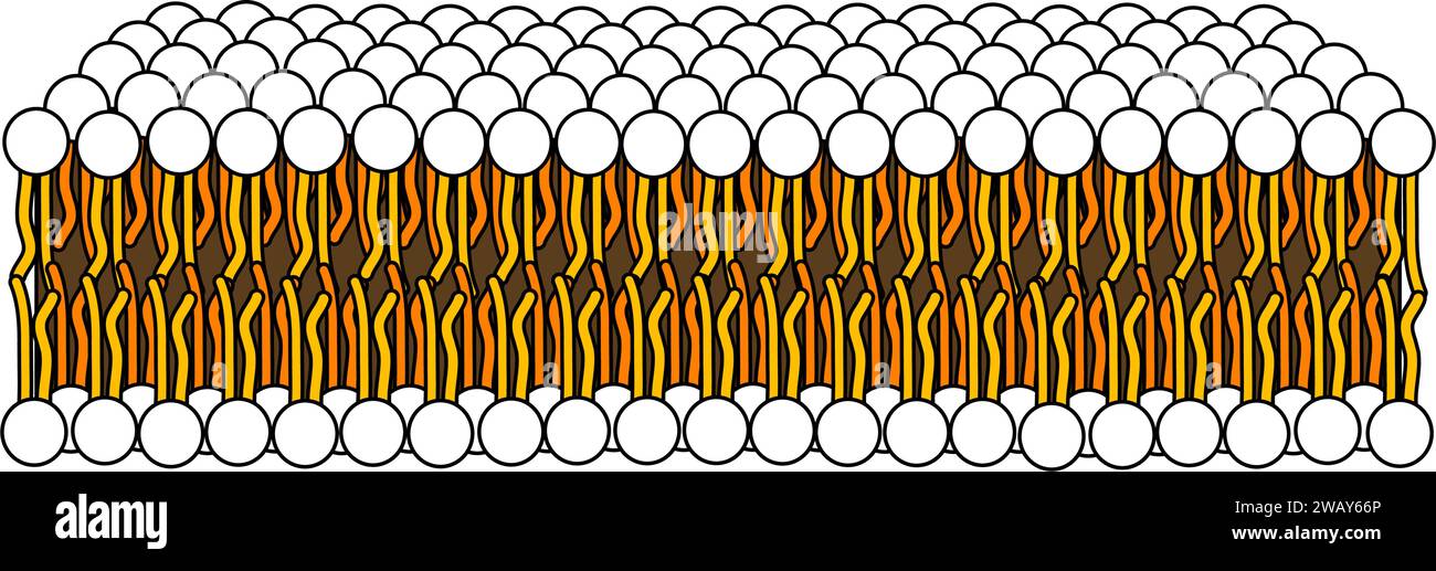 Structure of Phospholipid Molecule in Bilayer .Vector illustration. Stock Vectorhttps://www.alamy.com/image-license-details/?v=1https://www.alamy.com/structure-of-phospholipid-molecule-in-bilayer-vector-illustration-image591896670.html
RF2WAY66P–Structure of Phospholipid Molecule in Bilayer .Vector illustration.
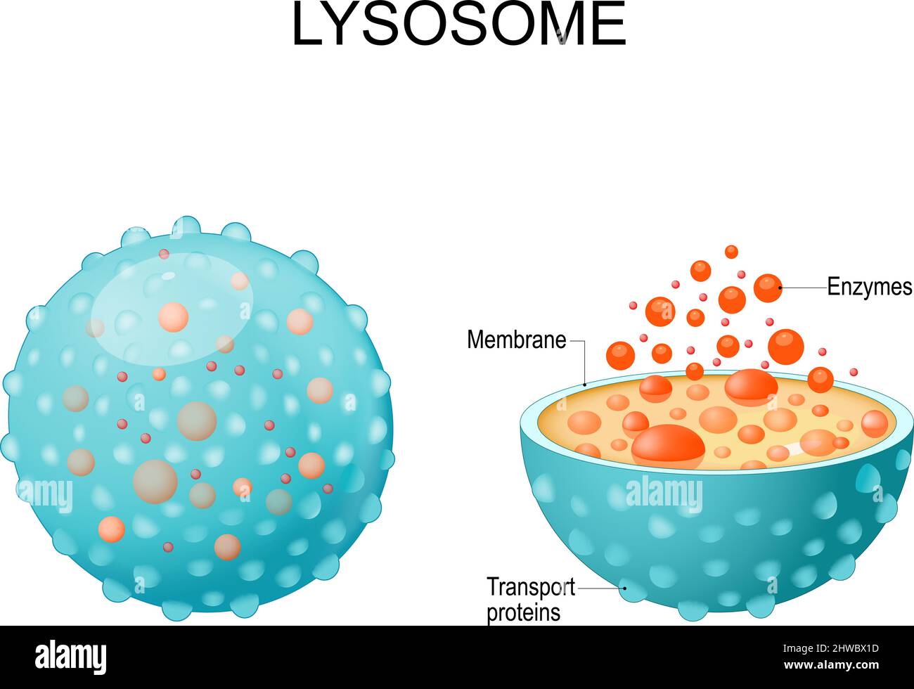 Lysosome. appearance, exterior and interior view. Cross section and Anatomy of the Lysosome: Hydrolytic enzymes, Membrane and transport proteins Stock Vectorhttps://www.alamy.com/image-license-details/?v=1https://www.alamy.com/lysosome-appearance-exterior-and-interior-view-cross-section-and-anatomy-of-the-lysosome-hydrolytic-enzymes-membrane-and-transport-proteins-image463097865.html
RF2HWBX1D–Lysosome. appearance, exterior and interior view. Cross section and Anatomy of the Lysosome: Hydrolytic enzymes, Membrane and transport proteins
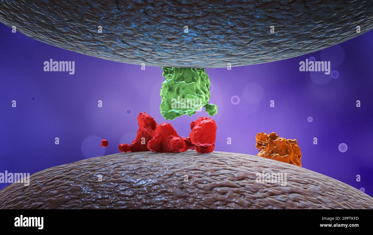 Protein or enzymes or hormones on cell membrane. Stock Photohttps://www.alamy.com/image-license-details/?v=1https://www.alamy.com/protein-or-enzymes-or-hormones-on-cell-membrane-image547586017.html
RF2PPTKFD–Protein or enzymes or hormones on cell membrane.
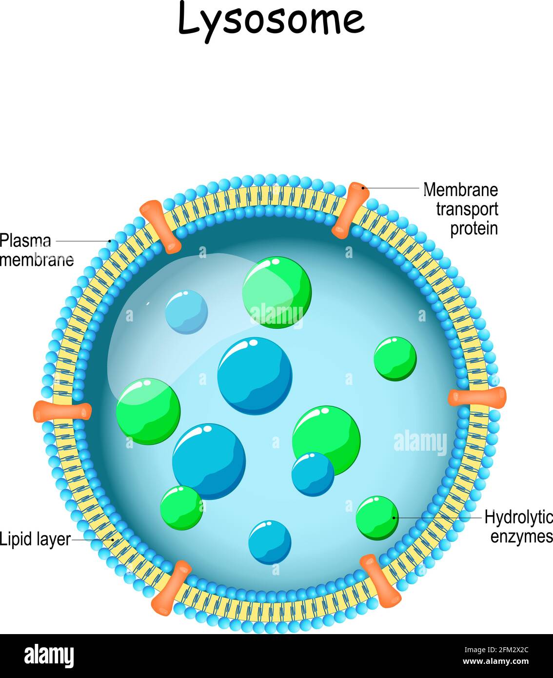 Lysosome. Anatomy of the Lysosome: Hydrolytic enzymes, Membrane and transport proteins. organelle use the enzymes to break down and digest food Stock Vectorhttps://www.alamy.com/image-license-details/?v=1https://www.alamy.com/lysosome-anatomy-of-the-lysosome-hydrolytic-enzymes-membrane-and-transport-proteins-organelle-use-the-enzymes-to-break-down-and-digest-food-image425406308.html
RF2FM2X2C–Lysosome. Anatomy of the Lysosome: Hydrolytic enzymes, Membrane and transport proteins. organelle use the enzymes to break down and digest food
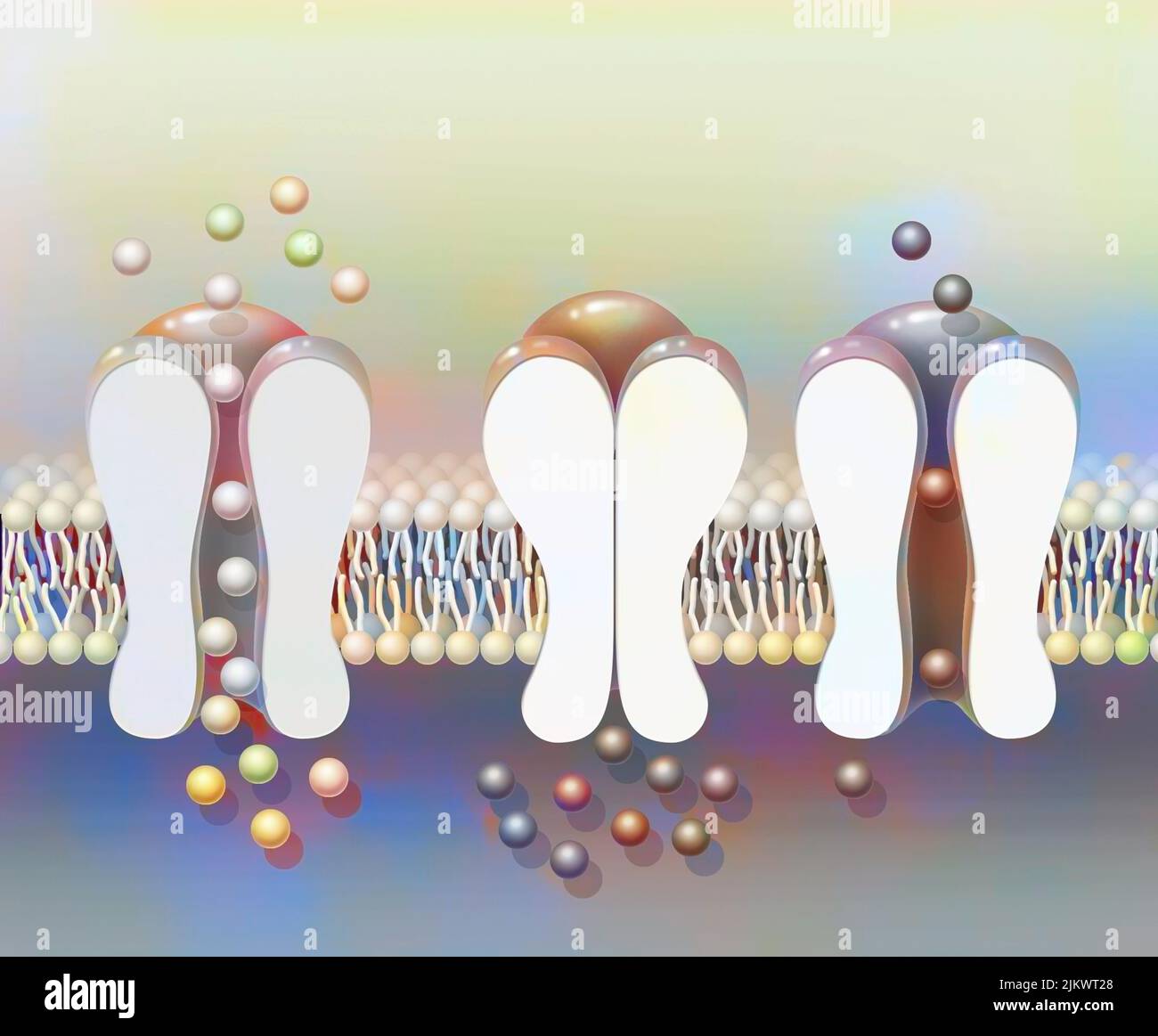 Depolarization: phospholipid membrane with NA + and K + ion channels. Stock Photohttps://www.alamy.com/image-license-details/?v=1https://www.alamy.com/depolarization-phospholipid-membrane-with-na-and-k-ion-channels-image476926080.html
RF2JKWT28–Depolarization: phospholipid membrane with NA + and K + ion channels.
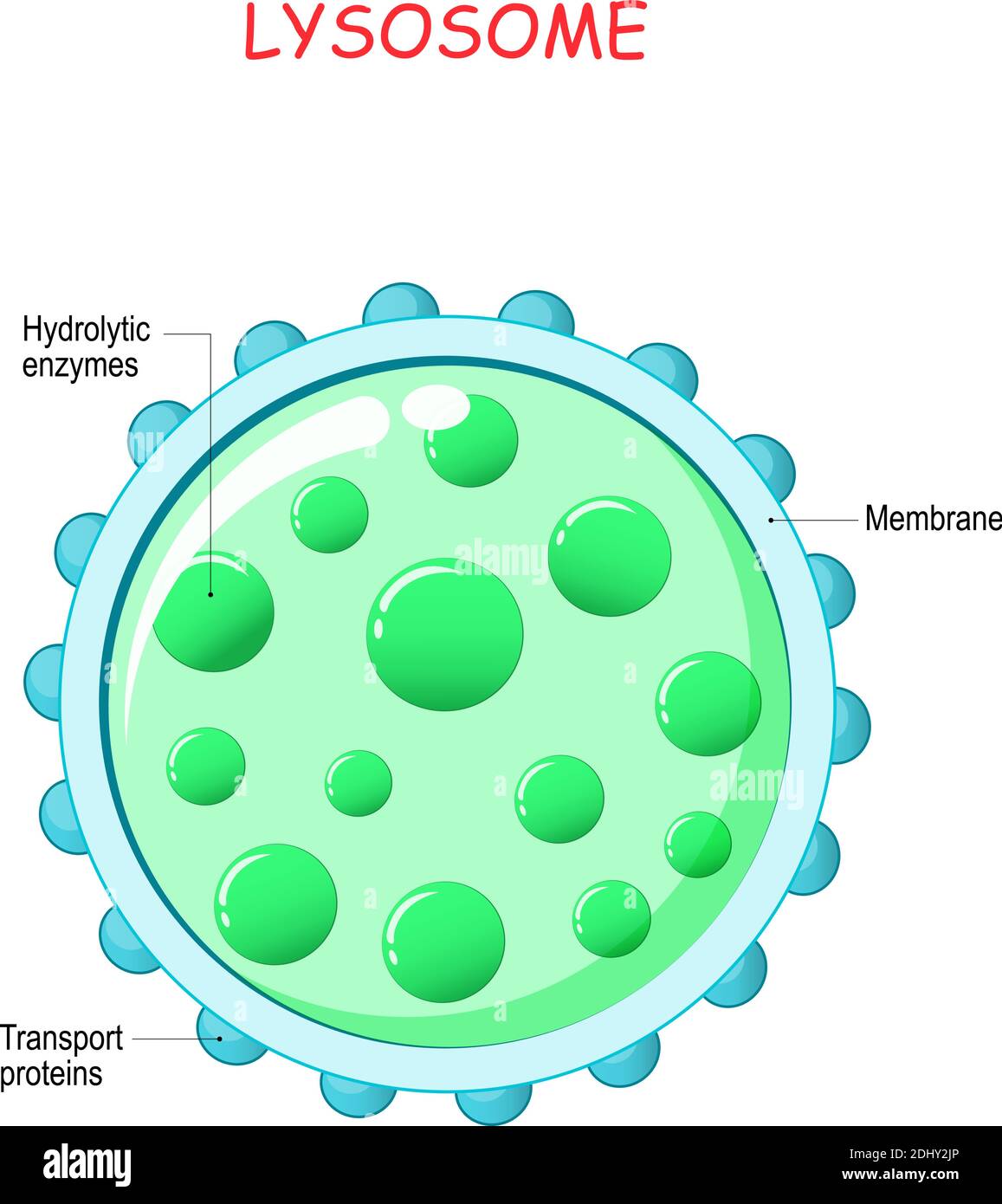 lysosome anatomy. Hydrolytic enzymes, Membrane and transport proteins. This organelle use the enzymes to break-down virus particles or bacteria Stock Vectorhttps://www.alamy.com/image-license-details/?v=1https://www.alamy.com/lysosome-anatomy-hydrolytic-enzymes-membrane-and-transport-proteins-this-organelle-use-the-enzymes-to-break-down-virus-particles-or-bacteria-image389672046.html
RF2DHY2JP–lysosome anatomy. Hydrolytic enzymes, Membrane and transport proteins. This organelle use the enzymes to break-down virus particles or bacteria
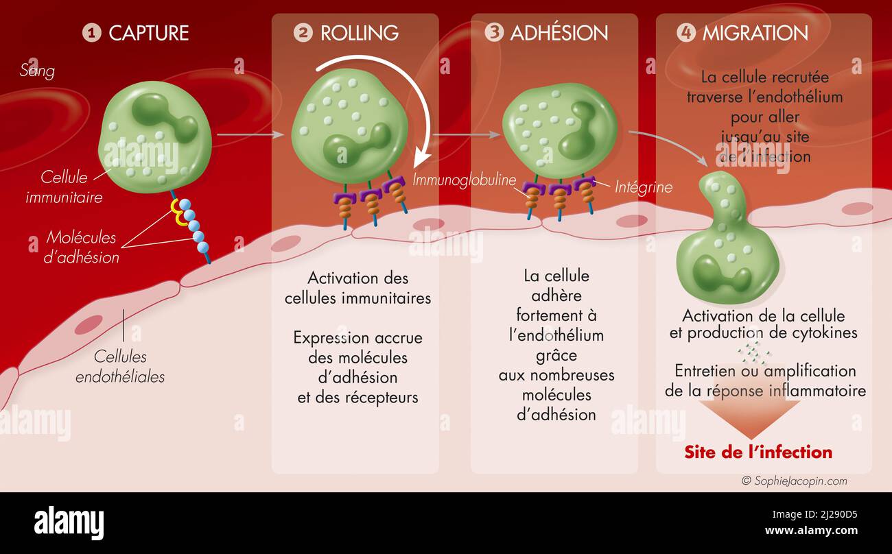 Immunity-membrane membership Stock Photohttps://www.alamy.com/image-license-details/?v=1https://www.alamy.com/immunity-membrane-membership-image466107185.html
RM2J290D5–Immunity-membrane membership
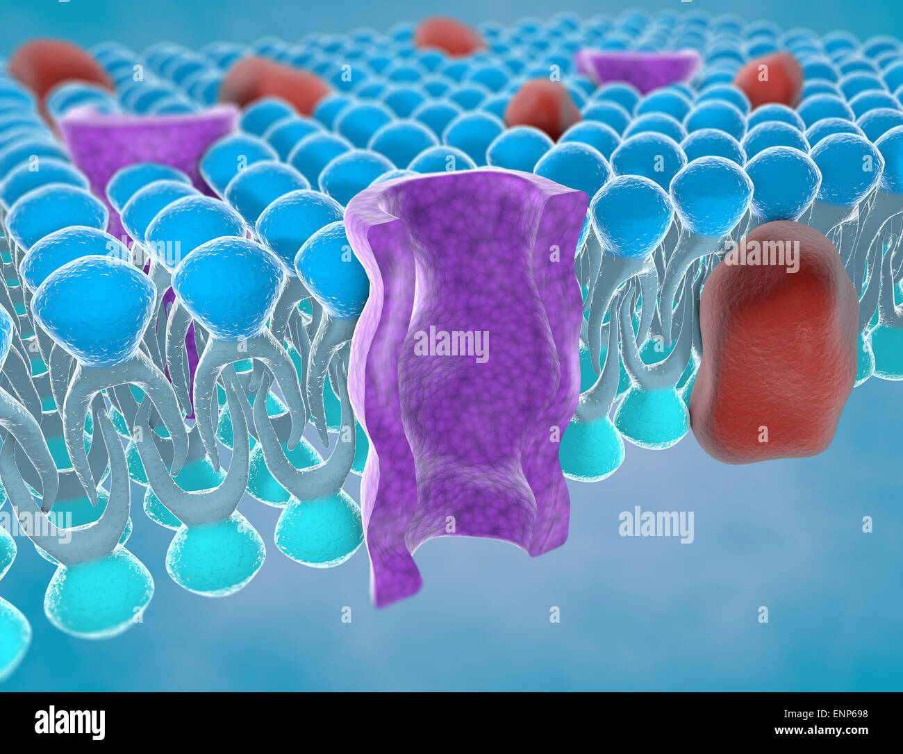 Structure of the plasma membrane of a cell Stock Photohttps://www.alamy.com/image-license-details/?v=1https://www.alamy.com/stock-photo-structure-of-the-plasma-membrane-of-a-cell-82237156.html
RMENP698–Structure of the plasma membrane of a cell
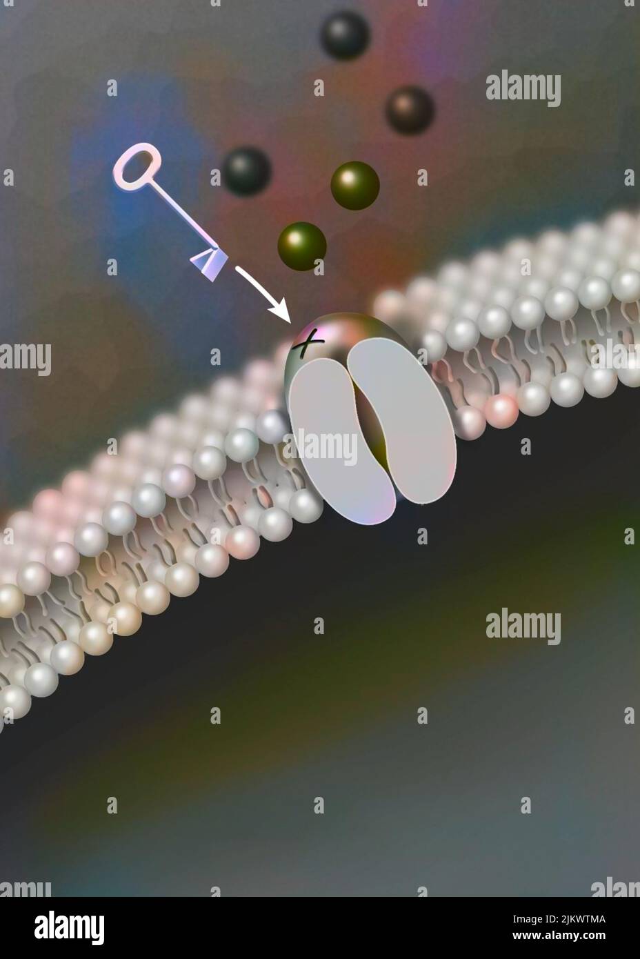 Ligand-dependent ion channel: attachment of a particular molecule causes the channel to open. Stock Photohttps://www.alamy.com/image-license-details/?v=1https://www.alamy.com/ligand-dependent-ion-channel-attachment-of-a-particular-molecule-causes-the-channel-to-open-image476926586.html
RF2JKWTMA–Ligand-dependent ion channel: attachment of a particular molecule causes the channel to open.
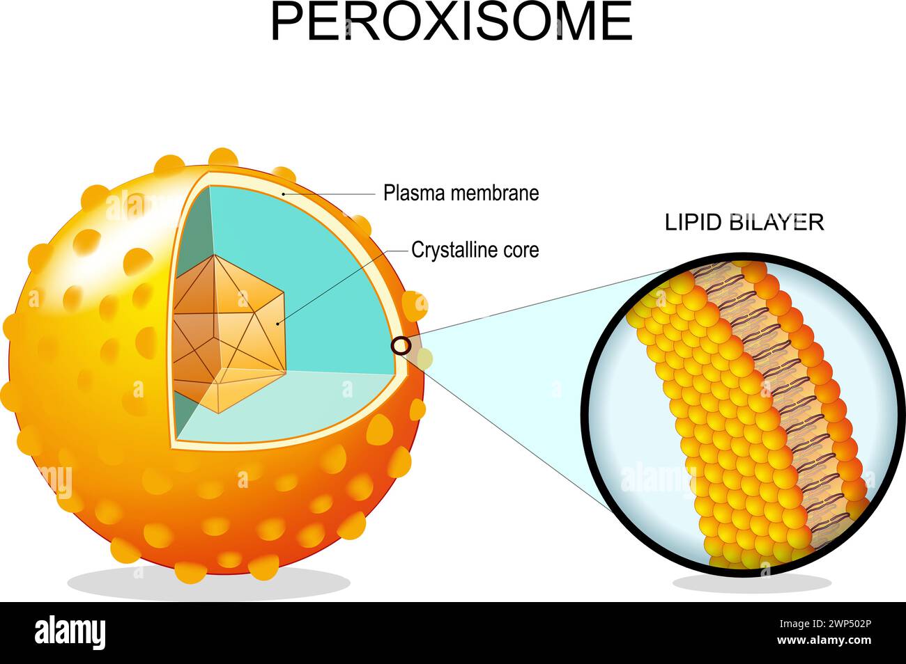 Peroxisome anatomy. Cross section of a cell organelle. Close-up of a Lipid bilayer Plasma membrane, Crystalline core, transport proteins. Vector illus Stock Vectorhttps://www.alamy.com/image-license-details/?v=1https://www.alamy.com/peroxisome-anatomy-cross-section-of-a-cell-organelle-close-up-of-a-lipid-bilayer-plasma-membrane-crystalline-core-transport-proteins-vector-illus-image598784782.html
RF2WP502P–Peroxisome anatomy. Cross section of a cell organelle. Close-up of a Lipid bilayer Plasma membrane, Crystalline core, transport proteins. Vector illus
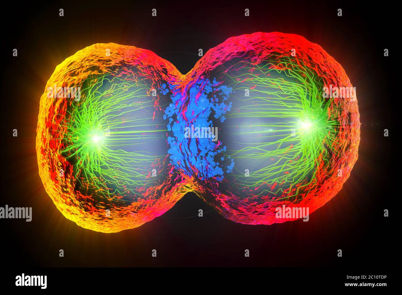 3d illustration of colorful cell division, cell membrane and splitting nucleus Stock Photohttps://www.alamy.com/image-license-details/?v=1https://www.alamy.com/3d-illustration-of-colorful-cell-division-cell-membrane-and-splitting-nucleus-image362051586.html
RF2C10TDP–3d illustration of colorful cell division, cell membrane and splitting nucleus
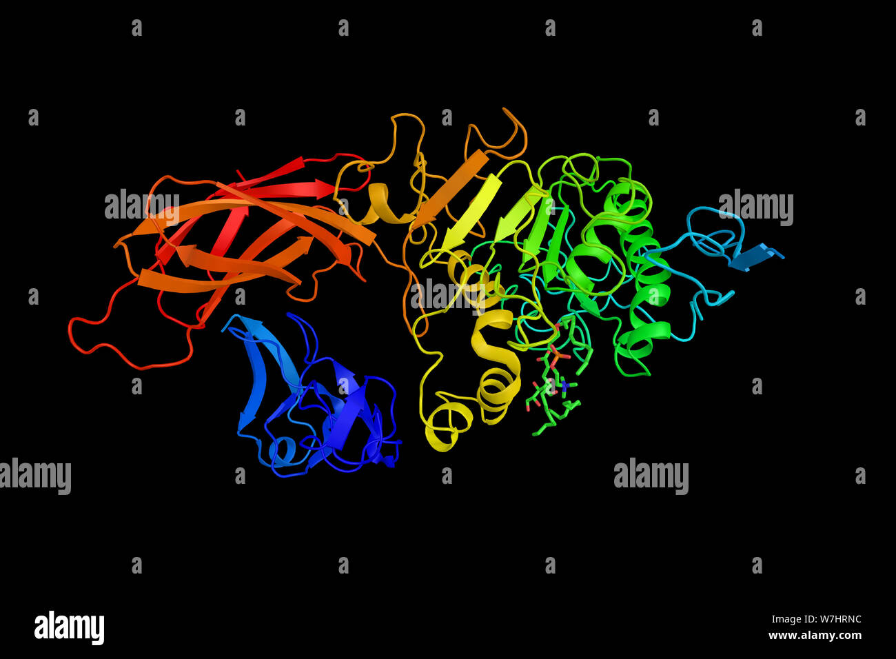 Example of PLAT/LH2 domain, a protein domain found in a variety of membrane or lipid associated proteins. 3d rendering. Stock Photohttps://www.alamy.com/image-license-details/?v=1https://www.alamy.com/example-of-platlh2-domain-a-protein-domain-found-in-a-variety-of-membrane-or-lipid-associated-proteins-3d-rendering-image262849928.html
RFW7HRNC–Example of PLAT/LH2 domain, a protein domain found in a variety of membrane or lipid associated proteins. 3d rendering.
 Interior wide-angle view of the Coronavirus (SARS-CoV-2, Covid 19). An accurate model based on scientific structural data from the PDB. Stock Photohttps://www.alamy.com/image-license-details/?v=1https://www.alamy.com/interior-wide-angle-view-of-the-coronavirus-sars-cov-2-covid-19-an-accurate-model-based-on-scientific-structural-data-from-the-pdb-image382874087.html
RM2D6WBPF–Interior wide-angle view of the Coronavirus (SARS-CoV-2, Covid 19). An accurate model based on scientific structural data from the PDB.
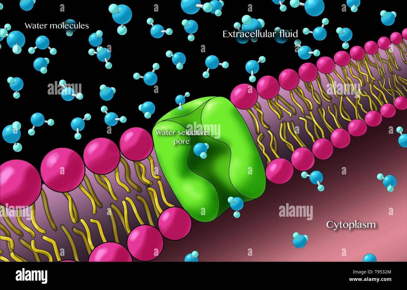 Aquaporins also called water channels, are integral membrane proteins from a larger family of major intrinsic proteins that form pores in the membrane of biological cells and allow water to flow between cells. Stock Photohttps://www.alamy.com/image-license-details/?v=1https://www.alamy.com/aquaporins-also-called-water-channels-are-integral-membrane-proteins-from-a-larger-family-of-major-intrinsic-proteins-that-form-pores-in-the-membrane-of-biological-cells-and-allow-water-to-flow-between-cells-image246589244.html
RMT9532M–Aquaporins also called water channels, are integral membrane proteins from a larger family of major intrinsic proteins that form pores in the membrane of biological cells and allow water to flow between cells.
 Covid-19 coronavirus particles, illustration. The SARS-CoV-2 coronavirus was first identified in Wuhan, China, in December 2019. It is an enveloped RNA (ribonucleic acid) virus. Within the membrane are spike proteins (large protrusions) as well as membrane proteins and envelope proteins. SARS-CoV-2 causes the respiratory infection Covid-19, which can lead to fatal pneumonia. As of March 2020, the virus has spread to many countries worldwide and has been declared a pandemic. Hundreds of thousands have been infected with tens of thousands of deaths. Stock Photohttps://www.alamy.com/image-license-details/?v=1https://www.alamy.com/covid-19-coronavirus-particles-illustration-the-sars-cov-2-coronavirus-was-first-identified-in-wuhan-china-in-december-2019-it-is-an-enveloped-rna-ribonucleic-acid-virus-within-the-membrane-are-spike-proteins-large-protrusions-as-well-as-membrane-proteins-and-envelope-proteins-sars-cov-2-causes-the-respiratory-infection-covid-19-which-can-lead-to-fatal-pneumonia-as-of-march-2020-the-virus-has-spread-to-many-countries-worldwide-and-has-been-declared-a-pandemic-hundreds-of-thousands-have-been-infected-with-tens-of-thousands-of-deaths-image352901287.html
RF2BE415B–Covid-19 coronavirus particles, illustration. The SARS-CoV-2 coronavirus was first identified in Wuhan, China, in December 2019. It is an enveloped RNA (ribonucleic acid) virus. Within the membrane are spike proteins (large protrusions) as well as membrane proteins and envelope proteins. SARS-CoV-2 causes the respiratory infection Covid-19, which can lead to fatal pneumonia. As of March 2020, the virus has spread to many countries worldwide and has been declared a pandemic. Hundreds of thousands have been infected with tens of thousands of deaths.
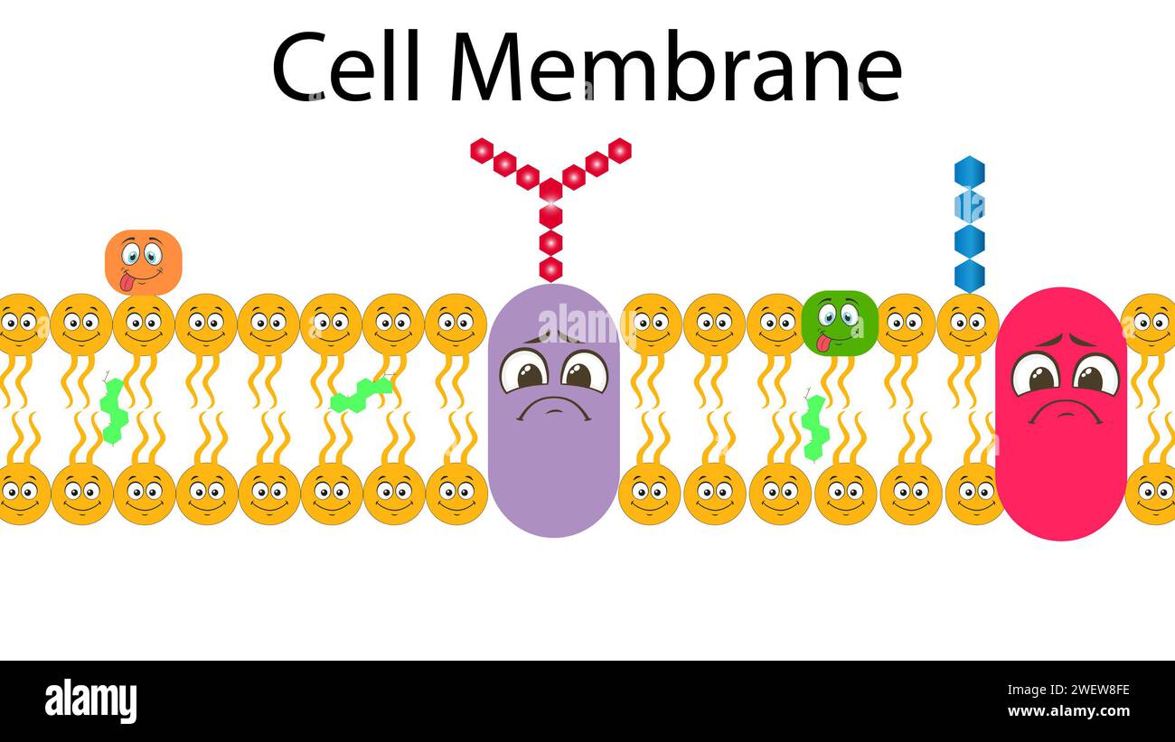 Plasma Membrane Or Cell Membrane Or Plasmalemma Stock Photohttps://www.alamy.com/image-license-details/?v=1https://www.alamy.com/plasma-membrane-or-cell-membrane-or-plasmalemma-image594313202.html
RF2WEW8FE–Plasma Membrane Or Cell Membrane Or Plasmalemma
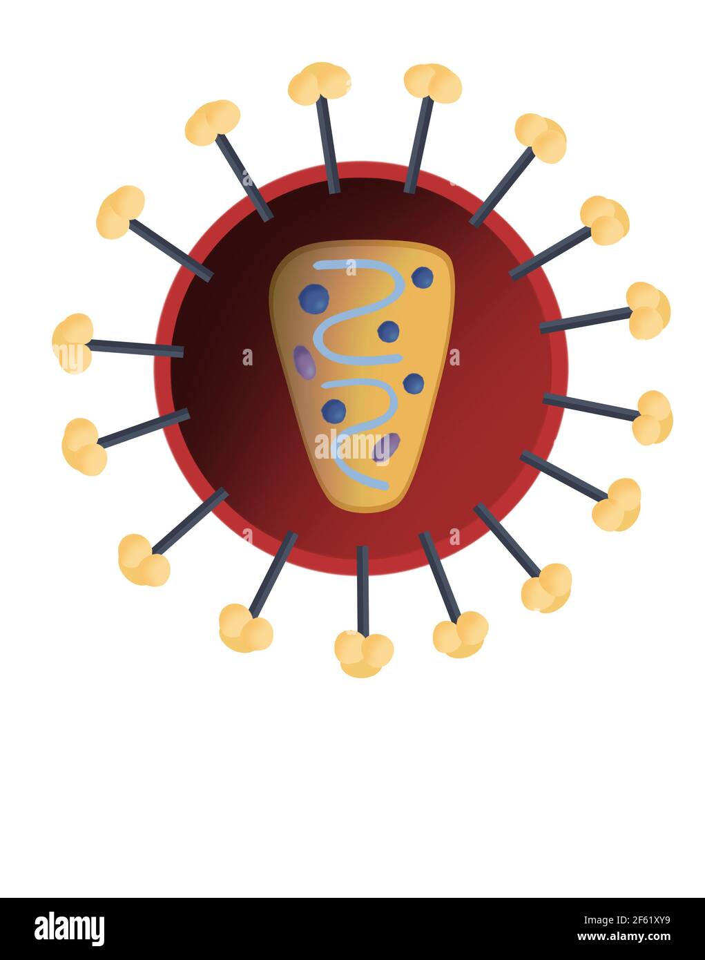 Structure of HIV Stock Photohttps://www.alamy.com/image-license-details/?v=1https://www.alamy.com/structure-of-hiv-image416779869.html
RM2F61XY9–Structure of HIV
 Potsdam, Germany. 16th June, 2020. George Soultoukis, research assistant, is preparing a flow cytometer for operation in the Laboratory for Fat Cell Development and Nutrition (ADE) of the German Institute of Nutrition Research Potsdam-Rehbrücke (DIfE). With the flow cytometer, stem cells are separated into different subpopulations using unique marker proteins on their cell membrane. Credit: Christoph Soeder/dpa-Zentralbild/dpa/Alamy Live News Stock Photohttps://www.alamy.com/image-license-details/?v=1https://www.alamy.com/potsdam-germany-16th-june-2020-george-soultoukis-research-assistant-is-preparing-a-flow-cytometer-for-operation-in-the-laboratory-for-fat-cell-development-and-nutrition-ade-of-the-german-institute-of-nutrition-research-potsdam-rehbrcke-dife-with-the-flow-cytometer-stem-cells-are-separated-into-different-subpopulations-using-unique-marker-proteins-on-their-cell-membrane-credit-christoph-soederdpa-zentralbilddpaalamy-live-news-image365732741.html
RM2C70FRH–Potsdam, Germany. 16th June, 2020. George Soultoukis, research assistant, is preparing a flow cytometer for operation in the Laboratory for Fat Cell Development and Nutrition (ADE) of the German Institute of Nutrition Research Potsdam-Rehbrücke (DIfE). With the flow cytometer, stem cells are separated into different subpopulations using unique marker proteins on their cell membrane. Credit: Christoph Soeder/dpa-Zentralbild/dpa/Alamy Live News
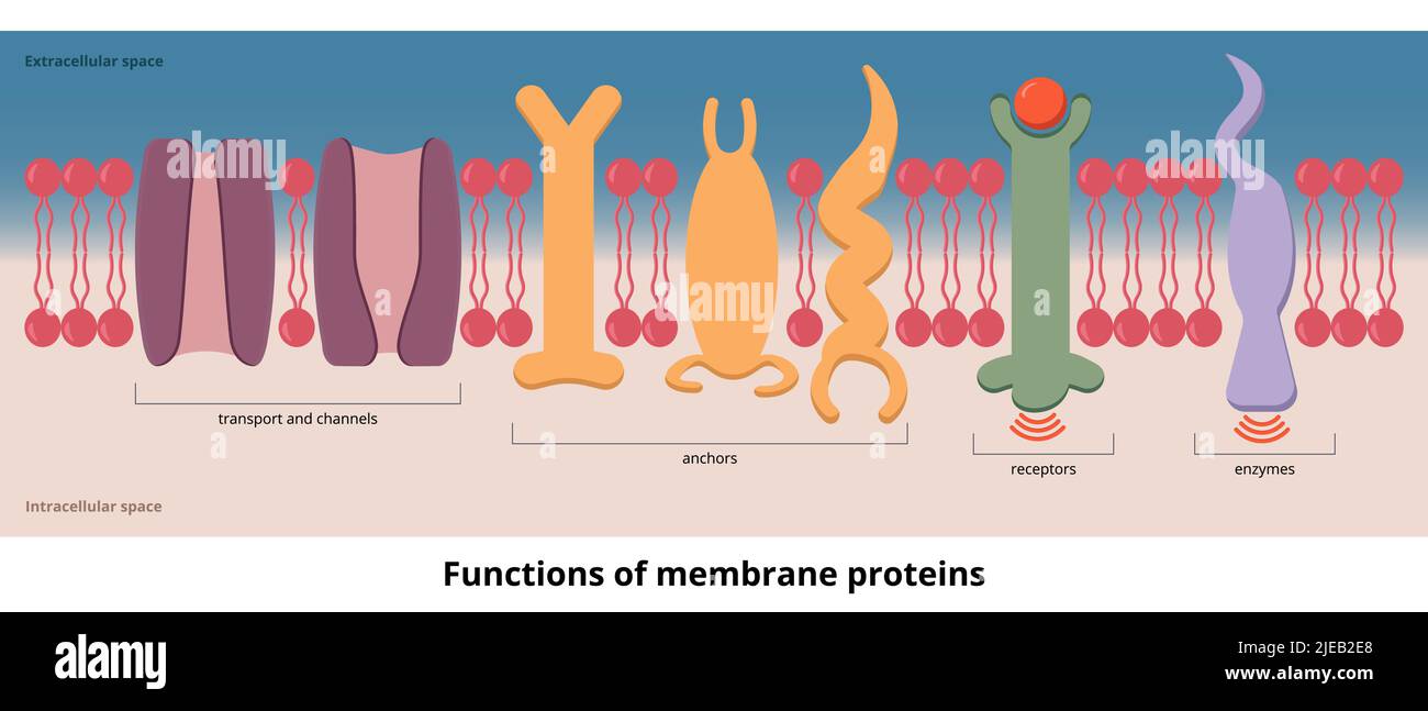 Functions of membrane proteins.Functions of protein visualization include transport, channels, receptors, and enzymes that are placed on cell membrane Stock Vectorhttps://www.alamy.com/image-license-details/?v=1https://www.alamy.com/functions-of-membrane-proteinsfunctions-of-protein-visualization-include-transport-channels-receptors-and-enzymes-that-are-placed-on-cell-membrane-image473528560.html
RF2JEB2E8–Functions of membrane proteins.Functions of protein visualization include transport, channels, receptors, and enzymes that are placed on cell membrane
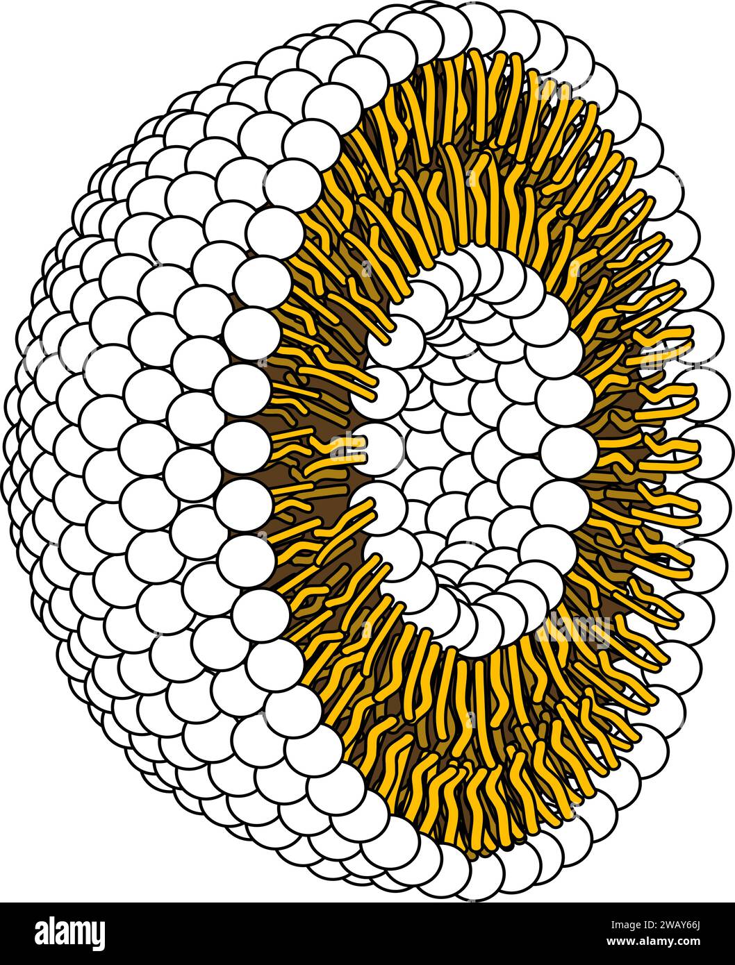 Structure of Phospholipid Molecule in Liposome.Vector illustration. Stock Vectorhttps://www.alamy.com/image-license-details/?v=1https://www.alamy.com/structure-of-phospholipid-molecule-in-liposomevector-illustration-image591896666.html
RF2WAY66J–Structure of Phospholipid Molecule in Liposome.Vector illustration.
 Lysosome anatomy. Cross section of a cell organelle. Close-up of a Lipid bilayer membrane, hydrolytic enzymes, transport proteins. Autophagy. Vector Stock Vectorhttps://www.alamy.com/image-license-details/?v=1https://www.alamy.com/lysosome-anatomy-cross-section-of-a-cell-organelle-close-up-of-a-lipid-bilayer-membrane-hydrolytic-enzymes-transport-proteins-autophagy-vector-image630188310.html
RF2YH7FHA–Lysosome anatomy. Cross section of a cell organelle. Close-up of a Lipid bilayer membrane, hydrolytic enzymes, transport proteins. Autophagy. Vector
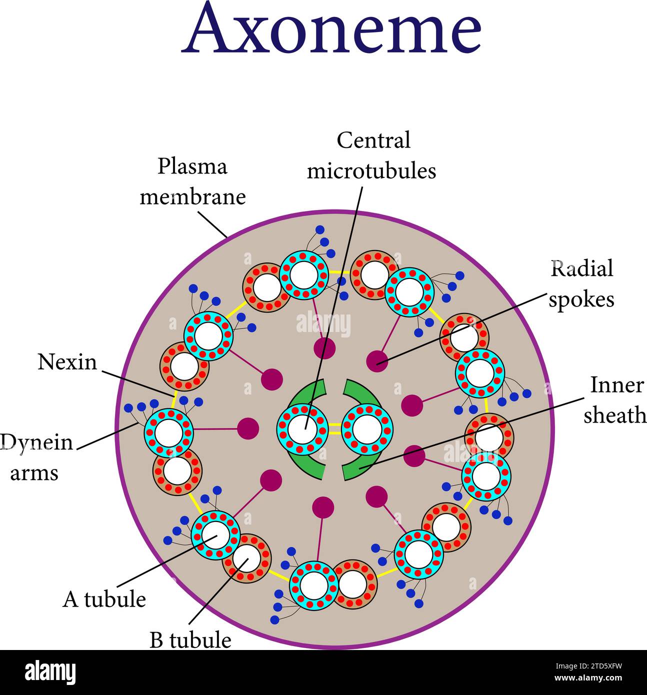 A Cross section of an axoneme .Vector illustration. Stock Vectorhttps://www.alamy.com/image-license-details/?v=1https://www.alamy.com/a-cross-section-of-an-axoneme-vector-illustration-image576063261.html
RF2TD5XFW–A Cross section of an axoneme .Vector illustration.
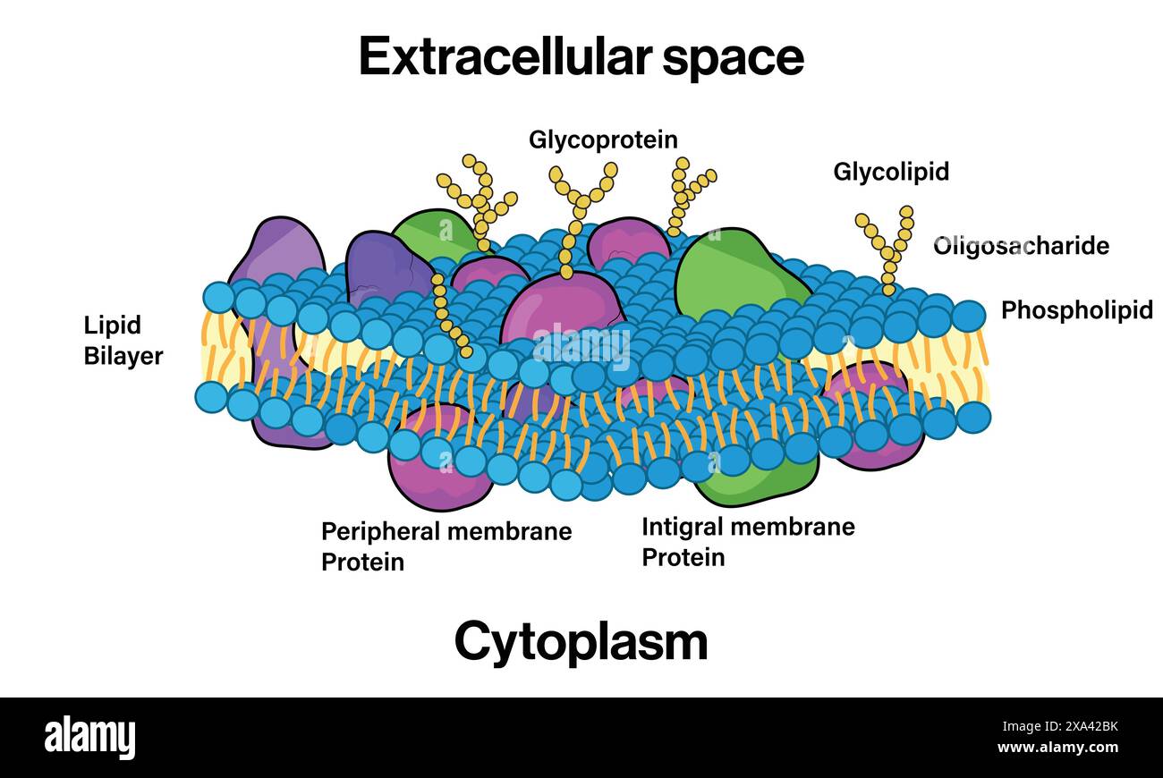 Detailed Vector Illustration of Plasma Membrane Structure for Cell Biology and Biochemistry Education on White Background. Stock Vectorhttps://www.alamy.com/image-license-details/?v=1https://www.alamy.com/detailed-vector-illustration-of-plasma-membrane-structure-for-cell-biology-and-biochemistry-education-on-white-background-image608599143.html
RF2XA42BK–Detailed Vector Illustration of Plasma Membrane Structure for Cell Biology and Biochemistry Education on White Background.
 In the foreground is a cross-sectional view of the extracellular enveloped version of the mpox virion. Here it is shown binding with glycosaminoglycans GAGs or mucopolysaccharides. These are represented as feathery projections embedded in the lipid bilayer of a human host cell. Within the human host are proteins and small molecules shown in blue green. The outer envelope of mpox binds with GAGs and later fuses with the human host membrane. The mature virion then enters the human host cell. The mature virion is studded with tubular protein structures and other viral proteins shown in red. Below Stock Photohttps://www.alamy.com/image-license-details/?v=1https://www.alamy.com/in-the-foreground-is-a-cross-sectional-view-of-the-extracellular-enveloped-version-of-the-mpox-virion-here-it-is-shown-binding-with-glycosaminoglycans-gags-or-mucopolysaccharides-these-are-represented-as-feathery-projections-embedded-in-the-lipid-bilayer-of-a-human-host-cell-within-the-human-host-are-proteins-and-small-molecules-shown-in-blue-green-the-outer-envelope-of-mpox-binds-with-gags-and-later-fuses-with-the-human-host-membrane-the-mature-virion-then-enters-the-human-host-cell-the-mature-virion-is-studded-with-tubular-protein-structures-and-other-viral-proteins-shown-in-red-below-image627781183.html
RM2YD9W8F–In the foreground is a cross-sectional view of the extracellular enveloped version of the mpox virion. Here it is shown binding with glycosaminoglycans GAGs or mucopolysaccharides. These are represented as feathery projections embedded in the lipid bilayer of a human host cell. Within the human host are proteins and small molecules shown in blue green. The outer envelope of mpox binds with GAGs and later fuses with the human host membrane. The mature virion then enters the human host cell. The mature virion is studded with tubular protein structures and other viral proteins shown in red. Below
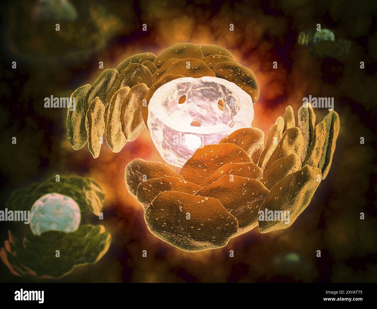 Conceptual image of endoplasmic reticulum around a cell nucleus. Endoplasmic reticulum is an organelle that forms a continuous membrane system of flat Stock Photohttps://www.alamy.com/image-license-details/?v=1https://www.alamy.com/conceptual-image-of-endoplasmic-reticulum-around-a-cell-nucleus-endoplasmic-reticulum-is-an-organelle-that-forms-a-continuous-membrane-system-of-flat-image619197129.html
RM2XYAT75–Conceptual image of endoplasmic reticulum around a cell nucleus. Endoplasmic reticulum is an organelle that forms a continuous membrane system of flat
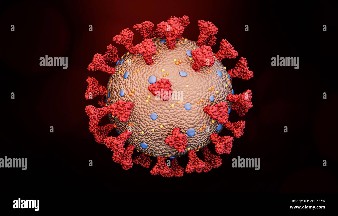 Accurate scientific render of a coronavirus cell like covid or a flu virus structure with spikes glycoprotein, M proteins, E proteins, hemagglutinin a Stock Photohttps://www.alamy.com/image-license-details/?v=1https://www.alamy.com/accurate-scientific-render-of-a-coronavirus-cell-like-covid-or-a-flu-virus-structure-with-spikes-glycoprotein-m-proteins-e-proteins-hemagglutinin-a-image352959914.html
RF2BE6KY6–Accurate scientific render of a coronavirus cell like covid or a flu virus structure with spikes glycoprotein, M proteins, E proteins, hemagglutinin a
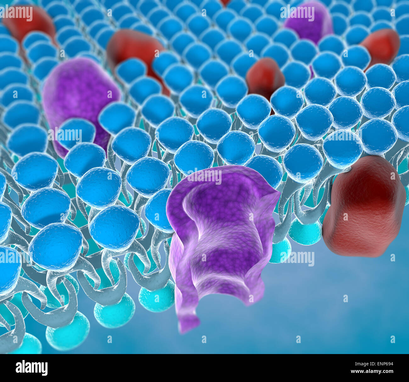 Structure of the plasma membrane of a cell Stock Photohttps://www.alamy.com/image-license-details/?v=1https://www.alamy.com/stock-photo-structure-of-the-plasma-membrane-of-a-cell-82237152.html
RMENP694–Structure of the plasma membrane of a cell
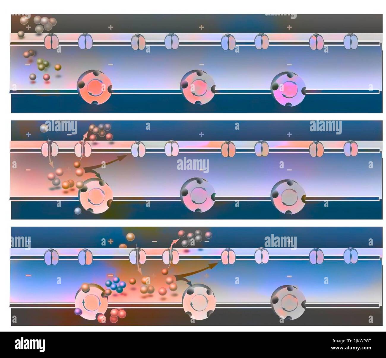 Movement of the action potential (impulse) in the axon of the neuron and from one neuron to another. Stock Photohttps://www.alamy.com/image-license-details/?v=1https://www.alamy.com/movement-of-the-action-potential-impulse-in-the-axon-of-the-neuron-and-from-one-neuron-to-another-image476924920.html
RF2JKWPGT–Movement of the action potential (impulse) in the axon of the neuron and from one neuron to another.
 3d rendering of nanodisc consists of Phospholipids and A stabilizing belt that holds the phospholipids together. Stock Photohttps://www.alamy.com/image-license-details/?v=1https://www.alamy.com/3d-rendering-of-nanodisc-consists-of-phospholipids-and-a-stabilizing-belt-that-holds-the-phospholipids-together-image591330901.html
RF2WA1CGN–3d rendering of nanodisc consists of Phospholipids and A stabilizing belt that holds the phospholipids together.
 Virus with surface proteins concept. Medical of science 3D rendering background. Stock Photohttps://www.alamy.com/image-license-details/?v=1https://www.alamy.com/virus-with-surface-proteins-concept-medical-of-science-3d-rendering-background-image383855007.html
RF2D8E2YB–Virus with surface proteins concept. Medical of science 3D rendering background.
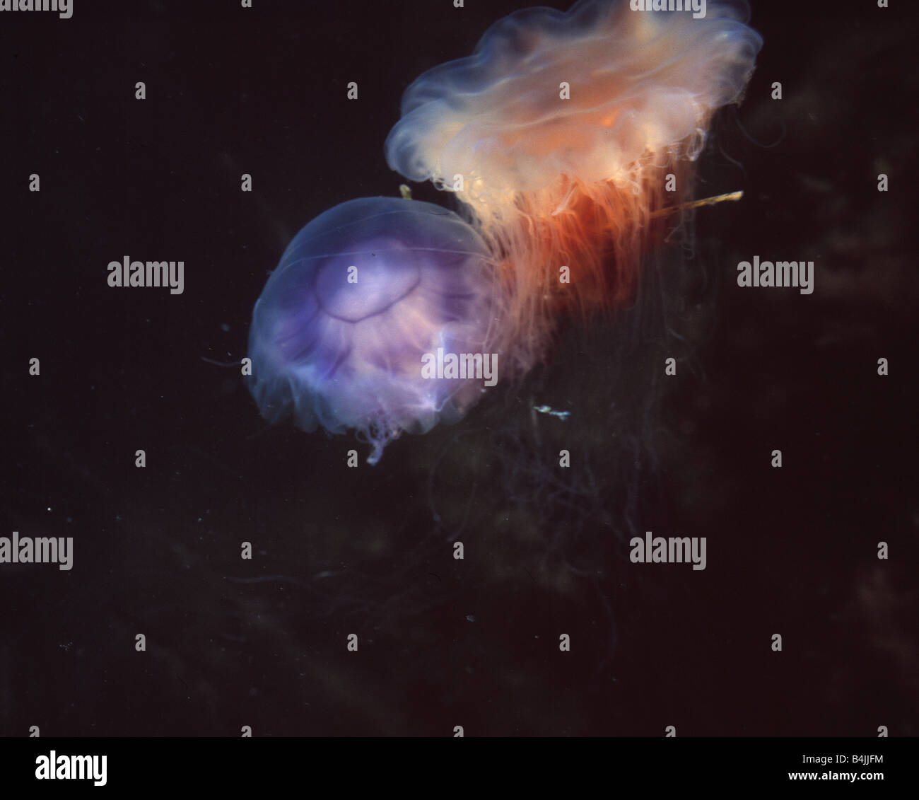 Two species of jellyfish 'Cyanea capillata' (red) and Cyanea lamarkii (blue) Stock Photohttps://www.alamy.com/image-license-details/?v=1https://www.alamy.com/stock-photo-two-species-of-jellyfish-cyanea-capillata-red-and-cyanea-lamarkii-20078680.html
RMB4JJFM–Two species of jellyfish 'Cyanea capillata' (red) and Cyanea lamarkii (blue)
 Interior wide-angle view of the Coronavirus (SARS-CoV-2, Covid 19). An accurate model based on scientific structural data from the PDB. Stock Photohttps://www.alamy.com/image-license-details/?v=1https://www.alamy.com/interior-wide-angle-view-of-the-coronavirus-sars-cov-2-covid-19-an-accurate-model-based-on-scientific-structural-data-from-the-pdb-image382874098.html
RM2D6WBPX–Interior wide-angle view of the Coronavirus (SARS-CoV-2, Covid 19). An accurate model based on scientific structural data from the PDB.
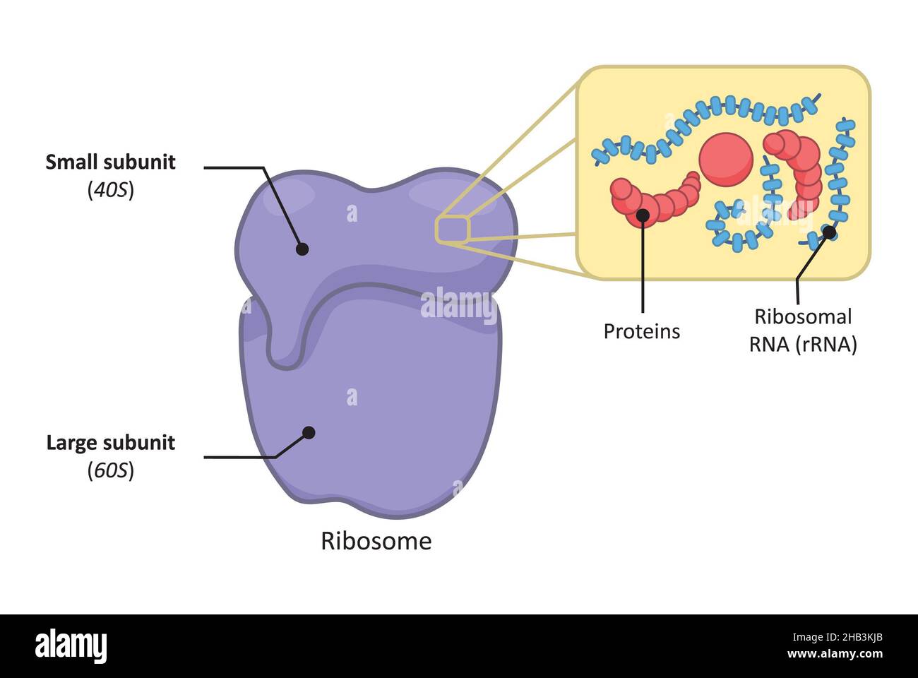 Simple structure of a 80s ribosome in eukaryotic cell. Stock Photohttps://www.alamy.com/image-license-details/?v=1https://www.alamy.com/simple-structure-of-a-80s-ribosome-in-eukaryotic-cell-image454312051.html
RF2HB3KJB–Simple structure of a 80s ribosome in eukaryotic cell.
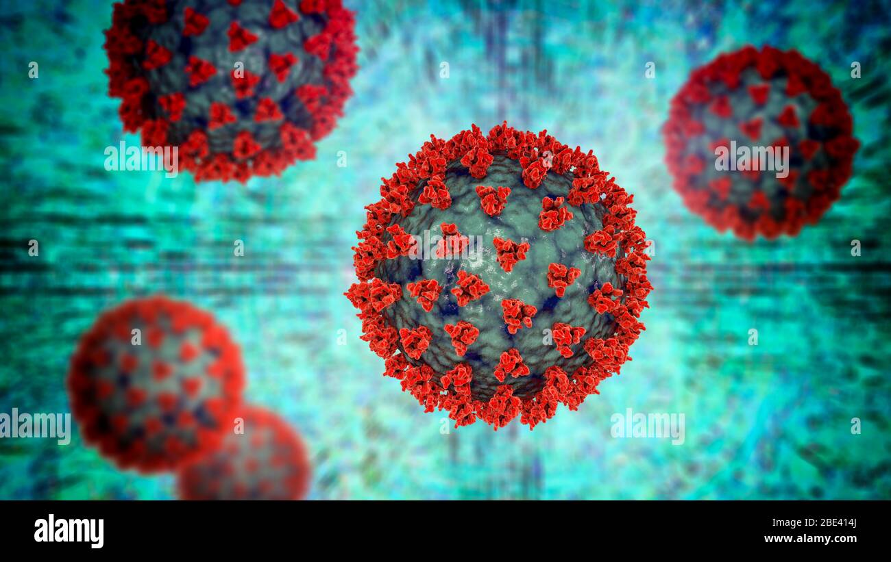 Covid-19 coronavirus particles, illustration. The SARS-CoV-2 coronavirus was first identified in Wuhan, China, in December 2019. It is an enveloped RNA (ribonucleic acid) virus. Within the membrane are spike proteins (large protrusions) as well as membrane proteins and envelope proteins. SARS-CoV-2 causes the respiratory infection Covid-19, which can lead to fatal pneumonia. As of March 2020, the virus has spread to many countries worldwide and has been declared a pandemic. Hundreds of thousands have been infected with tens of thousands of deaths. Stock Photohttps://www.alamy.com/image-license-details/?v=1https://www.alamy.com/covid-19-coronavirus-particles-illustration-the-sars-cov-2-coronavirus-was-first-identified-in-wuhan-china-in-december-2019-it-is-an-enveloped-rna-ribonucleic-acid-virus-within-the-membrane-are-spike-proteins-large-protrusions-as-well-as-membrane-proteins-and-envelope-proteins-sars-cov-2-causes-the-respiratory-infection-covid-19-which-can-lead-to-fatal-pneumonia-as-of-march-2020-the-virus-has-spread-to-many-countries-worldwide-and-has-been-declared-a-pandemic-hundreds-of-thousands-have-been-infected-with-tens-of-thousands-of-deaths-image352901266.html
RF2BE414J–Covid-19 coronavirus particles, illustration. The SARS-CoV-2 coronavirus was first identified in Wuhan, China, in December 2019. It is an enveloped RNA (ribonucleic acid) virus. Within the membrane are spike proteins (large protrusions) as well as membrane proteins and envelope proteins. SARS-CoV-2 causes the respiratory infection Covid-19, which can lead to fatal pneumonia. As of March 2020, the virus has spread to many countries worldwide and has been declared a pandemic. Hundreds of thousands have been infected with tens of thousands of deaths.
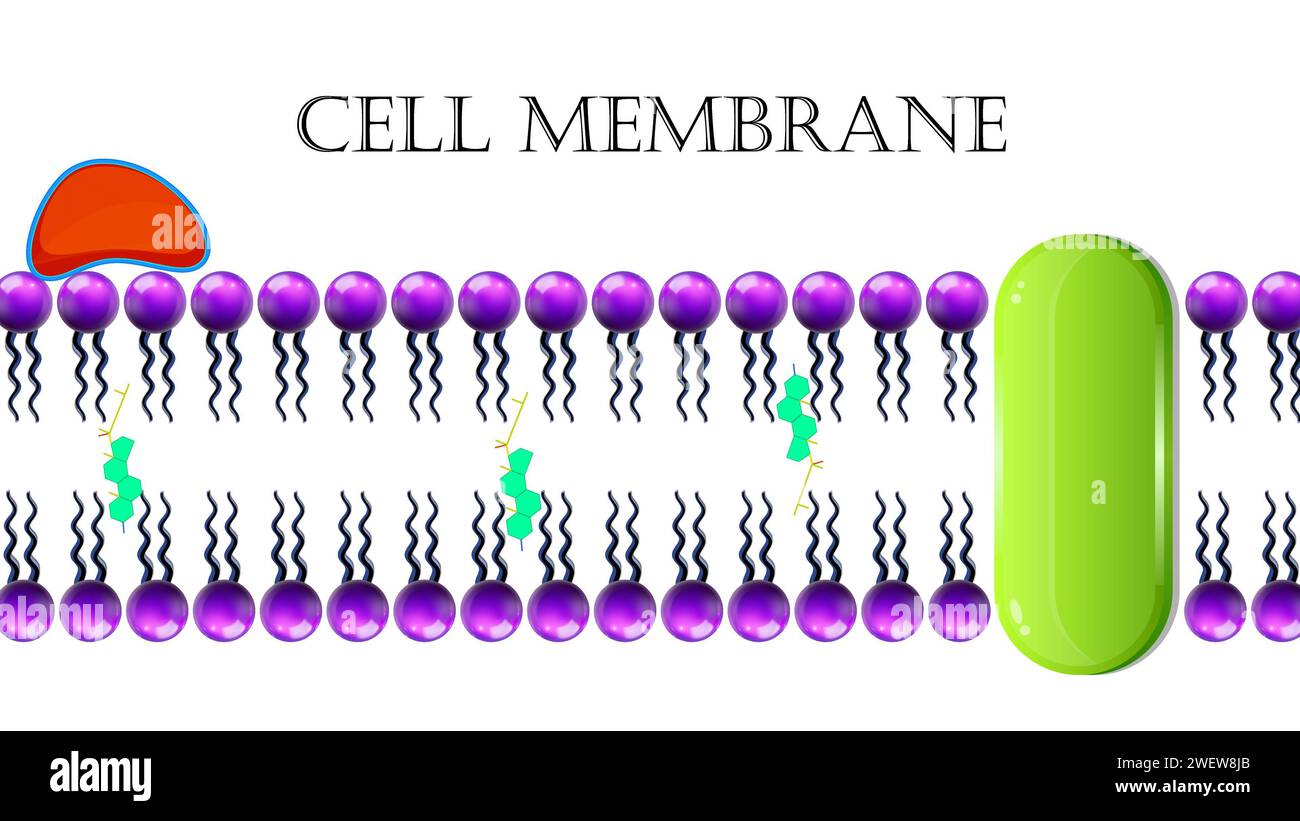 Cell Membrane Or Plasma Membrane Stock Photohttps://www.alamy.com/image-license-details/?v=1https://www.alamy.com/cell-membrane-or-plasma-membrane-image594313283.html
RF2WEW8JB–Cell Membrane Or Plasma Membrane
 Structure of HIV Stock Photohttps://www.alamy.com/image-license-details/?v=1https://www.alamy.com/structure-of-hiv-image416779862.html
RM2F61XY2–Structure of HIV
 Wild horse Stock Photohttps://www.alamy.com/image-license-details/?v=1https://www.alamy.com/stock-photo-wild-horse-48775723.html
RMCR9WX3–Wild horse
 Golgi Apparatus Stock Photohttps://www.alamy.com/image-license-details/?v=1https://www.alamy.com/golgi-apparatus-image353175240.html
RM2BEGEHC–Golgi Apparatus
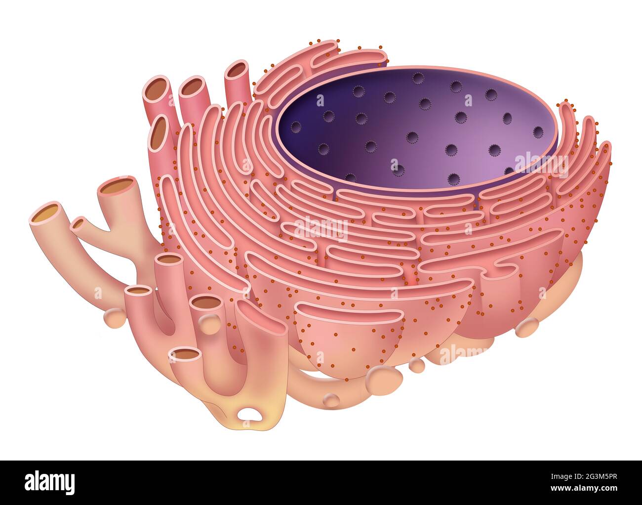 The endoplasmic reticulum is organelle found in eukaryotic cells Stock Photohttps://www.alamy.com/image-license-details/?v=1https://www.alamy.com/the-endoplasmic-reticulum-is-organelle-found-in-eukaryotic-cells-image432546767.html
RF2G3M5PR–The endoplasmic reticulum is organelle found in eukaryotic cells
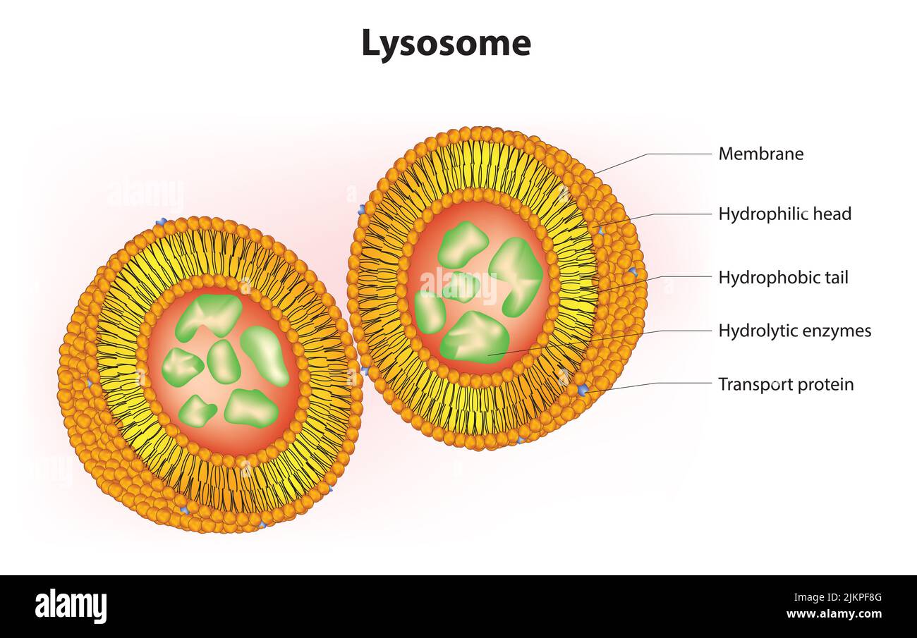 3D Anatomy of lysosome Stock Photohttps://www.alamy.com/image-license-details/?v=1https://www.alamy.com/3d-anatomy-of-lysosome-image476853344.html
RF2JKPF8G–3D Anatomy of lysosome
 A Cross section of an axoneme .Vector illustration. Stock Vectorhttps://www.alamy.com/image-license-details/?v=1https://www.alamy.com/a-cross-section-of-an-axoneme-vector-illustration-image576063283.html
RF2TD5XGK–A Cross section of an axoneme .Vector illustration.
 Detailed Vector Illustration of Types of Membrane Transport: Uniport, Symport, and Antiport for Cell Biology and Biochemistry on White Background. Stock Vectorhttps://www.alamy.com/image-license-details/?v=1https://www.alamy.com/detailed-vector-illustration-of-types-of-membrane-transport-uniport-symport-and-antiport-for-cell-biology-and-biochemistry-on-white-background-image608622995.html
RF2XA54RF–Detailed Vector Illustration of Types of Membrane Transport: Uniport, Symport, and Antiport for Cell Biology and Biochemistry on White Background.
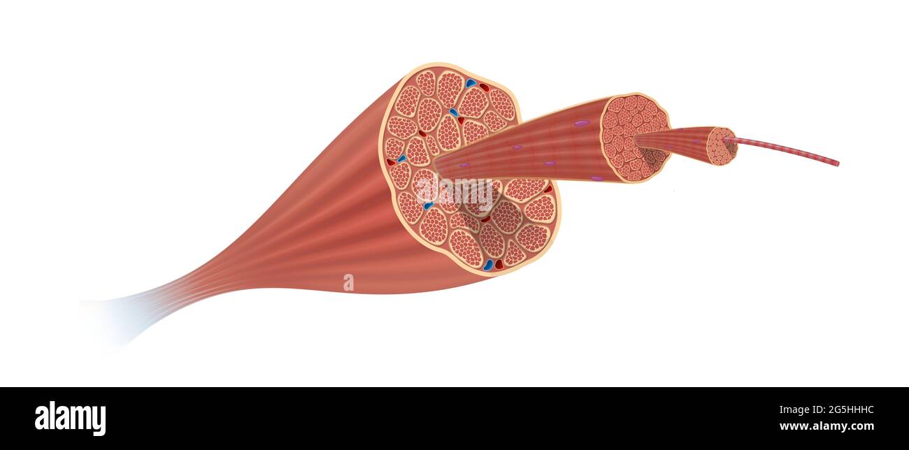 Structure Skeletal Muscle Anatomy Stock Photohttps://www.alamy.com/image-license-details/?v=1https://www.alamy.com/structure-skeletal-muscle-anatomy-image433719480.html
RF2G5HHHC–Structure Skeletal Muscle Anatomy
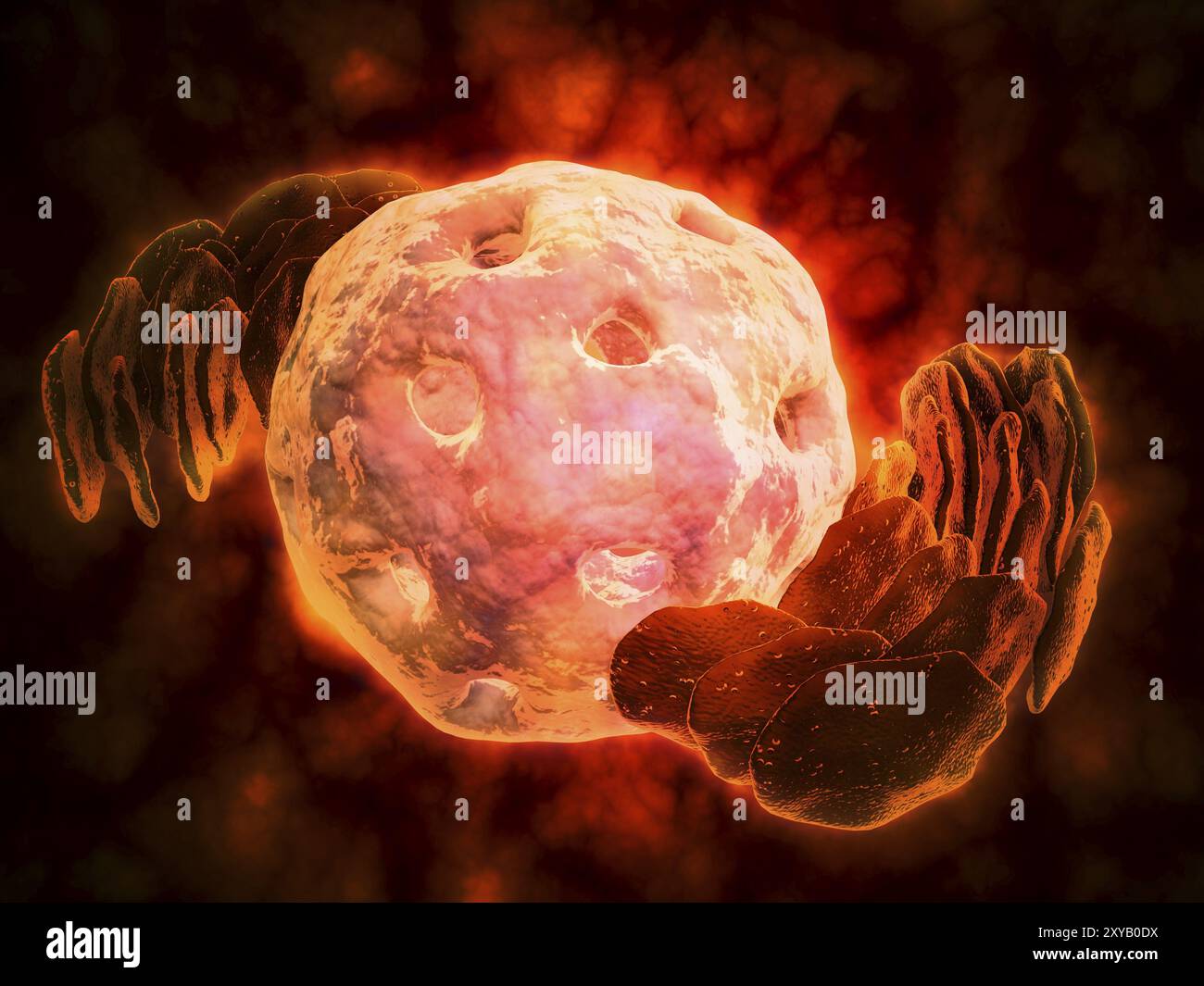 Conceptual image of endoplasmic reticulum around a cell nucleus. Endoplasmic reticulum is an organelle that forms a continuous membrane system of flat Stock Photohttps://www.alamy.com/image-license-details/?v=1https://www.alamy.com/conceptual-image-of-endoplasmic-reticulum-around-a-cell-nucleus-endoplasmic-reticulum-is-an-organelle-that-forms-a-continuous-membrane-system-of-flat-image619200454.html
RM2XYB0DX–Conceptual image of endoplasmic reticulum around a cell nucleus. Endoplasmic reticulum is an organelle that forms a continuous membrane system of flat
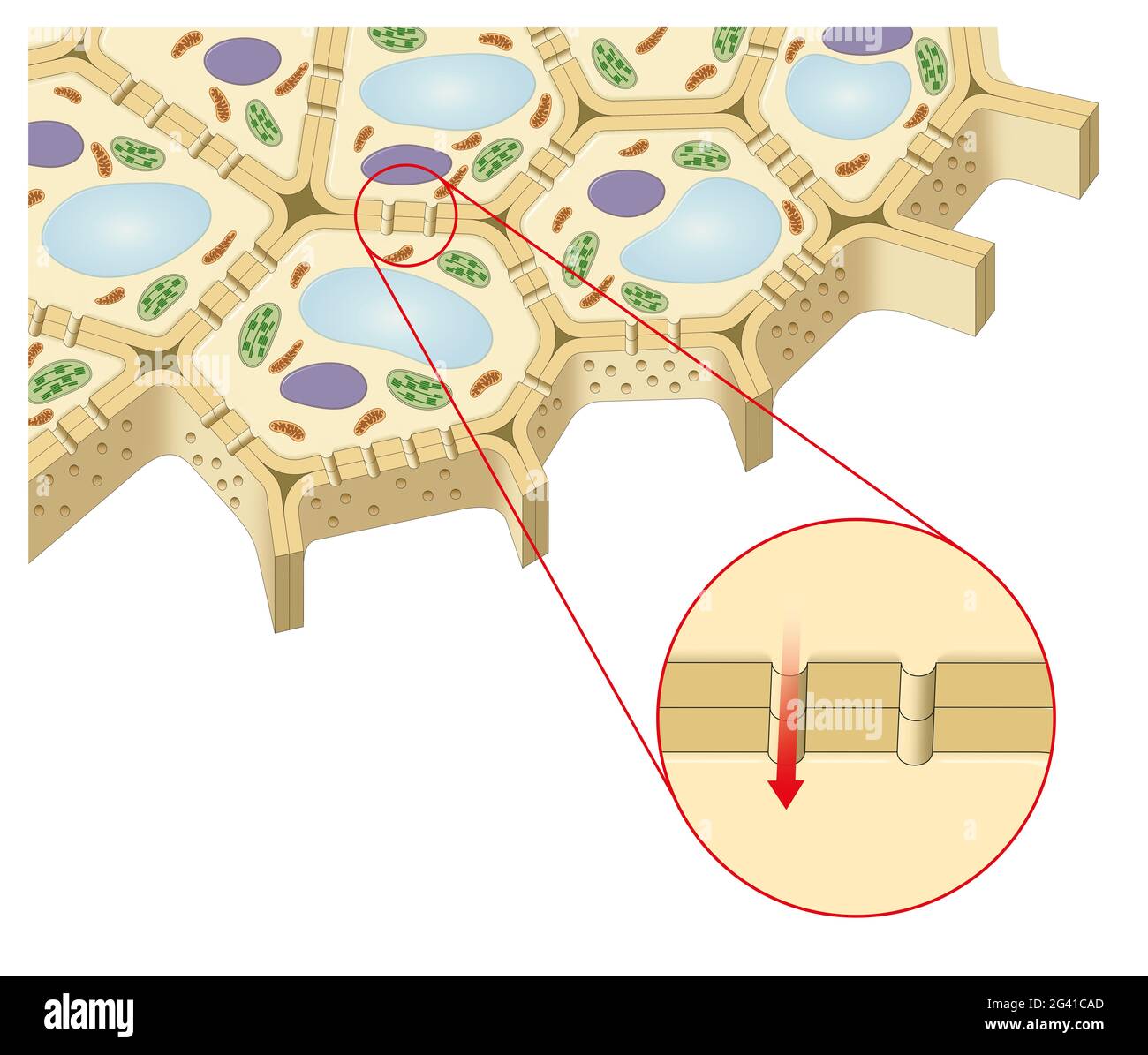 Plant cell. Plasmodesmata Stock Photohttps://www.alamy.com/image-license-details/?v=1https://www.alamy.com/plant-cell-plasmodesmata-image432749477.html
RF2G41CAD–Plant cell. Plasmodesmata
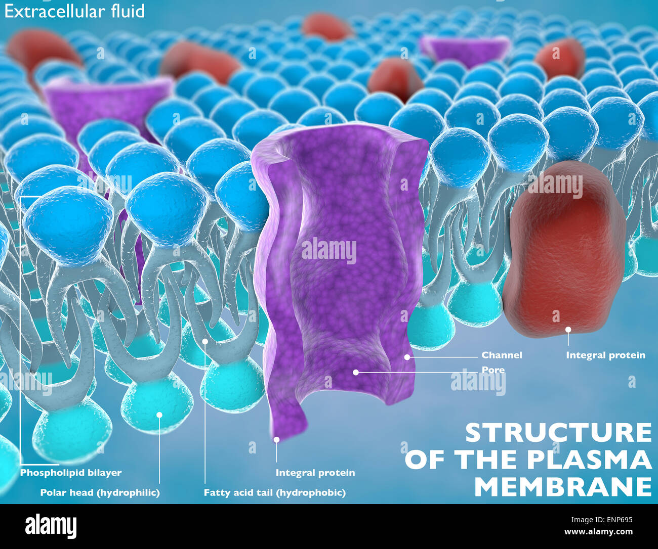 Structure of the plasma membrane of a cell Stock Photohttps://www.alamy.com/image-license-details/?v=1https://www.alamy.com/stock-photo-structure-of-the-plasma-membrane-of-a-cell-82237153.html
RMENP695–Structure of the plasma membrane of a cell
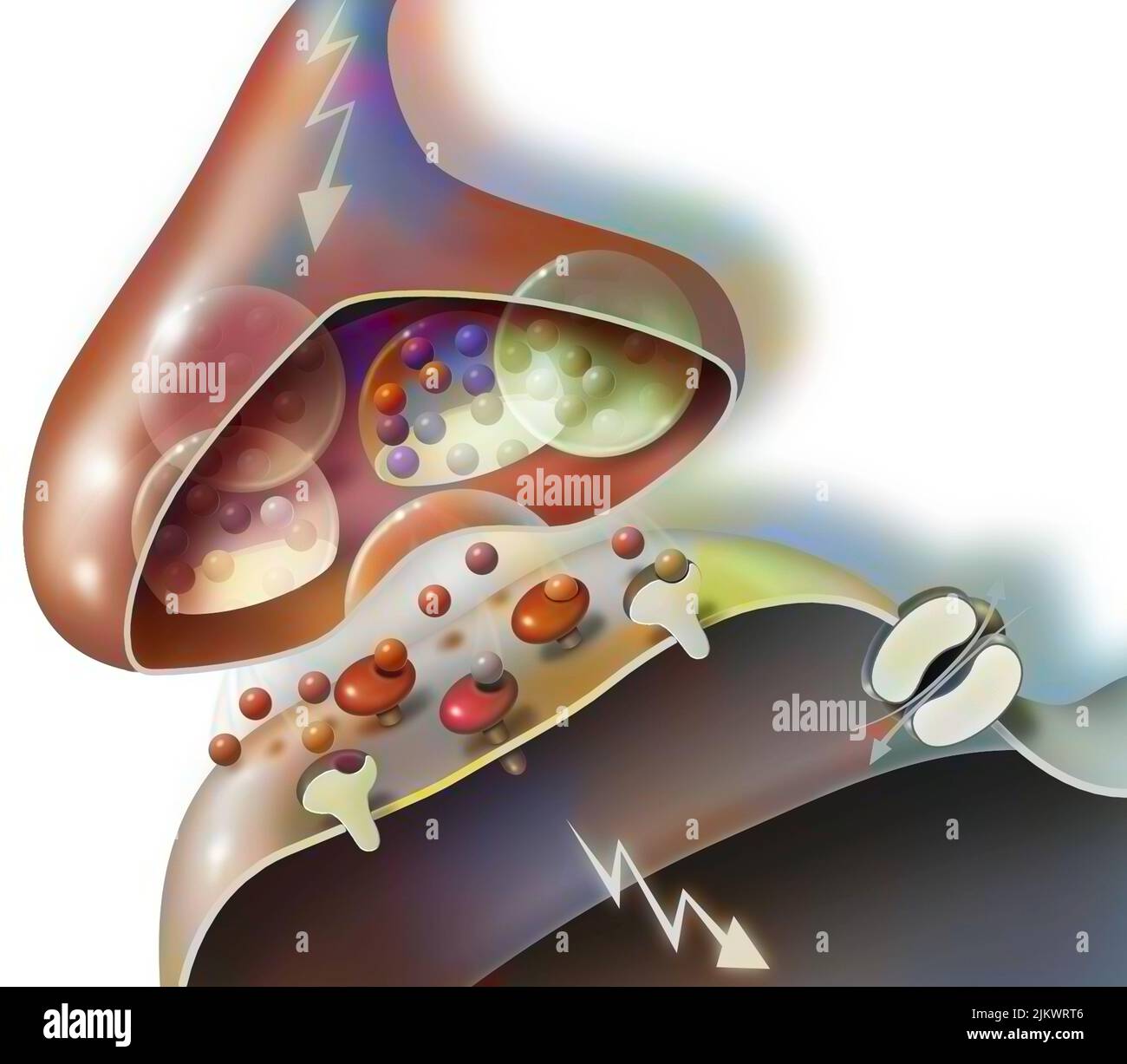 Transmission of nerve impulses from a synapse of a neuron A to a dendritic button. Stock Photohttps://www.alamy.com/image-license-details/?v=1https://www.alamy.com/transmission-of-nerve-impulses-from-a-synapse-of-a-neuron-a-to-a-dendritic-button-image476925910.html
RF2JKWRT6–Transmission of nerve impulses from a synapse of a neuron A to a dendritic button.
 3d rendering of nanodisc consists of Phospholipids and A stabilizing belt that holds the phospholipids together. Stock Photohttps://www.alamy.com/image-license-details/?v=1https://www.alamy.com/3d-rendering-of-nanodisc-consists-of-phospholipids-and-a-stabilizing-belt-that-holds-the-phospholipids-together-image591330904.html
RF2WA1CGT–3d rendering of nanodisc consists of Phospholipids and A stabilizing belt that holds the phospholipids together.
 Broken egg shell with on white background, isolated, with bursts and cracks Stock Photohttps://www.alamy.com/image-license-details/?v=1https://www.alamy.com/broken-egg-shell-with-on-white-background-isolated-with-bursts-and-cracks-image207329599.html
RFP18K0F–Broken egg shell with on white background, isolated, with bursts and cracks
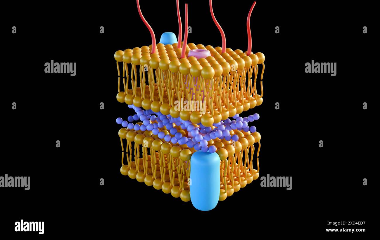 3d rendering of Gram-negative bacteria, have a thin peptidoglycan cell wall Stock Photohttps://www.alamy.com/image-license-details/?v=1https://www.alamy.com/3d-rendering-of-gram-negative-bacteria-have-a-thin-peptidoglycan-cell-wall-image610452563.html
RF2XD4ED7–3d rendering of Gram-negative bacteria, have a thin peptidoglycan cell wall
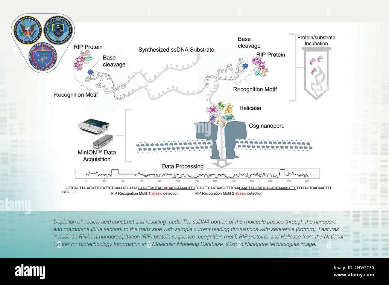 Depiction of nucleic acid (DNA) construct and resulting reads. The ssDNA portion of the molecule passes through the nanopore and membrane (blue section) to the trans side with sample current reading fluctuations with sequence (bottom). Features include an RNA immunoprecipitation (RIP) protein sequence recognition motif, RIP proteins, and Helicase from the National Center for Biotechnology Information and Molecular Modeling Database. (Oxford Nanopore Technologies image) Stock Photohttps://www.alamy.com/image-license-details/?v=1https://www.alamy.com/depiction-of-nucleic-acid-dna-construct-and-resulting-reads-the-ssdna-portion-of-the-molecule-passes-through-the-nanopore-and-membrane-blue-section-to-the-trans-side-with-sample-current-reading-fluctuations-with-sequence-bottom-features-include-an-rna-immunoprecipitation-rip-protein-sequence-recognition-motif-rip-proteins-and-helicase-from-the-national-center-for-biotechnology-information-and-molecular-modeling-database-oxford-nanopore-technologies-image-image531797000.html
RM2NW5CE0–Depiction of nucleic acid (DNA) construct and resulting reads. The ssDNA portion of the molecule passes through the nanopore and membrane (blue section) to the trans side with sample current reading fluctuations with sequence (bottom). Features include an RNA immunoprecipitation (RIP) protein sequence recognition motif, RIP proteins, and Helicase from the National Center for Biotechnology Information and Molecular Modeling Database. (Oxford Nanopore Technologies image)
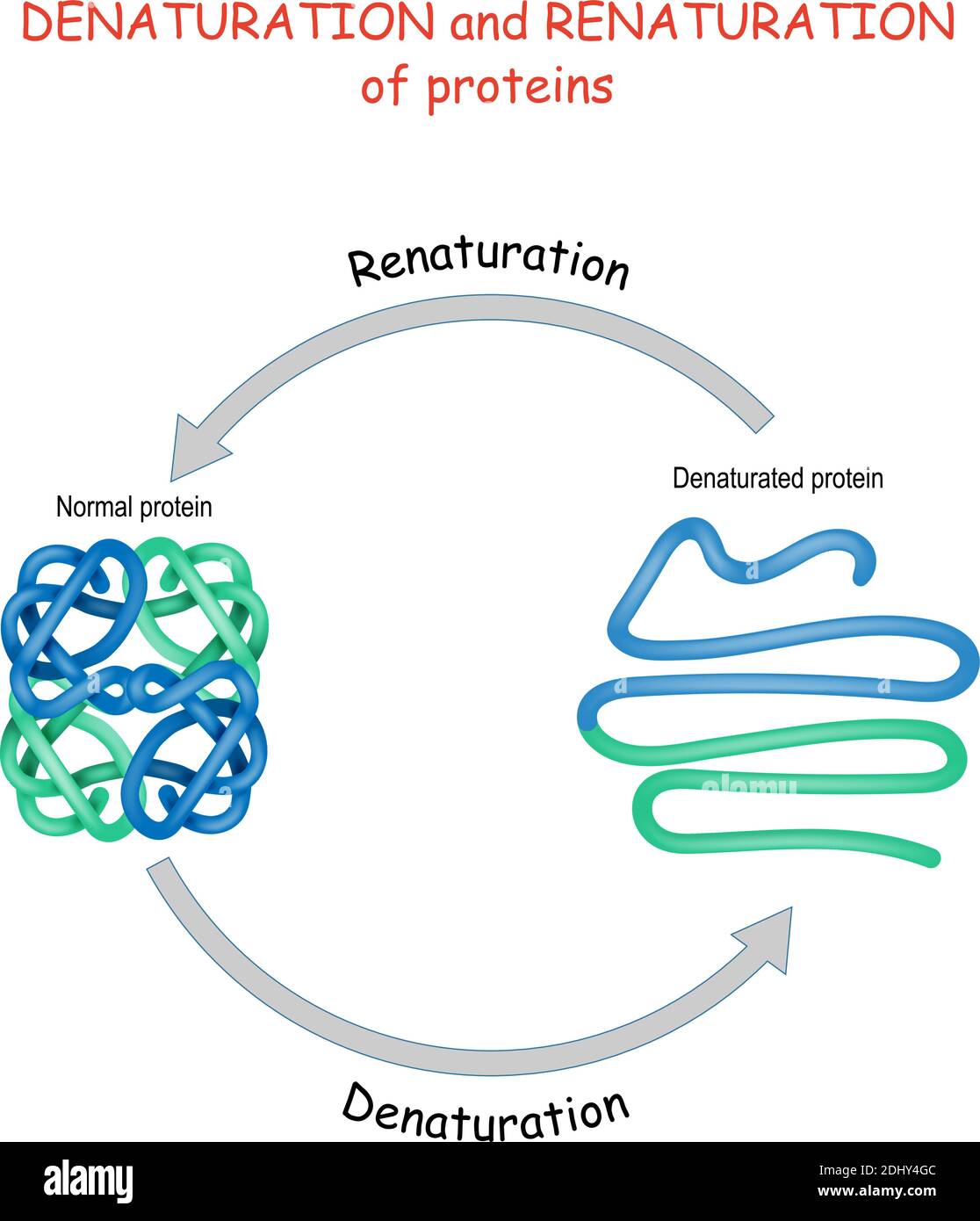 Process of Denaturation and renaturation of proteins. Vector diagram for science, education, and medical use. Stock Vectorhttps://www.alamy.com/image-license-details/?v=1https://www.alamy.com/process-of-denaturation-and-renaturation-of-proteins-vector-diagram-for-science-education-and-medical-use-image389673548.html
RF2DHY4GC–Process of Denaturation and renaturation of proteins. Vector diagram for science, education, and medical use.
 Covid-19 coronavirus particles, illustration. The SARS-CoV-2 coronavirus was first identified in Wuhan, China, in December 2019. It is an enveloped RNA (ribonucleic acid) virus. Within the membrane are spike proteins (large protrusions) as well as membrane proteins and envelope proteins. SARS-CoV-2 causes the respiratory infection Covid-19, which can lead to fatal pneumonia. As of March 2020, the virus has spread to many countries worldwide and has been declared a pandemic. Hundreds of thousands have been infected with tens of thousands of deaths. Stock Photohttps://www.alamy.com/image-license-details/?v=1https://www.alamy.com/covid-19-coronavirus-particles-illustration-the-sars-cov-2-coronavirus-was-first-identified-in-wuhan-china-in-december-2019-it-is-an-enveloped-rna-ribonucleic-acid-virus-within-the-membrane-are-spike-proteins-large-protrusions-as-well-as-membrane-proteins-and-envelope-proteins-sars-cov-2-causes-the-respiratory-infection-covid-19-which-can-lead-to-fatal-pneumonia-as-of-march-2020-the-virus-has-spread-to-many-countries-worldwide-and-has-been-declared-a-pandemic-hundreds-of-thousands-have-been-infected-with-tens-of-thousands-of-deaths-image352901286.html
RF2BE415A–Covid-19 coronavirus particles, illustration. The SARS-CoV-2 coronavirus was first identified in Wuhan, China, in December 2019. It is an enveloped RNA (ribonucleic acid) virus. Within the membrane are spike proteins (large protrusions) as well as membrane proteins and envelope proteins. SARS-CoV-2 causes the respiratory infection Covid-19, which can lead to fatal pneumonia. As of March 2020, the virus has spread to many countries worldwide and has been declared a pandemic. Hundreds of thousands have been infected with tens of thousands of deaths.
 RBC Membrane Proteins SDS-PAGE gel Stock Photohttps://www.alamy.com/image-license-details/?v=1https://www.alamy.com/stock-image-rbc-membrane-proteins-sds-page-gel-165944397.html
RMKHYBNH–RBC Membrane Proteins SDS-PAGE gel
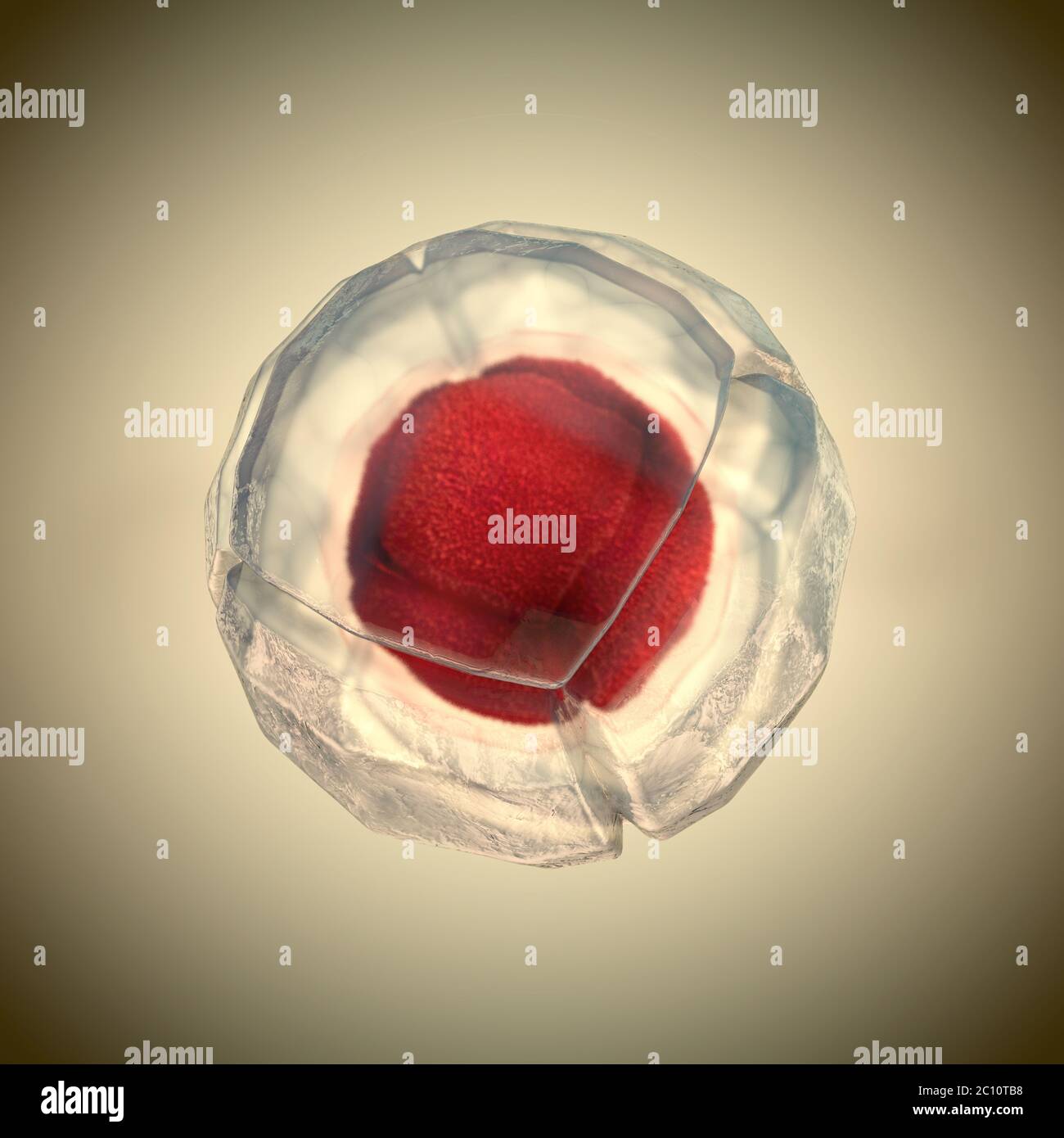 3d illustration of cell division, cell membrane and a splitting red nucleus Stock Photohttps://www.alamy.com/image-license-details/?v=1https://www.alamy.com/3d-illustration-of-cell-division-cell-membrane-and-a-splitting-red-nucleus-image362051516.html
RF2C10TB8–3d illustration of cell division, cell membrane and a splitting red nucleus
 Chimeric antigen receptor T cell and Antibody molecule. IgE and CAR. Artificial T cell receptors are proteins that have been engineered for cancer Stock Vectorhttps://www.alamy.com/image-license-details/?v=1https://www.alamy.com/chimeric-antigen-receptor-t-cell-and-antibody-molecule-ige-and-car-artificial-t-cell-receptors-are-proteins-that-have-been-engineered-for-cancer-image389332951.html
RF2DHBJ47–Chimeric antigen receptor T cell and Antibody molecule. IgE and CAR. Artificial T cell receptors are proteins that have been engineered for cancer
 Golgi Apparatus Stock Photohttps://www.alamy.com/image-license-details/?v=1https://www.alamy.com/golgi-apparatus-image353175279.html
RM2BEGEJR–Golgi Apparatus
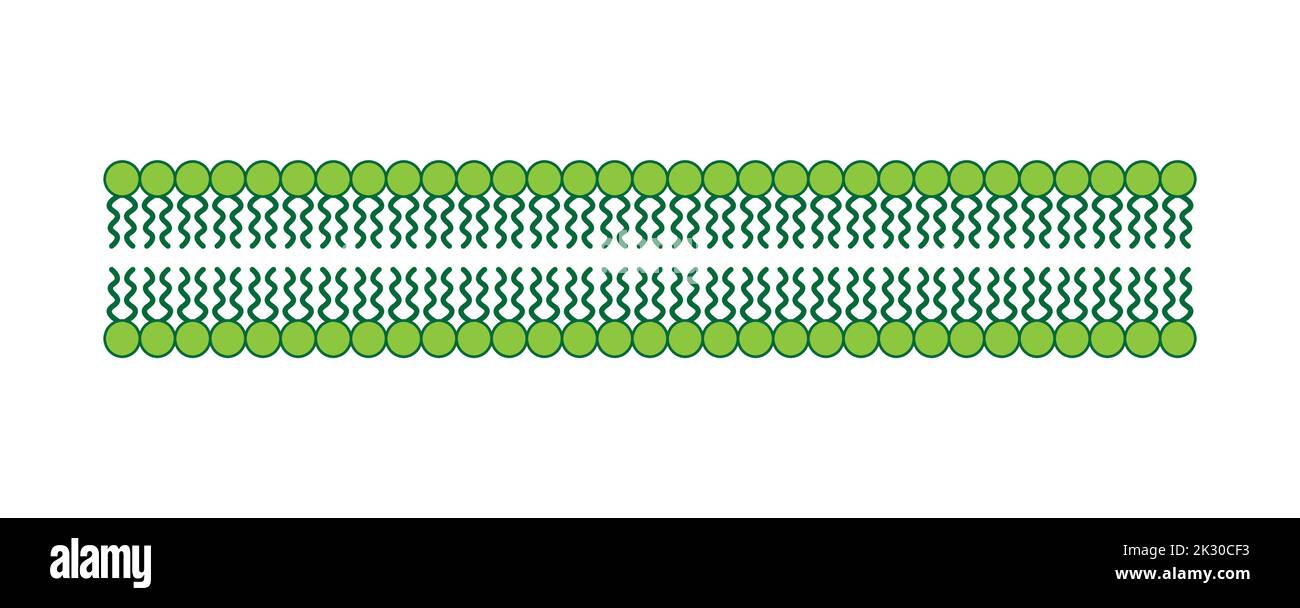 Scientific Designing of Phospholipid Bilayer Structure. The Cell Membrane Structure. Vector Illustration. Stock Vectorhttps://www.alamy.com/image-license-details/?v=1https://www.alamy.com/scientific-designing-of-phospholipid-bilayer-structure-the-cell-membrane-structure-vector-illustration-image483744103.html
RF2K30CF3–Scientific Designing of Phospholipid Bilayer Structure. The Cell Membrane Structure. Vector Illustration.
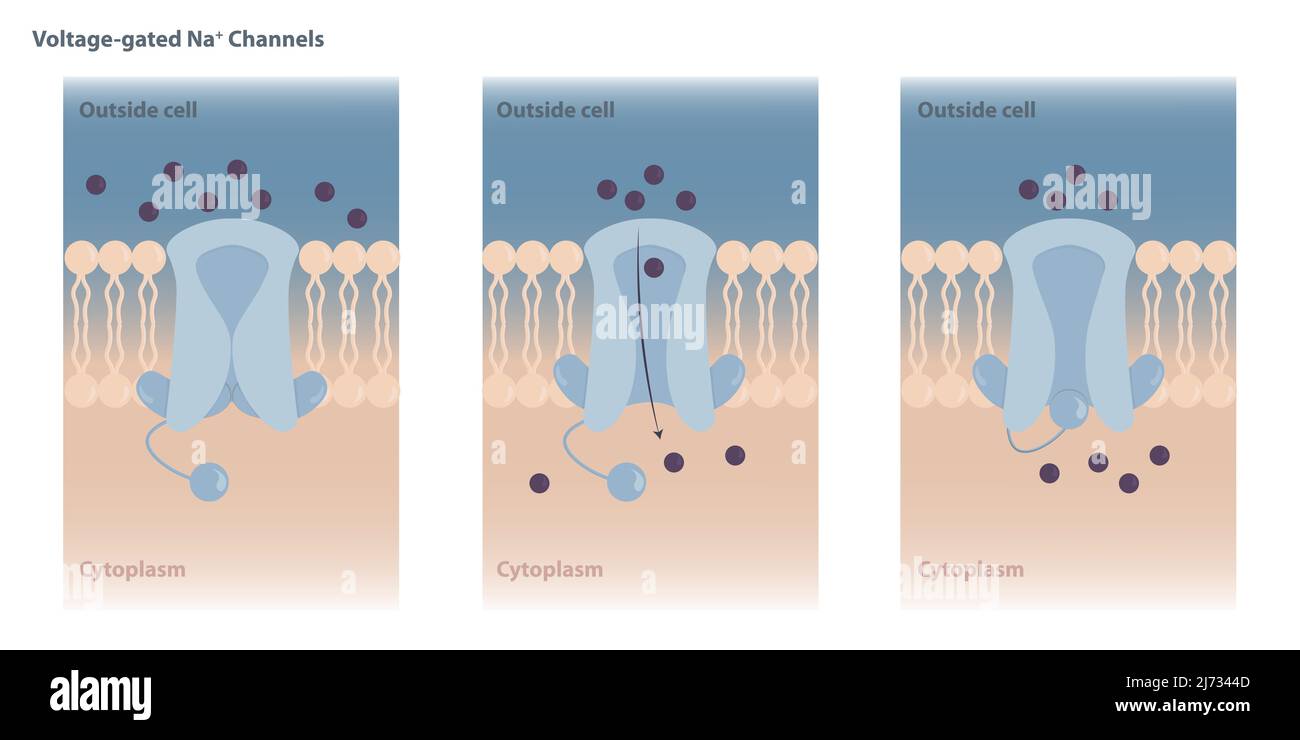 Neuronal charged membranes. Voltage-gated ion channels are closed at the resting potential and open in response to changes in membrane voltage. Stock Vectorhttps://www.alamy.com/image-license-details/?v=1https://www.alamy.com/neuronal-charged-membranes-voltage-gated-ion-channels-are-closed-at-the-resting-potential-and-open-in-response-to-changes-in-membrane-voltage-image469051645.html
RF2J7344D–Neuronal charged membranes. Voltage-gated ion channels are closed at the resting potential and open in response to changes in membrane voltage.
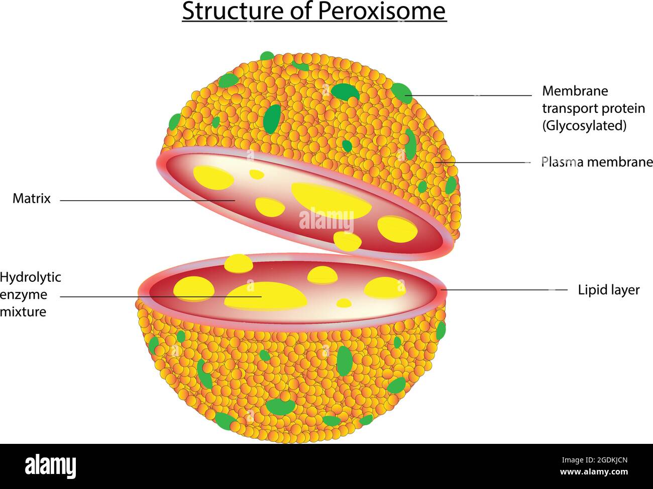 Biological illustration of peroxisome, peroxysome. Peroxisomes are globular organelles, membrane proteins that are critical for various functions Stock Vectorhttps://www.alamy.com/image-license-details/?v=1https://www.alamy.com/biological-illustration-of-peroxisome-peroxysome-peroxisomes-are-globular-organelles-membrane-proteins-that-are-critical-for-various-functions-image438681285.html
RF2GDKJCN–Biological illustration of peroxisome, peroxysome. Peroxisomes are globular organelles, membrane proteins that are critical for various functions
 Injection on a virus. Injecting an red glowing antidote vaccine on a virus. Medical concept of pandemic immunization. 3D rendering. Stock Photohttps://www.alamy.com/image-license-details/?v=1https://www.alamy.com/injection-on-a-virus-injecting-an-red-glowing-antidote-vaccine-on-a-virus-medical-concept-of-pandemic-immunization-3d-rendering-image383854964.html
RF2D8E2WT–Injection on a virus. Injecting an red glowing antidote vaccine on a virus. Medical concept of pandemic immunization. 3D rendering.
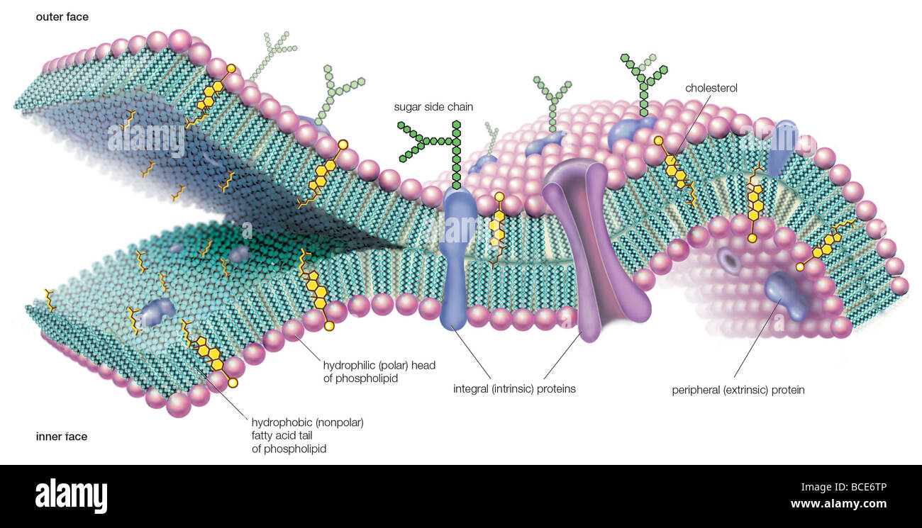 A molecular view of the cell membrane highlighting phospholipids, cholesterol, and intrinsic and extrinsic proteins. Stock Photohttps://www.alamy.com/image-license-details/?v=1https://www.alamy.com/stock-photo-a-molecular-view-of-the-cell-membrane-highlighting-phospholipids-cholesterol-24898966.html
RMBCE6TP–A molecular view of the cell membrane highlighting phospholipids, cholesterol, and intrinsic and extrinsic proteins.
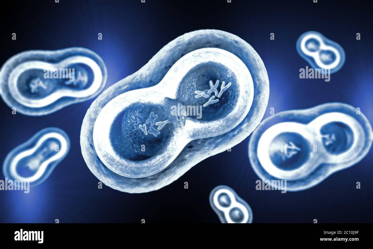 Transparent cells with splitting nucleus, cell membrane and visible chromosomes Stock Photohttps://www.alamy.com/image-license-details/?v=1https://www.alamy.com/transparent-cells-with-splitting-nucleus-cell-membrane-and-visible-chromosomes-image362046763.html
RF2C10J9F–Transparent cells with splitting nucleus, cell membrane and visible chromosomes
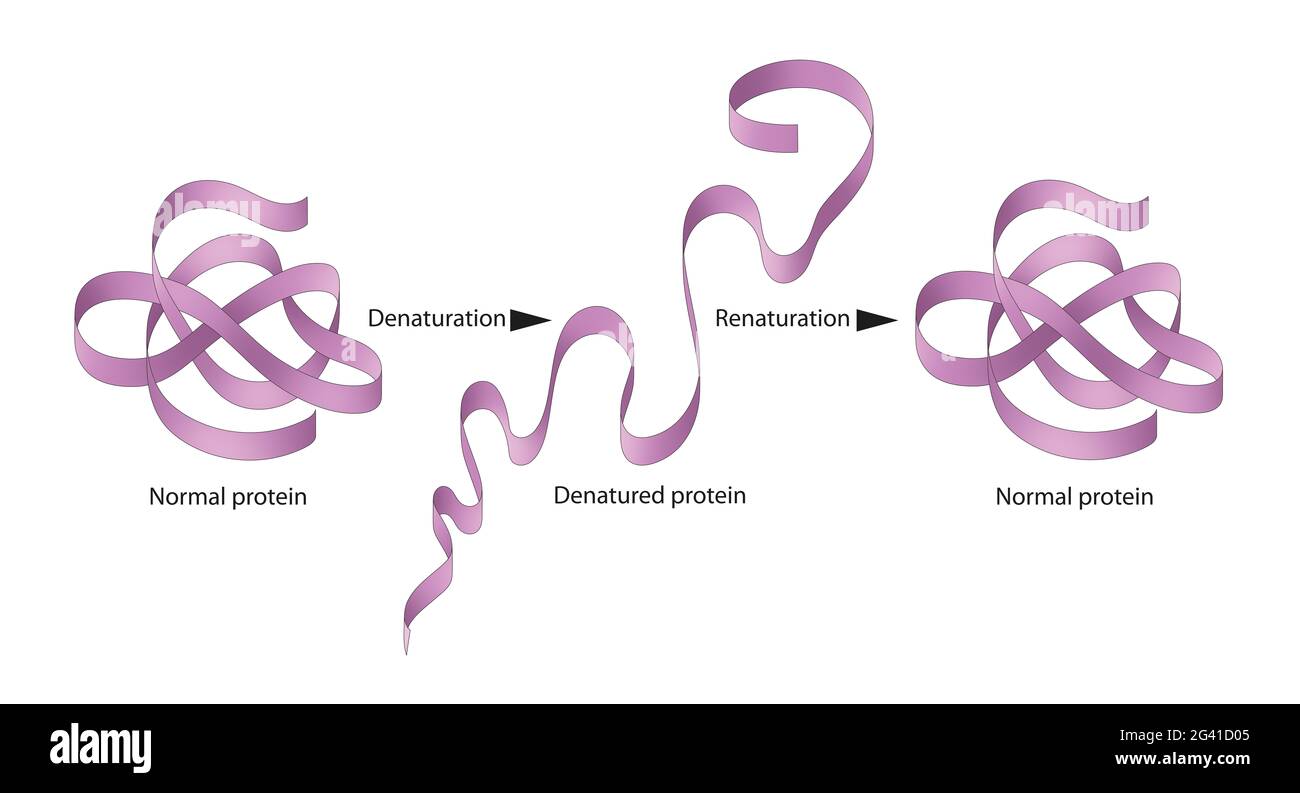 Denaturation and renaturation of Proteins Stock Photohttps://www.alamy.com/image-license-details/?v=1https://www.alamy.com/denaturation-and-renaturation-of-proteins-image432749973.html
RF2G41D05–Denaturation and renaturation of Proteins
 A Western blot, genetic test, screen for genes or proteins Stock Photohttps://www.alamy.com/image-license-details/?v=1https://www.alamy.com/a-western-blot-genetic-test-screen-for-genes-or-proteins-image458417795.html
RM2HHPMG3–A Western blot, genetic test, screen for genes or proteins
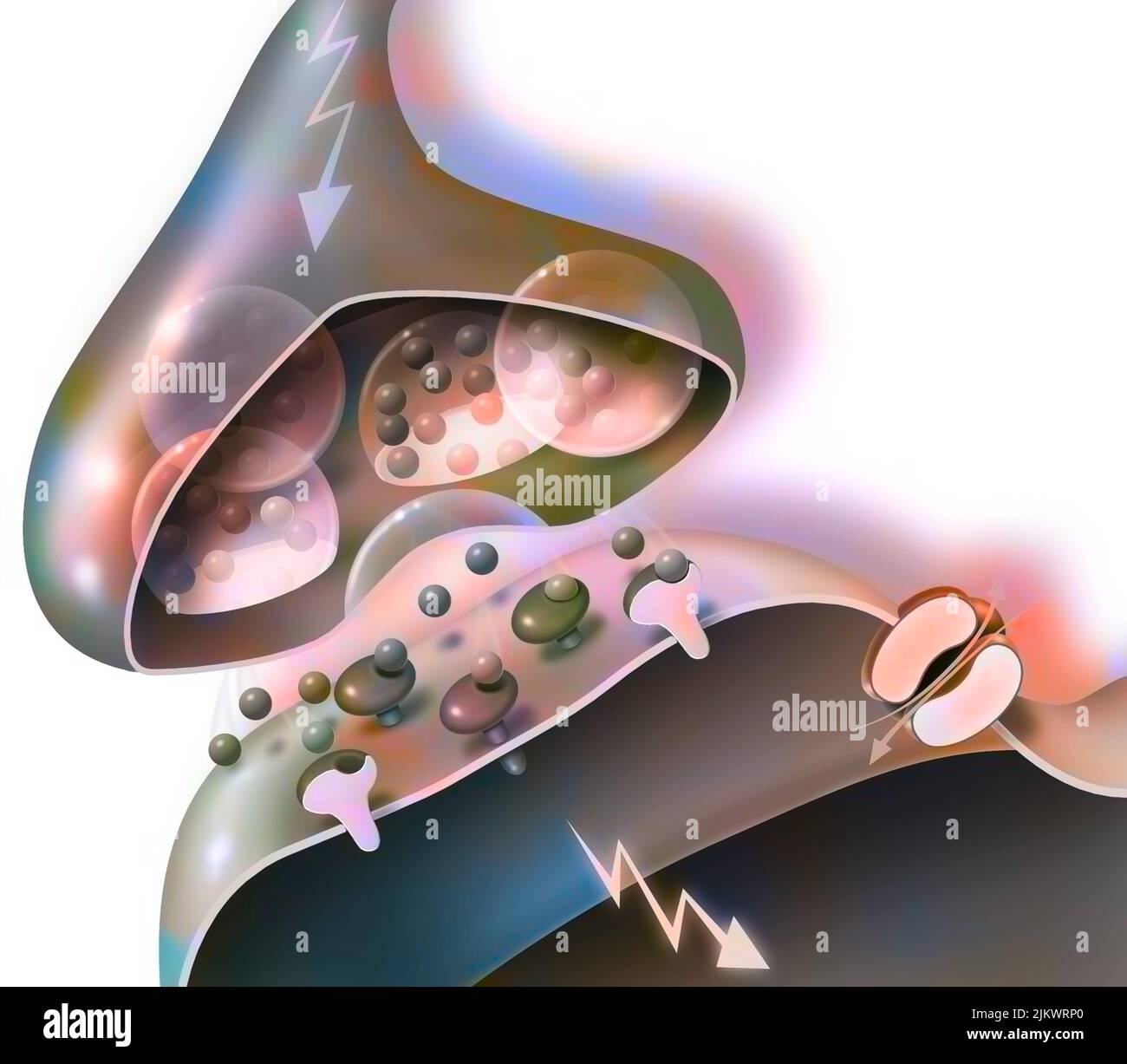 Transmission of nerve impulses from a synapse of a neuron A to a dendritic button. Stock Photohttps://www.alamy.com/image-license-details/?v=1https://www.alamy.com/transmission-of-nerve-impulses-from-a-synapse-of-a-neuron-a-to-a-dendritic-button-image476925848.html
RF2JKWRP0–Transmission of nerve impulses from a synapse of a neuron A to a dendritic button.
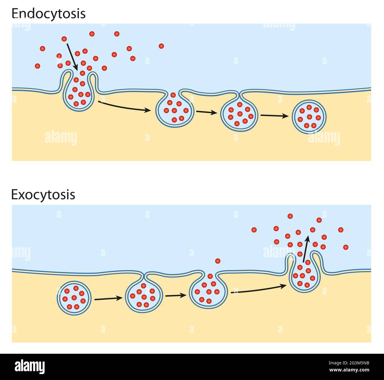 Endocytosis, exocytosis. The cell transports proteins into the cell Stock Photohttps://www.alamy.com/image-license-details/?v=1https://www.alamy.com/endocytosis-exocytosis-the-cell-transports-proteins-into-the-cell-image432546727.html
RF2G3M5NB–Endocytosis, exocytosis. The cell transports proteins into the cell
 Raw bread dough in the makng Stock Photohttps://www.alamy.com/image-license-details/?v=1https://www.alamy.com/raw-bread-dough-in-the-makng-image419232851.html
RF2FA1KNR–Raw bread dough in the makng
 3d rendering of Gram-negative bacteria, have a thin peptidoglycan cell wall Stock Photohttps://www.alamy.com/image-license-details/?v=1https://www.alamy.com/3d-rendering-of-gram-negative-bacteria-have-a-thin-peptidoglycan-cell-wall-image610452612.html
RF2XD4EF0–3d rendering of Gram-negative bacteria, have a thin peptidoglycan cell wall
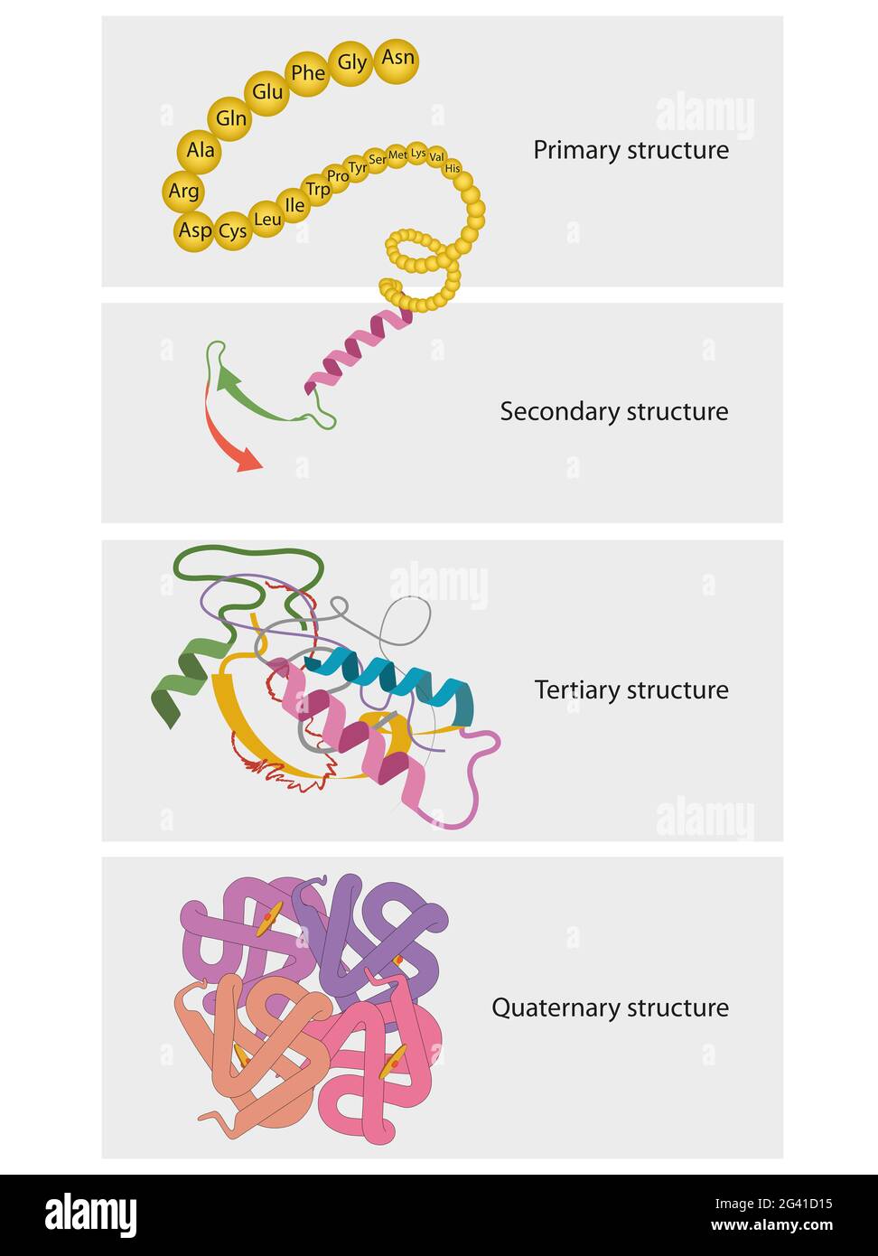 Types of Protein Structure. Proteins are biological polymers composed of amino acids Stock Photohttps://www.alamy.com/image-license-details/?v=1https://www.alamy.com/types-of-protein-structure-proteins-are-biological-polymers-composed-of-amino-acids-image432750001.html
RF2G41D15–Types of Protein Structure. Proteins are biological polymers composed of amino acids
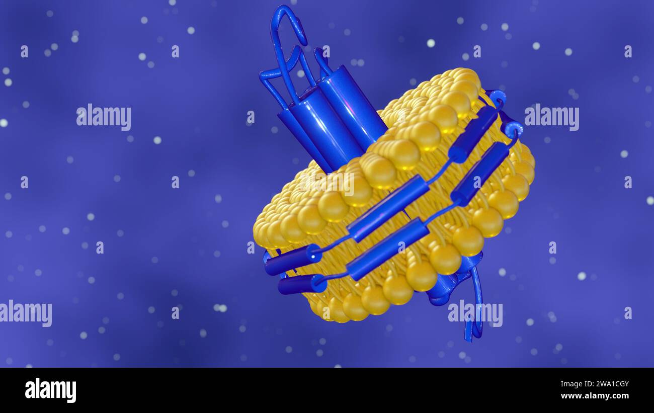 3d rendering of nanodisc consists of Phospholipids and A stabilizing belt that holds the phospholipids together. Stock Photohttps://www.alamy.com/image-license-details/?v=1https://www.alamy.com/3d-rendering-of-nanodisc-consists-of-phospholipids-and-a-stabilizing-belt-that-holds-the-phospholipids-together-image591330907.html
RF2WA1CGY–3d rendering of nanodisc consists of Phospholipids and A stabilizing belt that holds the phospholipids together.
 Covid-19 coronavirus particles, illustration. The SARS-CoV-2 coronavirus was first identified in Wuhan, China, in December 2019. It is an enveloped RNA (ribonucleic acid) virus. Within the membrane are spike proteins (large protrusions) as well as membrane proteins and envelope proteins. SARS-CoV-2 causes the respiratory infection Covid-19, which can lead to fatal pneumonia. As of March 2020, the virus has spread to many countries worldwide and has been declared a pandemic. Hundreds of thousands have been infected with tens of thousands of deaths. Stock Photohttps://www.alamy.com/image-license-details/?v=1https://www.alamy.com/covid-19-coronavirus-particles-illustration-the-sars-cov-2-coronavirus-was-first-identified-in-wuhan-china-in-december-2019-it-is-an-enveloped-rna-ribonucleic-acid-virus-within-the-membrane-are-spike-proteins-large-protrusions-as-well-as-membrane-proteins-and-envelope-proteins-sars-cov-2-causes-the-respiratory-infection-covid-19-which-can-lead-to-fatal-pneumonia-as-of-march-2020-the-virus-has-spread-to-many-countries-worldwide-and-has-been-declared-a-pandemic-hundreds-of-thousands-have-been-infected-with-tens-of-thousands-of-deaths-image352901293.html
RF2BE415H–Covid-19 coronavirus particles, illustration. The SARS-CoV-2 coronavirus was first identified in Wuhan, China, in December 2019. It is an enveloped RNA (ribonucleic acid) virus. Within the membrane are spike proteins (large protrusions) as well as membrane proteins and envelope proteins. SARS-CoV-2 causes the respiratory infection Covid-19, which can lead to fatal pneumonia. As of March 2020, the virus has spread to many countries worldwide and has been declared a pandemic. Hundreds of thousands have been infected with tens of thousands of deaths.
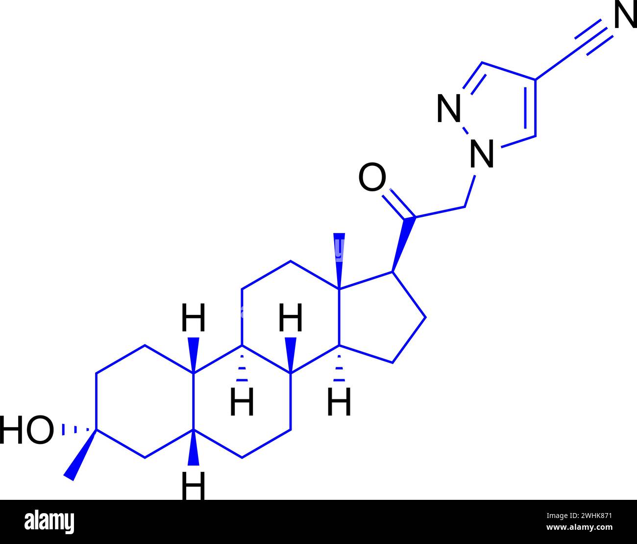 Structure of Adenosine monophosphate .Vector illustration. Stock Vectorhttps://www.alamy.com/image-license-details/?v=1https://www.alamy.com/structure-of-adenosine-monophosphate-vector-illustration-image596025221.html
RF2WHK871–Structure of Adenosine monophosphate .Vector illustration.
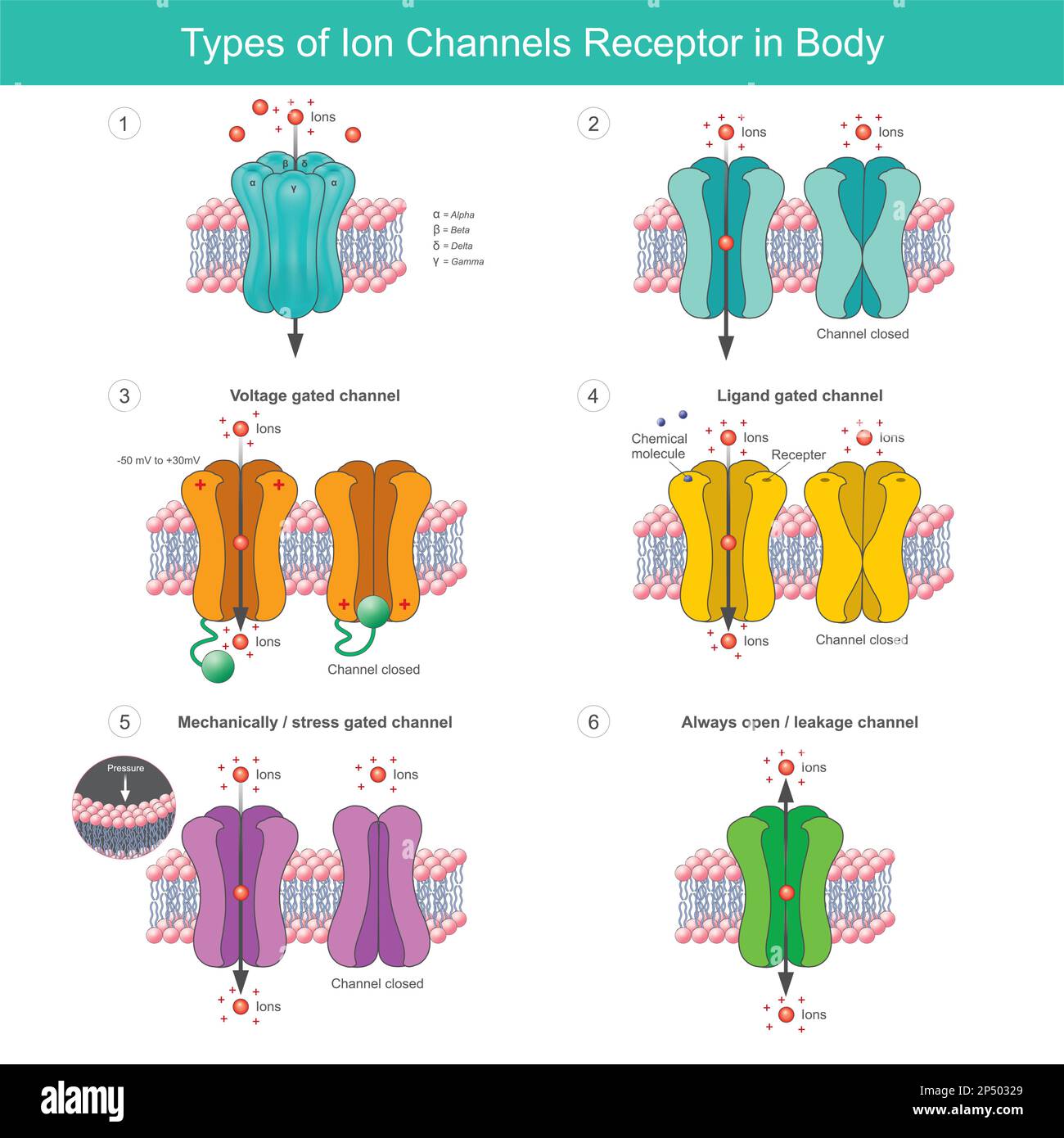 Types of Ion Channels Receptor In Body. Membrane proteins with transport of specific ions in or out of the cell of body. Stock Vectorhttps://www.alamy.com/image-license-details/?v=1https://www.alamy.com/types-of-ion-channels-receptor-in-body-membrane-proteins-with-transport-of-specific-ions-in-or-out-of-the-cell-of-body-image536597105.html
RF2P50329–Types of Ion Channels Receptor In Body. Membrane proteins with transport of specific ions in or out of the cell of body.
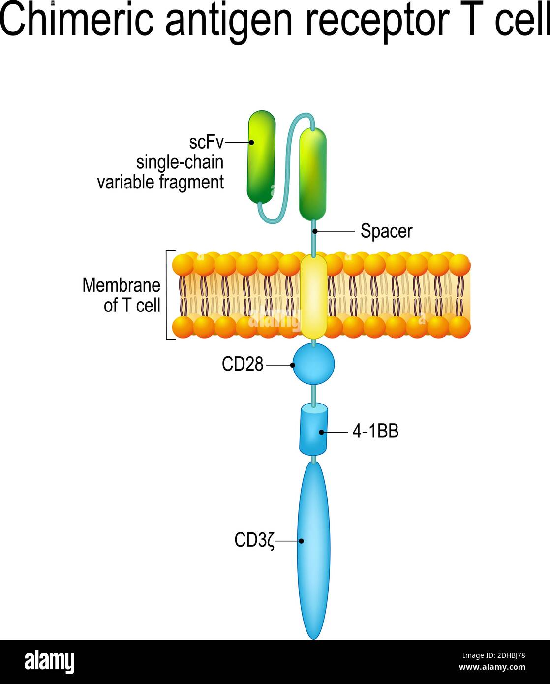 Chimeric antigen receptor T cell (CAR). Artificial T cell receptors are proteins that have been engineered for cancer therapy (killing of tumor cells) Stock Vectorhttps://www.alamy.com/image-license-details/?v=1https://www.alamy.com/chimeric-antigen-receptor-t-cell-car-artificial-t-cell-receptors-are-proteins-that-have-been-engineered-for-cancer-therapy-killing-of-tumor-cells-image389333036.html
RF2DHBJ78–Chimeric antigen receptor T cell (CAR). Artificial T cell receptors are proteins that have been engineered for cancer therapy (killing of tumor cells)