Quick filters:
Methyltransferase Stock Photos and Images
 NpmA methyltransferase Stock Photohttps://www.alamy.com/image-license-details/?v=1https://www.alamy.com/npma-methyltransferase-image65210419.html
RFDP2GFF–NpmA methyltransferase
 3D image of Aesculetin skeletal formula - molecular chemical structure of cichorigenin isolated on white background Stock Photohttps://www.alamy.com/image-license-details/?v=1https://www.alamy.com/3d-image-of-aesculetin-skeletal-formula-molecular-chemical-structure-of-cichorigenin-isolated-on-white-background-image500102269.html
RF2M1HHF9–3D image of Aesculetin skeletal formula - molecular chemical structure of cichorigenin isolated on white background
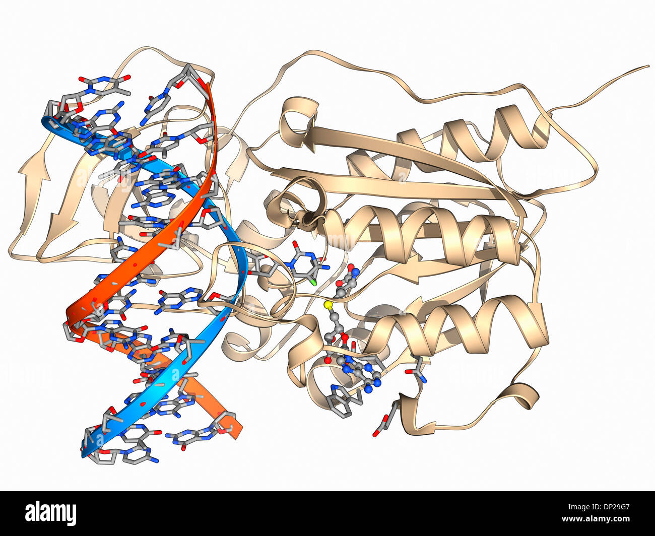 Methyltransferase and DNA Stock Photohttps://www.alamy.com/image-license-details/?v=1https://www.alamy.com/methyltransferase-and-dna-image65204951.html
RFDP29G7–Methyltransferase and DNA
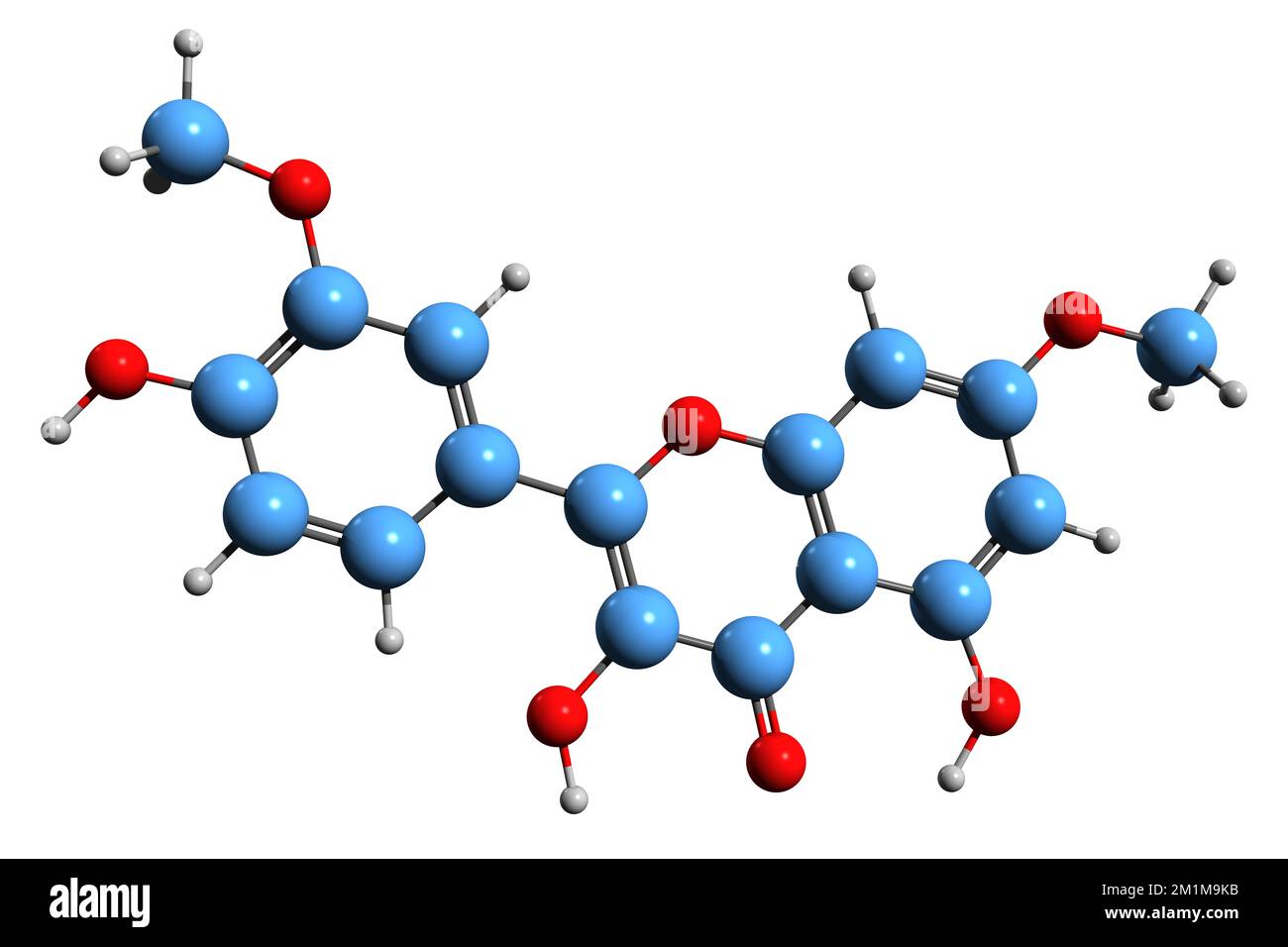 3D image of Rhamnazin skeletal formula - molecular chemical structure of O-methylated flavonol isolated on white background Stock Photohttps://www.alamy.com/image-license-details/?v=1https://www.alamy.com/3d-image-of-rhamnazin-skeletal-formula-molecular-chemical-structure-of-o-methylated-flavonol-isolated-on-white-background-image500161967.html
RF2M1M9KB–3D image of Rhamnazin skeletal formula - molecular chemical structure of O-methylated flavonol isolated on white background
 Methyltransferase complexed with DNA Stock Photohttps://www.alamy.com/image-license-details/?v=1https://www.alamy.com/methyltransferase-complexed-with-dna-image65210536.html
RFDP2GKM–Methyltransferase complexed with DNA
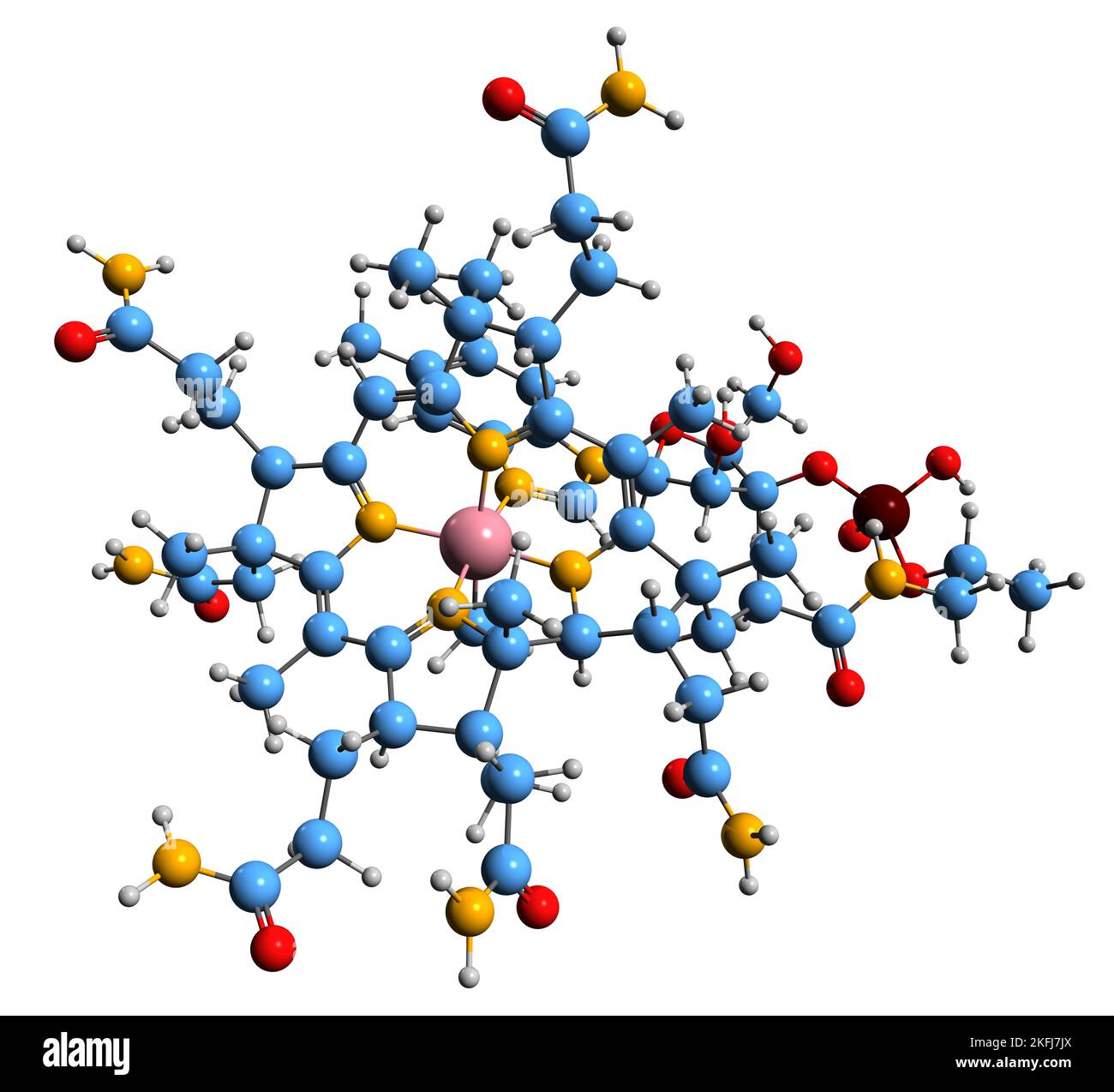 3D image of Methylcobalamin skeletal formula - molecular chemical structure of vitamin B12 isolated on white background Stock Photohttps://www.alamy.com/image-license-details/?v=1https://www.alamy.com/3d-image-of-methylcobalamin-skeletal-formula-molecular-chemical-structure-of-vitamin-b12-isolated-on-white-background-image491511298.html
RF2KFJ7JX–3D image of Methylcobalamin skeletal formula - molecular chemical structure of vitamin B12 isolated on white background
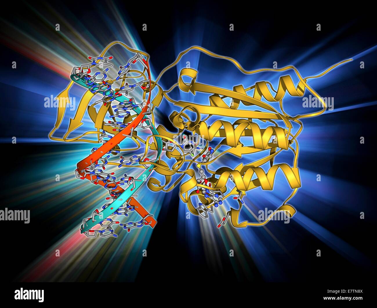 Methyltransferase and DNA. Molecular model of the enzyme HhaI methyltransferase (beige) complexed with a molecule of DNA (deoxyribonucleic acid, red and blue). Methyltransferases are enzymes that catalyse the transfer of methyl groups (CH3) to DNA nucleot Stock Photohttps://www.alamy.com/image-license-details/?v=1https://www.alamy.com/stock-photo-methyltransferase-and-dna-molecular-model-of-the-enzyme-hhai-methyltransferase-73687626.html
RFE7TN8X–Methyltransferase and DNA. Molecular model of the enzyme HhaI methyltransferase (beige) complexed with a molecule of DNA (deoxyribonucleic acid, red and blue). Methyltransferases are enzymes that catalyse the transfer of methyl groups (CH3) to DNA nucleot
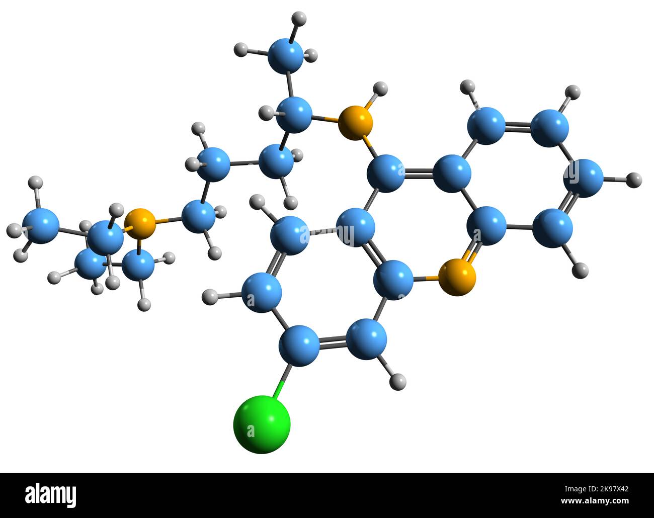 3D image of Mepacrine skeletal formula - molecular chemical structure of quinacrine isolated on white background Stock Photohttps://www.alamy.com/image-license-details/?v=1https://www.alamy.com/3d-image-of-mepacrine-skeletal-formula-molecular-chemical-structure-of-quinacrine-isolated-on-white-background-image487596370.html
RF2K97X42–3D image of Mepacrine skeletal formula - molecular chemical structure of quinacrine isolated on white background
 NpmA methyltransferase, molecular model. Methyltransferase enzymes act to add methyl groups to nucleic acids such as DNA (deoxyribonucleic acid), a process called DNA methylation. This can silence and regulate genes without changing the genetic sequence. Stock Photohttps://www.alamy.com/image-license-details/?v=1https://www.alamy.com/stock-photo-npma-methyltransferase-molecular-model-methyltransferase-enzymes-act-73688326.html
RFE7TP5X–NpmA methyltransferase, molecular model. Methyltransferase enzymes act to add methyl groups to nucleic acids such as DNA (deoxyribonucleic acid), a process called DNA methylation. This can silence and regulate genes without changing the genetic sequence.
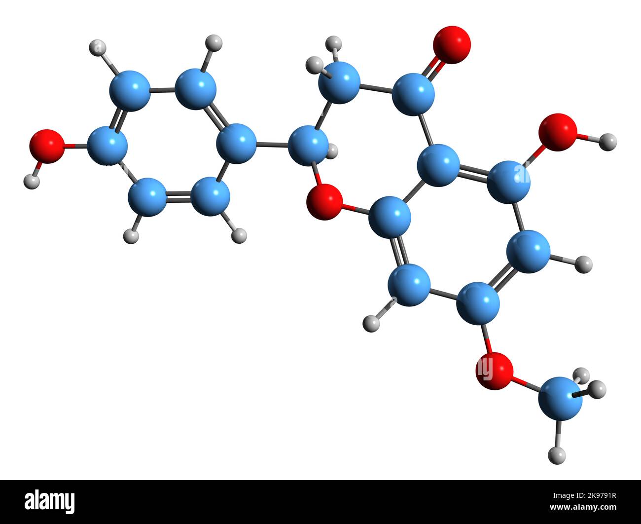 3D image of Sakuranetin skeletal formula - molecular chemical structure of flavan-on isolated on white background Stock Photohttps://www.alamy.com/image-license-details/?v=1https://www.alamy.com/3d-image-of-sakuranetin-skeletal-formula-molecular-chemical-structure-of-flavan-on-isolated-on-white-background-image487582979.html
RF2K9791R–3D image of Sakuranetin skeletal formula - molecular chemical structure of flavan-on isolated on white background
 Guadecitabine cancer drug molecule (DNA methyltransferase inhibitor). Skeletal formula. Stock Photohttps://www.alamy.com/image-license-details/?v=1https://www.alamy.com/guadecitabine-cancer-drug-molecule-dna-methyltransferase-inhibitor-image154245572.html
RFJXXDPC–Guadecitabine cancer drug molecule (DNA methyltransferase inhibitor). Skeletal formula.
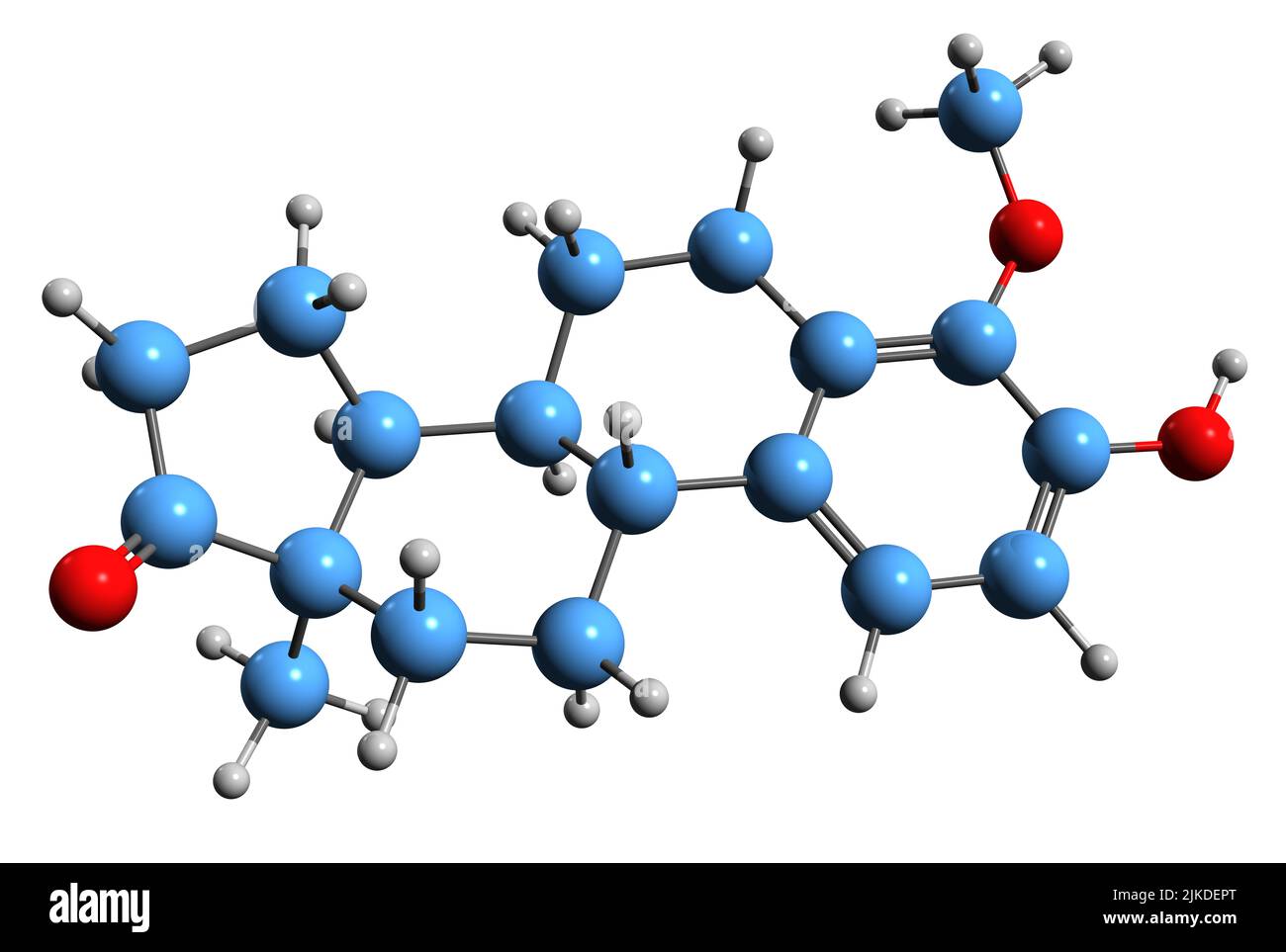 3D image of Methoxyestrone skeletal formula - molecular chemical structure of methoxylated catechol estrogen isolated on white background Stock Photohttps://www.alamy.com/image-license-details/?v=1https://www.alamy.com/3d-image-of-methoxyestrone-skeletal-formula-molecular-chemical-structure-of-methoxylated-catechol-estrogen-isolated-on-white-background-image476655392.html
RF2JKDEPT–3D image of Methoxyestrone skeletal formula - molecular chemical structure of methoxylated catechol estrogen isolated on white background
 Guadecitabine cancer drug molecule (DNA methyltransferase inhibitor). Skeletal formula. Stock Photohttps://www.alamy.com/image-license-details/?v=1https://www.alamy.com/guadecitabine-cancer-drug-molecule-dna-methyltransferase-inhibitor-skeletal-formula-image211355854.html
RFP7T2FA–Guadecitabine cancer drug molecule (DNA methyltransferase inhibitor). Skeletal formula.
 3D image of Isorhamnetin skeletal formula - molecular chemical structure of O-methylated flavonol isolated on white background Stock Photohttps://www.alamy.com/image-license-details/?v=1https://www.alamy.com/3d-image-of-isorhamnetin-skeletal-formula-molecular-chemical-structure-of-o-methylated-flavonol-isolated-on-white-background-image476641762.html
RF2JKCWC2–3D image of Isorhamnetin skeletal formula - molecular chemical structure of O-methylated flavonol isolated on white background
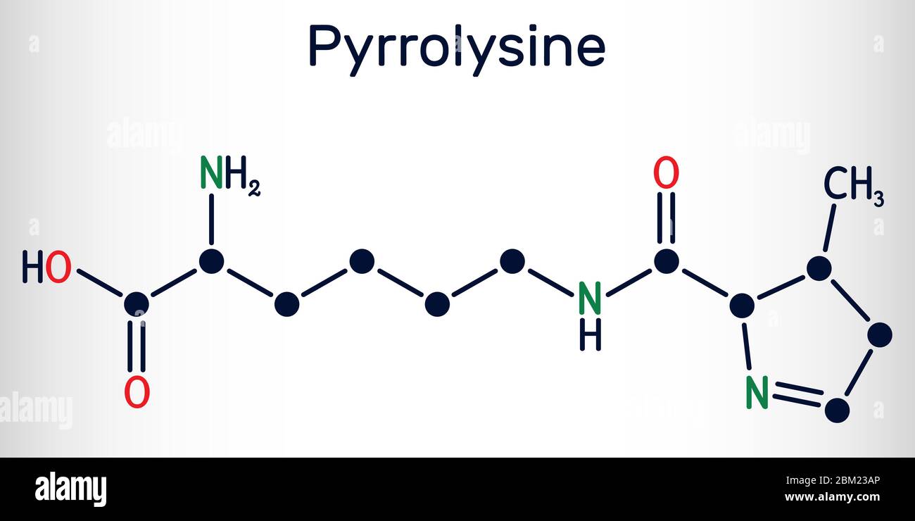 Pyrrolysine, l-pyrrolysine, Pyl, C12H21N3O3 molecule. It is amino acid, is used in biosynthesis of proteins. Structural chemical formula. Vector illus Stock Vectorhttps://www.alamy.com/image-license-details/?v=1https://www.alamy.com/pyrrolysine-l-pyrrolysine-pyl-c12h21n3o3-molecule-it-is-amino-acid-is-used-in-biosynthesis-of-proteins-structural-chemical-formula-vector-illus-image356547038.html
RF2BM23AP–Pyrrolysine, l-pyrrolysine, Pyl, C12H21N3O3 molecule. It is amino acid, is used in biosynthesis of proteins. Structural chemical formula. Vector illus
 3D image of Glycitein skeletal formula - molecular chemical structure of O-methylated isoflavone isolated on white background Stock Photohttps://www.alamy.com/image-license-details/?v=1https://www.alamy.com/3d-image-of-glycitein-skeletal-formula-molecular-chemical-structure-of-o-methylated-isoflavone-isolated-on-white-background-image476466084.html
RF2JK4W9T–3D image of Glycitein skeletal formula - molecular chemical structure of O-methylated isoflavone isolated on white background
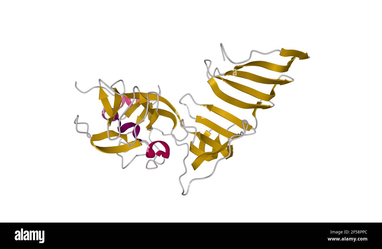 Crystal structure of the histone methyltransferase SET79, 3D cartoon model isolated with differently colored elements of the secondary structure Stock Photohttps://www.alamy.com/image-license-details/?v=1https://www.alamy.com/crystal-structure-of-the-histone-methyltransferase-set79-3d-cartoon-model-isolated-with-differently-colored-elements-of-the-secondary-structure-image416315604.html
RF2F58PPC–Crystal structure of the histone methyltransferase SET79, 3D cartoon model isolated with differently colored elements of the secondary structure
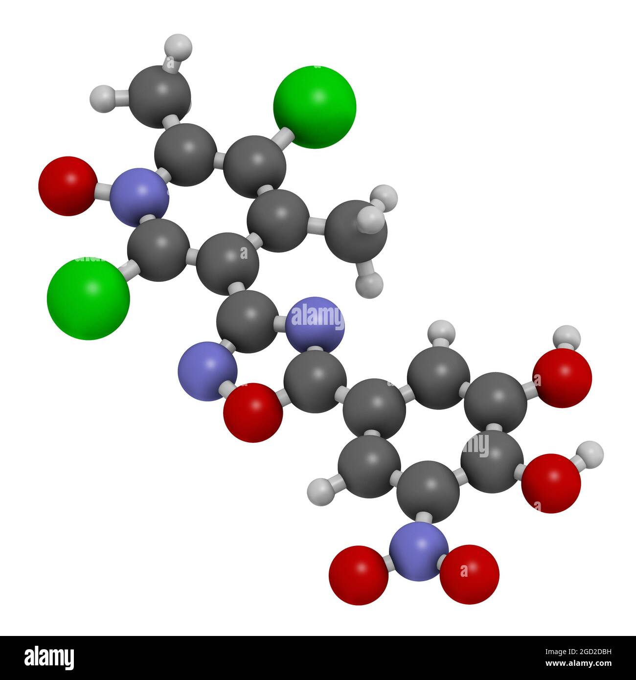 Opicapone Parkinson's disease drug molecule. 3D rendering. Stock Photohttps://www.alamy.com/image-license-details/?v=1https://www.alamy.com/opicapone-parkinsons-disease-drug-molecule-3d-rendering-image438304149.html
RF2GD2DBH–Opicapone Parkinson's disease drug molecule. 3D rendering.
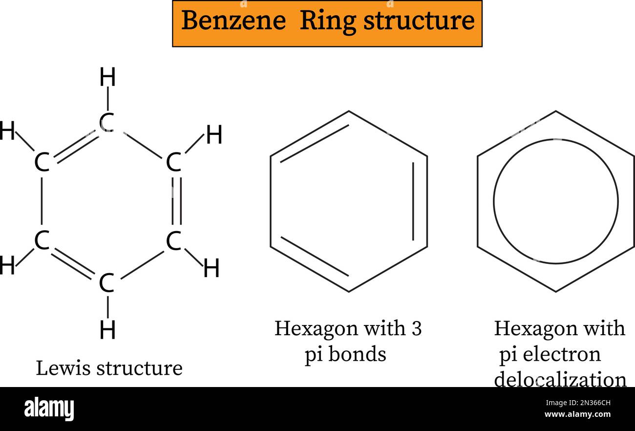 Benzene Ring structure .vector image Stock Vectorhttps://www.alamy.com/image-license-details/?v=1https://www.alamy.com/benzene-ring-structure-vector-image-image518291777.html
RF2N366CH–Benzene Ring structure .vector image
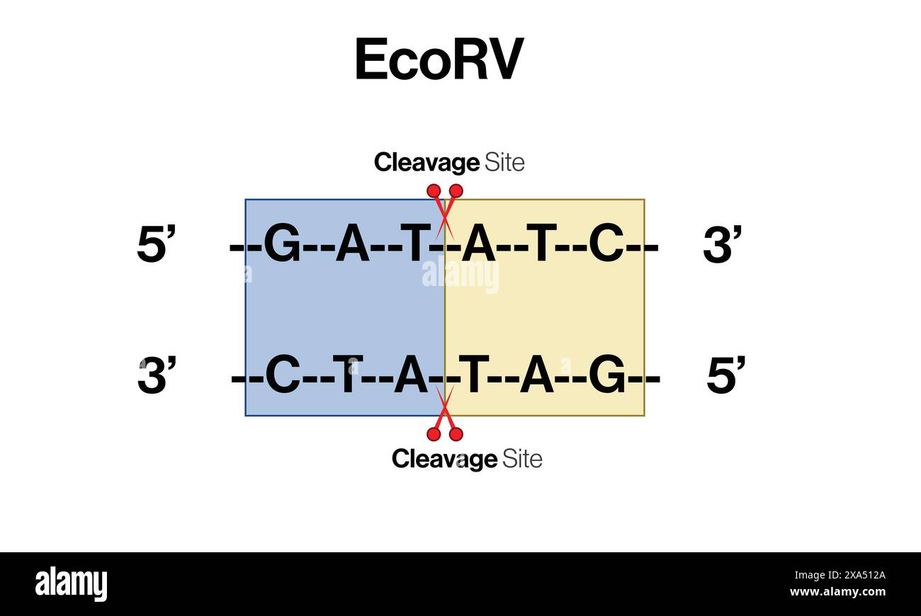 Detailed Vector Illustration of EcoRV Restriction Enzyme Action on DNA Sequence for Molecular Biology and Genetic Engineering on White Background. Stock Vectorhttps://www.alamy.com/image-license-details/?v=1https://www.alamy.com/detailed-vector-illustration-of-ecorv-restriction-enzyme-action-on-dna-sequence-for-molecular-biology-and-genetic-engineering-on-white-background-image608620050.html
RF2XA512A–Detailed Vector Illustration of EcoRV Restriction Enzyme Action on DNA Sequence for Molecular Biology and Genetic Engineering on White Background.
 Human L-Isoaspartyl Methyltransferase - PDB id 1KR5. Stock Photohttps://www.alamy.com/image-license-details/?v=1https://www.alamy.com/human-l-isoaspartyl-methyltransferase-pdb-id-1kr5-image355932570.html
RM2BK23HE–Human L-Isoaspartyl Methyltransferase - PDB id 1KR5.
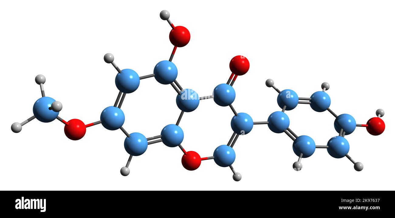 3D image of Prunetin skeletal formula - molecular chemical structure of O-methylated isoflavone isolated on white background Stock Photohttps://www.alamy.com/image-license-details/?v=1https://www.alamy.com/3d-image-of-prunetin-skeletal-formula-molecular-chemical-structure-of-o-methylated-isoflavone-isolated-on-white-background-image487580667.html
RF2K97637–3D image of Prunetin skeletal formula - molecular chemical structure of O-methylated isoflavone isolated on white background
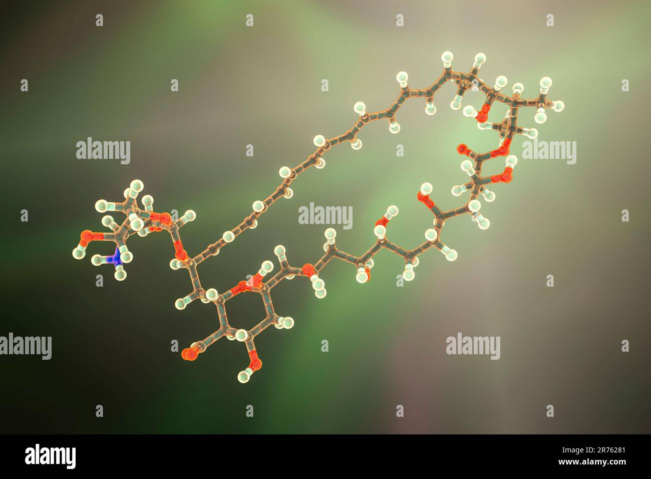 Amphotericin B antifungal drug molecule. Chemical formula is C47H73NO17. Atoms are represented as spheres: carbon (grey), hydrogen (light yellow), nit Stock Photohttps://www.alamy.com/image-license-details/?v=1https://www.alamy.com/amphotericin-b-antifungal-drug-molecule-chemical-formula-is-c47h73no17-atoms-are-represented-as-spheres-carbon-grey-hydrogen-light-yellow-nit-image555167873.html
RF2R76281–Amphotericin B antifungal drug molecule. Chemical formula is C47H73NO17. Atoms are represented as spheres: carbon (grey), hydrogen (light yellow), nit
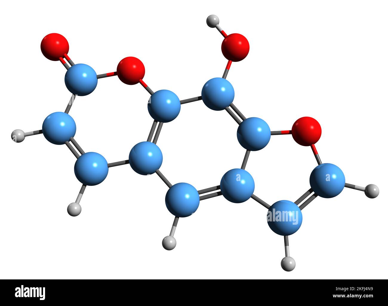 3D image of Xanthotoxol skeletal formula - molecular chemical structure of furanocoumarin isolated on white background Stock Photohttps://www.alamy.com/image-license-details/?v=1https://www.alamy.com/3d-image-of-xanthotoxol-skeletal-formula-molecular-chemical-structure-of-furanocoumarin-isolated-on-white-background-image491509013.html
RF2KFJ4N9–3D image of Xanthotoxol skeletal formula - molecular chemical structure of furanocoumarin isolated on white background
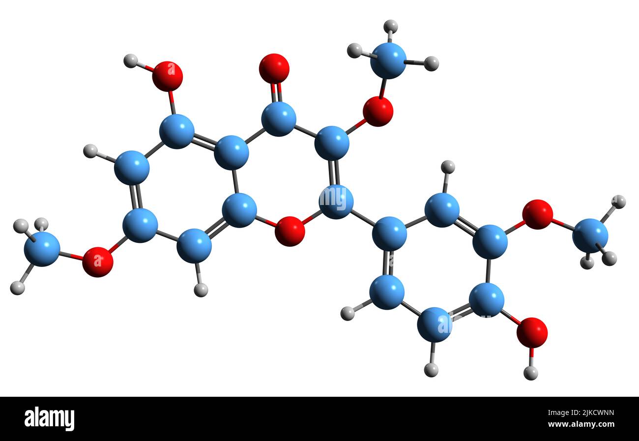 3D image of Pachypodol skeletal formula - molecular chemical structure of O-methylated flavonol isolated on white background Stock Photohttps://www.alamy.com/image-license-details/?v=1https://www.alamy.com/3d-image-of-pachypodol-skeletal-formula-molecular-chemical-structure-of-o-methylated-flavonol-isolated-on-white-background-image476642033.html
RF2JKCWNN–3D image of Pachypodol skeletal formula - molecular chemical structure of O-methylated flavonol isolated on white background
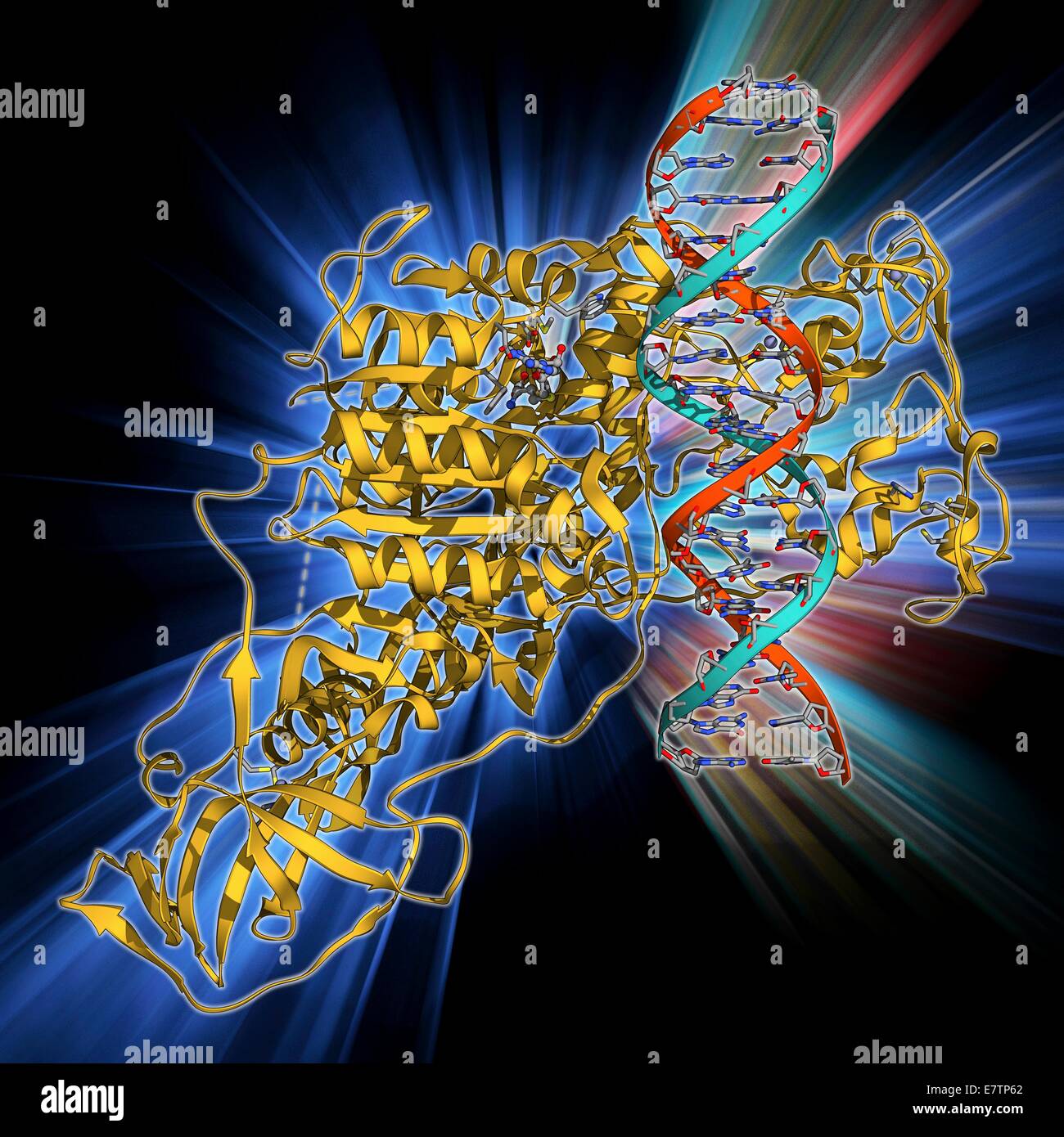 Methyltransferase complexed with DNA, molecular model. The strand of DNA (deoxyribonucleic acid, red and blue) is enclosed by DNA methyltransferase 1 (DNMT-1, beige). This enzyme acts to add methyl groups to the DNA, a process called DNA methylation, whic Stock Photohttps://www.alamy.com/image-license-details/?v=1https://www.alamy.com/stock-photo-methyltransferase-complexed-with-dna-molecular-model-the-strand-of-73688330.html
RFE7TP62–Methyltransferase complexed with DNA, molecular model. The strand of DNA (deoxyribonucleic acid, red and blue) is enclosed by DNA methyltransferase 1 (DNMT-1, beige). This enzyme acts to add methyl groups to the DNA, a process called DNA methylation, whic
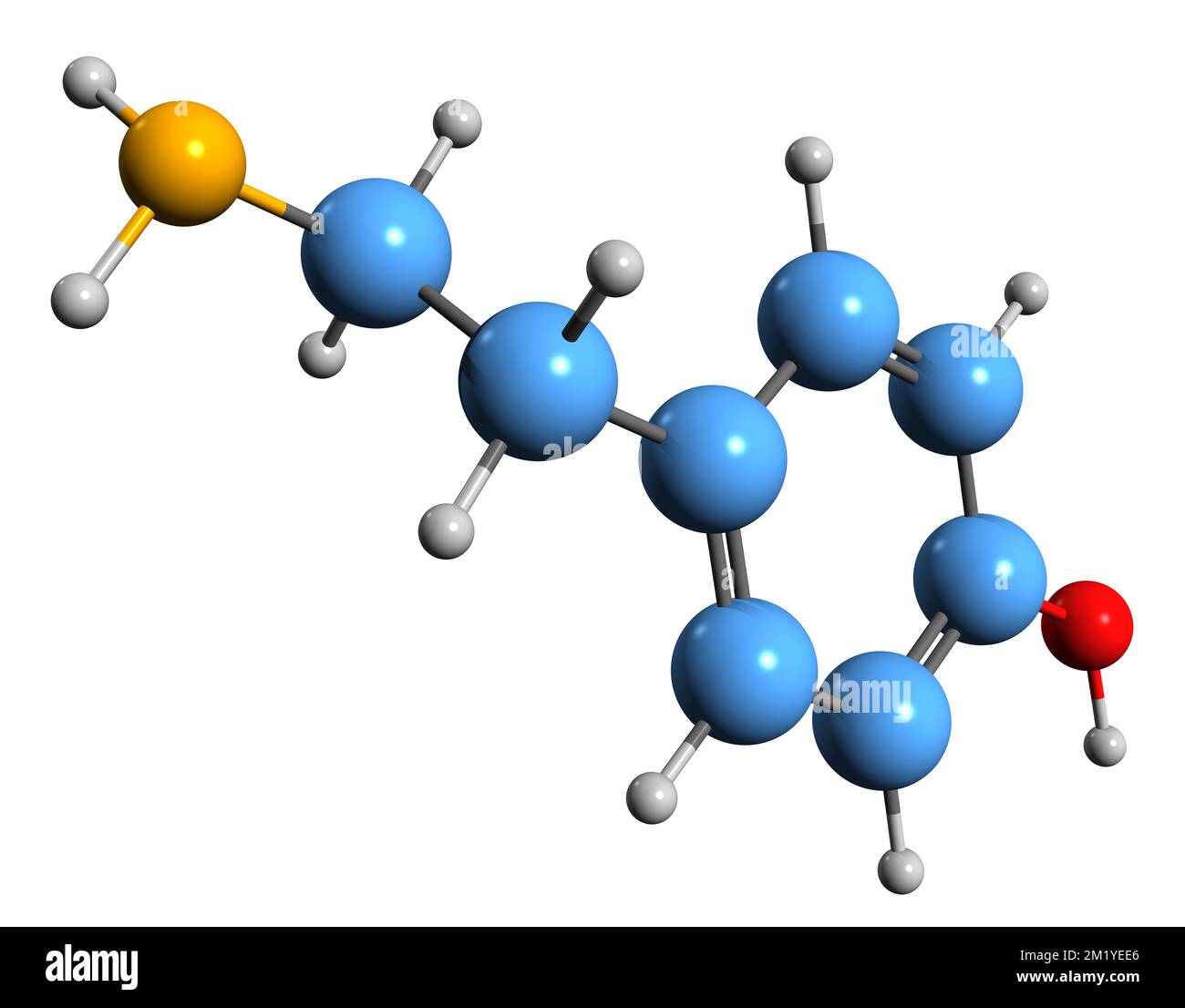 3D image of Tyramine skeletal formula - molecular chemical structure of catecholamine releasing agent isolated on white background Stock Photohttps://www.alamy.com/image-license-details/?v=1https://www.alamy.com/3d-image-of-tyramine-skeletal-formula-molecular-chemical-structure-of-catecholamine-releasing-agent-isolated-on-white-background-image500319406.html
RF2M1YEE6–3D image of Tyramine skeletal formula - molecular chemical structure of catecholamine releasing agent isolated on white background
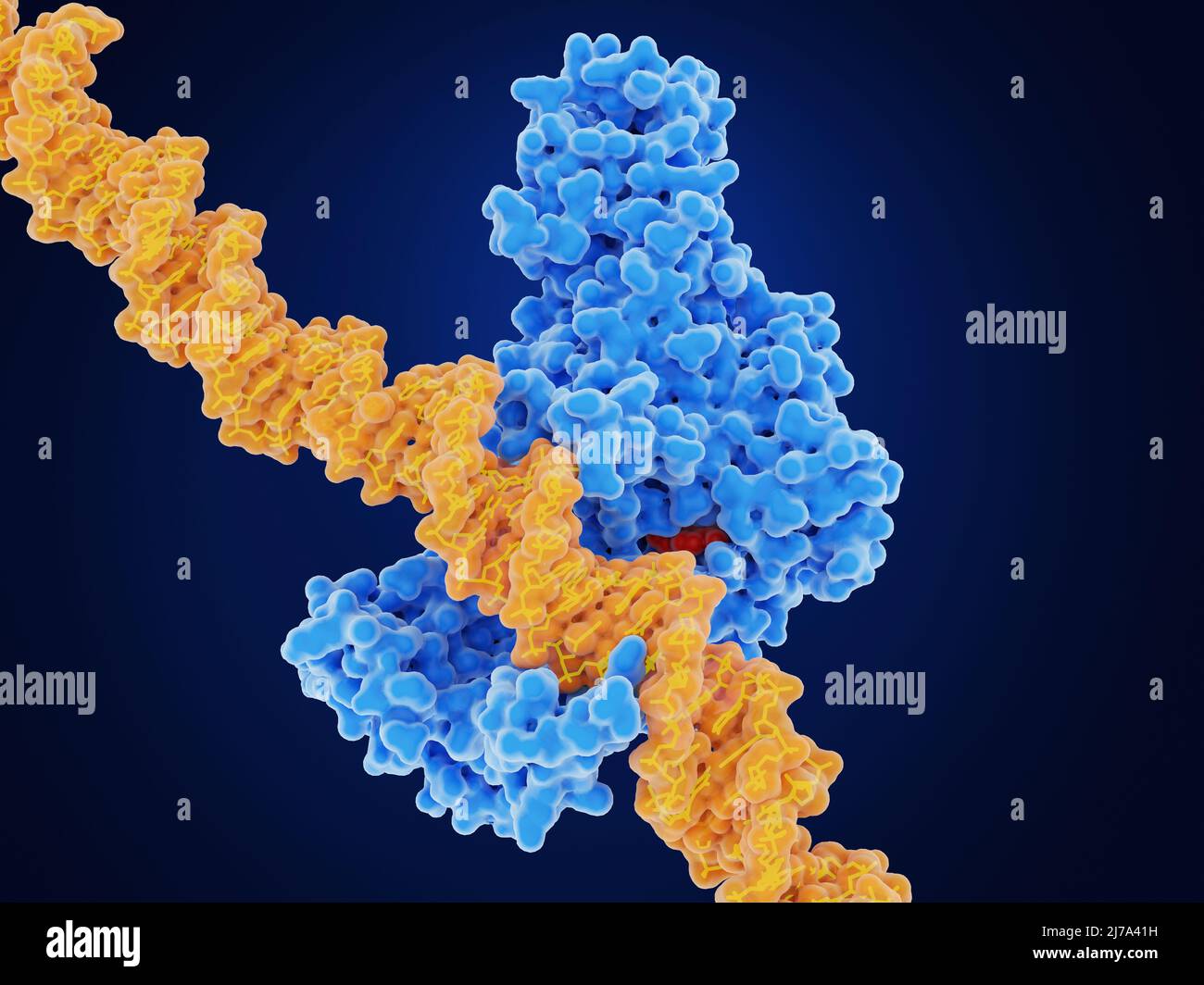 DNA methyl transferase--1 and DNA, illustration Stock Photohttps://www.alamy.com/image-license-details/?v=1https://www.alamy.com/dna-methyl-transferase-1-and-dna-illustration-image469205229.html
RF2J7A41H–DNA methyl transferase--1 and DNA, illustration
 Illustration of the drug MRTX1719 bound to its target protein, protein arginine methyltransferase 5 (PRMT5). This molecule treats solid tumours that contain the methylthioadenosine phosphorylase (MTAP) mutation. Stock Photohttps://www.alamy.com/image-license-details/?v=1https://www.alamy.com/illustration-of-the-drug-mrtx1719-bound-to-its-target-protein-protein-arginine-methyltransferase-5-prmt5-this-molecule-treats-solid-tumours-that-contain-the-methylthioadenosine-phosphorylase-mtap-mutation-image618634348.html
RF2XXD6BT–Illustration of the drug MRTX1719 bound to its target protein, protein arginine methyltransferase 5 (PRMT5). This molecule treats solid tumours that contain the methylthioadenosine phosphorylase (MTAP) mutation.
 Guadecitabine cancer drug molecule (DNA methyltransferase inhibitor). Skeletal formula. Stock Photohttps://www.alamy.com/image-license-details/?v=1https://www.alamy.com/guadecitabine-cancer-drug-molecule-dna-methyltransferase-inhibitor-image154244693.html
RFJXXCK1–Guadecitabine cancer drug molecule (DNA methyltransferase inhibitor). Skeletal formula.
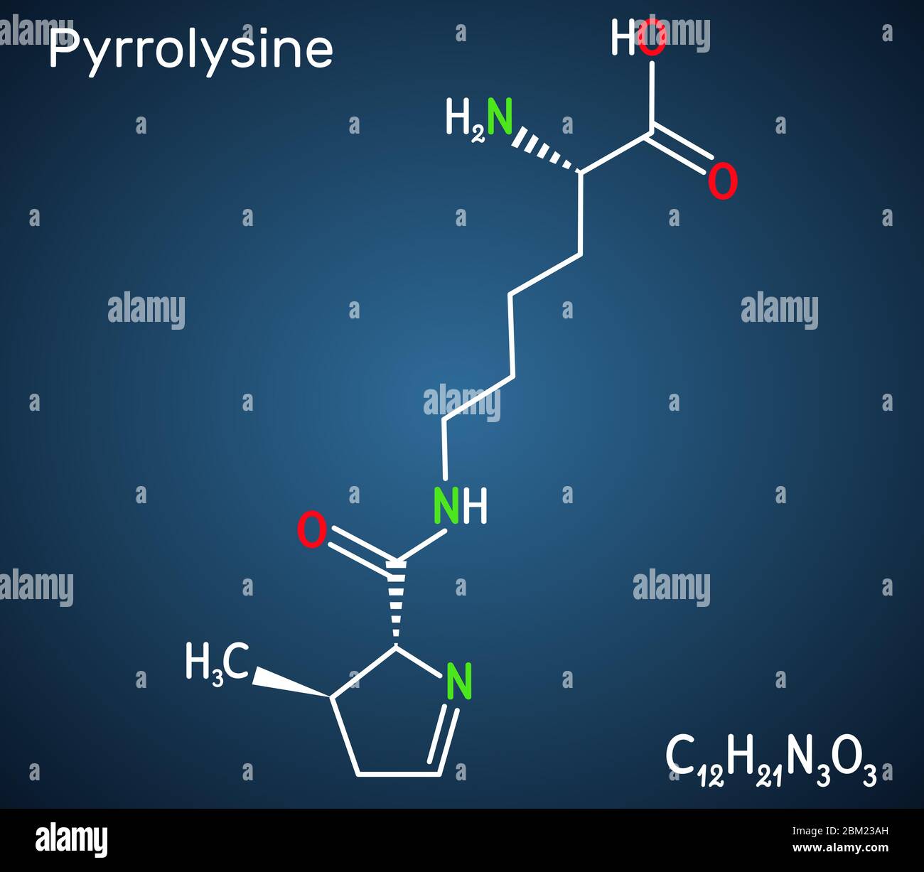 Pyrrolysine, l-pyrrolysine, Pyl, C12H21N3O3 molecule. It is amino acid, is used in biosynthesis of proteins. Structural chemical formula on the dark b Stock Vectorhttps://www.alamy.com/image-license-details/?v=1https://www.alamy.com/pyrrolysine-l-pyrrolysine-pyl-c12h21n3o3-molecule-it-is-amino-acid-is-used-in-biosynthesis-of-proteins-structural-chemical-formula-on-the-dark-b-image356547033.html
RF2BM23AH–Pyrrolysine, l-pyrrolysine, Pyl, C12H21N3O3 molecule. It is amino acid, is used in biosynthesis of proteins. Structural chemical formula on the dark b
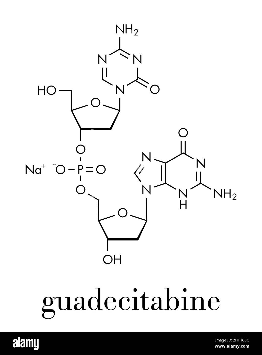 Guadecitabine cancer drug molecule (DNA methyltransferase inhibitor). Skeletal formula. Stock Vectorhttps://www.alamy.com/image-license-details/?v=1https://www.alamy.com/guadecitabine-cancer-drug-molecule-dna-methyltransferase-inhibitor-skeletal-formula-image457075152.html
RF2HFHG0G–Guadecitabine cancer drug molecule (DNA methyltransferase inhibitor). Skeletal formula.
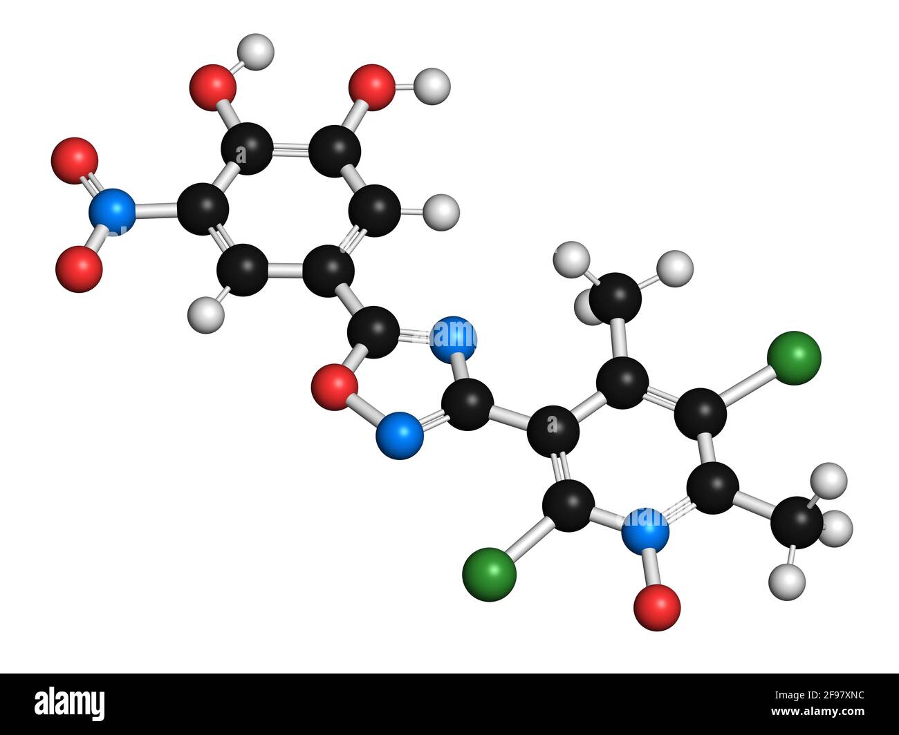 Opicapone Parkinson's disease drug molecule, illustration Stock Photohttps://www.alamy.com/image-license-details/?v=1https://www.alamy.com/opicapone-parkinsons-disease-drug-molecule-illustration-image418755384.html
RF2F97XNC–Opicapone Parkinson's disease drug molecule, illustration
 Histamine methyltransferase complexed with the antihistamine drug diphenhydramine (red). 3D cartoon and Gaussian surface model, PDB 2aot Stock Photohttps://www.alamy.com/image-license-details/?v=1https://www.alamy.com/histamine-methyltransferase-complexed-with-the-antihistamine-drug-diphenhydramine-red-3d-cartoon-and-gaussian-surface-model-pdb-2aot-image472903320.html
RF2JDAH08–Histamine methyltransferase complexed with the antihistamine drug diphenhydramine (red). 3D cartoon and Gaussian surface model, PDB 2aot
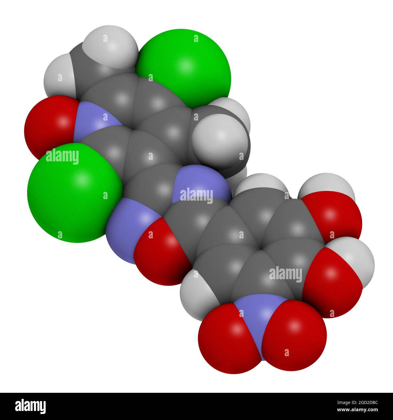 Opicapone Parkinson's disease drug molecule. 3D rendering. Stock Photohttps://www.alamy.com/image-license-details/?v=1https://www.alamy.com/opicapone-parkinsons-disease-drug-molecule-3d-rendering-image438304144.html
RF2GD2DBC–Opicapone Parkinson's disease drug molecule. 3D rendering.
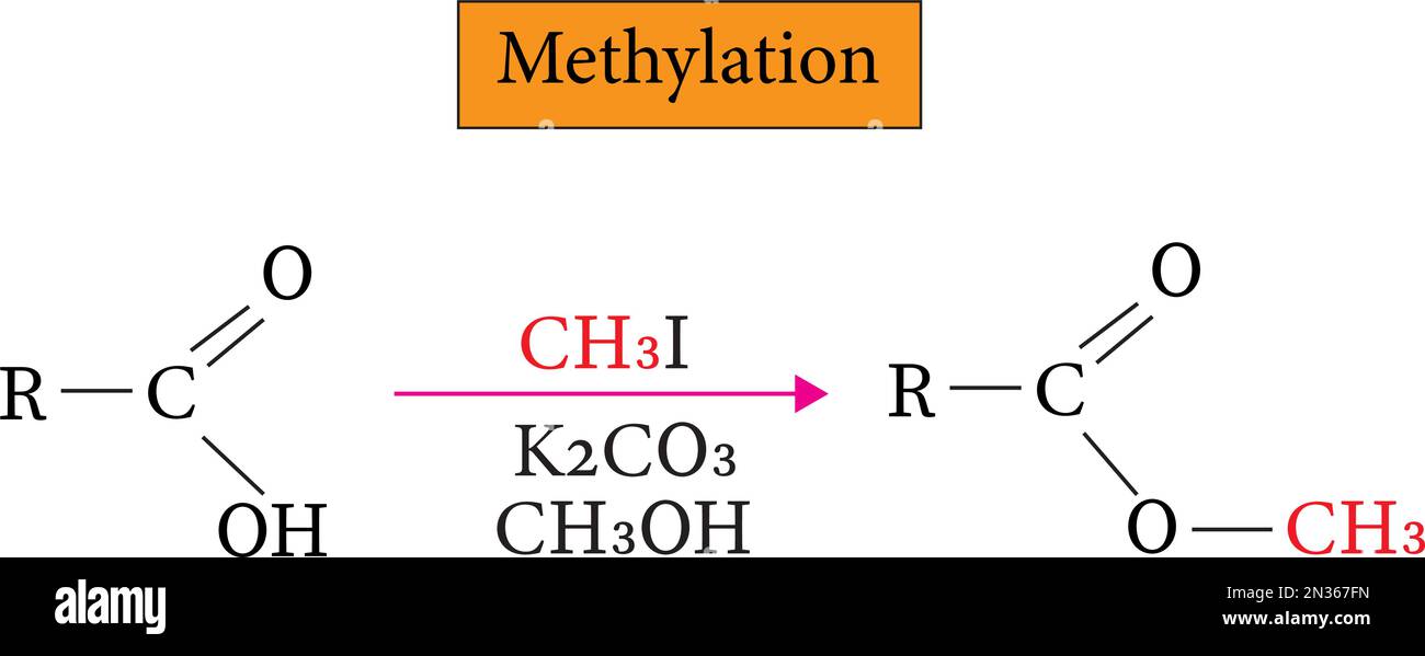 Methylation is the process of adding a methyl group to a molecule , vector image Stock Vectorhttps://www.alamy.com/image-license-details/?v=1https://www.alamy.com/methylation-is-the-process-of-adding-a-methyl-group-to-a-molecule-vector-image-image518292649.html
RF2N367FN–Methylation is the process of adding a methyl group to a molecule , vector image
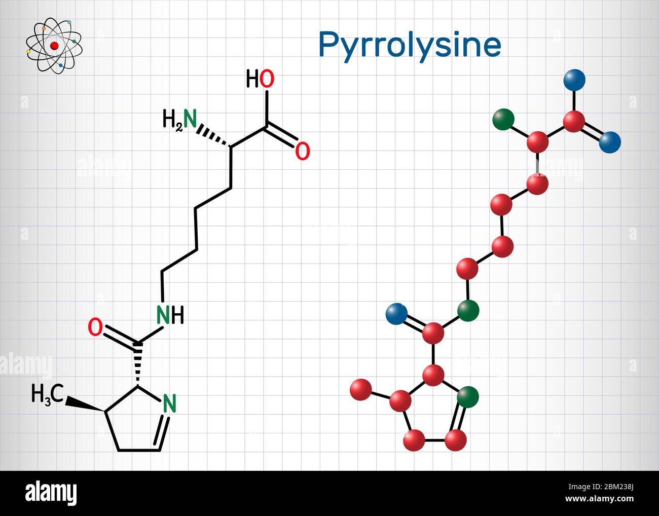 Pyrrolysine, l-pyrrolysine, Pyl, C12H21N3O3 molecule. It is amino acid, is used in biosynthesis of proteins. Structural chemical formula and molecule Stock Vectorhttps://www.alamy.com/image-license-details/?v=1https://www.alamy.com/pyrrolysine-l-pyrrolysine-pyl-c12h21n3o3-molecule-it-is-amino-acid-is-used-in-biosynthesis-of-proteins-structural-chemical-formula-and-molecule-image356546978.html
RF2BM238J–Pyrrolysine, l-pyrrolysine, Pyl, C12H21N3O3 molecule. It is amino acid, is used in biosynthesis of proteins. Structural chemical formula and molecule
 Guadecitabine cancer drug molecule (DNA methyltransferase inhibitor). Skeletal formula. Stock Vectorhttps://www.alamy.com/image-license-details/?v=1https://www.alamy.com/guadecitabine-cancer-drug-molecule-dna-methyltransferase-inhibitor-skeletal-formula-image457076787.html
RF2HFHJ2Y–Guadecitabine cancer drug molecule (DNA methyltransferase inhibitor). Skeletal formula.
 Tazemetostat cancer drug molecule, illustration Stock Photohttps://www.alamy.com/image-license-details/?v=1https://www.alamy.com/tazemetostat-cancer-drug-molecule-illustration-image418755481.html
RF2F97XTW–Tazemetostat cancer drug molecule, illustration
 Guadecitabine cancer drug molecule (DNA methyltransferase inhibitor). Stylized 2D renderings and conventional skeletal formula. Stock Vectorhttps://www.alamy.com/image-license-details/?v=1https://www.alamy.com/stock-photo-guadecitabine-cancer-drug-molecule-dna-methyltransferase-inhibitor-136263307.html
RFHWK96K–Guadecitabine cancer drug molecule (DNA methyltransferase inhibitor). Stylized 2D renderings and conventional skeletal formula.
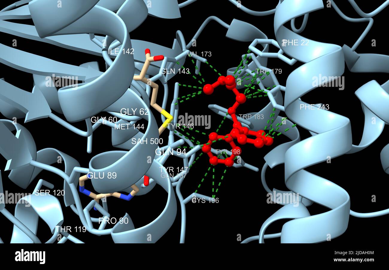 Histamine methyltransferase complexed with the antihistamine drug diphenhydramine (red). 3D cartoon model with interacting residues labeled, PDB 2aot Stock Photohttps://www.alamy.com/image-license-details/?v=1https://www.alamy.com/histamine-methyltransferase-complexed-with-the-antihistamine-drug-diphenhydramine-red-3d-cartoon-model-with-interacting-residues-labeled-pdb-2aot-image472903332.html
RF2JDAH0M–Histamine methyltransferase complexed with the antihistamine drug diphenhydramine (red). 3D cartoon model with interacting residues labeled, PDB 2aot
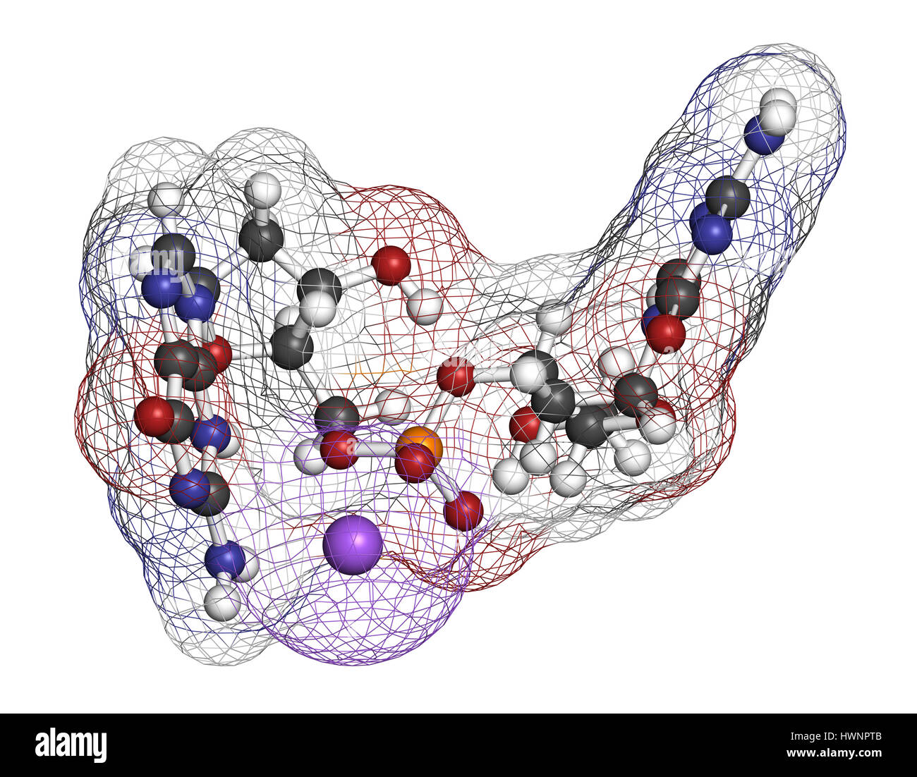 Guadecitabine cancer drug molecule (DNA methyltransferase inhibitor). 3D rendering. Atoms are represented as spheres with conventional color coding: h Stock Photohttps://www.alamy.com/image-license-details/?v=1https://www.alamy.com/stock-photo-guadecitabine-cancer-drug-molecule-dna-methyltransferase-inhibitor-136317899.html
RFHWNPTB–Guadecitabine cancer drug molecule (DNA methyltransferase inhibitor). 3D rendering. Atoms are represented as spheres with conventional color coding: h
 Guadecitabine cancer drug molecule (DNA methyltransferase inhibitor). Skeletal formula. Stock Photohttps://www.alamy.com/image-license-details/?v=1https://www.alamy.com/guadecitabine-cancer-drug-molecule-dna-methyltransferase-inhibitor-skeletal-formula-image364971722.html
RF2C5NW4A–Guadecitabine cancer drug molecule (DNA methyltransferase inhibitor). Skeletal formula.
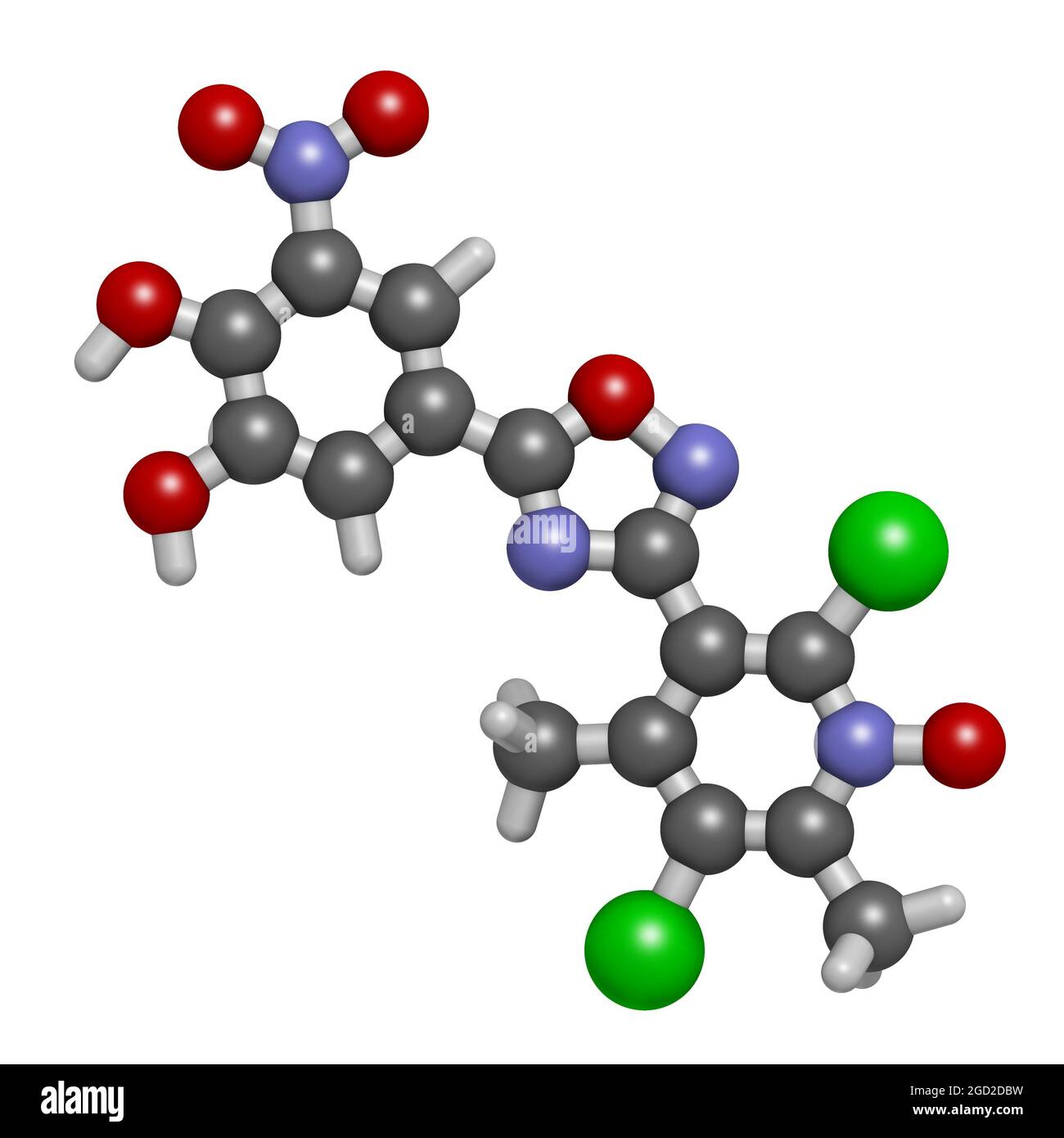 Opicapone Parkinson's disease drug molecule. 3D rendering. Stock Photohttps://www.alamy.com/image-license-details/?v=1https://www.alamy.com/opicapone-parkinsons-disease-drug-molecule-3d-rendering-image438304157.html
RF2GD2DBW–Opicapone Parkinson's disease drug molecule. 3D rendering.
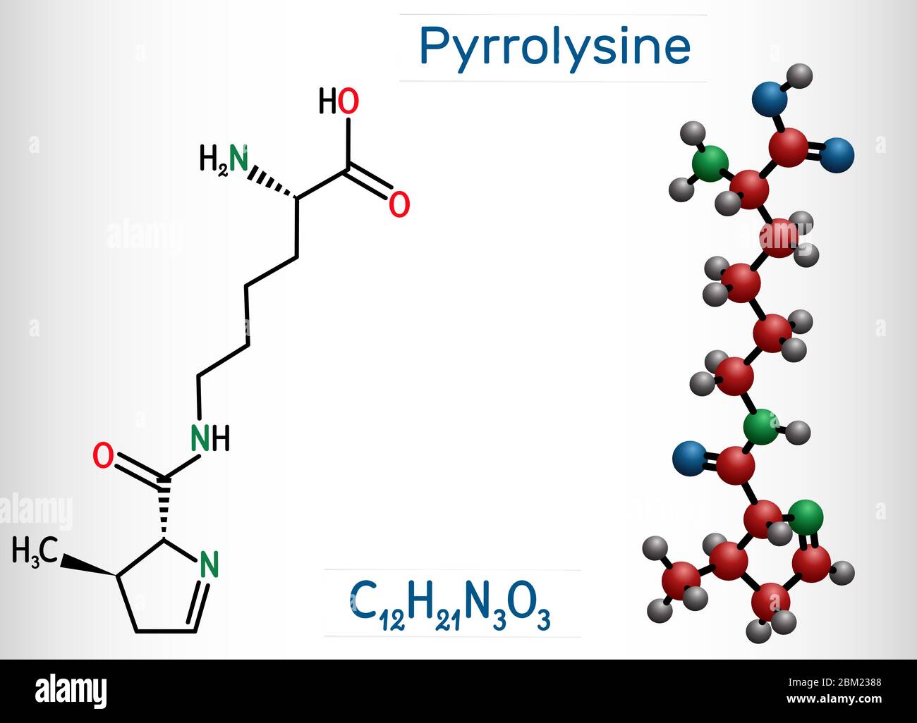 Pyrrolysine, l-pyrrolysine, Pyl, C12H21N3O3 molecule. It is amino acid, is used in biosynthesis of proteins. Structural chemical formula and molecule Stock Vectorhttps://www.alamy.com/image-license-details/?v=1https://www.alamy.com/pyrrolysine-l-pyrrolysine-pyl-c12h21n3o3-molecule-it-is-amino-acid-is-used-in-biosynthesis-of-proteins-structural-chemical-formula-and-molecule-image356546968.html
RF2BM2388–Pyrrolysine, l-pyrrolysine, Pyl, C12H21N3O3 molecule. It is amino acid, is used in biosynthesis of proteins. Structural chemical formula and molecule
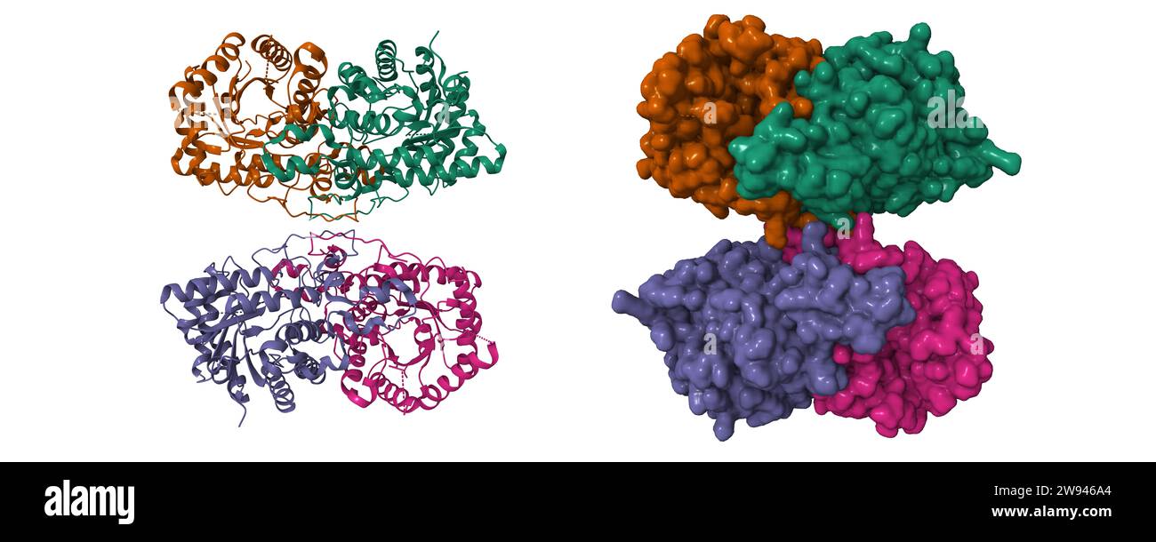 Cryo-EM structure of human kidney betaine-homocysteine methyltransferase. 3D cartoon and Gaussian surface models, chain id color scheme, PDB 8d45 Stock Photohttps://www.alamy.com/image-license-details/?v=1https://www.alamy.com/cryo-em-structure-of-human-kidney-betaine-homocysteine-methyltransferase-3d-cartoon-and-gaussian-surface-models-chain-id-color-scheme-pdb-8d45-image590777212.html
RF2W946A4–Cryo-EM structure of human kidney betaine-homocysteine methyltransferase. 3D cartoon and Gaussian surface models, chain id color scheme, PDB 8d45
 Guadecitabine cancer drug molecule (DNA methyltransferase inhibitor). 3D rendering. Atoms are represented as spheres with conventional color coding: h Stock Photohttps://www.alamy.com/image-license-details/?v=1https://www.alamy.com/stock-photo-guadecitabine-cancer-drug-molecule-dna-methyltransferase-inhibitor-136317890.html
RFHWNPT2–Guadecitabine cancer drug molecule (DNA methyltransferase inhibitor). 3D rendering. Atoms are represented as spheres with conventional color coding: h
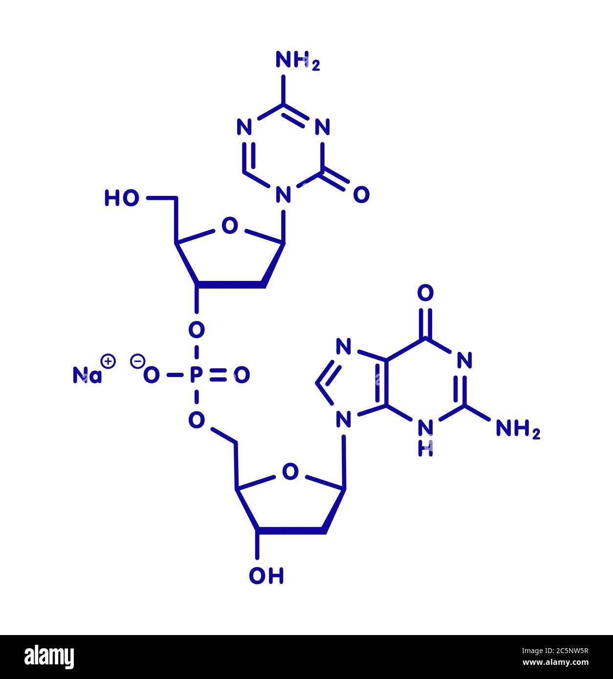 Guadecitabine cancer drug molecule (DNA methyltransferase inhibitor). Skeletal formula. Stock Photohttps://www.alamy.com/image-license-details/?v=1https://www.alamy.com/guadecitabine-cancer-drug-molecule-dna-methyltransferase-inhibitor-skeletal-formula-image364971763.html
RF2C5NW5R–Guadecitabine cancer drug molecule (DNA methyltransferase inhibitor). Skeletal formula.
 Tazemetostat cancer drug molecule. 3D rendering. Stock Photohttps://www.alamy.com/image-license-details/?v=1https://www.alamy.com/tazemetostat-cancer-drug-molecule-3d-rendering-image438304412.html
RF2GD2DN0–Tazemetostat cancer drug molecule. 3D rendering.
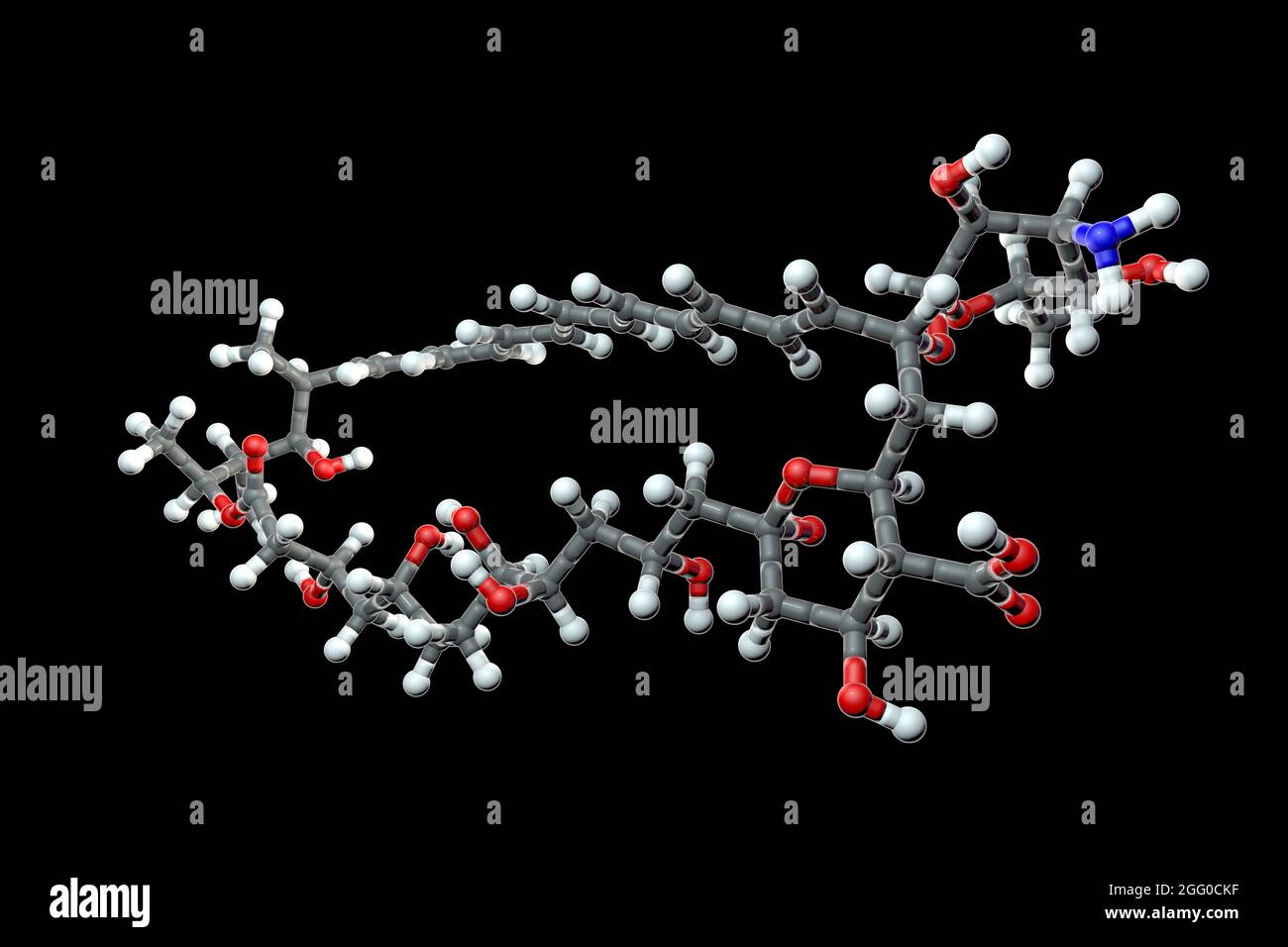 Amphotericin B antifungal drug molecule, illustration. Chemical formula is C47H73NO17. Atoms are represented as spheres: carbon (grey), hydrogen (white), nitrogen (blue), oxygen (red). Stock Photohttps://www.alamy.com/image-license-details/?v=1https://www.alamy.com/amphotericin-b-antifungal-drug-molecule-illustration-chemical-formula-is-c47h73no17-atoms-are-represented-as-spheres-carbon-grey-hydrogen-white-nitrogen-blue-oxygen-red-image440103651.html
RF2GG0CKF–Amphotericin B antifungal drug molecule, illustration. Chemical formula is C47H73NO17. Atoms are represented as spheres: carbon (grey), hydrogen (white), nitrogen (blue), oxygen (red).
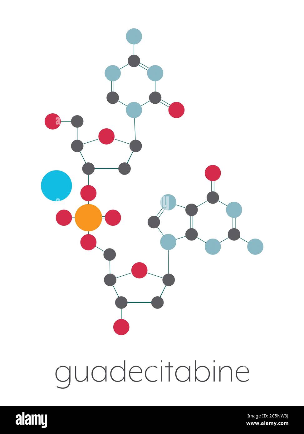 Guadecitabine cancer drug molecule (DNA methyltransferase inhibitor). Stylized skeletal formula (chemical structure): Atoms are shown as color-coded circles: hydrogen (white), carbon (grey), nitrogen (blue), oxygen (red), phosphorus (orange). Stock Photohttps://www.alamy.com/image-license-details/?v=1https://www.alamy.com/guadecitabine-cancer-drug-molecule-dna-methyltransferase-inhibitor-stylized-skeletal-formula-chemical-structure-atoms-are-shown-as-color-coded-circles-hydrogen-white-carbon-grey-nitrogen-blue-oxygen-red-phosphorus-orange-image364971702.html
RF2C5NW3J–Guadecitabine cancer drug molecule (DNA methyltransferase inhibitor). Stylized skeletal formula (chemical structure): Atoms are shown as color-coded circles: hydrogen (white), carbon (grey), nitrogen (blue), oxygen (red), phosphorus (orange).
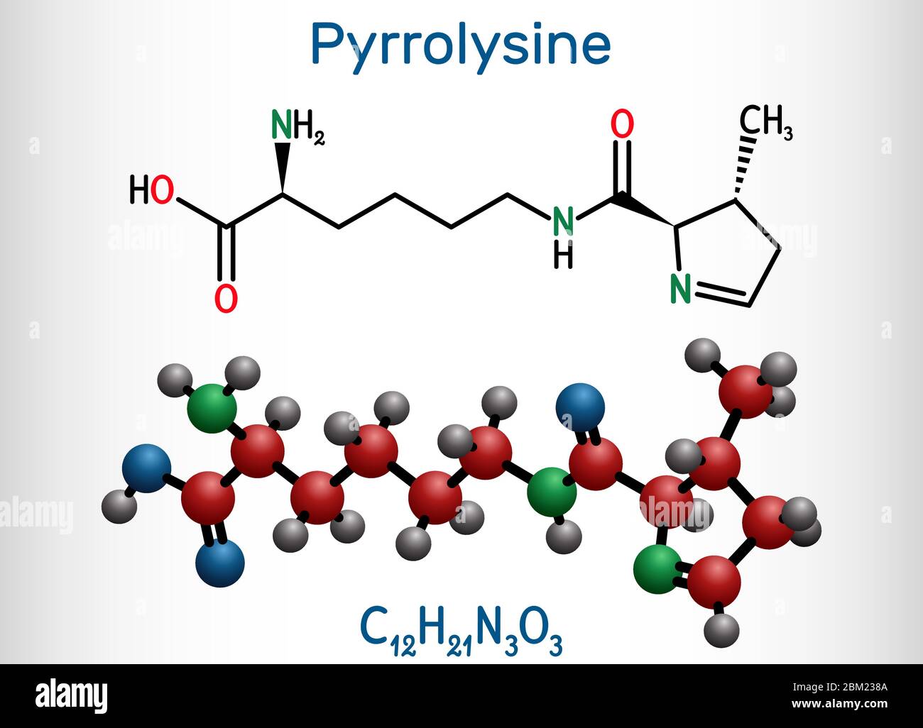 Pyrrolysine, l-pyrrolysine, Pyl, C12H21N3O3 molecule. It is amino acid, is used in biosynthesis of proteins. Structural chemical formula and molecule Stock Vectorhttps://www.alamy.com/image-license-details/?v=1https://www.alamy.com/pyrrolysine-l-pyrrolysine-pyl-c12h21n3o3-molecule-it-is-amino-acid-is-used-in-biosynthesis-of-proteins-structural-chemical-formula-and-molecule-image356546970.html
RF2BM238A–Pyrrolysine, l-pyrrolysine, Pyl, C12H21N3O3 molecule. It is amino acid, is used in biosynthesis of proteins. Structural chemical formula and molecule
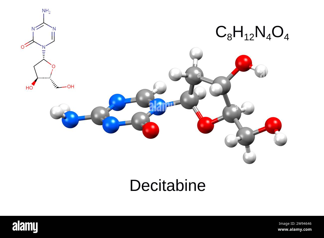 Chemical formula, skeletal formula, and 3D ball-and-stick model of hypomethylation agent decitabine, white background Stock Photohttps://www.alamy.com/image-license-details/?v=1https://www.alamy.com/chemical-formula-skeletal-formula-and-3d-ball-and-stick-model-of-hypomethylation-agent-decitabine-white-background-image590777046.html
RF2W94646–Chemical formula, skeletal formula, and 3D ball-and-stick model of hypomethylation agent decitabine, white background
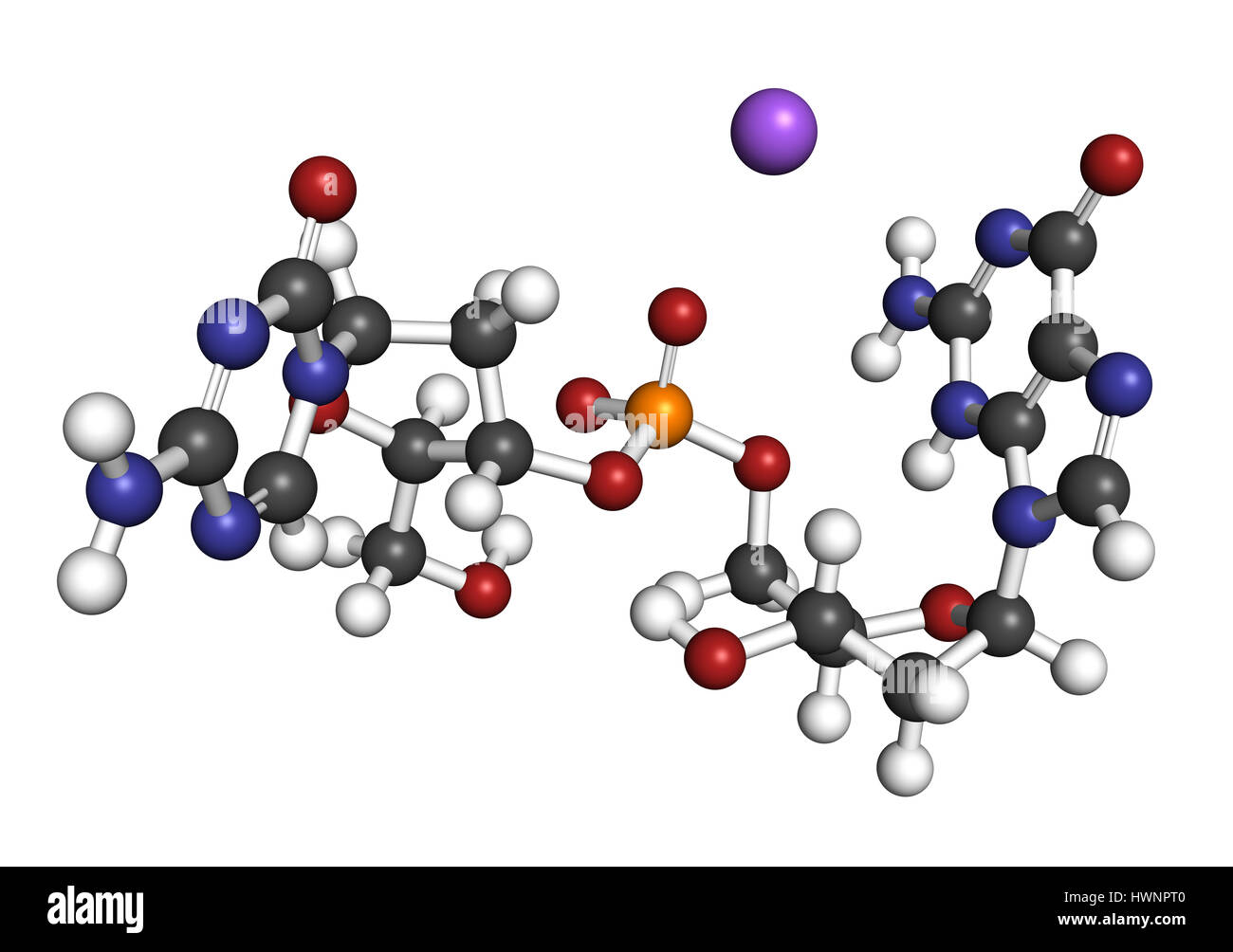 Guadecitabine cancer drug molecule (DNA methyltransferase inhibitor). 3D rendering. Atoms are represented as spheres with conventional color coding: h Stock Photohttps://www.alamy.com/image-license-details/?v=1https://www.alamy.com/stock-photo-guadecitabine-cancer-drug-molecule-dna-methyltransferase-inhibitor-136317888.html
RFHWNPT0–Guadecitabine cancer drug molecule (DNA methyltransferase inhibitor). 3D rendering. Atoms are represented as spheres with conventional color coding: h
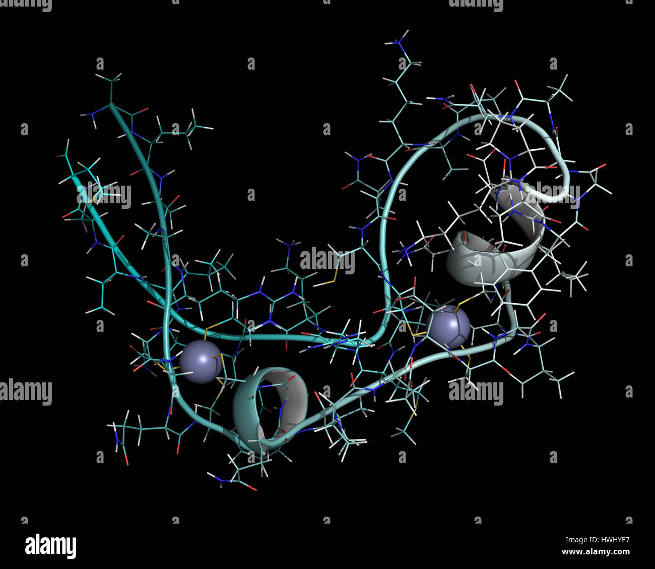 Zinc finger protein domain (from histone-lysine N-methyltransferase A2). Zinc fingers are protein domains that bind to DNA sequences. Cartoon & wirefr Stock Photohttps://www.alamy.com/image-license-details/?v=1https://www.alamy.com/stock-photo-zinc-finger-protein-domain-from-histone-lysine-n-methyltransferase-136233727.html
RFHWHYE7–Zinc finger protein domain (from histone-lysine N-methyltransferase A2). Zinc fingers are protein domains that bind to DNA sequences. Cartoon & wirefr
 Tazemetostat cancer drug molecule. 3D rendering. Stock Photohttps://www.alamy.com/image-license-details/?v=1https://www.alamy.com/tazemetostat-cancer-drug-molecule-3d-rendering-image438304429.html
RF2GD2DNH–Tazemetostat cancer drug molecule. 3D rendering.
 Guadecitabine cancer drug molecule (DNA methyltransferase inhibitor). Stylized skeletal formula (chemical structure): Atoms are shown as color-coded circles: hydrogen (white), carbon (grey), nitrogen (blue), oxygen (red), phosphorus (orange). Stock Photohttps://www.alamy.com/image-license-details/?v=1https://www.alamy.com/guadecitabine-cancer-drug-molecule-dna-methyltransferase-inhibitor-stylized-skeletal-formula-chemical-structure-atoms-are-shown-as-color-coded-circles-hydrogen-white-carbon-grey-nitrogen-blue-oxygen-red-phosphorus-orange-image364971755.html
RF2C5NW5F–Guadecitabine cancer drug molecule (DNA methyltransferase inhibitor). Stylized skeletal formula (chemical structure): Atoms are shown as color-coded circles: hydrogen (white), carbon (grey), nitrogen (blue), oxygen (red), phosphorus (orange).
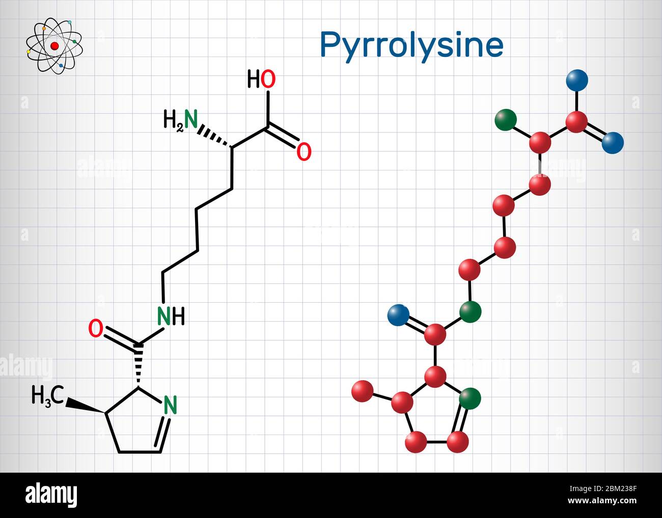 Pyrrolysine, l-pyrrolysine, Pyl, C12H21N3O3 molecule. It is amino acid, is used in biosynthesis of proteins. Structural chemical formula and molecule Stock Vectorhttps://www.alamy.com/image-license-details/?v=1https://www.alamy.com/pyrrolysine-l-pyrrolysine-pyl-c12h21n3o3-molecule-it-is-amino-acid-is-used-in-biosynthesis-of-proteins-structural-chemical-formula-and-molecule-image356546975.html
RF2BM238F–Pyrrolysine, l-pyrrolysine, Pyl, C12H21N3O3 molecule. It is amino acid, is used in biosynthesis of proteins. Structural chemical formula and molecule
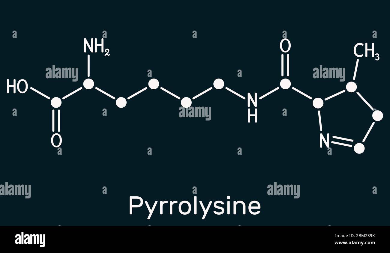 Pyrrolysine, l-pyrrolysine, Pyl, C12H21N3O3 molecule. It is amino acid, is used in biosynthesis of proteins. Skeletal chemical formula on the dark blu Stock Photohttps://www.alamy.com/image-license-details/?v=1https://www.alamy.com/pyrrolysine-l-pyrrolysine-pyl-c12h21n3o3-molecule-it-is-amino-acid-is-used-in-biosynthesis-of-proteins-skeletal-chemical-formula-on-the-dark-blu-image356547007.html
RF2BM239K–Pyrrolysine, l-pyrrolysine, Pyl, C12H21N3O3 molecule. It is amino acid, is used in biosynthesis of proteins. Skeletal chemical formula on the dark blu
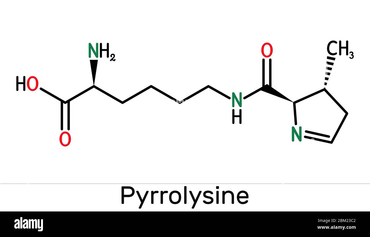 Pyrrolysine, l-pyrrolysine, Pyl, C12H21N3O3 molecule. It is amino acid, is used in biosynthesis of proteins. Skeletal chemical formula. Illustration Stock Photohttps://www.alamy.com/image-license-details/?v=1https://www.alamy.com/pyrrolysine-l-pyrrolysine-pyl-c12h21n3o3-molecule-it-is-amino-acid-is-used-in-biosynthesis-of-proteins-skeletal-chemical-formula-illustration-image356547074.html
RF2BM23C2–Pyrrolysine, l-pyrrolysine, Pyl, C12H21N3O3 molecule. It is amino acid, is used in biosynthesis of proteins. Skeletal chemical formula. Illustration
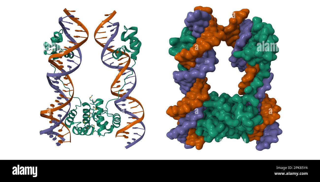 DNA-binding domain of human SETMAR in complex with Hsmar1 terminal inverted repeat (TIR) DNA. 3D cartoon and Gaussian surface models, PDB 7s03 Stock Photohttps://www.alamy.com/image-license-details/?v=1https://www.alamy.com/dna-binding-domain-of-human-setmar-in-complex-with-hsmar1-terminal-inverted-repeat-tir-dna-3d-cartoon-and-gaussian-surface-models-pdb-7s03-image545380168.html
RF2PK85Y4–DNA-binding domain of human SETMAR in complex with Hsmar1 terminal inverted repeat (TIR) DNA. 3D cartoon and Gaussian surface models, PDB 7s03
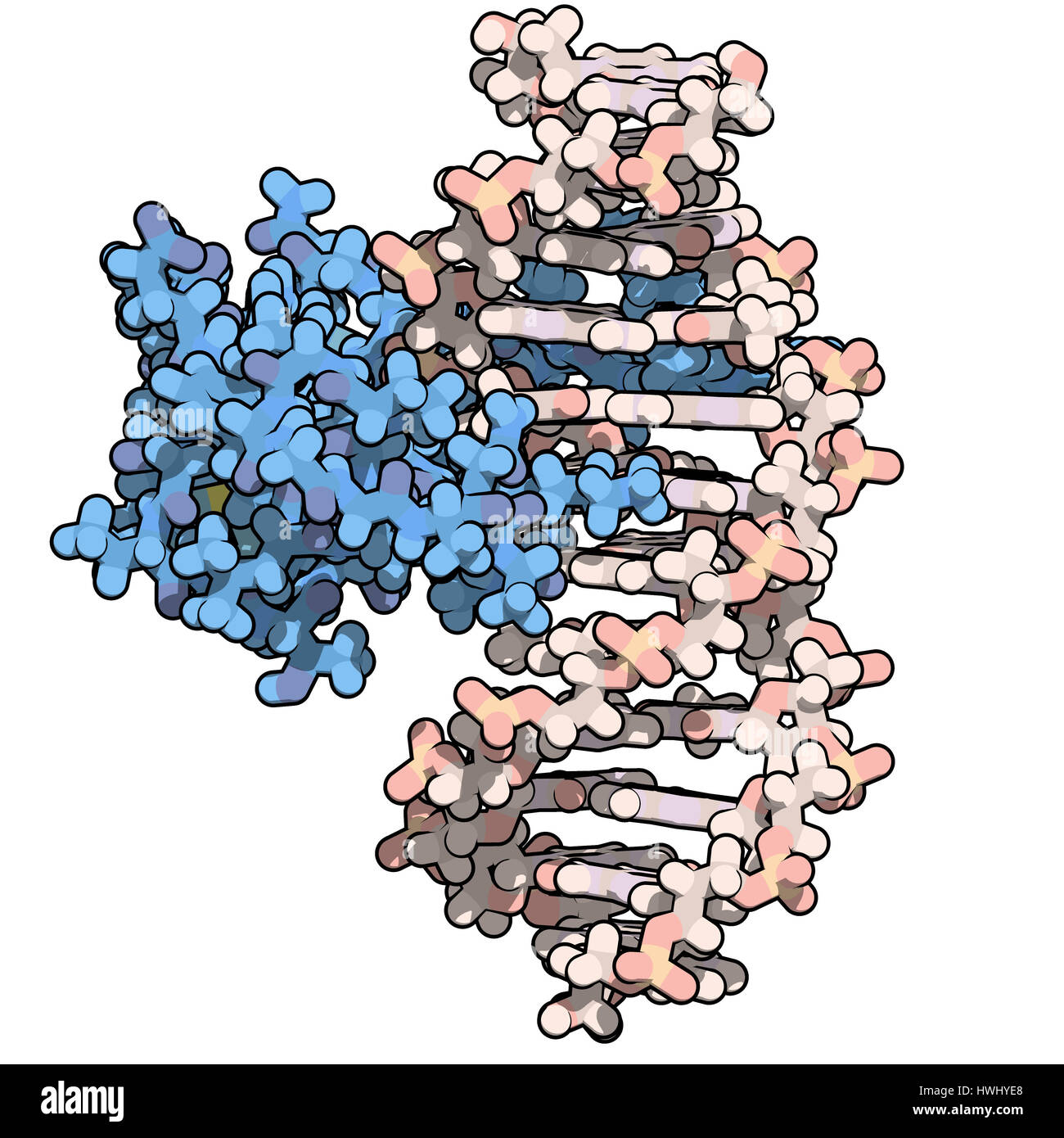 Zinc finger protein domain (from histone-lysine N-methyltransferase A2). Zinc fingers are protein domains that bind to DNA sequences. Atoms are repres Stock Photohttps://www.alamy.com/image-license-details/?v=1https://www.alamy.com/stock-photo-zinc-finger-protein-domain-from-histone-lysine-n-methyltransferase-136233728.html
RFHWHYE8–Zinc finger protein domain (from histone-lysine N-methyltransferase A2). Zinc fingers are protein domains that bind to DNA sequences. Atoms are repres
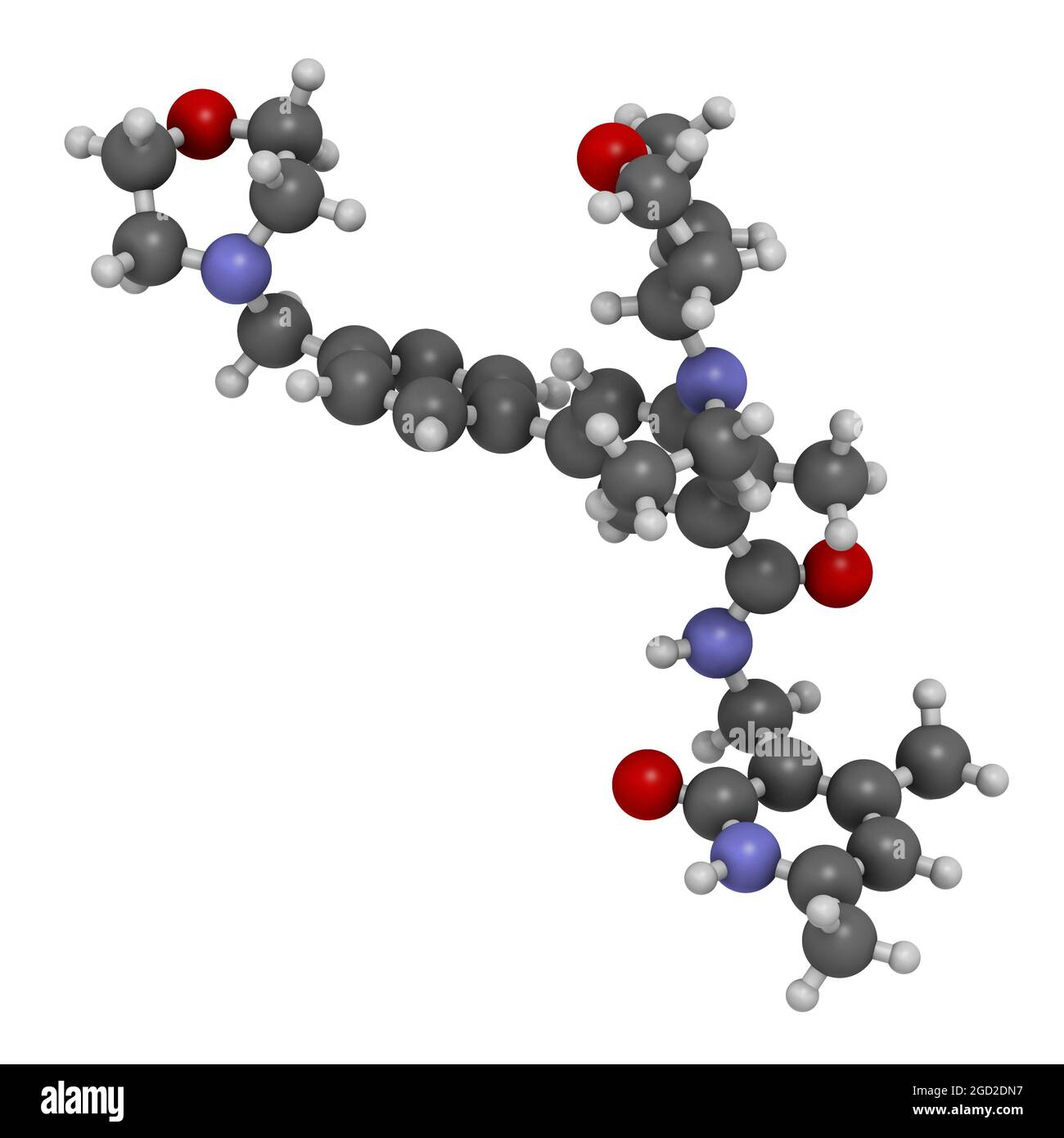 Tazemetostat cancer drug molecule. 3D rendering. Stock Photohttps://www.alamy.com/image-license-details/?v=1https://www.alamy.com/tazemetostat-cancer-drug-molecule-3d-rendering-image438304419.html
RF2GD2DN7–Tazemetostat cancer drug molecule. 3D rendering.
 Guadecitabine cancer drug molecule (DNA methyltransferase inhibitor). Stylized skeletal formula (chemical structure): Atoms are shown as color-coded circles: hydrogen (white), carbon (grey), nitrogen (blue), oxygen (red), phosphorus (orange). Stock Photohttps://www.alamy.com/image-license-details/?v=1https://www.alamy.com/guadecitabine-cancer-drug-molecule-dna-methyltransferase-inhibitor-stylized-skeletal-formula-chemical-structure-atoms-are-shown-as-color-coded-circles-hydrogen-white-carbon-grey-nitrogen-blue-oxygen-red-phosphorus-orange-image364971814.html
RF2C5NW7J–Guadecitabine cancer drug molecule (DNA methyltransferase inhibitor). Stylized skeletal formula (chemical structure): Atoms are shown as color-coded circles: hydrogen (white), carbon (grey), nitrogen (blue), oxygen (red), phosphorus (orange).
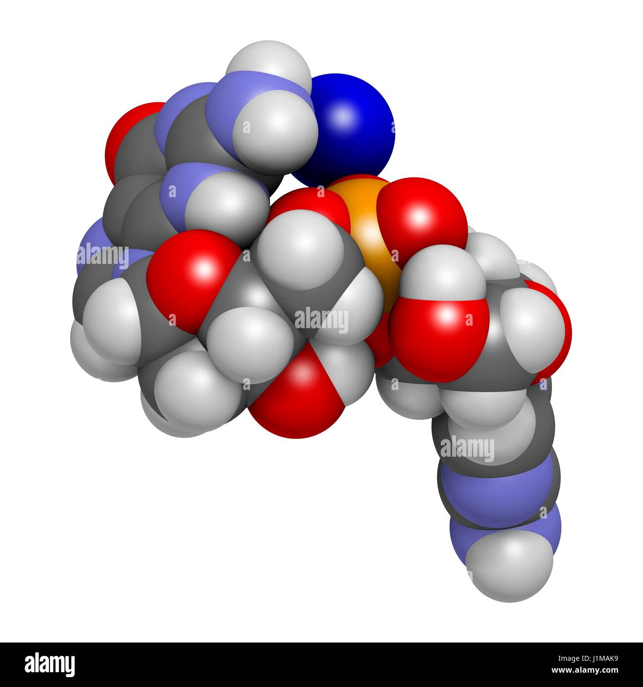 Guadecitabine cancer drug molecule (DNA methyltransferase inhibitor). 3D rendering. Atoms are represented as spheres with conventional colour coding: hydrogen (white), carbon (grey), nitrogen (blue), oxygen (red), phosphorus (orange). Stock Photohttps://www.alamy.com/image-license-details/?v=1https://www.alamy.com/stock-photo-guadecitabine-cancer-drug-molecule-dna-methyltransferase-inhibitor-138745021.html
RFJ1MAK9–Guadecitabine cancer drug molecule (DNA methyltransferase inhibitor). 3D rendering. Atoms are represented as spheres with conventional colour coding: hydrogen (white), carbon (grey), nitrogen (blue), oxygen (red), phosphorus (orange).
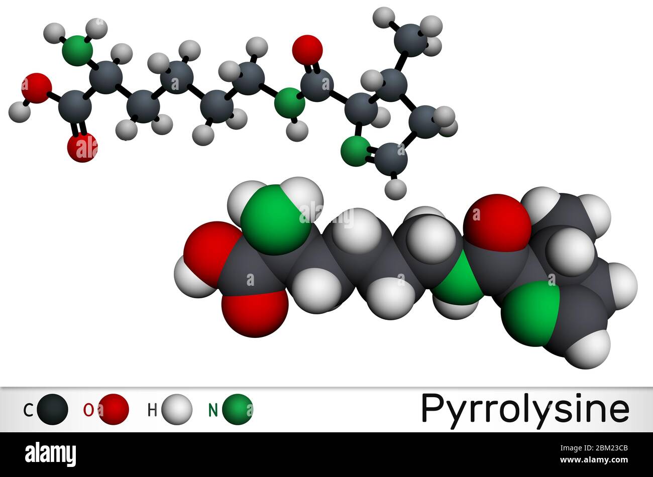 Pyrrolysine, l-pyrrolysine, Pyl, C12H21N3O3 molecule. It is amino acid, is used in biosynthesis of proteins. Molecular model. 3D rendering Stock Photohttps://www.alamy.com/image-license-details/?v=1https://www.alamy.com/pyrrolysine-l-pyrrolysine-pyl-c12h21n3o3-molecule-it-is-amino-acid-is-used-in-biosynthesis-of-proteins-molecular-model-3d-rendering-image356547083.html
RF2BM23CB–Pyrrolysine, l-pyrrolysine, Pyl, C12H21N3O3 molecule. It is amino acid, is used in biosynthesis of proteins. Molecular model. 3D rendering
 Cryo-EM structure of retinoblastoma-binding protein 5 (green) bound to the nucleosome. 3D cartoon and Gaussian surface models, PDB 6pwx Stock Photohttps://www.alamy.com/image-license-details/?v=1https://www.alamy.com/cryo-em-structure-of-retinoblastoma-binding-protein-5-green-bound-to-the-nucleosome-3d-cartoon-and-gaussian-surface-models-pdb-6pwx-image590777105.html
RF2W94669–Cryo-EM structure of retinoblastoma-binding protein 5 (green) bound to the nucleosome. 3D cartoon and Gaussian surface models, PDB 6pwx
 Amodiaquine, ADQ molecule. It is aminoquinoline, used for the therapy of malaria. Molecular model. 3D rendering. Illustration Stock Photohttps://www.alamy.com/image-license-details/?v=1https://www.alamy.com/amodiaquine-adq-molecule-it-is-aminoquinoline-used-for-the-therapy-of-malaria-molecular-model-3d-rendering-illustration-image475073526.html
RF2JGWD3J–Amodiaquine, ADQ molecule. It is aminoquinoline, used for the therapy of malaria. Molecular model. 3D rendering. Illustration
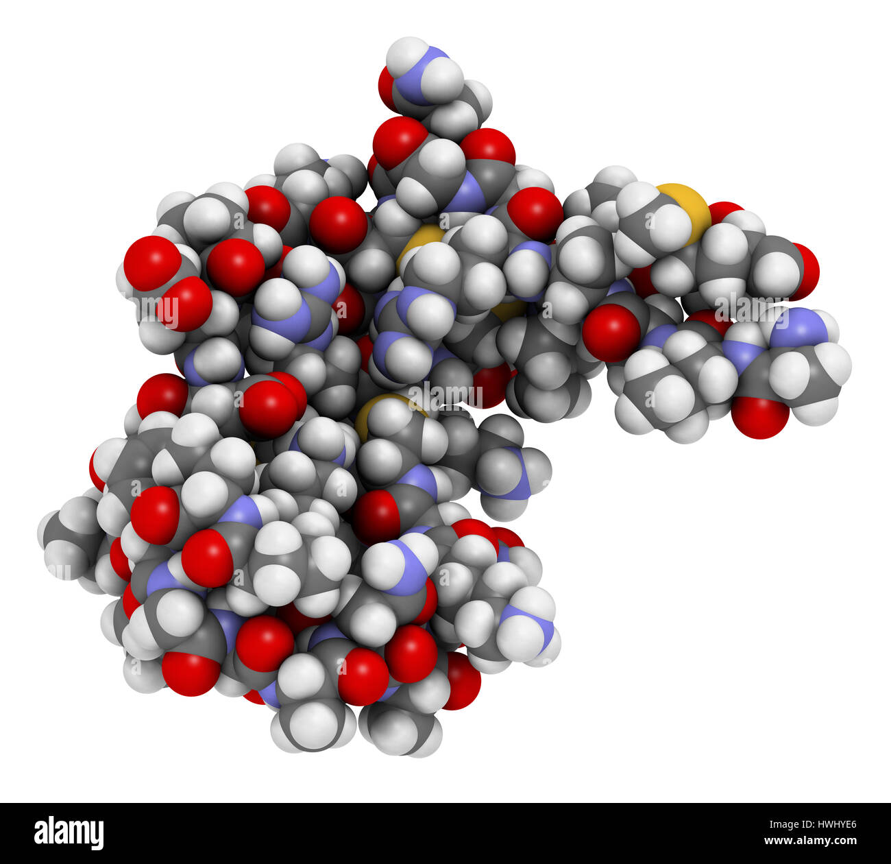 Zinc finger protein domain (from histone-lysine N-methyltransferase A2). Zinc fingers are protein domains that bind to DNA sequences. Atoms are repres Stock Photohttps://www.alamy.com/image-license-details/?v=1https://www.alamy.com/stock-photo-zinc-finger-protein-domain-from-histone-lysine-n-methyltransferase-136233726.html
RFHWHYE6–Zinc finger protein domain (from histone-lysine N-methyltransferase A2). Zinc fingers are protein domains that bind to DNA sequences. Atoms are repres
 Amodiaquine, ADQ molecule. It is aminoquinoline, used for the therapy of malaria. Molecular model. 3D rendering. Illustration Stock Photohttps://www.alamy.com/image-license-details/?v=1https://www.alamy.com/amodiaquine-adq-molecule-it-is-aminoquinoline-used-for-the-therapy-of-malaria-molecular-model-3d-rendering-illustration-image475073524.html
RF2JGWD3G–Amodiaquine, ADQ molecule. It is aminoquinoline, used for the therapy of malaria. Molecular model. 3D rendering. Illustration
 Decibatine drug molecule. 3D rendering. Stock Photohttps://www.alamy.com/image-license-details/?v=1https://www.alamy.com/decibatine-drug-molecule-3d-rendering-image438473280.html
RF2GDA540–Decibatine drug molecule. 3D rendering.
 Amodiaquine, ADQ molecule. It is aminoquinoline, used for the therapy of malaria. Structural chemical formula, molecule model. Vector illustration Stock Vectorhttps://www.alamy.com/image-license-details/?v=1https://www.alamy.com/amodiaquine-adq-molecule-it-is-aminoquinoline-used-for-the-therapy-of-malaria-structural-chemical-formula-molecule-model-vector-illustration-image490638779.html
RF2KE6ENF–Amodiaquine, ADQ molecule. It is aminoquinoline, used for the therapy of malaria. Structural chemical formula, molecule model. Vector illustration
 Guadecitabine cancer drug molecule (DNA methyltransferase inhibitor). 3D rendering. Atoms are represented as spheres with conventional colour coding: hydrogen (white), carbon (grey), nitrogen (blue), oxygen (red), phosphorus (orange). Stock Photohttps://www.alamy.com/image-license-details/?v=1https://www.alamy.com/stock-photo-guadecitabine-cancer-drug-molecule-dna-methyltransferase-inhibitor-138745014.html
RFJ1MAK2–Guadecitabine cancer drug molecule (DNA methyltransferase inhibitor). 3D rendering. Atoms are represented as spheres with conventional colour coding: hydrogen (white), carbon (grey), nitrogen (blue), oxygen (red), phosphorus (orange).
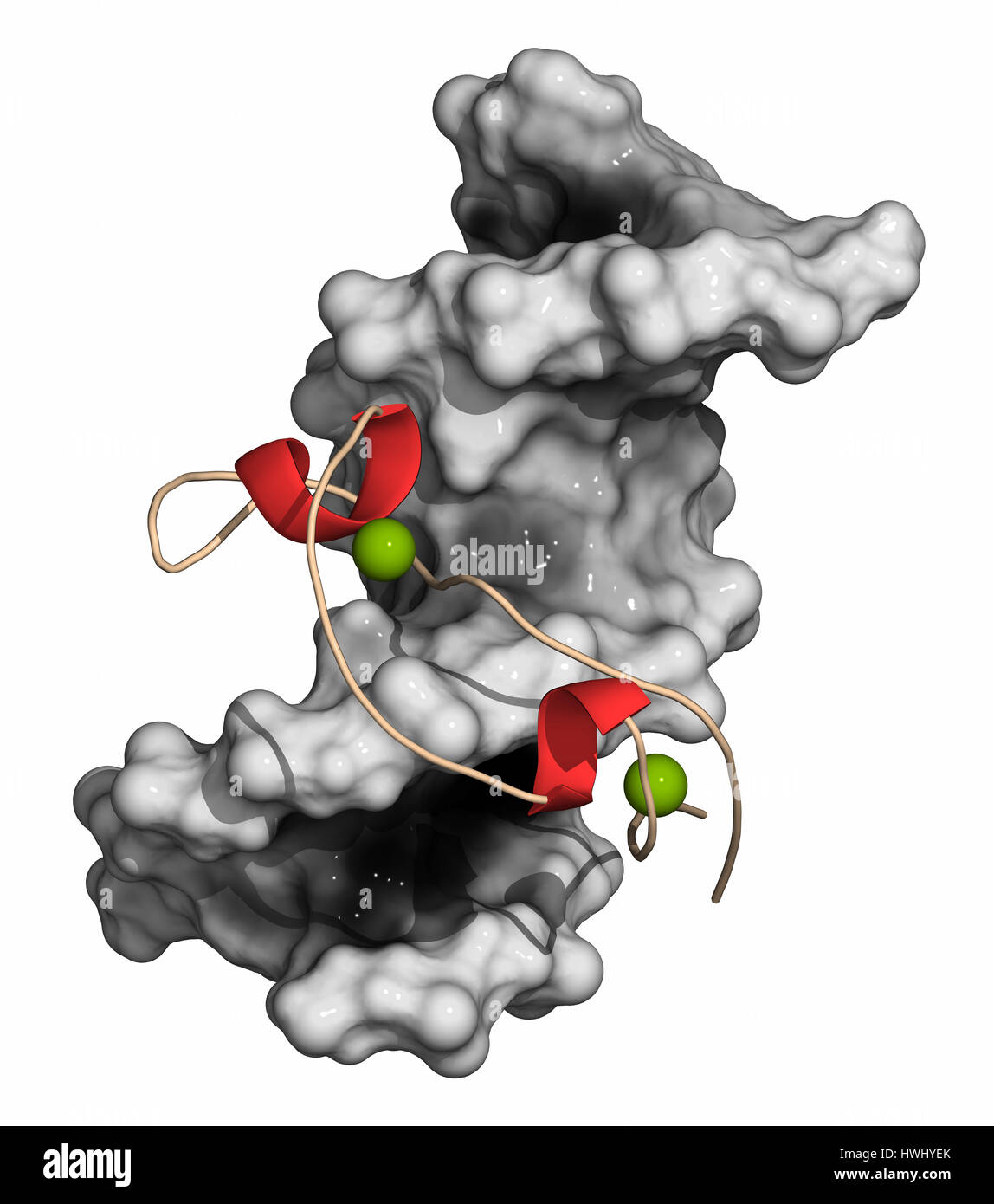 Zinc finger protein domain (from histone-lysine N-methyltransferase A2). Zinc fingers are protein domains that bind to DNA sequences. DNA: molecular s Stock Photohttps://www.alamy.com/image-license-details/?v=1https://www.alamy.com/stock-photo-zinc-finger-protein-domain-from-histone-lysine-n-methyltransferase-136233739.html
RFHWHYEK–Zinc finger protein domain (from histone-lysine N-methyltransferase A2). Zinc fingers are protein domains that bind to DNA sequences. DNA: molecular s
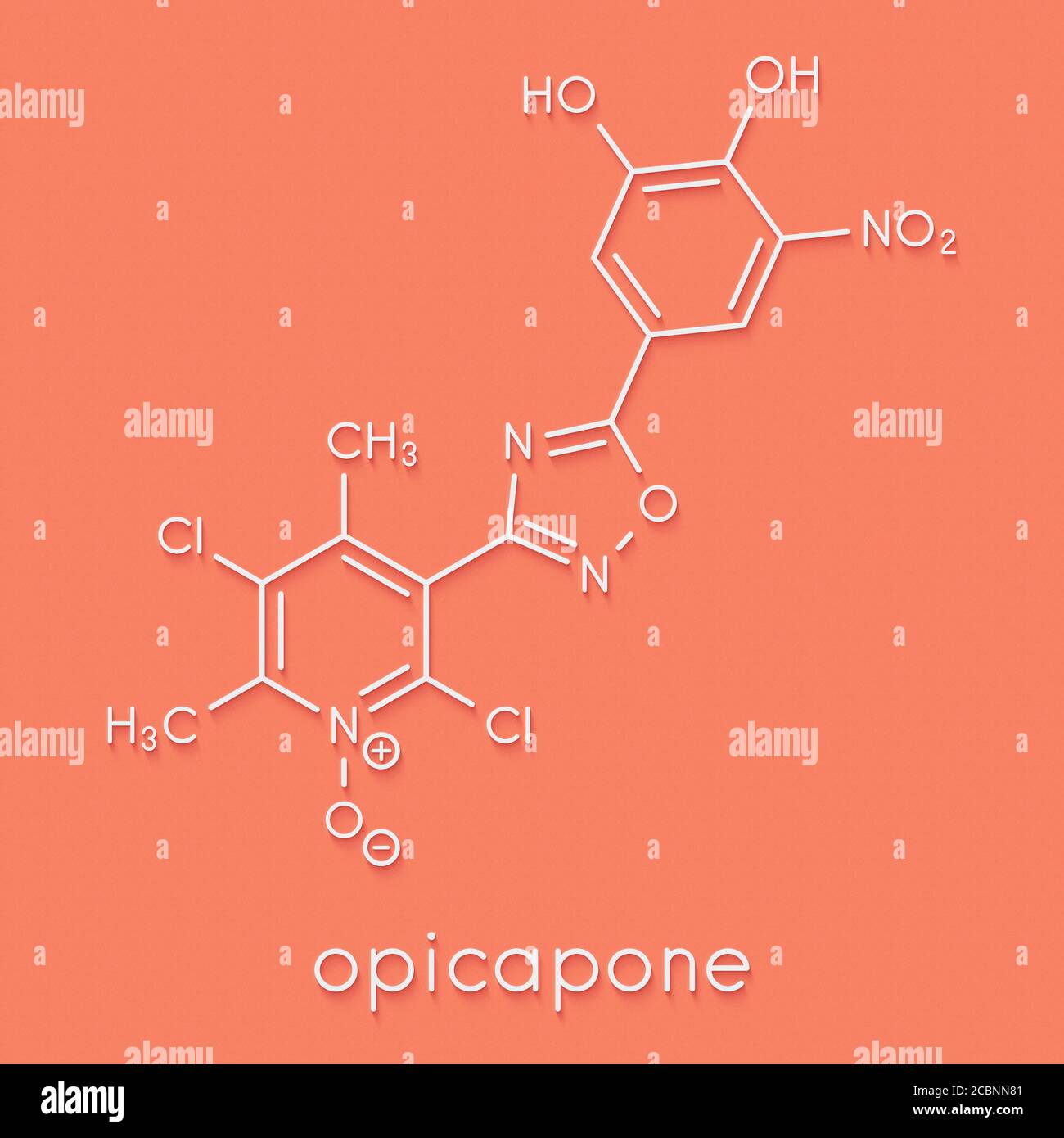 Opicapone Parkinson's disease drug molecule. Skeletal formula. Stock Photohttps://www.alamy.com/image-license-details/?v=1https://www.alamy.com/opicapone-parkinsons-disease-drug-molecule-skeletal-formula-image368656625.html
RF2CBNN81–Opicapone Parkinson's disease drug molecule. Skeletal formula.
 Amodiaquine, ADQ molecule. It is aminoquinoline, used for the therapy of malaria. Structural chemical formula, molecule model. Sheet of paper in a cag Stock Vectorhttps://www.alamy.com/image-license-details/?v=1https://www.alamy.com/amodiaquine-adq-molecule-it-is-aminoquinoline-used-for-the-therapy-of-malaria-structural-chemical-formula-molecule-model-sheet-of-paper-in-a-cag-image490641612.html
RF2KE6JAM–Amodiaquine, ADQ molecule. It is aminoquinoline, used for the therapy of malaria. Structural chemical formula, molecule model. Sheet of paper in a cag
 Pyrrolysine (l-pyrrolysine, Pyl, O) amino acid molecule. 3D rendering. Scaled sphere molecular model shown floating just above a liquid paint surface. Stock Photohttps://www.alamy.com/image-license-details/?v=1https://www.alamy.com/pyrrolysine-l-pyrrolysine-pyl-o-amino-acid-molecule-3d-rendering-scaled-sphere-molecular-model-shown-floating-just-above-a-liquid-paint-surface-image397082077.html
RF2E20J6N–Pyrrolysine (l-pyrrolysine, Pyl, O) amino acid molecule. 3D rendering. Scaled sphere molecular model shown floating just above a liquid paint surface.
 Guadecitabine cancer drug molecule (DNA methyltransferase inhibitor). 3D rendering. Atoms are represented as spheres with conventional colour coding: hydrogen (white), carbon (grey), nitrogen (blue), oxygen (red), phosphorus (orange). Stock Photohttps://www.alamy.com/image-license-details/?v=1https://www.alamy.com/stock-photo-guadecitabine-cancer-drug-molecule-dna-methyltransferase-inhibitor-138745004.html
RFJ1MAJM–Guadecitabine cancer drug molecule (DNA methyltransferase inhibitor). 3D rendering. Atoms are represented as spheres with conventional colour coding: hydrogen (white), carbon (grey), nitrogen (blue), oxygen (red), phosphorus (orange).
 Pyrrolysine (l-pyrrolysine, Pyl, O) amino acid molecule. 3D rendering. Atoms are represented as spheres with conventional color coding: hydrogen (whit Stock Photohttps://www.alamy.com/image-license-details/?v=1https://www.alamy.com/pyrrolysine-l-pyrrolysine-pyl-o-amino-acid-molecule-3d-rendering-atoms-are-represented-as-spheres-with-conventional-color-coding-hydrogen-whit-image397082078.html
RF2E20J6P–Pyrrolysine (l-pyrrolysine, Pyl, O) amino acid molecule. 3D rendering. Atoms are represented as spheres with conventional color coding: hydrogen (whit
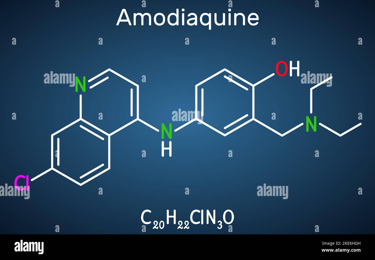 Amodiaquine, ADQ molecule. It is aminoquinoline, used for the therapy of malaria. Structural chemical formula on the dark blue background. Vector illu Stock Vectorhttps://www.alamy.com/image-license-details/?v=1https://www.alamy.com/amodiaquine-adq-molecule-it-is-aminoquinoline-used-for-the-therapy-of-malaria-structural-chemical-formula-on-the-dark-blue-background-vector-illu-image490640993.html
RF2KE6HGH–Amodiaquine, ADQ molecule. It is aminoquinoline, used for the therapy of malaria. Structural chemical formula on the dark blue background. Vector illu
 Guadecitabine cancer drug molecule (DNA methyltransferase inhibitor). 3D rendering. Atoms are represented as spheres with conventional colour coding: hydrogen (white), carbon (black), nitrogen (blue), oxygen (red), phosphorus (orange). Stock Photohttps://www.alamy.com/image-license-details/?v=1https://www.alamy.com/stock-photo-guadecitabine-cancer-drug-molecule-dna-methyltransferase-inhibitor-138745025.html
RFJ1MAKD–Guadecitabine cancer drug molecule (DNA methyltransferase inhibitor). 3D rendering. Atoms are represented as spheres with conventional colour coding: hydrogen (white), carbon (black), nitrogen (blue), oxygen (red), phosphorus (orange).
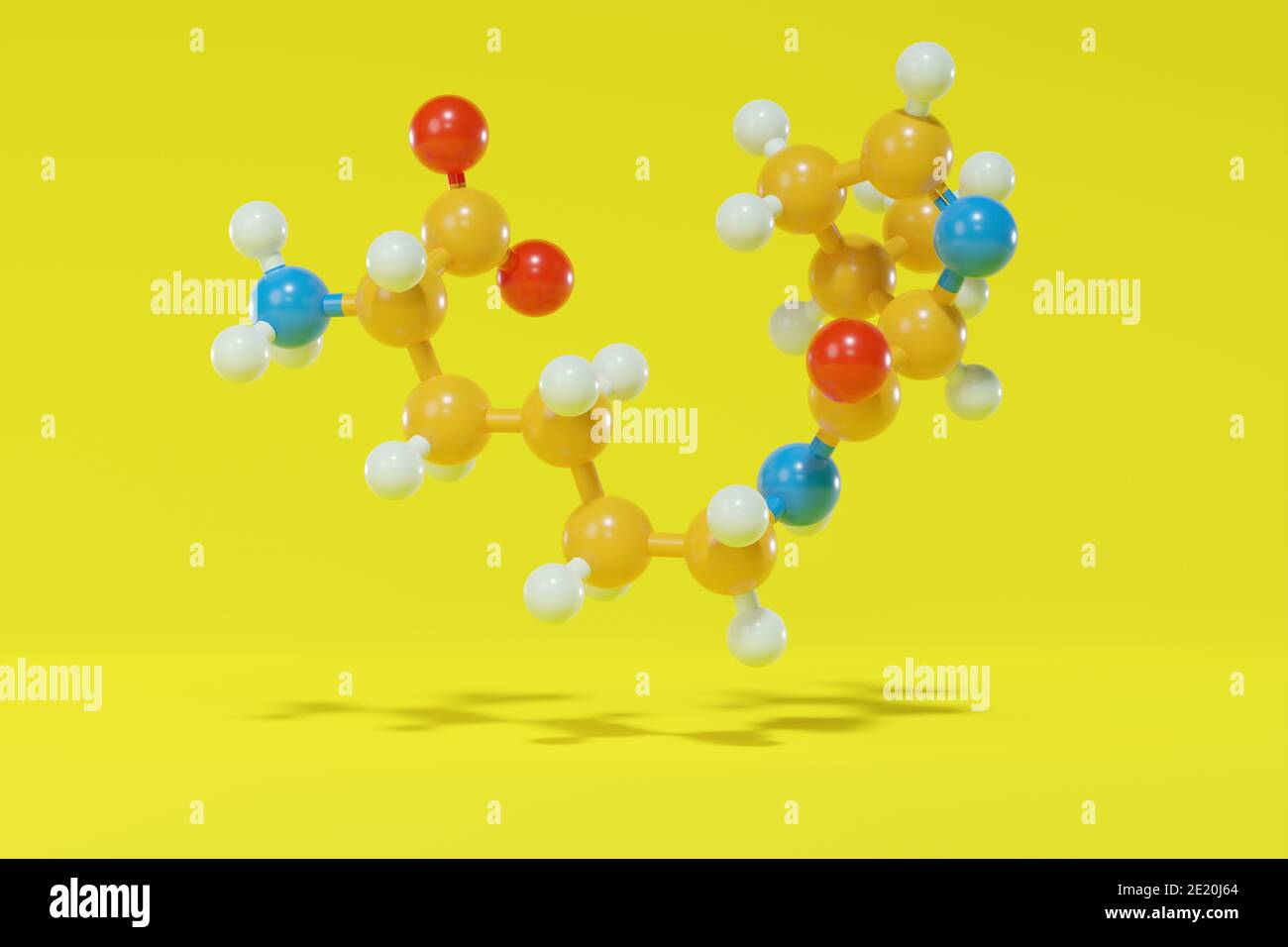 Pyrrolysine (l-pyrrolysine, Pyl, O) amino acid molecule. 3D rendering. Ball and stick molecular model with atoms shown as color-coded spheres: hydroge Stock Photohttps://www.alamy.com/image-license-details/?v=1https://www.alamy.com/pyrrolysine-l-pyrrolysine-pyl-o-amino-acid-molecule-3d-rendering-ball-and-stick-molecular-model-with-atoms-shown-as-color-coded-spheres-hydroge-image397082060.html
RF2E20J64–Pyrrolysine (l-pyrrolysine, Pyl, O) amino acid molecule. 3D rendering. Ball and stick molecular model with atoms shown as color-coded spheres: hydroge
 Pyrrolysine (l-pyrrolysine, Pyl, O) amino acid molecule. 3D rendering. Atoms are shown as spheres with conventional color coding: hydrogen white, carb Stock Photohttps://www.alamy.com/image-license-details/?v=1https://www.alamy.com/pyrrolysine-l-pyrrolysine-pyl-o-amino-acid-molecule-3d-rendering-atoms-are-shown-as-spheres-with-conventional-color-coding-hydrogen-white-carb-image397082075.html
RF2E20J6K–Pyrrolysine (l-pyrrolysine, Pyl, O) amino acid molecule. 3D rendering. Atoms are shown as spheres with conventional color coding: hydrogen white, carb
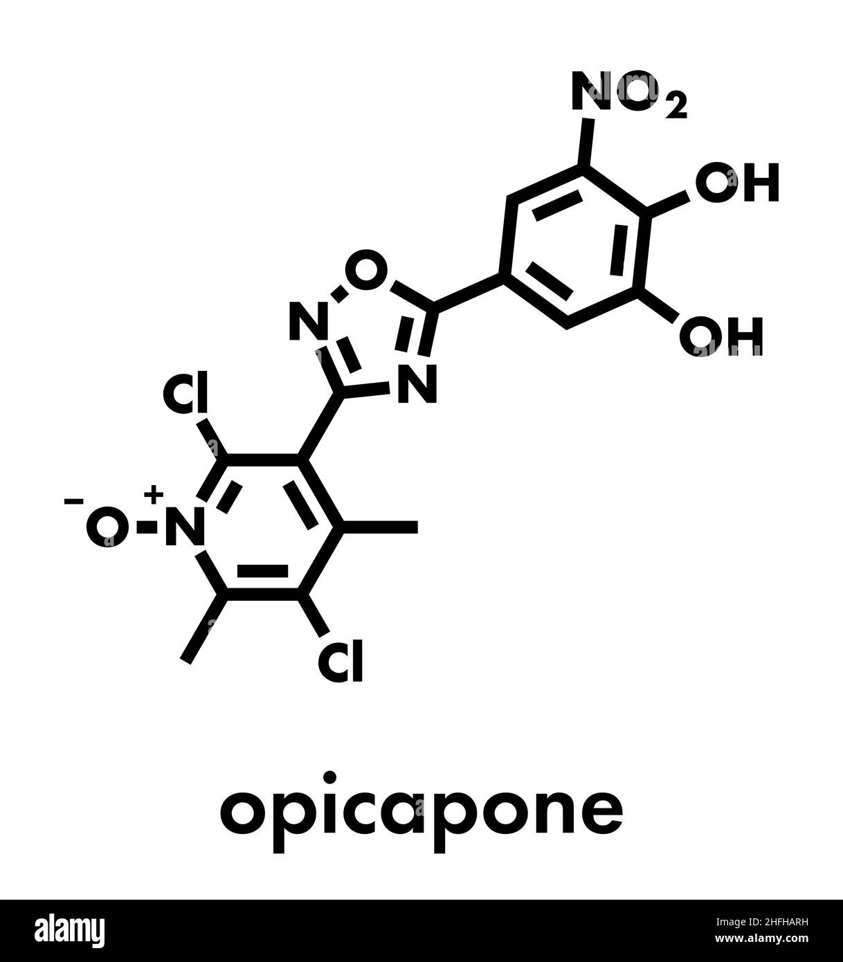 Opicapone Parkinson's disease drug molecule. Skeletal formula. Stock Vectorhttps://www.alamy.com/image-license-details/?v=1https://www.alamy.com/opicapone-parkinsons-disease-drug-molecule-skeletal-formula-image457071093.html
RF2HFHARH–Opicapone Parkinson's disease drug molecule. Skeletal formula.
 Amodiaquine, ADQ molecule. It is aminoquinoline, used for the therapy of malaria. Skeletal chemical formula. Paper packaging for drugs. Vector illustr Stock Vectorhttps://www.alamy.com/image-license-details/?v=1https://www.alamy.com/amodiaquine-adq-molecule-it-is-aminoquinoline-used-for-the-therapy-of-malaria-skeletal-chemical-formula-paper-packaging-for-drugs-vector-illustr-image490640858.html
RF2KE6HBP–Amodiaquine, ADQ molecule. It is aminoquinoline, used for the therapy of malaria. Skeletal chemical formula. Paper packaging for drugs. Vector illustr
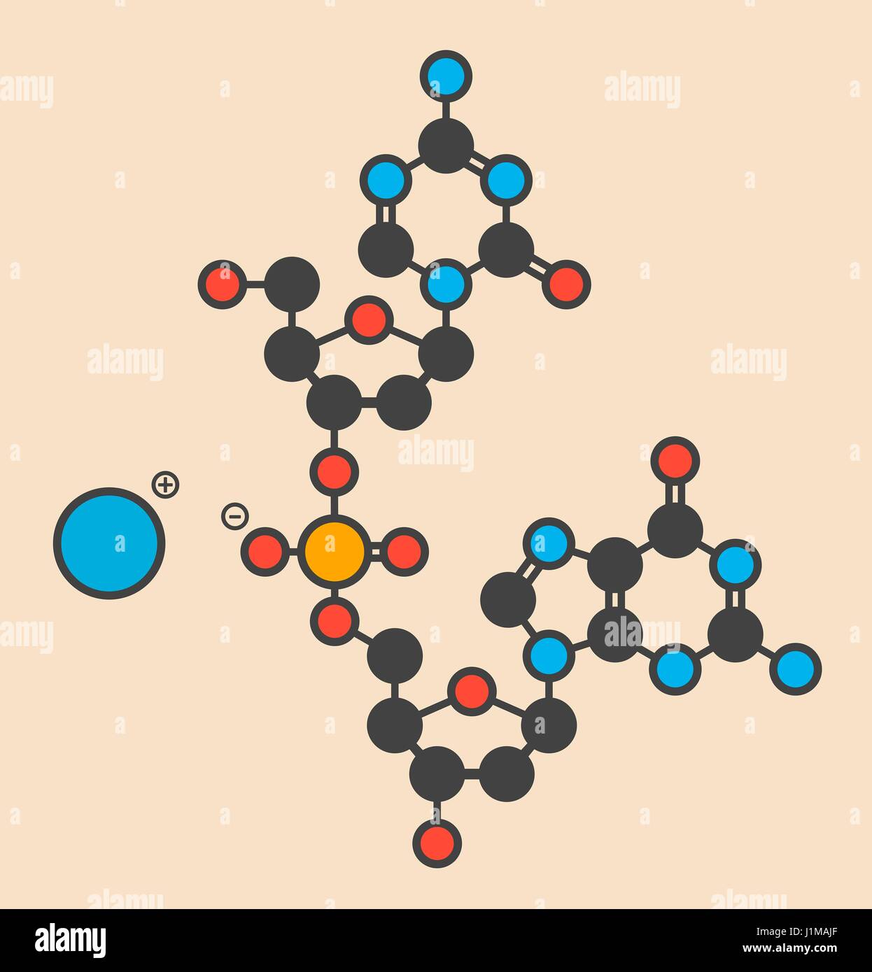 Guadecitabine cancer drug molecule (DNA methyltransferase inhibitor). Stylized skeletal formula (chemical structure): Atoms are shown as color-coded circles: hydrogen (hidden), carbon (grey), nitrogen (blue), oxygen (red), phosphorus (orange). Stock Photohttps://www.alamy.com/image-license-details/?v=1https://www.alamy.com/stock-photo-guadecitabine-cancer-drug-molecule-dna-methyltransferase-inhibitor-138744999.html
RFJ1MAJF–Guadecitabine cancer drug molecule (DNA methyltransferase inhibitor). Stylized skeletal formula (chemical structure): Atoms are shown as color-coded circles: hydrogen (hidden), carbon (grey), nitrogen (blue), oxygen (red), phosphorus (orange).
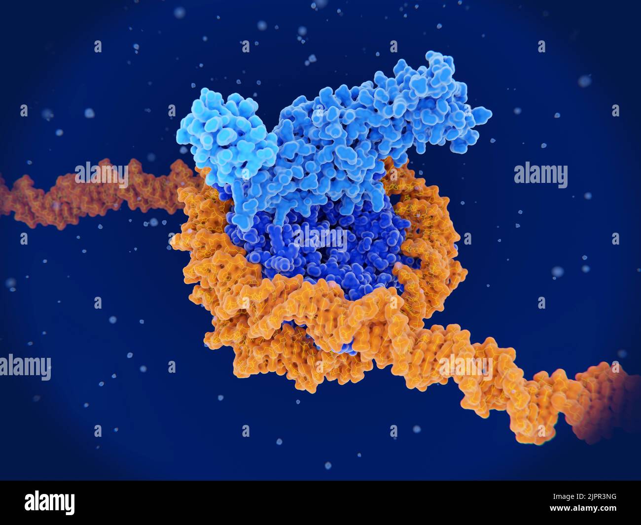 Histone methylation, molecular model Stock Photohttps://www.alamy.com/image-license-details/?v=1https://www.alamy.com/histone-methylation-molecular-model-image478710220.html
RF2JPR3NG–Histone methylation, molecular model
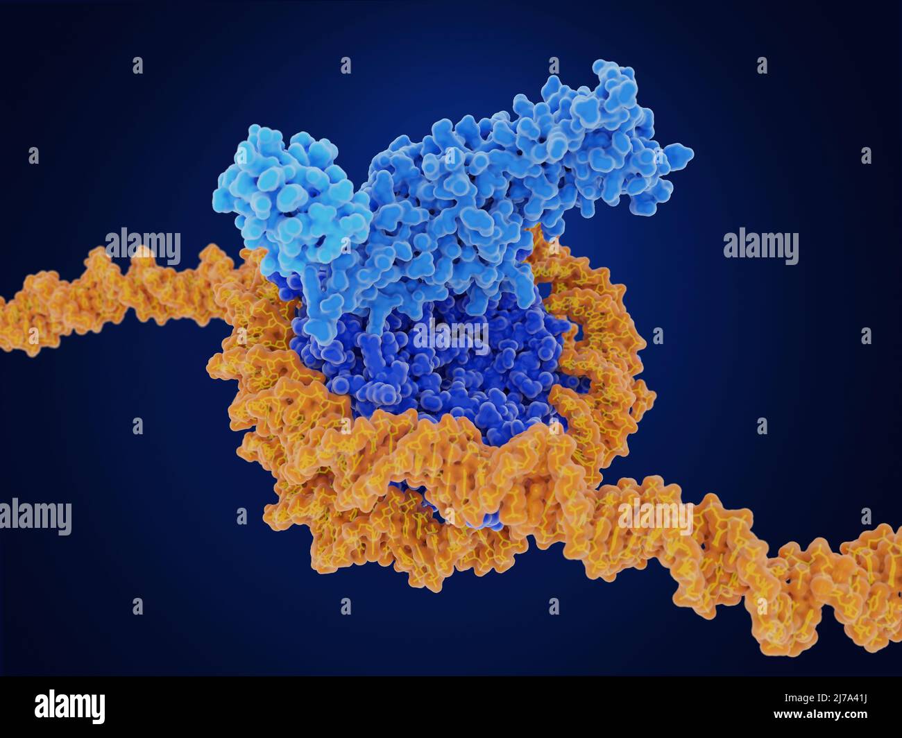 Histone methylation, molecular model Stock Photohttps://www.alamy.com/image-license-details/?v=1https://www.alamy.com/histone-methylation-molecular-model-image469205230.html
RF2J7A41J–Histone methylation, molecular model
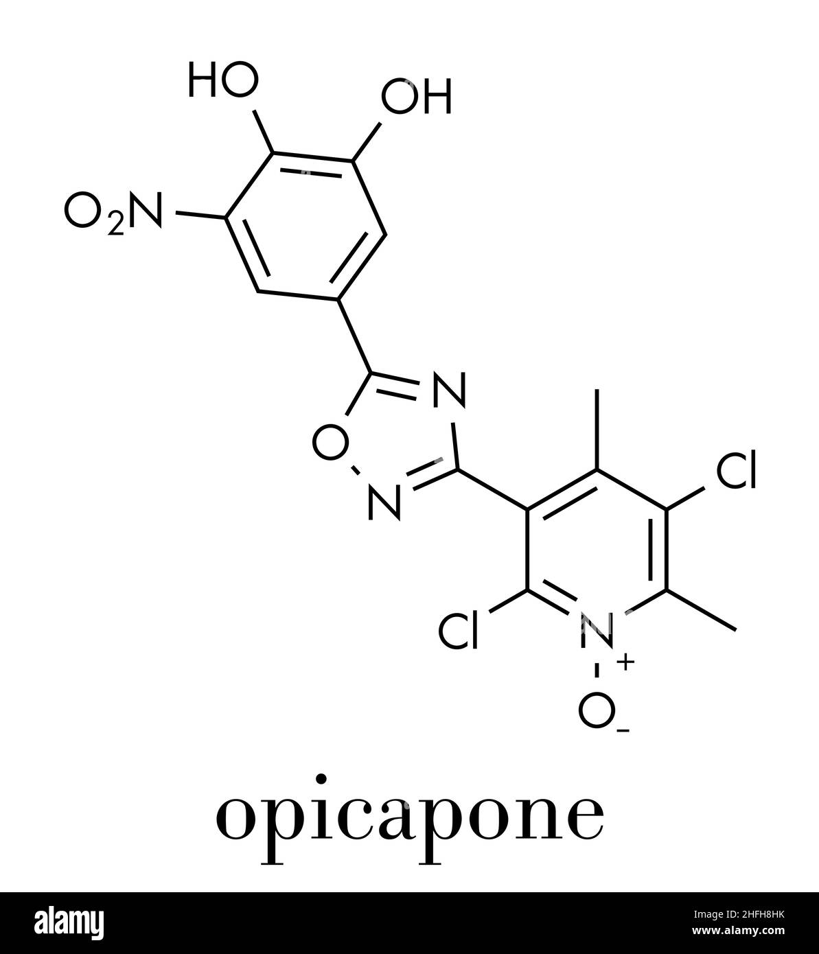 Opicapone Parkinson's disease drug molecule. Skeletal formula. Stock Vectorhttps://www.alamy.com/image-license-details/?v=1https://www.alamy.com/opicapone-parkinsons-disease-drug-molecule-skeletal-formula-image457069359.html
RF2HFH8HK–Opicapone Parkinson's disease drug molecule. Skeletal formula.
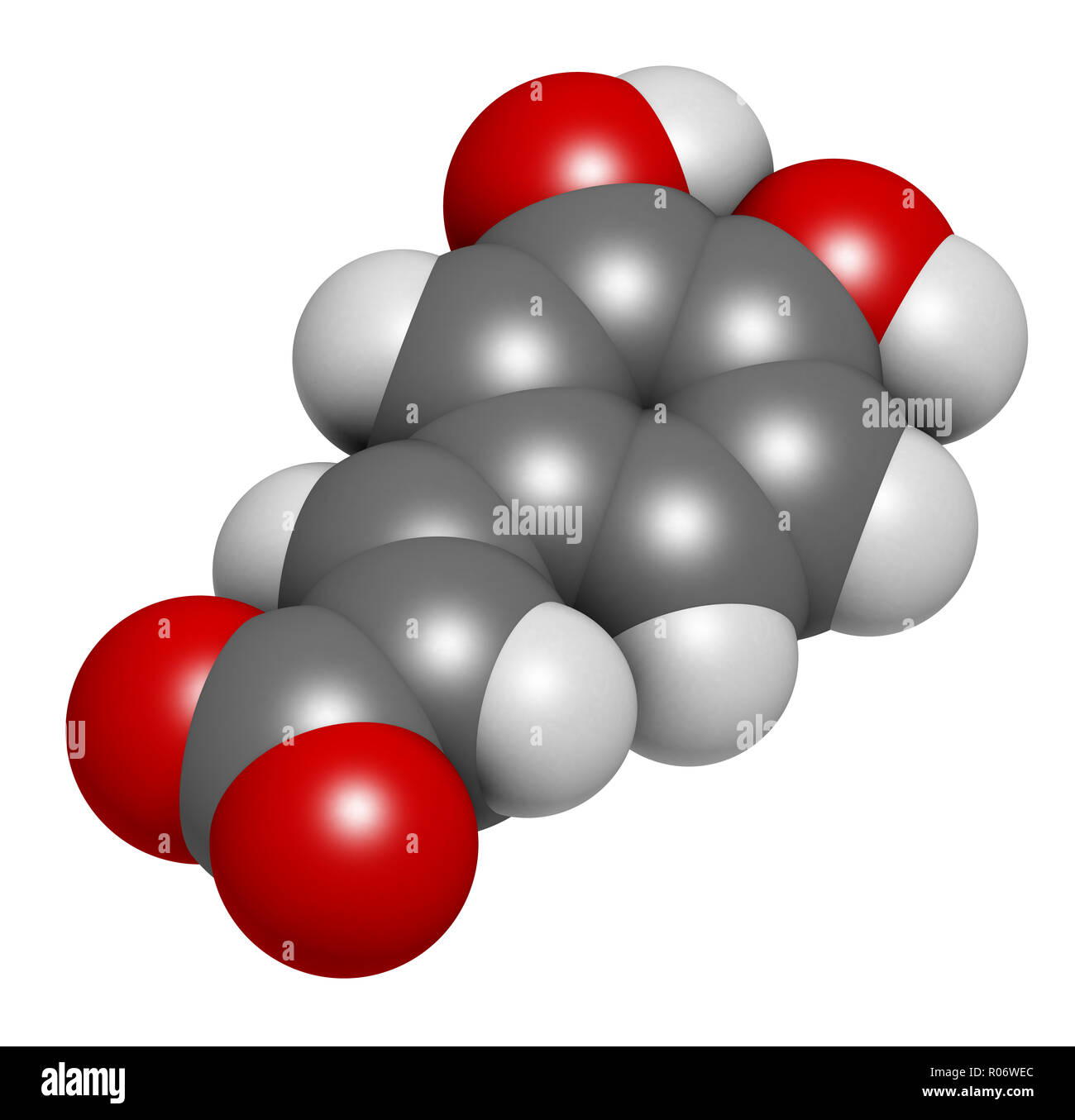 Caffeic acid molecule. Intermediate in the biosynthesis of lignin. 3D rendering. Atoms are represented as spheres with conventional color coding: hydr Stock Photohttps://www.alamy.com/image-license-details/?v=1https://www.alamy.com/caffeic-acid-molecule-intermediate-in-the-biosynthesis-of-lignin-3d-rendering-atoms-are-represented-as-spheres-with-conventional-color-coding-hydr-image223886500.html
RFR06WEC–Caffeic acid molecule. Intermediate in the biosynthesis of lignin. 3D rendering. Atoms are represented as spheres with conventional color coding: hydr
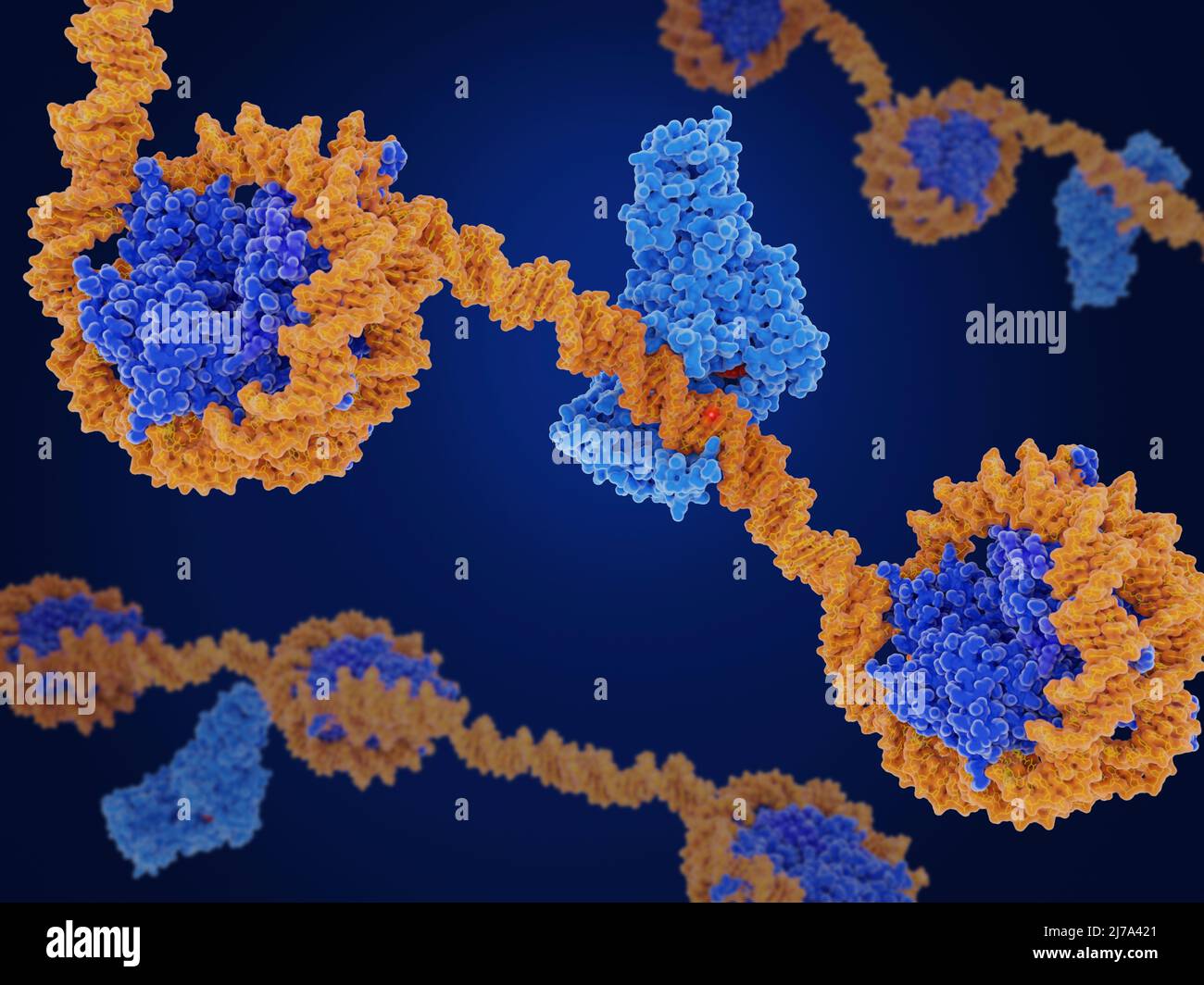 DNA methyl transferase--1 and DNA, illustration Stock Photohttps://www.alamy.com/image-license-details/?v=1https://www.alamy.com/dna-methyl-transferase-1-and-dna-illustration-image469205241.html
RF2J7A421–DNA methyl transferase--1 and DNA, illustration
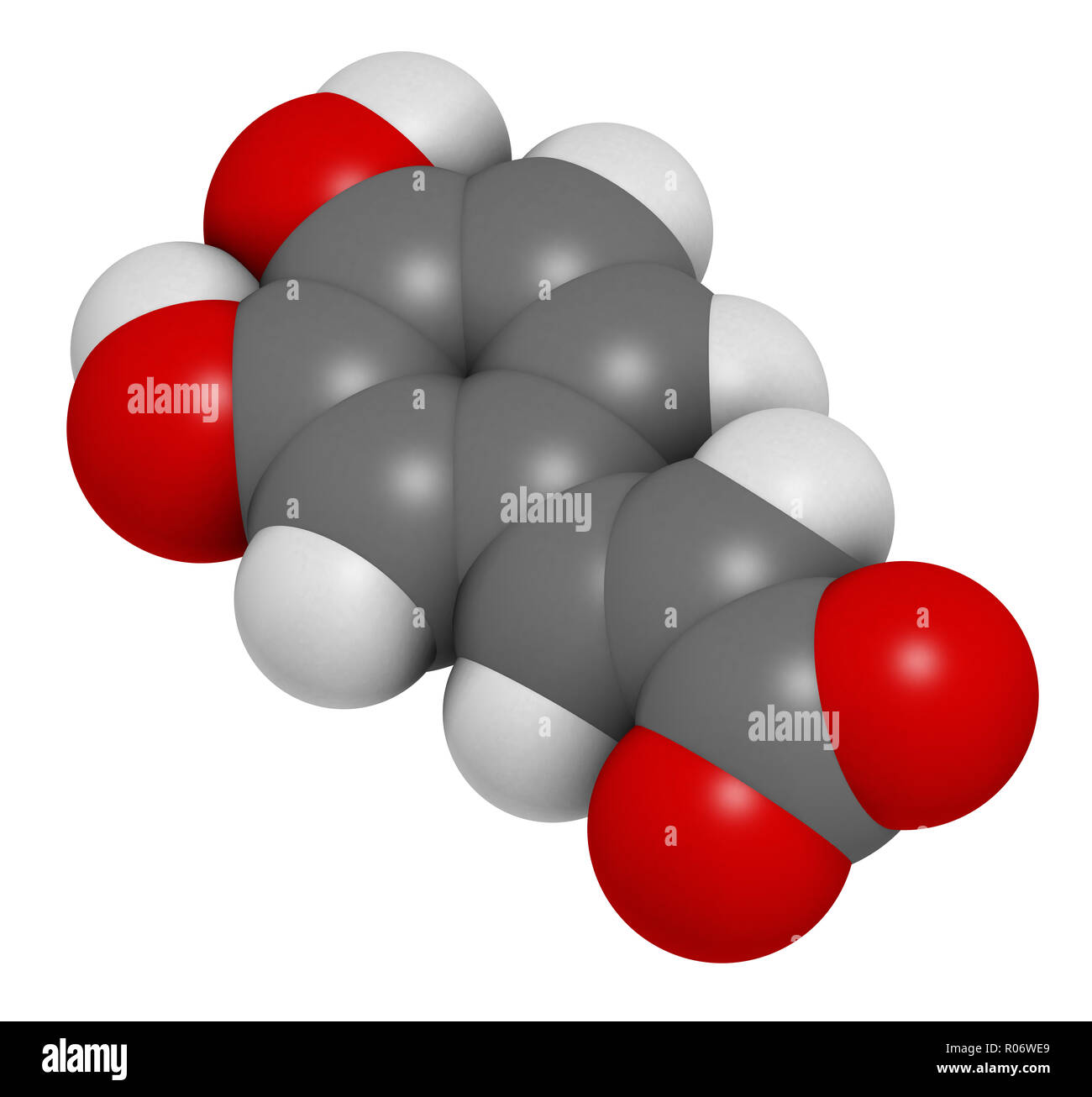 Caffeic acid molecule. Intermediate in the biosynthesis of lignin. 3D rendering. Atoms are represented as spheres with conventional color coding: hydr Stock Photohttps://www.alamy.com/image-license-details/?v=1https://www.alamy.com/caffeic-acid-molecule-intermediate-in-the-biosynthesis-of-lignin-3d-rendering-atoms-are-represented-as-spheres-with-conventional-color-coding-hydr-image223886497.html
RFR06WE9–Caffeic acid molecule. Intermediate in the biosynthesis of lignin. 3D rendering. Atoms are represented as spheres with conventional color coding: hydr
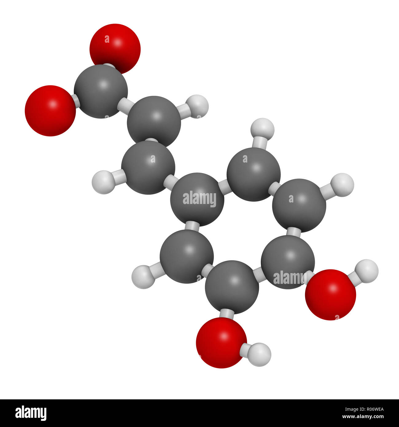 Caffeic acid molecule. Intermediate in the biosynthesis of lignin. 3D rendering. Atoms are represented as spheres with conventional color coding: hydr Stock Photohttps://www.alamy.com/image-license-details/?v=1https://www.alamy.com/caffeic-acid-molecule-intermediate-in-the-biosynthesis-of-lignin-3d-rendering-atoms-are-represented-as-spheres-with-conventional-color-coding-hydr-image223886498.html
RFR06WEA–Caffeic acid molecule. Intermediate in the biosynthesis of lignin. 3D rendering. Atoms are represented as spheres with conventional color coding: hydr
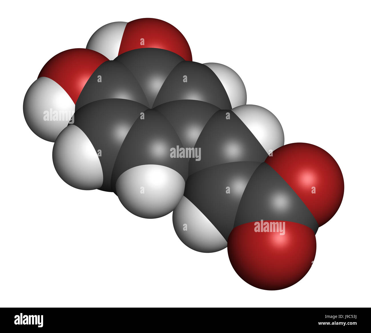 Caffeic acid molecule. Intermediate in the biosynthesis of lignin. 3D rendering. Atoms are represented as spheres with conventional color coding: hydr Stock Photohttps://www.alamy.com/image-license-details/?v=1https://www.alamy.com/stock-photo-caffeic-acid-molecule-intermediate-in-the-biosynthesis-of-lignin-3d-143482294.html
RFJ9C53J–Caffeic acid molecule. Intermediate in the biosynthesis of lignin. 3D rendering. Atoms are represented as spheres with conventional color coding: hydr
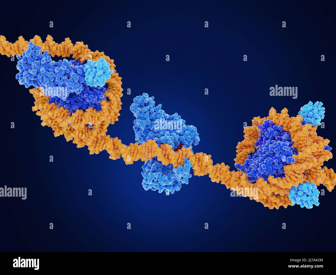 Epigenetic modifications, molecular model Stock Photohttps://www.alamy.com/image-license-details/?v=1https://www.alamy.com/epigenetic-modifications-molecular-model-image469205260.html
RF2J7A42M–Epigenetic modifications, molecular model
 Azacitidine (5-azacytidine) myelodysplastic syndrome drug molecule. Chemical formula is C8H12N4O5. Atoms are represented as spheres: carbon (grey), hydrogen (white), nitrogen (blue), oxygen (red). Illustration. Stock Photohttps://www.alamy.com/image-license-details/?v=1https://www.alamy.com/stock-photo-azacitidine-5-azacytidine-myelodysplastic-syndrome-drug-molecule-chemical-137665161.html
RFHYY58W–Azacitidine (5-azacytidine) myelodysplastic syndrome drug molecule. Chemical formula is C8H12N4O5. Atoms are represented as spheres: carbon (grey), hydrogen (white), nitrogen (blue), oxygen (red). Illustration.
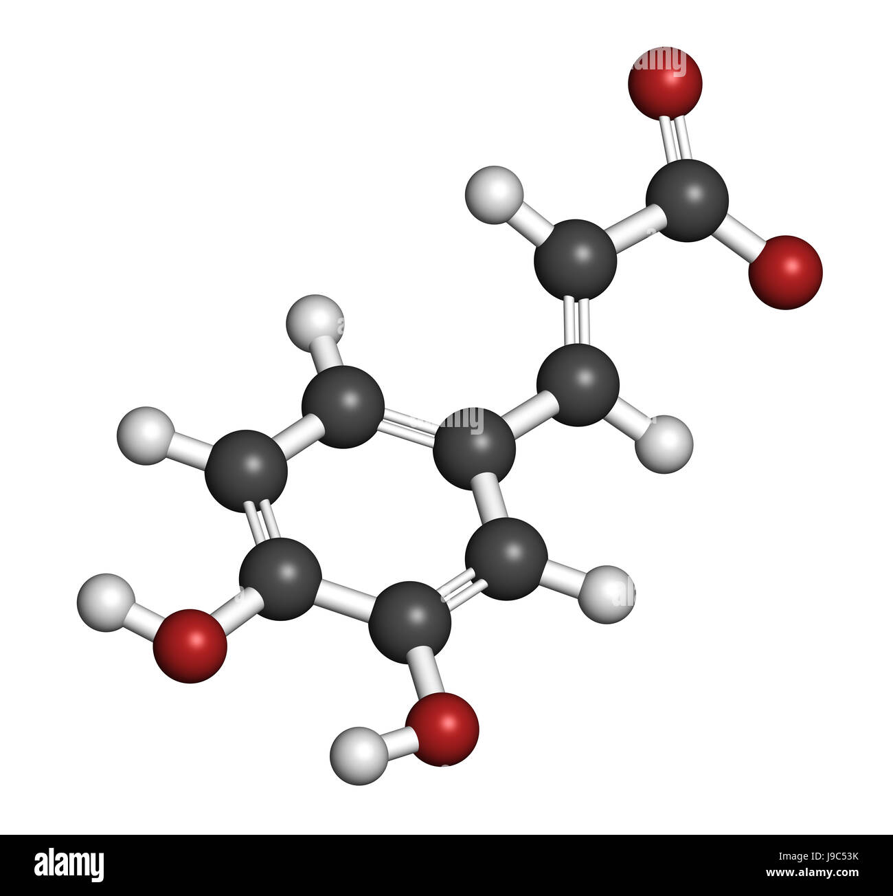 Caffeic acid molecule. Intermediate in the biosynthesis of lignin. 3D rendering. Atoms are represented as spheres with conventional color coding: hydr Stock Photohttps://www.alamy.com/image-license-details/?v=1https://www.alamy.com/stock-photo-caffeic-acid-molecule-intermediate-in-the-biosynthesis-of-lignin-3d-143482295.html
RFJ9C53K–Caffeic acid molecule. Intermediate in the biosynthesis of lignin. 3D rendering. Atoms are represented as spheres with conventional color coding: hydr
 Amodiaquine anti-malarial drug molecule. Chemical formula is C20H22ClN3O. Atoms are represented as spheres: carbon (grey), hydrogen (white), chlorine (green), nitrogen (blue), oxygen (red). Illustration. Stock Photohttps://www.alamy.com/image-license-details/?v=1https://www.alamy.com/stock-photo-amodiaquine-anti-malarial-drug-molecule-chemical-formula-is-c20h22cln3o-137665138.html
RFHYY582–Amodiaquine anti-malarial drug molecule. Chemical formula is C20H22ClN3O. Atoms are represented as spheres: carbon (grey), hydrogen (white), chlorine (green), nitrogen (blue), oxygen (red). Illustration.
 Amodiaquine anti-malarial drug molecule Stylized skeletal formula (chemical structure) Atoms are shown as color-coded circles: Stock Photohttps://www.alamy.com/image-license-details/?v=1https://www.alamy.com/stock-photo-amodiaquine-anti-malarial-drug-molecule-stylized-skeletal-formula-101777667.html
RFFWGACK–Amodiaquine anti-malarial drug molecule Stylized skeletal formula (chemical structure) Atoms are shown as color-coded circles:
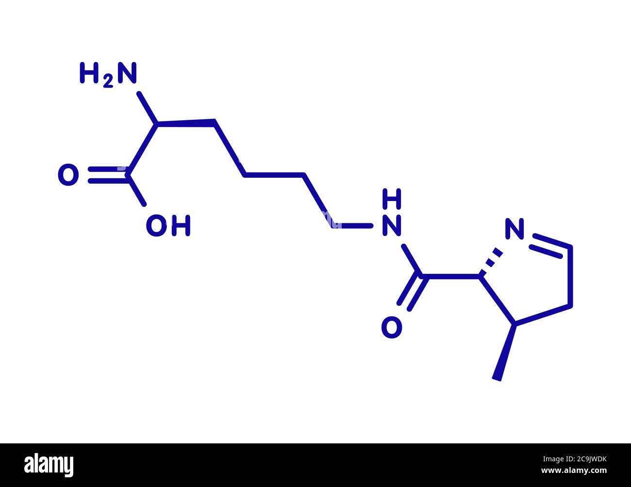 Pyrrolysine (l-pyrrolysine, Pyl, O) amino acid molecule. Blue skeletal formula on white background. Stock Photohttps://www.alamy.com/image-license-details/?v=1https://www.alamy.com/pyrrolysine-l-pyrrolysine-pyl-o-amino-acid-molecule-blue-skeletal-formula-on-white-background-image367364751.html
RF2C9JWDK–Pyrrolysine (l-pyrrolysine, Pyl, O) amino acid molecule. Blue skeletal formula on white background.