Quick filters:
Micrographs Stock Photos and Images
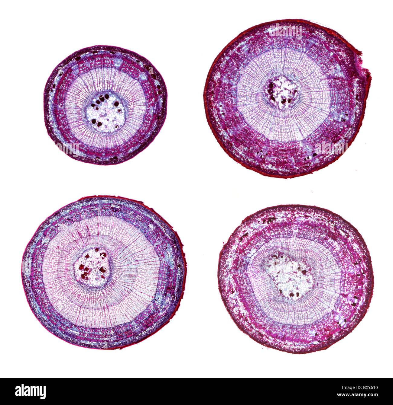 Light micrographs of basswood (Tilia americana) stems in cross section. One-year through three-year specimens. Stock Photohttps://www.alamy.com/image-license-details/?v=1https://www.alamy.com/stock-photo-light-micrographs-of-basswood-tilia-americana-stems-in-cross-section-33788860.html
RMBXY610–Light micrographs of basswood (Tilia americana) stems in cross section. One-year through three-year specimens.
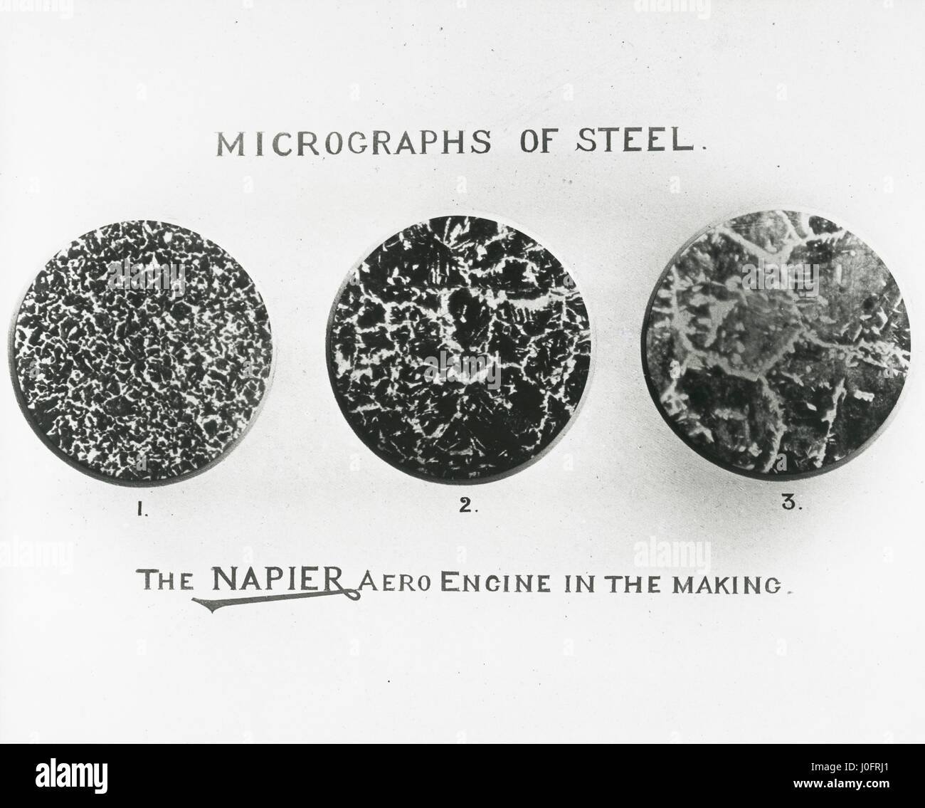 The Napier aero engine in the making: micrographs of steel Stock Photohttps://www.alamy.com/image-license-details/?v=1https://www.alamy.com/stock-photo-the-napier-aero-engine-in-the-making-micrographs-of-steel-138030761.html
RMJ0FRJ1–The Napier aero engine in the making: micrographs of steel
 A section trough different tongue cells under the microscope Stock Photohttps://www.alamy.com/image-license-details/?v=1https://www.alamy.com/a-section-trough-different-tongue-cells-under-the-microscope-image624947331.html
RF2Y8MPKF–A section trough different tongue cells under the microscope
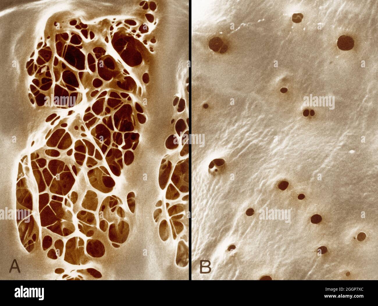 Colorized transmission electron micrographs showing, A: internal elastic lamina of an aorta, characterized by large fenestrations traversed by an irregular meshwork of elastic strands, and, B: a similar view of the internal elastic lamina of the femoral artery shows small fenestrations. Stock Photohttps://www.alamy.com/image-license-details/?v=1https://www.alamy.com/colorized-transmission-electron-micrographs-showing-a-internal-elastic-lamina-of-an-aorta-characterized-by-large-fenestrations-traversed-by-an-irregular-meshwork-of-elastic-strands-and-b-a-similar-view-of-the-internal-elastic-lamina-of-the-femoral-artery-shows-small-fenestrations-image440582868.html
RF2GGP7XC–Colorized transmission electron micrographs showing, A: internal elastic lamina of an aorta, characterized by large fenestrations traversed by an irregular meshwork of elastic strands, and, B: a similar view of the internal elastic lamina of the femoral artery shows small fenestrations.
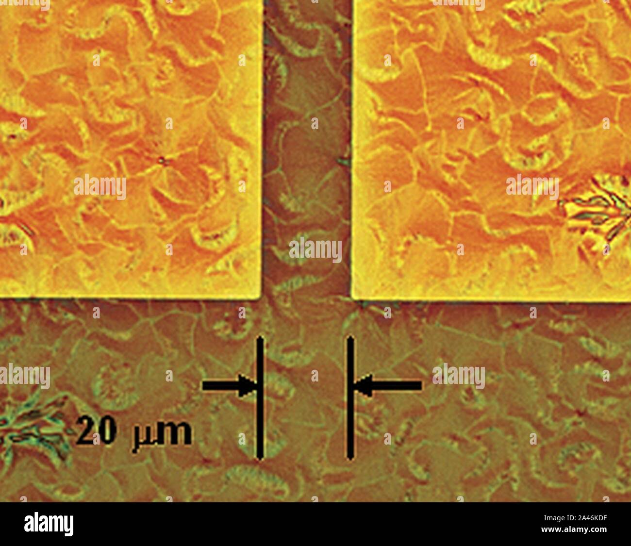 FET structures; optical micrographs Stock Photohttps://www.alamy.com/image-license-details/?v=1https://www.alamy.com/fet-structures-optical-micrographs-image329602603.html
RM2A46KDF–FET structures; optical micrographs
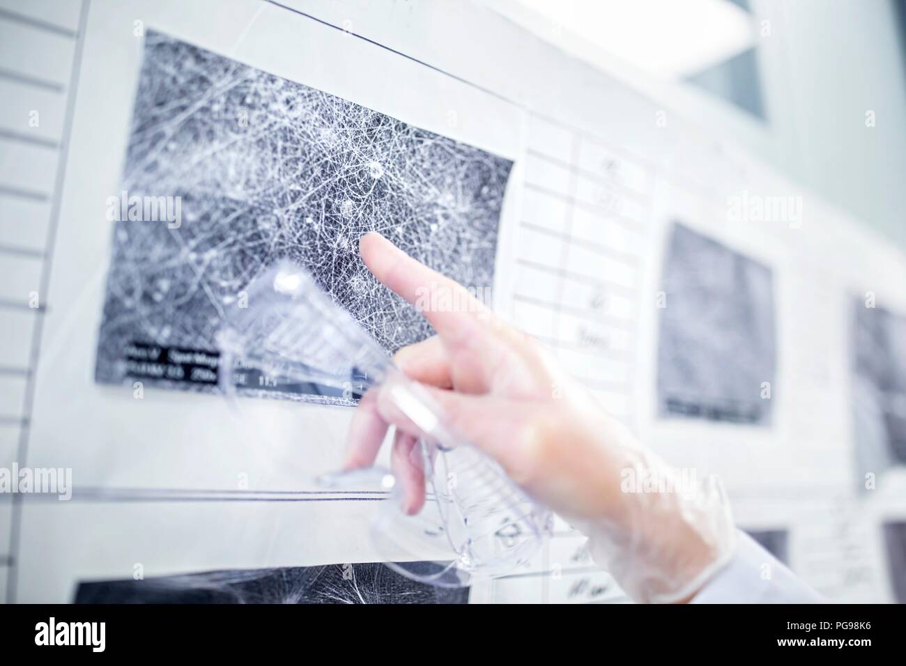 Scientist looking at scanning electron micrographs (SEMs) of different nanofibre structures. Stock Photohttps://www.alamy.com/image-license-details/?v=1https://www.alamy.com/scientist-looking-at-scanning-electron-micrographs-sems-of-different-nanofibre-structures-image216563290.html
RFPG98K6–Scientist looking at scanning electron micrographs (SEMs) of different nanofibre structures.
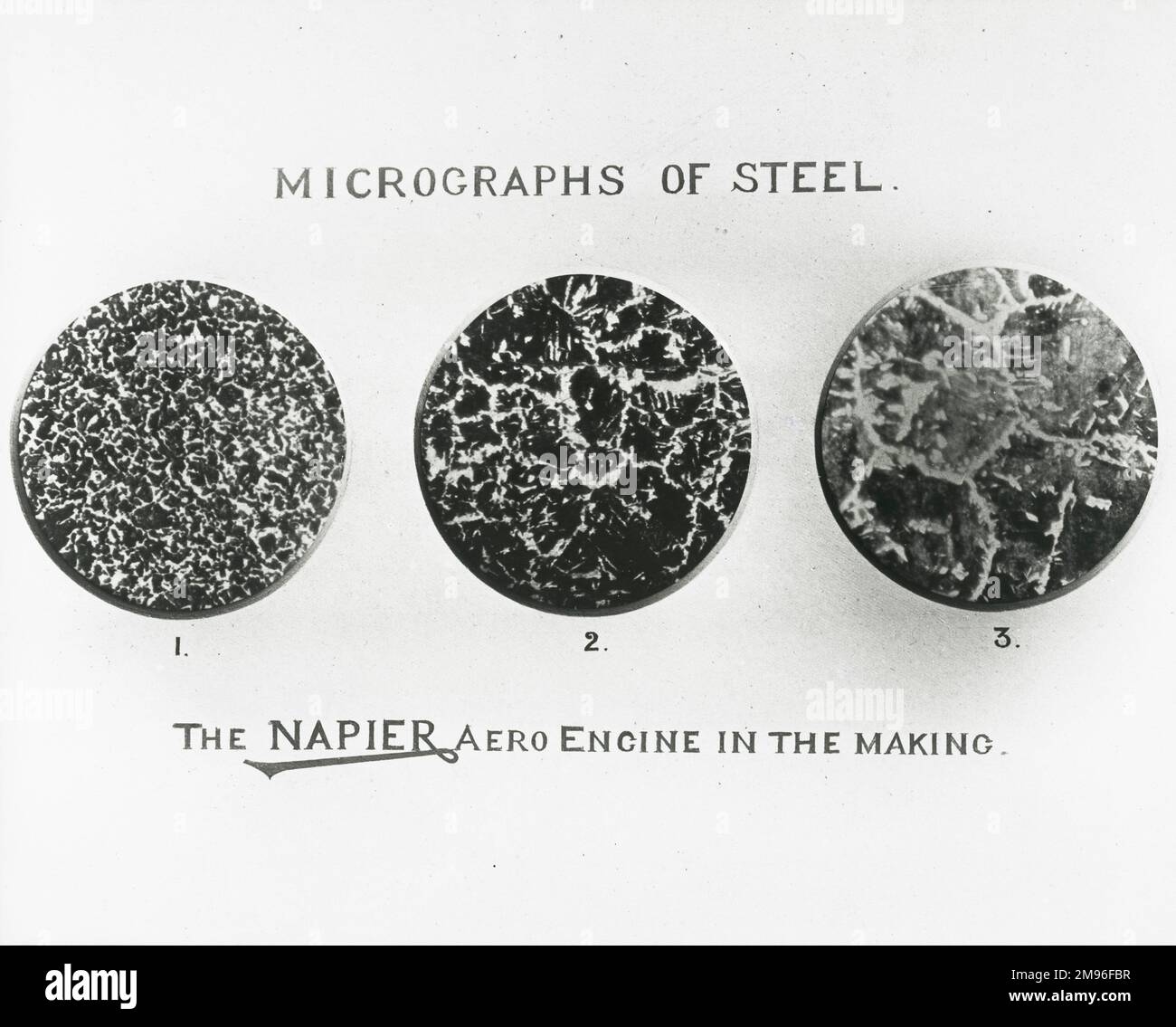 The Napier aero engine in the making micrographs of steel Stock Photohttps://www.alamy.com/image-license-details/?v=1https://www.alamy.com/the-napier-aero-engine-in-the-making-micrographs-of-steel-image504776379.html
RM2M96FBR–The Napier aero engine in the making micrographs of steel
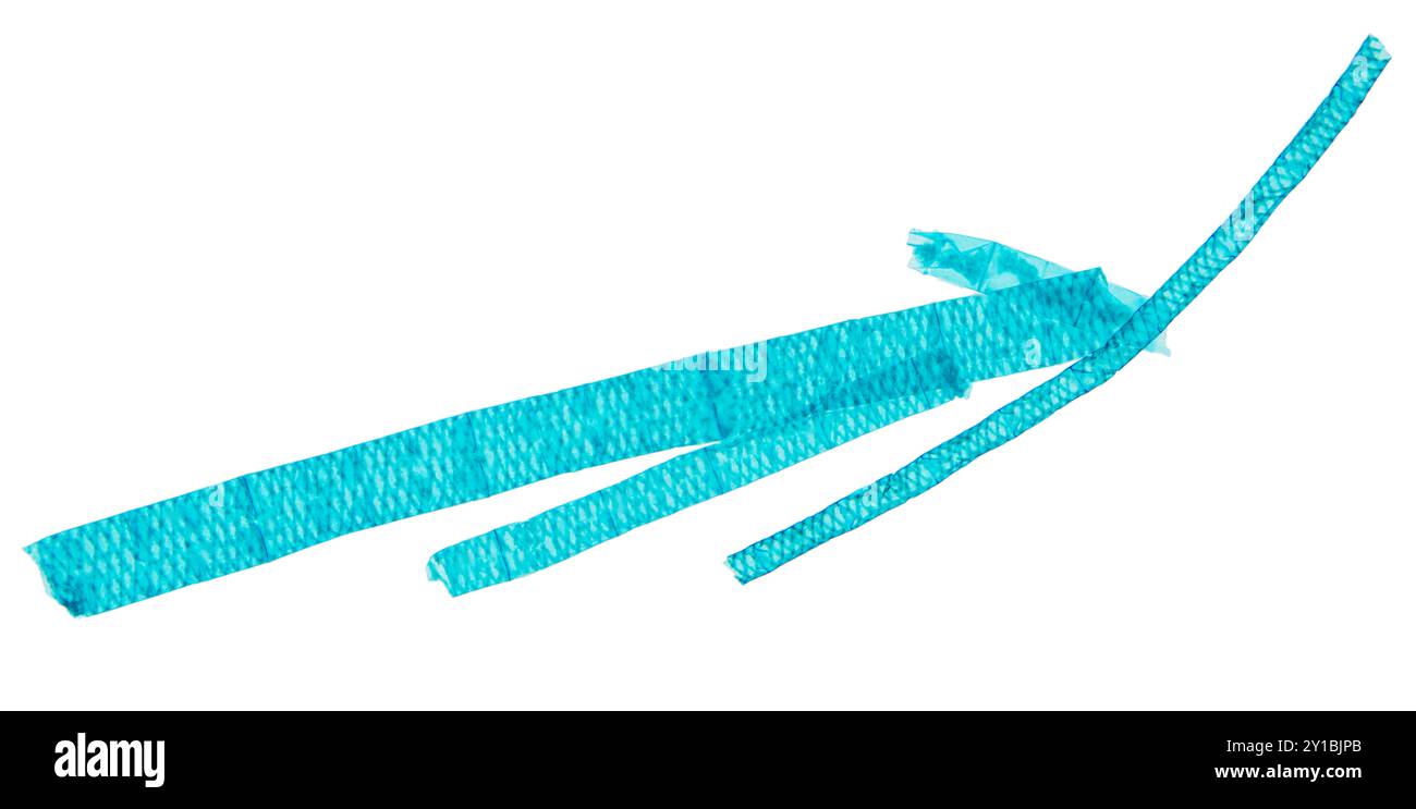 Spirogyra, whole mount, under a light microscope. 20X light micrographs, 2 combined photos of stained water silk, also known as blanket weed. Stock Photohttps://www.alamy.com/image-license-details/?v=1https://www.alamy.com/spirogyra-whole-mount-under-a-light-microscope-20x-light-micrographs-2-combined-photos-of-stained-water-silk-also-known-as-blanket-weed-image620444115.html
RF2Y1BJPB–Spirogyra, whole mount, under a light microscope. 20X light micrographs, 2 combined photos of stained water silk, also known as blanket weed.
 Hefei. 20th Apr, 2023. This photo taken on April 19, 2023 shows focus stacking micrographs of lunar soil particles displayed at an exhibition themed on lunar soil research achievements in University of Science and Technology of China, in Hefei, east China's Anhui Province. The exhibition starting Monday displays exhibits made by scientists, artists and engineers based on micrographs of lunar soil particles brought back by the Chang'e-5 probe. In 2020, China's Chang'e-5 mission retrieved samples from the moon weighing about 1,731 grams. Credit: Xinhua/Alamy Live News Stock Photohttps://www.alamy.com/image-license-details/?v=1https://www.alamy.com/hefei-20th-apr-2023-this-photo-taken-on-april-19-2023-shows-focus-stacking-micrographs-of-lunar-soil-particles-displayed-at-an-exhibition-themed-on-lunar-soil-research-achievements-in-university-of-science-and-technology-of-china-in-hefei-east-chinas-anhui-province-the-exhibition-starting-monday-displays-exhibits-made-by-scientists-artists-and-engineers-based-on-micrographs-of-lunar-soil-particles-brought-back-by-the-change-5-probe-in-2020-chinas-change-5-mission-retrieved-samples-from-the-moon-weighing-about-1731-grams-credit-xinhuaalamy-live-news-image546944062.html
RM2PNRCME–Hefei. 20th Apr, 2023. This photo taken on April 19, 2023 shows focus stacking micrographs of lunar soil particles displayed at an exhibition themed on lunar soil research achievements in University of Science and Technology of China, in Hefei, east China's Anhui Province. The exhibition starting Monday displays exhibits made by scientists, artists and engineers based on micrographs of lunar soil particles brought back by the Chang'e-5 probe. In 2020, China's Chang'e-5 mission retrieved samples from the moon weighing about 1,731 grams. Credit: Xinhua/Alamy Live News
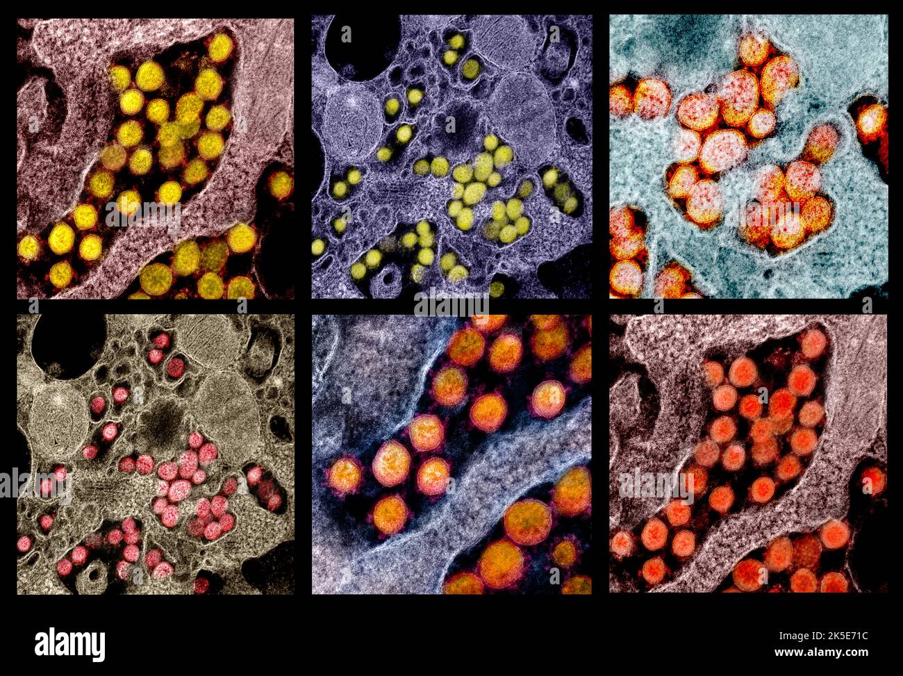 A composite of images of the Novel Coronavirus SARS-CoV-2. Transmission electron micrographs of SARS-CoV-2 virus particles, isolated from a patient. An optimised and enhanced unique composite version of six scanning electron micrograph images, Credit: NIAID Stock Photohttps://www.alamy.com/image-license-details/?v=1https://www.alamy.com/a-composite-of-images-of-the-novel-coronavirus-sars-cov-2-transmission-electron-micrographs-of-sars-cov-2-virus-particles-isolated-from-a-patient-an-optimised-and-enhanced-unique-composite-version-of-six-scanning-electron-micrograph-images-credit-niaid-image485276440.html
RM2K5E71C–A composite of images of the Novel Coronavirus SARS-CoV-2. Transmission electron micrographs of SARS-CoV-2 virus particles, isolated from a patient. An optimised and enhanced unique composite version of six scanning electron micrograph images, Credit: NIAID
RM2F77D3H–Skiopticon image from the Department of Photography at the Royal Institute of Technology. Use by Professor Helmer Bäckström as lecture material. Bäckström was Sweden's first professor in photography at the Royal Institute of Technology in Stockholm 1948-1958.Ever the image: Micrographs showing how the developing inserts on single discrete points in the various silver bromide grains. The bottom image: Same as SKC7128bäckström, Helmer. Photographic manual. Nature and Culture. Stockholm. 1942. p. 290-291 and cosmos. SWEDISH SCIENCE PRESS. Lazy. 1944. p.122.
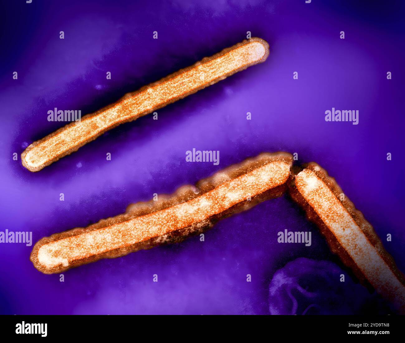 Three influenza A H5N1/bird flu virus particles rod-shaped orange. Note: Layout incorporates two CDC transmission electron micrographs that have been repositioned and colorized by NIAID. Influenza A Virus H5N1/Bird Flu 016867 312 Stock Photohttps://www.alamy.com/image-license-details/?v=1https://www.alamy.com/three-influenza-a-h5n1bird-flu-virus-particles-rod-shaped-orange-note-layout-incorporates-two-cdc-transmission-electron-micrographs-that-have-been-repositioned-and-colorized-by-niaid-influenza-a-virus-h5n1bird-flu-016867-312-image627780756.html
RM2YD9TN8–Three influenza A H5N1/bird flu virus particles rod-shaped orange. Note: Layout incorporates two CDC transmission electron micrographs that have been repositioned and colorized by NIAID. Influenza A Virus H5N1/Bird Flu 016867 312
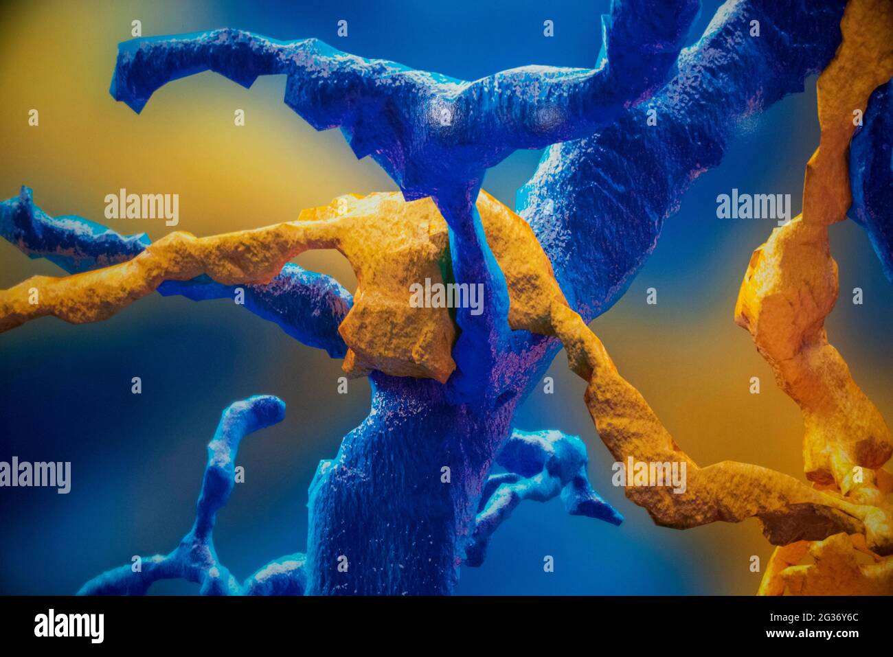 Swissnapse, digital reconstruction from serial section electron micrographs. Brain exhibition Inside MIT Museum Building at 265 Massachusetts Avenue C Stock Photohttps://www.alamy.com/image-license-details/?v=1https://www.alamy.com/swissnapse-digital-reconstruction-from-serial-section-electron-micrographs-brain-exhibition-inside-mit-museum-building-at-265-massachusetts-avenue-c-image432256228.html
RM2G36Y6C–Swissnapse, digital reconstruction from serial section electron micrographs. Brain exhibition Inside MIT Museum Building at 265 Massachusetts Avenue C
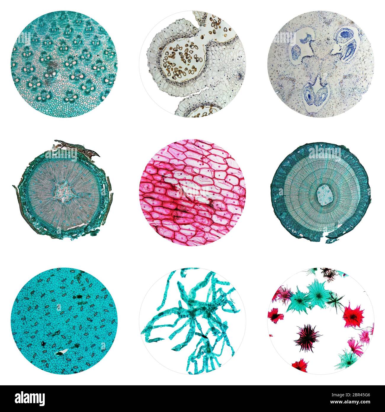 Light photomicrograph of many plants cells seen through a microscope Stock Photohttps://www.alamy.com/image-license-details/?v=1https://www.alamy.com/light-photomicrograph-of-many-plants-cells-seen-through-a-microscope-image358436630.html
RF2BR45G6–Light photomicrograph of many plants cells seen through a microscope
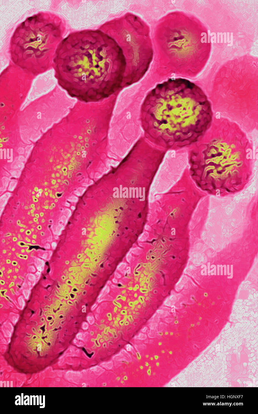 PENICILLIUM CHRYSOGENUM Stock Photohttps://www.alamy.com/image-license-details/?v=1https://www.alamy.com/stock-photo-penicillium-chrysogenum-130788875.html
RMHGNXF7–PENICILLIUM CHRYSOGENUM
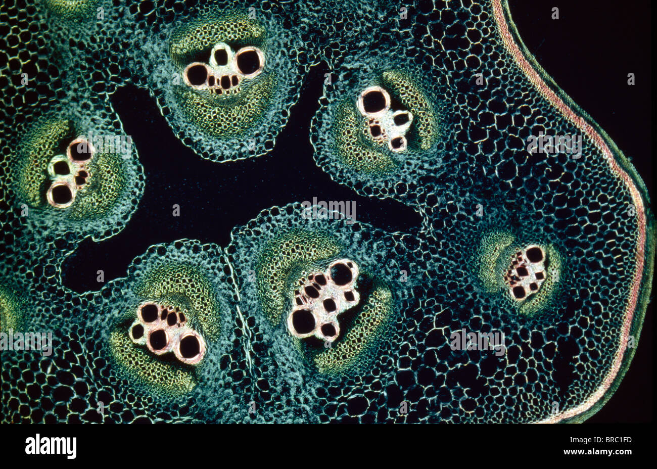 Light Micrograph (LM) of a transverse section of a stem of a Marrow (Cucurbita sp.), magnification x12 Stock Photohttps://www.alamy.com/image-license-details/?v=1https://www.alamy.com/stock-photo-light-micrograph-lm-of-a-transverse-section-of-a-stem-of-a-marrow-31612097.html
RMBRC1FD–Light Micrograph (LM) of a transverse section of a stem of a Marrow (Cucurbita sp.), magnification x12
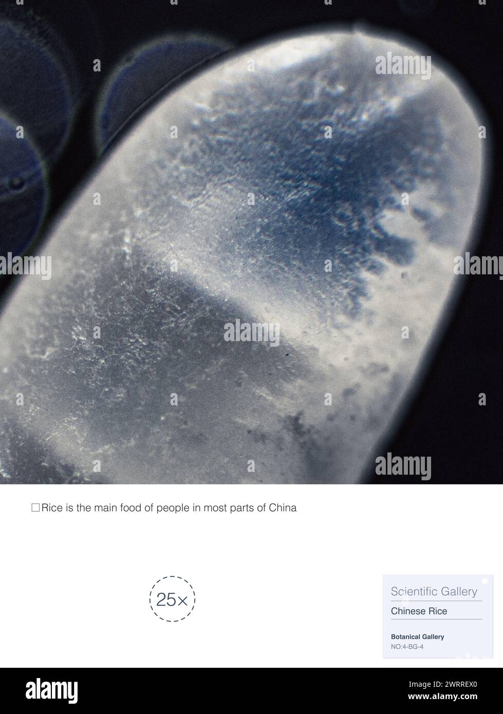 Rice is a food made from paddy through processes such as cleaning, hulling, milling, and finished product sorting. Stock Photohttps://www.alamy.com/image-license-details/?v=1https://www.alamy.com/rice-is-a-food-made-from-paddy-through-processes-such-as-cleaning-hulling-milling-and-finished-product-sorting-image599806200.html
RM2WRREX0–Rice is a food made from paddy through processes such as cleaning, hulling, milling, and finished product sorting.
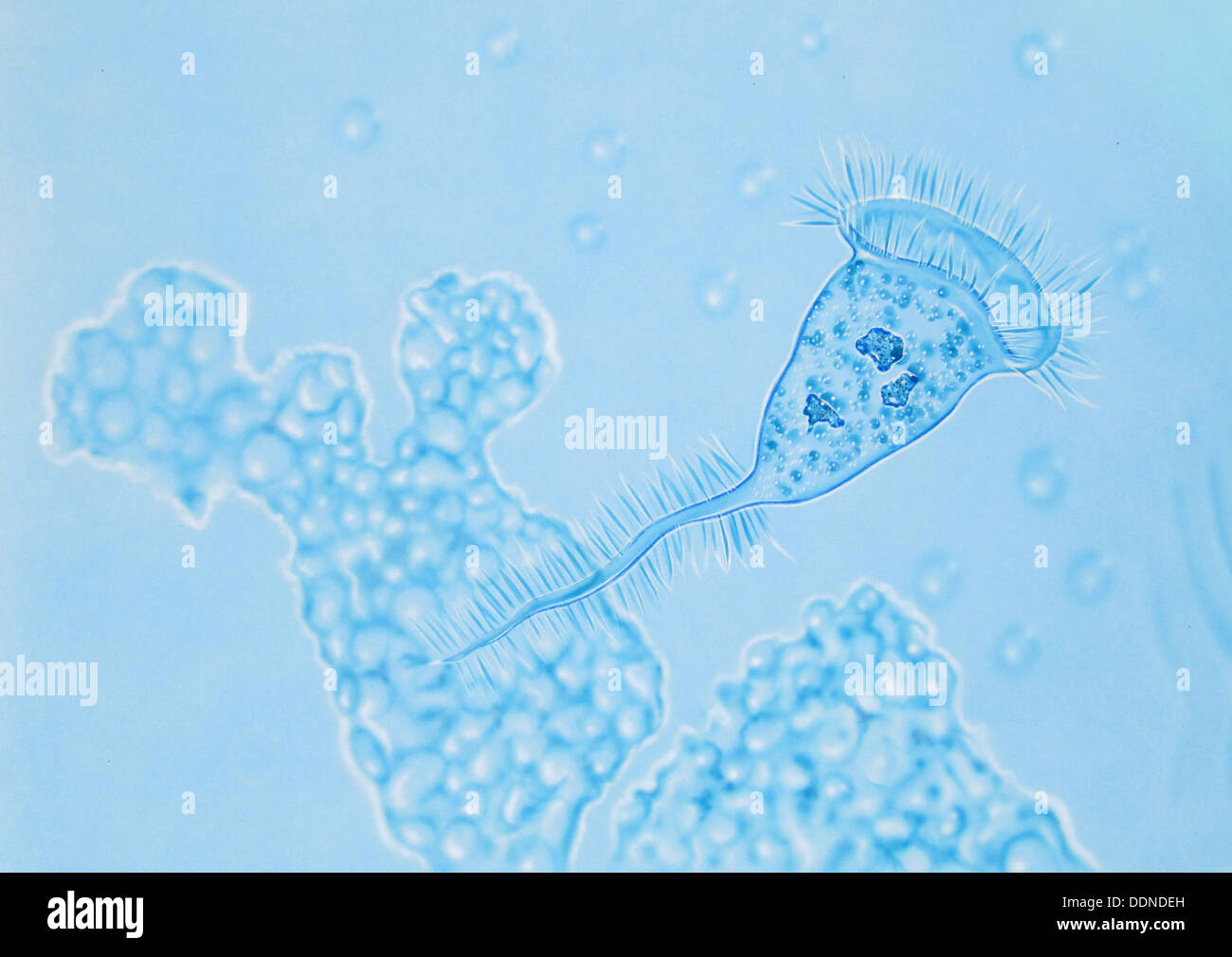 Bakteria in water Stock Photohttps://www.alamy.com/image-license-details/?v=1https://www.alamy.com/bakteria-in-water-image60093225.html
RMDDNDEH–Bakteria in water
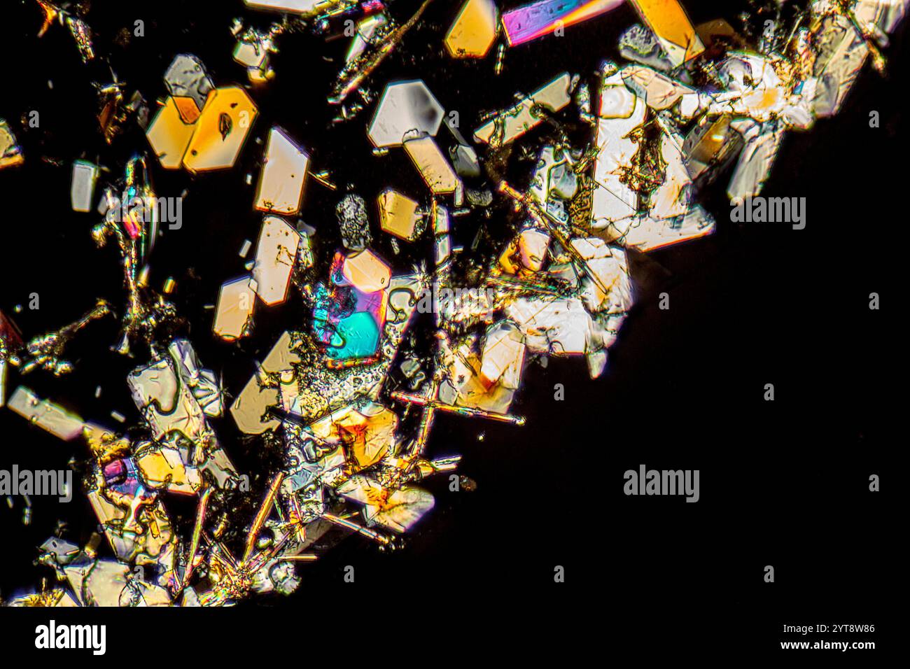 Microscopic shot showing colorful microcrystals in polarized light Stock Photohttps://www.alamy.com/image-license-details/?v=1https://www.alamy.com/microscopic-shot-showing-colorful-microcrystals-in-polarized-light-image634520438.html
RF2YT8W86–Microscopic shot showing colorful microcrystals in polarized light
 Coloured SEM of grease pencil Stock Photohttps://www.alamy.com/image-license-details/?v=1https://www.alamy.com/coloured-sem-of-grease-pencil-image67265900.html
RFDWC69G–Coloured SEM of grease pencil
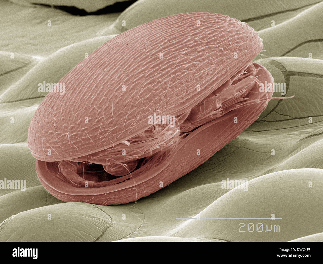 Coloured SEM of plankton Stock Photohttps://www.alamy.com/image-license-details/?v=1https://www.alamy.com/coloured-sem-of-plankton-image67264492.html
RFDWC4F8–Coloured SEM of plankton
 Coumarins are substances that can be found in many plants. They are used for perfume production and many other purposes. The photo is made with a pola Stock Photohttps://www.alamy.com/image-license-details/?v=1https://www.alamy.com/coumarins-are-substances-that-can-be-found-in-many-plants-they-are-used-for-perfume-production-and-many-other-purposes-the-photo-is-made-with-a-pola-image624950564.html
RF2Y8MXR0–Coumarins are substances that can be found in many plants. They are used for perfume production and many other purposes. The photo is made with a pola
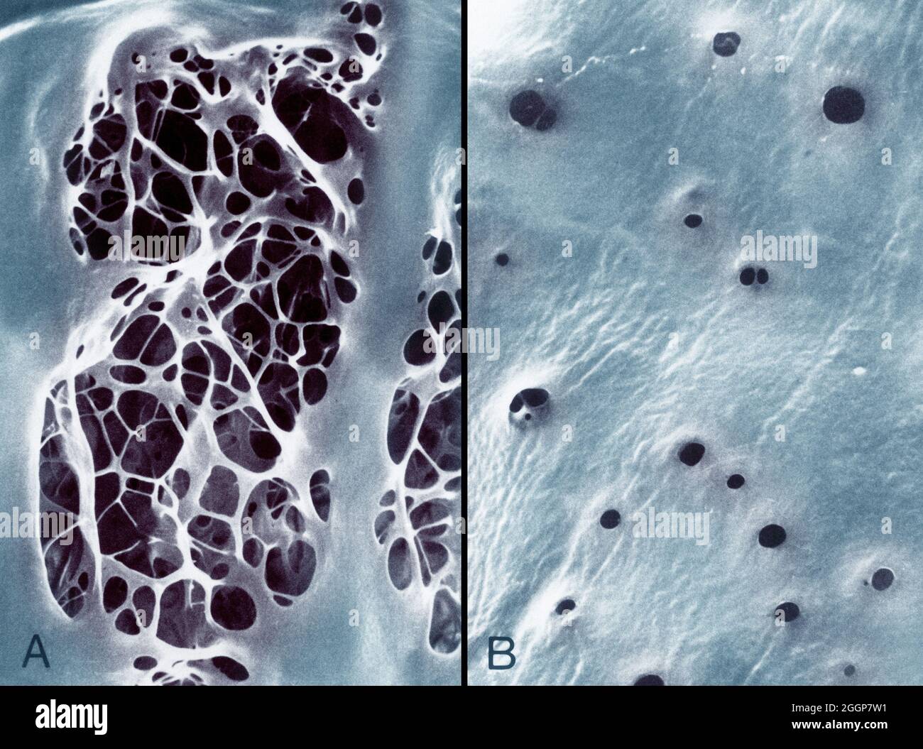 Colorized transmission electron micrographs showing, A: internal elastic lamina of an aorta, characterized by large fenestrations traversed by an irregular meshwork of elastic strands, and, B: a similar view of the internal elastic lamina of the femoral artery shows small fenestrations. Stock Photohttps://www.alamy.com/image-license-details/?v=1https://www.alamy.com/colorized-transmission-electron-micrographs-showing-a-internal-elastic-lamina-of-an-aorta-characterized-by-large-fenestrations-traversed-by-an-irregular-meshwork-of-elastic-strands-and-b-a-similar-view-of-the-internal-elastic-lamina-of-the-femoral-artery-shows-small-fenestrations-image440582829.html
RF2GGP7W1–Colorized transmission electron micrographs showing, A: internal elastic lamina of an aorta, characterized by large fenestrations traversed by an irregular meshwork of elastic strands, and, B: a similar view of the internal elastic lamina of the femoral artery shows small fenestrations.
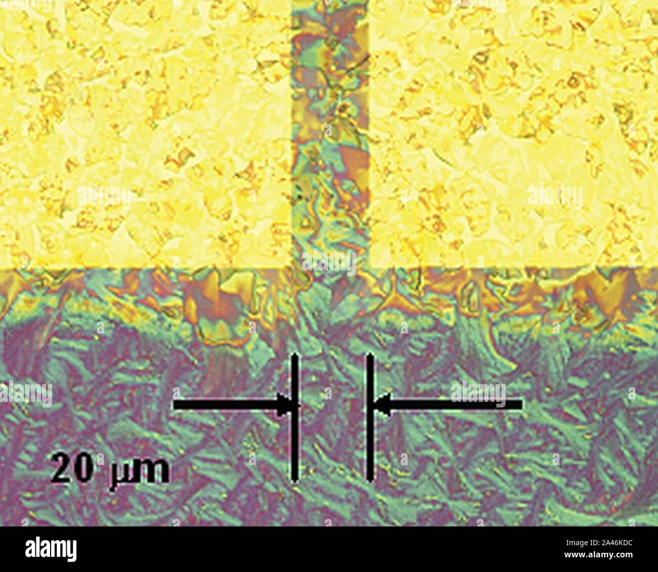 FET structures; optical micrographs Stock Photohttps://www.alamy.com/image-license-details/?v=1https://www.alamy.com/fet-structures-optical-micrographs-image329602600.html
RM2A46KDC–FET structures; optical micrographs
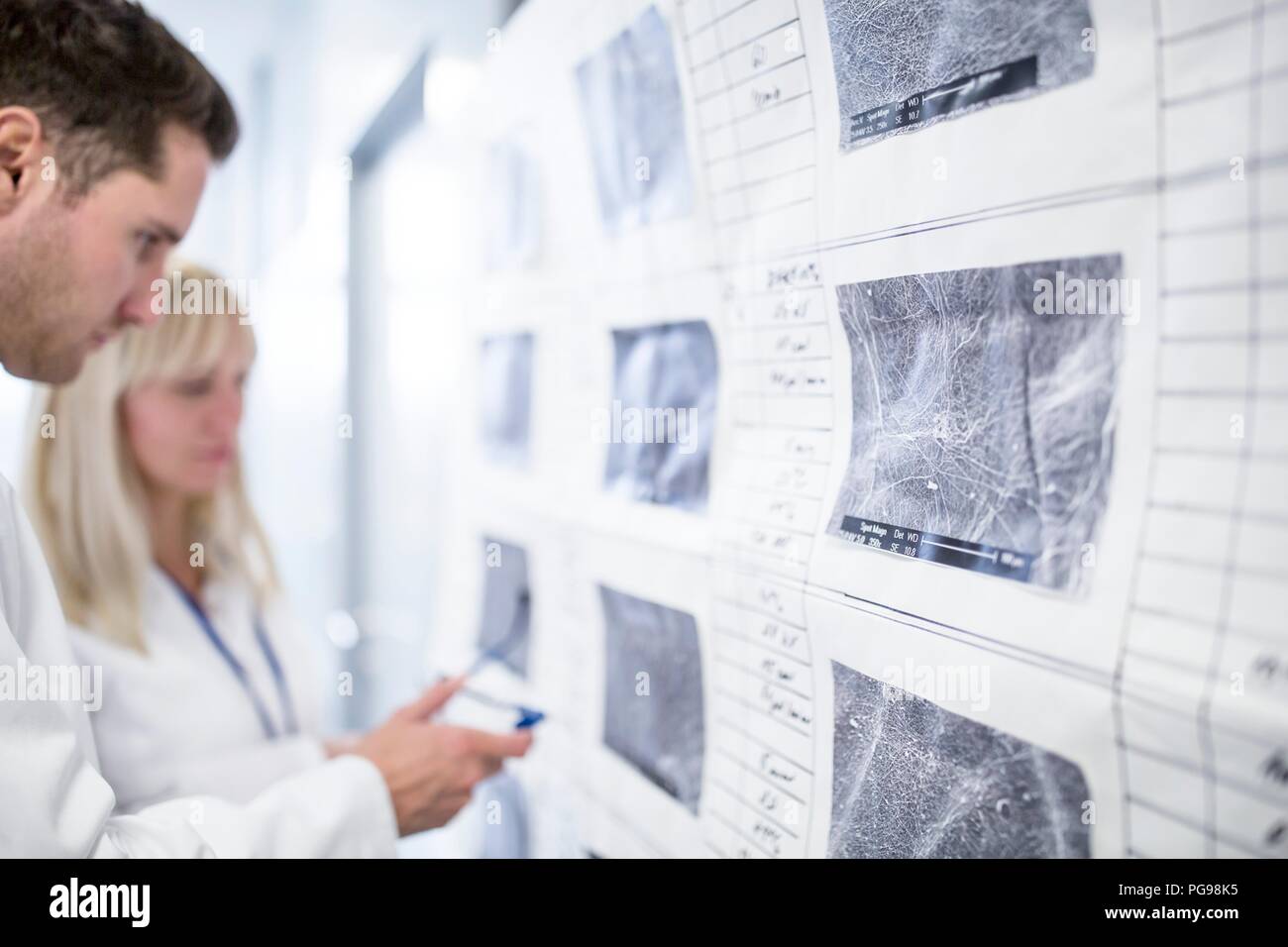 Scientists looking at scanning electron micrographs (SEMs) of different nanofibre structures. Stock Photohttps://www.alamy.com/image-license-details/?v=1https://www.alamy.com/scientists-looking-at-scanning-electron-micrographs-sems-of-different-nanofibre-structures-image216563289.html
RFPG98K5–Scientists looking at scanning electron micrographs (SEMs) of different nanofibre structures.
 MICROGRAPHS. Report on Steam Boilers Exhibited at the Recent Fair of the American Institute. The Gold Fire Still Burning. ObituaryBeath of IfIr. John Begnon. 1, scientific american, 1869-12-25 Stock Photohttps://www.alamy.com/image-license-details/?v=1https://www.alamy.com/micrographs-report-on-steam-boilers-exhibited-at-the-recent-fair-of-the-american-institute-the-gold-fire-still-burning-obituarybeath-of-ifir-john-begnon-1-scientific-american-1869-12-25-image334311194.html
RM2ABW59E–MICROGRAPHS. Report on Steam Boilers Exhibited at the Recent Fair of the American Institute. The Gold Fire Still Burning. ObituaryBeath of IfIr. John Begnon. 1, scientific american, 1869-12-25
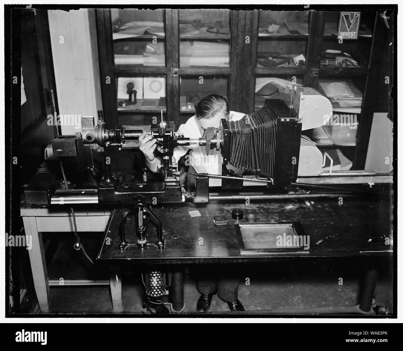 Bureau of Standards speeds up photomicrography with new apparatus. Washington, D.C., Aug. 18. By means of this new apparatus just designed by the metallurgical divisions of the National Bureau of Standards, it is now possible to make 700 micrographs per working day of the structural effect of corrosion on cross sections of metallic specimans. This new machine uses roll film of 900 negatives at one loading, thus dispensing with the repeated loading and unloading of individual film holders by which method the Bureau was only formerly able to make 12 micrographs per working day. Willard H. Mutohl Stock Photohttps://www.alamy.com/image-license-details/?v=1https://www.alamy.com/bureau-of-standards-speeds-up-photomicrography-with-new-apparatus-washington-dc-aug-18-by-means-of-this-new-apparatus-just-designed-by-the-metallurgical-divisions-of-the-national-bureau-of-standards-it-is-now-possible-to-make-700-micrographs-per-working-day-of-the-structural-effect-of-corrosion-on-cross-sections-of-metallic-specimans-this-new-machine-uses-roll-film-of-900-negatives-at-one-loading-thus-dispensing-with-the-repeated-loading-and-unloading-of-individual-film-holders-by-which-method-the-bureau-was-only-formerly-able-to-make-12-micrographs-per-working-day-willard-h-mutohl-image264480683.html
RMWA83PK–Bureau of Standards speeds up photomicrography with new apparatus. Washington, D.C., Aug. 18. By means of this new apparatus just designed by the metallurgical divisions of the National Bureau of Standards, it is now possible to make 700 micrographs per working day of the structural effect of corrosion on cross sections of metallic specimans. This new machine uses roll film of 900 negatives at one loading, thus dispensing with the repeated loading and unloading of individual film holders by which method the Bureau was only formerly able to make 12 micrographs per working day. Willard H. Mutohl
 Hefei. 20th Apr, 2023. This combo photo shows cross-polarized micrographs of different lunar soil particles displayed at an exhibition themed on lunar soil research achievements in University of Science and Technology of China, in Hefei, east China's Anhui Province on April 19, 2023. The exhibition starting Monday displays exhibits made by scientists, artists and engineers based on micrographs of lunar soil particles brought back by the Chang'e-5 probe. In 2020, China's Chang'e-5 mission retrieved samples from the moon weighing about 1,731 grams. Credit: Xinhua/Alamy Live News Stock Photohttps://www.alamy.com/image-license-details/?v=1https://www.alamy.com/hefei-20th-apr-2023-this-combo-photo-shows-cross-polarized-micrographs-of-different-lunar-soil-particles-displayed-at-an-exhibition-themed-on-lunar-soil-research-achievements-in-university-of-science-and-technology-of-china-in-hefei-east-chinas-anhui-province-on-april-19-2023-the-exhibition-starting-monday-displays-exhibits-made-by-scientists-artists-and-engineers-based-on-micrographs-of-lunar-soil-particles-brought-back-by-the-change-5-probe-in-2020-chinas-change-5-mission-retrieved-samples-from-the-moon-weighing-about-1731-grams-credit-xinhuaalamy-live-news-image546944064.html
RM2PNRCMG–Hefei. 20th Apr, 2023. This combo photo shows cross-polarized micrographs of different lunar soil particles displayed at an exhibition themed on lunar soil research achievements in University of Science and Technology of China, in Hefei, east China's Anhui Province on April 19, 2023. The exhibition starting Monday displays exhibits made by scientists, artists and engineers based on micrographs of lunar soil particles brought back by the Chang'e-5 probe. In 2020, China's Chang'e-5 mission retrieved samples from the moon weighing about 1,731 grams. Credit: Xinhua/Alamy Live News
 The Quarterly Journal of the Geological Society of London, v. 28 (1872), London, geology, periodicals, This illustration showcases three distinct micrographs of dolerite samples from Queensland, labeled 1, 2, and 3. Each section presents a unique texture and mineral composition, highlighting the geological characteristics of dolerite. The visual representation emphasizes the intricacies of the rock's crystalline structure, with varying grain sizes and arrangements indicating different cooling histories and mineralogical variations. This detailed examination serves to enhance the understanding Stock Photohttps://www.alamy.com/image-license-details/?v=1https://www.alamy.com/the-quarterly-journal-of-the-geological-society-of-london-v-28-1872-london-geology-periodicals-this-illustration-showcases-three-distinct-micrographs-of-dolerite-samples-from-queensland-labeled-1-2-and-3-each-section-presents-a-unique-texture-and-mineral-composition-highlighting-the-geological-characteristics-of-dolerite-the-visual-representation-emphasizes-the-intricacies-of-the-rocks-crystalline-structure-with-varying-grain-sizes-and-arrangements-indicating-different-cooling-histories-and-mineralogical-variations-this-detailed-examination-serves-to-enhance-the-understanding-image636100694.html
RM2YXTTWX–The Quarterly Journal of the Geological Society of London, v. 28 (1872), London, geology, periodicals, This illustration showcases three distinct micrographs of dolerite samples from Queensland, labeled 1, 2, and 3. Each section presents a unique texture and mineral composition, highlighting the geological characteristics of dolerite. The visual representation emphasizes the intricacies of the rock's crystalline structure, with varying grain sizes and arrangements indicating different cooling histories and mineralogical variations. This detailed examination serves to enhance the understanding
![Français : Photomicrographie de la progression des fissures dues à la fatigue dans un matériau. Image tirée de J. A. Ewing et J. C. W. Humfrey. « The fracture of metals under repeated alternations of stress », Proceedings of the Royal Society of London (1854-1905), 1903 [1]. English: Micrographs showing how surface fatigue cracks grow as material is further cycled. From Ewing & Humfrey (1903). 155 Ewing and Humfrey fatigue cracks Stock Photo Français : Photomicrographie de la progression des fissures dues à la fatigue dans un matériau. Image tirée de J. A. Ewing et J. C. W. Humfrey. « The fracture of metals under repeated alternations of stress », Proceedings of the Royal Society of London (1854-1905), 1903 [1]. English: Micrographs showing how surface fatigue cracks grow as material is further cycled. From Ewing & Humfrey (1903). 155 Ewing and Humfrey fatigue cracks Stock Photo](https://c8.alamy.com/comp/P7X662/franais-photomicrographie-de-la-progression-des-fissures-dues-la-fatigue-dans-un-matriau-image-tire-de-j-a-ewing-et-j-c-w-humfrey-the-fracture-of-metals-under-repeated-alternations-of-stress-proceedings-of-the-royal-society-of-london-1854-1905-1903-1-english-micrographs-showing-how-surface-fatigue-cracks-grow-as-material-is-further-cycled-from-ewing-humfrey-1903-155-ewing-and-humfrey-fatigue-cracks-P7X662.jpg) Français : Photomicrographie de la progression des fissures dues à la fatigue dans un matériau. Image tirée de J. A. Ewing et J. C. W. Humfrey. « The fracture of metals under repeated alternations of stress », Proceedings of the Royal Society of London (1854-1905), 1903 [1]. English: Micrographs showing how surface fatigue cracks grow as material is further cycled. From Ewing & Humfrey (1903). 155 Ewing and Humfrey fatigue cracks Stock Photohttps://www.alamy.com/image-license-details/?v=1https://www.alamy.com/franais-photomicrographie-de-la-progression-des-fissures-dues-la-fatigue-dans-un-matriau-image-tire-de-j-a-ewing-et-j-c-w-humfrey-the-fracture-of-metals-under-repeated-alternations-of-stress-proceedings-of-the-royal-society-of-london-1854-1905-1903-1-english-micrographs-showing-how-surface-fatigue-cracks-grow-as-material-is-further-cycled-from-ewing-humfrey-1903-155-ewing-and-humfrey-fatigue-cracks-image211402634.html
Français : Photomicrographie de la progression des fissures dues à la fatigue dans un matériau. Image tirée de J. A. Ewing et J. C. W. Humfrey. « The fracture of metals under repeated alternations of stress », Proceedings of the Royal Society of London (1854-1905), 1903 [1]. English: Micrographs showing how surface fatigue cracks grow as material is further cycled. From Ewing & Humfrey (1903). 155 Ewing and Humfrey fatigue cracks Stock Photohttps://www.alamy.com/image-license-details/?v=1https://www.alamy.com/franais-photomicrographie-de-la-progression-des-fissures-dues-la-fatigue-dans-un-matriau-image-tire-de-j-a-ewing-et-j-c-w-humfrey-the-fracture-of-metals-under-repeated-alternations-of-stress-proceedings-of-the-royal-society-of-london-1854-1905-1903-1-english-micrographs-showing-how-surface-fatigue-cracks-grow-as-material-is-further-cycled-from-ewing-humfrey-1903-155-ewing-and-humfrey-fatigue-cracks-image211402634.htmlRMP7X662–Français : Photomicrographie de la progression des fissures dues à la fatigue dans un matériau. Image tirée de J. A. Ewing et J. C. W. Humfrey. « The fracture of metals under repeated alternations of stress », Proceedings of the Royal Society of London (1854-1905), 1903 [1]. English: Micrographs showing how surface fatigue cracks grow as material is further cycled. From Ewing & Humfrey (1903). 155 Ewing and Humfrey fatigue cracks
 Three influenza A H5N1/bird flu virus particles rod-shaped. Note: Layout incorporates two CDC transmission electron micrographs that have been inverted, repositioned, and colorized by NIAID. Influenza A Virus H5N1/Bird Flu 016867 310 Stock Photohttps://www.alamy.com/image-license-details/?v=1https://www.alamy.com/three-influenza-a-h5n1bird-flu-virus-particles-rod-shaped-note-layout-incorporates-two-cdc-transmission-electron-micrographs-that-have-been-inverted-repositioned-and-colorized-by-niaid-influenza-a-virus-h5n1bird-flu-016867-310-image627780768.html
RM2YD9TNM–Three influenza A H5N1/bird flu virus particles rod-shaped. Note: Layout incorporates two CDC transmission electron micrographs that have been inverted, repositioned, and colorized by NIAID. Influenza A Virus H5N1/Bird Flu 016867 310
 Electron micrographs of the family of retroviruses that reproduce in t-lymphocytes ca. November 1988 Stock Photohttps://www.alamy.com/image-license-details/?v=1https://www.alamy.com/electron-micrographs-of-the-family-of-retroviruses-that-reproduce-in-t-lymphocytes-ca-november-1988-image461145055.html
RM2HP6Y67–Electron micrographs of the family of retroviruses that reproduce in t-lymphocytes ca. November 1988
 Electron micrographs of Variola, Varicella and Vaccinia virions. Electron micrographs from top to bottom: Variola virion (forms M and C), Varicella virion and virion core, Vaccinia virion (forms M and C), 1975. Image courtesy CDC/Dr. James Nakano. Stock Photohttps://www.alamy.com/image-license-details/?v=1https://www.alamy.com/stock-image-electron-micrographs-of-variola-varicella-and-vaccinia-virions-electron-169078077.html
RMKR24PN–Electron micrographs of Variola, Varicella and Vaccinia virions. Electron micrographs from top to bottom: Variola virion (forms M and C), Varicella virion and virion core, Vaccinia virion (forms M and C), 1975. Image courtesy CDC/Dr. James Nakano.
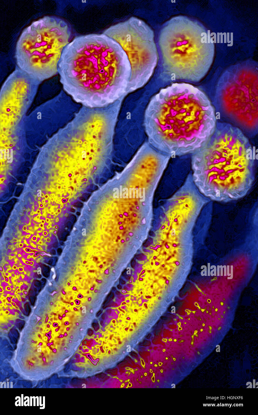 PENICILLIUM CHRYSOGENUM Stock Photohttps://www.alamy.com/image-license-details/?v=1https://www.alamy.com/stock-photo-penicillium-chrysogenum-130788874.html
RMHGNXF6–PENICILLIUM CHRYSOGENUM
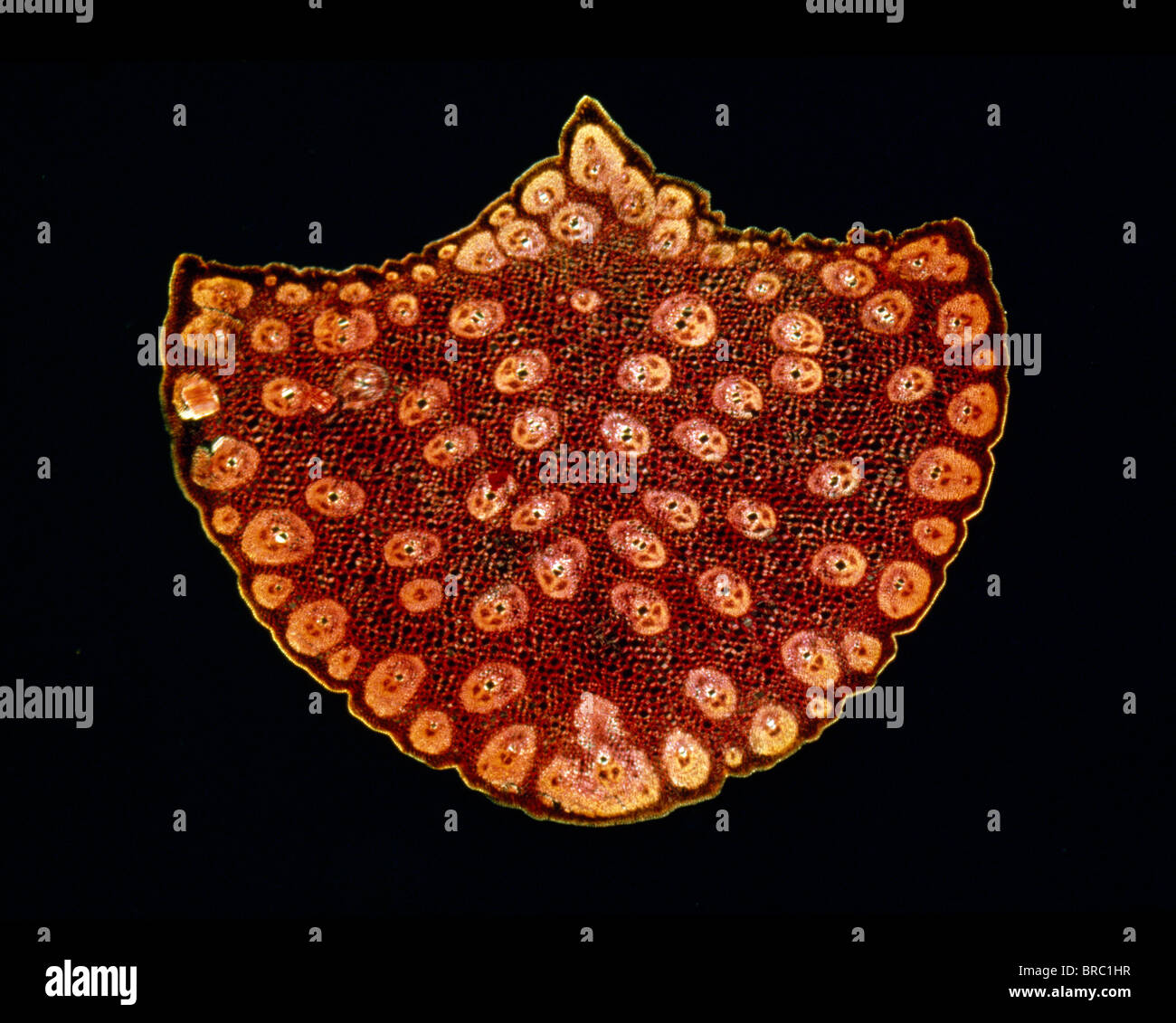 Light Micrograph (LM) of a transverse section of a stem of a Palm, magnification x12 Stock Photohttps://www.alamy.com/image-license-details/?v=1https://www.alamy.com/stock-photo-light-micrograph-lm-of-a-transverse-section-of-a-stem-of-a-palm-magnification-31612163.html
RMBRC1HR–Light Micrograph (LM) of a transverse section of a stem of a Palm, magnification x12
 Rice is a food made from paddy through processes such as cleaning, hulling, milling, and finished product sorting. Stock Photohttps://www.alamy.com/image-license-details/?v=1https://www.alamy.com/rice-is-a-food-made-from-paddy-through-processes-such-as-cleaning-hulling-milling-and-finished-product-sorting-image599422027.html
RM2WR60WF–Rice is a food made from paddy through processes such as cleaning, hulling, milling, and finished product sorting.
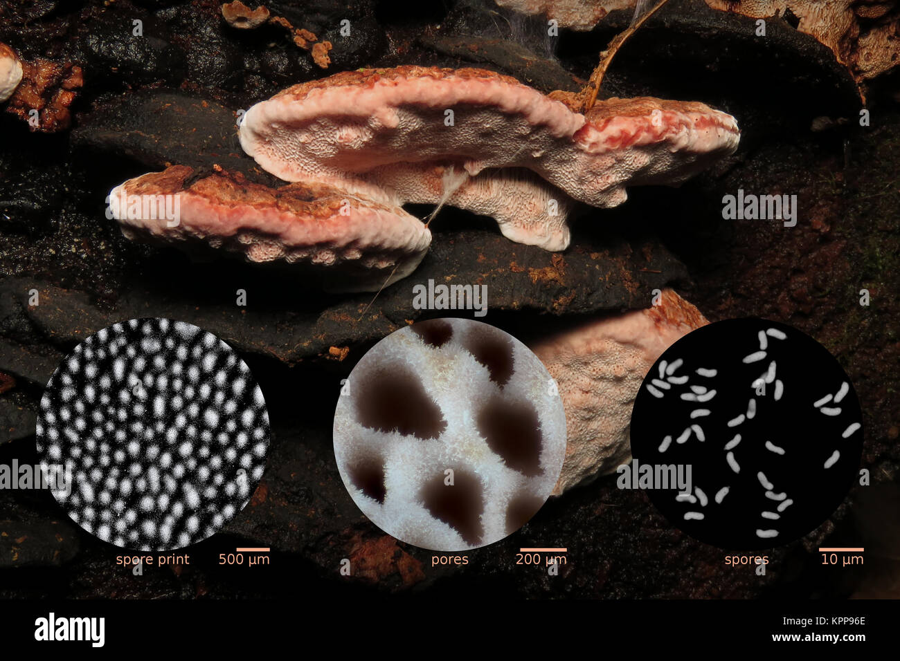 Rosy conk (Fomitopsis cajanderi, synonyms Fomitopsis subrosea and Fomes subrosea) with micrographs of the spore print, pores, and individual spores Stock Photohttps://www.alamy.com/image-license-details/?v=1https://www.alamy.com/stock-image-rosy-conk-fomitopsis-cajanderi-synonyms-fomitopsis-subrosea-and-fomes-168905926.html
RMKPP96E–Rosy conk (Fomitopsis cajanderi, synonyms Fomitopsis subrosea and Fomes subrosea) with micrographs of the spore print, pores, and individual spores
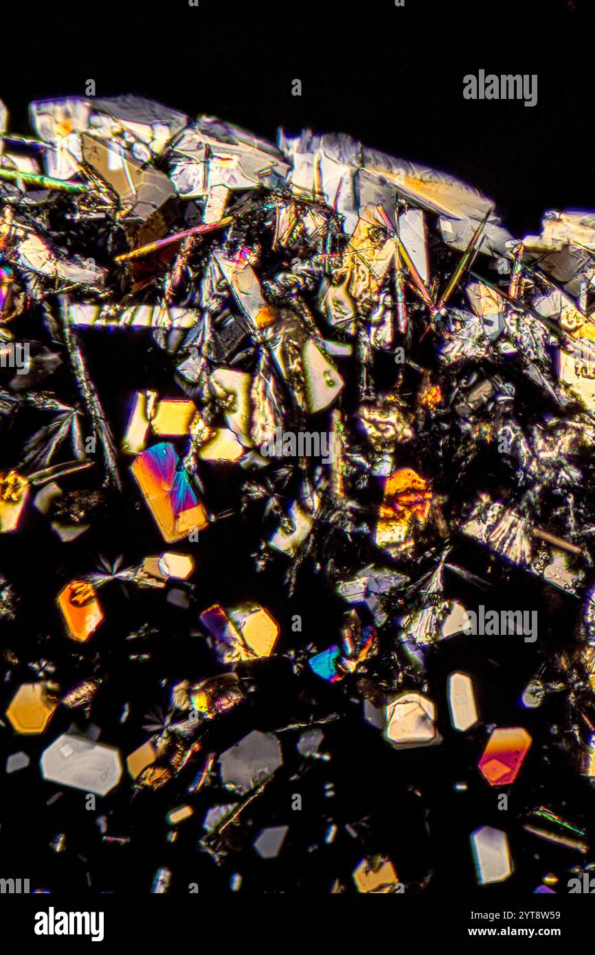 Microscopic shot showing colorful microcrystals in polarized light Stock Photohttps://www.alamy.com/image-license-details/?v=1https://www.alamy.com/microscopic-shot-showing-colorful-microcrystals-in-polarized-light-image634520357.html
RF2YT8W59–Microscopic shot showing colorful microcrystals in polarized light
 Coloured SEM of grease pencil Stock Photohttps://www.alamy.com/image-license-details/?v=1https://www.alamy.com/coloured-sem-of-grease-pencil-image67265908.html
RFDWC69T–Coloured SEM of grease pencil
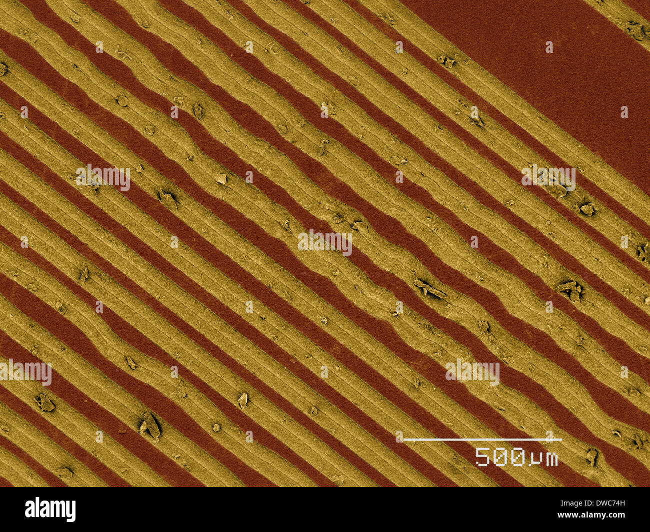 Coloured SEM of grooves on vinyl record Stock Photohttps://www.alamy.com/image-license-details/?v=1https://www.alamy.com/coloured-sem-of-grooves-on-vinyl-record-image67266545.html
RFDWC74H–Coloured SEM of grooves on vinyl record
 Onion cells under the microscope Stock Photohttps://www.alamy.com/image-license-details/?v=1https://www.alamy.com/onion-cells-under-the-microscope-image624949433.html
RF2Y8MWAH–Onion cells under the microscope
 Colorized transmission electron micrographs showing, A: internal elastic lamina of an aorta, characterized by large fenestrations traversed by an irregular meshwork of elastic strands, and, B: a similar view of the internal elastic lamina of the femoral artery shows small fenestrations. Stock Photohttps://www.alamy.com/image-license-details/?v=1https://www.alamy.com/colorized-transmission-electron-micrographs-showing-a-internal-elastic-lamina-of-an-aorta-characterized-by-large-fenestrations-traversed-by-an-irregular-meshwork-of-elastic-strands-and-b-a-similar-view-of-the-internal-elastic-lamina-of-the-femoral-artery-shows-small-fenestrations-image440582882.html
RF2GGP7XX–Colorized transmission electron micrographs showing, A: internal elastic lamina of an aorta, characterized by large fenestrations traversed by an irregular meshwork of elastic strands, and, B: a similar view of the internal elastic lamina of the femoral artery shows small fenestrations.
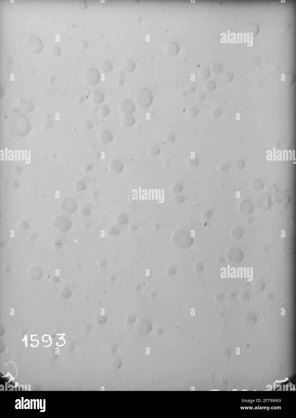 Micrographs of milk, stored in different ways. Each photography includes about 1/1070 cubic etc. Milk. Magnification 640 times. Stock Photohttps://www.alamy.com/image-license-details/?v=1https://www.alamy.com/micrographs-of-milk-stored-in-different-ways-each-photography-includes-about-11070-cubic-etc-milk-magnification-640-times-image417568769.html
RM2F79W69–Micrographs of milk, stored in different ways. Each photography includes about 1/1070 cubic etc. Milk. Magnification 640 times.
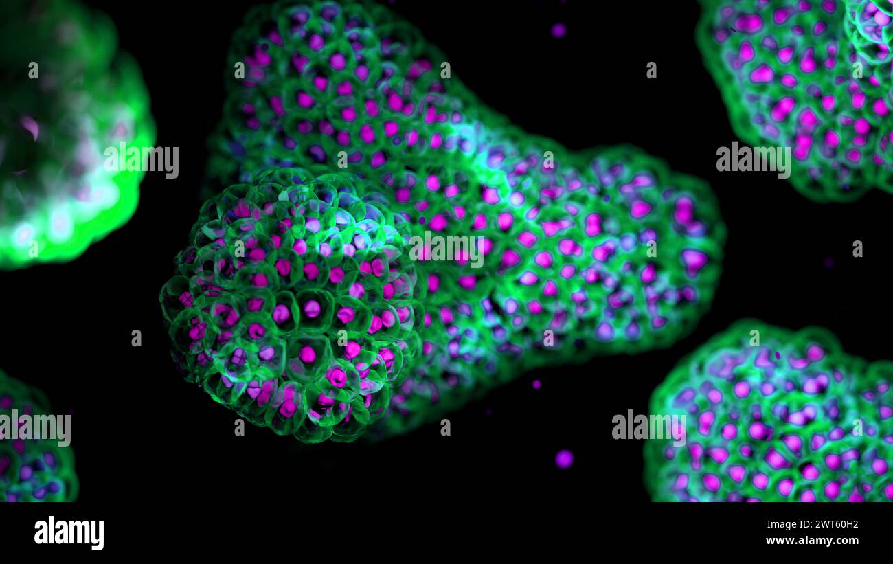 Illustration based on fluorescence light micrographs of organoids. Cell nuclei are purple and cell membranes green. Organoids are three dimensional, miniature, simplified versions of organs grown in the laboratory. They are able to survive for months in controlled conditions allowing diseases to be studied over time and the testing of targeted therapies. Stock Photohttps://www.alamy.com/image-license-details/?v=1https://www.alamy.com/illustration-based-on-fluorescence-light-micrographs-of-organoids-cell-nuclei-are-purple-and-cell-membranes-green-organoids-are-three-dimensional-miniature-simplified-versions-of-organs-grown-in-the-laboratory-they-are-able-to-survive-for-months-in-controlled-conditions-allowing-diseases-to-be-studied-over-time-and-the-testing-of-targeted-therapies-image600036446.html
RF2WT60H2–Illustration based on fluorescence light micrographs of organoids. Cell nuclei are purple and cell membranes green. Organoids are three dimensional, miniature, simplified versions of organs grown in the laboratory. They are able to survive for months in controlled conditions allowing diseases to be studied over time and the testing of targeted therapies.
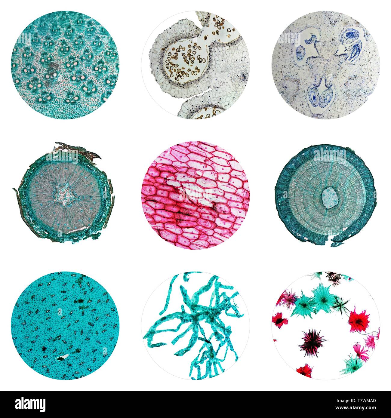 Light photomicrograph of many plants cells seen through a microscope Stock Photohttps://www.alamy.com/image-license-details/?v=1https://www.alamy.com/light-photomicrograph-of-many-plants-cells-seen-through-a-microscope-image245812517.html
RFT7WMAD–Light photomicrograph of many plants cells seen through a microscope
 Sheet fifteen micrographs of the cellular structure of pepper, saffron and cardamom Leaf with fifteen micrographs of the cellular structure of pepper, saffron and cardamom object type: photomechanical print page Item number: RP-F 2001-7-1484-3 Inscriptions / Brands: number, recto, printed: 'Taf. VII.' Manufacturer : Photographer: Franz Friedrich Bernard Elsner (attributed to) clichémaker: Photographische anstalt Otto Wigand (listed building) button: Photo Graphical anstalt Otto Wigand (listed building) Publisher: Wilhelm Knapp Verlag (listed property) Place manufacture: clichémaker: Zeitz Publ Stock Photohttps://www.alamy.com/image-license-details/?v=1https://www.alamy.com/sheet-fifteen-micrographs-of-the-cellular-structure-of-pepper-saffron-and-cardamom-leaf-with-fifteen-micrographs-of-the-cellular-structure-of-pepper-saffron-and-cardamom-object-type-photomechanical-print-page-item-number-rp-f-2001-7-1484-3-inscriptions-brands-number-recto-printed-taf-vii-manufacturer-photographer-franz-friedrich-bernard-elsner-attributed-to-clichmaker-photographische-anstalt-otto-wigand-listed-building-button-photo-graphical-anstalt-otto-wigand-listed-building-publisher-wilhelm-knapp-verlag-listed-property-place-manufacture-clichmaker-zeitz-publ-image348202688.html
RM2B6E028–Sheet fifteen micrographs of the cellular structure of pepper, saffron and cardamom Leaf with fifteen micrographs of the cellular structure of pepper, saffron and cardamom object type: photomechanical print page Item number: RP-F 2001-7-1484-3 Inscriptions / Brands: number, recto, printed: 'Taf. VII.' Manufacturer : Photographer: Franz Friedrich Bernard Elsner (attributed to) clichémaker: Photographische anstalt Otto Wigand (listed building) button: Photo Graphical anstalt Otto Wigand (listed building) Publisher: Wilhelm Knapp Verlag (listed property) Place manufacture: clichémaker: Zeitz Publ
 Hefei, China's Anhui Province. 19th Apr, 2023. A visitor views focus stacking micrographs of lunar soil particles at an exhibition themed on lunar soil research achievements in University of Science and Technology of China, in Hefei, east China's Anhui Province, April 19, 2023. The exhibition starting Monday displays exhibits made by scientists, artists and engineers based on micrographs of lunar soil particles brought back by the Chang'e-5 probe. In 2020, China's Chang'e-5 mission retrieved samples from the moon weighing about 1,731 grams. Credit: Huang Bohan/Xinhua/Alamy Live News Stock Photohttps://www.alamy.com/image-license-details/?v=1https://www.alamy.com/hefei-chinas-anhui-province-19th-apr-2023-a-visitor-views-focus-stacking-micrographs-of-lunar-soil-particles-at-an-exhibition-themed-on-lunar-soil-research-achievements-in-university-of-science-and-technology-of-china-in-hefei-east-chinas-anhui-province-april-19-2023-the-exhibition-starting-monday-displays-exhibits-made-by-scientists-artists-and-engineers-based-on-micrographs-of-lunar-soil-particles-brought-back-by-the-change-5-probe-in-2020-chinas-change-5-mission-retrieved-samples-from-the-moon-weighing-about-1731-grams-credit-huang-bohanxinhuaalamy-live-news-image546944070.html
RM2PNRCMP–Hefei, China's Anhui Province. 19th Apr, 2023. A visitor views focus stacking micrographs of lunar soil particles at an exhibition themed on lunar soil research achievements in University of Science and Technology of China, in Hefei, east China's Anhui Province, April 19, 2023. The exhibition starting Monday displays exhibits made by scientists, artists and engineers based on micrographs of lunar soil particles brought back by the Chang'e-5 probe. In 2020, China's Chang'e-5 mission retrieved samples from the moon weighing about 1,731 grams. Credit: Huang Bohan/Xinhua/Alamy Live News
 The Quarterly Journal of the Geological Society of London, v.21 (1865) London, geology, periodicals, The display features various illustrations of geological and botanical textures, emphasizing intricate patterns found in nature. The left section showcases detailed micrographs of cellular structures, revealing complex shapes and arrangements. Adjacent to this, an elongated vertical illustration captures a striking agate or rock formation with swirling designs. The lower section includes close-up views of what appear to be plant cells or mineral deposits, highlighting their unique formations. E Stock Photohttps://www.alamy.com/image-license-details/?v=1https://www.alamy.com/the-quarterly-journal-of-the-geological-society-of-london-v21-1865-london-geology-periodicals-the-display-features-various-illustrations-of-geological-and-botanical-textures-emphasizing-intricate-patterns-found-in-nature-the-left-section-showcases-detailed-micrographs-of-cellular-structures-revealing-complex-shapes-and-arrangements-adjacent-to-this-an-elongated-vertical-illustration-captures-a-striking-agate-or-rock-formation-with-swirling-designs-the-lower-section-includes-close-up-views-of-what-appear-to-be-plant-cells-or-mineral-deposits-highlighting-their-unique-formations-e-image636070554.html
RM2YXREDE–The Quarterly Journal of the Geological Society of London, v.21 (1865) London, geology, periodicals, The display features various illustrations of geological and botanical textures, emphasizing intricate patterns found in nature. The left section showcases detailed micrographs of cellular structures, revealing complex shapes and arrangements. Adjacent to this, an elongated vertical illustration captures a striking agate or rock formation with swirling designs. The lower section includes close-up views of what appear to be plant cells or mineral deposits, highlighting their unique formations. E
 The chili spice is considered the hottest chili spice ever. This sharpness can only be topped if different chili spices are mixed. Stock Photohttps://www.alamy.com/image-license-details/?v=1https://www.alamy.com/the-chili-spice-is-considered-the-hottest-chili-spice-ever-this-sharpness-can-only-be-topped-if-different-chili-spices-are-mixed-image236757987.html
RFRN576B–The chili spice is considered the hottest chili spice ever. This sharpness can only be topped if different chili spices are mixed.
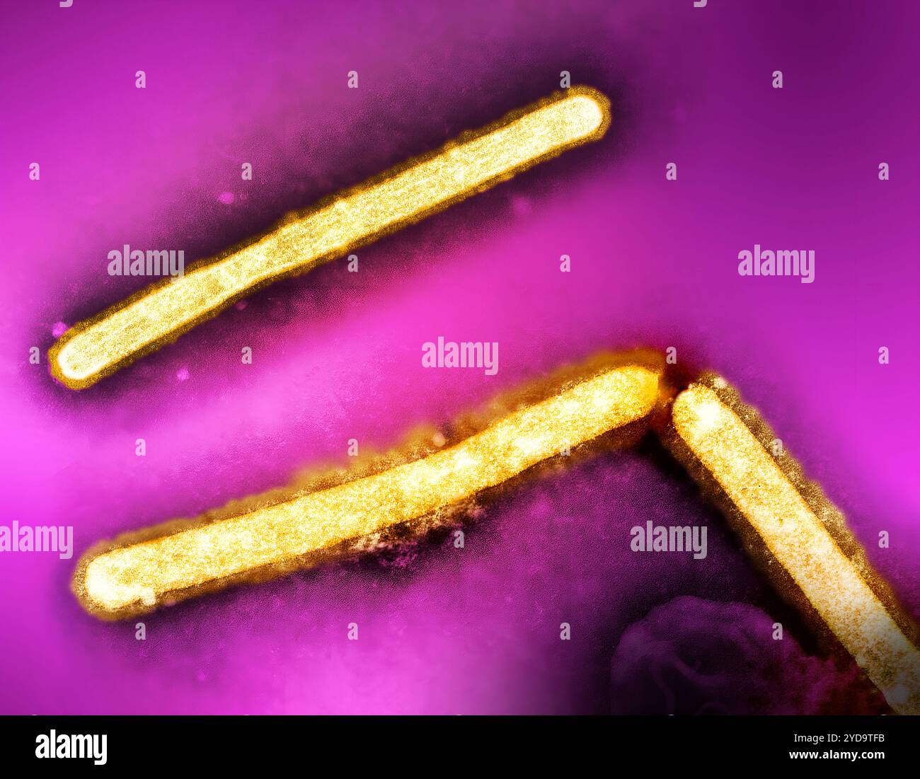 Three influenza A H5N1/bird flu virus particles rod-shaped yellow. Note: Layout incorporates two CDC transmission electron micrographs that have been repositioned and colorized by NIAID. Influenza A Virus H5N1/Bird Flu 016867 315 Stock Photohttps://www.alamy.com/image-license-details/?v=1https://www.alamy.com/three-influenza-a-h5n1bird-flu-virus-particles-rod-shaped-yellow-note-layout-incorporates-two-cdc-transmission-electron-micrographs-that-have-been-repositioned-and-colorized-by-niaid-influenza-a-virus-h5n1bird-flu-016867-315-image627780591.html
RM2YD9TFB–Three influenza A H5N1/bird flu virus particles rod-shaped yellow. Note: Layout incorporates two CDC transmission electron micrographs that have been repositioned and colorized by NIAID. Influenza A Virus H5N1/Bird Flu 016867 315
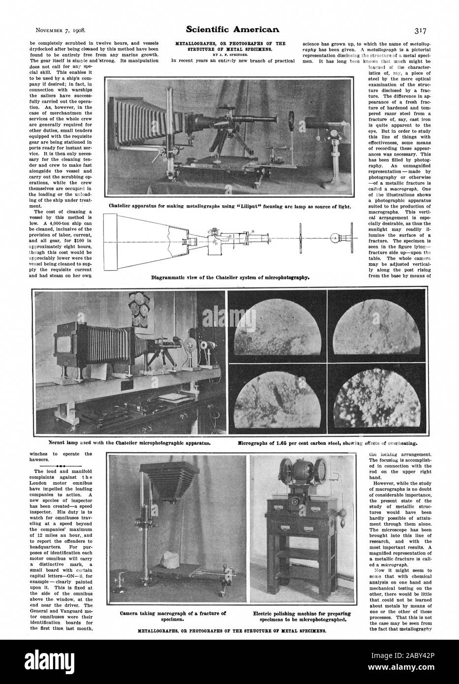 BY J. P. SPRINGER. p as source of light. Chatelier apparatus for making metallographs using “Liliput' focusing arc lam Di rammatic view of the Chatelier s stem of' micro hotogra ag P Y Nernst lamp used with the Chatelier microphotographic apparatus. Micrographs of 1.65 per cent carbon steel showing effects of overheating. Camera taking macrograph of a fracture of Electric polishing machine for preparing specimen. specimens to be microphotographed., scientific american, 1908-11-07 Stock Photohttps://www.alamy.com/image-license-details/?v=1https://www.alamy.com/by-j-p-springer-p-as-source-of-light-chatelier-apparatus-for-making-metallographs-using-liliput-focusing-arc-lam-di-rammatic-view-of-the-chatelier-s-stem-of-micro-hotogra-ag-p-y-nernst-lamp-used-with-the-chatelier-microphotographic-apparatus-micrographs-of-165-per-cent-carbon-steel-showing-effects-of-overheating-camera-taking-macrograph-of-a-fracture-of-electric-polishing-machine-for-preparing-specimen-specimens-to-be-microphotographed-scientific-american-1908-11-07-image334354126.html
RM2ABY42P–BY J. P. SPRINGER. p as source of light. Chatelier apparatus for making metallographs using “Liliput' focusing arc lam Di rammatic view of the Chatelier s stem of' micro hotogra ag P Y Nernst lamp used with the Chatelier microphotographic apparatus. Micrographs of 1.65 per cent carbon steel showing effects of overheating. Camera taking macrograph of a fracture of Electric polishing machine for preparing specimen. specimens to be microphotographed., scientific american, 1908-11-07
 Archive image from page 28 of Deposition, corrosion and coloration of. Deposition, corrosion and coloration of tungsten trioxide electrochromic thin films depositioncorros00suns Year: 1983 ')'/'' ' /± -.;' , (B) Figure 2.6. Scanning electron micrographs of corroded surface (3.6 N H SO,, 4 days) of WO films (P = 1 x 10 Torr) showing the forms of interfaciales'delamination. (A) wrinkles, (B) rosette pattern. Stock Photohttps://www.alamy.com/image-license-details/?v=1https://www.alamy.com/archive-image-from-page-28-of-deposition-corrosion-and-coloration-of-deposition-corrosion-and-coloration-of-tungsten-trioxide-electrochromic-thin-films-depositioncorros00suns-year-1983-b-figure-26-scanning-electron-micrographs-of-corroded-surface-36-n-h-so-4-days-of-wo-films-p-=-1-x-10-torr-showing-the-forms-of-interfacialesdelamination-a-wrinkles-b-rosette-pattern-image259538683.html
RMW2706K–Archive image from page 28 of Deposition, corrosion and coloration of. Deposition, corrosion and coloration of tungsten trioxide electrochromic thin films depositioncorros00suns Year: 1983 ')'/'' ' /± -.;' , (B) Figure 2.6. Scanning electron micrographs of corroded surface (3.6 N H SO,, 4 days) of WO films (P = 1 x 10 Torr) showing the forms of interfaciales'delamination. (A) wrinkles, (B) rosette pattern.
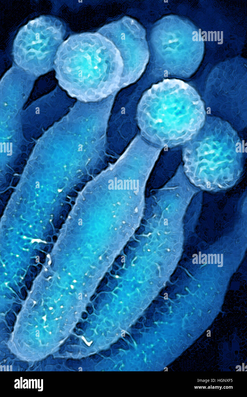 PENICILLIUM CHRYSOGENUM Stock Photohttps://www.alamy.com/image-license-details/?v=1https://www.alamy.com/stock-photo-penicillium-chrysogenum-130788873.html
RMHGNXF5–PENICILLIUM CHRYSOGENUM
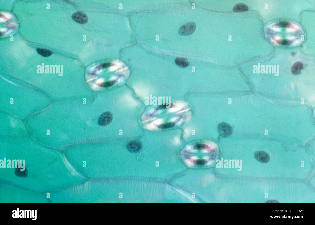 Light Micrograph (LM) of a transverse section of a leaf of a Tulip (Tulipa sp.) showing stomata, magnification x1200 Stock Photohttps://www.alamy.com/image-license-details/?v=1https://www.alamy.com/stock-photo-light-micrograph-lm-of-a-transverse-section-of-a-leaf-of-a-tulip-tulipa-31611971.html
RMBRC1AY–Light Micrograph (LM) of a transverse section of a leaf of a Tulip (Tulipa sp.) showing stomata, magnification x1200
 Stomata are openings formed around two guard cells, surrounded by accessory guard cells. Stock Photohttps://www.alamy.com/image-license-details/?v=1https://www.alamy.com/stomata-are-openings-formed-around-two-guard-cells-surrounded-by-accessory-guard-cells-image599785233.html
RM2WRPG55–Stomata are openings formed around two guard cells, surrounded by accessory guard cells.
![[Frustules of Diatoms]. Artist: Attributed to Julius Wiesner (Austrian, 1838-1916). Dimensions: 9.8 x 7.9 cm (3 7/8 x 3 1/8 in.). Date: ca. 1870. Museum: Metropolitan Museum of Art, New York, USA. Stock Photo [Frustules of Diatoms]. Artist: Attributed to Julius Wiesner (Austrian, 1838-1916). Dimensions: 9.8 x 7.9 cm (3 7/8 x 3 1/8 in.). Date: ca. 1870. Museum: Metropolitan Museum of Art, New York, USA. Stock Photo](https://c8.alamy.com/comp/PB9RH2/frustules-of-diatoms-artist-attributed-to-julius-wiesner-austrian-1838-1916-dimensions-98-x-79-cm-3-78-x-3-18-in-date-ca-1870-museum-metropolitan-museum-of-art-new-york-usa-PB9RH2.jpg) [Frustules of Diatoms]. Artist: Attributed to Julius Wiesner (Austrian, 1838-1916). Dimensions: 9.8 x 7.9 cm (3 7/8 x 3 1/8 in.). Date: ca. 1870. Museum: Metropolitan Museum of Art, New York, USA. Stock Photohttps://www.alamy.com/image-license-details/?v=1https://www.alamy.com/frustules-of-diatoms-artist-attributed-to-julius-wiesner-austrian-1838-1916-dimensions-98-x-79-cm-3-78-x-3-18-in-date-ca-1870-museum-metropolitan-museum-of-art-new-york-usa-image213501710.html
[Frustules of Diatoms]. Artist: Attributed to Julius Wiesner (Austrian, 1838-1916). Dimensions: 9.8 x 7.9 cm (3 7/8 x 3 1/8 in.). Date: ca. 1870. Museum: Metropolitan Museum of Art, New York, USA. Stock Photohttps://www.alamy.com/image-license-details/?v=1https://www.alamy.com/frustules-of-diatoms-artist-attributed-to-julius-wiesner-austrian-1838-1916-dimensions-98-x-79-cm-3-78-x-3-18-in-date-ca-1870-museum-metropolitan-museum-of-art-new-york-usa-image213501710.htmlRMPB9RH2–[Frustules of Diatoms]. Artist: Attributed to Julius Wiesner (Austrian, 1838-1916). Dimensions: 9.8 x 7.9 cm (3 7/8 x 3 1/8 in.). Date: ca. 1870. Museum: Metropolitan Museum of Art, New York, USA.
 Microscopic shot showing colorful microcrystals in polarized light Stock Photohttps://www.alamy.com/image-license-details/?v=1https://www.alamy.com/microscopic-shot-showing-colorful-microcrystals-in-polarized-light-image634520396.html
RF2YT8W6M–Microscopic shot showing colorful microcrystals in polarized light
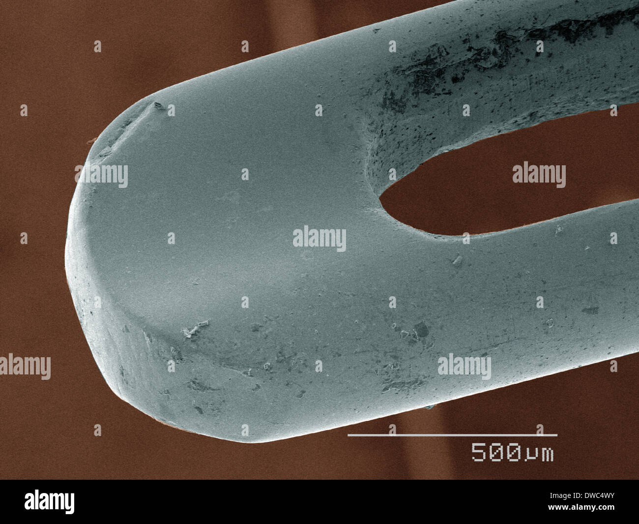 Coloured SEM of needle Stock Photohttps://www.alamy.com/image-license-details/?v=1https://www.alamy.com/coloured-sem-of-needle-image67264791.html
RFDWC4WY–Coloured SEM of needle
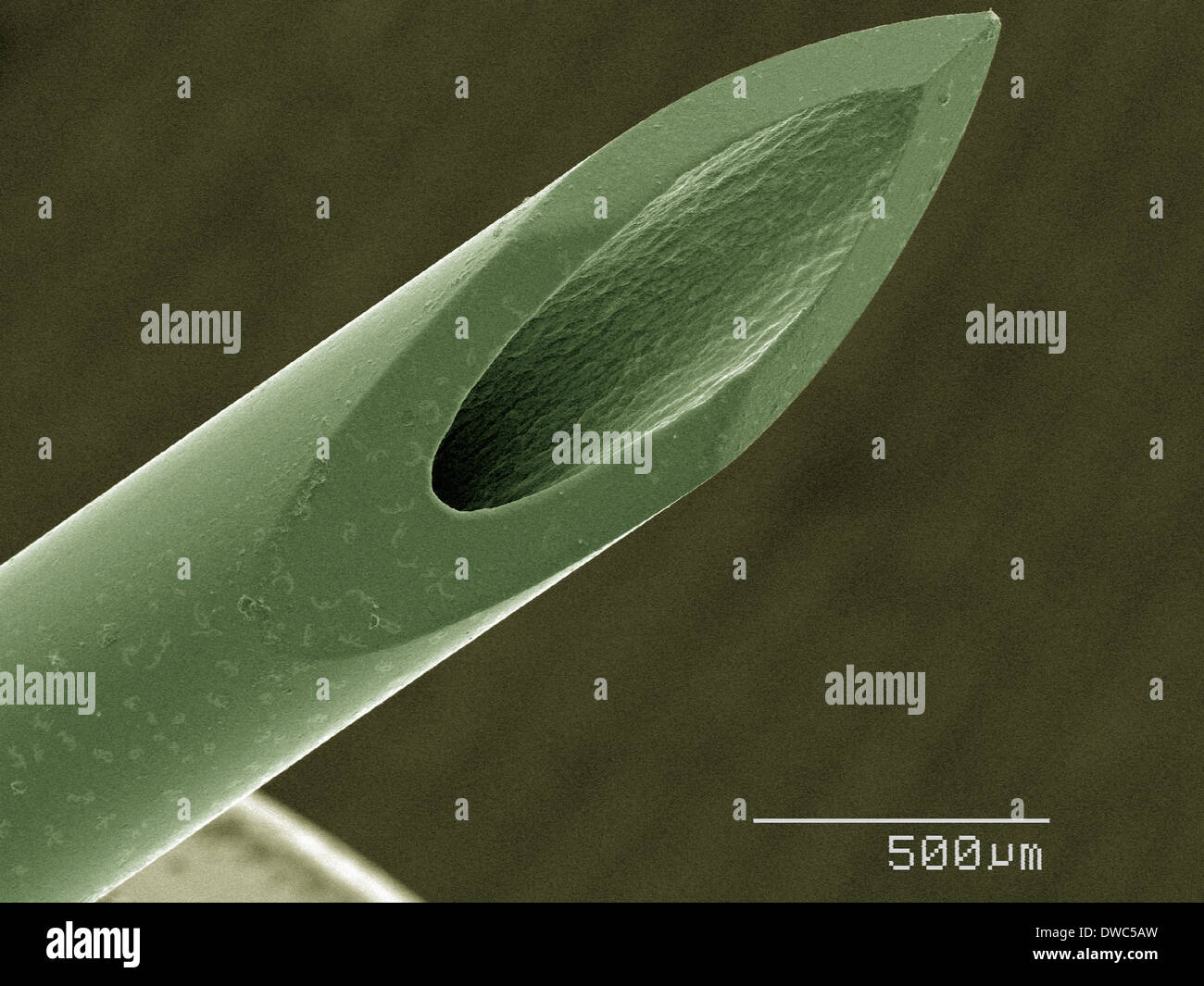 Coloured SEM of syringe Stock Photohttps://www.alamy.com/image-license-details/?v=1https://www.alamy.com/coloured-sem-of-syringe-image67265153.html
RFDWC5AW–Coloured SEM of syringe
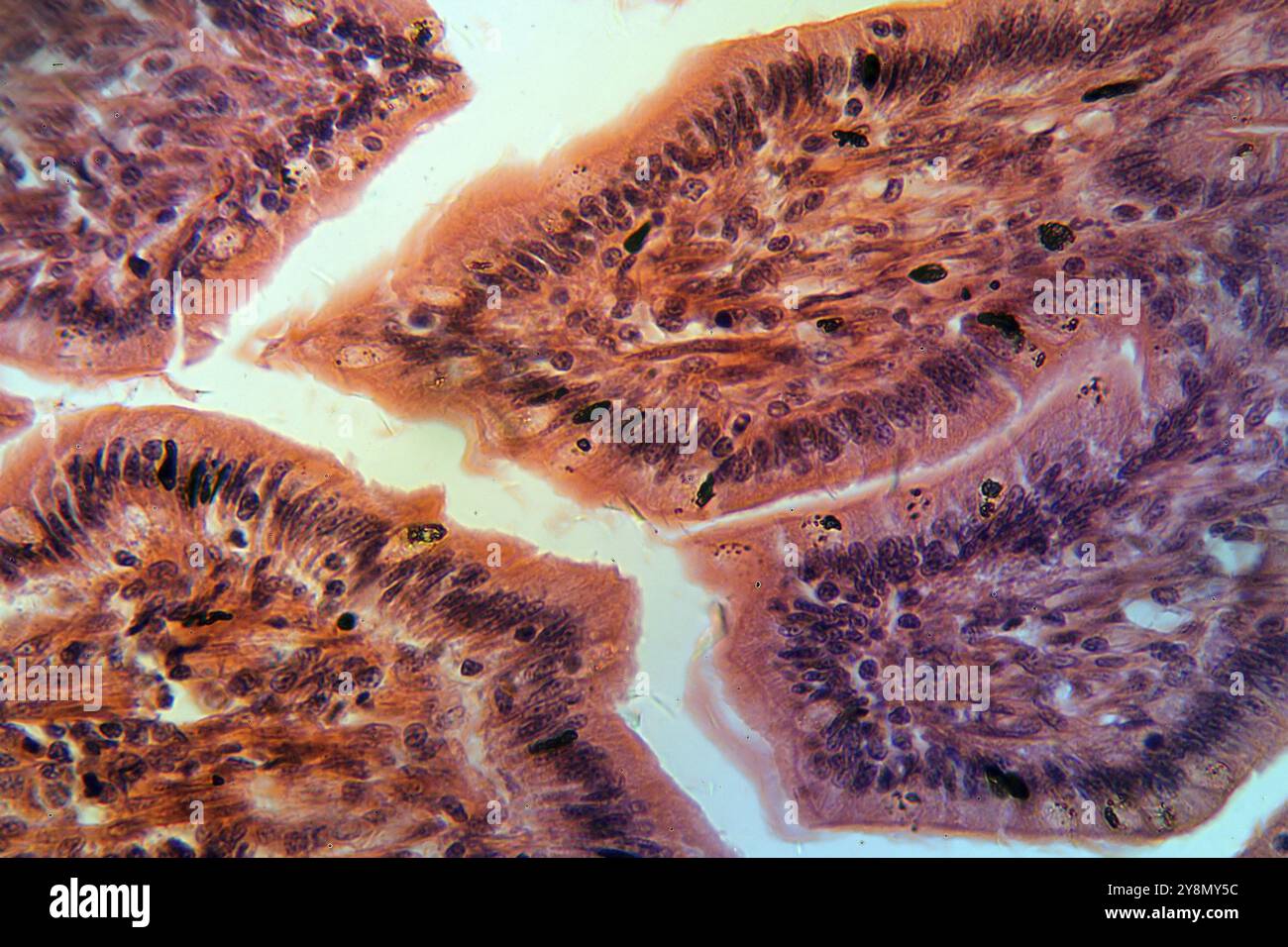 A section trough cells of a small intestine under the microscope Stock Photohttps://www.alamy.com/image-license-details/?v=1https://www.alamy.com/a-section-trough-cells-of-a-small-intestine-under-the-microscope-image624950856.html
RF2Y8MY5C–A section trough cells of a small intestine under the microscope
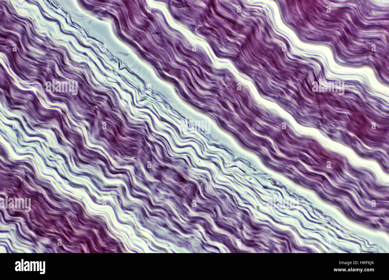 Tendon Stock Photohttps://www.alamy.com/image-license-details/?v=1https://www.alamy.com/stock-photo-tendon-134944171.html
RMHRF6JK–Tendon
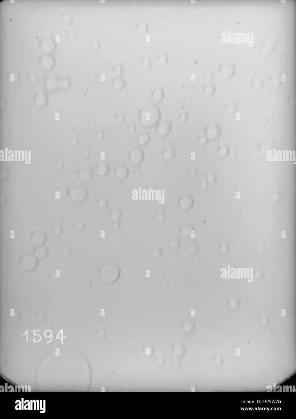 Micrographs of milk, stored in different ways. Each photography includes about 1/1070 cubic etc. Milk. Magnification 640 times. Stock Photohttps://www.alamy.com/image-license-details/?v=1https://www.alamy.com/micrographs-of-milk-stored-in-different-ways-each-photography-includes-about-11070-cubic-etc-milk-magnification-640-times-image417568804.html
RM2F79W7G–Micrographs of milk, stored in different ways. Each photography includes about 1/1070 cubic etc. Milk. Magnification 640 times.
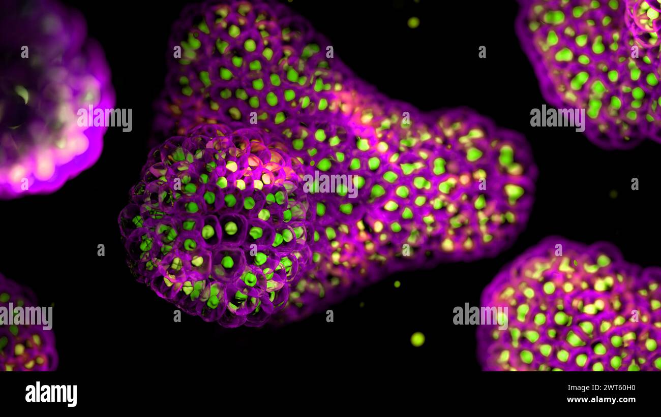 Illustration based on fluorescence light micrographs of organoids. Cell nuclei are green and cell membranes purple. Organoids are three dimensional, miniature, simplified versions of organs grown in the laboratory. They are able to survive for months in controlled conditions allowing diseases to be studied over time and the testing of targeted therapies. Stock Photohttps://www.alamy.com/image-license-details/?v=1https://www.alamy.com/illustration-based-on-fluorescence-light-micrographs-of-organoids-cell-nuclei-are-green-and-cell-membranes-purple-organoids-are-three-dimensional-miniature-simplified-versions-of-organs-grown-in-the-laboratory-they-are-able-to-survive-for-months-in-controlled-conditions-allowing-diseases-to-be-studied-over-time-and-the-testing-of-targeted-therapies-image600036444.html
RF2WT60H0–Illustration based on fluorescence light micrographs of organoids. Cell nuclei are green and cell membranes purple. Organoids are three dimensional, miniature, simplified versions of organs grown in the laboratory. They are able to survive for months in controlled conditions allowing diseases to be studied over time and the testing of targeted therapies.
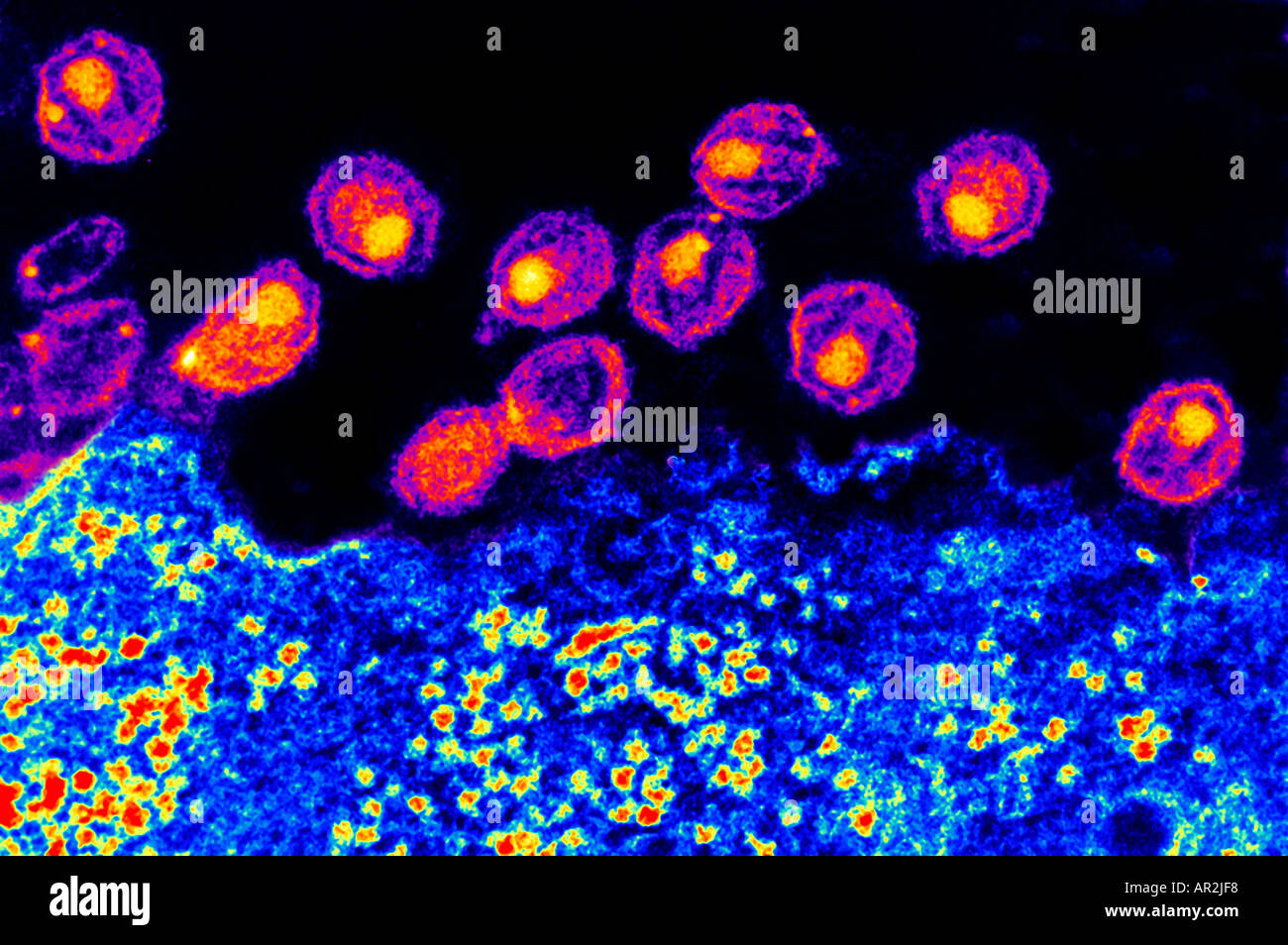 TEM Transmission Electron Microscope of AIDS virus Stock Photohttps://www.alamy.com/image-license-details/?v=1https://www.alamy.com/tem-transmission-electron-microscope-of-aids-virus-image5196535.html
RMAR2JF8–TEM Transmission Electron Microscope of AIDS virus
 Leaf with micrographs of the cellular structure of fifteen types of flour and starches Leaf with micrographs of the cellular structure of fifteen flours and starches object type: photomechanical print page Item number: RP-F 2001-7-1484-2 Inscriptions / Brands: number, recto, printed: 'Taf. X.' Manufacturer : Photographer: Franz Friedrich Bernard Elsner (attributed to) clichémaker: Photographische anstalt Otto Wigand (listed building) button: Photo Graphical anstalt Otto Wigand (listed building) Publisher: Wilhelm Knapp Verlag (listed property) Place manufacture: clichémaker: Zeitz Publisher: Z Stock Photohttps://www.alamy.com/image-license-details/?v=1https://www.alamy.com/leaf-with-micrographs-of-the-cellular-structure-of-fifteen-types-of-flour-and-starches-leaf-with-micrographs-of-the-cellular-structure-of-fifteen-flours-and-starches-object-type-photomechanical-print-page-item-number-rp-f-2001-7-1484-2-inscriptions-brands-number-recto-printed-taf-x-manufacturer-photographer-franz-friedrich-bernard-elsner-attributed-to-clichmaker-photographische-anstalt-otto-wigand-listed-building-button-photo-graphical-anstalt-otto-wigand-listed-building-publisher-wilhelm-knapp-verlag-listed-property-place-manufacture-clichmaker-zeitz-publisher-z-image348201965.html
RM2B6DY4D–Leaf with micrographs of the cellular structure of fifteen types of flour and starches Leaf with micrographs of the cellular structure of fifteen flours and starches object type: photomechanical print page Item number: RP-F 2001-7-1484-2 Inscriptions / Brands: number, recto, printed: 'Taf. X.' Manufacturer : Photographer: Franz Friedrich Bernard Elsner (attributed to) clichémaker: Photographische anstalt Otto Wigand (listed building) button: Photo Graphical anstalt Otto Wigand (listed building) Publisher: Wilhelm Knapp Verlag (listed property) Place manufacture: clichémaker: Zeitz Publisher: Z
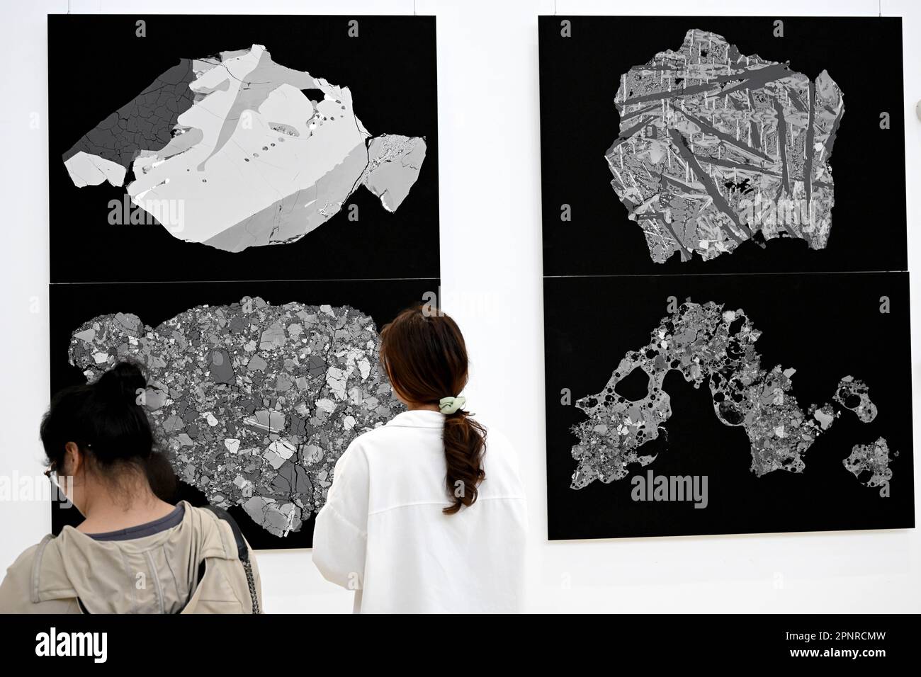 Hefei, China's Anhui Province. 19th Apr, 2023. Visitors view back-scatter electron micrographs of lunar soil particles at an exhibition themed on lunar soil research achievements in University of Science and Technology of China, in Hefei, east China's Anhui Province, April 19, 2023. The exhibition starting Monday displays exhibits made by scientists, artists and engineers based on micrographs of lunar soil particles brought back by the Chang'e-5 probe. In 2020, China's Chang'e-5 mission retrieved samples from the moon weighing about 1,731 grams. Credit: Huang Bohan/Xinhua/Alamy Live News Stock Photohttps://www.alamy.com/image-license-details/?v=1https://www.alamy.com/hefei-chinas-anhui-province-19th-apr-2023-visitors-view-back-scatter-electron-micrographs-of-lunar-soil-particles-at-an-exhibition-themed-on-lunar-soil-research-achievements-in-university-of-science-and-technology-of-china-in-hefei-east-chinas-anhui-province-april-19-2023-the-exhibition-starting-monday-displays-exhibits-made-by-scientists-artists-and-engineers-based-on-micrographs-of-lunar-soil-particles-brought-back-by-the-change-5-probe-in-2020-chinas-change-5-mission-retrieved-samples-from-the-moon-weighing-about-1731-grams-credit-huang-bohanxinhuaalamy-live-news-image546944073.html
RM2PNRCMW–Hefei, China's Anhui Province. 19th Apr, 2023. Visitors view back-scatter electron micrographs of lunar soil particles at an exhibition themed on lunar soil research achievements in University of Science and Technology of China, in Hefei, east China's Anhui Province, April 19, 2023. The exhibition starting Monday displays exhibits made by scientists, artists and engineers based on micrographs of lunar soil particles brought back by the Chang'e-5 probe. In 2020, China's Chang'e-5 mission retrieved samples from the moon weighing about 1,731 grams. Credit: Huang Bohan/Xinhua/Alamy Live News
 Atlas der Diatomaceen-Kunde, Leipzig, O.R. Reisland, 1874-19, atlases, Bacillariophyceae, The illustration features a collection of meticulously detailed micrographs showcasing various diatom species. Each specimen is numbered and exhibits a unique arrangement of intricate patterns and shapes, highlighting the diverse morphology of these microscopic algae. The designs range from elongated, slender forms to broader, more robust structures, characterized by elaborate surface textures and markings. These depictions serve as a scientific reference, illustrating the complexity and variety found wit Stock Photohttps://www.alamy.com/image-license-details/?v=1https://www.alamy.com/atlas-der-diatomaceen-kunde-leipzig-or-reisland-1874-19-atlases-bacillariophyceae-the-illustration-features-a-collection-of-meticulously-detailed-micrographs-showcasing-various-diatom-species-each-specimen-is-numbered-and-exhibits-a-unique-arrangement-of-intricate-patterns-and-shapes-highlighting-the-diverse-morphology-of-these-microscopic-algae-the-designs-range-from-elongated-slender-forms-to-broader-more-robust-structures-characterized-by-elaborate-surface-textures-and-markings-these-depictions-serve-as-a-scientific-reference-illustrating-the-complexity-and-variety-found-wit-image636044390.html
RM2YXP932–Atlas der Diatomaceen-Kunde, Leipzig, O.R. Reisland, 1874-19, atlases, Bacillariophyceae, The illustration features a collection of meticulously detailed micrographs showcasing various diatom species. Each specimen is numbered and exhibits a unique arrangement of intricate patterns and shapes, highlighting the diverse morphology of these microscopic algae. The designs range from elongated, slender forms to broader, more robust structures, characterized by elaborate surface textures and markings. These depictions serve as a scientific reference, illustrating the complexity and variety found wit
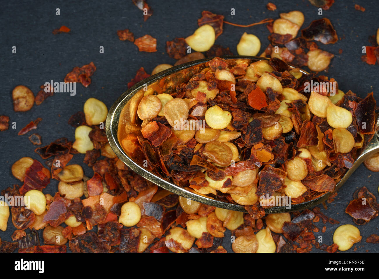 The chili spice is considered the hottest chili spice ever. This sharpness can only be topped if different chili spices are mixed. Stock Photohttps://www.alamy.com/image-license-details/?v=1https://www.alamy.com/the-chili-spice-is-considered-the-hottest-chili-spice-ever-this-sharpness-can-only-be-topped-if-different-chili-spices-are-mixed-image236757959.html
RFRN575B–The chili spice is considered the hottest chili spice ever. This sharpness can only be topped if different chili spices are mixed.
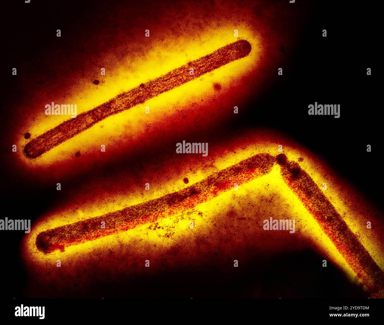 Three influenza A H5N1/bird flu virus particles rod-shaped. Note: Layout incorporates two CDC transmission electron micrographs that have been inverted, repositioned, and colorized by NIAID. Influenza A Virus H5N1/Bird Flu 016867 310 Stock Photohttps://www.alamy.com/image-license-details/?v=1https://www.alamy.com/three-influenza-a-h5n1bird-flu-virus-particles-rod-shaped-note-layout-incorporates-two-cdc-transmission-electron-micrographs-that-have-been-inverted-repositioned-and-colorized-by-niaid-influenza-a-virus-h5n1bird-flu-016867-310-image627780544.html
RM2YD9TDM–Three influenza A H5N1/bird flu virus particles rod-shaped. Note: Layout incorporates two CDC transmission electron micrographs that have been inverted, repositioned, and colorized by NIAID. Influenza A Virus H5N1/Bird Flu 016867 310
 Bulletins of American paleontology . Cretaceous Radiolaria: Pessagno 429 Explanation of Plate 30 Both figures scanning electron micrographs of Microsciadiocapsa lipmanaePesagno, n. sp. Paratype (USNM 164233). NSF 350: early Cenomanian partof the Antelope Shale7Fiske Creek Formation. Figure Page 1. Cut away side view with most of cephalis removed 405 showing cephalic skeletal elements. Note juncture of cephalicskeletal needles (primary lateral, secondary lateral, dorsal,and verticle) with rim bordering shelflike collar structure.Marker ^ 10 microns*a = Apical bar (broken),b = Axial spine,c = Ve Stock Photohttps://www.alamy.com/image-license-details/?v=1https://www.alamy.com/bulletins-of-american-paleontology-cretaceous-radiolaria-pessagno-429-explanation-of-plate-30-both-figures-scanning-electron-micrographs-of-microsciadiocapsa-lipmanaepesagno-n-sp-paratype-usnm-164233-nsf-350-early-cenomanian-partof-the-antelope-shale7fiske-creek-formation-figure-page-1-cut-away-side-view-with-most-of-cephalis-removed-405-showing-cephalic-skeletal-elements-note-juncture-of-cephalicskeletal-needles-primary-lateral-secondary-lateral-dorsaland-verticle-with-rim-bordering-shelflike-collar-structuremarker-10-micronsa-=-apical-bar-brokenb-=-axial-spinec-=-ve-image338314017.html
RM2AJBEYD–Bulletins of American paleontology . Cretaceous Radiolaria: Pessagno 429 Explanation of Plate 30 Both figures scanning electron micrographs of Microsciadiocapsa lipmanaePesagno, n. sp. Paratype (USNM 164233). NSF 350: early Cenomanian partof the Antelope Shale7Fiske Creek Formation. Figure Page 1. Cut away side view with most of cephalis removed 405 showing cephalic skeletal elements. Note juncture of cephalicskeletal needles (primary lateral, secondary lateral, dorsal,and verticle) with rim bordering shelflike collar structure.Marker ^ 10 microns*a = Apical bar (broken),b = Axial spine,c = Ve
 Archive image from page 27 of Deposition, corrosion and coloration of. Deposition, corrosion and coloration of tungsten trioxide electrochromic thin films depositioncorros00suns Year: 1983 21 (B) Figure 2.5. Scanning electron micrographs of as-deposited WC>3 films on glass substrates; (A) p = 1 x 10 Torr, x 500, (B) Pres-= 1 x 10 Torr, x 3,000 °2 Stock Photohttps://www.alamy.com/image-license-details/?v=1https://www.alamy.com/archive-image-from-page-27-of-deposition-corrosion-and-coloration-of-deposition-corrosion-and-coloration-of-tungsten-trioxide-electrochromic-thin-films-depositioncorros00suns-year-1983-21-b-figure-25-scanning-electron-micrographs-of-as-deposited-wcgt3-films-on-glass-substrates-a-p-=-1-x-10-torr-x-500-b-pres-=-1-x-10-torr-x-3000-2-image259538410.html
RMW26YTX–Archive image from page 27 of Deposition, corrosion and coloration of. Deposition, corrosion and coloration of tungsten trioxide electrochromic thin films depositioncorros00suns Year: 1983 21 (B) Figure 2.5. Scanning electron micrographs of as-deposited WC>3 films on glass substrates; (A) p = 1 x 10 Torr, x 500, (B) Pres-= 1 x 10 Torr, x 3,000 °2
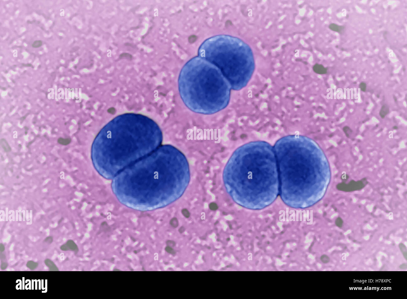 NEISSERIA MENINGITIDIS Stock Photohttps://www.alamy.com/image-license-details/?v=1https://www.alamy.com/stock-photo-neisseria-meningitidis-124971796.html
RMH78XPC–NEISSERIA MENINGITIDIS
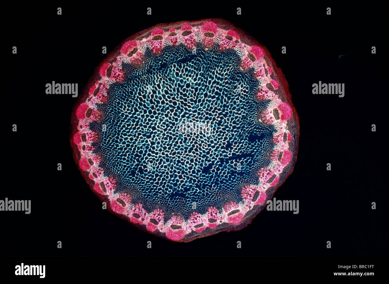 Light Micrograph (LM) of a transverse section of a Helianthus stem, magnification x 18 Stock Photohttps://www.alamy.com/image-license-details/?v=1https://www.alamy.com/stock-photo-light-micrograph-lm-of-a-transverse-section-of-a-helianthus-stem-magnification-31612108.html
RMBRC1FT–Light Micrograph (LM) of a transverse section of a Helianthus stem, magnification x 18
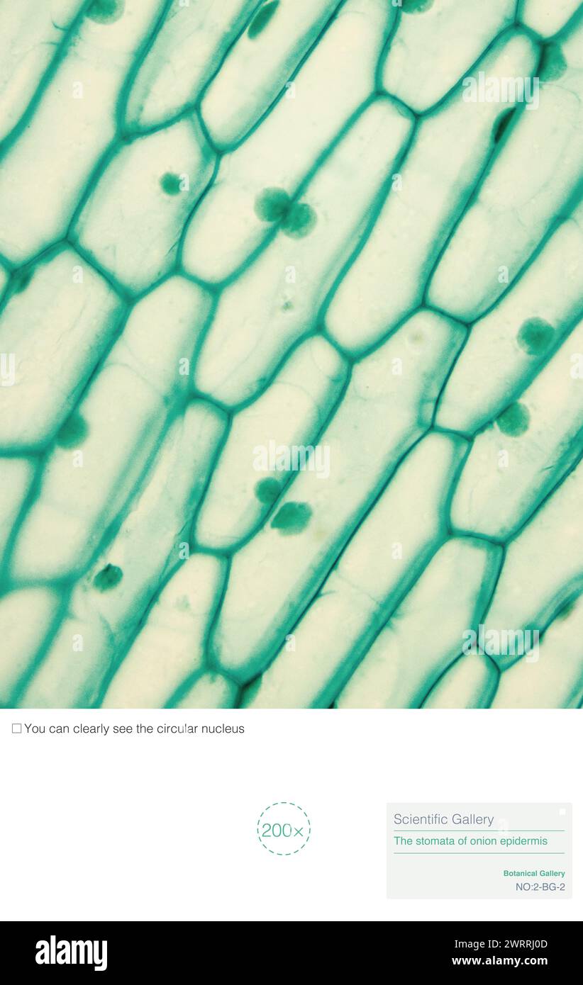 The structure of onion epidermal cells includes cell membrane, cytoplasm, nucleus, cell wall, vacuoles, and no chloroplasts. Stock Photohttps://www.alamy.com/image-license-details/?v=1https://www.alamy.com/the-structure-of-onion-epidermal-cells-includes-cell-membrane-cytoplasm-nucleus-cell-wall-vacuoles-and-no-chloroplasts-image599808621.html
RM2WRRJ0D–The structure of onion epidermal cells includes cell membrane, cytoplasm, nucleus, cell wall, vacuoles, and no chloroplasts.
![[Microscopic view of an insect]. Artist: Alois Auer (Austrian, Wels 1813-1869 Vienna). Date: ca. 1853. Museum: Metropolitan Museum of Art, New York, USA. Stock Photo [Microscopic view of an insect]. Artist: Alois Auer (Austrian, Wels 1813-1869 Vienna). Date: ca. 1853. Museum: Metropolitan Museum of Art, New York, USA. Stock Photo](https://c8.alamy.com/comp/PB6474/microscopic-view-of-an-insect-artist-alois-auer-austrian-wels-1813-1869-vienna-date-ca-1853-museum-metropolitan-museum-of-art-new-york-usa-PB6474.jpg) [Microscopic view of an insect]. Artist: Alois Auer (Austrian, Wels 1813-1869 Vienna). Date: ca. 1853. Museum: Metropolitan Museum of Art, New York, USA. Stock Photohttps://www.alamy.com/image-license-details/?v=1https://www.alamy.com/microscopic-view-of-an-insect-artist-alois-auer-austrian-wels-1813-1869-vienna-date-ca-1853-museum-metropolitan-museum-of-art-new-york-usa-image213420680.html
[Microscopic view of an insect]. Artist: Alois Auer (Austrian, Wels 1813-1869 Vienna). Date: ca. 1853. Museum: Metropolitan Museum of Art, New York, USA. Stock Photohttps://www.alamy.com/image-license-details/?v=1https://www.alamy.com/microscopic-view-of-an-insect-artist-alois-auer-austrian-wels-1813-1869-vienna-date-ca-1853-museum-metropolitan-museum-of-art-new-york-usa-image213420680.htmlRMPB6474–[Microscopic view of an insect]. Artist: Alois Auer (Austrian, Wels 1813-1869 Vienna). Date: ca. 1853. Museum: Metropolitan Museum of Art, New York, USA.
 Microscopic shot showing colorful microcrystals in polarized light Stock Photohttps://www.alamy.com/image-license-details/?v=1https://www.alamy.com/microscopic-shot-showing-colorful-microcrystals-in-polarized-light-image634520588.html
RF2YT8WDG–Microscopic shot showing colorful microcrystals in polarized light
 Coloured SEM of rivets Stock Photohttps://www.alamy.com/image-license-details/?v=1https://www.alamy.com/coloured-sem-of-rivets-image67264357.html
RFDWC4AD–Coloured SEM of rivets
 Coloured SEM of hook and loop fastener Stock Photohttps://www.alamy.com/image-license-details/?v=1https://www.alamy.com/coloured-sem-of-hook-and-loop-fastener-image67263945.html
RFDWC3RN–Coloured SEM of hook and loop fastener
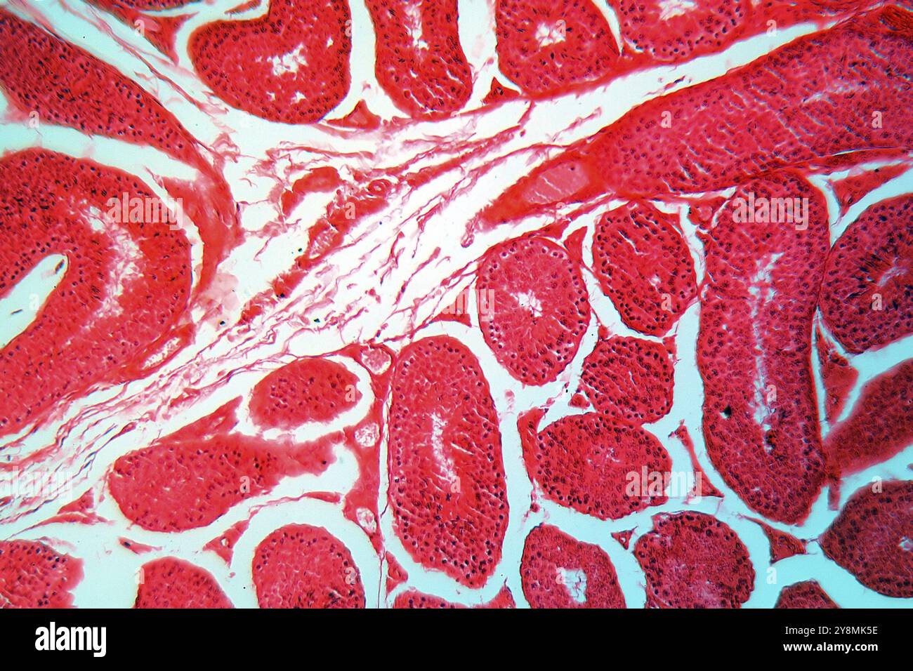 A section trough testicle cells under the microscope Stock Photohttps://www.alamy.com/image-license-details/?v=1https://www.alamy.com/a-section-trough-testicle-cells-under-the-microscope-image624944586.html
RF2Y8MK5E–A section trough testicle cells under the microscope
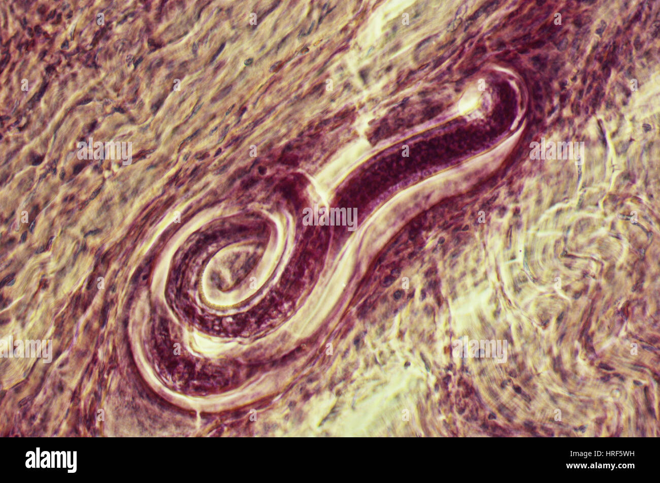 Trichinella Spiralis Stock Photohttps://www.alamy.com/image-license-details/?v=1https://www.alamy.com/stock-photo-trichinella-spiralis-134943581.html
RMHRF5WH–Trichinella Spiralis
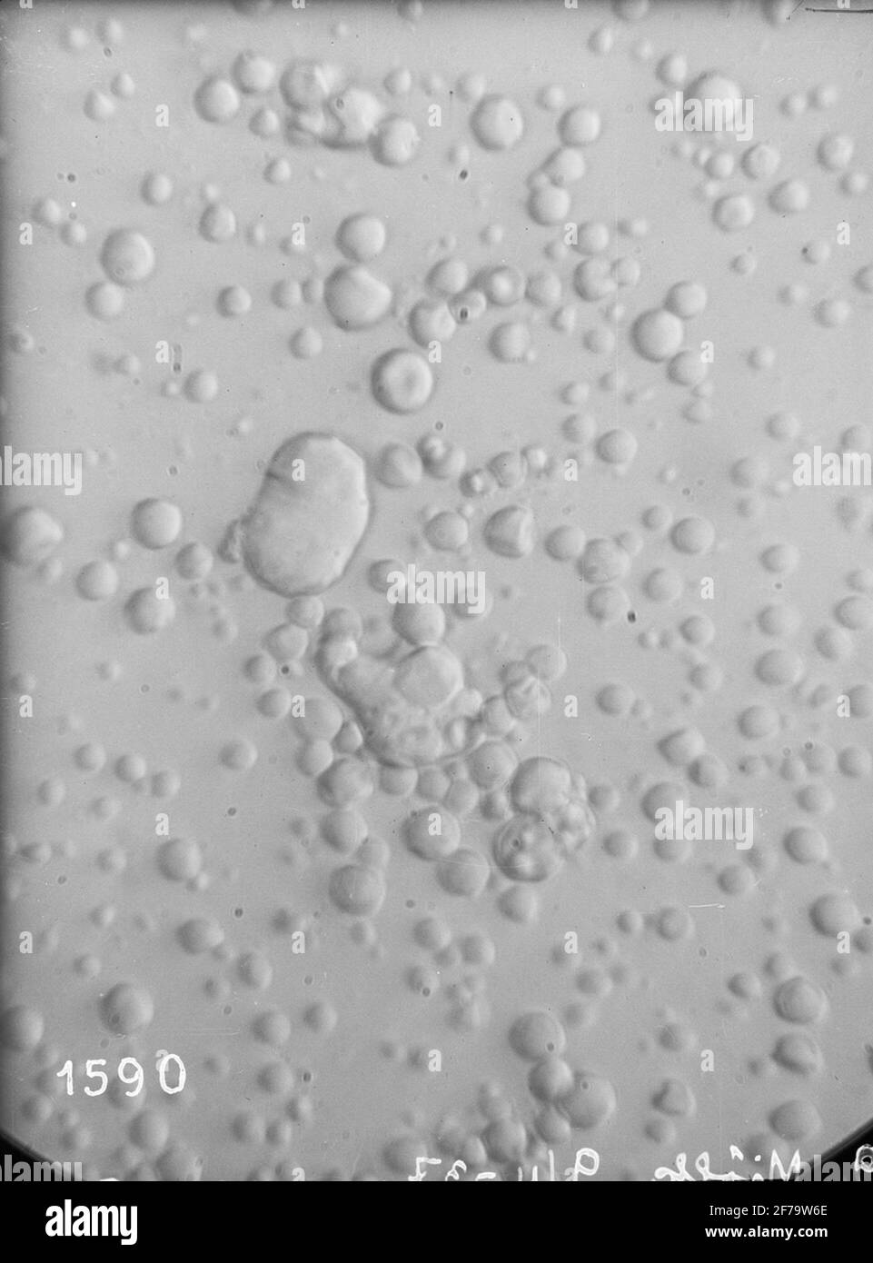 Micrographs of milk, stored in different ways. Each photography includes about 1/1070 cubic etc. Milk. Magnification 640 times. Stock Photohttps://www.alamy.com/image-license-details/?v=1https://www.alamy.com/micrographs-of-milk-stored-in-different-ways-each-photography-includes-about-11070-cubic-etc-milk-magnification-640-times-image417568774.html
RM2F79W6E–Micrographs of milk, stored in different ways. Each photography includes about 1/1070 cubic etc. Milk. Magnification 640 times.
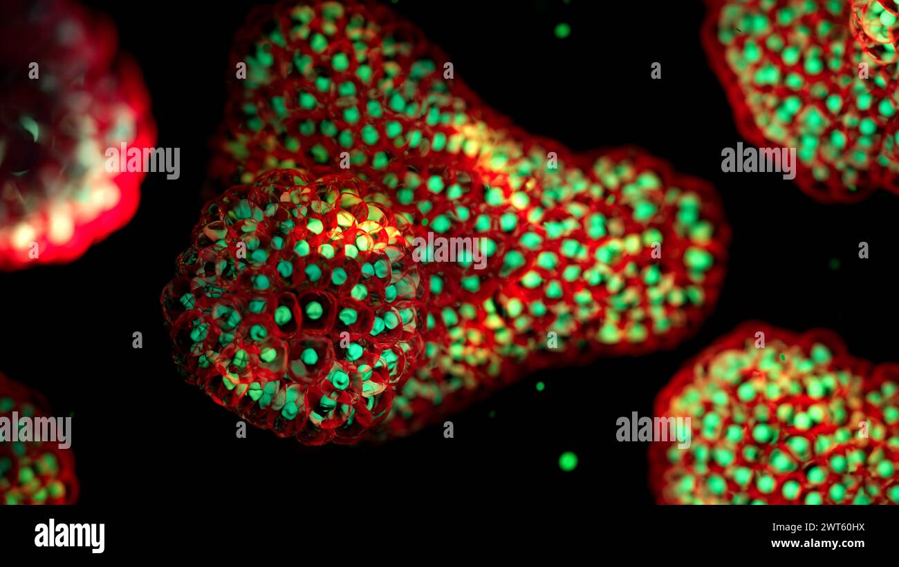 Illustration based on fluorescence light micrographs of organoids. Cell nuclei are green and cell membranes red. Organoids are three dimensional, miniature, simplified versions of organs grown in the laboratory. They are able to survive for months in controlled conditions allowing diseases to be studied over time and the testing of targeted therapies. Stock Photohttps://www.alamy.com/image-license-details/?v=1https://www.alamy.com/illustration-based-on-fluorescence-light-micrographs-of-organoids-cell-nuclei-are-green-and-cell-membranes-red-organoids-are-three-dimensional-miniature-simplified-versions-of-organs-grown-in-the-laboratory-they-are-able-to-survive-for-months-in-controlled-conditions-allowing-diseases-to-be-studied-over-time-and-the-testing-of-targeted-therapies-image600036470.html
RF2WT60HX–Illustration based on fluorescence light micrographs of organoids. Cell nuclei are green and cell membranes red. Organoids are three dimensional, miniature, simplified versions of organs grown in the laboratory. They are able to survive for months in controlled conditions allowing diseases to be studied over time and the testing of targeted therapies.
 TEM Transmission Electron Microscope of AIDS virus Stock Photohttps://www.alamy.com/image-license-details/?v=1https://www.alamy.com/tem-transmission-electron-microscope-of-aids-virus-image5196533.html
RMAR2JF6–TEM Transmission Electron Microscope of AIDS virus
 Sheet with seventeen micrographs of the cellular structure of clove and vanilla Sheet with seventeen micrographs of the cellular structure of clove and vanilla object type: photomechanical printing page Object number: RP-F-2001-7-1484-5 Inscriptions / Brands: number, recto, printed, 'Taf. V.' Manufacturer : Photographer: Franz Friedrich Bernard Elsner (attributed to) clichémaker: Photographische anstalt Otto Wigand (listed building) button: Photo Graphical anstalt Otto Wigand (listed building) Publisher: Wilhelm Knapp Verlag (listed property) Place manufacture: clichémaker: Zeitz Printer: Zeit Stock Photohttps://www.alamy.com/image-license-details/?v=1https://www.alamy.com/sheet-with-seventeen-micrographs-of-the-cellular-structure-of-clove-and-vanilla-sheet-with-seventeen-micrographs-of-the-cellular-structure-of-clove-and-vanilla-object-type-photomechanical-printing-page-object-number-rp-f-2001-7-1484-5-inscriptions-brands-number-recto-printed-taf-v-manufacturer-photographer-franz-friedrich-bernard-elsner-attributed-to-clichmaker-photographische-anstalt-otto-wigand-listed-building-button-photo-graphical-anstalt-otto-wigand-listed-building-publisher-wilhelm-knapp-verlag-listed-property-place-manufacture-clichmaker-zeitz-printer-zeit-image348202701.html
RM2B6E02N–Sheet with seventeen micrographs of the cellular structure of clove and vanilla Sheet with seventeen micrographs of the cellular structure of clove and vanilla object type: photomechanical printing page Object number: RP-F-2001-7-1484-5 Inscriptions / Brands: number, recto, printed, 'Taf. V.' Manufacturer : Photographer: Franz Friedrich Bernard Elsner (attributed to) clichémaker: Photographische anstalt Otto Wigand (listed building) button: Photo Graphical anstalt Otto Wigand (listed building) Publisher: Wilhelm Knapp Verlag (listed property) Place manufacture: clichémaker: Zeitz Printer: Zeit
 Hefei, China's Anhui Province. 19th Apr, 2023. A visitor views cross-polarized micrographs of lunar soil particles at an exhibition themed on lunar soil research achievements in University of Science and Technology of China, in Hefei, east China's Anhui Province, April 19, 2023. The exhibition starting Monday displays exhibits made by scientists, artists and engineers based on micrographs of lunar soil particles brought back by the Chang'e-5 probe. In 2020, China's Chang'e-5 mission retrieved samples from the moon weighing about 1,731 grams. Credit: Huang Bohan/Xinhua/Alamy Live News Stock Photohttps://www.alamy.com/image-license-details/?v=1https://www.alamy.com/hefei-chinas-anhui-province-19th-apr-2023-a-visitor-views-cross-polarized-micrographs-of-lunar-soil-particles-at-an-exhibition-themed-on-lunar-soil-research-achievements-in-university-of-science-and-technology-of-china-in-hefei-east-chinas-anhui-province-april-19-2023-the-exhibition-starting-monday-displays-exhibits-made-by-scientists-artists-and-engineers-based-on-micrographs-of-lunar-soil-particles-brought-back-by-the-change-5-probe-in-2020-chinas-change-5-mission-retrieved-samples-from-the-moon-weighing-about-1731-grams-credit-huang-bohanxinhuaalamy-live-news-image546944095.html
RM2PNRCNK–Hefei, China's Anhui Province. 19th Apr, 2023. A visitor views cross-polarized micrographs of lunar soil particles at an exhibition themed on lunar soil research achievements in University of Science and Technology of China, in Hefei, east China's Anhui Province, April 19, 2023. The exhibition starting Monday displays exhibits made by scientists, artists and engineers based on micrographs of lunar soil particles brought back by the Chang'e-5 probe. In 2020, China's Chang'e-5 mission retrieved samples from the moon weighing about 1,731 grams. Credit: Huang Bohan/Xinhua/Alamy Live News
 Proceedings of the Zoological Society of London London Academic Press Periodicals Zoology, The display features a series of detailed micrographs showcasing various spicules from calcareous sponges. Each labeled section highlights distinct structural characteristics. The images capture the intricate geometry of the spicules, illustrating their diverse forms and sizes. The top row presents three different spicule types, emphasizing variations in shape and cross-section. The bottom segment includes two additional spicules, with a focus on the elongated structure and fine details that reveal the c Stock Photohttps://www.alamy.com/image-license-details/?v=1https://www.alamy.com/proceedings-of-the-zoological-society-of-london-london-academic-press-periodicals-zoology-the-display-features-a-series-of-detailed-micrographs-showcasing-various-spicules-from-calcareous-sponges-each-labeled-section-highlights-distinct-structural-characteristics-the-images-capture-the-intricate-geometry-of-the-spicules-illustrating-their-diverse-forms-and-sizes-the-top-row-presents-three-different-spicule-types-emphasizing-variations-in-shape-and-cross-section-the-bottom-segment-includes-two-additional-spicules-with-a-focus-on-the-elongated-structure-and-fine-details-that-reveal-the-c-image636088895.html
RM2YXT9TF–Proceedings of the Zoological Society of London London Academic Press Periodicals Zoology, The display features a series of detailed micrographs showcasing various spicules from calcareous sponges. Each labeled section highlights distinct structural characteristics. The images capture the intricate geometry of the spicules, illustrating their diverse forms and sizes. The top row presents three different spicule types, emphasizing variations in shape and cross-section. The bottom segment includes two additional spicules, with a focus on the elongated structure and fine details that reveal the c
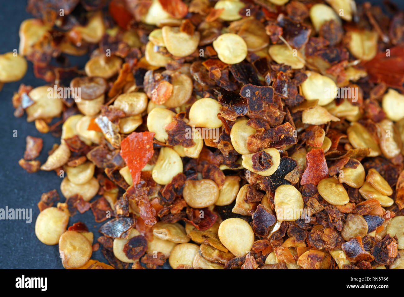 The chili spice is considered the hottest chili spice ever. This sharpness can only be topped if different chili spices are mixed. Stock Photohttps://www.alamy.com/image-license-details/?v=1https://www.alamy.com/the-chili-spice-is-considered-the-hottest-chili-spice-ever-this-sharpness-can-only-be-topped-if-different-chili-spices-are-mixed-image236757982.html
RFRN5766–The chili spice is considered the hottest chili spice ever. This sharpness can only be topped if different chili spices are mixed.
 Three influenza A H5N1/bird flu virus particles rod-shaped yellow. Note: Layout incorporates two CDC transmission electron micrographs that have been repositioned and colorized by NIAID. Influenza A Virus H5N1/Bird Flu 016867 315 Stock Photohttps://www.alamy.com/image-license-details/?v=1https://www.alamy.com/three-influenza-a-h5n1bird-flu-virus-particles-rod-shaped-yellow-note-layout-incorporates-two-cdc-transmission-electron-micrographs-that-have-been-repositioned-and-colorized-by-niaid-influenza-a-virus-h5n1bird-flu-016867-315-image627780754.html
RM2YD9TN6–Three influenza A H5N1/bird flu virus particles rod-shaped yellow. Note: Layout incorporates two CDC transmission electron micrographs that have been repositioned and colorized by NIAID. Influenza A Virus H5N1/Bird Flu 016867 315
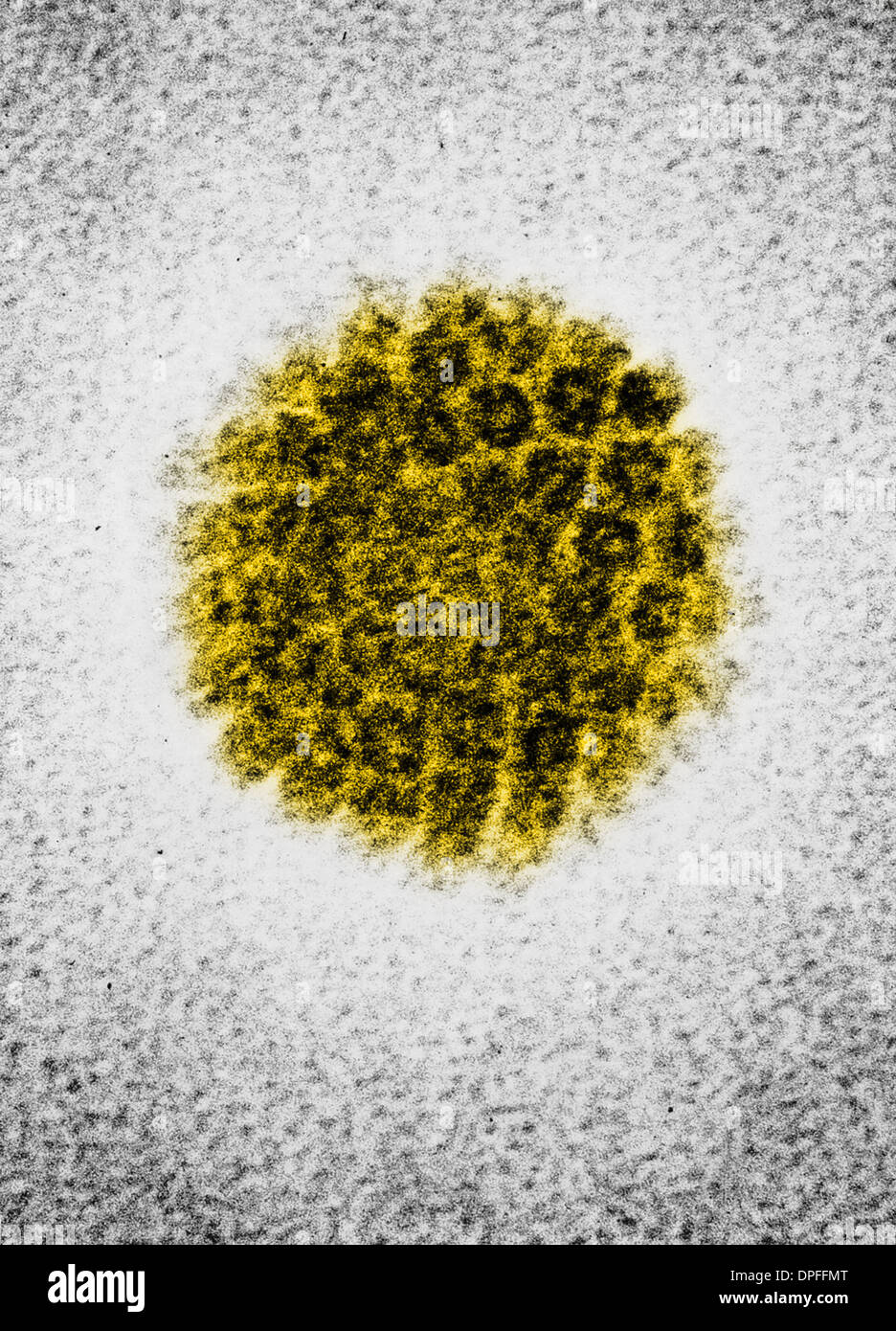 EM of a human papilloma virus (HBV) Stock Photohttps://www.alamy.com/image-license-details/?v=1https://www.alamy.com/em-of-a-human-papilloma-virus-hbv-image65495160.html
RFDPFFMT–EM of a human papilloma virus (HBV)
 Archive image from page 35 of The cytology and life-history of. The cytology and life-history of bacteria cytologylifehist00biss Year: 1955 THE CYTOLOGY AND LIFE-HISTORY OF BACTERIA SECTIONS OF BACTERIA Diagrams drawn from electron micrographs of Bacillus cereus are compared. Diagram A is taken from the study by Chapman and HilUer, and B from material prepared in this laboratory. The former has been treated with 2°,, osmium tetroxide which has had the effect of rendering the cell wall very clearly visible but may also have coagulated the softer structures. Section B has been accorded mini Stock Photohttps://www.alamy.com/image-license-details/?v=1https://www.alamy.com/archive-image-from-page-35-of-the-cytology-and-life-history-of-the-cytology-and-life-history-of-bacteria-cytologylifehist00biss-year-1955-the-cytology-and-life-history-of-bacteria-sections-of-bacteria-diagrams-drawn-from-electron-micrographs-of-bacillus-cereus-are-compared-diagram-a-is-taken-from-the-study-by-chapman-and-hiluer-and-b-from-material-prepared-in-this-laboratory-the-former-has-been-treated-with-2-osmium-tetroxide-which-has-had-the-effect-of-rendering-the-cell-wall-very-clearly-visible-but-may-also-have-coagulated-the-softer-structures-section-b-has-been-accorded-mini-image259541013.html
RMW2735W–Archive image from page 35 of The cytology and life-history of. The cytology and life-history of bacteria cytologylifehist00biss Year: 1955 THE CYTOLOGY AND LIFE-HISTORY OF BACTERIA SECTIONS OF BACTERIA Diagrams drawn from electron micrographs of Bacillus cereus are compared. Diagram A is taken from the study by Chapman and HilUer, and B from material prepared in this laboratory. The former has been treated with 2°,, osmium tetroxide which has had the effect of rendering the cell wall very clearly visible but may also have coagulated the softer structures. Section B has been accorded mini
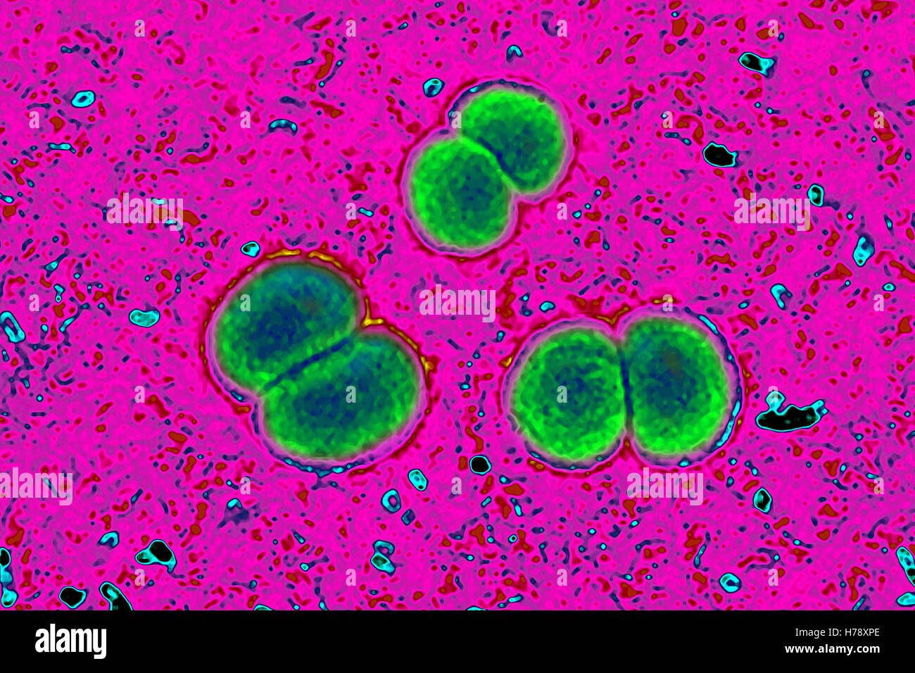 NEISSERIA MENINGITIDIS Stock Photohttps://www.alamy.com/image-license-details/?v=1https://www.alamy.com/stock-photo-neisseria-meningitidis-124971798.html
RMH78XPE–NEISSERIA MENINGITIDIS
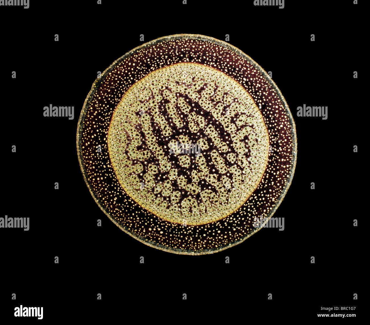 Light Micrograph (LM) of a transverse section of an aerial root of a Pandanus sp., magnification x30 Stock Photohttps://www.alamy.com/image-license-details/?v=1https://www.alamy.com/stock-photo-light-micrograph-lm-of-a-transverse-section-of-an-aerial-root-of-a-31612119.html
RMBRC1G7–Light Micrograph (LM) of a transverse section of an aerial root of a Pandanus sp., magnification x30
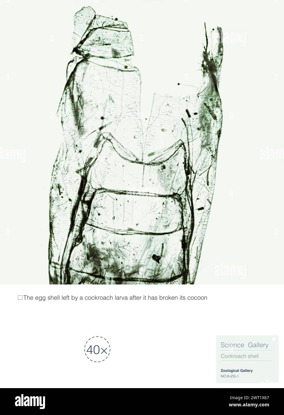 This is a cockroach egg. After hatching and breaking the cocoon, the broken shell left by the larvae is covered with dust and very dirty. Stock Photohttps://www.alamy.com/image-license-details/?v=1https://www.alamy.com/this-is-a-cockroach-egg-after-hatching-and-breaking-the-cocoon-the-broken-shell-left-by-the-larvae-is-covered-with-dust-and-very-dirty-image599946767.html
RM2WT1X67–This is a cockroach egg. After hatching and breaking the cocoon, the broken shell left by the larvae is covered with dust and very dirty.
 Lace in the Solar Microscope, 400 times magnified in surface. Artist: William Henry Fox Talbot (British, Dorset 1800-1877 Lacock). Dimensions: 10 x 18.3 cm (3 15/16 x 7 3/16 in.), irregularly trimmed. Date: 1839. Museum: Metropolitan Museum of Art, New York, USA. Stock Photohttps://www.alamy.com/image-license-details/?v=1https://www.alamy.com/lace-in-the-solar-microscope-400-times-magnified-in-surface-artist-william-henry-fox-talbot-british-dorset-1800-1877-lacock-dimensions-10-x-183-cm-3-1516-x-7-316-in-irregularly-trimmed-date-1839-museum-metropolitan-museum-of-art-new-york-usa-image233303312.html
RMRFFTN4–Lace in the Solar Microscope, 400 times magnified in surface. Artist: William Henry Fox Talbot (British, Dorset 1800-1877 Lacock). Dimensions: 10 x 18.3 cm (3 15/16 x 7 3/16 in.), irregularly trimmed. Date: 1839. Museum: Metropolitan Museum of Art, New York, USA.
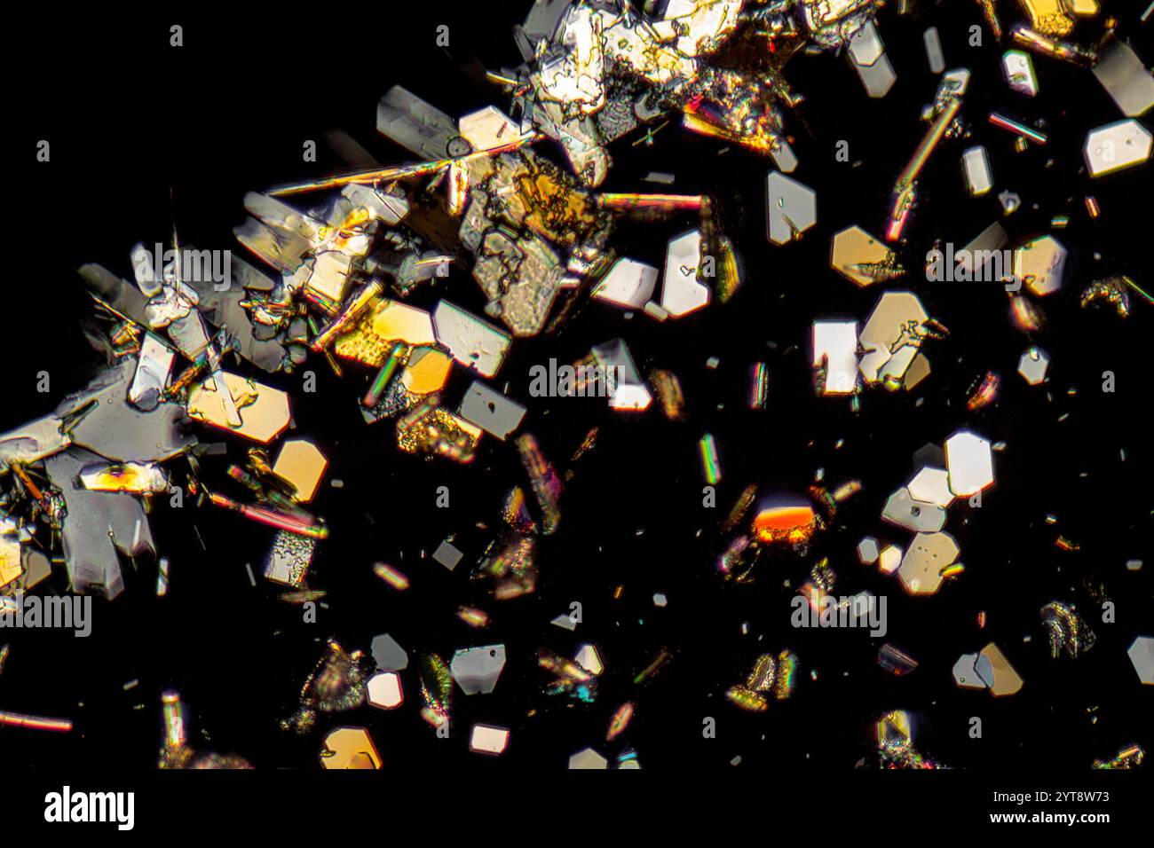 Microscopic shot showing colorful microcrystals in polarized light Stock Photohttps://www.alamy.com/image-license-details/?v=1https://www.alamy.com/microscopic-shot-showing-colorful-microcrystals-in-polarized-light-image634520407.html
RF2YT8W73–Microscopic shot showing colorful microcrystals in polarized light
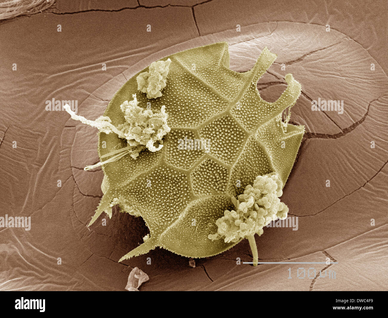 Coloured SEM of plankton Stock Photohttps://www.alamy.com/image-license-details/?v=1https://www.alamy.com/coloured-sem-of-plankton-image67264493.html
RFDWC4F9–Coloured SEM of plankton
 Coloured SEM of sundew (Drosera) plant Stock Photohttps://www.alamy.com/image-license-details/?v=1https://www.alamy.com/coloured-sem-of-sundew-drosera-plant-image67264079.html
RFDWC40F–Coloured SEM of sundew (Drosera) plant
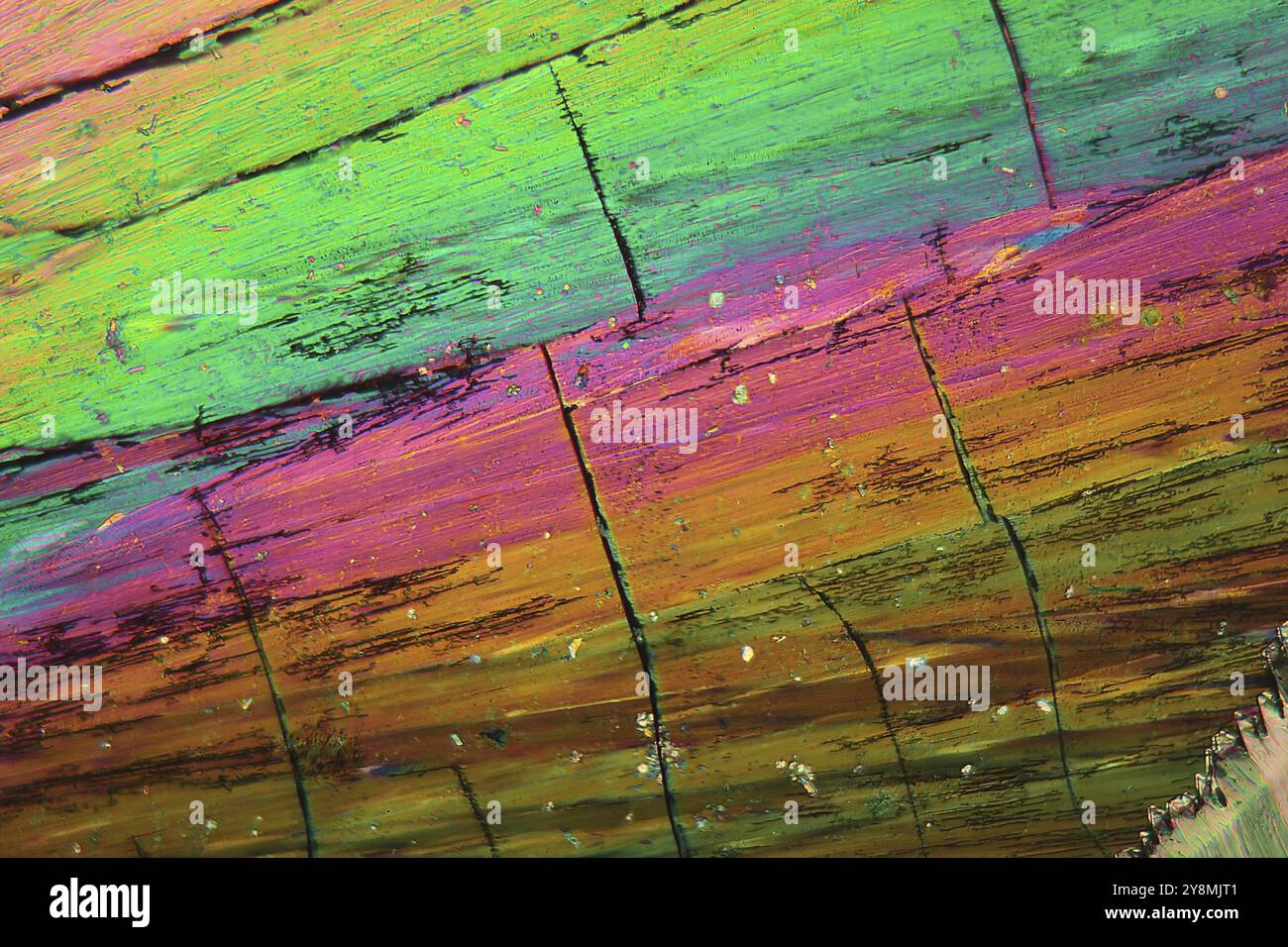 Thulium is a rare earth element. The crystals are precipitated from a solution on a microscope slide and photographed in polarized light Stock Photohttps://www.alamy.com/image-license-details/?v=1https://www.alamy.com/thulium-is-a-rare-earth-element-the-crystals-are-precipitated-from-a-solution-on-a-microscope-slide-and-photographed-in-polarized-light-image624944321.html
RF2Y8MJT1–Thulium is a rare earth element. The crystals are precipitated from a solution on a microscope slide and photographed in polarized light
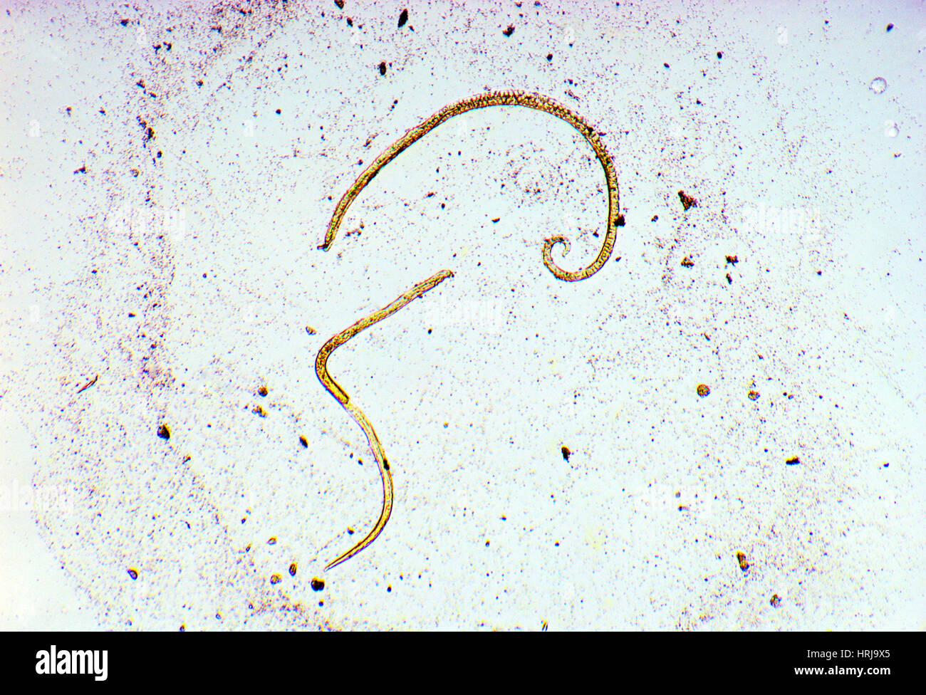 Trichinella Spiralis Stock Photohttps://www.alamy.com/image-license-details/?v=1https://www.alamy.com/stock-photo-trichinella-spiralis-135012589.html
RMHRJ9X5–Trichinella Spiralis
 Micrographs of milk, stored in different ways. Each photography includes about 1/1070 cubic etc. Milk. Magnification 640 times. Stock Photohttps://www.alamy.com/image-license-details/?v=1https://www.alamy.com/micrographs-of-milk-stored-in-different-ways-each-photography-includes-about-11070-cubic-etc-milk-magnification-640-times-image417568833.html
RM2F79W8H–Micrographs of milk, stored in different ways. Each photography includes about 1/1070 cubic etc. Milk. Magnification 640 times.