Microscopic image bacteria Black & White Stock Photos
(73)See microscopic image bacteria stock video clipsMicroscopic image bacteria Black & White Stock Photos
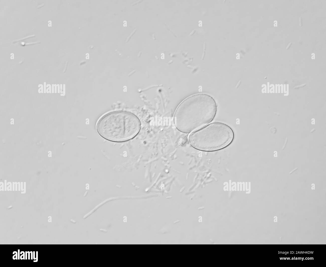 Dirty water under the microscope - lots of long rod-shaped bacteria and three fungal ascospores Stock Photohttps://www.alamy.com/image-license-details/?v=1https://www.alamy.com/dirty-water-under-the-microscope-lots-of-long-rod-shaped-bacteria-and-three-fungal-ascospores-image342740101.html
RM2AWH4DW–Dirty water under the microscope - lots of long rod-shaped bacteria and three fungal ascospores
 The image of the earth in the form of a virus. Illustration dedicated to the spread and pandemic of the coronovirus throughout the Earth. Epidemic and Stock Vectorhttps://www.alamy.com/image-license-details/?v=1https://www.alamy.com/the-image-of-the-earth-in-the-form-of-a-virus-illustration-dedicated-to-the-spread-and-pandemic-of-the-coronovirus-throughout-the-earth-epidemic-and-image351050828.html
RF2BB3MWG–The image of the earth in the form of a virus. Illustration dedicated to the spread and pandemic of the coronovirus throughout the Earth. Epidemic and
 Illustration of a Dna Stock Photohttps://www.alamy.com/image-license-details/?v=1https://www.alamy.com/stock-photo-illustration-of-a-dna-132566209.html
RFHKJWFD–Illustration of a Dna
 Isolated viral microbe. 3d illustration Stock Photohttps://www.alamy.com/image-license-details/?v=1https://www.alamy.com/isolated-viral-microbe-3d-illustration-image448945607.html
RF2H2B6KK–Isolated viral microbe. 3d illustration
 Scientific Vector Illustration of Laboratory Flask and Microscopic View of Microorganisms Stock Vectorhttps://www.alamy.com/image-license-details/?v=1https://www.alamy.com/scientific-vector-illustration-of-laboratory-flask-and-microscopic-view-of-microorganisms-image634407434.html
RF2YT3N4A–Scientific Vector Illustration of Laboratory Flask and Microscopic View of Microorganisms
 The image of the earth in the form of a virus. Illustration dedicated to the spread and pandemic of the coronovirus throughout the Earth. Epidemic and Stock Vectorhttps://www.alamy.com/image-license-details/?v=1https://www.alamy.com/the-image-of-the-earth-in-the-form-of-a-virus-illustration-dedicated-to-the-spread-and-pandemic-of-the-coronovirus-throughout-the-earth-epidemic-and-image350761326.html
RF2BAJFJ6–The image of the earth in the form of a virus. Illustration dedicated to the spread and pandemic of the coronovirus throughout the Earth. Epidemic and
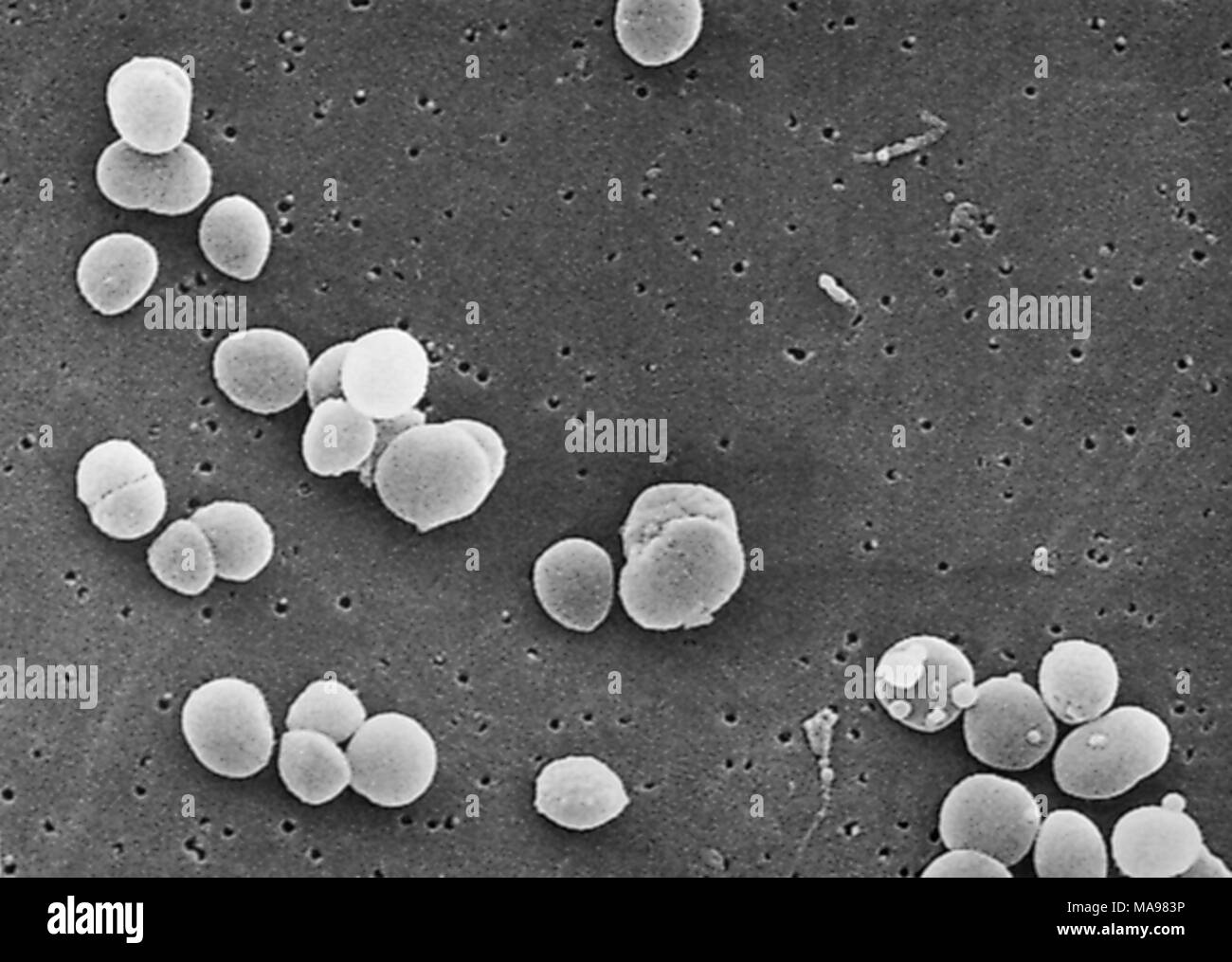 Gram-positive Staphylococcus aureus bacteria revealed in the scanning electron microscopic (SEM) image, 2003. Image courtesy Centers for Disease Control (CDC). () Stock Photohttps://www.alamy.com/image-license-details/?v=1https://www.alamy.com/gram-positive-staphylococcus-aureus-bacteria-revealed-in-the-scanning-electron-microscopic-sem-image-2003-image-courtesy-centers-for-disease-control-cdc-image178454186.html
RMMA983P–Gram-positive Staphylococcus aureus bacteria revealed in the scanning electron microscopic (SEM) image, 2003. Image courtesy Centers for Disease Control (CDC). ()
 abstract monochrome image of a three circles Stock Photohttps://www.alamy.com/image-license-details/?v=1https://www.alamy.com/abstract-monochrome-image-of-a-three-circles-image234263641.html
RFRH3HJH–abstract monochrome image of a three circles
 Rod-shaped Gram-negative Salmonella infantis bacteria revealed in the scanning electron microscopic (SEM) image, 2005. Image courtesy Centers for Disease Control (CDC) / Janice Haney Carr. () Stock Photohttps://www.alamy.com/image-license-details/?v=1https://www.alamy.com/rod-shaped-gram-negative-salmonella-infantis-bacteria-revealed-in-the-scanning-electron-microscopic-sem-image-2005-image-courtesy-centers-for-disease-control-cdc-janice-haney-carr-image178455277.html
RMMA99EN–Rod-shaped Gram-negative Salmonella infantis bacteria revealed in the scanning electron microscopic (SEM) image, 2005. Image courtesy Centers for Disease Control (CDC) / Janice Haney Carr. ()
 A Set of Doodle Bacteria illustration Stock Vectorhttps://www.alamy.com/image-license-details/?v=1https://www.alamy.com/a-set-of-doodle-bacteria-illustration-image223200572.html
RFPY3JH0–A Set of Doodle Bacteria illustration
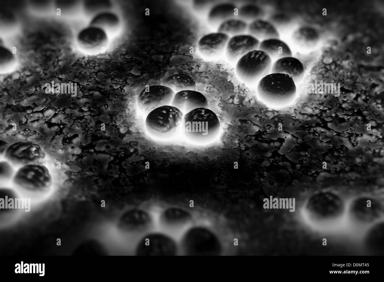 Clusters of 'superbug' (MRSA) bacteria. Stock Photohttps://www.alamy.com/image-license-details/?v=1https://www.alamy.com/stock-photo-clusters-of-superbug-mrsa-bacteria-52089077.html
RMD0MT45–Clusters of 'superbug' (MRSA) bacteria.
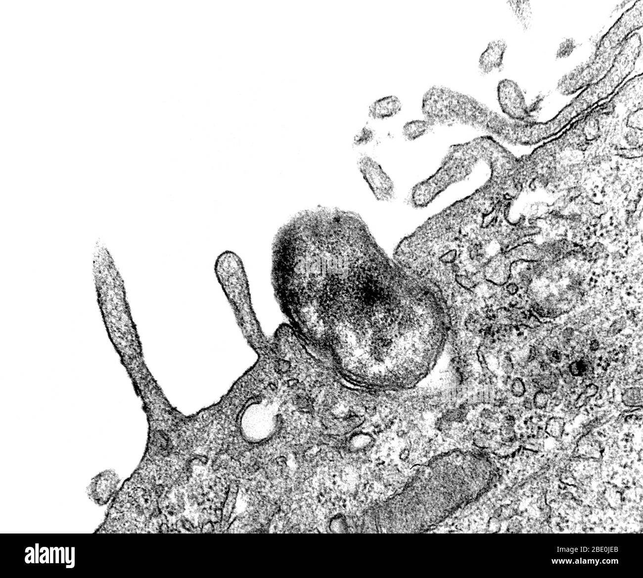 Transmission Electron Microscope (TEM) image captured as the process of phagocytosis was underway. Here, you are able to see as an Orientia tsutsugamushi bacterium, formerly known as Rickettsia tsutsugamushi, was being ingested by a mouse peritoneal mesothelial cell. Note how the would-be host cell membrane had not yet entirely enveloped the bacterium. Magnification: unknown. Stock Photohttps://www.alamy.com/image-license-details/?v=1https://www.alamy.com/transmission-electron-microscope-tem-image-captured-as-the-process-of-phagocytosis-was-underway-here-you-are-able-to-see-as-an-orientia-tsutsugamushi-bacterium-formerly-known-as-rickettsia-tsutsugamushi-was-being-ingested-by-a-mouse-peritoneal-mesothelial-cell-note-how-the-would-be-host-cell-membrane-had-not-yet-entirely-enveloped-the-bacterium-magnification-unknown-image352827059.html
RM2BE0JEB–Transmission Electron Microscope (TEM) image captured as the process of phagocytosis was underway. Here, you are able to see as an Orientia tsutsugamushi bacterium, formerly known as Rickettsia tsutsugamushi, was being ingested by a mouse peritoneal mesothelial cell. Note how the would-be host cell membrane had not yet entirely enveloped the bacterium. Magnification: unknown.
 Vector seamless monochrome texture - a pattern of irregular cells Stock Vectorhttps://www.alamy.com/image-license-details/?v=1https://www.alamy.com/vector-seamless-monochrome-texture-a-pattern-of-irregular-cells-image555577588.html
RF2R7TMTM–Vector seamless monochrome texture - a pattern of irregular cells
 Virus or coronavirus abstract background. 3d render Stock Photohttps://www.alamy.com/image-license-details/?v=1https://www.alamy.com/virus-or-coronavirus-abstract-background-3d-render-image342932908.html
RF2AWWXBT–Virus or coronavirus abstract background. 3d render
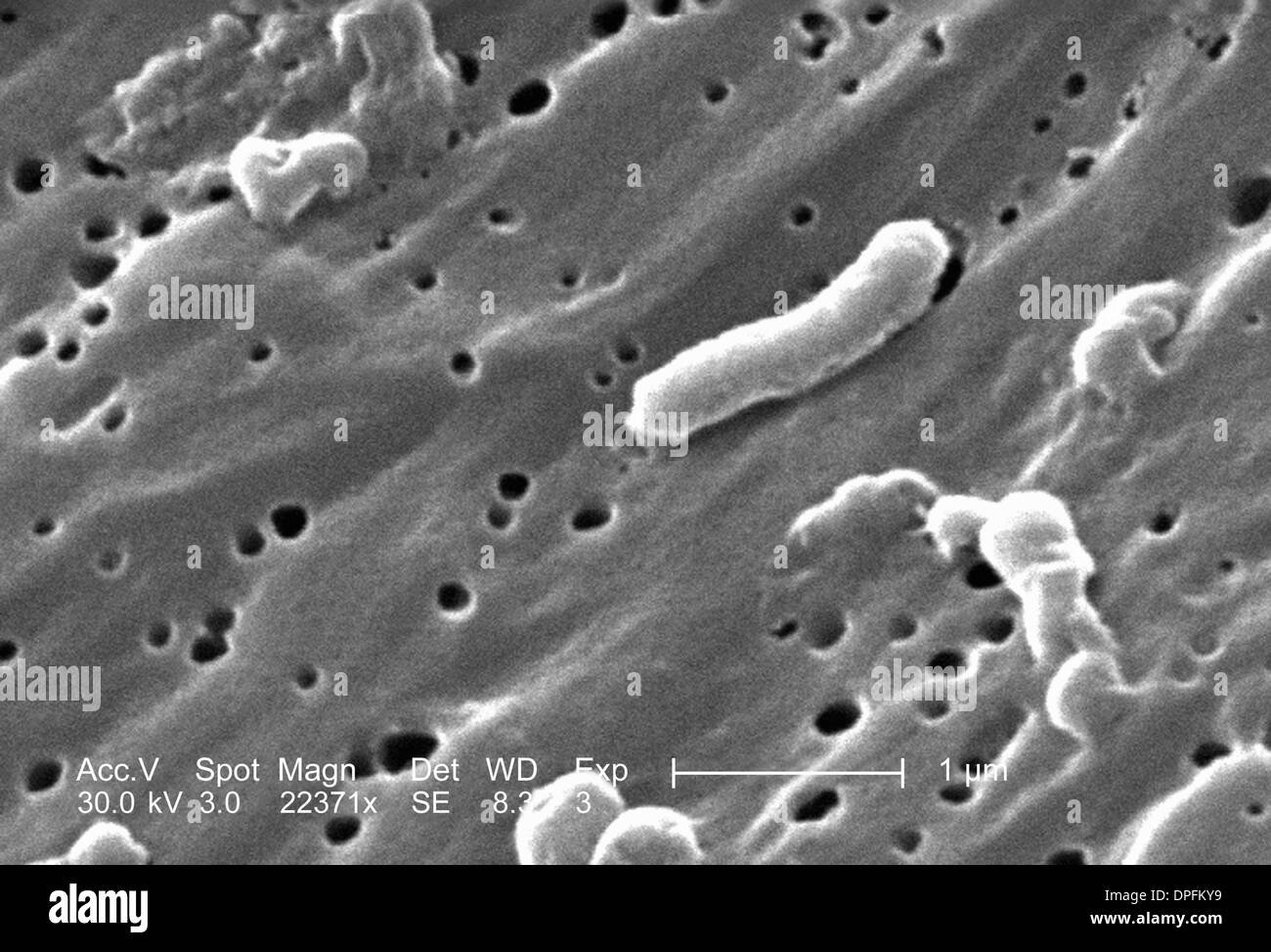 SEM of Vibrio vulnificus bacteria Stock Photohttps://www.alamy.com/image-license-details/?v=1https://www.alamy.com/sem-of-vibrio-vulnificus-bacteria-image65498477.html
RFDPFKY9–SEM of Vibrio vulnificus bacteria
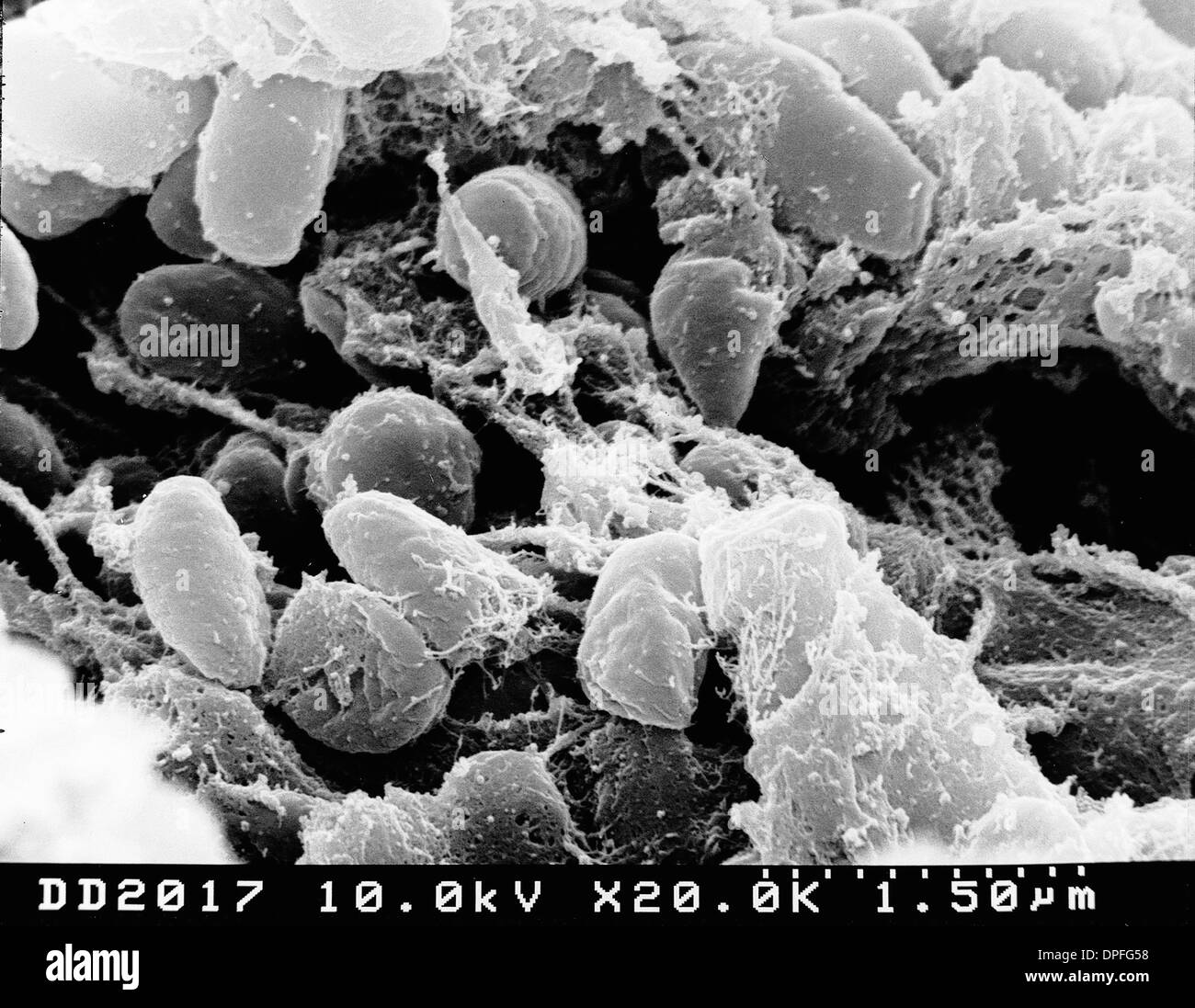 SEM of plague bacteria Stock Photohttps://www.alamy.com/image-license-details/?v=1https://www.alamy.com/sem-of-plague-bacteria-image65495508.html
RFDPFG58–SEM of plague bacteria
RF2WA6KAX–Cancer cell color line icon. Microorganisms microbes, bacteria. Vector isolated element. Editable stroke.
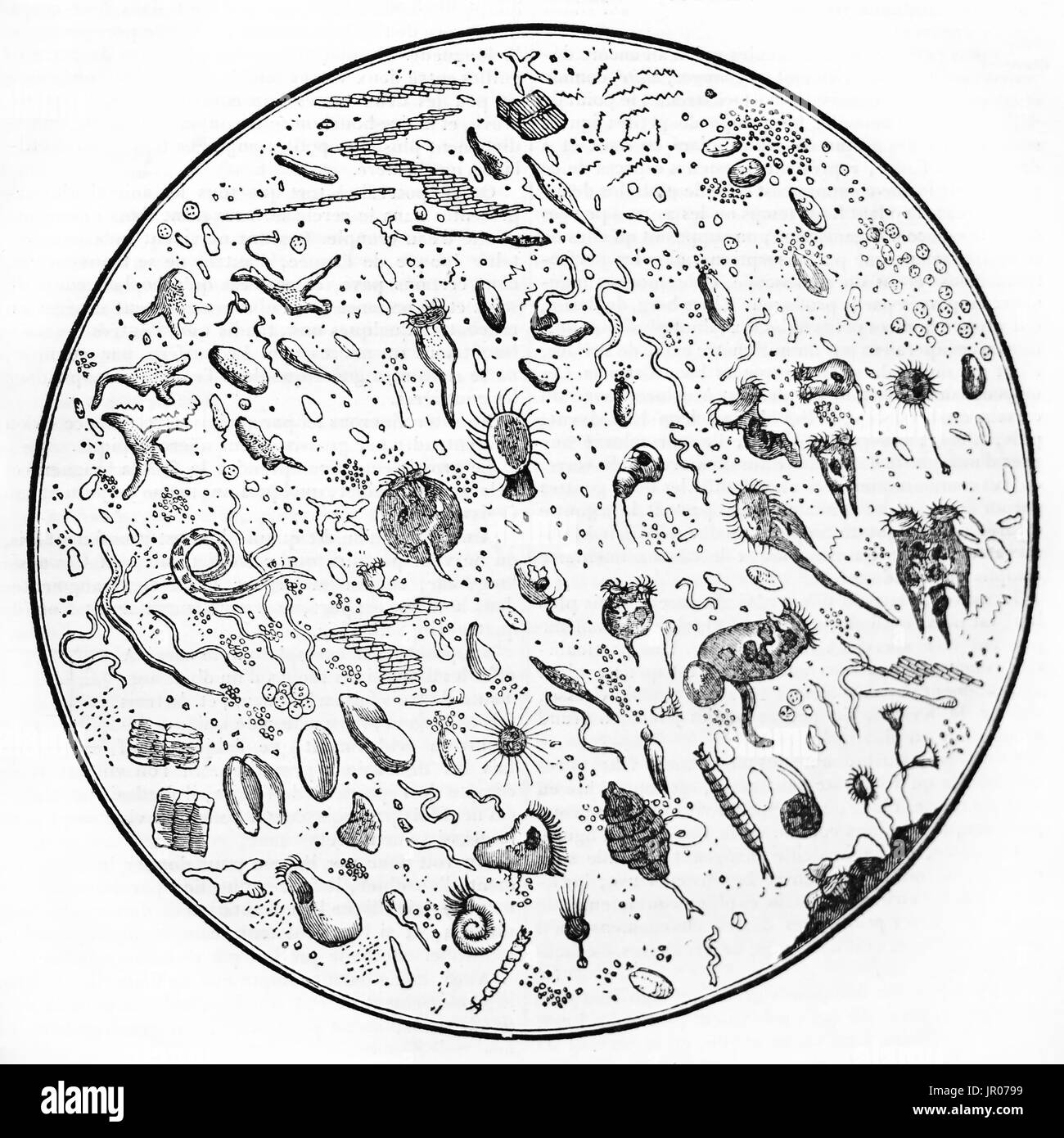 Water drop under a microscope, old illustration. By unidentified author, published on Magasin Pittoresque, Paris, 1833. Stock Photohttps://www.alamy.com/image-license-details/?v=1https://www.alamy.com/water-drop-under-a-microscope-old-illustration-by-unidentified-author-image151825781.html
RFJR0799–Water drop under a microscope, old illustration. By unidentified author, published on Magasin Pittoresque, Paris, 1833.
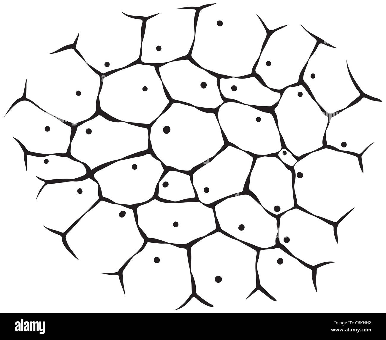 The cells of living tissue - a monochrome illustration Stock Photohttps://www.alamy.com/image-license-details/?v=1https://www.alamy.com/stock-photo-the-cells-of-living-tissue-a-monochrome-illustration-38539566.html
RFC6KHH2–The cells of living tissue - a monochrome illustration
 Bacteria vector simple seamless pattern or background in thin line style Stock Vectorhttps://www.alamy.com/image-license-details/?v=1https://www.alamy.com/bacteria-vector-simple-seamless-pattern-or-background-in-thin-line-style-image571833945.html
RF2T6980W–Bacteria vector simple seamless pattern or background in thin line style
 Close-up of parasitic viral cells under a microscope in a specialized laboratory on a gray background black and white image 2020 Stock Photohttps://www.alamy.com/image-license-details/?v=1https://www.alamy.com/close-up-of-parasitic-viral-cells-under-a-microscope-in-a-specialized-laboratory-on-a-gray-background-black-and-white-image-2020-image349767374.html
RF2B917RX–Close-up of parasitic viral cells under a microscope in a specialized laboratory on a gray background black and white image 2020
 Illustration of a Dna Stock Photohttps://www.alamy.com/image-license-details/?v=1https://www.alamy.com/stock-photo-illustration-of-a-dna-132566208.html
RFHKJWFC–Illustration of a Dna
 . Pathogenic micro-organisms. A text-book of microbiology for physicians and students of medicine. (Based upon Williams' Bacteriology). Bacteriology; Pathogenic bacteria. THE MICROSCOPE AND MICROSCOPIC METHODS 17 also that the sharpness of outline of the image increases and the brilliancy diminishes as the size of the aperture is decreased. If the simple aperture be replaced by a convex lens and the object and the screen be set at the conjugate foci of the lens, it will be seen that magnification is again the quotient of the aper-. PiG. 2.— Image formation by a single lens. Note that the image Stock Photohttps://www.alamy.com/image-license-details/?v=1https://www.alamy.com/pathogenic-micro-organisms-a-text-book-of-microbiology-for-physicians-and-students-of-medicine-based-upon-williams-bacteriology-bacteriology-pathogenic-bacteria-the-microscope-and-microscopic-methods-17-also-that-the-sharpness-of-outline-of-the-image-increases-and-the-brilliancy-diminishes-as-the-size-of-the-aperture-is-decreased-if-the-simple-aperture-be-replaced-by-a-convex-lens-and-the-object-and-the-screen-be-set-at-the-conjugate-foci-of-the-lens-it-will-be-seen-that-magnification-is-again-the-quotient-of-the-aper-pig-2-image-formation-by-a-single-lens-note-that-the-image-image232419636.html
RMRE3HH8–. Pathogenic micro-organisms. A text-book of microbiology for physicians and students of medicine. (Based upon Williams' Bacteriology). Bacteriology; Pathogenic bacteria. THE MICROSCOPE AND MICROSCOPIC METHODS 17 also that the sharpness of outline of the image increases and the brilliancy diminishes as the size of the aperture is decreased. If the simple aperture be replaced by a convex lens and the object and the screen be set at the conjugate foci of the lens, it will be seen that magnification is again the quotient of the aper-. PiG. 2.— Image formation by a single lens. Note that the image
 Scientific Vector Illustration of Laboratory Flask and Microscopic View of Microorganisms Stock Vectorhttps://www.alamy.com/image-license-details/?v=1https://www.alamy.com/scientific-vector-illustration-of-laboratory-flask-and-microscopic-view-of-microorganisms-image634407478.html
RF2YT3N5X–Scientific Vector Illustration of Laboratory Flask and Microscopic View of Microorganisms
RFKHDDAT–Bacteria icons set, outline style
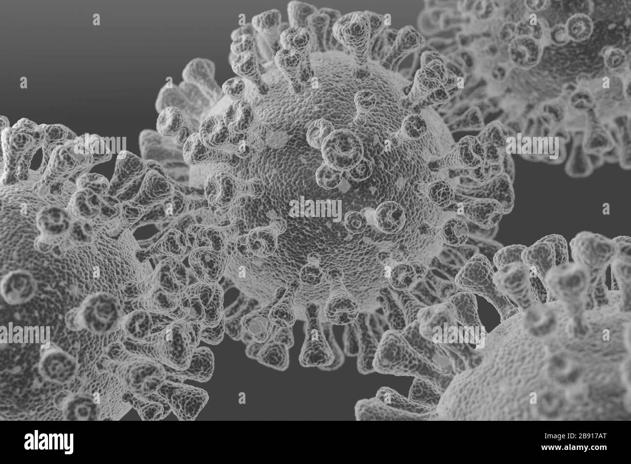 Close-up of parasitic viral cells under a microscope in a specialized laboratory on a gray background black and white image 2020 Stock Photohttps://www.alamy.com/image-license-details/?v=1https://www.alamy.com/close-up-of-parasitic-viral-cells-under-a-microscope-in-a-specialized-laboratory-on-a-gray-background-black-and-white-image-2020-image349767008.html
RF2B917AT–Close-up of parasitic viral cells under a microscope in a specialized laboratory on a gray background black and white image 2020
 abstract monochrome image of a three circles Stock Photohttps://www.alamy.com/image-license-details/?v=1https://www.alamy.com/abstract-monochrome-image-of-a-three-circles-image234237858.html
RFRH2CNP–abstract monochrome image of a three circles
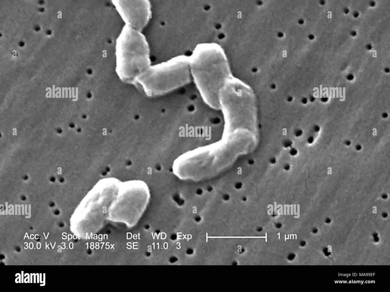 Rod-shaped Gram-negative Salmonella infantis bacteria revealed in the scanning electron microscopic (SEM) image, 2005. Image courtesy Centers for Disease Control (CDC) / Janice Haney Carr. () Stock Photohttps://www.alamy.com/image-license-details/?v=1https://www.alamy.com/rod-shaped-gram-negative-salmonella-infantis-bacteria-revealed-in-the-scanning-electron-microscopic-sem-image-2005-image-courtesy-centers-for-disease-control-cdc-janice-haney-carr-image178455271.html
RMMA99EF–Rod-shaped Gram-negative Salmonella infantis bacteria revealed in the scanning electron microscopic (SEM) image, 2005. Image courtesy Centers for Disease Control (CDC) / Janice Haney Carr. ()
 Old fashioned microscope isolated on white Stock Vectorhttps://www.alamy.com/image-license-details/?v=1https://www.alamy.com/old-fashioned-microscope-isolated-on-white-image555524796.html
RF2R7P9F8–Old fashioned microscope isolated on white
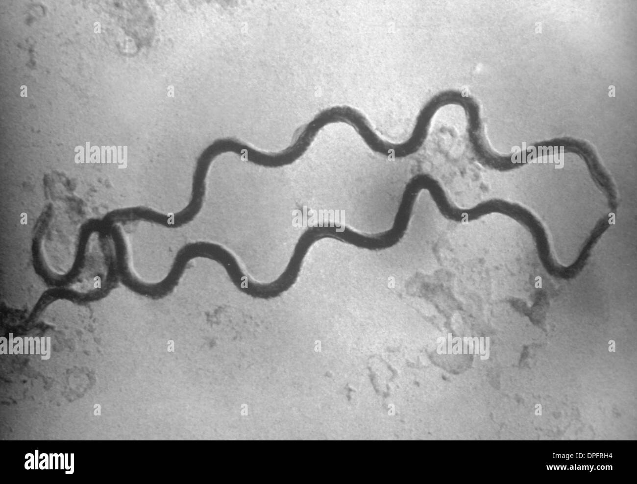 EM of two Treponema pallidum bacteria Stock Photohttps://www.alamy.com/image-license-details/?v=1https://www.alamy.com/em-of-two-treponema-pallidum-bacteria-image65501328.html
RFDPFRH4–EM of two Treponema pallidum bacteria
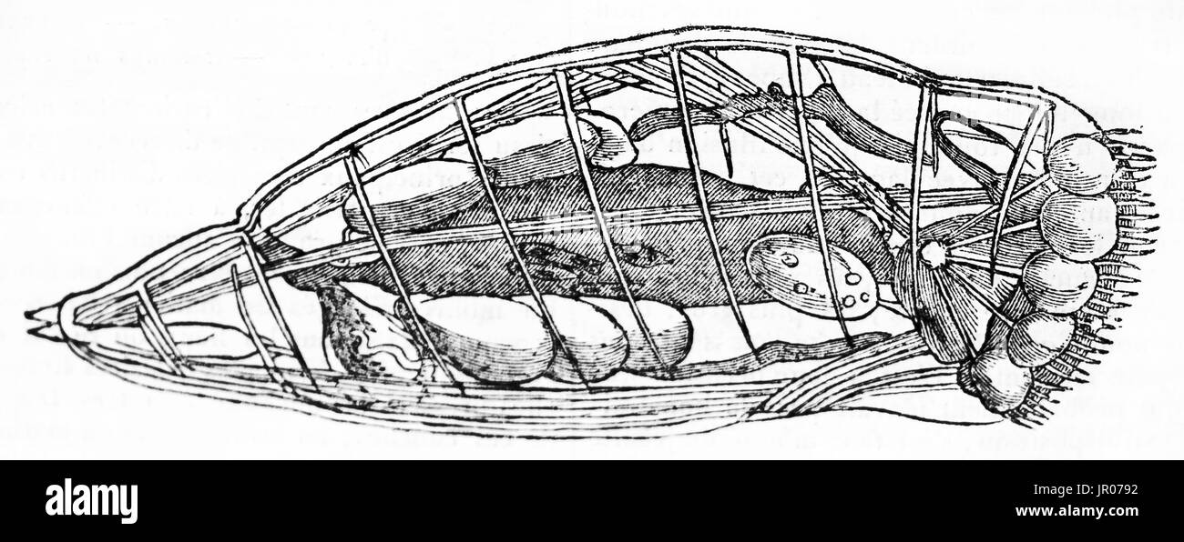 Vorticella senta (protozoa) 144400 times magnification, old illustration. By unidentified author, published on Magasin Pittoresque, Paris, 1833. Stock Photohttps://www.alamy.com/image-license-details/?v=1https://www.alamy.com/vorticella-senta-protozoa-144400-times-magnification-old-illustration-image151825774.html
RFJR0792–Vorticella senta (protozoa) 144400 times magnification, old illustration. By unidentified author, published on Magasin Pittoresque, Paris, 1833.
 Seamless monochrome texture - a pattern of irregular cells Stock Photohttps://www.alamy.com/image-license-details/?v=1https://www.alamy.com/stock-photo-seamless-monochrome-texture-a-pattern-of-irregular-cells-43614883.html
RFCEXR6B–Seamless monochrome texture - a pattern of irregular cells
 Infection bacteria and virus vector minimal seamless pattern or background in thin line style Stock Vectorhttps://www.alamy.com/image-license-details/?v=1https://www.alamy.com/infection-bacteria-and-virus-vector-minimal-seamless-pattern-or-background-in-thin-line-style-image571838068.html
RF2T69D84–Infection bacteria and virus vector minimal seamless pattern or background in thin line style
 . Pathogenic micro-organisms. A text-book of microbiology for physicians and students of medicine. (Based upon Williams' Bacteriology). Bacteriology; Pathogenic bacteria. THE MICROSCOPE AND MICROSCOPIC METHODS 21. Pig. 7.—Sectional view of a compound microscope illustrating the course of two beams proceeding from two points in the object {P and Q) and indicating the subjective interpretation of the image formed on the retina.. Please note that these images are extracted from scanned page images that may have been digitally enhanced for readability - coloration and appearance of these illustrat Stock Photohttps://www.alamy.com/image-license-details/?v=1https://www.alamy.com/pathogenic-micro-organisms-a-text-book-of-microbiology-for-physicians-and-students-of-medicine-based-upon-williams-bacteriology-bacteriology-pathogenic-bacteria-the-microscope-and-microscopic-methods-21-pig-7sectional-view-of-a-compound-microscope-illustrating-the-course-of-two-beams-proceeding-from-two-points-in-the-object-p-and-q-and-indicating-the-subjective-interpretation-of-the-image-formed-on-the-retina-please-note-that-these-images-are-extracted-from-scanned-page-images-that-may-have-been-digitally-enhanced-for-readability-coloration-and-appearance-of-these-illustrat-image232419618.html
RMRE3HGJ–. Pathogenic micro-organisms. A text-book of microbiology for physicians and students of medicine. (Based upon Williams' Bacteriology). Bacteriology; Pathogenic bacteria. THE MICROSCOPE AND MICROSCOPIC METHODS 21. Pig. 7.—Sectional view of a compound microscope illustrating the course of two beams proceeding from two points in the object {P and Q) and indicating the subjective interpretation of the image formed on the retina.. Please note that these images are extracted from scanned page images that may have been digitally enhanced for readability - coloration and appearance of these illustrat
 Scientific Vector Illustration of Laboratory Flask and Microscopic View of Microorganisms Stock Vectorhttps://www.alamy.com/image-license-details/?v=1https://www.alamy.com/scientific-vector-illustration-of-laboratory-flask-and-microscopic-view-of-microorganisms-image634407307.html
RF2YT3MYR–Scientific Vector Illustration of Laboratory Flask and Microscopic View of Microorganisms
RFKHDDB0–Illness icons set, outline style
 Rod-shaped Gram-negative Salmonella infantis bacteria revealed in the scanning electron microscopic (SEM) image, 2005. Image courtesy Centers for Disease Control (CDC) / Janice Haney Carr. () Stock Photohttps://www.alamy.com/image-license-details/?v=1https://www.alamy.com/rod-shaped-gram-negative-salmonella-infantis-bacteria-revealed-in-the-scanning-electron-microscopic-sem-image-2005-image-courtesy-centers-for-disease-control-cdc-janice-haney-carr-image178455286.html
RMMA99F2–Rod-shaped Gram-negative Salmonella infantis bacteria revealed in the scanning electron microscopic (SEM) image, 2005. Image courtesy Centers for Disease Control (CDC) / Janice Haney Carr. ()
RF2T69FTE–Seamless vector pattern with virus, bacterium, pathogen and microbe white icons in thin line style with black background
 Scientific Vector Illustration of Laboratory Flask and Microscopic View of Microorganisms Stock Vectorhttps://www.alamy.com/image-license-details/?v=1https://www.alamy.com/scientific-vector-illustration-of-laboratory-flask-and-microscopic-view-of-microorganisms-image634407166.html
RF2YT3MPP–Scientific Vector Illustration of Laboratory Flask and Microscopic View of Microorganisms
RFKHDDAR–Viruses icons set, outline style
 Four rod-shaped Gram-negative Salmonella infantis bacteria revealed in the scanning electron microscopic (SEM) image, 2005. Image courtesy Centers for Disease Control (CDC) / Janice Haney Carr. () Stock Photohttps://www.alamy.com/image-license-details/?v=1https://www.alamy.com/four-rod-shaped-gram-negative-salmonella-infantis-bacteria-revealed-in-the-scanning-electron-microscopic-sem-image-2005-image-courtesy-centers-for-disease-control-cdc-janice-haney-carr-image178455279.html
RMMA99ER–Four rod-shaped Gram-negative Salmonella infantis bacteria revealed in the scanning electron microscopic (SEM) image, 2005. Image courtesy Centers for Disease Control (CDC) / Janice Haney Carr. ()
RFMJB6XP–Virus icon set vector white isolated on grey background
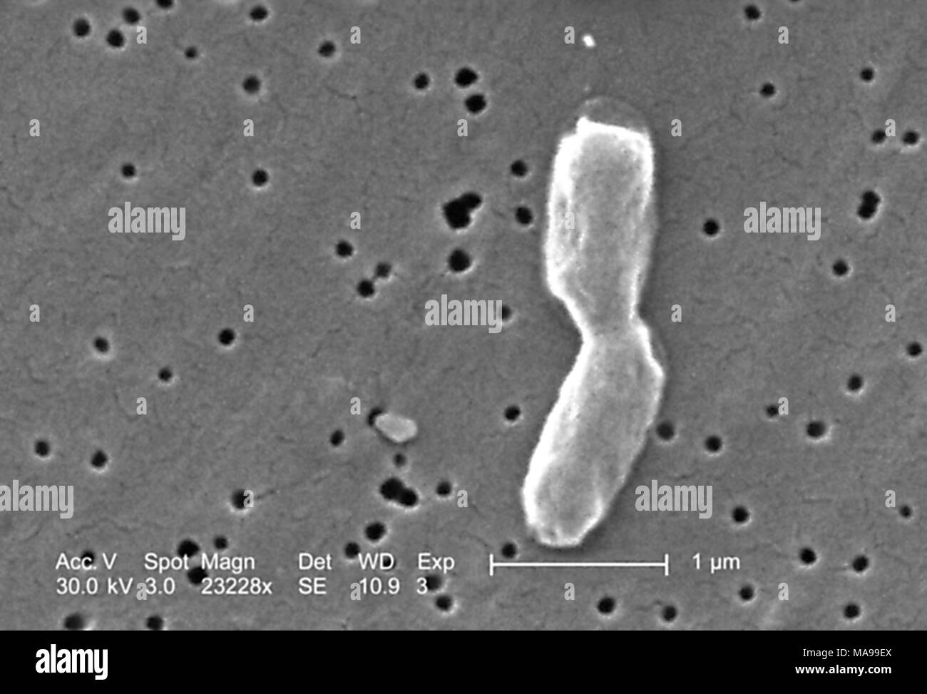 Two rod-shaped Gram-negative Salmonella infantis bacteria revealed in the scanning electron microscopic (SEM) image, 2005. Image courtesy Centers for Disease Control (CDC) / Janice Haney Carr. () Stock Photohttps://www.alamy.com/image-license-details/?v=1https://www.alamy.com/two-rod-shaped-gram-negative-salmonella-infantis-bacteria-revealed-in-the-scanning-electron-microscopic-sem-image-2005-image-courtesy-centers-for-disease-control-cdc-janice-haney-carr-image178455282.html
RMMA99EX–Two rod-shaped Gram-negative Salmonella infantis bacteria revealed in the scanning electron microscopic (SEM) image, 2005. Image courtesy Centers for Disease Control (CDC) / Janice Haney Carr. ()
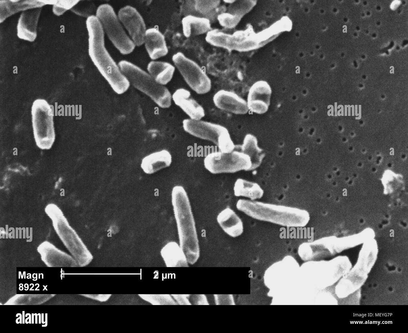 Segniliparus rotundus bacteria revealed in the 8922x magnified scanning electron microscopic (SEM) image, 2005. Image courtesy Centers for Disease Control (CDC) / Ray Butler, M.S. Janice Haney Carr. () Stock Photohttps://www.alamy.com/image-license-details/?v=1https://www.alamy.com/segniliparus-rotundus-bacteria-revealed-in-the-8922x-magnified-scanning-electron-microscopic-sem-image-2005-image-courtesy-centers-for-disease-control-cdc-ray-butler-ms-janice-haney-carr-image181314330.html
RMMEYG7P–Segniliparus rotundus bacteria revealed in the 8922x magnified scanning electron microscopic (SEM) image, 2005. Image courtesy Centers for Disease Control (CDC) / Ray Butler, M.S. Janice Haney Carr. ()
 Segniliparus rotundus bacteria revealed in the 5460x magnified scanning electron microscopic (SEM) image, 2005. Image courtesy Centers for Disease Control (CDC) / Ray Butler, M.S. Janice Haney Carr. () Stock Photohttps://www.alamy.com/image-license-details/?v=1https://www.alamy.com/segniliparus-rotundus-bacteria-revealed-in-the-5460x-magnified-scanning-electron-microscopic-sem-image-2005-image-courtesy-centers-for-disease-control-cdc-ray-butler-ms-janice-haney-carr-image181314296.html
RMMEYG6G–Segniliparus rotundus bacteria revealed in the 5460x magnified scanning electron microscopic (SEM) image, 2005. Image courtesy Centers for Disease Control (CDC) / Ray Butler, M.S. Janice Haney Carr. ()
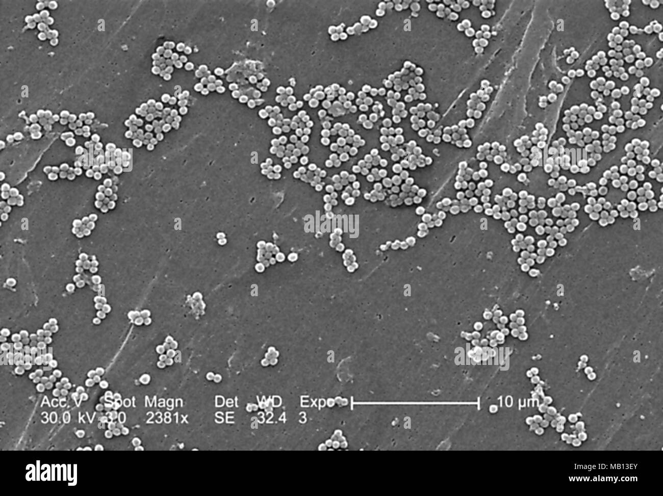 Clumps of methicillin-resistant Staphylococcus aureus bacteria (MRSA) revealed in the 2381x magnified scanning electron microscopic (SEM) image, 2005. Image courtesy Centers for Disease Control (CDC) / Janice Haney Carr, Jeff Hageman. () Stock Photohttps://www.alamy.com/image-license-details/?v=1https://www.alamy.com/clumps-of-methicillin-resistant-staphylococcus-aureus-bacteria-mrsa-revealed-in-the-2381x-magnified-scanning-electron-microscopic-sem-image-2005-image-courtesy-centers-for-disease-control-cdc-janice-haney-carr-jeff-hageman-image178889619.html
RMMB13EY–Clumps of methicillin-resistant Staphylococcus aureus bacteria (MRSA) revealed in the 2381x magnified scanning electron microscopic (SEM) image, 2005. Image courtesy Centers for Disease Control (CDC) / Janice Haney Carr, Jeff Hageman. ()
 Clumps of methicillin-resistant Staphylococcus aureus bacteria (MRSA) revealed in the 9560x magnified scanning electron microscopic (SEM) image, 2005. Image courtesy Centers for Disease Control (CDC) / Janice Haney Carr, Jeff Hageman. () Stock Photohttps://www.alamy.com/image-license-details/?v=1https://www.alamy.com/clumps-of-methicillin-resistant-staphylococcus-aureus-bacteria-mrsa-revealed-in-the-9560x-magnified-scanning-electron-microscopic-sem-image-2005-image-courtesy-centers-for-disease-control-cdc-janice-haney-carr-jeff-hageman-image178889633.html
RMMB13FD–Clumps of methicillin-resistant Staphylococcus aureus bacteria (MRSA) revealed in the 9560x magnified scanning electron microscopic (SEM) image, 2005. Image courtesy Centers for Disease Control (CDC) / Janice Haney Carr, Jeff Hageman. ()
 Clumps of methicillin-resistant Staphylococcus aureus bacteria (MRSA) revealed in the 2390x magnified scanning electron microscopic (SEM) image, 2005. Image courtesy Centers for Disease Control (CDC) / Janice Haney Carr, Jeff Hageman. () Stock Photohttps://www.alamy.com/image-license-details/?v=1https://www.alamy.com/clumps-of-methicillin-resistant-staphylococcus-aureus-bacteria-mrsa-revealed-in-the-2390x-magnified-scanning-electron-microscopic-sem-image-2005-image-courtesy-centers-for-disease-control-cdc-janice-haney-carr-jeff-hageman-image178889627.html
RMMB13F7–Clumps of methicillin-resistant Staphylococcus aureus bacteria (MRSA) revealed in the 2390x magnified scanning electron microscopic (SEM) image, 2005. Image courtesy Centers for Disease Control (CDC) / Janice Haney Carr, Jeff Hageman. ()
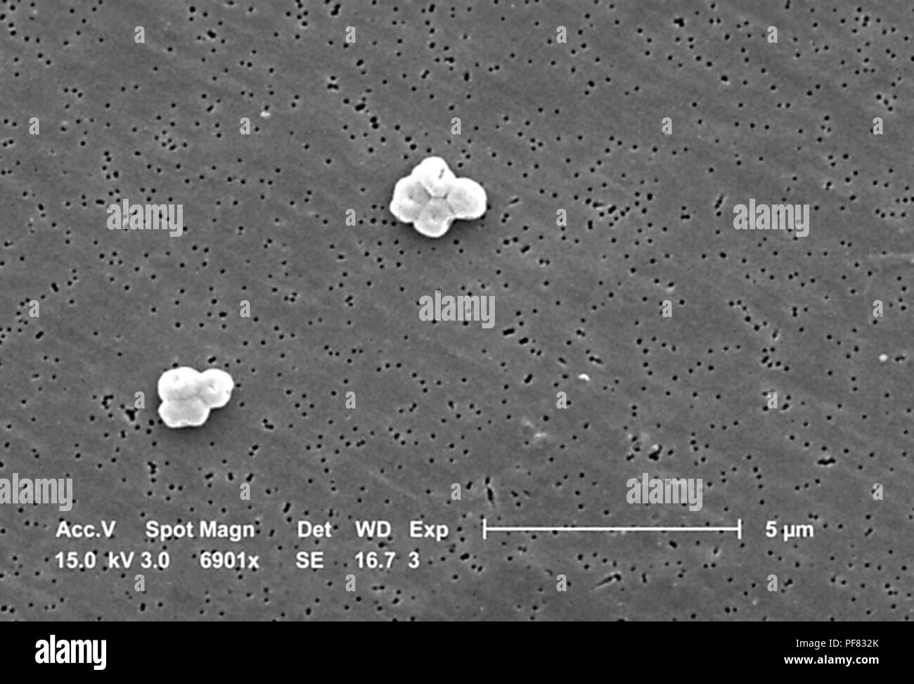 Gram-negative, non-motile Acinetobacter baumannii bacteria revealed in the 6901x magnified scanning electron microscopic (SEM) image, 2004. Image courtesy Centers for Disease Control (CDC) / Matthew J. Arduino, DrPH, Janice Carr, Jana Swenson. () Stock Photohttps://www.alamy.com/image-license-details/?v=1https://www.alamy.com/gram-negative-non-motile-acinetobacter-baumannii-bacteria-revealed-in-the-6901x-magnified-scanning-electron-microscopic-sem-image-2004-image-courtesy-centers-for-disease-control-cdc-matthew-j-arduino-drph-janice-carr-jana-swenson-image215922299.html
RMPF832K–Gram-negative, non-motile Acinetobacter baumannii bacteria revealed in the 6901x magnified scanning electron microscopic (SEM) image, 2004. Image courtesy Centers for Disease Control (CDC) / Matthew J. Arduino, DrPH, Janice Carr, Jana Swenson. ()
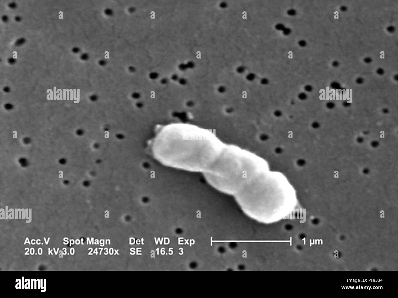 Gram-negative, non-motile Acinetobacter baumannii bacteria revealed in the 24730x magnified scanning electron microscopic (SEM) image, 2004. Image courtesy Centers for Disease Control (CDC) / Matthew J. Arduino, DrPH, Janice Carr, Jana Swenson. () Stock Photohttps://www.alamy.com/image-license-details/?v=1https://www.alamy.com/gram-negative-non-motile-acinetobacter-baumannii-bacteria-revealed-in-the-24730x-magnified-scanning-electron-microscopic-sem-image-2004-image-courtesy-centers-for-disease-control-cdc-matthew-j-arduino-drph-janice-carr-jana-swenson-image215922312.html
RMPF8334–Gram-negative, non-motile Acinetobacter baumannii bacteria revealed in the 24730x magnified scanning electron microscopic (SEM) image, 2004. Image courtesy Centers for Disease Control (CDC) / Matthew J. Arduino, DrPH, Janice Carr, Jana Swenson. ()
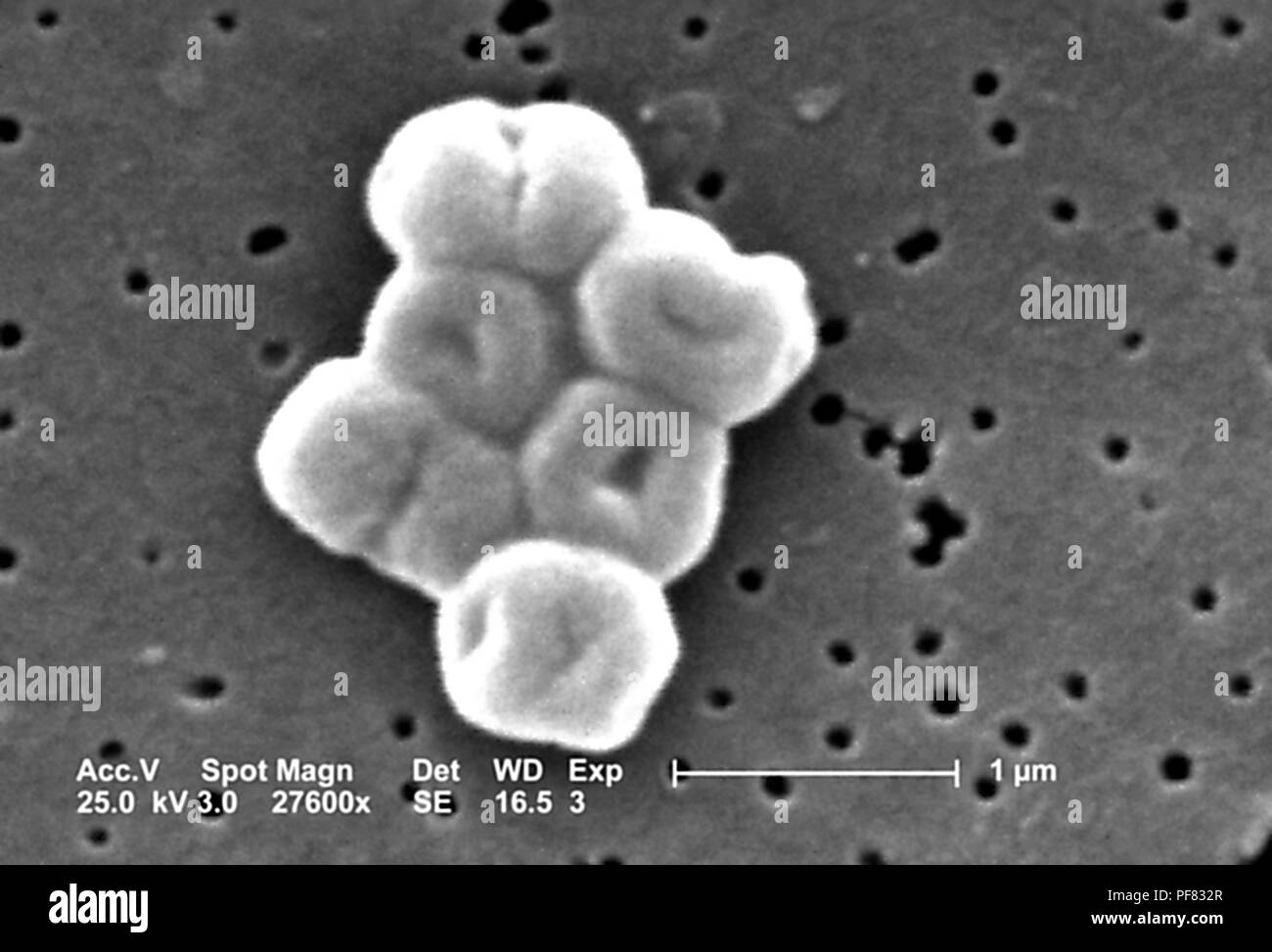 Gram-negative, non-motile Acinetobacter baumannii bacteria revealed in the 27600x magnified scanning electron microscopic (SEM) image, 2004. Image courtesy Centers for Disease Control (CDC) / Matthew J. Arduino, DrPH, Janice Carr, Jana Swenson. () Stock Photohttps://www.alamy.com/image-license-details/?v=1https://www.alamy.com/gram-negative-non-motile-acinetobacter-baumannii-bacteria-revealed-in-the-27600x-magnified-scanning-electron-microscopic-sem-image-2004-image-courtesy-centers-for-disease-control-cdc-matthew-j-arduino-drph-janice-carr-jana-swenson-image215922303.html
RMPF832R–Gram-negative, non-motile Acinetobacter baumannii bacteria revealed in the 27600x magnified scanning electron microscopic (SEM) image, 2004. Image courtesy Centers for Disease Control (CDC) / Matthew J. Arduino, DrPH, Janice Carr, Jana Swenson. ()
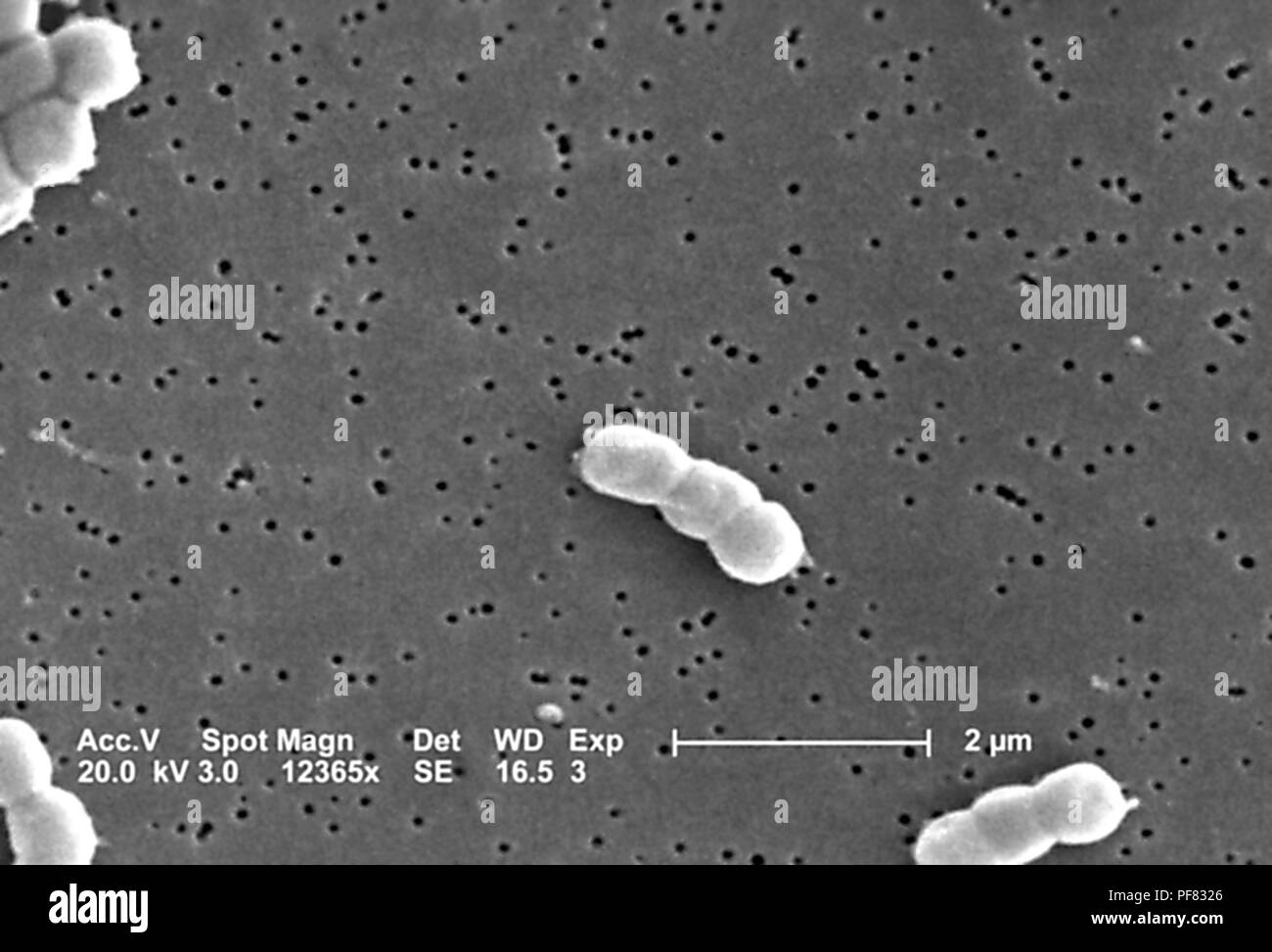 Gram-negative, non-motile Acinetobacter baumannii bacteria revealed in the 12365x magnified scanning electron microscopic (SEM) image, 2004. Image courtesy Centers for Disease Control (CDC) / Matthew J. Arduino, DrPH, Janice Carr, Jana Swenson. () Stock Photohttps://www.alamy.com/image-license-details/?v=1https://www.alamy.com/gram-negative-non-motile-acinetobacter-baumannii-bacteria-revealed-in-the-12365x-magnified-scanning-electron-microscopic-sem-image-2004-image-courtesy-centers-for-disease-control-cdc-matthew-j-arduino-drph-janice-carr-jana-swenson-image215922286.html
RMPF8326–Gram-negative, non-motile Acinetobacter baumannii bacteria revealed in the 12365x magnified scanning electron microscopic (SEM) image, 2004. Image courtesy Centers for Disease Control (CDC) / Matthew J. Arduino, DrPH, Janice Carr, Jana Swenson. ()
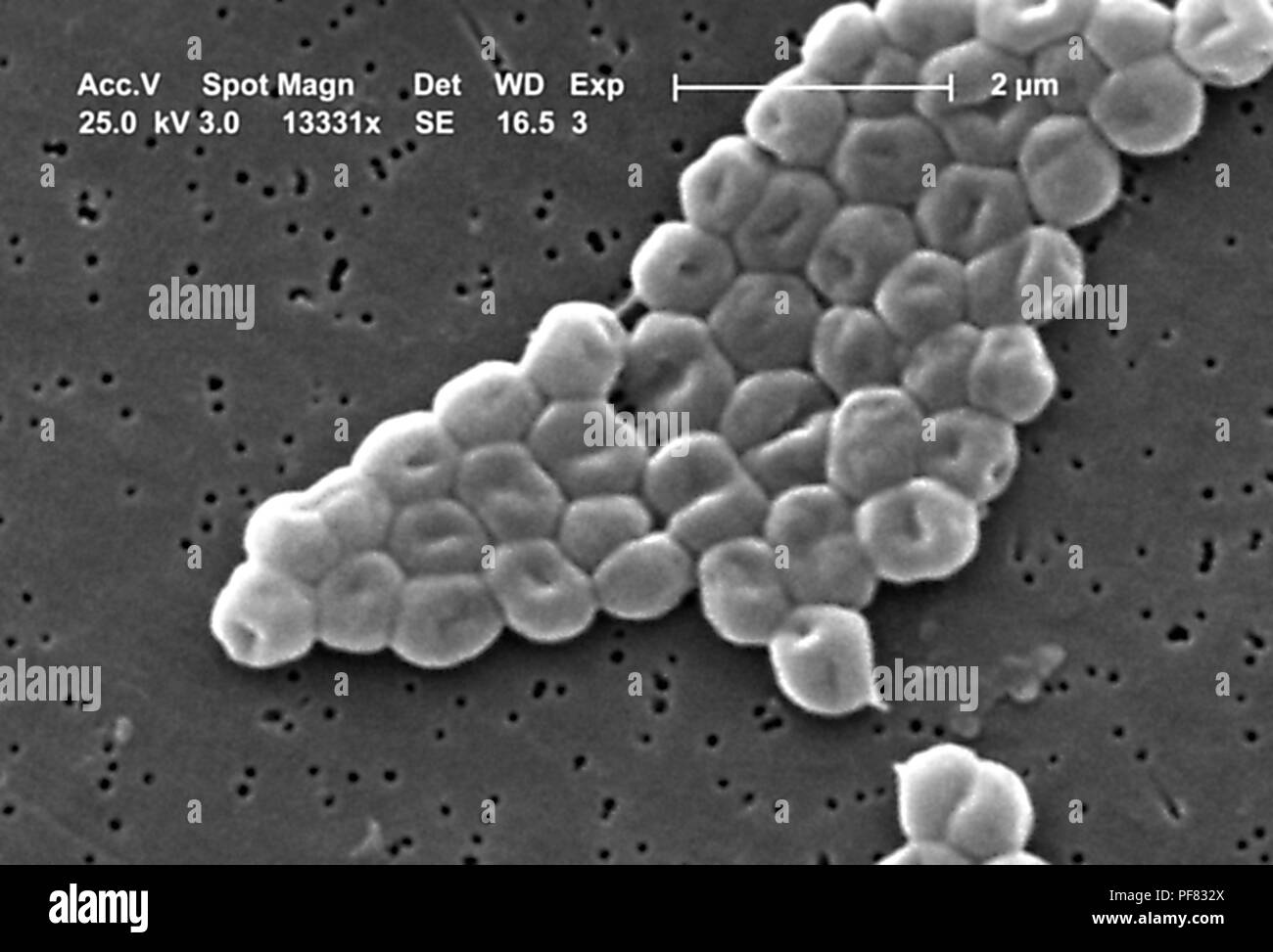 Gram-negative, non-motile Acinetobacter baumannii bacteria revealed in the 13331x magnified scanning electron microscopic (SEM) image, 2004. Image courtesy Centers for Disease Control (CDC) / Matthew J. Arduino, DrPH, Janice Carr, Jana Swenson. () Stock Photohttps://www.alamy.com/image-license-details/?v=1https://www.alamy.com/gram-negative-non-motile-acinetobacter-baumannii-bacteria-revealed-in-the-13331x-magnified-scanning-electron-microscopic-sem-image-2004-image-courtesy-centers-for-disease-control-cdc-matthew-j-arduino-drph-janice-carr-jana-swenson-image215922306.html
RMPF832X–Gram-negative, non-motile Acinetobacter baumannii bacteria revealed in the 13331x magnified scanning electron microscopic (SEM) image, 2004. Image courtesy Centers for Disease Control (CDC) / Matthew J. Arduino, DrPH, Janice Carr, Jana Swenson. ()
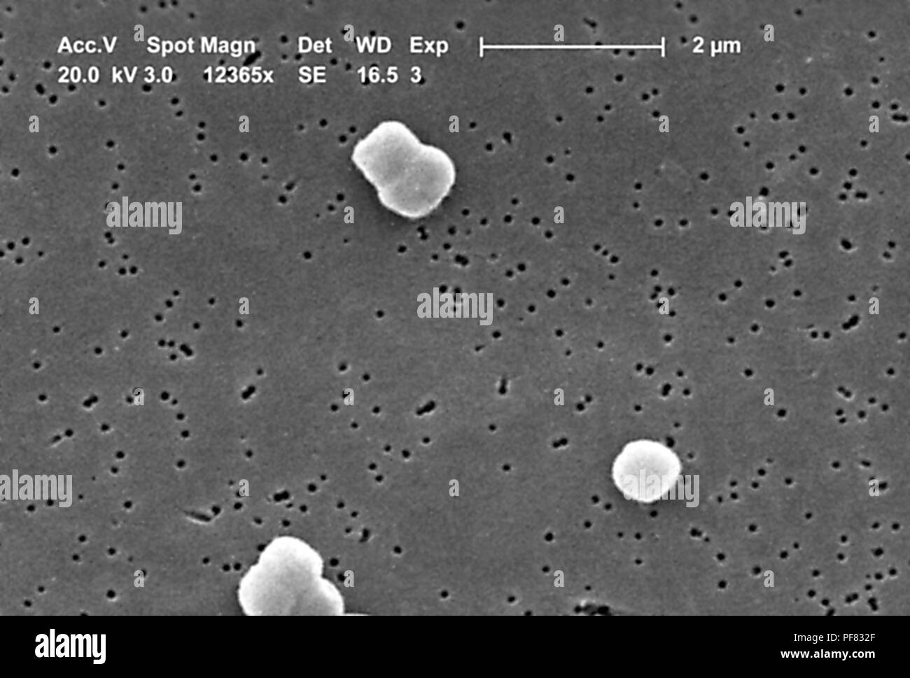 Gram-negative, non-motile Acinetobacter baumannii bacteria revealed in the 12365x magnified scanning electron microscopic (SEM) image, 2004. Image courtesy Centers for Disease Control (CDC) / Matthew J. Arduino, DrPH, Janice Carr, Jana Swenson. () Stock Photohttps://www.alamy.com/image-license-details/?v=1https://www.alamy.com/gram-negative-non-motile-acinetobacter-baumannii-bacteria-revealed-in-the-12365x-magnified-scanning-electron-microscopic-sem-image-2004-image-courtesy-centers-for-disease-control-cdc-matthew-j-arduino-drph-janice-carr-jana-swenson-image215922295.html
RMPF832F–Gram-negative, non-motile Acinetobacter baumannii bacteria revealed in the 12365x magnified scanning electron microscopic (SEM) image, 2004. Image courtesy Centers for Disease Control (CDC) / Matthew J. Arduino, DrPH, Janice Carr, Jana Swenson. ()
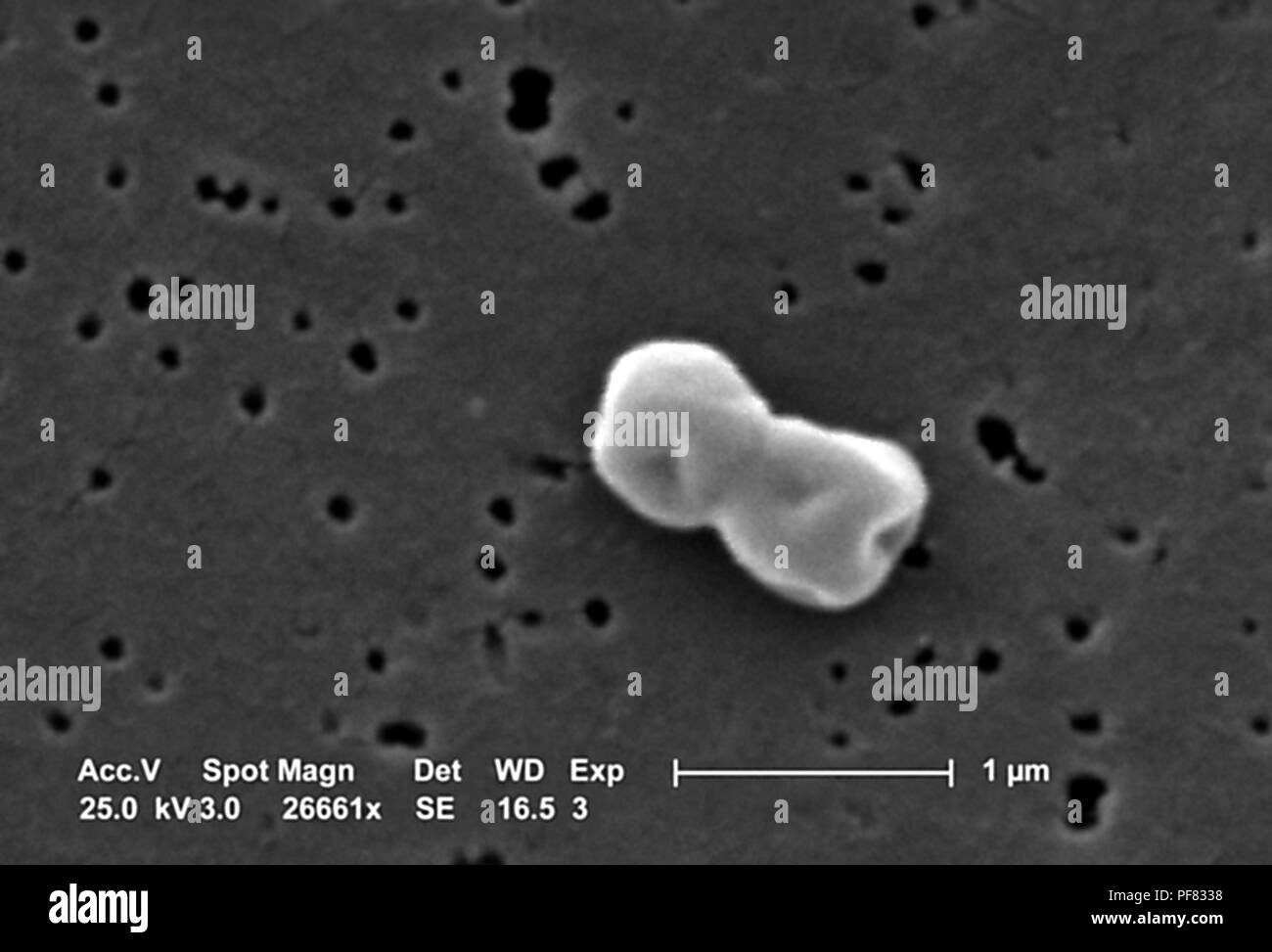 Gram-negative, non-motile Acinetobacter baumannii bacteria revealed in the 26661x magnified scanning electron microscopic (SEM) image, 2004. Image courtesy Centers for Disease Control (CDC) / Matthew J. Arduino, DrPH, Janice Carr, Jana Swenson. () Stock Photohttps://www.alamy.com/image-license-details/?v=1https://www.alamy.com/gram-negative-non-motile-acinetobacter-baumannii-bacteria-revealed-in-the-26661x-magnified-scanning-electron-microscopic-sem-image-2004-image-courtesy-centers-for-disease-control-cdc-matthew-j-arduino-drph-janice-carr-jana-swenson-image215922316.html
RMPF8338–Gram-negative, non-motile Acinetobacter baumannii bacteria revealed in the 26661x magnified scanning electron microscopic (SEM) image, 2004. Image courtesy Centers for Disease Control (CDC) / Matthew J. Arduino, DrPH, Janice Carr, Jana Swenson. ()
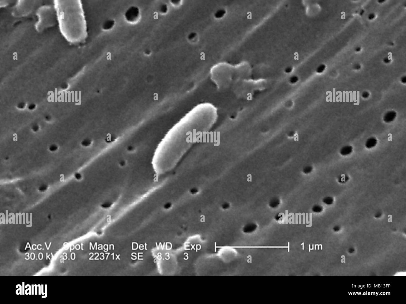 Vibrio cholerae bacteria revealed in the 22371x magnified scanning electron microscopic (SEM) image, 2005. Image courtesy Centers for Disease Control (CDC) / Janice Haney Carr. () Stock Photohttps://www.alamy.com/image-license-details/?v=1https://www.alamy.com/vibrio-cholerae-bacteria-revealed-in-the-22371x-magnified-scanning-electron-microscopic-sem-image-2005-image-courtesy-centers-for-disease-control-cdc-janice-haney-carr-image178889642.html
RMMB13FP–Vibrio cholerae bacteria revealed in the 22371x magnified scanning electron microscopic (SEM) image, 2005. Image courtesy Centers for Disease Control (CDC) / Janice Haney Carr. ()
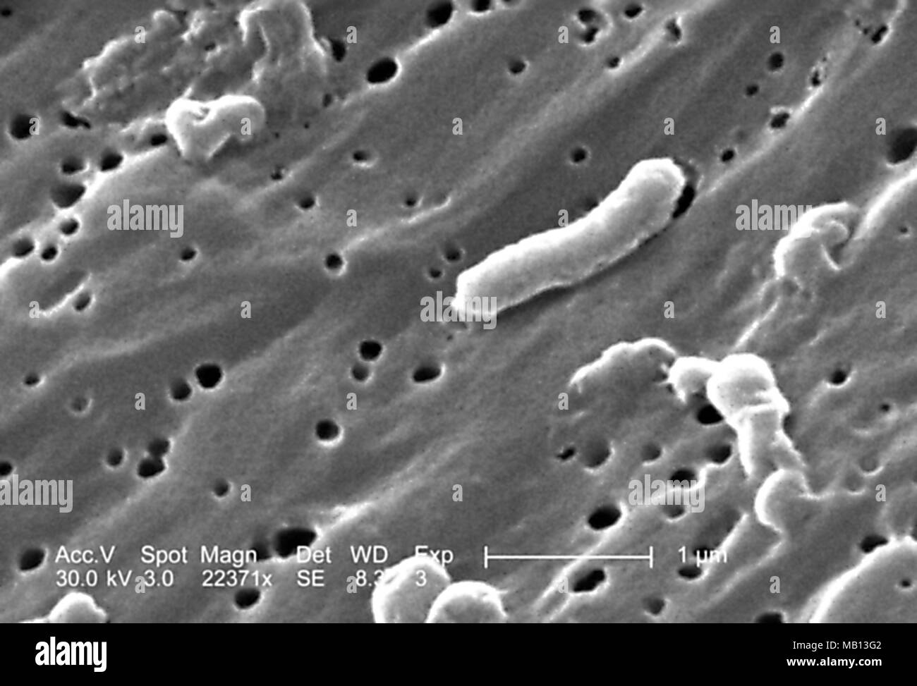 Vibrio cholerae bacteria revealed in the 22371x magnified scanning electron microscopic (SEM) image, 2005. Image courtesy Centers for Disease Control (CDC) / Janice Haney Carr. () Stock Photohttps://www.alamy.com/image-license-details/?v=1https://www.alamy.com/vibrio-cholerae-bacteria-revealed-in-the-22371x-magnified-scanning-electron-microscopic-sem-image-2005-image-courtesy-centers-for-disease-control-cdc-janice-haney-carr-image178889650.html
RMMB13G2–Vibrio cholerae bacteria revealed in the 22371x magnified scanning electron microscopic (SEM) image, 2005. Image courtesy Centers for Disease Control (CDC) / Janice Haney Carr. ()
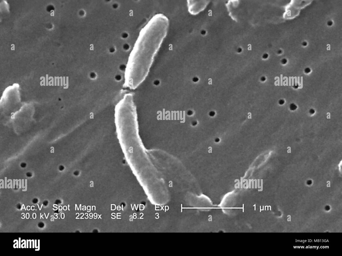 Separation of the two Vibrio cholerae bacteria revealed in the 22399x magnified scanning electron microscopic (SEM) image, 2005. Image courtesy Centers for Disease Control (CDC) / Janice Haney Carr. () Stock Photohttps://www.alamy.com/image-license-details/?v=1https://www.alamy.com/separation-of-the-two-vibrio-cholerae-bacteria-revealed-in-the-22399x-magnified-scanning-electron-microscopic-sem-image-2005-image-courtesy-centers-for-disease-control-cdc-janice-haney-carr-image178889658.html
RMMB13GA–Separation of the two Vibrio cholerae bacteria revealed in the 22399x magnified scanning electron microscopic (SEM) image, 2005. Image courtesy Centers for Disease Control (CDC) / Janice Haney Carr. ()
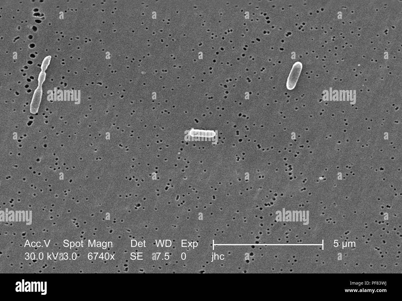 Three Ralstonia mannitolilytica bacteria revealed in the 6740x magnified scanning electron microscopic (SEM) image, 2006. Image courtesy Centers for Disease Control (CDC) / Judith Noble-Wang, Ph.D. () Stock Photohttps://www.alamy.com/image-license-details/?v=1https://www.alamy.com/three-ralstonia-mannitolilytica-bacteria-revealed-in-the-6740x-magnified-scanning-electron-microscopic-sem-image-2006-image-courtesy-centers-for-disease-control-cdc-judith-noble-wang-phd-image215922942.html
RMPF83WJ–Three Ralstonia mannitolilytica bacteria revealed in the 6740x magnified scanning electron microscopic (SEM) image, 2006. Image courtesy Centers for Disease Control (CDC) / Judith Noble-Wang, Ph.D. ()
 Grouping of Ralstonia mannitolilytica bacteria revealed in the 6740x magnified scanning electron microscopic (SEM) image, 2006. Image courtesy Centers for Disease Control (CDC) / Judith Noble-Wang, Ph.D. () Stock Photohttps://www.alamy.com/image-license-details/?v=1https://www.alamy.com/grouping-of-ralstonia-mannitolilytica-bacteria-revealed-in-the-6740x-magnified-scanning-electron-microscopic-sem-image-2006-image-courtesy-centers-for-disease-control-cdc-judith-noble-wang-phd-image215922972.html
RMPF83XM–Grouping of Ralstonia mannitolilytica bacteria revealed in the 6740x magnified scanning electron microscopic (SEM) image, 2006. Image courtesy Centers for Disease Control (CDC) / Judith Noble-Wang, Ph.D. ()
 Cellular division of a Vibrio cholerae bacteria revealed in the 14213x magnified scanning electron microscopic (SEM) image, 2005. Image courtesy Centers for Disease Control (CDC) / Janice Haney Carr. () Stock Photohttps://www.alamy.com/image-license-details/?v=1https://www.alamy.com/cellular-division-of-a-vibrio-cholerae-bacteria-revealed-in-the-14213x-magnified-scanning-electron-microscopic-sem-image-2005-image-courtesy-centers-for-disease-control-cdc-janice-haney-carr-image178889654.html
RMMB13G6–Cellular division of a Vibrio cholerae bacteria revealed in the 14213x magnified scanning electron microscopic (SEM) image, 2005. Image courtesy Centers for Disease Control (CDC) / Janice Haney Carr. ()
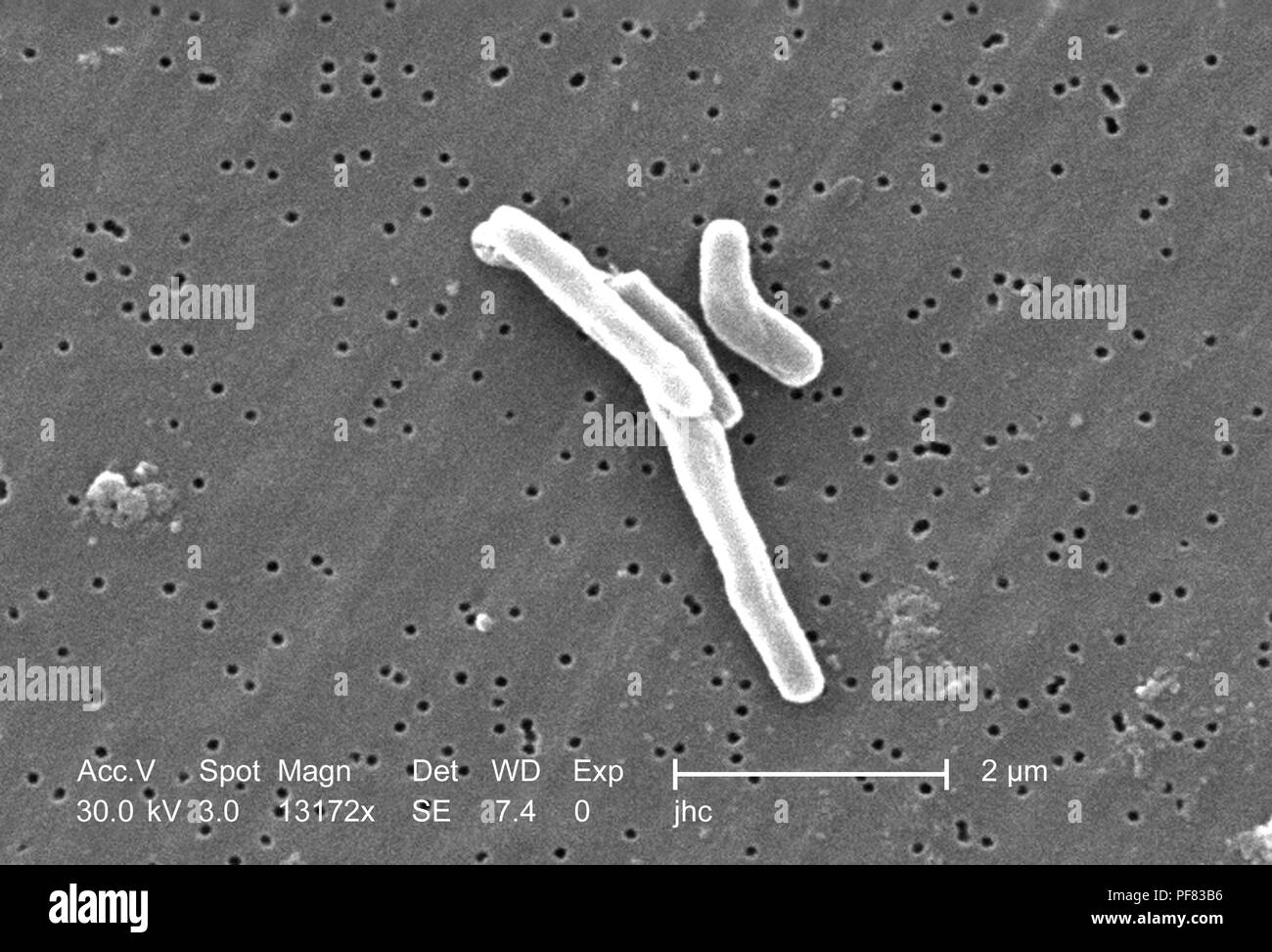 Gram-positive Mycobacterium tuberculosis bacteria revealed in the 13172x magnified scanning electron microscopic (SEM) image, 2006. Image courtesy Centers for Disease Control (CDC) / Ray Butler, MS, Janice Haney Carr. () Stock Photohttps://www.alamy.com/image-license-details/?v=1https://www.alamy.com/gram-positive-mycobacterium-tuberculosis-bacteria-revealed-in-the-13172x-magnified-scanning-electron-microscopic-sem-image-2006-image-courtesy-centers-for-disease-control-cdc-ray-butler-ms-janice-haney-carr-image215922538.html
RMPF83B6–Gram-positive Mycobacterium tuberculosis bacteria revealed in the 13172x magnified scanning electron microscopic (SEM) image, 2006. Image courtesy Centers for Disease Control (CDC) / Ray Butler, MS, Janice Haney Carr. ()
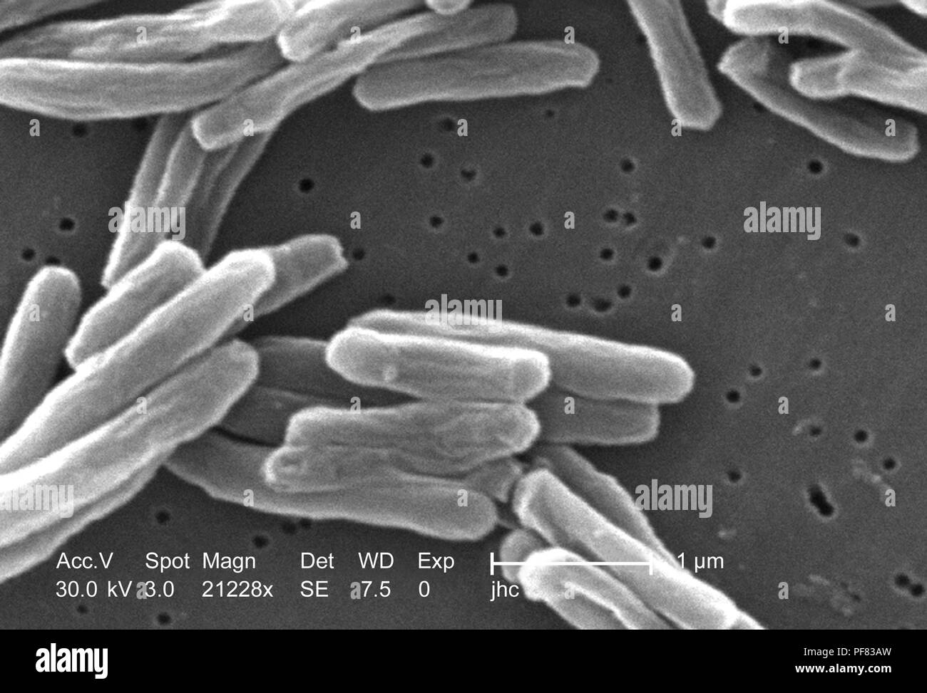 Ultrastructural details of Gram-positive Mycobacterium tuberculosis bacteria revealed in the 21228x magnified scanning electron microscopic (SEM) image, 2006. Image courtesy Centers for Disease Control (CDC) / Ray Butler, MS, Janice Haney Carr. () Stock Photohttps://www.alamy.com/image-license-details/?v=1https://www.alamy.com/ultrastructural-details-of-gram-positive-mycobacterium-tuberculosis-bacteria-revealed-in-the-21228x-magnified-scanning-electron-microscopic-sem-image-2006-image-courtesy-centers-for-disease-control-cdc-ray-butler-ms-janice-haney-carr-image215922529.html
RMPF83AW–Ultrastructural details of Gram-positive Mycobacterium tuberculosis bacteria revealed in the 21228x magnified scanning electron microscopic (SEM) image, 2006. Image courtesy Centers for Disease Control (CDC) / Ray Butler, MS, Janice Haney Carr. ()
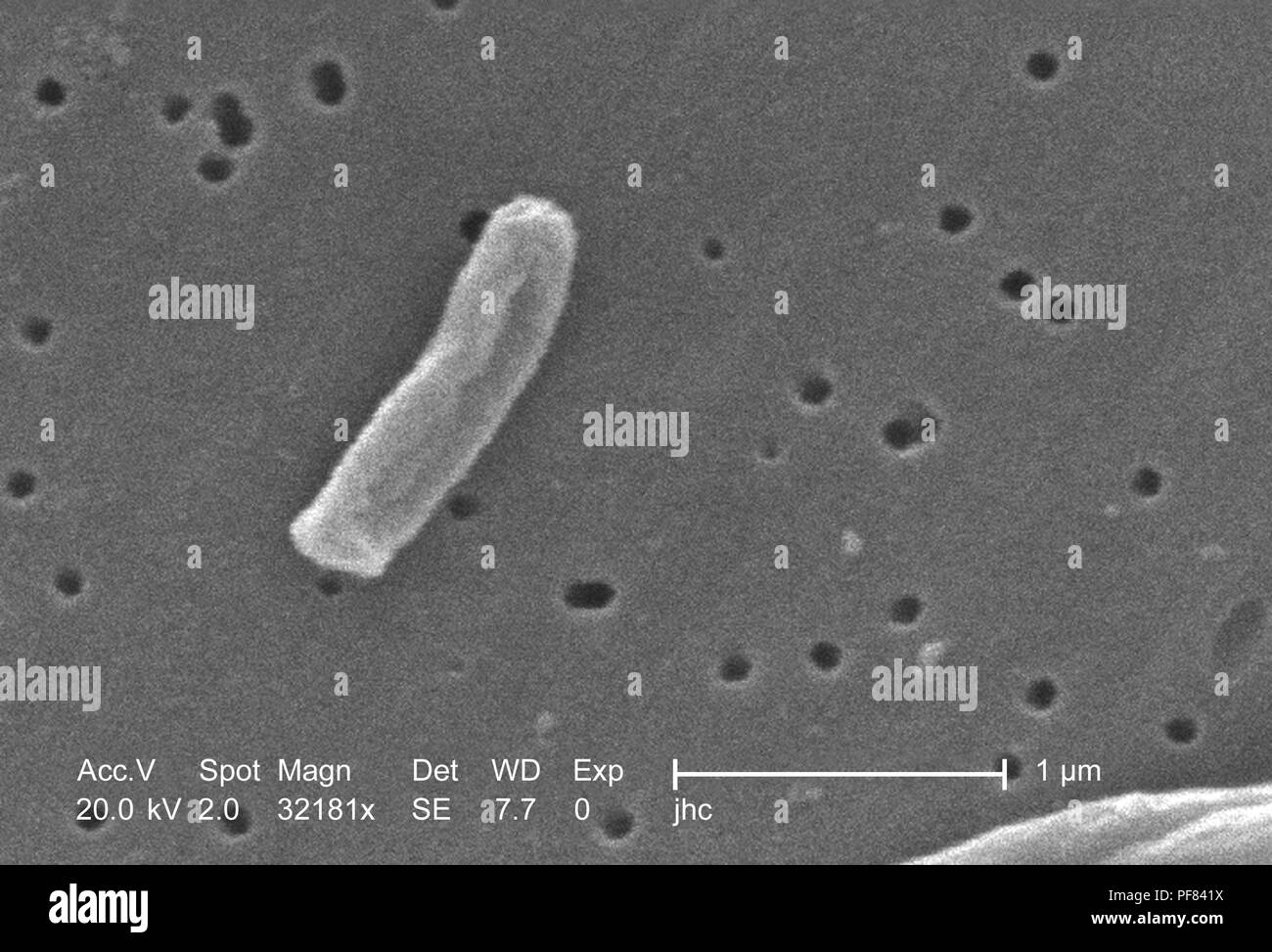 Ultrastructural details of Gram-positive Mycobacterium tuberculosis bacteria revealed in the 32181x magnified scanning electron microscopic (SEM) image, 2006. Image courtesy Centers for Disease Control (CDC) / Ray Butler, MS, Janice Haney Carr. () Stock Photohttps://www.alamy.com/image-license-details/?v=1https://www.alamy.com/ultrastructural-details-of-gram-positive-mycobacterium-tuberculosis-bacteria-revealed-in-the-32181x-magnified-scanning-electron-microscopic-sem-image-2006-image-courtesy-centers-for-disease-control-cdc-ray-butler-ms-janice-haney-carr-image215923062.html
RMPF841X–Ultrastructural details of Gram-positive Mycobacterium tuberculosis bacteria revealed in the 32181x magnified scanning electron microscopic (SEM) image, 2006. Image courtesy Centers for Disease Control (CDC) / Ray Butler, MS, Janice Haney Carr. ()
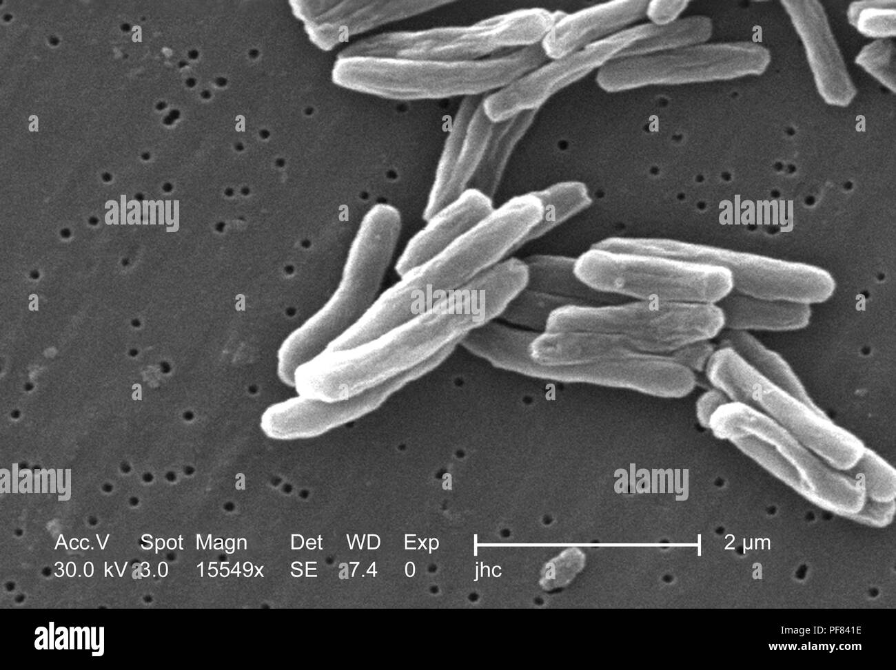 Ultrastructural details of Gram-positive Mycobacterium tuberculosis bacteria revealed in the 15549x magnified scanning electron microscopic (SEM) image, 2006. Image courtesy Centers for Disease Control (CDC) / Ray Butler, MS, Janice Haney Carr. () Stock Photohttps://www.alamy.com/image-license-details/?v=1https://www.alamy.com/ultrastructural-details-of-gram-positive-mycobacterium-tuberculosis-bacteria-revealed-in-the-15549x-magnified-scanning-electron-microscopic-sem-image-2006-image-courtesy-centers-for-disease-control-cdc-ray-butler-ms-janice-haney-carr-image215923050.html
RMPF841E–Ultrastructural details of Gram-positive Mycobacterium tuberculosis bacteria revealed in the 15549x magnified scanning electron microscopic (SEM) image, 2006. Image courtesy Centers for Disease Control (CDC) / Ray Butler, MS, Janice Haney Carr. ()
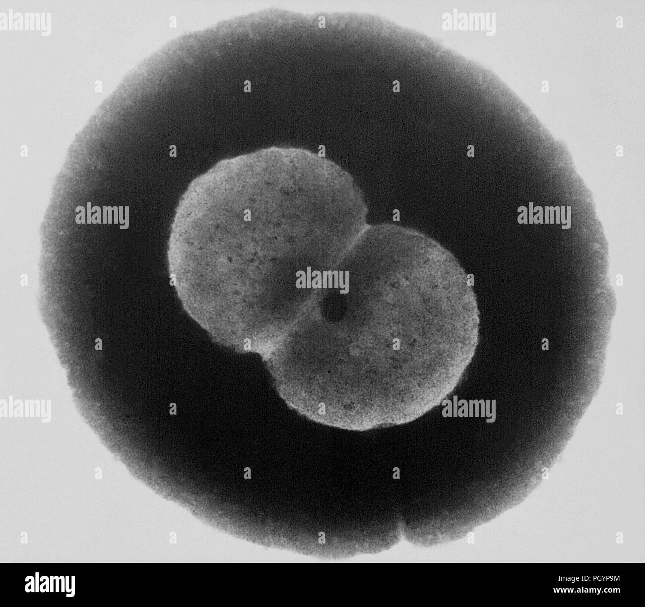 Diplococcal pair of Gram-negative Neisseria gonorrhoeae bacteria revealed in the transmission electron microscopic (TEM) image, 1972. Image courtesy Centers for Disease Control (CDC) / Dr Wiesner. () Stock Photohttps://www.alamy.com/image-license-details/?v=1https://www.alamy.com/diplococcal-pair-of-gram-negative-neisseria-gonorrhoeae-bacteria-revealed-in-the-transmission-electron-microscopic-tem-image-1972-image-courtesy-centers-for-disease-control-cdc-dr-wiesner-image216969136.html
RMPGYP9M–Diplococcal pair of Gram-negative Neisseria gonorrhoeae bacteria revealed in the transmission electron microscopic (TEM) image, 1972. Image courtesy Centers for Disease Control (CDC) / Dr Wiesner. ()
 Number of Gram-negative Escherichia coli bacteria of the strain O157:H7, revealed in the 6836x magnified scanning electron microscopic (SEM) image, 2006. Image courtesy Centers for Disease Control (CDC) / National Escherichia, Shigella, Vibrio Reference Unit at CDC. () Stock Photohttps://www.alamy.com/image-license-details/?v=1https://www.alamy.com/number-of-gram-negative-escherichia-coli-bacteria-of-the-strain-o157h7-revealed-in-the-6836x-magnified-scanning-electron-microscopic-sem-image-2006-image-courtesy-centers-for-disease-control-cdc-national-escherichia-shigella-vibrio-reference-unit-at-cdc-image215923306.html
RMPF84AJ–Number of Gram-negative Escherichia coli bacteria of the strain O157:H7, revealed in the 6836x magnified scanning electron microscopic (SEM) image, 2006. Image courtesy Centers for Disease Control (CDC) / National Escherichia, Shigella, Vibrio Reference Unit at CDC. ()
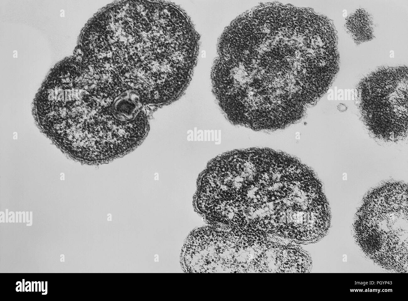 Gram-negative Neisseria gonorrhoeae bacteria revealed in a transmission electron microscopic (TEM) image, 1980. Image courtesy Centers for Disease Control (CDC) / Joe Miller. () Stock Photohttps://www.alamy.com/image-license-details/?v=1https://www.alamy.com/gram-negative-neisseria-gonorrhoeae-bacteria-revealed-in-a-transmission-electron-microscopic-tem-image-1980-image-courtesy-centers-for-disease-control-cdc-joe-miller-image216968979.html
RMPGYP43–Gram-negative Neisseria gonorrhoeae bacteria revealed in a transmission electron microscopic (TEM) image, 1980. Image courtesy Centers for Disease Control (CDC) / Joe Miller. ()
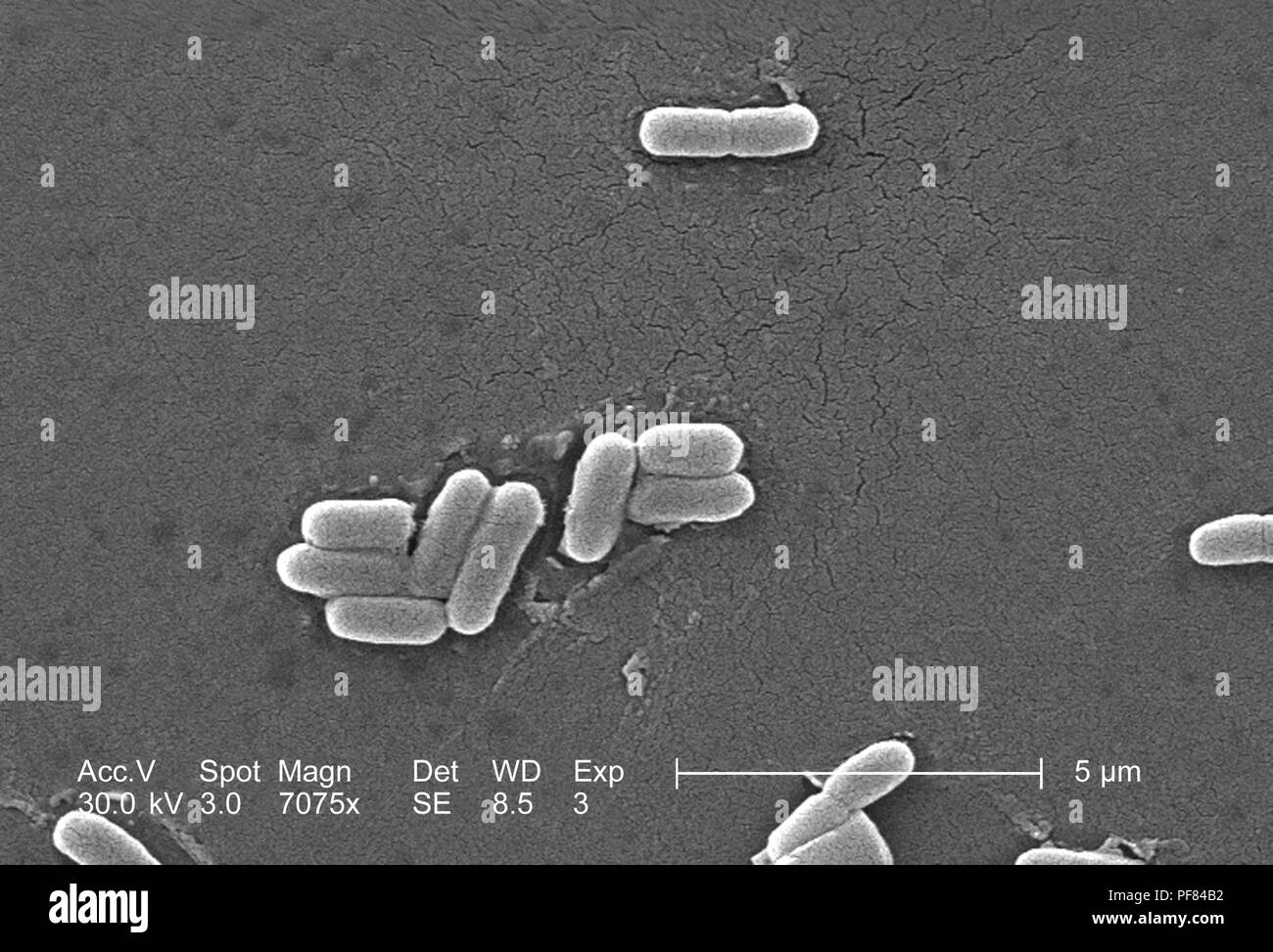 Gram-negative Escherichia coli bacteria of the strain O157:H7, revealed in the 7075x magnified scanning electron microscopic (SEM) image, 2006. Image courtesy Centers for Disease Control (CDC) / National Escherichia, Shigella, Vibrio Reference Unit at CDC. () Stock Photohttps://www.alamy.com/image-license-details/?v=1https://www.alamy.com/gram-negative-escherichia-coli-bacteria-of-the-strain-o157h7-revealed-in-the-7075x-magnified-scanning-electron-microscopic-sem-image-2006-image-courtesy-centers-for-disease-control-cdc-national-escherichia-shigella-vibrio-reference-unit-at-cdc-image215923318.html
RMPF84B2–Gram-negative Escherichia coli bacteria of the strain O157:H7, revealed in the 7075x magnified scanning electron microscopic (SEM) image, 2006. Image courtesy Centers for Disease Control (CDC) / National Escherichia, Shigella, Vibrio Reference Unit at CDC. ()
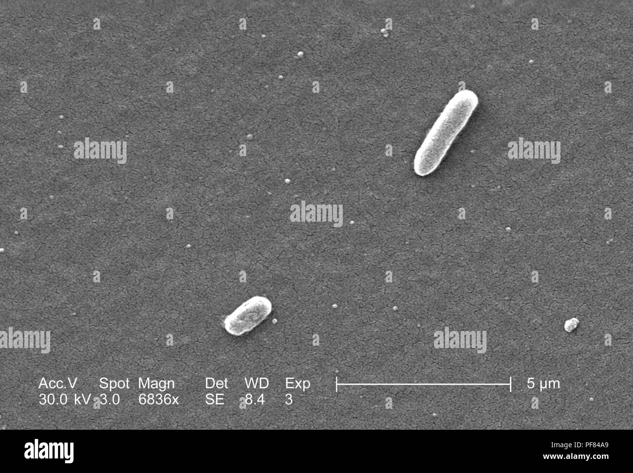 Gram-negative Escherichia coli bacteria of the strain O157:H7, revealed in the 6836x magnified scanning electron microscopic (SEM) image, 2006. Image courtesy Centers for Disease Control (CDC) / National Escherichia, Shigella, Vibrio Reference Unit at CDC. () Stock Photohttps://www.alamy.com/image-license-details/?v=1https://www.alamy.com/gram-negative-escherichia-coli-bacteria-of-the-strain-o157h7-revealed-in-the-6836x-magnified-scanning-electron-microscopic-sem-image-2006-image-courtesy-centers-for-disease-control-cdc-national-escherichia-shigella-vibrio-reference-unit-at-cdc-image215923297.html
RMPF84A9–Gram-negative Escherichia coli bacteria of the strain O157:H7, revealed in the 6836x magnified scanning electron microscopic (SEM) image, 2006. Image courtesy Centers for Disease Control (CDC) / National Escherichia, Shigella, Vibrio Reference Unit at CDC. ()
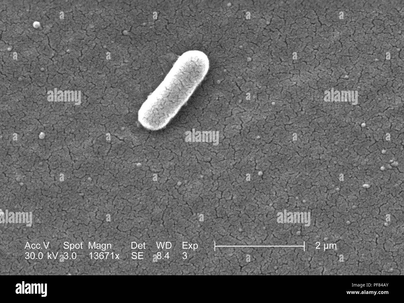 Gram-negative Escherichia coli bacteria of the strain O157:H7, revealed in the 13671x magnified scanning electron microscopic (SEM) image, 2006. Image courtesy Centers for Disease Control (CDC) / National Escherichia, Shigella, Vibrio Reference Unit at CDC. () Stock Photohttps://www.alamy.com/image-license-details/?v=1https://www.alamy.com/gram-negative-escherichia-coli-bacteria-of-the-strain-o157h7-revealed-in-the-13671x-magnified-scanning-electron-microscopic-sem-image-2006-image-courtesy-centers-for-disease-control-cdc-national-escherichia-shigella-vibrio-reference-unit-at-cdc-image215923315.html
RMPF84AY–Gram-negative Escherichia coli bacteria of the strain O157:H7, revealed in the 13671x magnified scanning electron microscopic (SEM) image, 2006. Image courtesy Centers for Disease Control (CDC) / National Escherichia, Shigella, Vibrio Reference Unit at CDC. ()
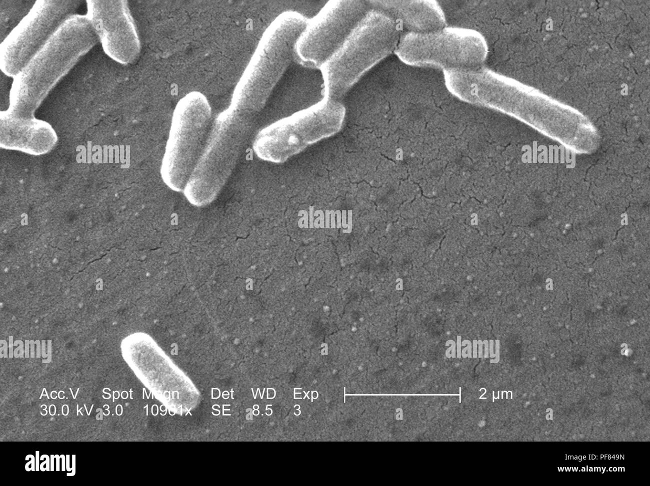 Gram-negative Escherichia coli bacteria of the strain O157:H7, revealed in the 10961x magnified scanning electron microscopic (SEM) image, 2006. Image courtesy Centers for Disease Control (CDC) / National Escherichia, Shigella, Vibrio Reference Unit at CDC. () Stock Photohttps://www.alamy.com/image-license-details/?v=1https://www.alamy.com/gram-negative-escherichia-coli-bacteria-of-the-strain-o157h7-revealed-in-the-10961x-magnified-scanning-electron-microscopic-sem-image-2006-image-courtesy-centers-for-disease-control-cdc-national-escherichia-shigella-vibrio-reference-unit-at-cdc-image215923281.html
RMPF849N–Gram-negative Escherichia coli bacteria of the strain O157:H7, revealed in the 10961x magnified scanning electron microscopic (SEM) image, 2006. Image courtesy Centers for Disease Control (CDC) / National Escherichia, Shigella, Vibrio Reference Unit at CDC. ()
 Gram-negative Escherichia coli bacteria of the strain O157:H7, revealed in the 3418x magnified scanning electron microscopic (SEM) image, 2006. Image courtesy Centers for Disease Control (CDC) / National Escherichia, Shigella, Vibrio Reference Unit at CDC. () Stock Photohttps://www.alamy.com/image-license-details/?v=1https://www.alamy.com/gram-negative-escherichia-coli-bacteria-of-the-strain-o157h7-revealed-in-the-3418x-magnified-scanning-electron-microscopic-sem-image-2006-image-courtesy-centers-for-disease-control-cdc-national-escherichia-shigella-vibrio-reference-unit-at-cdc-image215923295.html
RMPF84A7–Gram-negative Escherichia coli bacteria of the strain O157:H7, revealed in the 3418x magnified scanning electron microscopic (SEM) image, 2006. Image courtesy Centers for Disease Control (CDC) / National Escherichia, Shigella, Vibrio Reference Unit at CDC. ()