Quick filters:
Mri scan body Stock Photos and Images
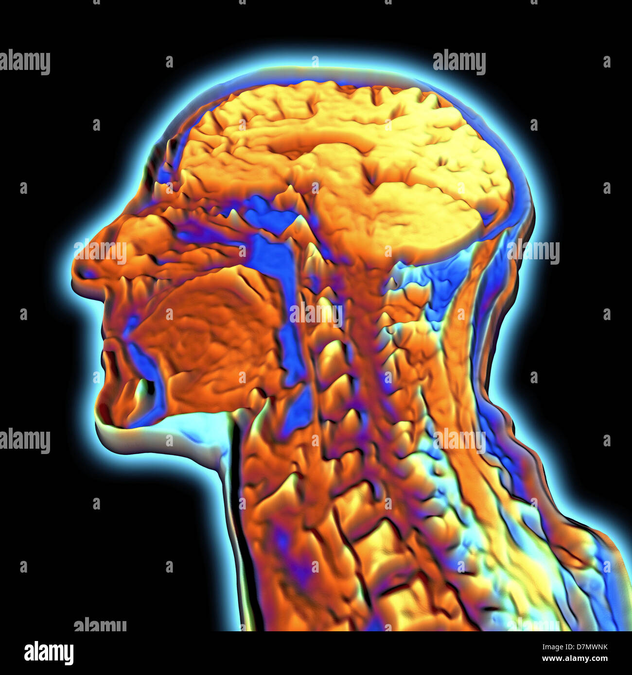 Coloured MRI scan of the human hea Stock Photohttps://www.alamy.com/image-license-details/?v=1https://www.alamy.com/stock-photo-coloured-mri-scan-of-the-human-hea-56392943.html
RFD7MWNK–Coloured MRI scan of the human hea
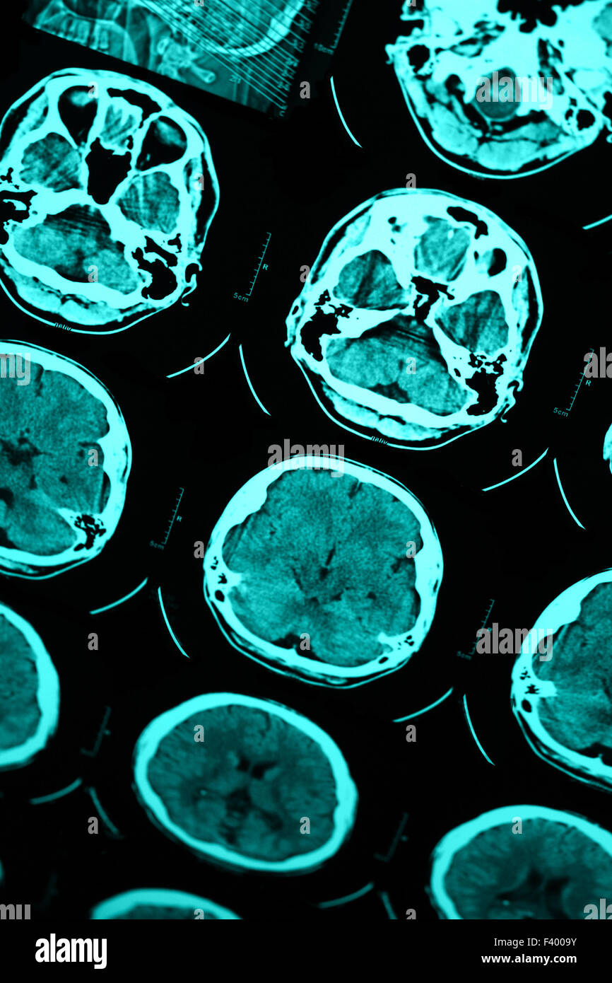 Photo of the CT and MRI of the skull Stock Photohttps://www.alamy.com/image-license-details/?v=1https://www.alamy.com/stock-photo-photo-of-the-ct-and-mri-of-the-skull-88510743.html
RFF4009Y–Photo of the CT and MRI of the skull
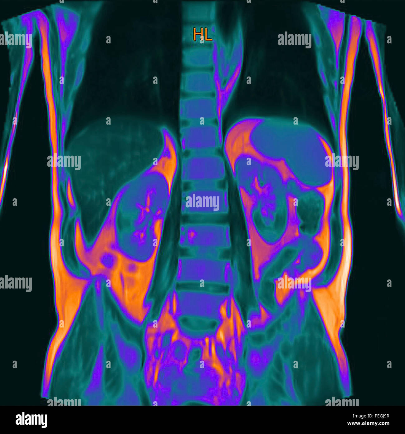 Frontal Abdomen MRI scan of a 60 year old male patient. This patient suffers from a kidney stone Stock Photohttps://www.alamy.com/image-license-details/?v=1https://www.alamy.com/frontal-abdomen-mri-scan-of-a-60-year-old-male-patient-this-patient-suffers-from-a-kidney-stone-image215495219.html
RMPEGJ9R–Frontal Abdomen MRI scan of a 60 year old male patient. This patient suffers from a kidney stone
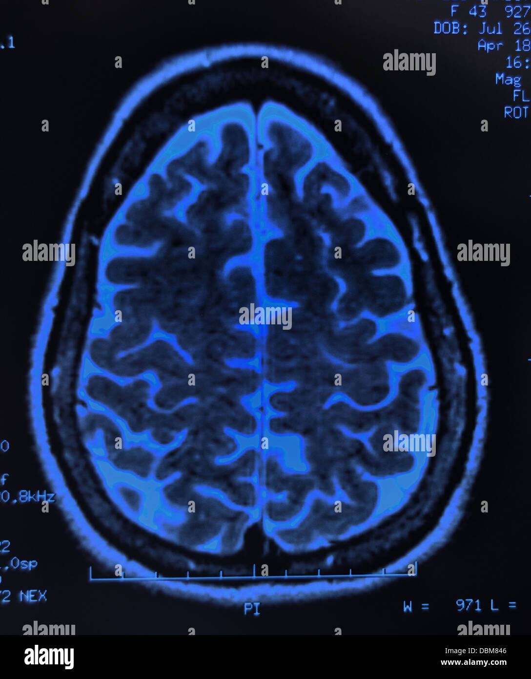 Human brain scan / MRI, X-ray Stock Photohttps://www.alamy.com/image-license-details/?v=1https://www.alamy.com/stock-photo-human-brain-scan-mri-x-ray-58837750.html
RFDBM846–Human brain scan / MRI, X-ray
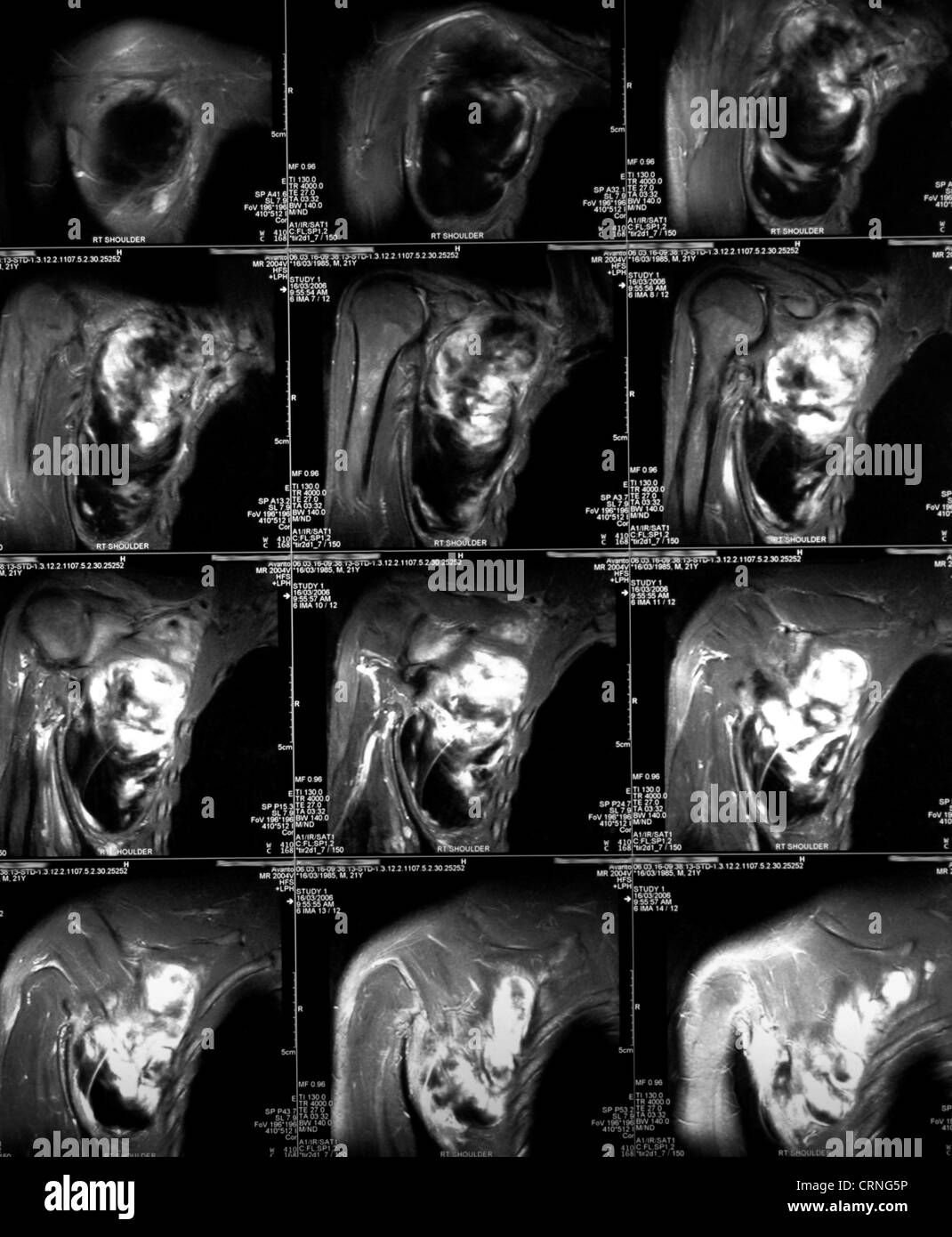 An MRI (magnetic resonance image) scan. Primarily used in radiology as way of visualising the structure and function of the body. The detail of the imagery makes MRI especially useful in musculoskeletal, neurological, oncological and cardiovascular imaging. Stock Photohttps://www.alamy.com/image-license-details/?v=1https://www.alamy.com/stock-photo-an-mri-magnetic-resonance-image-scan-primarily-used-in-radiology-as-49031522.html
RMCRNG5P–An MRI (magnetic resonance image) scan. Primarily used in radiology as way of visualising the structure and function of the body. The detail of the imagery makes MRI especially useful in musculoskeletal, neurological, oncological and cardiovascular imaging.
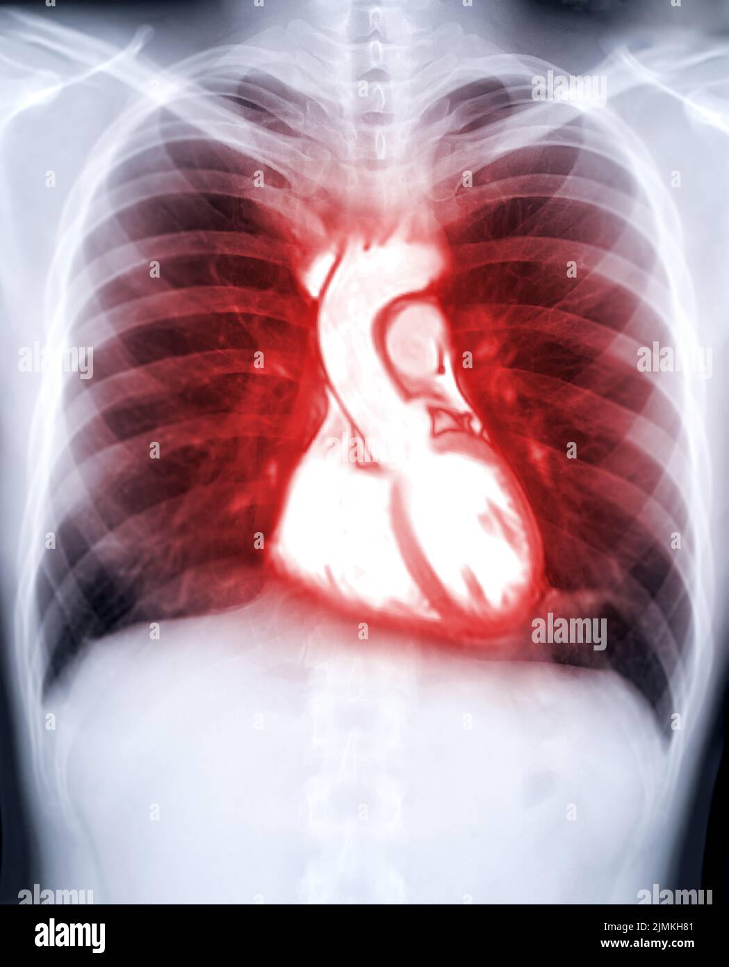 Chest x-ray image fusion mri heart for diagnosis heart diseases. Stock Photohttps://www.alamy.com/image-license-details/?v=1https://www.alamy.com/chest-x-ray-image-fusion-mri-heart-for-diagnosis-heart-diseases-image477403697.html
RF2JMKH81–Chest x-ray image fusion mri heart for diagnosis heart diseases.
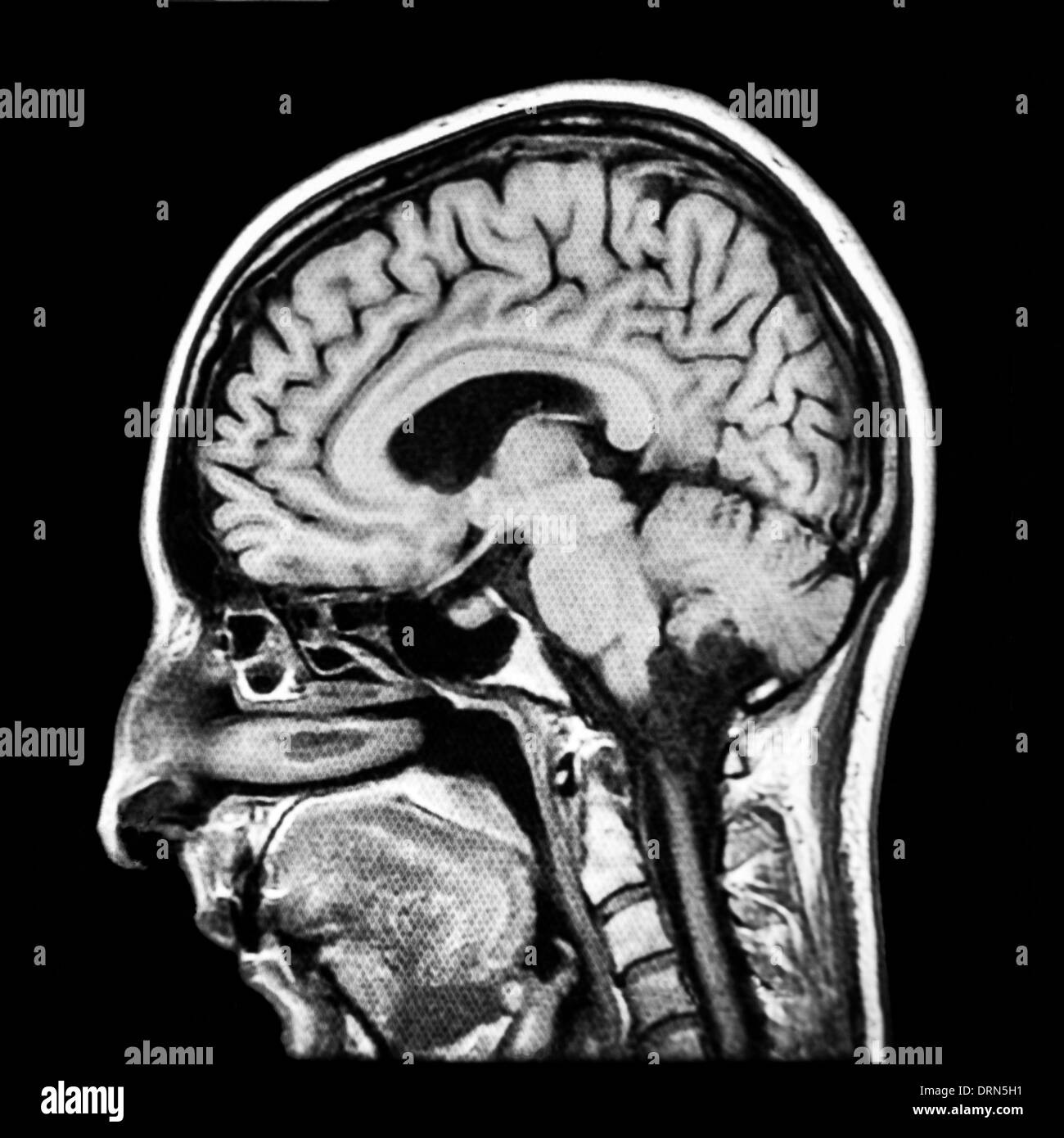 Vertical section of human brain MRI scan Stock Photohttps://www.alamy.com/image-license-details/?v=1https://www.alamy.com/vertical-section-of-human-brain-mri-scan-image66233581.html
RFDRN5H1–Vertical section of human brain MRI scan
 Complete real MRI scan of the human body, from the front and back Stock Photohttps://www.alamy.com/image-license-details/?v=1https://www.alamy.com/complete-real-mri-scan-of-the-human-body-from-the-front-and-back-image62150653.html
RFDH35P5–Complete real MRI scan of the human body, from the front and back
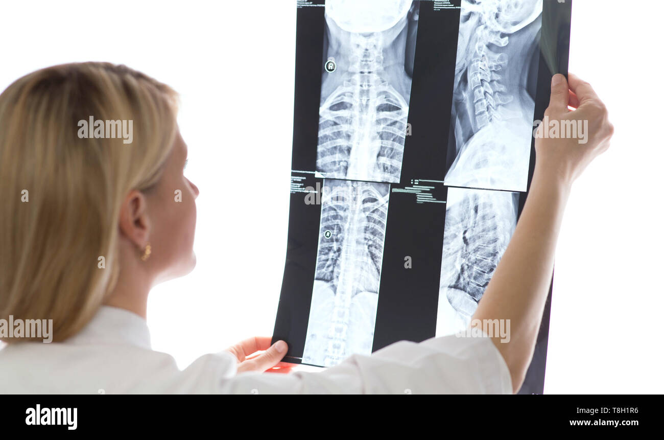 doctor examining patients x-ray and MRI scans Stock Photohttps://www.alamy.com/image-license-details/?v=1https://www.alamy.com/doctor-examining-patients-x-ray-and-mri-scans-image246237018.html
RFT8H1R6–doctor examining patients x-ray and MRI scans
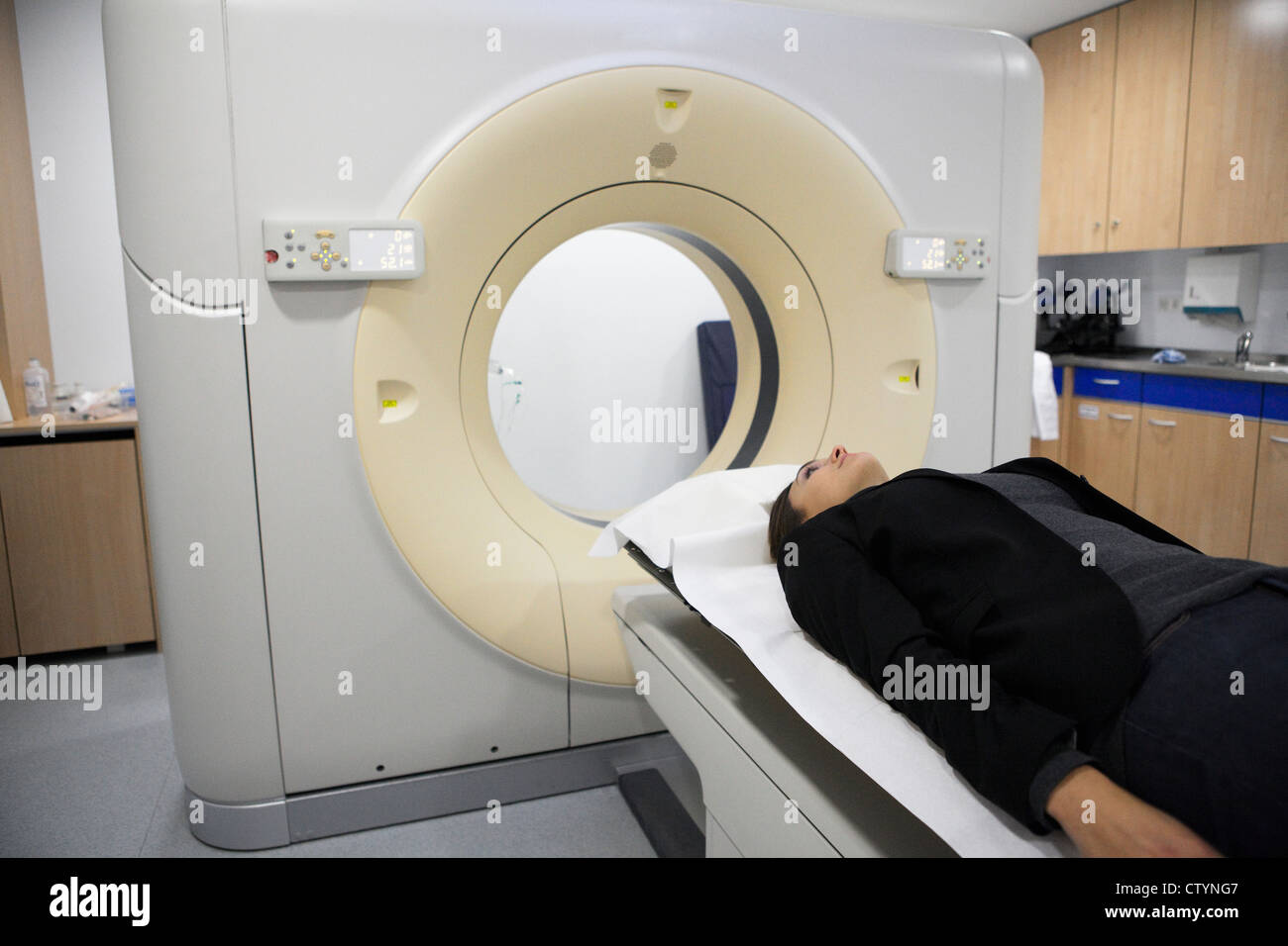 MRI scan Stock Photohttps://www.alamy.com/image-license-details/?v=1https://www.alamy.com/stock-photo-mri-scan-49782103.html
RFCTYNG7–MRI scan
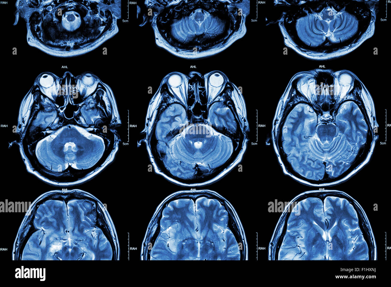 MRI of Brain ( cross section of brain ) ( Medical , Health care , Science background ) Stock Photohttps://www.alamy.com/image-license-details/?v=1https://www.alamy.com/stock-photo-mri-of-brain-cross-section-of-brain-medical-health-care-science-background-87060670.html
RFF1HXNJ–MRI of Brain ( cross section of brain ) ( Medical , Health care , Science background )
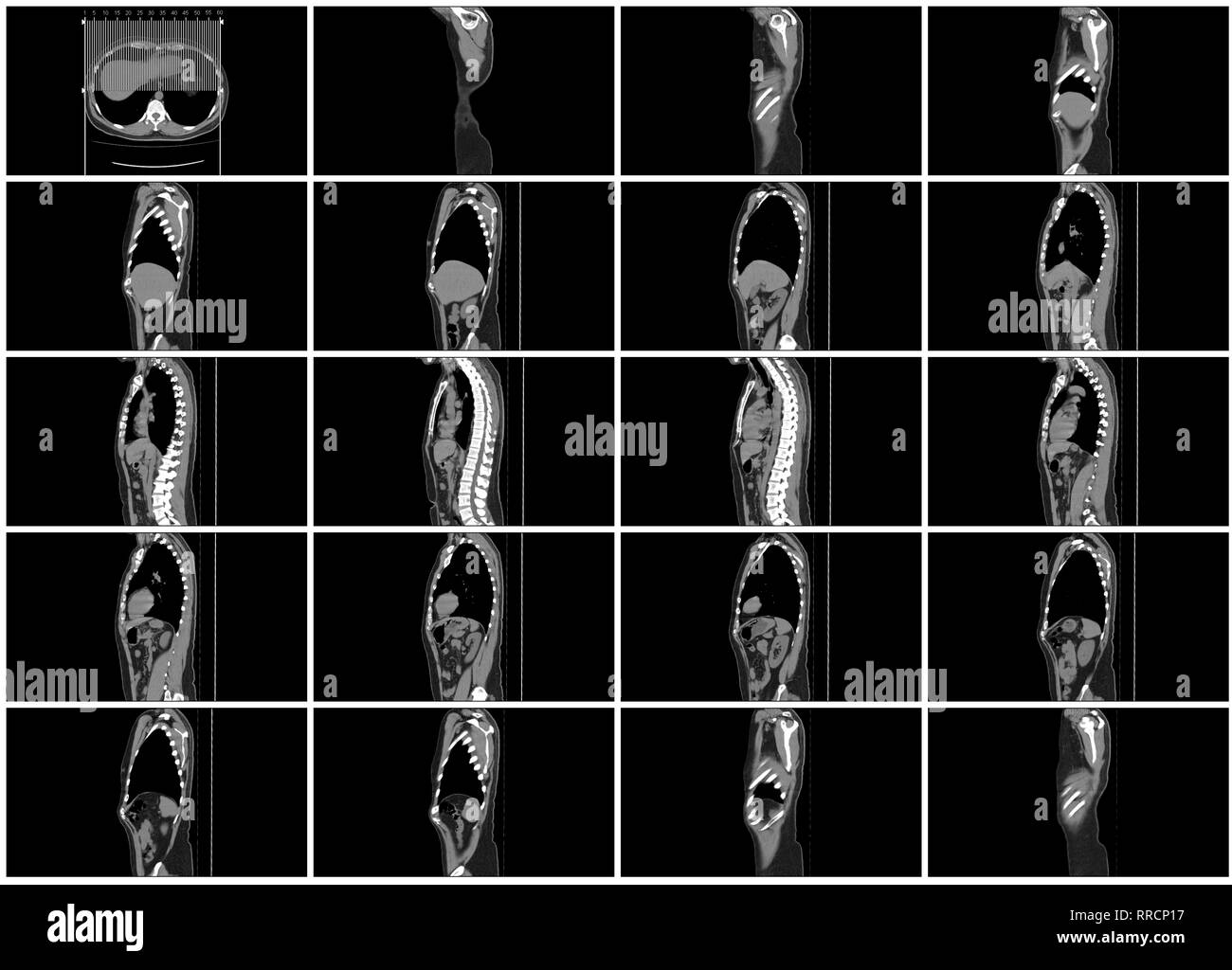 ct scan step set of body sagittal view Stock Photohttps://www.alamy.com/image-license-details/?v=1https://www.alamy.com/ct-scan-step-set-of-body-sagittal-view-image238152579.html
RFRRCP17–ct scan step set of body sagittal view
 Man doctor in protective mask looking at MRI picture with lungs x-ray scan Stock Photohttps://www.alamy.com/image-license-details/?v=1https://www.alamy.com/man-doctor-in-protective-mask-looking-at-mri-picture-with-lungs-x-ray-scan-image424612366.html
RF2FJPNBA–Man doctor in protective mask looking at MRI picture with lungs x-ray scan
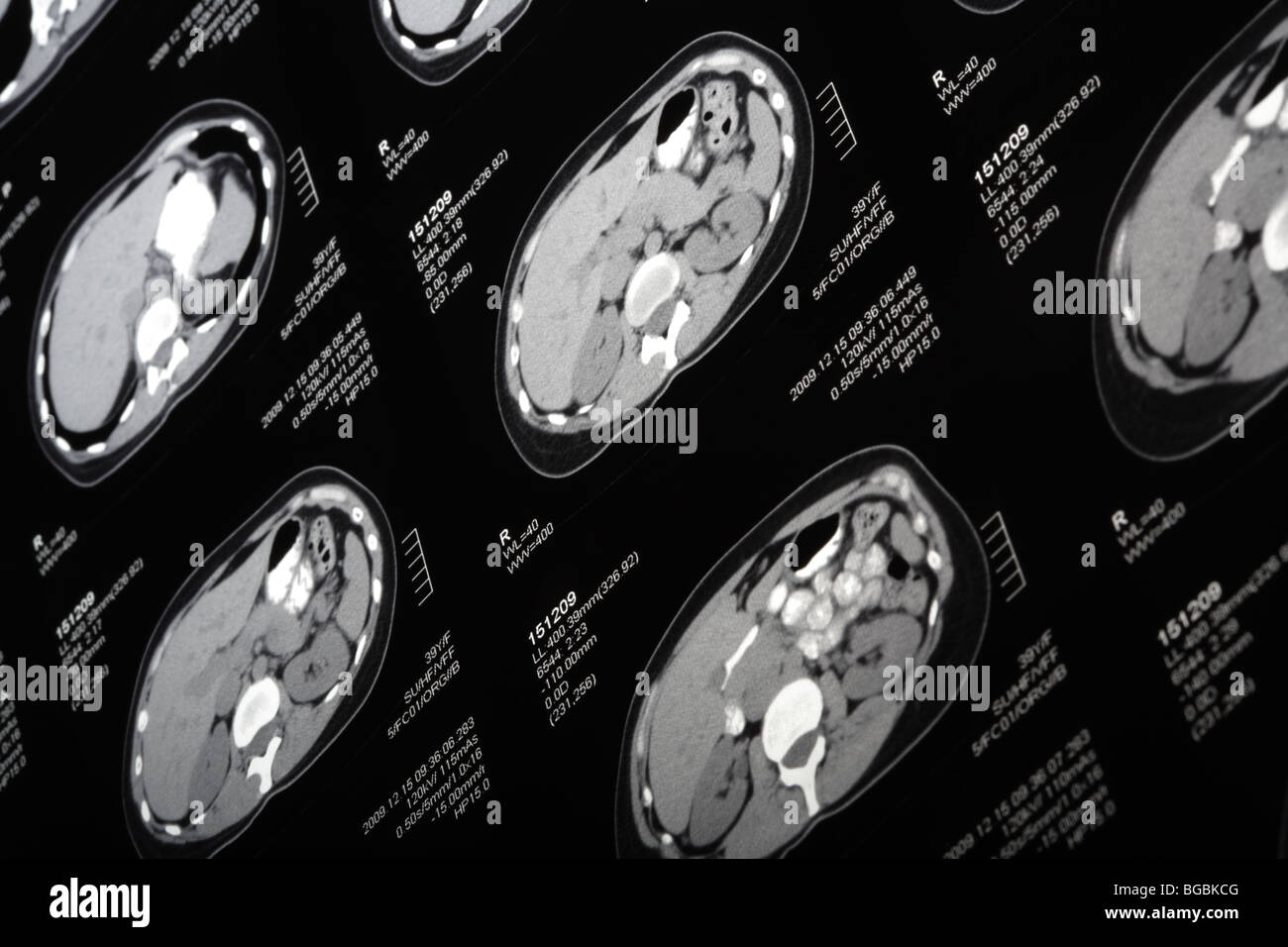 MRI scan close-up. Tilt view. Stock Photohttps://www.alamy.com/image-license-details/?v=1https://www.alamy.com/stock-photo-mri-scan-close-up-tilt-view-27301584.html
RFBGBKCG–MRI scan close-up. Tilt view.
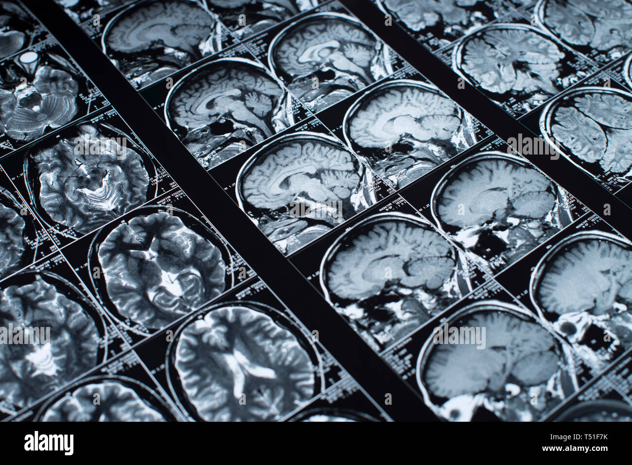 MRI Brain Scan of head and skull Stock Photohttps://www.alamy.com/image-license-details/?v=1https://www.alamy.com/mri-brain-scan-of-head-and-skull-image244052359.html
RFT51F7K–MRI Brain Scan of head and skull
 Human body examination in HUD style. Modern healthcare with body scan (Anatomy, Dna formula, Ecg monitor, data organs, X-ray, Statistic and Diagrams Stock Vectorhttps://www.alamy.com/image-license-details/?v=1https://www.alamy.com/human-body-examination-in-hud-style-modern-healthcare-with-body-scan-anatomy-dna-formula-ecg-monitor-data-organs-x-ray-statistic-and-diagrams-image425168682.html
RF2FKM2YP–Human body examination in HUD style. Modern healthcare with body scan (Anatomy, Dna formula, Ecg monitor, data organs, X-ray, Statistic and Diagrams
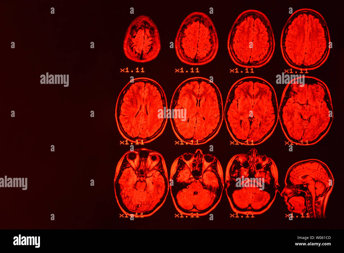 MRI of the brain on a black background with red backlight. Medical background Stock Photohttps://www.alamy.com/image-license-details/?v=1https://www.alamy.com/mri-of-the-brain-on-a-black-background-with-red-backlight-medical-background-image258288365.html
RFW061CD–MRI of the brain on a black background with red backlight. Medical background
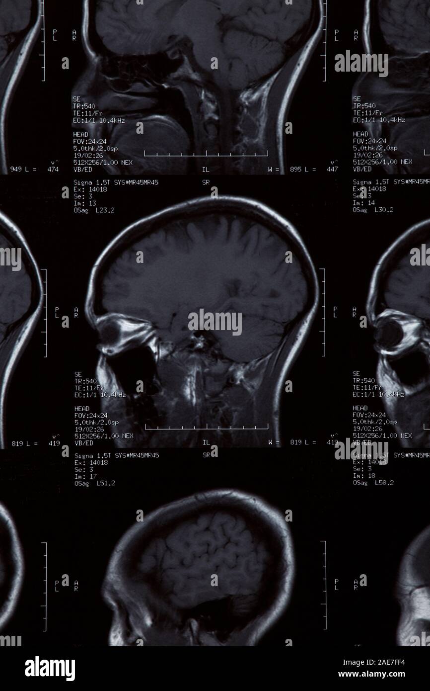 MRI Scan close up Stock Photohttps://www.alamy.com/image-license-details/?v=1https://www.alamy.com/mri-scan-close-up-image335768024.html
RM2AE7FF4–MRI Scan close up
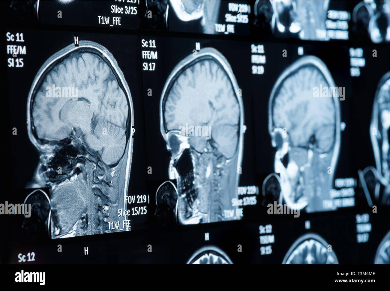 Head MRI scan, personal data removed Stock Photohttps://www.alamy.com/image-license-details/?v=1https://www.alamy.com/head-mri-scan-personal-data-removed-image243233438.html
RFT3M6ME–Head MRI scan, personal data removed
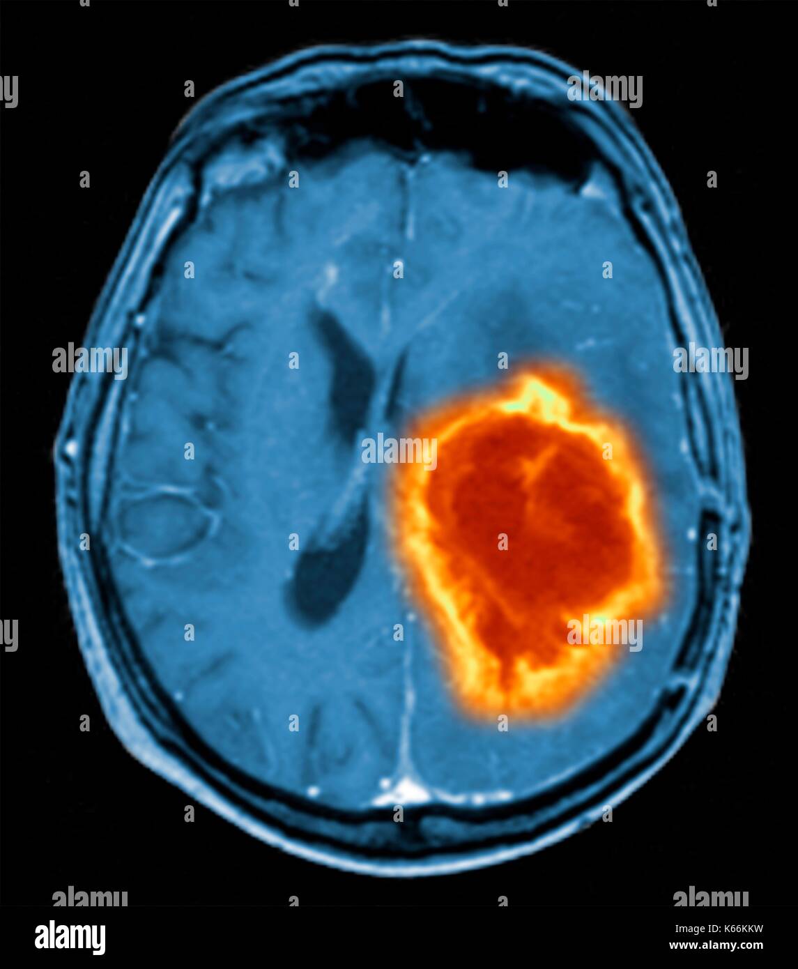 Brain tumour. Coloured Magnetic Resonance Imaging (MRI) scan of an axial section through the brain showing a metastatic tumour. At bottom left is the tumour (red-yellow) This tumour occurs within one cerebral hemisphere; the other hemisphere is at right. The eyeballs - not visible -are at top. Metastatic cancer is a secondary disease spread from cancer elsewhere in the body. Metastatic brain tumours are malignant. Typically they cause brain compression and nerve damage Stock Photohttps://www.alamy.com/image-license-details/?v=1https://www.alamy.com/brain-tumour-coloured-magnetic-resonance-imaging-mri-scan-of-an-axial-image158728413.html
RFK66KKW–Brain tumour. Coloured Magnetic Resonance Imaging (MRI) scan of an axial section through the brain showing a metastatic tumour. At bottom left is the tumour (red-yellow) This tumour occurs within one cerebral hemisphere; the other hemisphere is at right. The eyeballs - not visible -are at top. Metastatic cancer is a secondary disease spread from cancer elsewhere in the body. Metastatic brain tumours are malignant. Typically they cause brain compression and nerve damage
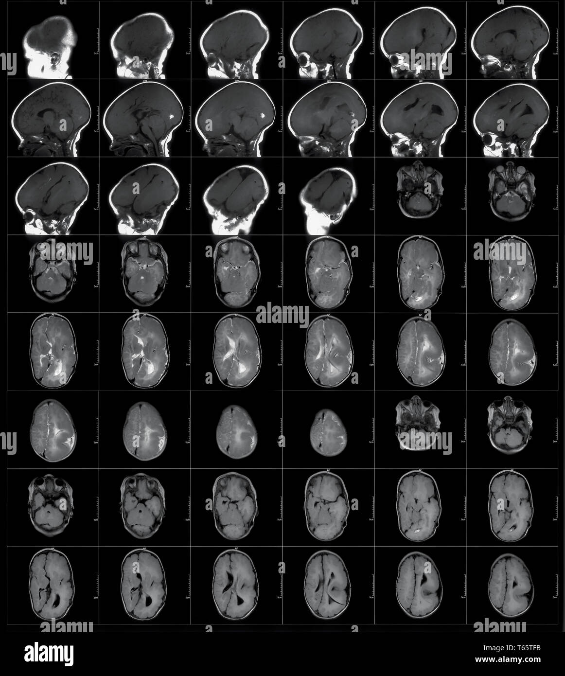 MRI scan Stock Photohttps://www.alamy.com/image-license-details/?v=1https://www.alamy.com/mri-scan-image244762095.html
RMT65TFB–MRI scan
 MRI scan Stock Photohttps://www.alamy.com/image-license-details/?v=1https://www.alamy.com/stock-photo-mri-scan-59780610.html
RFDD76NP–MRI scan
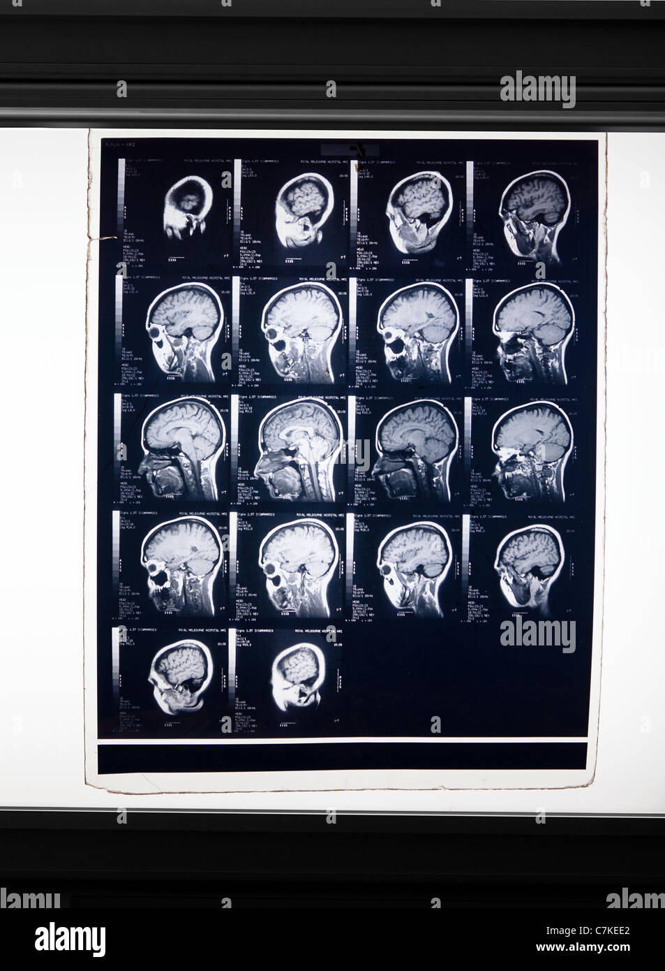 MRI brain scan Stock Photohttps://www.alamy.com/image-license-details/?v=1https://www.alamy.com/stock-photo-mri-brain-scan-39151786.html
RFC7KEE2–MRI brain scan
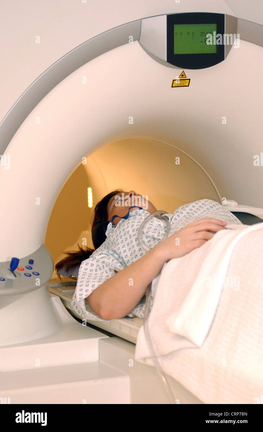 A woman having a CT scan Stock Photohttps://www.alamy.com/image-license-details/?v=1https://www.alamy.com/stock-photo-a-woman-having-a-ct-scan-49046501.html
RMCRP78N–A woman having a CT scan
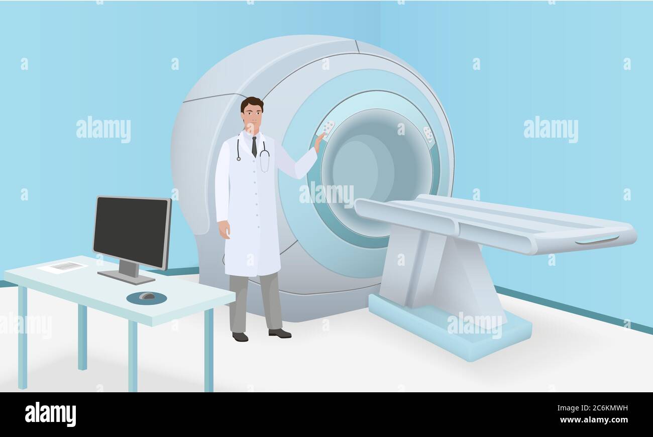 Doctor invites patient to body brain scan of MRI machine. MRI scan and diagnostics process in procedure room. Realistic vector Stock Vectorhttps://www.alamy.com/image-license-details/?v=1https://www.alamy.com/doctor-invites-patient-to-body-brain-scan-of-mri-machine-mri-scan-and-diagnostics-process-in-procedure-room-realistic-vector-image365539149.html
RF2C6KMWH–Doctor invites patient to body brain scan of MRI machine. MRI scan and diagnostics process in procedure room. Realistic vector
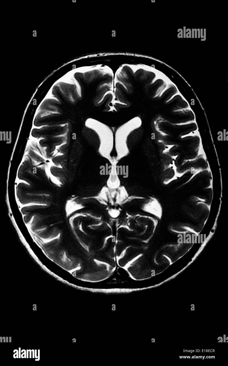 Horizontal section of a human brain - MRI scan Stock Photohttps://www.alamy.com/image-license-details/?v=1https://www.alamy.com/horizontal-section-of-a-human-brain-mri-scan-image69643079.html
RFE18ECR–Horizontal section of a human brain - MRI scan
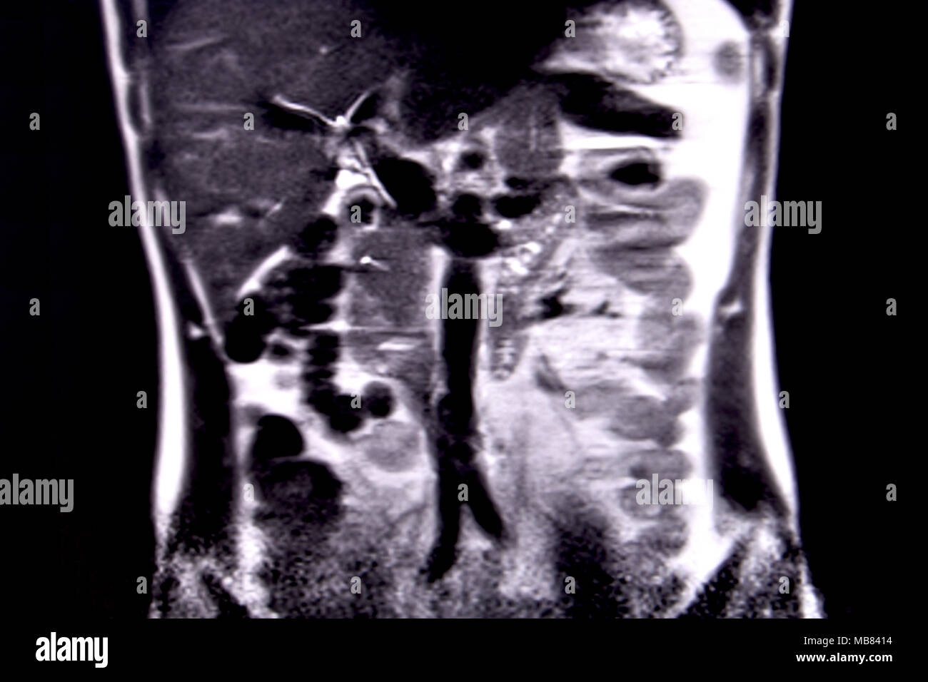 Ride through the human abdomen and chest by means of 18 MRI cuts (coronal view). Picture 3/18 Stock Photohttps://www.alamy.com/image-license-details/?v=1https://www.alamy.com/ride-through-the-human-abdomen-and-chest-by-means-of-18-mri-cuts-coronal-view-picture-318-image179043680.html
RFMB8414–Ride through the human abdomen and chest by means of 18 MRI cuts (coronal view). Picture 3/18
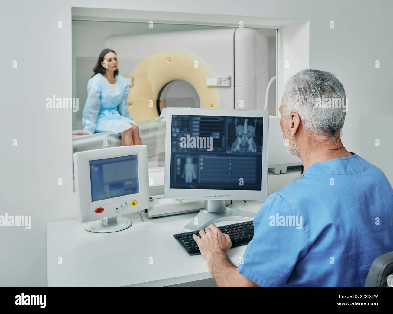 Radiologist before CT scan of female patient's body using CT scanner from control room behind protective window. Computed Tomography Stock Photohttps://www.alamy.com/image-license-details/?v=1https://www.alamy.com/radiologist-before-ct-scan-of-female-patients-body-using-ct-scanner-from-control-room-behind-protective-window-computed-tomography-image481032689.html
RF2JXGX2W–Radiologist before CT scan of female patient's body using CT scanner from control room behind protective window. Computed Tomography
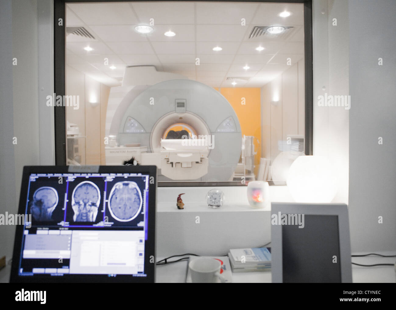 MRI scan- control room Stock Photohttps://www.alamy.com/image-license-details/?v=1https://www.alamy.com/stock-photo-mri-scan-control-room-49782052.html
RFCTYNEC–MRI scan- control room
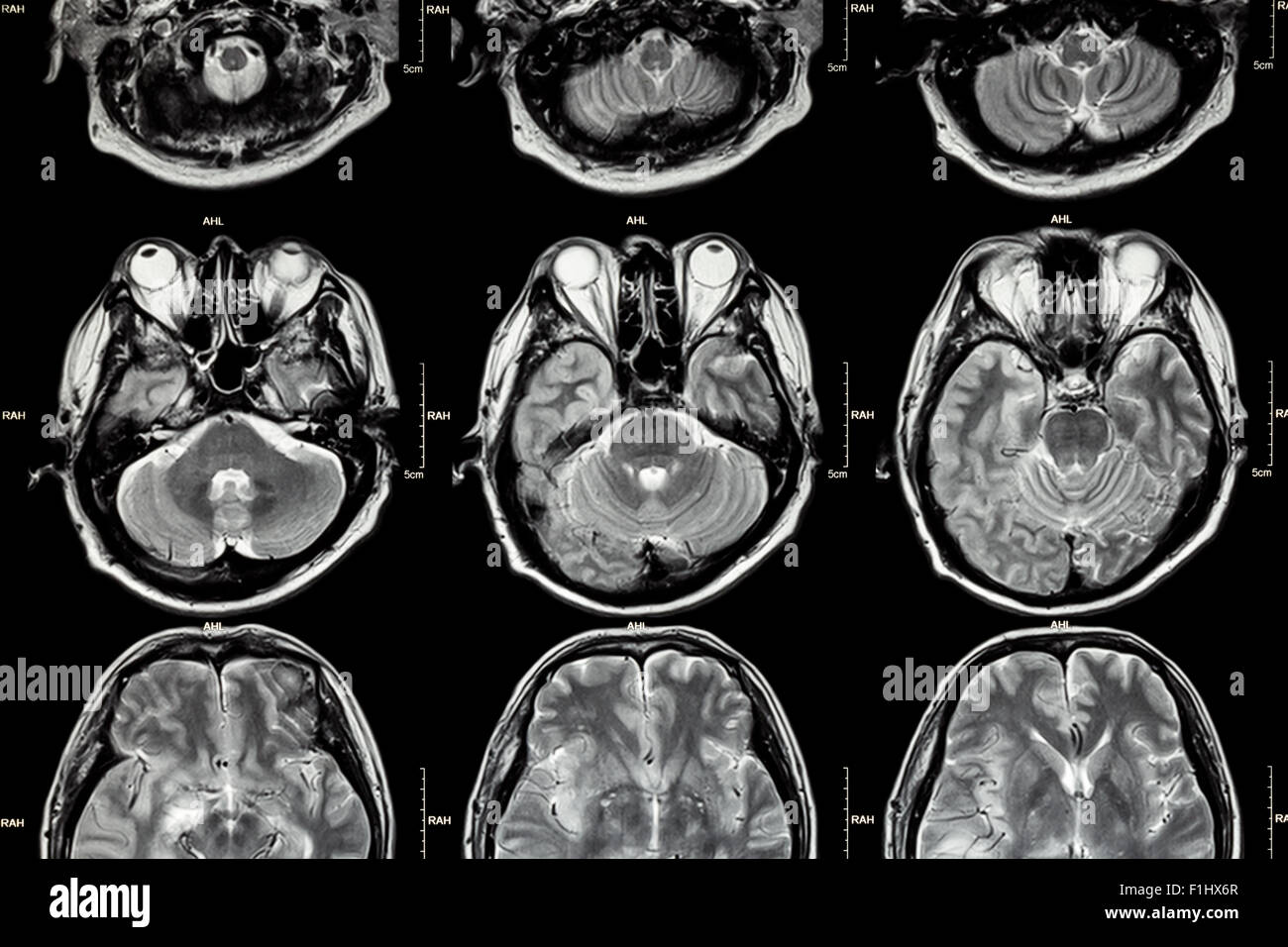 MRI of Brain ( cross section of brain ) ( Medical , Health care , Science background ) Stock Photohttps://www.alamy.com/image-license-details/?v=1https://www.alamy.com/stock-photo-mri-of-brain-cross-section-of-brain-medical-health-care-science-background-87060255.html
RFF1HX6R–MRI of Brain ( cross section of brain ) ( Medical , Health care , Science background )
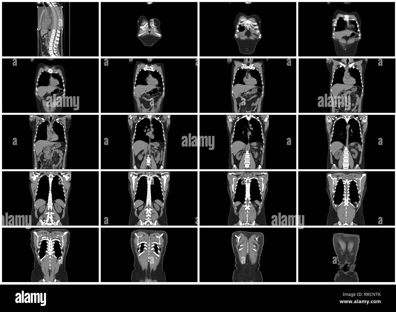 ct scan step set of body coronal view Stock Photohttps://www.alamy.com/image-license-details/?v=1https://www.alamy.com/ct-scan-step-set-of-body-coronal-view-image238152451.html
RFRRCNTK–ct scan step set of body coronal view
 An X-ray of a human body Stock Photohttps://www.alamy.com/image-license-details/?v=1https://www.alamy.com/an-x-ray-of-a-human-body-image610719165.html
RF2XDGJEN–An X-ray of a human body
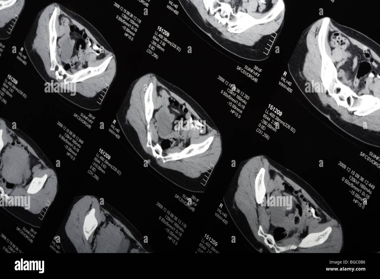 MRI scan close-up. Tilt view. Stock Photohttps://www.alamy.com/image-license-details/?v=1https://www.alamy.com/stock-photo-mri-scan-close-up-tilt-view-27308602.html
RFBGC0B6–MRI scan close-up. Tilt view.
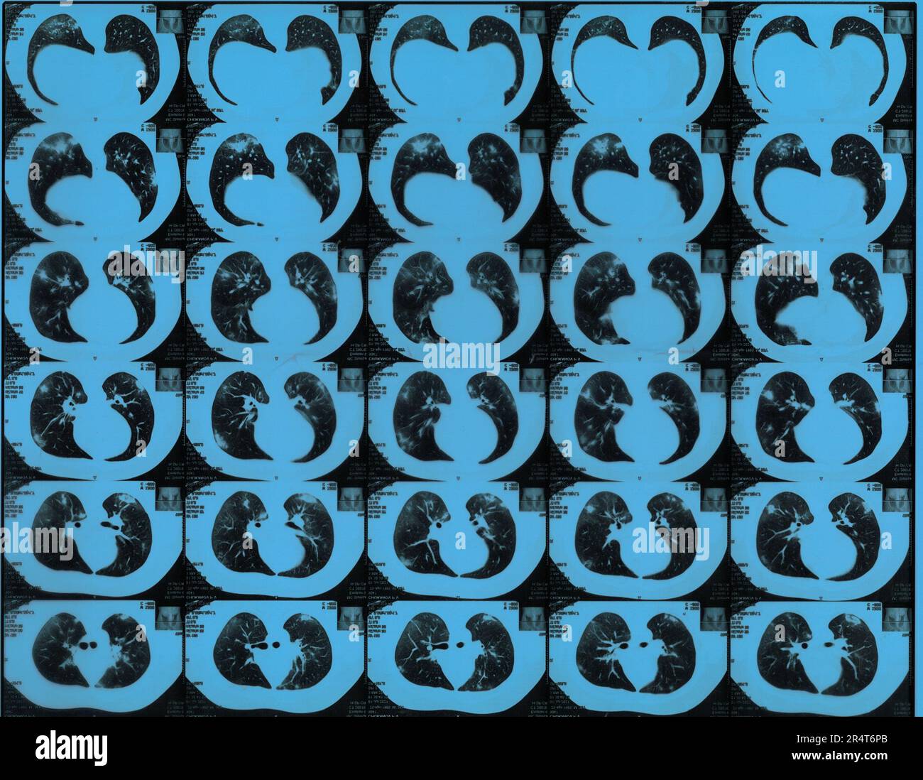 MRI lung scan of male patient after COVID infection Stock Photohttps://www.alamy.com/image-license-details/?v=1https://www.alamy.com/mri-lung-scan-of-male-patient-after-covid-infection-image553722579.html
RF2R4T6PB–MRI lung scan of male patient after COVID infection
 Mri scanner examination vector illustration. Cartoon flat medical scan hospital lab room with Mri machine, doctor characters exam patient, discuss diagnosis and treatment. Medicine research background Stock Vectorhttps://www.alamy.com/image-license-details/?v=1https://www.alamy.com/mri-scanner-examination-vector-illustration-cartoon-flat-medical-scan-hospital-lab-room-with-mri-machine-doctor-characters-exam-patient-discuss-diagnosis-and-treatment-medicine-research-background-image376973977.html
RF2CW8J49–Mri scanner examination vector illustration. Cartoon flat medical scan hospital lab room with Mri machine, doctor characters exam patient, discuss diagnosis and treatment. Medicine research background
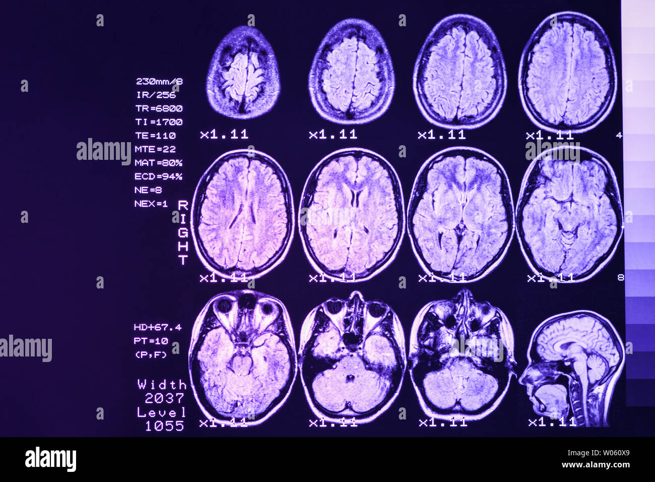 MRI scan or magnetic resonance image of head and brain scan. The result is an MRI of the brain with values and numbers with lilac backlight. Stock Photohttps://www.alamy.com/image-license-details/?v=1https://www.alamy.com/mri-scan-or-magnetic-resonance-image-of-head-and-brain-scan-the-result-is-an-mri-of-the-brain-with-values-and-numbers-with-lilac-backlight-image258287969.html
RFW060X9–MRI scan or magnetic resonance image of head and brain scan. The result is an MRI of the brain with values and numbers with lilac backlight.
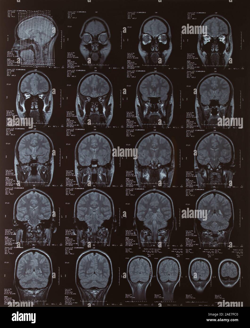 MRI Scan of the female brain Stock Photohttps://www.alamy.com/image-license-details/?v=1https://www.alamy.com/mri-scan-of-the-female-brain-image335767936.html
RM2AE7FC0–MRI Scan of the female brain
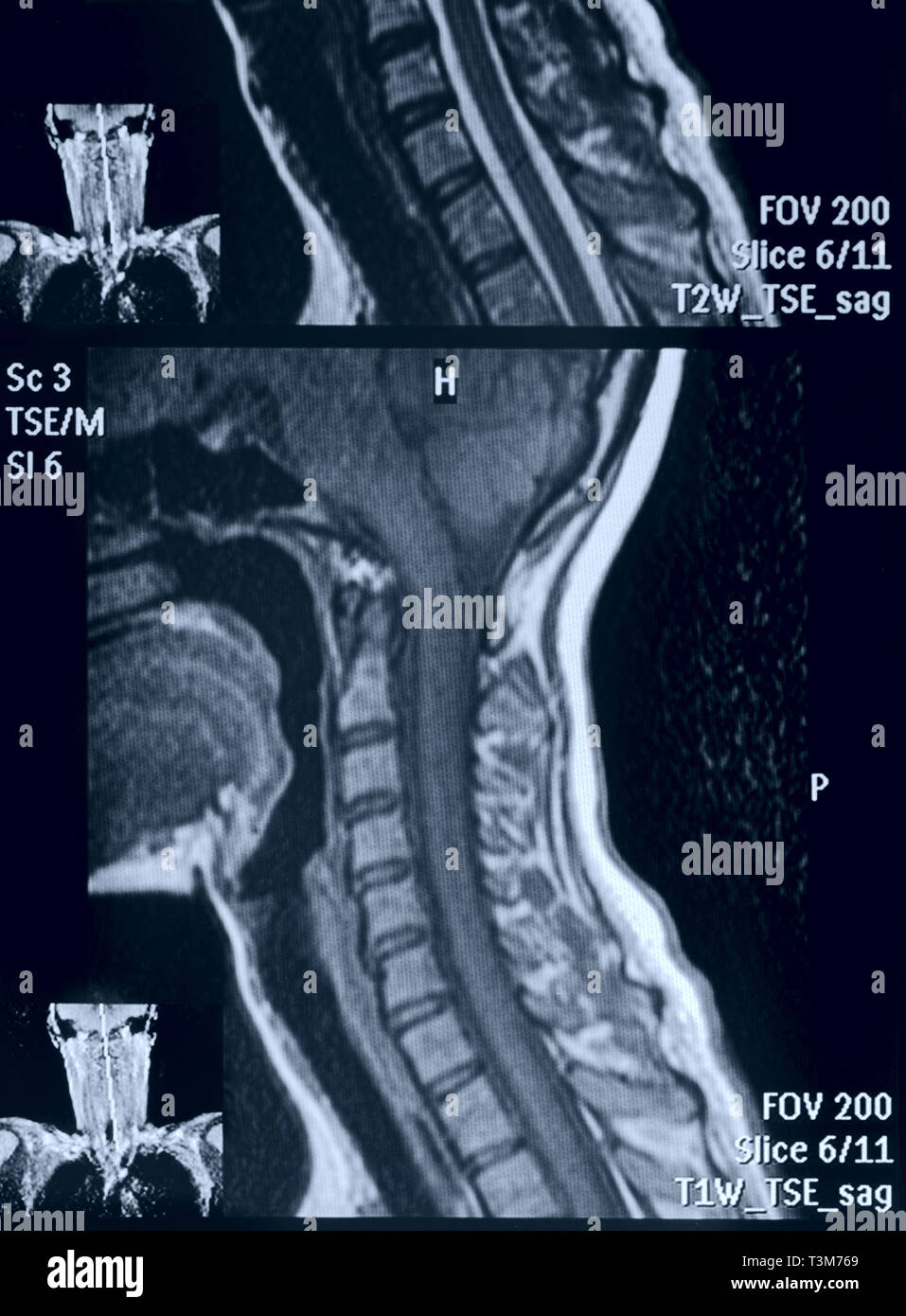 Head and neck MRI scan, no personal data Stock Photohttps://www.alamy.com/image-license-details/?v=1https://www.alamy.com/head-and-neck-mri-scan-no-personal-data-image243233825.html
RFT3M769–Head and neck MRI scan, no personal data
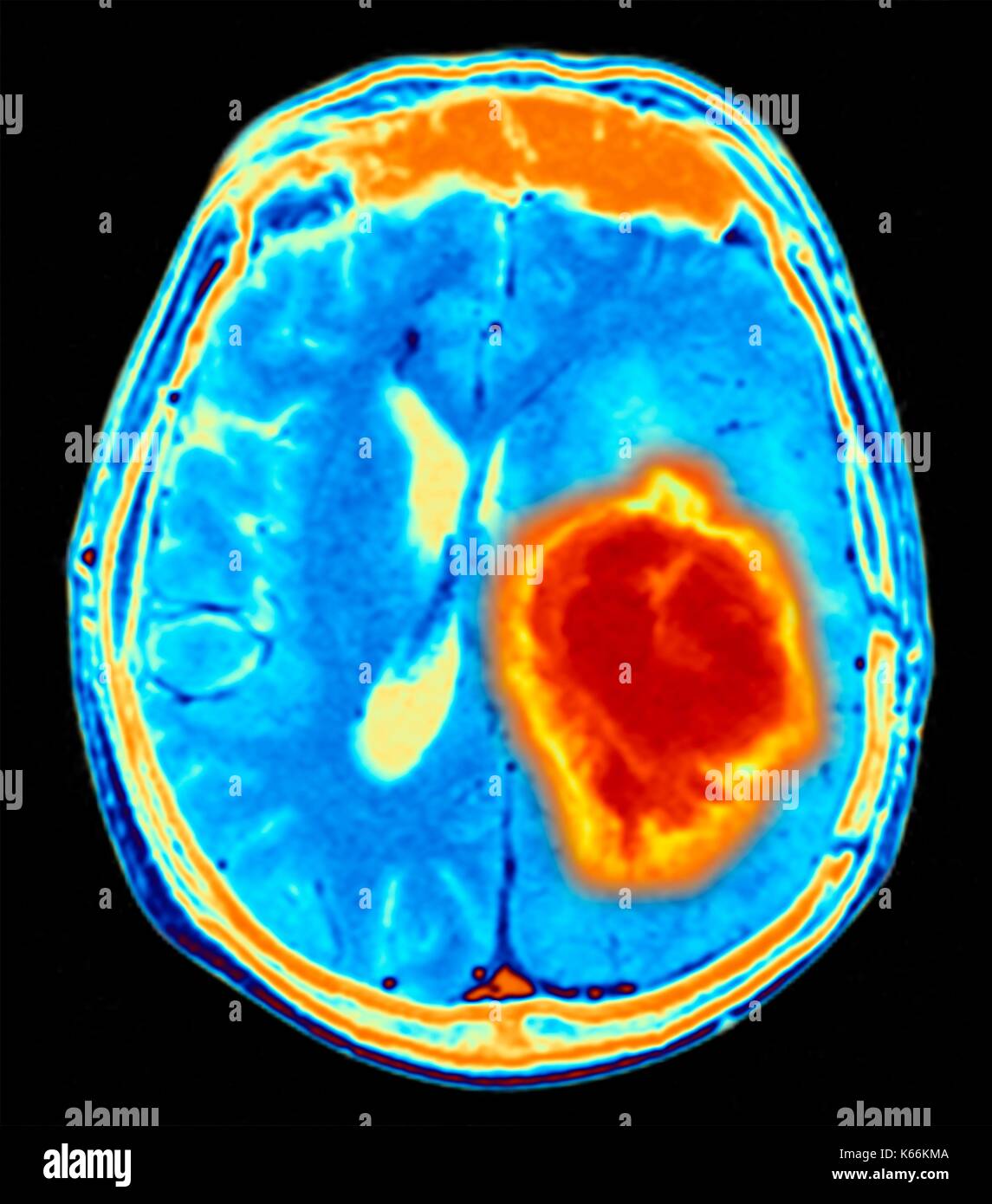 Brain tumour. Coloured Magnetic Resonance Imaging (MRI) scan of an axial section through the brain showing a metastatic tumour. At bottom left is the tumour (red-yellow) This tumour occurs within one cerebral hemisphere; the other hemisphere is at right. The eyeballs - not visible -are at top. Metastatic cancer is a secondary disease spread from cancer elsewhere in the body. Metastatic brain tumours are malignant. Typically they cause brain compression and nerve damage Stock Photohttps://www.alamy.com/image-license-details/?v=1https://www.alamy.com/brain-tumour-coloured-magnetic-resonance-imaging-mri-scan-of-an-axial-image158728426.html
RFK66KMA–Brain tumour. Coloured Magnetic Resonance Imaging (MRI) scan of an axial section through the brain showing a metastatic tumour. At bottom left is the tumour (red-yellow) This tumour occurs within one cerebral hemisphere; the other hemisphere is at right. The eyeballs - not visible -are at top. Metastatic cancer is a secondary disease spread from cancer elsewhere in the body. Metastatic brain tumours are malignant. Typically they cause brain compression and nerve damage
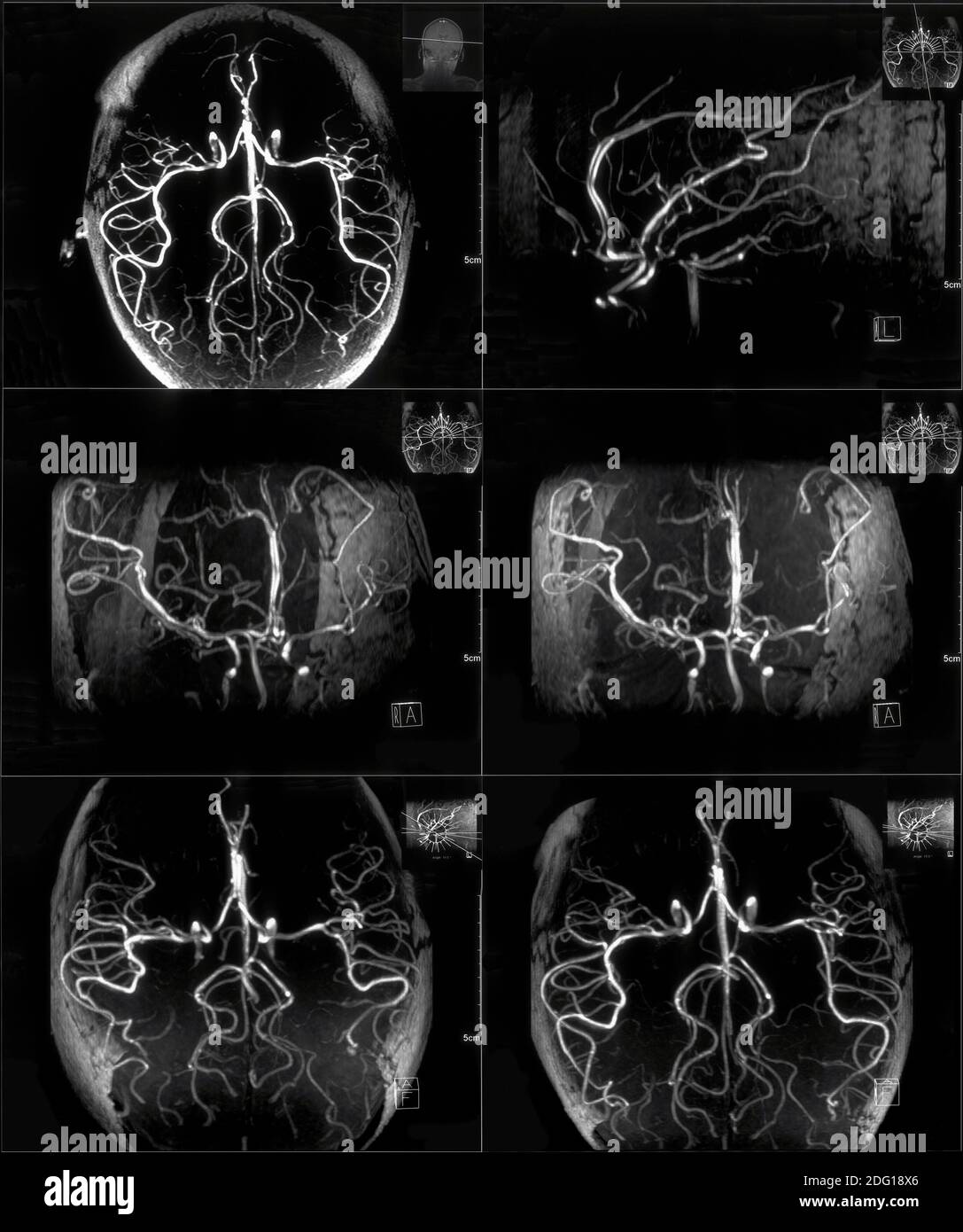 Contrast MRI scan Stock Photohttps://www.alamy.com/image-license-details/?v=1https://www.alamy.com/contrast-mri-scan-image388491550.html
RF2DG18X6–Contrast MRI scan
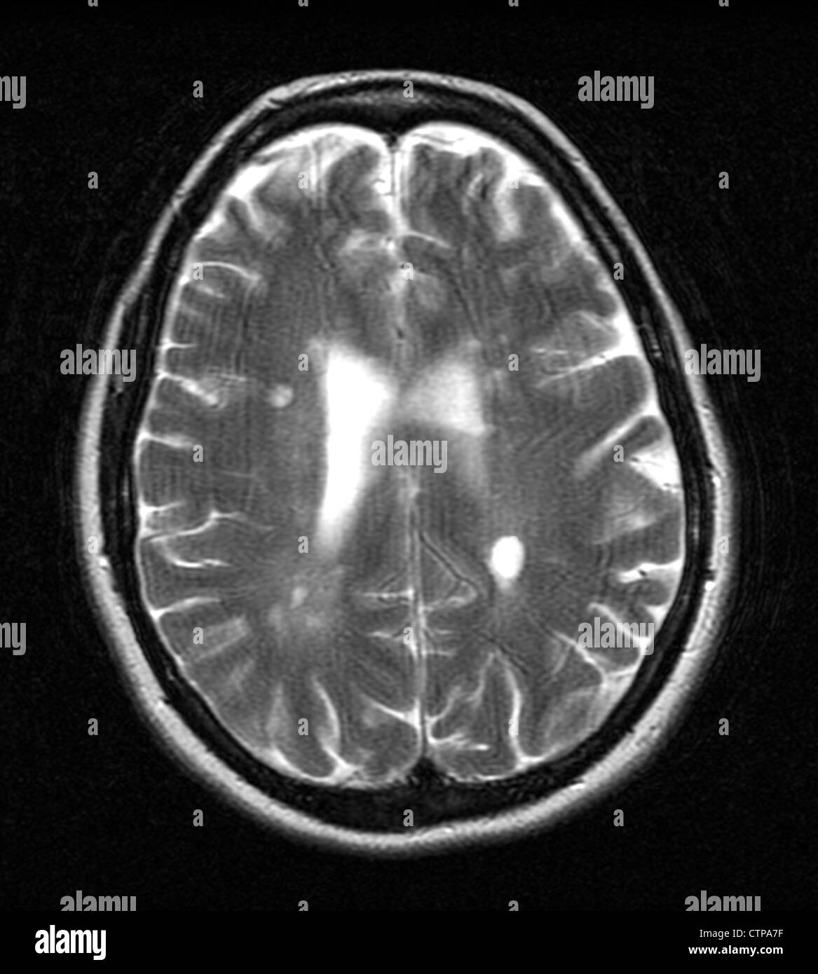 MRI showing multiple sclerosis in a 42 year old woman Stock Photohttps://www.alamy.com/image-license-details/?v=1https://www.alamy.com/stock-photo-mri-showing-multiple-sclerosis-in-a-42-year-old-woman-49663475.html
RMCTPA7F–MRI showing multiple sclerosis in a 42 year old woman
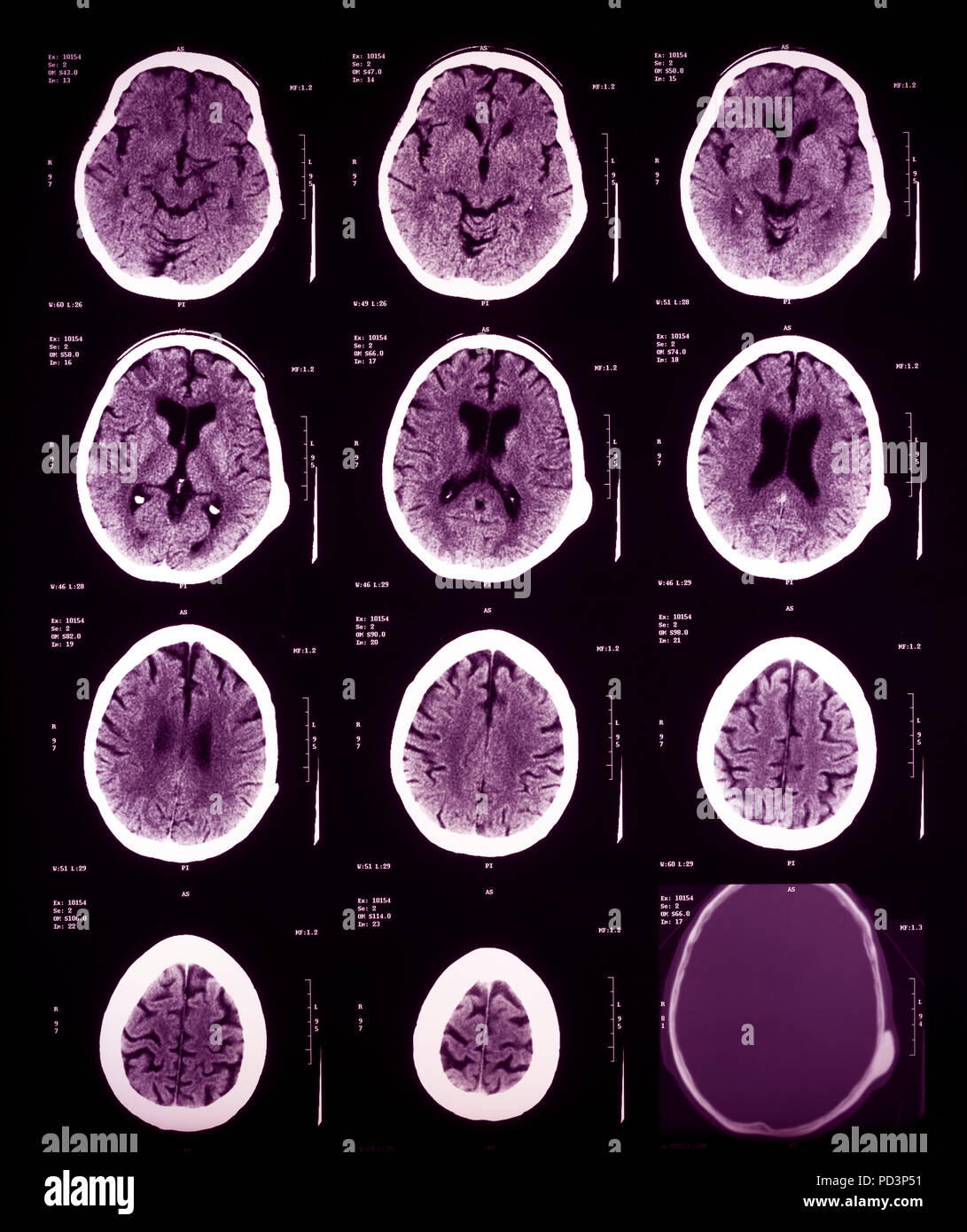 Sequence of horizontal sections of a female human brain, MRI scans, magnetic resonance imaging, Stock Photohttps://www.alamy.com/image-license-details/?v=1https://www.alamy.com/sequence-of-horizontal-sections-of-a-female-human-brain-mri-scans-magnetic-resonance-imaging-image214598189.html
RMPD3P51–Sequence of horizontal sections of a female human brain, MRI scans, magnetic resonance imaging,
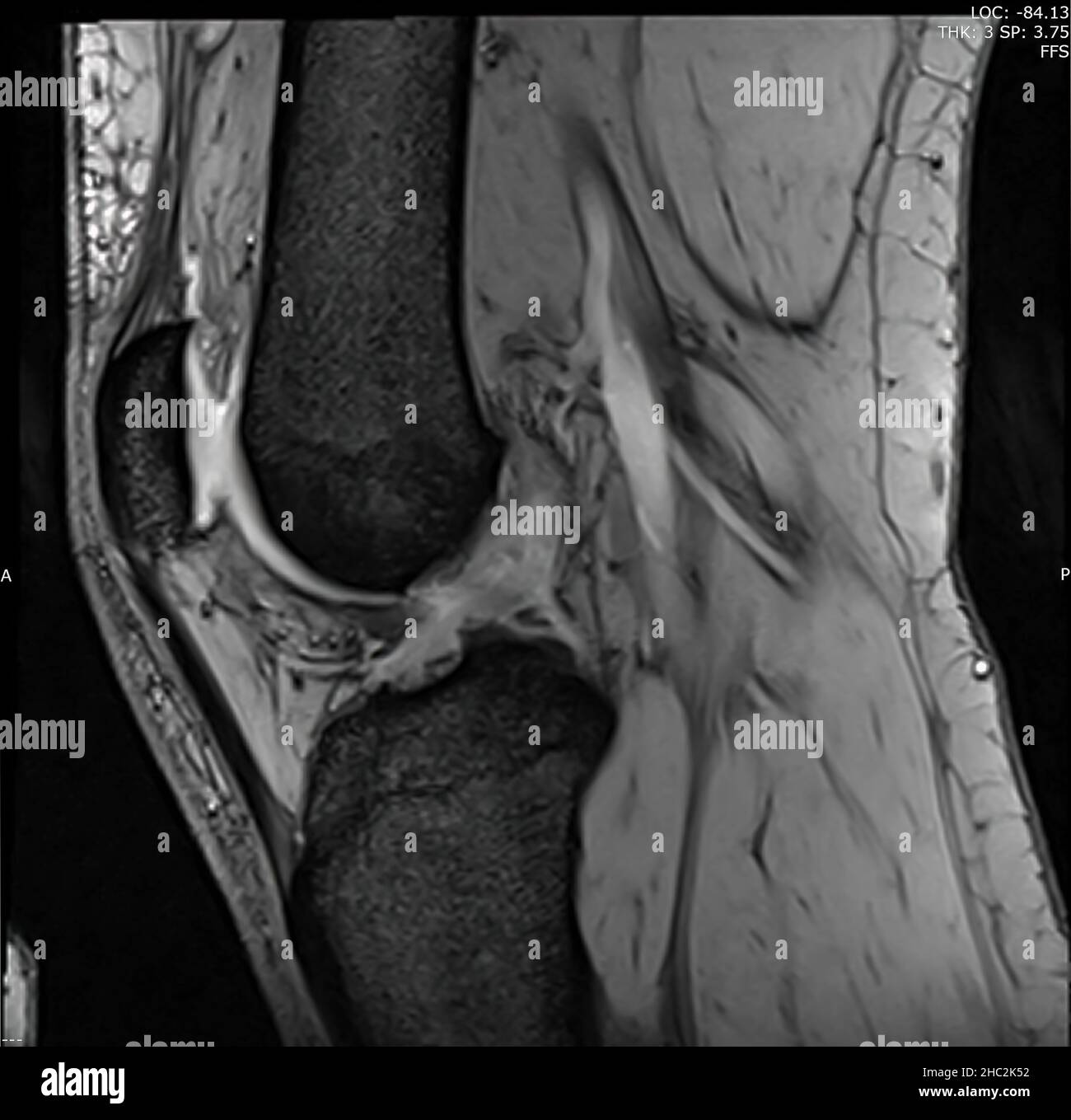 Magnetic resonance image of the knee joint (MRI knee)in sagittal plan showing complete Anterior cruciate ligament tear (ACL tear) Stock Photohttps://www.alamy.com/image-license-details/?v=1https://www.alamy.com/magnetic-resonance-image-of-the-knee-joint-mri-kneein-sagittal-plan-showing-complete-anterior-cruciate-ligament-tear-acl-tear-image454904382.html
RF2HC2K52–Magnetic resonance image of the knee joint (MRI knee)in sagittal plan showing complete Anterior cruciate ligament tear (ACL tear)
 Brain Tomography Stock Photohttps://www.alamy.com/image-license-details/?v=1https://www.alamy.com/stock-photo-brain-tomography-12412997.html
RFA9P23J–Brain Tomography
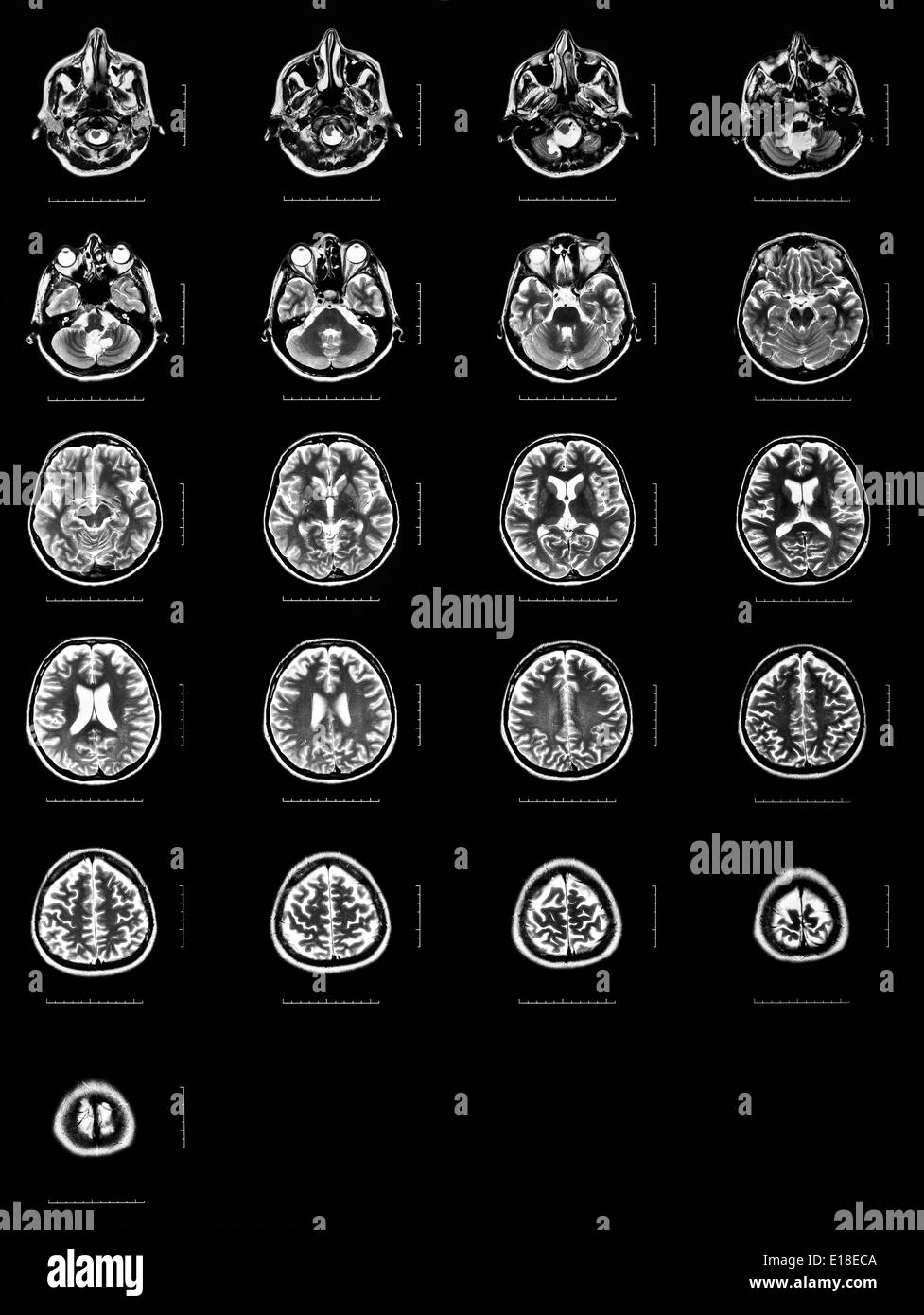 Sequence of horizontal sections of a human brain - MRI scan Stock Photohttps://www.alamy.com/image-license-details/?v=1https://www.alamy.com/sequence-of-horizontal-sections-of-a-human-brain-mri-scan-image69643066.html
RFE18ECA–Sequence of horizontal sections of a human brain - MRI scan
 Ride through the human abdomen and chest by means of 18 MRI cuts (coronal view). Picture 12/18 Stock Photohttps://www.alamy.com/image-license-details/?v=1https://www.alamy.com/ride-through-the-human-abdomen-and-chest-by-means-of-18-mri-cuts-coronal-view-picture-1218-image179043666.html
RFMB840J–Ride through the human abdomen and chest by means of 18 MRI cuts (coronal view). Picture 12/18
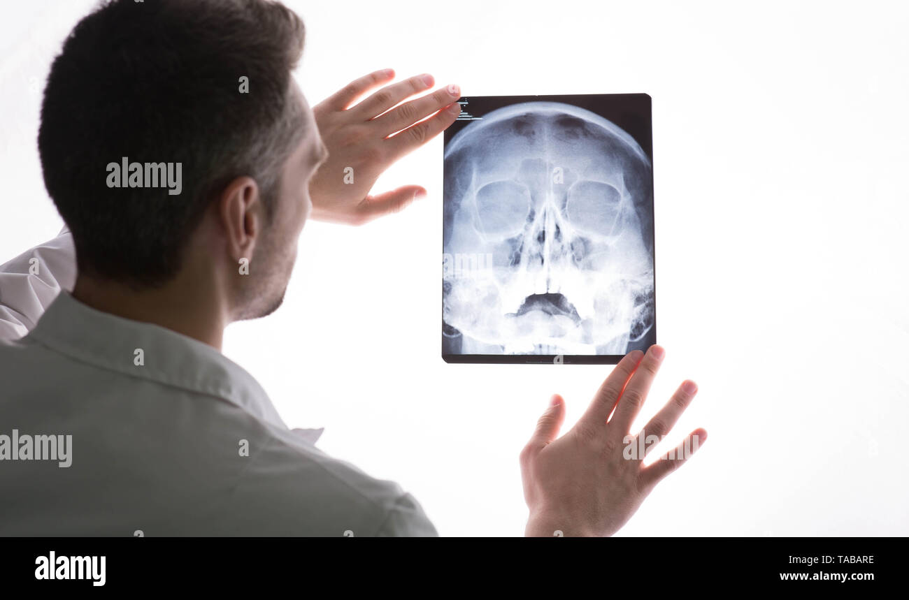 doctor examining patients x-ray and MRI scans. Human Head Sinusitis Stock Photohttps://www.alamy.com/image-license-details/?v=1https://www.alamy.com/doctor-examining-patients-x-ray-and-mri-scans-human-head-sinusitis-image247341682.html
RFTABARE–doctor examining patients x-ray and MRI scans. Human Head Sinusitis
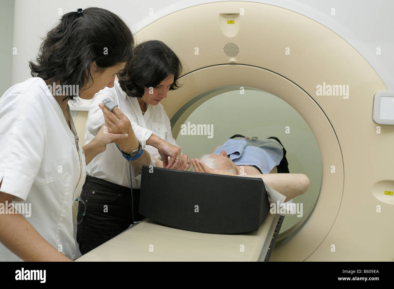 Radiologist and nurse getting a patient ready for a full MRI scan Stock Photohttps://www.alamy.com/image-license-details/?v=1https://www.alamy.com/stock-photo-radiologist-and-nurse-getting-a-patient-ready-for-a-full-mri-scan-20905762.html
RMB609EA–Radiologist and nurse getting a patient ready for a full MRI scan
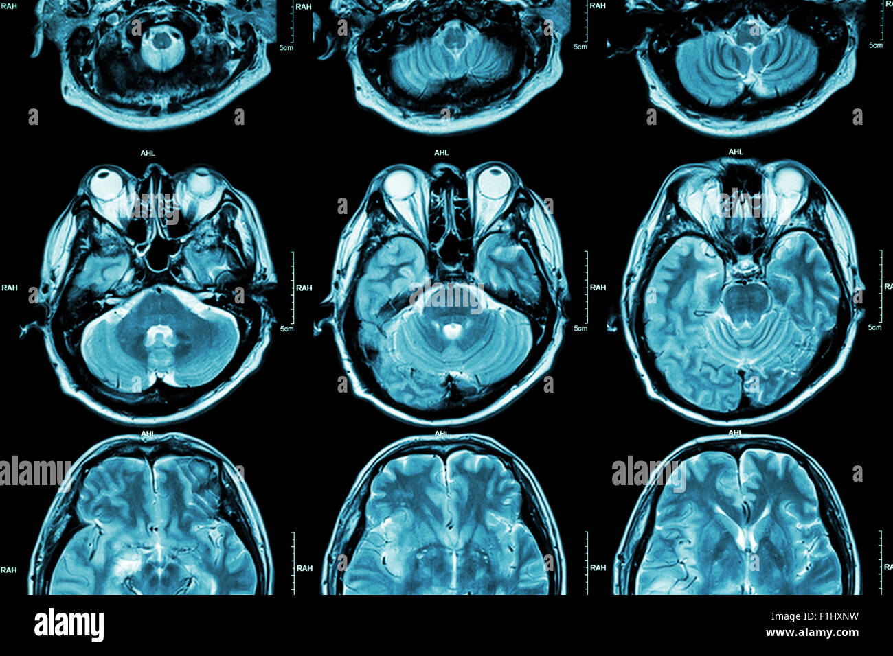 MRI of Brain ( cross section of brain ) ( Medical , Health care , Science background ) Stock Photohttps://www.alamy.com/image-license-details/?v=1https://www.alamy.com/stock-photo-mri-of-brain-cross-section-of-brain-medical-health-care-science-background-87060677.html
RFF1HXNW–MRI of Brain ( cross section of brain ) ( Medical , Health care , Science background )
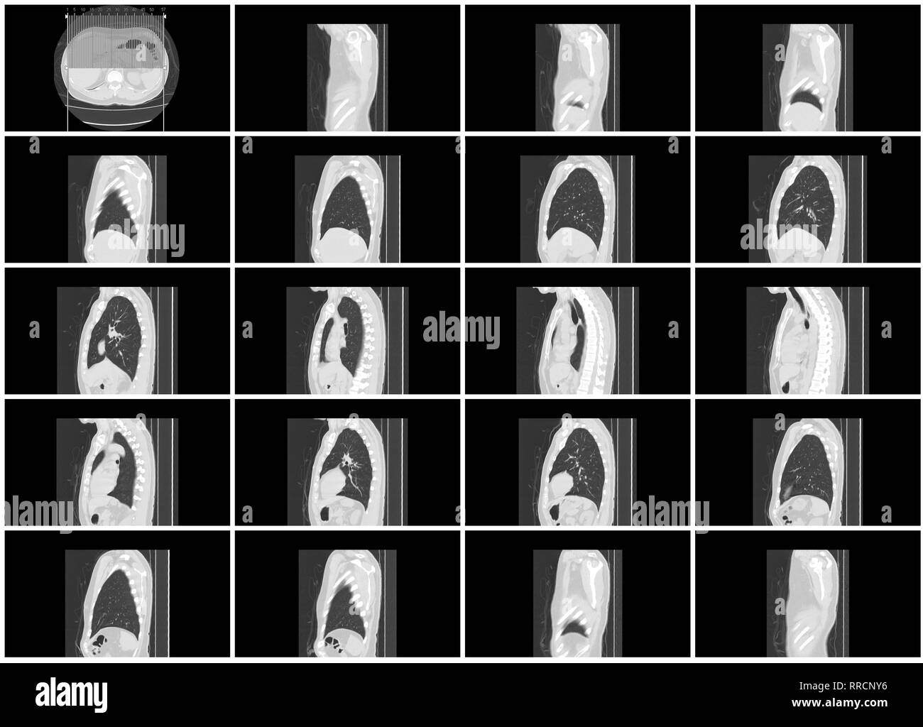 ct scan step set of body lung sagittal view Stock Photohttps://www.alamy.com/image-license-details/?v=1https://www.alamy.com/ct-scan-step-set-of-body-lung-sagittal-view-image238152522.html
RFRRCNY6–ct scan step set of body lung sagittal view
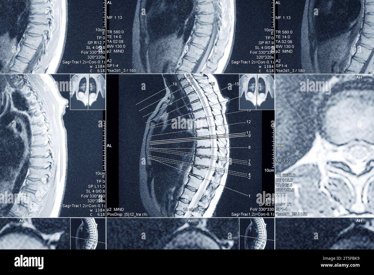 MRI scan of the thoracic spine for diagnosis. Medical examination for the prevention of diseases. Stock Photohttps://www.alamy.com/image-license-details/?v=1https://www.alamy.com/mri-scan-of-the-thoracic-spine-for-diagnosis-medical-examination-for-the-prevention-of-diseases-image571353869.html
RF2T5FBK9–MRI scan of the thoracic spine for diagnosis. Medical examination for the prevention of diseases.
 Doctor points to a section on an MRI scan Stock Photohttps://www.alamy.com/image-license-details/?v=1https://www.alamy.com/stock-photo-doctor-points-to-a-section-on-an-mri-scan-27301597.html
RFBGBKD1–Doctor points to a section on an MRI scan
 Covid-19 scan body xray in male doctor hand wearing gloves closeup shot Stock Photohttps://www.alamy.com/image-license-details/?v=1https://www.alamy.com/covid-19-scan-body-xray-in-male-doctor-hand-wearing-gloves-closeup-shot-image424612611.html
RF2FJPNM3–Covid-19 scan body xray in male doctor hand wearing gloves closeup shot
 Mri scanner examination vector illustration. Cartoon flat medical scan hospital lab room with Mri machine, doctor characters exam patient, discuss diagnosis and treatment. Medicine research background Stock Vectorhttps://www.alamy.com/image-license-details/?v=1https://www.alamy.com/mri-scanner-examination-vector-illustration-cartoon-flat-medical-scan-hospital-lab-room-with-mri-machine-doctor-characters-exam-patient-discuss-diagnosis-and-treatment-medicine-research-background-image368396670.html
RF2CB9WKX–Mri scanner examination vector illustration. Cartoon flat medical scan hospital lab room with Mri machine, doctor characters exam patient, discuss diagnosis and treatment. Medicine research background
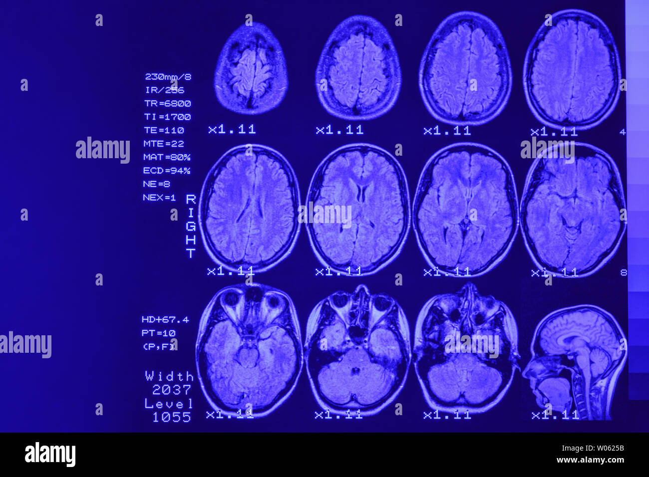 MRI scan or magnetic resonance image of head and brain scan. The result is an MRI of the brain with values and numbers with blue backlight. Stock Photohttps://www.alamy.com/image-license-details/?v=1https://www.alamy.com/mri-scan-or-magnetic-resonance-image-of-head-and-brain-scan-the-result-is-an-mri-of-the-brain-with-values-and-numbers-with-blue-backlight-image258288951.html
RFW0625B–MRI scan or magnetic resonance image of head and brain scan. The result is an MRI of the brain with values and numbers with blue backlight.
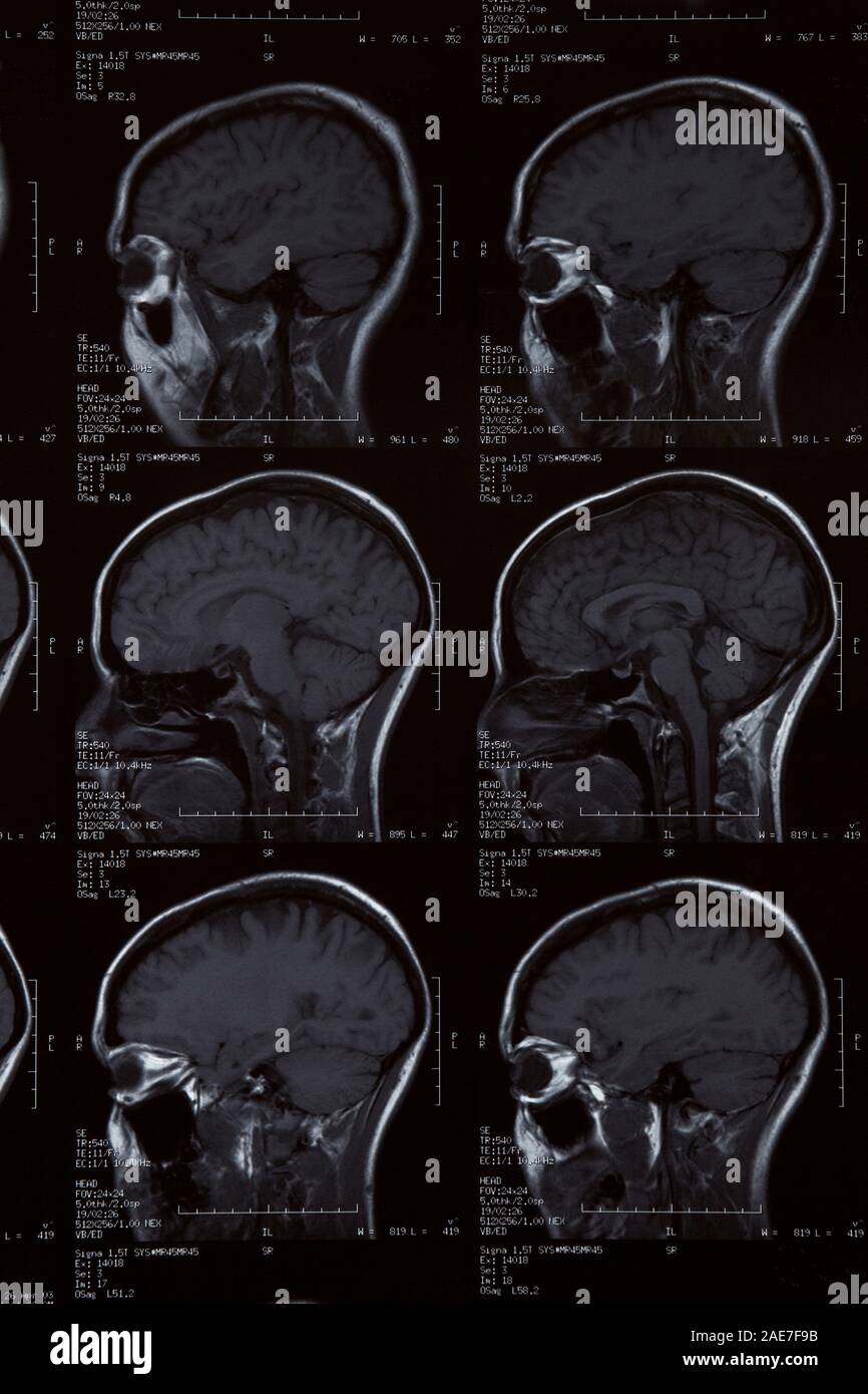 MRI Scan of female human brain Stock Photohttps://www.alamy.com/image-license-details/?v=1https://www.alamy.com/mri-scan-of-female-human-brain-image335767863.html
RM2AE7F9B–MRI Scan of female human brain
 Head and neck MRI scan, private info deleted, shallow focus Stock Photohttps://www.alamy.com/image-license-details/?v=1https://www.alamy.com/head-and-neck-mri-scan-private-info-deleted-shallow-focus-image243233439.html
RFT3M6MF–Head and neck MRI scan, private info deleted, shallow focus
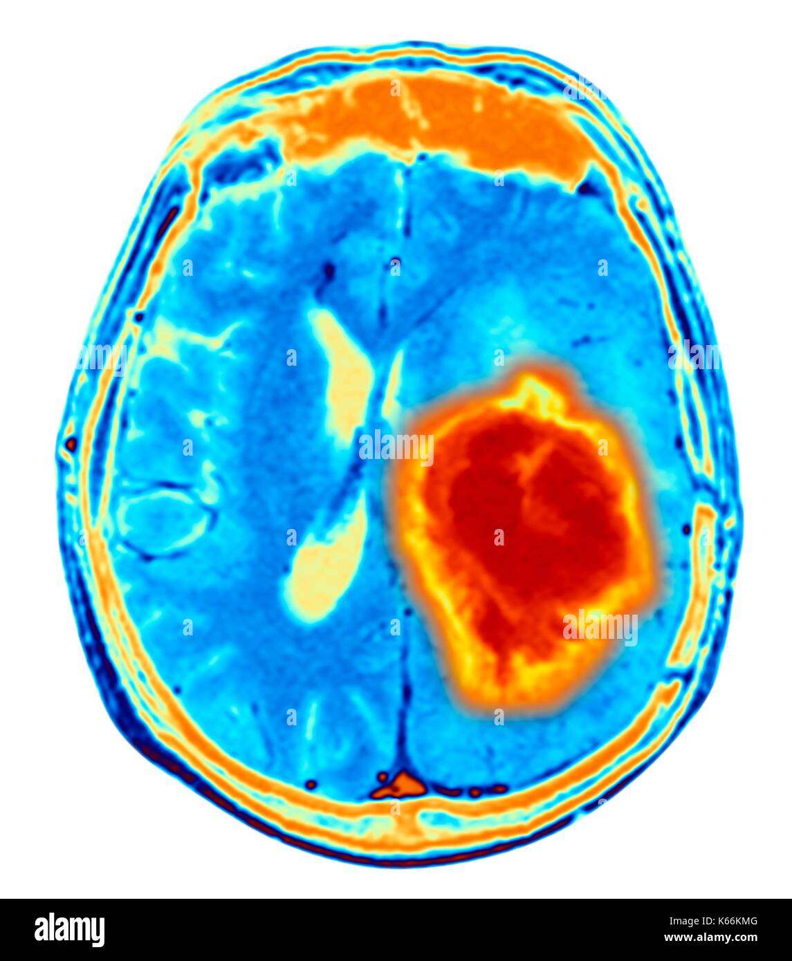 Brain tumour. Coloured Magnetic Resonance Imaging (MRI) scan of an axial section through the brain showing a metastatic tumour. At bottom left is the tumour (red-yellow) This tumour occurs within one cerebral hemisphere; the other hemisphere is at right. The eyeballs - not visible -are at top. Metastatic cancer is a secondary disease spread from cancer elsewhere in the body. Metastatic brain tumours are malignant. Typically they cause brain compression and nerve damage Stock Photohttps://www.alamy.com/image-license-details/?v=1https://www.alamy.com/brain-tumour-coloured-magnetic-resonance-imaging-mri-scan-of-an-axial-image158728432.html
RFK66KMG–Brain tumour. Coloured Magnetic Resonance Imaging (MRI) scan of an axial section through the brain showing a metastatic tumour. At bottom left is the tumour (red-yellow) This tumour occurs within one cerebral hemisphere; the other hemisphere is at right. The eyeballs - not visible -are at top. Metastatic cancer is a secondary disease spread from cancer elsewhere in the body. Metastatic brain tumours are malignant. Typically they cause brain compression and nerve damage
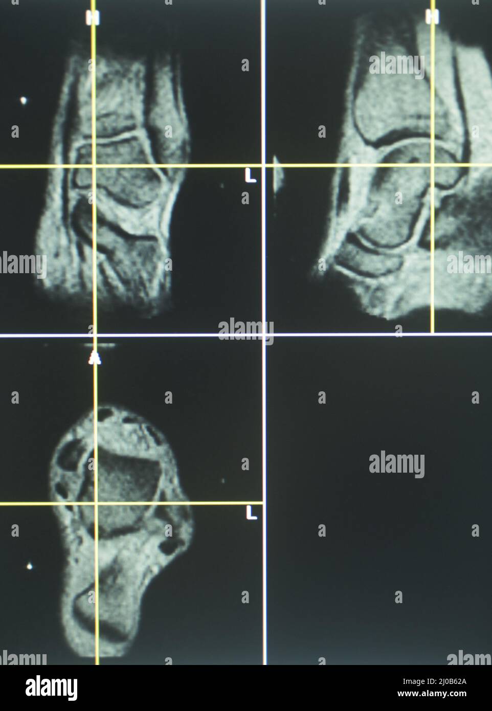 Human knee open MRI CT scan Stock Photohttps://www.alamy.com/image-license-details/?v=1https://www.alamy.com/human-knee-open-mri-ct-scan-image464926178.html
RF2J0B62A–Human knee open MRI CT scan
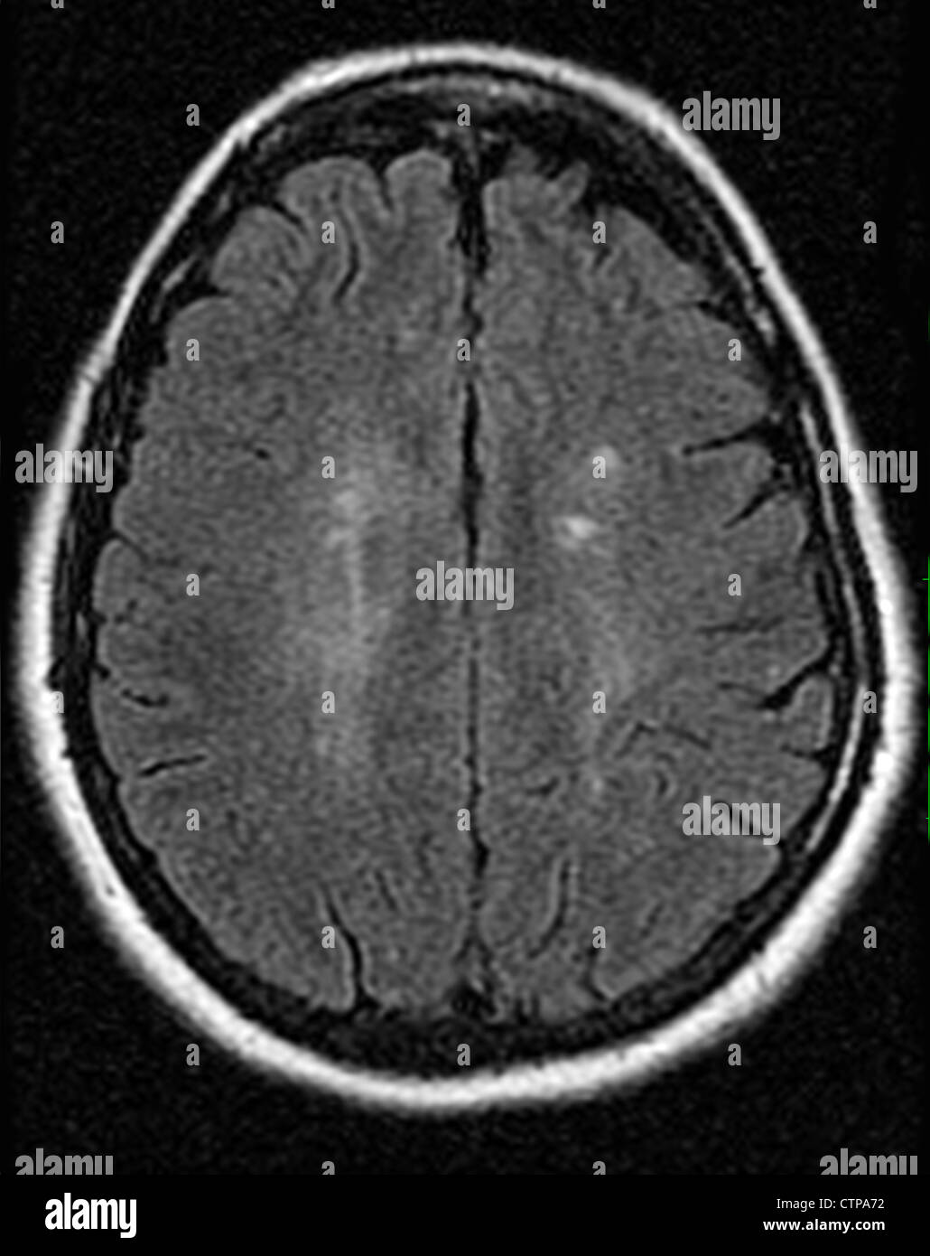 MRI showing multiple sclerosis in a 42 year old woman Stock Photohttps://www.alamy.com/image-license-details/?v=1https://www.alamy.com/stock-photo-mri-showing-multiple-sclerosis-in-a-42-year-old-woman-49663462.html
RMCTPA72–MRI showing multiple sclerosis in a 42 year old woman
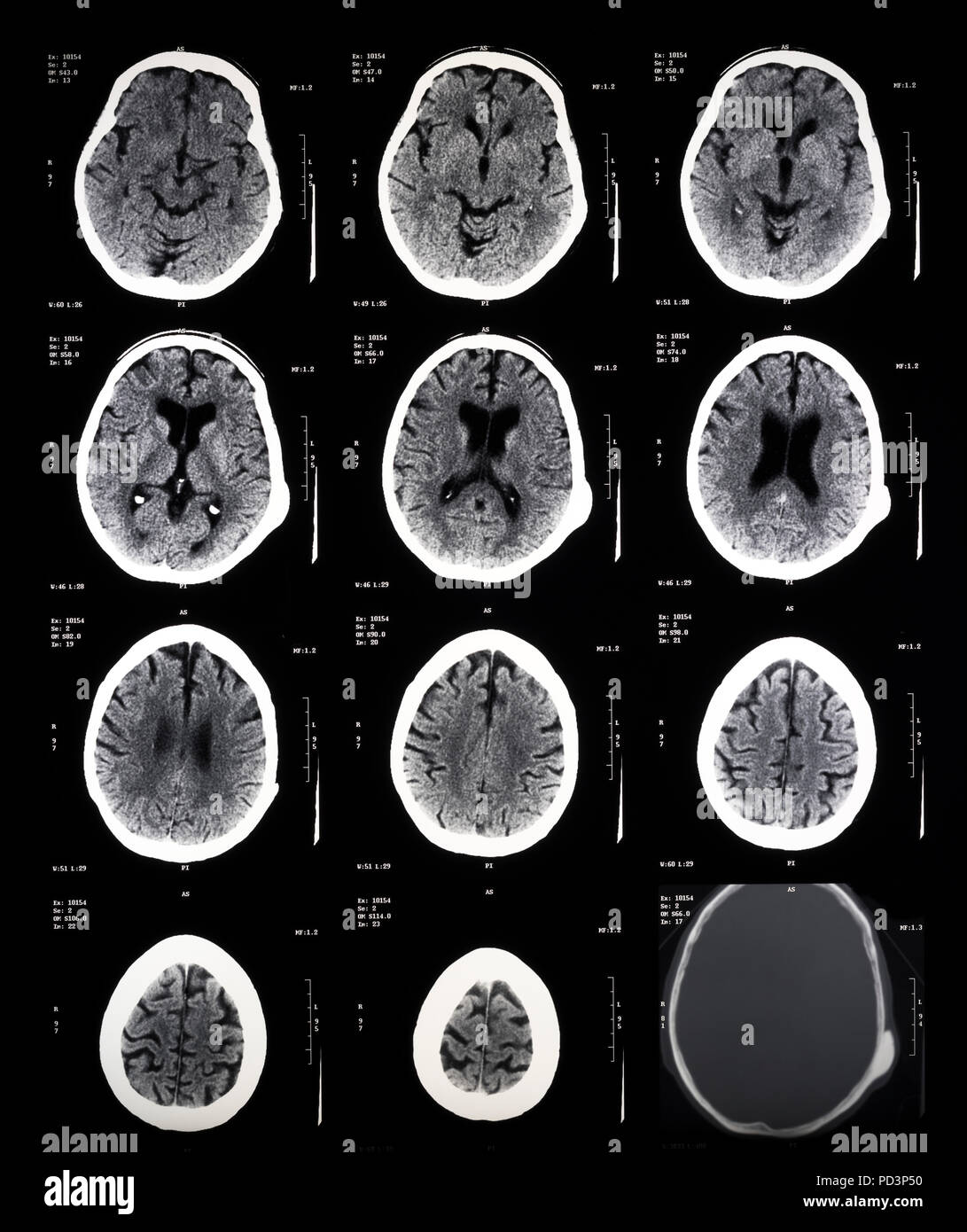 Sequence of horizontal sections of a female human brain, MRI scans, magnetic resonance imaging, Stock Photohttps://www.alamy.com/image-license-details/?v=1https://www.alamy.com/sequence-of-horizontal-sections-of-a-female-human-brain-mri-scans-magnetic-resonance-imaging-image214598188.html
RMPD3P50–Sequence of horizontal sections of a female human brain, MRI scans, magnetic resonance imaging,
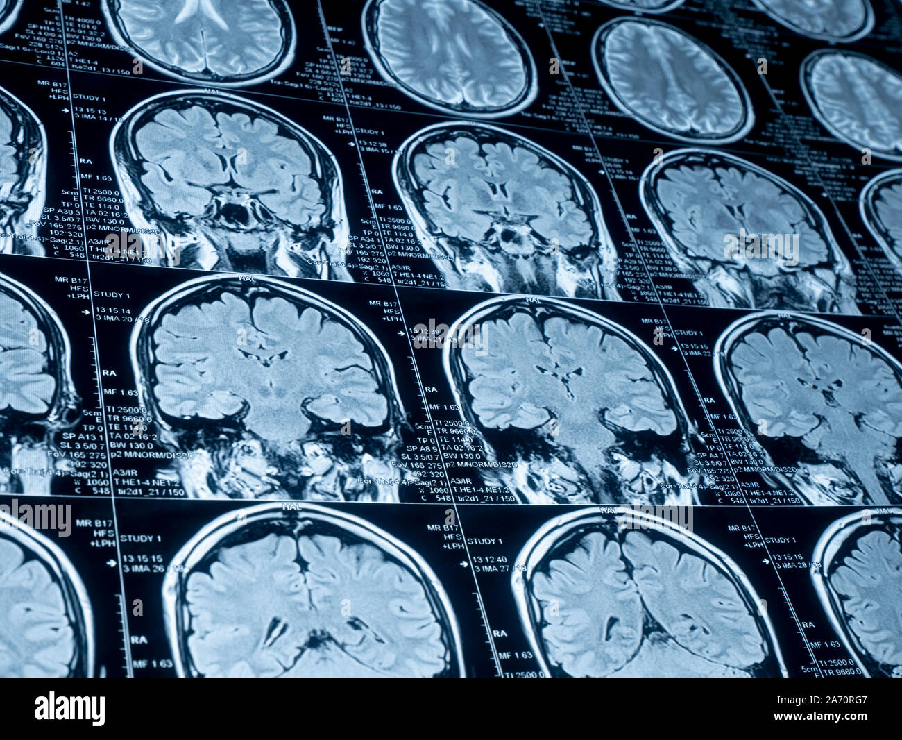 Magnetic resonance image (MRI) of the brain Stock Photohttps://www.alamy.com/image-license-details/?v=1https://www.alamy.com/magnetic-resonance-image-mri-of-the-brain-image331318071.html
RF2A70RG7–Magnetic resonance image (MRI) of the brain
 Mature Patient With Health Problems And Doctor In Medical Center Stock Photohttps://www.alamy.com/image-license-details/?v=1https://www.alamy.com/mature-patient-with-health-problems-and-doctor-in-medical-center-image245080072.html
RFT6MA3M–Mature Patient With Health Problems And Doctor In Medical Center
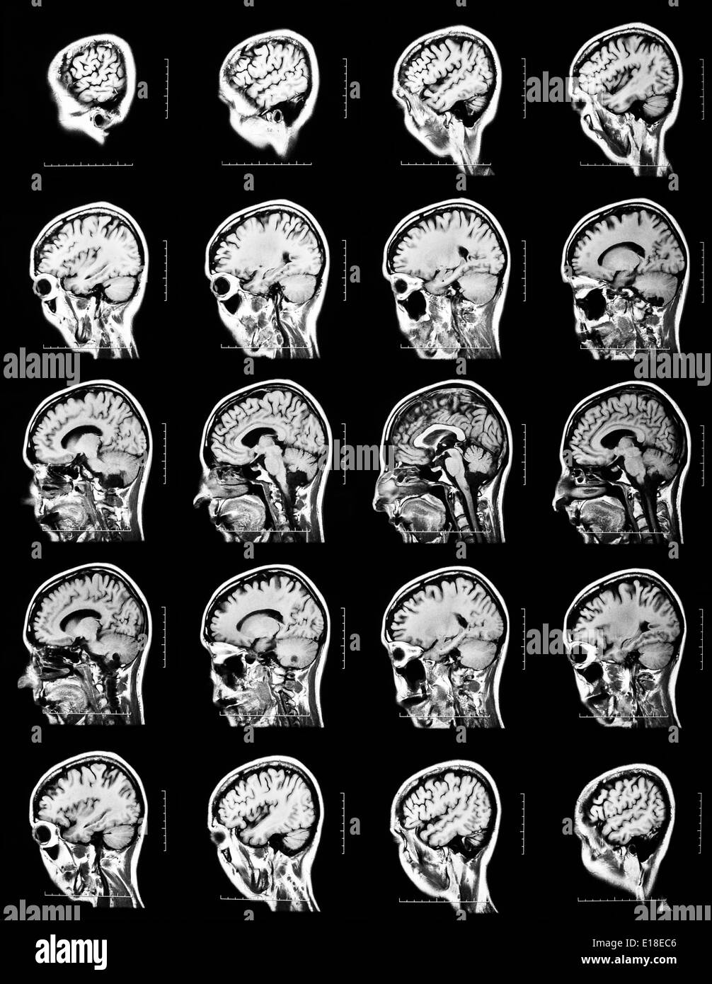 Sequence of vertical sections of a human brain - MRI scan Stock Photohttps://www.alamy.com/image-license-details/?v=1https://www.alamy.com/sequence-of-vertical-sections-of-a-human-brain-mri-scan-image69643062.html
RFE18EC6–Sequence of vertical sections of a human brain - MRI scan
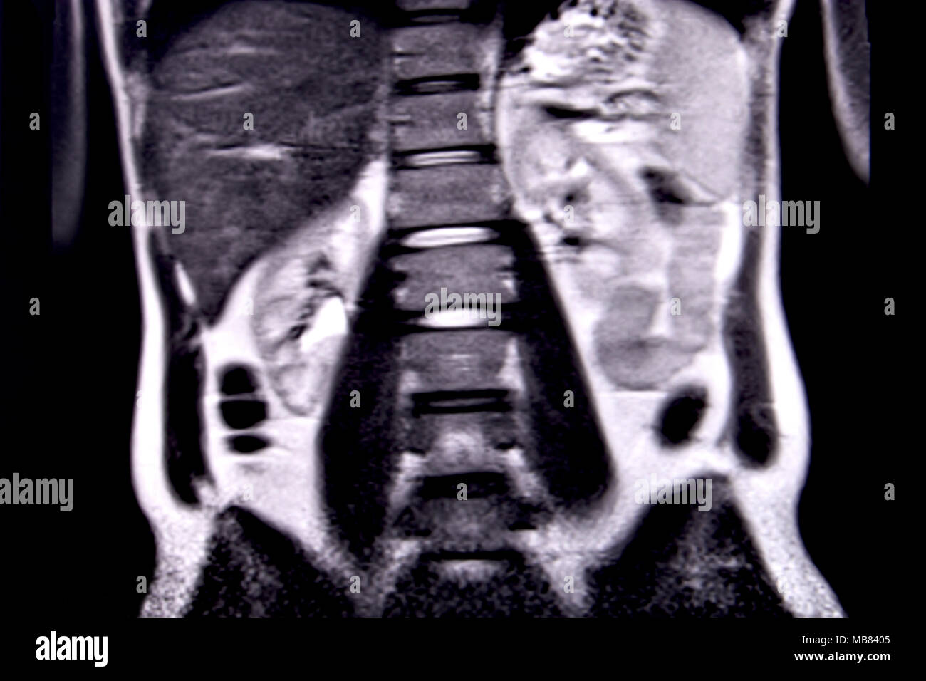 Ride through the human abdomen and chest by means of 18 MRI cuts (coronal view). Picture 9/18 Stock Photohttps://www.alamy.com/image-license-details/?v=1https://www.alamy.com/ride-through-the-human-abdomen-and-chest-by-means-of-18-mri-cuts-coronal-view-picture-918-image179043653.html
RFMB8405–Ride through the human abdomen and chest by means of 18 MRI cuts (coronal view). Picture 9/18
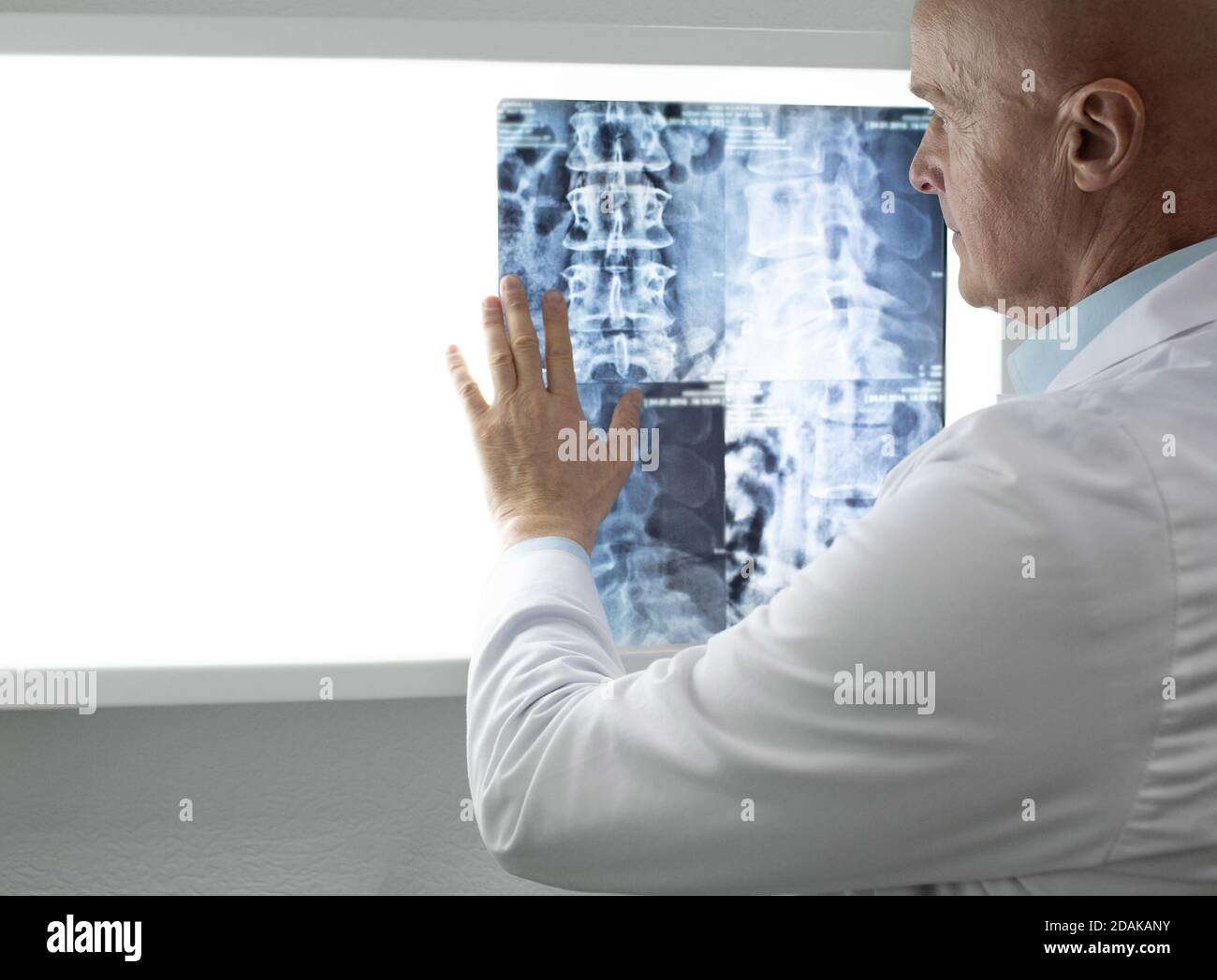 doctor examining patients Spine x-ray and MRI scans. Diagnosis of diseases of the spine, injuries of the back and cervical spine Stock Photohttps://www.alamy.com/image-license-details/?v=1https://www.alamy.com/doctor-examining-patients-spine-x-ray-and-mri-scans-diagnosis-of-diseases-of-the-spine-injuries-of-the-back-and-cervical-spine-image385200199.html
RF2DAKANY–doctor examining patients Spine x-ray and MRI scans. Diagnosis of diseases of the spine, injuries of the back and cervical spine
 Radiologist examines MRI scanned image of a human heart Stock Photohttps://www.alamy.com/image-license-details/?v=1https://www.alamy.com/stock-photo-radiologist-examines-mri-scanned-image-of-a-human-heart-49783522.html
RFCTYRAX–Radiologist examines MRI scanned image of a human heart
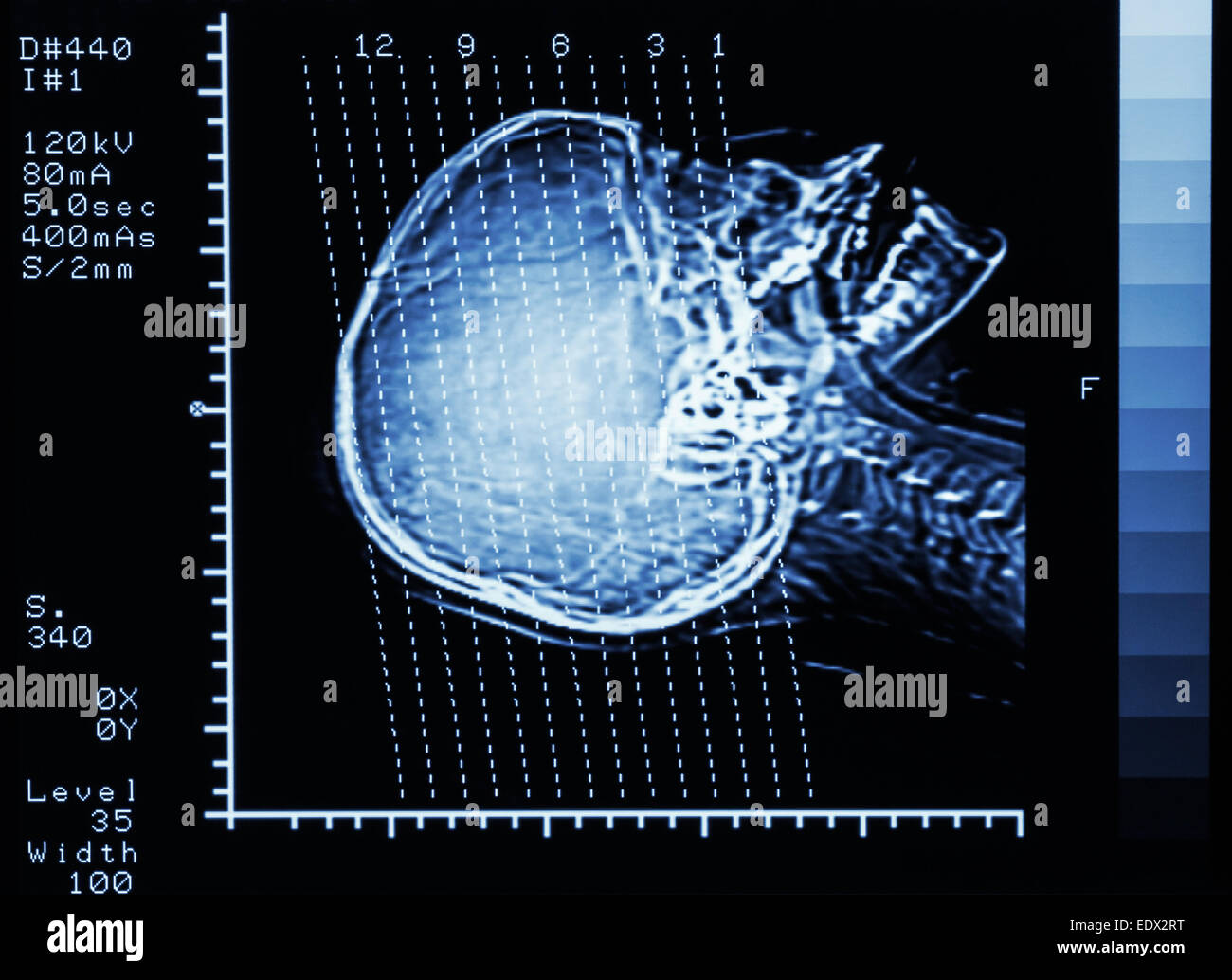 film CT scan (Computed tomography) brain : show section line on skull Stock Photohttps://www.alamy.com/image-license-details/?v=1https://www.alamy.com/stock-photo-film-ct-scan-computed-tomography-brain-show-section-line-on-skull-77404988.html
RFEDX2RT–film CT scan (Computed tomography) brain : show section line on skull
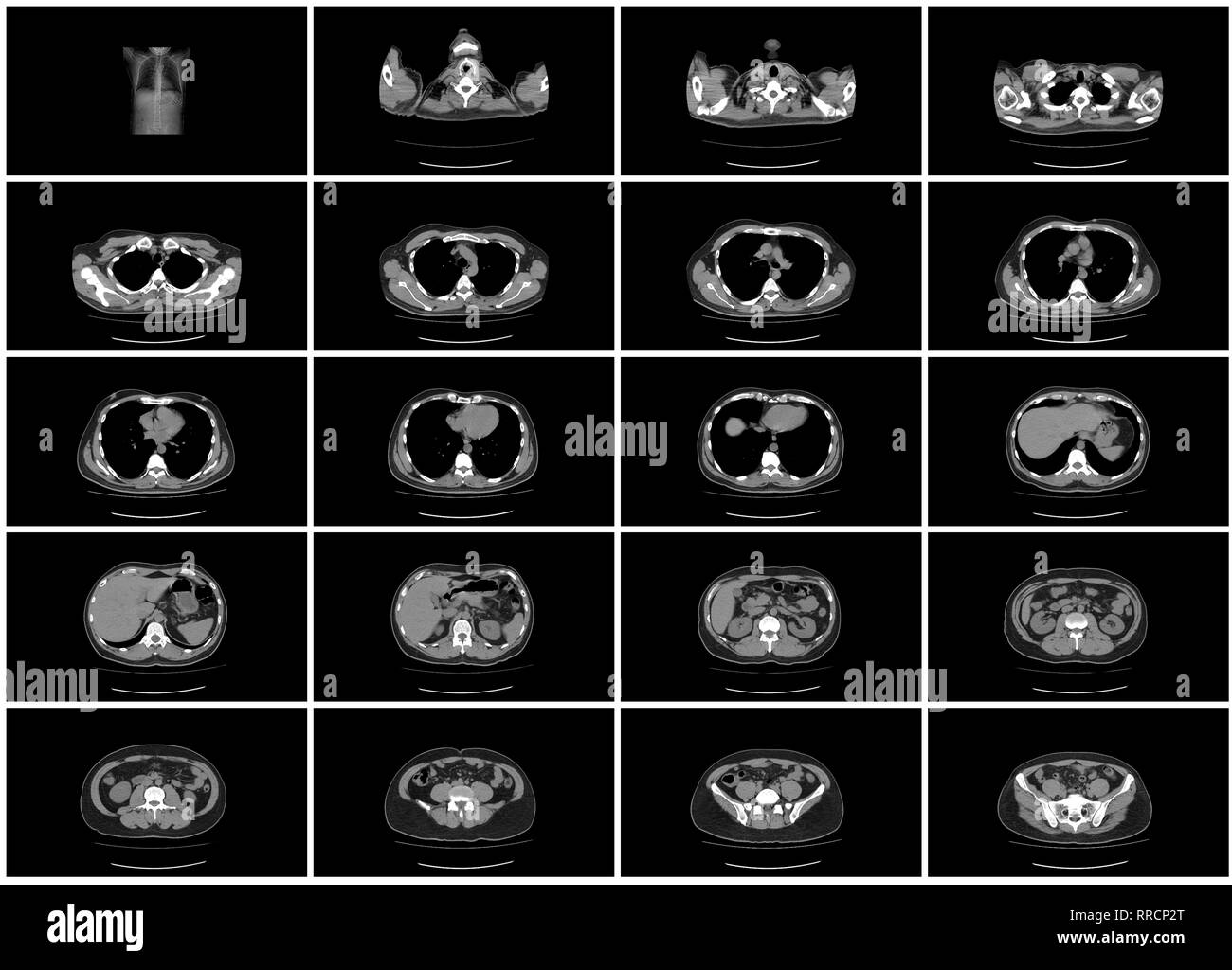 ct scan step set of upper body abdomen top view Stock Photohttps://www.alamy.com/image-license-details/?v=1https://www.alamy.com/ct-scan-step-set-of-upper-body-abdomen-top-view-image238152624.html
RFRRCP2T–ct scan step set of upper body abdomen top view
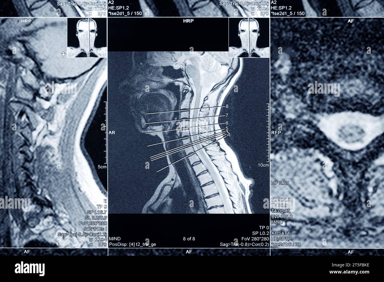 MRI scan of the cervical spine for diagnosis. Medical examination for the prevention of diseases. Stock Photohttps://www.alamy.com/image-license-details/?v=1https://www.alamy.com/mri-scan-of-the-cervical-spine-for-diagnosis-medical-examination-for-the-prevention-of-diseases-image571353874.html
RF2T5FBKE–MRI scan of the cervical spine for diagnosis. Medical examination for the prevention of diseases.
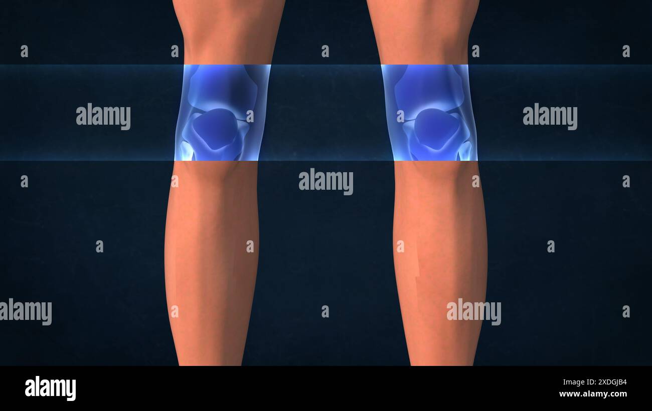 An X-ray of a human knees Stock Photohttps://www.alamy.com/image-license-details/?v=1https://www.alamy.com/an-x-ray-of-a-human-knees-image610719064.html
RF2XDGJB4–An X-ray of a human knees
 Healthcare Technology and Medical Scan of a Body Diagnosis Stock Photohttps://www.alamy.com/image-license-details/?v=1https://www.alamy.com/stock-photo-healthcare-technology-and-medical-scan-of-a-body-diagnosis-99663226.html
RFFP41CX–Healthcare Technology and Medical Scan of a Body Diagnosis
 Magnetic resonance imaging of spine with spine model and copy space Stock Photohttps://www.alamy.com/image-license-details/?v=1https://www.alamy.com/magnetic-resonance-imaging-of-spine-with-spine-model-and-copy-space-image521541142.html
RF2N8E71A–Magnetic resonance imaging of spine with spine model and copy space
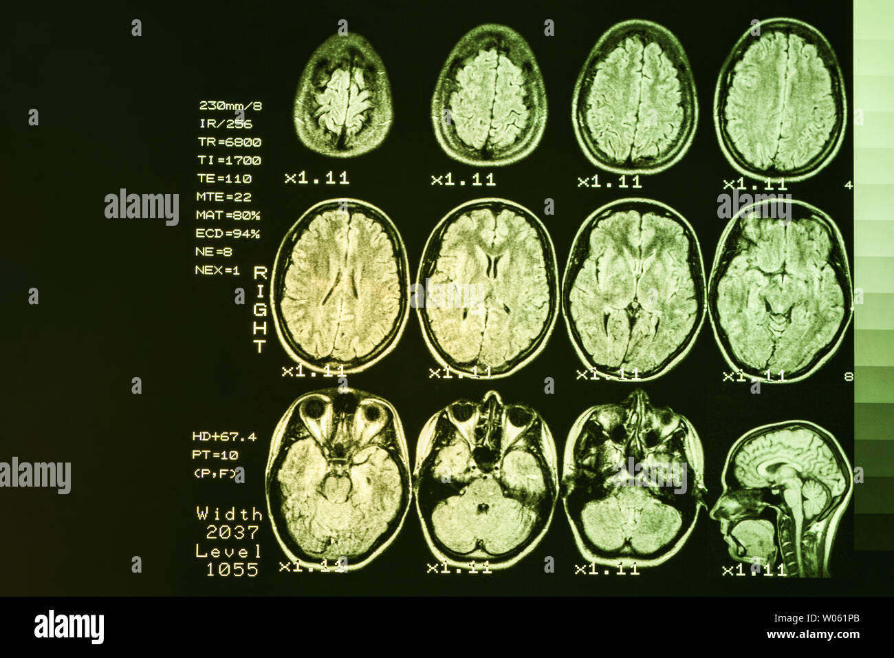 MRI scan or magnetic resonance image of head and brain scan. The result is an MRI of the brain with values and numbers with yellow backlight. Stock Photohttps://www.alamy.com/image-license-details/?v=1https://www.alamy.com/mri-scan-or-magnetic-resonance-image-of-head-and-brain-scan-the-result-is-an-mri-of-the-brain-with-values-and-numbers-with-yellow-backlight-image258288643.html
RFW061PB–MRI scan or magnetic resonance image of head and brain scan. The result is an MRI of the brain with values and numbers with yellow backlight.
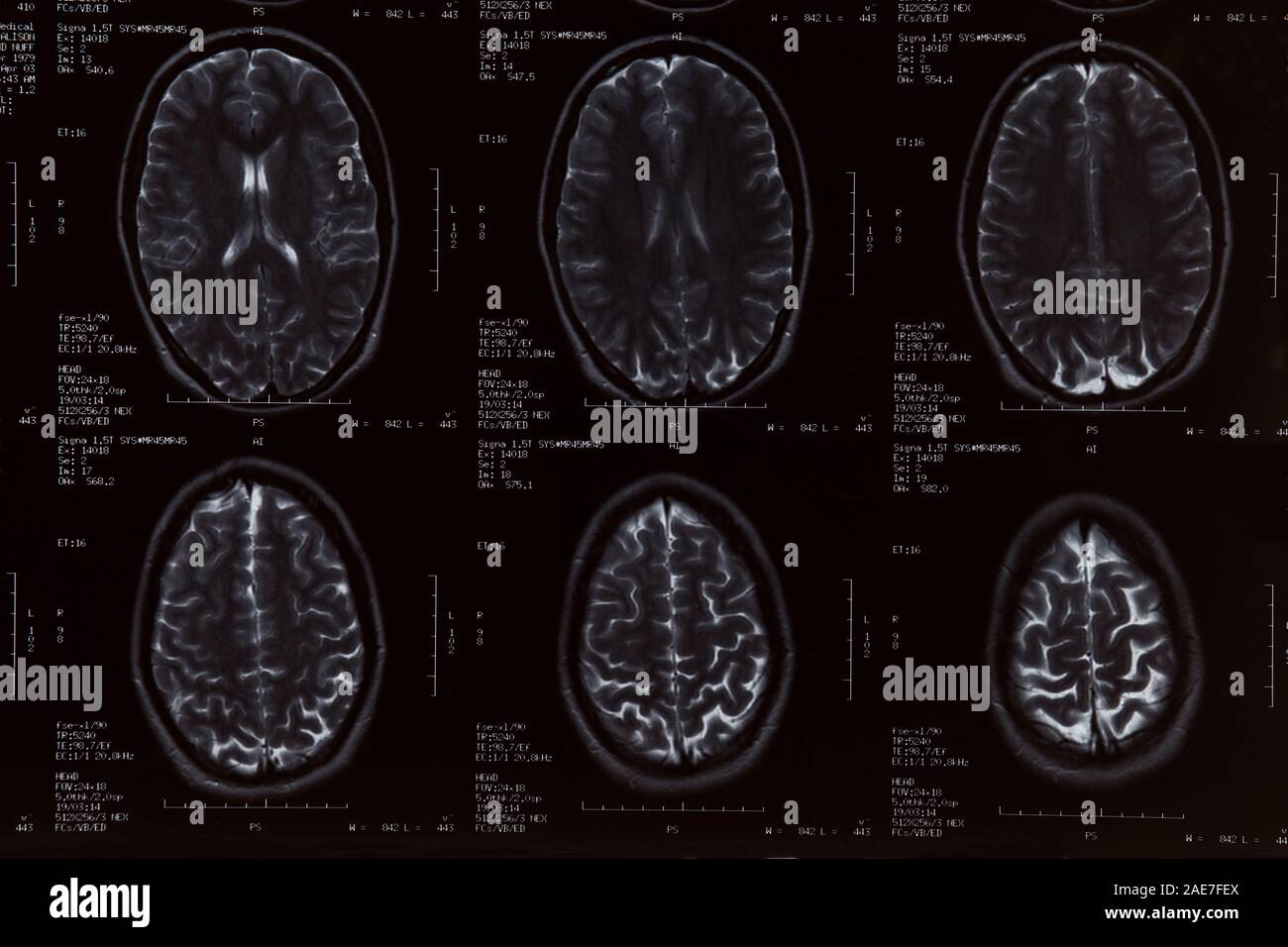 MRI Scan close up of the female brain Stock Photohttps://www.alamy.com/image-license-details/?v=1https://www.alamy.com/mri-scan-close-up-of-the-female-brain-image335768018.html
RM2AE7FEX–MRI Scan close up of the female brain
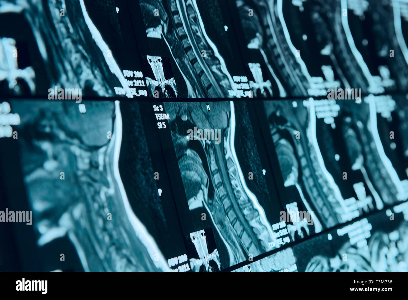 Head and neck MRI scan, anonymized, shallow focus depth Stock Photohttps://www.alamy.com/image-license-details/?v=1https://www.alamy.com/head-and-neck-mri-scan-anonymized-shallow-focus-depth-image243233738.html
RFT3M736–Head and neck MRI scan, anonymized, shallow focus depth
 Healthy head, MRI scan Stock Photohttps://www.alamy.com/image-license-details/?v=1https://www.alamy.com/healthy-head-mri-scan-image478710252.html
RF2JPR3PM–Healthy head, MRI scan
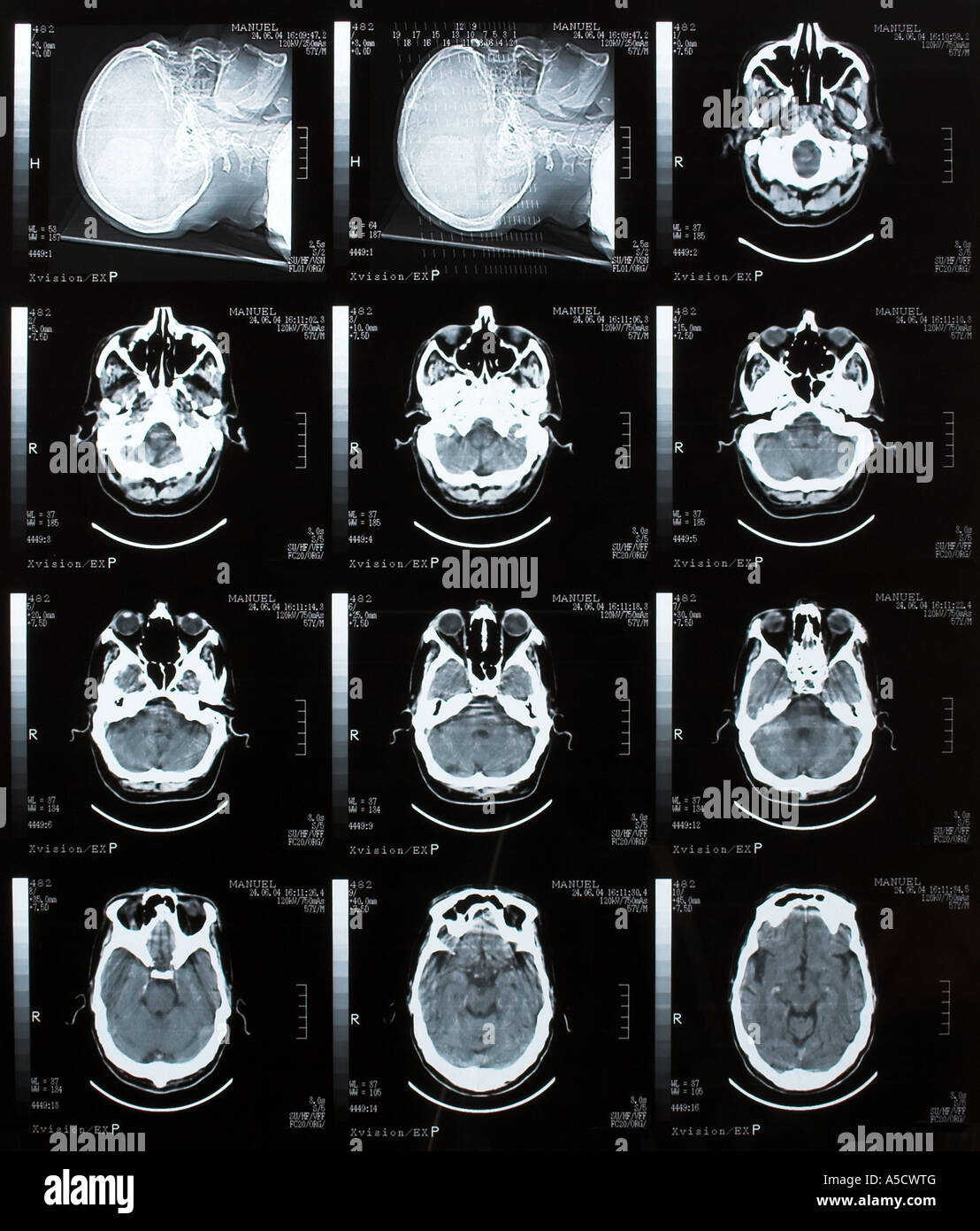 Brain Tomography Stock Photohttps://www.alamy.com/image-license-details/?v=1https://www.alamy.com/stock-photo-brain-tomography-11273199.html
RFA5CWTG–Brain Tomography
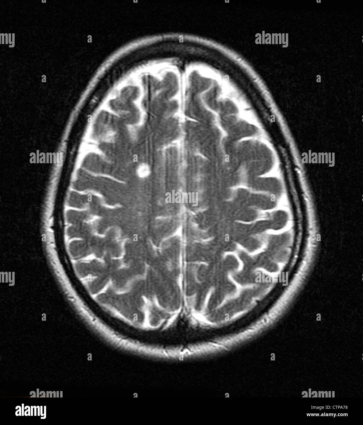 MRI showing multiple sclerosis in a 42 year old woman Stock Photohttps://www.alamy.com/image-license-details/?v=1https://www.alamy.com/stock-photo-mri-showing-multiple-sclerosis-in-a-42-year-old-woman-49663468.html
RMCTPA78–MRI showing multiple sclerosis in a 42 year old woman
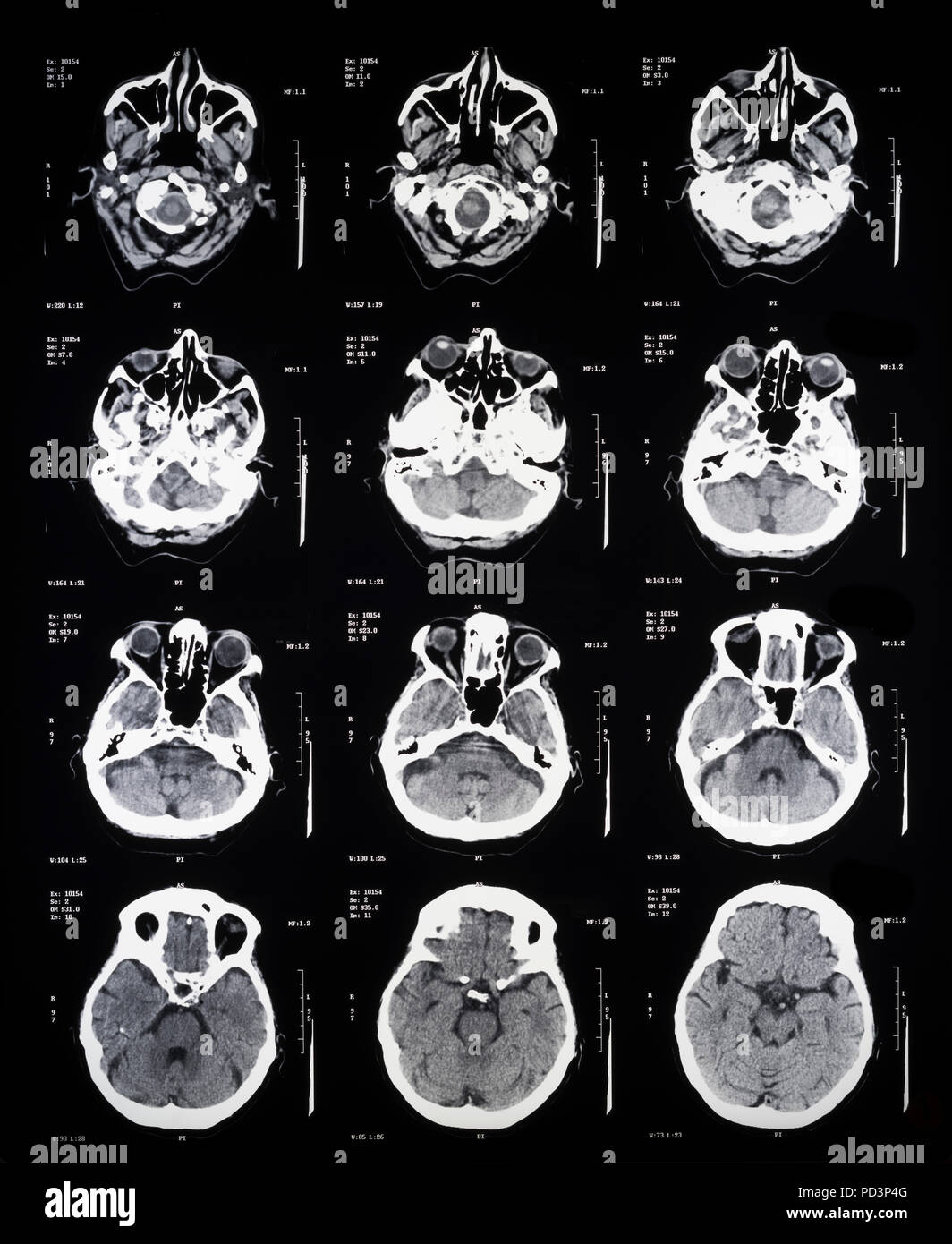 Sequence of horizontal sections of a female human brain, MRI scans, magnetic resonance imaging, Stock Photohttps://www.alamy.com/image-license-details/?v=1https://www.alamy.com/sequence-of-horizontal-sections-of-a-female-human-brain-mri-scans-magnetic-resonance-imaging-image214598176.html
RMPD3P4G–Sequence of horizontal sections of a female human brain, MRI scans, magnetic resonance imaging,
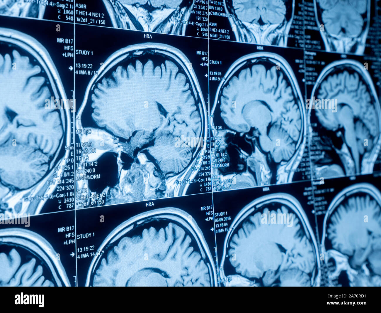 Magnetic resonance image (MRI) of the brain Stock Photohttps://www.alamy.com/image-license-details/?v=1https://www.alamy.com/magnetic-resonance-image-mri-of-the-brain-image331317981.html
RF2A70RD1–Magnetic resonance image (MRI) of the brain
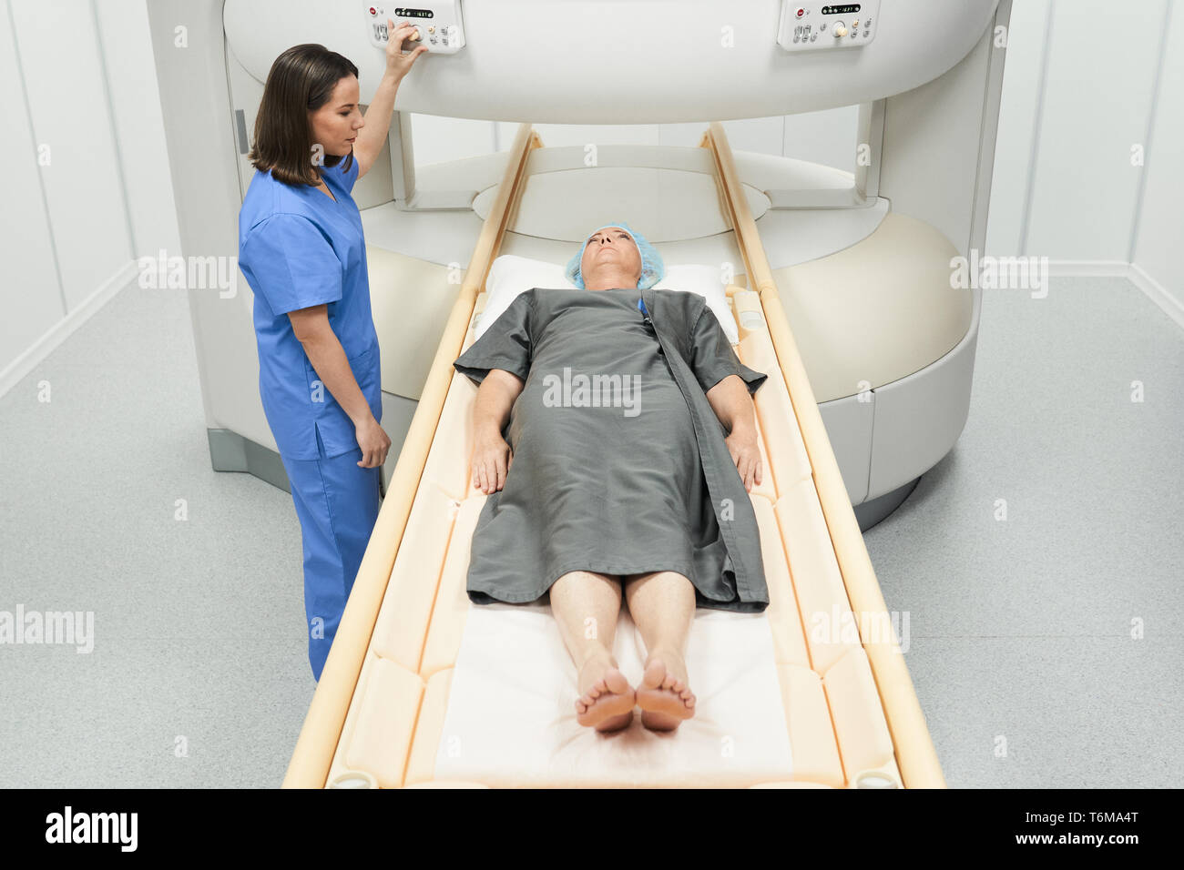 Mature Patient With Health Problems And Doctor In Medical Center Stock Photohttps://www.alamy.com/image-license-details/?v=1https://www.alamy.com/mature-patient-with-health-problems-and-doctor-in-medical-center-image245080104.html
RFT6MA4T–Mature Patient With Health Problems And Doctor In Medical Center
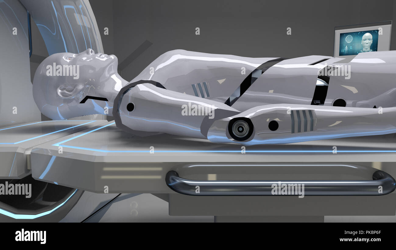 Robot in a medical facility with futuristic body scan. 3d rendering Stock Photohttps://www.alamy.com/image-license-details/?v=1https://www.alamy.com/robot-in-a-medical-facility-with-futuristic-body-scan-3d-rendering-image218461783.html
RFPKBP6F–Robot in a medical facility with futuristic body scan. 3d rendering
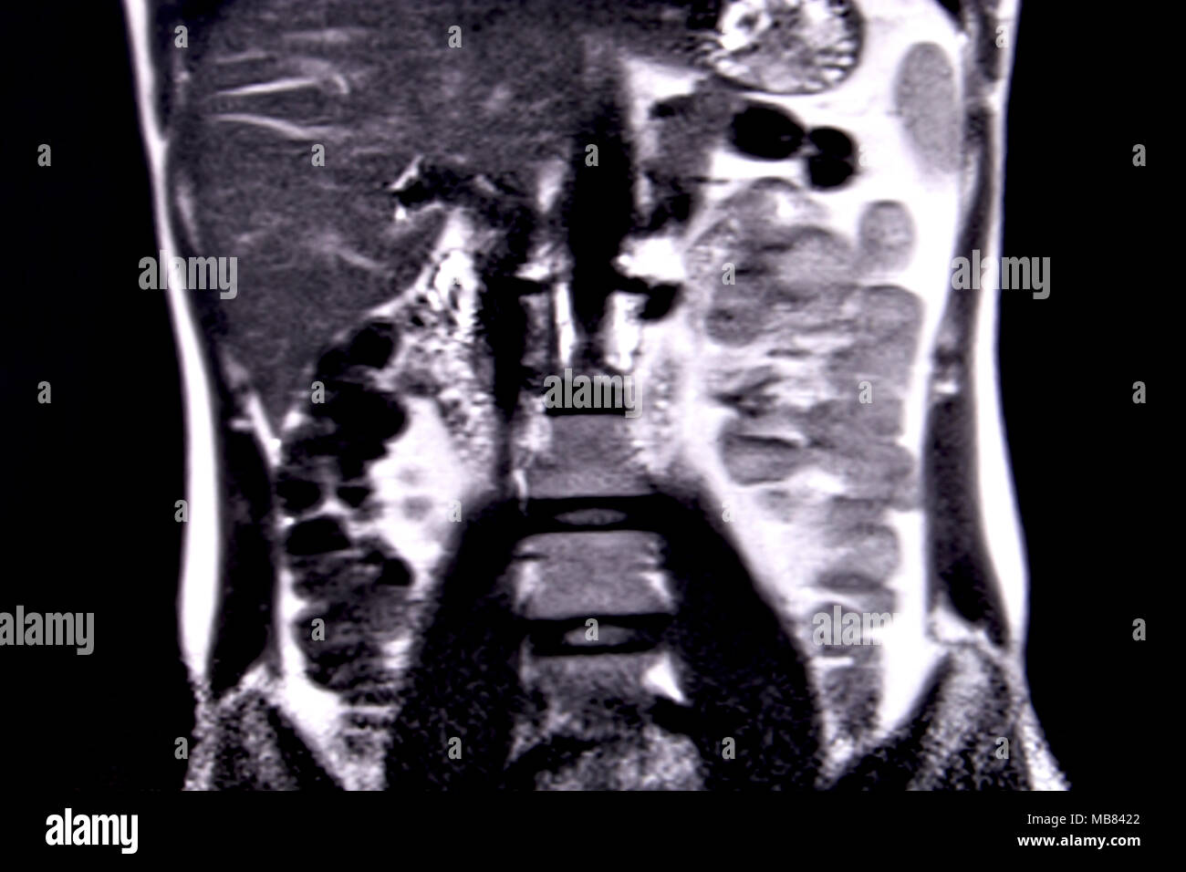 Ride through the human abdomen and chest by means of 18 MRI cuts (coronal view). Picture 5/18 Stock Photohttps://www.alamy.com/image-license-details/?v=1https://www.alamy.com/ride-through-the-human-abdomen-and-chest-by-means-of-18-mri-cuts-coronal-view-picture-518-image179043706.html
RFMB8422–Ride through the human abdomen and chest by means of 18 MRI cuts (coronal view). Picture 5/18
 doctor radiographer waiting in control room behind protective glass while medical assistant prepares female patient for CT scan. Computed Tomography s Stock Photohttps://www.alamy.com/image-license-details/?v=1https://www.alamy.com/doctor-radiographer-waiting-in-control-room-behind-protective-glass-while-medical-assistant-prepares-female-patient-for-ct-scan-computed-tomography-s-image478577419.html
RF2JPH2AK–doctor radiographer waiting in control room behind protective glass while medical assistant prepares female patient for CT scan. Computed Tomography s
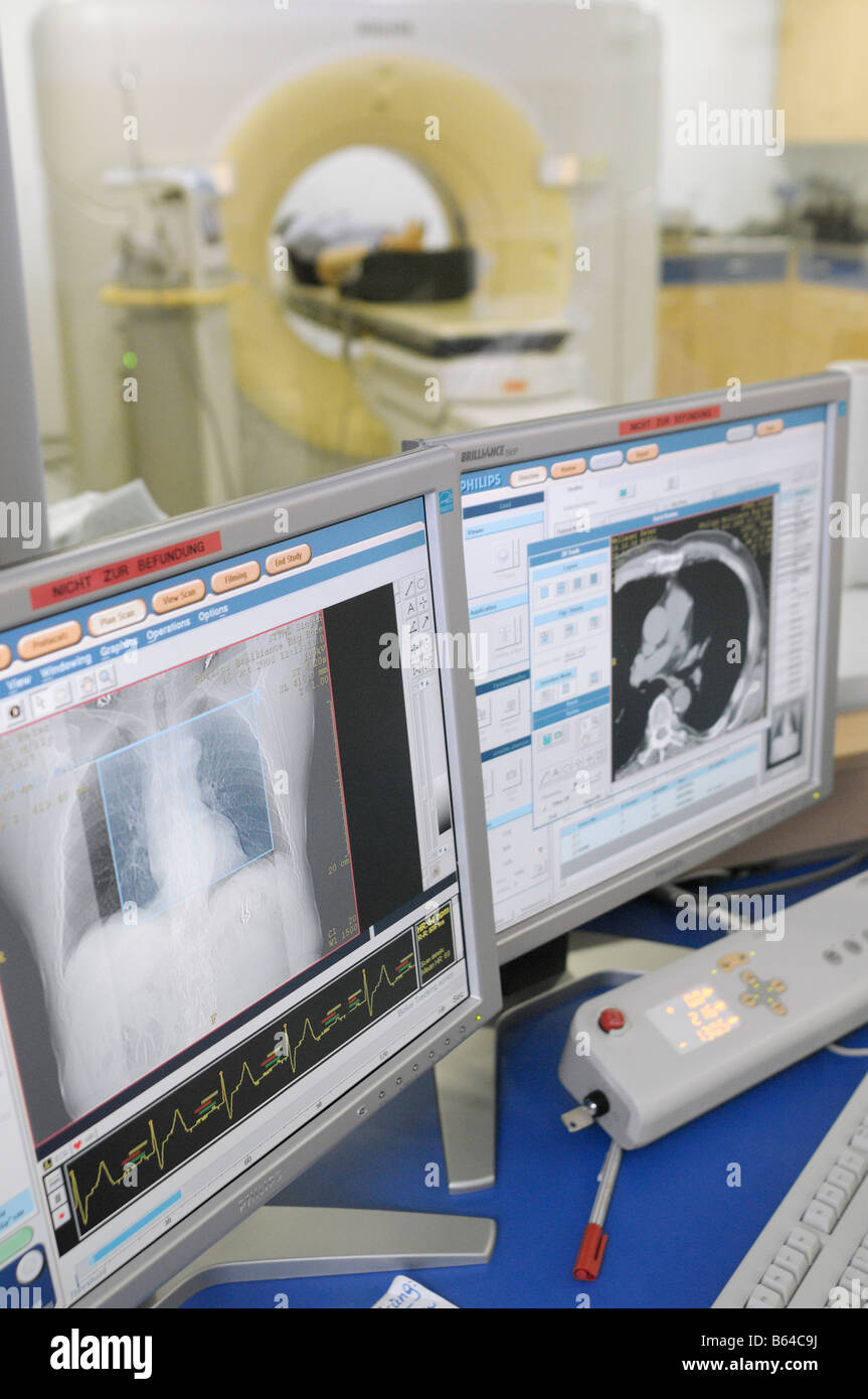 Computer tomography Stock Photohttps://www.alamy.com/image-license-details/?v=1https://www.alamy.com/stock-photo-computer-tomography-20995790.html
RMB64C9J–Computer tomography
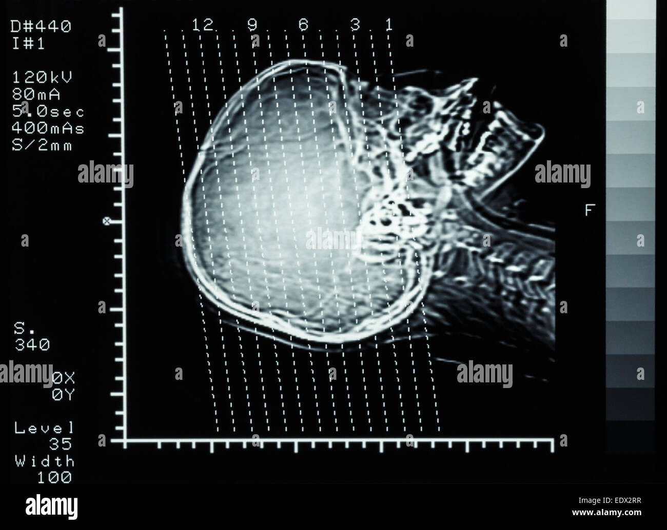 film CT scan (Computed tomography) brain : show section line on skull Stock Photohttps://www.alamy.com/image-license-details/?v=1https://www.alamy.com/stock-photo-film-ct-scan-computed-tomography-brain-show-section-line-on-skull-77404987.html
RFEDX2RR–film CT scan (Computed tomography) brain : show section line on skull
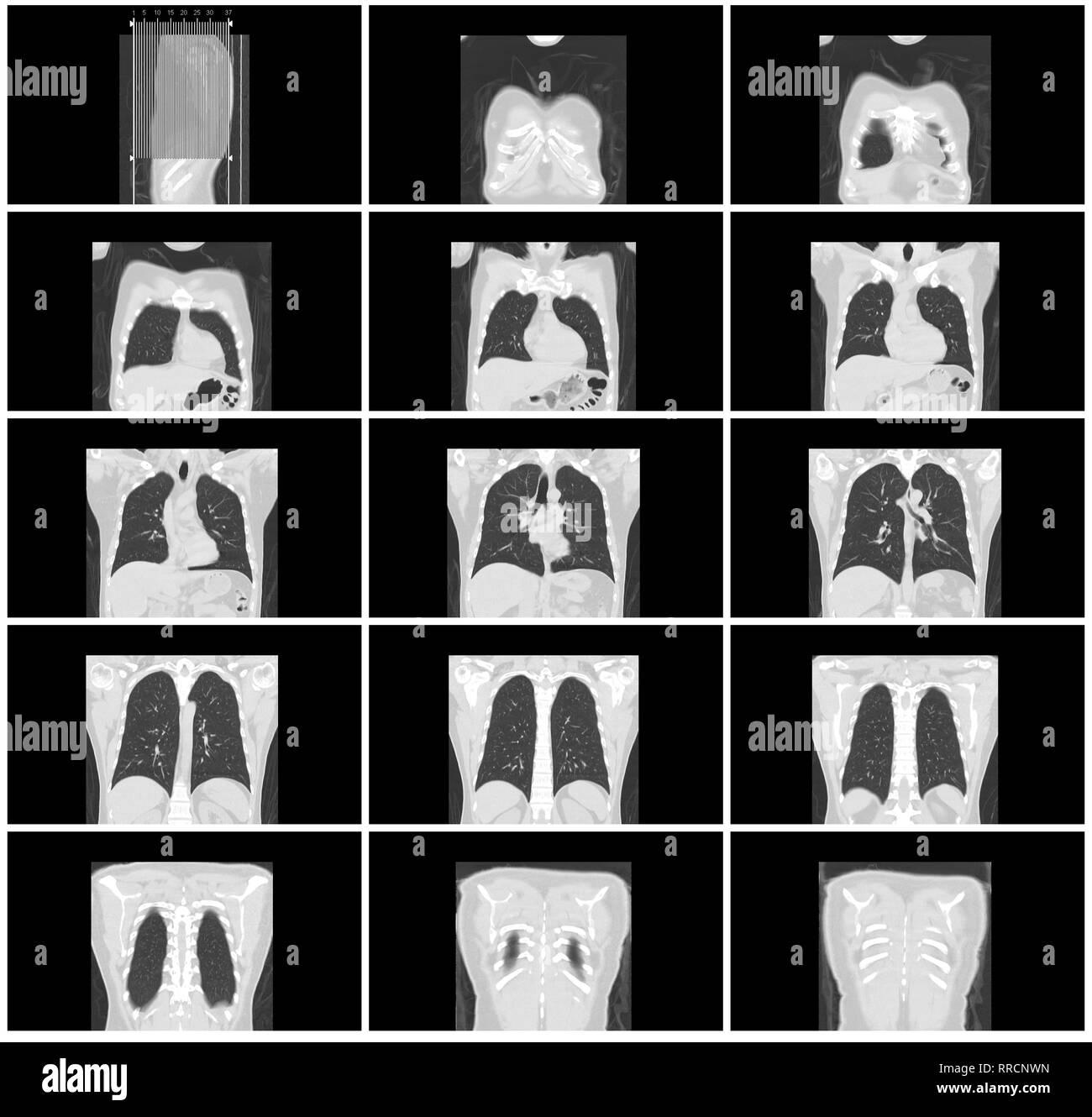 ct scan step set of body lung coronal view Stock Photohttps://www.alamy.com/image-license-details/?v=1https://www.alamy.com/ct-scan-step-set-of-body-lung-coronal-view-image238152481.html
RFRRCNWN–ct scan step set of body lung coronal view
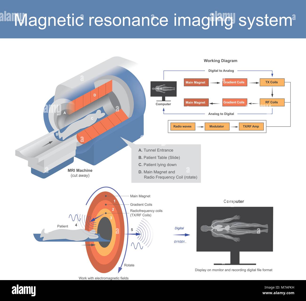 The Mechanical technique used in radio wave to form pictures of the anatomy and the magnetic physiological processes of the body in both health and di Stock Vectorhttps://www.alamy.com/image-license-details/?v=1https://www.alamy.com/stock-photo-the-mechanical-technique-used-in-radio-wave-to-form-pictures-of-the-176638101.html
RFM7AFKH–The Mechanical technique used in radio wave to form pictures of the anatomy and the magnetic physiological processes of the body in both health and di
 Serious female radiologist examining MRI scan image Stock Photohttps://www.alamy.com/image-license-details/?v=1https://www.alamy.com/stock-image-serious-female-radiologist-examining-mri-scan-image-163202344.html
RFKDEE74–Serious female radiologist examining MRI scan image
 Healthcare Technology and Medical Scan of a Body Diagnosis Stock Photohttps://www.alamy.com/image-license-details/?v=1https://www.alamy.com/stock-photo-healthcare-technology-and-medical-scan-of-a-body-diagnosis-119896709.html
RFGY1ND9–Healthcare Technology and Medical Scan of a Body Diagnosis
 Close-up photo of an MRI of the skull and brain of a person with severe headaches; magnetic and nuclear resonance as a diagnostic method in neurology Stock Photohttps://www.alamy.com/image-license-details/?v=1https://www.alamy.com/close-up-photo-of-an-mri-of-the-skull-and-brain-of-a-person-with-severe-headaches-magnetic-and-nuclear-resonance-as-a-diagnostic-method-in-neurology-image448197043.html
RF2H153W7–Close-up photo of an MRI of the skull and brain of a person with severe headaches; magnetic and nuclear resonance as a diagnostic method in neurology
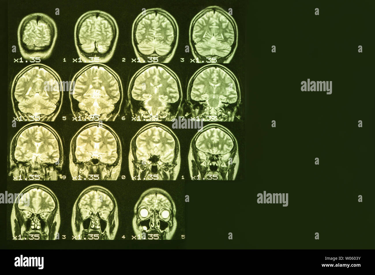 MRI of the brain on a black background with yellow backlight. Right place for advertising inscription Stock Photohttps://www.alamy.com/image-license-details/?v=1https://www.alamy.com/mri-of-the-brain-on-a-black-background-with-yellow-backlight-right-place-for-advertising-inscription-image258287343.html
RFW0603Y–MRI of the brain on a black background with yellow backlight. Right place for advertising inscription
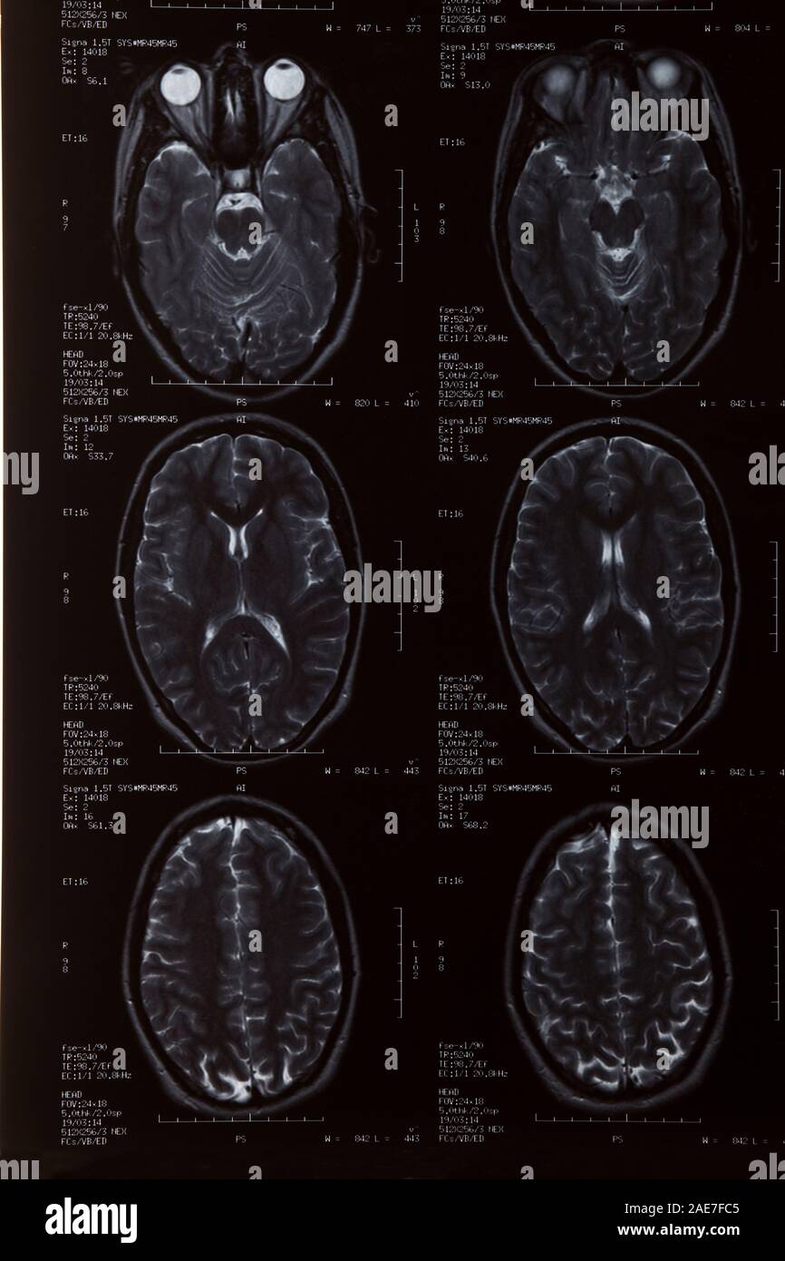 MRI Scan close up of female brain Stock Photohttps://www.alamy.com/image-license-details/?v=1https://www.alamy.com/mri-scan-close-up-of-female-brain-image335767941.html
RM2AE7FC5–MRI Scan close up of female brain
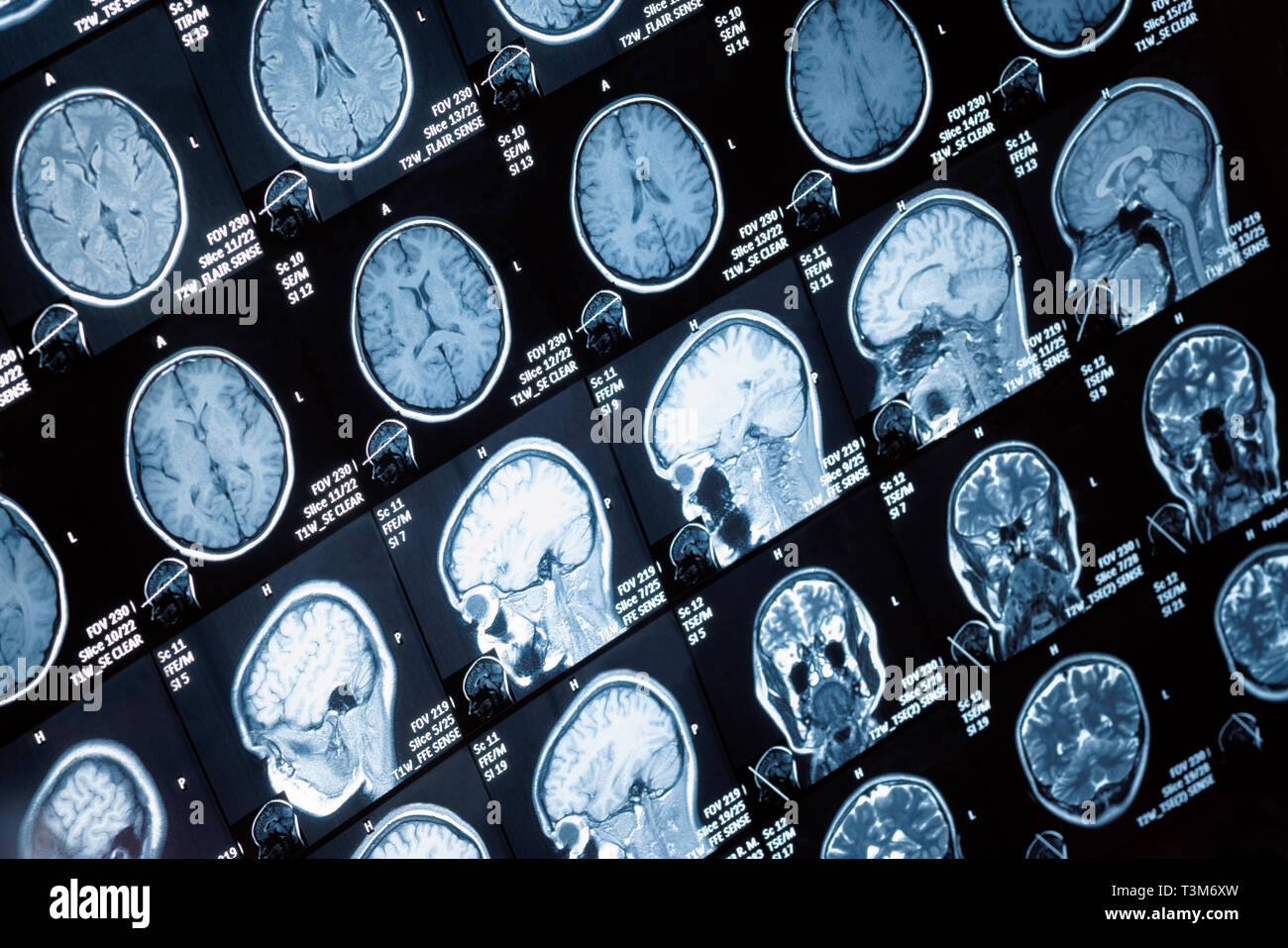 Head and neck MRI scan, anonymized, shallow focus depth Stock Photohttps://www.alamy.com/image-license-details/?v=1https://www.alamy.com/head-and-neck-mri-scan-anonymized-shallow-focus-depth-image243233617.html
RFT3M6XW–Head and neck MRI scan, anonymized, shallow focus depth
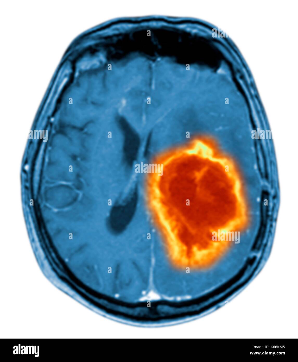 Brain tumour. Coloured Magnetic Resonance Imaging (MRI) scan of an axial section through the brain showing a metastatic tumour. At bottom left is the tumour (red-yellow) This tumour occurs within one cerebral hemisphere; the other hemisphere is at right. The eyeballs - not visible -are at top. Metastatic cancer is a secondary disease spread from cancer elsewhere in the body. Metastatic brain tumours are malignant. Typically they cause brain compression and nerve damage Stock Photohttps://www.alamy.com/image-license-details/?v=1https://www.alamy.com/brain-tumour-coloured-magnetic-resonance-imaging-mri-scan-of-an-axial-image158728421.html
RFK66KM5–Brain tumour. Coloured Magnetic Resonance Imaging (MRI) scan of an axial section through the brain showing a metastatic tumour. At bottom left is the tumour (red-yellow) This tumour occurs within one cerebral hemisphere; the other hemisphere is at right. The eyeballs - not visible -are at top. Metastatic cancer is a secondary disease spread from cancer elsewhere in the body. Metastatic brain tumours are malignant. Typically they cause brain compression and nerve damage
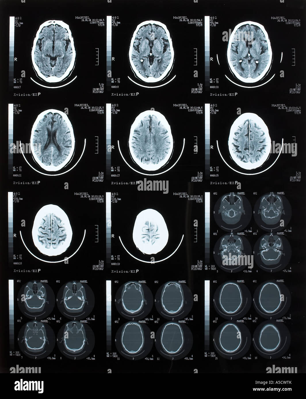 Brain Tomography Stock Photohttps://www.alamy.com/image-license-details/?v=1https://www.alamy.com/stock-photo-brain-tomography-11273202.html
RFA5CWTK–Brain Tomography
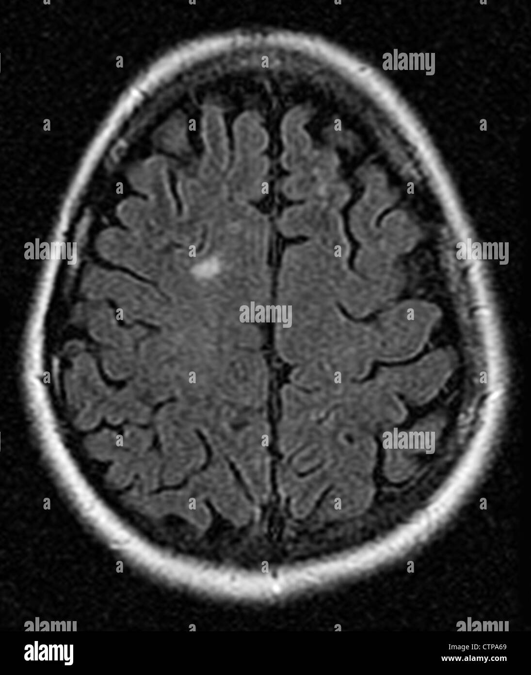 MRI showing multiple sclerosis in a 42 year old woman Stock Photohttps://www.alamy.com/image-license-details/?v=1https://www.alamy.com/stock-photo-mri-showing-multiple-sclerosis-in-a-42-year-old-woman-49663441.html
RMCTPA69–MRI showing multiple sclerosis in a 42 year old woman
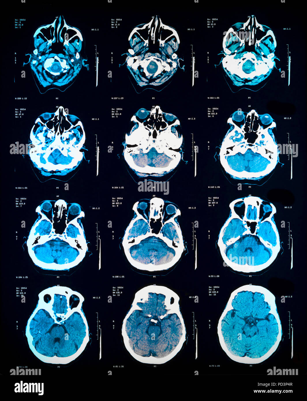 Sequence of horizontal sections of a female human brain, MRI scans, magnetic resonance imaging, Stock Photohttps://www.alamy.com/image-license-details/?v=1https://www.alamy.com/sequence-of-horizontal-sections-of-a-female-human-brain-mri-scans-magnetic-resonance-imaging-image214598183.html
RMPD3P4R–Sequence of horizontal sections of a female human brain, MRI scans, magnetic resonance imaging,
