Quick filters:
Nasal vestibule Stock Photos and Images
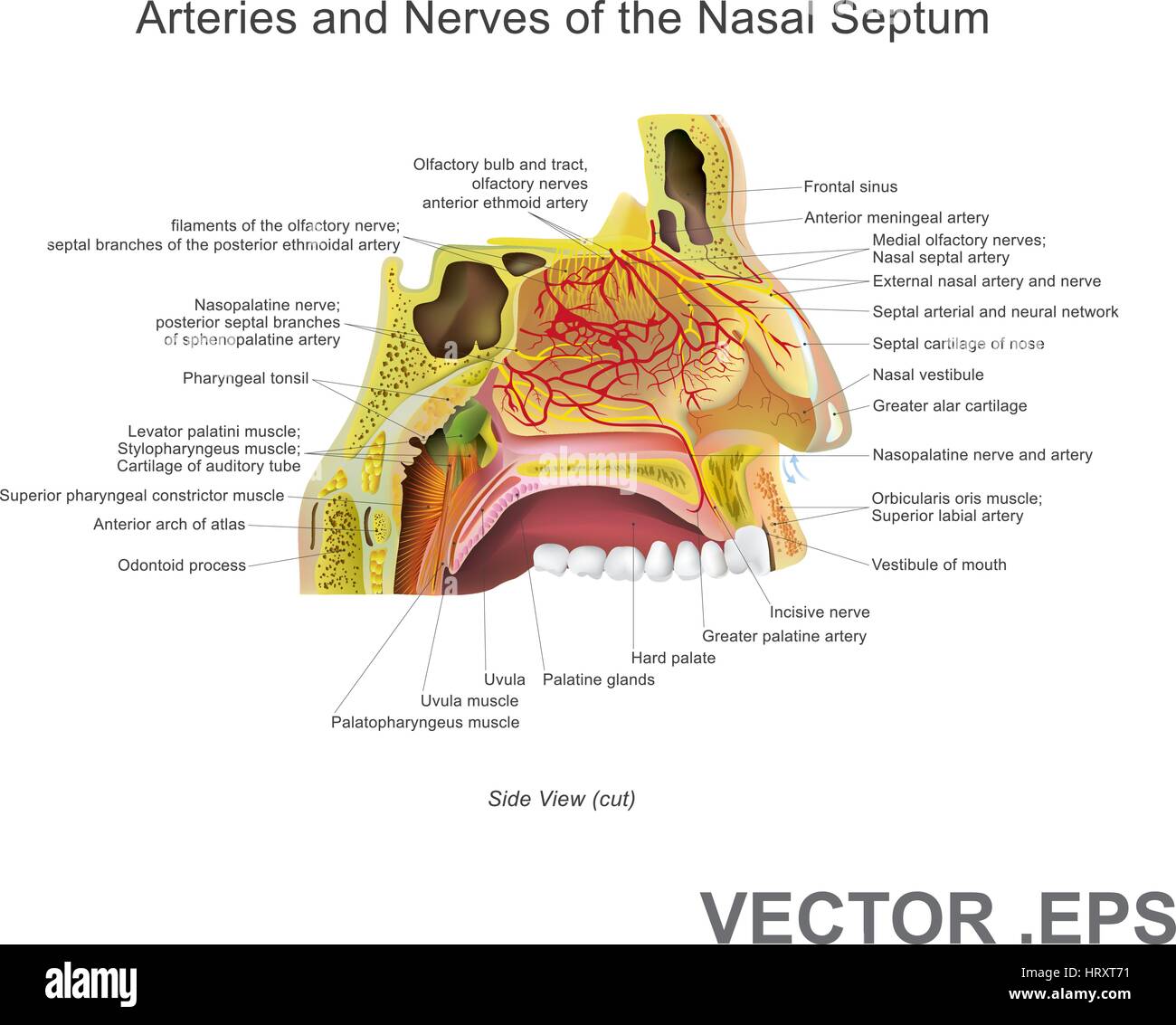 The nasal cavity (or nasal fossa) is a large air filled space above and behind the nose in the middle of the face. Each cavity is the continuation of Stock Vectorhttps://www.alamy.com/image-license-details/?v=1https://www.alamy.com/stock-photo-the-nasal-cavity-or-nasal-fossa-is-a-large-air-filled-space-above-135199429.html
RFHRXT71–The nasal cavity (or nasal fossa) is a large air filled space above and behind the nose in the middle of the face. Each cavity is the continuation of
RF2CGXGG0–Nose with bacteria grey icon. Bacterial infection of the nasal vestibule symbol
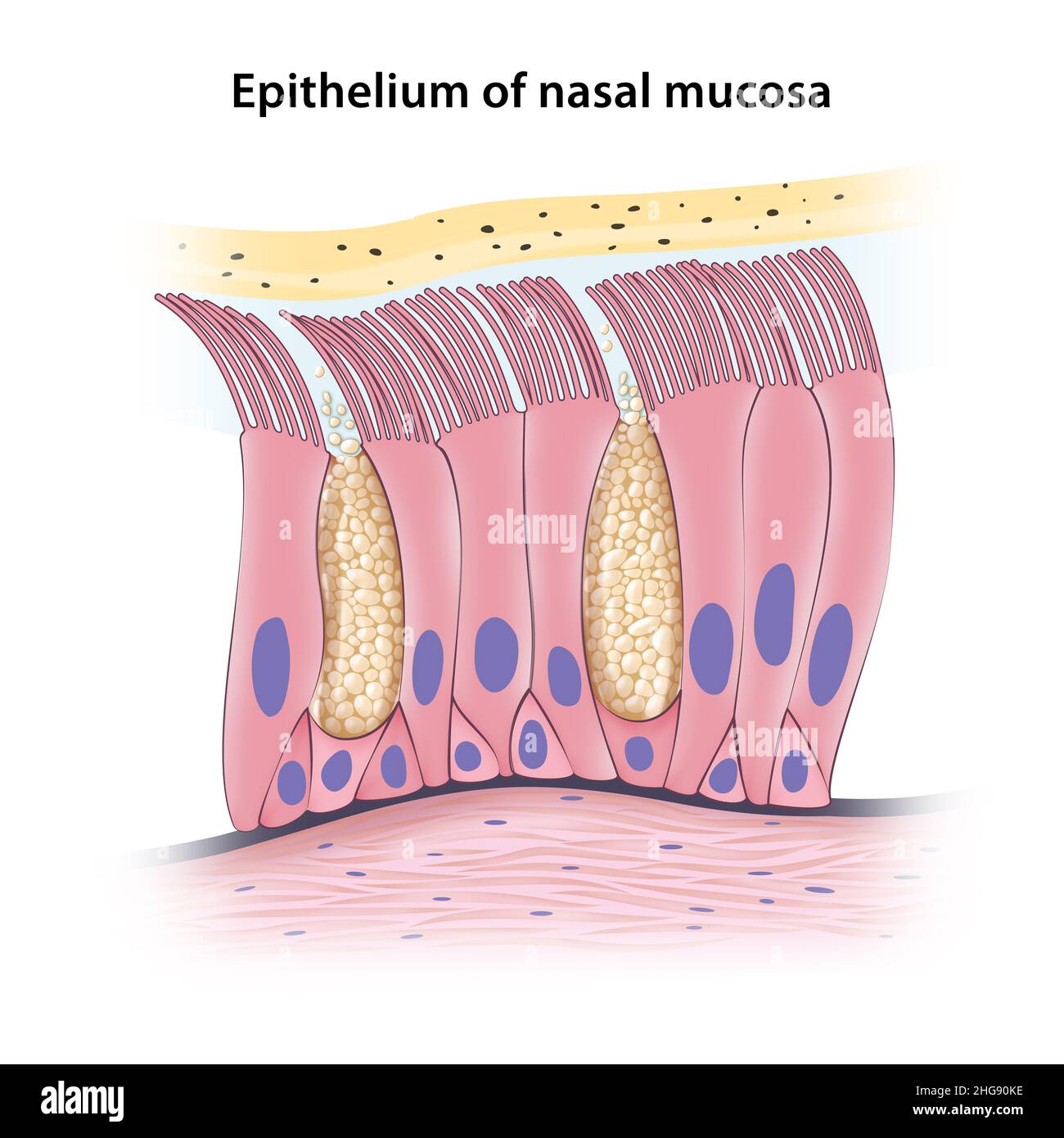 Pseudostratified columnar epithelium of nasal mucosa Stock Photohttps://www.alamy.com/image-license-details/?v=1https://www.alamy.com/pseudostratified-columnar-epithelium-of-nasal-mucosa-image457502178.html
RF2HG90KE–Pseudostratified columnar epithelium of nasal mucosa
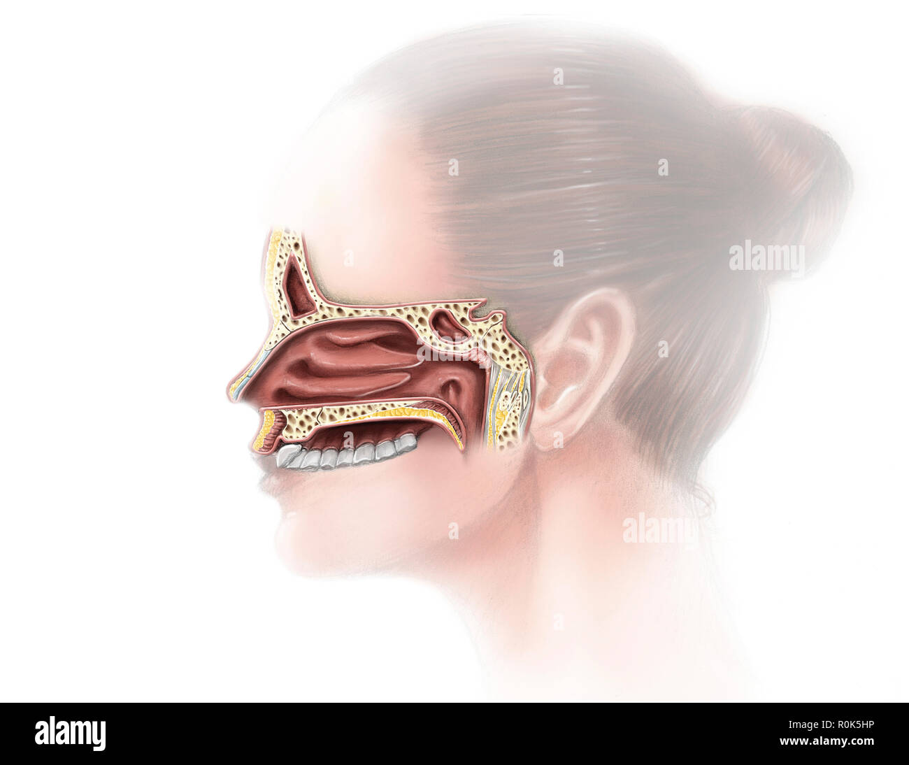 Digital illustration of nose and nasal sinus anatomy (no labels) Stock Photohttps://www.alamy.com/image-license-details/?v=1https://www.alamy.com/digital-illustration-of-nose-and-nasal-sinus-anatomy-no-labels-image224156290.html
RFR0K5HP–Digital illustration of nose and nasal sinus anatomy (no labels)
RFFYAPAP–Human Nose, Medical Doctors Otolaryngology icon, line icon Style
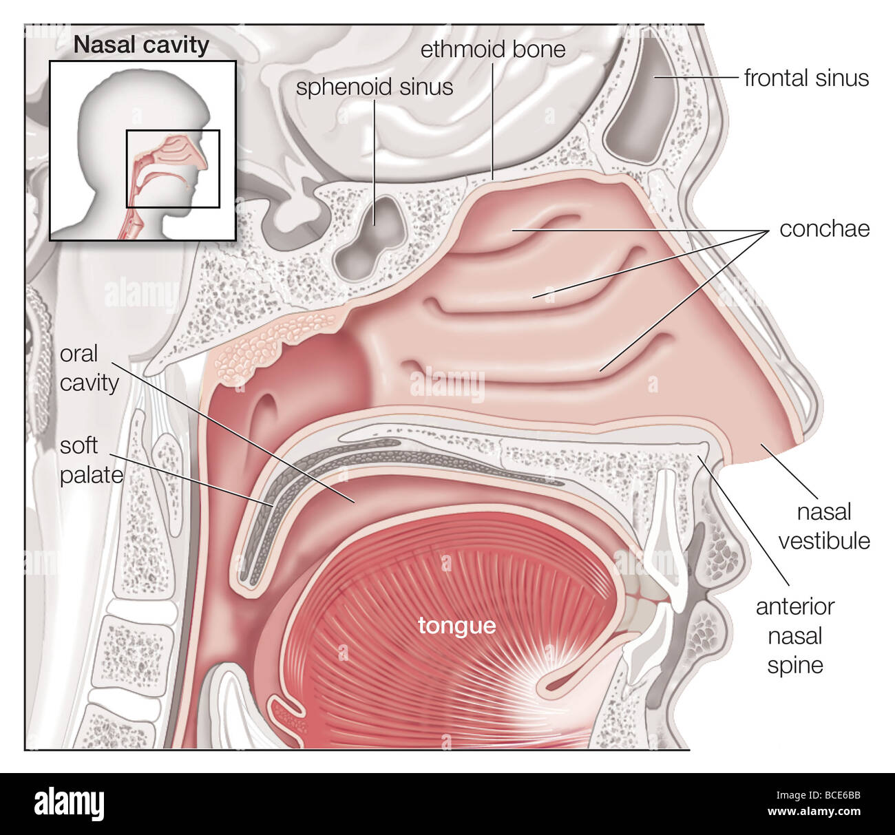 Sagittal view of the human nasal cavity. Stock Photohttps://www.alamy.com/image-license-details/?v=1https://www.alamy.com/stock-photo-sagittal-view-of-the-human-nasal-cavity-24898591.html
RMBCE6BB–Sagittal view of the human nasal cavity.
RFFYAP9C–Human Nose, Medical Doctors Otolaryngology icon
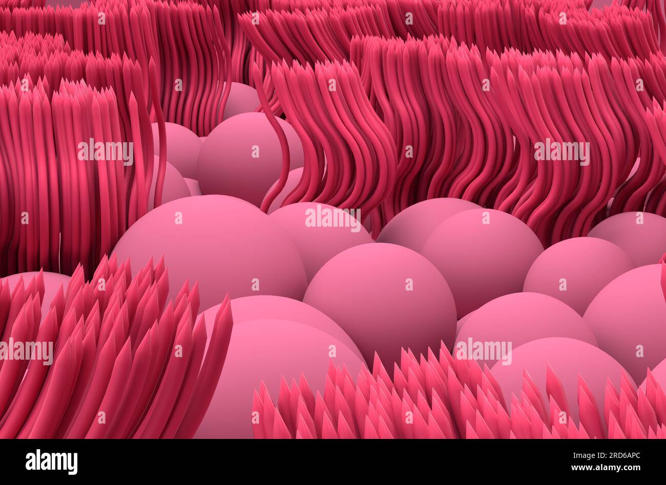 Nasal mucosa lines (respiratory epihtelium) - closeup view 3d illustration Stock Photohttps://www.alamy.com/image-license-details/?v=1https://www.alamy.com/nasal-mucosa-lines-respiratory-epihtelium-closeup-view-3d-illustration-image558862484.html
RF2RD6APC–Nasal mucosa lines (respiratory epihtelium) - closeup view 3d illustration
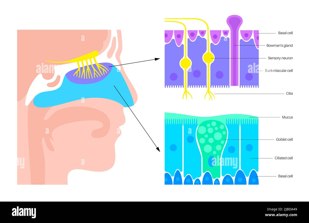 Nasal cavity anatomy, illustration Stock Photohttps://www.alamy.com/image-license-details/?v=1https://www.alamy.com/nasal-cavity-anatomy-illustration-image471734489.html
RF2JBDA49–Nasal cavity anatomy, illustration
 Journal of the Academy of Natural Sciences of Philadelphia . long.—Nasal vestibule macrolophic; incisoreminence none; spinal line trenchant, above level of floor of nose; alveolar linenone; premaxillary crest .complete, high spine, sharply projecting. Alveolus small.—Hard palate parabolic, small, not flat,Alveolar process high. Spinous process long,overlapping sphenoido-tympanic suture.—Petrosa moderately inflated; cerebellarfossa lies below the plane of the occipitalcondyles. Foramen lacerum medium open.—Malar bone enters into the spheno-maxil-lary fissure ; marginal process small; suture-tra Stock Photohttps://www.alamy.com/image-license-details/?v=1https://www.alamy.com/journal-of-the-academy-of-natural-sciences-of-philadelphia-longnasal-vestibule-macrolophic-incisoreminence-none-spinal-line-trenchant-above-level-of-floor-of-nose-alveolar-linenone-premaxillary-crest-complete-high-spine-sharply-projecting-alveolus-smallhard-palate-parabolic-small-not-flatalveolar-process-high-spinous-process-longoverlapping-sphenoido-tympanic-suturepetrosa-moderately-inflated-cerebellarfossa-lies-below-the-plane-of-the-occipitalcondyles-foramen-lacerum-medium-openmalar-bone-enters-into-the-spheno-maxil-lary-fissure-marginal-process-small-suture-tra-image340031601.html
RM2AN5NNN–Journal of the Academy of Natural Sciences of Philadelphia . long.—Nasal vestibule macrolophic; incisoreminence none; spinal line trenchant, above level of floor of nose; alveolar linenone; premaxillary crest .complete, high spine, sharply projecting. Alveolus small.—Hard palate parabolic, small, not flat,Alveolar process high. Spinous process long,overlapping sphenoido-tympanic suture.—Petrosa moderately inflated; cerebellarfossa lies below the plane of the occipitalcondyles. Foramen lacerum medium open.—Malar bone enters into the spheno-maxil-lary fissure ; marginal process small; suture-tra
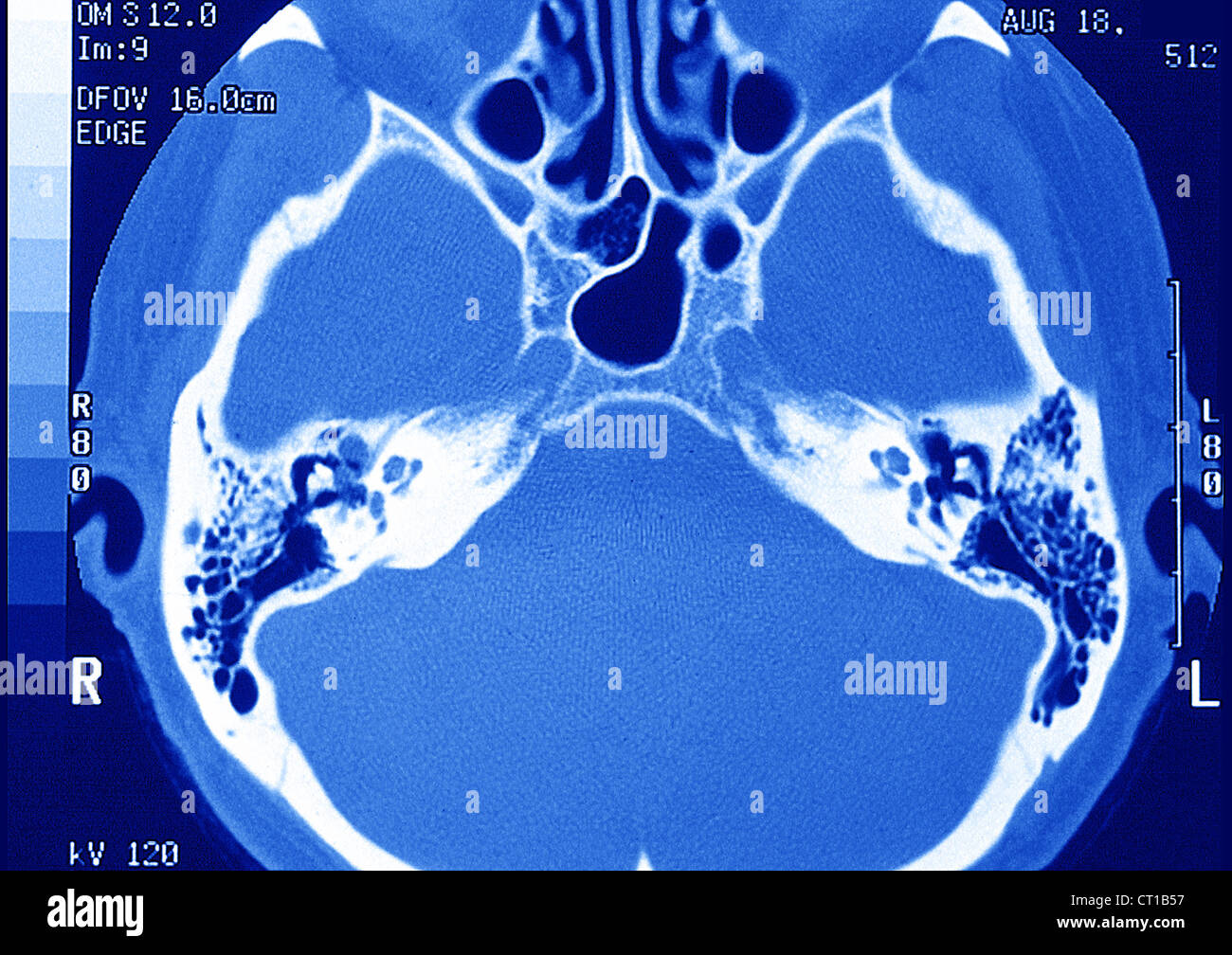 INTERNAL EAR, MRI Stock Photohttps://www.alamy.com/image-license-details/?v=1https://www.alamy.com/stock-photo-internal-ear-mri-49203203.html
RMCT1B57–INTERNAL EAR, MRI
 . Smell, taste, and allied senses in the vertebrates . Senses and sensation; Vertebrates. ANATOMY OF THE OLFACTORY ORGAN 25 the lateral wall of each nasal chamber into its cavity and partly divide that cavity into three approximately hori- zontal passages: the inferior meatus under the inferior concha, the middle meatus under the middle concha and the superior meatus under the superior concha. (Fig. 2). The external naris leads at once to the first chamber of the nose, the vestibule, which connects almost directly with the inferior meatus, less directly with the su- perior meatus and through t Stock Photohttps://www.alamy.com/image-license-details/?v=1https://www.alamy.com/smell-taste-and-allied-senses-in-the-vertebrates-senses-and-sensation-vertebrates-anatomy-of-the-olfactory-organ-25-the-lateral-wall-of-each-nasal-chamber-into-its-cavity-and-partly-divide-that-cavity-into-three-approximately-hori-zontal-passages-the-inferior-meatus-under-the-inferior-concha-the-middle-meatus-under-the-middle-concha-and-the-superior-meatus-under-the-superior-concha-fig-2-the-external-naris-leads-at-once-to-the-first-chamber-of-the-nose-the-vestibule-which-connects-almost-directly-with-the-inferior-meatus-less-directly-with-the-su-perior-meatus-and-through-t-image216456570.html
RMPG4CFP–. Smell, taste, and allied senses in the vertebrates . Senses and sensation; Vertebrates. ANATOMY OF THE OLFACTORY ORGAN 25 the lateral wall of each nasal chamber into its cavity and partly divide that cavity into three approximately hori- zontal passages: the inferior meatus under the inferior concha, the middle meatus under the middle concha and the superior meatus under the superior concha. (Fig. 2). The external naris leads at once to the first chamber of the nose, the vestibule, which connects almost directly with the inferior meatus, less directly with the su- perior meatus and through t
RF2CTX9WR–Nose with bacteria colored icon. Diseased of nose and paranasal sinuse, bacterial infection of the nasal vestibule, nasal vestibulitis symbol
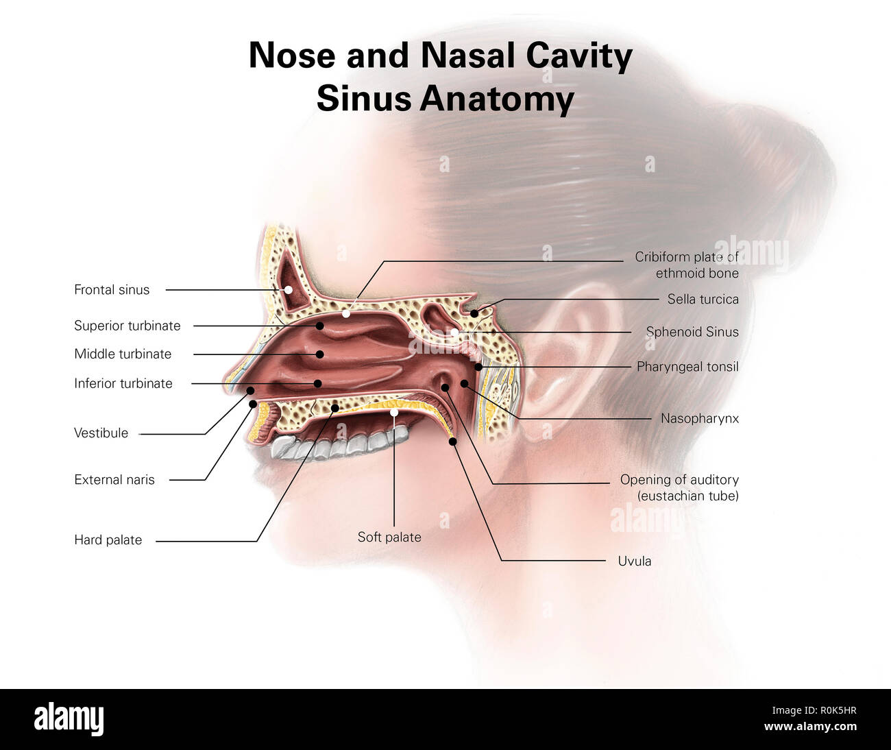 Digital illustration of nose and nasal sinus anatomy (no labels) Stock Photohttps://www.alamy.com/image-license-details/?v=1https://www.alamy.com/digital-illustration-of-nose-and-nasal-sinus-anatomy-no-labels-image224156291.html
RFR0K5HR–Digital illustration of nose and nasal sinus anatomy (no labels)
RF2BJ4BW7–Human nose with bacteria line icon. Disease of paranasal sinuse, bacterial infection, nasal vestibulitis symbol
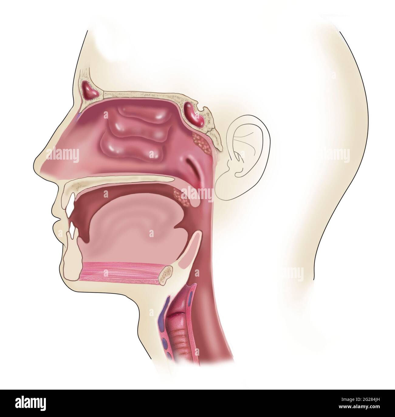 Side view of head showing location of swollen and enlarged tonsils. Stock Photohttps://www.alamy.com/image-license-details/?v=1https://www.alamy.com/side-view-of-head-showing-location-of-swollen-and-enlarged-tonsils-image431667785.html
RF2G284JH–Side view of head showing location of swollen and enlarged tonsils.
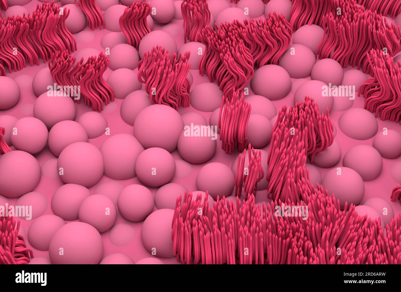 Nasal mucosa lines (respiratory epihtelium) - isometric view 3d illustration Stock Photohttps://www.alamy.com/image-license-details/?v=1https://www.alamy.com/nasal-mucosa-lines-respiratory-epihtelium-isometric-view-3d-illustration-image558862525.html
RF2RD6ARW–Nasal mucosa lines (respiratory epihtelium) - isometric view 3d illustration
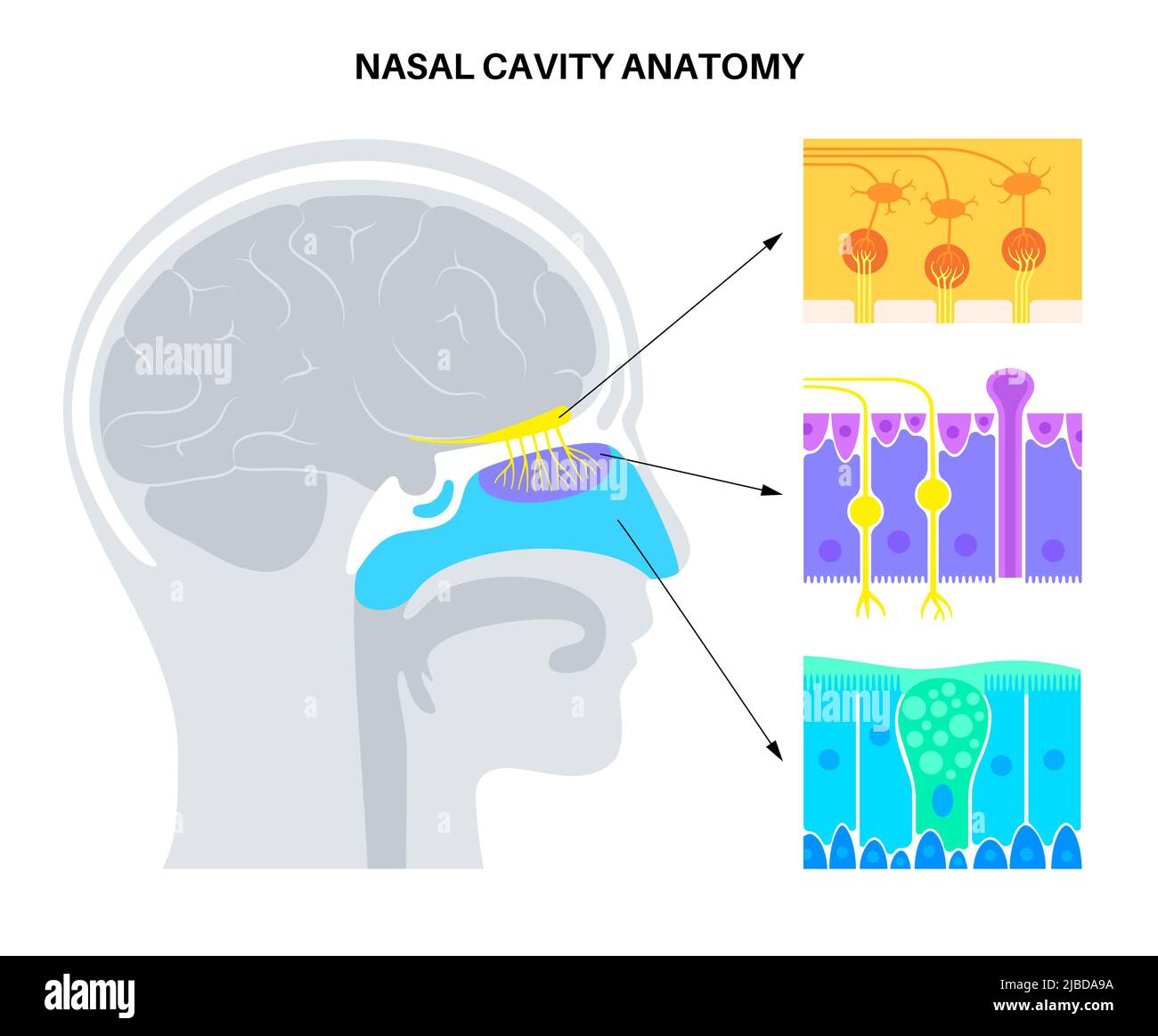 Nasal cavity anatomy, illustration Stock Photohttps://www.alamy.com/image-license-details/?v=1https://www.alamy.com/nasal-cavity-anatomy-illustration-image471734630.html
RF2JBDA9A–Nasal cavity anatomy, illustration
 Journal of the Academy of Natural Sciences of Philadelphia . gonal.—Skull rests on opisthion. Wormianbones present in lambdoidal suture.—Ossa plana greatly deformed, uneven, withethmoid cells opening into orbit; bulla ethmoidals present right and left. Lachry-mal bone absent congenitally on right side; lost on left. 406 CRANIA FROM THE MOUNDS OF FLORIDA. 730 <?, Seminole. Aged 25 years,—stenocephalia. Figure 3. Glabella small; forehead slightly receding; outer part orbital arch abruptlyinclined.—Nasal bones convex. Nasal vestibule microlophic; incisor eminenceconspicuous; alveolar line defe Stock Photohttps://www.alamy.com/image-license-details/?v=1https://www.alamy.com/journal-of-the-academy-of-natural-sciences-of-philadelphia-gonalskull-rests-on-opisthion-wormianbones-present-in-lambdoidal-sutureossa-plana-greatly-deformed-uneven-withethmoid-cells-opening-into-orbit-bulla-ethmoidals-present-right-and-left-lachry-mal-bone-absent-congenitally-on-right-side-lost-on-left-406-crania-from-the-mounds-of-florida-730-lt-seminole-aged-25-yearsstenocephalia-figure-3-glabella-small-forehead-slightly-receding-outer-part-orbital-arch-abruptlyinclinednasal-bones-convex-nasal-vestibule-microlophic-incisor-eminenceconspicuous-alveolar-line-defe-image340031350.html
RM2AN5NCP–Journal of the Academy of Natural Sciences of Philadelphia . gonal.—Skull rests on opisthion. Wormianbones present in lambdoidal suture.—Ossa plana greatly deformed, uneven, withethmoid cells opening into orbit; bulla ethmoidals present right and left. Lachry-mal bone absent congenitally on right side; lost on left. 406 CRANIA FROM THE MOUNDS OF FLORIDA. 730 <?, Seminole. Aged 25 years,—stenocephalia. Figure 3. Glabella small; forehead slightly receding; outer part orbital arch abruptlyinclined.—Nasal bones convex. Nasal vestibule microlophic; incisor eminenceconspicuous; alveolar line defe
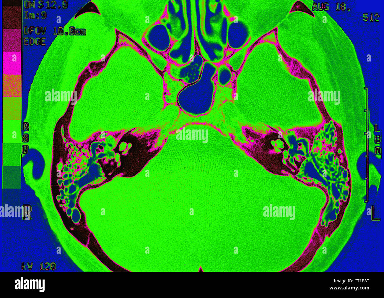 INTERNAL EAR, MRI Stock Photohttps://www.alamy.com/image-license-details/?v=1https://www.alamy.com/stock-photo-internal-ear-mri-49203304.html
RMCT1B8T–INTERNAL EAR, MRI
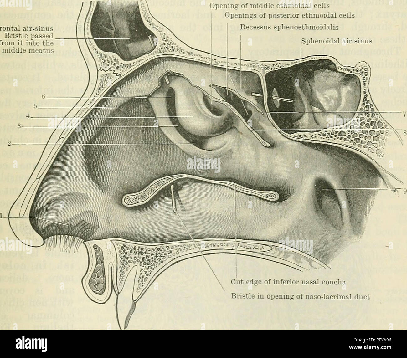 . Cunningham's Text-book of anatomy. Anatomy. Vomeronasal cartilages Vomero-nasal organs Fig. 673.—Section through Nose of a Kitten, showing position of the voinero-nasal organs. Frontal air-sinus Bristle passed from it into the middle meatus Opening of middle ethmoidal cells Openings of posterior ethmoidal cells Recessus sphenoethmoidalis Sphenoidal air-sinus. Cut edge of inferior nasal concha stle in opening of naso-lacrimal duct Fig. 674.—View of the Lateral Wall of the Nose—the Nasal Concha having been behoved. 1. Vestibule. 2. Opening of maxillary sinus. 3. Hiatus semilunaris. 4. Bulla et Stock Photohttps://www.alamy.com/image-license-details/?v=1https://www.alamy.com/cunninghams-text-book-of-anatomy-anatomy-vomeronasal-cartilages-vomero-nasal-organs-fig-673section-through-nose-of-a-kitten-showing-position-of-the-voinero-nasal-organs-frontal-air-sinus-bristle-passed-from-it-into-the-middle-meatus-opening-of-middle-ethmoidal-cells-openings-of-posterior-ethmoidal-cells-recessus-sphenoethmoidalis-sphenoidal-air-sinus-cut-edge-of-inferior-nasal-concha-stle-in-opening-of-naso-lacrimal-duct-fig-674view-of-the-lateral-wall-of-the-nosethe-nasal-concha-having-been-behoved-1-vestibule-2-opening-of-maxillary-sinus-3-hiatus-semilunaris-4-bulla-et-image216345058.html
RMPFYA96–. Cunningham's Text-book of anatomy. Anatomy. Vomeronasal cartilages Vomero-nasal organs Fig. 673.—Section through Nose of a Kitten, showing position of the voinero-nasal organs. Frontal air-sinus Bristle passed from it into the middle meatus Opening of middle ethmoidal cells Openings of posterior ethmoidal cells Recessus sphenoethmoidalis Sphenoidal air-sinus. Cut edge of inferior nasal concha stle in opening of naso-lacrimal duct Fig. 674.—View of the Lateral Wall of the Nose—the Nasal Concha having been behoved. 1. Vestibule. 2. Opening of maxillary sinus. 3. Hiatus semilunaris. 4. Bulla et
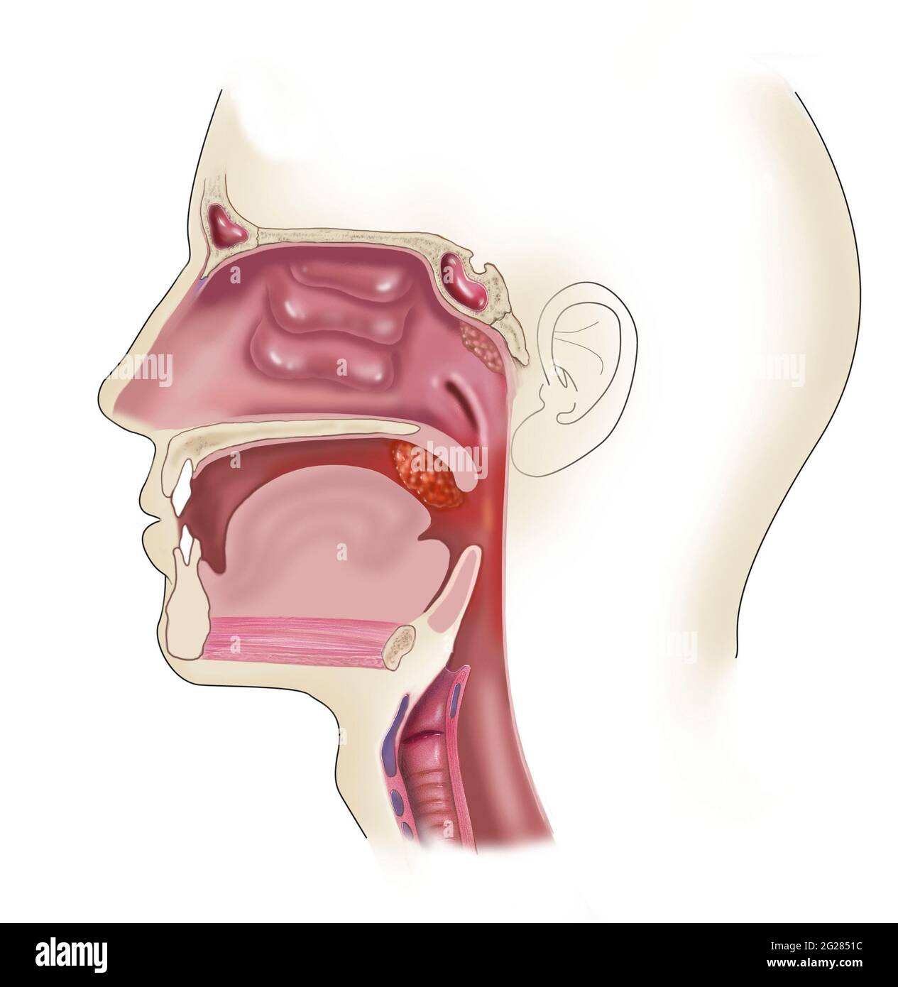 Side view of head showing location of swollen and enlarged tonsils. Stock Photohttps://www.alamy.com/image-license-details/?v=1https://www.alamy.com/side-view-of-head-showing-location-of-swollen-and-enlarged-tonsils-image431668088.html
RM2G2851C–Side view of head showing location of swollen and enlarged tonsils.
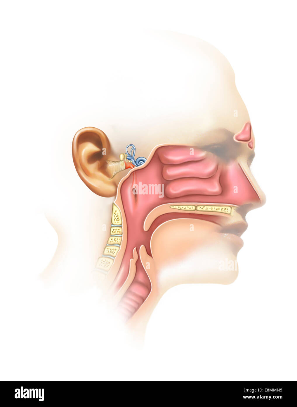 Anatomy of inner ear and sinuses. Stock Photohttps://www.alamy.com/image-license-details/?v=1https://www.alamy.com/stock-photo-anatomy-of-inner-ear-and-sinuses-74214033.html
RME8MMN5–Anatomy of inner ear and sinuses.
 Nasal cavity anatomy, illustration Stock Photohttps://www.alamy.com/image-license-details/?v=1https://www.alamy.com/nasal-cavity-anatomy-illustration-image471734611.html
RF2JBDA8K–Nasal cavity anatomy, illustration
 . Bulletin of the British Museum (Natural History) Zoology. posterior border of vestibule principal nasal cavity. concha Fig. 17 Snouts of lacertid lizards with left nasal cavity opened to show variation in thickness of postnasal region and length of nasal vestibule, a. 'Lacerta' jacksoni; b. Acanthodactylus cantoris. Cut areas are hatched.. Please note that these images are extracted from scanned page images that may have been digitally enhanced for readability - coloration and appearance of these illustrations may not perfectly resemble the original work.. Farn, Alexander E. London : Butterw Stock Photohttps://www.alamy.com/image-license-details/?v=1https://www.alamy.com/bulletin-of-the-british-museum-natural-history-zoology-posterior-border-of-vestibule-principal-nasal-cavity-concha-fig-17-snouts-of-lacertid-lizards-with-left-nasal-cavity-opened-to-show-variation-in-thickness-of-postnasal-region-and-length-of-nasal-vestibule-a-lacerta-jacksoni-b-acanthodactylus-cantoris-cut-areas-are-hatched-please-note-that-these-images-are-extracted-from-scanned-page-images-that-may-have-been-digitally-enhanced-for-readability-coloration-and-appearance-of-these-illustrations-may-not-perfectly-resemble-the-original-work-farn-alexander-e-london-butterw-image233912103.html
RMRGFH7K–. Bulletin of the British Museum (Natural History) Zoology. posterior border of vestibule principal nasal cavity. concha Fig. 17 Snouts of lacertid lizards with left nasal cavity opened to show variation in thickness of postnasal region and length of nasal vestibule, a. 'Lacerta' jacksoni; b. Acanthodactylus cantoris. Cut areas are hatched.. Please note that these images are extracted from scanned page images that may have been digitally enhanced for readability - coloration and appearance of these illustrations may not perfectly resemble the original work.. Farn, Alexander E. London : Butterw
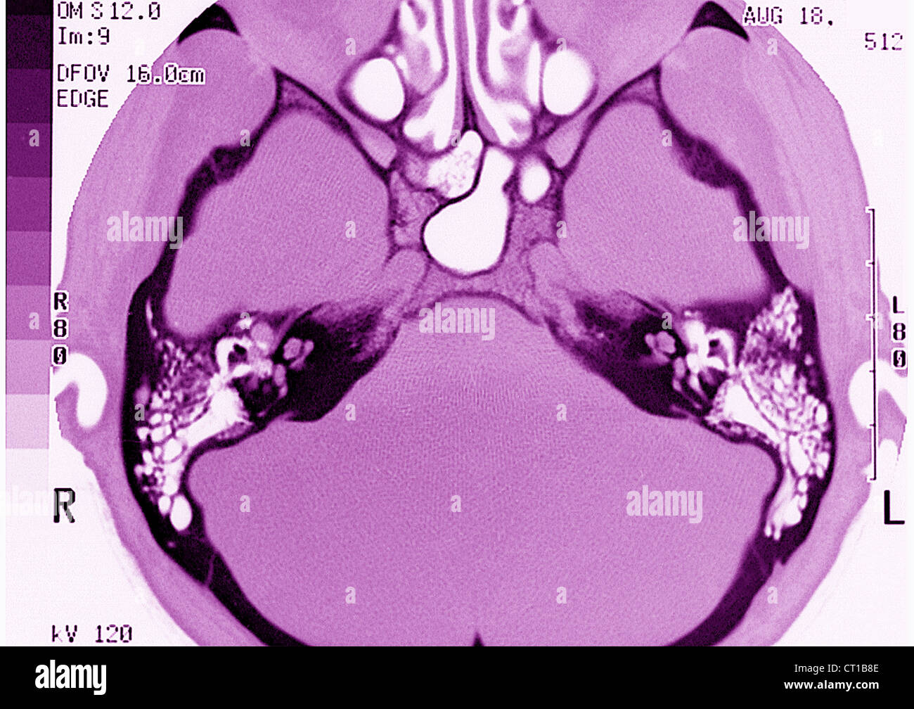 INTERNAL EAR, MRI Stock Photohttps://www.alamy.com/image-license-details/?v=1https://www.alamy.com/stock-photo-internal-ear-mri-49203294.html
RMCT1B8E–INTERNAL EAR, MRI
 Nasal cavity anatomy, illustration Stock Photohttps://www.alamy.com/image-license-details/?v=1https://www.alamy.com/nasal-cavity-anatomy-illustration-image471734491.html
RF2JBDA4B–Nasal cavity anatomy, illustration
 . The anatomy of the domestic animals . Veterinary anatomy. Fig 451.—Cast of Left Nostril, Nasal Diverticulum, and Nasal Vestibule OF Horse; Dorsal View. A^, Nostril; D, diverticulum; 1, super- ior commissure of nostril; 2, inferior com- missure; 3, space occupied by alar fold; 4, groove occupied by dorsal turbinate fold; 5, dorsal meatus. shaped somewhat like a com- ma, the convex margin being medial (Fig. 453). The cartil- ages are attached by fibrous tissue to the extremity of the Fig. 452.—Choss-section of Nasal Region or Horse. The section is out about two inches (5 cm.) behind the nostri Stock Photohttps://www.alamy.com/image-license-details/?v=1https://www.alamy.com/the-anatomy-of-the-domestic-animals-veterinary-anatomy-fig-451cast-of-left-nostril-nasal-diverticulum-and-nasal-vestibule-of-horse-dorsal-view-a-nostril-d-diverticulum-1-super-ior-commissure-of-nostril-2-inferior-com-missure-3-space-occupied-by-alar-fold-4-groove-occupied-by-dorsal-turbinate-fold-5-dorsal-meatus-shaped-somewhat-like-a-com-ma-the-convex-margin-being-medial-fig-453-the-cartil-ages-are-attached-by-fibrous-tissue-to-the-extremity-of-the-fig-452choss-section-of-nasal-region-or-horse-the-section-is-out-about-two-inches-5-cm-behind-the-nostri-image232324650.html
RMRDY8CX–. The anatomy of the domestic animals . Veterinary anatomy. Fig 451.—Cast of Left Nostril, Nasal Diverticulum, and Nasal Vestibule OF Horse; Dorsal View. A^, Nostril; D, diverticulum; 1, super- ior commissure of nostril; 2, inferior com- missure; 3, space occupied by alar fold; 4, groove occupied by dorsal turbinate fold; 5, dorsal meatus. shaped somewhat like a com- ma, the convex margin being medial (Fig. 453). The cartil- ages are attached by fibrous tissue to the extremity of the Fig. 452.—Choss-section of Nasal Region or Horse. The section is out about two inches (5 cm.) behind the nostri
 Nasal cavity anatomy, illustration Stock Photohttps://www.alamy.com/image-license-details/?v=1https://www.alamy.com/nasal-cavity-anatomy-illustration-image471734549.html
RF2JBDA6D–Nasal cavity anatomy, illustration
 . The anatomy of the domestic animals. Veterinary anatomy. Fig 451.—Cast of Left Nostbil. Nasal Diverticulum, and Nasal Vestibule OF Horse; Dorsal View. N, Nostril; U, diverticulum; 1, super- ; of nostril: 2, inferior com- 3, space occupied by alar fold; 4, groove occupied by dorsal turbinate fold; 5, dorsal meatus. Fig. 452.—Cross-section of Nasal Region of Horse. The section is cut about two inches (5 cm.) behind the nostrils and a little more than half an inch behind the labial commissure, a, Dorsal meatus; b, middle meatus; c, ventral meatus; d, diver- ticulum nasi; e, dorsal turbinate fol Stock Photohttps://www.alamy.com/image-license-details/?v=1https://www.alamy.com/the-anatomy-of-the-domestic-animals-veterinary-anatomy-fig-451cast-of-left-nostbil-nasal-diverticulum-and-nasal-vestibule-of-horse-dorsal-view-n-nostril-u-diverticulum-1-super-of-nostril-2-inferior-com-3-space-occupied-by-alar-fold-4-groove-occupied-by-dorsal-turbinate-fold-5-dorsal-meatus-fig-452cross-section-of-nasal-region-of-horse-the-section-is-cut-about-two-inches-5-cm-behind-the-nostrils-and-a-little-more-than-half-an-inch-behind-the-labial-commissure-a-dorsal-meatus-b-middle-meatus-c-ventral-meatus-d-diver-ticulum-nasi-e-dorsal-turbinate-fol-image236760605.html
RMRN5AFW–. The anatomy of the domestic animals. Veterinary anatomy. Fig 451.—Cast of Left Nostbil. Nasal Diverticulum, and Nasal Vestibule OF Horse; Dorsal View. N, Nostril; U, diverticulum; 1, super- ; of nostril: 2, inferior com- 3, space occupied by alar fold; 4, groove occupied by dorsal turbinate fold; 5, dorsal meatus. Fig. 452.—Cross-section of Nasal Region of Horse. The section is cut about two inches (5 cm.) behind the nostrils and a little more than half an inch behind the labial commissure, a, Dorsal meatus; b, middle meatus; c, ventral meatus; d, diver- ticulum nasi; e, dorsal turbinate fol
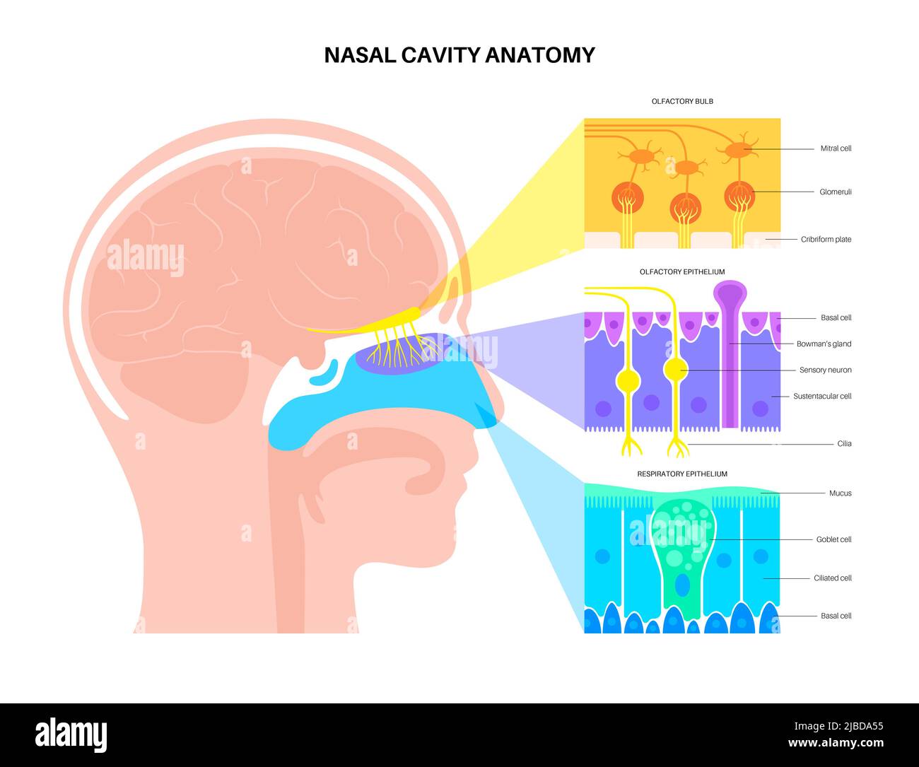 Nasal cavity anatomy, illustration Stock Photohttps://www.alamy.com/image-license-details/?v=1https://www.alamy.com/nasal-cavity-anatomy-illustration-image471734513.html
RF2JBDA55–Nasal cavity anatomy, illustration
 Diseases of the nose and throat; a text-book for students and practitioners . Fig. 12.—Spectaclk-Frame. or stands in front of the patient, who inclines his head slightlybackward. The tip and external surface of the nose having beeninspected, a closed nasal speculum is to be introduced. Theinstrument is held in the right hand for examination of the RHINOSCOPY EXAMINATION OF THE NASAL PASSAGES. 19 left nasal fossa, and in the left liand for the riglit fossa. Whenwell within the vestibule, the blades are separated or allowed toseparate according to the construction of the instrument. Caremust be Stock Photohttps://www.alamy.com/image-license-details/?v=1https://www.alamy.com/diseases-of-the-nose-and-throat-a-text-book-for-students-and-practitioners-fig-12spectaclk-frame-or-stands-in-front-of-the-patient-who-inclines-his-head-slightlybackward-the-tip-and-external-surface-of-the-nose-having-beeninspected-a-closed-nasal-speculum-is-to-be-introduced-theinstrument-is-held-in-the-right-hand-for-examination-of-the-rhinoscopy-examination-of-the-nasal-passages-19-left-nasal-fossa-and-in-the-left-liand-for-the-riglit-fossa-whenwell-within-the-vestibule-the-blades-are-separated-or-allowed-toseparate-according-to-the-construction-of-the-instrument-caremust-be-image339333886.html
RM2AM1YRA–Diseases of the nose and throat; a text-book for students and practitioners . Fig. 12.—Spectaclk-Frame. or stands in front of the patient, who inclines his head slightlybackward. The tip and external surface of the nose having beeninspected, a closed nasal speculum is to be introduced. Theinstrument is held in the right hand for examination of the RHINOSCOPY EXAMINATION OF THE NASAL PASSAGES. 19 left nasal fossa, and in the left liand for the riglit fossa. Whenwell within the vestibule, the blades are separated or allowed toseparate according to the construction of the instrument. Caremust be
 Olfactory system, illustration Stock Photohttps://www.alamy.com/image-license-details/?v=1https://www.alamy.com/olfactory-system-illustration-image471734560.html
RF2JBDA6T–Olfactory system, illustration
 Diseases of the nose and throat; a text-book for students and practitioners . Fig. 12.-Siectacle-Frame. or stands in front of the patient, who inclines his head slightlybackward. The tip and external surface of the nose having beeninspected, a closed nasal speculum is to be introduced. Theinstrument is held in the right hand for examination of the RHINOSCOPY EXAMINATION OF THE NASAL PASSAGES. 19 left nasal fossa, and in the left hand for the right fossa. Whenwell within the vestibule, the blades are separated or allowed toseparate according to the construction of the instrument. Caremust be ta Stock Photohttps://www.alamy.com/image-license-details/?v=1https://www.alamy.com/diseases-of-the-nose-and-throat-a-text-book-for-students-and-practitioners-fig-12-siectacle-frame-or-stands-in-front-of-the-patient-who-inclines-his-head-slightlybackward-the-tip-and-external-surface-of-the-nose-having-beeninspected-a-closed-nasal-speculum-is-to-be-introduced-theinstrument-is-held-in-the-right-hand-for-examination-of-the-rhinoscopy-examination-of-the-nasal-passages-19-left-nasal-fossa-and-in-the-left-hand-for-the-right-fossa-whenwell-within-the-vestibule-the-blades-are-separated-or-allowed-toseparate-according-to-the-construction-of-the-instrument-caremust-be-ta-image339226890.html
RM2AKW3A2–Diseases of the nose and throat; a text-book for students and practitioners . Fig. 12.-Siectacle-Frame. or stands in front of the patient, who inclines his head slightlybackward. The tip and external surface of the nose having beeninspected, a closed nasal speculum is to be introduced. Theinstrument is held in the right hand for examination of the RHINOSCOPY EXAMINATION OF THE NASAL PASSAGES. 19 left nasal fossa, and in the left hand for the right fossa. Whenwell within the vestibule, the blades are separated or allowed toseparate according to the construction of the instrument. Caremust be ta
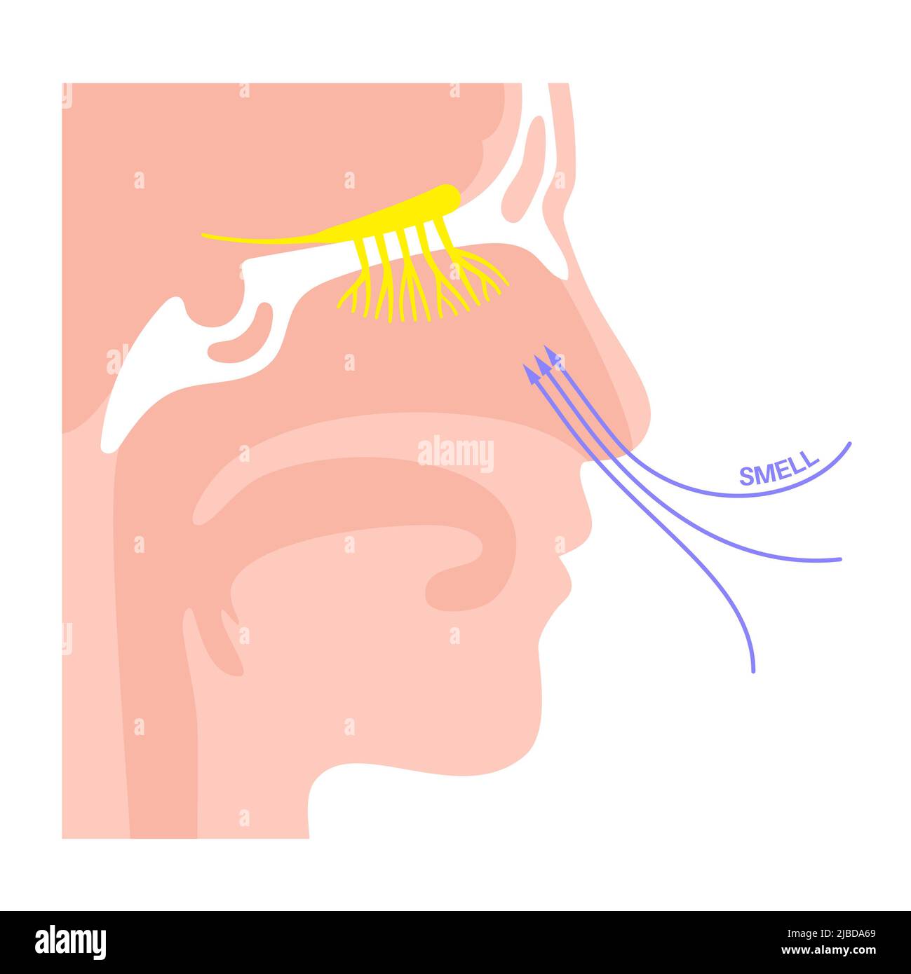 Olfactory system, illustration Stock Photohttps://www.alamy.com/image-license-details/?v=1https://www.alamy.com/olfactory-system-illustration-image471734545.html
RF2JBDA69–Olfactory system, illustration
 A manual of diseases of the nose and throat . t all is made upon thesides of the handle, the force of the spring may be sogreat, when exerted entirely upon the nasal orifice, asto cause discomfort to the patient. Where the vestibule contains many hairs the specu-lum which the author is in the habit of using is hismodification of the Chiari speculum (Fig. 9). Theblades, being solid, push the hairs aside, and the viewof the nares is no longer obstructed. It should be held in the hand, the blades being closedwhen inserted; pressure upon the handles forces theblades apart, thus dilating the orific Stock Photohttps://www.alamy.com/image-license-details/?v=1https://www.alamy.com/a-manual-of-diseases-of-the-nose-and-throat-t-all-is-made-upon-thesides-of-the-handle-the-force-of-the-spring-may-be-sogreat-when-exerted-entirely-upon-the-nasal-orifice-asto-cause-discomfort-to-the-patient-where-the-vestibule-contains-many-hairs-the-specu-lum-which-the-author-is-in-the-habit-of-using-is-hismodification-of-the-chiari-speculum-fig-9-theblades-being-solid-push-the-hairs-aside-and-the-viewof-the-nares-is-no-longer-obstructed-it-should-be-held-in-the-hand-the-blades-being-closedwhen-inserted-pressure-upon-the-handles-forces-theblades-apart-thus-dilating-the-orific-image338070029.html
RM2AJ0BNH–A manual of diseases of the nose and throat . t all is made upon thesides of the handle, the force of the spring may be sogreat, when exerted entirely upon the nasal orifice, asto cause discomfort to the patient. Where the vestibule contains many hairs the specu-lum which the author is in the habit of using is hismodification of the Chiari speculum (Fig. 9). Theblades, being solid, push the hairs aside, and the viewof the nares is no longer obstructed. It should be held in the hand, the blades being closedwhen inserted; pressure upon the handles forces theblades apart, thus dilating the orific
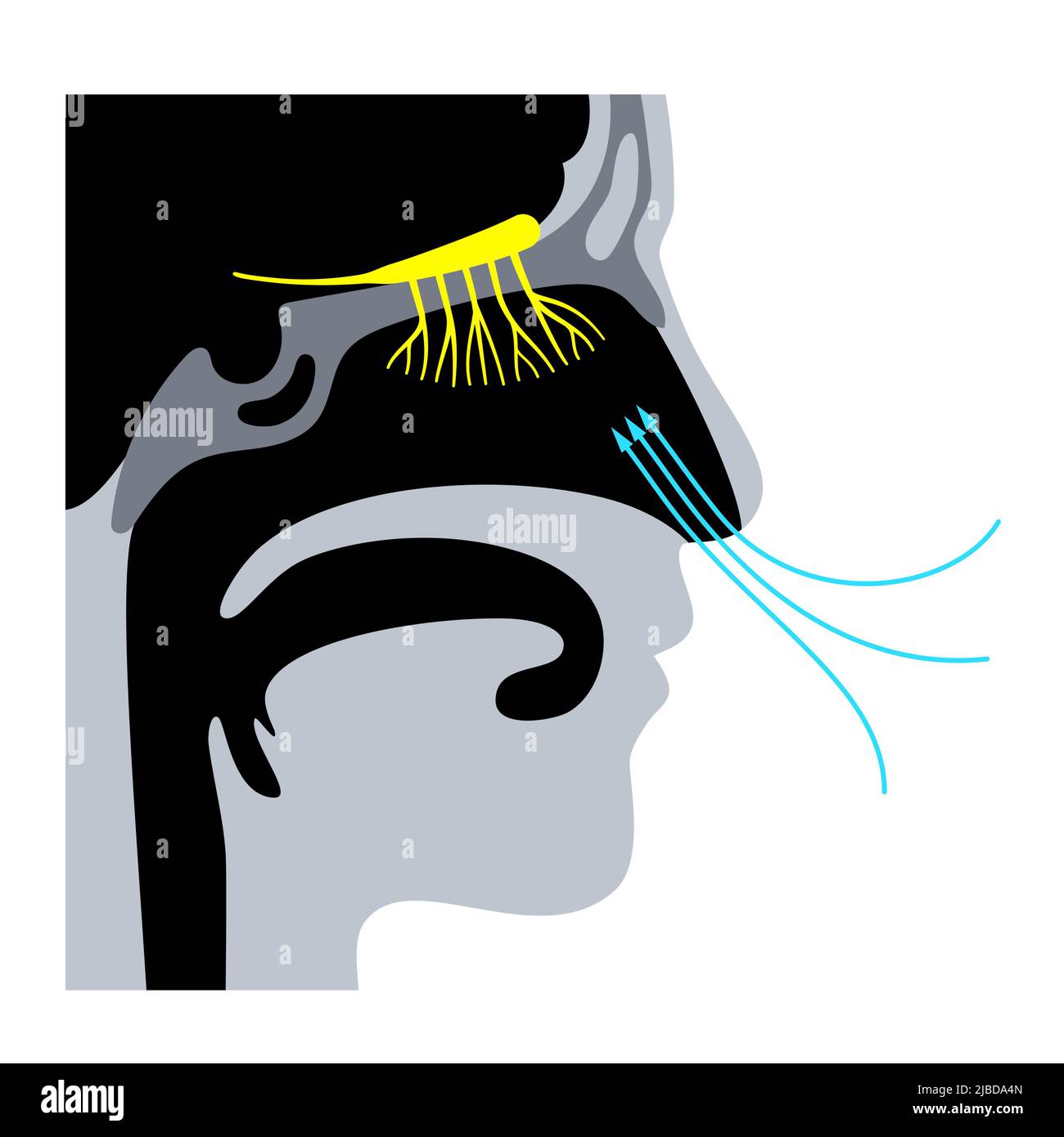 Olfactory system, illustration Stock Photohttps://www.alamy.com/image-license-details/?v=1https://www.alamy.com/olfactory-system-illustration-image471734501.html
RF2JBDA4N–Olfactory system, illustration
 Tri-State medical journal and practitioner . ntrast with that which lines the fossae. * This illustration only shows the two turbinated bodies; the third, however, lies just above the middle.+ Any further anatomy on the bones of the nasal fossae required to elucidate the contents of thismemoir can be found in any work on anatomy. 154 Original Articles. The character of the epithelium varies in the different parts, and bythis, in a general way, three regions of the nasal fossae may be distinguished.Thus the region of the external nostrils (the vestibule) is lined with strati-fied squamous epith Stock Photohttps://www.alamy.com/image-license-details/?v=1https://www.alamy.com/tri-state-medical-journal-and-practitioner-ntrast-with-that-which-lines-the-fossae-this-illustration-only-shows-the-two-turbinated-bodies-the-third-however-lies-just-above-the-middle-any-further-anatomy-on-the-bones-of-the-nasal-fossae-required-to-elucidate-the-contents-of-thismemoir-can-be-found-in-any-work-on-anatomy-154-original-articles-the-character-of-the-epithelium-varies-in-the-different-parts-and-bythis-in-a-general-way-three-regions-of-the-nasal-fossae-may-be-distinguishedthus-the-region-of-the-external-nostrils-the-vestibule-is-lined-with-strati-fied-squamous-epith-image342891150.html
RM2AWT14E–Tri-State medical journal and practitioner . ntrast with that which lines the fossae. * This illustration only shows the two turbinated bodies; the third, however, lies just above the middle.+ Any further anatomy on the bones of the nasal fossae required to elucidate the contents of thismemoir can be found in any work on anatomy. 154 Original Articles. The character of the epithelium varies in the different parts, and bythis, in a general way, three regions of the nasal fossae may be distinguished.Thus the region of the external nostrils (the vestibule) is lined with strati-fied squamous epith
 Olfactory system, illustration Stock Photohttps://www.alamy.com/image-license-details/?v=1https://www.alamy.com/olfactory-system-illustration-image471734529.html
RF2JBDA5N–Olfactory system, illustration
 . Diseases of the nose and throat . Fig. 12.—The lateral wall of the nasal cavity showing the position ofthe turbinates and the opening of the Eustachian tube. meatus. Anteriorly, the entrance into the cavity is throughthe vestibule and under the bony arch made by the free edgesof the nasal bones and nasal processes of the superior maxillae.Posteriorly, they communicate with the pharyngeal cavitythrough the choance, oval openings formed by the vomer, ANATOMY AND PHYSIOLOGY OF THE NOSE 41 sphenoid and palate bones. Externally, the lower halfof each cavity is separated from the maxillary antrum Stock Photohttps://www.alamy.com/image-license-details/?v=1https://www.alamy.com/diseases-of-the-nose-and-throat-fig-12the-lateral-wall-of-the-nasal-cavity-showing-the-position-ofthe-turbinates-and-the-opening-of-the-eustachian-tube-meatus-anteriorly-the-entrance-into-the-cavity-is-throughthe-vestibule-and-under-the-bony-arch-made-by-the-free-edgesof-the-nasal-bones-and-nasal-processes-of-the-superior-maxillaeposteriorly-they-communicate-with-the-pharyngeal-cavitythrough-the-choance-oval-openings-formed-by-the-vomer-anatomy-and-physiology-of-the-nose-41-sphenoid-and-palate-bones-externally-the-lower-halfof-each-cavity-is-separated-from-the-maxillary-antrum-image372298443.html
RM2CHKJCY–. Diseases of the nose and throat . Fig. 12.—The lateral wall of the nasal cavity showing the position ofthe turbinates and the opening of the Eustachian tube. meatus. Anteriorly, the entrance into the cavity is throughthe vestibule and under the bony arch made by the free edgesof the nasal bones and nasal processes of the superior maxillae.Posteriorly, they communicate with the pharyngeal cavitythrough the choance, oval openings formed by the vomer, ANATOMY AND PHYSIOLOGY OF THE NOSE 41 sphenoid and palate bones. Externally, the lower halfof each cavity is separated from the maxillary antrum
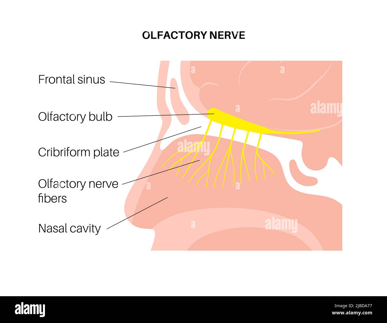 Olfactory system, illustration Stock Photohttps://www.alamy.com/image-license-details/?v=1https://www.alamy.com/olfactory-system-illustration-image471734571.html
RF2JBDA77–Olfactory system, illustration
 . Twentieth century practice; an international encyclopedia of modern medical science by leading authorities of Europe and America . Fig. 88.—Papilloma of the Ventricular Band Com-pletely Filling the Vestibule of the Larynx. Fig. 89.—Cystoma of the Epiglottis. fibroma, but the cliuical historj- should point to its origin and char-acter. The softer tumors, such as myxoma and cystoma, would ofcourse not be confounded with a growth of such density as a fibroma.Myxoma.—This growth iu the larynx somewhat resembles myx-oma of the nasal cavities, especially where it is broadly sessile, butwhen multip Stock Photohttps://www.alamy.com/image-license-details/?v=1https://www.alamy.com/twentieth-century-practice-an-international-encyclopedia-of-modern-medical-science-by-leading-authorities-of-europe-and-america-fig-88papilloma-of-the-ventricular-band-com-pletely-filling-the-vestibule-of-the-larynx-fig-89cystoma-of-the-epiglottis-fibroma-but-the-cliuical-historj-should-point-to-its-origin-and-char-acter-the-softer-tumors-such-as-myxoma-and-cystoma-would-ofcourse-not-be-confounded-with-a-growth-of-such-density-as-a-fibromamyxomathis-growth-iu-the-larynx-somewhat-resembles-myx-oma-of-the-nasal-cavities-especially-where-it-is-broadly-sessile-butwhen-multip-image369820112.html
RM2CDJN94–. Twentieth century practice; an international encyclopedia of modern medical science by leading authorities of Europe and America . Fig. 88.—Papilloma of the Ventricular Band Com-pletely Filling the Vestibule of the Larynx. Fig. 89.—Cystoma of the Epiglottis. fibroma, but the cliuical historj- should point to its origin and char-acter. The softer tumors, such as myxoma and cystoma, would ofcourse not be confounded with a growth of such density as a fibroma.Myxoma.—This growth iu the larynx somewhat resembles myx-oma of the nasal cavities, especially where it is broadly sessile, butwhen multip
 Olfactory system, illustration Stock Photohttps://www.alamy.com/image-license-details/?v=1https://www.alamy.com/olfactory-system-illustration-image471734572.html
RF2JBDA78–Olfactory system, illustration
 . Nursing in diseases of the eye, ear, nose, and throat . into a right and leftnasal cavity by the septum. The anterior opening ofeach chamber is called the vestibule, around the innerpart of which thick hairs grow, called vibrissa?. Theposterior openings are called the choanal or posteriornares. Each nasal cavity has a floor, which forms the supe-rior surface of the hard and soft palate; a roof, whichis formed by the cribriform plate of the ethmoid bone:an inner wall, which is the septum; and an outer wall,which is made up of the turbinated bodies which sepa-rate each chamber into three meati Stock Photohttps://www.alamy.com/image-license-details/?v=1https://www.alamy.com/nursing-in-diseases-of-the-eye-ear-nose-and-throat-into-a-right-and-leftnasal-cavity-by-the-septum-the-anterior-opening-ofeach-chamber-is-called-the-vestibule-around-the-innerpart-of-which-thick-hairs-grow-called-vibrissa-theposterior-openings-are-called-the-choanal-or-posteriornares-each-nasal-cavity-has-a-floor-which-forms-the-supe-rior-surface-of-the-hard-and-soft-palate-a-roof-whichis-formed-by-the-cribriform-plate-of-the-ethmoid-bonean-inner-wall-which-is-the-septum-and-an-outer-wallwhich-is-made-up-of-the-turbinated-bodies-which-sepa-rate-each-chamber-into-three-meati-image371641806.html
RM2CGHMWJ–. Nursing in diseases of the eye, ear, nose, and throat . into a right and leftnasal cavity by the septum. The anterior opening ofeach chamber is called the vestibule, around the innerpart of which thick hairs grow, called vibrissa?. Theposterior openings are called the choanal or posteriornares. Each nasal cavity has a floor, which forms the supe-rior surface of the hard and soft palate; a roof, whichis formed by the cribriform plate of the ethmoid bone:an inner wall, which is the septum; and an outer wall,which is made up of the turbinated bodies which sepa-rate each chamber into three meati
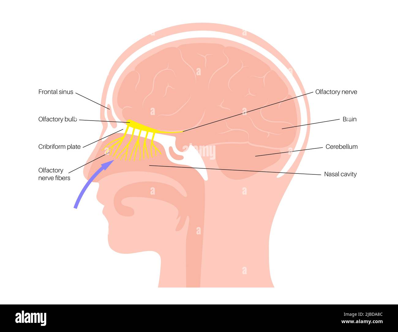 Olfactory system, illustration Stock Photohttps://www.alamy.com/image-license-details/?v=1https://www.alamy.com/olfactory-system-illustration-image471734604.html
RF2JBDA8C–Olfactory system, illustration
 . Twentieth century practice; an international encyclopedia of modern medical science by leading authorities of Europe and America . Fig. 88.—Papilloma of the Ventricular Band Com-pletely Filling the Vestibule of the Larynx. Fig. 89.—Cystoma of the Epiglottis. fibroma, but the cliuical historj- should point to its origin and char-acter. The softer tumors, such as myxoma and cystoma, would ofcourse not be confounded with a growth of such density as a fibroma.Myxoma.—This growth iu the larynx somewhat resembles myx-oma of the nasal cavities, especially where it is broadly sessile, butwhen multip Stock Photohttps://www.alamy.com/image-license-details/?v=1https://www.alamy.com/twentieth-century-practice-an-international-encyclopedia-of-modern-medical-science-by-leading-authorities-of-europe-and-america-fig-88papilloma-of-the-ventricular-band-com-pletely-filling-the-vestibule-of-the-larynx-fig-89cystoma-of-the-epiglottis-fibroma-but-the-cliuical-historj-should-point-to-its-origin-and-char-acter-the-softer-tumors-such-as-myxoma-and-cystoma-would-ofcourse-not-be-confounded-with-a-growth-of-such-density-as-a-fibromamyxomathis-growth-iu-the-larynx-somewhat-resembles-myx-oma-of-the-nasal-cavities-especially-where-it-is-broadly-sessile-butwhen-multip-image369820202.html
RM2CDJNCA–. Twentieth century practice; an international encyclopedia of modern medical science by leading authorities of Europe and America . Fig. 88.—Papilloma of the Ventricular Band Com-pletely Filling the Vestibule of the Larynx. Fig. 89.—Cystoma of the Epiglottis. fibroma, but the cliuical historj- should point to its origin and char-acter. The softer tumors, such as myxoma and cystoma, would ofcourse not be confounded with a growth of such density as a fibroma.Myxoma.—This growth iu the larynx somewhat resembles myx-oma of the nasal cavities, especially where it is broadly sessile, butwhen multip
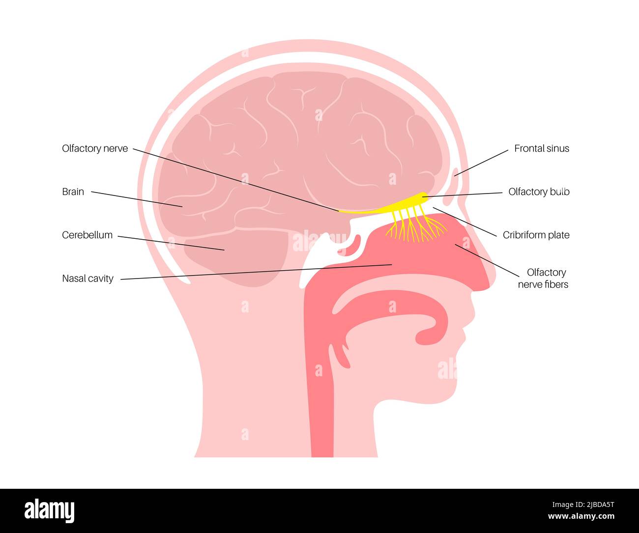 Olfactory system, illustration Stock Photohttps://www.alamy.com/image-license-details/?v=1https://www.alamy.com/olfactory-system-illustration-image471734532.html
RF2JBDA5T–Olfactory system, illustration
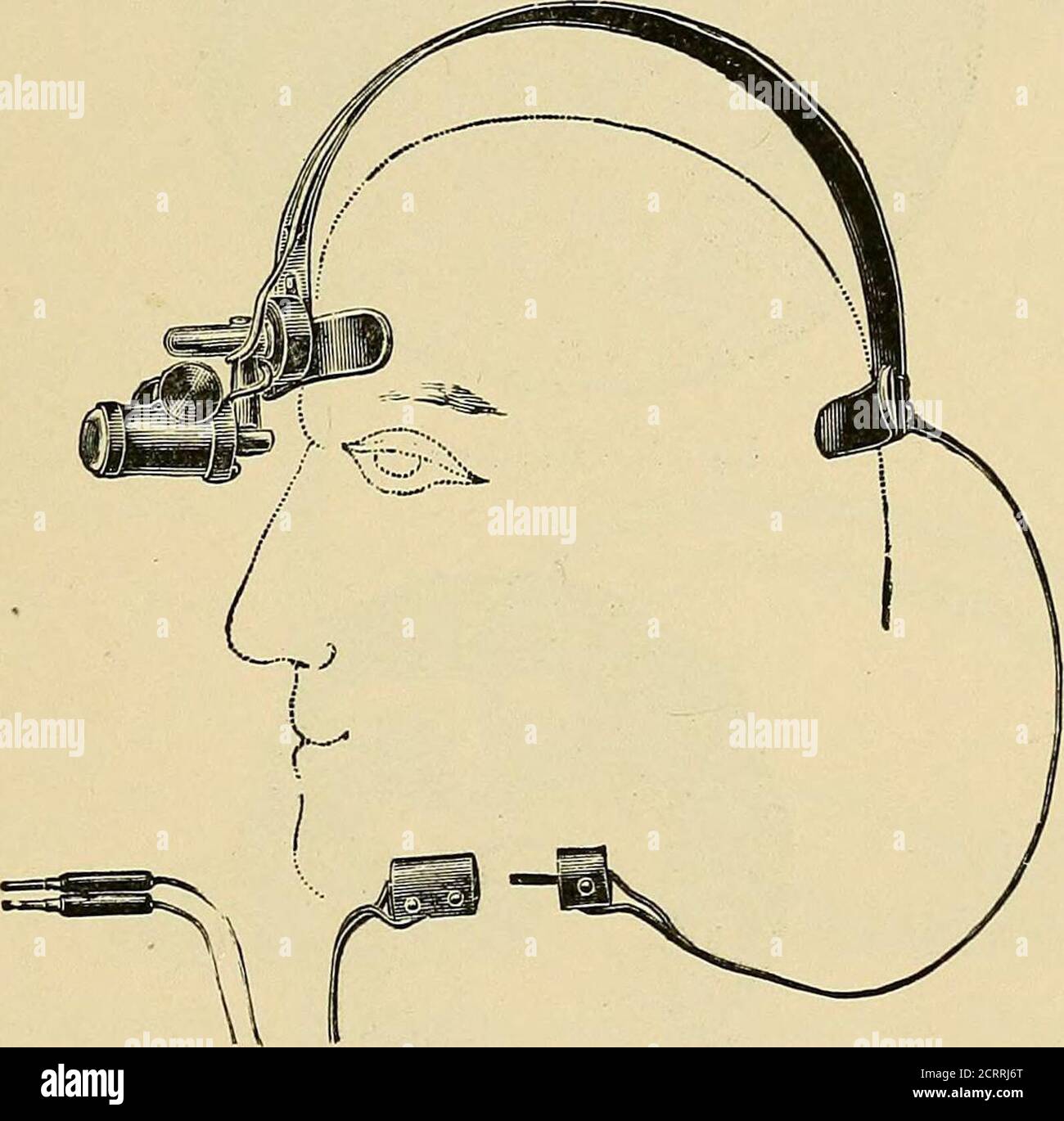 . Diseases of the nose and throat . ing uponthe subject. Still, one thing is certain, that, whereas the mucus ofthe vestibule is always loaded with microscopical germs, that in theback parts of the normal nasal passages is almost, if not entirely, freefrom them. It is possible that a great deal of the cleansing processis clue, however, to the oft-repeated efforts of Nature to eject, byforcible expulsion, anything that irritates the nasal passages. The special function of the large antra of Highmore is probablyone of phonation. Filled, as they are, by air when in a healthy con-dition, with free Stock Photohttps://www.alamy.com/image-license-details/?v=1https://www.alamy.com/diseases-of-the-nose-and-throat-ing-uponthe-subject-still-one-thing-is-certain-that-whereas-the-mucus-ofthe-vestibule-is-always-loaded-with-microscopical-germs-that-in-theback-parts-of-the-normal-nasal-passages-is-almost-if-not-entirely-freefrom-them-it-is-possible-that-a-great-deal-of-the-cleansing-processis-clue-however-to-the-oft-repeated-efforts-of-nature-to-eject-byforcible-expulsion-anything-that-irritates-the-nasal-passages-the-special-function-of-the-large-antra-of-highmore-is-probablyone-of-phonation-filled-as-they-are-by-air-when-in-a-healthy-con-dition-with-free-image376074016.html
RM2CRRJ6T–. Diseases of the nose and throat . ing uponthe subject. Still, one thing is certain, that, whereas the mucus ofthe vestibule is always loaded with microscopical germs, that in theback parts of the normal nasal passages is almost, if not entirely, freefrom them. It is possible that a great deal of the cleansing processis clue, however, to the oft-repeated efforts of Nature to eject, byforcible expulsion, anything that irritates the nasal passages. The special function of the large antra of Highmore is probablyone of phonation. Filled, as they are, by air when in a healthy con-dition, with free
 Olfactory system, illustration Stock Photohttps://www.alamy.com/image-license-details/?v=1https://www.alamy.com/olfactory-system-illustration-image471734524.html
RF2JBDA5G–Olfactory system, illustration
 . Diseases of the nose and throat . Fig. i8.—Cross section through the snout of a wolf, showing thehigh development of the olfactory region (Ingersoll). extent a retrograde development. The ethmoidal turbinalsare reduced to two or three, the accessory sinuses are nearlyclosed, of doubtful use, and show different partial partitionsand grooves which suggest the gradual disappearance of amore complex structure. The principal function of the nasal organ in man is towarm, moisten and filter the inspired air. The external noseserves as a protection and contains the vestibule to thenasal cavities. Th Stock Photohttps://www.alamy.com/image-license-details/?v=1https://www.alamy.com/diseases-of-the-nose-and-throat-fig-i8cross-section-through-the-snout-of-a-wolf-showing-thehigh-development-of-the-olfactory-region-ingersoll-extent-a-retrograde-development-the-ethmoidal-turbinalsare-reduced-to-two-or-three-the-accessory-sinuses-are-nearlyclosed-of-doubtful-use-and-show-different-partial-partitionsand-grooves-which-suggest-the-gradual-disappearance-of-amore-complex-structure-the-principal-function-of-the-nasal-organ-in-man-is-towarm-moisten-and-filter-the-inspired-air-the-external-noseserves-as-a-protection-and-contains-the-vestibule-to-thenasal-cavities-th-image372294804.html
RM2CHKDR0–. Diseases of the nose and throat . Fig. i8.—Cross section through the snout of a wolf, showing thehigh development of the olfactory region (Ingersoll). extent a retrograde development. The ethmoidal turbinalsare reduced to two or three, the accessory sinuses are nearlyclosed, of doubtful use, and show different partial partitionsand grooves which suggest the gradual disappearance of amore complex structure. The principal function of the nasal organ in man is towarm, moisten and filter the inspired air. The external noseserves as a protection and contains the vestibule to thenasal cavities. Th
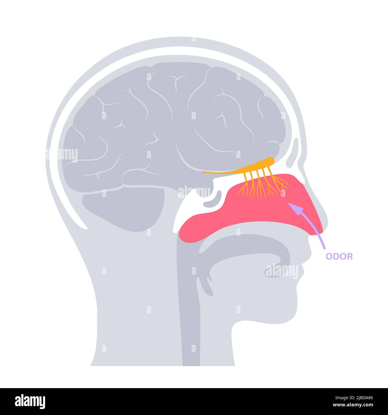 Olfactory system, illustration Stock Photohttps://www.alamy.com/image-license-details/?v=1https://www.alamy.com/olfactory-system-illustration-image471734598.html
RF2JBDA86–Olfactory system, illustration
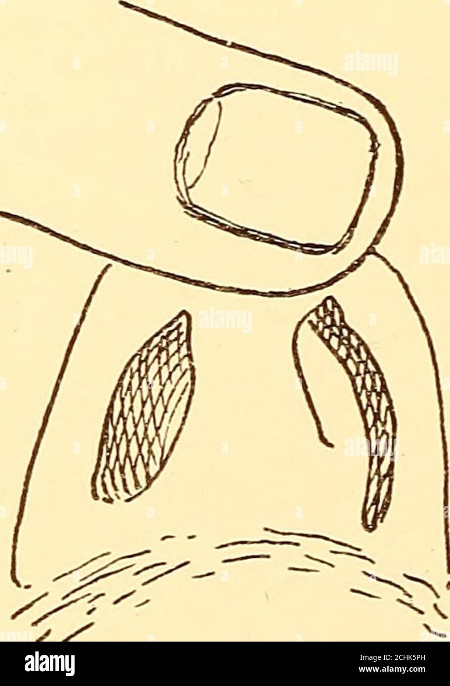 . Diseases of the nose and throat . correcting asymmetries, especially the turbinates oppositea concave septum, may be more noticeable to casual inspec-tion than the asymmetry of the septum itself. The nasal processes of the superior maxilla do nottake part in this adjustment, consequently a deviation ofthe septal cartilage which extends forward into the vestibuleobstructs respiration on that side more than would an equaldegree of deviation farther back. When the deviation ex-tends forward into the vestibule the patient may noticethat on one side it is only by drawing the alae and skin 78 DISE Stock Photohttps://www.alamy.com/image-license-details/?v=1https://www.alamy.com/diseases-of-the-nose-and-throat-correcting-asymmetries-especially-the-turbinates-oppositea-concave-septum-may-be-more-noticeable-to-casual-inspec-tion-than-the-asymmetry-of-the-septum-itself-the-nasal-processes-of-the-superior-maxilla-do-nottake-part-in-this-adjustment-consequently-a-deviation-ofthe-septal-cartilage-which-extends-forward-into-the-vestibuleobstructs-respiration-on-that-side-more-than-would-an-equaldegree-of-deviation-farther-back-when-the-deviation-ex-tends-forward-into-the-vestibule-the-patient-may-noticethat-on-one-side-it-is-only-by-drawing-the-alae-and-skin-78-dise-image372288521.html
RM2CHK5PH–. Diseases of the nose and throat . correcting asymmetries, especially the turbinates oppositea concave septum, may be more noticeable to casual inspec-tion than the asymmetry of the septum itself. The nasal processes of the superior maxilla do nottake part in this adjustment, consequently a deviation ofthe septal cartilage which extends forward into the vestibuleobstructs respiration on that side more than would an equaldegree of deviation farther back. When the deviation ex-tends forward into the vestibule the patient may noticethat on one side it is only by drawing the alae and skin 78 DISE
 Olfactory system, illustration Stock Photohttps://www.alamy.com/image-license-details/?v=1https://www.alamy.com/olfactory-system-illustration-image471734542.html
RF2JBDA66–Olfactory system, illustration
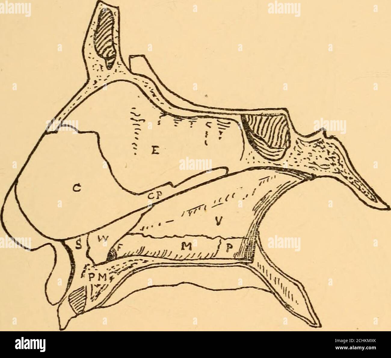 . Diseases of the nose and throat . A, Alar cartilage;S, Sesamoid cartilages; C, Septal cartilage; V, Vestibule; N, Nasalspine. THE NASAL SEPTUM The nasal septum is a plate formed by the vomer, theperpendicular plate of the ethmoid, and the triangular orquadrangular septal cartilage. It is attached above to theethmoid, below to the maxillae and palate bones, anteriorlyto the frontal and nasal bones, and posteriorly to thesphenoid. The lower anterior angle has no bony attach-ment. The lower posterior edge formed by the vomer isfree, separating the choanae. Between the vomer and theperpendicular Stock Photohttps://www.alamy.com/image-license-details/?v=1https://www.alamy.com/diseases-of-the-nose-and-throat-a-alar-cartilages-sesamoid-cartilages-c-septal-cartilage-v-vestibule-n-nasalspine-the-nasal-septum-the-nasal-septum-is-a-plate-formed-by-the-vomer-theperpendicular-plate-of-the-ethmoid-and-the-triangular-orquadrangular-septal-cartilage-it-is-attached-above-to-theethmoid-below-to-the-maxillae-and-palate-bones-anteriorlyto-the-frontal-and-nasal-bones-and-posteriorly-to-thesphenoid-the-lower-anterior-angle-has-no-bony-attach-ment-the-lower-posterior-edge-formed-by-the-vomer-isfree-separating-the-choanae-between-the-vomer-and-theperpendicular-image372300395.html
RM2CHKMXK–. Diseases of the nose and throat . A, Alar cartilage;S, Sesamoid cartilages; C, Septal cartilage; V, Vestibule; N, Nasalspine. THE NASAL SEPTUM The nasal septum is a plate formed by the vomer, theperpendicular plate of the ethmoid, and the triangular orquadrangular septal cartilage. It is attached above to theethmoid, below to the maxillae and palate bones, anteriorlyto the frontal and nasal bones, and posteriorly to thesphenoid. The lower anterior angle has no bony attach-ment. The lower posterior edge formed by the vomer isfree, separating the choanae. Between the vomer and theperpendicular
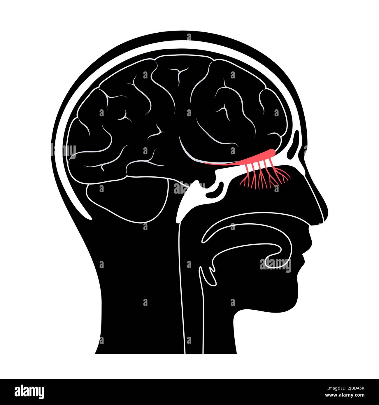 Olfactory system, illustration Stock Photohttps://www.alamy.com/image-license-details/?v=1https://www.alamy.com/olfactory-system-illustration-image471734555.html
RF2JBDA6K–Olfactory system, illustration
 . Chordate anatomy. Chordata; Anatomy, Comparative. 402 CHORDATE ANATOMY A lacrimal duct from each eye opens into the nasal passage, and serves to moisten the olfactory epithelium. Some reptiles, lizards for example, have added to the nasal passage a more expanded and glandular vestibule, which is apparently a mechanism for eliminating dust from the air taken into the lungs. Novel also in this group are the paired turbinal bones or conchae, which project into the nasal passages, and serve to increase and support the olfactory membrane. A nasopharyngeal cavity distinct from the mouth cavity als Stock Photohttps://www.alamy.com/image-license-details/?v=1https://www.alamy.com/chordate-anatomy-chordata-anatomy-comparative-402-chordate-anatomy-a-lacrimal-duct-from-each-eye-opens-into-the-nasal-passage-and-serves-to-moisten-the-olfactory-epithelium-some-reptiles-lizards-for-example-have-added-to-the-nasal-passage-a-more-expanded-and-glandular-vestibule-which-is-apparently-a-mechanism-for-eliminating-dust-from-the-air-taken-into-the-lungs-novel-also-in-this-group-are-the-paired-turbinal-bones-or-conchae-which-project-into-the-nasal-passages-and-serve-to-increase-and-support-the-olfactory-membrane-a-nasopharyngeal-cavity-distinct-from-the-mouth-cavity-als-image234923647.html
RMRJ5KE7–. Chordate anatomy. Chordata; Anatomy, Comparative. 402 CHORDATE ANATOMY A lacrimal duct from each eye opens into the nasal passage, and serves to moisten the olfactory epithelium. Some reptiles, lizards for example, have added to the nasal passage a more expanded and glandular vestibule, which is apparently a mechanism for eliminating dust from the air taken into the lungs. Novel also in this group are the paired turbinal bones or conchae, which project into the nasal passages, and serve to increase and support the olfactory membrane. A nasopharyngeal cavity distinct from the mouth cavity als
 . Smell, taste, and allied senses in the vertebrates . Senses and sensation; Vertebrates. ANATOMY OF THE OLFACTORY ORGAN 25 the lateral wall of each nasal chamber into its cavity and partly divide that cavity into three approximately hori- zontal passages: the inferior meatus under the inferior concha, the middle meatus under the middle concha and the superior meatus under the superior concha. (Fig. 2). The external naris leads at once to the first chamber of the nose, the vestibule, which connects almost directly with the inferior meatus, less directly with the su- perior meatus and through t Stock Photohttps://www.alamy.com/image-license-details/?v=1https://www.alamy.com/smell-taste-and-allied-senses-in-the-vertebrates-senses-and-sensation-vertebrates-anatomy-of-the-olfactory-organ-25-the-lateral-wall-of-each-nasal-chamber-into-its-cavity-and-partly-divide-that-cavity-into-three-approximately-hori-zontal-passages-the-inferior-meatus-under-the-inferior-concha-the-middle-meatus-under-the-middle-concha-and-the-superior-meatus-under-the-superior-concha-fig-2-the-external-naris-leads-at-once-to-the-first-chamber-of-the-nose-the-vestibule-which-connects-almost-directly-with-the-inferior-meatus-less-directly-with-the-su-perior-meatus-and-through-t-image232032969.html
RMRDE0BN–. Smell, taste, and allied senses in the vertebrates . Senses and sensation; Vertebrates. ANATOMY OF THE OLFACTORY ORGAN 25 the lateral wall of each nasal chamber into its cavity and partly divide that cavity into three approximately hori- zontal passages: the inferior meatus under the inferior concha, the middle meatus under the middle concha and the superior meatus under the superior concha. (Fig. 2). The external naris leads at once to the first chamber of the nose, the vestibule, which connects almost directly with the inferior meatus, less directly with the su- perior meatus and through t
 . The anatomy of the domestic animals. Veterinary anatomy. THE NASAL CAVITY 509 the naso-lacrimal duct, is seen when the nostril is dilated; it is situated on the floor of the vestibule, auout two inches (ca. 5 cm.) from the lower commissure, perforat- ing the skin <'lose to its junction with the mucous membrane. (It is not rare to find one or two accessory orifices further Ijack.) Structure.—The skin around the nostrils presents long tactile hairs as well as the ordinary ones. It is continued around the ahe and lines the vestibule. The skin of the diverticulum is thin and usually black, an Stock Photohttps://www.alamy.com/image-license-details/?v=1https://www.alamy.com/the-anatomy-of-the-domestic-animals-veterinary-anatomy-the-nasal-cavity-509-the-naso-lacrimal-duct-is-seen-when-the-nostril-is-dilated-it-is-situated-on-the-floor-of-the-vestibule-auout-two-inches-ca-5-cm-from-the-lower-commissure-perforat-ing-the-skin-ltlose-to-its-junction-with-the-mucous-membrane-it-is-not-rare-to-find-one-or-two-accessory-orifices-further-ijack-structurethe-skin-around-the-nostrils-presents-long-tactile-hairs-as-well-as-the-ordinary-ones-it-is-continued-around-the-ahe-and-lines-the-vestibule-the-skin-of-the-diverticulum-is-thin-and-usually-black-an-image236760604.html
RMRN5AFT–. The anatomy of the domestic animals. Veterinary anatomy. THE NASAL CAVITY 509 the naso-lacrimal duct, is seen when the nostril is dilated; it is situated on the floor of the vestibule, auout two inches (ca. 5 cm.) from the lower commissure, perforat- ing the skin <'lose to its junction with the mucous membrane. (It is not rare to find one or two accessory orifices further Ijack.) Structure.—The skin around the nostrils presents long tactile hairs as well as the ordinary ones. It is continued around the ahe and lines the vestibule. The skin of the diverticulum is thin and usually black, an
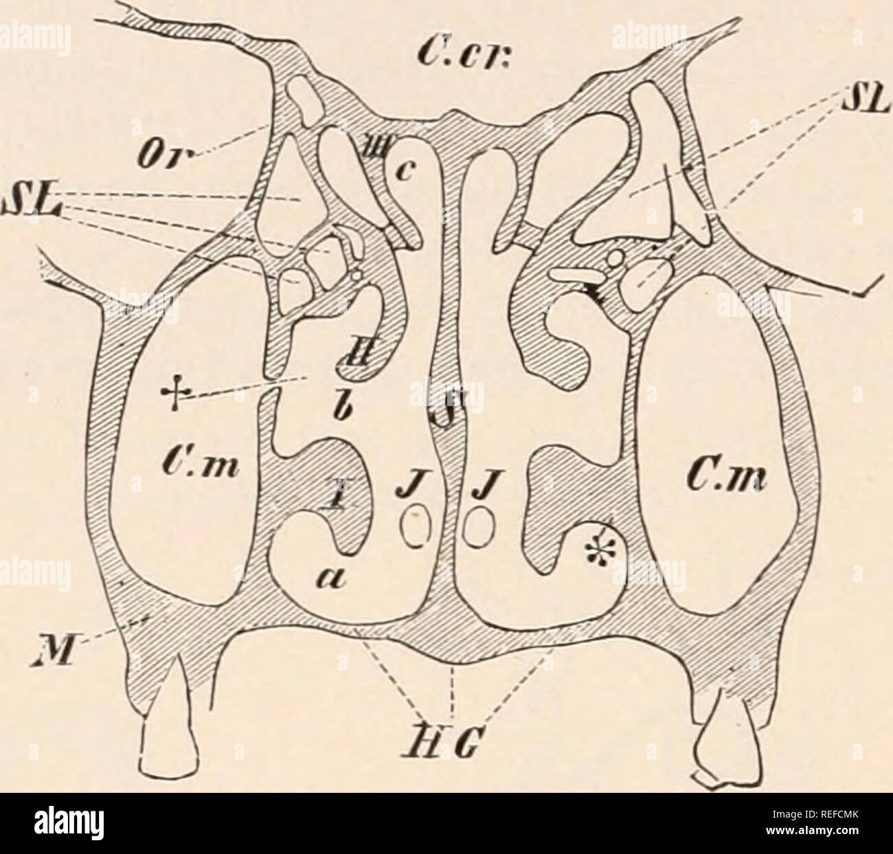 . Comparative anatomy of vertebrates. Anatomy, Comparative; Vertebrates. 270 COMPARATIVE ANATOMY. but with the reduction of this sense, they lose tleir primary function, often persisting merely as air-sinuses or wen disap- pearing entirelyl (Pinnipedia). The nasal glands of Mammals may be divided into two sets, —numerous small, diffuse Bowman s glands, and a large Stcnson's aland. The latter appears early in the embryo, and in many cases undergoes reduction; it is situated in the lateral or basal walls of the nasal cavity, opening into the vestibule of the nose, and may extend into the maxil- Stock Photohttps://www.alamy.com/image-license-details/?v=1https://www.alamy.com/comparative-anatomy-of-vertebrates-anatomy-comparative-vertebrates-270-comparative-anatomy-but-with-the-reduction-of-this-sense-they-lose-tleir-primary-function-often-persisting-merely-as-air-sinuses-or-wen-disap-pearing-entirelyl-pinnipedia-the-nasal-glands-of-mammals-may-be-divided-into-two-sets-numerous-small-diffuse-bowman-s-glands-and-a-large-stcnsons-aland-the-latter-appears-early-in-the-embryo-and-in-many-cases-undergoes-reduction-it-is-situated-in-the-lateral-or-basal-walls-of-the-nasal-cavity-opening-into-the-vestibule-of-the-nose-and-may-extend-into-the-maxil-image232679235.html
RMREFCMK–. Comparative anatomy of vertebrates. Anatomy, Comparative; Vertebrates. 270 COMPARATIVE ANATOMY. but with the reduction of this sense, they lose tleir primary function, often persisting merely as air-sinuses or wen disap- pearing entirelyl (Pinnipedia). The nasal glands of Mammals may be divided into two sets, —numerous small, diffuse Bowman s glands, and a large Stcnson's aland. The latter appears early in the embryo, and in many cases undergoes reduction; it is situated in the lateral or basal walls of the nasal cavity, opening into the vestibule of the nose, and may extend into the maxil-
 . The anatomy of the domestic animals. Veterinary anatomy. THE NASAL CAVITY 509 the naso-lacrimal duct, is seen when the nostril is thlated; it is situated on the floor of the vestibule, aoout two inches (ca. 5 cm.) from the lower commissure, perforat- ing the skin close to its junction ^'ith the mucous membrane. (It is not rare to find one or two accessory orifices further back.) Structure.—The skin around the nostrils presents long tactile hairs as well as the ordinary ones. It is continued around the alse and lines the vestibule. The skin of the diverticulum is thin and usually black, and Stock Photohttps://www.alamy.com/image-license-details/?v=1https://www.alamy.com/the-anatomy-of-the-domestic-animals-veterinary-anatomy-the-nasal-cavity-509-the-naso-lacrimal-duct-is-seen-when-the-nostril-is-thlated-it-is-situated-on-the-floor-of-the-vestibule-aoout-two-inches-ca-5-cm-from-the-lower-commissure-perforat-ing-the-skin-close-to-its-junction-ith-the-mucous-membrane-it-is-not-rare-to-find-one-or-two-accessory-orifices-further-back-structurethe-skin-around-the-nostrils-presents-long-tactile-hairs-as-well-as-the-ordinary-ones-it-is-continued-around-the-alse-and-lines-the-vestibule-the-skin-of-the-diverticulum-is-thin-and-usually-black-and-image236760626.html
RMRN5AGJ–. The anatomy of the domestic animals. Veterinary anatomy. THE NASAL CAVITY 509 the naso-lacrimal duct, is seen when the nostril is thlated; it is situated on the floor of the vestibule, aoout two inches (ca. 5 cm.) from the lower commissure, perforat- ing the skin close to its junction ^'ith the mucous membrane. (It is not rare to find one or two accessory orifices further back.) Structure.—The skin around the nostrils presents long tactile hairs as well as the ordinary ones. It is continued around the alse and lines the vestibule. The skin of the diverticulum is thin and usually black, and
 . The anatomy of the domestic animals . Veterinary anatomy. THE NASAL CAVITY 509 the naso-lacrimal duct, is seen when the nostril is dilated; it is situated on the floor ot the vestibule, aoout two inches (ca. 5 cm.) from the lower commissure, perforat- mg the skm close to its junction with the mucous membrane. (It is not rare to find one or two accessorj^ orifices further back.) Staacture.—The skin around the nostrils presents long tactile hairs as well as the ordinary ones. It is continued around the alse and lines the vestibule. The skm of the diverticulum is thin and usually black, and is Stock Photohttps://www.alamy.com/image-license-details/?v=1https://www.alamy.com/the-anatomy-of-the-domestic-animals-veterinary-anatomy-the-nasal-cavity-509-the-naso-lacrimal-duct-is-seen-when-the-nostril-is-dilated-it-is-situated-on-the-floor-ot-the-vestibule-aoout-two-inches-ca-5-cm-from-the-lower-commissure-perforat-mg-the-skm-close-to-its-junction-with-the-mucous-membrane-it-is-not-rare-to-find-one-or-two-accessorj-orifices-further-back-staacturethe-skin-around-the-nostrils-presents-long-tactile-hairs-as-well-as-the-ordinary-ones-it-is-continued-around-the-alse-and-lines-the-vestibule-the-skm-of-the-diverticulum-is-thin-and-usually-black-and-is-image232324654.html
RMRDY8D2–. The anatomy of the domestic animals . Veterinary anatomy. THE NASAL CAVITY 509 the naso-lacrimal duct, is seen when the nostril is dilated; it is situated on the floor ot the vestibule, aoout two inches (ca. 5 cm.) from the lower commissure, perforat- mg the skm close to its junction with the mucous membrane. (It is not rare to find one or two accessorj^ orifices further back.) Staacture.—The skin around the nostrils presents long tactile hairs as well as the ordinary ones. It is continued around the alse and lines the vestibule. The skm of the diverticulum is thin and usually black, and is
 . Comparative anatomy of vertebrates. Anatomy, Comparative; Vertebrates -- Anatomy. OLFACTORY ORGANS. IQ7 Stenson's gland; in other mammals, so far as known, its duct becomes cut off from the nasal cavity and opens into the naso-palatal canal. Its medial wall is covered with sensory epithelium, supplied by a branch of the olfactory nerve. In the primates the organ is more or less de- generate in the adult. There are two kinds of glands in the nasal cavity, the smaller and scattered Bowman's glands and the larger Stenson's gland lying in the lateral ventral wall and opening into the vestibule. Stock Photohttps://www.alamy.com/image-license-details/?v=1https://www.alamy.com/comparative-anatomy-of-vertebrates-anatomy-comparative-vertebrates-anatomy-olfactory-organs-iq7-stensons-gland-in-other-mammals-so-far-as-known-its-duct-becomes-cut-off-from-the-nasal-cavity-and-opens-into-the-naso-palatal-canal-its-medial-wall-is-covered-with-sensory-epithelium-supplied-by-a-branch-of-the-olfactory-nerve-in-the-primates-the-organ-is-more-or-less-de-generate-in-the-adult-there-are-two-kinds-of-glands-in-the-nasal-cavity-the-smaller-and-scattered-bowmans-glands-and-the-larger-stensons-gland-lying-in-the-lateral-ventral-wall-and-opening-into-the-vestibule-image232667543.html
RMREEWR3–. Comparative anatomy of vertebrates. Anatomy, Comparative; Vertebrates -- Anatomy. OLFACTORY ORGANS. IQ7 Stenson's gland; in other mammals, so far as known, its duct becomes cut off from the nasal cavity and opens into the naso-palatal canal. Its medial wall is covered with sensory epithelium, supplied by a branch of the olfactory nerve. In the primates the organ is more or less de- generate in the adult. There are two kinds of glands in the nasal cavity, the smaller and scattered Bowman's glands and the larger Stenson's gland lying in the lateral ventral wall and opening into the vestibule.
 . Outlines of zoology. Zoology. ALIMENTARY SYSTEM. 5°5 â PC.V The eyes are large but lidless; the small nasal sacs with plaited walls have double anterior apertures; the vestibule of the ear contains a large solid otolith, and another very small one in a pos- terior chamber. The dark lateral line, covered over by modified scales, lodges sensory cells, and is innervated by a branch of the vagus. Alimentary system.âTeeth are borne by the premaxillae, the vomer, and the superior pharyngeal bones above, by the dentaries and the in- ferior pharyngeal bones beneath. There are no salivary glands, no Stock Photohttps://www.alamy.com/image-license-details/?v=1https://www.alamy.com/outlines-of-zoology-zoology-alimentary-system-55-pcv-the-eyes-are-large-but-lidless-the-small-nasal-sacs-with-plaited-walls-have-double-anterior-apertures-the-vestibule-of-the-ear-contains-a-large-solid-otolith-and-another-very-small-one-in-a-pos-terior-chamber-the-dark-lateral-line-covered-over-by-modified-scales-lodges-sensory-cells-and-is-innervated-by-a-branch-of-the-vagus-alimentary-systemteeth-are-borne-by-the-premaxillae-the-vomer-and-the-superior-pharyngeal-bones-above-by-the-dentaries-and-the-in-ferior-pharyngeal-bones-beneath-there-are-no-salivary-glands-no-image232213471.html
RMRDP6J7–. Outlines of zoology. Zoology. ALIMENTARY SYSTEM. 5°5 â PC.V The eyes are large but lidless; the small nasal sacs with plaited walls have double anterior apertures; the vestibule of the ear contains a large solid otolith, and another very small one in a pos- terior chamber. The dark lateral line, covered over by modified scales, lodges sensory cells, and is innervated by a branch of the vagus. Alimentary system.âTeeth are borne by the premaxillae, the vomer, and the superior pharyngeal bones above, by the dentaries and the in- ferior pharyngeal bones beneath. There are no salivary glands, no
 . A manual of elementary zoology . Zoology. APPENDIX 577 Sa. 9a. Cut away the roof of the cranium and expose the brain. Note its regions and trace out the nerves. The ophthalmic is the superficial one (Fig. 410). Cut through the auditory capsule. Note labyrinth, with large otolith in vestibule. In skull, note the following bones : in cranium, basioccipital, exoccipitals, supraoccipital, parietals, alisphenoids, frontals, parasphenoid; in auditory capsule of one side, epiotic, pterotic, opisthotic, prootic, sphenotic; in either orbit lacrymal, suborbitals; in nasal region, nasals, vomer, meseth Stock Photohttps://www.alamy.com/image-license-details/?v=1https://www.alamy.com/a-manual-of-elementary-zoology-zoology-appendix-577-sa-9a-cut-away-the-roof-of-the-cranium-and-expose-the-brain-note-its-regions-and-trace-out-the-nerves-the-ophthalmic-is-the-superficial-one-fig-410-cut-through-the-auditory-capsule-note-labyrinth-with-large-otolith-in-vestibule-in-skull-note-the-following-bones-in-cranium-basioccipital-exoccipitals-supraoccipital-parietals-alisphenoids-frontals-parasphenoid-in-auditory-capsule-of-one-side-epiotic-pterotic-opisthotic-prootic-sphenotic-in-either-orbit-lacrymal-suborbitals-in-nasal-region-nasals-vomer-meseth-image232116039.html
RMRDHPAF–. A manual of elementary zoology . Zoology. APPENDIX 577 Sa. 9a. Cut away the roof of the cranium and expose the brain. Note its regions and trace out the nerves. The ophthalmic is the superficial one (Fig. 410). Cut through the auditory capsule. Note labyrinth, with large otolith in vestibule. In skull, note the following bones : in cranium, basioccipital, exoccipitals, supraoccipital, parietals, alisphenoids, frontals, parasphenoid; in auditory capsule of one side, epiotic, pterotic, opisthotic, prootic, sphenotic; in either orbit lacrymal, suborbitals; in nasal region, nasals, vomer, meseth
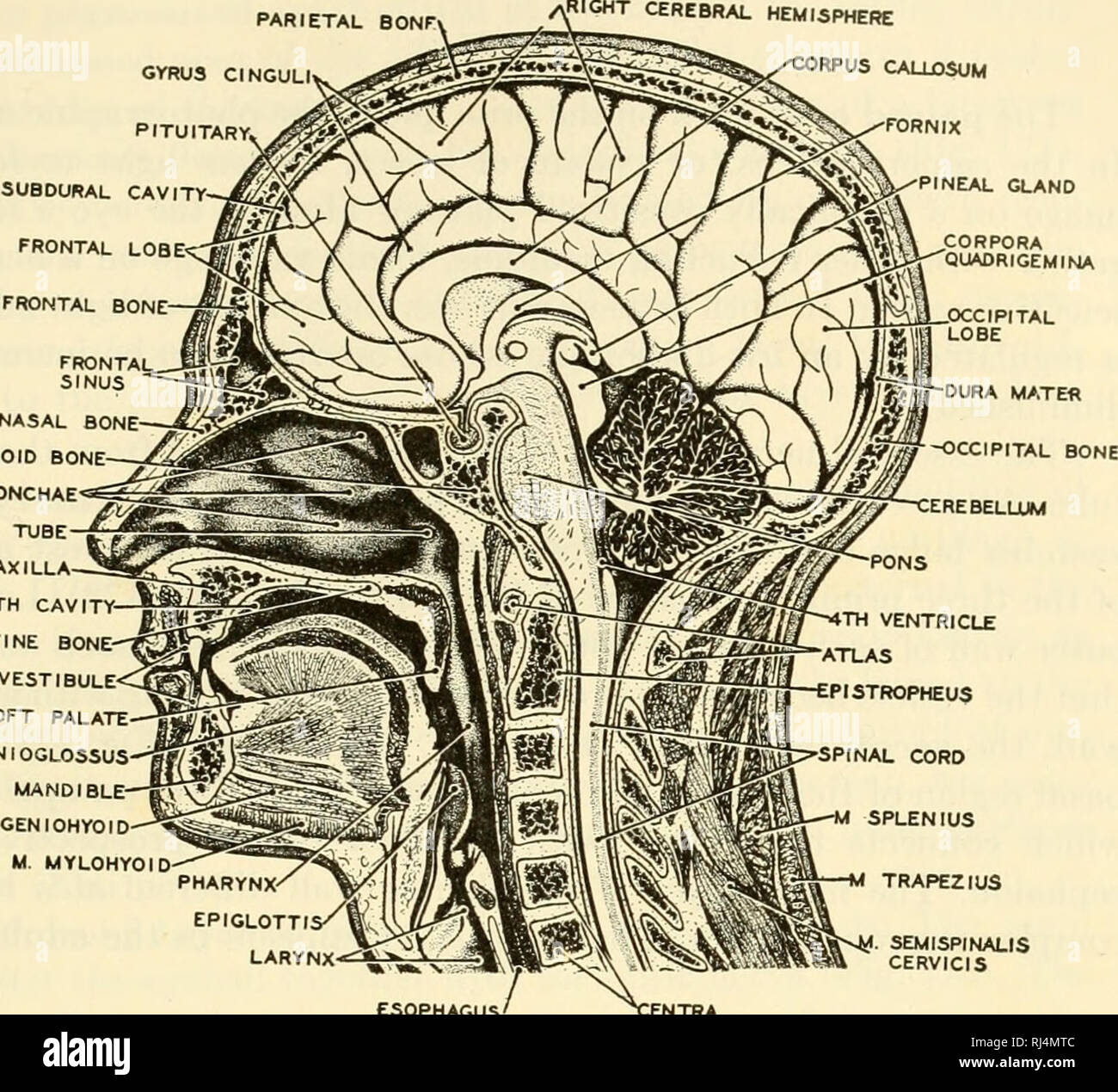 . The chordates. Chordata. itegrative Systems fiHIGHT CEREBRAL HEMISPHERE 199. NASAL BONE SPHENOID BONE NASAL CONCHAE EUSTACHIAN MAXILLA MOUTH CAVITY- PALATINE BONE VESTIBULE SOFT PALATE M GENI0GL0S5US MANDIBLE M. GENIOHYOID M. MYLOHYOID SEMISPINALS CERVICIS esophagus' Fig. 171. A sagittal section of the human head showing the relations between digestive and respiratory passages in the pharyngeal region. (After Braus. Courtesy, Neal and Rand: "Chordate Anatomy," Philadelphia, The Blakiston Company.) groups the development of a horizontal partition (secondary palate) across the oral c Stock Photohttps://www.alamy.com/image-license-details/?v=1https://www.alamy.com/the-chordates-chordata-itegrative-systems-fihight-cerebral-hemisphere-199-nasal-bone-sphenoid-bone-nasal-conchae-eustachian-maxilla-mouth-cavity-palatine-bone-vestibule-soft-palate-m-geni0gl0s5us-mandible-m-geniohyoid-m-mylohyoid-semispinals-cervicis-esophagus-fig-171-a-sagittal-section-of-the-human-head-showing-the-relations-between-digestive-and-respiratory-passages-in-the-pharyngeal-region-after-braus-courtesy-neal-and-rand-quotchordate-anatomyquot-philadelphia-the-blakiston-company-groups-the-development-of-a-horizontal-partition-secondary-palate-across-the-oral-c-image234902764.html
RMRJ4MTC–. The chordates. Chordata. itegrative Systems fiHIGHT CEREBRAL HEMISPHERE 199. NASAL BONE SPHENOID BONE NASAL CONCHAE EUSTACHIAN MAXILLA MOUTH CAVITY- PALATINE BONE VESTIBULE SOFT PALATE M GENI0GL0S5US MANDIBLE M. GENIOHYOID M. MYLOHYOID SEMISPINALS CERVICIS esophagus' Fig. 171. A sagittal section of the human head showing the relations between digestive and respiratory passages in the pharyngeal region. (After Braus. Courtesy, Neal and Rand: "Chordate Anatomy," Philadelphia, The Blakiston Company.) groups the development of a horizontal partition (secondary palate) across the oral c
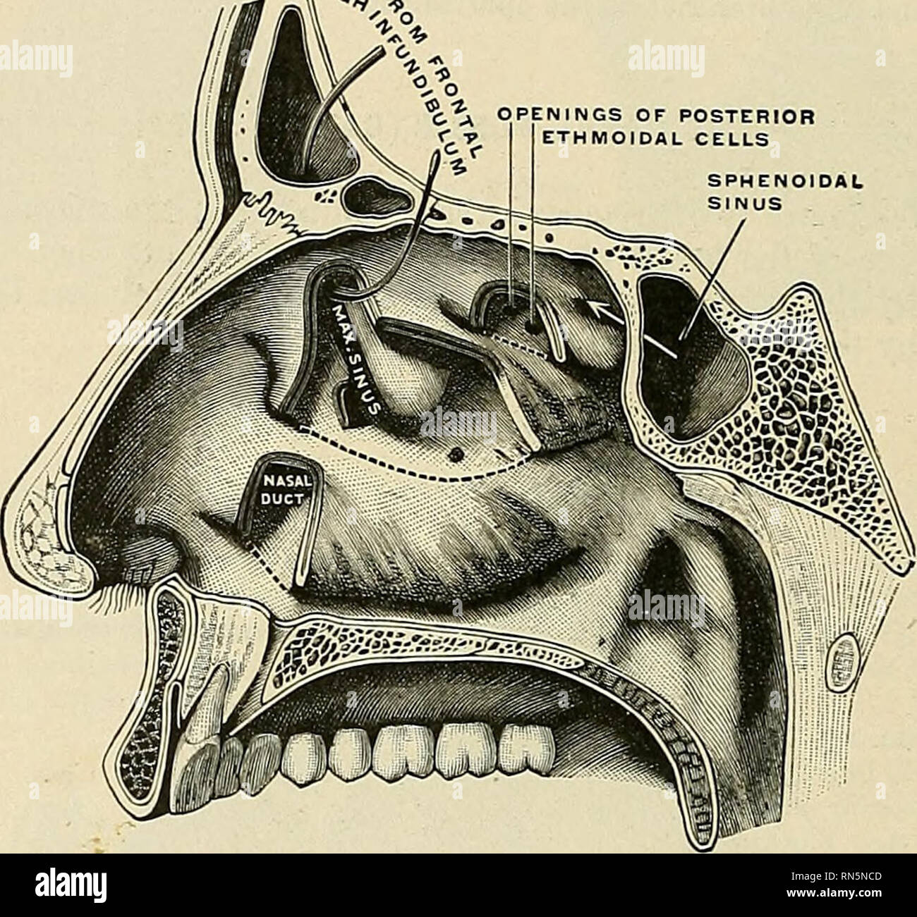 . Anatomy, descriptive and applied. Anatomy. 1082 THE ORGANS OF SPECIAL SENSE mucous membrane. Each measures an inch (2.5 cm.) in the vertical and half an inch (1.2 cm.) in the transverse direction in a M^ell-developed adult skull. For the description of the bony boundaries of the nasal fossae see page 138. Inside the aperture of the nostril is a slight dilatation, the vestibule (vestibulum nasi), which extends as a small pouch, the ventricle, toward the point of the nose. Above and behind the vestibule is surrounded by a prominence (limen nasi). Below the prominence the vestibule ic lined wit Stock Photohttps://www.alamy.com/image-license-details/?v=1https://www.alamy.com/anatomy-descriptive-and-applied-anatomy-1082-the-organs-of-special-sense-mucous-membrane-each-measures-an-inch-25-cm-in-the-vertical-and-half-an-inch-12-cm-in-the-transverse-direction-in-a-mell-developed-adult-skull-for-the-description-of-the-bony-boundaries-of-the-nasal-fossae-see-page-138-inside-the-aperture-of-the-nostril-is-a-slight-dilatation-the-vestibule-vestibulum-nasi-which-extends-as-a-small-pouch-the-ventricle-toward-the-point-of-the-nose-above-and-behind-the-vestibule-is-surrounded-by-a-prominence-limen-nasi-below-the-prominence-the-vestibule-ic-lined-wit-image236769133.html
RMRN5NCD–. Anatomy, descriptive and applied. Anatomy. 1082 THE ORGANS OF SPECIAL SENSE mucous membrane. Each measures an inch (2.5 cm.) in the vertical and half an inch (1.2 cm.) in the transverse direction in a M^ell-developed adult skull. For the description of the bony boundaries of the nasal fossae see page 138. Inside the aperture of the nostril is a slight dilatation, the vestibule (vestibulum nasi), which extends as a small pouch, the ventricle, toward the point of the nose. Above and behind the vestibule is surrounded by a prominence (limen nasi). Below the prominence the vestibule ic lined wit
 . An atlas of human anatomy for students and physicians. Anatomy. CEPHALIC AND CERVICAL PORTIONS OF THE DIGESTIVE ORGANS 411 Nasal cavity Cavum nasi Cartilaginous septum of the nose Septum cartilagineum nasi Colunma nasi Septum mobile Oral cavity* Cavum oris Aperture of the mouth Kima oris Vestibule of the mouth Vestibulum oris Upper surface, or dorsum, of the tongue Dorsum linguas Lymphatic gland L mphoglanduia Mylohyoid muscle M. mylohyoideus Hyold bone—Os hyoideum Vestibule of the larynx 'estibulum larjngib Prominence of the larynx Prominentia !arn,ea Laryngeal cavity Ca um larj ngis Tr Stock Photohttps://www.alamy.com/image-license-details/?v=1https://www.alamy.com/an-atlas-of-human-anatomy-for-students-and-physicians-anatomy-cephalic-and-cervical-portions-of-the-digestive-organs-411-nasal-cavity-cavum-nasi-cartilaginous-septum-of-the-nose-septum-cartilagineum-nasi-colunma-nasi-septum-mobile-oral-cavity-cavum-oris-aperture-of-the-mouth-kima-oris-vestibule-of-the-mouth-vestibulum-oris-upper-surface-or-dorsum-of-the-tongue-dorsum-linguas-lymphatic-gland-l-mphoglanduia-mylohyoid-muscle-m-mylohyoideus-hyold-boneos-hyoideum-vestibule-of-the-larynx-estibulum-larjngib-prominence-of-the-larynx-prominentia-!arnea-laryngeal-cavity-ca-um-larj-ngis-tr-image235399023.html
RMRJY9RY–. An atlas of human anatomy for students and physicians. Anatomy. CEPHALIC AND CERVICAL PORTIONS OF THE DIGESTIVE ORGANS 411 Nasal cavity Cavum nasi Cartilaginous septum of the nose Septum cartilagineum nasi Colunma nasi Septum mobile Oral cavity* Cavum oris Aperture of the mouth Kima oris Vestibule of the mouth Vestibulum oris Upper surface, or dorsum, of the tongue Dorsum linguas Lymphatic gland L mphoglanduia Mylohyoid muscle M. mylohyoideus Hyold bone—Os hyoideum Vestibule of the larynx 'estibulum larjngib Prominence of the larynx Prominentia !arn,ea Laryngeal cavity Ca um larj ngis Tr
 . Bulletin of the British Museum (Natural History) Zoology. Fig. 16 Scaling on digits, a. No lateral scale row present, b, c. Partial and complete lateral row present, d. Transverse section of digit with a posterior lateral row. e. Transverse section of digit with posterior and anterior lateral rows. From Arnold (1986<i). vestibule , nostn postnasal area. posterior border of vestibule principal nasal cavity. Please note that these images are extracted from scanned page images that may have been digitally enhanced for readability - coloration and appearance of these illustrations may not pe Stock Photohttps://www.alamy.com/image-license-details/?v=1https://www.alamy.com/bulletin-of-the-british-museum-natural-history-zoology-fig-16-scaling-on-digits-a-no-lateral-scale-row-present-b-c-partial-and-complete-lateral-row-present-d-transverse-section-of-digit-with-a-posterior-lateral-row-e-transverse-section-of-digit-with-posterior-and-anterior-lateral-rows-from-arnold-1986lti-vestibule-nostn-postnasal-area-posterior-border-of-vestibule-principal-nasal-cavity-please-note-that-these-images-are-extracted-from-scanned-page-images-that-may-have-been-digitally-enhanced-for-readability-coloration-and-appearance-of-these-illustrations-may-not-pe-image233912107.html
RMRGFH7R–. Bulletin of the British Museum (Natural History) Zoology. Fig. 16 Scaling on digits, a. No lateral scale row present, b, c. Partial and complete lateral row present, d. Transverse section of digit with a posterior lateral row. e. Transverse section of digit with posterior and anterior lateral rows. From Arnold (1986<i). vestibule , nostn postnasal area. posterior border of vestibule principal nasal cavity. Please note that these images are extracted from scanned page images that may have been digitally enhanced for readability - coloration and appearance of these illustrations may not pe
 . Chordate anatomy. Chordata; Anatomy, Comparative. 2^6 CHORDATE ANATOMY PARIETAL BONE' GYRUS CINGULI PITUITARY, SUBDURAL CAVITY- FRONTAL LOBE FRONTAL BONE- yRIGHT CEREBRAL HEMISPHERE iRPUS CALU3SUM NASAL BONE SPHENOID BONE NASAL CONCHAE EUSTACHIAN TUBE MAXILLA MOUTH CAVITY- PALATINE BONE VESTIBULE SOFT PALATE M. GENIOGLOSSUS MANDIBLE M. GENIOHYOID M. MYLOHYOID. •FORNIX PINEAL GLAND CORPORA QUADRIGEMINA dura mater -occipital bone :erebellum ESOPHAGUS' ':ENTRA Fig. 237.-—A median longitudinal section of the human head showing the relations between digestive and respiratory passages in the phar Stock Photohttps://www.alamy.com/image-license-details/?v=1https://www.alamy.com/chordate-anatomy-chordata-anatomy-comparative-26-chordate-anatomy-parietal-bone-gyrus-cinguli-pituitary-subdural-cavity-frontal-lobe-frontal-bone-yright-cerebral-hemisphere-irpus-calu3sum-nasal-bone-sphenoid-bone-nasal-conchae-eustachian-tube-maxilla-mouth-cavity-palatine-bone-vestibule-soft-palate-m-genioglossus-mandible-m-geniohyoid-m-mylohyoid-fornix-pineal-gland-corpora-quadrigemina-dura-mater-occipital-bone-erebellum-esophagus-entra-fig-237-a-median-longitudinal-section-of-the-human-head-showing-the-relations-between-digestive-and-respiratory-passages-in-the-phar-image234923800.html
RMRJ5KKM–. Chordate anatomy. Chordata; Anatomy, Comparative. 2^6 CHORDATE ANATOMY PARIETAL BONE' GYRUS CINGULI PITUITARY, SUBDURAL CAVITY- FRONTAL LOBE FRONTAL BONE- yRIGHT CEREBRAL HEMISPHERE iRPUS CALU3SUM NASAL BONE SPHENOID BONE NASAL CONCHAE EUSTACHIAN TUBE MAXILLA MOUTH CAVITY- PALATINE BONE VESTIBULE SOFT PALATE M. GENIOGLOSSUS MANDIBLE M. GENIOHYOID M. MYLOHYOID. •FORNIX PINEAL GLAND CORPORA QUADRIGEMINA dura mater -occipital bone :erebellum ESOPHAGUS' ':ENTRA Fig. 237.-—A median longitudinal section of the human head showing the relations between digestive and respiratory passages in the phar
 . Cunningham's Text-book of anatomy. Anatomy. Vomeronasal cartilages Vomero-nasal organs Fig. 673.—Section through Nose of a Kitten, showing position of the voinero-nasal organs. Frontal air-sinus Bristle passed from it into the middle meatus Opening of middle ethmoidal cells Openings of posterior ethmoidal cells Recessus sphenoethmoidalis Sphenoidal air-sinus. Cut edge of inferior nasal concha stle in opening of naso-lacrimal duct Fig. 674.—View of the Lateral Wall of the Nose—the Nasal Concha having been behoved. 1. Vestibule. 2. Opening of maxillary sinus. 3. Hiatus semilunaris. 4. Bulla et Stock Photohttps://www.alamy.com/image-license-details/?v=1https://www.alamy.com/cunninghams-text-book-of-anatomy-anatomy-vomeronasal-cartilages-vomero-nasal-organs-fig-673section-through-nose-of-a-kitten-showing-position-of-the-voinero-nasal-organs-frontal-air-sinus-bristle-passed-from-it-into-the-middle-meatus-opening-of-middle-ethmoidal-cells-openings-of-posterior-ethmoidal-cells-recessus-sphenoethmoidalis-sphenoidal-air-sinus-cut-edge-of-inferior-nasal-concha-stle-in-opening-of-naso-lacrimal-duct-fig-674view-of-the-lateral-wall-of-the-nosethe-nasal-concha-having-been-behoved-1-vestibule-2-opening-of-maxillary-sinus-3-hiatus-semilunaris-4-bulla-et-image231856432.html
RMRD5Y6T–. Cunningham's Text-book of anatomy. Anatomy. Vomeronasal cartilages Vomero-nasal organs Fig. 673.—Section through Nose of a Kitten, showing position of the voinero-nasal organs. Frontal air-sinus Bristle passed from it into the middle meatus Opening of middle ethmoidal cells Openings of posterior ethmoidal cells Recessus sphenoethmoidalis Sphenoidal air-sinus. Cut edge of inferior nasal concha stle in opening of naso-lacrimal duct Fig. 674.—View of the Lateral Wall of the Nose—the Nasal Concha having been behoved. 1. Vestibule. 2. Opening of maxillary sinus. 3. Hiatus semilunaris. 4. Bulla et