Nasenspiegel Stock Photos and Images
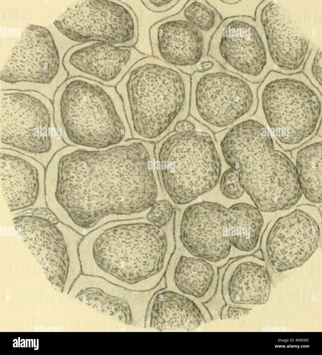
 . Anatomischer Anzeiger. Anatomy, Comparative; Anatomy, Comparative. h Fig. 8. Fig. 9. Fig. 8. Katzen-Nasenspiegel. Fiontalschnitt (ganz oberflächlich). Vergröß. 1: 50. Fig. 9. Katzen-Nasenspiegel. Frontalschnitt (etwas tiefer als in Fig. 8). Ver- größerung 1: 50. NF. Please note that these images are extracted from scanned page images that may have been digitally enhanced for readability - coloration and appearance of these illustrations may not perfectly resemble the original work.. Anatomische Gesellschaft. Jena : G. Fischer Stock Photohttps://www.alamy.com/image-license-details/?v=1https://www.alamy.com/anatomischer-anzeiger-anatomy-comparative-anatomy-comparative-h-fig-8-fig-9-fig-8-katzen-nasenspiegel-fiontalschnitt-ganz-oberflchlich-vergr-1-50-fig-9-katzen-nasenspiegel-frontalschnitt-etwas-tiefer-als-in-fig-8-ver-grerung-1-50-nf-please-note-that-these-images-are-extracted-from-scanned-page-images-that-may-have-been-digitally-enhanced-for-readability-coloration-and-appearance-of-these-illustrations-may-not-perfectly-resemble-the-original-work-anatomische-gesellschaft-jena-g-fischer-image236820540.html
RMRN830C–. Anatomischer Anzeiger. Anatomy, Comparative; Anatomy, Comparative. h Fig. 8. Fig. 9. Fig. 8. Katzen-Nasenspiegel. Fiontalschnitt (ganz oberflächlich). Vergröß. 1: 50. Fig. 9. Katzen-Nasenspiegel. Frontalschnitt (etwas tiefer als in Fig. 8). Ver- größerung 1: 50. NF. Please note that these images are extracted from scanned page images that may have been digitally enhanced for readability - coloration and appearance of these illustrations may not perfectly resemble the original work.. Anatomische Gesellschaft. Jena : G. Fischer
 skin, dog, sleep, sleeping, puppy, terrier, breed, dog breeding, pink, Stock Photohttps://www.alamy.com/image-license-details/?v=1https://www.alamy.com/stock-photo-skin-dog-sleep-sleeping-puppy-terrier-breed-dog-breeding-pink-132032390.html
RFHJPGJE–skin, dog, sleep, sleeping, puppy, terrier, breed, dog breeding, pink,
 . Anatomischer Anzeiger. Anatomy, Comparative; Anatomy, Comparative. Fig. 5. Sulcus nasoraedianus (mittlere Nasenrinne) eines Hunde-Nasenspiegels. Horizontalschnitt (Vergr. 1: 20). sF Seitenfurchen im Sulcus nasomed.. Fig. 6. Hunde-Nasenspiegel (bei schwacher Vergrößerung). Fel- der- und Furchensystem am An- fangsteile des Sulcus nasoraedianus. Fig. 7. Hunde - Nasenspiegel. Vergrößerung 1: 20. Frontalschnitt. Diese die Oberfläche der Hauptfelder durchziehenden feinen Furchen stehen nait den Hauptfurchen in direktem Zusammenhange.. Please note that these images are extracted from scanned page i Stock Photohttps://www.alamy.com/image-license-details/?v=1https://www.alamy.com/anatomischer-anzeiger-anatomy-comparative-anatomy-comparative-fig-5-sulcus-nasoraedianus-mittlere-nasenrinne-eines-hunde-nasenspiegels-horizontalschnitt-vergr-1-20-sf-seitenfurchen-im-sulcus-nasomed-fig-6-hunde-nasenspiegel-bei-schwacher-vergrerung-fel-der-und-furchensystem-am-an-fangsteile-des-sulcus-nasoraedianus-fig-7-hunde-nasenspiegel-vergrerung-1-20-frontalschnitt-diese-die-oberflche-der-hauptfelder-durchziehenden-feinen-furchen-stehen-nait-den-hauptfurchen-in-direktem-zusammenhange-please-note-that-these-images-are-extracted-from-scanned-page-i-image236820554.html
RMRN830X–. Anatomischer Anzeiger. Anatomy, Comparative; Anatomy, Comparative. Fig. 5. Sulcus nasoraedianus (mittlere Nasenrinne) eines Hunde-Nasenspiegels. Horizontalschnitt (Vergr. 1: 20). sF Seitenfurchen im Sulcus nasomed.. Fig. 6. Hunde-Nasenspiegel (bei schwacher Vergrößerung). Fel- der- und Furchensystem am An- fangsteile des Sulcus nasoraedianus. Fig. 7. Hunde - Nasenspiegel. Vergrößerung 1: 20. Frontalschnitt. Diese die Oberfläche der Hauptfelder durchziehenden feinen Furchen stehen nait den Hauptfurchen in direktem Zusammenhange.. Please note that these images are extracted from scanned page i
 food aliment nose to gorge engulf devour cow smell food aliment animal mouth Stock Photohttps://www.alamy.com/image-license-details/?v=1https://www.alamy.com/stock-photo-food-aliment-nose-to-gorge-engulf-devour-cow-smell-food-aliment-animal-141288086.html
RFJ5T6B2–food aliment nose to gorge engulf devour cow smell food aliment animal mouth
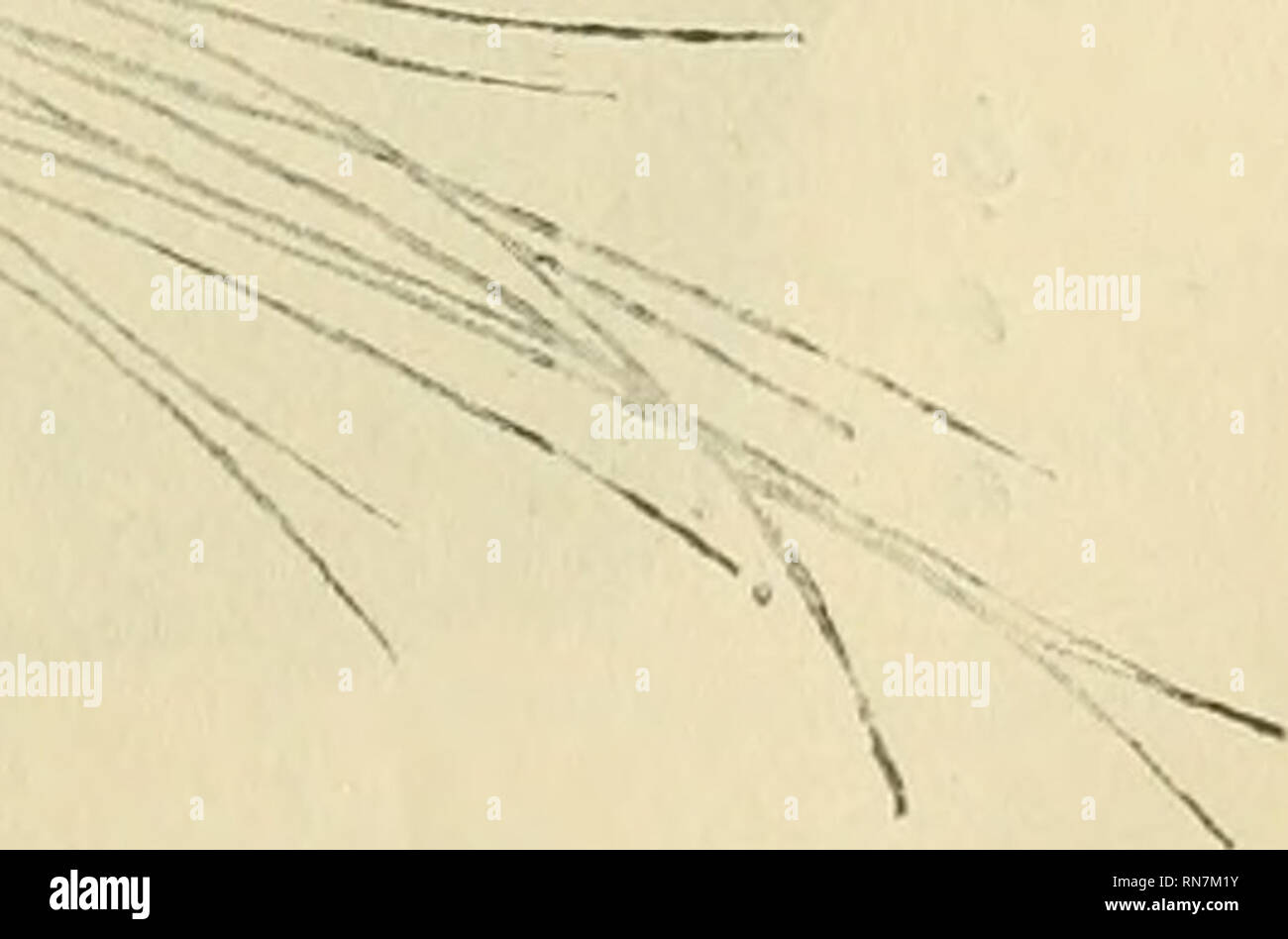
 . Anatomischer Anzeiger. Anatomy, Comparative; Anatomy, Comparative. ?^ 1 ' 1/ ^:^;^. Fig. 4. Der Nasensj^iegel (Planum nasale) der Katze, a Nasenspiegel; b Ober- lippe; c Lippen rinne; d Nasenloch; 2 Pars supralabialis; 3 Pars internarica; 4 Pars supranarica; 4' Alae nasi; 5 Pars dorsouasalis.. Please note that these images are extracted from scanned page images that may have been digitally enhanced for readability - coloration and appearance of these illustrations may not perfectly resemble the original work.. Anatomische Gesellschaft. Jena : G. Fischer Stock Photohttps://www.alamy.com/image-license-details/?v=1https://www.alamy.com/anatomischer-anzeiger-anatomy-comparative-anatomy-comparative-1-1-fig-4-der-nasensjiegel-planum-nasale-der-katze-a-nasenspiegel-b-ober-lippe-c-lippen-rinne-d-nasenloch-2-pars-supralabialis-3-pars-internarica-4-pars-supranarica-4-alae-nasi-5-pars-dorsouasalis-please-note-that-these-images-are-extracted-from-scanned-page-images-that-may-have-been-digitally-enhanced-for-readability-coloration-and-appearance-of-these-illustrations-may-not-perfectly-resemble-the-original-work-anatomische-gesellschaft-jena-g-fischer-image236811959.html
RMRN7M1Y–. Anatomischer Anzeiger. Anatomy, Comparative; Anatomy, Comparative. ?^ 1 ' 1/ ^:^;^. Fig. 4. Der Nasensj^iegel (Planum nasale) der Katze, a Nasenspiegel; b Ober- lippe; c Lippen rinne; d Nasenloch; 2 Pars supralabialis; 3 Pars internarica; 4 Pars supranarica; 4' Alae nasi; 5 Pars dorsouasalis.. Please note that these images are extracted from scanned page images that may have been digitally enhanced for readability - coloration and appearance of these illustrations may not perfectly resemble the original work.. Anatomische Gesellschaft. Jena : G. Fischer
 macro close-up macro admission close up view green black swarthy jetblack deep Stock Photohttps://www.alamy.com/image-license-details/?v=1https://www.alamy.com/stock-photo-macro-close-up-macro-admission-close-up-view-green-black-swarthy-jetblack-141600599.html
RFJ6AD07–macro close-up macro admission close up view green black swarthy jetblack deep
 . Anatomischer Anzeiger. Anatomy, Comparative; Anatomy, Comparative. h Fig. 8. Fig. 9. Fig. 8. Katzen-Nasenspiegel. Fiontalschnitt (ganz oberflächlich). Vergröß. 1: 50. Fig. 9. Katzen-Nasenspiegel. Frontalschnitt (etwas tiefer als in Fig. 8). Ver- größerung 1: 50. NF. Fig. 10. Fig. 11. Fig. 10. Hunde-Nasenspiegel. Sagittalschnitt (Vergrößerung 1: 20). NF Nasen- spiegel-Feld (-Areale). 'pF primäre Nasenspiegel-Furchen. sF sekundäre Nasenspiegel- Furche. Fig. 11. Katzen-Nasenspiegel. Sagittalschnitt (Vergrößerung 1:20). NF Nasen- spiegel-Furchen. NP Nasenspiegel-Felder.. Please note that these Stock Photohttps://www.alamy.com/image-license-details/?v=1https://www.alamy.com/anatomischer-anzeiger-anatomy-comparative-anatomy-comparative-h-fig-8-fig-9-fig-8-katzen-nasenspiegel-fiontalschnitt-ganz-oberflchlich-vergr-1-50-fig-9-katzen-nasenspiegel-frontalschnitt-etwas-tiefer-als-in-fig-8-ver-grerung-1-50-nf-fig-10-fig-11-fig-10-hunde-nasenspiegel-sagittalschnitt-vergrerung-1-20-nf-nasen-spiegel-feld-areale-pf-primre-nasenspiegel-furchen-sf-sekundre-nasenspiegel-furche-fig-11-katzen-nasenspiegel-sagittalschnitt-vergrerung-120-nf-nasen-spiegel-furchen-np-nasenspiegel-felder-please-note-that-these-image236820533.html
RMRN8305–. Anatomischer Anzeiger. Anatomy, Comparative; Anatomy, Comparative. h Fig. 8. Fig. 9. Fig. 8. Katzen-Nasenspiegel. Fiontalschnitt (ganz oberflächlich). Vergröß. 1: 50. Fig. 9. Katzen-Nasenspiegel. Frontalschnitt (etwas tiefer als in Fig. 8). Ver- größerung 1: 50. NF. Fig. 10. Fig. 11. Fig. 10. Hunde-Nasenspiegel. Sagittalschnitt (Vergrößerung 1: 20). NF Nasen- spiegel-Feld (-Areale). 'pF primäre Nasenspiegel-Furchen. sF sekundäre Nasenspiegel- Furche. Fig. 11. Katzen-Nasenspiegel. Sagittalschnitt (Vergrößerung 1:20). NF Nasen- spiegel-Furchen. NP Nasenspiegel-Felder.. Please note that these
 . Anatomischer Anzeiger. Anatomy, Comparative; Anatomy, Comparative. 117 der Nasenwinkel verfolgen. Beim Pferde, das schon eine weniger deut- liche Lippenrinne (Fig. 6 a) besitzt, fehlt die Nasenrinne, der Sulcus nasomedianus. Was nun die Form f der Nasenlöcher (Nares) | unserer Haussäugetiere ! anlangt, so sind sie beim ' Pferde (Fig. 6 b und c) halbmondförmig, der dorsale Winkelteil des Nasenloches des Pferdes setzt sich in eigenartiger, ?.t^tfUltK.. — ^ :« Fig. 3. Der Nasen- spiegel (Planum Basale) des Hundes, a Nasenspiegel; b Oberlippe; c Lippeuriune; d Nasenloch; e Sulcus alaris ventrali Stock Photohttps://www.alamy.com/image-license-details/?v=1https://www.alamy.com/anatomischer-anzeiger-anatomy-comparative-anatomy-comparative-117-der-nasenwinkel-verfolgen-beim-pferde-das-schon-eine-weniger-deut-liche-lippenrinne-fig-6-a-besitzt-fehlt-die-nasenrinne-der-sulcus-nasomedianus-was-nun-die-form-f-der-nasenlcher-nares-unserer-haussugetiere-!-anlangt-so-sind-sie-beim-pferde-fig-6-b-und-c-halbmondfrmig-der-dorsale-winkelteil-des-nasenloches-des-pferdes-setzt-sich-in-eigenartiger-ttfultk-fig-3-der-nasen-spiegel-planum-basale-des-hundes-a-nasenspiegel-b-oberlippe-c-lippeuriune-d-nasenloch-e-sulcus-alaris-ventrali-image236811989.html
RMRN7M31–. Anatomischer Anzeiger. Anatomy, Comparative; Anatomy, Comparative. 117 der Nasenwinkel verfolgen. Beim Pferde, das schon eine weniger deut- liche Lippenrinne (Fig. 6 a) besitzt, fehlt die Nasenrinne, der Sulcus nasomedianus. Was nun die Form f der Nasenlöcher (Nares) | unserer Haussäugetiere ! anlangt, so sind sie beim ' Pferde (Fig. 6 b und c) halbmondförmig, der dorsale Winkelteil des Nasenloches des Pferdes setzt sich in eigenartiger, ?.t^tfUltK.. — ^ :« Fig. 3. Der Nasen- spiegel (Planum Basale) des Hundes, a Nasenspiegel; b Oberlippe; c Lippeuriune; d Nasenloch; e Sulcus alaris ventrali
 . Anatomischer Anzeiger. Anatomy, Comparative; Anatomy, Comparative. — ^ :« Fig. 3. Der Nasen- spiegel (Planum Basale) des Hundes, a Nasenspiegel; b Oberlippe; c Lippeuriune; d Nasenloch; e Sulcus alaris ventralis; 1 Regio labialis superior; 3 Pai's supralabialis der Formatio paroralis; 2' ihr lateraler Fortsatz; 3 Pars in- temarica der Formatio para- nasalis bezw. des Planum na- sale ; 4- Pars supranarica; 4' ihre flügelartigen Fortsätze, Alae nasi; 5 Pars dorsouasalis. --.-2 allgemein bekannter Art von dem Hauptteil des Nasenlochs ab und wird als „falsches Nasenloch" (Fig. 6 c) bezeichn Stock Photohttps://www.alamy.com/image-license-details/?v=1https://www.alamy.com/anatomischer-anzeiger-anatomy-comparative-anatomy-comparative-fig-3-der-nasen-spiegel-planum-basale-des-hundes-a-nasenspiegel-b-oberlippe-c-lippeuriune-d-nasenloch-e-sulcus-alaris-ventralis-1-regio-labialis-superior-3-pais-supralabialis-der-formatio-paroralis-2-ihr-lateraler-fortsatz-3-pars-in-temarica-der-formatio-para-nasalis-bezw-des-planum-na-sale-4-pars-supranarica-4-ihre-flgelartigen-fortstze-alae-nasi-5-pars-dorsouasalis-2-allgemein-bekannter-art-von-dem-hauptteil-des-nasenlochs-ab-und-wird-als-falsches-nasenlochquot-fig-6-c-bezeichn-image236811972.html
RMRN7M2C–. Anatomischer Anzeiger. Anatomy, Comparative; Anatomy, Comparative. — ^ :« Fig. 3. Der Nasen- spiegel (Planum Basale) des Hundes, a Nasenspiegel; b Oberlippe; c Lippeuriune; d Nasenloch; e Sulcus alaris ventralis; 1 Regio labialis superior; 3 Pai's supralabialis der Formatio paroralis; 2' ihr lateraler Fortsatz; 3 Pars in- temarica der Formatio para- nasalis bezw. des Planum na- sale ; 4- Pars supranarica; 4' ihre flügelartigen Fortsätze, Alae nasi; 5 Pars dorsouasalis. --.-2 allgemein bekannter Art von dem Hauptteil des Nasenlochs ab und wird als „falsches Nasenloch" (Fig. 6 c) bezeichn