Ossification Black & White Stock Photos
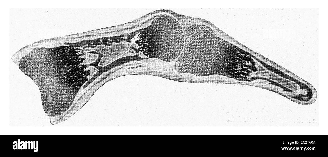 Longitudinal segment through the finger of a human embryo of a certain age, vintage engraved illustration. From the Universe and Humanity, 1910. Stock Photohttps://www.alamy.com/image-license-details/?v=1https://www.alamy.com/longitudinal-segment-through-the-finger-of-a-human-embryo-of-a-certain-age-vintage-engraved-illustration-from-the-universe-and-humanity-1910-image363178602.html
RF2C2T60A–Longitudinal segment through the finger of a human embryo of a certain age, vintage engraved illustration. From the Universe and Humanity, 1910.
RF2Y722EW–skull line icon set vector designs
RF2F9XCYD–Osteology color line icon. Pictogram for web page, mobile app, promo. UI UX GUI design element. Editable stroke.
 A photomicrograph of osteochondritis revealing the cytoarchitectural changes due to congenital syphilis. There is marked irregularity in the provisional zone of ossification producing a 'saw-toothed' appearance, 1971. Note small islands of cartilage remaining in the ossified bone, HandE stain, magnification 100X. Image courtesy CDC/Susan Lindsley. Stock Photohttps://www.alamy.com/image-license-details/?v=1https://www.alamy.com/stock-image-a-photomicrograph-of-osteochondritis-revealing-the-cytoarchitectural-169110806.html
RMKR3JFJ–A photomicrograph of osteochondritis revealing the cytoarchitectural changes due to congenital syphilis. There is marked irregularity in the provisional zone of ossification producing a 'saw-toothed' appearance, 1971. Note small islands of cartilage remaining in the ossified bone, HandE stain, magnification 100X. Image courtesy CDC/Susan Lindsley.
 . Text book of vertebrate zoology. Vertebrates; Anatomy, Comparative. 134 MORFHOLGY OF THE ORGAXS OF VERTEBRATES. increase is impossible. Increase in size is effected here by additions to the exterior, and in the case of the long bones, bodies of the vertebrae, etc., by the appearance of more than one centre of ossification in the cartilage. From these centres ossification e.xtends in all directions, but for a time there remains a cartilaginous region between the ends (epiphyses) and the main portion in which increase in length is possible. Later these epiph} ses usually become so united or an Stock Photohttps://www.alamy.com/image-license-details/?v=1https://www.alamy.com/text-book-of-vertebrate-zoology-vertebrates-anatomy-comparative-134-morfholgy-of-the-orgaxs-of-vertebrates-increase-is-impossible-increase-in-size-is-effected-here-by-additions-to-the-exterior-and-in-the-case-of-the-long-bones-bodies-of-the-vertebrae-etc-by-the-appearance-of-more-than-one-centre-of-ossification-in-the-cartilage-from-these-centres-ossification-extends-in-all-directions-but-for-a-time-there-remains-a-cartilaginous-region-between-the-ends-epiphyses-and-the-main-portion-in-which-increase-in-length-is-possible-later-these-epiph-ses-usually-become-so-united-or-an-image232252780.html
RMRDT0P4–. Text book of vertebrate zoology. Vertebrates; Anatomy, Comparative. 134 MORFHOLGY OF THE ORGAXS OF VERTEBRATES. increase is impossible. Increase in size is effected here by additions to the exterior, and in the case of the long bones, bodies of the vertebrae, etc., by the appearance of more than one centre of ossification in the cartilage. From these centres ossification e.xtends in all directions, but for a time there remains a cartilaginous region between the ends (epiphyses) and the main portion in which increase in length is possible. Later these epiph} ses usually become so united or an
RF2HBBE07–Osteology color line icon. Pictogram for web page, mobile app, promo. UI UX GUI design element. Editable stroke.
 . Veterinary obstetrics; a compendium for the use of students and practitioners. Veterinary obstetrics. ANATOMY. 17 The diameters of the outlet are more equal, being about those of the transverse diameter of the inlet. The cavity is more cylindrical, and less conical than that of the Mare. In the Sheep and Goat, ossification occurring at a 'much later period, allows of the pelvic cavity being increased during parturition, and permits of the act being performed with fewer difficulties in these animals. In the Pig, the general conformation of the pelvis. Fig. 8. Inlet of the Cow's Pelvis. <j Stock Photohttps://www.alamy.com/image-license-details/?v=1https://www.alamy.com/veterinary-obstetrics-a-compendium-for-the-use-of-students-and-practitioners-veterinary-obstetrics-anatomy-17-the-diameters-of-the-outlet-are-more-equal-being-about-those-of-the-transverse-diameter-of-the-inlet-the-cavity-is-more-cylindrical-and-less-conical-than-that-of-the-mare-in-the-sheep-and-goat-ossification-occurring-at-a-much-later-period-allows-of-the-pelvic-cavity-being-increased-during-parturition-and-permits-of-the-act-being-performed-with-fewer-difficulties-in-these-animals-in-the-pig-the-general-conformation-of-the-pelvis-fig-8-inlet-of-the-cows-pelvis-ltj-image216388105.html
RMPG196H–. Veterinary obstetrics; a compendium for the use of students and practitioners. Veterinary obstetrics. ANATOMY. 17 The diameters of the outlet are more equal, being about those of the transverse diameter of the inlet. The cavity is more cylindrical, and less conical than that of the Mare. In the Sheep and Goat, ossification occurring at a 'much later period, allows of the pelvic cavity being increased during parturition, and permits of the act being performed with fewer difficulties in these animals. In the Pig, the general conformation of the pelvis. Fig. 8. Inlet of the Cow's Pelvis. <j
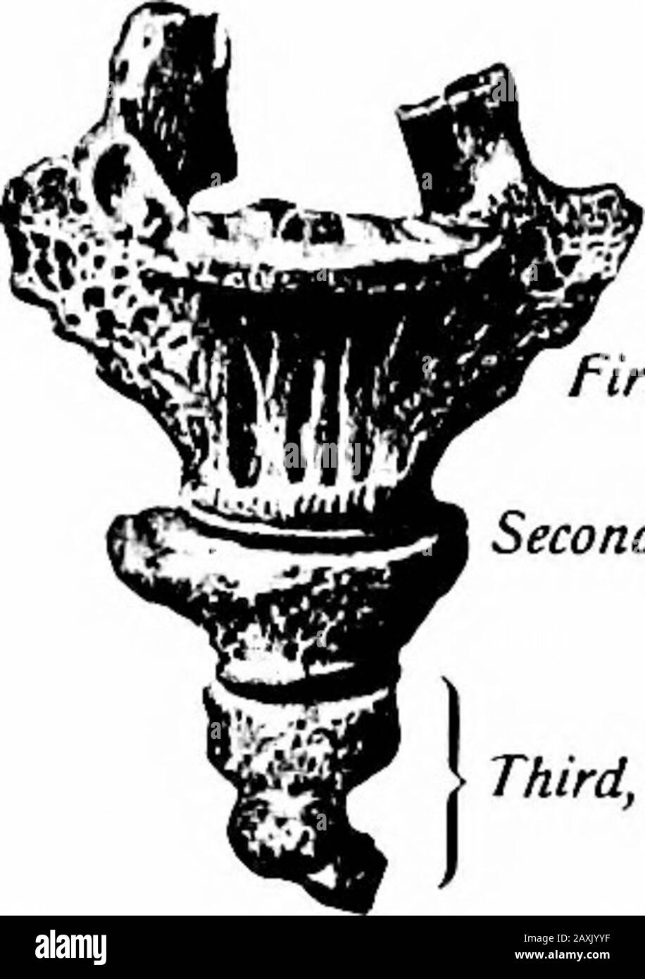 A manual of anatomy . tta and McMurricti.) that articulates with the sacrum. The dorsal surface presents twoupward-projecting processes (cornua coccygea) representing articularprocesses for articulation with the cornua sacrahs. These assist informing two foramina for the fifth pair of sacral nerves. Laterallythis segment presents a rudimentary transverse process on each side.The second may also present such processes but the remaining seg-ments are rudimentary nodules of bone. Ossification.—The coccyx is developed from four centers, one for each segment.That for the first segment appears durin Stock Photohttps://www.alamy.com/image-license-details/?v=1https://www.alamy.com/a-manual-of-anatomy-tta-and-mcmurricti-that-articulates-with-the-sacrum-the-dorsal-surface-presents-twoupward-projecting-processes-cornua-coccygea-representing-articularprocesses-for-articulation-with-the-cornua-sacrahs-these-assist-informing-two-foramina-for-the-fifth-pair-of-sacral-nerves-laterallythis-segment-presents-a-rudimentary-transverse-process-on-each-sidethe-second-may-also-present-such-processes-but-the-remaining-seg-ments-are-rudimentary-nodules-of-bone-ossificationthe-coccyx-is-developed-from-four-centers-one-for-each-segmentthat-for-the-first-segment-appears-durin-image343395123.html
RM2AXJYYF–A manual of anatomy . tta and McMurricti.) that articulates with the sacrum. The dorsal surface presents twoupward-projecting processes (cornua coccygea) representing articularprocesses for articulation with the cornua sacrahs. These assist informing two foramina for the fifth pair of sacral nerves. Laterallythis segment presents a rudimentary transverse process on each side.The second may also present such processes but the remaining seg-ments are rudimentary nodules of bone. Ossification.—The coccyx is developed from four centers, one for each segment.That for the first segment appears durin
 . On the anatomy of vertebrates. Vertebrates; Anatomy, Comparative; 1866. ANATOMY OF VBRTEBKATES. 33 fluent at tlieir apices, which are perforated, and the notochord, reduced to a beaded form, is continued through them: the exterior of the bony cones is occupied Ijy a clear cartilage. In the Porbeagle Shark {Lamna cornubica) further ossification of the conical plate has reduced the central communication to a minute foramen. Os- seous plates have also been developed in the exterior clear cartilage: these plates are triangular, parallel with the axis of the vertebra, their apices converging towa Stock Photohttps://www.alamy.com/image-license-details/?v=1https://www.alamy.com/on-the-anatomy-of-vertebrates-vertebrates-anatomy-comparative-1866-anatomy-of-vbrtebkates-33-fluent-at-tlieir-apices-which-are-perforated-and-the-notochord-reduced-to-a-beaded-form-is-continued-through-them-the-exterior-of-the-bony-cones-is-occupied-ijy-a-clear-cartilage-in-the-porbeagle-shark-lamna-cornubica-further-ossification-of-the-conical-plate-has-reduced-the-central-communication-to-a-minute-foramen-os-seous-plates-have-also-been-developed-in-the-exterior-clear-cartilage-these-plates-are-triangular-parallel-with-the-axis-of-the-vertebra-their-apices-converging-towa-image216417906.html
RMPG2K6X–. On the anatomy of vertebrates. Vertebrates; Anatomy, Comparative; 1866. ANATOMY OF VBRTEBKATES. 33 fluent at tlieir apices, which are perforated, and the notochord, reduced to a beaded form, is continued through them: the exterior of the bony cones is occupied Ijy a clear cartilage. In the Porbeagle Shark {Lamna cornubica) further ossification of the conical plate has reduced the central communication to a minute foramen. Os- seous plates have also been developed in the exterior clear cartilage: these plates are triangular, parallel with the axis of the vertebra, their apices converging towa
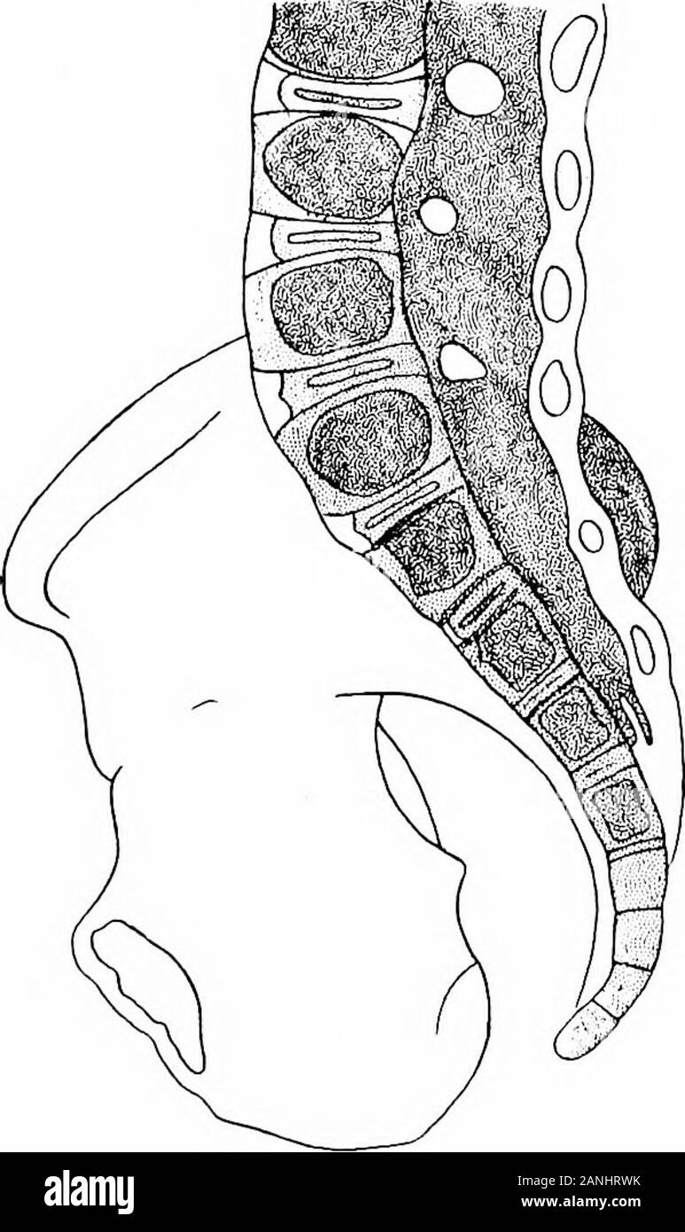 The development of the human body; a manual of human embryology . slight diminution. The latter isreadily understood when it is remembered that the area ofthe skin, granting that the geometric form of the bodyremains the same, would increase as the square of thelength, while the mass of the body would increase as thecube, and hence in comparing weights the skin might beexpected to show a diminution even greater than thatshown in the table. The increase in the weight of the skeleton is due to acertain extent to growth, but chiefly to a completion ofthe ossification of the cartilage largely pres Stock Photohttps://www.alamy.com/image-license-details/?v=1https://www.alamy.com/the-development-of-the-human-body-a-manual-of-human-embryology-slight-diminution-the-latter-isreadily-understood-when-it-is-remembered-that-the-area-ofthe-skin-granting-that-the-geometric-form-of-the-bodyremains-the-same-would-increase-as-the-square-of-thelength-while-the-mass-of-the-body-would-increase-as-thecube-and-hence-in-comparing-weights-the-skin-might-beexpected-to-show-a-diminution-even-greater-than-thatshown-in-the-table-the-increase-in-the-weight-of-the-skeleton-is-due-to-acertain-extent-to-growth-but-chiefly-to-a-completion-ofthe-ossification-of-the-cartilage-largely-pres-image340296703.html
RM2ANHRWK–The development of the human body; a manual of human embryology . slight diminution. The latter isreadily understood when it is remembered that the area ofthe skin, granting that the geometric form of the bodyremains the same, would increase as the square of thelength, while the mass of the body would increase as thecube, and hence in comparing weights the skin might beexpected to show a diminution even greater than thatshown in the table. The increase in the weight of the skeleton is due to acertain extent to growth, but chiefly to a completion ofthe ossification of the cartilage largely pres
 . On the anatomy of vertebrates. Vertebrates; Anatomy, Comparative; 1866. skeleton of Lepidosircii anncctens. xxxiij. hypapophysial ridge forms, by defect of ossification on each side, the under part of the centrum. A parapophysial ridge extends from a short anterior parapophysis to the longer parapophysial part of the posterior transverse process. A diapophysial ridge extends above, and nearly parallel with the former, from the anterior zygajiophysis to the diapophysial part of the posterior transverse jn'ocess. Thence a third short ridge is continued ti3 the posterior zygapoj^hysis. The vacu Stock Photohttps://www.alamy.com/image-license-details/?v=1https://www.alamy.com/on-the-anatomy-of-vertebrates-vertebrates-anatomy-comparative-1866-skeleton-of-lepidosircii-anncctens-xxxiij-hypapophysial-ridge-forms-by-defect-of-ossification-on-each-side-the-under-part-of-the-centrum-a-parapophysial-ridge-extends-from-a-short-anterior-parapophysis-to-the-longer-parapophysial-part-of-the-posterior-transverse-process-a-diapophysial-ridge-extends-above-and-nearly-parallel-with-the-former-from-the-anterior-zygajiophysis-to-the-diapophysial-part-of-the-posterior-transverse-jnocess-thence-a-third-short-ridge-is-continued-ti3-the-posterior-zygapojhysis-the-vacu-image216399146.html
RMPG1R8X–. On the anatomy of vertebrates. Vertebrates; Anatomy, Comparative; 1866. skeleton of Lepidosircii anncctens. xxxiij. hypapophysial ridge forms, by defect of ossification on each side, the under part of the centrum. A parapophysial ridge extends from a short anterior parapophysis to the longer parapophysial part of the posterior transverse process. A diapophysial ridge extends above, and nearly parallel with the former, from the anterior zygajiophysis to the diapophysial part of the posterior transverse jn'ocess. Thence a third short ridge is continued ti3 the posterior zygapoj^hysis. The vacu
 The development of the human body; a manual of human embryology . n to the fusion of two or some-times four centers of ossification, appearing in the mem-branous roof of the embryonic skull. The bone so formed(ip) represents.the interparietal of lower vertebrates and, at an early stage, uniteswith the supraoccipital,although even at birthan indication of the lineof union of the two partsis to be seen in two deepincisions at the sides ofthe bone. The union ofthe exoccipitals and su-praoccipital takes placein the course of the firstor second year afterbirth, but the basioccipi-tal does not fuse Stock Photohttps://www.alamy.com/image-license-details/?v=1https://www.alamy.com/the-development-of-the-human-body-a-manual-of-human-embryology-n-to-the-fusion-of-two-or-some-times-four-centers-of-ossification-appearing-in-the-mem-branous-roof-of-the-embryonic-skull-the-bone-so-formedip-representsthe-interparietal-of-lower-vertebrates-and-at-an-early-stage-uniteswith-the-supraoccipitalalthough-even-at-birthan-indication-of-the-lineof-union-of-the-two-partsis-to-be-seen-in-two-deepincisions-at-the-sides-ofthe-bone-the-union-ofthe-exoccipitals-and-su-praoccipital-takes-placein-the-course-of-the-firstor-second-year-afterbirth-but-the-basioccipi-tal-does-not-fuse-image342697968.html
RM2AWF6N4–The development of the human body; a manual of human embryology . n to the fusion of two or some-times four centers of ossification, appearing in the mem-branous roof of the embryonic skull. The bone so formed(ip) represents.the interparietal of lower vertebrates and, at an early stage, uniteswith the supraoccipital,although even at birthan indication of the lineof union of the two partsis to be seen in two deepincisions at the sides ofthe bone. The union ofthe exoccipitals and su-praoccipital takes placein the course of the firstor second year afterbirth, but the basioccipi-tal does not fuse
 . On the anatomy of vertebrates. Vertebrates; Anatomy, Comparative; 1866. ANATOMY OF VERTEBRATES. 47 The Siren lacertlna has betwe en eighty and ninety trunk-vertebrae. They have many longitudinal ridges, the neural arch has coalesced with the centrum, the neural spine forms the highest ridge and bifurcates posteriorly to terminate upon the zygapophysis. A 41. skeleton of Lepidosircii anncctens. xxxiij. hypapophysial ridge forms, by defect of ossification on each side, the under part of the centrum. A parapophysial ridge extends from a short anterior parapophysis to the longer parapophysial pa Stock Photohttps://www.alamy.com/image-license-details/?v=1https://www.alamy.com/on-the-anatomy-of-vertebrates-vertebrates-anatomy-comparative-1866-anatomy-of-vertebrates-47-the-siren-lacertlna-has-betwe-en-eighty-and-ninety-trunk-vertebrae-they-have-many-longitudinal-ridges-the-neural-arch-has-coalesced-with-the-centrum-the-neural-spine-forms-the-highest-ridge-and-bifurcates-posteriorly-to-terminate-upon-the-zygapophysis-a-41-skeleton-of-lepidosircii-anncctens-xxxiij-hypapophysial-ridge-forms-by-defect-of-ossification-on-each-side-the-under-part-of-the-centrum-a-parapophysial-ridge-extends-from-a-short-anterior-parapophysis-to-the-longer-parapophysial-pa-image216399148.html
RMPG1R90–. On the anatomy of vertebrates. Vertebrates; Anatomy, Comparative; 1866. ANATOMY OF VERTEBRATES. 47 The Siren lacertlna has betwe en eighty and ninety trunk-vertebrae. They have many longitudinal ridges, the neural arch has coalesced with the centrum, the neural spine forms the highest ridge and bifurcates posteriorly to terminate upon the zygapophysis. A 41. skeleton of Lepidosircii anncctens. xxxiij. hypapophysial ridge forms, by defect of ossification on each side, the under part of the centrum. A parapophysial ridge extends from a short anterior parapophysis to the longer parapophysial pa
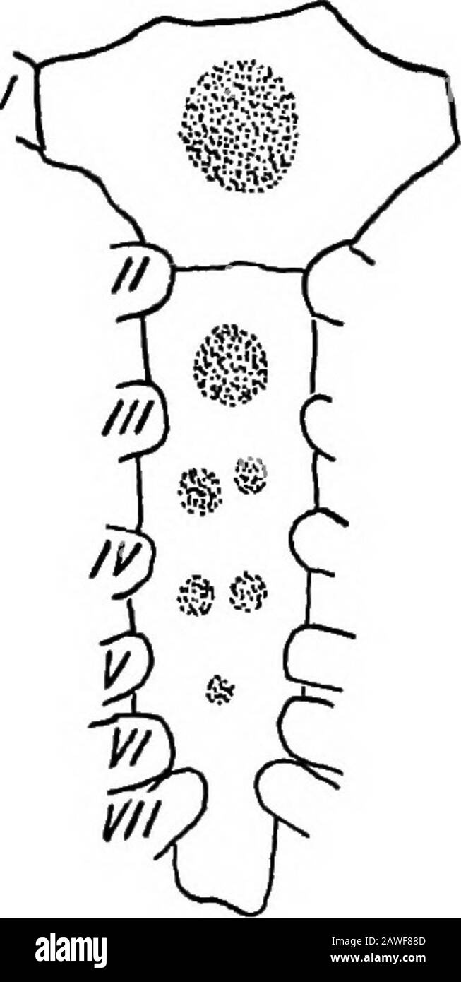 The development of the human body; a manual of human embryology . T OF THE HUMAN BODY. the bars formed by these posterior ribs constitute the ensi-form process. The ossification of the sternum (Fig. 97) partakes to acertain extent of the original bilateral segmental origin ofthe cartilage, but a marked condensation of the centers ofossification also occurs. In the portion of the cartilagewhich lies below the junction of the third costal cartilagesa series of pairs of centers appears just about birth, eachcenter probably representing anepiphysial center of a correspond-ing rib. Later the center Stock Photohttps://www.alamy.com/image-license-details/?v=1https://www.alamy.com/the-development-of-the-human-body-a-manual-of-human-embryology-t-of-the-human-body-the-bars-formed-by-these-posterior-ribs-constitute-the-ensi-form-process-the-ossification-of-the-sternum-fig-97-partakes-to-acertain-extent-of-the-original-bilateral-segmental-origin-ofthe-cartilage-but-a-marked-condensation-of-the-centers-ofossification-also-occurs-in-the-portion-of-the-cartilagewhich-lies-below-the-junction-of-the-third-costal-cartilagesa-series-of-pairs-of-centers-appears-just-about-birth-eachcenter-probably-representing-anepiphysial-center-of-a-correspond-ing-rib-later-the-center-image342699181.html
RM2AWF88D–The development of the human body; a manual of human embryology . T OF THE HUMAN BODY. the bars formed by these posterior ribs constitute the ensi-form process. The ossification of the sternum (Fig. 97) partakes to acertain extent of the original bilateral segmental origin ofthe cartilage, but a marked condensation of the centers ofossification also occurs. In the portion of the cartilagewhich lies below the junction of the third costal cartilagesa series of pairs of centers appears just about birth, eachcenter probably representing anepiphysial center of a correspond-ing rib. Later the center
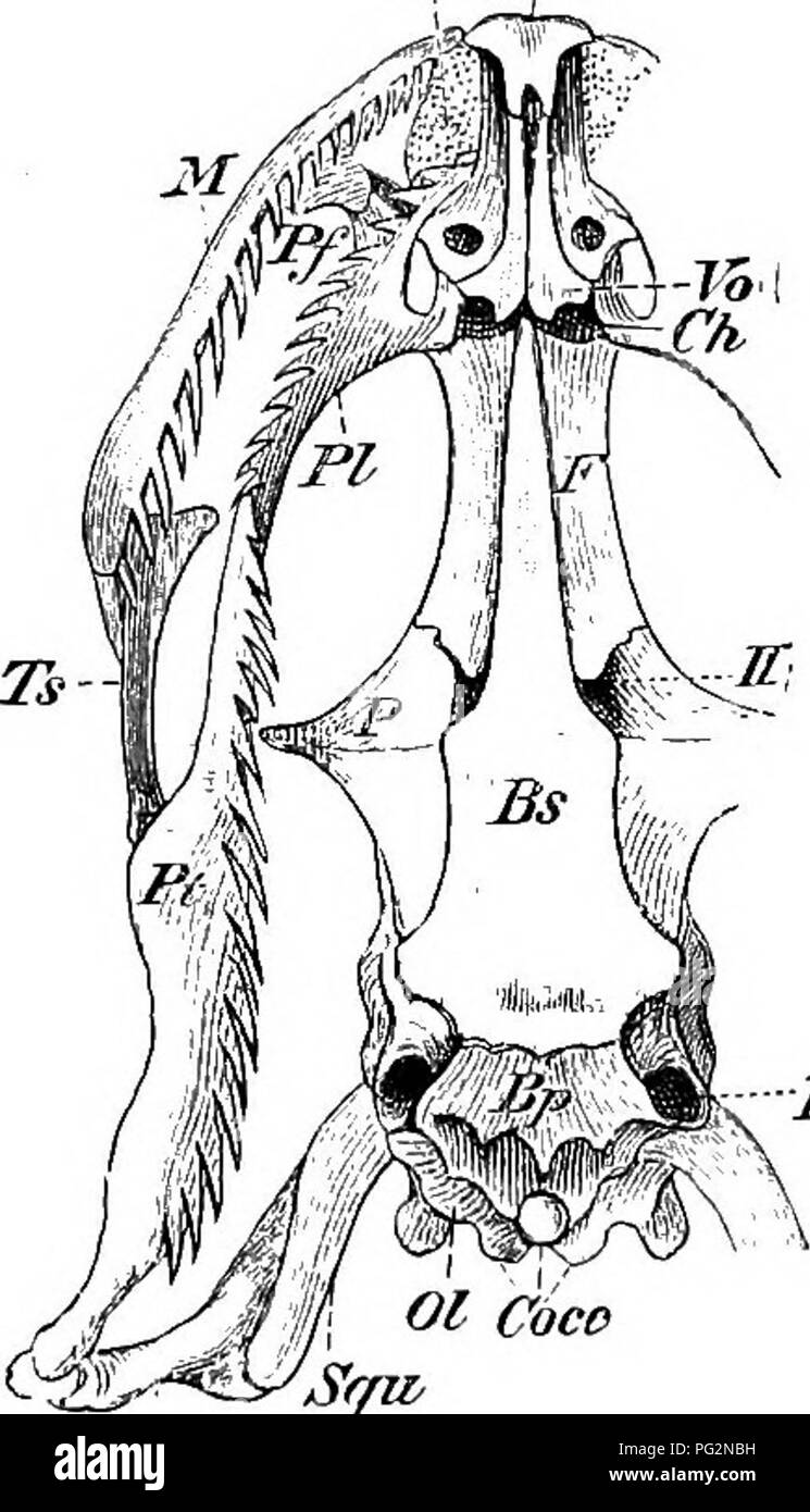 . Elements of the comparative anatomy of vertebrates. Anatomy, Comparative. 00 COMPARATIVE ANATOMY of ossification, which gives the skull a very firm and solid appear- ance ; only amongst Lizards (Fig. 71), and especially in Hatteria is the cartilage retained to any considerable extent, and owing to the conformation of the bones in the posterior region, the skull in these forms presents a number of distinct spaces or fossae in the dry state. In Snakes and Amphisbsenians the cranial cavity extends forwards between the orbits as far as the ethmoidal region, while in the Lacertilia, Chelonia, and Stock Photohttps://www.alamy.com/image-license-details/?v=1https://www.alamy.com/elements-of-the-comparative-anatomy-of-vertebrates-anatomy-comparative-00-comparative-anatomy-of-ossification-which-gives-the-skull-a-very-firm-and-solid-appear-ance-only-amongst-lizards-fig-71-and-especially-in-hatteria-is-the-cartilage-retained-to-any-considerable-extent-and-owing-to-the-conformation-of-the-bones-in-the-posterior-region-the-skull-in-these-forms-presents-a-number-of-distinct-spaces-or-fossae-in-the-dry-state-in-snakes-and-amphisbsenians-the-cranial-cavity-extends-forwards-between-the-orbits-as-far-as-the-ethmoidal-region-while-in-the-lacertilia-chelonia-and-image216419605.html
RMPG2NBH–. Elements of the comparative anatomy of vertebrates. Anatomy, Comparative. 00 COMPARATIVE ANATOMY of ossification, which gives the skull a very firm and solid appear- ance ; only amongst Lizards (Fig. 71), and especially in Hatteria is the cartilage retained to any considerable extent, and owing to the conformation of the bones in the posterior region, the skull in these forms presents a number of distinct spaces or fossae in the dry state. In Snakes and Amphisbsenians the cranial cavity extends forwards between the orbits as far as the ethmoidal region, while in the Lacertilia, Chelonia, and
 A manual of anatomy . sertion to the mm. vasti medialis andlateralis, respectively. 98 OSTEOLOGY Nutrient foramina are seen upon the ventral surface. Articulations.—With the femur. Ossification.—It is of endochondral origin and develops from one, or twocenters, which appear during the third year. Ossilication is usually com-pleted by the age of puberty. Muscles Attached.—Insertions.—Mm. vasti medialis, lateralis and intermediusand the rectus femoris (four). THE TIBIA The tibia or shin bone is the larger and stronger bone of the leg.It consists of a proximal extremity, shaft and distal extremit Stock Photohttps://www.alamy.com/image-license-details/?v=1https://www.alamy.com/a-manual-of-anatomy-sertion-to-the-mm-vasti-medialis-andlateralis-respectively-98-osteology-nutrient-foramina-are-seen-upon-the-ventral-surface-articulationswith-the-femur-ossificationit-is-of-endochondral-origin-and-develops-from-one-or-twocenters-which-appear-during-the-third-year-ossilication-is-usually-com-pleted-by-the-age-of-puberty-muscles-attachedinsertionsmm-vasti-medialis-lateralis-and-intermediusand-the-rectus-femoris-four-the-tibia-the-tibia-or-shin-bone-is-the-larger-and-stronger-bone-of-the-legit-consists-of-a-proximal-extremity-shaft-and-distal-extremit-image343379969.html
RM2AXJ8J9–A manual of anatomy . sertion to the mm. vasti medialis andlateralis, respectively. 98 OSTEOLOGY Nutrient foramina are seen upon the ventral surface. Articulations.—With the femur. Ossification.—It is of endochondral origin and develops from one, or twocenters, which appear during the third year. Ossilication is usually com-pleted by the age of puberty. Muscles Attached.—Insertions.—Mm. vasti medialis, lateralis and intermediusand the rectus femoris (four). THE TIBIA The tibia or shin bone is the larger and stronger bone of the leg.It consists of a proximal extremity, shaft and distal extremit
 . Veterinary obstetrics; a compendium for the use of students and practitioners. Veterinary obstetrics. 14 VETERINARY OBSTETRICS. With the exception of the equine species, the sacrum is joined to the last lumbar vertebra by three diarthrodial surfaces only—the head of the body and two transverse processes. The ischio pubic symphysis in the Cow is con- siderably longer than in the Mare and not rectilinear; ossification of the symphysis is less complete, and takes place much later than in the Mare. 6. Fig. s. Ligaments of the Lumbar Vertebrae, Sacrum and Pelvis. {Seen from below.) a. Intertransv Stock Photohttps://www.alamy.com/image-license-details/?v=1https://www.alamy.com/veterinary-obstetrics-a-compendium-for-the-use-of-students-and-practitioners-veterinary-obstetrics-14-veterinary-obstetrics-with-the-exception-of-the-equine-species-the-sacrum-is-joined-to-the-last-lumbar-vertebra-by-three-diarthrodial-surfaces-onlythe-head-of-the-body-and-two-transverse-processes-the-ischio-pubic-symphysis-in-the-cow-is-con-siderably-longer-than-in-the-mare-and-not-rectilinear-ossification-of-the-symphysis-is-less-complete-and-takes-place-much-later-than-in-the-mare-6-fig-s-ligaments-of-the-lumbar-vertebrae-sacrum-and-pelvis-seen-from-below-a-intertransv-image216388099.html
RMPG196B–. Veterinary obstetrics; a compendium for the use of students and practitioners. Veterinary obstetrics. 14 VETERINARY OBSTETRICS. With the exception of the equine species, the sacrum is joined to the last lumbar vertebra by three diarthrodial surfaces only—the head of the body and two transverse processes. The ischio pubic symphysis in the Cow is con- siderably longer than in the Mare and not rectilinear; ossification of the symphysis is less complete, and takes place much later than in the Mare. 6. Fig. s. Ligaments of the Lumbar Vertebrae, Sacrum and Pelvis. {Seen from below.) a. Intertransv
 The development of the human body; a manual of human embryology . Fig. 101.—Occipitai, Bone of a Fetus at Term. bo, Basioccipital; eo, exoccipital; ip, interparietal; so, supraoccipital. THE SKULL. 197 In the first place, the basal portion of the cartilage ossifiesto form two bones, an anterior or presphenoid and a poste-rior or basisphenoid (Fig. 102, b), and on each side of eachof these an ossification appears giving rise to two lesserwings or orbito sphenoids (os) and two greater wings oralisphenoids (as), and an additional center appears oneach side of the basisphenoid to form the lingula Stock Photohttps://www.alamy.com/image-license-details/?v=1https://www.alamy.com/the-development-of-the-human-body-a-manual-of-human-embryology-fig-101occipitai-bone-of-a-fetus-at-term-bo-basioccipital-eo-exoccipital-ip-interparietal-so-supraoccipital-the-skull-197-in-the-first-place-the-basal-portion-of-the-cartilage-ossifiesto-form-two-bones-an-anterior-or-presphenoid-and-a-poste-rior-or-basisphenoid-fig-102-b-and-on-each-side-of-eachof-these-an-ossification-appears-giving-rise-to-two-lesserwings-or-orbito-sphenoids-os-and-two-greater-wings-oralisphenoids-as-and-an-additional-center-appears-oneach-side-of-the-basisphenoid-to-form-the-lingula-image342697626.html
RM2AWF68X–The development of the human body; a manual of human embryology . Fig. 101.—Occipitai, Bone of a Fetus at Term. bo, Basioccipital; eo, exoccipital; ip, interparietal; so, supraoccipital. THE SKULL. 197 In the first place, the basal portion of the cartilage ossifiesto form two bones, an anterior or presphenoid and a poste-rior or basisphenoid (Fig. 102, b), and on each side of eachof these an ossification appears giving rise to two lesserwings or orbito sphenoids (os) and two greater wings oralisphenoids (as), and an additional center appears oneach side of the basisphenoid to form the lingula
 . On the anatomy of vertebrates. Vertebrates; Anatomy, Comparative; 1866. ANATOMY or VERTEBRATES. 73 circumscribe elliptical spaces outside the Y)respheuoicl plate : these appear to-represent the pterygoid arches, fig. 61, i, but, as in the embryo of higher fishes, are uot sejiarated from the base of the skull by distinct joints. The basal cartilages, after forming the car-capsules, ib. g, exteud upon the sides of the cra- nium, ib. h, arch over its back part, and leave only its upper and middle part membranous, as in the human embryo when ossification of the cranium commences. The cranium is Stock Photohttps://www.alamy.com/image-license-details/?v=1https://www.alamy.com/on-the-anatomy-of-vertebrates-vertebrates-anatomy-comparative-1866-anatomy-or-vertebrates-73-circumscribe-elliptical-spaces-outside-the-yrespheuoicl-plate-these-appear-to-represent-the-pterygoid-arches-fig-61-i-but-as-in-the-embryo-of-higher-fishes-are-uot-sejiarated-from-the-base-of-the-skull-by-distinct-joints-the-basal-cartilages-after-forming-the-car-capsules-ib-g-exteud-upon-the-sides-of-the-cra-nium-ib-h-arch-over-its-back-part-and-leave-only-its-upper-and-middle-part-membranous-as-in-the-human-embryo-when-ossification-of-the-cranium-commences-the-cranium-is-image216417845.html
RMPG2K4N–. On the anatomy of vertebrates. Vertebrates; Anatomy, Comparative; 1866. ANATOMY or VERTEBRATES. 73 circumscribe elliptical spaces outside the Y)respheuoicl plate : these appear to-represent the pterygoid arches, fig. 61, i, but, as in the embryo of higher fishes, are uot sejiarated from the base of the skull by distinct joints. The basal cartilages, after forming the car-capsules, ib. g, exteud upon the sides of the cra- nium, ib. h, arch over its back part, and leave only its upper and middle part membranous, as in the human embryo when ossification of the cranium commences. The cranium is
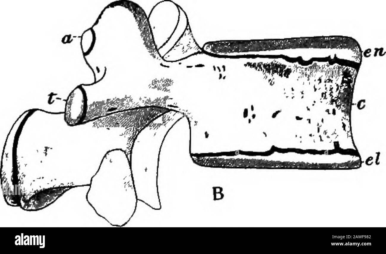 The development of the human body; a manual of human embryology . Fig. 94.—A, A Vertebra at Birth; B, Lumbar Vertebra showingSecondary Centers of Ossification. a, Center for the articular process; c, centrum; el, lower epiphysialplate; en, upper epiphysial plate; na, neural arch; s, center forspinous process; t, center for transverse process.—(Sappey.) process and gradually extends to form the bony lamina,pedicle, and the greater portion of the transverse andspinous processes; a double center (see p. 178) gives riseto the body of the vertebra; and each rib ossifies from asingle center. These v Stock Photohttps://www.alamy.com/image-license-details/?v=1https://www.alamy.com/the-development-of-the-human-body-a-manual-of-human-embryology-fig-94a-a-vertebra-at-birth-b-lumbar-vertebra-showingsecondary-centers-of-ossification-a-center-for-the-articular-process-c-centrum-el-lower-epiphysialplate-en-upper-epiphysial-plate-na-neural-arch-s-center-forspinous-process-t-center-for-transverse-processsappey-process-and-gradually-extends-to-form-the-bony-laminapedicle-and-the-greater-portion-of-the-transverse-andspinous-processes-a-double-center-see-p-178-gives-riseto-the-body-of-the-vertebra-and-each-rib-ossifies-from-asingle-center-these-v-image342699954.html
RM2AWF982–The development of the human body; a manual of human embryology . Fig. 94.—A, A Vertebra at Birth; B, Lumbar Vertebra showingSecondary Centers of Ossification. a, Center for the articular process; c, centrum; el, lower epiphysialplate; en, upper epiphysial plate; na, neural arch; s, center forspinous process; t, center for transverse process.—(Sappey.) process and gradually extends to form the bony lamina,pedicle, and the greater portion of the transverse andspinous processes; a double center (see p. 178) gives riseto the body of the vertebra; and each rib ossifies from asingle center. These v
 . A manual of zoology. Zoology. 4o6 CIIORDATA axial skeleton and arise from tlie ossifications in the skin (scales) or in the mouth (teeth), alreaih" referred to (p. 451). They sink into the deeper portions and apply ihemsehes to the axial skeleton, especially to those parts where, from lack of cartilage, no primary bones can be formed. It is not settled how far these distinctions are 'alid. According to Gegen- baur all ossification arose jirimarily in the skin or mucous membranes, and primary bones are merely memlirane bones which have entered the. Fir.. 515.—Chondrocranium of A }>ip Stock Photohttps://www.alamy.com/image-license-details/?v=1https://www.alamy.com/a-manual-of-zoology-zoology-4o6-ciiordata-axial-skeleton-and-arise-from-tlie-ossifications-in-the-skin-scales-or-in-the-mouth-teeth-alreaihquot-referred-to-p-451-they-sink-into-the-deeper-portions-and-apply-ihemsehes-to-the-axial-skeleton-especially-to-those-parts-where-from-lack-of-cartilage-no-primary-bones-can-be-formed-it-is-not-settled-how-far-these-distinctions-are-alid-according-to-gegen-baur-all-ossification-arose-jirimarily-in-the-skin-or-mucous-membranes-and-primary-bones-are-merely-memlirane-bones-which-have-entered-the-fir-515chondrocranium-of-a-gtip-image216441666.html
RMPG3NFE–. A manual of zoology. Zoology. 4o6 CIIORDATA axial skeleton and arise from tlie ossifications in the skin (scales) or in the mouth (teeth), alreaih" referred to (p. 451). They sink into the deeper portions and apply ihemsehes to the axial skeleton, especially to those parts where, from lack of cartilage, no primary bones can be formed. It is not settled how far these distinctions are 'alid. According to Gegen- baur all ossification arose jirimarily in the skin or mucous membranes, and primary bones are merely memlirane bones which have entered the. Fir.. 515.—Chondrocranium of A }>ip
 . Outlines of zoology. k., ceratohyal; e^.k., epihyali.h., interhyal; Op., opercular; S.op., sub-opercular; i.op.,inter-opercular; p.op., prEe-opercular. vance upon that of the skate : first, in the ossification of theprimitive cartilage; and second, in the addition of membranebones. Of the latter, the parietals and frontals cover overthe spaces which in the skate form the fontanelles. 554 PISCES—FISHES. The first or mandibular arch is believed by many to form Meclcels •cartilage beneath, and the palato-pterygo-quadrate cartilage above.Meckels cartilage becomes the foundation of the lower jaw, Stock Photohttps://www.alamy.com/image-license-details/?v=1https://www.alamy.com/outlines-of-zoology-k-ceratohyal-ek-epihyalih-interhyal-op-opercular-sop-sub-opercular-iopinter-opercular-pop-pree-opercular-vance-upon-that-of-the-skate-first-in-the-ossification-of-theprimitive-cartilage-and-second-in-the-addition-of-membranebones-of-the-latter-the-parietals-and-frontals-cover-overthe-spaces-which-in-the-skate-form-the-fontanelles-554-piscesfishes-the-first-or-mandibular-arch-is-believed-by-many-to-form-meclcels-cartilage-beneath-and-the-palato-pterygo-quadrate-cartilage-abovemeckels-cartilage-becomes-the-foundation-of-the-lower-jaw-image337126469.html
RM2AGDC71–. Outlines of zoology. k., ceratohyal; e^.k., epihyali.h., interhyal; Op., opercular; S.op., sub-opercular; i.op.,inter-opercular; p.op., prEe-opercular. vance upon that of the skate : first, in the ossification of theprimitive cartilage; and second, in the addition of membranebones. Of the latter, the parietals and frontals cover overthe spaces which in the skate form the fontanelles. 554 PISCES—FISHES. The first or mandibular arch is believed by many to form Meclcels •cartilage beneath, and the palato-pterygo-quadrate cartilage above.Meckels cartilage becomes the foundation of the lower jaw,
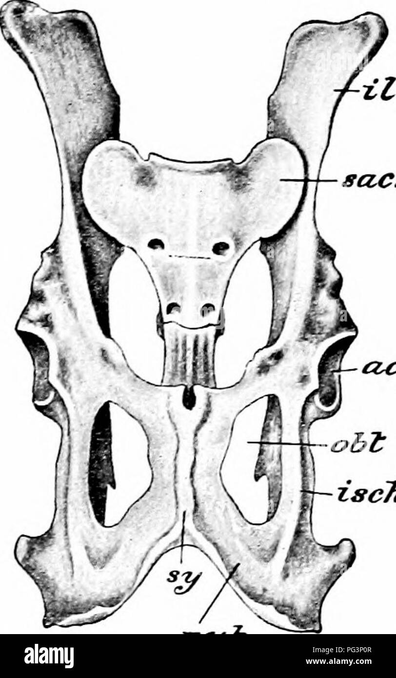 . A manual of zoology. PHYLUM CHORDATA 5°3 the pubis, but by a small intercalated ossification, the cotyloid bone. The ilium (il) has a rough surface for articulation with the sacrum. Between the. pubis (pub) in front and the ischium (isch) behind is a large aperture, the obturator foramen (obi). The femur has at its proximal end a prominent head for articulation with the acetabulum, external to this a prominent process, the great trochanter, and internally a much smaller, the lesser trochanter, while a small process or third trochanter is situated on the outer border a little below the great Stock Photohttps://www.alamy.com/image-license-details/?v=1https://www.alamy.com/a-manual-of-zoology-phylum-chordata-53-the-pubis-but-by-a-small-intercalated-ossification-the-cotyloid-bone-the-ilium-il-has-a-rough-surface-for-articulation-with-the-sacrum-between-the-pubis-pub-in-front-and-the-ischium-isch-behind-is-a-large-aperture-the-obturator-foramen-obi-the-femur-has-at-its-proximal-end-a-prominent-head-for-articulation-with-the-acetabulum-external-to-this-a-prominent-process-the-great-trochanter-and-internally-a-much-smaller-the-lesser-trochanter-while-a-small-process-or-third-trochanter-is-situated-on-the-outer-border-a-little-below-the-great-image216442039.html
RMPG3P0R–. A manual of zoology. PHYLUM CHORDATA 5°3 the pubis, but by a small intercalated ossification, the cotyloid bone. The ilium (il) has a rough surface for articulation with the sacrum. Between the. pubis (pub) in front and the ischium (isch) behind is a large aperture, the obturator foramen (obi). The femur has at its proximal end a prominent head for articulation with the acetabulum, external to this a prominent process, the great trochanter, and internally a much smaller, the lesser trochanter, while a small process or third trochanter is situated on the outer border a little below the great
 Researches on the Structure, Organization, and Classification of the Fossil Reptilia VII Further Observations on Pareiasaurus . Pelvis and Femur, Pareiasaurus Baini.PLATE 22. Fig. 1. Anterior aspect of sacrum and pelvis of Pareiasaurus Baini, showing thelarge size of the neural spine and second pair (s.) of sacral ribs and the depthgiven to the pelvis by the downward reflection of the pubis (p.) and inter-pubic ossification (ip.). The pubic foramen (f.) is well seen on the left side.The anterior angles on the iliac bones (il.) are seen to extend outward. Fig. 2. Infero-posterior aspect of righ Stock Photohttps://www.alamy.com/image-license-details/?v=1https://www.alamy.com/researches-on-the-structure-organization-and-classification-of-the-fossil-reptilia-vii-further-observations-on-pareiasaurus-pelvis-and-femur-pareiasaurus-bainiplate-22-fig-1-anterior-aspect-of-sacrum-and-pelvis-of-pareiasaurus-baini-showing-thelarge-size-of-the-neural-spine-and-second-pair-s-of-sacral-ribs-and-the-depthgiven-to-the-pelvis-by-the-downward-reflection-of-the-pubis-p-and-inter-pubic-ossification-ip-the-pubic-foramen-f-is-well-seen-on-the-left-sidethe-anterior-angles-on-the-iliac-bones-il-are-seen-to-extend-outward-fig-2-infero-posterior-aspect-of-righ-image338431317.html
RM2AJGTGN–Researches on the Structure, Organization, and Classification of the Fossil Reptilia VII Further Observations on Pareiasaurus . Pelvis and Femur, Pareiasaurus Baini.PLATE 22. Fig. 1. Anterior aspect of sacrum and pelvis of Pareiasaurus Baini, showing thelarge size of the neural spine and second pair (s.) of sacral ribs and the depthgiven to the pelvis by the downward reflection of the pubis (p.) and inter-pubic ossification (ip.). The pubic foramen (f.) is well seen on the left side.The anterior angles on the iliac bones (il.) are seen to extend outward. Fig. 2. Infero-posterior aspect of righ
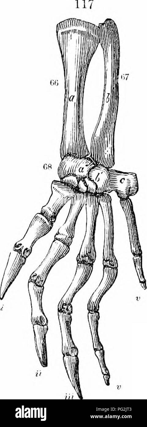 . On the anatomy of vertebrates. Vertebrates; Anatomy, Comparative; 1866. ANATOMY OF VEKTEBRATES. 187 shaft is almost straight, sliglitly expanded at the distal end, at the back pai't of which the condyles are fceljly indicated. In Terra- penes, fig. 51, w, and Tortoises, the femur equals or exceeds the luimerus in length : its shaft is more bent: the trochanter is divided into two processes, most distinct in Trionyx. In no Clielonian is there a medullary cavity : ossification extends throughout the bone : the two l)ones of the leg, ib. x, Y, are nearly straight; the til)ia is the largest, wit Stock Photohttps://www.alamy.com/image-license-details/?v=1https://www.alamy.com/on-the-anatomy-of-vertebrates-vertebrates-anatomy-comparative-1866-anatomy-of-vektebrates-187-shaft-is-almost-straight-sliglitly-expanded-at-the-distal-end-at-the-back-pait-of-which-the-condyles-are-fceljly-indicated-in-terra-penes-fig-51-w-and-tortoises-the-femur-equals-or-exceeds-the-luimerus-in-length-its-shaft-is-more-bent-the-trochanter-is-divided-into-two-processes-most-distinct-in-trionyx-in-no-clielonian-is-there-a-medullary-cavity-ossification-extends-throughout-the-bone-the-two-lones-of-the-leg-ib-x-y-are-nearly-straight-the-tilia-is-the-largest-wit-image216417603.html
RMPG2JT3–. On the anatomy of vertebrates. Vertebrates; Anatomy, Comparative; 1866. ANATOMY OF VEKTEBRATES. 187 shaft is almost straight, sliglitly expanded at the distal end, at the back pai't of which the condyles are fceljly indicated. In Terra- penes, fig. 51, w, and Tortoises, the femur equals or exceeds the luimerus in length : its shaft is more bent: the trochanter is divided into two processes, most distinct in Trionyx. In no Clielonian is there a medullary cavity : ossification extends throughout the bone : the two l)ones of the leg, ib. x, Y, are nearly straight; the til)ia is the largest, wit
 Researches on the Structure, Organization, and Classification of the Fossil Reptilia VII Further Observations on Pareiasaurus . the depthgiven to the pelvis by the downward reflection of the pubis (p.) and inter-pubic ossification (ip.). The pubic foramen (f.) is well seen on the left side.The anterior angles on the iliac bones ^7.) are seen to extend outward. Fig. 2. Infero-posterior aspect of right femur : art., proximal articulation ; t., internaltrochanter ; cl, c.2, flattened distal condyles. Jl JLA-lJli 23. Fig. 1. Right humerus of Pareiasaurus Baini, seen from the superior aspect; one-t Stock Photohttps://www.alamy.com/image-license-details/?v=1https://www.alamy.com/researches-on-the-structure-organization-and-classification-of-the-fossil-reptilia-vii-further-observations-on-pareiasaurus-the-depthgiven-to-the-pelvis-by-the-downward-reflection-of-the-pubis-p-and-inter-pubic-ossification-ip-the-pubic-foramen-f-is-well-seen-on-the-left-sidethe-anterior-angles-on-the-iliac-bones-7-are-seen-to-extend-outward-fig-2-infero-posterior-aspect-of-right-femur-art-proximal-articulation-t-internaltrochanter-cl-c2-flattened-distal-condyles-jl-jla-ljli-23-fig-1-right-humerus-of-pareiasaurus-baini-seen-from-the-superior-aspect-one-t-image338445463.html
RM2AJHEHY–Researches on the Structure, Organization, and Classification of the Fossil Reptilia VII Further Observations on Pareiasaurus . the depthgiven to the pelvis by the downward reflection of the pubis (p.) and inter-pubic ossification (ip.). The pubic foramen (f.) is well seen on the left side.The anterior angles on the iliac bones ^7.) are seen to extend outward. Fig. 2. Infero-posterior aspect of right femur : art., proximal articulation ; t., internaltrochanter ; cl, c.2, flattened distal condyles. Jl JLA-lJli 23. Fig. 1. Right humerus of Pareiasaurus Baini, seen from the superior aspect; one-t
 . A manual of zoology. Zoology. IV. VERTEBRATA: MAMMALIA 547 mal number of young at a birth. Although the mamma? are present in Ijoth sexes, they are functional only in the female, and here only after the birth of the young. A dermal skeleton occurs in few species (e.g., the firm l^ony plates of the armadillos); on the other hand the axial skeleton shows many features not occurring elsewhere. In the skull many of the Ijones already referred to are evident only as centres of ossification, fusing early with their neigh- bors to form larger bones. As the temporal bone shows, parts of diverse orig Stock Photohttps://www.alamy.com/image-license-details/?v=1https://www.alamy.com/a-manual-of-zoology-zoology-iv-vertebrata-mammalia-547-mal-number-of-young-at-a-birth-although-the-mamma-are-present-in-ijoth-sexes-they-are-functional-only-in-the-female-and-here-only-after-the-birth-of-the-young-a-dermal-skeleton-occurs-in-few-species-eg-the-firm-lony-plates-of-the-armadillos-on-the-other-hand-the-axial-skeleton-shows-many-features-not-occurring-elsewhere-in-the-skull-many-of-the-ijones-already-referred-to-are-evident-only-as-centres-of-ossification-fusing-early-with-their-neigh-bors-to-form-larger-bones-as-the-temporal-bone-shows-parts-of-diverse-orig-image216441360.html
RMPG3N4G–. A manual of zoology. Zoology. IV. VERTEBRATA: MAMMALIA 547 mal number of young at a birth. Although the mamma? are present in Ijoth sexes, they are functional only in the female, and here only after the birth of the young. A dermal skeleton occurs in few species (e.g., the firm l^ony plates of the armadillos); on the other hand the axial skeleton shows many features not occurring elsewhere. In the skull many of the Ijones already referred to are evident only as centres of ossification, fusing early with their neigh- bors to form larger bones. As the temporal bone shows, parts of diverse orig
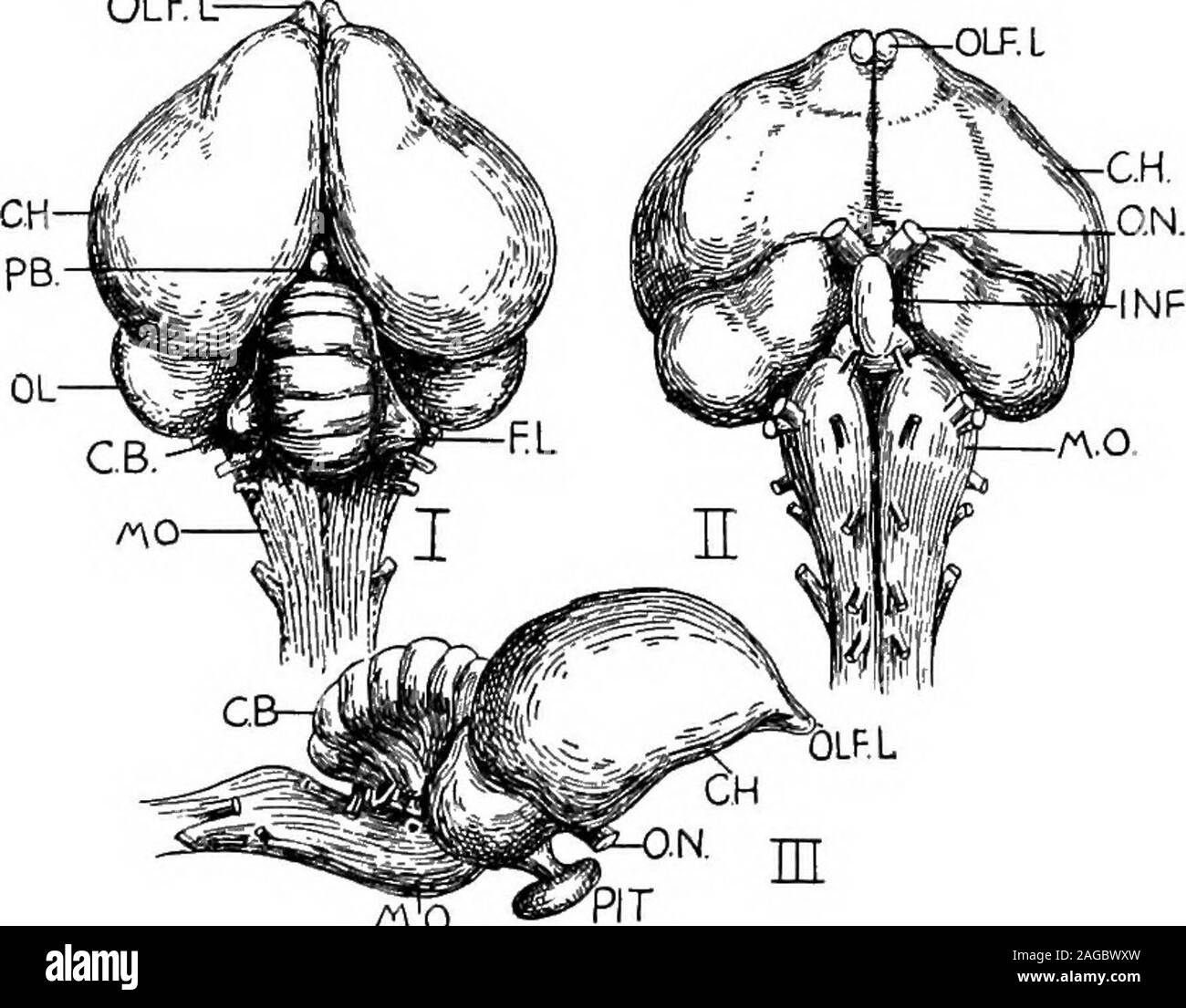 . Outlines of zoology. dorsalilia fused to the complex sacral region,of ischia sloping backwards, and ofpubes running parallel to the ischia.The incomplete ossification of theacetabulum and the absence of ventralsymphyses are noteworthy. The hind-limb consists of a shortstout femur, a tibia to which theproximal tarsals (astragalus and oscalcis) are fused (forming a tibio-tar-sus), an incomplete fibula joined to m Fig. 367.—Bones ofhind-limb of eagle, yr, Femur; i.i.,tibio-tarsus;y/f., fibula; a., ankle-joint; jn.f., tarso-meta*tarsus; in.t., first meta-tarsal (free). 664 BIRDS. the tibia, th Stock Photohttps://www.alamy.com/image-license-details/?v=1https://www.alamy.com/outlines-of-zoology-dorsalilia-fused-to-the-complex-sacral-regionof-ischia-sloping-backwards-and-ofpubes-running-parallel-to-the-ischiathe-incomplete-ossification-of-theacetabulum-and-the-absence-of-ventralsymphyses-are-noteworthy-the-hind-limb-consists-of-a-shortstout-femur-a-tibia-to-which-theproximal-tarsals-astragalus-and-oscalcis-are-fused-forming-a-tibio-tar-sus-an-incomplete-fibula-joined-to-m-fig-367bones-ofhind-limb-of-eagle-yr-femur-iitibio-tarsusyf-fibula-a-ankle-joint-jnf-tarso-metatarsus-int-first-meta-tarsal-free-664-birds-the-tibia-th-image337093313.html
RM2AGBWXW–. Outlines of zoology. dorsalilia fused to the complex sacral region,of ischia sloping backwards, and ofpubes running parallel to the ischia.The incomplete ossification of theacetabulum and the absence of ventralsymphyses are noteworthy. The hind-limb consists of a shortstout femur, a tibia to which theproximal tarsals (astragalus and oscalcis) are fused (forming a tibio-tar-sus), an incomplete fibula joined to m Fig. 367.—Bones ofhind-limb of eagle, yr, Femur; i.i.,tibio-tarsus;y/f., fibula; a., ankle-joint; jn.f., tarso-meta*tarsus; in.t., first meta-tarsal (free). 664 BIRDS. the tibia, th
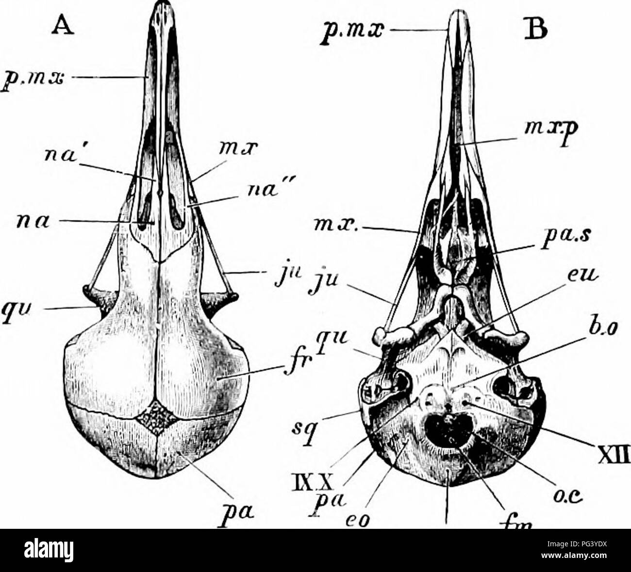 . A manual of zoology. 466 MANUAL OF ZOOLOGY istic parts of the bird's skeleton. It is a broad plate of bone produced ventrally, in the sagittal plane, into a deep keel or carina sterni (car), formed, in the young bird, from a separate centre of ossification. The posterior border of the sternum presents two pairs of notches, covered, in the recent state,. r *% lt , i.o.s wfr p n.etlh I ''i. Please note that these images are extracted from scanned page images that may have been digitally enhanced for readability - coloration and appearance of these illustrations may not perfectly resemble t Stock Photohttps://www.alamy.com/image-license-details/?v=1https://www.alamy.com/a-manual-of-zoology-466-manual-of-zoology-istic-parts-of-the-birds-skeleton-it-is-a-broad-plate-of-bone-produced-ventrally-in-the-sagittal-plane-into-a-deep-keel-or-carina-sterni-car-formed-in-the-young-bird-from-a-separate-centre-of-ossification-the-posterior-border-of-the-sternum-presents-two-pairs-of-notches-covered-in-the-recent-state-r-lt-ios-wfr-p-netlh-i-i-please-note-that-these-images-are-extracted-from-scanned-page-images-that-may-have-been-digitally-enhanced-for-readability-coloration-and-appearance-of-these-illustrations-may-not-perfectly-resemble-t-image216446326.html
RMPG3YDX–. A manual of zoology. 466 MANUAL OF ZOOLOGY istic parts of the bird's skeleton. It is a broad plate of bone produced ventrally, in the sagittal plane, into a deep keel or carina sterni (car), formed, in the young bird, from a separate centre of ossification. The posterior border of the sternum presents two pairs of notches, covered, in the recent state,. r *% lt , i.o.s wfr p n.etlh I ''i. Please note that these images are extracted from scanned page images that may have been digitally enhanced for readability - coloration and appearance of these illustrations may not perfectly resemble t
 Brightness and dullness in children . se normally J Anatomical or Physiological Age Versus Chronological Age,Pedagogical Seminary, vol. xv, 1908, pp. 230-237; and PhysiologicalAge, American Physical Education Review, vol. xiii, 1908, pp. 141-154, 214-^227, 268-283. and 345-358. ., „ Rontgen-ray Methods Applied to the Grading of Early Life.American Physical Education Review, vol. xv, 1010, pp. 396-420. * Bulletins of the State College of Kentucky: 1005, Develop-ment of the Bones of the Hand, pp. 30; 1906, Ossification of theEpiphyses of the Hand, pp. 35; 1908, The Chronology and Orderof Ossific Stock Photohttps://www.alamy.com/image-license-details/?v=1https://www.alamy.com/brightness-and-dullness-in-children-se-normally-j-anatomical-or-physiological-age-versus-chronological-agepedagogical-seminary-vol-xv-1908-pp-230-237-and-physiologicalage-american-physical-education-review-vol-xiii-1908-pp-141-154-214-227-268-283-and-345-358-rontgen-ray-methods-applied-to-the-grading-of-early-lifeamerican-physical-education-review-vol-xv-1010-pp-396-420-bulletins-of-the-state-college-of-kentucky-1005-develop-ment-of-the-bones-of-the-hand-pp-30-1906-ossification-of-theepiphyses-of-the-hand-pp-35-1908-the-chronology-and-orderof-ossific-image343390716.html
RM2AXJPA4–Brightness and dullness in children . se normally J Anatomical or Physiological Age Versus Chronological Age,Pedagogical Seminary, vol. xv, 1908, pp. 230-237; and PhysiologicalAge, American Physical Education Review, vol. xiii, 1908, pp. 141-154, 214-^227, 268-283. and 345-358. ., „ Rontgen-ray Methods Applied to the Grading of Early Life.American Physical Education Review, vol. xv, 1010, pp. 396-420. * Bulletins of the State College of Kentucky: 1005, Develop-ment of the Bones of the Hand, pp. 30; 1906, Ossification of theEpiphyses of the Hand, pp. 35; 1908, The Chronology and Orderof Ossific
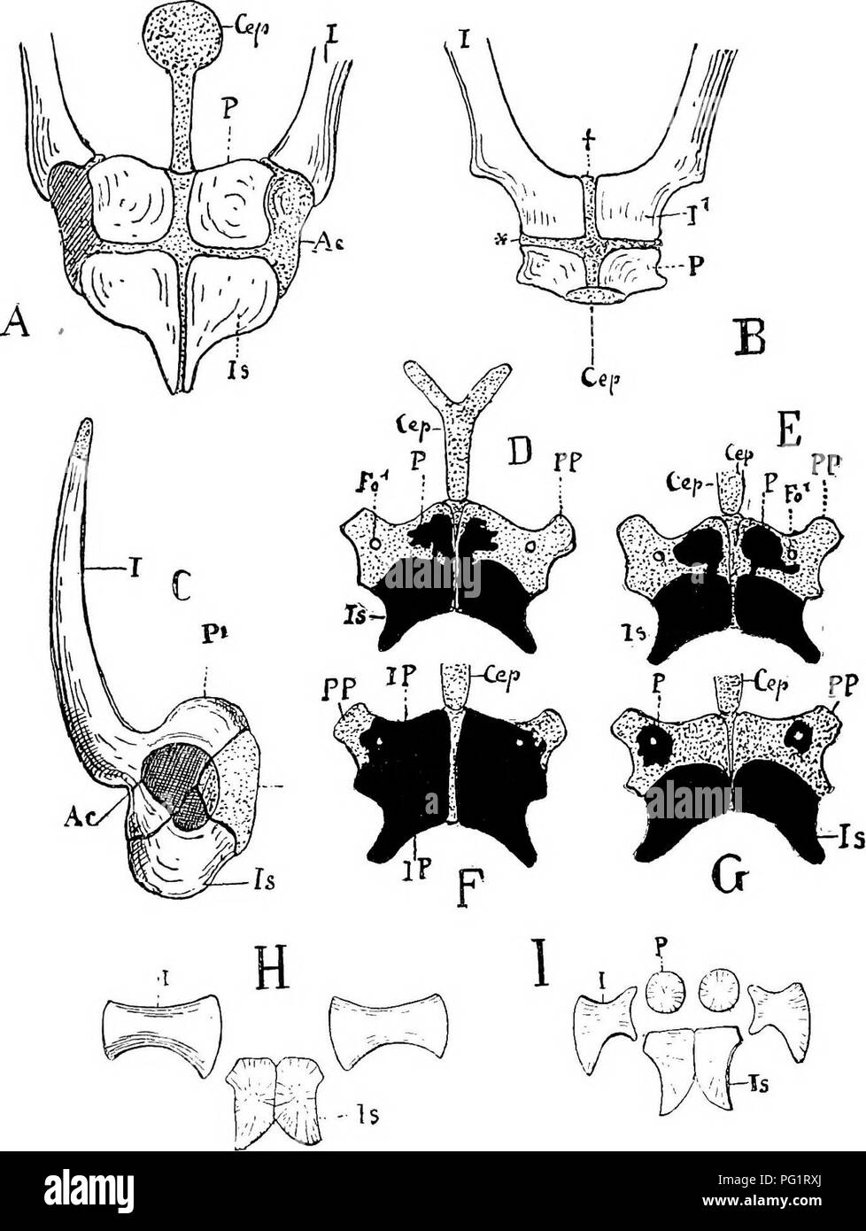 . Elements of the comparative anatomy of vertebrates. Anatomy, Comparative. PELVIC ARCH 113. Fig. 91.—Pelvis op Yakious Amphibia. A, Xenopus [Dactyhthra), from below ; B, the same from the front ; C, Rana e^cuJtnta, from the right side; 1) and E, Sa/aiiiandra ntra ; F and G, Salanmndra mnrii/oia ; H, Branrhio.taiirn.s ; I, fJi-iivsaiinis. D-I, from the ventral side. (Figs. H and I after Creduer.) /, ilium ; Is, ischium ; P, pubis (P^ in Rana, pubic end of ilium); IP, fused ischio- pubic ossification; PP, prepubis ; Cep, epipubic cartilage ; Fo^, obturator foramen ; /' (in Xenopus), the proxima Stock Photohttps://www.alamy.com/image-license-details/?v=1https://www.alamy.com/elements-of-the-comparative-anatomy-of-vertebrates-anatomy-comparative-pelvic-arch-113-fig-91pelvis-op-yakious-amphibia-a-xenopus-dactyhthra-from-below-b-the-same-from-the-front-c-rana-ecujtnta-from-the-right-side-1-and-e-saaiiiandra-ntra-f-and-g-salanmndra-mnriioia-h-branrhiotaiirns-i-fji-iivsaiinis-d-i-from-the-ventral-side-figs-h-and-i-after-creduer-ilium-is-ischium-p-pubis-p-in-rana-pubic-end-of-ilium-ip-fused-ischio-pubic-ossification-pp-prepubis-cep-epipubic-cartilage-fo-obturator-foramen-in-xenopus-the-proxima-image216399642.html
RMPG1RXJ–. Elements of the comparative anatomy of vertebrates. Anatomy, Comparative. PELVIC ARCH 113. Fig. 91.—Pelvis op Yakious Amphibia. A, Xenopus [Dactyhthra), from below ; B, the same from the front ; C, Rana e^cuJtnta, from the right side; 1) and E, Sa/aiiiandra ntra ; F and G, Salanmndra mnrii/oia ; H, Branrhio.taiirn.s ; I, fJi-iivsaiinis. D-I, from the ventral side. (Figs. H and I after Creduer.) /, ilium ; Is, ischium ; P, pubis (P^ in Rana, pubic end of ilium); IP, fused ischio- pubic ossification; PP, prepubis ; Cep, epipubic cartilage ; Fo^, obturator foramen ; /' (in Xenopus), the proxima
 Researches on the Structure, Organization, and Classification of the Fossil Reptilia VII Further Observations on Pareiasaurus . / Sacrum and Pelvis, Pareiasaurus Baini. PLATE 21. Sacrum and pelvis of Pareiasaurus Baini, seen from the ventral aspect, s.1, s.8, s.3, s.*,sacral ribs; il., ilium; 1pp., ossification only seen on the left side,which appears to be an anterior prolongation of the pubis on the inner sideof the ilium (also seen in P. bombidens, Phil. Trans., 1888, B, Plate 19,figs. 1 and 2); f. is the foramen, which passes longitudinally through thepubic bone; p., the pubis, reflected d Stock Photohttps://www.alamy.com/image-license-details/?v=1https://www.alamy.com/researches-on-the-structure-organization-and-classification-of-the-fossil-reptilia-vii-further-observations-on-pareiasaurus-sacrum-and-pelvis-pareiasaurus-baini-plate-21-sacrum-and-pelvis-of-pareiasaurus-baini-seen-from-the-ventral-aspect-s1-s8-s3-ssacral-ribs-il-ilium-1pp-ossification-only-seen-on-the-left-sidewhich-appears-to-be-an-anterior-prolongation-of-the-pubis-on-the-inner-sideof-the-ilium-also-seen-in-p-bombidens-phil-trans-1888-b-plate-19figs-1-and-2-f-is-the-foramen-which-passes-longitudinally-through-thepubic-bone-p-the-pubis-reflected-d-image338431511.html
RM2AJGTRK–Researches on the Structure, Organization, and Classification of the Fossil Reptilia VII Further Observations on Pareiasaurus . / Sacrum and Pelvis, Pareiasaurus Baini. PLATE 21. Sacrum and pelvis of Pareiasaurus Baini, seen from the ventral aspect, s.1, s.8, s.3, s.*,sacral ribs; il., ilium; 1pp., ossification only seen on the left side,which appears to be an anterior prolongation of the pubis on the inner sideof the ilium (also seen in P. bombidens, Phil. Trans., 1888, B, Plate 19,figs. 1 and 2); f. is the foramen, which passes longitudinally through thepubic bone; p., the pubis, reflected d
 A manual of anatomy . inous at birth. According to J. N. Pryor the centers appearearlier than formerly supposed. They appear earlier in the femalethan in the male. Each has one center of ossification that appearsas follows: tJiUALLY GIVEN PkyuR Capitatum appears about 12th mo. (i). 3d to loth mo. Os iiamatum 14th mo. (2). 5th to the i2mo. Os triquetrum 3d year (3). 2nd to 3d year Os lunatum 5th to 6th year (4). 3d to 4th year Greater multangular 6th year (7). 4th to 6th year Navicular 6th year (5). 4th to sth year Lesser multangular 6th to 7th year (6). 4th to 6th year Os pisiforme nth to 12th Stock Photohttps://www.alamy.com/image-license-details/?v=1https://www.alamy.com/a-manual-of-anatomy-inous-at-birth-according-to-j-n-pryor-the-centers-appearearlier-than-formerly-supposed-they-appear-earlier-in-the-femalethan-in-the-male-each-has-one-center-of-ossification-that-appearsas-follows-tjiually-given-pkyur-capitatum-appears-about-12th-mo-i-3d-to-loth-mo-os-iiamatum-14th-mo-2-5th-to-the-i2mo-os-triquetrum-3d-year-3-2nd-to-3d-year-os-lunatum-5th-to-6th-year-4-3d-to-4th-year-greater-multangular-6th-year-7-4th-to-6th-year-navicular-6th-year-5-4th-to-sth-year-lesser-multangular-6th-to-7th-year-6-4th-to-6th-year-os-pisiforme-nth-to-12th-image343383037.html
RM2AXJCFW–A manual of anatomy . inous at birth. According to J. N. Pryor the centers appearearlier than formerly supposed. They appear earlier in the femalethan in the male. Each has one center of ossification that appearsas follows: tJiUALLY GIVEN PkyuR Capitatum appears about 12th mo. (i). 3d to loth mo. Os iiamatum 14th mo. (2). 5th to the i2mo. Os triquetrum 3d year (3). 2nd to 3d year Os lunatum 5th to 6th year (4). 3d to 4th year Greater multangular 6th year (7). 4th to 6th year Navicular 6th year (5). 4th to sth year Lesser multangular 6th to 7th year (6). 4th to 6th year Os pisiforme nth to 12th
![. Anthropology . of a series of cranial deformities depending on thedevelopment of the bones, for which the usual theories would notaccount. The effects of rickets when it unexpectedly conies on Chap, v.] EICKETS OF THE CRANIUM. 169 after birth are better understood. Giving warning of its approachbefore the fontanelles and the fibro-cartilaginous laminse which giveform to the bones during the process of ossification are sufficientlyconsolidated, rickets causes them to become soft, lessens theirresistance, leaving the cranium to struggle against the continualgrowth of its contents. Here and the Stock Photo . Anthropology . of a series of cranial deformities depending on thedevelopment of the bones, for which the usual theories would notaccount. The effects of rickets when it unexpectedly conies on Chap, v.] EICKETS OF THE CRANIUM. 169 after birth are better understood. Giving warning of its approachbefore the fontanelles and the fibro-cartilaginous laminse which giveform to the bones during the process of ossification are sufficientlyconsolidated, rickets causes them to become soft, lessens theirresistance, leaving the cranium to struggle against the continualgrowth of its contents. Here and the Stock Photo](https://c8.alamy.com/comp/2CE65CD/anthropology-of-a-series-of-cranial-deformities-depending-on-thedevelopment-of-the-bones-for-which-the-usual-theories-would-notaccount-the-effects-of-rickets-when-it-unexpectedly-conies-on-chap-v-eickets-of-the-cranium-169-after-birth-are-better-understood-giving-warning-of-its-approachbefore-the-fontanelles-and-the-fibro-cartilaginous-laminse-which-giveform-to-the-bones-during-the-process-of-ossification-are-sufficientlyconsolidated-rickets-causes-them-to-become-soft-lessens-theirresistance-leaving-the-cranium-to-struggle-against-the-continualgrowth-of-its-contents-here-and-the-2CE65CD.jpg) . Anthropology . of a series of cranial deformities depending on thedevelopment of the bones, for which the usual theories would notaccount. The effects of rickets when it unexpectedly conies on Chap, v.] EICKETS OF THE CRANIUM. 169 after birth are better understood. Giving warning of its approachbefore the fontanelles and the fibro-cartilaginous laminse which giveform to the bones during the process of ossification are sufficientlyconsolidated, rickets causes them to become soft, lessens theirresistance, leaving the cranium to struggle against the continualgrowth of its contents. Here and the Stock Photohttps://www.alamy.com/image-license-details/?v=1https://www.alamy.com/anthropology-of-a-series-of-cranial-deformities-depending-on-thedevelopment-of-the-bones-for-which-the-usual-theories-would-notaccount-the-effects-of-rickets-when-it-unexpectedly-conies-on-chap-v-eickets-of-the-cranium-169-after-birth-are-better-understood-giving-warning-of-its-approachbefore-the-fontanelles-and-the-fibro-cartilaginous-laminse-which-giveform-to-the-bones-during-the-process-of-ossification-are-sufficientlyconsolidated-rickets-causes-them-to-become-soft-lessens-theirresistance-leaving-the-cranium-to-struggle-against-the-continualgrowth-of-its-contents-here-and-the-image370158893.html
. Anthropology . of a series of cranial deformities depending on thedevelopment of the bones, for which the usual theories would notaccount. The effects of rickets when it unexpectedly conies on Chap, v.] EICKETS OF THE CRANIUM. 169 after birth are better understood. Giving warning of its approachbefore the fontanelles and the fibro-cartilaginous laminse which giveform to the bones during the process of ossification are sufficientlyconsolidated, rickets causes them to become soft, lessens theirresistance, leaving the cranium to struggle against the continualgrowth of its contents. Here and the Stock Photohttps://www.alamy.com/image-license-details/?v=1https://www.alamy.com/anthropology-of-a-series-of-cranial-deformities-depending-on-thedevelopment-of-the-bones-for-which-the-usual-theories-would-notaccount-the-effects-of-rickets-when-it-unexpectedly-conies-on-chap-v-eickets-of-the-cranium-169-after-birth-are-better-understood-giving-warning-of-its-approachbefore-the-fontanelles-and-the-fibro-cartilaginous-laminse-which-giveform-to-the-bones-during-the-process-of-ossification-are-sufficientlyconsolidated-rickets-causes-them-to-become-soft-lessens-theirresistance-leaving-the-cranium-to-struggle-against-the-continualgrowth-of-its-contents-here-and-the-image370158893.htmlRM2CE65CD–. Anthropology . of a series of cranial deformities depending on thedevelopment of the bones, for which the usual theories would notaccount. The effects of rickets when it unexpectedly conies on Chap, v.] EICKETS OF THE CRANIUM. 169 after birth are better understood. Giving warning of its approachbefore the fontanelles and the fibro-cartilaginous laminse which giveform to the bones during the process of ossification are sufficientlyconsolidated, rickets causes them to become soft, lessens theirresistance, leaving the cranium to struggle against the continualgrowth of its contents. Here and the
 . The structure and classification of birds . h thetemporal muscles are partly attached. The bone varies insize, is not ankylosed to the skull, and is probably to belooked upon as an ossification in the septum between thetwo muscles. In these birds alsothe quadrate is peculiar in form inthat the anterior process is shortand slender and at right angles tothe rest of the bone. Sula is nearest to Phalacroco-rax. It has the same peculiarform of the quadrate bone, andthe equivalent of the small boneseated upon the jugal bone isapparently there, though anky-losed. The nostrils too are re-duced to a Stock Photohttps://www.alamy.com/image-license-details/?v=1https://www.alamy.com/the-structure-and-classification-of-birds-h-thetemporal-muscles-are-partly-attached-the-bone-varies-insize-is-not-ankylosed-to-the-skull-and-is-probably-to-belooked-upon-as-an-ossification-in-the-septum-between-thetwo-muscles-in-these-birds-alsothe-quadrate-is-peculiar-in-form-inthat-the-anterior-process-is-shortand-slender-and-at-right-angles-tothe-rest-of-the-bone-sula-is-nearest-to-phalacroco-rax-it-has-the-same-peculiarform-of-the-quadrate-bone-andthe-equivalent-of-the-small-boneseated-upon-the-jugal-bone-isapparently-there-though-anky-losed-the-nostrils-too-are-re-duced-to-a-image375185205.html
RM2CPB4FH–. The structure and classification of birds . h thetemporal muscles are partly attached. The bone varies insize, is not ankylosed to the skull, and is probably to belooked upon as an ossification in the septum between thetwo muscles. In these birds alsothe quadrate is peculiar in form inthat the anterior process is shortand slender and at right angles tothe rest of the bone. Sula is nearest to Phalacroco-rax. It has the same peculiarform of the quadrate bone, andthe equivalent of the small boneseated upon the jugal bone isapparently there, though anky-losed. The nostrils too are re-duced to a
 . Natural history with anecdotes: illustrating the nature, habits, manners and customs of animals, birds, fishes, reptiles, insects, etc., etc., etc . INDIAN RHINOCEROS{Rhinoceros unicornis). CAMEL Digftm&i})^mdm^ft® Digitized by Microsoft® THE RHINOCEROS. 185 tremendous thick ossification in Which it ends above thenostrils. It is on this mass that the horn is supported.The horns are not connected with the skull, being attachedmerely by the skin, and they may thus be separated fromthe head by a sharp knife. They are hard and perfectlysolid throughout. The eyes of the rhinoceros are small andsp Stock Photohttps://www.alamy.com/image-license-details/?v=1https://www.alamy.com/natural-history-with-anecdotes-illustrating-the-nature-habits-manners-and-customs-of-animals-birds-fishes-reptiles-insects-etc-etc-etc-indian-rhinocerosrhinoceros-unicornis-camel-digftmimdmft-digitized-by-microsoft-the-rhinoceros-185-tremendous-thick-ossification-in-which-it-ends-above-thenostrils-it-is-on-this-mass-that-the-horn-is-supportedthe-horns-are-not-connected-with-the-skull-being-attachedmerely-by-the-skin-and-they-may-thus-be-separated-fromthe-head-by-a-sharp-knife-they-are-hard-and-perfectlysolid-throughout-the-eyes-of-the-rhinoceros-are-small-andsp-image374928279.html
RM2CNYCRK–. Natural history with anecdotes: illustrating the nature, habits, manners and customs of animals, birds, fishes, reptiles, insects, etc., etc., etc . INDIAN RHINOCEROS{Rhinoceros unicornis). CAMEL Digftm&i})^mdm^ft® Digitized by Microsoft® THE RHINOCEROS. 185 tremendous thick ossification in Which it ends above thenostrils. It is on this mass that the horn is supported.The horns are not connected with the skull, being attachedmerely by the skin, and they may thus be separated fromthe head by a sharp knife. They are hard and perfectlysolid throughout. The eyes of the rhinoceros are small andsp
 . The structure and classification of birds . eptions to this aregiven, which range from the typicalcondition observable in Sterna to anexcavated extremity, such as charac-terises Becurvirostra. In Chionis thevomer ends in the typical manner, i.e.in a point; but it is exceedingly broadbefore its termination, and thereforequite unusual. In Thinocorus and Attdgis thevomer is short and broad, and almostpasserine in form. The maxillo-palatines are, as arule, thin and scroll-like plates, whichare bent downwards and often defi-cient in ossification, leaving holes hereand there. The palatines have as Stock Photohttps://www.alamy.com/image-license-details/?v=1https://www.alamy.com/the-structure-and-classification-of-birds-eptions-to-this-aregiven-which-range-from-the-typicalcondition-observable-in-sterna-to-anexcavated-extremity-such-as-charac-terises-becurvirostra-in-chionis-thevomer-ends-in-the-typical-manner-iein-a-point-but-it-is-exceedingly-broadbefore-its-termination-and-thereforequite-unusual-in-thinocorus-and-attdgis-thevomer-is-short-and-broad-and-almostpasserine-in-form-the-maxillo-palatines-are-as-arule-thin-and-scroll-like-plates-whichare-bent-downwards-and-often-defi-cient-in-ossification-leaving-holes-hereand-there-the-palatines-have-as-image375187213.html
RM2CPB739–. The structure and classification of birds . eptions to this aregiven, which range from the typicalcondition observable in Sterna to anexcavated extremity, such as charac-terises Becurvirostra. In Chionis thevomer ends in the typical manner, i.e.in a point; but it is exceedingly broadbefore its termination, and thereforequite unusual. In Thinocorus and Attdgis thevomer is short and broad, and almostpasserine in form. The maxillo-palatines are, as arule, thin and scroll-like plates, whichare bent downwards and often defi-cient in ossification, leaving holes hereand there. The palatines have as
 . The principles and practice of veterinary surgery . Fig. 70.—Absorption of inferior edge of os pedis, a,Anterior aspect. 6, Plantar edge; tlie dark shade repre-sents a hollow space from which the hone has been absorbed,c, Basilar process, d, Pyramidal process. particularly at its toe and sides, whilst its whole structurebecomes brittle (Id.) by ossification of the exudate within itsinterstices; and {2d) by removal or absorption, as alreadydescribed under Navicular Disease. 360 DISEASES OF THE FEET. In addition to the results indicated by the above-describedpathological changes, ossification Stock Photohttps://www.alamy.com/image-license-details/?v=1https://www.alamy.com/the-principles-and-practice-of-veterinary-surgery-fig-70absorption-of-inferior-edge-of-os-pedis-aanterior-aspect-6-plantar-edge-tlie-dark-shade-repre-sents-a-hollow-space-from-which-the-hone-has-been-absorbedc-basilar-process-d-pyramidal-process-particularly-at-its-toe-and-sides-whilst-its-whole-structurebecomes-brittle-id-by-ossification-of-the-exudate-within-itsinterstices-and-2d-by-removal-or-absorption-as-alreadydescribed-under-navicular-disease-360-diseases-of-the-feet-in-addition-to-the-results-indicated-by-the-above-describedpathological-changes-ossification-image370120006.html
RM2CE4BRJ–. The principles and practice of veterinary surgery . Fig. 70.—Absorption of inferior edge of os pedis, a,Anterior aspect. 6, Plantar edge; tlie dark shade repre-sents a hollow space from which the hone has been absorbed,c, Basilar process, d, Pyramidal process. particularly at its toe and sides, whilst its whole structurebecomes brittle (Id.) by ossification of the exudate within itsinterstices; and {2d) by removal or absorption, as alreadydescribed under Navicular Disease. 360 DISEASES OF THE FEET. In addition to the results indicated by the above-describedpathological changes, ossification
 . The principles and practice of veterinary surgery . Fig. 69 distinguishes them from the rings of a healthy unrasped foot,in which they are regular, and have wider interspaces. The bone, pressed downwards by the exudate, becomesabsorbed at its borders, by which it is reduced in bulk, more. Fig. 70.—Absorption of inferior edge of os pedis, a,Anterior aspect. 6, Plantar edge; tlie dark shade repre-sents a hollow space from which the hone has been absorbed,c, Basilar process, d, Pyramidal process. particularly at its toe and sides, whilst its whole structurebecomes brittle (Id.) by ossification Stock Photohttps://www.alamy.com/image-license-details/?v=1https://www.alamy.com/the-principles-and-practice-of-veterinary-surgery-fig-69-distinguishes-them-from-the-rings-of-a-healthy-unrasped-footin-which-they-are-regular-and-have-wider-interspaces-the-bone-pressed-downwards-by-the-exudate-becomesabsorbed-at-its-borders-by-which-it-is-reduced-in-bulk-more-fig-70absorption-of-inferior-edge-of-os-pedis-aanterior-aspect-6-plantar-edge-tlie-dark-shade-repre-sents-a-hollow-space-from-which-the-hone-has-been-absorbedc-basilar-process-d-pyramidal-process-particularly-at-its-toe-and-sides-whilst-its-whole-structurebecomes-brittle-id-by-ossification-image370120062.html
RM2CE4BWJ–. The principles and practice of veterinary surgery . Fig. 69 distinguishes them from the rings of a healthy unrasped foot,in which they are regular, and have wider interspaces. The bone, pressed downwards by the exudate, becomesabsorbed at its borders, by which it is reduced in bulk, more. Fig. 70.—Absorption of inferior edge of os pedis, a,Anterior aspect. 6, Plantar edge; tlie dark shade repre-sents a hollow space from which the hone has been absorbed,c, Basilar process, d, Pyramidal process. particularly at its toe and sides, whilst its whole structurebecomes brittle (Id.) by ossification
 . Anthropology . Fig. 20.—Section of the tibia at the union of the upper fourth with the lower three-fourths.No. 1, Normal triangular tibia ; 2, Rickety tibia at iifc lateral curvature; 3, Ricketytibia at its antero-posterior curvature ; I, Internal border; E, External border; A,Anterior border or crest of the tibia; AET, No. 2, shows the way in which thedeformity is produced. a later period, when the sutures are more advanced, the effects aredifferent. Subsequently a cure takes place by a kind of porous orcondensed callus, ossification proceeds with undue energy, especiallyin the serratures, Stock Photohttps://www.alamy.com/image-license-details/?v=1https://www.alamy.com/anthropology-fig-20section-of-the-tibia-at-the-union-of-the-upper-fourth-with-the-lower-three-fourthsno-1-normal-triangular-tibia-2-rickety-tibia-at-iifc-lateral-curvature-3-ricketytibia-at-its-antero-posterior-curvature-i-internal-border-e-external-border-aanterior-border-or-crest-of-the-tibia-aet-no-2-shows-the-way-in-which-thedeformity-is-produced-a-later-period-when-the-sutures-are-more-advanced-the-effects-aredifferent-subsequently-a-cure-takes-place-by-a-kind-of-porous-orcondensed-callus-ossification-proceeds-with-undue-energy-especiallyin-the-serratures-image370158807.html
RM2CE659B–. Anthropology . Fig. 20.—Section of the tibia at the union of the upper fourth with the lower three-fourths.No. 1, Normal triangular tibia ; 2, Rickety tibia at iifc lateral curvature; 3, Ricketytibia at its antero-posterior curvature ; I, Internal border; E, External border; A,Anterior border or crest of the tibia; AET, No. 2, shows the way in which thedeformity is produced. a later period, when the sutures are more advanced, the effects aredifferent. Subsequently a cure takes place by a kind of porous orcondensed callus, ossification proceeds with undue energy, especiallyin the serratures,
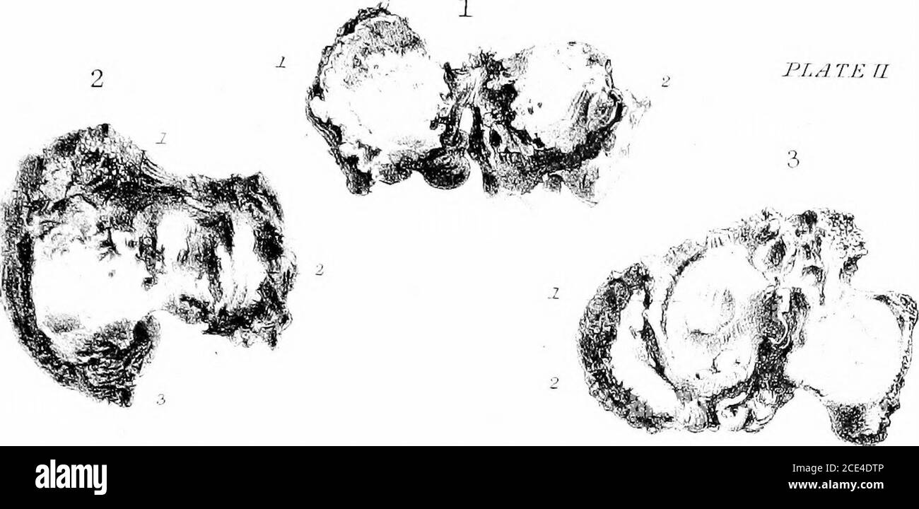 . The principles and practice of veterinary surgery . not yet removed, but which in the specimen is tunnelled under by absorption. (3.) Lower end of tibia; whichat (4.) contains a pit-Kke ulcer. 6. Hock-Joint in Ehbumatoid Boq-Spavin. (1.) Grooving of arti-cular surface into which the porcelain is deposited. (2.) Addimentarybones, formed by ossification of the synovial fringes. 6. Fracture op Astragalus and Bonb-Spavin. (1.) Astragalus.(2.) Bone-spavin. (3.) and (4.) Metatarsal bones. 7. Caries op the Point of Os Calois (Capped Hock). (1.) Sup-erior, and (2.) Inferior extremities of the bone. Stock Photohttps://www.alamy.com/image-license-details/?v=1https://www.alamy.com/the-principles-and-practice-of-veterinary-surgery-not-yet-removed-but-which-in-the-specimen-is-tunnelled-under-by-absorption-3-lower-end-of-tibia-whichat-4-contains-a-pit-kke-ulcer-6-hock-joint-in-ehbumatoid-boq-spavin-1-grooving-of-arti-cular-surface-into-which-the-porcelain-is-deposited-2-addimentarybones-formed-by-ossification-of-the-synovial-fringes-6-fracture-op-astragalus-and-bonb-spavin-1-astragalus2-bone-spavin-3-and-4-metatarsal-bones-7-caries-op-the-point-of-os-calois-capped-hock-1-sup-erior-and-2-inferior-extremities-of-the-bone-image370121606.html
RM2CE4DTP–. The principles and practice of veterinary surgery . not yet removed, but which in the specimen is tunnelled under by absorption. (3.) Lower end of tibia; whichat (4.) contains a pit-Kke ulcer. 6. Hock-Joint in Ehbumatoid Boq-Spavin. (1.) Grooving of arti-cular surface into which the porcelain is deposited. (2.) Addimentarybones, formed by ossification of the synovial fringes. 6. Fracture op Astragalus and Bonb-Spavin. (1.) Astragalus.(2.) Bone-spavin. (3.) and (4.) Metatarsal bones. 7. Caries op the Point of Os Calois (Capped Hock). (1.) Sup-erior, and (2.) Inferior extremities of the bone.
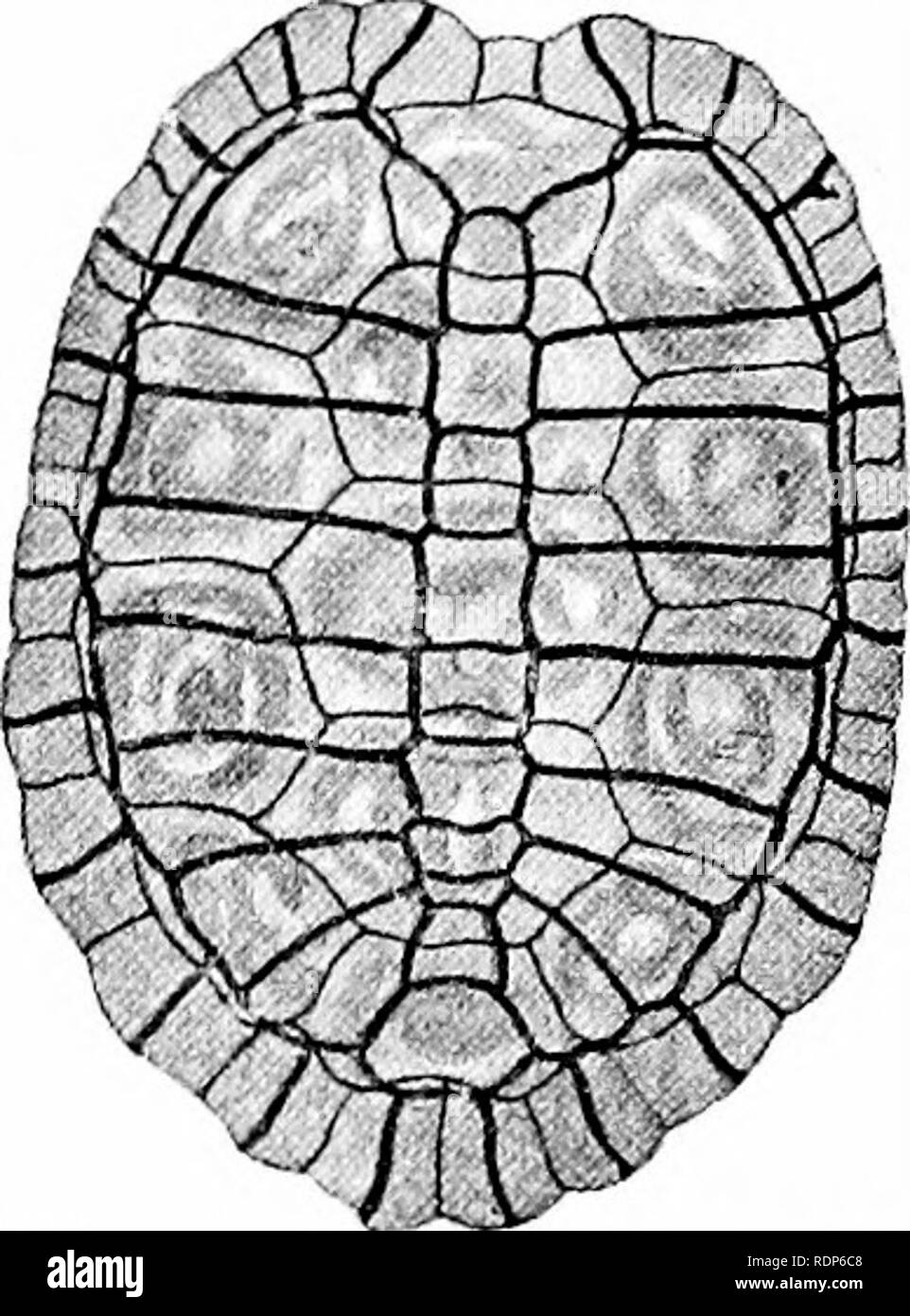 . Outlines of zoology. Zoology. 564 REPTILES. carapace, for this must tend to make respiration less active. The lungs are divided into a number of compartments. All are oviparous. The eggs have firm, usually calcareous, shells. Some Peculiarities in the Skeleton of Chelonia. The dorsal vertebras seem to be without transverse processes, and along with the ribs are for the most part immovably fused in the carapace. The tail and the neck are the only flexible regions. The greater part of the dorsal shield seems to be due to a coalescence of rib-cartilages ; to an ossification of these and of the Stock Photohttps://www.alamy.com/image-license-details/?v=1https://www.alamy.com/outlines-of-zoology-zoology-564-reptiles-carapace-for-this-must-tend-to-make-respiration-less-active-the-lungs-are-divided-into-a-number-of-compartments-all-are-oviparous-the-eggs-have-firm-usually-calcareous-shells-some-peculiarities-in-the-skeleton-of-chelonia-the-dorsal-vertebras-seem-to-be-without-transverse-processes-and-along-with-the-ribs-are-for-the-most-part-immovably-fused-in-the-carapace-the-tail-and-the-neck-are-the-only-flexible-regions-the-greater-part-of-the-dorsal-shield-seems-to-be-due-to-a-coalescence-of-rib-cartilages-to-an-ossification-of-these-and-of-the-image232213304.html
RMRDP6C8–. Outlines of zoology. Zoology. 564 REPTILES. carapace, for this must tend to make respiration less active. The lungs are divided into a number of compartments. All are oviparous. The eggs have firm, usually calcareous, shells. Some Peculiarities in the Skeleton of Chelonia. The dorsal vertebras seem to be without transverse processes, and along with the ribs are for the most part immovably fused in the carapace. The tail and the neck are the only flexible regions. The greater part of the dorsal shield seems to be due to a coalescence of rib-cartilages ; to an ossification of these and of the
 . The anatomy of the domestic animals . Veterinary anatomy. 118 THE SKELETON OF THE HORSE The distal extremity is fused with the tibia, constituting the lateral malleolus. Development.—This resembles that of the ulna. The embrj^onic cartilaginous fibula extends the entire length of the leg, but does not articulate with the femur. The distal part of the shaft is usually reduced to a fibrous band. Three centers of ossification appear, one each for the shaft and the extremities. The distal end unites early with the tibia, forming the lateral malleolus. It is interesting to note that in some cases Stock Photohttps://www.alamy.com/image-license-details/?v=1https://www.alamy.com/the-anatomy-of-the-domestic-animals-veterinary-anatomy-118-the-skeleton-of-the-horse-the-distal-extremity-is-fused-with-the-tibia-constituting-the-lateral-malleolus-developmentthis-resembles-that-of-the-ulna-the-embrjonic-cartilaginous-fibula-extends-the-entire-length-of-the-leg-but-does-not-articulate-with-the-femur-the-distal-part-of-the-shaft-is-usually-reduced-to-a-fibrous-band-three-centers-of-ossification-appear-one-each-for-the-shaft-and-the-extremities-the-distal-end-unites-early-with-the-tibia-forming-the-lateral-malleolus-it-is-interesting-to-note-that-in-some-cases-image232314692.html
RMRDXRN8–. The anatomy of the domestic animals . Veterinary anatomy. 118 THE SKELETON OF THE HORSE The distal extremity is fused with the tibia, constituting the lateral malleolus. Development.—This resembles that of the ulna. The embrj^onic cartilaginous fibula extends the entire length of the leg, but does not articulate with the femur. The distal part of the shaft is usually reduced to a fibrous band. Three centers of ossification appear, one each for the shaft and the extremities. The distal end unites early with the tibia, forming the lateral malleolus. It is interesting to note that in some cases
 . A text book of veterinary pathology, for students and practitioners. Veterinary pathology. 191 VETERINARY PATHOLOGY. the enveloping fibrous capsule of the follicle. The hyperplastic fibrous tissue usually fuses with the cementum, and the entire mass may later become calcified or ossified. These odontomata are most common in ruminants, goats especially being affected. They are prone to occur in animals afflicted with, rickets. Cementomata (Usteocystoma capsulare dentiferum) are formed by ossification of excess tissue developed around the tooth follicle, The livperplastic cementum mav include Stock Photohttps://www.alamy.com/image-license-details/?v=1https://www.alamy.com/a-text-book-of-veterinary-pathology-for-students-and-practitioners-veterinary-pathology-191-veterinary-pathology-the-enveloping-fibrous-capsule-of-the-follicle-the-hyperplastic-fibrous-tissue-usually-fuses-with-the-cementum-and-the-entire-mass-may-later-become-calcified-or-ossified-these-odontomata-are-most-common-in-ruminants-goats-especially-being-affected-they-are-prone-to-occur-in-animals-afflicted-with-rickets-cementomata-usteocystoma-capsulare-dentiferum-are-formed-by-ossification-of-excess-tissue-developed-around-the-tooth-follicle-the-livperplastic-cementum-mav-include-image232341866.html
RMRE02BP–. A text book of veterinary pathology, for students and practitioners. Veterinary pathology. 191 VETERINARY PATHOLOGY. the enveloping fibrous capsule of the follicle. The hyperplastic fibrous tissue usually fuses with the cementum, and the entire mass may later become calcified or ossified. These odontomata are most common in ruminants, goats especially being affected. They are prone to occur in animals afflicted with, rickets. Cementomata (Usteocystoma capsulare dentiferum) are formed by ossification of excess tissue developed around the tooth follicle, The livperplastic cementum mav include
RMRDYAGK–. The anatomy of the domestic animals . Veterinary anatomy. 214 THE ARTICULATIONS OF THE HORSE vaded by the process of ossification early, so that the consolidation of the sacrum is usually complete, or nearly so, at three years. The coccygeal vertebrae are united by relatively thick intervertebral fibro-cartil- ages, which have the form of biconcave discs. Special ligaments are not present, but there is a continuous sheath of fibrous tissue. The movement in this region is exten- sive and varied. In old horses the first coccygeal vertebra is often fused with the sacrum. MOVEMENTS OF THE VERTEB
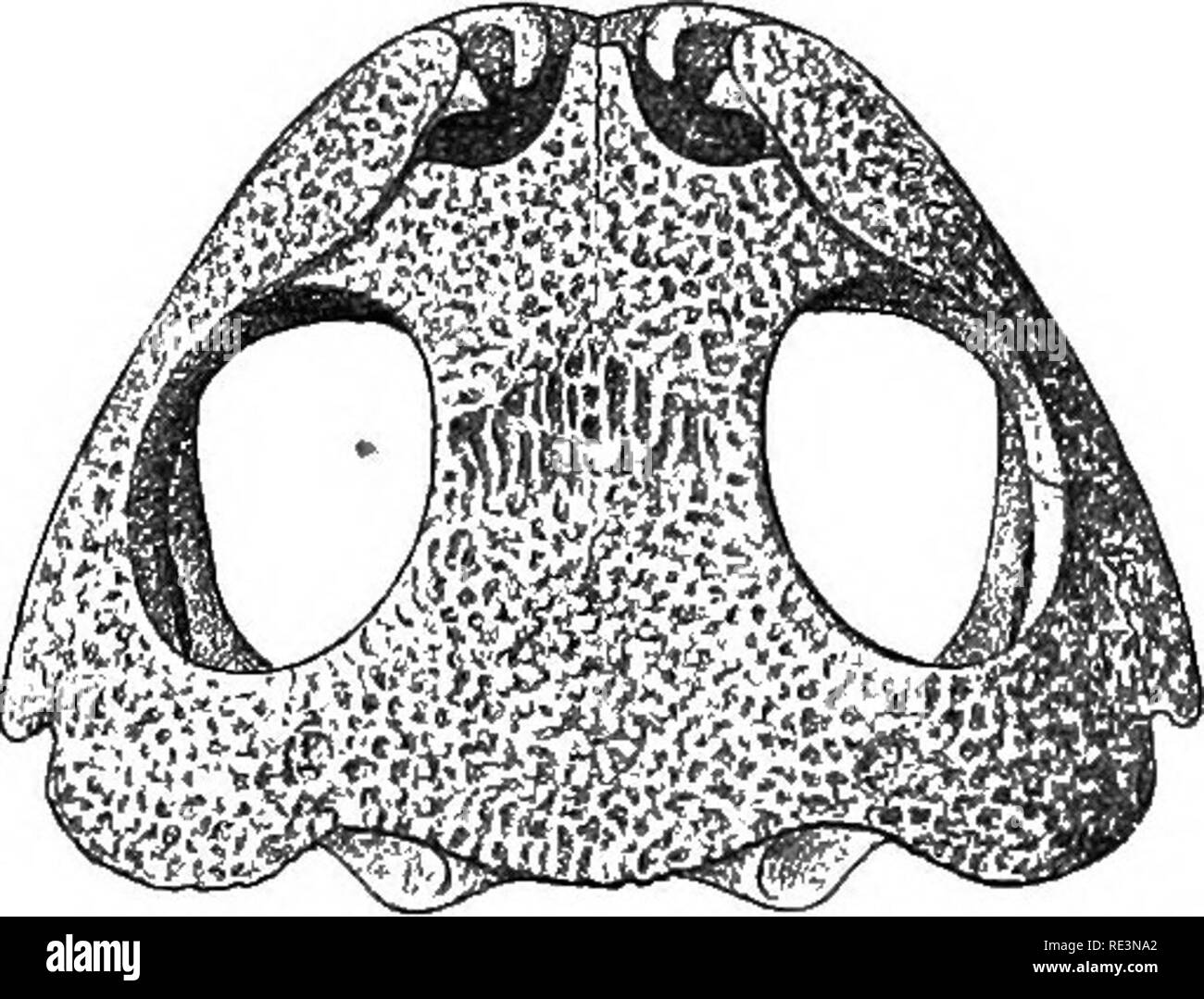 . The tailless batrachians of Europe. Frogs; Amphibians. kSkbleton. 35 The membrane bones, all paired with the excep- tion of the parasphenoid (and in Pelobates the fronto- parietal), are the following :—Prsemaxillary, maxillary, squamosal, pterygoid, palatine, vomer, parasphenoid, nasal (often called prefrontal), and fronto-parietal, A small ossification behind the narial opening is the turbinal, regarded by some as the true nasal. The cranial ossification may be feeble, and a considerable portion of the cartilaginous primordial cranium remain exposed,—as, for instance, in Bombinator, which, Stock Photohttps://www.alamy.com/image-license-details/?v=1https://www.alamy.com/the-tailless-batrachians-of-europe-frogs-amphibians-kskbleton-35-the-membrane-bones-all-paired-with-the-excep-tion-of-the-parasphenoid-and-in-pelobates-the-fronto-parietal-are-the-following-prsemaxillary-maxillary-squamosal-pterygoid-palatine-vomer-parasphenoid-nasal-often-called-prefrontal-and-fronto-parietal-a-small-ossification-behind-the-narial-opening-is-the-turbinal-regarded-by-some-as-the-true-nasal-the-cranial-ossification-may-be-feeble-and-a-considerable-portion-of-the-cartilaginous-primordial-cranium-remain-exposedas-for-instance-in-bombinator-which-image232422570.html
RMRE3NA2–. The tailless batrachians of Europe. Frogs; Amphibians. kSkbleton. 35 The membrane bones, all paired with the excep- tion of the parasphenoid (and in Pelobates the fronto- parietal), are the following :—Prsemaxillary, maxillary, squamosal, pterygoid, palatine, vomer, parasphenoid, nasal (often called prefrontal), and fronto-parietal, A small ossification behind the narial opening is the turbinal, regarded by some as the true nasal. The cranial ossification may be feeble, and a considerable portion of the cartilaginous primordial cranium remain exposed,—as, for instance, in Bombinator, which,
 . Veterinary obstetrics; a compendium for the use of students and practitioners. Veterinary obstetrics. ANATOMY. 17 The diameters of the outlet are more equal, being about those of the transverse diameter of the inlet. The cavity is more cylindrical, and less conical than that of the Mare. In the Sheep and Goat, ossification occurring at a 'much later period, allows of the pelvic cavity being increased during parturition, and permits of the act being performed with fewer difficulties in these animals. In the Pig, the general conformation of the pelvis. Fig. 8. Inlet of the Cow's Pelvis. <j Stock Photohttps://www.alamy.com/image-license-details/?v=1https://www.alamy.com/veterinary-obstetrics-a-compendium-for-the-use-of-students-and-practitioners-veterinary-obstetrics-anatomy-17-the-diameters-of-the-outlet-are-more-equal-being-about-those-of-the-transverse-diameter-of-the-inlet-the-cavity-is-more-cylindrical-and-less-conical-than-that-of-the-mare-in-the-sheep-and-goat-ossification-occurring-at-a-much-later-period-allows-of-the-pelvic-cavity-being-increased-during-parturition-and-permits-of-the-act-being-performed-with-fewer-difficulties-in-these-animals-in-the-pig-the-general-conformation-of-the-pelvis-fig-8-inlet-of-the-cows-pelvis-ltj-image232003553.html
RMRDCJW5–. Veterinary obstetrics; a compendium for the use of students and practitioners. Veterinary obstetrics. ANATOMY. 17 The diameters of the outlet are more equal, being about those of the transverse diameter of the inlet. The cavity is more cylindrical, and less conical than that of the Mare. In the Sheep and Goat, ossification occurring at a 'much later period, allows of the pelvic cavity being increased during parturition, and permits of the act being performed with fewer difficulties in these animals. In the Pig, the general conformation of the pelvis. Fig. 8. Inlet of the Cow's Pelvis. <j
 . Elements of the comparative anatomy of vertebrates. Anatomy, Comparative. 00 COMPARATIVE ANATOMY of ossification, which gives the skull a very firm and solid appear- ance ; only amongst Lizards (Fig. 71), and especially in Hatteria is the cartilage retained to any considerable extent, and owing to the conformation of the bones in the posterior region, the skull in these forms presents a number of distinct spaces or fossae in the dry state. In Snakes and Amphisbsenians the cranial cavity extends forwards between the orbits as far as the ethmoidal region, while in the Lacertilia, Chelonia, and Stock Photohttps://www.alamy.com/image-license-details/?v=1https://www.alamy.com/elements-of-the-comparative-anatomy-of-vertebrates-anatomy-comparative-00-comparative-anatomy-of-ossification-which-gives-the-skull-a-very-firm-and-solid-appear-ance-only-amongst-lizards-fig-71-and-especially-in-hatteria-is-the-cartilage-retained-to-any-considerable-extent-and-owing-to-the-conformation-of-the-bones-in-the-posterior-region-the-skull-in-these-forms-presents-a-number-of-distinct-spaces-or-fossae-in-the-dry-state-in-snakes-and-amphisbsenians-the-cranial-cavity-extends-forwards-between-the-orbits-as-far-as-the-ethmoidal-region-while-in-the-lacertilia-chelonia-and-image232075537.html
RMRDFXM1–. Elements of the comparative anatomy of vertebrates. Anatomy, Comparative. 00 COMPARATIVE ANATOMY of ossification, which gives the skull a very firm and solid appear- ance ; only amongst Lizards (Fig. 71), and especially in Hatteria is the cartilage retained to any considerable extent, and owing to the conformation of the bones in the posterior region, the skull in these forms presents a number of distinct spaces or fossae in the dry state. In Snakes and Amphisbsenians the cranial cavity extends forwards between the orbits as far as the ethmoidal region, while in the Lacertilia, Chelonia, and
![. The illustrated natural history [microform]. Natural history; Sciences naturelles. Bicrirni!* ^Lat. his, twice; cornu, a hoiti), the Two-JIunicd Wdnocerua, or Hhincutei: Their food consists almost entirely of the thoi-ny branches of the wait-a-bit thorns. Their horns are much shorter than those of the other varieties, seldom exceeding eighteen inclies in length. They are finely polished with constant rubbiuij; against the trees. The skrdl is remarkably formed, its most striking feature being the tremendous thick ossification in which it ends above the nostrils. It is on this mass tliat tlie Stock Photo . The illustrated natural history [microform]. Natural history; Sciences naturelles. Bicrirni!* ^Lat. his, twice; cornu, a hoiti), the Two-JIunicd Wdnocerua, or Hhincutei: Their food consists almost entirely of the thoi-ny branches of the wait-a-bit thorns. Their horns are much shorter than those of the other varieties, seldom exceeding eighteen inclies in length. They are finely polished with constant rubbiuij; against the trees. The skrdl is remarkably formed, its most striking feature being the tremendous thick ossification in which it ends above the nostrils. It is on this mass tliat tlie Stock Photo](https://c8.alamy.com/comp/RERNYC/the-illustrated-natural-history-microform-natural-history-sciences-naturelles-bicrirni!-lat-his-twice-cornu-a-hoiti-the-two-jiunicd-wdnocerua-or-hhincutei-their-food-consists-almost-entirely-of-the-thoi-ny-branches-of-the-wait-a-bit-thorns-their-horns-are-much-shorter-than-those-of-the-other-varieties-seldom-exceeding-eighteen-inclies-in-length-they-are-finely-polished-with-constant-rubbiuij-against-the-trees-the-skrdl-is-remarkably-formed-its-most-striking-feature-being-the-tremendous-thick-ossification-in-which-it-ends-above-the-nostrils-it-is-on-this-mass-tliat-tlie-RERNYC.jpg) . The illustrated natural history [microform]. Natural history; Sciences naturelles. Bicrirni!* ^Lat. his, twice; cornu, a hoiti), the Two-JIunicd Wdnocerua, or Hhincutei: Their food consists almost entirely of the thoi-ny branches of the wait-a-bit thorns. Their horns are much shorter than those of the other varieties, seldom exceeding eighteen inclies in length. They are finely polished with constant rubbiuij; against the trees. The skrdl is remarkably formed, its most striking feature being the tremendous thick ossification in which it ends above the nostrils. It is on this mass tliat tlie Stock Photohttps://www.alamy.com/image-license-details/?v=1https://www.alamy.com/the-illustrated-natural-history-microform-natural-history-sciences-naturelles-bicrirni!-lat-his-twice-cornu-a-hoiti-the-two-jiunicd-wdnocerua-or-hhincutei-their-food-consists-almost-entirely-of-the-thoi-ny-branches-of-the-wait-a-bit-thorns-their-horns-are-much-shorter-than-those-of-the-other-varieties-seldom-exceeding-eighteen-inclies-in-length-they-are-finely-polished-with-constant-rubbiuij-against-the-trees-the-skrdl-is-remarkably-formed-its-most-striking-feature-being-the-tremendous-thick-ossification-in-which-it-ends-above-the-nostrils-it-is-on-this-mass-tliat-tlie-image232862096.html
. The illustrated natural history [microform]. Natural history; Sciences naturelles. Bicrirni!* ^Lat. his, twice; cornu, a hoiti), the Two-JIunicd Wdnocerua, or Hhincutei: Their food consists almost entirely of the thoi-ny branches of the wait-a-bit thorns. Their horns are much shorter than those of the other varieties, seldom exceeding eighteen inclies in length. They are finely polished with constant rubbiuij; against the trees. The skrdl is remarkably formed, its most striking feature being the tremendous thick ossification in which it ends above the nostrils. It is on this mass tliat tlie Stock Photohttps://www.alamy.com/image-license-details/?v=1https://www.alamy.com/the-illustrated-natural-history-microform-natural-history-sciences-naturelles-bicrirni!-lat-his-twice-cornu-a-hoiti-the-two-jiunicd-wdnocerua-or-hhincutei-their-food-consists-almost-entirely-of-the-thoi-ny-branches-of-the-wait-a-bit-thorns-their-horns-are-much-shorter-than-those-of-the-other-varieties-seldom-exceeding-eighteen-inclies-in-length-they-are-finely-polished-with-constant-rubbiuij-against-the-trees-the-skrdl-is-remarkably-formed-its-most-striking-feature-being-the-tremendous-thick-ossification-in-which-it-ends-above-the-nostrils-it-is-on-this-mass-tliat-tlie-image232862096.htmlRMRERNYC–. The illustrated natural history [microform]. Natural history; Sciences naturelles. Bicrirni!* ^Lat. his, twice; cornu, a hoiti), the Two-JIunicd Wdnocerua, or Hhincutei: Their food consists almost entirely of the thoi-ny branches of the wait-a-bit thorns. Their horns are much shorter than those of the other varieties, seldom exceeding eighteen inclies in length. They are finely polished with constant rubbiuij; against the trees. The skrdl is remarkably formed, its most striking feature being the tremendous thick ossification in which it ends above the nostrils. It is on this mass tliat tlie
 . The comparative anatomy of the domesticated animals. Veterinary anatomy. 912 EMBBYOLOGY. It is only in the third month that ossification begins in the vertebral column. The number of osseous nuclei, primary and complementary, is not the same in all species; they have Ijeen enumerated at page 20. In a large number, the spinous process is regarded as the result of the joining together of the two moieties of the vertebral arch; in the Sheep, on the contrary, the spinous process forms a nucleus altogether independent of the vertebral arches. Thomas has noted this disposition, and he considers it Stock Photohttps://www.alamy.com/image-license-details/?v=1https://www.alamy.com/the-comparative-anatomy-of-the-domesticated-animals-veterinary-anatomy-912-embbyology-it-is-only-in-the-third-month-that-ossification-begins-in-the-vertebral-column-the-number-of-osseous-nuclei-primary-and-complementary-is-not-the-same-in-all-species-they-have-ijeen-enumerated-at-page-20-in-a-large-number-the-spinous-process-is-regarded-as-the-result-of-the-joining-together-of-the-two-moieties-of-the-vertebral-arch-in-the-sheep-on-the-contrary-the-spinous-process-forms-a-nucleus-altogether-independent-of-the-vertebral-arches-thomas-has-noted-this-disposition-and-he-considers-it-image232451236.html
RMRE51WT–. The comparative anatomy of the domesticated animals. Veterinary anatomy. 912 EMBBYOLOGY. It is only in the third month that ossification begins in the vertebral column. The number of osseous nuclei, primary and complementary, is not the same in all species; they have Ijeen enumerated at page 20. In a large number, the spinous process is regarded as the result of the joining together of the two moieties of the vertebral arch; in the Sheep, on the contrary, the spinous process forms a nucleus altogether independent of the vertebral arches. Thomas has noted this disposition, and he considers it
 . On the anatomy of vertebrates. Vertebrates; Anatomy, Comparative; 1866. ANATOMY OF VERTEBRATES. 47 The Siren lacertlna has betwe en eighty and ninety trunk-vertebrae. They have many longitudinal ridges, the neural arch has coalesced with the centrum, the neural spine forms the highest ridge and bifurcates posteriorly to terminate upon the zygapophysis. A 41. skeleton of Lepidosircii anncctens. xxxiij. hypapophysial ridge forms, by defect of ossification on each side, the under part of the centrum. A parapophysial ridge extends from a short anterior parapophysis to the longer parapophysial pa Stock Photohttps://www.alamy.com/image-license-details/?v=1https://www.alamy.com/on-the-anatomy-of-vertebrates-vertebrates-anatomy-comparative-1866-anatomy-of-vertebrates-47-the-siren-lacertlna-has-betwe-en-eighty-and-ninety-trunk-vertebrae-they-have-many-longitudinal-ridges-the-neural-arch-has-coalesced-with-the-centrum-the-neural-spine-forms-the-highest-ridge-and-bifurcates-posteriorly-to-terminate-upon-the-zygapophysis-a-41-skeleton-of-lepidosircii-anncctens-xxxiij-hypapophysial-ridge-forms-by-defect-of-ossification-on-each-side-the-under-part-of-the-centrum-a-parapophysial-ridge-extends-from-a-short-anterior-parapophysis-to-the-longer-parapophysial-pa-image232103699.html
RMRDH6HR–. On the anatomy of vertebrates. Vertebrates; Anatomy, Comparative; 1866. ANATOMY OF VERTEBRATES. 47 The Siren lacertlna has betwe en eighty and ninety trunk-vertebrae. They have many longitudinal ridges, the neural arch has coalesced with the centrum, the neural spine forms the highest ridge and bifurcates posteriorly to terminate upon the zygapophysis. A 41. skeleton of Lepidosircii anncctens. xxxiij. hypapophysial ridge forms, by defect of ossification on each side, the under part of the centrum. A parapophysial ridge extends from a short anterior parapophysis to the longer parapophysial pa
 . The comparative anatomy of the domesticated animals. Veterinary anatomy. 912 EMBBYOLOGY, It is only in tlie tliird montli that ossification begins in the vertebral column. The number of osseous nuclei, primary and complementary, is not the same in all species; they have been enumerated at page 20. In a large number, the spinous process is regarded as the result of the joining together of the two moieties of the vertebral arch; in the Sheep, on the contrary, the spinous process forms a nucleus altogether independent of the vertebral arches. Thomas has noted this disposition, and he considers Stock Photohttps://www.alamy.com/image-license-details/?v=1https://www.alamy.com/the-comparative-anatomy-of-the-domesticated-animals-veterinary-anatomy-912-embbyology-it-is-only-in-tlie-tliird-montli-that-ossification-begins-in-the-vertebral-column-the-number-of-osseous-nuclei-primary-and-complementary-is-not-the-same-in-all-species-they-have-been-enumerated-at-page-20-in-a-large-number-the-spinous-process-is-regarded-as-the-result-of-the-joining-together-of-the-two-moieties-of-the-vertebral-arch-in-the-sheep-on-the-contrary-the-spinous-process-forms-a-nucleus-altogether-independent-of-the-vertebral-arches-thomas-has-noted-this-disposition-and-he-considers-image237843978.html
RMRPXMBP–. The comparative anatomy of the domesticated animals. Veterinary anatomy. 912 EMBBYOLOGY, It is only in tlie tliird montli that ossification begins in the vertebral column. The number of osseous nuclei, primary and complementary, is not the same in all species; they have been enumerated at page 20. In a large number, the spinous process is regarded as the result of the joining together of the two moieties of the vertebral arch; in the Sheep, on the contrary, the spinous process forms a nucleus altogether independent of the vertebral arches. Thomas has noted this disposition, and he considers
 . Outlines of zoology. Zoology. 644 MAMMALIA. Associated with the olfactory chambers are the nasals above, the vomers beneath, the mesethmoid in the median line, while internally there are several thin scroll-like turbinal bones. As special characters of the skull should be noted the incomplete ossification of certain of the bones, e.g. of the maxilla, and the development of slender rod-like processes from some of them, e.g. the squamosal, which help to keep the parts of the skull firmly connected. The lower jaw or mandible consists in adult life of a single bone or ramus on each side, but thi Stock Photohttps://www.alamy.com/image-license-details/?v=1https://www.alamy.com/outlines-of-zoology-zoology-644-mammalia-associated-with-the-olfactory-chambers-are-the-nasals-above-the-vomers-beneath-the-mesethmoid-in-the-median-line-while-internally-there-are-several-thin-scroll-like-turbinal-bones-as-special-characters-of-the-skull-should-be-noted-the-incomplete-ossification-of-certain-of-the-bones-eg-of-the-maxilla-and-the-development-of-slender-rod-like-processes-from-some-of-them-eg-the-squamosal-which-help-to-keep-the-parts-of-the-skull-firmly-connected-the-lower-jaw-or-mandible-consists-in-adult-life-of-a-single-bone-or-ramus-on-each-side-but-thi-image232213108.html
RMRDP658–. Outlines of zoology. Zoology. 644 MAMMALIA. Associated with the olfactory chambers are the nasals above, the vomers beneath, the mesethmoid in the median line, while internally there are several thin scroll-like turbinal bones. As special characters of the skull should be noted the incomplete ossification of certain of the bones, e.g. of the maxilla, and the development of slender rod-like processes from some of them, e.g. the squamosal, which help to keep the parts of the skull firmly connected. The lower jaw or mandible consists in adult life of a single bone or ramus on each side, but thi
 . Diseases of the horse's foot . Hoofs; Horses. GENERAL OBSEEVATIONS 55 tion of the processes is a condensation of the epithehal cells immediately above the rete Malpighii, with a partial or total loss of their nuclei. This is the first appearance of true horn, and its commencement is almost coincident with the first stages of ossification of the os pedis. With the appearance of horn comes difficulty of sectioning. The last specimen that Professor Mettam was able to satis-. Fig. 29.—Section of an Epithelial Ingeowth from an Equine FCETUS. It shows commencing secondary laminar ridges. In the ce Stock Photohttps://www.alamy.com/image-license-details/?v=1https://www.alamy.com/diseases-of-the-horses-foot-hoofs-horses-general-obseevations-55-tion-of-the-processes-is-a-condensation-of-the-epithehal-cells-immediately-above-the-rete-malpighii-with-a-partial-or-total-loss-of-their-nuclei-this-is-the-first-appearance-of-true-horn-and-its-commencement-is-almost-coincident-with-the-first-stages-of-ossification-of-the-os-pedis-with-the-appearance-of-horn-comes-difficulty-of-sectioning-the-last-specimen-that-professor-mettam-was-able-to-satis-fig-29section-of-an-epithelial-ingeowth-from-an-equine-fcetus-it-shows-commencing-secondary-laminar-ridges-in-the-ce-image232318899.html
RMRDY13F–. Diseases of the horse's foot . Hoofs; Horses. GENERAL OBSEEVATIONS 55 tion of the processes is a condensation of the epithehal cells immediately above the rete Malpighii, with a partial or total loss of their nuclei. This is the first appearance of true horn, and its commencement is almost coincident with the first stages of ossification of the os pedis. With the appearance of horn comes difficulty of sectioning. The last specimen that Professor Mettam was able to satis-. Fig. 29.—Section of an Epithelial Ingeowth from an Equine FCETUS. It shows commencing secondary laminar ridges. In the ce
 . A guide to the study of fishes. Fishes; Zoology; Fishes. The Ganoids 23 Woodward places these fishes with the SemionotidcB and Ha- lecomorphi in his suborder of Protospondyli. It seems preferable, however, to consider them as forming a distinct order. Order Lepidostei.—^We may place, following Eastman's edition of Zittel, the allies and predecessors of the garpike in a single order, for which Hiixley's name Lepidostei may well be used. In this group the notochord is persistent, and the vertebrse are in various degrees of ossification and of different forms. The. Fig. 13.—Mesturus verrucosus Stock Photohttps://www.alamy.com/image-license-details/?v=1https://www.alamy.com/a-guide-to-the-study-of-fishes-fishes-zoology-fishes-the-ganoids-23-woodward-places-these-fishes-with-the-semionotidcb-and-ha-lecomorphi-in-his-suborder-of-protospondyli-it-seems-preferable-however-to-consider-them-as-forming-a-distinct-order-order-lepidosteiwe-may-place-following-eastmans-edition-of-zittel-the-allies-and-predecessors-of-the-garpike-in-a-single-order-for-which-hiixleys-name-lepidostei-may-well-be-used-in-this-group-the-notochord-is-persistent-and-the-vertebrse-are-in-various-degrees-of-ossification-and-of-different-forms-the-fig-13mesturus-verrucosus-image232171206.html
RMRDM8MP–. A guide to the study of fishes. Fishes; Zoology; Fishes. The Ganoids 23 Woodward places these fishes with the SemionotidcB and Ha- lecomorphi in his suborder of Protospondyli. It seems preferable, however, to consider them as forming a distinct order. Order Lepidostei.—^We may place, following Eastman's edition of Zittel, the allies and predecessors of the garpike in a single order, for which Hiixley's name Lepidostei may well be used. In this group the notochord is persistent, and the vertebrse are in various degrees of ossification and of different forms. The. Fig. 13.—Mesturus verrucosus
 . Text book of zoology. Zoology. 366 Vertehrata. is the large dentigeroue dent ale, which meets its fellow of the other side anteriorly. The upper portion of the hyoid, which is united with the coiTesponding part of the mandibular arch, is represented by two bones; a large one, the hyomandibular, articulated with the skull, and a smaller, lower ossification, the symplectic. For the membrane bones connected with the hyoid, see below, under the gUl apparatus. In the Dipnoi, the upper portions of the mandibular and hyoid arches ai-e ooncrescent, and partly ossified, thus possessing so far, the sa Stock Photohttps://www.alamy.com/image-license-details/?v=1https://www.alamy.com/text-book-of-zoology-zoology-366-vertehrata-is-the-large-dentigeroue-dent-ale-which-meets-its-fellow-of-the-other-side-anteriorly-the-upper-portion-of-the-hyoid-which-is-united-with-the-coitesponding-part-of-the-mandibular-arch-is-represented-by-two-bones-a-large-one-the-hyomandibular-articulated-with-the-skull-and-a-smaller-lower-ossification-the-symplectic-for-the-membrane-bones-connected-with-the-hyoid-see-below-under-the-gul-apparatus-in-the-dipnoi-the-upper-portions-of-the-mandibular-and-hyoid-arches-ai-e-ooncrescent-and-partly-ossified-thus-possessing-so-far-the-sa-image232421747.html
RMRE3M8K–. Text book of zoology. Zoology. 366 Vertehrata. is the large dentigeroue dent ale, which meets its fellow of the other side anteriorly. The upper portion of the hyoid, which is united with the coiTesponding part of the mandibular arch, is represented by two bones; a large one, the hyomandibular, articulated with the skull, and a smaller, lower ossification, the symplectic. For the membrane bones connected with the hyoid, see below, under the gUl apparatus. In the Dipnoi, the upper portions of the mandibular and hyoid arches ai-e ooncrescent, and partly ossified, thus possessing so far, the sa
 . The anatomy of the domestic animals . Veterinary anatomy. DEVELOPMENT AND GROWTH OF BONE 23 endings (Vater-Pacini corpuscles) in the periosteum are to be regarded as sensory and probably are concerned in mediating the muscle sense (kinesthesia) DEVELOPMENT AND GROWTH OF BONEi whiciy^h/?'''*'!? embryonal skeleton consists of cartilage and fibrous tissue, in which the bones develop The process is termed ossification or osteogenesis, ;nd IS effected essentially by bone-producing cells, called osteoblasts. It is customary therefore, to designate as membrane bones those which are developed in fib Stock Photohttps://www.alamy.com/image-license-details/?v=1https://www.alamy.com/the-anatomy-of-the-domestic-animals-veterinary-anatomy-development-and-growth-of-bone-23-endings-vater-pacini-corpuscles-in-the-periosteum-are-to-be-regarded-as-sensory-and-probably-are-concerned-in-mediating-the-muscle-sense-kinesthesia-development-and-growth-of-bonei-whiciyh!-embryonal-skeleton-consists-of-cartilage-and-fibrous-tissue-in-which-the-bones-develop-the-process-is-termed-ossification-or-osteogenesis-nd-is-effected-essentially-by-bone-producing-cells-called-osteoblasts-it-is-customary-therefore-to-designate-as-membrane-bones-those-which-are-developed-in-fib-image232327929.html
RMRDYCJ1–. The anatomy of the domestic animals . Veterinary anatomy. DEVELOPMENT AND GROWTH OF BONE 23 endings (Vater-Pacini corpuscles) in the periosteum are to be regarded as sensory and probably are concerned in mediating the muscle sense (kinesthesia) DEVELOPMENT AND GROWTH OF BONEi whiciy^h/?'''*'!? embryonal skeleton consists of cartilage and fibrous tissue, in which the bones develop The process is termed ossification or osteogenesis, ;nd IS effected essentially by bone-producing cells, called osteoblasts. It is customary therefore, to designate as membrane bones those which are developed in fib
![. The illustrated natural history [microform]. Natural history; Sciences naturelles. 178 NATtJIlAL HI8T0UV. RII1N0CCR08.. Bicrirni!* ^Lat. his, twice; cornu, a hoiti), the Two-JIunicd Wdnocerua, or Hhincutei: Their food consists almost entirely of the thoi-ny branches of the wait-a-bit thorns. Their horns are much shorter than those of the other varieties, seldom exceeding eighteen inclies in length. They are finely polished with constant rubbiuij; against the trees. The skrdl is remarkably formed, its most striking feature being the tremendous thick ossification in which it ends above the nos Stock Photo . The illustrated natural history [microform]. Natural history; Sciences naturelles. 178 NATtJIlAL HI8T0UV. RII1N0CCR08.. Bicrirni!* ^Lat. his, twice; cornu, a hoiti), the Two-JIunicd Wdnocerua, or Hhincutei: Their food consists almost entirely of the thoi-ny branches of the wait-a-bit thorns. Their horns are much shorter than those of the other varieties, seldom exceeding eighteen inclies in length. They are finely polished with constant rubbiuij; against the trees. The skrdl is remarkably formed, its most striking feature being the tremendous thick ossification in which it ends above the nos Stock Photo](https://c8.alamy.com/comp/RERNYJ/the-illustrated-natural-history-microform-natural-history-sciences-naturelles-178-nattjilal-hi8t0uv-rii1n0ccr08-bicrirni!-lat-his-twice-cornu-a-hoiti-the-two-jiunicd-wdnocerua-or-hhincutei-their-food-consists-almost-entirely-of-the-thoi-ny-branches-of-the-wait-a-bit-thorns-their-horns-are-much-shorter-than-those-of-the-other-varieties-seldom-exceeding-eighteen-inclies-in-length-they-are-finely-polished-with-constant-rubbiuij-against-the-trees-the-skrdl-is-remarkably-formed-its-most-striking-feature-being-the-tremendous-thick-ossification-in-which-it-ends-above-the-nos-RERNYJ.jpg) . The illustrated natural history [microform]. Natural history; Sciences naturelles. 178 NATtJIlAL HI8T0UV. RII1N0CCR08.. Bicrirni!* ^Lat. his, twice; cornu, a hoiti), the Two-JIunicd Wdnocerua, or Hhincutei: Their food consists almost entirely of the thoi-ny branches of the wait-a-bit thorns. Their horns are much shorter than those of the other varieties, seldom exceeding eighteen inclies in length. They are finely polished with constant rubbiuij; against the trees. The skrdl is remarkably formed, its most striking feature being the tremendous thick ossification in which it ends above the nos Stock Photohttps://www.alamy.com/image-license-details/?v=1https://www.alamy.com/the-illustrated-natural-history-microform-natural-history-sciences-naturelles-178-nattjilal-hi8t0uv-rii1n0ccr08-bicrirni!-lat-his-twice-cornu-a-hoiti-the-two-jiunicd-wdnocerua-or-hhincutei-their-food-consists-almost-entirely-of-the-thoi-ny-branches-of-the-wait-a-bit-thorns-their-horns-are-much-shorter-than-those-of-the-other-varieties-seldom-exceeding-eighteen-inclies-in-length-they-are-finely-polished-with-constant-rubbiuij-against-the-trees-the-skrdl-is-remarkably-formed-its-most-striking-feature-being-the-tremendous-thick-ossification-in-which-it-ends-above-the-nos-image232862102.html
. The illustrated natural history [microform]. Natural history; Sciences naturelles. 178 NATtJIlAL HI8T0UV. RII1N0CCR08.. Bicrirni!* ^Lat. his, twice; cornu, a hoiti), the Two-JIunicd Wdnocerua, or Hhincutei: Their food consists almost entirely of the thoi-ny branches of the wait-a-bit thorns. Their horns are much shorter than those of the other varieties, seldom exceeding eighteen inclies in length. They are finely polished with constant rubbiuij; against the trees. The skrdl is remarkably formed, its most striking feature being the tremendous thick ossification in which it ends above the nos Stock Photohttps://www.alamy.com/image-license-details/?v=1https://www.alamy.com/the-illustrated-natural-history-microform-natural-history-sciences-naturelles-178-nattjilal-hi8t0uv-rii1n0ccr08-bicrirni!-lat-his-twice-cornu-a-hoiti-the-two-jiunicd-wdnocerua-or-hhincutei-their-food-consists-almost-entirely-of-the-thoi-ny-branches-of-the-wait-a-bit-thorns-their-horns-are-much-shorter-than-those-of-the-other-varieties-seldom-exceeding-eighteen-inclies-in-length-they-are-finely-polished-with-constant-rubbiuij-against-the-trees-the-skrdl-is-remarkably-formed-its-most-striking-feature-being-the-tremendous-thick-ossification-in-which-it-ends-above-the-nos-image232862102.htmlRMRERNYJ–. The illustrated natural history [microform]. Natural history; Sciences naturelles. 178 NATtJIlAL HI8T0UV. RII1N0CCR08.. Bicrirni!* ^Lat. his, twice; cornu, a hoiti), the Two-JIunicd Wdnocerua, or Hhincutei: Their food consists almost entirely of the thoi-ny branches of the wait-a-bit thorns. Their horns are much shorter than those of the other varieties, seldom exceeding eighteen inclies in length. They are finely polished with constant rubbiuij; against the trees. The skrdl is remarkably formed, its most striking feature being the tremendous thick ossification in which it ends above the nos
 . Veterinary obstetrics; a compendium for the use of students and practitioners. Veterinary obstetrics. 14 VETERINARY OBSTETRICS. With the exception of the equine species, the sacrum is joined to the last lumbar vertebra by three diarthrodial surfaces only—the head of the body and two transverse processes. The ischio pubic symphysis in the Cow is con- siderably longer than in the Mare and not rectilinear; ossification of the symphysis is less complete, and takes place much later than in the Mare. 6. Fig. s. Ligaments of the Lumbar Vertebrae, Sacrum and Pelvis. {Seen from below.) a. Intertransv Stock Photohttps://www.alamy.com/image-license-details/?v=1https://www.alamy.com/veterinary-obstetrics-a-compendium-for-the-use-of-students-and-practitioners-veterinary-obstetrics-14-veterinary-obstetrics-with-the-exception-of-the-equine-species-the-sacrum-is-joined-to-the-last-lumbar-vertebra-by-three-diarthrodial-surfaces-onlythe-head-of-the-body-and-two-transverse-processes-the-ischio-pubic-symphysis-in-the-cow-is-con-siderably-longer-than-in-the-mare-and-not-rectilinear-ossification-of-the-symphysis-is-less-complete-and-takes-place-much-later-than-in-the-mare-6-fig-s-ligaments-of-the-lumbar-vertebrae-sacrum-and-pelvis-seen-from-below-a-intertransv-image232003565.html
RMRDCJWH–. Veterinary obstetrics; a compendium for the use of students and practitioners. Veterinary obstetrics. 14 VETERINARY OBSTETRICS. With the exception of the equine species, the sacrum is joined to the last lumbar vertebra by three diarthrodial surfaces only—the head of the body and two transverse processes. The ischio pubic symphysis in the Cow is con- siderably longer than in the Mare and not rectilinear; ossification of the symphysis is less complete, and takes place much later than in the Mare. 6. Fig. s. Ligaments of the Lumbar Vertebrae, Sacrum and Pelvis. {Seen from below.) a. Intertransv
 . The anatomy of the domestic animals . Veterinary anatomy. THE SKELETON OF THE HORSE Ventral border Costal"' cartilages"--.^ Cariniform cartilage Ribs costarum). Except in the case of the first, the cartilage does not continue the direction of the rib, but forms with the latter an angle which is open in front, and increases from second to last. More or less extensive ossification is to be regarded as a normal occurrence, especially in the cartilages of the sternal ribs. The Sternum The sternum of the horse is shaped somewhat like a canoe; it is compressed laterally, except in its p Stock Photohttps://www.alamy.com/image-license-details/?v=1https://www.alamy.com/the-anatomy-of-the-domestic-animals-veterinary-anatomy-the-skeleton-of-the-horse-ventral-border-costalquot-cartilagesquot-cariniform-cartilage-ribs-costarum-except-in-the-case-of-the-first-the-cartilage-does-not-continue-the-direction-of-the-rib-but-forms-with-the-latter-an-angle-which-is-open-in-front-and-increases-from-second-to-last-more-or-less-extensive-ossification-is-to-be-regarded-as-a-normal-occurrence-especially-in-the-cartilages-of-the-sternal-ribs-the-sternum-the-sternum-of-the-horse-is-shaped-somewhat-like-a-canoe-it-is-compressed-laterally-except-in-its-p-image232327785.html
RMRDYCCW–. The anatomy of the domestic animals . Veterinary anatomy. THE SKELETON OF THE HORSE Ventral border Costal"' cartilages"--.^ Cariniform cartilage Ribs costarum). Except in the case of the first, the cartilage does not continue the direction of the rib, but forms with the latter an angle which is open in front, and increases from second to last. More or less extensive ossification is to be regarded as a normal occurrence, especially in the cartilages of the sternal ribs. The Sternum The sternum of the horse is shaped somewhat like a canoe; it is compressed laterally, except in its p
 . The comparative anatomy of the domesticated animals. Veterinary anatomy. THE HEAD. 53 inferior incisors, and behind these, in male animals only, there is an additional alveolus for the tusk. The portion included on each side between the last incisor and first molar, forms a more or Jess sharp ridge, which constitutes the inferior interdental space or bars. Structure and development,—Formed, like all the flat bones, by two compact plates separated by spongy tissue, the inferior maxilla is developed from two centres of ossification, which correspond to each branch, and which coalesce some time Stock Photohttps://www.alamy.com/image-license-details/?v=1https://www.alamy.com/the-comparative-anatomy-of-the-domesticated-animals-veterinary-anatomy-the-head-53-inferior-incisors-and-behind-these-in-male-animals-only-there-is-an-additional-alveolus-for-the-tusk-the-portion-included-on-each-side-between-the-last-incisor-and-first-molar-forms-a-more-or-jess-sharp-ridge-which-constitutes-the-inferior-interdental-space-or-bars-structure-and-developmentformed-like-all-the-flat-bones-by-two-compact-plates-separated-by-spongy-tissue-the-inferior-maxilla-is-developed-from-two-centres-of-ossification-which-correspond-to-each-branch-and-which-coalesce-some-time-image237850480.html
RMRPY0M0–. The comparative anatomy of the domesticated animals. Veterinary anatomy. THE HEAD. 53 inferior incisors, and behind these, in male animals only, there is an additional alveolus for the tusk. The portion included on each side between the last incisor and first molar, forms a more or Jess sharp ridge, which constitutes the inferior interdental space or bars. Structure and development,—Formed, like all the flat bones, by two compact plates separated by spongy tissue, the inferior maxilla is developed from two centres of ossification, which correspond to each branch, and which coalesce some time
 . Fishes. Fishes. The Ganoids 259 Woodward places these fishes with the Semionotidm and Ha- lecomorphi in his suborder of Protospondyli. It seems preferable, however, to consider them as forming a distinct order. Order Lepidostei.—We may place, following Eastman's edition of Zittel, the allies and predecessors of the garpike in a single order, for which Huxley's name Lepidostei may well be used. In this group the notochord is persistent, and the vertebra are in various degrees of ossification and of different forms. The. Fig. 196.—Mesturus verrucosus Wagner. Family Pycnodontidce. (After Woodwa Stock Photohttps://www.alamy.com/image-license-details/?v=1https://www.alamy.com/fishes-fishes-the-ganoids-259-woodward-places-these-fishes-with-the-semionotidm-and-ha-lecomorphi-in-his-suborder-of-protospondyli-it-seems-preferable-however-to-consider-them-as-forming-a-distinct-order-order-lepidosteiwe-may-place-following-eastmans-edition-of-zittel-the-allies-and-predecessors-of-the-garpike-in-a-single-order-for-which-huxleys-name-lepidostei-may-well-be-used-in-this-group-the-notochord-is-persistent-and-the-vertebra-are-in-various-degrees-of-ossification-and-of-different-forms-the-fig-196mesturus-verrucosus-wagner-family-pycnodontidce-after-woodwa-image232218672.html
RMRDPD80–. Fishes. Fishes. The Ganoids 259 Woodward places these fishes with the Semionotidm and Ha- lecomorphi in his suborder of Protospondyli. It seems preferable, however, to consider them as forming a distinct order. Order Lepidostei.—We may place, following Eastman's edition of Zittel, the allies and predecessors of the garpike in a single order, for which Huxley's name Lepidostei may well be used. In this group the notochord is persistent, and the vertebra are in various degrees of ossification and of different forms. The. Fig. 196.—Mesturus verrucosus Wagner. Family Pycnodontidce. (After Woodwa
 . On the anatomy of vertebrates. Vertebrates; Anatomy, Comparative; 1866. skeleton of Lepidosircii anncctens. xxxiij. hypapophysial ridge forms, by defect of ossification on each side, the under part of the centrum. A parapophysial ridge extends from a short anterior parapophysis to the longer parapophysial part of the posterior transverse process. A diapophysial ridge extends above, and nearly parallel with the former, from the anterior zygajiophysis to the diapophysial part of the posterior transverse jn'ocess. Thence a third short ridge is continued ti3 the posterior zygapoj^hysis. The vacu Stock Photohttps://www.alamy.com/image-license-details/?v=1https://www.alamy.com/on-the-anatomy-of-vertebrates-vertebrates-anatomy-comparative-1866-skeleton-of-lepidosircii-anncctens-xxxiij-hypapophysial-ridge-forms-by-defect-of-ossification-on-each-side-the-under-part-of-the-centrum-a-parapophysial-ridge-extends-from-a-short-anterior-parapophysis-to-the-longer-parapophysial-part-of-the-posterior-transverse-process-a-diapophysial-ridge-extends-above-and-nearly-parallel-with-the-former-from-the-anterior-zygajiophysis-to-the-diapophysial-part-of-the-posterior-transverse-jnocess-thence-a-third-short-ridge-is-continued-ti3-the-posterior-zygapojhysis-the-vacu-image232103697.html
RMRDH6HN–. On the anatomy of vertebrates. Vertebrates; Anatomy, Comparative; 1866. skeleton of Lepidosircii anncctens. xxxiij. hypapophysial ridge forms, by defect of ossification on each side, the under part of the centrum. A parapophysial ridge extends from a short anterior parapophysis to the longer parapophysial part of the posterior transverse process. A diapophysial ridge extends above, and nearly parallel with the former, from the anterior zygajiophysis to the diapophysial part of the posterior transverse jn'ocess. Thence a third short ridge is continued ti3 the posterior zygapoj^hysis. The vacu
 . On the anatomy of vertebrates. Vertebrates; Anatomy, Comparative; 1866. ANATOMY OF VBRTEBKATES. 33 fluent at tlieir apices, which are perforated, and the notochord, reduced to a beaded form, is continued through them: the exterior of the bony cones is occupied Ijy a clear cartilage. In the Porbeagle Shark {Lamna cornubica) further ossification of the conical plate has reduced the central communication to a minute foramen. Os- seous plates have also been developed in the exterior clear cartilage: these plates are triangular, parallel with the axis of the vertebra, their apices converging towa Stock Photohttps://www.alamy.com/image-license-details/?v=1https://www.alamy.com/on-the-anatomy-of-vertebrates-vertebrates-anatomy-comparative-1866-anatomy-of-vbrtebkates-33-fluent-at-tlieir-apices-which-are-perforated-and-the-notochord-reduced-to-a-beaded-form-is-continued-through-them-the-exterior-of-the-bony-cones-is-occupied-ijy-a-clear-cartilage-in-the-porbeagle-shark-lamna-cornubica-further-ossification-of-the-conical-plate-has-reduced-the-central-communication-to-a-minute-foramen-os-seous-plates-have-also-been-developed-in-the-exterior-clear-cartilage-these-plates-are-triangular-parallel-with-the-axis-of-the-vertebra-their-apices-converging-towa-image232103737.html
RMRDH6K5–. On the anatomy of vertebrates. Vertebrates; Anatomy, Comparative; 1866. ANATOMY OF VBRTEBKATES. 33 fluent at tlieir apices, which are perforated, and the notochord, reduced to a beaded form, is continued through them: the exterior of the bony cones is occupied Ijy a clear cartilage. In the Porbeagle Shark {Lamna cornubica) further ossification of the conical plate has reduced the central communication to a minute foramen. Os- seous plates have also been developed in the exterior clear cartilage: these plates are triangular, parallel with the axis of the vertebra, their apices converging towa
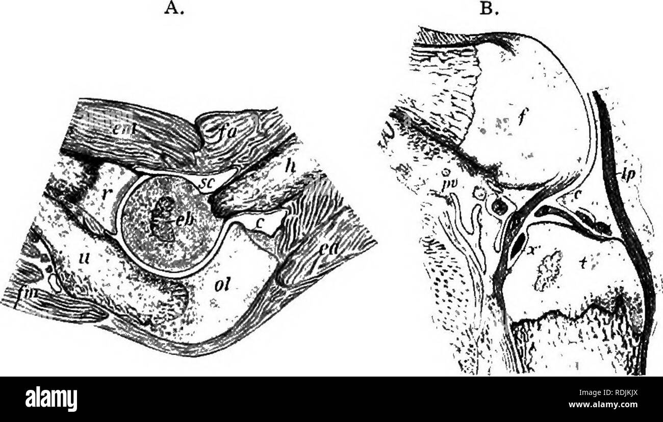 . Practical anatomy of the rabbit : an elementary laboratory textbook in mammalian anatomy . Rabbits; Anatomy, Comparative. 32 Anatomy of the Rabbit. In a comparison of the adult skeleton with the more primitive embryonic skeleton, several differences in the arrangement of the elements are evident. Thus many bones, notwithstanding their possession of several centres of ossification, are to be looked upon as individual either in the cartilage or in the bone condition. In other cases, as in the basal portion of the skull, separate bone elements are produced in a mass of partilage primarily conti Stock Photohttps://www.alamy.com/image-license-details/?v=1https://www.alamy.com/practical-anatomy-of-the-rabbit-an-elementary-laboratory-textbook-in-mammalian-anatomy-rabbits-anatomy-comparative-32-anatomy-of-the-rabbit-in-a-comparison-of-the-adult-skeleton-with-the-more-primitive-embryonic-skeleton-several-differences-in-the-arrangement-of-the-elements-are-evident-thus-many-bones-notwithstanding-their-possession-of-several-centres-of-ossification-are-to-be-looked-upon-as-individual-either-in-the-cartilage-or-in-the-bone-condition-in-other-cases-as-in-the-basal-portion-of-the-skull-separate-bone-elements-are-produced-in-a-mass-of-partilage-primarily-conti-image232135874.html
RMRDJKJX–. Practical anatomy of the rabbit : an elementary laboratory textbook in mammalian anatomy . Rabbits; Anatomy, Comparative. 32 Anatomy of the Rabbit. In a comparison of the adult skeleton with the more primitive embryonic skeleton, several differences in the arrangement of the elements are evident. Thus many bones, notwithstanding their possession of several centres of ossification, are to be looked upon as individual either in the cartilage or in the bone condition. In other cases, as in the basal portion of the skull, separate bone elements are produced in a mass of partilage primarily conti
 . An introduction to the study of mammals living and extinct. Mammals. THE SKELETON 37. between the prcmaxilla and maxilla of either side. Behind the nasals and maxiliai, the anterior part of the brain-case is formed by the large paircil frontals (Figs. G, 7, Fr), behind which are the parietals, which may be of still larger size, and form the greater i)art of the brain-case. A median interparietal Si!'^'i''''''( ^ 'ivii/}-^'^^ I ^- C^-si>X nir ossification (Fig. 0, //') may divide the }:)ariotals jiosteriorly, and is itself articn- lated with the snpra- occipital, to the lat- eral boriler Stock Photohttps://www.alamy.com/image-license-details/?v=1https://www.alamy.com/an-introduction-to-the-study-of-mammals-living-and-extinct-mammals-the-skeleton-37-between-the-prcmaxilla-and-maxilla-of-either-side-behind-the-nasals-and-maxiliai-the-anterior-part-of-the-brain-case-is-formed-by-the-large-paircil-frontals-figs-g-7-fr-behind-which-are-the-parietals-which-may-be-of-still-larger-size-and-form-the-greater-iart-of-the-brain-case-a-median-interparietal-si!i-ivii-i-c-sigtx-nir-ossification-fig-0-may-divide-the-ariotals-jiosteriorly-and-is-itself-articn-lated-with-the-snpra-occipital-to-the-lat-eral-boriler-image232348371.html
RMRE0AM3–. An introduction to the study of mammals living and extinct. Mammals. THE SKELETON 37. between the prcmaxilla and maxilla of either side. Behind the nasals and maxiliai, the anterior part of the brain-case is formed by the large paircil frontals (Figs. G, 7, Fr), behind which are the parietals, which may be of still larger size, and form the greater i)art of the brain-case. A median interparietal Si!'^'i''''''( ^ 'ivii/}-^'^^ I ^- C^-si>X nir ossification (Fig. 0, //') may divide the }:)ariotals jiosteriorly, and is itself articn- lated with the snpra- occipital, to the lat- eral boriler
 . The comparative anatomy of the domesticated animals. Veterinary anatomy. TEE HEAD. 53 Fig. 25, inferior incisors, and behind these, in male animak only, there is an additional alveolus for the tusk. The portion included on each side between the last incisor and first molar, forms a more or Jess sharp ridge, which constitutes the inferior interdental space or hars. Structure and development.—Formed, like all the flat bones, by two compact plates separated by spongy tissue, the inferior maxilla is developed from two centres of ossification, which correspond to each branch, and which coalesce s Stock Photohttps://www.alamy.com/image-license-details/?v=1https://www.alamy.com/the-comparative-anatomy-of-the-domesticated-animals-veterinary-anatomy-tee-head-53-fig-25-inferior-incisors-and-behind-these-in-male-animak-only-there-is-an-additional-alveolus-for-the-tusk-the-portion-included-on-each-side-between-the-last-incisor-and-first-molar-forms-a-more-or-jess-sharp-ridge-which-constitutes-the-inferior-interdental-space-or-hars-structure-and-developmentformed-like-all-the-flat-bones-by-two-compact-plates-separated-by-spongy-tissue-the-inferior-maxilla-is-developed-from-two-centres-of-ossification-which-correspond-to-each-branch-and-which-coalesce-s-image232453407.html
RMRE54KB–. The comparative anatomy of the domesticated animals. Veterinary anatomy. TEE HEAD. 53 Fig. 25, inferior incisors, and behind these, in male animak only, there is an additional alveolus for the tusk. The portion included on each side between the last incisor and first molar, forms a more or Jess sharp ridge, which constitutes the inferior interdental space or hars. Structure and development.—Formed, like all the flat bones, by two compact plates separated by spongy tissue, the inferior maxilla is developed from two centres of ossification, which correspond to each branch, and which coalesce s
 . A manual of zoology. Zoology. 4o6 CIIORDATA axial skeleton and arise from tlie ossifications in the skin (scales) or in the mouth (teeth), alreaih" referred to (p. 451). They sink into the deeper portions and apply ihemsehes to the axial skeleton, especially to those parts where, from lack of cartilage, no primary bones can be formed. It is not settled how far these distinctions are 'alid. According to Gegen- baur all ossification arose jirimarily in the skin or mucous membranes, and primary bones are merely memlirane bones which have entered the. Fir.. 515.—Chondrocranium of A }>ip Stock Photohttps://www.alamy.com/image-license-details/?v=1https://www.alamy.com/a-manual-of-zoology-zoology-4o6-ciiordata-axial-skeleton-and-arise-from-tlie-ossifications-in-the-skin-scales-or-in-the-mouth-teeth-alreaihquot-referred-to-p-451-they-sink-into-the-deeper-portions-and-apply-ihemsehes-to-the-axial-skeleton-especially-to-those-parts-where-from-lack-of-cartilage-no-primary-bones-can-be-formed-it-is-not-settled-how-far-these-distinctions-are-alid-according-to-gegen-baur-all-ossification-arose-jirimarily-in-the-skin-or-mucous-membranes-and-primary-bones-are-merely-memlirane-bones-which-have-entered-the-fir-515chondrocranium-of-a-gtip-image232124824.html
RMRDJ5G8–. A manual of zoology. Zoology. 4o6 CIIORDATA axial skeleton and arise from tlie ossifications in the skin (scales) or in the mouth (teeth), alreaih" referred to (p. 451). They sink into the deeper portions and apply ihemsehes to the axial skeleton, especially to those parts where, from lack of cartilage, no primary bones can be formed. It is not settled how far these distinctions are 'alid. According to Gegen- baur all ossification arose jirimarily in the skin or mucous membranes, and primary bones are merely memlirane bones which have entered the. Fir.. 515.—Chondrocranium of A }>ip
 . Text book of zoology. Zoology. Glass B. Amphihia. 395 Tlie shoulder girdle of tlie Urodela is represented by a cartilaginous arcli on either side^ in wMch two regions are dis- tinguislied, one dorsal the other ventral, is the glenoid for the arm. The dorsal portion, the scapula, is narrower than the ventral, the coracoid, which partially overlaps its fellow of the other side. The lower part of the scapula is ossified to a varying extent, the ossification often reaches into the coracoid region, but the upper and lower portions of the girdle remain cartilaginous (Fig. 321). In the Anura (Fig. Stock Photohttps://www.alamy.com/image-license-details/?v=1https://www.alamy.com/text-book-of-zoology-zoology-glass-b-amphihia-395-tlie-shoulder-girdle-of-tlie-urodela-is-represented-by-a-cartilaginous-arcli-on-either-side-in-wmch-two-regions-are-dis-tinguislied-one-dorsal-the-other-ventral-is-the-glenoid-for-the-arm-the-dorsal-portion-the-scapula-is-narrower-than-the-ventral-the-coracoid-which-partially-overlaps-its-fellow-of-the-other-side-the-lower-part-of-the-scapula-is-ossified-to-a-varying-extent-the-ossification-often-reaches-into-the-coracoid-region-but-the-upper-and-lower-portions-of-the-girdle-remain-cartilaginous-fig-321-in-the-anura-fig-image232416319.html
RMRE3DAR–. Text book of zoology. Zoology. Glass B. Amphihia. 395 Tlie shoulder girdle of tlie Urodela is represented by a cartilaginous arcli on either side^ in wMch two regions are dis- tinguislied, one dorsal the other ventral, is the glenoid for the arm. The dorsal portion, the scapula, is narrower than the ventral, the coracoid, which partially overlaps its fellow of the other side. The lower part of the scapula is ossified to a varying extent, the ossification often reaches into the coracoid region, but the upper and lower portions of the girdle remain cartilaginous (Fig. 321). In the Anura (Fig.
 . Text book of zoology. Zoology. 412 Vertebrata. â¢exoccipitals; the squamosal, in the same region, projects far foi-wards in Snakes, and is connected with the quadi-ate; the basisphenoid,in front of the basi-occipital, and like this, an ossification in the lower wall of the skull. A parasphenoidisnot developed (c/.. Pish and Amphibia). The anterior wall of the brain-case is often unossified and membranous, sometimes with isolated ossifications. Dorsllly there is a number of bones; the parietals, which are generally (Snakes, Lizards, Oi-ocodiles) fused; the f rentals, an unpaired bone, in Croc Stock Photohttps://www.alamy.com/image-license-details/?v=1https://www.alamy.com/text-book-of-zoology-zoology-412-vertebrata-exoccipitals-the-squamosal-in-the-same-region-projects-far-foi-wards-in-snakes-and-is-connected-with-the-quadi-ate-the-basisphenoidin-front-of-the-basi-occipital-and-like-this-an-ossification-in-the-lower-wall-of-the-skull-a-parasphenoidisnot-developed-c-pish-and-amphibia-the-anterior-wall-of-the-brain-case-is-often-unossified-and-membranous-sometimes-with-isolated-ossifications-dorsllly-there-is-a-number-of-bones-the-parietals-which-are-generally-snakes-lizards-oi-ocodiles-fused-the-f-rentals-an-unpaired-bone-in-croc-image232416268.html
RMRE3D90–. Text book of zoology. Zoology. 412 Vertebrata. â¢exoccipitals; the squamosal, in the same region, projects far foi-wards in Snakes, and is connected with the quadi-ate; the basisphenoid,in front of the basi-occipital, and like this, an ossification in the lower wall of the skull. A parasphenoidisnot developed (c/.. Pish and Amphibia). The anterior wall of the brain-case is often unossified and membranous, sometimes with isolated ossifications. Dorsllly there is a number of bones; the parietals, which are generally (Snakes, Lizards, Oi-ocodiles) fused; the f rentals, an unpaired bone, in Croc
 . The Cambridge natural history. Zoology. 560 DOROUCOULIS that of Mycefefi. ISTevertheless Professor Weldon^ has found in a female of C. ijigot a patch of ossification on the thyroid cartilage of the larynx which may be an indication of something more in the male. There are eleven species. JSfycMpitJiecus, the Doroucouli Monkeys, is a genus of some- what Lemurine appearance, caused by their large eyes. But they reminded Bates of an Owl or a Tiger-cat! They have a long, but not prehensile tail. As in the Marmosets, the lower incisors project forwards in a Lemurine fashion. The thumb is very sho Stock Photohttps://www.alamy.com/image-license-details/?v=1https://www.alamy.com/the-cambridge-natural-history-zoology-560-doroucoulis-that-of-mycefefi-istevertheless-professor-weldon-has-found-in-a-female-of-c-ijigot-a-patch-of-ossification-on-the-thyroid-cartilage-of-the-larynx-which-may-be-an-indication-of-something-more-in-the-male-there-are-eleven-species-jsfycmpitjiecus-the-doroucouli-monkeys-is-a-genus-of-some-what-lemurine-appearance-caused-by-their-large-eyes-but-they-reminded-bates-of-an-owl-or-a-tiger-cat!-they-have-a-long-but-not-prehensile-tail-as-in-the-marmosets-the-lower-incisors-project-forwards-in-a-lemurine-fashion-the-thumb-is-very-sho-image232148567.html
RMRDK7T7–. The Cambridge natural history. Zoology. 560 DOROUCOULIS that of Mycefefi. ISTevertheless Professor Weldon^ has found in a female of C. ijigot a patch of ossification on the thyroid cartilage of the larynx which may be an indication of something more in the male. There are eleven species. JSfycMpitJiecus, the Doroucouli Monkeys, is a genus of some- what Lemurine appearance, caused by their large eyes. But they reminded Bates of an Owl or a Tiger-cat! They have a long, but not prehensile tail. As in the Marmosets, the lower incisors project forwards in a Lemurine fashion. The thumb is very sho
 . Outlines of zoology. Zoology. 664 MAMMALIA. vomers beneath, the mesethmoid in the median line, while internally there are several thin scroll-like turbinal bones. The lower jaw or mandible consists in adult life of a single bone or ramus on each side, but this is formed around Meckel's cartilage from several centres of ossification. Its condyle works on the squamosal. The hyoid lies between the rami of the mandible, in the back of the mouth, and consists of a median " body," and two pairs of horns or cornua extending backwards. The Appendicular Skeleton consists of the bones of the Stock Photohttps://www.alamy.com/image-license-details/?v=1https://www.alamy.com/outlines-of-zoology-zoology-664-mammalia-vomers-beneath-the-mesethmoid-in-the-median-line-while-internally-there-are-several-thin-scroll-like-turbinal-bones-the-lower-jaw-or-mandible-consists-in-adult-life-of-a-single-bone-or-ramus-on-each-side-but-this-is-formed-around-meckels-cartilage-from-several-centres-of-ossification-its-condyle-works-on-the-squamosal-the-hyoid-lies-between-the-rami-of-the-mandible-in-the-back-of-the-mouth-and-consists-of-a-median-quot-bodyquot-and-two-pairs-of-horns-or-cornua-extending-backwards-the-appendicular-skeleton-consists-of-the-bones-of-the-image232345128.html
RMRE06G8–. Outlines of zoology. Zoology. 664 MAMMALIA. vomers beneath, the mesethmoid in the median line, while internally there are several thin scroll-like turbinal bones. The lower jaw or mandible consists in adult life of a single bone or ramus on each side, but this is formed around Meckel's cartilage from several centres of ossification. Its condyle works on the squamosal. The hyoid lies between the rami of the mandible, in the back of the mouth, and consists of a median " body," and two pairs of horns or cornua extending backwards. The Appendicular Skeleton consists of the bones of the
 . A manual of zoology. PHYLUM CHORDATA 5°3 the pubis, but by a small intercalated ossification, the cotyloid bone. The ilium (il) has a rough surface for articulation with the sacrum. Between the. pubis (pub) in front and the ischium (isch) behind is a large aperture, the obturator foramen (obi). The femur has at its proximal end a prominent head for articulation with the acetabulum, external to this a prominent process, the great trochanter, and internally a much smaller, the lesser trochanter, while a small process or third trochanter is situated on the outer border a little below the great Stock Photohttps://www.alamy.com/image-license-details/?v=1https://www.alamy.com/a-manual-of-zoology-phylum-chordata-53-the-pubis-but-by-a-small-intercalated-ossification-the-cotyloid-bone-the-ilium-il-has-a-rough-surface-for-articulation-with-the-sacrum-between-the-pubis-pub-in-front-and-the-ischium-isch-behind-is-a-large-aperture-the-obturator-foramen-obi-the-femur-has-at-its-proximal-end-a-prominent-head-for-articulation-with-the-acetabulum-external-to-this-a-prominent-process-the-great-trochanter-and-internally-a-much-smaller-the-lesser-trochanter-while-a-small-process-or-third-trochanter-is-situated-on-the-outer-border-a-little-below-the-great-image232125429.html
RMRDJ69W–. A manual of zoology. PHYLUM CHORDATA 5°3 the pubis, but by a small intercalated ossification, the cotyloid bone. The ilium (il) has a rough surface for articulation with the sacrum. Between the. pubis (pub) in front and the ischium (isch) behind is a large aperture, the obturator foramen (obi). The femur has at its proximal end a prominent head for articulation with the acetabulum, external to this a prominent process, the great trochanter, and internally a much smaller, the lesser trochanter, while a small process or third trochanter is situated on the outer border a little below the great
 . On the anatomy of vertebrates. Vertebrates; Anatomy, Comparative; 1866. ANATOMY or VERTEBRATES. 73 circumscribe elliptical spaces outside the Y)respheuoicl plate : these appear to-represent the pterygoid arches, fig. 61, i, but, as in the embryo of higher fishes, are uot sejiarated from the base of the skull by distinct joints. The basal cartilages, after forming the car-capsules, ib. g, exteud upon the sides of the cra- nium, ib. h, arch over its back part, and leave only its upper and middle part membranous, as in the human embryo when ossification of the cranium commences. The cranium is Stock Photohttps://www.alamy.com/image-license-details/?v=1https://www.alamy.com/on-the-anatomy-of-vertebrates-vertebrates-anatomy-comparative-1866-anatomy-or-vertebrates-73-circumscribe-elliptical-spaces-outside-the-yrespheuoicl-plate-these-appear-to-represent-the-pterygoid-arches-fig-61-i-but-as-in-the-embryo-of-higher-fishes-are-uot-sejiarated-from-the-base-of-the-skull-by-distinct-joints-the-basal-cartilages-after-forming-the-car-capsules-ib-g-exteud-upon-the-sides-of-the-cra-nium-ib-h-arch-over-its-back-part-and-leave-only-its-upper-and-middle-part-membranous-as-in-the-human-embryo-when-ossification-of-the-cranium-commences-the-cranium-is-image232103653.html
RMRDH6G5–. On the anatomy of vertebrates. Vertebrates; Anatomy, Comparative; 1866. ANATOMY or VERTEBRATES. 73 circumscribe elliptical spaces outside the Y)respheuoicl plate : these appear to-represent the pterygoid arches, fig. 61, i, but, as in the embryo of higher fishes, are uot sejiarated from the base of the skull by distinct joints. The basal cartilages, after forming the car-capsules, ib. g, exteud upon the sides of the cra- nium, ib. h, arch over its back part, and leave only its upper and middle part membranous, as in the human embryo when ossification of the cranium commences. The cranium is
 . The Cambridge natural history. Zoology. 190 ANURA other, differing only in small points, for instance in the extent of the webs to the fingers and toes, the configuration of the vomerine teeth, the size and appearance of the tympanic disc, and the relative length of the hind-limbs. In some of the West Indian, and in one Brazilian species, H. nigromciculata, the upper surface of the head is rough, owing to the cutis being involved in the cranial ossification. Bony or perhaps only calcareous deposits in other parts of tlie skin are rare, but are notably developed in H. dasynotus of Brazil, in Stock Photohttps://www.alamy.com/image-license-details/?v=1https://www.alamy.com/the-cambridge-natural-history-zoology-190-anura-other-differing-only-in-small-points-for-instance-in-the-extent-of-the-webs-to-the-fingers-and-toes-the-configuration-of-the-vomerine-teeth-the-size-and-appearance-of-the-tympanic-disc-and-the-relative-length-of-the-hind-limbs-in-some-of-the-west-indian-and-in-one-brazilian-species-h-nigromciculata-the-upper-surface-of-the-head-is-rough-owing-to-the-cutis-being-involved-in-the-cranial-ossification-bony-or-perhaps-only-calcareous-deposits-in-other-parts-of-tlie-skin-are-rare-but-are-notably-developed-in-h-dasynotus-of-brazil-in-image232173793.html
RMRDMC15–. The Cambridge natural history. Zoology. 190 ANURA other, differing only in small points, for instance in the extent of the webs to the fingers and toes, the configuration of the vomerine teeth, the size and appearance of the tympanic disc, and the relative length of the hind-limbs. In some of the West Indian, and in one Brazilian species, H. nigromciculata, the upper surface of the head is rough, owing to the cutis being involved in the cranial ossification. Bony or perhaps only calcareous deposits in other parts of tlie skin are rare, but are notably developed in H. dasynotus of Brazil, in
 . Text book of zoology. Zoology. 396 Vertebrata. usually a single ossification only. Anteriorly the pelvis is prolonged into a narrow, unpaired, usually Y-staped, cartilage {cartilaga ypdloides. In the Anura the ilia are backwardly-directed bony rods; the ischio-pubes have fused to form a compressed vertical disc. The hind limb closely resembles the fore limb in structure. In the Anura the tibia and fibula are fused, and the two Fig. 323. Pig. 324.. Please note that these images are extracted from scanned page images that may have been digitally enhanced for readability - coloration and appear Stock Photohttps://www.alamy.com/image-license-details/?v=1https://www.alamy.com/text-book-of-zoology-zoology-396-vertebrata-usually-a-single-ossification-only-anteriorly-the-pelvis-is-prolonged-into-a-narrow-unpaired-usually-y-staped-cartilage-cartilaga-ypdloides-in-the-anura-the-ilia-are-backwardly-directed-bony-rods-the-ischio-pubes-have-fused-to-form-a-compressed-vertical-disc-the-hind-limb-closely-resembles-the-fore-limb-in-structure-in-the-anura-the-tibia-and-fibula-are-fused-and-the-two-fig-323-pig-324-please-note-that-these-images-are-extracted-from-scanned-page-images-that-may-have-been-digitally-enhanced-for-readability-coloration-and-appear-image232416310.html
RMRE3DAE–. Text book of zoology. Zoology. 396 Vertebrata. usually a single ossification only. Anteriorly the pelvis is prolonged into a narrow, unpaired, usually Y-staped, cartilage {cartilaga ypdloides. In the Anura the ilia are backwardly-directed bony rods; the ischio-pubes have fused to form a compressed vertical disc. The hind limb closely resembles the fore limb in structure. In the Anura the tibia and fibula are fused, and the two Fig. 323. Pig. 324.. Please note that these images are extracted from scanned page images that may have been digitally enhanced for readability - coloration and appear
 . Elementary text-book of zoology. Lower Incisor. First Molar. Angle of Mandible. | Paroccipital Process. Note the dentition witli only two lower incisors, no canines and five cheek-teeth. Also the'Metatherian characters. Fig. 344.—Ventral View of Skull of Kangaroo x J. (Ad nat.) Incisor Teeth. the situation of the lacrymal foramen outside the orbit, the incomplete ossification of the palate and the in- flected angle of the lower jaw. The dentition is pecu- liar. There are three upper incisors, flat and chisel- shaped, then a space or diastema in which there are no teeth. The canines are absen Stock Photohttps://www.alamy.com/image-license-details/?v=1https://www.alamy.com/elementary-text-book-of-zoology-lower-incisor-first-molar-angle-of-mandible-paroccipital-process-note-the-dentition-witli-only-two-lower-incisors-no-canines-and-five-cheek-teeth-also-themetatherian-characters-fig-344ventral-view-of-skull-of-kangaroo-x-j-ad-nat-incisor-teeth-the-situation-of-the-lacrymal-foramen-outside-the-orbit-the-incomplete-ossification-of-the-palate-and-the-in-flected-angle-of-the-lower-jaw-the-dentition-is-pecu-liar-there-are-three-upper-incisors-flat-and-chisel-shaped-then-a-space-or-diastema-in-which-there-are-no-teeth-the-canines-are-absen-image232113107.html
RMRDHJHR–. Elementary text-book of zoology. Lower Incisor. First Molar. Angle of Mandible. | Paroccipital Process. Note the dentition witli only two lower incisors, no canines and five cheek-teeth. Also the'Metatherian characters. Fig. 344.—Ventral View of Skull of Kangaroo x J. (Ad nat.) Incisor Teeth. the situation of the lacrymal foramen outside the orbit, the incomplete ossification of the palate and the in- flected angle of the lower jaw. The dentition is pecu- liar. There are three upper incisors, flat and chisel- shaped, then a space or diastema in which there are no teeth. The canines are absen
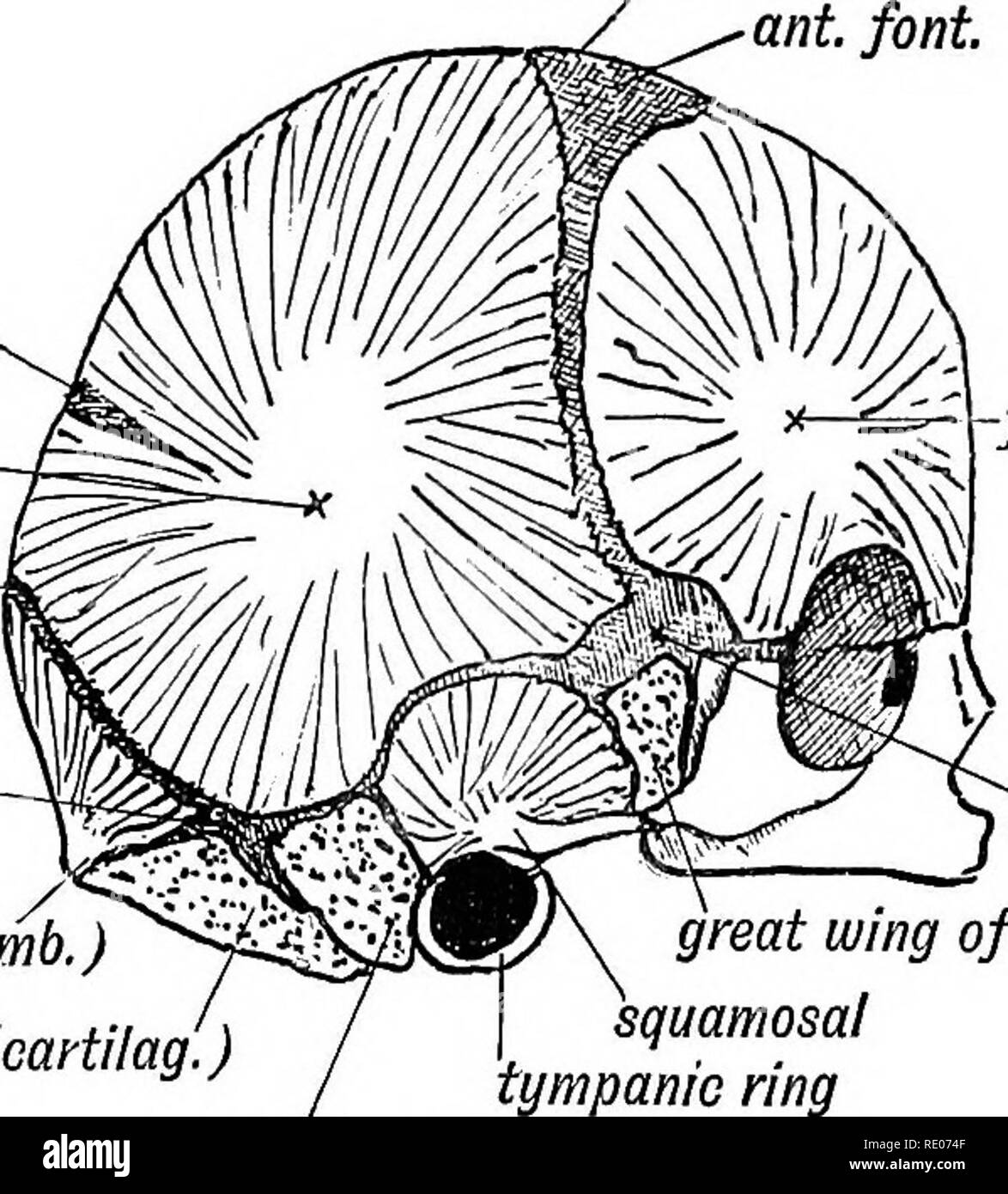 . Human embryology and morphology. Embryology, Human; Morphology. THE CRANIUM. 163 (2) For the parietal, at the position of the parietal eminence; (3) For the squamosalâat the base of the zygoma (Fig. 130); (4) For the membranous part of supra-occipital (part above superior curved line). bregma ant. font.. Sag. font, parietal. lambda aster/on. oceip. (memb.) occip. (oartilag.) â frontal pterion great wing of Sphen. Squamosal tympanic ring petro-mast Fig. 130.âThe Centres of Ossification for the Dermal Bones of the Skull. The Bones which are formed in Cartilage are stippled. The two occipital c Stock Photohttps://www.alamy.com/image-license-details/?v=1https://www.alamy.com/human-embryology-and-morphology-embryology-human-morphology-the-cranium-163-2-for-the-parietal-at-the-position-of-the-parietal-eminence-3-for-the-squamosalat-the-base-of-the-zygoma-fig-130-4-for-the-membranous-part-of-supra-occipital-part-above-superior-curved-line-bregma-ant-font-sag-font-parietal-lambda-asteron-oceip-memb-occip-oartilag-frontal-pterion-great-wing-of-sphen-squamosal-tympanic-ring-petro-mast-fig-130the-centres-of-ossification-for-the-dermal-bones-of-the-skull-the-bones-which-are-formed-in-cartilage-are-stippled-the-two-occipital-c-image232345583.html
RMRE074F–. Human embryology and morphology. Embryology, Human; Morphology. THE CRANIUM. 163 (2) For the parietal, at the position of the parietal eminence; (3) For the squamosalâat the base of the zygoma (Fig. 130); (4) For the membranous part of supra-occipital (part above superior curved line). bregma ant. font.. Sag. font, parietal. lambda aster/on. oceip. (memb.) occip. (oartilag.) â frontal pterion great wing of Sphen. Squamosal tympanic ring petro-mast Fig. 130.âThe Centres of Ossification for the Dermal Bones of the Skull. The Bones which are formed in Cartilage are stippled. The two occipital c
 . Human embryology and morphology. Embryology, Human; Morphology. trans, proa dors, head rib vent, head Flo. 123.—The Bicipital Rib of a Lower Vertebrate (crocodile). by a separate- centre of ossification. The costal process of the 7 th may develop into a rudiment or even a fully formed rib which reaches the sternum. In the lumbar vertebrae only the first shows a separate centre for the formation of the costal process; it fuses with the tip of the transverse process in the later months of foetal life; in the other lumbar vertebrae the tips or perhaps the whole of the transverse processes repre Stock Photohttps://www.alamy.com/image-license-details/?v=1https://www.alamy.com/human-embryology-and-morphology-embryology-human-morphology-trans-proa-dors-head-rib-vent-head-flo-123the-bicipital-rib-of-a-lower-vertebrate-crocodile-by-a-separate-centre-of-ossification-the-costal-process-of-the-7-th-may-develop-into-a-rudiment-or-even-a-fully-formed-rib-which-reaches-the-sternum-in-the-lumbar-vertebrae-only-the-first-shows-a-separate-centre-for-the-formation-of-the-costal-process-it-fuses-with-the-tip-of-the-transverse-process-in-the-later-months-of-foetal-life-in-the-other-lumbar-vertebrae-the-tips-or-perhaps-the-whole-of-the-transverse-processes-repre-image232345614.html
RMRE075J–. Human embryology and morphology. Embryology, Human; Morphology. trans, proa dors, head rib vent, head Flo. 123.—The Bicipital Rib of a Lower Vertebrate (crocodile). by a separate- centre of ossification. The costal process of the 7 th may develop into a rudiment or even a fully formed rib which reaches the sternum. In the lumbar vertebrae only the first shows a separate centre for the formation of the costal process; it fuses with the tip of the transverse process in the later months of foetal life; in the other lumbar vertebrae the tips or perhaps the whole of the transverse processes repre
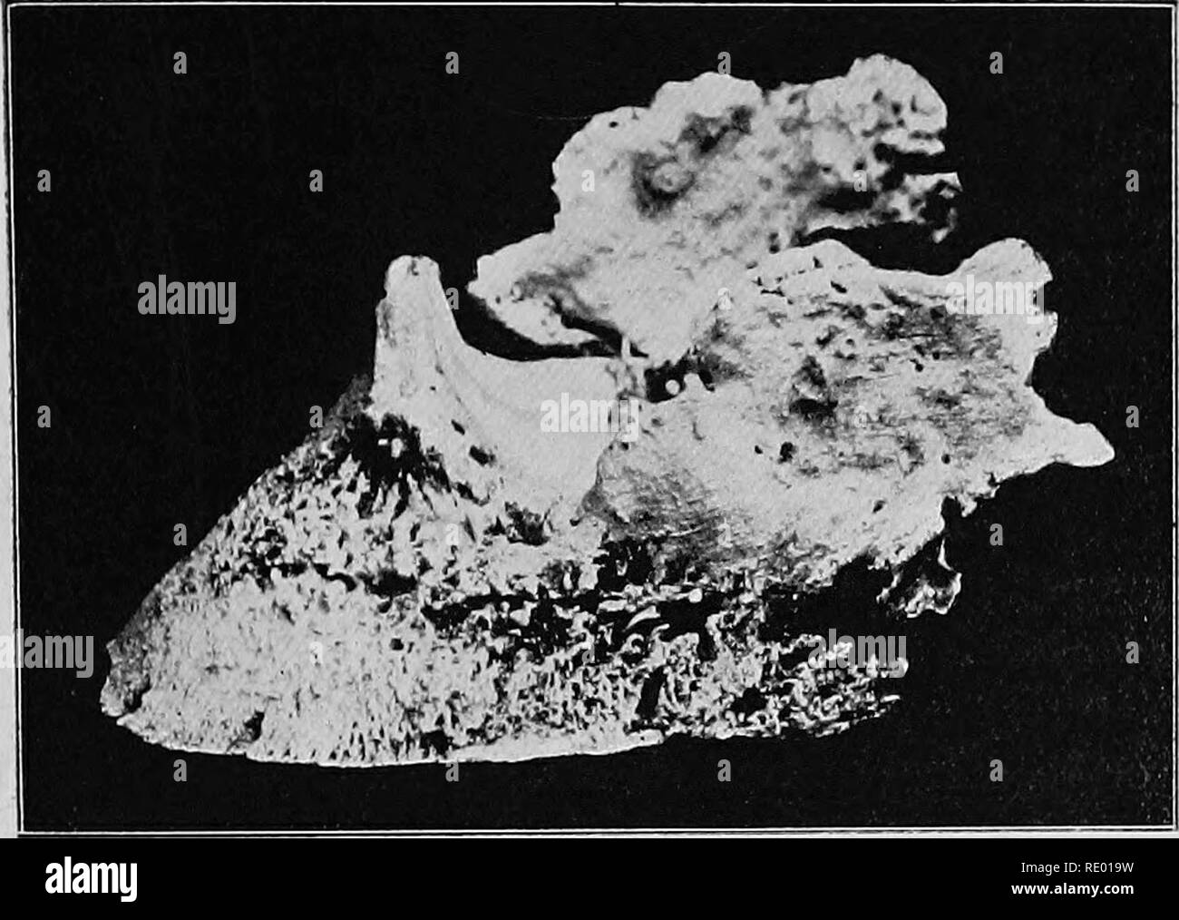 . Diseases of the horse's foot . Hoofs; Horses. DISEASES OF THE LATEEAL CAETILAGES 3G5 fibrous tissue. In such cases benefit is sometimes derived from the application of a shoe with an extended toe-piece (see Figs. 84 and 108). C. OSSIFICATION OP THE LATERAL CARTILAGES, OR SIDE-BONES. Definition.—An abnormal condition of the lateral carti- lages, in which the substance of the cartilage becomes gradually removed and bone formed in its place.. Fig. 143.—Ossified Lateral Caktilagbs (Side-bones). Symptoms and Diagnosis.—Side-bones are nearly always -met with in heavy draught animals, and are rarel Stock Photohttps://www.alamy.com/image-license-details/?v=1https://www.alamy.com/diseases-of-the-horses-foot-hoofs-horses-diseases-of-the-lateeal-caetilages-3g5-fibrous-tissue-in-such-cases-benefit-is-sometimes-derived-from-the-application-of-a-shoe-with-an-extended-toe-piece-see-figs-84-and-108-c-ossification-op-the-lateral-cartilages-or-side-bones-definitionan-abnormal-condition-of-the-lateral-carti-lages-in-which-the-substance-of-the-cartilage-becomes-gradually-removed-and-bone-formed-in-its-place-fig-143ossified-lateral-caktilagbs-side-bones-symptoms-and-diagnosisside-bones-are-nearly-always-met-with-in-heavy-draught-animals-and-are-rarel-image232341029.html
RMRE019W–. Diseases of the horse's foot . Hoofs; Horses. DISEASES OF THE LATEEAL CAETILAGES 3G5 fibrous tissue. In such cases benefit is sometimes derived from the application of a shoe with an extended toe-piece (see Figs. 84 and 108). C. OSSIFICATION OP THE LATERAL CARTILAGES, OR SIDE-BONES. Definition.—An abnormal condition of the lateral carti- lages, in which the substance of the cartilage becomes gradually removed and bone formed in its place.. Fig. 143.—Ossified Lateral Caktilagbs (Side-bones). Symptoms and Diagnosis.—Side-bones are nearly always -met with in heavy draught animals, and are rarel
 . On the anatomy of vertebrates. Vertebrates; Anatomy, Comparative; 1866. ANATOMY OF VEKTEBRATES. 187 shaft is almost straight, sliglitly expanded at the distal end, at the back pai't of which the condyles are fceljly indicated. In Terra- penes, fig. 51, w, and Tortoises, the femur equals or exceeds the luimerus in length : its shaft is more bent: the trochanter is divided into two processes, most distinct in Trionyx. In no Clielonian is there a medullary cavity : ossification extends throughout the bone : the two l)ones of the leg, ib. x, Y, are nearly straight; the til)ia is the largest, wit Stock Photohttps://www.alamy.com/image-license-details/?v=1https://www.alamy.com/on-the-anatomy-of-vertebrates-vertebrates-anatomy-comparative-1866-anatomy-of-vektebrates-187-shaft-is-almost-straight-sliglitly-expanded-at-the-distal-end-at-the-back-pait-of-which-the-condyles-are-fceljly-indicated-in-terra-penes-fig-51-w-and-tortoises-the-femur-equals-or-exceeds-the-luimerus-in-length-its-shaft-is-more-bent-the-trochanter-is-divided-into-two-processes-most-distinct-in-trionyx-in-no-clielonian-is-there-a-medullary-cavity-ossification-extends-throughout-the-bone-the-two-lones-of-the-leg-ib-x-y-are-nearly-straight-the-tilia-is-the-largest-wit-image232103389.html
RMRDH66N–. On the anatomy of vertebrates. Vertebrates; Anatomy, Comparative; 1866. ANATOMY OF VEKTEBRATES. 187 shaft is almost straight, sliglitly expanded at the distal end, at the back pai't of which the condyles are fceljly indicated. In Terra- penes, fig. 51, w, and Tortoises, the femur equals or exceeds the luimerus in length : its shaft is more bent: the trochanter is divided into two processes, most distinct in Trionyx. In no Clielonian is there a medullary cavity : ossification extends throughout the bone : the two l)ones of the leg, ib. x, Y, are nearly straight; the til)ia is the largest, wit
 . Veterinary studies for agricultural students. Veterinary medicine. 2 VETERINARY STUDIES Bones grow in diameter by the production of new bone cells at the inner surface of the periosteum. They grow in length by the development of bone cells in a cartilage matrix between centers of bone formation in the shaft and extremities of the bone. A long bone, for instance, may have three centers of ossification, one in the shaft and one in each end, with a layer of this cartilage matrix in the end between the centers of ossification. Bone cells in the lacunas (spaces) throughout the substances of the b Stock Photohttps://www.alamy.com/image-license-details/?v=1https://www.alamy.com/veterinary-studies-for-agricultural-students-veterinary-medicine-2-veterinary-studies-bones-grow-in-diameter-by-the-production-of-new-bone-cells-at-the-inner-surface-of-the-periosteum-they-grow-in-length-by-the-development-of-bone-cells-in-a-cartilage-matrix-between-centers-of-bone-formation-in-the-shaft-and-extremities-of-the-bone-a-long-bone-for-instance-may-have-three-centers-of-ossification-one-in-the-shaft-and-one-in-each-end-with-a-layer-of-this-cartilage-matrix-in-the-end-between-the-centers-of-ossification-bone-cells-in-the-lacunas-spaces-throughout-the-substances-of-the-b-image232453957.html
RMRE55B1–. Veterinary studies for agricultural students. Veterinary medicine. 2 VETERINARY STUDIES Bones grow in diameter by the production of new bone cells at the inner surface of the periosteum. They grow in length by the development of bone cells in a cartilage matrix between centers of bone formation in the shaft and extremities of the bone. A long bone, for instance, may have three centers of ossification, one in the shaft and one in each end, with a layer of this cartilage matrix in the end between the centers of ossification. Bone cells in the lacunas (spaces) throughout the substances of the b
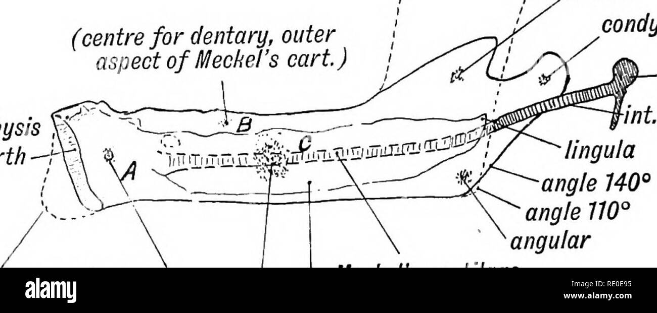 . Human embryology and morphology. Embryology, Human; Morphology. 14 HUMAN EMBRYOLOGY AND MORPHOLOGY. The structures formed from Meckel's cartilage are shown in Figs. IOC, 10D, and 12. Development and Ossification of the Lower Jaw.—In some animals, such as the kangaroo, the two halves of the lower jaw, each developed in its own mandibular process, never unite. In man ossific union takes place early in the second year. In figure 12 are shown the manner of formation and ossification ot the lower jaw, with the changes that take place with age. (relatiue length and angle of / ( J y asc. ramus in Stock Photohttps://www.alamy.com/image-license-details/?v=1https://www.alamy.com/human-embryology-and-morphology-embryology-human-morphology-14-human-embryology-and-morphology-the-structures-formed-from-meckels-cartilage-are-shown-in-figs-ioc-10d-and-12-development-and-ossification-of-the-lower-jawin-some-animals-such-as-the-kangaroo-the-two-halves-of-the-lower-jaw-each-developed-in-its-own-mandibular-process-never-unite-in-man-ossific-union-takes-place-early-in-the-second-year-in-figure-12-are-shown-the-manner-of-formation-and-ossification-ot-the-lower-jaw-with-the-changes-that-take-place-with-age-relatiue-length-and-angle-of-j-y-asc-ramus-in-image232351201.html
RMRE0E95–. Human embryology and morphology. Embryology, Human; Morphology. 14 HUMAN EMBRYOLOGY AND MORPHOLOGY. The structures formed from Meckel's cartilage are shown in Figs. IOC, 10D, and 12. Development and Ossification of the Lower Jaw.—In some animals, such as the kangaroo, the two halves of the lower jaw, each developed in its own mandibular process, never unite. In man ossific union takes place early in the second year. In figure 12 are shown the manner of formation and ossification ot the lower jaw, with the changes that take place with age. (relatiue length and angle of / ( J y asc. ramus in
 . Text book of vertebrate zoology. Vertebrates; Anatomy, Comparative. SKELETON. 163 usually occur. These may extend back to the angle of the jaw, or a jugal (malar) and a quadratojugal may intervene, the lat- ter connecting with the quadrate, and in some cases arising in part from an ossification of a process of the quadrate cartilage. In the roof of the mouth in front are usually a pair of vomers, and behind these, and extending back usually to meet the ptery- goids, are a pair of palatines; while in some groups an os trans-. FlG. 172. Skull of Cyclodus from the side and split through the mid Stock Photohttps://www.alamy.com/image-license-details/?v=1https://www.alamy.com/text-book-of-vertebrate-zoology-vertebrates-anatomy-comparative-skeleton-163-usually-occur-these-may-extend-back-to-the-angle-of-the-jaw-or-a-jugal-malar-and-a-quadratojugal-may-intervene-the-lat-ter-connecting-with-the-quadrate-and-in-some-cases-arising-in-part-from-an-ossification-of-a-process-of-the-quadrate-cartilage-in-the-roof-of-the-mouth-in-front-are-usually-a-pair-of-vomers-and-behind-these-and-extending-back-usually-to-meet-the-ptery-goids-are-a-pair-of-palatines-while-in-some-groups-an-os-trans-flg-172-skull-of-cyclodus-from-the-side-and-split-through-the-mid-image232252673.html
RMRDT0J9–. Text book of vertebrate zoology. Vertebrates; Anatomy, Comparative. SKELETON. 163 usually occur. These may extend back to the angle of the jaw, or a jugal (malar) and a quadratojugal may intervene, the lat- ter connecting with the quadrate, and in some cases arising in part from an ossification of a process of the quadrate cartilage. In the roof of the mouth in front are usually a pair of vomers, and behind these, and extending back usually to meet the ptery- goids, are a pair of palatines; while in some groups an os trans-. FlG. 172. Skull of Cyclodus from the side and split through the mid
 . Human embryology and morphology. Embryology, Human; Morphology. 164 HUMAN EMBRYOLOGY AND MORPHOLOGY. are seen the ossifying fibres of the squamosal. The base of the skull is formed of cartilage which is covered, or ensheathed, by a perichondrium continuous with the membranous capsule. In the cartilage appear the centres of ossification for the sphenoid. sup. long. sin. «"£ font â fibrous capsule spicules of parietal squam.. pencran. parietal %rdura mater temp. fas. temp. muse, squam. basi-sphen. ^ /(?c md Fig. 131.âA coronal section of the Skull of a Foetus, 4£ months old. As the bo Stock Photohttps://www.alamy.com/image-license-details/?v=1https://www.alamy.com/human-embryology-and-morphology-embryology-human-morphology-164-human-embryology-and-morphology-are-seen-the-ossifying-fibres-of-the-squamosal-the-base-of-the-skull-is-formed-of-cartilage-which-is-covered-or-ensheathed-by-a-perichondrium-continuous-with-the-membranous-capsule-in-the-cartilage-appear-the-centres-of-ossification-for-the-sphenoid-sup-long-sin-quot-font-fibrous-capsule-spicules-of-parietal-squam-pencran-parietal-rdura-mater-temp-fas-temp-muse-squam-basi-sphen-c-md-fig-131a-coronal-section-of-the-skull-of-a-foetus-4-months-old-as-the-bo-image232345578.html
RMRE074A–. Human embryology and morphology. Embryology, Human; Morphology. 164 HUMAN EMBRYOLOGY AND MORPHOLOGY. are seen the ossifying fibres of the squamosal. The base of the skull is formed of cartilage which is covered, or ensheathed, by a perichondrium continuous with the membranous capsule. In the cartilage appear the centres of ossification for the sphenoid. sup. long. sin. «"£ font â fibrous capsule spicules of parietal squam.. pencran. parietal %rdura mater temp. fas. temp. muse, squam. basi-sphen. ^ /(?c md Fig. 131.âA coronal section of the Skull of a Foetus, 4£ months old. As the bo
 . Human embryology and morphology. Embryology, Human; Morphology. THE CRANIUM. 167 Separate centres of ossification appear in the parachordal cartilages to form (1) the basi-occipital, (2) the two exocci- pitals, and (3) the supra-occipital. The occipital consists of $jS?-parach. cart/7. notochord parach. cart/1 alar proc. £3â 1st ceruic. â 2nd ceruic. Fig. 133.. Please note that these images are extracted from scanned page images that may have been digitally enhanced for readability - coloration and appearance of these illustrations may not perfectly resemble the original work.. Keith, Arthu Stock Photohttps://www.alamy.com/image-license-details/?v=1https://www.alamy.com/human-embryology-and-morphology-embryology-human-morphology-the-cranium-167-separate-centres-of-ossification-appear-in-the-parachordal-cartilages-to-form-1-the-basi-occipital-2-the-two-exocci-pitals-and-3-the-supra-occipital-the-occipital-consists-of-js-parach-cart7-notochord-parach-cart1-alar-proc-3-1st-ceruic-2nd-ceruic-fig-133-please-note-that-these-images-are-extracted-from-scanned-page-images-that-may-have-been-digitally-enhanced-for-readability-coloration-and-appearance-of-these-illustrations-may-not-perfectly-resemble-the-original-work-keith-arthu-image232345564.html
RMRE073T–. Human embryology and morphology. Embryology, Human; Morphology. THE CRANIUM. 167 Separate centres of ossification appear in the parachordal cartilages to form (1) the basi-occipital, (2) the two exocci- pitals, and (3) the supra-occipital. The occipital consists of $jS?-parach. cart/7. notochord parach. cart/1 alar proc. £3â 1st ceruic. â 2nd ceruic. Fig. 133.. Please note that these images are extracted from scanned page images that may have been digitally enhanced for readability - coloration and appearance of these illustrations may not perfectly resemble the original work.. Keith, Arthu
 . A manual of zoology. Zoology. 574 CHORD AT A. cycloid scales. The living forms (the group appears in the trias) have ossi- fied opisthoccelous vertebrae and diphy- or homocercal tails. Lepidosteid^. Scales rhomboid, branchiostegal rays present, apseudo- )>ranch,but no spiracle. Xe^jicZo.s^g^w,* garpike. Amiid^, distinctly teleos- tean in appearance with cycloid scales, amphico5lous vertebrae, and heart with reduced conus (fig. 596, B). Amia* bow fin. Sub Glass III. Teleostei. The teleosts owe their name to the extensive ossification of the skeleton, which consists, in the trunk, of amplii Stock Photohttps://www.alamy.com/image-license-details/?v=1https://www.alamy.com/a-manual-of-zoology-zoology-574-chord-at-a-cycloid-scales-the-living-forms-the-group-appears-in-the-trias-have-ossi-fied-opisthoccelous-vertebrae-and-diphy-or-homocercal-tails-lepidosteid-scales-rhomboid-branchiostegal-rays-present-apseudo-gtranchbut-no-spiracle-xejiczosgw-garpike-amiid-distinctly-teleos-tean-in-appearance-with-cycloid-scales-amphico5lous-vertebrae-and-heart-with-reduced-conus-fig-596-b-amia-bow-fin-sub-glass-iii-teleostei-the-teleosts-owe-their-name-to-the-extensive-ossification-of-the-skeleton-which-consists-in-the-trunk-of-amplii-image232351155.html
RMRE0E7F–. A manual of zoology. Zoology. 574 CHORD AT A. cycloid scales. The living forms (the group appears in the trias) have ossi- fied opisthoccelous vertebrae and diphy- or homocercal tails. Lepidosteid^. Scales rhomboid, branchiostegal rays present, apseudo- )>ranch,but no spiracle. Xe^jicZo.s^g^w,* garpike. Amiid^, distinctly teleos- tean in appearance with cycloid scales, amphico5lous vertebrae, and heart with reduced conus (fig. 596, B). Amia* bow fin. Sub Glass III. Teleostei. The teleosts owe their name to the extensive ossification of the skeleton, which consists, in the trunk, of amplii
 . Human embryology and morphology. Embryology, Human; Morphology. 7th rib 8th rib 9th rib ensiform. 6th seg. „< centres of ossific - of 3rd seg. (vestigialof'5th & 6th segments) ensiform Fig. 231. Fig. 230. Fig. 230.—The Form of Sternum in a Pronograde (quadrupedal) Mammal. Fig. 231.—The Form of Sternum in a Mammal adapted to the orthograde (upright) Posture. The Points of Ossification are also shown. The chief changes in the human sternum are: 1. Each segment has become flat and wide; 2. The segments of the body fuse together during the years of adolescence, the fusion beginning behind Stock Photohttps://www.alamy.com/image-license-details/?v=1https://www.alamy.com/human-embryology-and-morphology-embryology-human-morphology-7th-rib-8th-rib-9th-rib-ensiform-6th-seg-lt-centres-of-ossific-of-3rd-seg-vestigialof5th-amp-6th-segments-ensiform-fig-231-fig-230-fig-230the-form-of-sternum-in-a-pronograde-quadrupedal-mammal-fig-231the-form-of-sternum-in-a-mammal-adapted-to-the-orthograde-upright-posture-the-points-of-ossification-are-also-shown-the-chief-changes-in-the-human-sternum-are-1-each-segment-has-become-flat-and-wide-2-the-segments-of-the-body-fuse-together-during-the-years-of-adolescence-the-fusion-beginning-behind-image232320618.html
RMRDY38X–. Human embryology and morphology. Embryology, Human; Morphology. 7th rib 8th rib 9th rib ensiform. 6th seg. „< centres of ossific - of 3rd seg. (vestigialof'5th & 6th segments) ensiform Fig. 231. Fig. 230. Fig. 230.—The Form of Sternum in a Pronograde (quadrupedal) Mammal. Fig. 231.—The Form of Sternum in a Mammal adapted to the orthograde (upright) Posture. The Points of Ossification are also shown. The chief changes in the human sternum are: 1. Each segment has become flat and wide; 2. The segments of the body fuse together during the years of adolescence, the fusion beginning behind
 . Outlines of zoology. Zoology. GENERAL CHARACTERS OF BIRDS. 595 -the articularâwhich works on the quadrate. Many of the skull bones have a spongy texture, due to cavities filled with air from the nasal and Eustachian tubes. There is a well-developed sternum, generally with a keel, with a separate centre of ossification, to which the pectoral muscles are in part attached. The strong coracoids reach and articulate with the sternum. In flying birds the clavicles a7'e usually well developed, and connected by an interclavicle, >» /*> t : â ** n-. c g. pr. Fig. 258.âPosition of organs in a b Stock Photohttps://www.alamy.com/image-license-details/?v=1https://www.alamy.com/outlines-of-zoology-zoology-general-characters-of-birds-595-the-articularwhich-works-on-the-quadrate-many-of-the-skull-bones-have-a-spongy-texture-due-to-cavities-filled-with-air-from-the-nasal-and-eustachian-tubes-there-is-a-well-developed-sternum-generally-with-a-keel-with-a-separate-centre-of-ossification-to-which-the-pectoral-muscles-are-in-part-attached-the-strong-coracoids-reach-and-articulate-with-the-sternum-in-flying-birds-the-clavicles-a7e-usually-well-developed-and-connected-by-an-interclavicle-gt-gt-t-n-c-g-pr-fig-258position-of-organs-in-a-b-image232213221.html
RMRDP699–. Outlines of zoology. Zoology. GENERAL CHARACTERS OF BIRDS. 595 -the articularâwhich works on the quadrate. Many of the skull bones have a spongy texture, due to cavities filled with air from the nasal and Eustachian tubes. There is a well-developed sternum, generally with a keel, with a separate centre of ossification, to which the pectoral muscles are in part attached. The strong coracoids reach and articulate with the sternum. In flying birds the clavicles a7'e usually well developed, and connected by an interclavicle, >» /*> t : â ** n-. c g. pr. Fig. 258.âPosition of organs in a b
![. The Batrachia of North America [microform]. Amphibians; Amphibiens. 1 2 Fill. 70. Sjiid liiiiiiiiiiiiulii iiilcriii'iiitiiiiti. No. l(i:ijii. I'l. Walla Wnllii; [. It represents the .S. Iinmiiiontli in more uortherii regions, and the com- plete cranial ossification and larger size mark it as a more fully devel o2)ed form. I found it associated with Biifo coUonbicitsis in a pond near tlu^ shore of Pyramid Lake. Like other allied s[»ecies, it was very noisy, almost obscuring' the voice of the less vociferous liido.. Please note that these images are extracted from scanned page images that may Stock Photo . The Batrachia of North America [microform]. Amphibians; Amphibiens. 1 2 Fill. 70. Sjiid liiiiiiiiiiiiulii iiilcriii'iiitiiiiti. No. l(i:ijii. I'l. Walla Wnllii; [. It represents the .S. Iinmiiiontli in more uortherii regions, and the com- plete cranial ossification and larger size mark it as a more fully devel o2)ed form. I found it associated with Biifo coUonbicitsis in a pond near tlu^ shore of Pyramid Lake. Like other allied s[»ecies, it was very noisy, almost obscuring' the voice of the less vociferous liido.. Please note that these images are extracted from scanned page images that may Stock Photo](https://c8.alamy.com/comp/RJ6HFB/the-batrachia-of-north-america-microform-amphibians-amphibiens-1-2-fill-70-sjiid-liiiiiiiiiiiiulii-iiilcriiiiiitiiiiti-no-liijii-il-walla-wnllii-it-represents-the-s-iinmiiiontli-in-more-uortherii-regions-and-the-com-plete-cranial-ossification-and-larger-size-mark-it-as-a-more-fully-devel-o2ed-form-i-found-it-associated-with-biifo-couonbicitsis-in-a-pond-near-tlu-shore-of-pyramid-lake-like-other-allied-s-ecies-it-was-very-noisy-almost-obscuring-the-voice-of-the-less-vociferous-liido-please-note-that-these-images-are-extracted-from-scanned-page-images-that-may-RJ6HFB.jpg) . The Batrachia of North America [microform]. Amphibians; Amphibiens. 1 2 Fill. 70. Sjiid liiiiiiiiiiiiulii iiilcriii'iiitiiiiti. No. l(i:ijii. I'l. Walla Wnllii; [. It represents the .S. Iinmiiiontli in more uortherii regions, and the com- plete cranial ossification and larger size mark it as a more fully devel o2)ed form. I found it associated with Biifo coUonbicitsis in a pond near tlu^ shore of Pyramid Lake. Like other allied s[»ecies, it was very noisy, almost obscuring' the voice of the less vociferous liido.. Please note that these images are extracted from scanned page images that may Stock Photohttps://www.alamy.com/image-license-details/?v=1https://www.alamy.com/the-batrachia-of-north-america-microform-amphibians-amphibiens-1-2-fill-70-sjiid-liiiiiiiiiiiiulii-iiilcriiiiiitiiiiti-no-liijii-il-walla-wnllii-it-represents-the-s-iinmiiiontli-in-more-uortherii-regions-and-the-com-plete-cranial-ossification-and-larger-size-mark-it-as-a-more-fully-devel-o2ed-form-i-found-it-associated-with-biifo-couonbicitsis-in-a-pond-near-tlu-shore-of-pyramid-lake-like-other-allied-s-ecies-it-was-very-noisy-almost-obscuring-the-voice-of-the-less-vociferous-liido-please-note-that-these-images-are-extracted-from-scanned-page-images-that-may-image234944063.html
. The Batrachia of North America [microform]. Amphibians; Amphibiens. 1 2 Fill. 70. Sjiid liiiiiiiiiiiiulii iiilcriii'iiitiiiiti. No. l(i:ijii. I'l. Walla Wnllii; [. It represents the .S. Iinmiiiontli in more uortherii regions, and the com- plete cranial ossification and larger size mark it as a more fully devel o2)ed form. I found it associated with Biifo coUonbicitsis in a pond near tlu^ shore of Pyramid Lake. Like other allied s[»ecies, it was very noisy, almost obscuring' the voice of the less vociferous liido.. Please note that these images are extracted from scanned page images that may Stock Photohttps://www.alamy.com/image-license-details/?v=1https://www.alamy.com/the-batrachia-of-north-america-microform-amphibians-amphibiens-1-2-fill-70-sjiid-liiiiiiiiiiiiulii-iiilcriiiiiitiiiiti-no-liijii-il-walla-wnllii-it-represents-the-s-iinmiiiontli-in-more-uortherii-regions-and-the-com-plete-cranial-ossification-and-larger-size-mark-it-as-a-more-fully-devel-o2ed-form-i-found-it-associated-with-biifo-couonbicitsis-in-a-pond-near-tlu-shore-of-pyramid-lake-like-other-allied-s-ecies-it-was-very-noisy-almost-obscuring-the-voice-of-the-less-vociferous-liido-please-note-that-these-images-are-extracted-from-scanned-page-images-that-may-image234944063.htmlRMRJ6HFB–. The Batrachia of North America [microform]. Amphibians; Amphibiens. 1 2 Fill. 70. Sjiid liiiiiiiiiiiiulii iiilcriii'iiitiiiiti. No. l(i:ijii. I'l. Walla Wnllii; [. It represents the .S. Iinmiiiontli in more uortherii regions, and the com- plete cranial ossification and larger size mark it as a more fully devel o2)ed form. I found it associated with Biifo coUonbicitsis in a pond near tlu^ shore of Pyramid Lake. Like other allied s[»ecies, it was very noisy, almost obscuring' the voice of the less vociferous liido.. Please note that these images are extracted from scanned page images that may
 . Elementary text-book of zoology. MAMMALIA 499 The skull is seen in side view in Fig. 343. Notice speci- ally the continuation of the malar to the glenoid cavity, and Fig. 343. -Lateral View of Skull of a Young Kangaroo. (Ad nat.) I.acrymal Foramen.. Lower Incisor. First Molar. Angle of Mandible. | Paroccipital Process. Note the dentition witli only two lower incisors, no canines and five cheek-teeth. Also the'Metatherian characters. Fig. 344.—Ventral View of Skull of Kangaroo x J. (Ad nat.) Incisor Teeth. the situation of the lacrymal foramen outside the orbit, the incomplete ossification of Stock Photohttps://www.alamy.com/image-license-details/?v=1https://www.alamy.com/elementary-text-book-of-zoology-mammalia-499-the-skull-is-seen-in-side-view-in-fig-343-notice-speci-ally-the-continuation-of-the-malar-to-the-glenoid-cavity-and-fig-343-lateral-view-of-skull-of-a-young-kangaroo-ad-nat-iacrymal-foramen-lower-incisor-first-molar-angle-of-mandible-paroccipital-process-note-the-dentition-witli-only-two-lower-incisors-no-canines-and-five-cheek-teeth-also-themetatherian-characters-fig-344ventral-view-of-skull-of-kangaroo-x-j-ad-nat-incisor-teeth-the-situation-of-the-lacrymal-foramen-outside-the-orbit-the-incomplete-ossification-of-image232113113.html
RMRDHJJ1–. Elementary text-book of zoology. MAMMALIA 499 The skull is seen in side view in Fig. 343. Notice speci- ally the continuation of the malar to the glenoid cavity, and Fig. 343. -Lateral View of Skull of a Young Kangaroo. (Ad nat.) I.acrymal Foramen.. Lower Incisor. First Molar. Angle of Mandible. | Paroccipital Process. Note the dentition witli only two lower incisors, no canines and five cheek-teeth. Also the'Metatherian characters. Fig. 344.—Ventral View of Skull of Kangaroo x J. (Ad nat.) Incisor Teeth. the situation of the lacrymal foramen outside the orbit, the incomplete ossification of
![. The illustrated natural history [microform]. Natural history; Sciences naturelles. NATUUAL III8T0UY. 17U. eo-IIomcd JUunoceive, or i thorny brandies of mucli shorter than â dUig eighteen inelies th constant rubbing .bly formed, its most thick ossification in in this mass that the connected with tlie 1, and they may thns 3 of a shai-p knife. ;hout, and arc a fine inking cups, mallets c. «kc. The horn is es of the rhinoceros readily observe the ' them. The skin is extremely thick, and only to be penetrated by bullets hardenetl witii solder. During the day, tlie riunoceros will be found lying Stock Photo . The illustrated natural history [microform]. Natural history; Sciences naturelles. NATUUAL III8T0UY. 17U. eo-IIomcd JUunoceive, or i thorny brandies of mucli shorter than â dUig eighteen inelies th constant rubbing .bly formed, its most thick ossification in in this mass that the connected with tlie 1, and they may thns 3 of a shai-p knife. ;hout, and arc a fine inking cups, mallets c. «kc. The horn is es of the rhinoceros readily observe the ' them. The skin is extremely thick, and only to be penetrated by bullets hardenetl witii solder. During the day, tlie riunoceros will be found lying Stock Photo](https://c8.alamy.com/comp/RERNY7/the-illustrated-natural-history-microform-natural-history-sciences-naturelles-natuual-iii8t0uy-17u-eo-iiomcd-juunoceive-or-i-thorny-brandies-of-mucli-shorter-than-duig-eighteen-inelies-th-constant-rubbing-bly-formed-its-most-thick-ossification-in-in-this-mass-that-the-connected-with-tlie-1-and-they-may-thns-3-of-a-shai-p-knife-hout-and-arc-a-fine-inking-cups-mallets-c-kc-the-horn-is-es-of-the-rhinoceros-readily-observe-the-them-the-skin-is-extremely-thick-and-only-to-be-penetrated-by-bullets-hardenetl-witii-solder-during-the-day-tlie-riunoceros-will-be-found-lying-RERNY7.jpg) . The illustrated natural history [microform]. Natural history; Sciences naturelles. NATUUAL III8T0UY. 17U. eo-IIomcd JUunoceive, or i thorny brandies of mucli shorter than â dUig eighteen inelies th constant rubbing .bly formed, its most thick ossification in in this mass that the connected with tlie 1, and they may thns 3 of a shai-p knife. ;hout, and arc a fine inking cups, mallets c. «kc. The horn is es of the rhinoceros readily observe the ' them. The skin is extremely thick, and only to be penetrated by bullets hardenetl witii solder. During the day, tlie riunoceros will be found lying Stock Photohttps://www.alamy.com/image-license-details/?v=1https://www.alamy.com/the-illustrated-natural-history-microform-natural-history-sciences-naturelles-natuual-iii8t0uy-17u-eo-iiomcd-juunoceive-or-i-thorny-brandies-of-mucli-shorter-than-duig-eighteen-inelies-th-constant-rubbing-bly-formed-its-most-thick-ossification-in-in-this-mass-that-the-connected-with-tlie-1-and-they-may-thns-3-of-a-shai-p-knife-hout-and-arc-a-fine-inking-cups-mallets-c-kc-the-horn-is-es-of-the-rhinoceros-readily-observe-the-them-the-skin-is-extremely-thick-and-only-to-be-penetrated-by-bullets-hardenetl-witii-solder-during-the-day-tlie-riunoceros-will-be-found-lying-image232862091.html
. The illustrated natural history [microform]. Natural history; Sciences naturelles. NATUUAL III8T0UY. 17U. eo-IIomcd JUunoceive, or i thorny brandies of mucli shorter than â dUig eighteen inelies th constant rubbing .bly formed, its most thick ossification in in this mass that the connected with tlie 1, and they may thns 3 of a shai-p knife. ;hout, and arc a fine inking cups, mallets c. «kc. The horn is es of the rhinoceros readily observe the ' them. The skin is extremely thick, and only to be penetrated by bullets hardenetl witii solder. During the day, tlie riunoceros will be found lying Stock Photohttps://www.alamy.com/image-license-details/?v=1https://www.alamy.com/the-illustrated-natural-history-microform-natural-history-sciences-naturelles-natuual-iii8t0uy-17u-eo-iiomcd-juunoceive-or-i-thorny-brandies-of-mucli-shorter-than-duig-eighteen-inelies-th-constant-rubbing-bly-formed-its-most-thick-ossification-in-in-this-mass-that-the-connected-with-tlie-1-and-they-may-thns-3-of-a-shai-p-knife-hout-and-arc-a-fine-inking-cups-mallets-c-kc-the-horn-is-es-of-the-rhinoceros-readily-observe-the-them-the-skin-is-extremely-thick-and-only-to-be-penetrated-by-bullets-hardenetl-witii-solder-during-the-day-tlie-riunoceros-will-be-found-lying-image232862091.htmlRMRERNY7–. The illustrated natural history [microform]. Natural history; Sciences naturelles. NATUUAL III8T0UY. 17U. eo-IIomcd JUunoceive, or i thorny brandies of mucli shorter than â dUig eighteen inelies th constant rubbing .bly formed, its most thick ossification in in this mass that the connected with tlie 1, and they may thns 3 of a shai-p knife. ;hout, and arc a fine inking cups, mallets c. «kc. The horn is es of the rhinoceros readily observe the ' them. The skin is extremely thick, and only to be penetrated by bullets hardenetl witii solder. During the day, tlie riunoceros will be found lying
 . A manual of zoology. Zoology. IV. VERTEBRATA: MAMMALIA 547 mal number of young at a birth. Although the mamma? are present in Ijoth sexes, they are functional only in the female, and here only after the birth of the young. A dermal skeleton occurs in few species (e.g., the firm l^ony plates of the armadillos); on the other hand the axial skeleton shows many features not occurring elsewhere. In the skull many of the Ijones already referred to are evident only as centres of ossification, fusing early with their neigh- bors to form larger bones. As the temporal bone shows, parts of diverse orig Stock Photohttps://www.alamy.com/image-license-details/?v=1https://www.alamy.com/a-manual-of-zoology-zoology-iv-vertebrata-mammalia-547-mal-number-of-young-at-a-birth-although-the-mamma-are-present-in-ijoth-sexes-they-are-functional-only-in-the-female-and-here-only-after-the-birth-of-the-young-a-dermal-skeleton-occurs-in-few-species-eg-the-firm-lony-plates-of-the-armadillos-on-the-other-hand-the-axial-skeleton-shows-many-features-not-occurring-elsewhere-in-the-skull-many-of-the-ijones-already-referred-to-are-evident-only-as-centres-of-ossification-fusing-early-with-their-neigh-bors-to-form-larger-bones-as-the-temporal-bone-shows-parts-of-diverse-orig-image232117550.html
RMRDHT8E–. A manual of zoology. Zoology. IV. VERTEBRATA: MAMMALIA 547 mal number of young at a birth. Although the mamma? are present in Ijoth sexes, they are functional only in the female, and here only after the birth of the young. A dermal skeleton occurs in few species (e.g., the firm l^ony plates of the armadillos); on the other hand the axial skeleton shows many features not occurring elsewhere. In the skull many of the Ijones already referred to are evident only as centres of ossification, fusing early with their neigh- bors to form larger bones. As the temporal bone shows, parts of diverse orig
 . Human embryology and morphology. Embryology, Human; Morphology. THE SPINAL COLUMN AND BACK. 153 present in every vertebra. In the cervical vertebrae the anterior part of the transverse processes represents a costal process, but only in the 6 th (sometimes) and 7 th is the costal process formed spine trans, proa costal facet centrum. trans, proa dors, head rib vent, head Flo. 123.—The Bicipital Rib of a Lower Vertebrate (crocodile). by a separate- centre of ossification. The costal process of the 7 th may develop into a rudiment or even a fully formed rib which reaches the sternum. In the lum Stock Photohttps://www.alamy.com/image-license-details/?v=1https://www.alamy.com/human-embryology-and-morphology-embryology-human-morphology-the-spinal-column-and-back-153-present-in-every-vertebra-in-the-cervical-vertebrae-the-anterior-part-of-the-transverse-processes-represents-a-costal-process-but-only-in-the-6-th-sometimes-and-7-th-is-the-costal-process-formed-spine-trans-proa-costal-facet-centrum-trans-proa-dors-head-rib-vent-head-flo-123the-bicipital-rib-of-a-lower-vertebrate-crocodile-by-a-separate-centre-of-ossification-the-costal-process-of-the-7-th-may-develop-into-a-rudiment-or-even-a-fully-formed-rib-which-reaches-the-sternum-in-the-lum-image232345619.html
RMRE075R–. Human embryology and morphology. Embryology, Human; Morphology. THE SPINAL COLUMN AND BACK. 153 present in every vertebra. In the cervical vertebrae the anterior part of the transverse processes represents a costal process, but only in the 6 th (sometimes) and 7 th is the costal process formed spine trans, proa costal facet centrum. trans, proa dors, head rib vent, head Flo. 123.—The Bicipital Rib of a Lower Vertebrate (crocodile). by a separate- centre of ossification. The costal process of the 7 th may develop into a rudiment or even a fully formed rib which reaches the sternum. In the lum
 . A history of British mammals . Mammals; Bats; Insectivores (Mammals); Rodents. PLATE I. m.e. Tsh.i.e. f,k. â z.e.-' Portions of Finger Skeletons of Bats (diagrammatic), enlarged TO SHOW Stages in Ossification of the Joints. m, Metacarpal;/)/j I, phalanx i;p/t2, phalanx 2; me, metacarpal epiphysis; fiiie. first phalangeal epiphysis ; phle^ second phalangeal epiphysis. A. Young specimen with epiphyses quite distinct from phalanges and metacarpal. B. Older but still immature specimen, in which ossification, although advanced. Is incom- plete, and the outHne of all the bones composing each joint Stock Photohttps://www.alamy.com/image-license-details/?v=1https://www.alamy.com/a-history-of-british-mammals-mammals-bats-insectivores-mammals-rodents-plate-i-me-tshie-fk-ze-portions-of-finger-skeletons-of-bats-diagrammatic-enlarged-to-show-stages-in-ossification-of-the-joints-m-metacarpalj-i-phalanx-ipt2-phalanx-2-me-metacarpal-epiphysis-fiiie-first-phalangeal-epiphysis-phle-second-phalangeal-epiphysis-a-young-specimen-with-epiphyses-quite-distinct-from-phalanges-and-metacarpal-b-older-but-still-immature-specimen-in-which-ossification-although-advanced-is-incom-plete-and-the-outhne-of-all-the-bones-composing-each-joint-image232250989.html
RMRDRXE5–. A history of British mammals . Mammals; Bats; Insectivores (Mammals); Rodents. PLATE I. m.e. Tsh.i.e. f,k. â z.e.-' Portions of Finger Skeletons of Bats (diagrammatic), enlarged TO SHOW Stages in Ossification of the Joints. m, Metacarpal;/)/j I, phalanx i;p/t2, phalanx 2; me, metacarpal epiphysis; fiiie. first phalangeal epiphysis ; phle^ second phalangeal epiphysis. A. Young specimen with epiphyses quite distinct from phalanges and metacarpal. B. Older but still immature specimen, in which ossification, although advanced. Is incom- plete, and the outHne of all the bones composing each joint
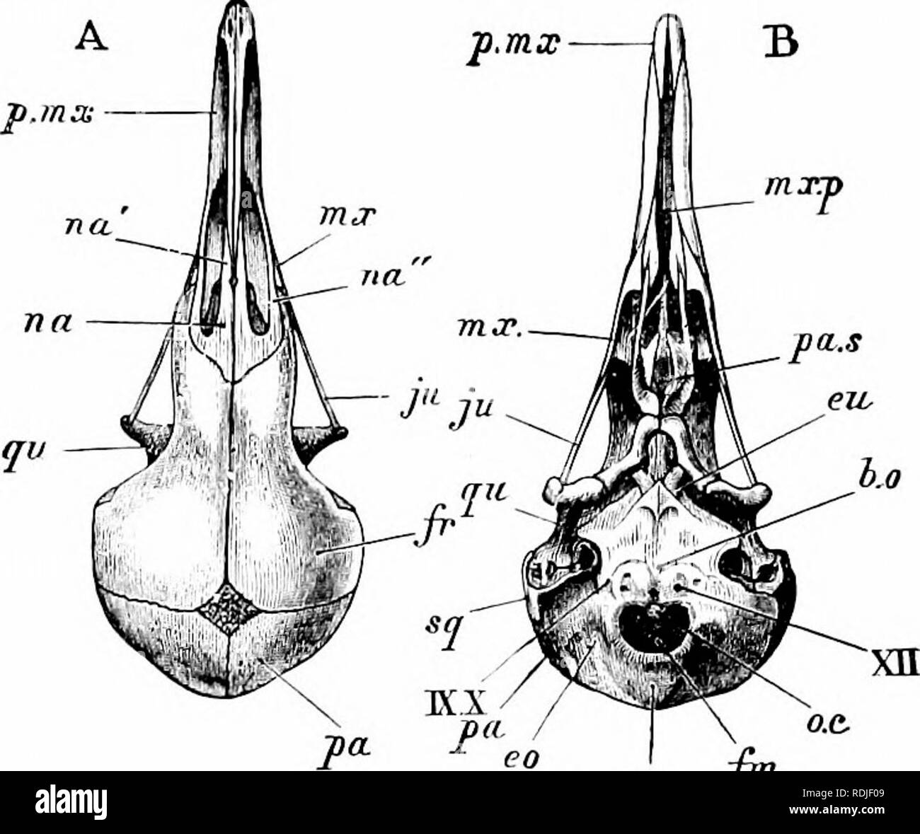 . A manual of zoology. 466 MANUAL OF ZOOLOGY istic parts of the bird's skeleton. It is a broad plate of bone produced ventrally, in the sagittal plane, into a deep keel or carina sterni (car), formed, in the young bird, from a separate centre of ossification. The posterior border of the sternum presents two pairs of notches, covered, in the recent state,. r *% lt , i.o.s wfr p n.etlh I ''i. Please note that these images are extracted from scanned page images that may have been digitally enhanced for readability - coloration and appearance of these illustrations may not perfectly resemble t Stock Photohttps://www.alamy.com/image-license-details/?v=1https://www.alamy.com/a-manual-of-zoology-466-manual-of-zoology-istic-parts-of-the-birds-skeleton-it-is-a-broad-plate-of-bone-produced-ventrally-in-the-sagittal-plane-into-a-deep-keel-or-carina-sterni-car-formed-in-the-young-bird-from-a-separate-centre-of-ossification-the-posterior-border-of-the-sternum-presents-two-pairs-of-notches-covered-in-the-recent-state-r-lt-ios-wfr-p-netlh-i-i-please-note-that-these-images-are-extracted-from-scanned-page-images-that-may-have-been-digitally-enhanced-for-readability-coloration-and-appearance-of-these-illustrations-may-not-perfectly-resemble-t-image232132217.html
RMRDJF09–. A manual of zoology. 466 MANUAL OF ZOOLOGY istic parts of the bird's skeleton. It is a broad plate of bone produced ventrally, in the sagittal plane, into a deep keel or carina sterni (car), formed, in the young bird, from a separate centre of ossification. The posterior border of the sternum presents two pairs of notches, covered, in the recent state,. r *% lt , i.o.s wfr p n.etlh I ''i. Please note that these images are extracted from scanned page images that may have been digitally enhanced for readability - coloration and appearance of these illustrations may not perfectly resemble t
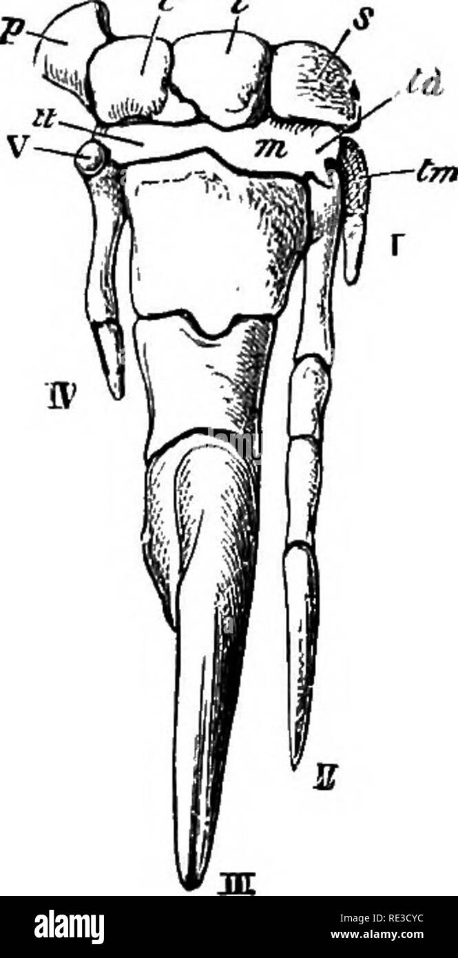 . Text book of zoology. Zoology. Pig. 421. j1 manus of the Great Anteater, B of the Two-toed Anteater, s scaphoid, I lunar, c cuneiform, p pisiform, tm trapezium, td trapezoid, m os magnum, u unciform. /—V digits.—After Flower. 4. Armadillos [Dasypodidse) are characterised throughout by a dorsal covering of large flattened scales, like those of Reptiles; these scales or plates are separated from one another by grooves, the outer surface being very homy, whilst within each scale there is a large ossification. They lie in several ti'ansverse rows, separated by soft skin in the median region of t Stock Photohttps://www.alamy.com/image-license-details/?v=1https://www.alamy.com/text-book-of-zoology-zoology-pig-421-j1-manus-of-the-great-anteater-b-of-the-two-toed-anteater-s-scaphoid-i-lunar-c-cuneiform-p-pisiform-tm-trapezium-td-trapezoid-m-os-magnum-u-unciform-v-digitsafter-flower-4-armadillos-dasypodidse-are-characterised-throughout-by-a-dorsal-covering-of-large-flattened-scales-like-those-of-reptiles-these-scales-or-plates-are-separated-from-one-another-by-grooves-the-outer-surface-being-very-homy-whilst-within-each-scale-there-is-a-large-ossification-they-lie-in-several-tiansverse-rows-separated-by-soft-skin-in-the-median-region-of-t-image232416000.html
RMRE3CYC–. Text book of zoology. Zoology. Pig. 421. j1 manus of the Great Anteater, B of the Two-toed Anteater, s scaphoid, I lunar, c cuneiform, p pisiform, tm trapezium, td trapezoid, m os magnum, u unciform. /—V digits.—After Flower. 4. Armadillos [Dasypodidse) are characterised throughout by a dorsal covering of large flattened scales, like those of Reptiles; these scales or plates are separated from one another by grooves, the outer surface being very homy, whilst within each scale there is a large ossification. They lie in several ti'ansverse rows, separated by soft skin in the median region of t
 . Elements of the comparative anatomy of vertebrates. Anatomy, Comparative. PELVIC ARCH 113. Fig. 91.—Pelvis op Yakious Amphibia. A, Xenopus [Dactyhthra), from below ; B, the same from the front ; C, Rana e^cuJtnta, from the right side; 1) and E, Sa/aiiiandra ntra ; F and G, Salanmndra mnrii/oia ; H, Branrhio.taiirn.s ; I, fJi-iivsaiinis. D-I, from the ventral side. (Figs. H and I after Creduer.) /, ilium ; Is, ischium ; P, pubis (P^ in Rana, pubic end of ilium); IP, fused ischio- pubic ossification; PP, prepubis ; Cep, epipubic cartilage ; Fo^, obturator foramen ; /' (in Xenopus), the proxima Stock Photohttps://www.alamy.com/image-license-details/?v=1https://www.alamy.com/elements-of-the-comparative-anatomy-of-vertebrates-anatomy-comparative-pelvic-arch-113-fig-91pelvis-op-yakious-amphibia-a-xenopus-dactyhthra-from-below-b-the-same-from-the-front-c-rana-ecujtnta-from-the-right-side-1-and-e-saaiiiandra-ntra-f-and-g-salanmndra-mnriioia-h-branrhiotaiirns-i-fji-iivsaiinis-d-i-from-the-ventral-side-figs-h-and-i-after-creduer-ilium-is-ischium-p-pubis-p-in-rana-pubic-end-of-ilium-ip-fused-ischio-pubic-ossification-pp-prepubis-cep-epipubic-cartilage-fo-obturator-foramen-in-xenopus-the-proxima-image232075430.html
RMRDFXG6–. Elements of the comparative anatomy of vertebrates. Anatomy, Comparative. PELVIC ARCH 113. Fig. 91.—Pelvis op Yakious Amphibia. A, Xenopus [Dactyhthra), from below ; B, the same from the front ; C, Rana e^cuJtnta, from the right side; 1) and E, Sa/aiiiandra ntra ; F and G, Salanmndra mnrii/oia ; H, Branrhio.taiirn.s ; I, fJi-iivsaiinis. D-I, from the ventral side. (Figs. H and I after Creduer.) /, ilium ; Is, ischium ; P, pubis (P^ in Rana, pubic end of ilium); IP, fused ischio- pubic ossification; PP, prepubis ; Cep, epipubic cartilage ; Fo^, obturator foramen ; /' (in Xenopus), the proxima
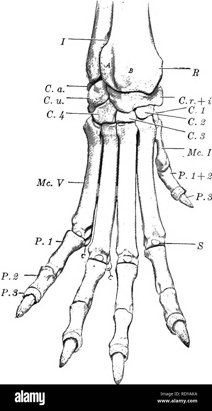 . The anatomy of the domestic animals . Veterinary anatomy. BONES OF THE THORACIC LIMB 201 to sWe on.v; T "IT 'k*'?'"' T^^'' ^°â¢^^1 by them is concave from side sur act; ofX. r ^TX^'fTl'^i ^^^ ^'«*^1 ^"^s (Capitula) have articular cent ?hp 11 wT ^ ^'^^' ^;^* ^'^' ^ '""^^^^^ "dg*^ °n the volar aspect, c-x- cept the first, which is grooved. Ossification is complete at five or six months of TheThw^nitL'hito'r^f Sr" """â â "'"'" ""= "-'â â¢"»â >'- '-⢠longest; the first is very short and does not come in Stock Photohttps://www.alamy.com/image-license-details/?v=1https://www.alamy.com/the-anatomy-of-the-domestic-animals-veterinary-anatomy-bones-of-the-thoracic-limb-201-to-swe-onv-t-quotit-kquot-t-1-by-them-is-concave-from-side-sur-act-ofx-r-txftli-1-quots-capitula-have-articular-cent-hp-11-wt-quotquot-quotdg-n-the-volar-aspect-c-x-cept-the-first-which-is-grooved-ossification-is-complete-at-five-or-six-months-of-thethwnitlhitorf-srquot-quotquotquot-quotquotquot-quotquot=-quot-quot-gt-longest-the-first-is-very-short-and-does-not-come-in-image232326398.html
RMRDYAKA–. The anatomy of the domestic animals . Veterinary anatomy. BONES OF THE THORACIC LIMB 201 to sWe on.v; T "IT 'k*'?'"' T^^'' ^°â¢^^1 by them is concave from side sur act; ofX. r ^TX^'fTl'^i ^^^ ^'«*^1 ^"^s (Capitula) have articular cent ?hp 11 wT ^ ^'^^' ^;^* ^'^' ^ '""^^^^^ "dg*^ °n the volar aspect, c-x- cept the first, which is grooved. Ossification is complete at five or six months of TheThw^nitL'hito'r^f Sr" """â â "'"'" ""= "-'â â¢"»â >'- '-⢠longest; the first is very short and does not come in