Quick filters:
Pacini corpuscle Stock Photos and Images
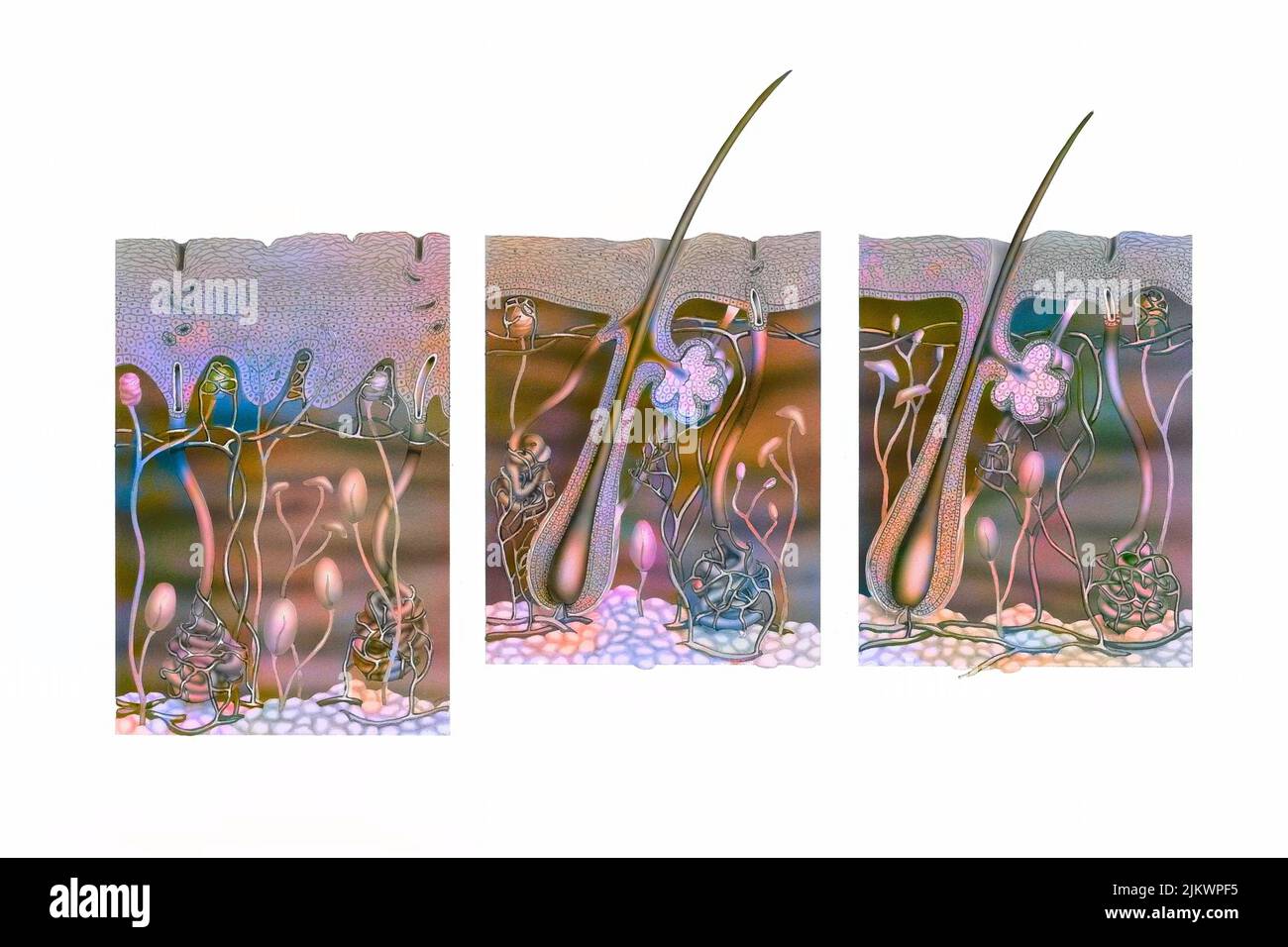 Cut of skin of the palm of the hand, armpit and the rest of the body. Stock Photohttps://www.alamy.com/image-license-details/?v=1https://www.alamy.com/cut-of-skin-of-the-palm-of-the-hand-armpit-and-the-rest-of-the-body-image476924873.html
RF2JKWPF5–Cut of skin of the palm of the hand, armpit and the rest of the body.
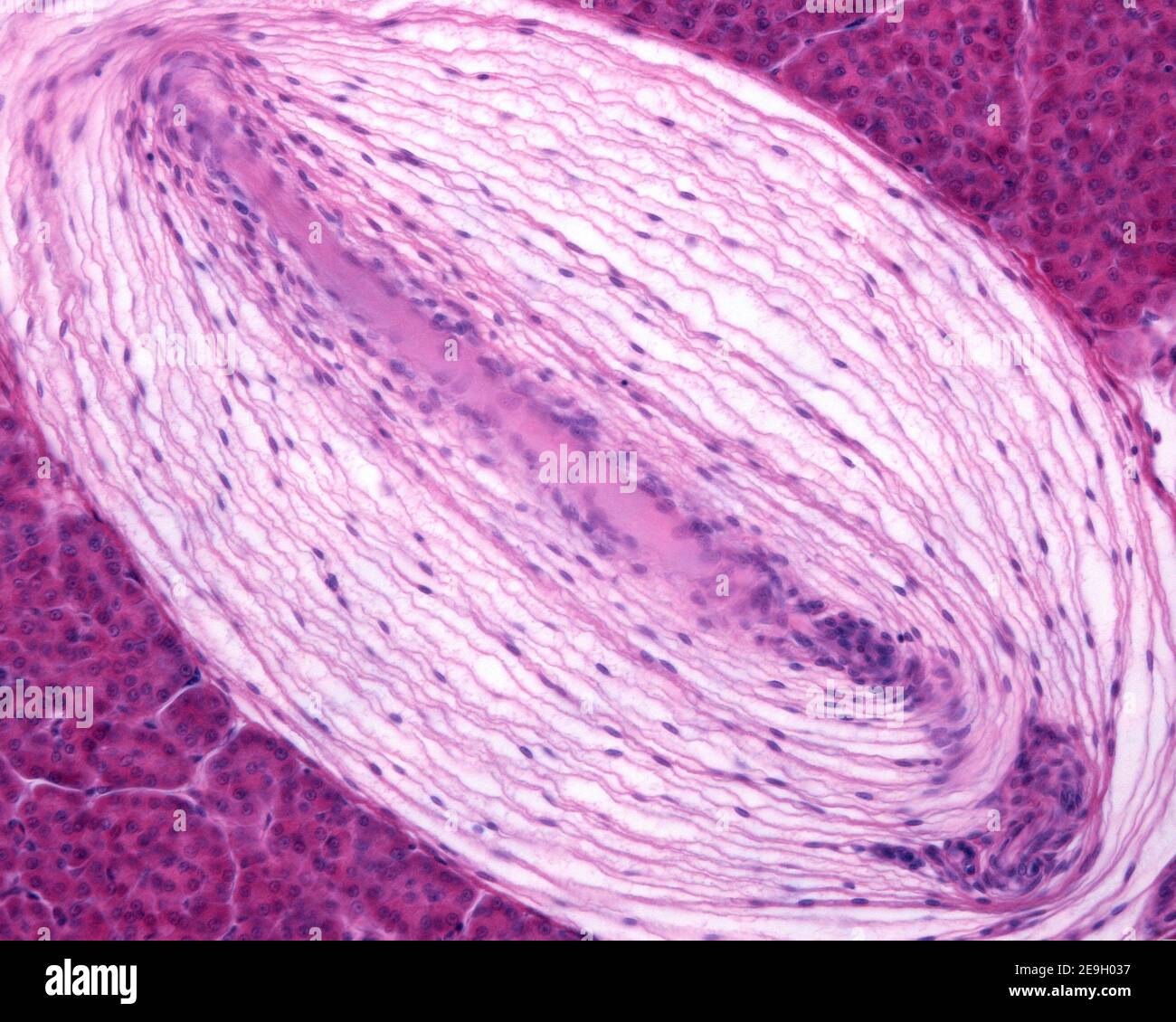 Longitudinal section of a Pacinian corpuscle of a cat pancreas, showing its oval shape. The central reddish band corresponding to the nerve fiber Stock Photohttps://www.alamy.com/image-license-details/?v=1https://www.alamy.com/longitudinal-section-of-a-pacinian-corpuscle-of-a-cat-pancreas-showing-its-oval-shape-the-central-reddish-band-corresponding-to-the-nerve-fiber-image401743643.html
RF2E9H037–Longitudinal section of a Pacinian corpuscle of a cat pancreas, showing its oval shape. The central reddish band corresponding to the nerve fiber
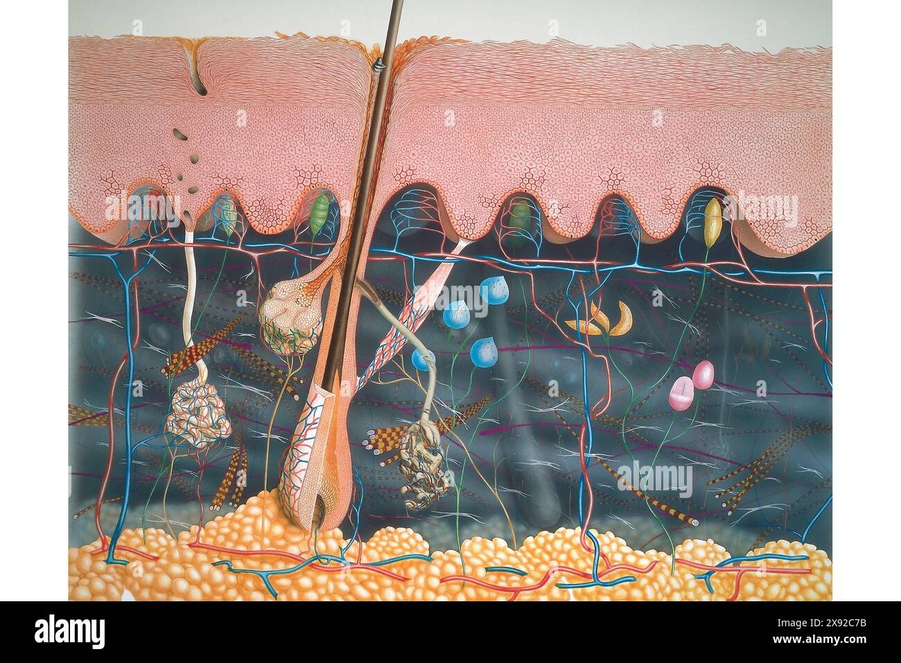 Epidermis, dermis, hypodermis, nerve endings, sebaceous glands and hair bulbs. SKIN, ILLUSTRATION 001248 010 Stock Photohttps://www.alamy.com/image-license-details/?v=1https://www.alamy.com/epidermis-dermis-hypodermis-nerve-endings-sebaceous-glands-and-hair-bulbs-skin-illustration-001248-010-image607948303.html
RM2X92C7B–Epidermis, dermis, hypodermis, nerve endings, sebaceous glands and hair bulbs. SKIN, ILLUSTRATION 001248 010
![Infographic that describes the functions and components of the skin, the largest organ in human beings. [QuarkXPress (.qxp); Adobe InDesign (.indd); QuarkXPress (.qxd); 4960x3188]. Stock Photo Infographic that describes the functions and components of the skin, the largest organ in human beings. [QuarkXPress (.qxp); Adobe InDesign (.indd); QuarkXPress (.qxd); 4960x3188]. Stock Photo](https://c8.alamy.com/comp/2NEBRXB/infographic-that-describes-the-functions-and-components-of-the-skin-the-largest-organ-in-human-beings-quarkxpress-qxp-adobe-indesign-indd-quarkxpress-qxd-4960x3188-2NEBRXB.jpg) Infographic that describes the functions and components of the skin, the largest organ in human beings. [QuarkXPress (.qxp); Adobe InDesign (.indd); QuarkXPress (.qxd); 4960x3188]. Stock Photohttps://www.alamy.com/image-license-details/?v=1https://www.alamy.com/infographic-that-describes-the-functions-and-components-of-the-skin-the-largest-organ-in-human-beings-quarkxpress-qxp-adobe-indesign-indd-quarkxpress-qxd-4960x3188-image525176467.html
Infographic that describes the functions and components of the skin, the largest organ in human beings. [QuarkXPress (.qxp); Adobe InDesign (.indd); QuarkXPress (.qxd); 4960x3188]. Stock Photohttps://www.alamy.com/image-license-details/?v=1https://www.alamy.com/infographic-that-describes-the-functions-and-components-of-the-skin-the-largest-organ-in-human-beings-quarkxpress-qxp-adobe-indesign-indd-quarkxpress-qxd-4960x3188-image525176467.htmlRM2NEBRXB–Infographic that describes the functions and components of the skin, the largest organ in human beings. [QuarkXPress (.qxp); Adobe InDesign (.indd); QuarkXPress (.qxd); 4960x3188].
 . Cunningham's Text-book of anatomy. Anatomy. Fig. 742. A, End bulb (Krause). B, Corpuscle of Pacini x 12 1 C, Corpuscle of Wagner aud Meissner/(after Ranviel')- Fig. 743.—Herbst Corpuscle of Duck (Sobotta). n, medullated nerve-fibre ; a, its axis cylinder ending in an enlarge- ment ; c, nuclei of cells of core ; t, nuclei of cells of outer tunics ; t', inner tunics. of the cat. The capsule of the corpuscle consists of a number of connective tissue tunics arranged concentrically around a central core of more or less clear proto- plasm ; the deeper tunics are closely applied to each other, but Stock Photohttps://www.alamy.com/image-license-details/?v=1https://www.alamy.com/cunninghams-text-book-of-anatomy-anatomy-fig-742-a-end-bulb-krause-b-corpuscle-of-pacini-x-12-1-c-corpuscle-of-wagner-aud-meissnerafter-ranviel-fig-743herbst-corpuscle-of-duck-sobotta-n-medullated-nerve-fibre-a-its-axis-cylinder-ending-in-an-enlarge-ment-c-nuclei-of-cells-of-core-t-nuclei-of-cells-of-outer-tunics-t-inner-tunics-of-the-cat-the-capsule-of-the-corpuscle-consists-of-a-number-of-connective-tissue-tunics-arranged-concentrically-around-a-central-core-of-more-or-less-clear-proto-plasm-the-deeper-tunics-are-closely-applied-to-each-other-but-image231855929.html
RMRD5XGW–. Cunningham's Text-book of anatomy. Anatomy. Fig. 742. A, End bulb (Krause). B, Corpuscle of Pacini x 12 1 C, Corpuscle of Wagner aud Meissner/(after Ranviel')- Fig. 743.—Herbst Corpuscle of Duck (Sobotta). n, medullated nerve-fibre ; a, its axis cylinder ending in an enlarge- ment ; c, nuclei of cells of core ; t, nuclei of cells of outer tunics ; t', inner tunics. of the cat. The capsule of the corpuscle consists of a number of connective tissue tunics arranged concentrically around a central core of more or less clear proto- plasm ; the deeper tunics are closely applied to each other, but
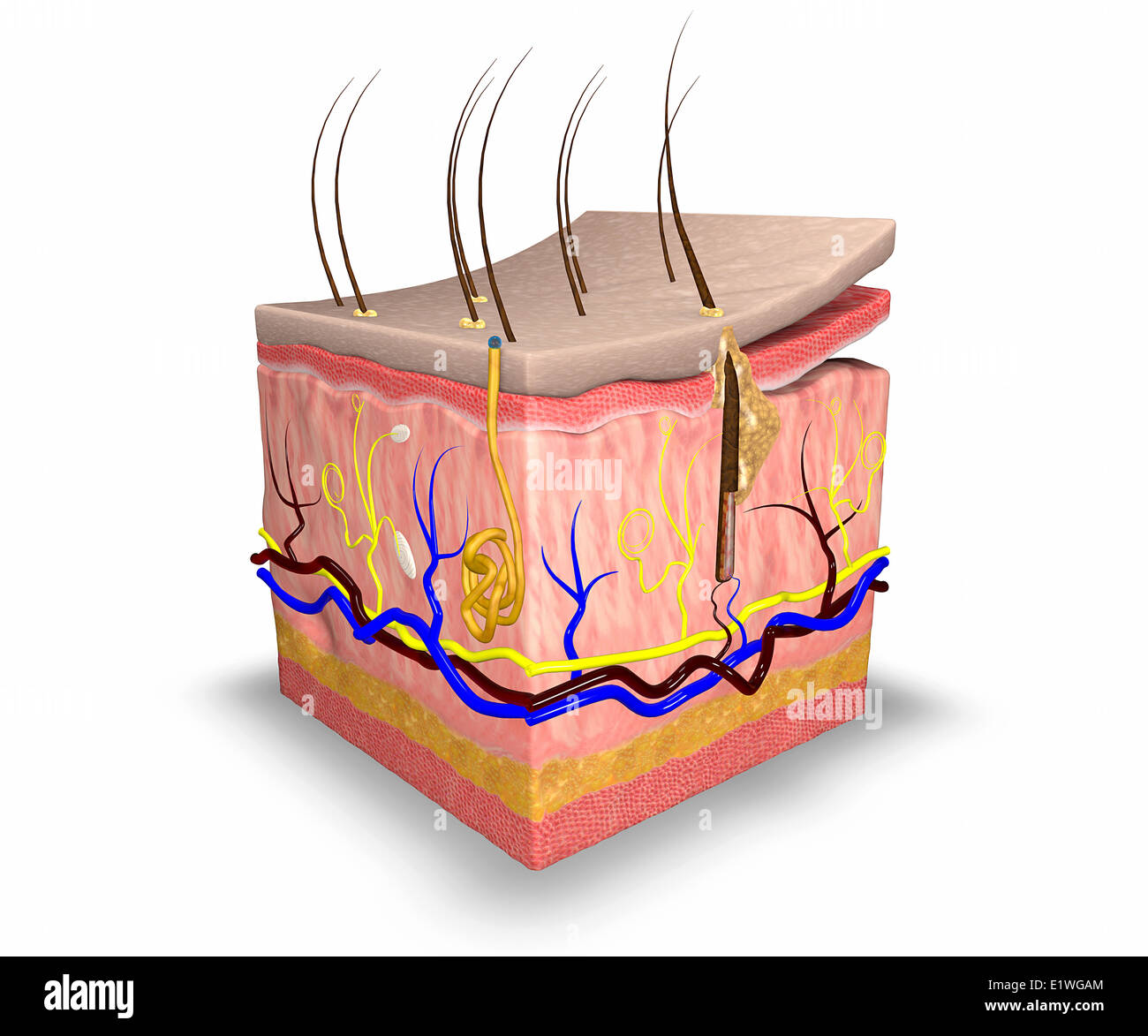 Section skin human body, anatomy Stock Photohttps://www.alamy.com/image-license-details/?v=1https://www.alamy.com/stock-photo-section-skin-human-body-anatomy-70017772.html
RFE1WGAM–Section skin human body, anatomy
 . Cunningham's Text-book of anatomy. Anatomy. Fig. 735.—Tactile Corpuscles. A, End bulb (Krause). B, Corpuscle of Pacini 1 . „, . , C, Corpuscle of Meissner / (after Ranvier)- Appendages of the Skin. The appendages of the skin are the nails, the hairs, the sebaceous glands, and the sudoriferous or sweat glands. Ungues.—The nails (Figs. 736, 737) are epidermal structures, and represent the hoofs and claws of the lower animals. The root of the nail is hidden. Please note that these images are extracted from scanned page images that may have been digitally enhanced for readability - coloration Stock Photohttps://www.alamy.com/image-license-details/?v=1https://www.alamy.com/cunninghams-text-book-of-anatomy-anatomy-fig-735tactile-corpuscles-a-end-bulb-krause-b-corpuscle-of-pacini-1-c-corpuscle-of-meissner-after-ranvier-appendages-of-the-skin-the-appendages-of-the-skin-are-the-nails-the-hairs-the-sebaceous-glands-and-the-sudoriferous-or-sweat-glands-unguesthe-nails-figs-736-737-are-epidermal-structures-and-represent-the-hoofs-and-claws-of-the-lower-animals-the-root-of-the-nail-is-hidden-please-note-that-these-images-are-extracted-from-scanned-page-images-that-may-have-been-digitally-enhanced-for-readability-coloration-image216344674.html
RMPFY9RE–. Cunningham's Text-book of anatomy. Anatomy. Fig. 735.—Tactile Corpuscles. A, End bulb (Krause). B, Corpuscle of Pacini 1 . „, . , C, Corpuscle of Meissner / (after Ranvier)- Appendages of the Skin. The appendages of the skin are the nails, the hairs, the sebaceous glands, and the sudoriferous or sweat glands. Ungues.—The nails (Figs. 736, 737) are epidermal structures, and represent the hoofs and claws of the lower animals. The root of the nail is hidden. Please note that these images are extracted from scanned page images that may have been digitally enhanced for readability - coloration
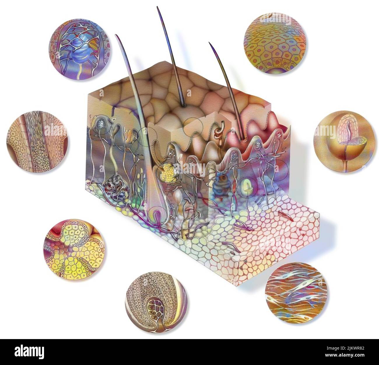 Cut of skin seen from 3/4 with the layers (epidermis, dermis, hypodermis, muscle) and its structures. Stock Photohttps://www.alamy.com/image-license-details/?v=1https://www.alamy.com/cut-of-skin-seen-from-34-with-the-layers-epidermis-dermis-hypodermis-muscle-and-its-structures-image476925458.html
RF2JKWR82–Cut of skin seen from 3/4 with the layers (epidermis, dermis, hypodermis, muscle) and its structures.
 Pacinian corpuscle located in a cat pancreas. It has a thick capsule organized in concentric lamellae. The pink central point is the nerve fiber Stock Photohttps://www.alamy.com/image-license-details/?v=1https://www.alamy.com/pacinian-corpuscle-located-in-a-cat-pancreas-it-has-a-thick-capsule-organized-in-concentric-lamellae-the-pink-central-point-is-the-nerve-fiber-image401743633.html
RF2E9H02W–Pacinian corpuscle located in a cat pancreas. It has a thick capsule organized in concentric lamellae. The pink central point is the nerve fiber
 Quain's elements of anatomy . uscle froji the Fig. 1( mesentery of the cat ; stained withnitrate of silver. Magnified. The epithelioid cells of the outermosttunic are shown, and their continuity, atthe peduncle, with those of the correspond-ing layer of the perineurium (from a, drawingby G. C. Henderson). by Pacini, the layers of the peri-neurium successively become con-tinuous with, or rather expand intothe tunics of the corpuscle. Since,however, in most Pacinian cor-puscles there are many more tunicsin the corpuscle than layersof the perineural sheath whichinvests the entering nerve, it ison Stock Photohttps://www.alamy.com/image-license-details/?v=1https://www.alamy.com/quains-elements-of-anatomy-uscle-froji-the-fig-1-mesentery-of-the-cat-stained-withnitrate-of-silver-magnified-the-epithelioid-cells-of-the-outermosttunic-are-shown-and-their-continuity-atthe-peduncle-with-those-of-the-correspond-ing-layer-of-the-perineurium-from-a-drawingby-g-c-henderson-by-pacini-the-layers-of-the-peri-neurium-successively-become-con-tinuous-with-or-rather-expand-intothe-tunics-of-the-corpuscle-sincehowever-in-most-pacinian-cor-puscles-there-are-many-more-tunicsin-the-corpuscle-than-layersof-the-perineural-sheath-whichinvests-the-entering-nerve-it-ison-image342785438.html
RM2AWK692–Quain's elements of anatomy . uscle froji the Fig. 1( mesentery of the cat ; stained withnitrate of silver. Magnified. The epithelioid cells of the outermosttunic are shown, and their continuity, atthe peduncle, with those of the correspond-ing layer of the perineurium (from a, drawingby G. C. Henderson). by Pacini, the layers of the peri-neurium successively become con-tinuous with, or rather expand intothe tunics of the corpuscle. Since,however, in most Pacinian cor-puscles there are many more tunicsin the corpuscle than layersof the perineural sheath whichinvests the entering nerve, it ison
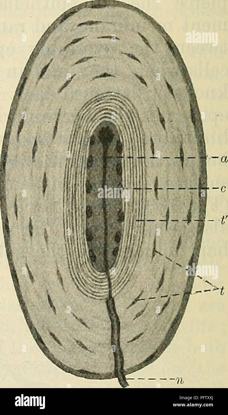 . Cunningham's Text-book of anatomy. Anatomy. Fig. 742. A, End bulb (Krause). B, Corpuscle of Pacini x 12 1 C, Corpuscle of Wagner aud Meissner/(after Ranviel')- Fig. 743.—Herbst Corpuscle of Duck (Sobotta). n, medullated nerve-fibre ; a, its axis cylinder ending in an enlarge- ment ; c, nuclei of cells of core ; t, nuclei of cells of outer tunics ; t', inner tunics. of the cat. The capsule of the corpuscle consists of a number of connective tissue tunics arranged concentrically around a central core of more or less clear proto- plasm ; the deeper tunics are closely applied to each other, but Stock Photohttps://www.alamy.com/image-license-details/?v=1https://www.alamy.com/cunninghams-text-book-of-anatomy-anatomy-fig-742-a-end-bulb-krause-b-corpuscle-of-pacini-x-12-1-c-corpuscle-of-wagner-aud-meissnerafter-ranviel-fig-743herbst-corpuscle-of-duck-sobotta-n-medullated-nerve-fibre-a-its-axis-cylinder-ending-in-an-enlarge-ment-c-nuclei-of-cells-of-core-t-nuclei-of-cells-of-outer-tunics-t-inner-tunics-of-the-cat-the-capsule-of-the-corpuscle-consists-of-a-number-of-connective-tissue-tunics-arranged-concentrically-around-a-central-core-of-more-or-less-clear-proto-plasm-the-deeper-tunics-are-closely-applied-to-each-other-but-image216292234.html
RMPFTXXJ–. Cunningham's Text-book of anatomy. Anatomy. Fig. 742. A, End bulb (Krause). B, Corpuscle of Pacini x 12 1 C, Corpuscle of Wagner aud Meissner/(after Ranviel')- Fig. 743.—Herbst Corpuscle of Duck (Sobotta). n, medullated nerve-fibre ; a, its axis cylinder ending in an enlarge- ment ; c, nuclei of cells of core ; t, nuclei of cells of outer tunics ; t', inner tunics. of the cat. The capsule of the corpuscle consists of a number of connective tissue tunics arranged concentrically around a central core of more or less clear proto- plasm ; the deeper tunics are closely applied to each other, but
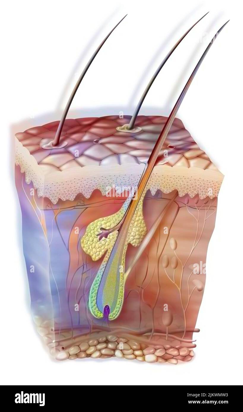 Hair in the skin with the sebaceous gland and the horripilator muscle. Stock Photohttps://www.alamy.com/image-license-details/?v=1https://www.alamy.com/hair-in-the-skin-with-the-sebaceous-gland-and-the-horripilator-muscle-image476923583.html
RF2JKWMW3–Hair in the skin with the sebaceous gland and the horripilator muscle.
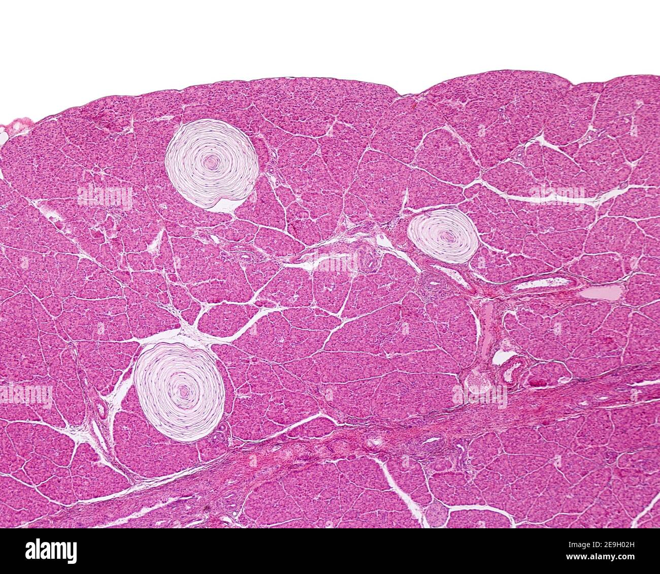 Three Pacinian corpuscles located inside a cat pancreas. They have a lamellar structure, with a thick capsule organized in concentric layers. Stock Photohttps://www.alamy.com/image-license-details/?v=1https://www.alamy.com/three-pacinian-corpuscles-located-inside-a-cat-pancreas-they-have-a-lamellar-structure-with-a-thick-capsule-organized-in-concentric-layers-image401743625.html
RF2E9H02H–Three Pacinian corpuscles located inside a cat pancreas. They have a lamellar structure, with a thick capsule organized in concentric layers.
 . Kirkes' handbook of physiology . Meissner, the tactile corpuscles of Krause, the tactile menisques,and the corpuscles of Golgi. The Pacinian Corpuscles. These nerve endings, named aftertheir discoverer Pacini, are elongated oval bodies situated on some of thecerebro-spinal and sympathetic nerves. They occur on the cutaneousnerves of the hands and feet, the branches of the large sympathetic plexusabout the abdominal aorta, the nerves of the mesentery, and have beenobserved also in the pancreas, lymphatic glands, and thyroid glands, figure 100.Each corpuscle is attached by a narrow pedicle to Stock Photohttps://www.alamy.com/image-license-details/?v=1https://www.alamy.com/kirkes-handbook-of-physiology-meissner-the-tactile-corpuscles-of-krause-the-tactile-menisquesand-the-corpuscles-of-golgi-the-pacinian-corpuscles-these-nerve-endings-named-aftertheir-discoverer-pacini-are-elongated-oval-bodies-situated-on-some-of-thecerebro-spinal-and-sympathetic-nerves-they-occur-on-the-cutaneousnerves-of-the-hands-and-feet-the-branches-of-the-large-sympathetic-plexusabout-the-abdominal-aorta-the-nerves-of-the-mesentery-and-have-beenobserved-also-in-the-pancreas-lymphatic-glands-and-thyroid-glands-figure-100each-corpuscle-is-attached-by-a-narrow-pedicle-to-image369997806.html
RM2CDXRYA–. Kirkes' handbook of physiology . Meissner, the tactile corpuscles of Krause, the tactile menisques,and the corpuscles of Golgi. The Pacinian Corpuscles. These nerve endings, named aftertheir discoverer Pacini, are elongated oval bodies situated on some of thecerebro-spinal and sympathetic nerves. They occur on the cutaneousnerves of the hands and feet, the branches of the large sympathetic plexusabout the abdominal aorta, the nerves of the mesentery, and have beenobserved also in the pancreas, lymphatic glands, and thyroid glands, figure 100.Each corpuscle is attached by a narrow pedicle to
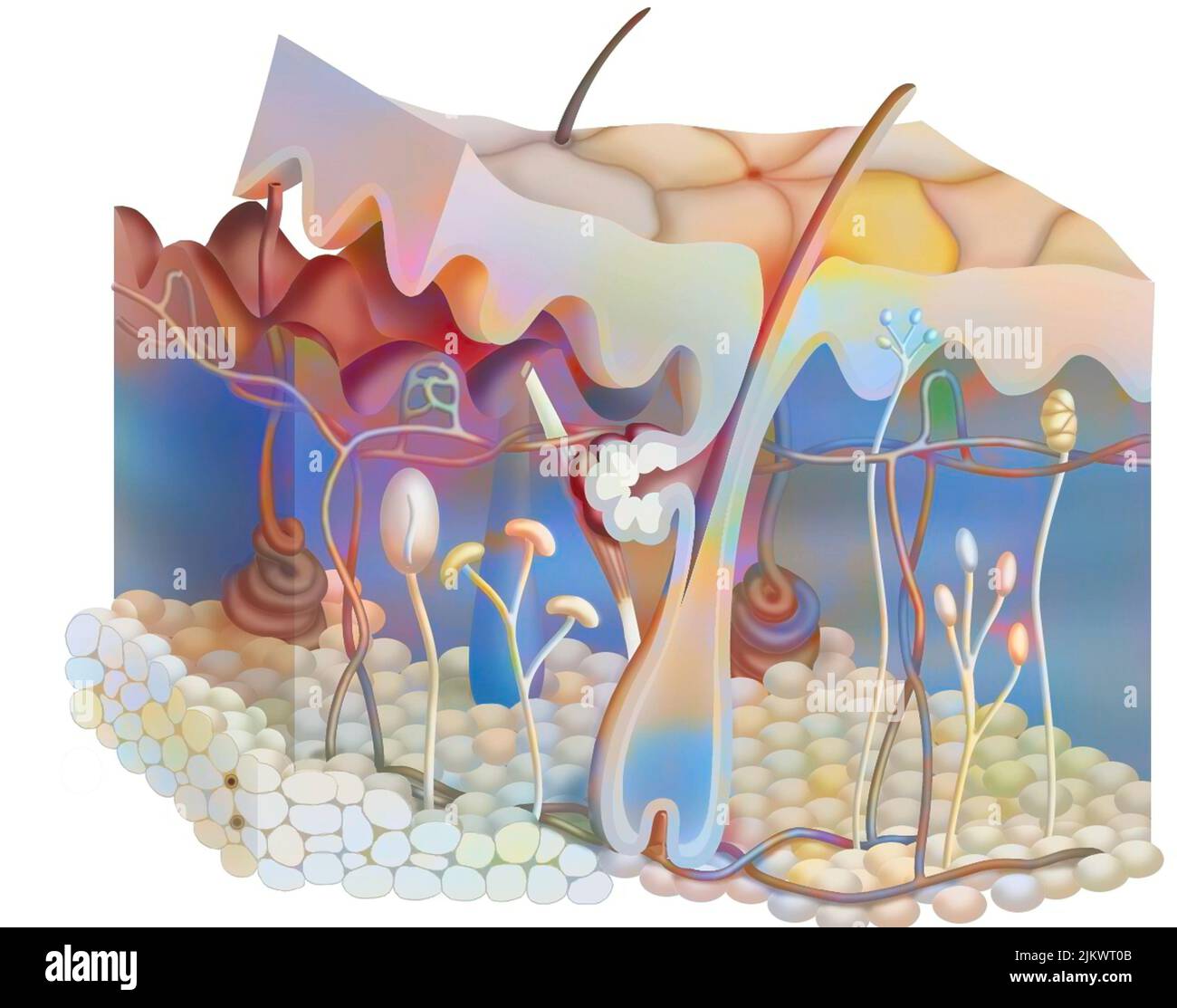 Anatomy of the skin showing the epidermis, dermis, hypodermis. Stock Photohttps://www.alamy.com/image-license-details/?v=1https://www.alamy.com/anatomy-of-the-skin-showing-the-epidermis-dermis-hypodermis-image476926027.html
RF2JKWT0B–Anatomy of the skin showing the epidermis, dermis, hypodermis.
 . Quain's elements of anatomy . corpuscle froh the Fig. n MESENTERY OF THE CAT ; STAINED WITHNITRATE OP SILVER. MAGNIFIED. The epithelioid cells of the outermosttunic are shown, and their continuity, atthe peduncle, with those of the correspond-ing layer of the perineurium (from a drawingby Gr. C. Henderson). by Pacini, the layers of the peri-neurium successively become con-tinuous with, or rather expand intothe tunics of the corpuscle. Since,however, in most Pacinian cor-puscles there are many more tunicsin the corpuscle than layersof the perineural sheath whichinvests the entering nerve, it Stock Photohttps://www.alamy.com/image-license-details/?v=1https://www.alamy.com/quains-elements-of-anatomy-corpuscle-froh-the-fig-n-mesentery-of-the-cat-stained-withnitrate-op-silver-magnified-the-epithelioid-cells-of-the-outermosttunic-are-shown-and-their-continuity-atthe-peduncle-with-those-of-the-correspond-ing-layer-of-the-perineurium-from-a-drawingby-gr-c-henderson-by-pacini-the-layers-of-the-peri-neurium-successively-become-con-tinuous-with-or-rather-expand-intothe-tunics-of-the-corpuscle-sincehowever-in-most-pacinian-cor-puscles-there-are-many-more-tunicsin-the-corpuscle-than-layersof-the-perineural-sheath-whichinvests-the-entering-nerve-it-image372582916.html
RM2CJ4H8M–. Quain's elements of anatomy . corpuscle froh the Fig. n MESENTERY OF THE CAT ; STAINED WITHNITRATE OP SILVER. MAGNIFIED. The epithelioid cells of the outermosttunic are shown, and their continuity, atthe peduncle, with those of the correspond-ing layer of the perineurium (from a drawingby Gr. C. Henderson). by Pacini, the layers of the peri-neurium successively become con-tinuous with, or rather expand intothe tunics of the corpuscle. Since,however, in most Pacinian cor-puscles there are many more tunicsin the corpuscle than layersof the perineural sheath whichinvests the entering nerve, it
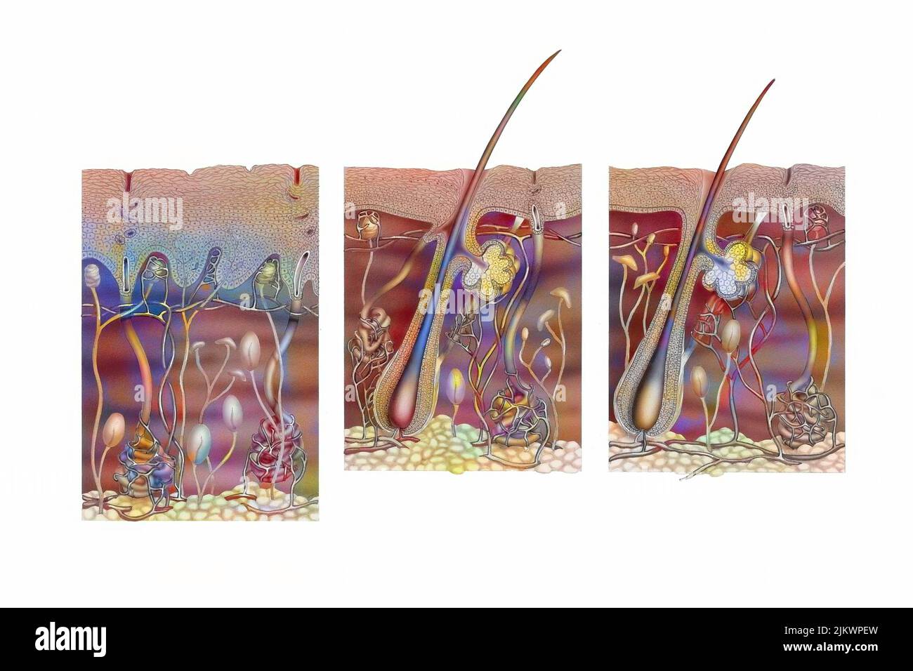 Cut of skin of the palm of the hand, armpit and the rest of the body. Stock Photohttps://www.alamy.com/image-license-details/?v=1https://www.alamy.com/cut-of-skin-of-the-palm-of-the-hand-armpit-and-the-rest-of-the-body-image476924865.html
RF2JKWPEW–Cut of skin of the palm of the hand, armpit and the rest of the body.
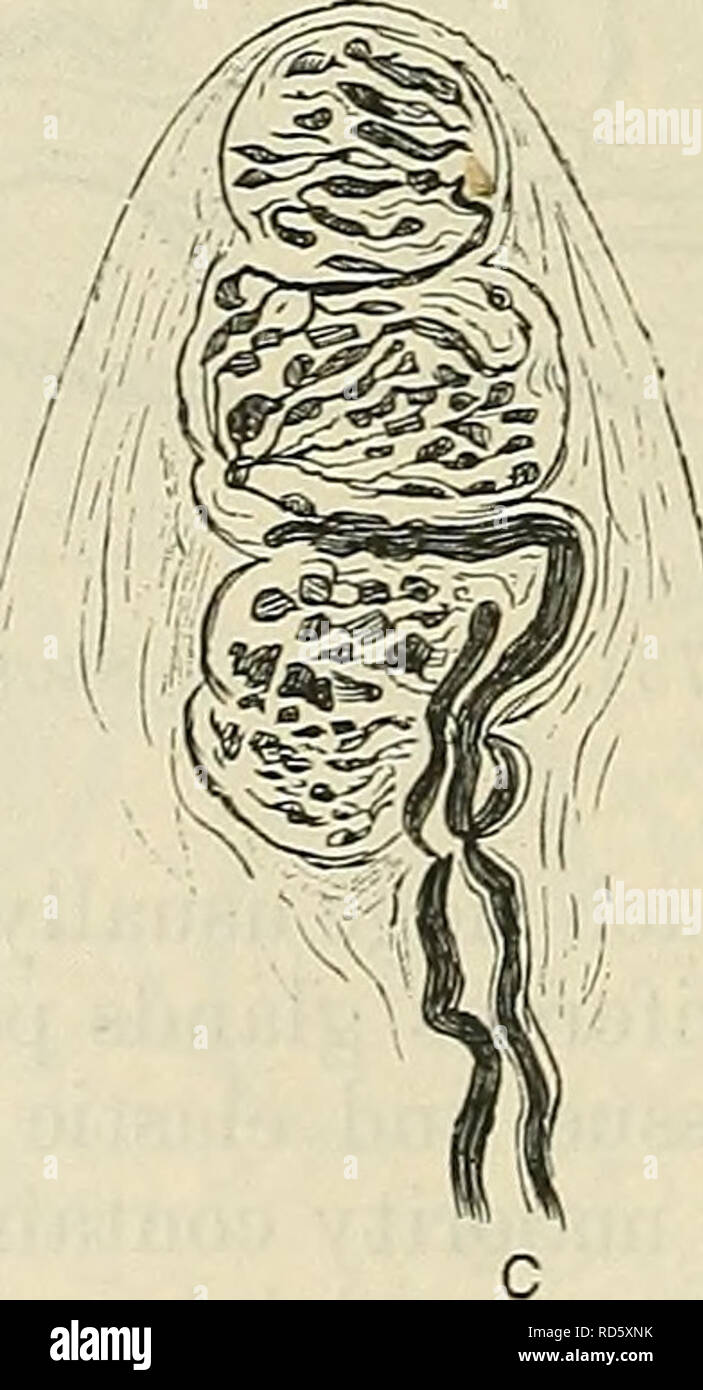 . Cunningham's Text-book of anatomy. Anatomy. Fig. 735.—Tactile Corpuscles. A, End bulb (Krause). B, Corpuscle of Pacini 1 . „, . , C, Corpuscle of Meissner / (after Ranvier)- Appendages of the Skin. The appendages of the skin are the nails, the hairs, the sebaceous glands, and the sudoriferous or sweat glands. Ungues.—The nails (Figs. 736, 737) are epidermal structures, and represent the hoofs and claws of the lower animals. The root of the nail is hidden. Please note that these images are extracted from scanned page images that may have been digitally enhanced for readability - coloration Stock Photohttps://www.alamy.com/image-license-details/?v=1https://www.alamy.com/cunninghams-text-book-of-anatomy-anatomy-fig-735tactile-corpuscles-a-end-bulb-krause-b-corpuscle-of-pacini-1-c-corpuscle-of-meissner-after-ranvier-appendages-of-the-skin-the-appendages-of-the-skin-are-the-nails-the-hairs-the-sebaceous-glands-and-the-sudoriferous-or-sweat-glands-unguesthe-nails-figs-736-737-are-epidermal-structures-and-represent-the-hoofs-and-claws-of-the-lower-animals-the-root-of-the-nail-is-hidden-please-note-that-these-images-are-extracted-from-scanned-page-images-that-may-have-been-digitally-enhanced-for-readability-coloration-image231856063.html
RMRD5XNK–. Cunningham's Text-book of anatomy. Anatomy. Fig. 735.—Tactile Corpuscles. A, End bulb (Krause). B, Corpuscle of Pacini 1 . „, . , C, Corpuscle of Meissner / (after Ranvier)- Appendages of the Skin. The appendages of the skin are the nails, the hairs, the sebaceous glands, and the sudoriferous or sweat glands. Ungues.—The nails (Figs. 736, 737) are epidermal structures, and represent the hoofs and claws of the lower animals. The root of the nail is hidden. Please note that these images are extracted from scanned page images that may have been digitally enhanced for readability - coloration
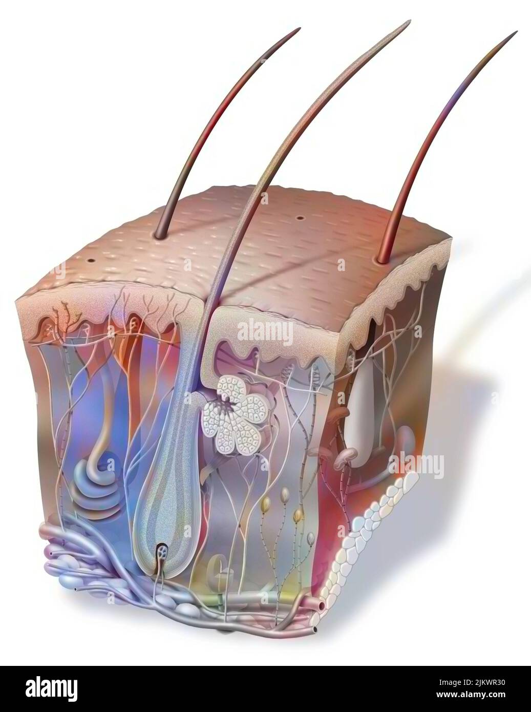 Section of skin with hair and all types of sensory receptors. Stock Photohttps://www.alamy.com/image-license-details/?v=1https://www.alamy.com/section-of-skin-with-hair-and-all-types-of-sensory-receptors-image476925316.html
RF2JKWR30–Section of skin with hair and all types of sensory receptors.
 . Text book of vertebrate zoology. Vertebrates; Anatomy, Comparative. Fig. 71. Pa- cinian corpuscle. //, axis cylinder of nerve. birds, two or more biscuit-shaped cells are included in a connec- tive tissue, while the connecting nen'e becomes flattened out into disks between each two cells. A more complicated type is found in the corpuscles of Vater or Pacini,— elliptical structures composed of layers of cells like the layers of an onion, into the centre of which projects the axis cylinder of a sensory nerve. Under the heading of tactile cor- puscles (Wagner's or Meissner's corpuscles) are inc Stock Photohttps://www.alamy.com/image-license-details/?v=1https://www.alamy.com/text-book-of-vertebrate-zoology-vertebrates-anatomy-comparative-fig-71-pa-cinian-corpuscle-axis-cylinder-of-nerve-birds-two-or-more-biscuit-shaped-cells-are-included-in-a-connec-tive-tissue-while-the-connecting-nene-becomes-flattened-out-into-disks-between-each-two-cells-a-more-complicated-type-is-found-in-the-corpuscles-of-vater-or-pacini-elliptical-structures-composed-of-layers-of-cells-like-the-layers-of-an-onion-into-the-centre-of-which-projects-the-axis-cylinder-of-a-sensory-nerve-under-the-heading-of-tactile-cor-puscles-wagners-or-meissners-corpuscles-are-inc-image232252997.html
RMRDT11W–. Text book of vertebrate zoology. Vertebrates; Anatomy, Comparative. Fig. 71. Pa- cinian corpuscle. //, axis cylinder of nerve. birds, two or more biscuit-shaped cells are included in a connec- tive tissue, while the connecting nen'e becomes flattened out into disks between each two cells. A more complicated type is found in the corpuscles of Vater or Pacini,— elliptical structures composed of layers of cells like the layers of an onion, into the centre of which projects the axis cylinder of a sensory nerve. Under the heading of tactile cor- puscles (Wagner's or Meissner's corpuscles) are inc
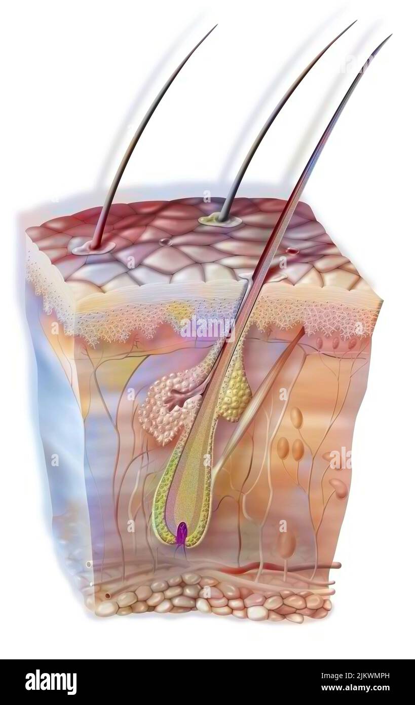 Hair in the skin with the sebaceous gland and the horripilator muscle. Stock Photohttps://www.alamy.com/image-license-details/?v=1https://www.alamy.com/hair-in-the-skin-with-the-sebaceous-gland-and-the-horripilator-muscle-image476923513.html
RF2JKWMPH–Hair in the skin with the sebaceous gland and the horripilator muscle.
 . Text book of vertebrate zoology. Vertebrates; Anatomy, Comparative. Fig. 70. Gran- dry's corpuscle, after Bohm and Davidoff. k, axis cylinder of nerve.. Fig. 71. Pa- cinian corpuscle. //, axis cylinder of nerve. birds, two or more biscuit-shaped cells are included in a connec- tive tissue, while the connecting nen'e becomes flattened out into disks between each two cells. A more complicated type is found in the corpuscles of Vater or Pacini,— elliptical structures composed of layers of cells like the layers of an onion, into the centre of which projects the axis cylinder of a sensory nerve. Stock Photohttps://www.alamy.com/image-license-details/?v=1https://www.alamy.com/text-book-of-vertebrate-zoology-vertebrates-anatomy-comparative-fig-70-gran-drys-corpuscle-after-bohm-and-davidoff-k-axis-cylinder-of-nerve-fig-71-pa-cinian-corpuscle-axis-cylinder-of-nerve-birds-two-or-more-biscuit-shaped-cells-are-included-in-a-connec-tive-tissue-while-the-connecting-nene-becomes-flattened-out-into-disks-between-each-two-cells-a-more-complicated-type-is-found-in-the-corpuscles-of-vater-or-pacini-elliptical-structures-composed-of-layers-of-cells-like-the-layers-of-an-onion-into-the-centre-of-which-projects-the-axis-cylinder-of-a-sensory-nerve-image232253000.html
RMRDT120–. Text book of vertebrate zoology. Vertebrates; Anatomy, Comparative. Fig. 70. Gran- dry's corpuscle, after Bohm and Davidoff. k, axis cylinder of nerve.. Fig. 71. Pa- cinian corpuscle. //, axis cylinder of nerve. birds, two or more biscuit-shaped cells are included in a connec- tive tissue, while the connecting nen'e becomes flattened out into disks between each two cells. A more complicated type is found in the corpuscles of Vater or Pacini,— elliptical structures composed of layers of cells like the layers of an onion, into the centre of which projects the axis cylinder of a sensory nerve.
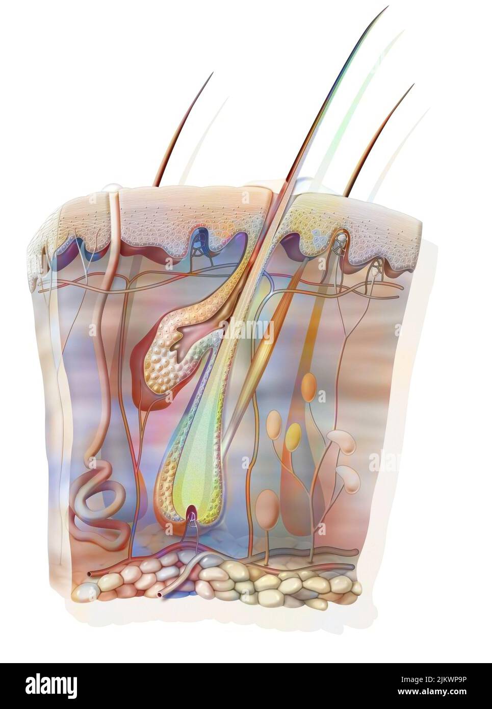 Cut of skin and a hair with the sebaceous gland. Stock Photohttps://www.alamy.com/image-license-details/?v=1https://www.alamy.com/cut-of-skin-and-a-hair-with-the-sebaceous-gland-image476924722.html
RF2JKWP9P–Cut of skin and a hair with the sebaceous gland.
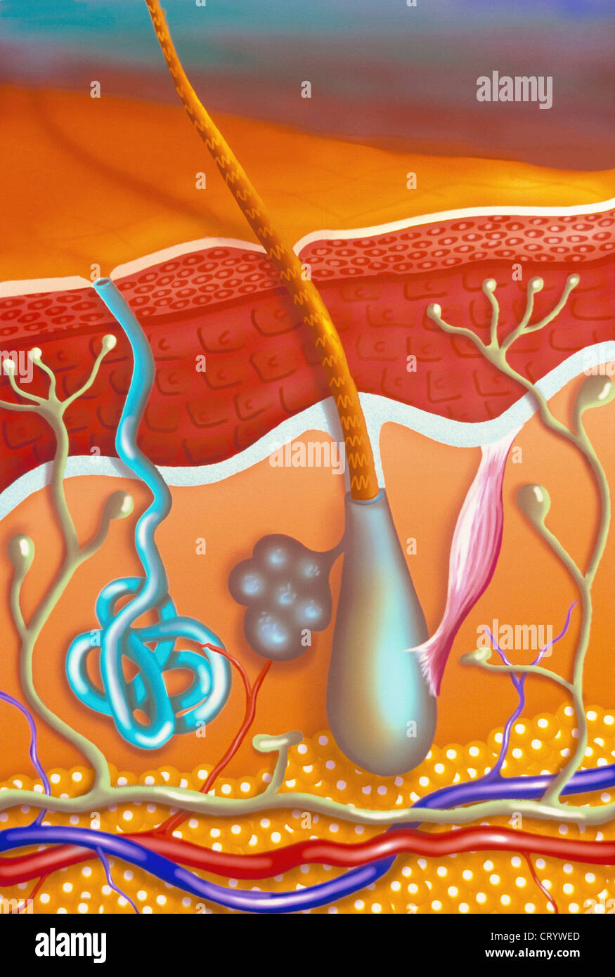
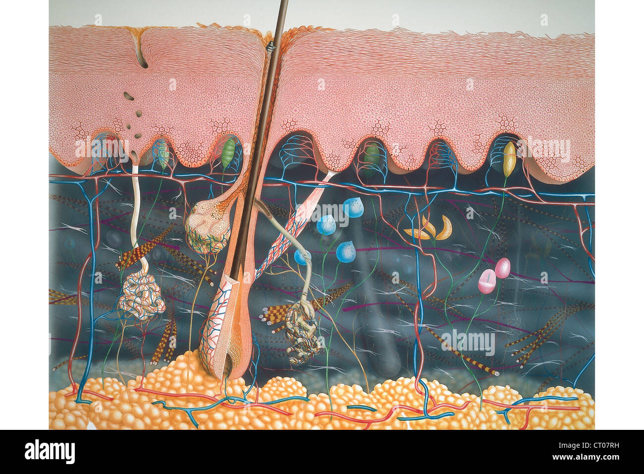 Skin, illustration Stock Photohttps://www.alamy.com/image-license-details/?v=1https://www.alamy.com/stock-photo-skin-illustration-49178629.html
RMCT07RH–Skin, illustration
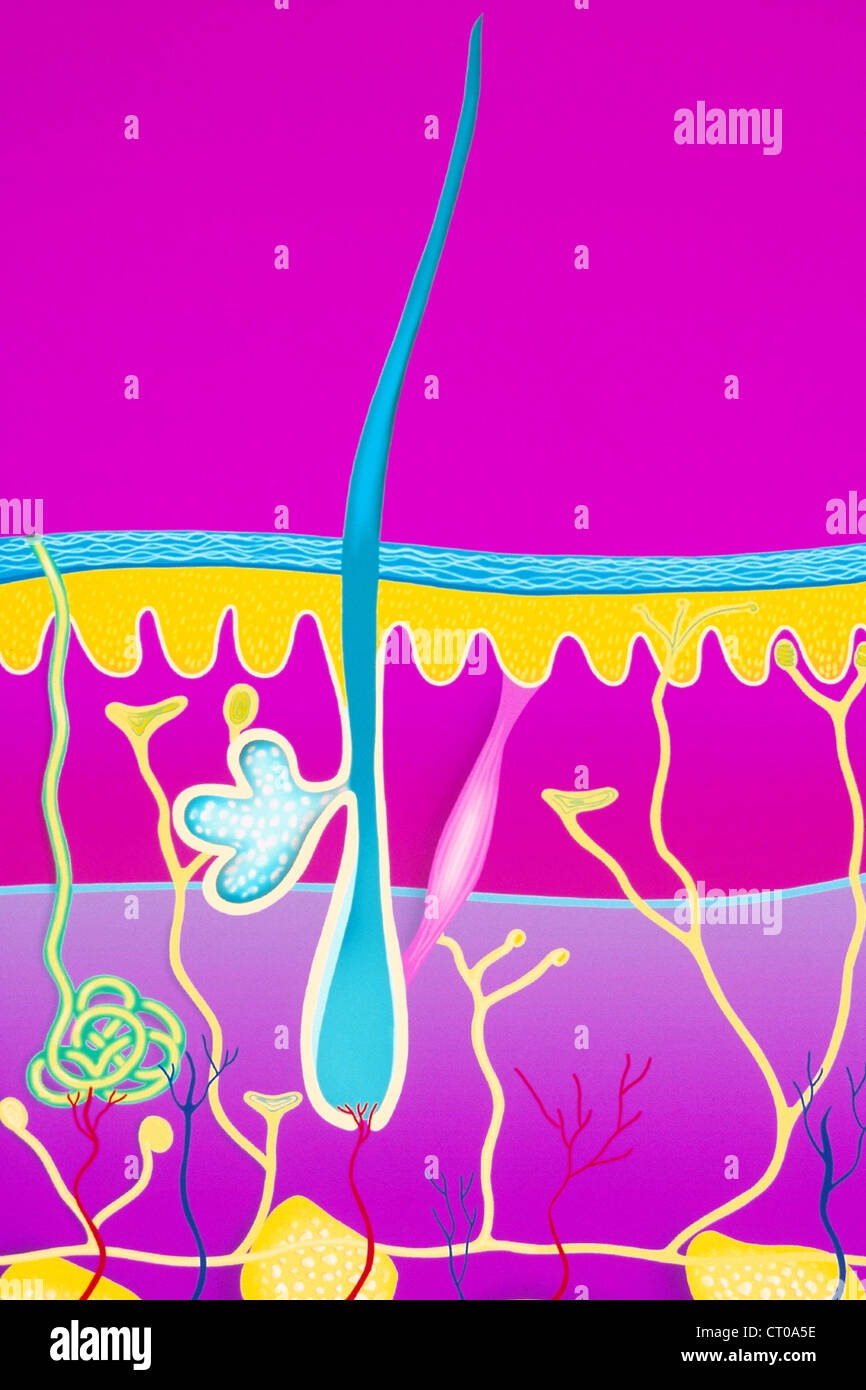
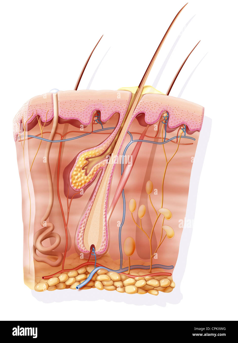 SKIN, ILLUSTRATION Stock Photohttps://www.alamy.com/image-license-details/?v=1https://www.alamy.com/stock-photo-skin-illustration-48381356.html
RMCPKXWG–SKIN, ILLUSTRATION
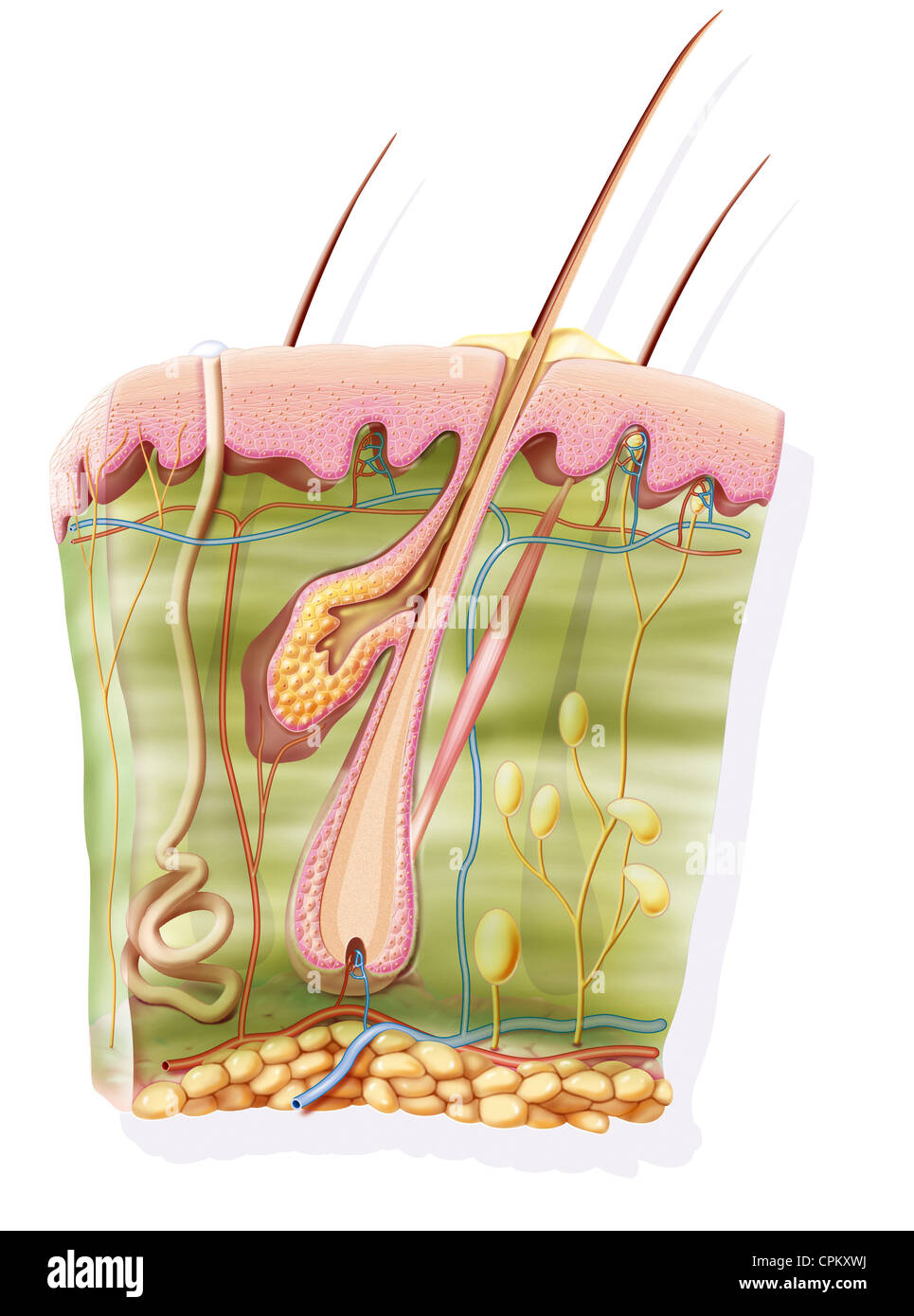 SKIN, ILLUSTRATION Stock Photohttps://www.alamy.com/image-license-details/?v=1https://www.alamy.com/stock-photo-skin-illustration-48381358.html
RMCPKXWJ–SKIN, ILLUSTRATION
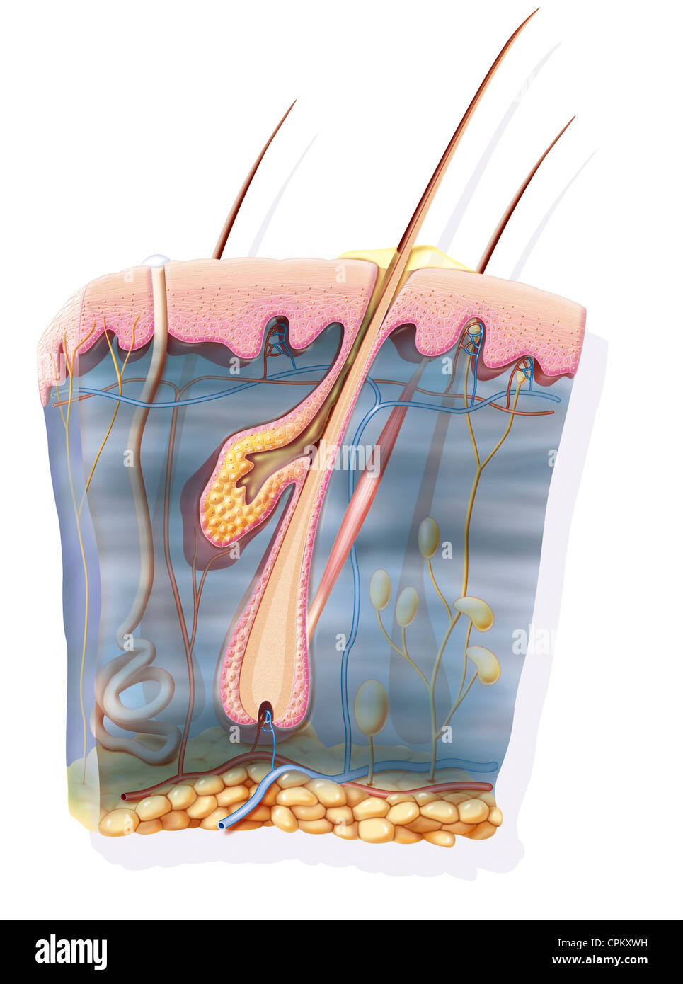 SKIN, ILLUSTRATION Stock Photohttps://www.alamy.com/image-license-details/?v=1https://www.alamy.com/stock-photo-skin-illustration-48381357.html
RMCPKXWH–SKIN, ILLUSTRATION
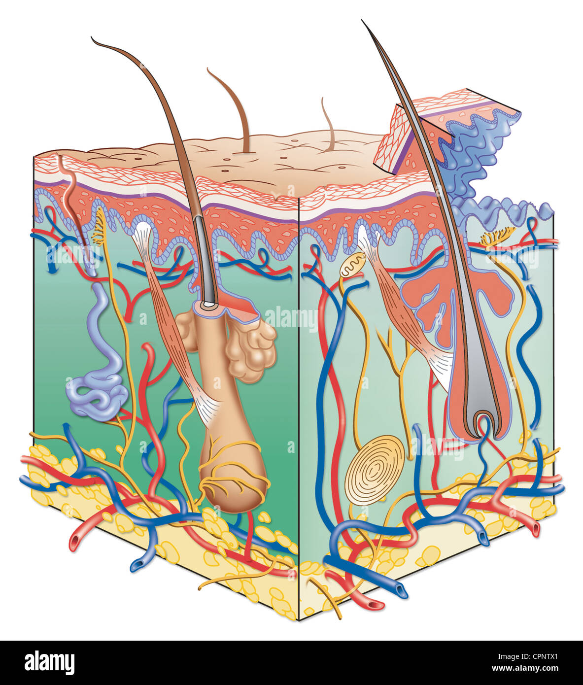 SKIN, ILLUSTRATION Stock Photohttps://www.alamy.com/image-license-details/?v=1https://www.alamy.com/stock-photo-skin-illustration-48423705.html
RMCPNTX1–SKIN, ILLUSTRATION
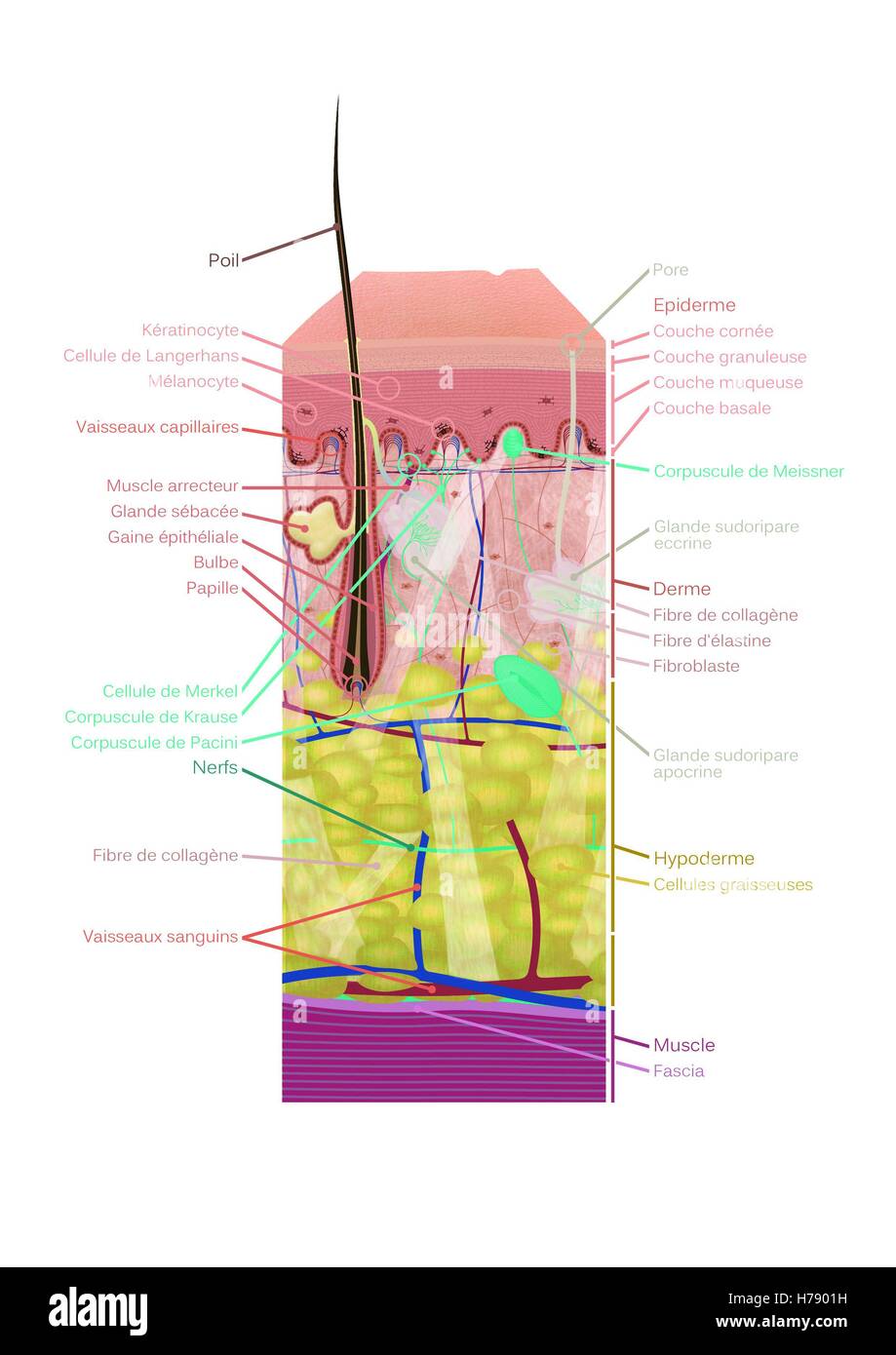 SKIN, ILLUSTRATION Stock Photohttps://www.alamy.com/image-license-details/?v=1https://www.alamy.com/stock-photo-skin-illustration-124972781.html
RMH7901H–SKIN, ILLUSTRATION
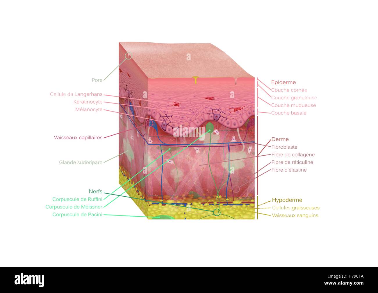 SKIN, ILLUSTRATION Stock Photohttps://www.alamy.com/image-license-details/?v=1https://www.alamy.com/stock-photo-skin-illustration-124972774.html
RMH7901A–SKIN, ILLUSTRATION
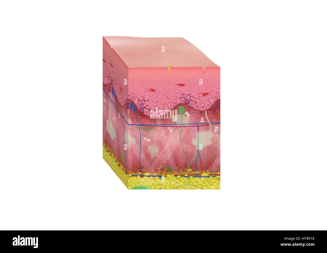 SKIN, ILLUSTRATION Stock Photohttps://www.alamy.com/image-license-details/?v=1https://www.alamy.com/stock-photo-skin-illustration-124972772.html
RMH79018–SKIN, ILLUSTRATION
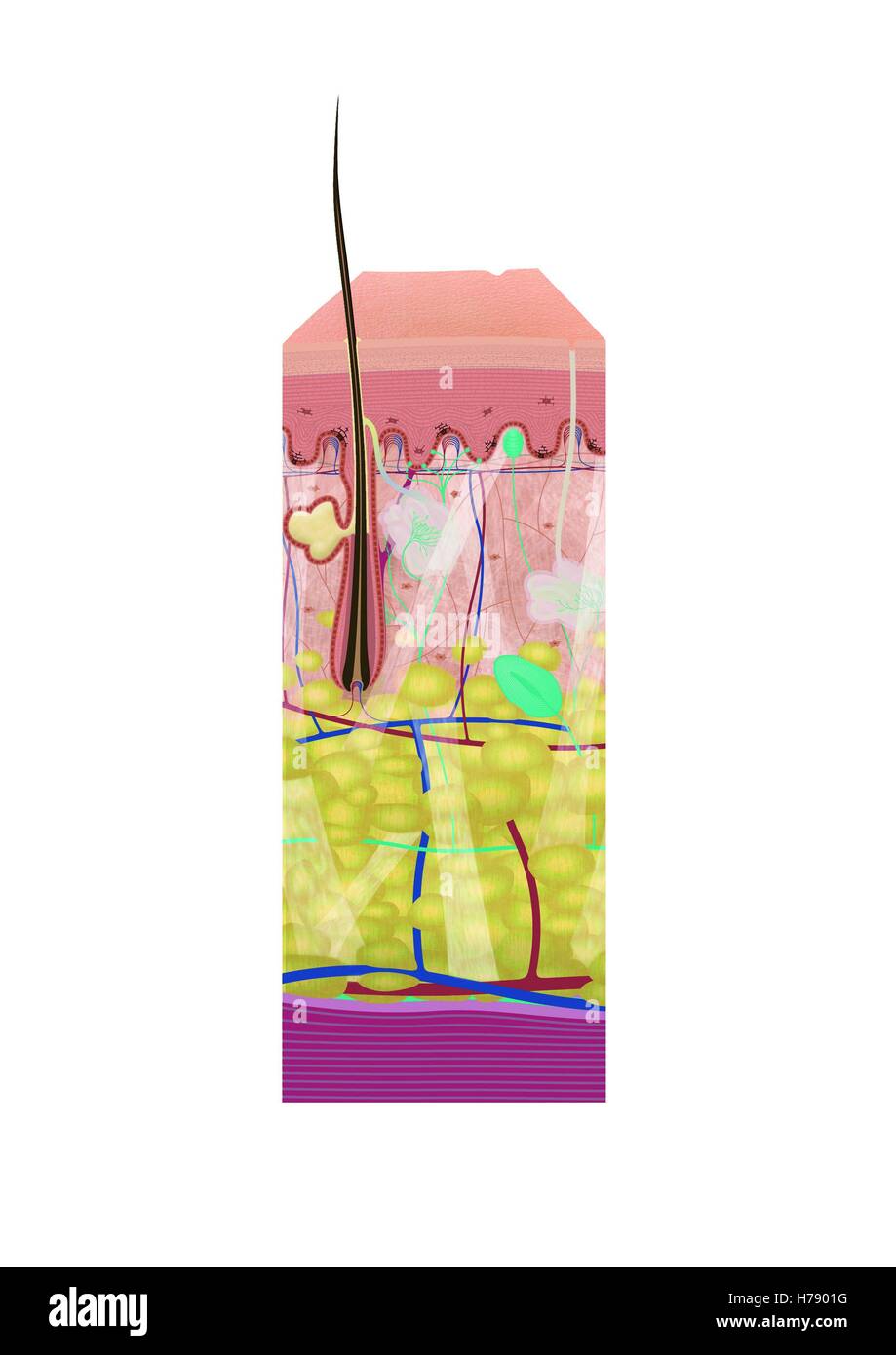 SKIN, ILLUSTRATION Stock Photohttps://www.alamy.com/image-license-details/?v=1https://www.alamy.com/stock-photo-skin-illustration-124972780.html
RMH7901G–SKIN, ILLUSTRATION
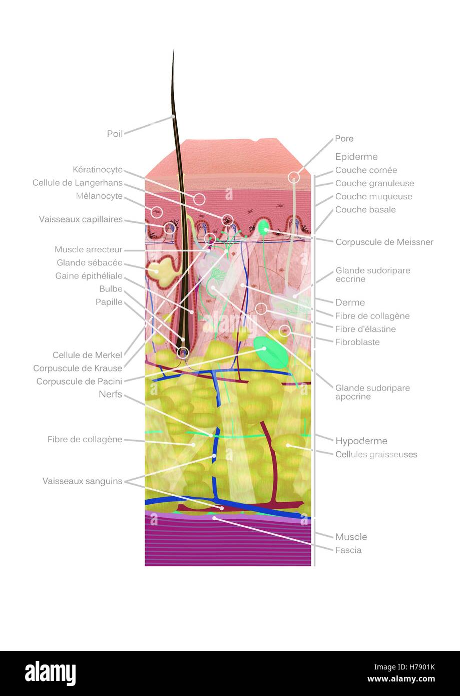 SKIN, ILLUSTRATION Stock Photohttps://www.alamy.com/image-license-details/?v=1https://www.alamy.com/stock-photo-skin-illustration-124972783.html
RMH7901K–SKIN, ILLUSTRATION
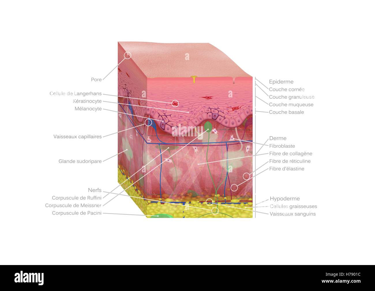 SKIN, ILLUSTRATION Stock Photohttps://www.alamy.com/image-license-details/?v=1https://www.alamy.com/stock-photo-skin-illustration-124972776.html
RMH7901C–SKIN, ILLUSTRATION
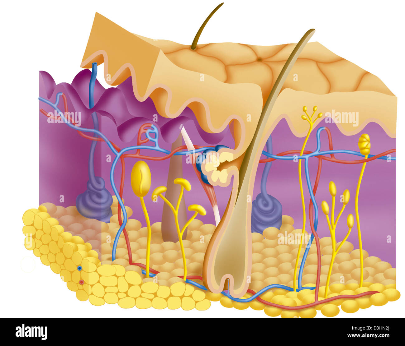 SKIN, ILLUSTRATION Stock Photohttps://www.alamy.com/image-license-details/?v=1https://www.alamy.com/stock-photo-skin-illustration-53864794.html
RMD3HN2J–SKIN, ILLUSTRATION
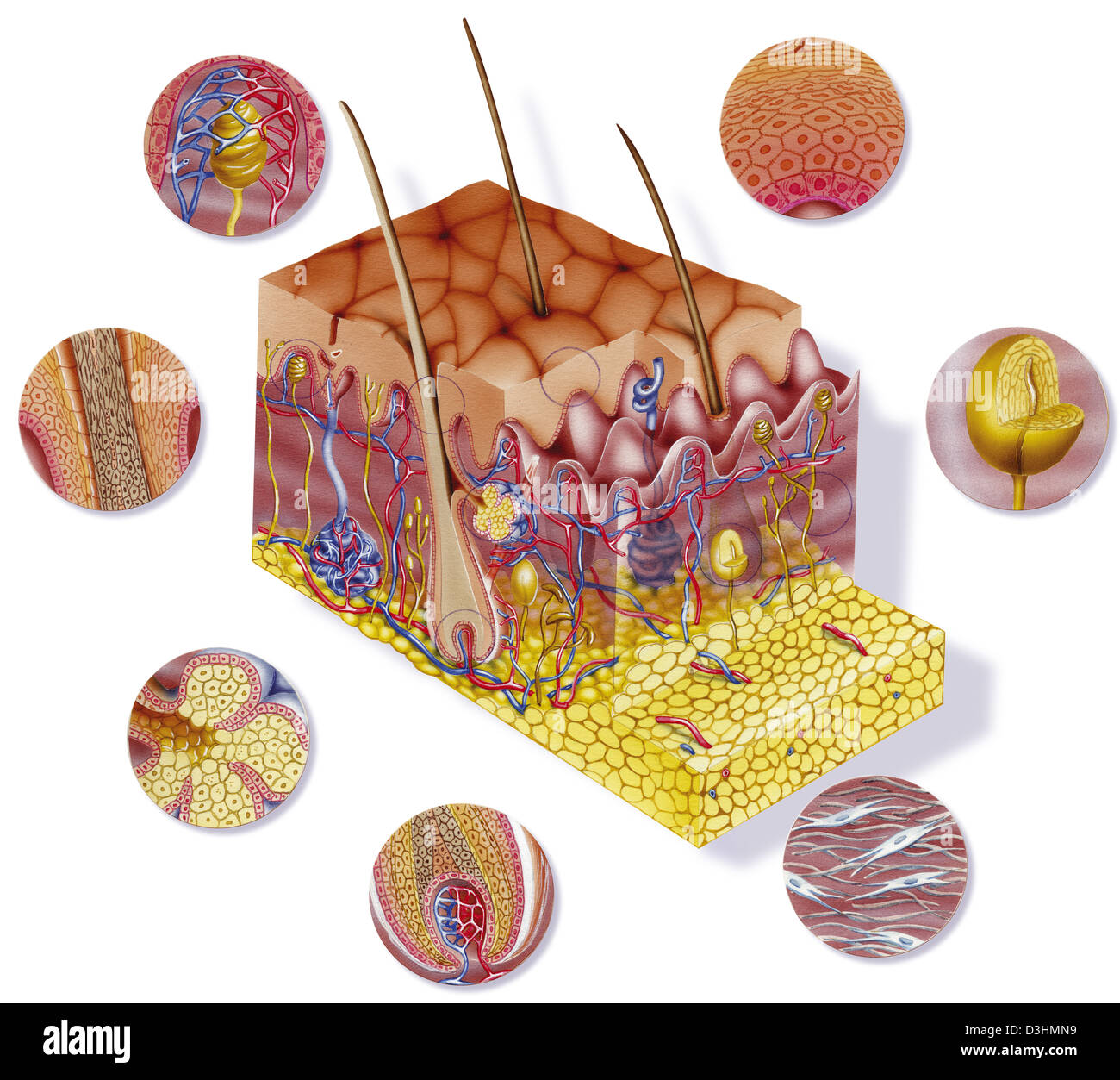 SKIN, ILLUSTRATION Stock Photohttps://www.alamy.com/image-license-details/?v=1https://www.alamy.com/stock-photo-skin-illustration-53864533.html
RMD3HMN9–SKIN, ILLUSTRATION
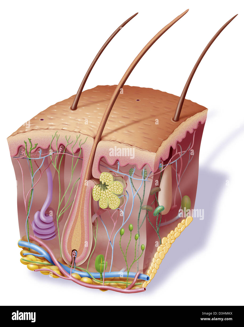 SKIN, ILLUSTRATION Stock Photohttps://www.alamy.com/image-license-details/?v=1https://www.alamy.com/stock-photo-skin-illustration-53864494.html
RMD3HMKX–SKIN, ILLUSTRATION
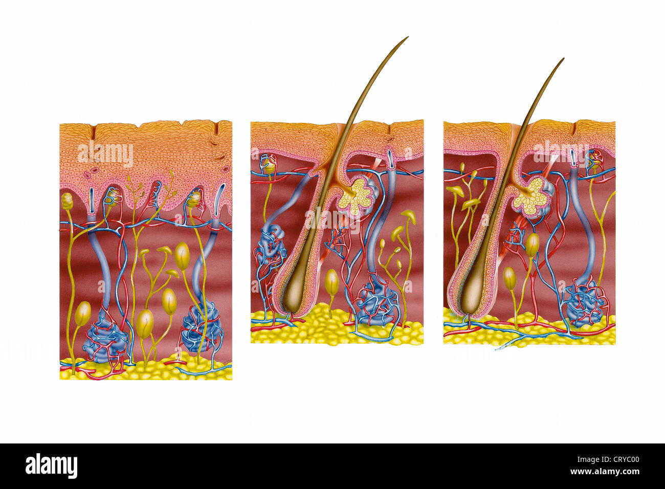 SKIN, ILLUSTRATION Stock Photohttps://www.alamy.com/image-license-details/?v=1https://www.alamy.com/stock-photo-skin-illustration-49159936.html
RMCRYC00–SKIN, ILLUSTRATION
