Pancreas cells Black & White Stock Photos
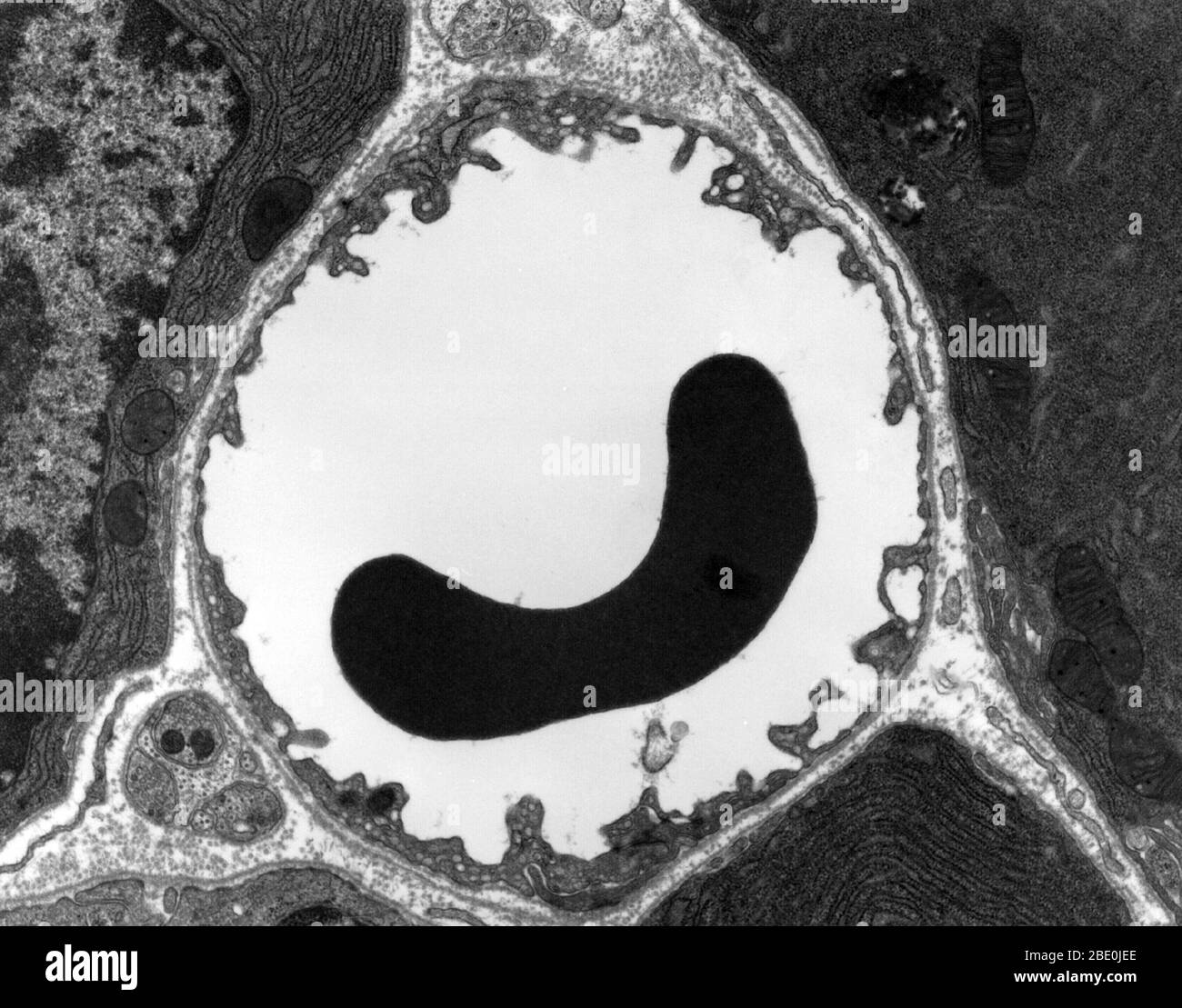 Transmission electron micrograph (TEM) of a thin section cut through the pancreas (mammalian). This image shows a capillary within the pancreatic tissue (acinar cells in this image). Note the abundance of rough endoplasmic reticulum in the acinar cells. There is a red blood cell within the capillary. The capillary lining consists of long, thin endothelial cells, connected by tight junctions. The image shows fenestration of these endothelial cells. The image also shows synaptic vesicles in the neuron (nerve cell) next to the capillary. Stock Photohttps://www.alamy.com/image-license-details/?v=1https://www.alamy.com/transmission-electron-micrograph-tem-of-a-thin-section-cut-through-the-pancreas-mammalian-this-image-shows-a-capillary-within-the-pancreatic-tissue-acinar-cells-in-this-image-note-the-abundance-of-rough-endoplasmic-reticulum-in-the-acinar-cells-there-is-a-red-blood-cell-within-the-capillary-the-capillary-lining-consists-of-long-thin-endothelial-cells-connected-by-tight-junctions-the-image-shows-fenestration-of-these-endothelial-cells-the-image-also-shows-synaptic-vesicles-in-the-neuron-nerve-cell-next-to-the-capillary-image352827062.html
RM2BE0JEE–Transmission electron micrograph (TEM) of a thin section cut through the pancreas (mammalian). This image shows a capillary within the pancreatic tissue (acinar cells in this image). Note the abundance of rough endoplasmic reticulum in the acinar cells. There is a red blood cell within the capillary. The capillary lining consists of long, thin endothelial cells, connected by tight junctions. The image shows fenestration of these endothelial cells. The image also shows synaptic vesicles in the neuron (nerve cell) next to the capillary.
 Type 1 diabetes, which is often diagnosed in children or young adults, is treated with insulin, and monitored by the testing of blood-sugar levels. The condition is caused by an abnormal immune reaction that destroys the insulin-producing cells of the pancreas, and treated with insulin injections, or consuming sugar when the body is suffering from low blood sugar levels. On 30 November 2019 in Los Angeles, CA, USA. (Photo by John Fredricks/NurPhoto) Stock Photohttps://www.alamy.com/image-license-details/?v=1https://www.alamy.com/type-1-diabetes-which-is-often-diagnosed-in-children-or-young-adults-is-treated-with-insulin-and-monitored-by-the-testing-of-blood-sugar-levels-the-condition-is-caused-by-an-abnormal-immune-reaction-that-destroys-the-insulin-producing-cells-of-the-pancreas-and-treated-with-insulin-injections-or-consuming-sugar-when-the-body-is-suffering-from-low-blood-sugar-levels-on-30-november-2019-in-los-angeles-ca-usa-photo-by-john-fredricksnurphoto-image488916708.html
RM2KBC270–Type 1 diabetes, which is often diagnosed in children or young adults, is treated with insulin, and monitored by the testing of blood-sugar levels. The condition is caused by an abnormal immune reaction that destroys the insulin-producing cells of the pancreas, and treated with insulin injections, or consuming sugar when the body is suffering from low blood sugar levels. On 30 November 2019 in Los Angeles, CA, USA. (Photo by John Fredricks/NurPhoto)
RF2C6T7P2–human organs line icons. linear set. quality vector line set such as skin, dna, reproductive, uterus, brain, reproductive, blood cells, pancreas, lung
 Insulin peptide hormone, 3D rendering. Important drug in treatme Stock Photohttps://www.alamy.com/image-license-details/?v=1https://www.alamy.com/insulin-peptide-hormone-3d-rendering-important-drug-in-treatme-image437698549.html
RF2GC2TY1–Insulin peptide hormone, 3D rendering. Important drug in treatme
RF2BH6CDE–Blood vessels of pancreas line icon. Pancreas blood supply, innervation symbol
 . Senescence and rejuvenescence. Age; Reproduction. igo SENESCENCE AND REJUVENESCENCE This cycle of changes, which may occur within a few hours and which may be repeated in a single cell, is, I believe, not funda- mentally different from the age cycle in organisms. All the essen- tial features of both senescence and rejuvenescence up to a certain. Figs. 66, 67.—Pancreas cells of toad: Fig. 66, fully loaded and almost quiescent; Fig. 67, partially discharged after stimulation. From preparations loaned by R. R. Bensley. point are present. The cell probably does not return to the em- bryonic cond Stock Photohttps://www.alamy.com/image-license-details/?v=1https://www.alamy.com/senescence-and-rejuvenescence-age-reproduction-igo-senescence-and-rejuvenescence-this-cycle-of-changes-which-may-occur-within-a-few-hours-and-which-may-be-repeated-in-a-single-cell-is-i-believe-not-funda-mentally-different-from-the-age-cycle-in-organisms-all-the-essen-tial-features-of-both-senescence-and-rejuvenescence-up-to-a-certain-figs-66-67pancreas-cells-of-toad-fig-66-fully-loaded-and-almost-quiescent-fig-67-partially-discharged-after-stimulation-from-preparations-loaned-by-r-r-bensley-point-are-present-the-cell-probably-does-not-return-to-the-em-bryonic-cond-image232349500.html
RMRE0C4C–. Senescence and rejuvenescence. Age; Reproduction. igo SENESCENCE AND REJUVENESCENCE This cycle of changes, which may occur within a few hours and which may be repeated in a single cell, is, I believe, not funda- mentally different from the age cycle in organisms. All the essen- tial features of both senescence and rejuvenescence up to a certain. Figs. 66, 67.—Pancreas cells of toad: Fig. 66, fully loaded and almost quiescent; Fig. 67, partially discharged after stimulation. From preparations loaned by R. R. Bensley. point are present. The cell probably does not return to the em- bryonic cond
RFMTA76H–Set Of 16 simple editable icons such as Men Hair, Skin Cells, Bones Joint, Human Skin, Stomach with Liquids, Knee, Teeth, Pancreas, Lymphonodus can be
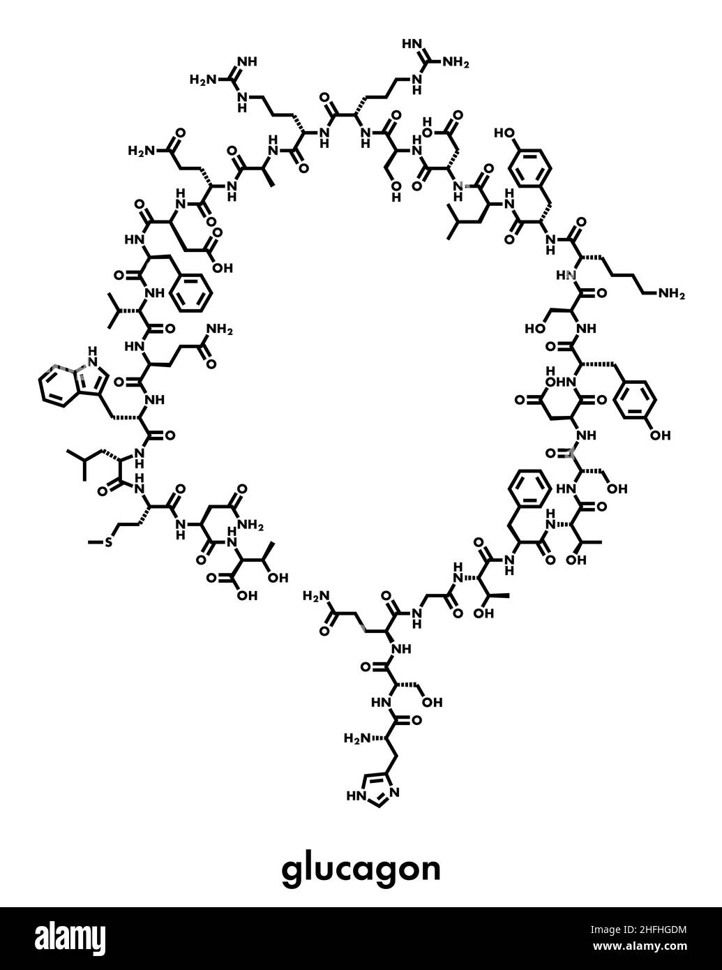 Glucagon hypoglycemia drug molecule. Skeletal formula. Stock Vectorhttps://www.alamy.com/image-license-details/?v=1https://www.alamy.com/glucagon-hypoglycemia-drug-molecule-skeletal-formula-image457075520.html
RF2HFHGDM–Glucagon hypoglycemia drug molecule. Skeletal formula.
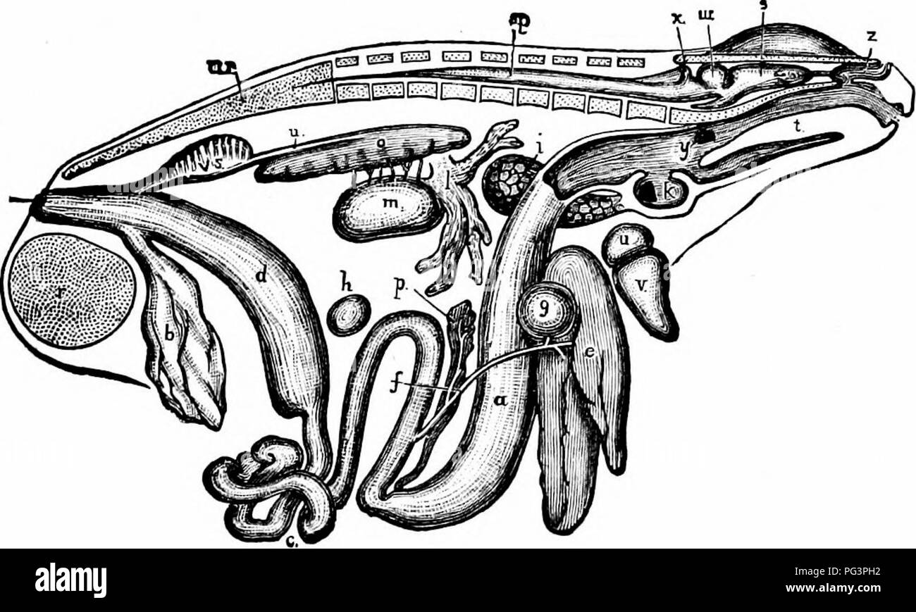 . A manual of zoology. THE METAZOA 60 ment. These digestive secretions are partly produced by the cells of the epithelium of the canal, which are modified to form unicellular or multicellular glands (p. 65), partly by certain large special digestive glands, salivary glands, liver, and pancreas. The nutrient parts of the food are by this means so acted upon that they are ready to be absorbed, and in most animals pass into the blood, to be distributed. Fig. 31. — General view of the viscera of a male frog, from the right side, a, stomach; fi, urinary bladder; c, small intestine; c/, cloacal aper Stock Photohttps://www.alamy.com/image-license-details/?v=1https://www.alamy.com/a-manual-of-zoology-the-metazoa-60-ment-these-digestive-secretions-are-partly-produced-by-the-cells-of-the-epithelium-of-the-canal-which-are-modified-to-form-unicellular-or-multicellular-glands-p-65-partly-by-certain-large-special-digestive-glands-salivary-glands-liver-and-pancreas-the-nutrient-parts-of-the-food-are-by-this-means-so-acted-upon-that-they-are-ready-to-be-absorbed-and-in-most-animals-pass-into-the-blood-to-be-distributed-fig-31-general-view-of-the-viscera-of-a-male-frog-from-the-right-side-a-stomach-fi-urinary-bladder-c-small-intestine-c-cloacal-aper-image216442494.html
RMPG3PH2–. A manual of zoology. THE METAZOA 60 ment. These digestive secretions are partly produced by the cells of the epithelium of the canal, which are modified to form unicellular or multicellular glands (p. 65), partly by certain large special digestive glands, salivary glands, liver, and pancreas. The nutrient parts of the food are by this means so acted upon that they are ready to be absorbed, and in most animals pass into the blood, to be distributed. Fig. 31. — General view of the viscera of a male frog, from the right side, a, stomach; fi, urinary bladder; c, small intestine; c/, cloacal aper
RF2WA6KHT–Pancreatic cell color line icon. Microorganisms microbes, bacteria. Vector isolated element. Editable stroke.
RF2C6TTHE–set of 12 thin outline icons such as ovary, colon, dna, bronchus, blood cells, testicle for web, mobile
 Insulin peptide hormone, 3D rendering. Important drug in treatme Stock Photohttps://www.alamy.com/image-license-details/?v=1https://www.alamy.com/insulin-peptide-hormone-3d-rendering-important-drug-in-treatme-image438408197.html
RF2GD763H–Insulin peptide hormone, 3D rendering. Important drug in treatme
 The development of the human body; a manual of human embryology . ions ofthe liver tissue, the cells disappearing completely, whilethe ducts and blood-vessels originally present persist, theformer constituting the vasa aberranlia of adult anatomy.These are usually espe-cially noticeable at theleft edge of the liver, be-tween the folds of theleft lateral ligament, butthey may also be foundalong the line of the venacava, around the gall-bladder, and in the re-gion of the left longitudi-nal fissure. The Development ofthe Pancreas.—The pan-creas arises a little laterthan the liver, as threeseparat Stock Photohttps://www.alamy.com/image-license-details/?v=1https://www.alamy.com/the-development-of-the-human-body-a-manual-of-human-embryology-ions-ofthe-liver-tissue-the-cells-disappearing-completely-whilethe-ducts-and-blood-vessels-originally-present-persist-theformer-constituting-the-vasa-aberranlia-of-adult-anatomythese-are-usually-espe-cially-noticeable-at-theleft-edge-of-the-liver-be-tween-the-folds-of-theleft-lateral-ligament-butthey-may-also-be-foundalong-the-line-of-the-venacava-around-the-gall-bladder-and-in-the-re-gion-of-the-left-longitudi-nal-fissure-the-development-ofthe-pancreasthe-pan-creas-arises-a-little-laterthan-the-liver-as-threeseparat-image342663834.html
RM2AWDK62–The development of the human body; a manual of human embryology . ions ofthe liver tissue, the cells disappearing completely, whilethe ducts and blood-vessels originally present persist, theformer constituting the vasa aberranlia of adult anatomy.These are usually espe-cially noticeable at theleft edge of the liver, be-tween the folds of theleft lateral ligament, butthey may also be foundalong the line of the venacava, around the gall-bladder, and in the re-gion of the left longitudi-nal fissure. The Development ofthe Pancreas.—The pan-creas arises a little laterthan the liver, as threeseparat
RFMT36BR–Set Of 13 simple editable icons such as White blood cell, Men Leg, Tonsil, Pancreas, Mucous Membrane, Skin Cells, Dishes Stack, Human Brain, Foot Bone
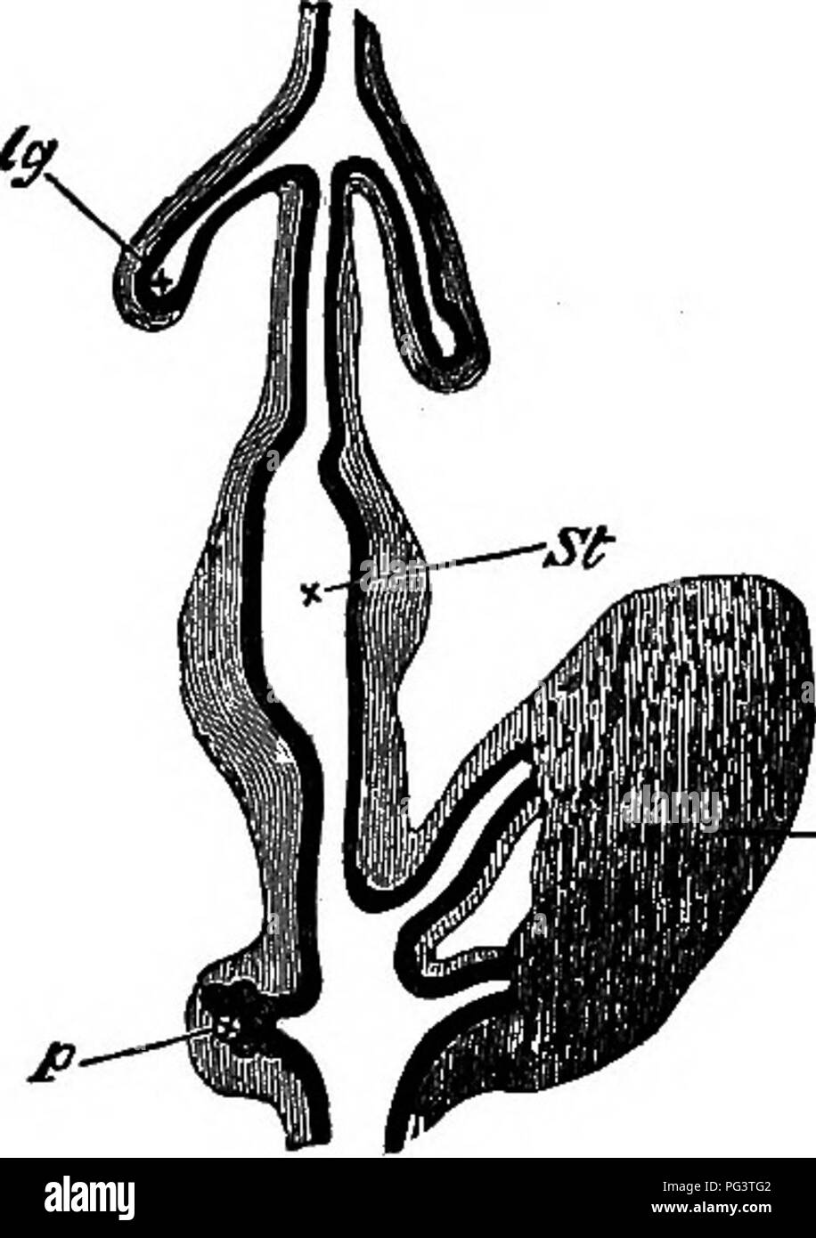 . The elements of embryology . Embryology. 174 THE THIRD DAT. Fig. 60. [chap.. DliGKAM OF A POKTION OF THE DlSBSTIVB TeACT OF A Chick upon the Fourth Day. (Copied from Gotte.) The black inner line represents the hypoblast, the outer shading the mesoblast. Ig. lung-diverticulum with expanded termi- nation, forming the primary lung-vesicle. St. stomach. I. two hepatic diverticula with their terminations united by cords of hypoblast cells, p. diverticulum of the pancreas with the vesicular diverticula coming from it. the duodenum by the fact that from it, as we shall presently point out, the rudi Stock Photohttps://www.alamy.com/image-license-details/?v=1https://www.alamy.com/the-elements-of-embryology-embryology-174-the-third-dat-fig-60-chap-dligkam-of-a-poktion-of-the-dlsbstivb-teact-of-a-chick-upon-the-fourth-day-copied-from-gotte-the-black-inner-line-represents-the-hypoblast-the-outer-shading-the-mesoblast-ig-lung-diverticulum-with-expanded-termi-nation-forming-the-primary-lung-vesicle-st-stomach-i-two-hepatic-diverticula-with-their-terminations-united-by-cords-of-hypoblast-cells-p-diverticulum-of-the-pancreas-with-the-vesicular-diverticula-coming-from-it-the-duodenum-by-the-fact-that-from-it-as-we-shall-presently-point-out-the-rudi-image216444034.html
RMPG3TG2–. The elements of embryology . Embryology. 174 THE THIRD DAT. Fig. 60. [chap.. DliGKAM OF A POKTION OF THE DlSBSTIVB TeACT OF A Chick upon the Fourth Day. (Copied from Gotte.) The black inner line represents the hypoblast, the outer shading the mesoblast. Ig. lung-diverticulum with expanded termi- nation, forming the primary lung-vesicle. St. stomach. I. two hepatic diverticula with their terminations united by cords of hypoblast cells, p. diverticulum of the pancreas with the vesicular diverticula coming from it. the duodenum by the fact that from it, as we shall presently point out, the rudi
 . The physiology of the domestic animals; a text-book for veterinary and medical students and practitioners. Physiology, Comparative; Domestic animals. 414 PHYSIOLOGY OF THE DOMESTIC ANIMALS. stomach and rapidly reaches its maximum two or three hours thereafter. Toward the fifth or seventh hour it decreases in activity, and about twelve hours after feeding again becomes increased in amount, the second increase being apparently coincident with the escape of the acid chyme into the Small intestine. The cells of the pancreas also undergo marked histological changes during the period of active sec Stock Photohttps://www.alamy.com/image-license-details/?v=1https://www.alamy.com/the-physiology-of-the-domestic-animals-a-text-book-for-veterinary-and-medical-students-and-practitioners-physiology-comparative-domestic-animals-414-physiology-of-the-domestic-animals-stomach-and-rapidly-reaches-its-maximum-two-or-three-hours-thereafter-toward-the-fifth-or-seventh-hour-it-decreases-in-activity-and-about-twelve-hours-after-feeding-again-becomes-increased-in-amount-the-second-increase-being-apparently-coincident-with-the-escape-of-the-acid-chyme-into-the-small-intestine-the-cells-of-the-pancreas-also-undergo-marked-histological-changes-during-the-period-of-active-sec-image232343026.html
RMRE03W6–. The physiology of the domestic animals; a text-book for veterinary and medical students and practitioners. Physiology, Comparative; Domestic animals. 414 PHYSIOLOGY OF THE DOMESTIC ANIMALS. stomach and rapidly reaches its maximum two or three hours thereafter. Toward the fifth or seventh hour it decreases in activity, and about twelve hours after feeding again becomes increased in amount, the second increase being apparently coincident with the escape of the acid chyme into the Small intestine. The cells of the pancreas also undergo marked histological changes during the period of active sec
RFMTA6JD–Set Of 16 simple editable icons such as Human Skin, Men Hair, Blood Transfusion, Liver, Platelets, Nostril, Ankle, Pancreas, Teeth can be used for mob
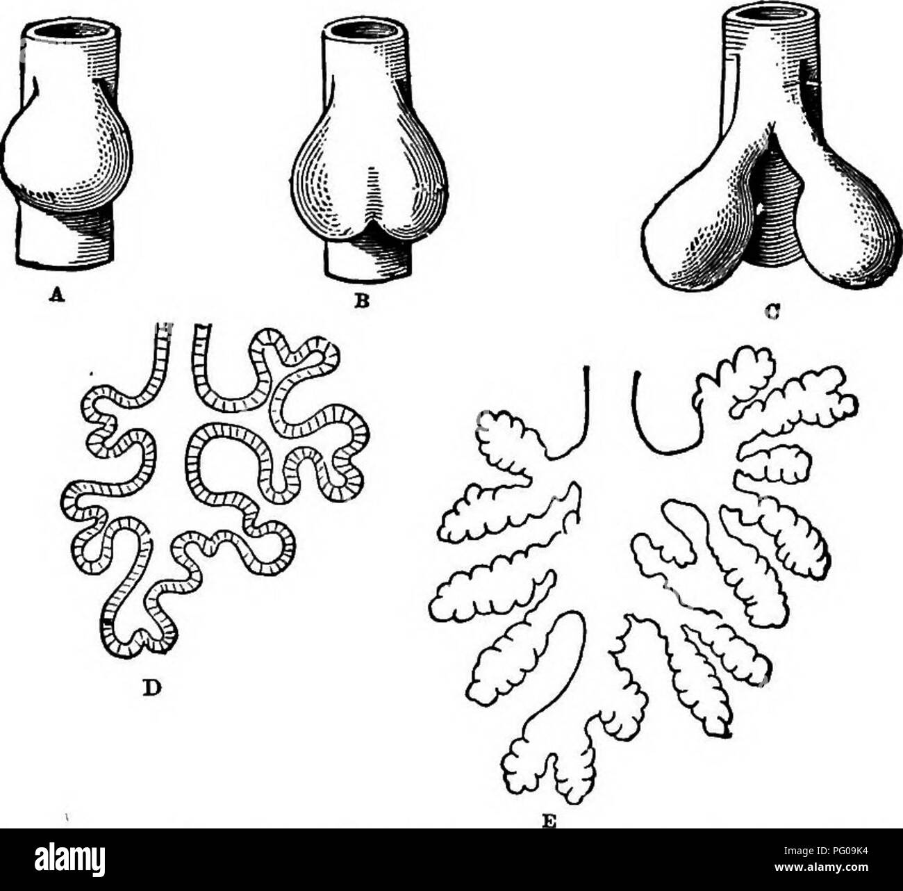 . Animal activities; a first book in zoo?logy. Zoology; Animal behavior. 248 yINlM/IL /ICTIVITIES. cases, the difference being only in the amount of fold- ing. The liver and pancreas of vertebrates arise in the same way, by folding of the walls of the food-tube. Other changes take place in as simple a way as this. Indeed, nearly all the organs of a complex body arise by foldings and pushings of layers of cells. When the egg of a very simple animal, like the hydra, develops, it first divides into a number of cells forming a spherical. Fig. 202.—Growth of Frog's Lung from Primitive Food-tube. bo Stock Photohttps://www.alamy.com/image-license-details/?v=1https://www.alamy.com/animal-activities-a-first-book-in-zoology-zoology-animal-behavior-248-yinlmil-ictivities-cases-the-difference-being-only-in-the-amount-of-fold-ing-the-liver-and-pancreas-of-vertebrates-arise-in-the-same-way-by-folding-of-the-walls-of-the-food-tube-other-changes-take-place-in-as-simple-a-way-as-this-indeed-nearly-all-the-organs-of-a-complex-body-arise-by-foldings-and-pushings-of-layers-of-cells-when-the-egg-of-a-very-simple-animal-like-the-hydra-develops-it-first-divides-into-a-number-of-cells-forming-a-spherical-fig-202growth-of-frogs-lung-from-primitive-food-tube-bo-image216366504.html
RMPG09K4–. Animal activities; a first book in zoo?logy. Zoology; Animal behavior. 248 yINlM/IL /ICTIVITIES. cases, the difference being only in the amount of fold- ing. The liver and pancreas of vertebrates arise in the same way, by folding of the walls of the food-tube. Other changes take place in as simple a way as this. Indeed, nearly all the organs of a complex body arise by foldings and pushings of layers of cells. When the egg of a very simple animal, like the hydra, develops, it first divides into a number of cells forming a spherical. Fig. 202.—Growth of Frog's Lung from Primitive Food-tube. bo
 . A manual of zoology. THE METAZOA 60 ment. These digestive secretions are partly produced by the cells of the epithelium of the canal, which are modified to form unicellular or multicellular glands (p. 65), partly by certain large special digestive glands, salivary glands, liver, and pancreas. The nutrient parts of the food are by this means so acted upon that they are ready to be absorbed, and in most animals pass into the blood, to be distributed. Fig. 31. — General view of the viscera of a male frog, from the right side, a, stomach; fi, urinary bladder; c, small intestine; c/, cloacal aper Stock Photohttps://www.alamy.com/image-license-details/?v=1https://www.alamy.com/a-manual-of-zoology-the-metazoa-60-ment-these-digestive-secretions-are-partly-produced-by-the-cells-of-the-epithelium-of-the-canal-which-are-modified-to-form-unicellular-or-multicellular-glands-p-65-partly-by-certain-large-special-digestive-glands-salivary-glands-liver-and-pancreas-the-nutrient-parts-of-the-food-are-by-this-means-so-acted-upon-that-they-are-ready-to-be-absorbed-and-in-most-animals-pass-into-the-blood-to-be-distributed-fig-31-general-view-of-the-viscera-of-a-male-frog-from-the-right-side-a-stomach-fi-urinary-bladder-c-small-intestine-c-cloacal-aper-image232109113.html
RMRDHDF5–. A manual of zoology. THE METAZOA 60 ment. These digestive secretions are partly produced by the cells of the epithelium of the canal, which are modified to form unicellular or multicellular glands (p. 65), partly by certain large special digestive glands, salivary glands, liver, and pancreas. The nutrient parts of the food are by this means so acted upon that they are ready to be absorbed, and in most animals pass into the blood, to be distributed. Fig. 31. — General view of the viscera of a male frog, from the right side, a, stomach; fi, urinary bladder; c, small intestine; c/, cloacal aper
RFMTA84X–Set Of 16 simple editable icons such as Human Spine, Mucous Membrane, White blood cell, Basophil, Skin, Pancreas, Men Back, DNA Sequence, Shoulder can
 . The anatomy of the domestic fowl . Domestic animals; Veterinary medicine; Poultry. Pig. 38.—Cellular structure of liT^er, pancreas, and trachea. A. Liver. 1, Liver cells. 2, Sinusoid. 4, Nucleus of cells. B. Pancreas, i. Island of Langerhans. 2, Alveolar cell.s. 3, Duct. C. Trachea, i. Ciliated epithelia. 2, Glands. 3, Hyaline cartilage. D. Section of wall of ovum at Fig. S7 letter d. i. Yolk. 2, Granular mem- brane. 3, Theca. 4, Blood-vessel. A microscopic study of the Uver of the fowl shows a compact mass of Uver ceUs polyhedral in shape, with large nuclei (Fig. 38, A). The Uver tissue dif Stock Photohttps://www.alamy.com/image-license-details/?v=1https://www.alamy.com/the-anatomy-of-the-domestic-fowl-domestic-animals-veterinary-medicine-poultry-pig-38cellular-structure-of-liter-pancreas-and-trachea-a-liver-1-liver-cells-2-sinusoid-4-nucleus-of-cells-b-pancreas-i-island-of-langerhans-2-alveolar-cells-3-duct-c-trachea-i-ciliated-epithelia-2-glands-3-hyaline-cartilage-d-section-of-wall-of-ovum-at-fig-s7-letter-d-i-yolk-2-granular-mem-brane-3-theca-4-blood-vessel-a-microscopic-study-of-the-uver-of-the-fowl-shows-a-compact-mass-of-uver-ceus-polyhedral-in-shape-with-large-nuclei-fig-38-a-the-uver-tissue-dif-image216389214.html
RMPG1AJ6–. The anatomy of the domestic fowl . Domestic animals; Veterinary medicine; Poultry. Pig. 38.—Cellular structure of liT^er, pancreas, and trachea. A. Liver. 1, Liver cells. 2, Sinusoid. 4, Nucleus of cells. B. Pancreas, i. Island of Langerhans. 2, Alveolar cell.s. 3, Duct. C. Trachea, i. Ciliated epithelia. 2, Glands. 3, Hyaline cartilage. D. Section of wall of ovum at Fig. S7 letter d. i. Yolk. 2, Granular mem- brane. 3, Theca. 4, Blood-vessel. A microscopic study of the Uver of the fowl shows a compact mass of Uver ceUs polyhedral in shape, with large nuclei (Fig. 38, A). The Uver tissue dif
 . A manual of elementary zoology . Zoology. THE FROG: HISTOLOGY, GERM CELLS, DEATH 91 in the cell when they are discharged. Such a gland is known as a tubular gland. The pancreas is an example of the more complicated class known as racemose glands, in which the tubes are branched and lined with low, cubical epithelium up to their ends, which are dilated and lined with glandular epithelium. The dilations are known as acini and the tubes leading to them as ducts. The liver is more complicated still, the tubes not only branchingbut rejoin- ing to form a mesh- work, whose walls consist of gland ce Stock Photohttps://www.alamy.com/image-license-details/?v=1https://www.alamy.com/a-manual-of-elementary-zoology-zoology-the-frog-histology-germ-cells-death-91-in-the-cell-when-they-are-discharged-such-a-gland-is-known-as-a-tubular-gland-the-pancreas-is-an-example-of-the-more-complicated-class-known-as-racemose-glands-in-which-the-tubes-are-branched-and-lined-with-low-cubical-epithelium-up-to-their-ends-which-are-dilated-and-lined-with-glandular-epithelium-the-dilations-are-known-as-acini-and-the-tubes-leading-to-them-as-ducts-the-liver-is-more-complicated-still-the-tubes-not-only-branchingbut-rejoin-ing-to-form-a-mesh-work-whose-walls-consist-of-gland-ce-image232123923.html
RMRDJ4C3–. A manual of elementary zoology . Zoology. THE FROG: HISTOLOGY, GERM CELLS, DEATH 91 in the cell when they are discharged. Such a gland is known as a tubular gland. The pancreas is an example of the more complicated class known as racemose glands, in which the tubes are branched and lined with low, cubical epithelium up to their ends, which are dilated and lined with glandular epithelium. The dilations are known as acini and the tubes leading to them as ducts. The liver is more complicated still, the tubes not only branchingbut rejoin- ing to form a mesh- work, whose walls consist of gland ce
RFMTAA32–Set Of 16 simple editable icons such as Men Elbow, Human Artery, Basophil, Skin, Teeth, Broken Bone, Chest, Hand Palm, Pancreas can be used for mobile
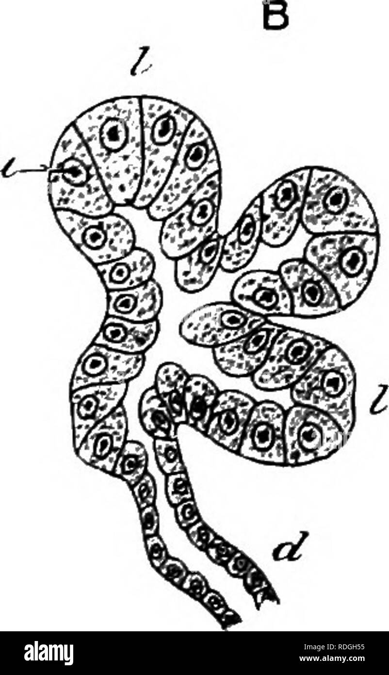 . An elementary course of practical zoology. Zoology. Fig. 42.—A, small portion of a section of the frog's pancreas; B, diagram showing the connection between the lobules and ducts. c, connective tissue covering of the gland ; d. duct; /. lobules ; nu. nuclei. called glycogen or animal starch. This is stored up in the cells in the form of minute insoluble granules, which, being afterwards transformed into soluble sugar, pass into the blood and so to the tissues. The Pancreas.—Sections of this gland (Fig. 42) show it to be made up of microscopic masses or lobules (/), each of which consists of Stock Photohttps://www.alamy.com/image-license-details/?v=1https://www.alamy.com/an-elementary-course-of-practical-zoology-zoology-fig-42a-small-portion-of-a-section-of-the-frogs-pancreas-b-diagram-showing-the-connection-between-the-lobules-and-ducts-c-connective-tissue-covering-of-the-gland-d-duct-lobules-nu-nuclei-called-glycogen-or-animal-starch-this-is-stored-up-in-the-cells-in-the-form-of-minute-insoluble-granules-which-being-afterwards-transformed-into-soluble-sugar-pass-into-the-blood-and-so-to-the-tissues-the-pancreassections-of-this-gland-fig-42-show-it-to-be-made-up-of-microscopic-masses-or-lobules-each-of-which-consists-of-image232090017.html
RMRDGH55–. An elementary course of practical zoology. Zoology. Fig. 42.—A, small portion of a section of the frog's pancreas; B, diagram showing the connection between the lobules and ducts. c, connective tissue covering of the gland ; d. duct; /. lobules ; nu. nuclei. called glycogen or animal starch. This is stored up in the cells in the form of minute insoluble granules, which, being afterwards transformed into soluble sugar, pass into the blood and so to the tissues. The Pancreas.—Sections of this gland (Fig. 42) show it to be made up of microscopic masses or lobules (/), each of which consists of
RFMTA316–Set Of 16 simple editable icons such as Bones Joint, Human Nostril, Rod Cell, White blood cell, Liver, Men Leg, Blood Transfusion, Shoulder, Skull Sid
 . The elements of embryology . Embryology. 174 THE THIRD DAT. Fig. 60. [chap.. DliGKAM OF A POKTION OF THE DlSBSTIVB TeACT OF A Chick upon the Fourth Day. (Copied from Gotte.) The black inner line represents the hypoblast, the outer shading the mesoblast. Ig. lung-diverticulum with expanded termi- nation, forming the primary lung-vesicle. St. stomach. I. two hepatic diverticula with their terminations united by cords of hypoblast cells, p. diverticulum of the pancreas with the vesicular diverticula coming from it. the duodenum by the fact that from it, as we shall presently point out, the rudi Stock Photohttps://www.alamy.com/image-license-details/?v=1https://www.alamy.com/the-elements-of-embryology-embryology-174-the-third-dat-fig-60-chap-dligkam-of-a-poktion-of-the-dlsbstivb-teact-of-a-chick-upon-the-fourth-day-copied-from-gotte-the-black-inner-line-represents-the-hypoblast-the-outer-shading-the-mesoblast-ig-lung-diverticulum-with-expanded-termi-nation-forming-the-primary-lung-vesicle-st-stomach-i-two-hepatic-diverticula-with-their-terminations-united-by-cords-of-hypoblast-cells-p-diverticulum-of-the-pancreas-with-the-vesicular-diverticula-coming-from-it-the-duodenum-by-the-fact-that-from-it-as-we-shall-presently-point-out-the-rudi-image232134424.html
RMRDJHR4–. The elements of embryology . Embryology. 174 THE THIRD DAT. Fig. 60. [chap.. DliGKAM OF A POKTION OF THE DlSBSTIVB TeACT OF A Chick upon the Fourth Day. (Copied from Gotte.) The black inner line represents the hypoblast, the outer shading the mesoblast. Ig. lung-diverticulum with expanded termi- nation, forming the primary lung-vesicle. St. stomach. I. two hepatic diverticula with their terminations united by cords of hypoblast cells, p. diverticulum of the pancreas with the vesicular diverticula coming from it. the duodenum by the fact that from it, as we shall presently point out, the rudi
RFMTAB7W–Set Of 16 simple editable icons such as Tooth and Gums, Men Leg, Blood Transfusion, Human Liver, Tongue Mouth, Nostril, Digestive System, Thyroid, Ski
 . The physiology of domestic animals ... Physiology, Comparative; Veterinary physiology. 414 PHYSIOLOGY OF THE DOMESTIC ANIMALS. stomach and rapidly reaches its maximum two or three hours thereafter. Toward the fifth or seventh hour it decreases in activity, and ahout twelve hours after feeding again becomes increased in amount, the second increase being apparently coincident with the escape of the acid chyme into the small intestine. The cells of the pancreas also undergo marked histological changes during the period of active secretion. In the pan- creas of a fasting dog two zones may be rec Stock Photohttps://www.alamy.com/image-license-details/?v=1https://www.alamy.com/the-physiology-of-domestic-animals-physiology-comparative-veterinary-physiology-414-physiology-of-the-domestic-animals-stomach-and-rapidly-reaches-its-maximum-two-or-three-hours-thereafter-toward-the-fifth-or-seventh-hour-it-decreases-in-activity-and-ahout-twelve-hours-after-feeding-again-becomes-increased-in-amount-the-second-increase-being-apparently-coincident-with-the-escape-of-the-acid-chyme-into-the-small-intestine-the-cells-of-the-pancreas-also-undergo-marked-histological-changes-during-the-period-of-active-secretion-in-the-pan-creas-of-a-fasting-dog-two-zones-may-be-rec-image232425866.html
RMRE3WFP–. The physiology of domestic animals ... Physiology, Comparative; Veterinary physiology. 414 PHYSIOLOGY OF THE DOMESTIC ANIMALS. stomach and rapidly reaches its maximum two or three hours thereafter. Toward the fifth or seventh hour it decreases in activity, and ahout twelve hours after feeding again becomes increased in amount, the second increase being apparently coincident with the escape of the acid chyme into the small intestine. The cells of the pancreas also undergo marked histological changes during the period of active secretion. In the pan- creas of a fasting dog two zones may be rec
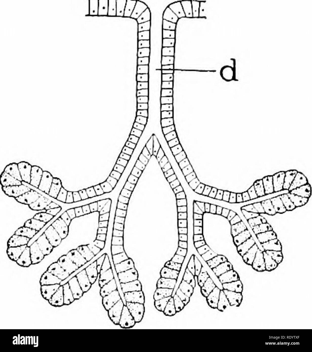 . Principles of modern biology. Biology. D EHm. Fig. 15-13. Structure of glands; the actual gland cells are shaded in each figure. A, single gland cells scattered among ordinary epithelial cells; B, simple multicellular gland, consisting of a group of gland cells lining a slight invagina- tion of the epithelium; C, simple tubular gland; D, simple alveolar gland; E, compound tubular gland (large glands, such as the liver and pancreas, are of this type, with thousands of secreting alveoli); d, duct. epithelial outpocketings (Fig. 15-13). Some multicellular glands are simple glands, like the gast Stock Photohttps://www.alamy.com/image-license-details/?v=1https://www.alamy.com/principles-of-modern-biology-biology-d-ehm-fig-15-13-structure-of-glands-the-actual-gland-cells-are-shaded-in-each-figure-a-single-gland-cells-scattered-among-ordinary-epithelial-cells-b-simple-multicellular-gland-consisting-of-a-group-of-gland-cells-lining-a-slight-invagina-tion-of-the-epithelium-c-simple-tubular-gland-d-simple-alveolar-gland-e-compound-tubular-gland-large-glands-such-as-the-liver-and-pancreas-are-of-this-type-with-thousands-of-secreting-alveoli-d-duct-epithelial-outpocketings-fig-15-13-some-multicellular-glands-are-simple-glands-like-the-gast-image232337575.html
RMRDYTXF–. Principles of modern biology. Biology. D EHm. Fig. 15-13. Structure of glands; the actual gland cells are shaded in each figure. A, single gland cells scattered among ordinary epithelial cells; B, simple multicellular gland, consisting of a group of gland cells lining a slight invagina- tion of the epithelium; C, simple tubular gland; D, simple alveolar gland; E, compound tubular gland (large glands, such as the liver and pancreas, are of this type, with thousands of secreting alveoli); d, duct. epithelial outpocketings (Fig. 15-13). Some multicellular glands are simple glands, like the gast
 . A laboratory manual and text-book of embryology. Embryology. Cystic duct Ductus choledoch Ventral pancreas Qorsal pancreas ball bladder Cystic duct' rlepaiic ducr ctus choledochus tral pancreas Duodenum. Duct of dorsal pancreas Head of dorsal pancreas Duodenum Tail of dorsal pancreas Fig. 178.—Reconstructions showing the development of the hepatic diverticulum and pancreatic anlages. A, 7.5 mm. embryo, X 36 (after Thyng); B, 10 mm. embryo, X 33 (specimen loaned by Dr. H. C. Tracy). time after birth, blood-cells are actively developed between the hepatic cells and the endothelium of the sinus Stock Photohttps://www.alamy.com/image-license-details/?v=1https://www.alamy.com/a-laboratory-manual-and-text-book-of-embryology-embryology-cystic-duct-ductus-choledoch-ventral-pancreas-qorsal-pancreas-ball-bladder-cystic-duct-rlepaiic-ducr-ctus-choledochus-tral-pancreas-duodenum-duct-of-dorsal-pancreas-head-of-dorsal-pancreas-duodenum-tail-of-dorsal-pancreas-fig-178reconstructions-showing-the-development-of-the-hepatic-diverticulum-and-pancreatic-anlages-a-75-mm-embryo-x-36-after-thyng-b-10-mm-embryo-x-33-specimen-loaned-by-dr-h-c-tracy-time-after-birth-blood-cells-are-actively-developed-between-the-hepatic-cells-and-the-endothelium-of-the-sinus-image232344969.html
RMRE06AH–. A laboratory manual and text-book of embryology. Embryology. Cystic duct Ductus choledoch Ventral pancreas Qorsal pancreas ball bladder Cystic duct' rlepaiic ducr ctus choledochus tral pancreas Duodenum. Duct of dorsal pancreas Head of dorsal pancreas Duodenum Tail of dorsal pancreas Fig. 178.—Reconstructions showing the development of the hepatic diverticulum and pancreatic anlages. A, 7.5 mm. embryo, X 36 (after Thyng); B, 10 mm. embryo, X 33 (specimen loaned by Dr. H. C. Tracy). time after birth, blood-cells are actively developed between the hepatic cells and the endothelium of the sinus
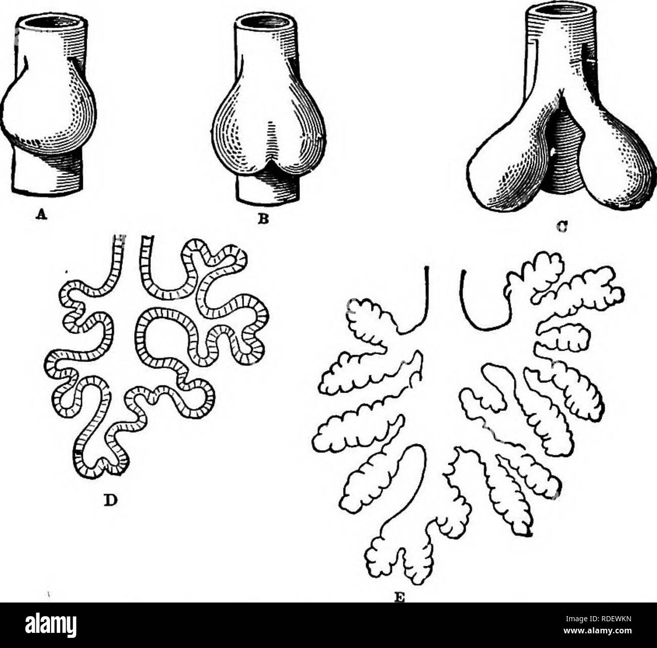 . Animal activities; a first book in zoo?logy. Zoology; Animal behavior. 248 yINlM/IL /ICTIVITIES. cases, the difference being only in the amount of fold- ing. The liver and pancreas of vertebrates arise in the same way, by folding of the walls of the food-tube. Other changes take place in as simple a way as this. Indeed, nearly all the organs of a complex body arise by foldings and pushings of layers of cells. When the egg of a very simple animal, like the hydra, develops, it first divides into a number of cells forming a spherical. Fig. 202.—Growth of Frog's Lung from Primitive Food-tube. bo Stock Photohttps://www.alamy.com/image-license-details/?v=1https://www.alamy.com/animal-activities-a-first-book-in-zoology-zoology-animal-behavior-248-yinlmil-ictivities-cases-the-difference-being-only-in-the-amount-of-fold-ing-the-liver-and-pancreas-of-vertebrates-arise-in-the-same-way-by-folding-of-the-walls-of-the-food-tube-other-changes-take-place-in-as-simple-a-way-as-this-indeed-nearly-all-the-organs-of-a-complex-body-arise-by-foldings-and-pushings-of-layers-of-cells-when-the-egg-of-a-very-simple-animal-like-the-hydra-develops-it-first-divides-into-a-number-of-cells-forming-a-spherical-fig-202growth-of-frogs-lung-from-primitive-food-tube-bo-image232052793.html
RMRDEWKN–. Animal activities; a first book in zoo?logy. Zoology; Animal behavior. 248 yINlM/IL /ICTIVITIES. cases, the difference being only in the amount of fold- ing. The liver and pancreas of vertebrates arise in the same way, by folding of the walls of the food-tube. Other changes take place in as simple a way as this. Indeed, nearly all the organs of a complex body arise by foldings and pushings of layers of cells. When the egg of a very simple animal, like the hydra, develops, it first divides into a number of cells forming a spherical. Fig. 202.—Growth of Frog's Lung from Primitive Food-tube. bo
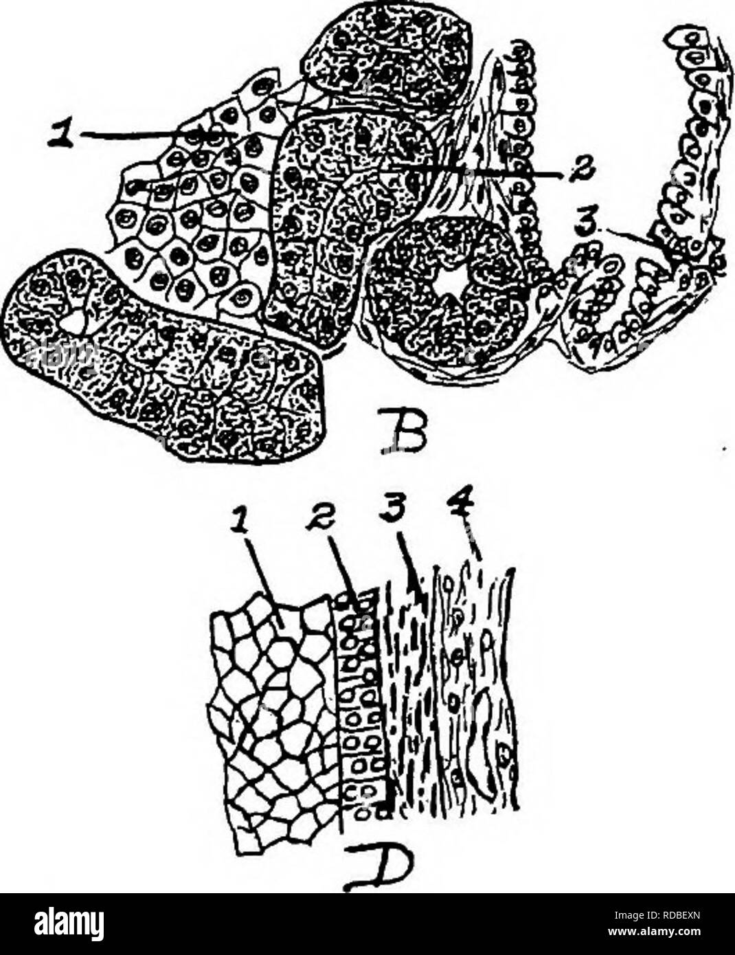 . The anatomy of the domestic fowl . Domestic animals; Veterinary medicine; Poultry. Pig. 38.—Cellular structure of liT^er, pancreas, and trachea. A. Liver. 1, Liver cells. 2, Sinusoid. 4, Nucleus of cells. B. Pancreas, i. Island of Langerhans. 2, Alveolar cell.s. 3, Duct. C. Trachea, i. Ciliated epithelia. 2, Glands. 3, Hyaline cartilage. D. Section of wall of ovum at Fig. S7 letter d. i. Yolk. 2, Granular mem- brane. 3, Theca. 4, Blood-vessel. A microscopic study of the Uver of the fowl shows a compact mass of Uver ceUs polyhedral in shape, with large nuclei (Fig. 38, A). The Uver tissue dif Stock Photohttps://www.alamy.com/image-license-details/?v=1https://www.alamy.com/the-anatomy-of-the-domestic-fowl-domestic-animals-veterinary-medicine-poultry-pig-38cellular-structure-of-liter-pancreas-and-trachea-a-liver-1-liver-cells-2-sinusoid-4-nucleus-of-cells-b-pancreas-i-island-of-langerhans-2-alveolar-cells-3-duct-c-trachea-i-ciliated-epithelia-2-glands-3-hyaline-cartilage-d-section-of-wall-of-ovum-at-fig-s7-letter-d-i-yolk-2-granular-mem-brane-3-theca-4-blood-vessel-a-microscopic-study-of-the-uver-of-the-fowl-shows-a-compact-mass-of-uver-ceus-polyhedral-in-shape-with-large-nuclei-fig-38-a-the-uver-tissue-dif-image231978509.html
RMRDBEXN–. The anatomy of the domestic fowl . Domestic animals; Veterinary medicine; Poultry. Pig. 38.—Cellular structure of liT^er, pancreas, and trachea. A. Liver. 1, Liver cells. 2, Sinusoid. 4, Nucleus of cells. B. Pancreas, i. Island of Langerhans. 2, Alveolar cell.s. 3, Duct. C. Trachea, i. Ciliated epithelia. 2, Glands. 3, Hyaline cartilage. D. Section of wall of ovum at Fig. S7 letter d. i. Yolk. 2, Granular mem- brane. 3, Theca. 4, Blood-vessel. A microscopic study of the Uver of the fowl shows a compact mass of Uver ceUs polyhedral in shape, with large nuclei (Fig. 38, A). The Uver tissue dif