Quick filters:
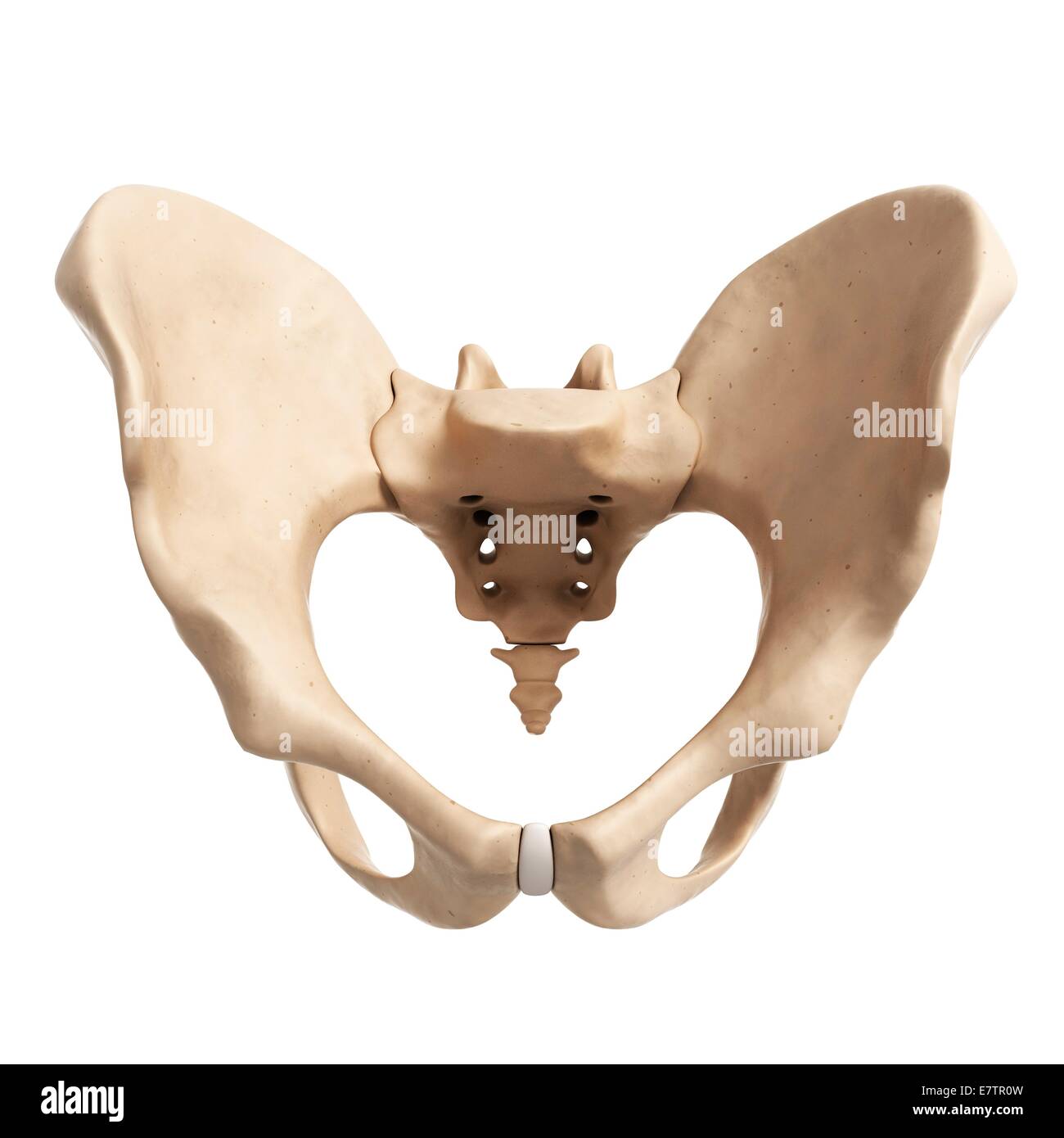
Pelvic bone Stock Photos and Images
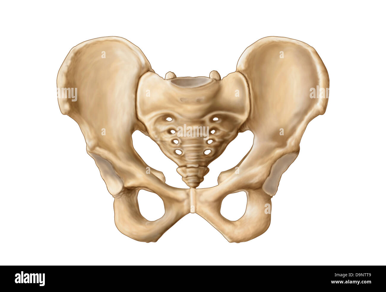 Anatomy of human pelvic bone. Stock Photohttps://www.alamy.com/image-license-details/?v=1https://www.alamy.com/stock-photo-anatomy-of-human-pelvic-bone-57643497.html
RFD9NTT9–Anatomy of human pelvic bone.
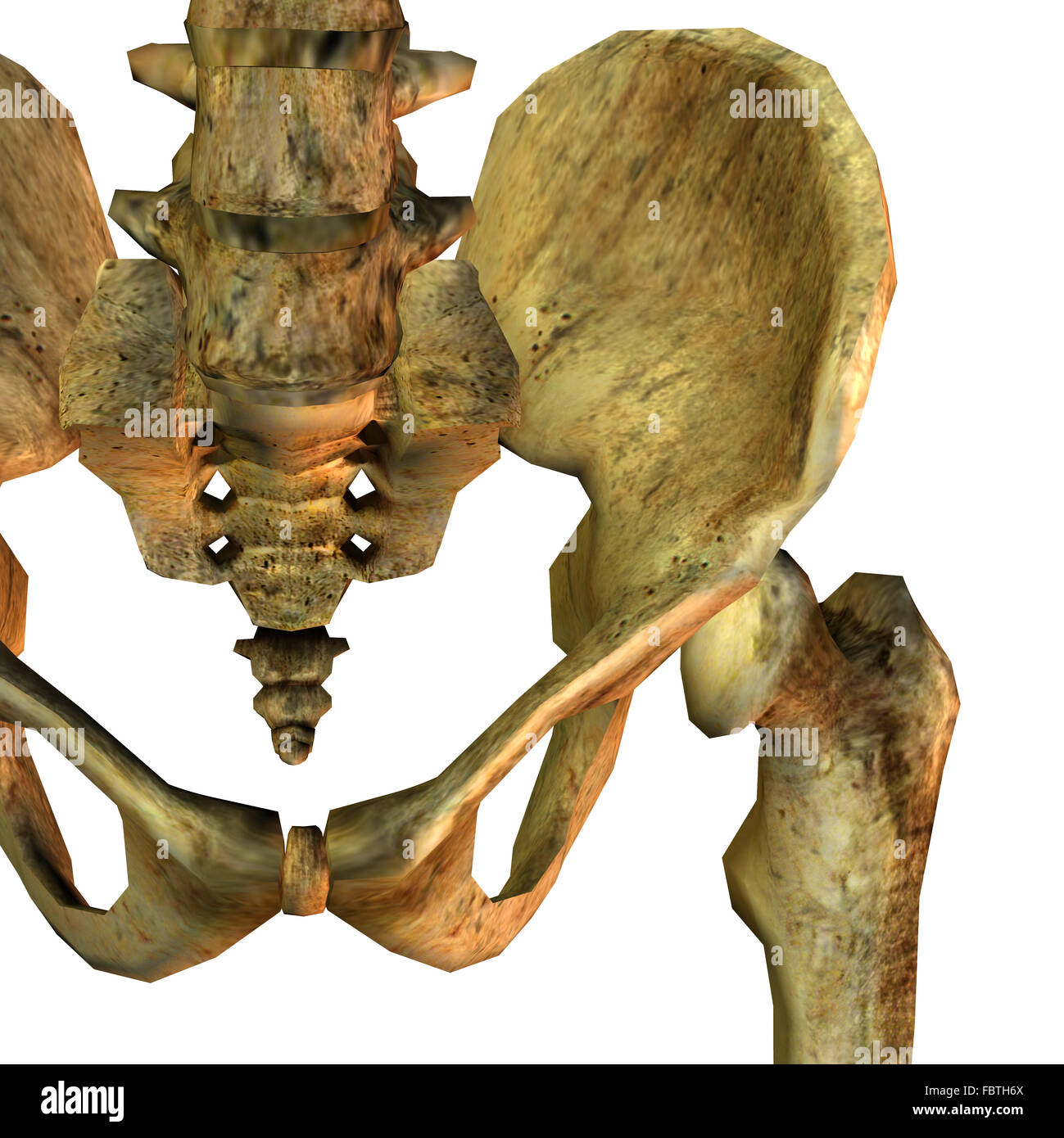 Detail left pelvic bone Stock Photohttps://www.alamy.com/image-license-details/?v=1https://www.alamy.com/stock-photo-detail-left-pelvic-bone-93353426.html
RMFBTH6X–Detail left pelvic bone
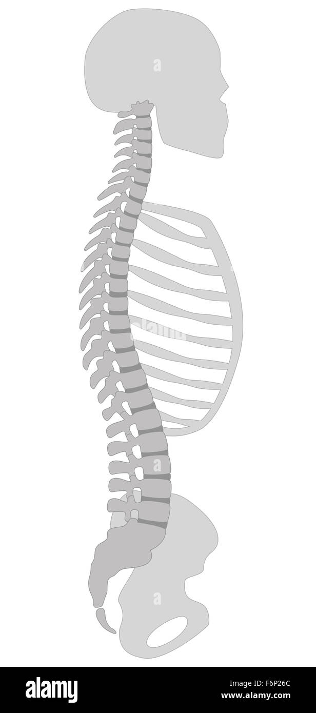 Human spine, skull, thorax and pelvic bone - vertical section. Illustration on white background. Stock Photohttps://www.alamy.com/image-license-details/?v=1https://www.alamy.com/stock-photo-human-spine-skull-thorax-and-pelvic-bone-vertical-section-illustration-90224468.html
RFF6P26C–Human spine, skull, thorax and pelvic bone - vertical section. Illustration on white background.
 Antique medical illustration of a pelvic bone Stock Photohttps://www.alamy.com/image-license-details/?v=1https://www.alamy.com/stock-photo-antique-medical-illustration-of-a-pelvic-bone-37347914.html
RFC4N9J2–Antique medical illustration of a pelvic bone
 X ray film of pelvic bone Stock Photohttps://www.alamy.com/image-license-details/?v=1https://www.alamy.com/stock-photo-x-ray-film-of-pelvic-bone-53441258.html
RFD2XCTA–X ray film of pelvic bone
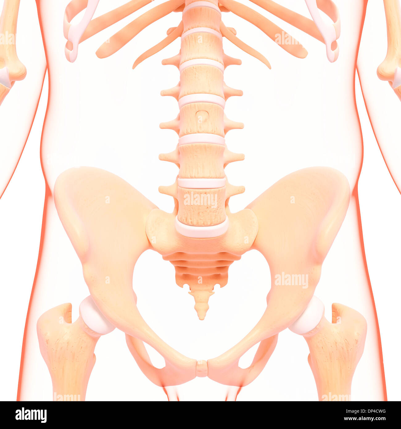 Human pelvic bones, artwork Stock Photohttps://www.alamy.com/image-license-details/?v=1https://www.alamy.com/human-pelvic-bones-artwork-image65251468.html
RFDP4CWG–Human pelvic bones, artwork
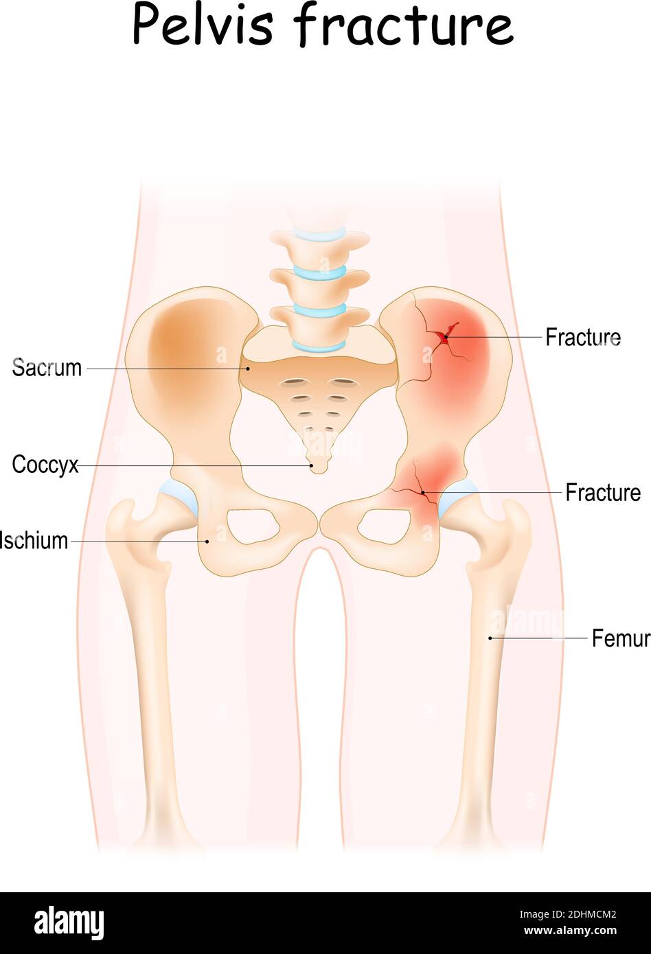 Pelvis Fracture. pelvic bones: sacrum, ilium, coccyx, pubis, ischium and femur. Vector illustration isolated on a white background Stock Vectorhttps://www.alamy.com/image-license-details/?v=1https://www.alamy.com/pelvis-fracture-pelvic-bones-sacrum-ilium-coccyx-pubis-ischium-and-femur-vector-illustration-isolated-on-a-white-background-image389526258.html
RF2DHMCM2–Pelvis Fracture. pelvic bones: sacrum, ilium, coccyx, pubis, ischium and femur. Vector illustration isolated on a white background
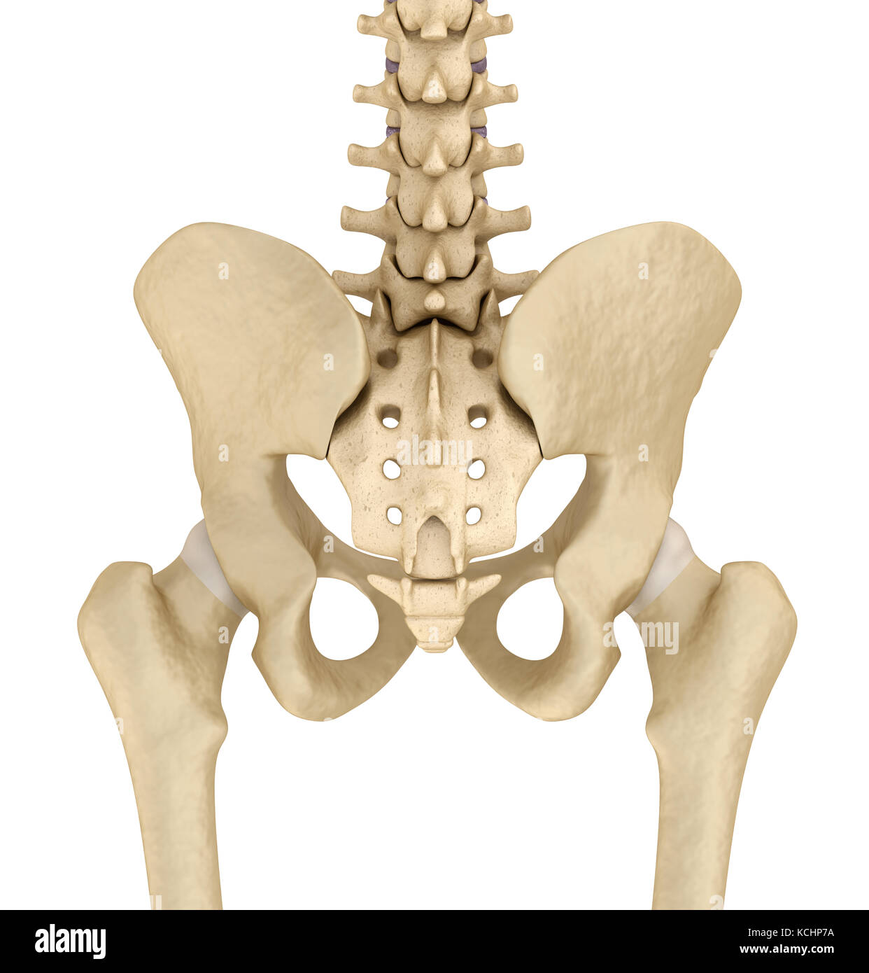 Pelvic area anatomy , backside view, 3d render Stock Photohttps://www.alamy.com/image-license-details/?v=1https://www.alamy.com/stock-image-pelvic-area-anatomy-backside-view-3d-render-162659822.html
RFKCHP7A–Pelvic area anatomy , backside view, 3d render
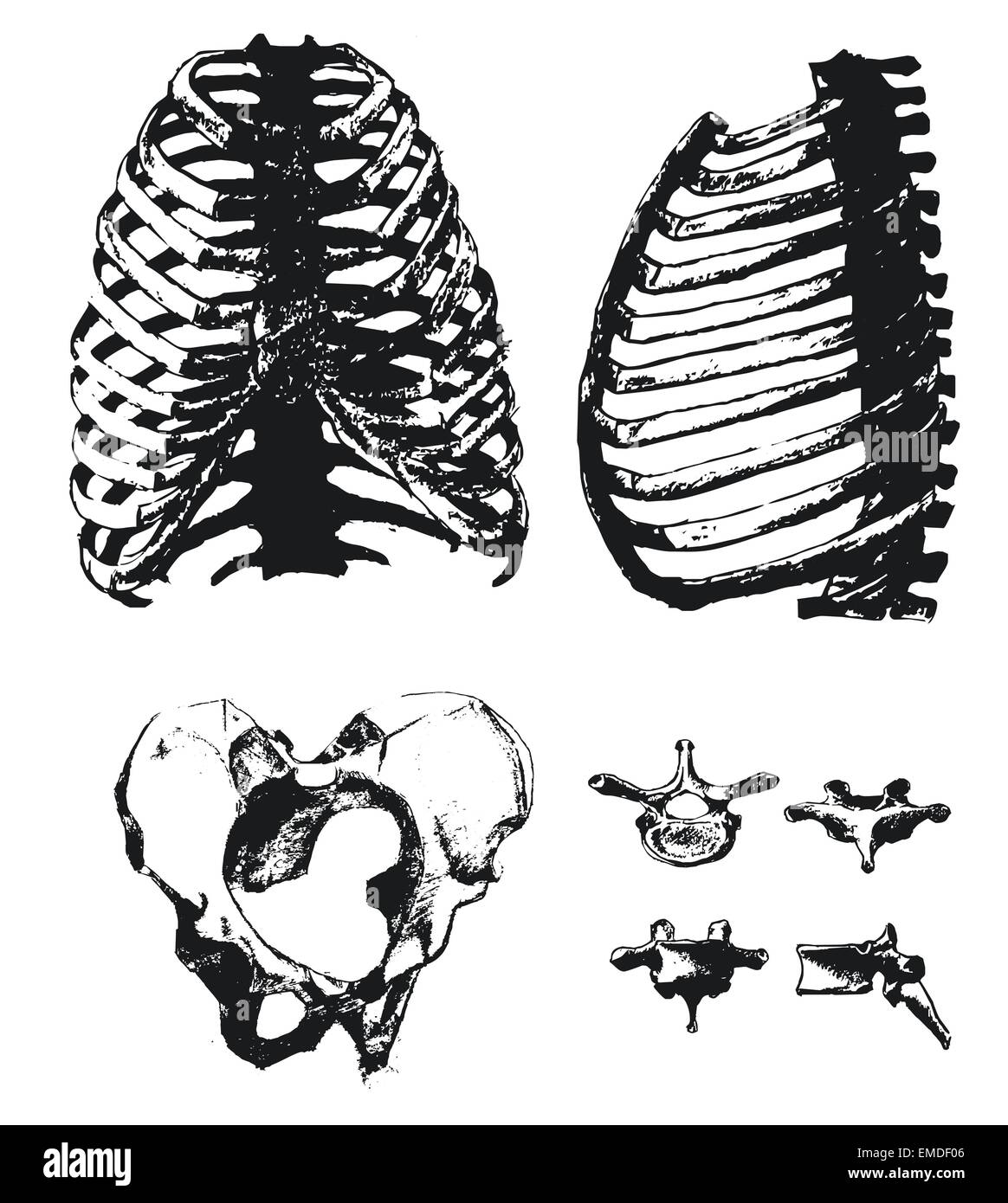 Hand drawn pelvic bone and rib Stock Vectorhttps://www.alamy.com/image-license-details/?v=1https://www.alamy.com/stock-photo-hand-drawn-pelvic-bone-and-rib-81431734.html
RFEMDF06–Hand drawn pelvic bone and rib
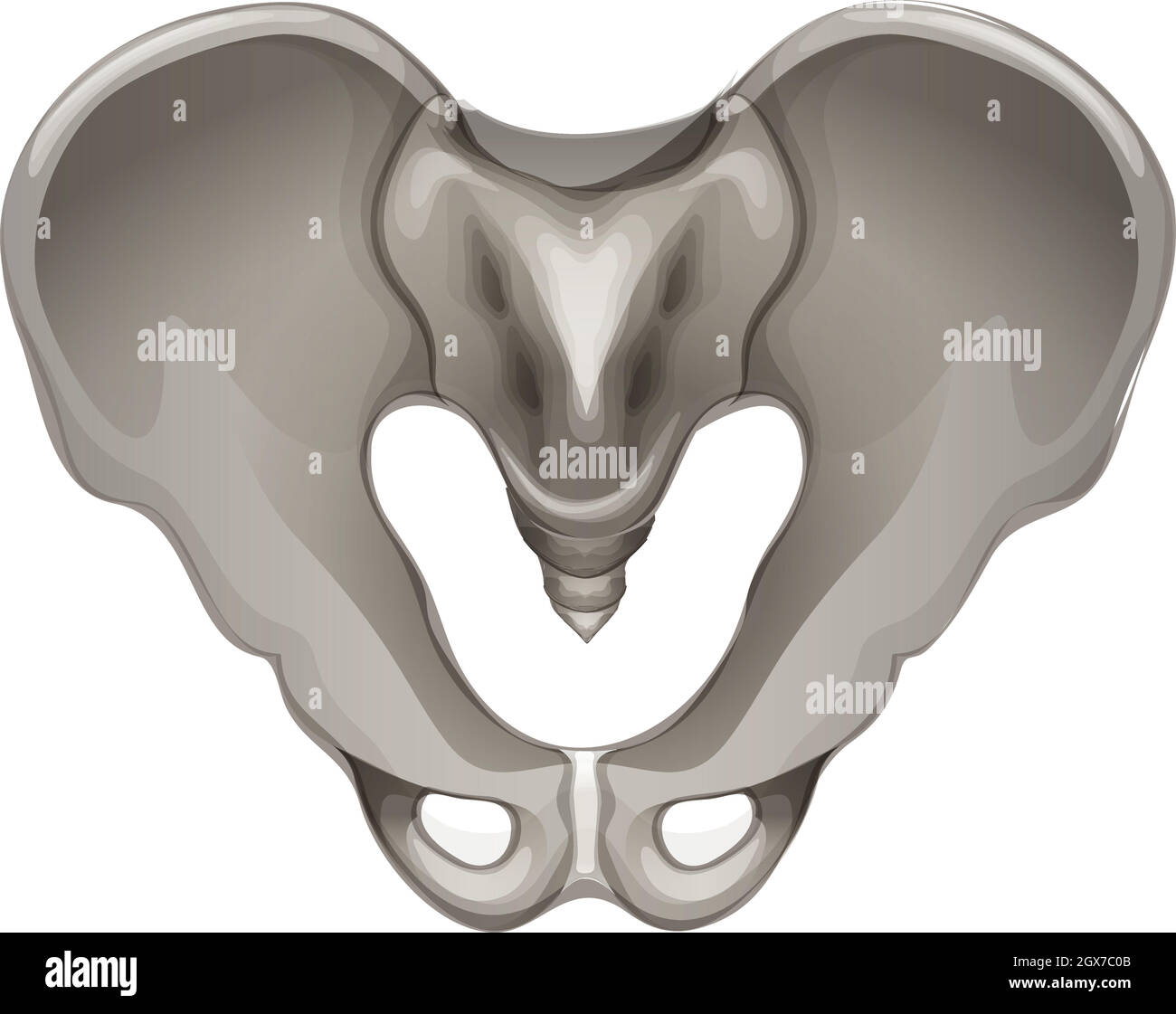 Pelvic bone Stock Vectorhttps://www.alamy.com/image-license-details/?v=1https://www.alamy.com/pelvic-bone-image446403339.html
RF2GX7C0B–Pelvic bone
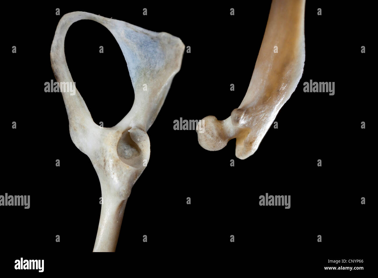 Barn owl (Tyto alba), pelvic and thigh bone of a mouse, undigested food residue from a pellet Stock Photohttps://www.alamy.com/image-license-details/?v=1https://www.alamy.com/stock-photo-barn-owl-tyto-alba-pelvic-and-thigh-bone-of-a-mouse-undigested-food-47938638.html
RMCNYP66–Barn owl (Tyto alba), pelvic and thigh bone of a mouse, undigested food residue from a pellet
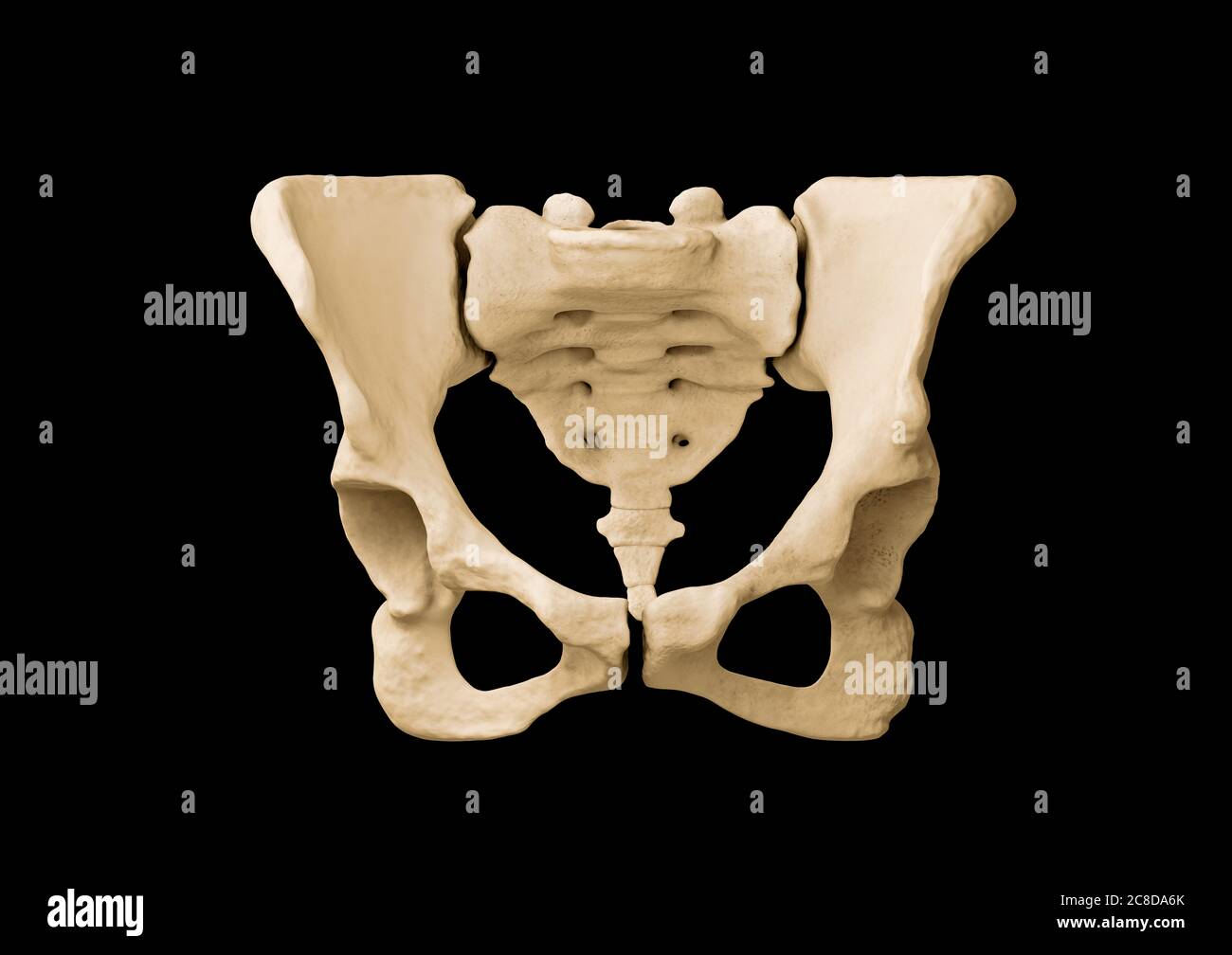 Pelvis, Human skeleton, Female Pelvic Bone anatomy, hip Stock Photohttps://www.alamy.com/image-license-details/?v=1https://www.alamy.com/pelvis-human-skeleton-female-pelvic-bone-anatomy-hip-image366628379.html
RF2C8DA6K–Pelvis, Human skeleton, Female Pelvic Bone anatomy, hip
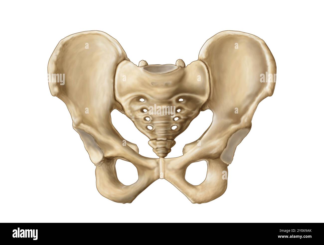 Anatomy of human pelvic bone Stock Photohttps://www.alamy.com/image-license-details/?v=1https://www.alamy.com/anatomy-of-human-pelvic-bone-image619712315.html
RM2Y069AK–Anatomy of human pelvic bone
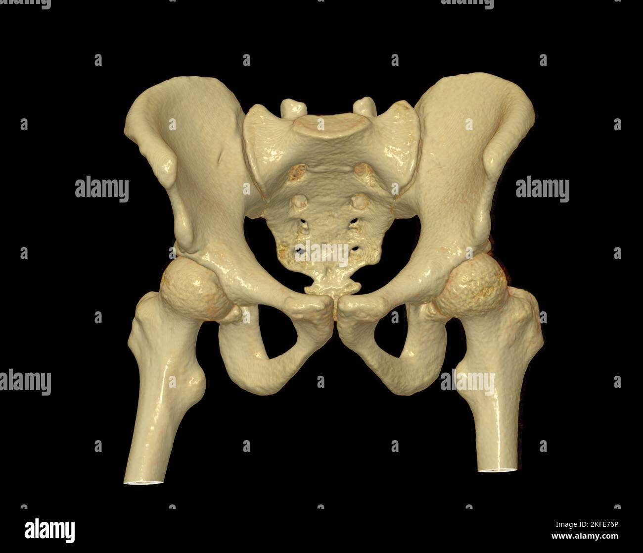 CT scan of Pelvic bone and hip joint 3D rendering for diagnosis fracture of Pelvic bone and hip joint isolated on black background. Stock Photohttps://www.alamy.com/image-license-details/?v=1https://www.alamy.com/ct-scan-of-pelvic-bone-and-hip-joint-3d-rendering-for-diagnosis-fracture-of-pelvic-bone-and-hip-joint-isolated-on-black-background-image491423150.html
RF2KFE76P–CT scan of Pelvic bone and hip joint 3D rendering for diagnosis fracture of Pelvic bone and hip joint isolated on black background.
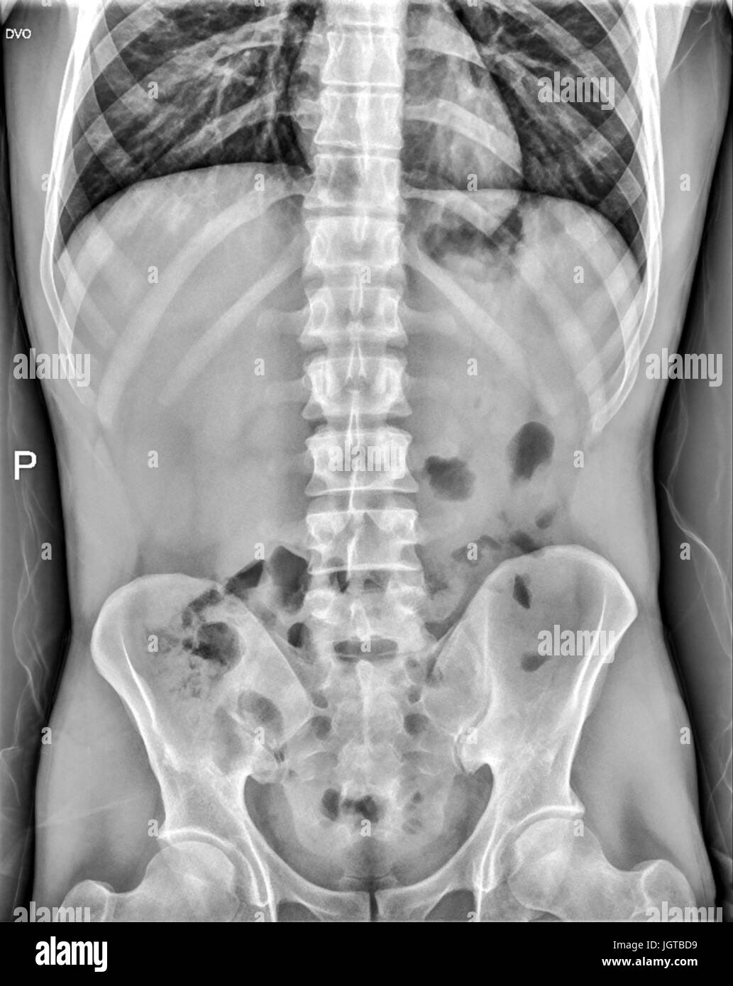 Abdomen medical xray, lungs, heart, rig cage, pelvic bone, spinal cord and internal organs Stock Photohttps://www.alamy.com/image-license-details/?v=1https://www.alamy.com/stock-photo-abdomen-medical-xray-lungs-heart-rig-cage-pelvic-bone-spinal-cord-148053285.html
RFJGTBD9–Abdomen medical xray, lungs, heart, rig cage, pelvic bone, spinal cord and internal organs
 Hand drawing a bone skeleton, anatomical drawing of pelvic bone man, print for Halloween,butterflies fly, skull Stock Photohttps://www.alamy.com/image-license-details/?v=1https://www.alamy.com/hand-drawing-a-bone-skeleton-anatomical-drawing-of-pelvic-bone-man-print-for-halloweenbutterflies-fly-skull-image222304899.html
RFPWJT4K–Hand drawing a bone skeleton, anatomical drawing of pelvic bone man, print for Halloween,butterflies fly, skull
RFP8HHDW–Pelvic Bone Icon
 X-ray of a 30 year old man with a pelvic fracture. Stock Photohttps://www.alamy.com/image-license-details/?v=1https://www.alamy.com/stock-photo-x-ray-of-a-30-year-old-man-with-a-pelvic-fracture-76783185.html
RMECWNMH–X-ray of a 30 year old man with a pelvic fracture.
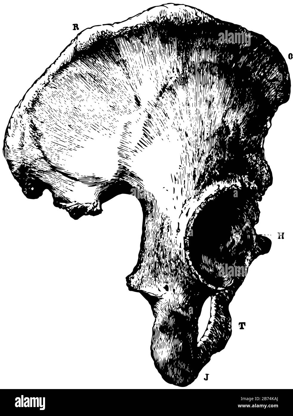 This illustration represents Part of the Human Pelvic Bone, vintage line drawing or engraving illustration. Stock Vectorhttps://www.alamy.com/image-license-details/?v=1https://www.alamy.com/this-illustration-represents-part-of-the-human-pelvic-bone-vintage-line-drawing-or-engraving-illustration-image348612954.html
RF2B74KAJ–This illustration represents Part of the Human Pelvic Bone, vintage line drawing or engraving illustration.
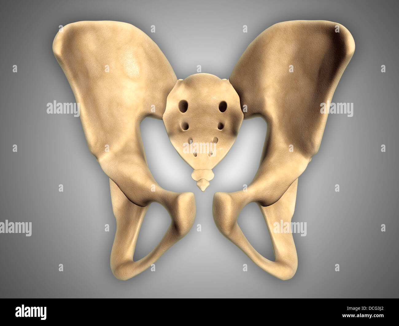 Anatomy of human pelvic bone. Stock Photohttps://www.alamy.com/image-license-details/?v=1https://www.alamy.com/stock-photo-anatomy-of-human-pelvic-bone-59361066.html
RFDCG3J2–Anatomy of human pelvic bone.
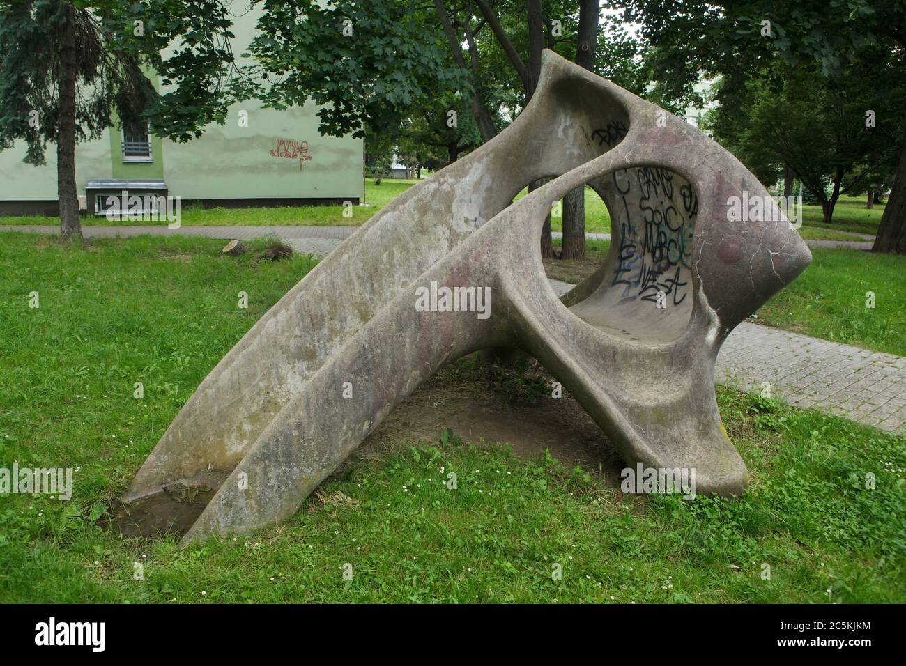 Concrete children's slide designed by Czech modernist sculptor Eleonora Haragsimová (1965) installed in the courtyard of the Invalidovna experimental housing complex in Karlín district in Prague, Czech Republic. The slide officially known as the Fish (Prolézačka Ryba) is nicknamed as the Mammoth pelvic bone by local people. In fact the children's slide is a modernist sculpture. During the communist regime in Czechoslovakia one of the few ways to install modernist artworks in public areas was to pretend the artwork is an element of a playground. Stock Photohttps://www.alamy.com/image-license-details/?v=1https://www.alamy.com/concrete-childrens-slide-designed-by-czech-modernist-sculptor-eleonora-haragsimov-1965-installed-in-the-courtyard-of-the-invalidovna-experimental-housing-complex-in-karln-district-in-prague-czech-republic-the-slide-officially-known-as-the-fish-prolzaka-ryba-is-nicknamed-as-the-mammoth-pelvic-bone-by-local-people-in-fact-the-childrens-slide-is-a-modernist-sculpture-during-the-communist-regime-in-czechoslovakia-one-of-the-few-ways-to-install-modernist-artworks-in-public-areas-was-to-pretend-the-artwork-is-an-element-of-a-playground-image364922760.html
RM2C5KJKM–Concrete children's slide designed by Czech modernist sculptor Eleonora Haragsimová (1965) installed in the courtyard of the Invalidovna experimental housing complex in Karlín district in Prague, Czech Republic. The slide officially known as the Fish (Prolézačka Ryba) is nicknamed as the Mammoth pelvic bone by local people. In fact the children's slide is a modernist sculpture. During the communist regime in Czechoslovakia one of the few ways to install modernist artworks in public areas was to pretend the artwork is an element of a playground.
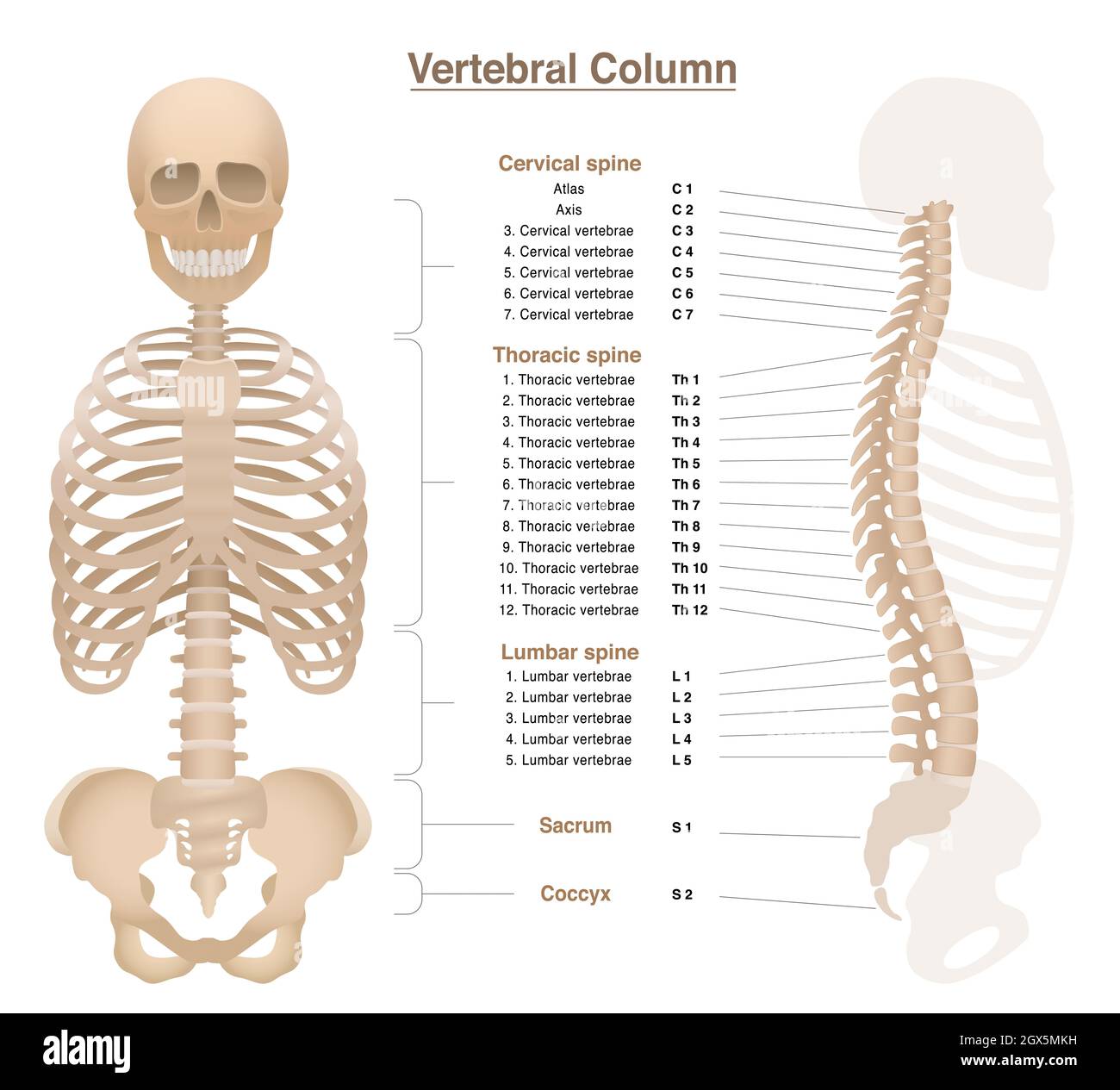 Skeleton with spine, thorax, pelvic bone and skull - labeled vertebral column chart with names and numbers of the vertebras - illustration on white. Stock Photohttps://www.alamy.com/image-license-details/?v=1https://www.alamy.com/skeleton-with-spine-thorax-pelvic-bone-and-skull-labeled-vertebral-column-chart-with-names-and-numbers-of-the-vertebras-illustration-on-white-image446366245.html
RF2GX5MKH–Skeleton with spine, thorax, pelvic bone and skull - labeled vertebral column chart with names and numbers of the vertebras - illustration on white.
 Antique medical illustration of a pelvic bone Stock Photohttps://www.alamy.com/image-license-details/?v=1https://www.alamy.com/stock-photo-antique-medical-illustration-of-a-pelvic-bone-37188570.html
RMC4E2B6–Antique medical illustration of a pelvic bone
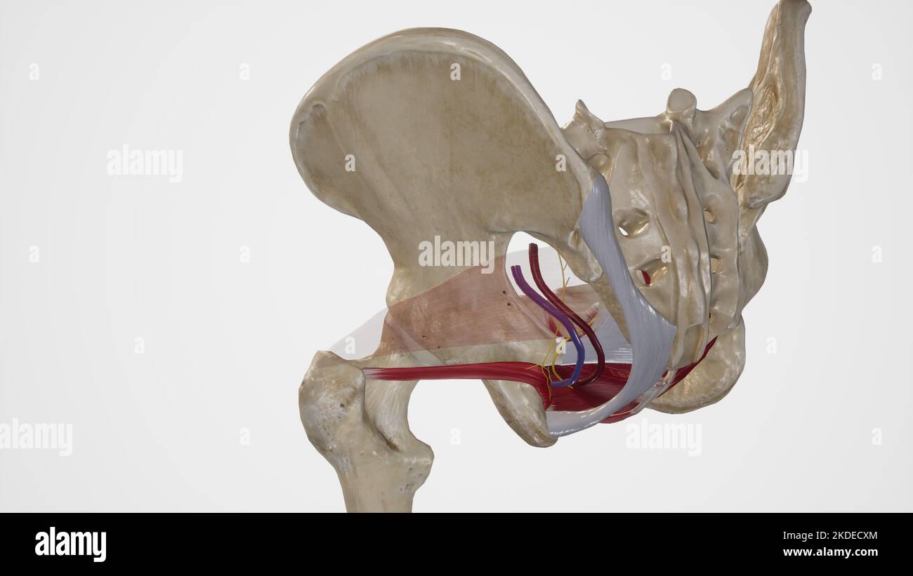 Anatomy of Lesser Sciatic Foramen Stock Photohttps://www.alamy.com/image-license-details/?v=1https://www.alamy.com/anatomy-of-lesser-sciatic-foramen-image490198316.html
RF2KDECXM–Anatomy of Lesser Sciatic Foramen
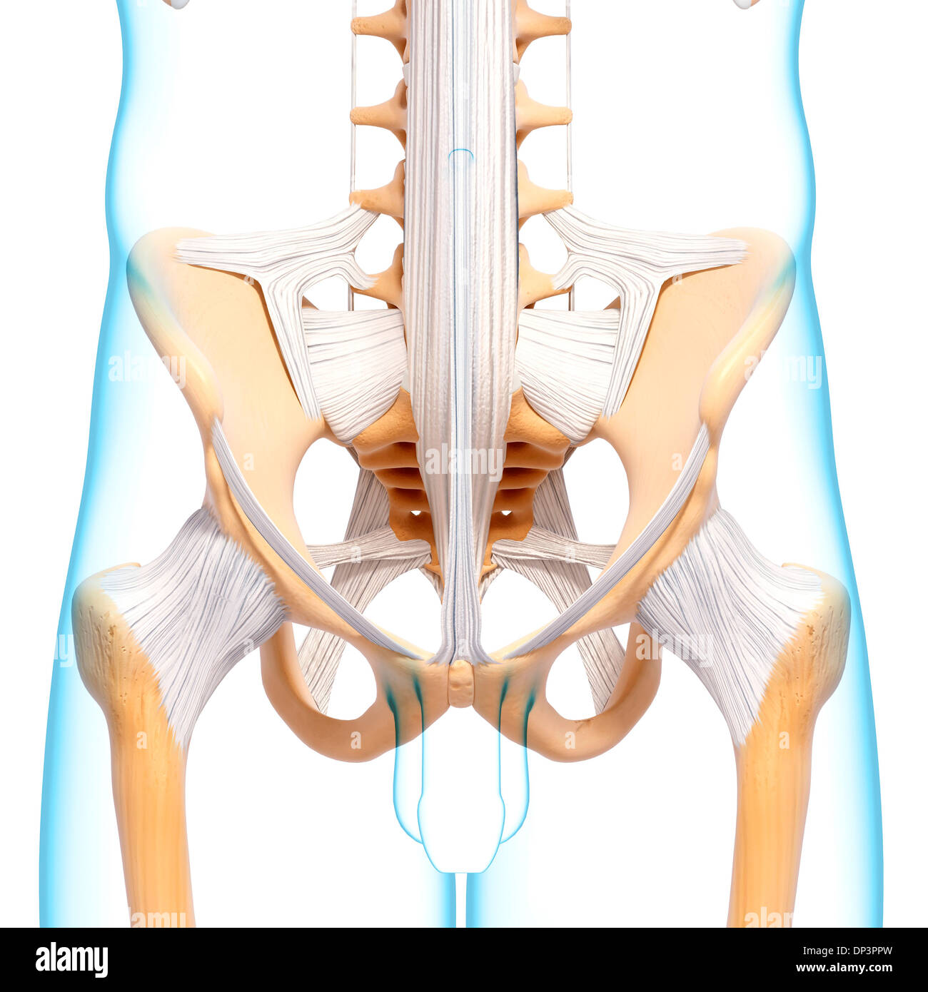 Human pelvic bones, artwork Stock Photohttps://www.alamy.com/image-license-details/?v=1https://www.alamy.com/human-pelvic-bones-artwork-image65237281.html
RFDP3PPW–Human pelvic bones, artwork
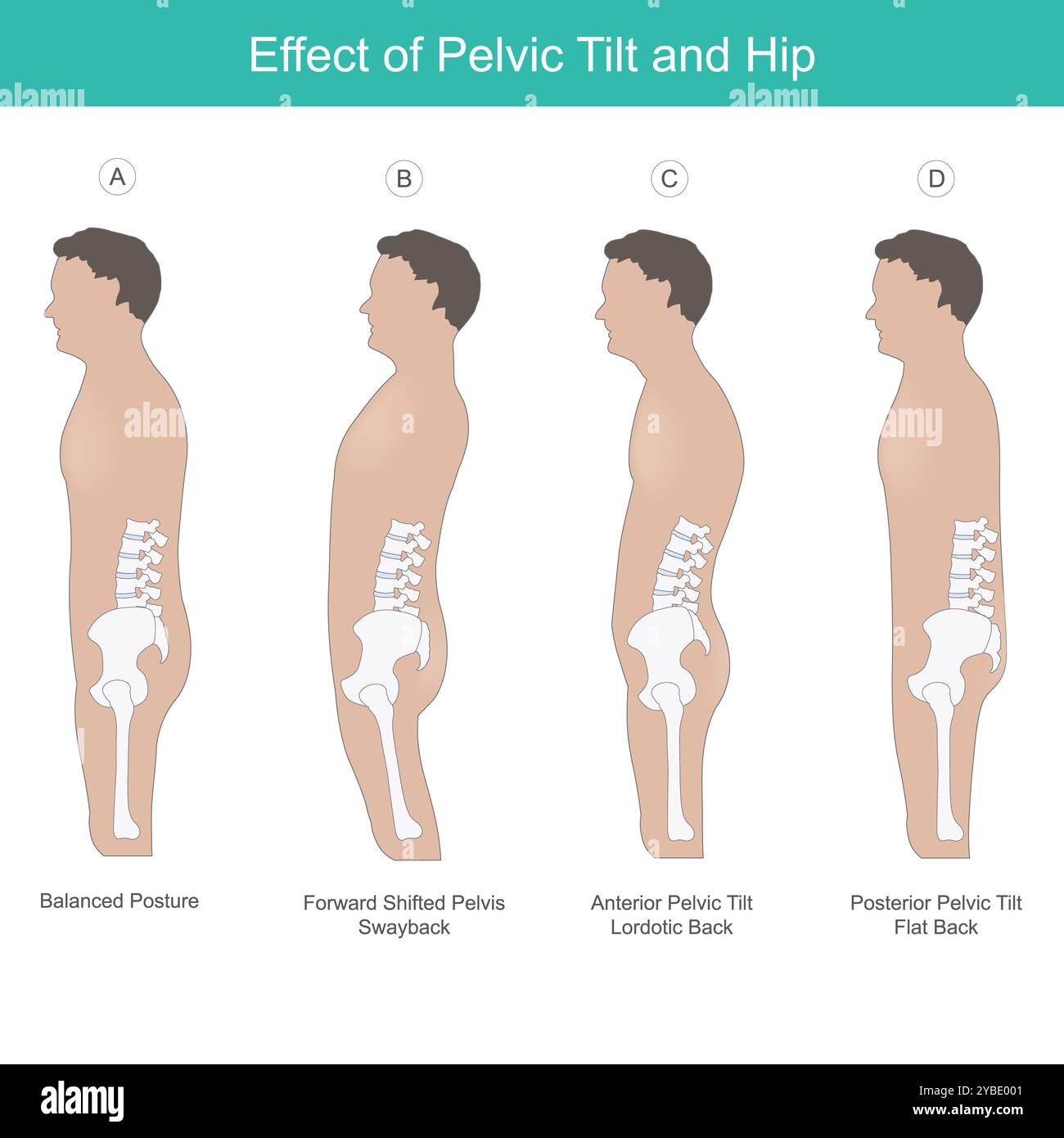 Effect of Pelvic Tilt and Hip. Comparison the pelvis bone and hips in the correct position human physiology. Stock Photohttps://www.alamy.com/image-license-details/?v=1https://www.alamy.com/effect-of-pelvic-tilt-and-hip-comparison-the-pelvis-bone-and-hips-in-the-correct-position-human-physiology-image626641793.html
RF2YBE001–Effect of Pelvic Tilt and Hip. Comparison the pelvis bone and hips in the correct position human physiology.
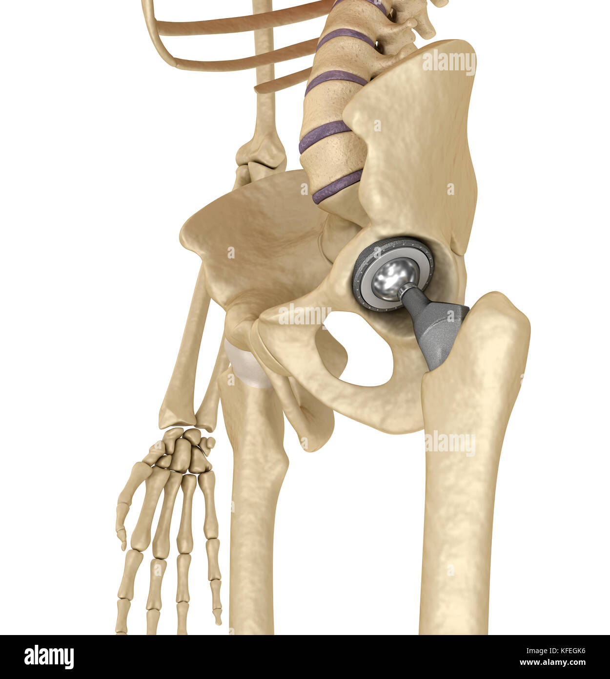 Hip replacement implant installed in the pelvis bone. Medically accurate 3D illustration Stock Photohttps://www.alamy.com/image-license-details/?v=1https://www.alamy.com/stock-image-hip-replacement-implant-installed-in-the-pelvis-bone-medically-accurate-164433562.html
RFKFEGK6–Hip replacement implant installed in the pelvis bone. Medically accurate 3D illustration
 Xray of a human pelvis Stock Photohttps://www.alamy.com/image-license-details/?v=1https://www.alamy.com/xray-of-a-human-pelvis-image333050311.html
RF2A9RN1Y–Xray of a human pelvis
 treatment of the pelvic bone of a dog in detail Stock Photohttps://www.alamy.com/image-license-details/?v=1https://www.alamy.com/treatment-of-the-pelvic-bone-of-a-dog-in-detail-image385829738.html
RF2DBM1NE–treatment of the pelvic bone of a dog in detail
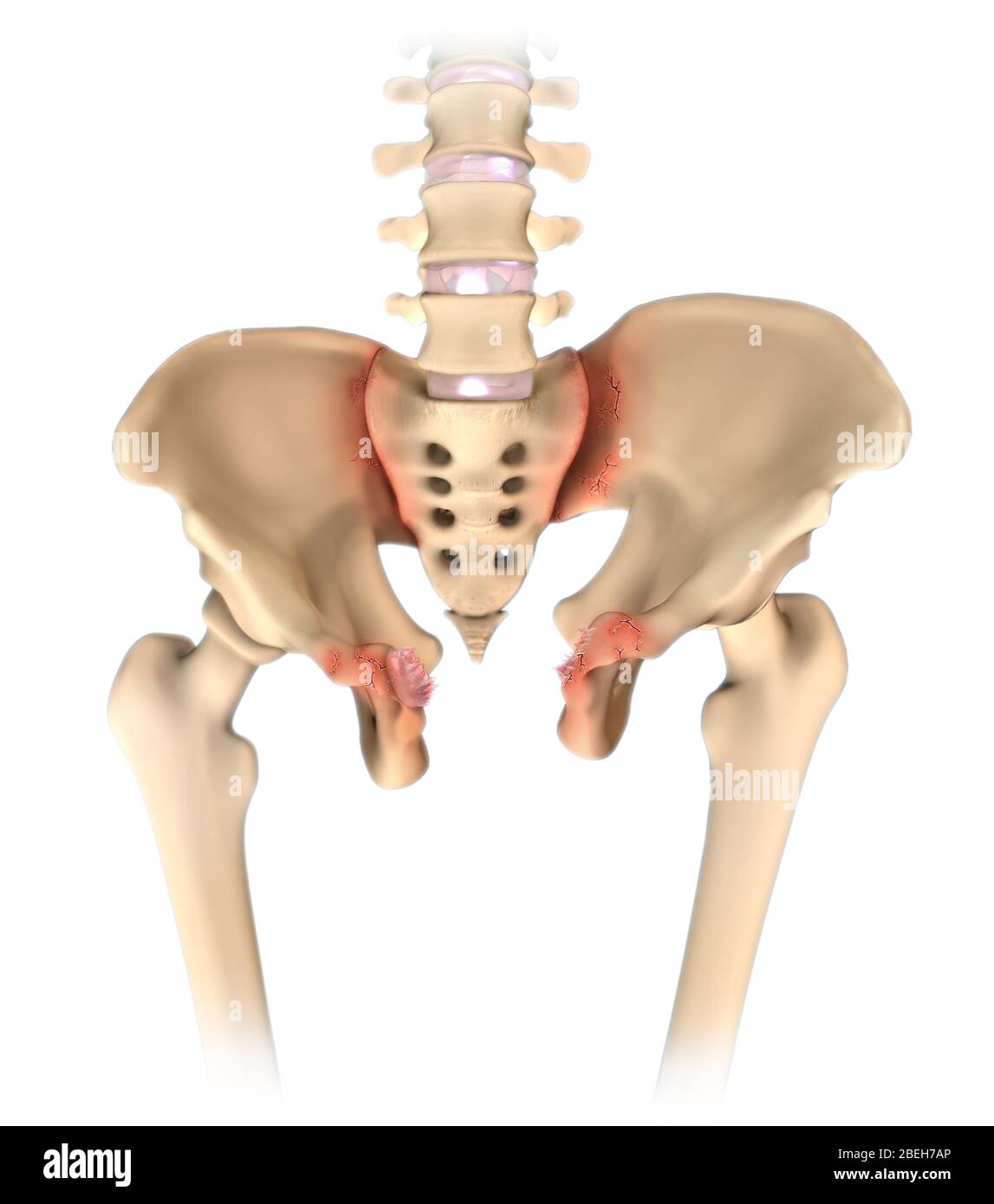 Open Book Pelvic Fracture Stock Photohttps://www.alamy.com/image-license-details/?v=1https://www.alamy.com/open-book-pelvic-fracture-image353191518.html
RM2BEH7AP–Open Book Pelvic Fracture
 Vertebrologist Orthopedic Scientists Doctors ExamineExamine Hip Joint Pain.Arthoplasty,Osteoarthritis Research.Clinical Investigation.X Ray,Rontgen Te Stock Photohttps://www.alamy.com/image-license-details/?v=1https://www.alamy.com/vertebrologist-orthopedic-scientists-doctors-examineexamine-hip-joint-painarthoplastyosteoarthritis-researchclinical-investigationx-rayrontgen-te-image514274354.html
RF2MTK656–Vertebrologist Orthopedic Scientists Doctors ExamineExamine Hip Joint Pain.Arthoplasty,Osteoarthritis Research.Clinical Investigation.X Ray,Rontgen Te
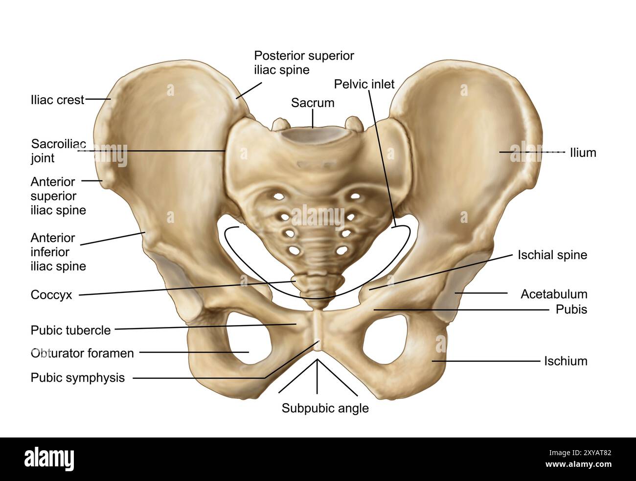 Anatomy of human pelvic bone Stock Photohttps://www.alamy.com/image-license-details/?v=1https://www.alamy.com/anatomy-of-human-pelvic-bone-image619197154.html
RM2XYAT82–Anatomy of human pelvic bone
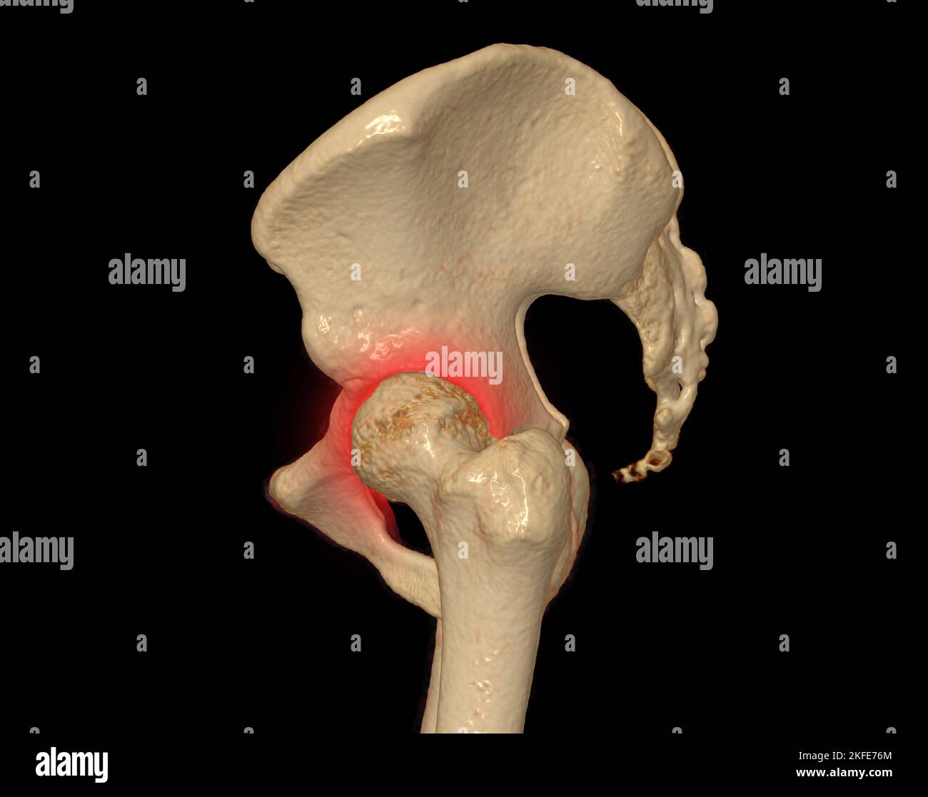 CT scan of Pelvic bone and hip joint 3D rendering for diagnosis fracture of Pelvic bone and hip joint isolated on black background. Stock Photohttps://www.alamy.com/image-license-details/?v=1https://www.alamy.com/ct-scan-of-pelvic-bone-and-hip-joint-3d-rendering-for-diagnosis-fracture-of-pelvic-bone-and-hip-joint-isolated-on-black-background-image491423148.html
RF2KFE76M–CT scan of Pelvic bone and hip joint 3D rendering for diagnosis fracture of Pelvic bone and hip joint isolated on black background.
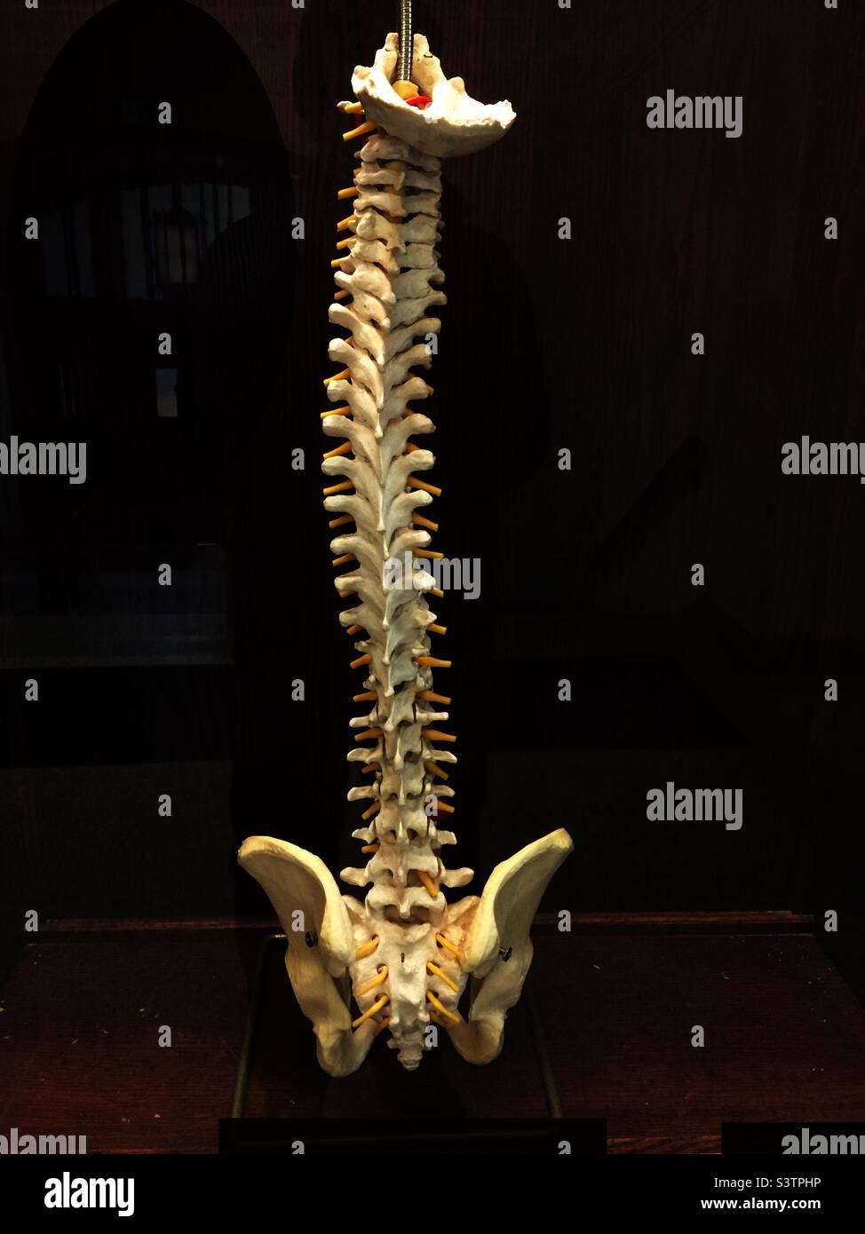 A complete human spine on display, Ontario, Canada. Stock Photohttps://www.alamy.com/image-license-details/?v=1https://www.alamy.com/a-complete-human-spine-on-display-ontario-canada-image312175186.html
RMS3TPHP–A complete human spine on display, Ontario, Canada.
 Hand drawing a bone skeleton, anatomical drawing of pelvic bone man, print for Halloween,butterflies fly, skull Stock Photohttps://www.alamy.com/image-license-details/?v=1https://www.alamy.com/hand-drawing-a-bone-skeleton-anatomical-drawing-of-pelvic-bone-man-print-for-halloweenbutterflies-fly-skull-image222304892.html
RFPWJT4C–Hand drawing a bone skeleton, anatomical drawing of pelvic bone man, print for Halloween,butterflies fly, skull
RFP8GR5B–Pelvic Bone Icon
 X-ray of a 30 year old man with a pelvic fracture. Stock Photohttps://www.alamy.com/image-license-details/?v=1https://www.alamy.com/stock-photo-x-ray-of-a-30-year-old-man-with-a-pelvic-fracture-76783186.html
RMECWNMJ–X-ray of a 30 year old man with a pelvic fracture.
 Pelvic bone, pelvis on skeleton body. High quality photo Stock Photohttps://www.alamy.com/image-license-details/?v=1https://www.alamy.com/pelvic-bone-pelvis-on-skeleton-body-high-quality-photo-image486139346.html
RF2K6WFKE–Pelvic bone, pelvis on skeleton body. High quality photo
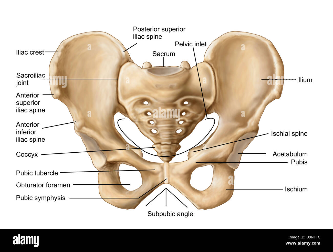 Anatomy of human pelvic bone. Stock Photohttps://www.alamy.com/image-license-details/?v=1https://www.alamy.com/stock-photo-anatomy-of-human-pelvic-bone-57643500.html
RFD9NTTC–Anatomy of human pelvic bone.
 The head of the femur articulates with the acetabulum in the pelvic bone forms the hip joint. Shown here is a femur sawed lengthwise, vintage line dra Stock Vectorhttps://www.alamy.com/image-license-details/?v=1https://www.alamy.com/the-head-of-the-femur-articulates-with-the-acetabulum-in-the-pelvic-bone-forms-the-hip-joint-shown-here-is-a-femur-sawed-lengthwise-vintage-line-dra-image348662876.html
RF2B76Y1G–The head of the femur articulates with the acetabulum in the pelvic bone forms the hip joint. Shown here is a femur sawed lengthwise, vintage line dra
 Field dressing a white-tailed buck in Wisconsin Stock Photohttps://www.alamy.com/image-license-details/?v=1https://www.alamy.com/stock-photo-field-dressing-a-white-tailed-buck-in-wisconsin-132457536.html
RMHKDXX8–Field dressing a white-tailed buck in Wisconsin
 Antique medical illustration of a pelvic bone Stock Photohttps://www.alamy.com/image-license-details/?v=1https://www.alamy.com/stock-photo-antique-medical-illustration-of-a-pelvic-bone-37188591.html
RFC4E2BY–Antique medical illustration of a pelvic bone
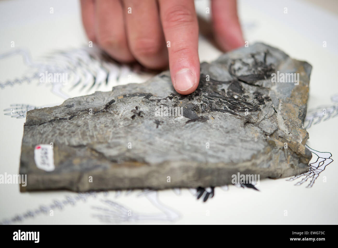 Stuttgart, Germany. 25th June, 2015. A man points to the pelvic bone of a 240-million-year-old prehistoric Pappochelys turtle fossil at the Loewentormuseum in Stuttgart, Germany, 25 June 2015. Palaeontologists have found the remnants of the turtle's ancestor near Schaebisch Hall, Germany. PHOTO: DANIEL NAUPOLD/dpa/Alamy Live News Stock Photohttps://www.alamy.com/image-license-details/?v=1https://www.alamy.com/stock-photo-stuttgart-germany-25th-june-2015-a-man-points-to-the-pelvic-bone-of-84564688.html
RMEWG73C–Stuttgart, Germany. 25th June, 2015. A man points to the pelvic bone of a 240-million-year-old prehistoric Pappochelys turtle fossil at the Loewentormuseum in Stuttgart, Germany, 25 June 2015. Palaeontologists have found the remnants of the turtle's ancestor near Schaebisch Hall, Germany. PHOTO: DANIEL NAUPOLD/dpa/Alamy Live News
 Human pelvic bones, artwork Stock Photohttps://www.alamy.com/image-license-details/?v=1https://www.alamy.com/human-pelvic-bones-artwork-image65230626.html
RFDP3E96–Human pelvic bones, artwork
 Usti Nad Labem, Czech Republic. 28th May, 2024. A custom-made pelvic bone implant in the new 3D printing laboratory, May 28, 2024, Masaryk Hospital, Usti nad Labem. The 3D printer will help doctors with pre-operative planning, and the laboratory can convert CT scans of bones or joints into three-dimensional images before more complex procedures. Credit: Libor Zavoral/CTK Photo/Alamy Live News Stock Photohttps://www.alamy.com/image-license-details/?v=1https://www.alamy.com/usti-nad-labem-czech-republic-28th-may-2024-a-custom-made-pelvic-bone-implant-in-the-new-3d-printing-laboratory-may-28-2024-masaryk-hospital-usti-nad-labem-the-3d-printer-will-help-doctors-with-pre-operative-planning-and-the-laboratory-can-convert-ct-scans-of-bones-or-joints-into-three-dimensional-images-before-more-complex-procedures-credit-libor-zavoralctk-photoalamy-live-news-image607919421.html
RM2X913BW–Usti Nad Labem, Czech Republic. 28th May, 2024. A custom-made pelvic bone implant in the new 3D printing laboratory, May 28, 2024, Masaryk Hospital, Usti nad Labem. The 3D printer will help doctors with pre-operative planning, and the laboratory can convert CT scans of bones or joints into three-dimensional images before more complex procedures. Credit: Libor Zavoral/CTK Photo/Alamy Live News
 Butcher removes every trace of meat from bones inside the Varvakeios Market, Athens Stock Photohttps://www.alamy.com/image-license-details/?v=1https://www.alamy.com/butcher-removes-every-trace-of-meat-from-bones-inside-the-varvakeios-market-athens-image545792949.html
RM2PKY0D9–Butcher removes every trace of meat from bones inside the Varvakeios Market, Athens
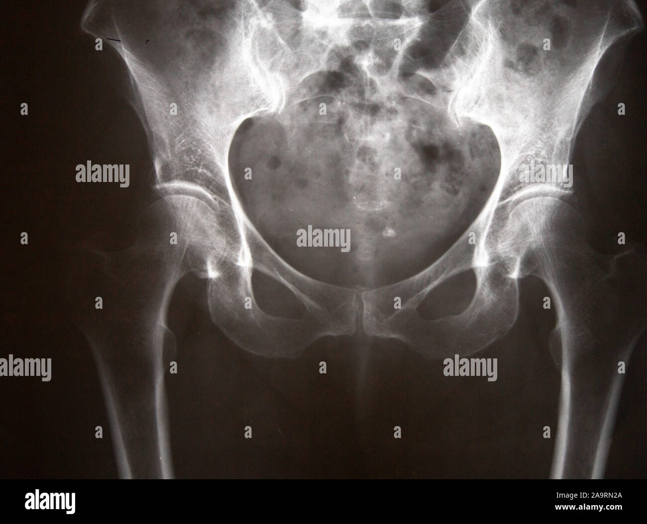 Xray of a human pelvis Stock Photohttps://www.alamy.com/image-license-details/?v=1https://www.alamy.com/xray-of-a-human-pelvis-image333050322.html
RF2A9RN2A–Xray of a human pelvis
 treatment of the pelvic bone of a dog in detail Stock Photohttps://www.alamy.com/image-license-details/?v=1https://www.alamy.com/treatment-of-the-pelvic-bone-of-a-dog-in-detail-image385829758.html
RF2DBM1P6–treatment of the pelvic bone of a dog in detail
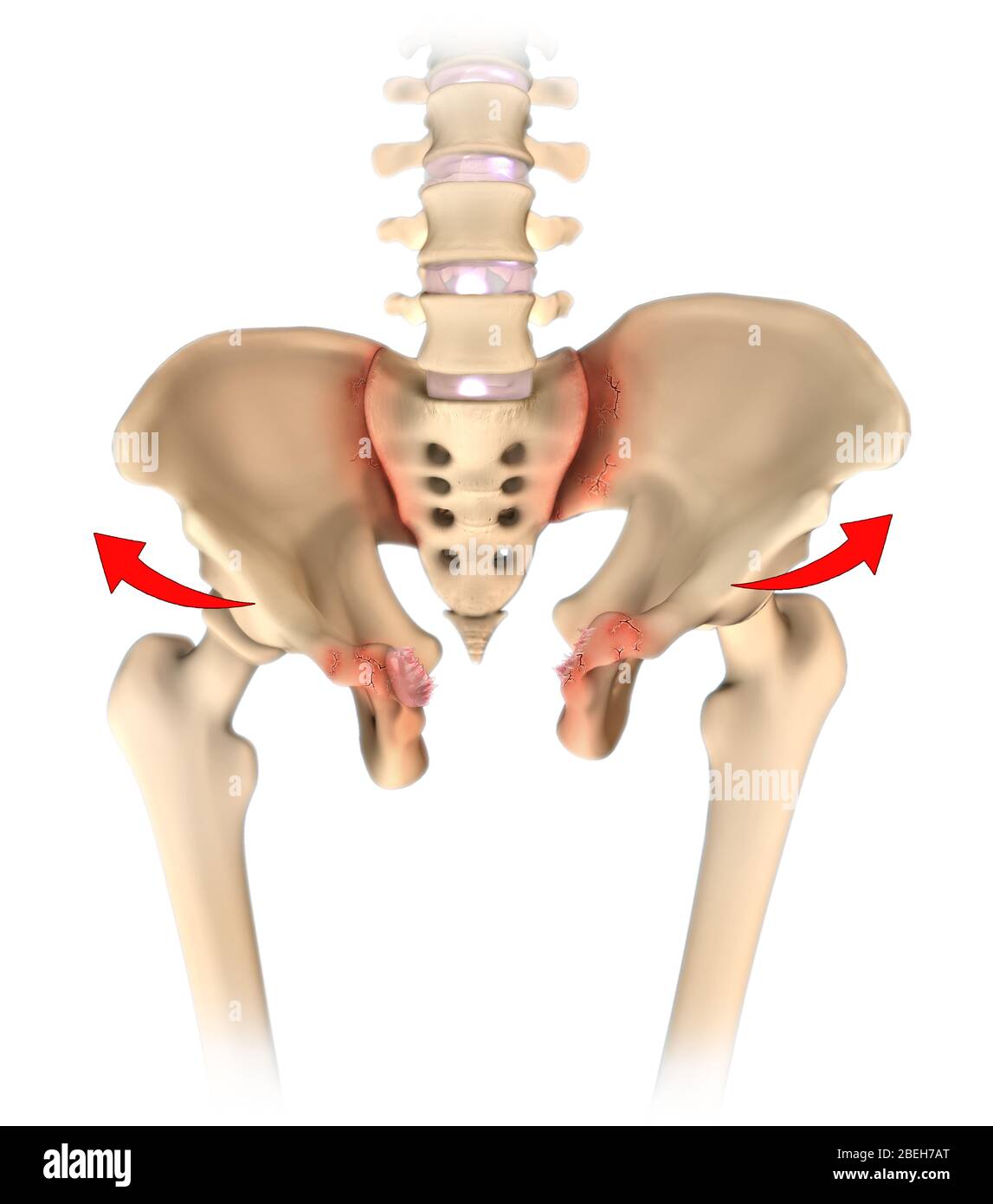 Open Book Pelvic Fracture Stock Photohttps://www.alamy.com/image-license-details/?v=1https://www.alamy.com/open-book-pelvic-fracture-image353191520.html
RM2BEH7AT–Open Book Pelvic Fracture
 Online Traumatologist Orthopedist Doctor Consultate Patient.Pain in Hip Joint.Arthoplasty,Osteoarthritis,Hip Joint Replacement.Diagnostic Checkup Trea Stock Photohttps://www.alamy.com/image-license-details/?v=1https://www.alamy.com/online-traumatologist-orthopedist-doctor-consultate-patientpain-in-hip-jointarthoplastyosteoarthritiship-joint-replacementdiagnostic-checkup-trea-image514274228.html
RF2MTK60M–Online Traumatologist Orthopedist Doctor Consultate Patient.Pain in Hip Joint.Arthoplasty,Osteoarthritis,Hip Joint Replacement.Diagnostic Checkup Trea
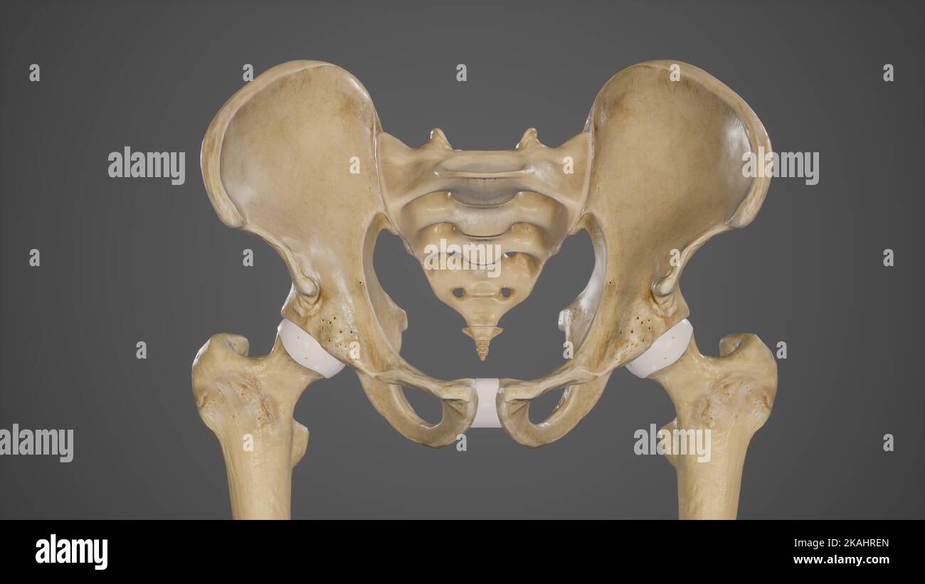 Medical Ilustration of Pelvic Bones-Hip Bone Stock Photohttps://www.alamy.com/image-license-details/?v=1https://www.alamy.com/medical-ilustration-of-pelvic-bones-hip-bone-image488428493.html
RF2KAHREN–Medical Ilustration of Pelvic Bones-Hip Bone
 CT scan of coccyx and pelvic bone 3D rendering for diagnosis fracture of coccyx bone isolated on black background. Stock Photohttps://www.alamy.com/image-license-details/?v=1https://www.alamy.com/ct-scan-of-coccyx-and-pelvic-bone-3d-rendering-for-diagnosis-fracture-of-coccyx-bone-isolated-on-black-background-image482028458.html
RF2K06862–CT scan of coccyx and pelvic bone 3D rendering for diagnosis fracture of coccyx bone isolated on black background.
 U.S. Marine Corps Lance Cpl. Ericka ValenciaReyes, a postal clerk with Headquarters and Support Battalion, Marine Corps Installations Pacific-Marine Corps Base Camp Butler, checks the mail at the Camp Foster Post Office on Okinawa, Japan, Jan. 10, 2024. At 9-years-old, ValenciaReyes left her hometown in N.C. for Mexico with her mother after her parents’ divorce. She decided to move back to the states and join the Marine Corps when she turned 18. Despite fracturing her pelvic bone in boot camp, she persevered and went on to graduate from Papa Company in Feb. 2021. She moved to Okinawa in July, Stock Photohttps://www.alamy.com/image-license-details/?v=1https://www.alamy.com/us-marine-corps-lance-cpl-ericka-valenciareyes-a-postal-clerk-with-headquarters-and-support-battalion-marine-corps-installations-pacific-marine-corps-base-camp-butler-checks-the-mail-at-the-camp-foster-post-office-on-okinawa-japan-jan-10-2024-at-9-years-old-valenciareyes-left-her-hometown-in-nc-for-mexico-with-her-mother-after-her-parents-divorce-she-decided-to-move-back-to-the-states-and-join-the-marine-corps-when-she-turned-18-despite-fracturing-her-pelvic-bone-in-boot-camp-she-persevered-and-went-on-to-graduate-from-papa-company-in-feb-2021-she-moved-to-okinawa-in-july-image594945308.html
RM2WFX2PM–U.S. Marine Corps Lance Cpl. Ericka ValenciaReyes, a postal clerk with Headquarters and Support Battalion, Marine Corps Installations Pacific-Marine Corps Base Camp Butler, checks the mail at the Camp Foster Post Office on Okinawa, Japan, Jan. 10, 2024. At 9-years-old, ValenciaReyes left her hometown in N.C. for Mexico with her mother after her parents’ divorce. She decided to move back to the states and join the Marine Corps when she turned 18. Despite fracturing her pelvic bone in boot camp, she persevered and went on to graduate from Papa Company in Feb. 2021. She moved to Okinawa in July,
 Hand drawing a bone skeleton, anatomical drawing of pelvic bone man, print for Halloween,butterflies fly Stock Photohttps://www.alamy.com/image-license-details/?v=1https://www.alamy.com/hand-drawing-a-bone-skeleton-anatomical-drawing-of-pelvic-bone-man-print-for-halloweenbutterflies-fly-image229170418.html
RFR8RH5P–Hand drawing a bone skeleton, anatomical drawing of pelvic bone man, print for Halloween,butterflies fly
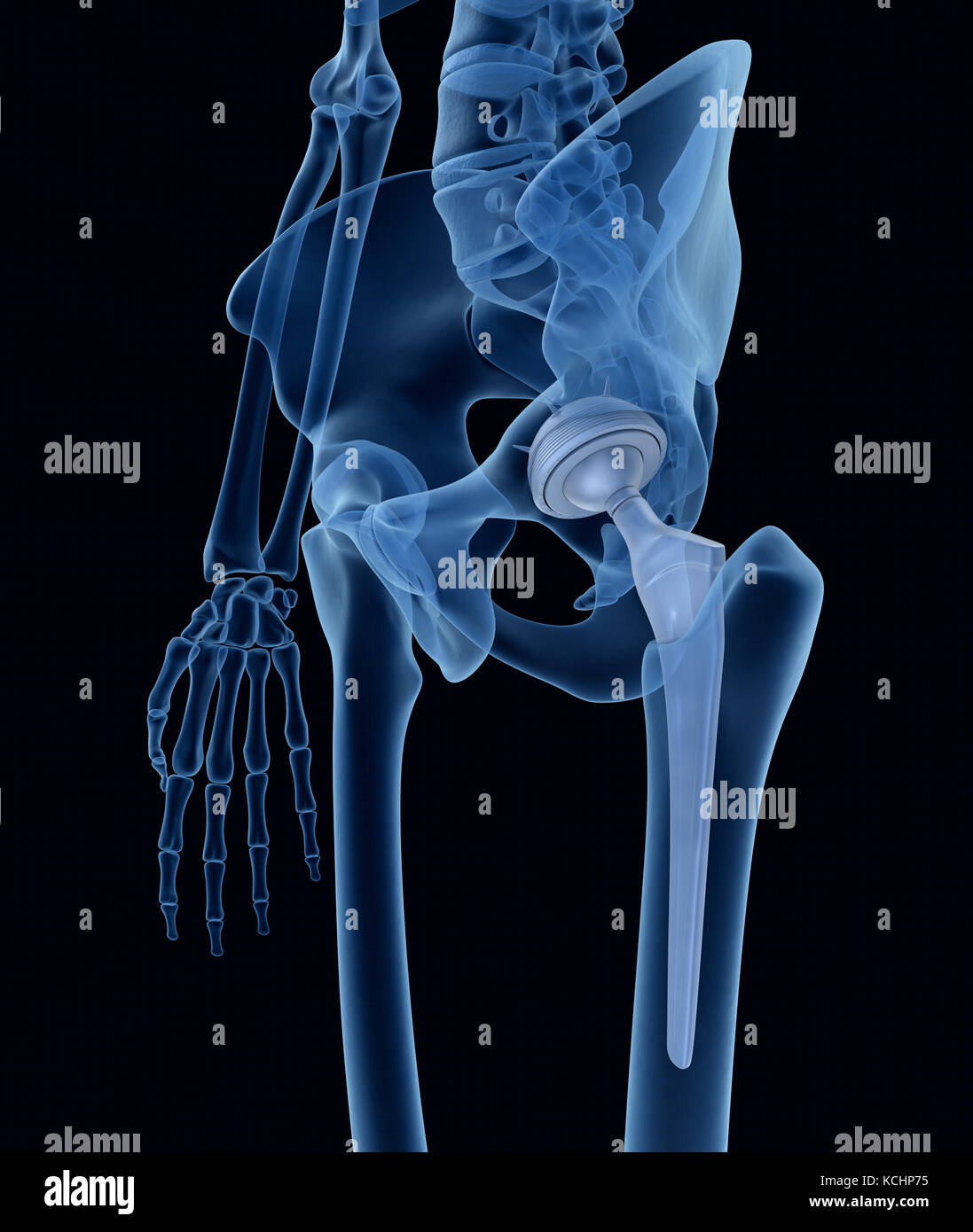 Hip replacement implant installed in the pelvis bone. X-ray view. Medically accurate 3D Stock Photohttps://www.alamy.com/image-license-details/?v=1https://www.alamy.com/stock-image-hip-replacement-implant-installed-in-the-pelvis-bone-x-ray-view-medically-162659817.html
RFKCHP75–Hip replacement implant installed in the pelvis bone. X-ray view. Medically accurate 3D
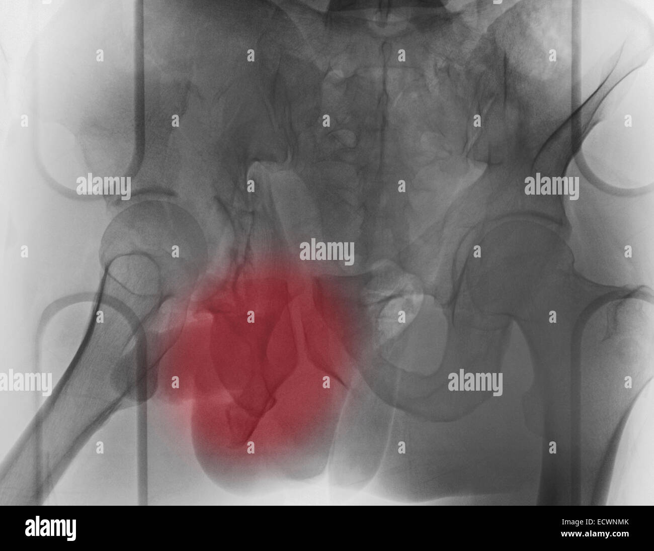 X-ray of a 30 year old man with a pelvic fracture. Stock Photohttps://www.alamy.com/image-license-details/?v=1https://www.alamy.com/stock-photo-x-ray-of-a-30-year-old-man-with-a-pelvic-fracture-76783187.html
RMECWNMK–X-ray of a 30 year old man with a pelvic fracture.
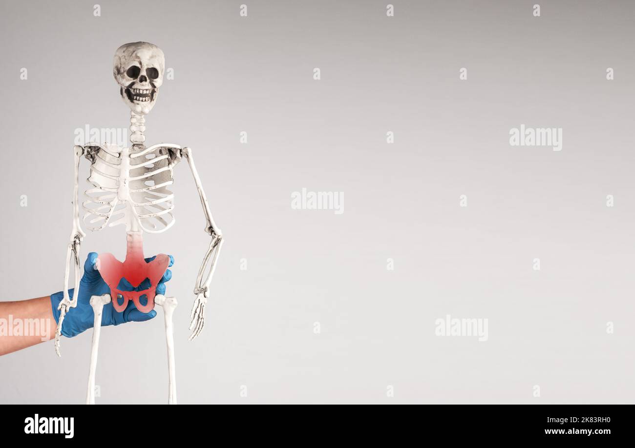 Pelvis, pelvic bone injury, inflammation and pain on skeleton model. High quality photo Stock Photohttps://www.alamy.com/image-license-details/?v=1https://www.alamy.com/pelvis-pelvic-bone-injury-inflammation-and-pain-on-skeleton-model-high-quality-photo-image486891916.html
RF2K83RH0–Pelvis, pelvic bone injury, inflammation and pain on skeleton model. High quality photo
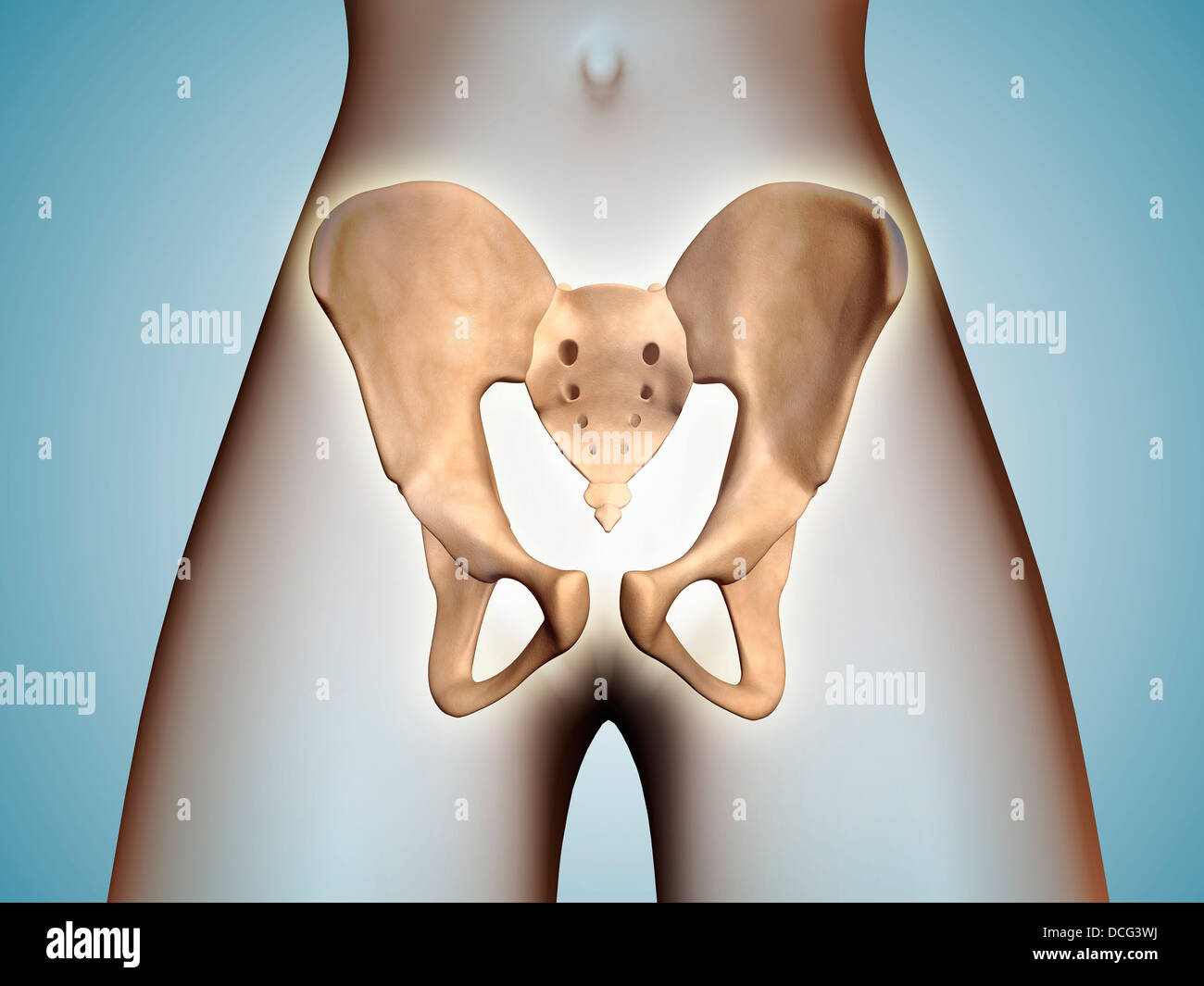 Anatomy of pelvic bone on female body. Stock Photohttps://www.alamy.com/image-license-details/?v=1https://www.alamy.com/stock-photo-anatomy-of-pelvic-bone-on-female-body-59361278.html
RFDCG3WJ–Anatomy of pelvic bone on female body.
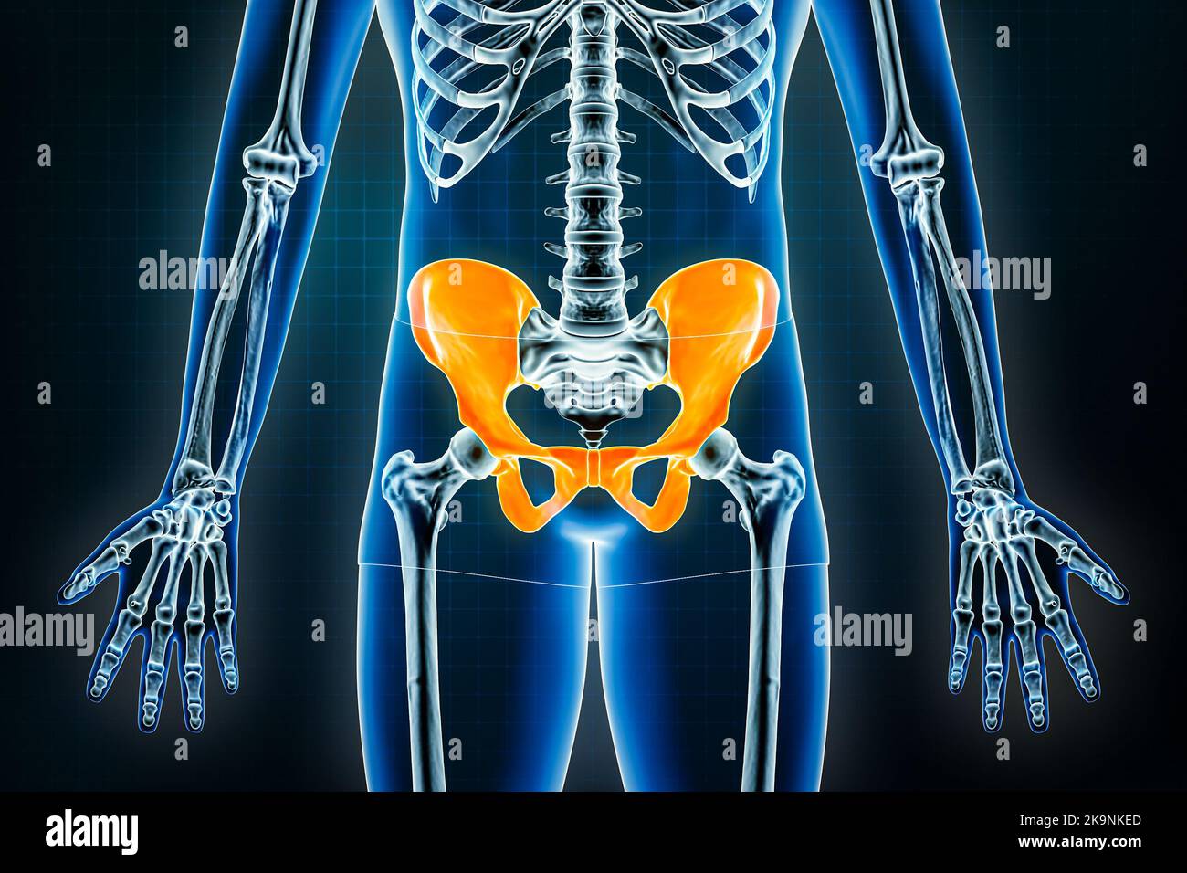 Pelvis x-ray front or anterior view. Osteology of the human skeleton, pelvic girdle bones 3D rendering illustration. Anatomy, medical, science, biolog Stock Photohttps://www.alamy.com/image-license-details/?v=1https://www.alamy.com/pelvis-x-ray-front-or-anterior-view-osteology-of-the-human-skeleton-pelvic-girdle-bones-3d-rendering-illustration-anatomy-medical-science-biolog-image487898501.html
RF2K9NKED–Pelvis x-ray front or anterior view. Osteology of the human skeleton, pelvic girdle bones 3D rendering illustration. Anatomy, medical, science, biolog
 Field dressing a white-tailed buck in Wisconsin Stock Photohttps://www.alamy.com/image-license-details/?v=1https://www.alamy.com/stock-photo-field-dressing-a-white-tailed-buck-in-wisconsin-132457514.html
RMHKDXWE–Field dressing a white-tailed buck in Wisconsin
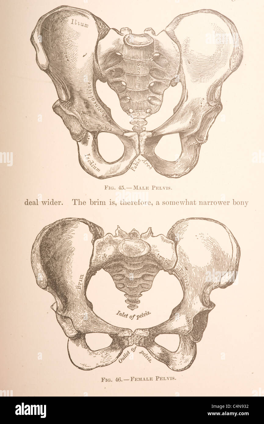 Antique medical illustration of a pelvic bone Stock Photohttps://www.alamy.com/image-license-details/?v=1https://www.alamy.com/stock-photo-antique-medical-illustration-of-a-pelvic-bone-37347494.html
RFC4N932–Antique medical illustration of a pelvic bone
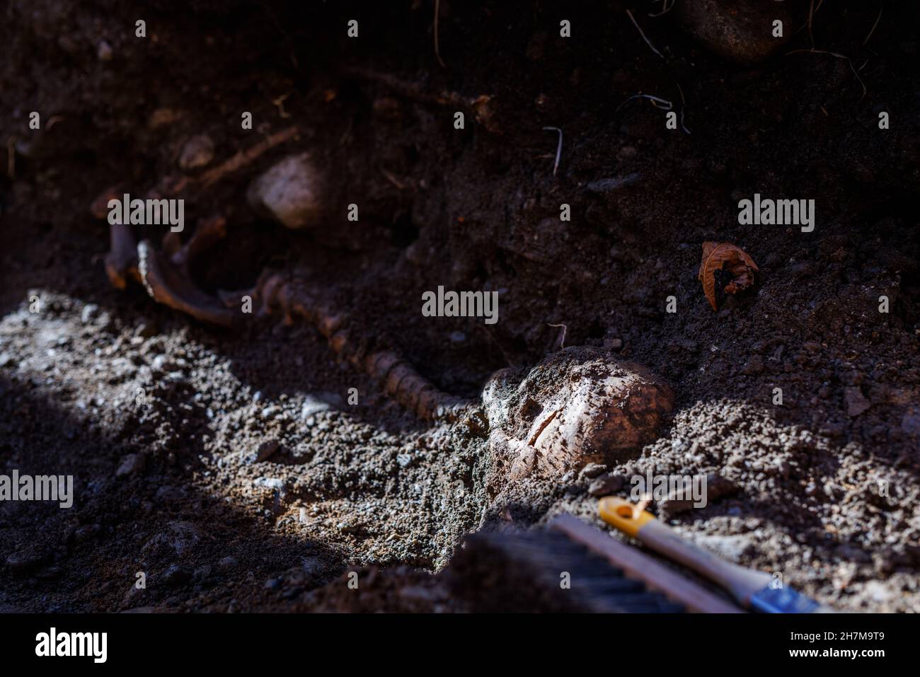 Freiburg, Germany. 08th Nov, 2021. A human skull as well as a spinal column and a pelvic bone are partially buried in the soil of the former leprosy cemetery in Freiburg. The discovery of around 400 skeletons in a former leprosy cemetery is of great importance for science. It also draws attention to a disease that has been eradicated in Germany but is still rampant in other parts of the world. Credit: Philipp von Ditfurth/dpa/Alamy Live News Stock Photohttps://www.alamy.com/image-license-details/?v=1https://www.alamy.com/freiburg-germany-08th-nov-2021-a-human-skull-as-well-as-a-spinal-column-and-a-pelvic-bone-are-partially-buried-in-the-soil-of-the-former-leprosy-cemetery-in-freiburg-the-discovery-of-around-400-skeletons-in-a-former-leprosy-cemetery-is-of-great-importance-for-science-it-also-draws-attention-to-a-disease-that-has-been-eradicated-in-germany-but-is-still-rampant-in-other-parts-of-the-world-credit-philipp-von-ditfurthdpaalamy-live-news-image452218937.html
RM2H7M9T9–Freiburg, Germany. 08th Nov, 2021. A human skull as well as a spinal column and a pelvic bone are partially buried in the soil of the former leprosy cemetery in Freiburg. The discovery of around 400 skeletons in a former leprosy cemetery is of great importance for science. It also draws attention to a disease that has been eradicated in Germany but is still rampant in other parts of the world. Credit: Philipp von Ditfurth/dpa/Alamy Live News
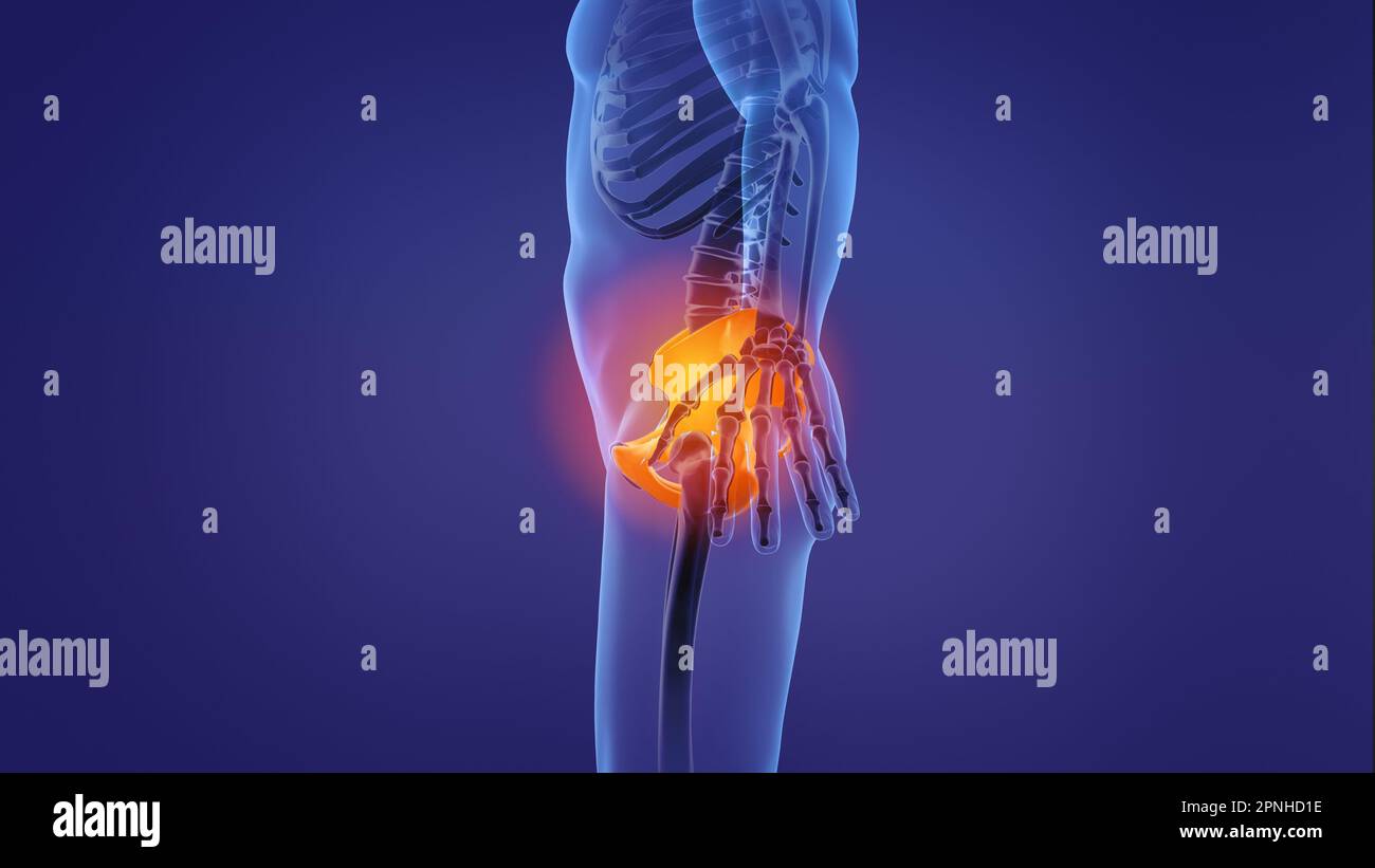 Animation of a painful pelvic girdle Stock Photohttps://www.alamy.com/image-license-details/?v=1https://www.alamy.com/animation-of-a-painful-pelvic-girdle-image546812602.html
RF2PNHD1E–Animation of a painful pelvic girdle
 Usti Nad Labem, Czech Republic. 28th May, 2024. Demonstration of 3D models of bones (pictured pelvic bone) made in the new 3D printing laboratory, May 28, 2024, Masaryk Hospital, Usti nad Labem. The 3D printer here will help doctors with pre-operative planning, and in the laboratory they can convert CT scans of bones or joints into three-dimensional form before more complex procedures. Credit: Libor Zavoral/CTK Photo/Alamy Live News Stock Photohttps://www.alamy.com/image-license-details/?v=1https://www.alamy.com/usti-nad-labem-czech-republic-28th-may-2024-demonstration-of-3d-models-of-bones-pictured-pelvic-bone-made-in-the-new-3d-printing-laboratory-may-28-2024-masaryk-hospital-usti-nad-labem-the-3d-printer-here-will-help-doctors-with-pre-operative-planning-and-in-the-laboratory-they-can-convert-ct-scans-of-bones-or-joints-into-three-dimensional-form-before-more-complex-procedures-credit-libor-zavoralctk-photoalamy-live-news-image607919233.html
RM2X91355–Usti Nad Labem, Czech Republic. 28th May, 2024. Demonstration of 3D models of bones (pictured pelvic bone) made in the new 3D printing laboratory, May 28, 2024, Masaryk Hospital, Usti nad Labem. The 3D printer here will help doctors with pre-operative planning, and in the laboratory they can convert CT scans of bones or joints into three-dimensional form before more complex procedures. Credit: Libor Zavoral/CTK Photo/Alamy Live News
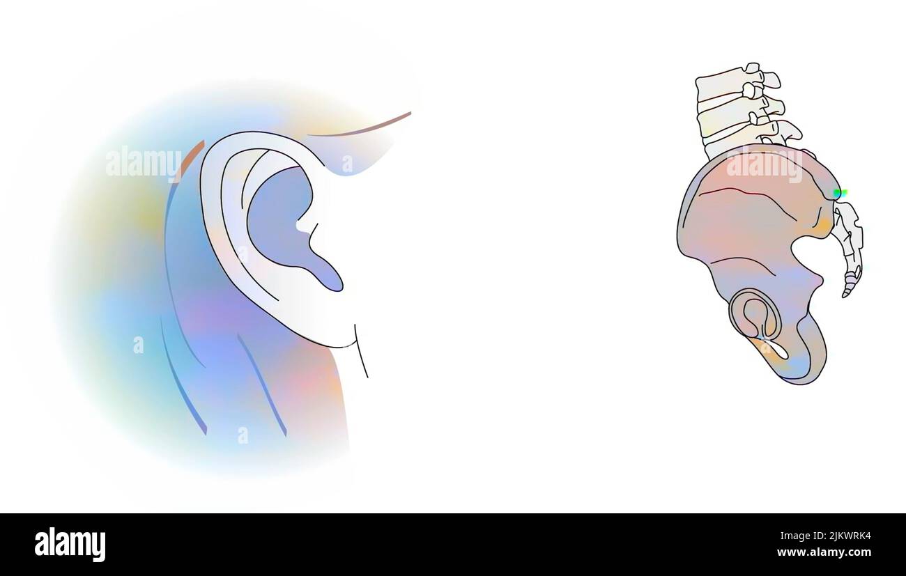 Analogy between the pinna of the ear and the iliac bone. Stock Photohttps://www.alamy.com/image-license-details/?v=1https://www.alamy.com/analogy-between-the-pinna-of-the-ear-and-the-iliac-bone-image476925768.html
RF2JKWRK4–Analogy between the pinna of the ear and the iliac bone.
 X-ray close up pelvic bone with pain symptoms in joints Stock Photohttps://www.alamy.com/image-license-details/?v=1https://www.alamy.com/x-ray-close-up-pelvic-bone-with-pain-symptoms-in-joints-image504201317.html
RF2M889WW–X-ray close up pelvic bone with pain symptoms in joints
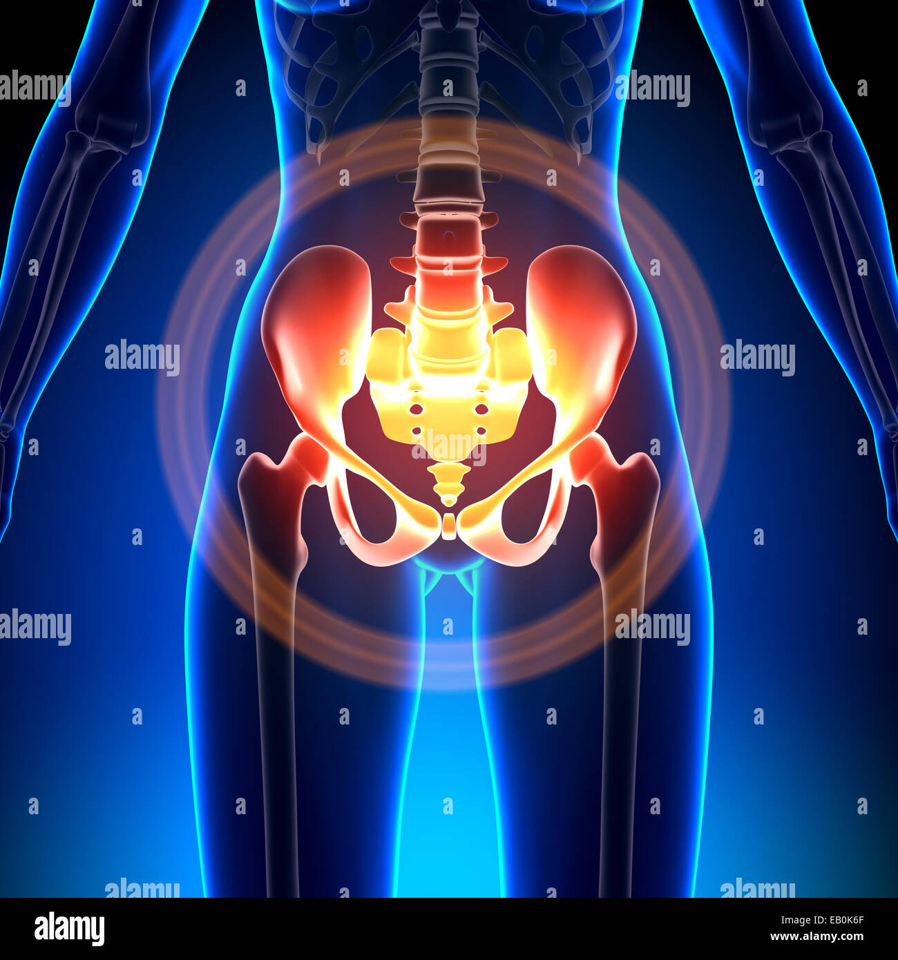 Female Hip / Sacrum / Pubis / Ischium / Ilium - Anatomy Bones Stock Photohttps://www.alamy.com/image-license-details/?v=1https://www.alamy.com/stock-photo-female-hip-sacrum-pubis-ischium-ilium-anatomy-bones-75617767.html
RFEB0K6F–Female Hip / Sacrum / Pubis / Ischium / Ilium - Anatomy Bones
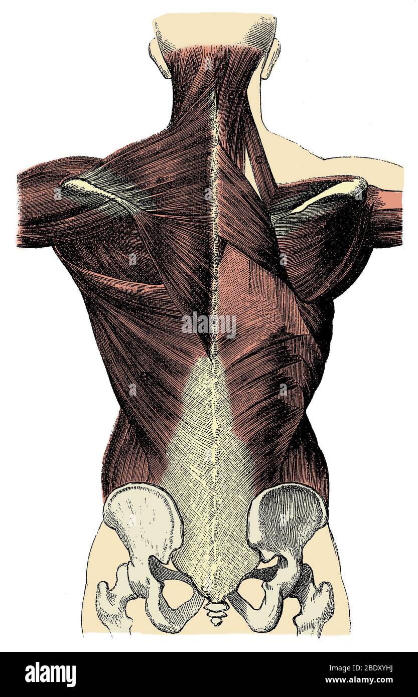 Back Muscles Stock Photohttps://www.alamy.com/image-license-details/?v=1https://www.alamy.com/back-muscles-image352790302.html
RM2BDXYHJ–Back Muscles
 Online Touchscreen Traumatology Orthopedics Mobile Application.Smartphone X Ray Radiography,Hip Joint Pain Replacement.Arthoplasty,Osteoarthritis.Ront Stock Photohttps://www.alamy.com/image-license-details/?v=1https://www.alamy.com/online-touchscreen-traumatology-orthopedics-mobile-applicationsmartphone-x-ray-radiographyhip-joint-pain-replacementarthoplastyosteoarthritisront-image514274289.html
RF2MTK62W–Online Touchscreen Traumatology Orthopedics Mobile Application.Smartphone X Ray Radiography,Hip Joint Pain Replacement.Arthoplasty,Osteoarthritis.Ront
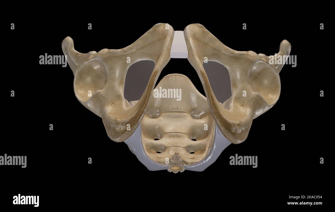 The Pelvic Girdle and Pelvic Outlet Stock Photohttps://www.alamy.com/image-license-details/?v=1https://www.alamy.com/the-pelvic-girdle-and-pelvic-outlet-image488320816.html
RF2KACX54–The Pelvic Girdle and Pelvic Outlet
 CT scan of Pelvic bone and hip joint 3D rendering for diagnosis fracture of Pelvic bone and hip joint isolated on white background. Stock Photohttps://www.alamy.com/image-license-details/?v=1https://www.alamy.com/ct-scan-of-pelvic-bone-and-hip-joint-3d-rendering-for-diagnosis-fracture-of-pelvic-bone-and-hip-joint-isolated-on-white-background-image481930954.html
RF2K01RRP–CT scan of Pelvic bone and hip joint 3D rendering for diagnosis fracture of Pelvic bone and hip joint isolated on white background.
 U.S. Marine Corps Lance Cpl. Ericka ValenciaReyes, a postal clerk with Headquarters and Support Battalion, Marine Corps Installations Pacific-Marine Corps Base Camp Butler, reviews the mail in the boxes at the Camp Foster Post Office, on Okinawa, Japan, Jan. 10, 2024. At 9-years-old, ValenciaReyes left her hometown in N.C. for Mexico with her mother after her parents’ divorce. She decided to move back to the states and join the Marine Corps when she turned 18. Despite fracturing her pelvic bone in boot camp, she persevered and went on to graduate from Papa Company in Feb. 2021. She moved to O Stock Photohttps://www.alamy.com/image-license-details/?v=1https://www.alamy.com/us-marine-corps-lance-cpl-ericka-valenciareyes-a-postal-clerk-with-headquarters-and-support-battalion-marine-corps-installations-pacific-marine-corps-base-camp-butler-reviews-the-mail-in-the-boxes-at-the-camp-foster-post-office-on-okinawa-japan-jan-10-2024-at-9-years-old-valenciareyes-left-her-hometown-in-nc-for-mexico-with-her-mother-after-her-parents-divorce-she-decided-to-move-back-to-the-states-and-join-the-marine-corps-when-she-turned-18-despite-fracturing-her-pelvic-bone-in-boot-camp-she-persevered-and-went-on-to-graduate-from-papa-company-in-feb-2021-she-moved-to-o-image594947383.html
RM2WFX5CR–U.S. Marine Corps Lance Cpl. Ericka ValenciaReyes, a postal clerk with Headquarters and Support Battalion, Marine Corps Installations Pacific-Marine Corps Base Camp Butler, reviews the mail in the boxes at the Camp Foster Post Office, on Okinawa, Japan, Jan. 10, 2024. At 9-years-old, ValenciaReyes left her hometown in N.C. for Mexico with her mother after her parents’ divorce. She decided to move back to the states and join the Marine Corps when she turned 18. Despite fracturing her pelvic bone in boot camp, she persevered and went on to graduate from Papa Company in Feb. 2021. She moved to O
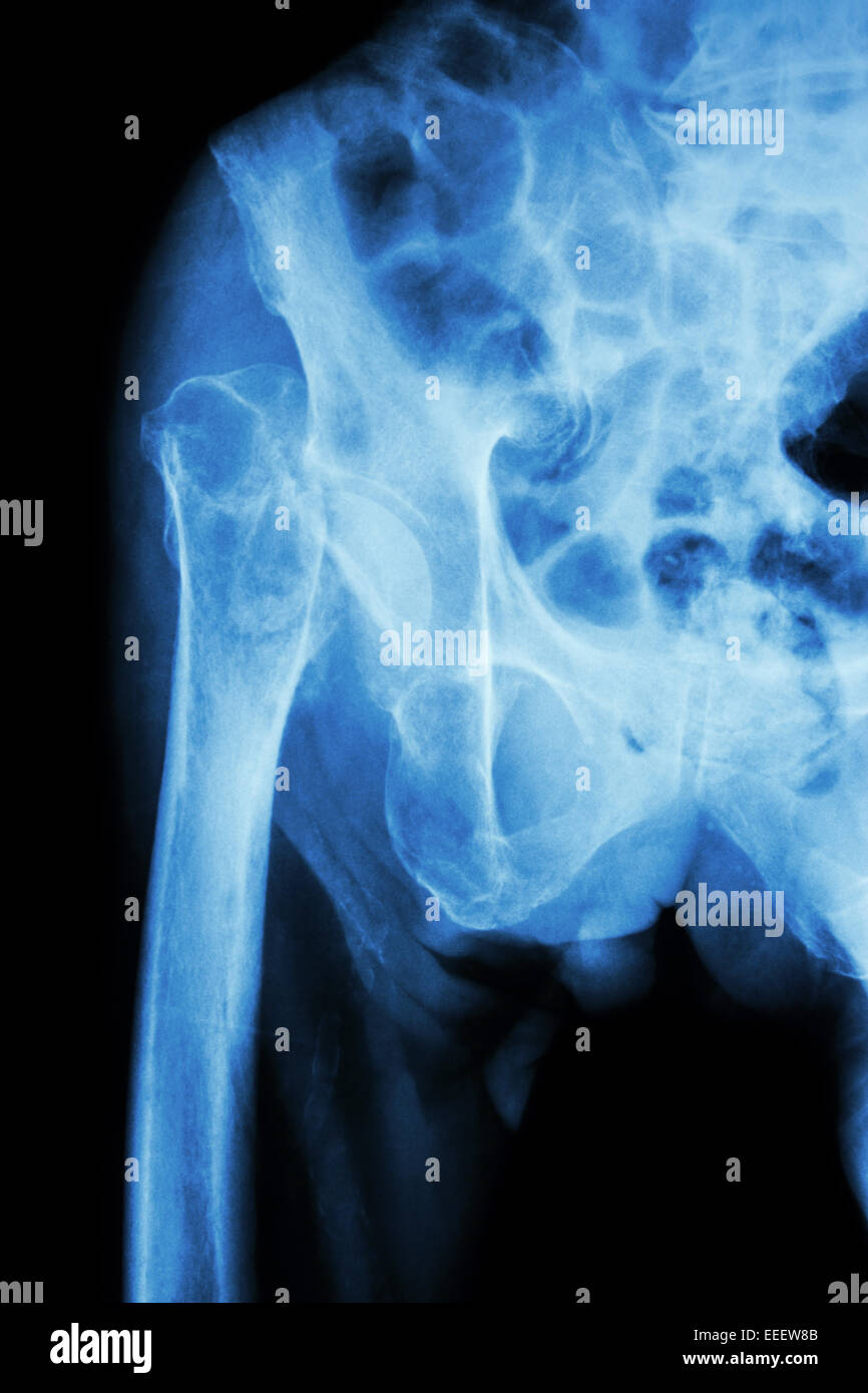 X-ray pelvis & hip joint : Fracture head of femur (thigh bone) Stock Photohttps://www.alamy.com/image-license-details/?v=1https://www.alamy.com/stock-photo-x-ray-pelvis-hip-joint-fracture-head-of-femur-thigh-bone-77773819.html
RFEEEW8B–X-ray pelvis & hip joint : Fracture head of femur (thigh bone)
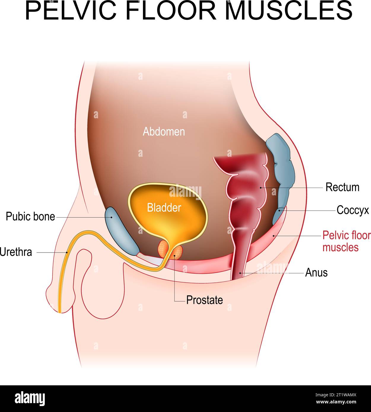 Pelvic floor muscles. Cross section of male abdomen with pelvic diaphragm, Prostate, bladder, rectum, pubic bone, urethra, anus, and coccyx. Kegel Stock Vectorhttps://www.alamy.com/image-license-details/?v=1https://www.alamy.com/pelvic-floor-muscles-cross-section-of-male-abdomen-with-pelvic-diaphragm-prostate-bladder-rectum-pubic-bone-urethra-anus-and-coccyx-kegel-image569114026.html
RF2T1WAMX–Pelvic floor muscles. Cross section of male abdomen with pelvic diaphragm, Prostate, bladder, rectum, pubic bone, urethra, anus, and coccyx. Kegel
 X-ray of a 30 year old man with a pelvic fracture. Stock Photohttps://www.alamy.com/image-license-details/?v=1https://www.alamy.com/stock-photo-x-ray-of-a-30-year-old-man-with-a-pelvic-fracture-76783184.html
RMECWNMG–X-ray of a 30 year old man with a pelvic fracture.
 Pelvis, pelvic bone trauma, injury and pain concept on background with copy space. High quality photo Stock Photohttps://www.alamy.com/image-license-details/?v=1https://www.alamy.com/pelvis-pelvic-bone-trauma-injury-and-pain-concept-on-background-with-copy-space-high-quality-photo-image485770327.html
RF2K68N07–Pelvis, pelvic bone trauma, injury and pain concept on background with copy space. High quality photo
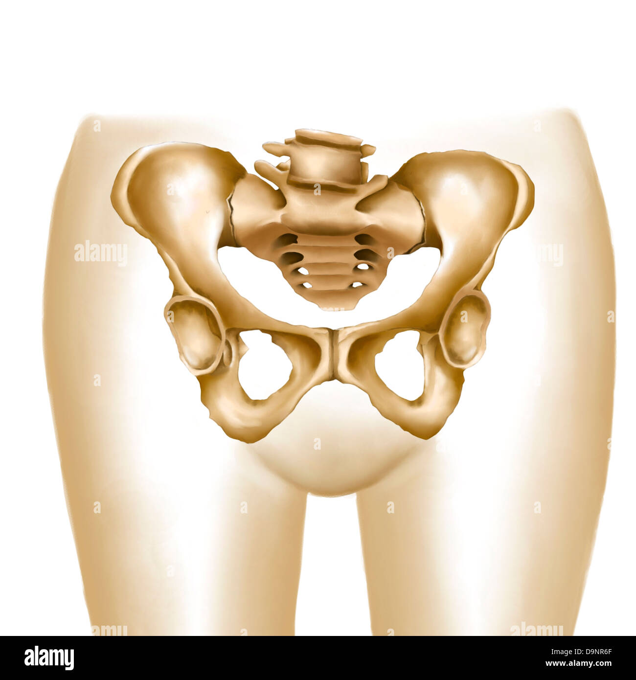 Anatomy of female hips and pelvic bones. Stock Photohttps://www.alamy.com/image-license-details/?v=1https://www.alamy.com/stock-photo-anatomy-of-female-hips-and-pelvic-bones-57642215.html
RFD9NR6F–Anatomy of female hips and pelvic bones.
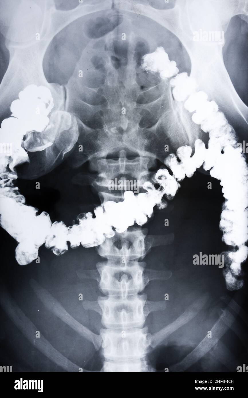 X-ray radiography of the pelvic area of a human. Visualization of the intestine. Stock Photohttps://www.alamy.com/image-license-details/?v=1https://www.alamy.com/x-ray-radiography-of-the-pelvic-area-of-a-human-visualization-of-the-intestine-image528936929.html
RF2NMF4CH–X-ray radiography of the pelvic area of a human. Visualization of the intestine.
 Field dressing a white-tailed buck in Wisconsin Stock Photohttps://www.alamy.com/image-license-details/?v=1https://www.alamy.com/stock-photo-field-dressing-a-white-tailed-buck-in-wisconsin-132457731.html
RMHKDY57–Field dressing a white-tailed buck in Wisconsin
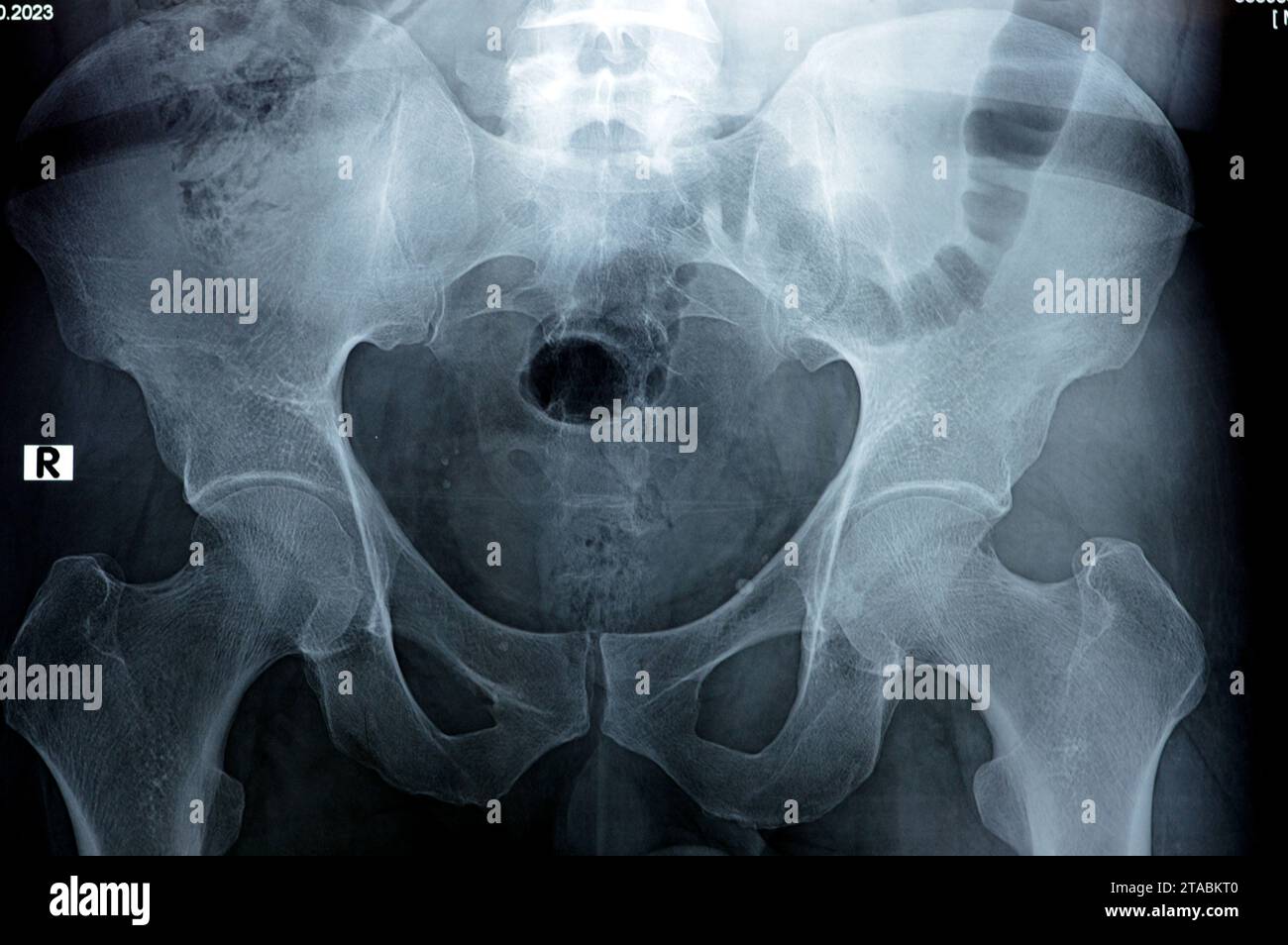 Hip joints digital radiographic examination reveals normal appearance of hip joins, multiple pelvic phleboli, bilateral coxa profunda, preserved spher Stock Photohttps://www.alamy.com/image-license-details/?v=1https://www.alamy.com/hip-joints-digital-radiographic-examination-reveals-normal-appearance-of-hip-joins-multiple-pelvic-phleboli-bilateral-coxa-profunda-preserved-spher-image574345744.html
RF2TABKT0–Hip joints digital radiographic examination reveals normal appearance of hip joins, multiple pelvic phleboli, bilateral coxa profunda, preserved spher
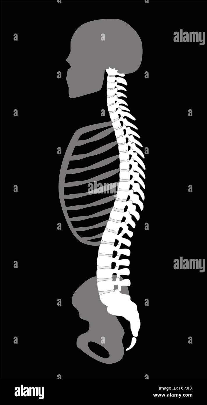 Upper body skeleton with backbone, cranial bone, ribs and pelvis - side view. Illustration on black background. Stock Photohttps://www.alamy.com/image-license-details/?v=1https://www.alamy.com/stock-photo-upper-body-skeleton-with-backbone-cranial-bone-ribs-and-pelvis-side-90223166.html
RFF6P0FX–Upper body skeleton with backbone, cranial bone, ribs and pelvis - side view. Illustration on black background.
 Animation of a painful pelvic girdle Stock Photohttps://www.alamy.com/image-license-details/?v=1https://www.alamy.com/animation-of-a-painful-pelvic-girdle-image546812635.html
RF2PNHD2K–Animation of a painful pelvic girdle
 Usti Nad Labem, Czech Republic. 28th May, 2024. Tomas Novotny, head of the orthopaedic clinic, shows a work-in-progress pelvic bone implant made in the new 3D printing laboratory, May 28, 2024, Masaryk Hospital, Usti nad Labem. The 3D printer will help doctors with pre-operative planning, and the laboratory can convert CT scans of bones or joints into three-dimensional form before more complex procedures. Credit: Libor Zavoral/CTK Photo/Alamy Live News Stock Photohttps://www.alamy.com/image-license-details/?v=1https://www.alamy.com/usti-nad-labem-czech-republic-28th-may-2024-tomas-novotny-head-of-the-orthopaedic-clinic-shows-a-work-in-progress-pelvic-bone-implant-made-in-the-new-3d-printing-laboratory-may-28-2024-masaryk-hospital-usti-nad-labem-the-3d-printer-will-help-doctors-with-pre-operative-planning-and-the-laboratory-can-convert-ct-scans-of-bones-or-joints-into-three-dimensional-form-before-more-complex-procedures-credit-libor-zavoralctk-photoalamy-live-news-image607919445.html
RM2X913CN–Usti Nad Labem, Czech Republic. 28th May, 2024. Tomas Novotny, head of the orthopaedic clinic, shows a work-in-progress pelvic bone implant made in the new 3D printing laboratory, May 28, 2024, Masaryk Hospital, Usti nad Labem. The 3D printer will help doctors with pre-operative planning, and the laboratory can convert CT scans of bones or joints into three-dimensional form before more complex procedures. Credit: Libor Zavoral/CTK Photo/Alamy Live News
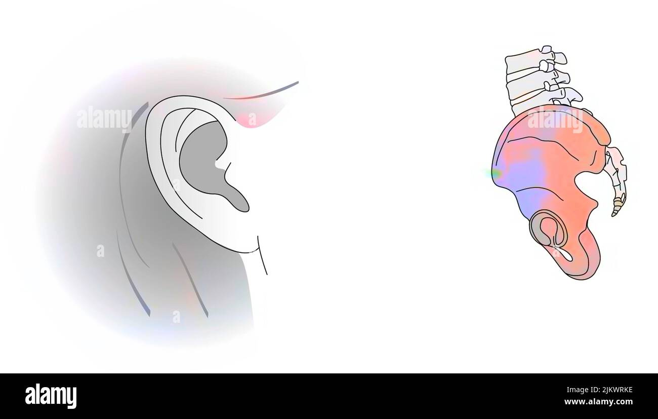 Analogy between the pinna of the ear and the iliac bone. Stock Photohttps://www.alamy.com/image-license-details/?v=1https://www.alamy.com/analogy-between-the-pinna-of-the-ear-and-the-iliac-bone-image476925778.html
RF2JKWRKE–Analogy between the pinna of the ear and the iliac bone.
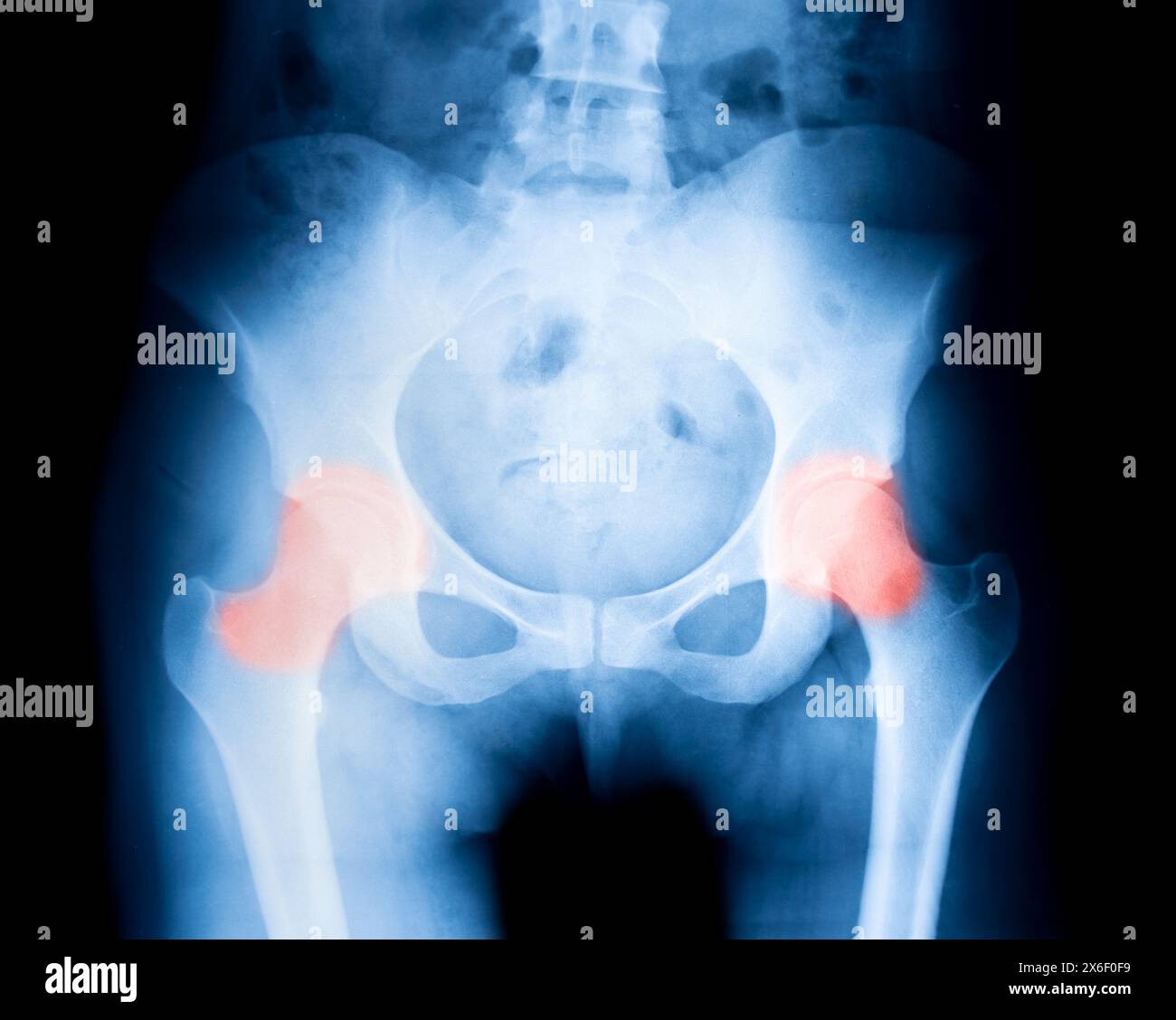 X-ray close up pelvic bone with pain symptoms in joints Stock Photohttps://www.alamy.com/image-license-details/?v=1https://www.alamy.com/x-ray-close-up-pelvic-bone-with-pain-symptoms-in-joints-image606380525.html
RF2X6F0F9–X-ray close up pelvic bone with pain symptoms in joints
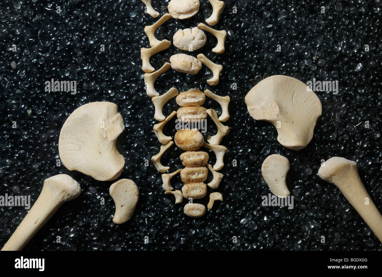 Disarticulated skeleton of a child showing vertebral column and pelvic girdle Stock Photohttps://www.alamy.com/image-license-details/?v=1https://www.alamy.com/stock-photo-disarticulated-skeleton-of-a-child-showing-vertebral-column-and-pelvic-27351088.html
RMBGDXGG–Disarticulated skeleton of a child showing vertebral column and pelvic girdle
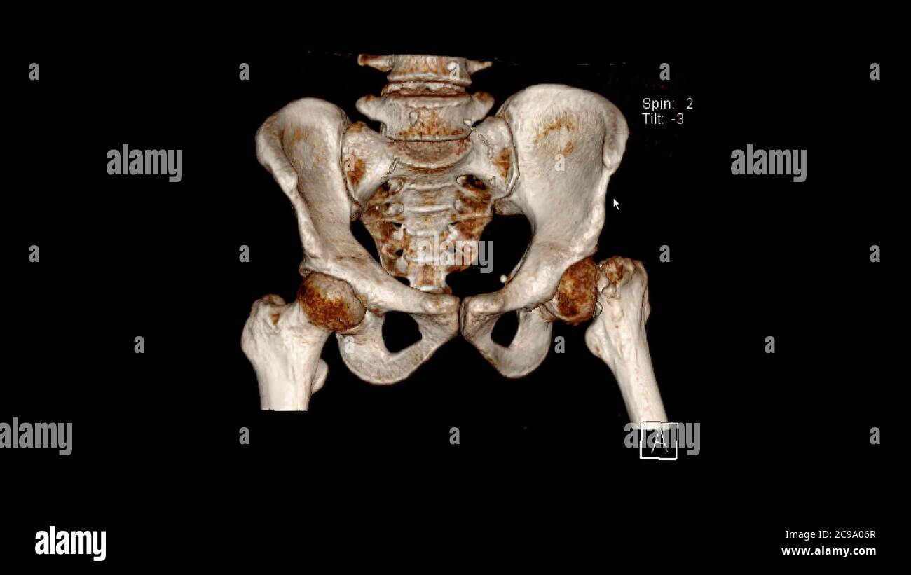 Computed Tomography Volume Rendering examination of the pelvis showing left femur neck fracture ( CT VR Pelvis). 3D rendering Stock Photohttps://www.alamy.com/image-license-details/?v=1https://www.alamy.com/computed-tomography-volume-rendering-examination-of-the-pelvis-showing-left-femur-neck-fracture-ct-vr-pelvis-3d-rendering-image367169343.html
RF2C9A06R–Computed Tomography Volume Rendering examination of the pelvis showing left femur neck fracture ( CT VR Pelvis). 3D rendering
 Scientist,traumatologist orthopedist Doctor Examine Hip Joint Pain.Arthoplasty,Osteoarthritis.Hip Joint Replacement Research.Smartphone Bones X Ray Ra Stock Photohttps://www.alamy.com/image-license-details/?v=1https://www.alamy.com/scientisttraumatologist-orthopedist-doctor-examine-hip-joint-painarthoplastyosteoarthritiship-joint-replacement-researchsmartphone-bones-x-ray-ra-image514274329.html
RF2MTK649–Scientist,traumatologist orthopedist Doctor Examine Hip Joint Pain.Arthoplasty,Osteoarthritis.Hip Joint Replacement Research.Smartphone Bones X Ray Ra
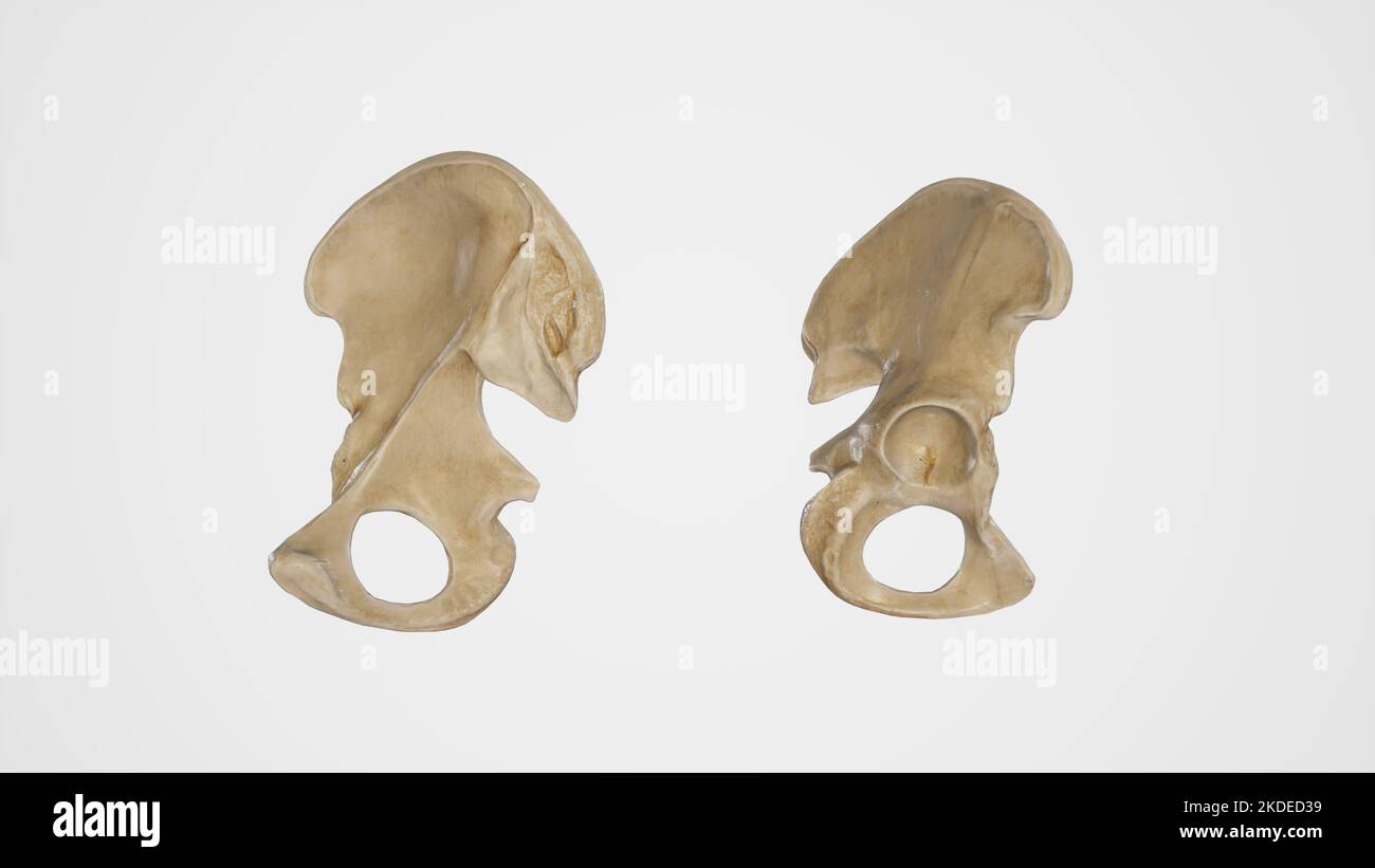 Medial and Lateral View of Hip Bone Stock Photohttps://www.alamy.com/image-license-details/?v=1https://www.alamy.com/medial-and-lateral-view-of-hip-bone-image490198445.html
RF2KDED39–Medial and Lateral View of Hip Bone
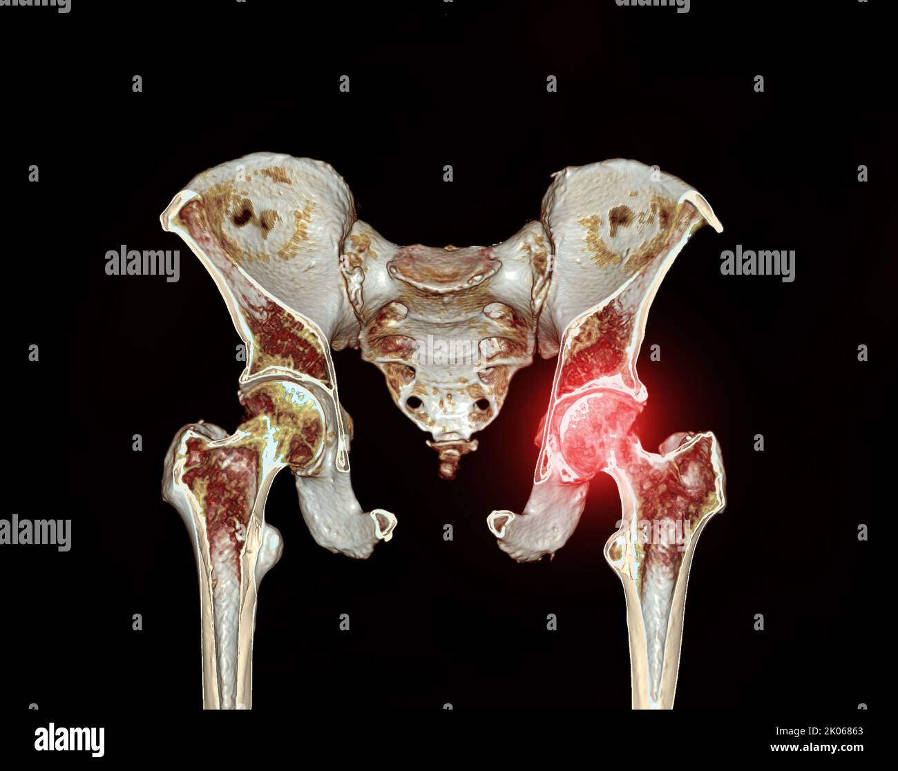 CT scan of Pelvic bone and hip joint 3D rendering for diagnosis fracture of Pelvic bone and hip joint isolated on black background. Stock Photohttps://www.alamy.com/image-license-details/?v=1https://www.alamy.com/ct-scan-of-pelvic-bone-and-hip-joint-3d-rendering-for-diagnosis-fracture-of-pelvic-bone-and-hip-joint-isolated-on-black-background-image482028459.html
RF2K06863–CT scan of Pelvic bone and hip joint 3D rendering for diagnosis fracture of Pelvic bone and hip joint isolated on black background.
 U.S. Marine Corps Lance Cpl. Ericka ValenciaReyes, a postal clerk with Headquarters and Support Battalion, Marine Corps Installations Pacific-Marine Corps Base Camp Butler, poses at the Camp Foster Post Office, on Okinawa, Japan, Jan. 10, 2024. At 9-years-old, ValenciaReyes left her hometown in N.C. for Mexico with her mother after her parents’ divorce. She decided to move back to the states and join the Marine Corps when she turned 18. Despite fracturing her pelvic bone in boot camp, she persevered and went on to graduate from Papa Company in Feb. 2021. She moved to Okinawa in July, 2022. Stock Photohttps://www.alamy.com/image-license-details/?v=1https://www.alamy.com/us-marine-corps-lance-cpl-ericka-valenciareyes-a-postal-clerk-with-headquarters-and-support-battalion-marine-corps-installations-pacific-marine-corps-base-camp-butler-poses-at-the-camp-foster-post-office-on-okinawa-japan-jan-10-2024-at-9-years-old-valenciareyes-left-her-hometown-in-nc-for-mexico-with-her-mother-after-her-parents-divorce-she-decided-to-move-back-to-the-states-and-join-the-marine-corps-when-she-turned-18-despite-fracturing-her-pelvic-bone-in-boot-camp-she-persevered-and-went-on-to-graduate-from-papa-company-in-feb-2021-she-moved-to-okinawa-in-july-2022-image594943488.html
RM2WFX0DM–U.S. Marine Corps Lance Cpl. Ericka ValenciaReyes, a postal clerk with Headquarters and Support Battalion, Marine Corps Installations Pacific-Marine Corps Base Camp Butler, poses at the Camp Foster Post Office, on Okinawa, Japan, Jan. 10, 2024. At 9-years-old, ValenciaReyes left her hometown in N.C. for Mexico with her mother after her parents’ divorce. She decided to move back to the states and join the Marine Corps when she turned 18. Despite fracturing her pelvic bone in boot camp, she persevered and went on to graduate from Papa Company in Feb. 2021. She moved to Okinawa in July, 2022.
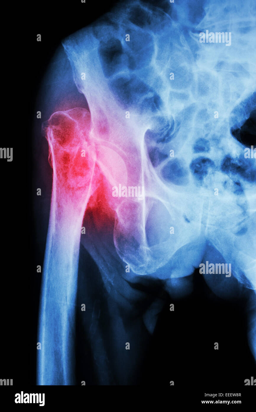 X-ray pelvis & hip joint : Fracture head of femur (thigh bone) Stock Photohttps://www.alamy.com/image-license-details/?v=1https://www.alamy.com/stock-photo-x-ray-pelvis-hip-joint-fracture-head-of-femur-thigh-bone-77773831.html
RFEEEW8R–X-ray pelvis & hip joint : Fracture head of femur (thigh bone)
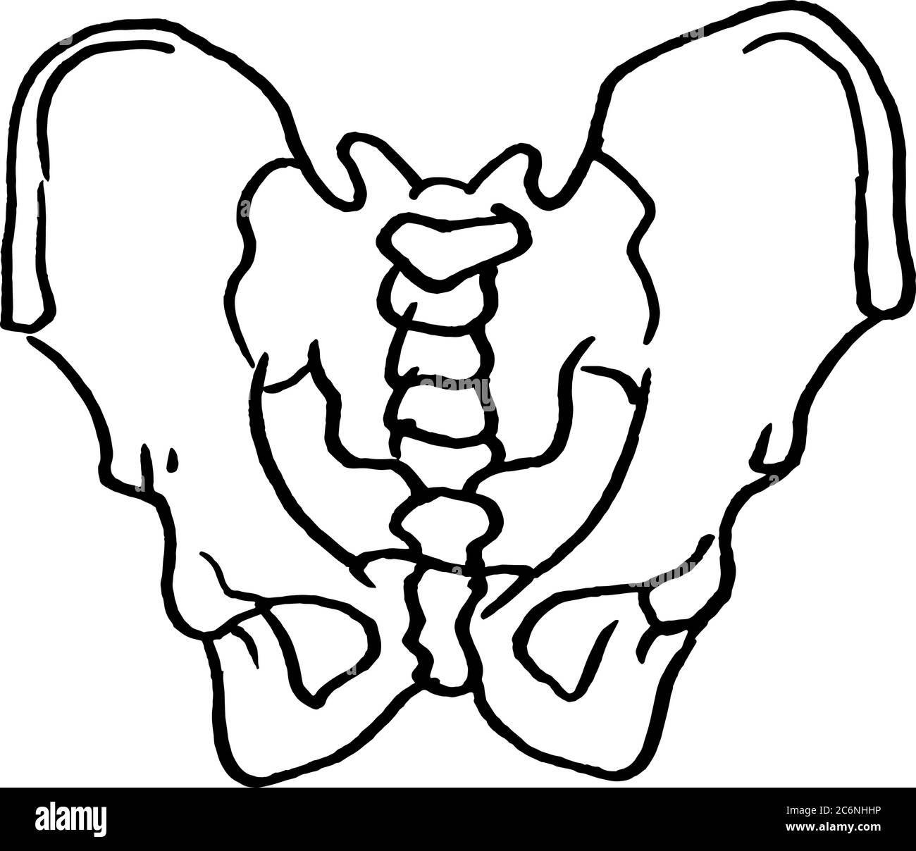 Contour vector outline drawing of human pelvis bones. Medical design editable template Stock Vectorhttps://www.alamy.com/image-license-details/?v=1https://www.alamy.com/contour-vector-outline-drawing-of-human-pelvis-bones-medical-design-editable-template-image365580482.html
RF2C6NHHP–Contour vector outline drawing of human pelvis bones. Medical design editable template
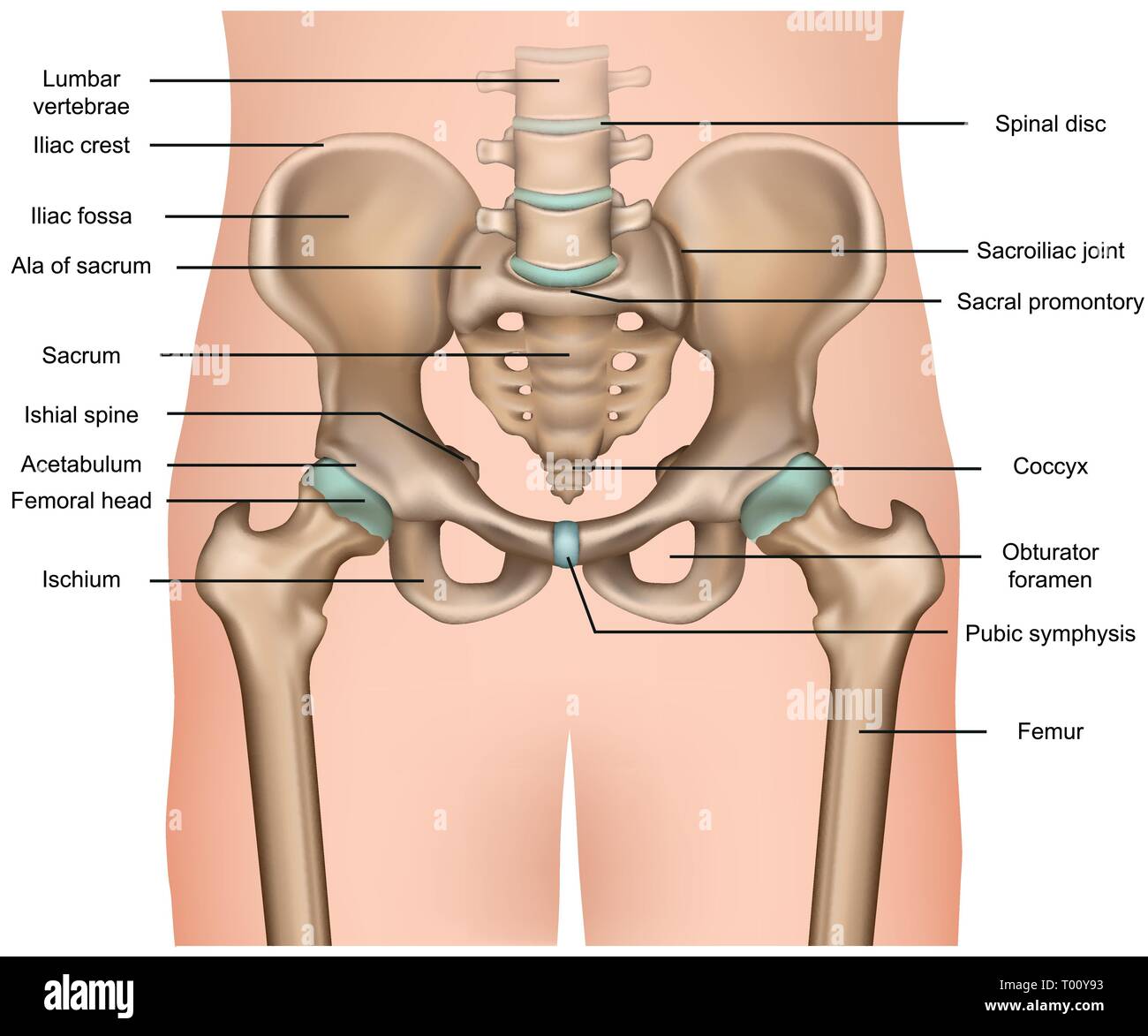 human pelvis anatomy 3d medical vector illustration on white background Stock Vectorhttps://www.alamy.com/image-license-details/?v=1https://www.alamy.com/human-pelvis-anatomy-3d-medical-vector-illustration-on-white-background-image240966575.html
RFT00Y93–human pelvis anatomy 3d medical vector illustration on white background
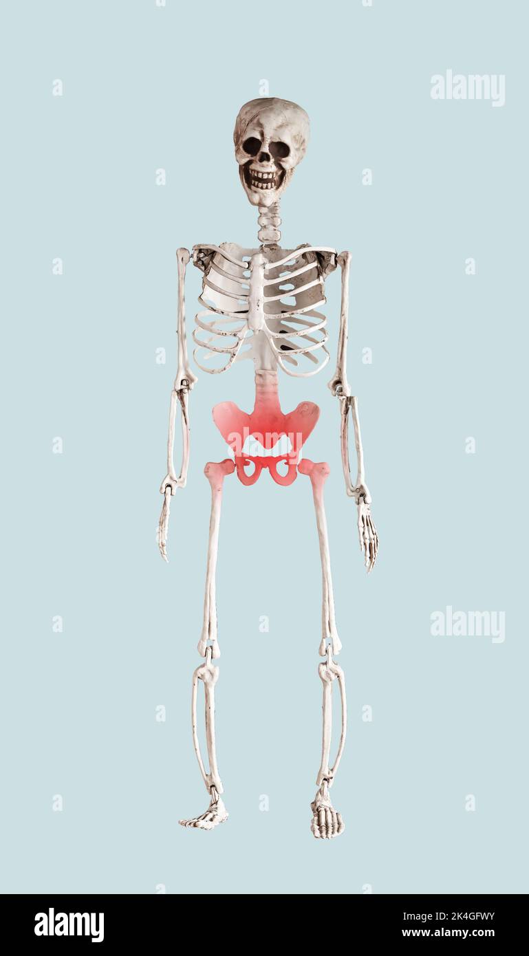 Skeleton with pelvis, pelvic bone pain, inflammation, trauma. High quality photo Stock Photohttps://www.alamy.com/image-license-details/?v=1https://www.alamy.com/skeleton-with-pelvis-pelvic-bone-pain-inflammation-trauma-high-quality-photo-image484712647.html
RF2K4GFWY–Skeleton with pelvis, pelvic bone pain, inflammation, trauma. High quality photo
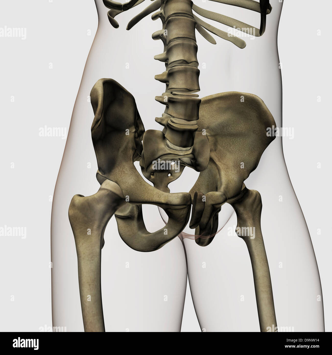 Three dimensional view of human pelvic bones. Stock Photohttps://www.alamy.com/image-license-details/?v=1https://www.alamy.com/stock-photo-three-dimensional-view-of-human-pelvic-bones-57643632.html
RFD9NW14–Three dimensional view of human pelvic bones.
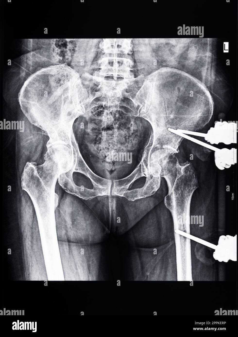 x-ray of pelvic with external fixation device Stock Photohttps://www.alamy.com/image-license-details/?v=1https://www.alamy.com/x-ray-of-pelvic-with-external-fixation-device-image547472570.html
RM2PPKERP–x-ray of pelvic with external fixation device
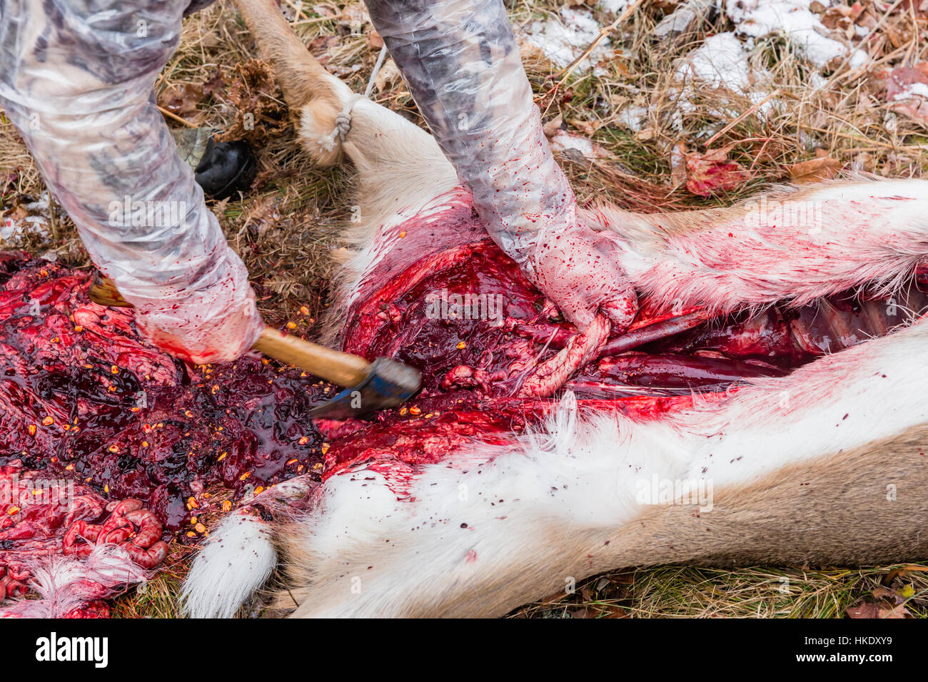 Field dressing a white-tailed buck in Wisconsin Stock Photohttps://www.alamy.com/image-license-details/?v=1https://www.alamy.com/stock-photo-field-dressing-a-white-tailed-buck-in-wisconsin-132457565.html
RMHKDXY9–Field dressing a white-tailed buck in Wisconsin
