Quick filters:
Pelvic outlet Stock Photos and Images
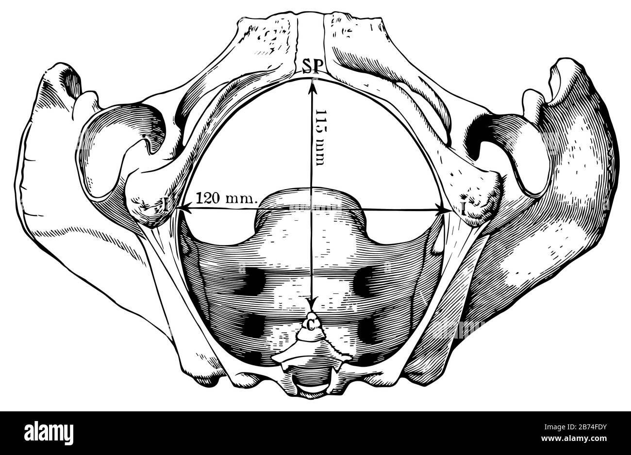 This illustration represents Pelvic Outlet, vintage line drawing or engraving illustration. Stock Vectorhttps://www.alamy.com/image-license-details/?v=1https://www.alamy.com/this-illustration-represents-pelvic-outlet-vintage-line-drawing-or-engraving-illustration-image348609911.html
RF2B74FDY–This illustration represents Pelvic Outlet, vintage line drawing or engraving illustration.
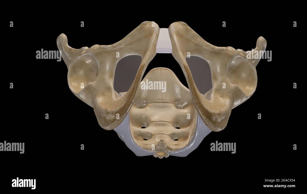 The Pelvic Girdle and Pelvic Outlet Stock Photohttps://www.alamy.com/image-license-details/?v=1https://www.alamy.com/the-pelvic-girdle-and-pelvic-outlet-image488320816.html
RF2KACX54–The Pelvic Girdle and Pelvic Outlet
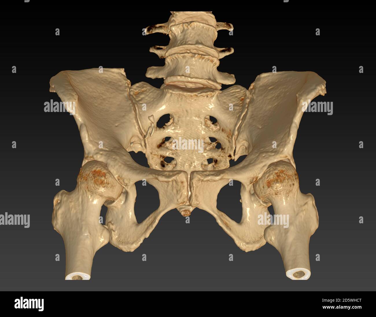 CT Scan of pelvic bone with both hip joint 3D rendering image Outlet view isolated on black background. Clipping path. Stock Photohttps://www.alamy.com/image-license-details/?v=1https://www.alamy.com/ct-scan-of-pelvic-bone-with-both-hip-joint-3d-rendering-image-outlet-view-isolated-on-black-background-clipping-path-image382263864.html
RF2D5WHCT–CT Scan of pelvic bone with both hip joint 3D rendering image Outlet view isolated on black background. Clipping path.
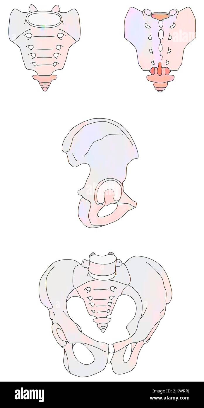 Pelvis and its three constituting bones: sacrum, coccyx and iliac bones. Stock Photohttps://www.alamy.com/image-license-details/?v=1https://www.alamy.com/pelvis-and-its-three-constituting-bones-sacrum-coccyx-and-iliac-bones-image476925894.html
RF2JKWRRJ–Pelvis and its three constituting bones: sacrum, coccyx and iliac bones.
 Manual of gynecology . , as it is misleading, especially in scientific obstetrics. It is betternamed the pelvic floor or pelvic diaphragm. The pelvic floor is a thick fleshy elastic layer, dovetailed in all roundto the bony pelvic outlet (Fig. 6). It may be considered as an irregularly-edged segment of a hollow sphere, with an outer skin aspect and an innerperitoneal one. On the outer skin aspect lie the external genitals alreadydescribed. On the inner peritoneal surface, we have the organ known asthe uterus, and its appendages, the Fallopian tubes and ovaries. Thevagina runs at an angle of 60 Stock Photohttps://www.alamy.com/image-license-details/?v=1https://www.alamy.com/manual-of-gynecology-as-it-is-misleading-especially-in-scientific-obstetrics-it-is-betternamed-the-pelvic-floor-or-pelvic-diaphragm-the-pelvic-floor-is-a-thick-fleshy-elastic-layer-dovetailed-in-all-roundto-the-bony-pelvic-outlet-fig-6-it-may-be-considered-as-an-irregularly-edged-segment-of-a-hollow-sphere-with-an-outer-skin-aspect-and-an-innerperitoneal-one-on-the-outer-skin-aspect-lie-the-external-genitals-alreadydescribed-on-the-inner-peritoneal-surface-we-have-the-organ-known-asthe-uterus-and-its-appendages-the-fallopian-tubes-and-ovaries-thevagina-runs-at-an-angle-of-60-image340180422.html
RM2ANCFGP–Manual of gynecology . , as it is misleading, especially in scientific obstetrics. It is betternamed the pelvic floor or pelvic diaphragm. The pelvic floor is a thick fleshy elastic layer, dovetailed in all roundto the bony pelvic outlet (Fig. 6). It may be considered as an irregularly-edged segment of a hollow sphere, with an outer skin aspect and an innerperitoneal one. On the outer skin aspect lie the external genitals alreadydescribed. On the inner peritoneal surface, we have the organ known asthe uterus, and its appendages, the Fallopian tubes and ovaries. Thevagina runs at an angle of 60
 PREPARING FOR DELIVERY Stock Photohttps://www.alamy.com/image-license-details/?v=1https://www.alamy.com/stock-photo-preparing-for-delivery-51879258.html
RMD0B8EJ–PREPARING FOR DELIVERY
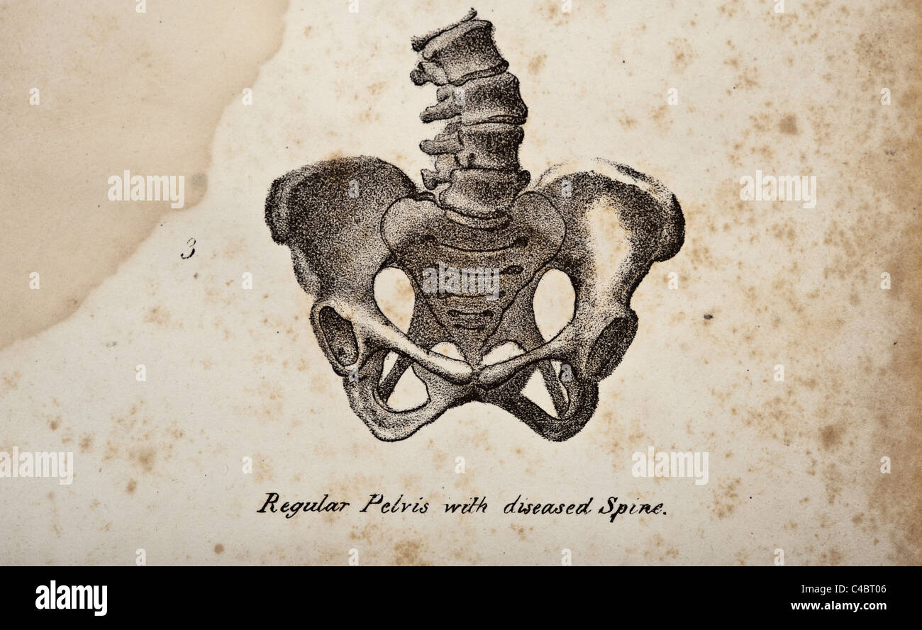
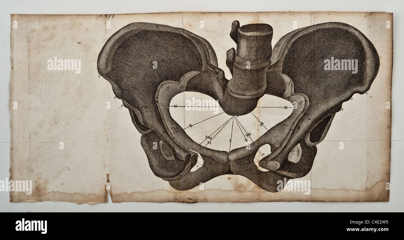 Illustration of the Human Pelvis copyright 1822 Stock Photohttps://www.alamy.com/image-license-details/?v=1https://www.alamy.com/stock-photo-illustration-of-the-human-pelvis-copyright-1822-37188961.html
RFC4E2W5–Illustration of the Human Pelvis copyright 1822
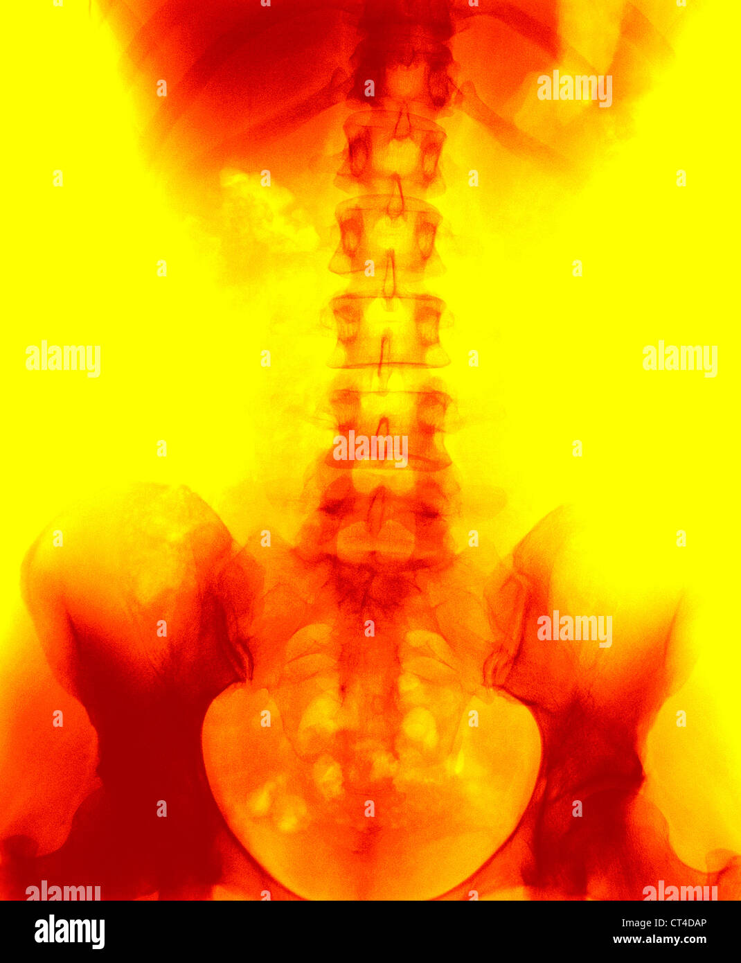 SPINAL COLUMN, X-RAY Stock Photohttps://www.alamy.com/image-license-details/?v=1https://www.alamy.com/stock-photo-spinal-column-x-ray-49270782.html
RMCT4DAP–SPINAL COLUMN, X-RAY
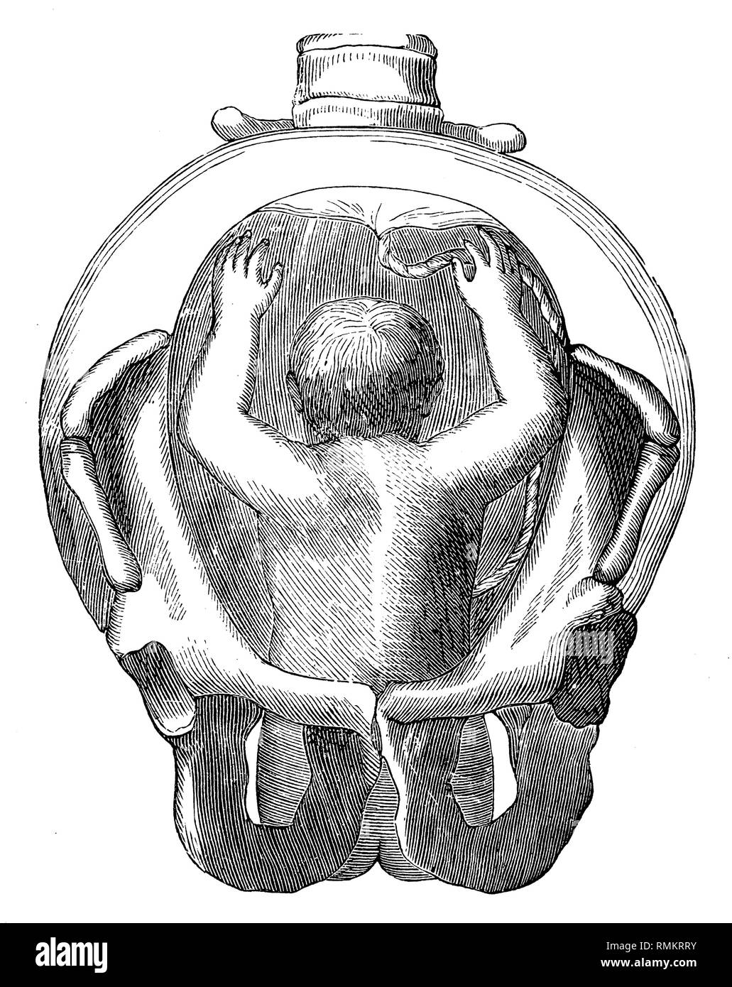 Fetus in the uterus. Breech. the rump comes through the pelvic outlet, 1900 Stock Photohttps://www.alamy.com/image-license-details/?v=1https://www.alamy.com/fetus-in-the-uterus-breech-the-rump-comes-through-the-pelvic-outlet-1900-image236463695.html
RMRMKRRY–Fetus in the uterus. Breech. the rump comes through the pelvic outlet, 1900
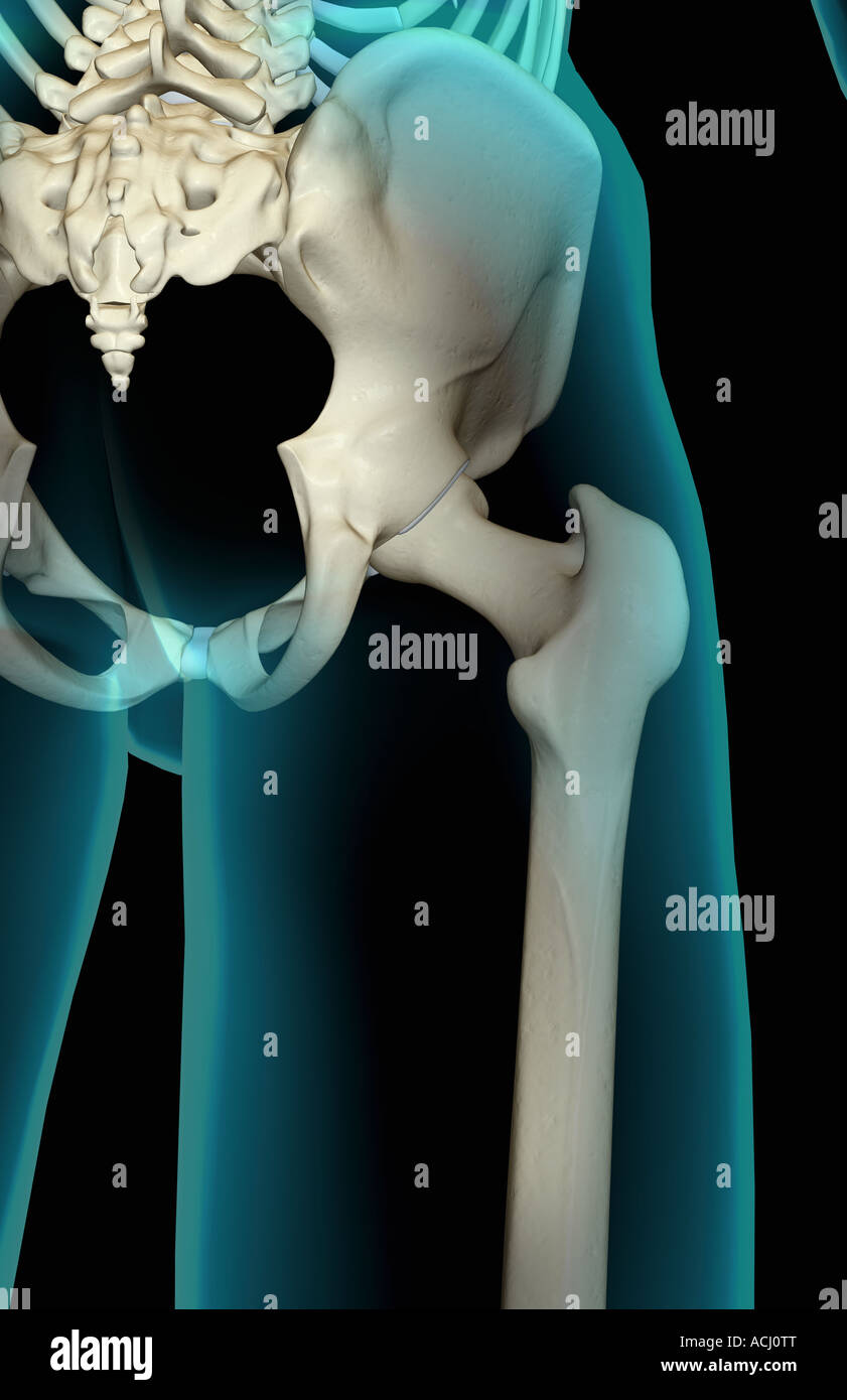 The pelvis Stock Photohttps://www.alamy.com/image-license-details/?v=1https://www.alamy.com/stock-photo-the-pelvis-13165223.html
RFACJ0TT–The pelvis
 . The cyclopædia of anatomy and physiology. Anatomy; Physiology; Zoology. PELVIS. 155 the anterior pelvic outlet, the transverse dia- meter is a little larger than the antero-pos- Fig. 94.. Pelvis of the Lion, side view. terior, and the acetabula are large and deep. In the Badger the ilia and ischia are large, expanded, and curved outwards at their free extremities. The iliac shaft is prismatic, with an ilio-lumbar angle of 140°. The pubes are rather long, with an elongated symphysis, and form an angle with the ilia of about 130°. The same general conformation is evident in the Racoons and Coa Stock Photohttps://www.alamy.com/image-license-details/?v=1https://www.alamy.com/the-cyclopdia-of-anatomy-and-physiology-anatomy-physiology-zoology-pelvis-155-the-anterior-pelvic-outlet-the-transverse-dia-meter-is-a-little-larger-than-the-antero-pos-fig-94-pelvis-of-the-lion-side-view-terior-and-the-acetabula-are-large-and-deep-in-the-badger-the-ilia-and-ischia-are-large-expanded-and-curved-outwards-at-their-free-extremities-the-iliac-shaft-is-prismatic-with-an-ilio-lumbar-angle-of-140-the-pubes-are-rather-long-with-an-elongated-symphysis-and-form-an-angle-with-the-ilia-of-about-130-the-same-general-conformation-is-evident-in-the-racoons-and-coa-image216210866.html
RMPFN74J–. The cyclopædia of anatomy and physiology. Anatomy; Physiology; Zoology. PELVIS. 155 the anterior pelvic outlet, the transverse dia- meter is a little larger than the antero-pos- Fig. 94.. Pelvis of the Lion, side view. terior, and the acetabula are large and deep. In the Badger the ilia and ischia are large, expanded, and curved outwards at their free extremities. The iliac shaft is prismatic, with an ilio-lumbar angle of 140°. The pubes are rather long, with an elongated symphysis, and form an angle with the ilia of about 130°. The same general conformation is evident in the Racoons and Coa
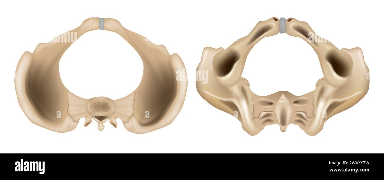 Pelvis anatomical skeleton structure. Anatomy of the Pelvis Superior view and Inferior view. Medical education scheme Stock Vectorhttps://www.alamy.com/image-license-details/?v=1https://www.alamy.com/pelvis-anatomical-skeleton-structure-anatomy-of-the-pelvis-superior-view-and-inferior-view-medical-education-scheme-image591891213.html
RF2WAXY7W–Pelvis anatomical skeleton structure. Anatomy of the Pelvis Superior view and Inferior view. Medical education scheme
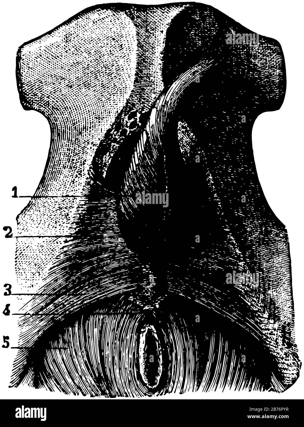 This illustration represents Muscles of the Pelvic Outlet, vintage line drawing or engraving illustration. Stock Vectorhttps://www.alamy.com/image-license-details/?v=1https://www.alamy.com/this-illustration-represents-muscles-of-the-pelvic-outlet-vintage-line-drawing-or-engraving-illustration-image348659691.html
RF2B76PYR–This illustration represents Muscles of the Pelvic Outlet, vintage line drawing or engraving illustration.
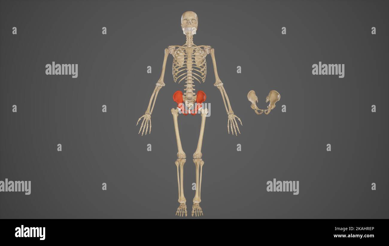 Medical Ilustration of Pelvic Bones Stock Photohttps://www.alamy.com/image-license-details/?v=1https://www.alamy.com/medical-ilustration-of-pelvic-bones-image488428494.html
RF2KAHREP–Medical Ilustration of Pelvic Bones
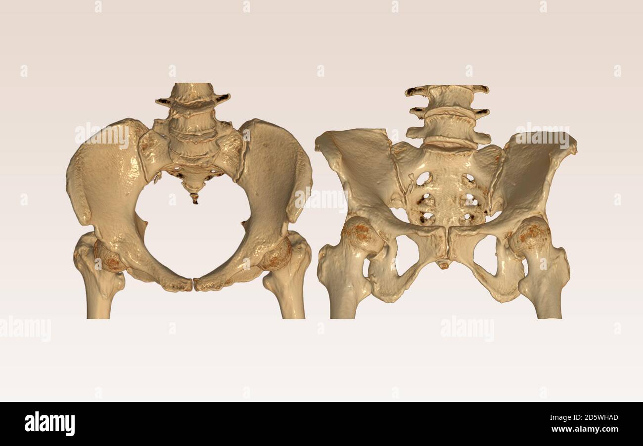 Compare of CT Scan pelvic bone with both hip joint 3D rendering image Inlet and Outlet view isolated on white background. Clipping path. Stock Photohttps://www.alamy.com/image-license-details/?v=1https://www.alamy.com/compare-of-ct-scan-pelvic-bone-with-both-hip-joint-3d-rendering-image-inlet-and-outlet-view-isolated-on-white-background-clipping-path-image382263797.html
RF2D5WHAD–Compare of CT Scan pelvic bone with both hip joint 3D rendering image Inlet and Outlet view isolated on white background. Clipping path.
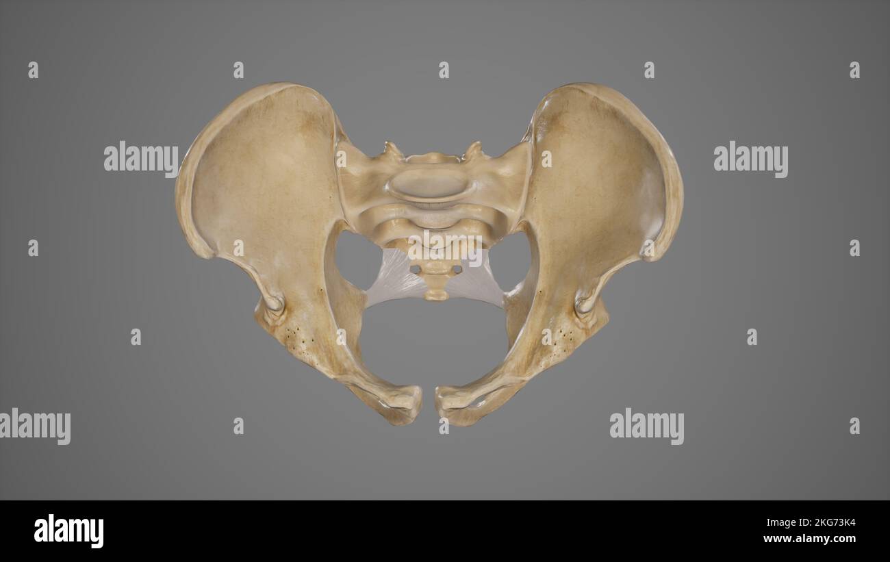 Medical Illustration of Sacrospinous Ligament Stock Photohttps://www.alamy.com/image-license-details/?v=1https://www.alamy.com/medical-illustration-of-sacrospinous-ligament-image491881352.html
RF2KG73K4–Medical Illustration of Sacrospinous Ligament
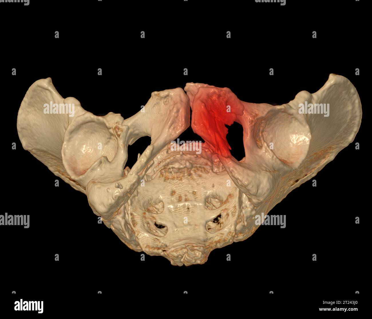 CT Scan of pelvic bone 3D rendering image showing superior pubic ramus fracture. Stock Photohttps://www.alamy.com/image-license-details/?v=1https://www.alamy.com/ct-scan-of-pelvic-bone-3d-rendering-image-showing-superior-pubic-ramus-fracture-image569262120.html
RF2T243J0–CT Scan of pelvic bone 3D rendering image showing superior pubic ramus fracture.
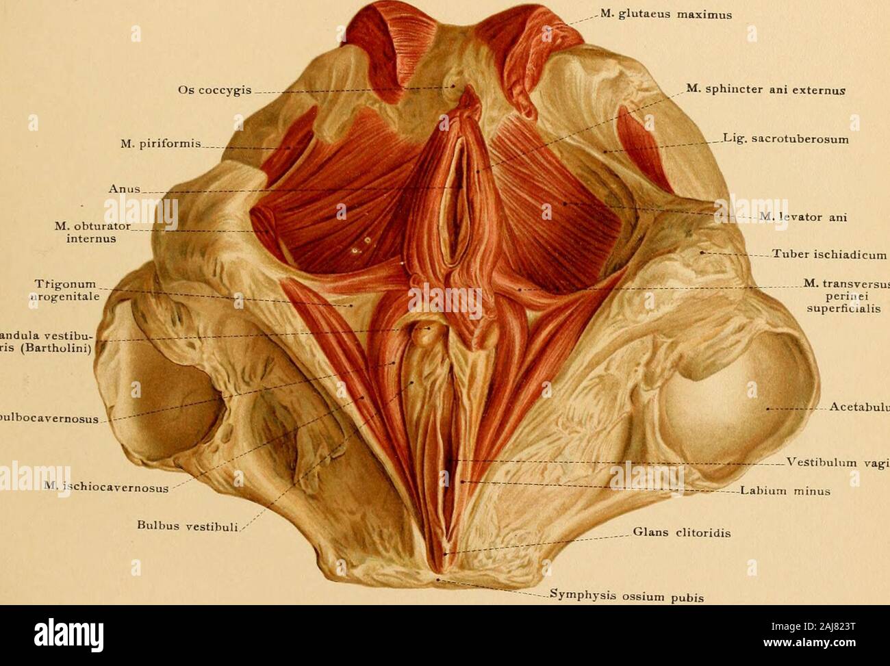 Atlas and text-book of topographic and applied anatomy . <riT/W plane orubral disc between the last -the pelvic outlet from the lower ma ters. Tab. 19.. M.bulbocavernosus THE PELVIC WALLS. 145 The pelvic inclination is the angle between the conjugata vera and a horizontal plane (about60 degrees). [The obliquity oj the pelvis varies in different individuals, is greater in the femalethan in the male, and is increased by hip-joint disease, particularly on standing. With a normalinclination of the pelvis the sacral promontory is about 9.5 cm. (3A inches) above the upper borderof the symphysi Stock Photohttps://www.alamy.com/image-license-details/?v=1https://www.alamy.com/atlas-and-text-book-of-topographic-and-applied-anatomy-ltritw-plane-orubral-disc-between-the-last-the-pelvic-outlet-from-the-lower-ma-ters-tab-19-mbulbocavernosus-the-pelvic-walls-145-the-pelvic-inclination-is-the-angle-between-the-conjugata-vera-and-a-horizontal-plane-about60-degrees-the-obliquity-oj-the-pelvis-varies-in-different-individuals-is-greater-in-the-femalethan-in-the-male-and-is-increased-by-hip-joint-disease-particularly-on-standing-with-a-normalinclination-of-the-pelvis-the-sacral-promontory-is-about-95-cm-3a-inches-above-the-upper-borderof-the-symphysi-image338238092.html
RM2AJ823T–Atlas and text-book of topographic and applied anatomy . <riT/W plane orubral disc between the last -the pelvic outlet from the lower ma ters. Tab. 19.. M.bulbocavernosus THE PELVIC WALLS. 145 The pelvic inclination is the angle between the conjugata vera and a horizontal plane (about60 degrees). [The obliquity oj the pelvis varies in different individuals, is greater in the femalethan in the male, and is increased by hip-joint disease, particularly on standing. With a normalinclination of the pelvis the sacral promontory is about 9.5 cm. (3A inches) above the upper borderof the symphysi
 Illustration of the Human Pelvis copyright 1822 Stock Photohttps://www.alamy.com/image-license-details/?v=1https://www.alamy.com/stock-photo-illustration-of-the-human-pelvis-copyright-1822-37188964.html
RFC4E2W8–Illustration of the Human Pelvis copyright 1822
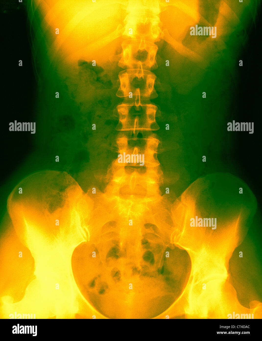 SPINAL COLUMN, X-RAY Stock Photohttps://www.alamy.com/image-license-details/?v=1https://www.alamy.com/stock-photo-spinal-column-x-ray-49270772.html
RMCT4DAC–SPINAL COLUMN, X-RAY
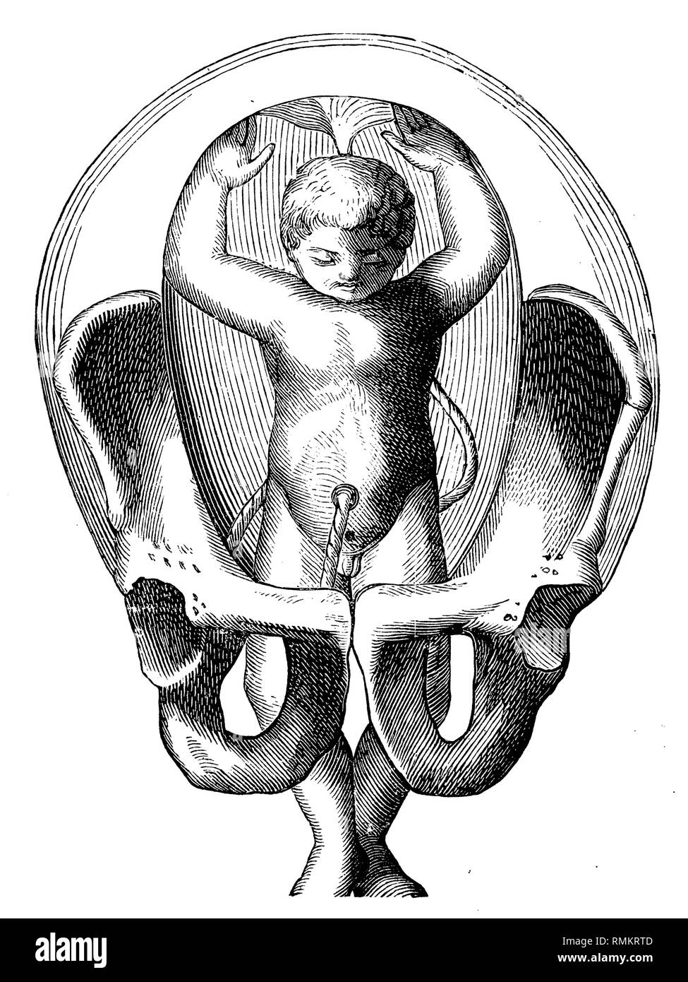 Fetus in the uterus. Fußgeburt. The feet are already kicking through the pelvic outlet, 1900 Stock Photohttps://www.alamy.com/image-license-details/?v=1https://www.alamy.com/fetus-in-the-uterus-fugeburt-the-feet-are-already-kicking-through-the-pelvic-outlet-1900-image236463709.html
RMRMKRTD–Fetus in the uterus. Fußgeburt. The feet are already kicking through the pelvic outlet, 1900
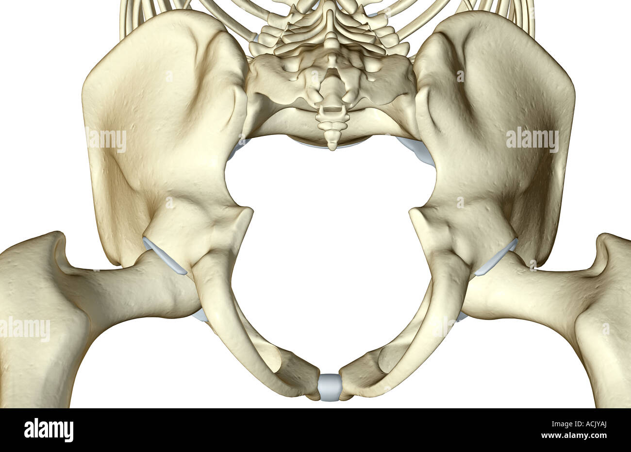 The bones of the pelvis Stock Photohttps://www.alamy.com/image-license-details/?v=1https://www.alamy.com/stock-photo-the-bones-of-the-pelvis-13174121.html
RFACJYAJ–The bones of the pelvis
 . The cyclopædia of anatomy and physiology. Anatomy; Physiology; Zoology. PELVIS. 189 pubic rami being approximated at the angular bend to f of an inch. In a case which was operated on by Dr. Hasbeke, and described in U Experience (No. 140.), the inferior pelvic outlet was nearly closed up entirely, the ischial tube- rosities being approximated to within two lines only, and the coccyx and pubes admitting only one finger between them.* In Mr. Kinder Wood's case, the deformity was rostrated, the most available space at the brim being a circle of 1 inch diameter to the left of the projecting prom Stock Photohttps://www.alamy.com/image-license-details/?v=1https://www.alamy.com/the-cyclopdia-of-anatomy-and-physiology-anatomy-physiology-zoology-pelvis-189-pubic-rami-being-approximated-at-the-angular-bend-to-f-of-an-inch-in-a-case-which-was-operated-on-by-dr-hasbeke-and-described-in-u-experience-no-140-the-inferior-pelvic-outlet-was-nearly-closed-up-entirely-the-ischial-tube-rosities-being-approximated-to-within-two-lines-only-and-the-coccyx-and-pubes-admitting-only-one-finger-between-them-in-mr-kinder-woods-case-the-deformity-was-rostrated-the-most-available-space-at-the-brim-being-a-circle-of-1-inch-diameter-to-the-left-of-the-projecting-prom-image216210701.html
RMPFN6XN–. The cyclopædia of anatomy and physiology. Anatomy; Physiology; Zoology. PELVIS. 189 pubic rami being approximated at the angular bend to f of an inch. In a case which was operated on by Dr. Hasbeke, and described in U Experience (No. 140.), the inferior pelvic outlet was nearly closed up entirely, the ischial tube- rosities being approximated to within two lines only, and the coccyx and pubes admitting only one finger between them.* In Mr. Kinder Wood's case, the deformity was rostrated, the most available space at the brim being a circle of 1 inch diameter to the left of the projecting prom
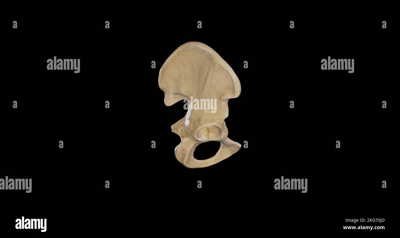 Lateral view of Right Hip Bone Stock Photohttps://www.alamy.com/image-license-details/?v=1https://www.alamy.com/lateral-view-of-right-hip-bone-image491878981.html
RF2KG70JD–Lateral view of Right Hip Bone
 CT Scan of pelvic bone 3D rendering image showing superior pubic ramus fracture. Stock Photohttps://www.alamy.com/image-license-details/?v=1https://www.alamy.com/ct-scan-of-pelvic-bone-3d-rendering-image-showing-superior-pubic-ramus-fracture-image569262153.html
RF2T243K5–CT Scan of pelvic bone 3D rendering image showing superior pubic ramus fracture.
 Atlas and text-book of topographic and applied anatomy . Glans clitoridis .. measured in the lronjugata diag the middle of. <riT/W plane orubral disc between the last -the pelvic outlet from the lower ma ters. Tab. 19. Stock Photohttps://www.alamy.com/image-license-details/?v=1https://www.alamy.com/atlas-and-text-book-of-topographic-and-applied-anatomy-glans-clitoridis-measured-in-the-lronjugata-diag-the-middle-of-ltritw-plane-orubral-disc-between-the-last-the-pelvic-outlet-from-the-lower-ma-ters-tab-19-image338238437.html
RM2AJ82G5–Atlas and text-book of topographic and applied anatomy . Glans clitoridis .. measured in the lronjugata diag the middle of. <riT/W plane orubral disc between the last -the pelvic outlet from the lower ma ters. Tab. 19.
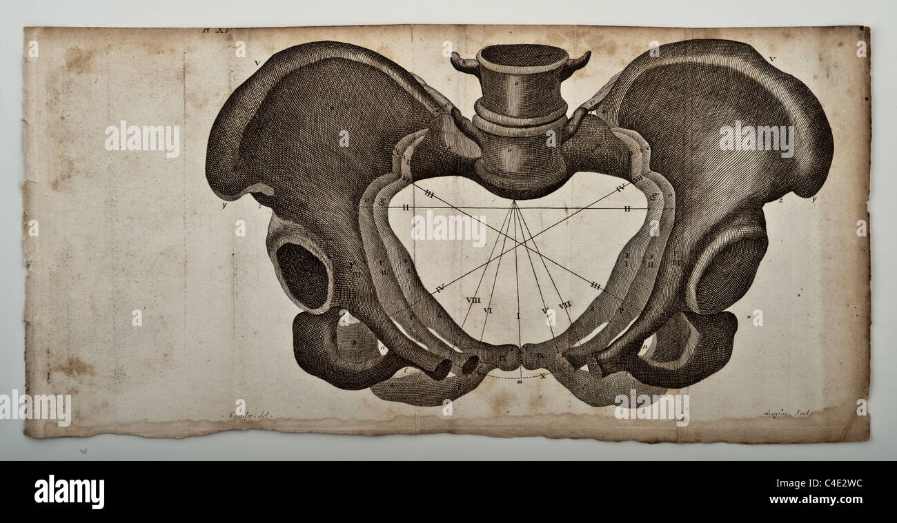 Illustration of the Human Pelvis copyright 1822 Stock Photohttps://www.alamy.com/image-license-details/?v=1https://www.alamy.com/stock-photo-illustration-of-the-human-pelvis-copyright-1822-37188968.html
RFC4E2WC–Illustration of the Human Pelvis copyright 1822
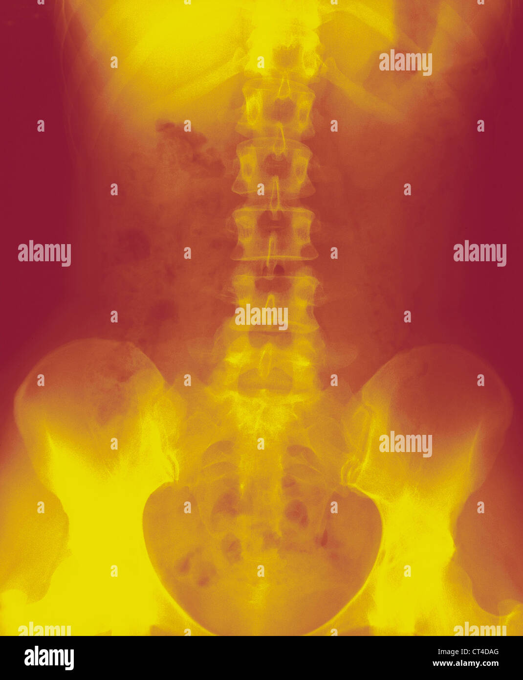 SPINAL COLUMN, X-RAY Stock Photohttps://www.alamy.com/image-license-details/?v=1https://www.alamy.com/stock-photo-spinal-column-x-ray-49270776.html
RMCT4DAG–SPINAL COLUMN, X-RAY
 PREPARING FOR DELIVERY Stock Photohttps://www.alamy.com/image-license-details/?v=1https://www.alamy.com/stock-photo-preparing-for-delivery-51879296.html
RMD0B8G0–PREPARING FOR DELIVERY
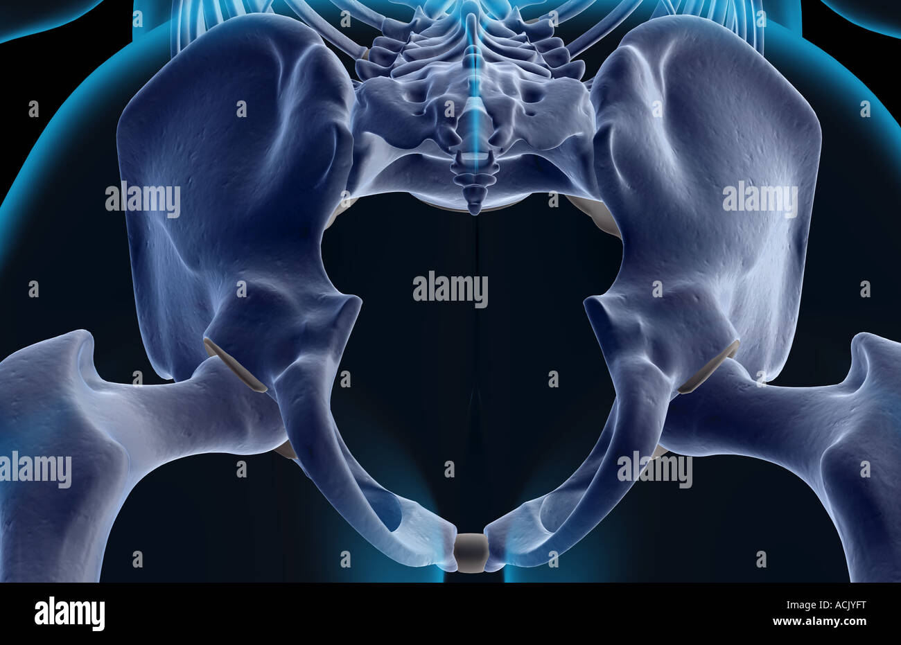 The bones of the pelvis Stock Photohttps://www.alamy.com/image-license-details/?v=1https://www.alamy.com/stock-photo-the-bones-of-the-pelvis-13174187.html
RFACJYFT–The bones of the pelvis
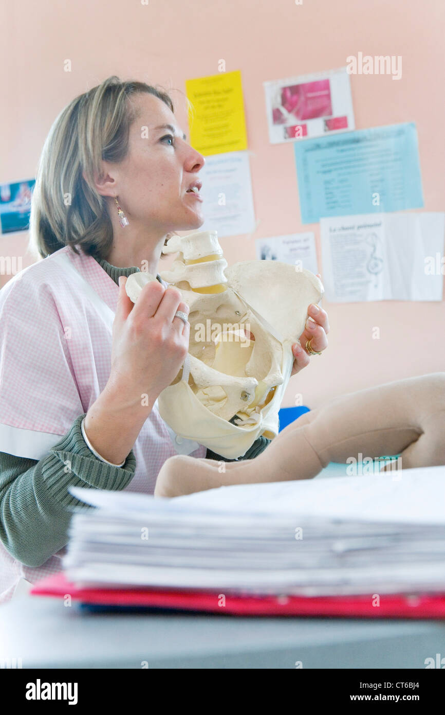 PREPARING FOR DELIVERY Stock Photohttps://www.alamy.com/image-license-details/?v=1https://www.alamy.com/stock-photo-preparing-for-delivery-49313324.html
RMCT6BJ4–PREPARING FOR DELIVERY
 . Veterinary obstetrics; a compendium for the use of students and practitioners. Veterinary obstetrics. ANATOMY. 17 The diameters of the outlet are more equal, being about those of the transverse diameter of the inlet. The cavity is more cylindrical, and less conical than that of the Mare. In the Sheep and Goat, ossification occurring at a 'much later period, allows of the pelvic cavity being increased during parturition, and permits of the act being performed with fewer difficulties in these animals. In the Pig, the general conformation of the pelvis. Fig. 8. Inlet of the Cow's Pelvis. <j Stock Photohttps://www.alamy.com/image-license-details/?v=1https://www.alamy.com/veterinary-obstetrics-a-compendium-for-the-use-of-students-and-practitioners-veterinary-obstetrics-anatomy-17-the-diameters-of-the-outlet-are-more-equal-being-about-those-of-the-transverse-diameter-of-the-inlet-the-cavity-is-more-cylindrical-and-less-conical-than-that-of-the-mare-in-the-sheep-and-goat-ossification-occurring-at-a-much-later-period-allows-of-the-pelvic-cavity-being-increased-during-parturition-and-permits-of-the-act-being-performed-with-fewer-difficulties-in-these-animals-in-the-pig-the-general-conformation-of-the-pelvis-fig-8-inlet-of-the-cows-pelvis-ltj-image216388105.html
RMPG196H–. Veterinary obstetrics; a compendium for the use of students and practitioners. Veterinary obstetrics. ANATOMY. 17 The diameters of the outlet are more equal, being about those of the transverse diameter of the inlet. The cavity is more cylindrical, and less conical than that of the Mare. In the Sheep and Goat, ossification occurring at a 'much later period, allows of the pelvic cavity being increased during parturition, and permits of the act being performed with fewer difficulties in these animals. In the Pig, the general conformation of the pelvis. Fig. 8. Inlet of the Cow's Pelvis. <j
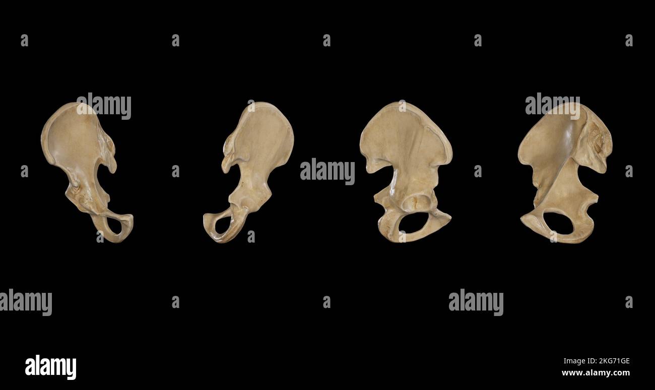 Right Hip Bone from multiple sides Stock Photohttps://www.alamy.com/image-license-details/?v=1https://www.alamy.com/right-hip-bone-from-multiple-sides-image491879710.html
RF2KG71GE–Right Hip Bone from multiple sides
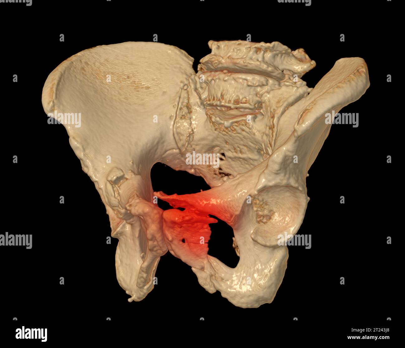 CT Scan of pelvic bone 3D rendering image showing superior pubic ramus fracture. Stock Photohttps://www.alamy.com/image-license-details/?v=1https://www.alamy.com/ct-scan-of-pelvic-bone-3d-rendering-image-showing-superior-pubic-ramus-fracture-image569262128.html
RF2T243J8–CT Scan of pelvic bone 3D rendering image showing superior pubic ramus fracture.
 Operative midwifery : a guide to the difficulties and complications of midwifery practice . ittlealtered, but the distances between the ischial spines and ischialtuberosities are decidedly diminished. The striking feature of thekyphotic pelvis is a diminution of all the diameters of the pelvic outlet;therefore, one finds difficulty in labour when the foetal head has reachedthe lower part of the cavity. It is somewhat curious that the head should so generally engage inthe oblique or transverse diameter, for one would expect that it wouldengage in the conjugate, as that is the diameter which is Stock Photohttps://www.alamy.com/image-license-details/?v=1https://www.alamy.com/operative-midwifery-a-guide-to-the-difficulties-and-complications-of-midwifery-practice-ittlealtered-but-the-distances-between-the-ischial-spines-and-ischialtuberosities-are-decidedly-diminished-the-striking-feature-of-thekyphotic-pelvis-is-a-diminution-of-all-the-diameters-of-the-pelvic-outlettherefore-one-finds-difficulty-in-labour-when-the-foetal-head-has-reachedthe-lower-part-of-the-cavity-it-is-somewhat-curious-that-the-head-should-so-generally-engage-inthe-oblique-or-transverse-diameter-for-one-would-expect-that-it-wouldengage-in-the-conjugate-as-that-is-the-diameter-which-is-image342839547.html
RM2AWNK9F–Operative midwifery : a guide to the difficulties and complications of midwifery practice . ittlealtered, but the distances between the ischial spines and ischialtuberosities are decidedly diminished. The striking feature of thekyphotic pelvis is a diminution of all the diameters of the pelvic outlet;therefore, one finds difficulty in labour when the foetal head has reachedthe lower part of the cavity. It is somewhat curious that the head should so generally engage inthe oblique or transverse diameter, for one would expect that it wouldengage in the conjugate, as that is the diameter which is
 Illustration of the Human Pelvis copyright 1822 Stock Photohttps://www.alamy.com/image-license-details/?v=1https://www.alamy.com/stock-photo-illustration-of-the-human-pelvis-copyright-1822-37188965.html
RFC4E2W9–Illustration of the Human Pelvis copyright 1822
 Illustration of the Human Pelvis copyright 1844 Stock Photohttps://www.alamy.com/image-license-details/?v=1https://www.alamy.com/stock-photo-illustration-of-the-human-pelvis-copyright-1844-37139902.html
RFC4BT92–Illustration of the Human Pelvis copyright 1844
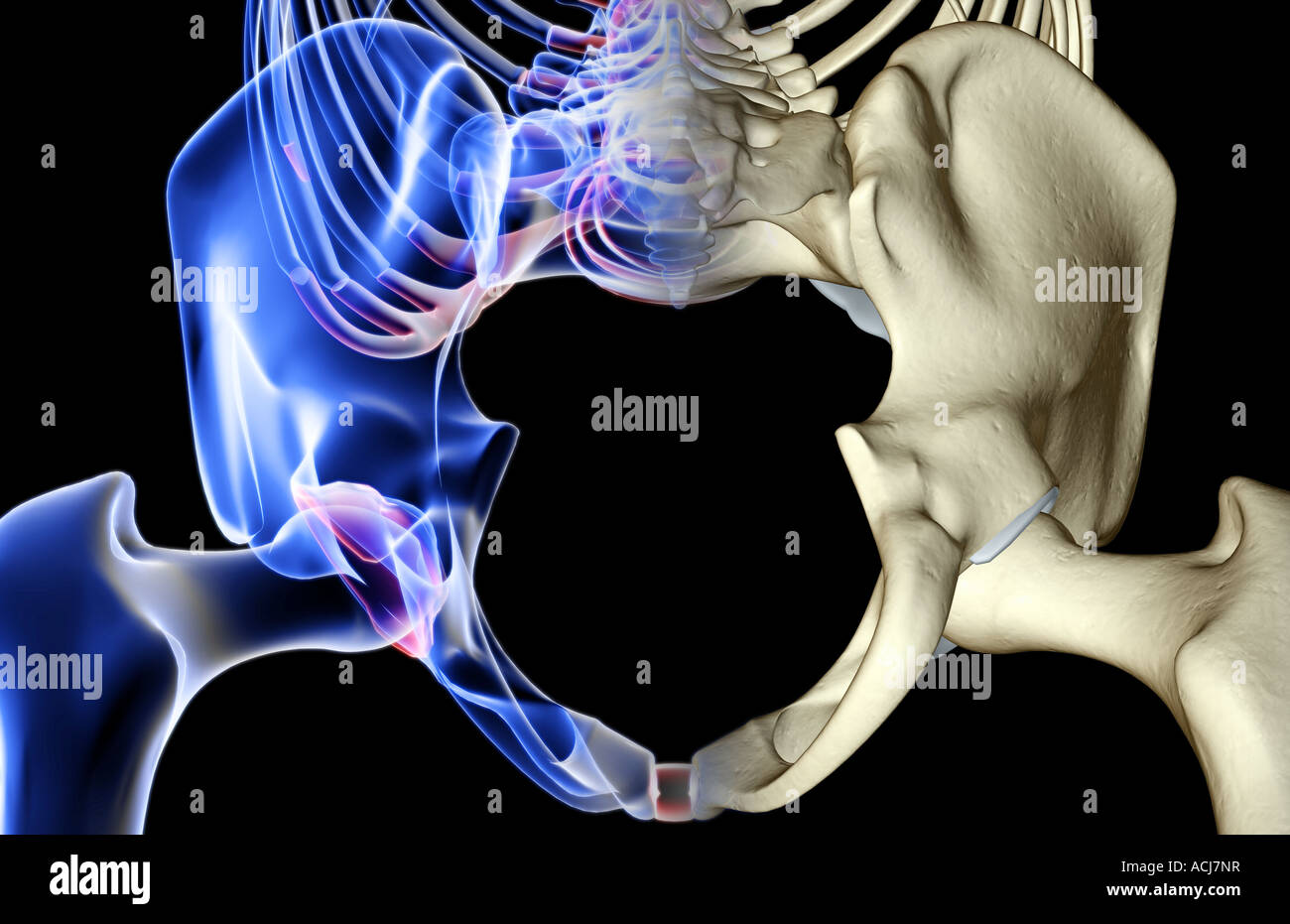 The bones of the pelvis Stock Photohttps://www.alamy.com/image-license-details/?v=1https://www.alamy.com/stock-photo-the-bones-of-the-pelvis-13167538.html
RFACJ7NR–The bones of the pelvis
 PREPARING FOR DELIVERY Stock Photohttps://www.alamy.com/image-license-details/?v=1https://www.alamy.com/stock-photo-preparing-for-delivery-49313391.html
RMCT6BMF–PREPARING FOR DELIVERY
 Illustration of the Human Pelvis copyright 1844 Stock Photohttps://www.alamy.com/image-license-details/?v=1https://www.alamy.com/stock-photo-illustration-of-the-human-pelvis-copyright-1844-37139650.html
RFC4BT02–Illustration of the Human Pelvis copyright 1844
 . Veterinary obstetrics; a compendium for the use of students and practitioners. Veterinary obstetrics. ANATOMY. 15: sacrum and first two or three coccygeal vertebrae; inferiorly, to the superior ischiatic spine and tuberosity of the ischium. The Mare's pelvis represents a somewhat cone- shaped cavity at the posterior part of the trunk, continuing the abdominal cavity. It has an internal, and an external surface, and two openings. The anterior opening is termed the inlet of the^ pelvis, by which the foetus enters the pelvic cavity. The posterior is known as the outlet or recto-urethral; openin Stock Photohttps://www.alamy.com/image-license-details/?v=1https://www.alamy.com/veterinary-obstetrics-a-compendium-for-the-use-of-students-and-practitioners-veterinary-obstetrics-anatomy-15-sacrum-and-first-two-or-three-coccygeal-vertebrae-inferiorly-to-the-superior-ischiatic-spine-and-tuberosity-of-the-ischium-the-mares-pelvis-represents-a-somewhat-cone-shaped-cavity-at-the-posterior-part-of-the-trunk-continuing-the-abdominal-cavity-it-has-an-internal-and-an-external-surface-and-two-openings-the-anterior-opening-is-termed-the-inlet-of-the-pelvis-by-which-the-foetus-enters-the-pelvic-cavity-the-posterior-is-known-as-the-outlet-or-recto-urethral-openin-image216388108.html
RMPG196M–. Veterinary obstetrics; a compendium for the use of students and practitioners. Veterinary obstetrics. ANATOMY. 15: sacrum and first two or three coccygeal vertebrae; inferiorly, to the superior ischiatic spine and tuberosity of the ischium. The Mare's pelvis represents a somewhat cone- shaped cavity at the posterior part of the trunk, continuing the abdominal cavity. It has an internal, and an external surface, and two openings. The anterior opening is termed the inlet of the^ pelvis, by which the foetus enters the pelvic cavity. The posterior is known as the outlet or recto-urethral; openin
 Posterior view of Right Hip Bone Stock Photohttps://www.alamy.com/image-license-details/?v=1https://www.alamy.com/posterior-view-of-right-hip-bone-image491878990.html
RF2KG70JP–Posterior view of Right Hip Bone
 CT Scan of pelvic bone 3D rendering image showing superior pubic ramus fracture. Stock Photohttps://www.alamy.com/image-license-details/?v=1https://www.alamy.com/ct-scan-of-pelvic-bone-3d-rendering-image-showing-superior-pubic-ramus-fracture-image569262116.html
RF2T243HT–CT Scan of pelvic bone 3D rendering image showing superior pubic ramus fracture.
 A textbook of obstetrics . e latter are pulledoutward and forward so that the pubic arch is greatly widened 444 TIIE r-l THOLOGY OF LABOR. and the transverse diameter of the pelvic outlet is increased.The anteroposterior diameter oi the outlet is somewhat dimin-ished by the excessive perpendicular curvature of the sacrum,but the contraction is relatively much less than in the conjugate of the inlet. The whole pelvis is tilted forward on its transverseaxis, so that the inclination of the superior strait is increasedand the external genitalia are displaced backward. The bones of a rachitic pelvi Stock Photohttps://www.alamy.com/image-license-details/?v=1https://www.alamy.com/a-textbook-of-obstetrics-e-latter-are-pulledoutward-and-forward-so-that-the-pubic-arch-is-greatly-widened-444-tiie-r-l-thology-of-labor-and-the-transverse-diameter-of-the-pelvic-outlet-is-increasedthe-anteroposterior-diameter-oi-the-outlet-is-somewhat-dimin-ished-by-the-excessive-perpendicular-curvature-of-the-sacrumbut-the-contraction-is-relatively-much-less-than-in-the-conjugate-of-the-inlet-the-whole-pelvis-is-tilted-forward-on-its-transverseaxis-so-that-the-inclination-of-the-superior-strait-is-increasedand-the-external-genitalia-are-displaced-backward-the-bones-of-a-rachitic-pelvi-image342968773.html
RM2AWYG4N–A textbook of obstetrics . e latter are pulledoutward and forward so that the pubic arch is greatly widened 444 TIIE r-l THOLOGY OF LABOR. and the transverse diameter of the pelvic outlet is increased.The anteroposterior diameter oi the outlet is somewhat dimin-ished by the excessive perpendicular curvature of the sacrum,but the contraction is relatively much less than in the conjugate of the inlet. The whole pelvis is tilted forward on its transverseaxis, so that the inclination of the superior strait is increasedand the external genitalia are displaced backward. The bones of a rachitic pelvi
 The bones of the pelvis Stock Photohttps://www.alamy.com/image-license-details/?v=1https://www.alamy.com/stock-photo-the-bones-of-the-pelvis-13168082.html
RFACJ9AY–The bones of the pelvis
 PREPARING FOR DELIVERY Stock Photohttps://www.alamy.com/image-license-details/?v=1https://www.alamy.com/stock-photo-preparing-for-delivery-49313323.html
RMCT6BJ3–PREPARING FOR DELIVERY
 Illustration of the Human Pelvis copyright 1844 Stock Photohttps://www.alamy.com/image-license-details/?v=1https://www.alamy.com/stock-photo-illustration-of-the-human-pelvis-copyright-1844-37139609.html
RFC4BRXH–Illustration of the Human Pelvis copyright 1844
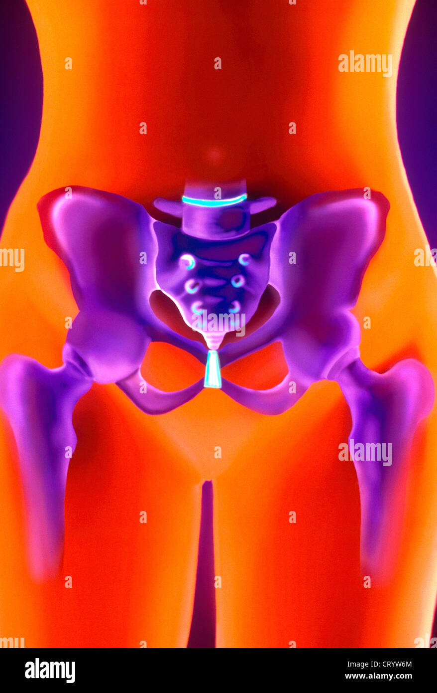 PELVIS, DRAWING Stock Photohttps://www.alamy.com/image-license-details/?v=1https://www.alamy.com/stock-photo-pelvis-drawing-49170316.html
RMCRYW6M–PELVIS, DRAWING
 . The cyclopædia of anatomy and physiology. Anatomy; Physiology; Zoology. 136 PELVIS. nml the tuberosities of the isthia are near each other so as to present a small opening at the interior outlet. Scemmerring remarks that the obturator foramen is more elliptical in the infant than in the adult. The depth and general appearance of the true pelvis is smaller than is proportionate to the iliac wings; and it is of nearly equal breadth throughout. The parallelism of the lateral, as well as of the anterior and posterior pelvic walls is, I think, sufficiently marked and general to be considered as a Stock Photohttps://www.alamy.com/image-license-details/?v=1https://www.alamy.com/the-cyclopdia-of-anatomy-and-physiology-anatomy-physiology-zoology-136-pelvis-nml-the-tuberosities-of-the-isthia-are-near-each-other-so-as-to-present-a-small-opening-at-the-interior-outlet-scemmerring-remarks-that-the-obturator-foramen-is-more-elliptical-in-the-infant-than-in-the-adult-the-depth-and-general-appearance-of-the-true-pelvis-is-smaller-than-is-proportionate-to-the-iliac-wings-and-it-is-of-nearly-equal-breadth-throughout-the-parallelism-of-the-lateral-as-well-as-of-the-anterior-and-posterior-pelvic-walls-is-i-think-sufficiently-marked-and-general-to-be-considered-as-a-image216210951.html
RMPFN77K–. The cyclopædia of anatomy and physiology. Anatomy; Physiology; Zoology. 136 PELVIS. nml the tuberosities of the isthia are near each other so as to present a small opening at the interior outlet. Scemmerring remarks that the obturator foramen is more elliptical in the infant than in the adult. The depth and general appearance of the true pelvis is smaller than is proportionate to the iliac wings; and it is of nearly equal breadth throughout. The parallelism of the lateral, as well as of the anterior and posterior pelvic walls is, I think, sufficiently marked and general to be considered as a
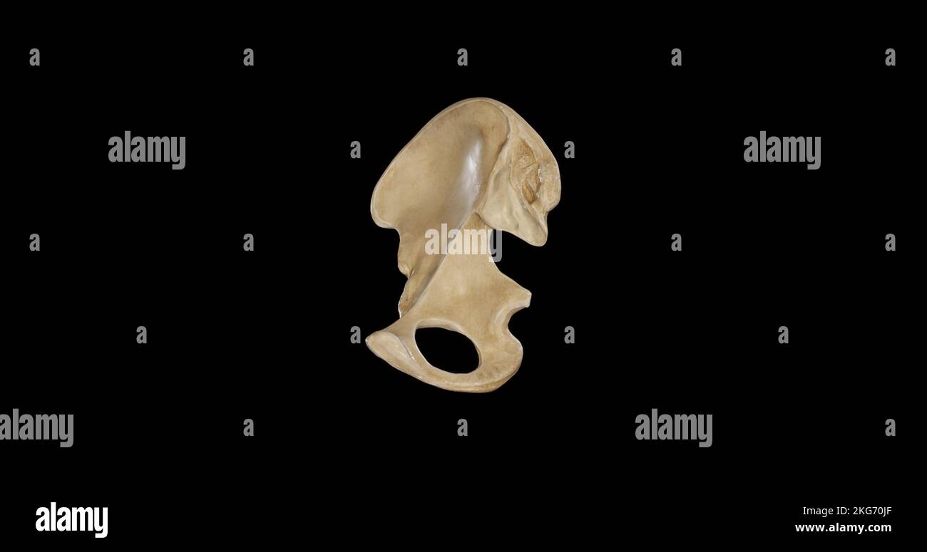 Medial view of Right Hip Bone Stock Photohttps://www.alamy.com/image-license-details/?v=1https://www.alamy.com/medial-view-of-right-hip-bone-image491878983.html
RF2KG70JF–Medial view of Right Hip Bone
 CT Scan of pelvic bone 3D rendering image showing superior pubic ramus fracture. Stock Photohttps://www.alamy.com/image-license-details/?v=1https://www.alamy.com/ct-scan-of-pelvic-bone-3d-rendering-image-showing-superior-pubic-ramus-fracture-image569262114.html
RF2T243HP–CT Scan of pelvic bone 3D rendering image showing superior pubic ramus fracture.
 A textbook of obstetrics . normally long conjugate diameter of the inlet, a diminisheddistance between the posterior superior spines, an approximationof the tuberosities of the ischiatic bones, and some diminution inthe anteroposterior diameter of the pelvic outlet. The buttocksare flat and pointed below, the external genitalia are displacedforward and upward, and the upper edge of the symphysis isabove the upper edge of the pubic hair. Care should always beexercised to detect asymmetry in these pelves, to discover an 47 ^ THE PATHOLOGY OF LABOR. arrested development with general contraction w Stock Photohttps://www.alamy.com/image-license-details/?v=1https://www.alamy.com/a-textbook-of-obstetrics-normally-long-conjugate-diameter-of-the-inlet-a-diminisheddistance-between-the-posterior-superior-spines-an-approximationof-the-tuberosities-of-the-ischiatic-bones-and-some-diminution-inthe-anteroposterior-diameter-of-the-pelvic-outlet-the-buttocksare-flat-and-pointed-below-the-external-genitalia-are-displacedforward-and-upward-and-the-upper-edge-of-the-symphysis-isabove-the-upper-edge-of-the-pubic-hair-care-should-always-beexercised-to-detect-asymmetry-in-these-pelves-to-discover-an-47-the-pathology-of-labor-arrested-development-with-general-contraction-w-image342952039.html
RM2AWXPR3–A textbook of obstetrics . normally long conjugate diameter of the inlet, a diminisheddistance between the posterior superior spines, an approximationof the tuberosities of the ischiatic bones, and some diminution inthe anteroposterior diameter of the pelvic outlet. The buttocksare flat and pointed below, the external genitalia are displacedforward and upward, and the upper edge of the symphysis isabove the upper edge of the pubic hair. Care should always beexercised to detect asymmetry in these pelves, to discover an 47 ^ THE PATHOLOGY OF LABOR. arrested development with general contraction w
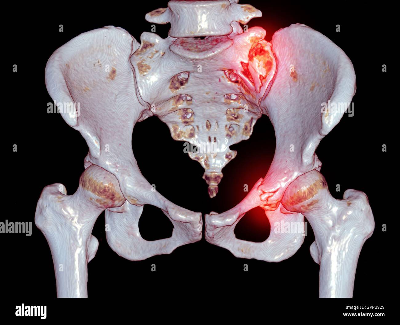 CT Scan pelvic bone with both hip joint 3D rendering showign fracture of sacrum and superior pubic rumus. Stock Photohttps://www.alamy.com/image-license-details/?v=1https://www.alamy.com/ct-scan-pelvic-bone-with-both-hip-joint-3d-rendering-showign-fracture-of-sacrum-and-superior-pubic-rumus-image547292433.html
RF2PPB929–CT Scan pelvic bone with both hip joint 3D rendering showign fracture of sacrum and superior pubic rumus.
 The bones of the pelvis Stock Photohttps://www.alamy.com/image-license-details/?v=1https://www.alamy.com/stock-photo-the-bones-of-the-pelvis-13203076.html
RFACP1FH–The bones of the pelvis
 Illustration of the Human Pelvis copyright 1844 Stock Photohttps://www.alamy.com/image-license-details/?v=1https://www.alamy.com/stock-photo-illustration-of-the-human-pelvis-copyright-1844-37139634.html
RFC4BRYE–Illustration of the Human Pelvis copyright 1844
 PELVIS, DRAWING Stock Photohttps://www.alamy.com/image-license-details/?v=1https://www.alamy.com/stock-photo-pelvis-drawing-49170319.html
RMCRYW6R–PELVIS, DRAWING
 Anterior view of Right Hip Bone Stock Photohttps://www.alamy.com/image-license-details/?v=1https://www.alamy.com/anterior-view-of-right-hip-bone-image491878999.html
RF2KG70K3–Anterior view of Right Hip Bone
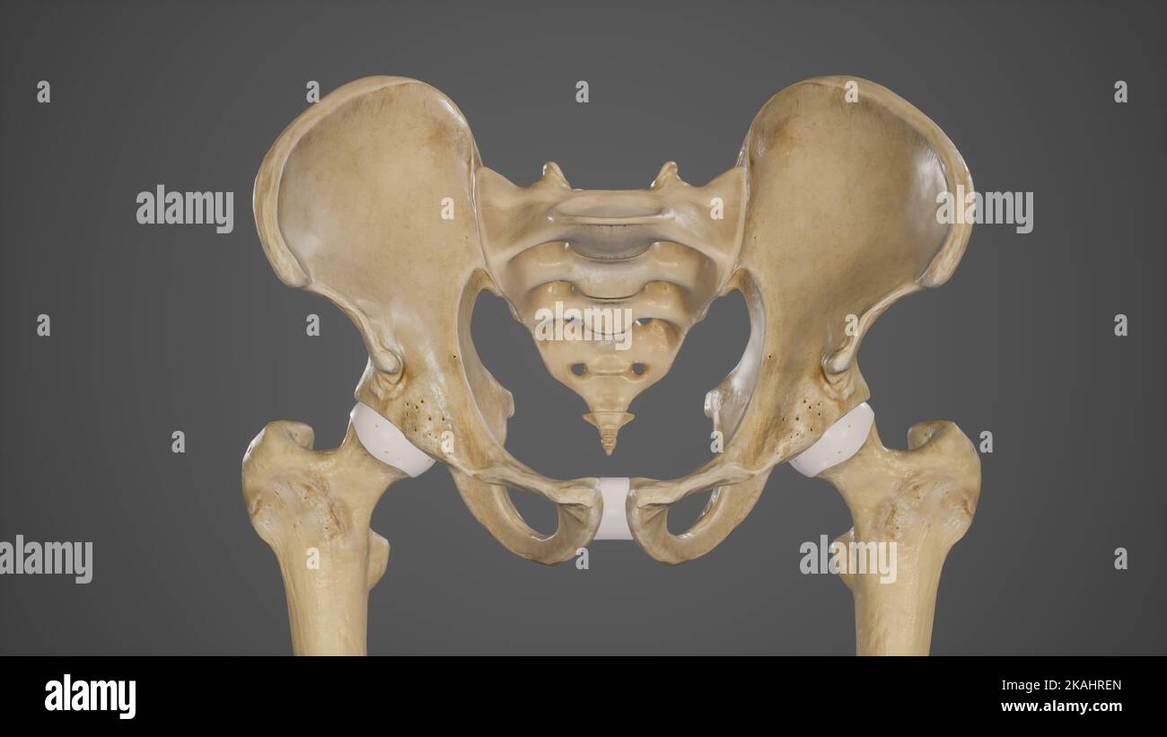 Medical Ilustration of Pelvic Bones-Hip Bone Stock Photohttps://www.alamy.com/image-license-details/?v=1https://www.alamy.com/medical-ilustration-of-pelvic-bones-hip-bone-image488428493.html
RF2KAHREN–Medical Ilustration of Pelvic Bones-Hip Bone
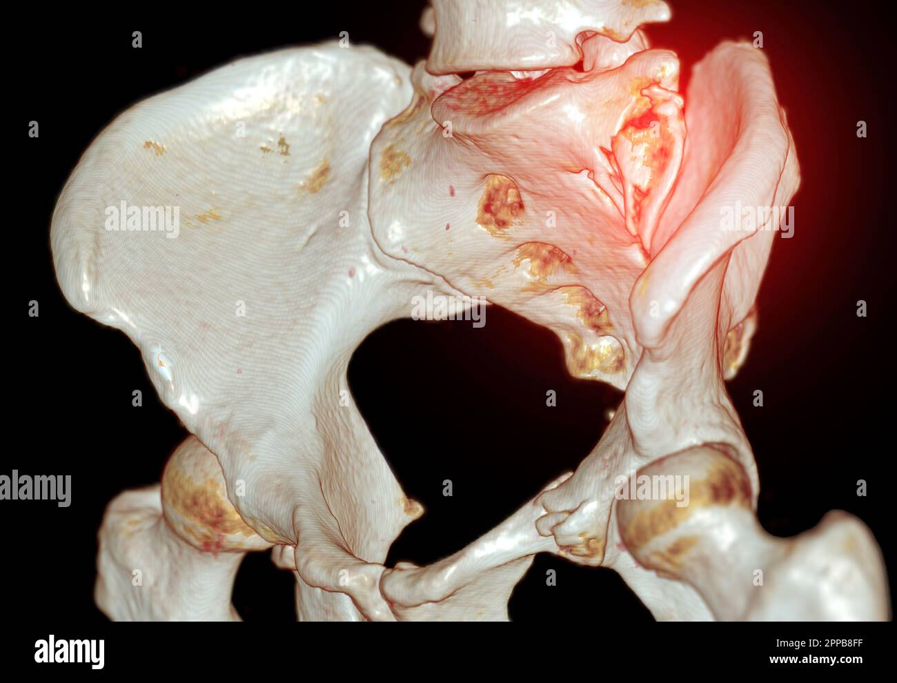 CT Scan pelvic bone with both hip joint 3D rendering showign fracture of sacrum and superior pubic rumus. Stock Photohttps://www.alamy.com/image-license-details/?v=1https://www.alamy.com/ct-scan-pelvic-bone-with-both-hip-joint-3d-rendering-showign-fracture-of-sacrum-and-superior-pubic-rumus-image547292019.html
RF2PPB8FF–CT Scan pelvic bone with both hip joint 3D rendering showign fracture of sacrum and superior pubic rumus.
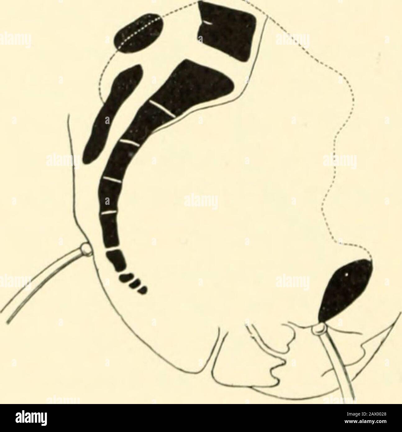 A textbook of obstetrics . imation of the tuberosities of the ischia, the depth of the pelvis, and the direction of itscanal ; by detecting, possibly, thepresence of an exostosis, an osteo-sarcoma, an abnormally project-ing spinous process, an old frac-ture, or asymmetry of the pelvicwalls from any cause. Measurement of the Trans-verseDiameter of the Pelvic Outlet.—Theanteroposterior diameter of the in-ferior strait is enlarged during laborby tin- displacement backward ofthe coccyx. The transverse diam-Fig 281.-Measurement of the eter betweeil the tuberosities ofanteroposterior diameter i the Stock Photohttps://www.alamy.com/image-license-details/?v=1https://www.alamy.com/a-textbook-of-obstetrics-imation-of-the-tuberosities-of-the-ischia-the-depth-of-the-pelvis-and-the-direction-of-itscanal-by-detecting-possibly-thepresence-of-an-exostosis-an-osteo-sarcoma-an-abnormally-project-ing-spinous-process-an-old-frac-ture-or-asymmetry-of-the-pelvicwalls-from-any-cause-measurement-of-the-trans-versediameter-of-the-pelvic-outlettheanteroposterior-diameter-of-the-in-ferior-strait-is-enlarged-during-laborby-tin-displacement-backward-ofthe-coccyx-the-transverse-diam-fig-281-measurement-of-the-eter-betweeil-the-tuberosities-ofanteroposterior-diameter-i-the-image342978112.html
RM2AX0028–A textbook of obstetrics . imation of the tuberosities of the ischia, the depth of the pelvis, and the direction of itscanal ; by detecting, possibly, thepresence of an exostosis, an osteo-sarcoma, an abnormally project-ing spinous process, an old frac-ture, or asymmetry of the pelvicwalls from any cause. Measurement of the Trans-verseDiameter of the Pelvic Outlet.—Theanteroposterior diameter of the in-ferior strait is enlarged during laborby tin- displacement backward ofthe coccyx. The transverse diam-Fig 281.-Measurement of the eter betweeil the tuberosities ofanteroposterior diameter i the
 The bones of the pelvis Stock Photohttps://www.alamy.com/image-license-details/?v=1https://www.alamy.com/stock-photo-the-bones-of-the-pelvis-13171856.html
RFACJMHN–The bones of the pelvis
 Illustration of the Human Pelvis copyright 1844 Stock Photohttps://www.alamy.com/image-license-details/?v=1https://www.alamy.com/stock-photo-illustration-of-the-human-pelvis-copyright-1844-37139612.html
RFC4BRXM–Illustration of the Human Pelvis copyright 1844
 PELVIS, DRAWING Stock Photohttps://www.alamy.com/image-license-details/?v=1https://www.alamy.com/stock-photo-pelvis-drawing-49170318.html
RMCRYW6P–PELVIS, DRAWING
 PELVIMETRY Stock Photohttps://www.alamy.com/image-license-details/?v=1https://www.alamy.com/stock-photo-pelvimetry-49262808.html
RMCT4360–PELVIMETRY
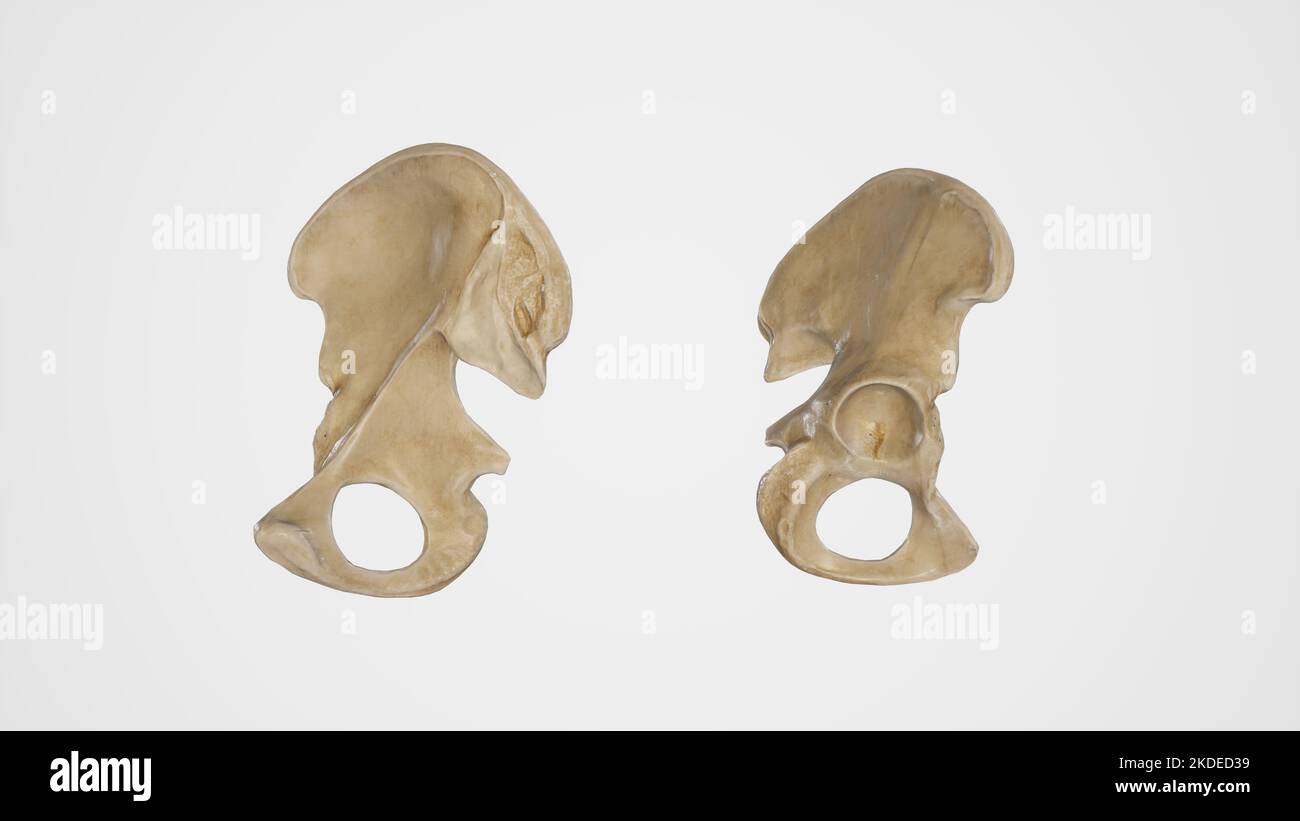 Medial and Lateral View of Hip Bone Stock Photohttps://www.alamy.com/image-license-details/?v=1https://www.alamy.com/medial-and-lateral-view-of-hip-bone-image490198445.html
RF2KDED39–Medial and Lateral View of Hip Bone
 CT Scan pelvic bone with both hip joint 3D rendering showign fracture of sacrum and superior pubic rumus. Stock Photohttps://www.alamy.com/image-license-details/?v=1https://www.alamy.com/ct-scan-pelvic-bone-with-both-hip-joint-3d-rendering-showign-fracture-of-sacrum-and-superior-pubic-rumus-image547292252.html
RF2PPB8RT–CT Scan pelvic bone with both hip joint 3D rendering showign fracture of sacrum and superior pubic rumus.
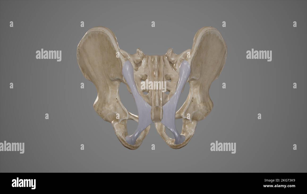 Medical Illustration of Sacrotuberous Ligament Stock Photohttps://www.alamy.com/image-license-details/?v=1https://www.alamy.com/medical-illustration-of-sacrotuberous-ligament-image491881357.html
RF2KG73K9–Medical Illustration of Sacrotuberous Ligament
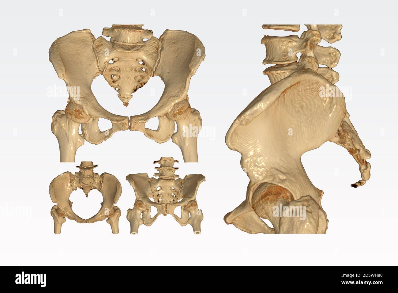 Collection of CT Scan pelvic bone with both hip joint 3D rendering image isolated on white background. Clipping path. Stock Photohttps://www.alamy.com/image-license-details/?v=1https://www.alamy.com/collection-of-ct-scan-pelvic-bone-with-both-hip-joint-3d-rendering-image-isolated-on-white-background-clipping-path-image382263728.html
RF2D5WH80–Collection of CT Scan pelvic bone with both hip joint 3D rendering image isolated on white background. Clipping path.
 The practice of obstetrics, designed for the use of students and practitioners of medicine . ing the Transverse Diameter of the Pelvic Outlet.The points of the pelvimeter are placed on the palmar surfaces of the tips of the index-fingers. See diagram, upper left of illustration.—{From a photograph.) mation of the true conjugate by this plan can only be approximated, since itdepends upon so many variable quantities. The method of taking the heightof the symphysis is described on page 166. For determining the thickness ofthe symphysis the pelvimeter of Skutsch, or one of its modifications, may b Stock Photohttps://www.alamy.com/image-license-details/?v=1https://www.alamy.com/the-practice-of-obstetrics-designed-for-the-use-of-students-and-practitioners-of-medicine-ing-the-transverse-diameter-of-the-pelvic-outletthe-points-of-the-pelvimeter-are-placed-on-the-palmar-surfaces-of-the-tips-of-the-index-fingers-see-diagram-upper-left-of-illustrationfrom-a-photograph-mation-of-the-true-conjugate-by-this-plan-can-only-be-approximated-since-itdepends-upon-so-many-variable-quantities-the-method-of-taking-the-heightof-the-symphysis-is-described-on-page-166-for-determining-the-thickness-ofthe-symphysis-the-pelvimeter-of-skutsch-or-one-of-its-modifications-may-b-image343324149.html
RM2AXFNCN–The practice of obstetrics, designed for the use of students and practitioners of medicine . ing the Transverse Diameter of the Pelvic Outlet.The points of the pelvimeter are placed on the palmar surfaces of the tips of the index-fingers. See diagram, upper left of illustration.—{From a photograph.) mation of the true conjugate by this plan can only be approximated, since itdepends upon so many variable quantities. The method of taking the heightof the symphysis is described on page 166. For determining the thickness ofthe symphysis the pelvimeter of Skutsch, or one of its modifications, may b
 The bones of the pelvis Stock Photohttps://www.alamy.com/image-license-details/?v=1https://www.alamy.com/stock-photo-the-bones-of-the-pelvis-13212869.html
RFACR2KJ–The bones of the pelvis
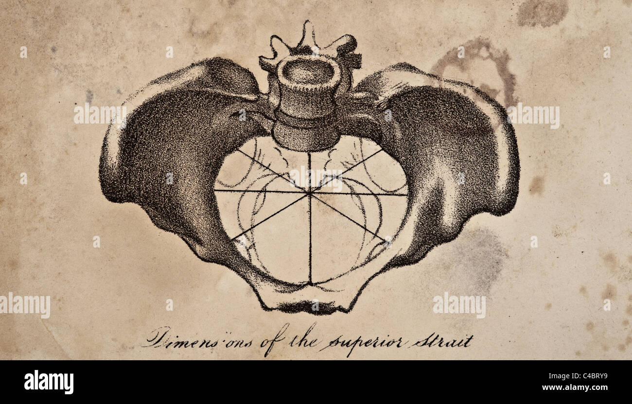 Illustration of the Human Pelvis copyright 1844 Stock Photohttps://www.alamy.com/image-license-details/?v=1https://www.alamy.com/stock-photo-illustration-of-the-human-pelvis-copyright-1844-37139629.html
RFC4BRY9–Illustration of the Human Pelvis copyright 1844
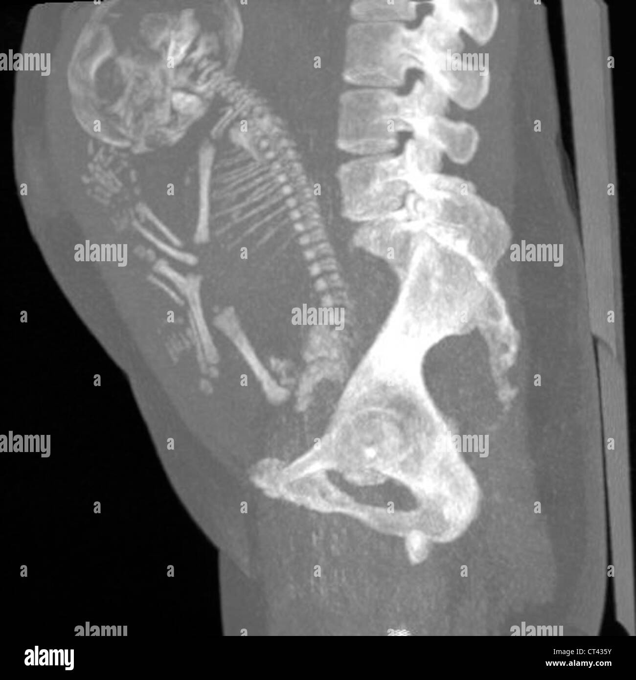 PELVIMETRY Stock Photohttps://www.alamy.com/image-license-details/?v=1https://www.alamy.com/stock-photo-pelvimetry-49262807.html
RMCT435Y–PELVIMETRY
 Medical Illustration of Sacrotuberous and Sacrospinous Ligaments Stock Photohttps://www.alamy.com/image-license-details/?v=1https://www.alamy.com/medical-illustration-of-sacrotuberous-and-sacrospinous-ligaments-image491881355.html
RF2KG73K7–Medical Illustration of Sacrotuberous and Sacrospinous Ligaments
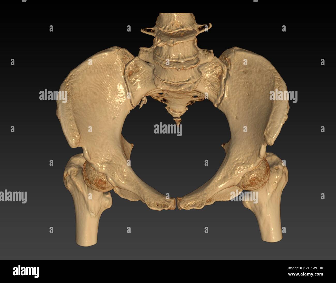 CT Scan of pelvic bone with both hip joint 3D rendering image Inlet view isolated on black background. Clipping path. Stock Photohttps://www.alamy.com/image-license-details/?v=1https://www.alamy.com/ct-scan-of-pelvic-bone-with-both-hip-joint-3d-rendering-image-inlet-view-isolated-on-black-background-clipping-path-image382263980.html
RF2D5WHH0–CT Scan of pelvic bone with both hip joint 3D rendering image Inlet view isolated on black background. Clipping path.
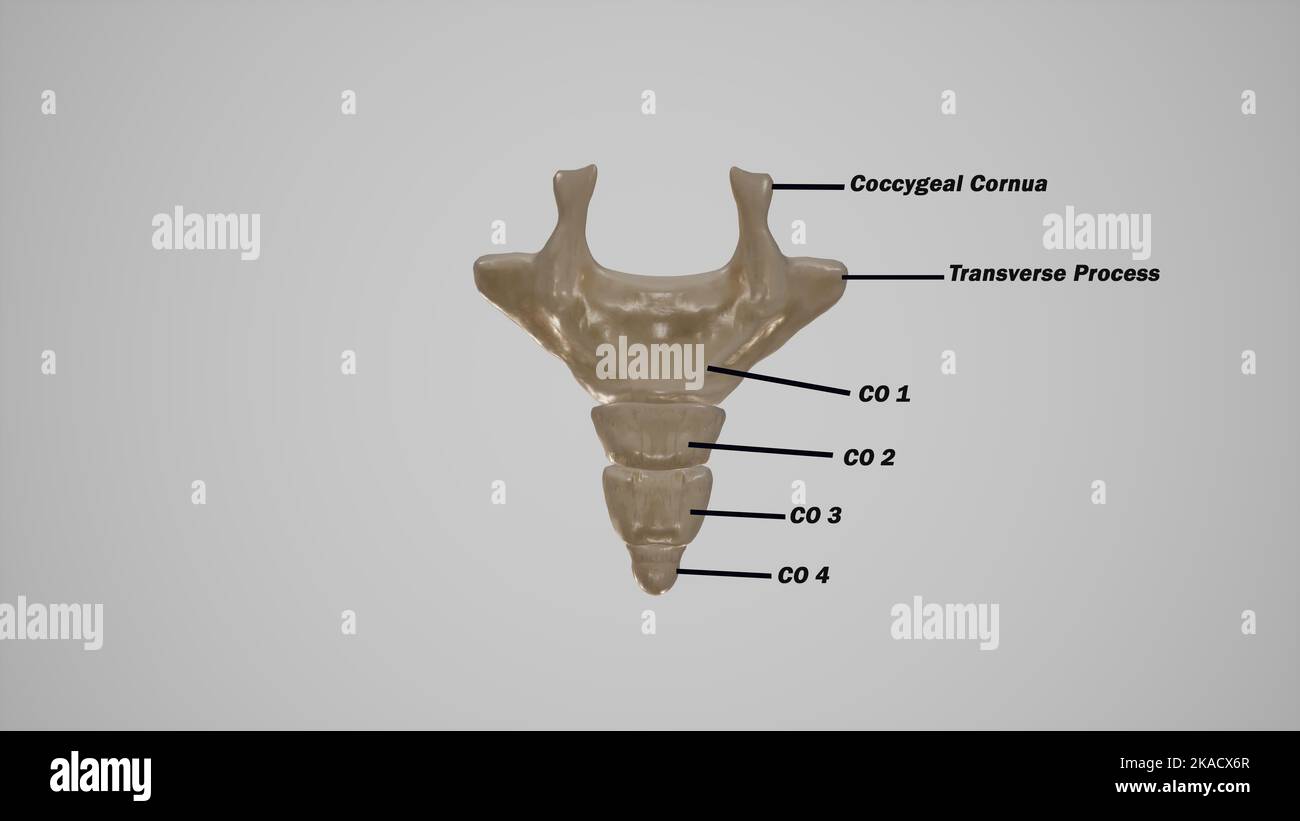 Coccyx bone anatomy labeled Stock Photohttps://www.alamy.com/image-license-details/?v=1https://www.alamy.com/coccyx-bone-anatomy-labeled-image488320863.html
RF2KACX6R–Coccyx bone anatomy labeled
 The bones of the pelvis Stock Photohttps://www.alamy.com/image-license-details/?v=1https://www.alamy.com/stock-photo-the-bones-of-the-pelvis-13170336.html
RFACJG2W–The bones of the pelvis
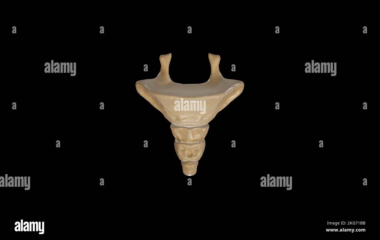 Anterior view of Coccyx Stock Photohttps://www.alamy.com/image-license-details/?v=1https://www.alamy.com/anterior-view-of-coccyx-image491879567.html
RF2KG71BB–Anterior view of Coccyx
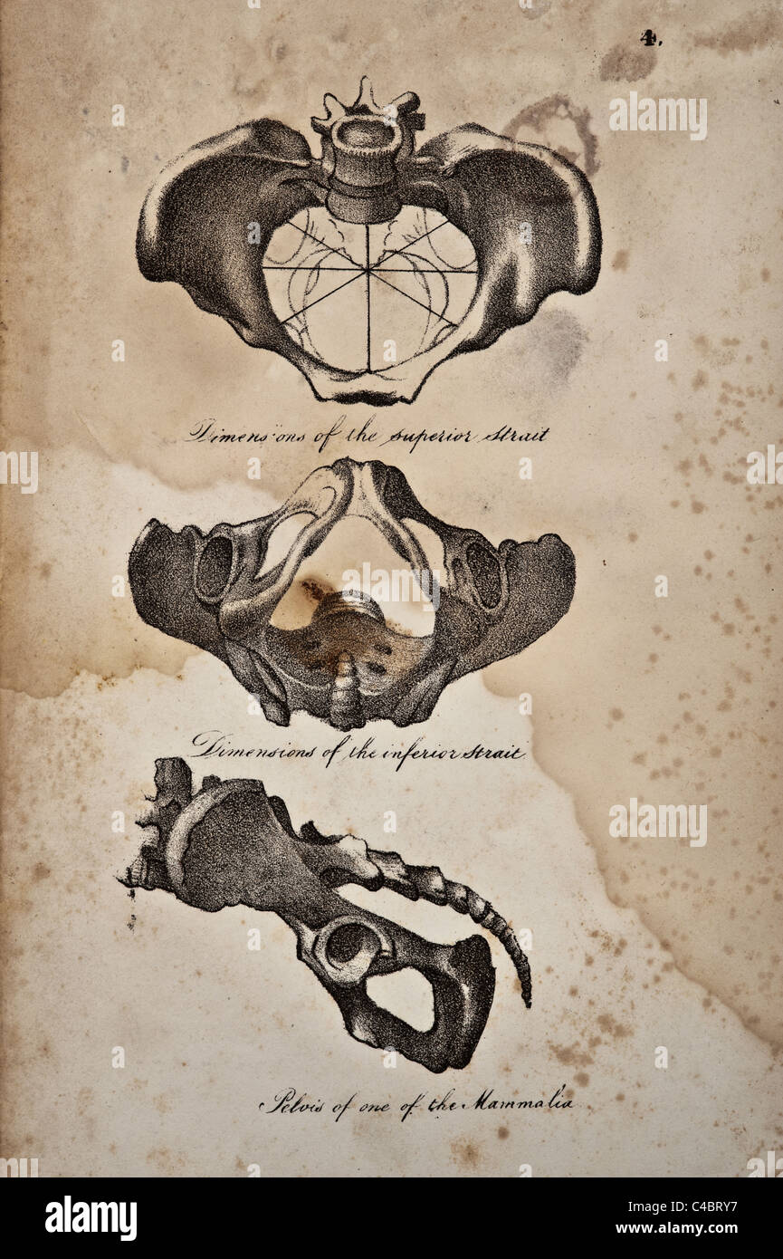 Illustration of the passage through the pelvis copyright 1844 Stock Photohttps://www.alamy.com/image-license-details/?v=1https://www.alamy.com/stock-photo-illustration-of-the-passage-through-the-pelvis-copyright-1844-37139627.html
RFC4BRY7–Illustration of the passage through the pelvis copyright 1844
 The practice of obstetrics, designed for the use of students and practitioners of medicine . ternalface of the sacro-sciatic ligaments and foramina. The direction of this part is PELVIC OUTLET. 383 the converse of the anterior, it being from above downward, from withoutinward, and from before backward. Pelvic Cavity Measurements.—(See Pelvimetry, page 167.) The depth of the pelvis at the symphysis is if inches (4 cm.). The depth ofthe lateral wall over the smooth surface of the ischial bones is 32 inches (9 cm.).The depth of the posterior wall, following the course of the sacrum and coccyxfrom Stock Photohttps://www.alamy.com/image-license-details/?v=1https://www.alamy.com/the-practice-of-obstetrics-designed-for-the-use-of-students-and-practitioners-of-medicine-ternalface-of-the-sacro-sciatic-ligaments-and-foramina-the-direction-of-this-part-is-pelvic-outlet-383-the-converse-of-the-anterior-it-being-from-above-downward-from-withoutinward-and-from-before-backward-pelvic-cavity-measurementssee-pelvimetry-page-167-the-depth-of-the-pelvis-at-the-symphysis-is-if-inches-4-cm-the-depth-ofthe-lateral-wall-over-the-smooth-surface-of-the-ischial-bones-is-32-inches-9-cmthe-depth-of-the-posterior-wall-following-the-course-of-the-sacrum-and-coccyxfrom-image343270339.html
RM2AXD8PY–The practice of obstetrics, designed for the use of students and practitioners of medicine . ternalface of the sacro-sciatic ligaments and foramina. The direction of this part is PELVIC OUTLET. 383 the converse of the anterior, it being from above downward, from withoutinward, and from before backward. Pelvic Cavity Measurements.—(See Pelvimetry, page 167.) The depth of the pelvis at the symphysis is if inches (4 cm.). The depth ofthe lateral wall over the smooth surface of the ischial bones is 32 inches (9 cm.).The depth of the posterior wall, following the course of the sacrum and coccyxfrom
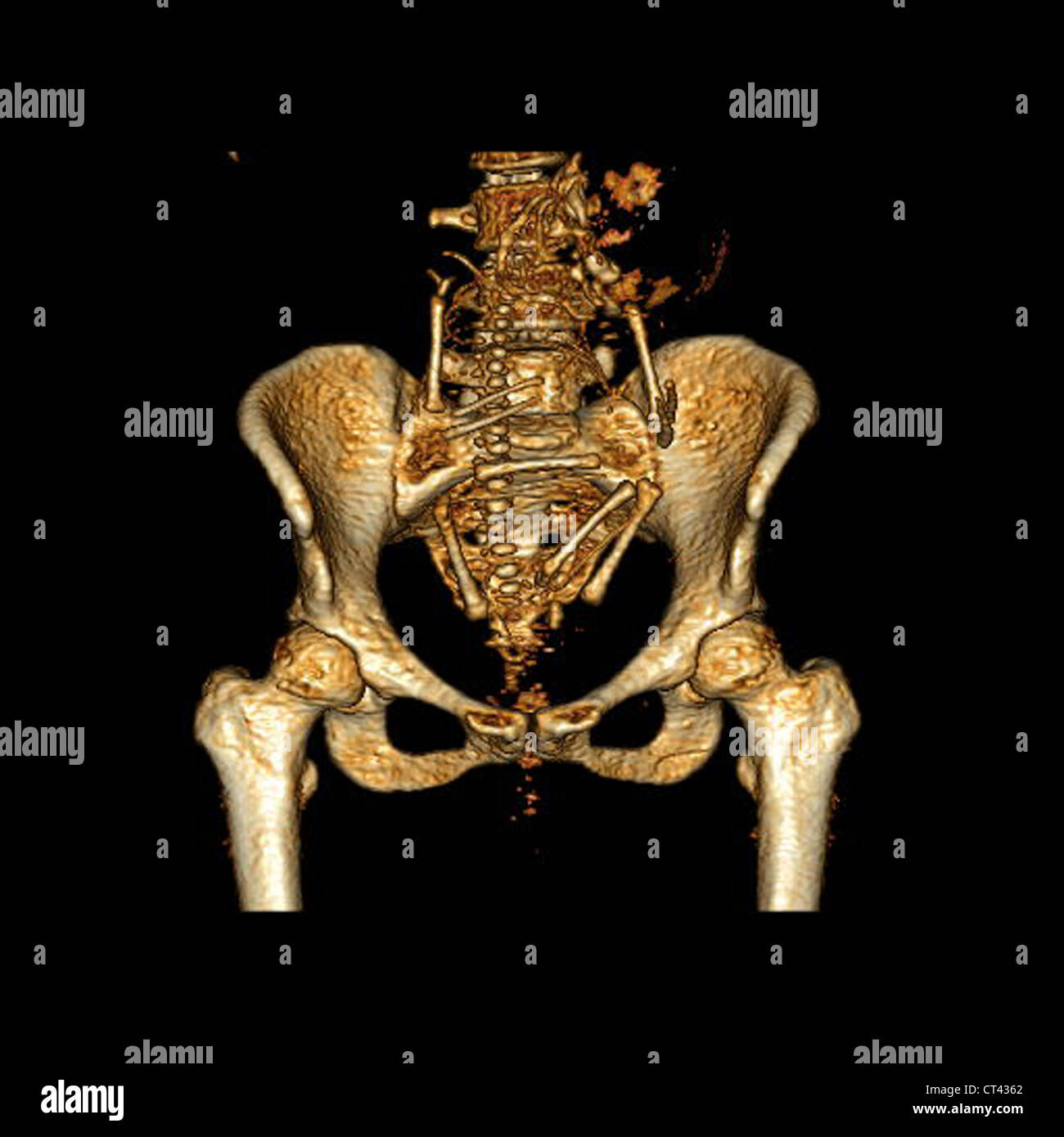 PELVIMETRY Stock Photohttps://www.alamy.com/image-license-details/?v=1https://www.alamy.com/stock-photo-pelvimetry-49262810.html
RMCT4362–PELVIMETRY
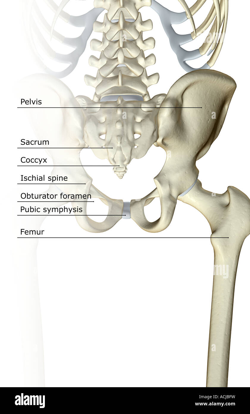 The bones of the pelvis Stock Photohttps://www.alamy.com/image-license-details/?v=1https://www.alamy.com/stock-photo-the-bones-of-the-pelvis-13168812.html
RFACJBFW–The bones of the pelvis
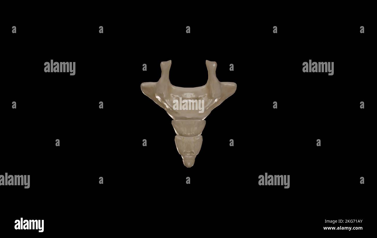 Posterior view of Coccyx Stock Photohttps://www.alamy.com/image-license-details/?v=1https://www.alamy.com/posterior-view-of-coccyx-image491879555.html
RF2KG71AY–Posterior view of Coccyx
 Illustration of the Human Pelvis copyright 1844 Stock Photohttps://www.alamy.com/image-license-details/?v=1https://www.alamy.com/stock-photo-illustration-of-the-human-pelvis-copyright-1844-37139664.html
RFC4BT0G–Illustration of the Human Pelvis copyright 1844
 The American text-book of obstetrics for practitioners and students . Fig. L8.—Skutscha method of measuring the trans- erse diameter of the pelvic inlet. Fig. 19.—Measurement f the antero-posterioidiameter of the pelvic outlet. the approximation of the tuberosities of the ischia, the depth of the pelvis,and the direction of its canal; by detecting, possibly, the presence of an exos-tosis, an osteosarcoma, an abnormally-projecting spinous process, an old frac-ture, or asymmetry of the pelvic walls from any cause. Measurement of the Transverse Diameter of the Pelvic Outlet.—The antero-posterior Stock Photohttps://www.alamy.com/image-license-details/?v=1https://www.alamy.com/the-american-text-book-of-obstetrics-for-practitioners-and-students-fig-l8skutscha-method-of-measuring-the-trans-erse-diameter-of-the-pelvic-inlet-fig-19measurement-f-the-antero-posterioidiameter-of-the-pelvic-outlet-the-approximation-of-the-tuberosities-of-the-ischia-the-depth-of-the-pelvisand-the-direction-of-its-canal-by-detecting-possibly-the-presence-of-an-exos-tosis-an-osteosarcoma-an-abnormally-projecting-spinous-process-an-old-frac-ture-or-asymmetry-of-the-pelvic-walls-from-any-cause-measurement-of-the-transverse-diameter-of-the-pelvic-outletthe-antero-posterior-image343174988.html
RM2AX8Y5G–The American text-book of obstetrics for practitioners and students . Fig. L8.—Skutscha method of measuring the trans- erse diameter of the pelvic inlet. Fig. 19.—Measurement f the antero-posterioidiameter of the pelvic outlet. the approximation of the tuberosities of the ischia, the depth of the pelvis,and the direction of its canal; by detecting, possibly, the presence of an exos-tosis, an osteosarcoma, an abnormally-projecting spinous process, an old frac-ture, or asymmetry of the pelvic walls from any cause. Measurement of the Transverse Diameter of the Pelvic Outlet.—The antero-posterior
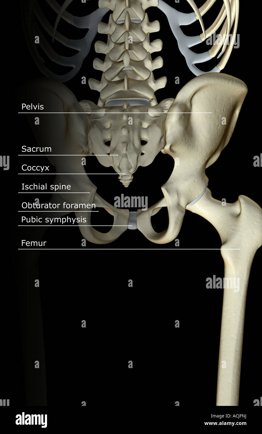 The bones of the pelvis Stock Photohttps://www.alamy.com/image-license-details/?v=1https://www.alamy.com/stock-photo-the-bones-of-the-pelvis-13170221.html
RFACJFNJ–The bones of the pelvis
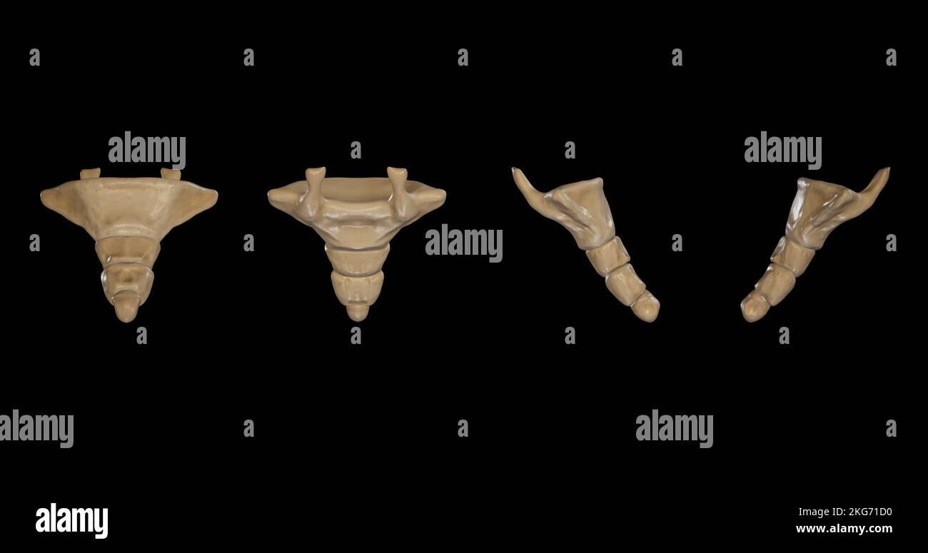 Coccyx-Multiple Views Stock Photohttps://www.alamy.com/image-license-details/?v=1https://www.alamy.com/coccyx-multiple-views-image491879612.html
RF2KG71D0–Coccyx-Multiple Views
 Illustration of the Human Pelvis copyright 1844 Stock Photohttps://www.alamy.com/image-license-details/?v=1https://www.alamy.com/stock-photo-illustration-of-the-human-pelvis-copyright-1844-37139598.html
RFC4BRX6–Illustration of the Human Pelvis copyright 1844
 A text-book of practical obstetrics, comprising pregnancy, labor, and the puerpal state, and obstetric surgery . The Plane and the Axis of the SuperiorStrait, or Pelvic Inlet. The Plane and the Axis of the InferiorStrait, or Pelvic Outlet. MECHANISM OF LABOR. 95 the foetus itself, which must be made to adapt itself to the shapeand the dimensions of the canal through which it must pass. The canal through which the foetus must pass consists ofthe pelvis and of the pelvic floor. The pelvis is divided, froman obstetrical stand-point, into a superior and an inferior por-tion. The superior portion o Stock Photohttps://www.alamy.com/image-license-details/?v=1https://www.alamy.com/a-text-book-of-practical-obstetrics-comprising-pregnancy-labor-and-the-puerpal-state-and-obstetric-surgery-the-plane-and-the-axis-of-the-superiorstrait-or-pelvic-inlet-the-plane-and-the-axis-of-the-inferiorstrait-or-pelvic-outlet-mechanism-of-labor-95-the-foetus-itself-which-must-be-made-to-adapt-itself-to-the-shapeand-the-dimensions-of-the-canal-through-which-it-must-pass-the-canal-through-which-the-foetus-must-pass-consists-ofthe-pelvis-and-of-the-pelvic-floor-the-pelvis-is-divided-froman-obstetrical-stand-point-into-a-superior-and-an-inferior-por-tion-the-superior-portion-o-image342978876.html
RM2AX011G–A text-book of practical obstetrics, comprising pregnancy, labor, and the puerpal state, and obstetric surgery . The Plane and the Axis of the SuperiorStrait, or Pelvic Inlet. The Plane and the Axis of the InferiorStrait, or Pelvic Outlet. MECHANISM OF LABOR. 95 the foetus itself, which must be made to adapt itself to the shapeand the dimensions of the canal through which it must pass. The canal through which the foetus must pass consists ofthe pelvis and of the pelvic floor. The pelvis is divided, froman obstetrical stand-point, into a superior and an inferior por-tion. The superior portion o
 The bones of the pelvis Stock Photohttps://www.alamy.com/image-license-details/?v=1https://www.alamy.com/stock-photo-the-bones-of-the-pelvis-13174798.html
RFACK1AR–The bones of the pelvis
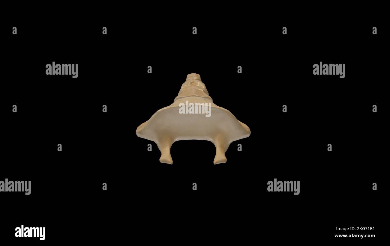 Superior view of Coccyx Stock Photohttps://www.alamy.com/image-license-details/?v=1https://www.alamy.com/superior-view-of-coccyx-image491879557.html
RF2KG71B1–Superior view of Coccyx
 Illustration of the Human Pelvis copyright 1844 Stock Photohttps://www.alamy.com/image-license-details/?v=1https://www.alamy.com/stock-photo-illustration-of-the-human-pelvis-copyright-1844-37139637.html
RFC4BRYH–Illustration of the Human Pelvis copyright 1844
 A textbook of obstetrics . Fig. JX). Martins pelvimeter. Fig. 207. — Harris Dickinson portable pelvimeter. ANOMALIES IX THE FORCES OF LABOR. 4II the foundations of pelvimetry, and his instrument and methodsare in use at the present time (Figs. 265—268). It is con-venient to describe the measurements of the diameters of thepelvic inlet, pelvic cavity, and pelvic outlet separately. Measurement of the Anteroposterior Diameter of the SuperiorStrait.—This measurement, the most important in the pelvis,can not be taken directly. It must be estimated by several. Fig. 268.—Measuring the external conjug Stock Photohttps://www.alamy.com/image-license-details/?v=1https://www.alamy.com/a-textbook-of-obstetrics-fig-jx-martins-pelvimeter-fig-207-harris-dickinson-portable-pelvimeter-anomalies-ix-the-forces-of-labor-4ii-the-foundations-of-pelvimetry-and-his-instrument-and-methodsare-in-use-at-the-present-time-figs-265268-it-is-con-venient-to-describe-the-measurements-of-the-diameters-of-thepelvic-inlet-pelvic-cavity-and-pelvic-outlet-separately-measurement-of-the-anteroposterior-diameter-of-the-superiorstraitthis-measurement-the-most-important-in-the-pelviscan-not-be-taken-directly-it-must-be-estimated-by-several-fig-268measuring-the-external-conjug-image342984211.html
RM2AX07T3–A textbook of obstetrics . Fig. JX). Martins pelvimeter. Fig. 207. — Harris Dickinson portable pelvimeter. ANOMALIES IX THE FORCES OF LABOR. 4II the foundations of pelvimetry, and his instrument and methodsare in use at the present time (Figs. 265—268). It is con-venient to describe the measurements of the diameters of thepelvic inlet, pelvic cavity, and pelvic outlet separately. Measurement of the Anteroposterior Diameter of the SuperiorStrait.—This measurement, the most important in the pelvis,can not be taken directly. It must be estimated by several. Fig. 268.—Measuring the external conjug
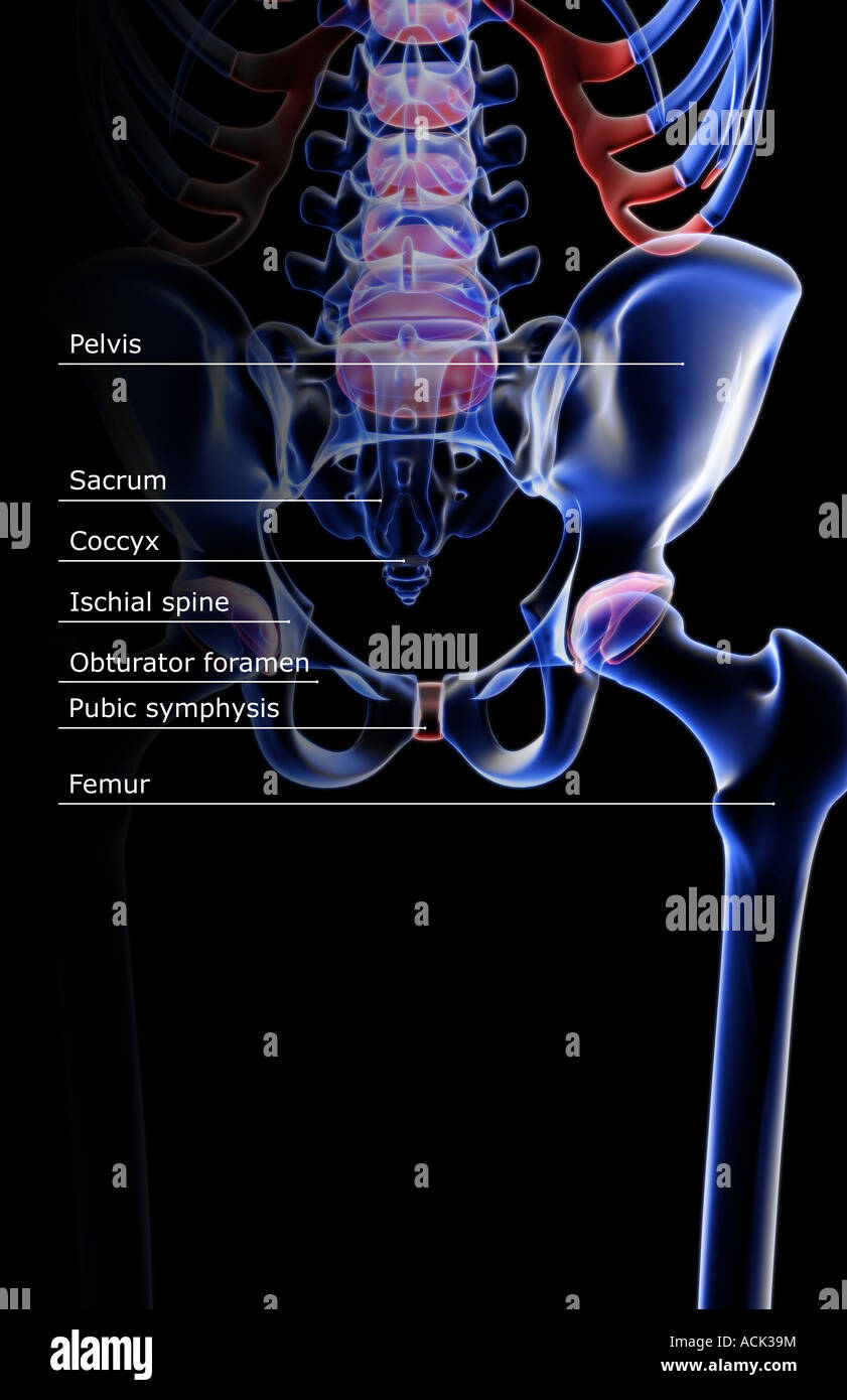 The bones of the pelvis Stock Photohttps://www.alamy.com/image-license-details/?v=1https://www.alamy.com/stock-photo-the-bones-of-the-pelvis-13175455.html
RFACK39M–The bones of the pelvis
 Illustration of the Human Pelvis copyright 1844 Stock Photohttps://www.alamy.com/image-license-details/?v=1https://www.alamy.com/stock-photo-illustration-of-the-human-pelvis-copyright-1844-37139620.html
RFC4BRY0–Illustration of the Human Pelvis copyright 1844
 A textbook of obstetrics . Fig. 5IQ.—Small forceps, modified by the author for use at (lie vulvar orifice and pelvic outlet. blades are of such length that the instrument may be used withequal convenience at the superior strait or at the pelvic outlet.The lock is the English lock, which has the great advantage of easy adjustment ; and the handles are provided with shouldersfor two fingers, and with depressions along the handle for the FORCEPS. 729 remaining fingers and thumb of the hand, so that a firm and con-venient grasp can be taken of the instrument in use. Another modern instrument deser Stock Photohttps://www.alamy.com/image-license-details/?v=1https://www.alamy.com/a-textbook-of-obstetrics-fig-5iqsmall-forceps-modified-by-the-author-for-use-at-lie-vulvar-orifice-and-pelvic-outlet-blades-are-of-such-length-that-the-instrument-may-be-used-withequal-convenience-at-the-superior-strait-or-at-the-pelvic-outletthe-lock-is-the-english-lock-which-has-the-great-advantage-of-easy-adjustment-and-the-handles-are-provided-with-shouldersfor-two-fingers-and-with-depressions-along-the-handle-for-the-forceps-729-remaining-fingers-and-thumb-of-the-hand-so-that-a-firm-and-con-venient-grasp-can-be-taken-of-the-instrument-in-use-another-modern-instrument-deser-image342885919.html
RM2AWRPDK–A textbook of obstetrics . Fig. 5IQ.—Small forceps, modified by the author for use at (lie vulvar orifice and pelvic outlet. blades are of such length that the instrument may be used withequal convenience at the superior strait or at the pelvic outlet.The lock is the English lock, which has the great advantage of easy adjustment ; and the handles are provided with shouldersfor two fingers, and with depressions along the handle for the FORCEPS. 729 remaining fingers and thumb of the hand, so that a firm and con-venient grasp can be taken of the instrument in use. Another modern instrument deser
 Illustration of the Human Pelvis copyright 1844 Stock Photohttps://www.alamy.com/image-license-details/?v=1https://www.alamy.com/stock-photo-illustration-of-the-human-pelvis-copyright-1844-37139656.html
RFC4BT08–Illustration of the Human Pelvis copyright 1844
 A manual of obstetrics . ft, and extend from one anterior superior spinous pro-cess of the ilium to the opposite posterior superior spinousprocess, which may be recognized by the distinct indentationoverlying it. This measurement is taken with the patientresting upon her side. .The circumference of the pelvis atthe upper margin is 90 cm. (35.4330 in.). Estimation of the Pelvic Outlet.—Only in one commonvariety of pelvic deformity is this portion of the pelvis con-tracted—namely, in the kyphotic pelvis. Therefore butslight import is attached to the measurements here. Inthe normal pelvis the tra Stock Photohttps://www.alamy.com/image-license-details/?v=1https://www.alamy.com/a-manual-of-obstetrics-ft-and-extend-from-one-anterior-superior-spinous-pro-cess-of-the-ilium-to-the-opposite-posterior-superior-spinousprocess-which-may-be-recognized-by-the-distinct-indentationoverlying-it-this-measurement-is-taken-with-the-patientresting-upon-her-side-the-circumference-of-the-pelvis-atthe-upper-margin-is-90-cm-354330-in-estimation-of-the-pelvic-outletonly-in-one-commonvariety-of-pelvic-deformity-is-this-portion-of-the-pelvis-con-tractednamely-in-the-kyphotic-pelvis-therefore-butslight-import-is-attached-to-the-measurements-here-inthe-normal-pelvis-the-tra-image338194505.html
RM2AJ62F5–A manual of obstetrics . ft, and extend from one anterior superior spinous pro-cess of the ilium to the opposite posterior superior spinousprocess, which may be recognized by the distinct indentationoverlying it. This measurement is taken with the patientresting upon her side. .The circumference of the pelvis atthe upper margin is 90 cm. (35.4330 in.). Estimation of the Pelvic Outlet.—Only in one commonvariety of pelvic deformity is this portion of the pelvis con-tracted—namely, in the kyphotic pelvis. Therefore butslight import is attached to the measurements here. Inthe normal pelvis the tra
 Illustration of the Human Pelvis copyright 1844 Stock Photohttps://www.alamy.com/image-license-details/?v=1https://www.alamy.com/stock-photo-illustration-of-the-human-pelvis-copyright-1844-37139632.html
RFC4BRYC–Illustration of the Human Pelvis copyright 1844
 A textbook of obstetrics . Fig. 51<S.—Davis forceps.. Fig. 5IQ.—Small forceps, modified by the author for use at (lie vulvar orifice and pelvic outlet. blades are of such length that the instrument may be used withequal convenience at the superior strait or at the pelvic outlet.The lock is the English lock, which has the great advantage of easy adjustment ; and the handles are provided with shouldersfor two fingers, and with depressions along the handle for the FORCEPS. 729 remaining fingers and thumb of the hand, so that a firm and con-venient grasp can be taken of the instrument in use. A Stock Photohttps://www.alamy.com/image-license-details/?v=1https://www.alamy.com/a-textbook-of-obstetrics-fig-51ltsdavis-forceps-fig-5iqsmall-forceps-modified-by-the-author-for-use-at-lie-vulvar-orifice-and-pelvic-outlet-blades-are-of-such-length-that-the-instrument-may-be-used-withequal-convenience-at-the-superior-strait-or-at-the-pelvic-outletthe-lock-is-the-english-lock-which-has-the-great-advantage-of-easy-adjustment-and-the-handles-are-provided-with-shouldersfor-two-fingers-and-with-depressions-along-the-handle-for-the-forceps-729-remaining-fingers-and-thumb-of-the-hand-so-that-a-firm-and-con-venient-grasp-can-be-taken-of-the-instrument-in-use-a-image342886428.html
RM2AWRR3T–A textbook of obstetrics . Fig. 51<S.—Davis forceps.. Fig. 5IQ.—Small forceps, modified by the author for use at (lie vulvar orifice and pelvic outlet. blades are of such length that the instrument may be used withequal convenience at the superior strait or at the pelvic outlet.The lock is the English lock, which has the great advantage of easy adjustment ; and the handles are provided with shouldersfor two fingers, and with depressions along the handle for the FORCEPS. 729 remaining fingers and thumb of the hand, so that a firm and con-venient grasp can be taken of the instrument in use. A
