Quick filters:
Peribronchial Stock Photos and Images
 Practical pathology; a manual for students and practitioners . however, being partially carried on by smaller vessels whichgrow in the new tissue, some of these, it is said, developing an elasticcoat of their own. ( X 300J.—Observe the tuberculous and fibroid masses in the deeplayer of the pleura, in the interlobular and interalveolar septa, and inthe peribronchial and perivascular tissue. In the wedge-shaped patchesof tubercle near the surface, the individual nodules, each surrounded 5o8 THE LUNG by pneumonic zones, should be further examined ; these patches maybe taken as typical of the patc Stock Photohttps://www.alamy.com/image-license-details/?v=1https://www.alamy.com/practical-pathology-a-manual-for-students-and-practitioners-however-being-partially-carried-on-by-smaller-vessels-whichgrow-in-the-new-tissue-some-of-these-it-is-said-developing-an-elasticcoat-of-their-own-x-300jobserve-the-tuberculous-and-fibroid-masses-in-the-deeplayer-of-the-pleura-in-the-interlobular-and-interalveolar-septa-and-inthe-peribronchial-and-perivascular-tissue-in-the-wedge-shaped-patchesof-tubercle-near-the-surface-the-individual-nodules-each-surrounded-5o8-the-lung-by-pneumonic-zones-should-be-further-examined-these-patches-maybe-taken-as-typical-of-the-patc-image342978507.html
RM2AX00GB–Practical pathology; a manual for students and practitioners . however, being partially carried on by smaller vessels whichgrow in the new tissue, some of these, it is said, developing an elasticcoat of their own. ( X 300J.—Observe the tuberculous and fibroid masses in the deeplayer of the pleura, in the interlobular and interalveolar septa, and inthe peribronchial and perivascular tissue. In the wedge-shaped patchesof tubercle near the surface, the individual nodules, each surrounded 5o8 THE LUNG by pneumonic zones, should be further examined ; these patches maybe taken as typical of the patc
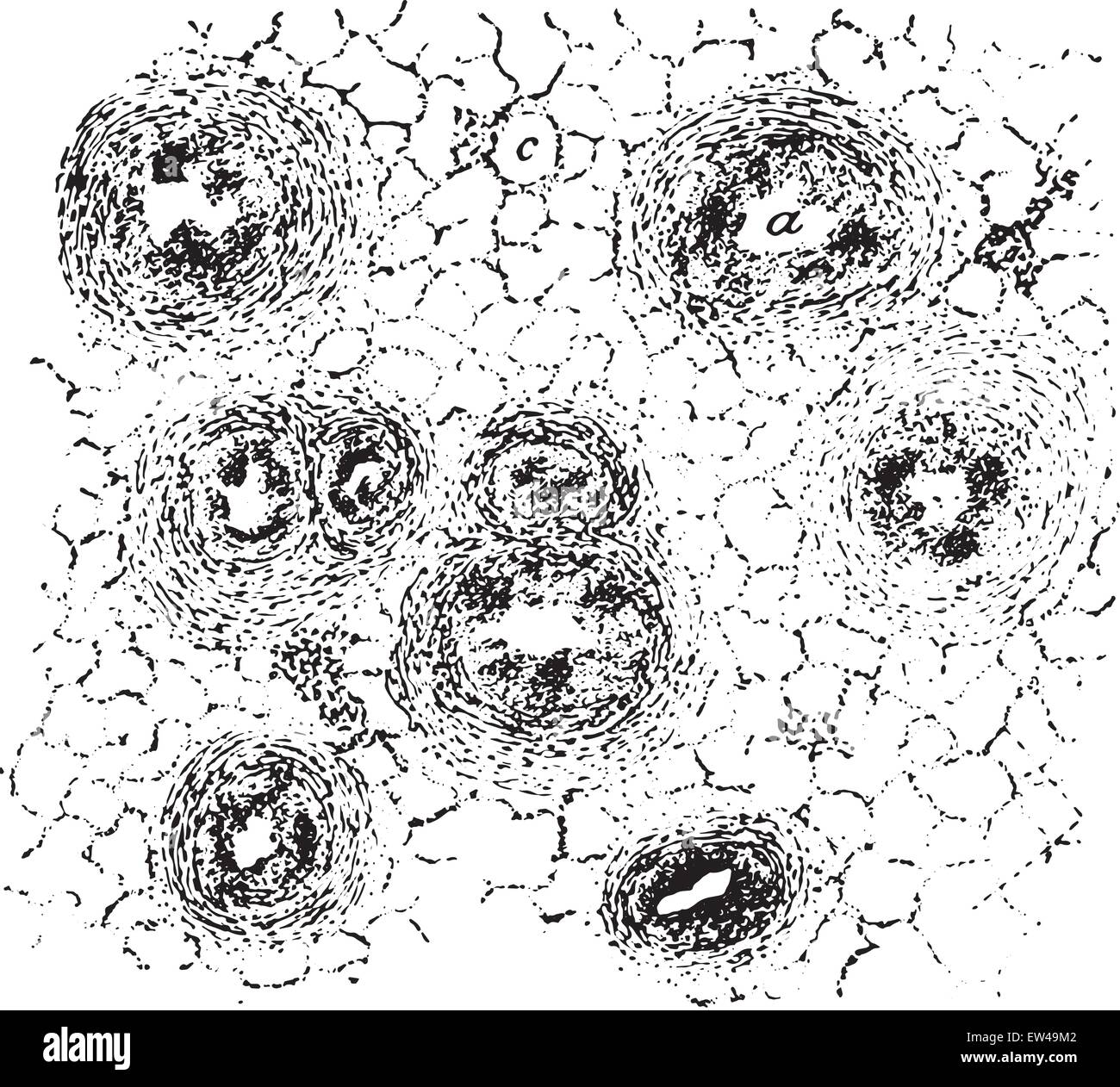
 Chronic caseous bronchitis, vintage engraved illustration. Stock Vectorhttps://www.alamy.com/image-license-details/?v=1https://www.alamy.com/stock-photo-chronic-caseous-bronchitis-vintage-engraved-illustration-84303298.html
RFEW49M2–Chronic caseous bronchitis, vintage engraved illustration.
 . FiG. II. Right lung of pig. The stippled portion is usually involved in cases of infectious pneumonia or sivine plague. {/>) ventral lobe, {c) cephalic lobe, {a)principal lobe. The ventral lobe is usually the seat of the more advanced disease and consequently the first to become hepafized. The cephalic portion of the principal lobe (.r) is usually hepatized and the reinaining portion deeply reddoied. tion (embolic pneumonia). If it enters by way of the pleura, the virus will creep along the interlobular and peribronchial tissue before it invades the parenchyma proper. In natural infection Stock Photohttps://www.alamy.com/image-license-details/?v=1https://www.alamy.com/fig-ii-right-lung-of-pig-the-stippled-portion-is-usually-involved-in-cases-of-infectious-pneumonia-or-sivine-plague-gt-ventral-lobe-c-cephalic-lobe-aprincipal-lobe-the-ventral-lobe-is-usually-the-seat-of-the-more-advanced-disease-and-consequently-the-first-to-become-hepafized-the-cephalic-portion-of-the-principal-lobe-r-is-usually-hepatized-and-the-reinaining-portion-deeply-reddoied-tion-embolic-pneumonia-if-it-enters-by-way-of-the-pleura-the-virus-will-creep-along-the-interlobular-and-peribronchial-tissue-before-it-invades-the-parenchyma-proper-in-natural-infection-image179937481.html
RMMCMT2H–. FiG. II. Right lung of pig. The stippled portion is usually involved in cases of infectious pneumonia or sivine plague. {/>) ventral lobe, {c) cephalic lobe, {a)principal lobe. The ventral lobe is usually the seat of the more advanced disease and consequently the first to become hepafized. The cephalic portion of the principal lobe (.r) is usually hepatized and the reinaining portion deeply reddoied. tion (embolic pneumonia). If it enters by way of the pleura, the virus will creep along the interlobular and peribronchial tissue before it invades the parenchyma proper. In natural infection
 Modern medicine : its theory and practice, in original contributions by American and foreign authors . not rarely affected, and, not being able to resist the high pressurecaused by the cough, dilatation occurs. When bronchopneumonia is so intense as to destroy the muscular andelastic fibers on which the strength of the bronchial wall depends, there isalso at the same time such inflammation of the surrounding lung tissue thatit becomes sclerosed in time, so that the two lesions are associated—dilatationof the bronchi and sclerosis of the peribronchial pulmonary tissue. In generalconditions whic Stock Photohttps://www.alamy.com/image-license-details/?v=1https://www.alamy.com/modern-medicine-its-theory-and-practice-in-original-contributions-by-american-and-foreign-authors-not-rarely-affected-and-not-being-able-to-resist-the-high-pressurecaused-by-the-cough-dilatation-occurs-when-bronchopneumonia-is-so-intense-as-to-destroy-the-muscular-andelastic-fibers-on-which-the-strength-of-the-bronchial-wall-depends-there-isalso-at-the-same-time-such-inflammation-of-the-surrounding-lung-tissue-thatit-becomes-sclerosed-in-time-so-that-the-two-lesions-are-associateddilatationof-the-bronchi-and-sclerosis-of-the-peribronchial-pulmonary-tissue-in-generalconditions-whic-image342763391.html
RM2AWJ65K–Modern medicine : its theory and practice, in original contributions by American and foreign authors . not rarely affected, and, not being able to resist the high pressurecaused by the cough, dilatation occurs. When bronchopneumonia is so intense as to destroy the muscular andelastic fibers on which the strength of the bronchial wall depends, there isalso at the same time such inflammation of the surrounding lung tissue thatit becomes sclerosed in time, so that the two lesions are associated—dilatationof the bronchi and sclerosis of the peribronchial pulmonary tissue. In generalconditions whic
 . The effect of tuberculosis vaccination upon cattle infected with tuberculosis . Fig. I. Left bronchial gland. Right lung. Necropsy. Weight 643 pounds; fair condition. The lesions of tuberculosis found in this animal were as follows: At the lower border of the middle lobe of the right lung was a slightly depressed area about one-half inch in diameter containing a collection of thick yellow pus filling a cavity the size of a large pea. The walls of this cavity are one- eighth of an inch thick, white, and of firm, dense texture. In one of the left peribronchial glands there is a caseocal- Stock Photohttps://www.alamy.com/image-license-details/?v=1https://www.alamy.com/the-effect-of-tuberculosis-vaccination-upon-cattle-infected-with-tuberculosis-fig-i-left-bronchial-gland-right-lung-necropsy-weight-643-pounds-fair-condition-the-lesions-of-tuberculosis-found-in-this-animal-were-as-follows-at-the-lower-border-of-the-middle-lobe-of-the-right-lung-was-a-slightly-depressed-area-about-one-half-inch-in-diameter-containing-a-collection-of-thick-yellow-pus-filling-a-cavity-the-size-of-a-large-pea-the-walls-of-this-cavity-are-one-eighth-of-an-inch-thick-white-and-of-firm-dense-texture-in-one-of-the-left-peribronchial-glands-there-is-a-caseocal-image178406464.html
RMMA737C–. The effect of tuberculosis vaccination upon cattle infected with tuberculosis . Fig. I. Left bronchial gland. Right lung. Necropsy. Weight 643 pounds; fair condition. The lesions of tuberculosis found in this animal were as follows: At the lower border of the middle lobe of the right lung was a slightly depressed area about one-half inch in diameter containing a collection of thick yellow pus filling a cavity the size of a large pea. The walls of this cavity are one- eighth of an inch thick, white, and of firm, dense texture. In one of the left peribronchial glands there is a caseocal-
 . Post-mortem pathology; a manual of post-mortem examinations and the interpretations to be drawn therefrom; a practical treatise for students and practitioners. Palatine arch incised Common carotid artery-Right subclavian artery Aorta Right bronchus Peribronchial lymph glands Fig. 87.—Examination of the organs of the neck. The arrows show the direction in which the incisionsin the tongue and in the posterior wall of the oesophagus are to be made. (After Nauwerck.). : Fig. 88.— Method of opening trachea posteriorly. The incision starts from above and extends downward. Stock Photohttps://www.alamy.com/image-license-details/?v=1https://www.alamy.com/post-mortem-pathology-a-manual-of-post-mortem-examinations-and-the-interpretations-to-be-drawn-therefrom-a-practical-treatise-for-students-and-practitioners-palatine-arch-incised-common-carotid-artery-right-subclavian-artery-aorta-right-bronchus-peribronchial-lymph-glands-fig-87examination-of-the-organs-of-the-neck-the-arrows-show-the-direction-in-which-the-incisionsin-the-tongue-and-in-the-posterior-wall-of-the-oesophagus-are-to-be-made-after-nauwerck-fig-88-method-of-opening-trachea-posteriorly-the-incision-starts-from-above-and-extends-downward-image336838601.html
RM2AG0921–. Post-mortem pathology; a manual of post-mortem examinations and the interpretations to be drawn therefrom; a practical treatise for students and practitioners. Palatine arch incised Common carotid artery-Right subclavian artery Aorta Right bronchus Peribronchial lymph glands Fig. 87.—Examination of the organs of the neck. The arrows show the direction in which the incisionsin the tongue and in the posterior wall of the oesophagus are to be made. (After Nauwerck.). : Fig. 88.— Method of opening trachea posteriorly. The incision starts from above and extends downward.
 . The effect of tuberculosis vaccination upon cattle infected with tuberculosis . Fig. 4. Left bronchial gland. Necropsy. Weight 566 pounds; good condition. The only evidence of tuberculosis in this animal is a calcareous nodule one-eighth of an inch in diameter situated in one of the left peribronchial glands. This nodule is sharply dif- ferentiated from the surrounding adjacent, quite normal glandular tissue, and has the appearance of a completely closed process. Guinea-pigs inoculated with an emulsion of this nodule developed tuberculosis. Fig. 5.âRed-and-white Heifer (16,019). 1902. Decemb Stock Photohttps://www.alamy.com/image-license-details/?v=1https://www.alamy.com/the-effect-of-tuberculosis-vaccination-upon-cattle-infected-with-tuberculosis-fig-4-left-bronchial-gland-necropsy-weight-566-pounds-good-condition-the-only-evidence-of-tuberculosis-in-this-animal-is-a-calcareous-nodule-one-eighth-of-an-inch-in-diameter-situated-in-one-of-the-left-peribronchial-glands-this-nodule-is-sharply-dif-ferentiated-from-the-surrounding-adjacent-quite-normal-glandular-tissue-and-has-the-appearance-of-a-completely-closed-process-guinea-pigs-inoculated-with-an-emulsion-of-this-nodule-developed-tuberculosis-fig-5red-and-white-heifer-16019-1902-decemb-image178406453.html
RMMA7371–. The effect of tuberculosis vaccination upon cattle infected with tuberculosis . Fig. 4. Left bronchial gland. Necropsy. Weight 566 pounds; good condition. The only evidence of tuberculosis in this animal is a calcareous nodule one-eighth of an inch in diameter situated in one of the left peribronchial glands. This nodule is sharply dif- ferentiated from the surrounding adjacent, quite normal glandular tissue, and has the appearance of a completely closed process. Guinea-pigs inoculated with an emulsion of this nodule developed tuberculosis. Fig. 5.âRed-and-white Heifer (16,019). 1902. Decemb
 American practitioner . particles in the inspiredair, including tubercle bacilli, impinge and lo the mucous sur- face of the trachea and larger hronchi. The bacilli, once t:in contact with the moist bronchial mucosa, are thereby imprisonedand their further progress ilong the bronchial tree is arrested. Manyof these are killed by the phagocytes, while a feu may escape. I 294 The American Practitioner are absorbed by tbe pigment cells of the mucosa, or find their waythrough some breach of continuity and pass on by way of the bron-chial and peribronchial network of lymphatics toward the hilus int Stock Photohttps://www.alamy.com/image-license-details/?v=1https://www.alamy.com/american-practitioner-particles-in-the-inspiredair-including-tubercle-bacilli-impinge-and-lo-the-mucous-sur-face-of-the-trachea-and-larger-hronchi-the-bacilli-once-tin-contact-with-the-moist-bronchial-mucosa-are-thereby-imprisonedand-their-further-progress-ilong-the-bronchial-tree-is-arrested-manyof-these-are-killed-by-the-phagocytes-while-a-feu-may-escape-i-294-the-american-practitioner-are-absorbed-by-tbe-pigment-cells-of-the-mucosa-or-find-their-waythrough-some-breach-of-continuity-and-pass-on-by-way-of-the-bron-chial-and-peribronchial-network-of-lymphatics-toward-the-hilus-int-image343395174.html
RM2AXK01A–American practitioner . particles in the inspiredair, including tubercle bacilli, impinge and lo the mucous sur- face of the trachea and larger hronchi. The bacilli, once t:in contact with the moist bronchial mucosa, are thereby imprisonedand their further progress ilong the bronchial tree is arrested. Manyof these are killed by the phagocytes, while a feu may escape. I 294 The American Practitioner are absorbed by tbe pigment cells of the mucosa, or find their waythrough some breach of continuity and pass on by way of the bron-chial and peribronchial network of lymphatics toward the hilus int
 . The effect of tuberculosis vaccination upon cattle infected with tuberculosis . Fig. 2. Postpharyngeal gland. Left bronchial gland. Mediastinal gland, middle and posterior. Necropsy. Weight 467 pounds; good condition. The following lesions of tuberculosis were found: In one of the left peribronchial glands a yellow, caseous area one-tenth of an inch in diameter. In the posterior mediastinal gland there is an area the size of a pea, yellow in color and quite calcareous, surrounded by a white, dense capsule. In the middle mediastinal gland there is a similar area, though much smaller, being bu Stock Photohttps://www.alamy.com/image-license-details/?v=1https://www.alamy.com/the-effect-of-tuberculosis-vaccination-upon-cattle-infected-with-tuberculosis-fig-2-postpharyngeal-gland-left-bronchial-gland-mediastinal-gland-middle-and-posterior-necropsy-weight-467-pounds-good-condition-the-following-lesions-of-tuberculosis-were-found-in-one-of-the-left-peribronchial-glands-a-yellow-caseous-area-one-tenth-of-an-inch-in-diameter-in-the-posterior-mediastinal-gland-there-is-an-area-the-size-of-a-pea-yellow-in-color-and-quite-calcareous-surrounded-by-a-white-dense-capsule-in-the-middle-mediastinal-gland-there-is-a-similar-area-though-much-smaller-being-bu-image178406462.html
RMMA737A–. The effect of tuberculosis vaccination upon cattle infected with tuberculosis . Fig. 2. Postpharyngeal gland. Left bronchial gland. Mediastinal gland, middle and posterior. Necropsy. Weight 467 pounds; good condition. The following lesions of tuberculosis were found: In one of the left peribronchial glands a yellow, caseous area one-tenth of an inch in diameter. In the posterior mediastinal gland there is an area the size of a pea, yellow in color and quite calcareous, surrounded by a white, dense capsule. In the middle mediastinal gland there is a similar area, though much smaller, being bu
 Diseases of the chest and the principles of physical diagnosis . peribronchial lymph nodes andthat infection of the lung is brought about by the rupture of a caseous areainto a bronchus near the hilus. Ghon, however, asserts that the reverse istrue. He believes, as a result of careful anatomical studies, that the pri-mary focus is in the lung and that the peribronchical nodes are involved 374 DISEASES OF THE BRONCHI, LUNGS, PLEURA, AND DIAPHRAGM secondarily. In children both lungs are involved but usually one is muchmore so than the other. In adults an old quiescent focus or partially healed c Stock Photohttps://www.alamy.com/image-license-details/?v=1https://www.alamy.com/diseases-of-the-chest-and-the-principles-of-physical-diagnosis-peribronchial-lymph-nodes-andthat-infection-of-the-lung-is-brought-about-by-the-rupture-of-a-caseous-areainto-a-bronchus-near-the-hilus-ghon-however-asserts-that-the-reverse-istrue-he-believes-as-a-result-of-careful-anatomical-studies-that-the-pri-mary-focus-is-in-the-lung-and-that-the-peribronchical-nodes-are-involved-374-diseases-of-the-bronchi-lungs-pleura-and-diaphragm-secondarily-in-children-both-lungs-are-involved-but-usually-one-is-muchmore-so-than-the-other-in-adults-an-old-quiescent-focus-or-partially-healed-c-image340240872.html
RM2ANF8KM–Diseases of the chest and the principles of physical diagnosis . peribronchial lymph nodes andthat infection of the lung is brought about by the rupture of a caseous areainto a bronchus near the hilus. Ghon, however, asserts that the reverse istrue. He believes, as a result of careful anatomical studies, that the pri-mary focus is in the lung and that the peribronchical nodes are involved 374 DISEASES OF THE BRONCHI, LUNGS, PLEURA, AND DIAPHRAGM secondarily. In children both lungs are involved but usually one is muchmore so than the other. In adults an old quiescent focus or partially healed c
 American practitioner . walls of the bronchi andin the peribronchial tissue, in which c ulations having a racemose arrangement are usually pr<Fowler also found that the lining membrai tnchi which • through such tuberculous bronchopneumonic re often in- tensely injected and swollen, and may even have undeisive tuberculous ulceration. He further found that tub - in- filtration of the peribronchial sheath ma; - marked thickening •Disi :i-cs of th« ? lcr and I 286 The American Practitioner of the tube and narrowing of its lumen. In some of these cases thisfibrous constriction and narrowing of Stock Photohttps://www.alamy.com/image-license-details/?v=1https://www.alamy.com/american-practitioner-walls-of-the-bronchi-andin-the-peribronchial-tissue-in-which-c-ulations-having-a-racemose-arrangement-are-usually-prltfowler-also-found-that-the-lining-membrai-tnchi-which-through-such-tuberculous-bronchopneumonic-re-often-in-tensely-injected-and-swollen-and-may-even-have-undeisive-tuberculous-ulceration-he-further-found-that-tub-in-filtration-of-the-peribronchial-sheath-ma-marked-thickening-disi-i-cs-of-th-lcr-and-i-286-the-american-practitioner-of-the-tube-and-narrowing-of-its-lumen-in-some-of-these-cases-thisfibrous-constriction-and-narrowing-of-image343397015.html
RM2AXK2B3–American practitioner . walls of the bronchi andin the peribronchial tissue, in which c ulations having a racemose arrangement are usually pr<Fowler also found that the lining membrai tnchi which • through such tuberculous bronchopneumonic re often in- tensely injected and swollen, and may even have undeisive tuberculous ulceration. He further found that tub - in- filtration of the peribronchial sheath ma; - marked thickening •Disi :i-cs of th« ? lcr and I 286 The American Practitioner of the tube and narrowing of its lumen. In some of these cases thisfibrous constriction and narrowing of
 . Medical diagnosis for the student and practitioner. erculosis.—Mottling of the Pulmonary Field in Early Cases.—Normal peribronchial glands do not obstruct the rays sufficiently to cast a shadow,but when they become swollen, as the result of bacterial invasion, they appear asfine nodules, which, increasing in number, tend to give the lung field a mottledappearance. While this mottling is observed early in many cases of tuberculosis, itis by no means characteristic of this disease but points only to some infectionor irritation of the bronchi. Tuberculosis of the adult type tends very early to Stock Photohttps://www.alamy.com/image-license-details/?v=1https://www.alamy.com/medical-diagnosis-for-the-student-and-practitioner-erculosismottling-of-the-pulmonary-field-in-early-casesnormal-peribronchial-glands-do-not-obstruct-the-rays-sufficiently-to-cast-a-shadowbut-when-they-become-swollen-as-the-result-of-bacterial-invasion-they-appear-asfine-nodules-which-increasing-in-number-tend-to-give-the-lung-field-a-mottledappearance-while-this-mottling-is-observed-early-in-many-cases-of-tuberculosis-itis-by-no-means-characteristic-of-this-disease-but-points-only-to-some-infectionor-irritation-of-the-bronchi-tuberculosis-of-the-adult-type-tends-very-early-to-image336898154.html
RM2AG310X–. Medical diagnosis for the student and practitioner. erculosis.—Mottling of the Pulmonary Field in Early Cases.—Normal peribronchial glands do not obstruct the rays sufficiently to cast a shadow,but when they become swollen, as the result of bacterial invasion, they appear asfine nodules, which, increasing in number, tend to give the lung field a mottledappearance. While this mottling is observed early in many cases of tuberculosis, itis by no means characteristic of this disease but points only to some infectionor irritation of the bronchi. Tuberculosis of the adult type tends very early to
 Practical pathology; a manual for students and practitioners . ^. «9.« e« «^o® »tl J.T.T Fig. 136.—Portion of a section of the wall of a bronchus in a caseof acute bronchitis. Stained with picro-carmine. ( x 300.) a. Irregular columnar and rounded cells resting on b. The swollen and hyaline basement membrane. c. Submucosa infiltrated, and with small hemorrhages in it. d. Muscular layer of the wall of the bronchus, through which, at one point, the cells of the adventitia have made their way.e. Peribronchial tissue (adventitia) in which are very numerous inflammatory cells (leucocytes and connec Stock Photohttps://www.alamy.com/image-license-details/?v=1https://www.alamy.com/practical-pathology-a-manual-for-students-and-practitioners-9-e-o-tl-jtt-fig-136portion-of-a-section-of-the-wall-of-a-bronchus-in-a-caseof-acute-bronchitis-stained-with-picro-carmine-x-300-a-irregular-columnar-and-rounded-cells-resting-on-b-the-swollen-and-hyaline-basement-membrane-c-submucosa-infiltrated-and-with-small-hemorrhages-in-it-d-muscular-layer-of-the-wall-of-the-bronchus-through-which-at-one-point-the-cells-of-the-adventitia-have-made-their-waye-peribronchial-tissue-adventitia-in-which-are-very-numerous-inflammatory-cells-leucocytes-and-connec-image342985261.html
RM2AX095H–Practical pathology; a manual for students and practitioners . ^. «9.« e« «^o® »tl J.T.T Fig. 136.—Portion of a section of the wall of a bronchus in a caseof acute bronchitis. Stained with picro-carmine. ( x 300.) a. Irregular columnar and rounded cells resting on b. The swollen and hyaline basement membrane. c. Submucosa infiltrated, and with small hemorrhages in it. d. Muscular layer of the wall of the bronchus, through which, at one point, the cells of the adventitia have made their way.e. Peribronchial tissue (adventitia) in which are very numerous inflammatory cells (leucocytes and connec
 . Post-mortem pathology; a manual of post-mortem examinations and the interpretations to be drawn therefrom; a practical treatise for students and practitioners. -Method of r<without i moving tongue, tonsils, oesophagus, trachea, etc., in a Bingle piece, icising the skin more than is done in the primary cut. Palatine arch incisedTonsil iThyroid gland. Common carotid arteryLeft subclavian artery (Esophagus laid opei:. Palatine arch incised Common carotid artery-Right subclavian artery Aorta Right bronchus Peribronchial lymph glands Fig. 87.—Examination of the organs of the neck. The arrows s Stock Photohttps://www.alamy.com/image-license-details/?v=1https://www.alamy.com/post-mortem-pathology-a-manual-of-post-mortem-examinations-and-the-interpretations-to-be-drawn-therefrom-a-practical-treatise-for-students-and-practitioners-method-of-rltwithout-i-moving-tongue-tonsils-oesophagus-trachea-etc-in-a-bingle-piece-icising-the-skin-more-than-is-done-in-the-primary-cut-palatine-arch-incisedtonsil-ithyroid-gland-common-carotid-arteryleft-subclavian-artery-esophagus-laid-opei-palatine-arch-incised-common-carotid-artery-right-subclavian-artery-aorta-right-bronchus-peribronchial-lymph-glands-fig-87examination-of-the-organs-of-the-neck-the-arrows-s-image336838862.html
RM2AG09BA–. Post-mortem pathology; a manual of post-mortem examinations and the interpretations to be drawn therefrom; a practical treatise for students and practitioners. -Method of r<without i moving tongue, tonsils, oesophagus, trachea, etc., in a Bingle piece, icising the skin more than is done in the primary cut. Palatine arch incisedTonsil iThyroid gland. Common carotid arteryLeft subclavian artery (Esophagus laid opei:. Palatine arch incised Common carotid artery-Right subclavian artery Aorta Right bronchus Peribronchial lymph glands Fig. 87.—Examination of the organs of the neck. The arrows s
 . The elements of pathological histology with special reference to practical methods . ssue increase, but a granula-tion tissue develops on the wall of the alveoli, also owing to growthof the fixed cells and blood-vessels, and this permeates the plugsof exudation, gradually fills up the lumen of the alveoli, and then,together with the perivascular and peribronchial infiltrations, changesby the formation of fibroblasts into a connective tissue which becomescontinually firmer and is for the most part pigmented {cirrliosis).The new connective tissue in the alveoli is frequently developedonly from Stock Photohttps://www.alamy.com/image-license-details/?v=1https://www.alamy.com/the-elements-of-pathological-histology-with-special-reference-to-practical-methods-ssue-increase-but-a-granula-tion-tissue-develops-on-the-wall-of-the-alveoli-also-owing-to-growthof-the-fixed-cells-and-blood-vessels-and-this-permeates-the-plugsof-exudation-gradually-fills-up-the-lumen-of-the-alveoli-and-thentogether-with-the-perivascular-and-peribronchial-infiltrations-changesby-the-formation-of-fibroblasts-into-a-connective-tissue-which-becomescontinually-firmer-and-is-for-the-most-part-pigmented-cirrliosisthe-new-connective-tissue-in-the-alveoli-is-frequently-developedonly-from-image369704322.html
RM2CDDDHP–. The elements of pathological histology with special reference to practical methods . ssue increase, but a granula-tion tissue develops on the wall of the alveoli, also owing to growthof the fixed cells and blood-vessels, and this permeates the plugsof exudation, gradually fills up the lumen of the alveoli, and then,together with the perivascular and peribronchial infiltrations, changesby the formation of fibroblasts into a connective tissue which becomescontinually firmer and is for the most part pigmented {cirrliosis).The new connective tissue in the alveoli is frequently developedonly from
 . Postmortem pathology; a manual of the technic of post-mortem examinations and the interpretations to be drawn therefrom;. Palatine arch incised Common carotid arteryRight subclavian artery Aorta Right bronchus Peribronchial lymph glands Fig. 91.—Examination of the organs of the neck. The arrows show the direction in which the incisionsin the tongue and in the posterior wall of the oesophagus are to be made. (After Nauwerck.). Fig. 92.—Method of opening trachea posteriorly. The incision starts from above and extends downwards. Stock Photohttps://www.alamy.com/image-license-details/?v=1https://www.alamy.com/postmortem-pathology-a-manual-of-the-technic-of-post-mortem-examinations-and-the-interpretations-to-be-drawn-therefrom-palatine-arch-incised-common-carotid-arteryright-subclavian-artery-aorta-right-bronchus-peribronchial-lymph-glands-fig-91examination-of-the-organs-of-the-neck-the-arrows-show-the-direction-in-which-the-incisionsin-the-tongue-and-in-the-posterior-wall-of-the-oesophagus-are-to-be-made-after-nauwerck-fig-92method-of-opening-trachea-posteriorly-the-incision-starts-from-above-and-extends-downwards-image370568182.html
RM2CETRDX–. Postmortem pathology; a manual of the technic of post-mortem examinations and the interpretations to be drawn therefrom;. Palatine arch incised Common carotid arteryRight subclavian artery Aorta Right bronchus Peribronchial lymph glands Fig. 91.—Examination of the organs of the neck. The arrows show the direction in which the incisionsin the tongue and in the posterior wall of the oesophagus are to be made. (After Nauwerck.). Fig. 92.—Method of opening trachea posteriorly. The incision starts from above and extends downwards.
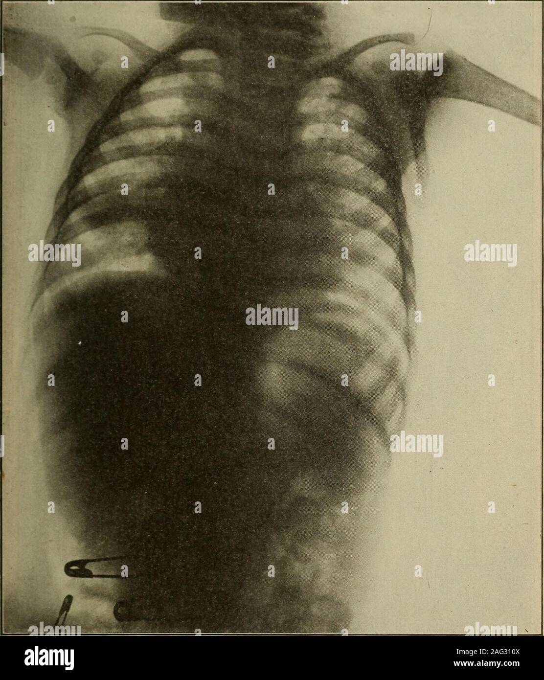
 . Radio-diagnosis of pleuro-pulmonary affection . No. 1389. J. K.bronchi on right. Male Radiograph 15years old. Pulmonary tuberculosis Dilatation of. Radiograph 16. DILATATION OF THE BRONCHINo. 101. P. C. Male. Age 35. Clinical history and examination: Cough;loss of weight; weakness. Hyperresonance over whole chest, harsh prolongedbreathing. No rales. X-ray findings: Emphysema. General bronchial dilata-tion. Some peribronchial thickening. Stock Photohttps://www.alamy.com/image-license-details/?v=1https://www.alamy.com/radio-diagnosis-of-pleuro-pulmonary-affection-no-1389-j-kbronchi-on-right-male-radiograph-15years-old-pulmonary-tuberculosis-dilatation-of-radiograph-16-dilatation-of-the-bronchino-101-p-c-male-age-35-clinical-history-and-examination-coughloss-of-weight-weakness-hyperresonance-over-whole-chest-harsh-prolongedbreathing-no-rales-x-ray-findings-emphysema-general-bronchial-dilata-tion-some-peribronchial-thickening-image375976697.html
RM2CRK635–. Radio-diagnosis of pleuro-pulmonary affection . No. 1389. J. K.bronchi on right. Male Radiograph 15years old. Pulmonary tuberculosis Dilatation of. Radiograph 16. DILATATION OF THE BRONCHINo. 101. P. C. Male. Age 35. Clinical history and examination: Cough;loss of weight; weakness. Hyperresonance over whole chest, harsh prolongedbreathing. No rales. X-ray findings: Emphysema. General bronchial dilata-tion. Some peribronchial thickening.
 . Radiography, x-ray therapeutics and radium therapy . th in the pulmonary systemrequire to be dealt with in the differential diagnosis. Cases with enlargedbronchial glands will cause some difficulty. The peribronchial forms ofphthisis give rise to marked increase of the hilus shadows, which are extremelydifficult and often impossible to differentiate from new growth. The chiefpoint of distinction between the two lies in the fact that in the peribronchialform of phthisis the increase of shading is more evenly distributed at bothroots, and may extend up to the apex or down to the base of the lu Stock Photohttps://www.alamy.com/image-license-details/?v=1https://www.alamy.com/radiography-x-ray-therapeutics-and-radium-therapy-th-in-the-pulmonary-systemrequire-to-be-dealt-with-in-the-differential-diagnosis-cases-with-enlargedbronchial-glands-will-cause-some-difficulty-the-peribronchial-forms-ofphthisis-give-rise-to-marked-increase-of-the-hilus-shadows-which-are-extremelydifficult-and-often-impossible-to-differentiate-from-new-growth-the-chiefpoint-of-distinction-between-the-two-lies-in-the-fact-that-in-the-peribronchialform-of-phthisis-the-increase-of-shading-is-more-evenly-distributed-at-bothroots-and-may-extend-up-to-the-apex-or-down-to-the-base-of-the-lu-image375976691.html
RM2CRK62Y–. Radiography, x-ray therapeutics and radium therapy . th in the pulmonary systemrequire to be dealt with in the differential diagnosis. Cases with enlargedbronchial glands will cause some difficulty. The peribronchial forms ofphthisis give rise to marked increase of the hilus shadows, which are extremelydifficult and often impossible to differentiate from new growth. The chiefpoint of distinction between the two lies in the fact that in the peribronchialform of phthisis the increase of shading is more evenly distributed at bothroots, and may extend up to the apex or down to the base of the lu
 . Radiography, x-ray therapeutics and radium therapy . the pulmonary systemrequire to be dealt with in the differential diagnosis. Cases with enlargedbronchial glands will cause some difficulty. The peribronchial forms ofphthisis give rise to marked increase of the hilus shadows, which are extremelydifl&cult and often impossible to differentiate from new growth. The chiefpoint of distinction between the two lies in the fact that in the peribronchialform of phthisis the increase of shading is more evenly distributed at bothroots, and may extend up to the apex or dowoi to the base of the lung,fo Stock Photohttps://www.alamy.com/image-license-details/?v=1https://www.alamy.com/radiography-x-ray-therapeutics-and-radium-therapy-the-pulmonary-systemrequire-to-be-dealt-with-in-the-differential-diagnosis-cases-with-enlargedbronchial-glands-will-cause-some-difficulty-the-peribronchial-forms-ofphthisis-give-rise-to-marked-increase-of-the-hilus-shadows-which-are-extremelydiflcult-and-often-impossible-to-differentiate-from-new-growth-the-chiefpoint-of-distinction-between-the-two-lies-in-the-fact-that-in-the-peribronchialform-of-phthisis-the-increase-of-shading-is-more-evenly-distributed-at-bothroots-and-may-extend-up-to-the-apex-or-dowoi-to-the-base-of-the-lungfo-image376038787.html
RM2CRP18K–. Radiography, x-ray therapeutics and radium therapy . the pulmonary systemrequire to be dealt with in the differential diagnosis. Cases with enlargedbronchial glands will cause some difficulty. The peribronchial forms ofphthisis give rise to marked increase of the hilus shadows, which are extremelydifl&cult and often impossible to differentiate from new growth. The chiefpoint of distinction between the two lies in the fact that in the peribronchialform of phthisis the increase of shading is more evenly distributed at bothroots, and may extend up to the apex or dowoi to the base of the lung,fo
 . Radiography, X-ray therapeutics and radium therapy . th in the pulmonary systemrequire to be dealt with in the differential diagnosis. Cases with enlargedbronchial glands will cause some difficulty. The peribronchial forms ofphthisis give rise to marked increase of the hilus shadows, which are extremelydifficult and often impossible to differentiate from new growth. The chiefpoint of distinction between the two lies in the fact that in the peribronchialform of phthisis the increase of shading is more evenly distributed at bothroots, and may extend up to the apex or down to the base of the lu Stock Photohttps://www.alamy.com/image-license-details/?v=1https://www.alamy.com/radiography-x-ray-therapeutics-and-radium-therapy-th-in-the-pulmonary-systemrequire-to-be-dealt-with-in-the-differential-diagnosis-cases-with-enlargedbronchial-glands-will-cause-some-difficulty-the-peribronchial-forms-ofphthisis-give-rise-to-marked-increase-of-the-hilus-shadows-which-are-extremelydifficult-and-often-impossible-to-differentiate-from-new-growth-the-chiefpoint-of-distinction-between-the-two-lies-in-the-fact-that-in-the-peribronchialform-of-phthisis-the-increase-of-shading-is-more-evenly-distributed-at-bothroots-and-may-extend-up-to-the-apex-or-down-to-the-base-of-the-lu-image375951319.html
RM2CRJ1MR–. Radiography, X-ray therapeutics and radium therapy . th in the pulmonary systemrequire to be dealt with in the differential diagnosis. Cases with enlargedbronchial glands will cause some difficulty. The peribronchial forms ofphthisis give rise to marked increase of the hilus shadows, which are extremelydifficult and often impossible to differentiate from new growth. The chiefpoint of distinction between the two lies in the fact that in the peribronchialform of phthisis the increase of shading is more evenly distributed at bothroots, and may extend up to the apex or down to the base of the lu
 . Journal of roentgenology . No. 10. Pericarditis with effusion. Diagnosis of partition in the pericardium,limiting the effusion to the right half of the pericardium. Note the en-largement of the pericardial shadow, uniform throughout, shadows limitedto the enlarged pericardium. No extension into peribronchial lung shadowor above level of clavicle. This case had a very distant apex beat andcardiac impulse in the fifth and sixth interspaces, to the left of the nippleline, which is usually obliterated in pericarditis with effusion. For thatreason, the diagnosis of malignancy was considered. This Stock Photohttps://www.alamy.com/image-license-details/?v=1https://www.alamy.com/journal-of-roentgenology-no-10-pericarditis-with-effusion-diagnosis-of-partition-in-the-pericardiumlimiting-the-effusion-to-the-right-half-of-the-pericardium-note-the-en-largement-of-the-pericardial-shadow-uniform-throughout-shadows-limitedto-the-enlarged-pericardium-no-extension-into-peribronchial-lung-shadowor-above-level-of-clavicle-this-case-had-a-very-distant-apex-beat-andcardiac-impulse-in-the-fifth-and-sixth-interspaces-to-the-left-of-the-nippleline-which-is-usually-obliterated-in-pericarditis-with-effusion-for-thatreason-the-diagnosis-of-malignancy-was-considered-this-image376058058.html
RM2CRPWTX–. Journal of roentgenology . No. 10. Pericarditis with effusion. Diagnosis of partition in the pericardium,limiting the effusion to the right half of the pericardium. Note the en-largement of the pericardial shadow, uniform throughout, shadows limitedto the enlarged pericardium. No extension into peribronchial lung shadowor above level of clavicle. This case had a very distant apex beat andcardiac impulse in the fifth and sixth interspaces, to the left of the nippleline, which is usually obliterated in pericarditis with effusion. For thatreason, the diagnosis of malignancy was considered. This
 . Radiography and radio-therapeutics . ients at middle lifeor later, where the ex-cess of fibrous tissue leads to very definite peribronchial shadows,which are due entirely to the thickening around the bronchi, and are diagnostic neither of tuberculosis nor cancer. In many of these cases the thickening of the bronchus may be seen on cross-section. It frequently happens, especially on the right side of the chest, that one or more nearly circular rings are shown. These are caused by a cross-section of the bronchus, where it bends nearly at right angles on its way to the deeper part of the lung. Stock Photohttps://www.alamy.com/image-license-details/?v=1https://www.alamy.com/radiography-and-radio-therapeutics-ients-at-middle-lifeor-later-where-the-ex-cess-of-fibrous-tissue-leads-to-very-definite-peribronchial-shadowswhich-are-due-entirely-to-the-thickening-around-the-bronchi-and-are-diagnostic-neither-of-tuberculosis-nor-cancer-in-many-of-these-cases-the-thickening-of-the-bronchus-may-be-seen-on-cross-section-it-frequently-happens-especially-on-the-right-side-of-the-chest-that-one-or-more-nearly-circular-rings-are-shown-these-are-caused-by-a-cross-section-of-the-bronchus-where-it-bends-nearly-at-right-angles-on-its-way-to-the-deeper-part-of-the-lung-image376019403.html
RM2CRN4GB–. Radiography and radio-therapeutics . ients at middle lifeor later, where the ex-cess of fibrous tissue leads to very definite peribronchial shadows,which are due entirely to the thickening around the bronchi, and are diagnostic neither of tuberculosis nor cancer. In many of these cases the thickening of the bronchus may be seen on cross-section. It frequently happens, especially on the right side of the chest, that one or more nearly circular rings are shown. These are caused by a cross-section of the bronchus, where it bends nearly at right angles on its way to the deeper part of the lung.
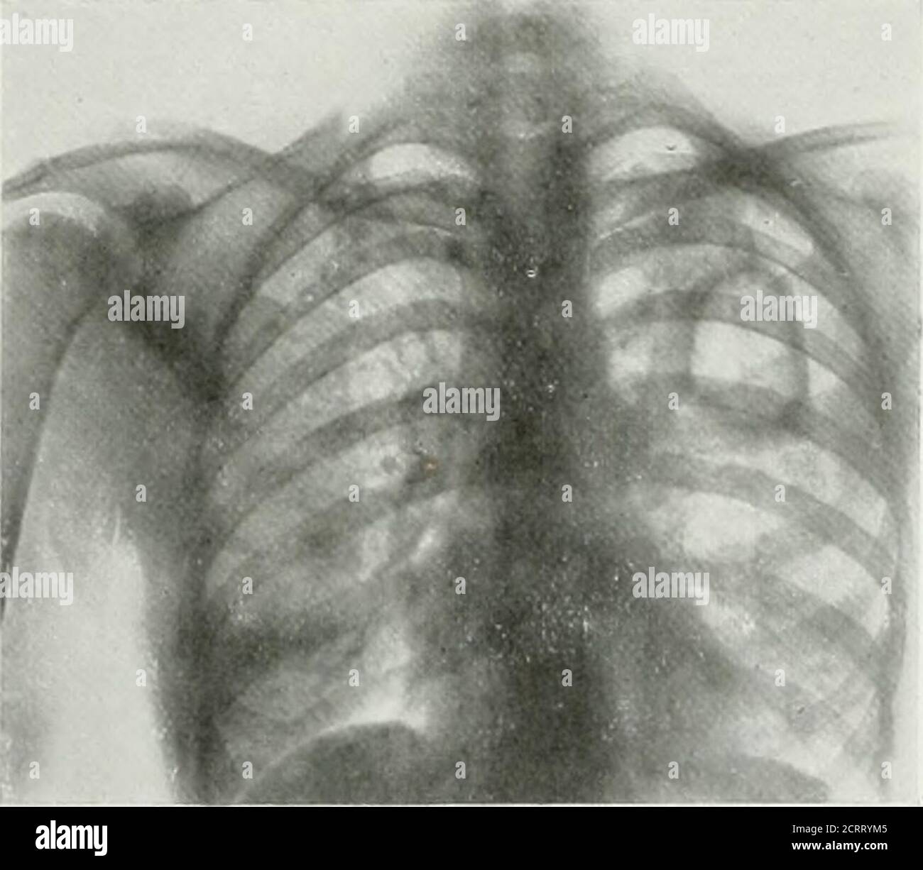
 . Radiography and radio-therapeutics . PLATE LIII.—Kadiogbams showing Pulmonary Tuberculosis. a, Post-mortem subject. Note fine shading in lung substance. Tuberculous broncho-pneumonia. b, Early tuberculosis of lungs, peribronchial thickening, irregularity of diaphragmatic shadow on rightside, with sharpness of all detail. Exposure z^ second, intensifying screen used. c, Acute general tuberculosis of both lungs (miliary tubercle). TUMOURS OF THE THORAX 289. Fig. 248.—Post-mortem specimenmediastinal tumour. Lympho-sarcoma. Tumours of the Mediastinum.—Cancer is the most common form oftumour in t Stock Photohttps://www.alamy.com/image-license-details/?v=1https://www.alamy.com/radiography-and-radio-therapeutics-plate-liiikadiogbams-showing-pulmonary-tuberculosis-a-post-mortem-subject-note-fine-shading-in-lung-substance-tuberculous-broncho-pneumonia-b-early-tuberculosis-of-lungs-peribronchial-thickening-irregularity-of-diaphragmatic-shadow-on-rightside-with-sharpness-of-all-detail-exposure-z-second-intensifying-screen-used-c-acute-general-tuberculosis-of-both-lungs-miliary-tubercle-tumours-of-the-thorax-289-fig-248post-mortem-specimenmediastinal-tumour-lympho-sarcoma-tumours-of-the-mediastinumcancer-is-the-most-common-form-oftumour-in-t-image376020330.html
RM2CRN5NE–. Radiography and radio-therapeutics . PLATE LIII.—Kadiogbams showing Pulmonary Tuberculosis. a, Post-mortem subject. Note fine shading in lung substance. Tuberculous broncho-pneumonia. b, Early tuberculosis of lungs, peribronchial thickening, irregularity of diaphragmatic shadow on rightside, with sharpness of all detail. Exposure z^ second, intensifying screen used. c, Acute general tuberculosis of both lungs (miliary tubercle). TUMOURS OF THE THORAX 289. Fig. 248.—Post-mortem specimenmediastinal tumour. Lympho-sarcoma. Tumours of the Mediastinum.—Cancer is the most common form oftumour in t
 . Radio-diagnosis of pleuro-pulmonary affection . Radiograph 16. DILATATION OF THE BRONCHINo. 101. P. C. Male. Age 35. Clinical history and examination: Cough;loss of weight; weakness. Hyperresonance over whole chest, harsh prolongedbreathing. No rales. X-ray findings: Emphysema. General bronchial dilata-tion. Some peribronchial thickening.. Radiograph 17. ADENOPATHY, PRINCIPALLY RIGHT MEDIASTINALAND HILUS REGION SECONDARILY There is a diffuse shadow the entire length of the right border of the medianshadow, commencing under the clavicle and extending as far as the diaphragmwith very appreciab Stock Photohttps://www.alamy.com/image-license-details/?v=1https://www.alamy.com/radio-diagnosis-of-pleuro-pulmonary-affection-radiograph-16-dilatation-of-the-bronchino-101-p-c-male-age-35-clinical-history-and-examination-coughloss-of-weight-weakness-hyperresonance-over-whole-chest-harsh-prolongedbreathing-no-rales-x-ray-findings-emphysema-general-bronchial-dilata-tion-some-peribronchial-thickening-radiograph-17-adenopathy-principally-right-mediastinaland-hilus-region-secondarily-there-is-a-diffuse-shadow-the-entire-length-of-the-right-border-of-the-medianshadow-commencing-under-the-clavicle-and-extending-as-far-as-the-diaphragmwith-very-appreciab-image375976650.html
RM2CRK61E–. Radio-diagnosis of pleuro-pulmonary affection . Radiograph 16. DILATATION OF THE BRONCHINo. 101. P. C. Male. Age 35. Clinical history and examination: Cough;loss of weight; weakness. Hyperresonance over whole chest, harsh prolongedbreathing. No rales. X-ray findings: Emphysema. General bronchial dilata-tion. Some peribronchial thickening.. Radiograph 17. ADENOPATHY, PRINCIPALLY RIGHT MEDIASTINALAND HILUS REGION SECONDARILY There is a diffuse shadow the entire length of the right border of the medianshadow, commencing under the clavicle and extending as far as the diaphragmwith very appreciab
 . The American journal of roentgenology, radium therapy and nuclear medicine . *4 *4 Fig. 3. Two Large Air Pockets in the Upper RightLung; One in Upper Left. The lower shadow ON THE right SIDE IS INTERLOBAR. the differences in the roentgenological andphysical diagnoses of chest conditions. Localized air pockets are observed inboth the peribronchial and parenchymaltypes of tuberculosis, the greater numberbeing seen in peribronchial infection. Thedevelopment of collections of air depends,first, upon a superficial involvement of lungtissue, the pathology either being inter-lobar or occurring in t Stock Photohttps://www.alamy.com/image-license-details/?v=1https://www.alamy.com/the-american-journal-of-roentgenology-radium-therapy-and-nuclear-medicine-4-4-fig-3-two-large-air-pockets-in-the-upper-rightlung-one-in-upper-left-the-lower-shadow-on-the-right-side-is-interlobar-the-differences-in-the-roentgenological-andphysical-diagnoses-of-chest-conditions-localized-air-pockets-are-observed-inboth-the-peribronchial-and-parenchymaltypes-of-tuberculosis-the-greater-numberbeing-seen-in-peribronchial-infection-thedevelopment-of-collections-of-air-dependsfirst-upon-a-superficial-involvement-of-lungtissue-the-pathology-either-being-inter-lobar-or-occurring-in-t-image376081445.html
RM2CRRYM5–. The American journal of roentgenology, radium therapy and nuclear medicine . *4 *4 Fig. 3. Two Large Air Pockets in the Upper RightLung; One in Upper Left. The lower shadow ON THE right SIDE IS INTERLOBAR. the differences in the roentgenological andphysical diagnoses of chest conditions. Localized air pockets are observed inboth the peribronchial and parenchymaltypes of tuberculosis, the greater numberbeing seen in peribronchial infection. Thedevelopment of collections of air depends,first, upon a superficial involvement of lungtissue, the pathology either being inter-lobar or occurring in t
 . Radiography and radio-therapeutics . IJ^ATE LI 11.—KADlOCiKAM.S SHOWINi; PULMONzVRY TUBERCULOSIS. a, Post-inorteiii subject. Note fine shading in lung substance. Tuberculous bronclio-pneumonia.h, Early tuberculosis of lungs, peribronchial thickening, irregularity of diaphragmatic sliadow ou rightside, with sharpness of all detail. Exposure liTi second, intensifying screen used.c, Acute general tuberculosis of both lungs (miliary tubercle). TUMOURS OF THE THORAX 289. Fig. 248.—Post-inortem specimenmediastinal tumour. Lympho-sarcoma. Tumours of the Mediastinum.—Cancer is the most common form o Stock Photohttps://www.alamy.com/image-license-details/?v=1https://www.alamy.com/radiography-and-radio-therapeutics-ijate-li-11kadlocikams-showini-pulmonzvry-tuberculosis-a-post-inorteiii-subject-note-fine-shading-in-lung-substance-tuberculous-bronclio-pneumoniah-early-tuberculosis-of-lungs-peribronchial-thickening-irregularity-of-diaphragmatic-sliadow-ou-rightside-with-sharpness-of-all-detail-exposure-liti-second-intensifying-screen-usedc-acute-general-tuberculosis-of-both-lungs-miliary-tubercle-tumours-of-the-thorax-289-fig-248post-inortem-specimenmediastinal-tumour-lympho-sarcoma-tumours-of-the-mediastinumcancer-is-the-most-common-form-o-image376009875.html
RM2CRMMC3–. Radiography and radio-therapeutics . IJ^ATE LI 11.—KADlOCiKAM.S SHOWINi; PULMONzVRY TUBERCULOSIS. a, Post-inorteiii subject. Note fine shading in lung substance. Tuberculous bronclio-pneumonia.h, Early tuberculosis of lungs, peribronchial thickening, irregularity of diaphragmatic sliadow ou rightside, with sharpness of all detail. Exposure liTi second, intensifying screen used.c, Acute general tuberculosis of both lungs (miliary tubercle). TUMOURS OF THE THORAX 289. Fig. 248.—Post-inortem specimenmediastinal tumour. Lympho-sarcoma. Tumours of the Mediastinum.—Cancer is the most common form o
 . Postmortem pathology; a manual of the technic of post-mortem examinations and the interpretations to be drawn therefrom;. Fig. 90.—Method of removing tongue, tonsils, oesophagus, trachea, etc., in a single piece,without incising the skin more than is done in the primary cut. Palatine arch incisedTonsil Thyroid gland Common carotid arteryLeft subclavian artery (Esophagus laid open Aorta.. Palatine arch incised Common carotid arteryRight subclavian artery Aorta Right bronchus Peribronchial lymph glands Fig. 91.—Examination of the organs of the neck. The arrows show the direction in which the i Stock Photohttps://www.alamy.com/image-license-details/?v=1https://www.alamy.com/postmortem-pathology-a-manual-of-the-technic-of-post-mortem-examinations-and-the-interpretations-to-be-drawn-therefrom-fig-90method-of-removing-tongue-tonsils-oesophagus-trachea-etc-in-a-single-piecewithout-incising-the-skin-more-than-is-done-in-the-primary-cut-palatine-arch-incisedtonsil-thyroid-gland-common-carotid-arteryleft-subclavian-artery-esophagus-laid-open-aorta-palatine-arch-incised-common-carotid-arteryright-subclavian-artery-aorta-right-bronchus-peribronchial-lymph-glands-fig-91examination-of-the-organs-of-the-neck-the-arrows-show-the-direction-in-which-the-i-image370568290.html
RM2CETRHP–. Postmortem pathology; a manual of the technic of post-mortem examinations and the interpretations to be drawn therefrom;. Fig. 90.—Method of removing tongue, tonsils, oesophagus, trachea, etc., in a single piece,without incising the skin more than is done in the primary cut. Palatine arch incisedTonsil Thyroid gland Common carotid arteryLeft subclavian artery (Esophagus laid open Aorta.. Palatine arch incised Common carotid arteryRight subclavian artery Aorta Right bronchus Peribronchial lymph glands Fig. 91.—Examination of the organs of the neck. The arrows show the direction in which the i
 . The American journal of roentgenology, radium therapy and nuclear medicine . eles, whoreported a foreign body in one of the largebronchial branches on the right side nearthe root of the right lung, with markedperibronchial thickening in the lower lobeof the right lung, some peribronchial thick-ening in the left side, and many filirouschanges, probably of non-tuberculous ori-gin. Roentgen ray examination by Dr.Manges (Fig. 2) led him to believe thatthere was a tubular metallic foreign bodypresent. The foreign body was removedby oral bronchoscopy April 21, 1917, byDr. Jackson. It proved to be Stock Photohttps://www.alamy.com/image-license-details/?v=1https://www.alamy.com/the-american-journal-of-roentgenology-radium-therapy-and-nuclear-medicine-eles-whoreported-a-foreign-body-in-one-of-the-largebronchial-branches-on-the-right-side-nearthe-root-of-the-right-lung-with-markedperibronchial-thickening-in-the-lower-lobeof-the-right-lung-some-peribronchial-thick-ening-in-the-left-side-and-many-filirouschanges-probably-of-non-tuberculous-ori-gin-roentgen-ray-examination-by-drmanges-fig-2-led-him-to-believe-thatthere-was-a-tubular-metallic-foreign-bodypresent-the-foreign-body-was-removedby-oral-bronchoscopy-april-21-1917-bydr-jackson-it-proved-to-be-image376090269.html
RM2CRTAY9–. The American journal of roentgenology, radium therapy and nuclear medicine . eles, whoreported a foreign body in one of the largebronchial branches on the right side nearthe root of the right lung, with markedperibronchial thickening in the lower lobeof the right lung, some peribronchial thick-ening in the left side, and many filirouschanges, probably of non-tuberculous ori-gin. Roentgen ray examination by Dr.Manges (Fig. 2) led him to believe thatthere was a tubular metallic foreign bodypresent. The foreign body was removedby oral bronchoscopy April 21, 1917, byDr. Jackson. It proved to be
 . The American journal of roentgenology, radium therapy and nuclear medicine . - part from apex to base seems accommodate five at one bases to the pleura and the hilu:; has its in all except the lowest, whichapex pointing toward the dia- twigs can be followed quite a distance outinto the lung structure. In regard to what isspoken of as peribronchial thickening wehave seen it change during the ten weeksstrike in 1920, becoming lessened in widthand irregular in border as the dust depositwas unevenly moved on to the hilus. Considering the lung lesion as it affects. Fig. 15 (166). Exposure twenty- Stock Photohttps://www.alamy.com/image-license-details/?v=1https://www.alamy.com/the-american-journal-of-roentgenology-radium-therapy-and-nuclear-medicine-part-from-apex-to-base-seems-accommodate-five-at-one-bases-to-the-pleura-and-the-hilu-has-its-in-all-except-the-lowest-whichapex-pointing-toward-the-dia-twigs-can-be-followed-quite-a-distance-outinto-the-lung-structure-in-regard-to-what-isspoken-of-as-peribronchial-thickening-wehave-seen-it-change-during-the-ten-weeksstrike-in-1920-becoming-lessened-in-widthand-irregular-in-border-as-the-dust-depositwas-unevenly-moved-on-to-the-hilus-considering-the-lung-lesion-as-it-affects-fig-15-166-exposure-twenty-image376041526.html
RM2CRP4PE–. The American journal of roentgenology, radium therapy and nuclear medicine . - part from apex to base seems accommodate five at one bases to the pleura and the hilu:; has its in all except the lowest, whichapex pointing toward the dia- twigs can be followed quite a distance outinto the lung structure. In regard to what isspoken of as peribronchial thickening wehave seen it change during the ten weeksstrike in 1920, becoming lessened in widthand irregular in border as the dust depositwas unevenly moved on to the hilus. Considering the lung lesion as it affects. Fig. 15 (166). Exposure twenty-
![. Diseases of infancy and childhood . ung of a two-year-old child.(a) caseous focus in the region of the an-terior border; (h) nontuberculous poster-ior border; (c) transverse section of bron-chus; (d,d^) caseated lymph glands; (c)pulmonary vein; (f) point of adhesion ofthe vein e with the lymph gland d^] (g)tubercle in the lymph vessels of thelung parenchyma; (h) periarterial; (i)peribronchial; (k) perivenous tubercles;pleura; (m) tubercle in its connective ti^(Ziegler.) (1) A vo>sel tulHMlo!^ of tliosue of tho liilus of the Uuh:-. X;>. ??1 490 THE INFECTIOUS DISEASES Baginsky r Stock Photo . Diseases of infancy and childhood . ung of a two-year-old child.(a) caseous focus in the region of the an-terior border; (h) nontuberculous poster-ior border; (c) transverse section of bron-chus; (d,d^) caseated lymph glands; (c)pulmonary vein; (f) point of adhesion ofthe vein e with the lymph gland d^] (g)tubercle in the lymph vessels of thelung parenchyma; (h) periarterial; (i)peribronchial; (k) perivenous tubercles;pleura; (m) tubercle in its connective ti^(Ziegler.) (1) A vo>sel tulHMlo!^ of tliosue of tho liilus of the Uuh:-. X;>. ??1 490 THE INFECTIOUS DISEASES Baginsky r Stock Photo](https://c8.alamy.com/comp/2CE2T92/diseases-of-infancy-and-childhood-ung-of-a-two-year-old-childa-caseous-focus-in-the-region-of-the-an-terior-border-h-nontuberculous-poster-ior-border-c-transverse-section-of-bron-chus-dd-caseated-lymph-glands-cpulmonary-vein-f-point-of-adhesion-ofthe-vein-e-with-the-lymph-gland-d-gtubercle-in-the-lymph-vessels-of-thelung-parenchyma-h-periarterial-iperibronchial-k-perivenous-tuberclespleura-m-tubercle-in-its-connective-tiziegler-1-a-vogtsel-tulhmlo!-of-tliosue-of-tho-liilus-of-the-uuh-xgt-1-490-the-infectious-diseases-baginsky-r-2CE2T92.jpg) . Diseases of infancy and childhood . ung of a two-year-old child.(a) caseous focus in the region of the an-terior border; (h) nontuberculous poster-ior border; (c) transverse section of bron-chus; (d,d^) caseated lymph glands; (c)pulmonary vein; (f) point of adhesion ofthe vein e with the lymph gland d^] (g)tubercle in the lymph vessels of thelung parenchyma; (h) periarterial; (i)peribronchial; (k) perivenous tubercles;pleura; (m) tubercle in its connective ti^(Ziegler.) (1) A vo>sel tulHMlo!^ of tliosue of tho liilus of the Uuh:-. X;>. ??1 490 THE INFECTIOUS DISEASES Baginsky r Stock Photohttps://www.alamy.com/image-license-details/?v=1https://www.alamy.com/diseases-of-infancy-and-childhood-ung-of-a-two-year-old-childa-caseous-focus-in-the-region-of-the-an-terior-border-h-nontuberculous-poster-ior-border-c-transverse-section-of-bron-chus-dd-caseated-lymph-glands-cpulmonary-vein-f-point-of-adhesion-ofthe-vein-e-with-the-lymph-gland-d-gtubercle-in-the-lymph-vessels-of-thelung-parenchyma-h-periarterial-iperibronchial-k-perivenous-tuberclespleura-m-tubercle-in-its-connective-tiziegler-1-a-vogtsel-tulhmlo!-of-tliosue-of-tho-liilus-of-the-uuh-xgt-1-490-the-infectious-diseases-baginsky-r-image370085886.html
. Diseases of infancy and childhood . ung of a two-year-old child.(a) caseous focus in the region of the an-terior border; (h) nontuberculous poster-ior border; (c) transverse section of bron-chus; (d,d^) caseated lymph glands; (c)pulmonary vein; (f) point of adhesion ofthe vein e with the lymph gland d^] (g)tubercle in the lymph vessels of thelung parenchyma; (h) periarterial; (i)peribronchial; (k) perivenous tubercles;pleura; (m) tubercle in its connective ti^(Ziegler.) (1) A vo>sel tulHMlo!^ of tliosue of tho liilus of the Uuh:-. X;>. ??1 490 THE INFECTIOUS DISEASES Baginsky r Stock Photohttps://www.alamy.com/image-license-details/?v=1https://www.alamy.com/diseases-of-infancy-and-childhood-ung-of-a-two-year-old-childa-caseous-focus-in-the-region-of-the-an-terior-border-h-nontuberculous-poster-ior-border-c-transverse-section-of-bron-chus-dd-caseated-lymph-glands-cpulmonary-vein-f-point-of-adhesion-ofthe-vein-e-with-the-lymph-gland-d-gtubercle-in-the-lymph-vessels-of-thelung-parenchyma-h-periarterial-iperibronchial-k-perivenous-tuberclespleura-m-tubercle-in-its-connective-tiziegler-1-a-vogtsel-tulhmlo!-of-tliosue-of-tho-liilus-of-the-uuh-xgt-1-490-the-infectious-diseases-baginsky-r-image370085886.htmlRM2CE2T92–. Diseases of infancy and childhood . ung of a two-year-old child.(a) caseous focus in the region of the an-terior border; (h) nontuberculous poster-ior border; (c) transverse section of bron-chus; (d,d^) caseated lymph glands; (c)pulmonary vein; (f) point of adhesion ofthe vein e with the lymph gland d^] (g)tubercle in the lymph vessels of thelung parenchyma; (h) periarterial; (i)peribronchial; (k) perivenous tubercles;pleura; (m) tubercle in its connective ti^(Ziegler.) (1) A vo>sel tulHMlo!^ of tliosue of tho liilus of the Uuh:-. X;>. ??1 490 THE INFECTIOUS DISEASES Baginsky r
 . The anatomical record. Anatomy; Anatomy. Fig. 1 Drawing showing the inside and lateral views of the duodenum and the relation of the diverticulum to the openings of the common bile and pancreatic ducts. cardium, aortic and mitral valves; chronic diffuse nephritis with atrophy of cortex and fatty degeneration of pyramids; chronic passive congestion of viscera; chronic bronchitis; fatty degeneration of liver and kidneys; chronic obliterative appendi- citis, prostatic hypertrophy; healed tuberculosis of peribronchial lymph nodes; old pleural adhesions; obesity; lipomatosis of pancreas with fat Stock Photohttps://www.alamy.com/image-license-details/?v=1https://www.alamy.com/the-anatomical-record-anatomy-anatomy-fig-1-drawing-showing-the-inside-and-lateral-views-of-the-duodenum-and-the-relation-of-the-diverticulum-to-the-openings-of-the-common-bile-and-pancreatic-ducts-cardium-aortic-and-mitral-valves-chronic-diffuse-nephritis-with-atrophy-of-cortex-and-fatty-degeneration-of-pyramids-chronic-passive-congestion-of-viscera-chronic-bronchitis-fatty-degeneration-of-liver-and-kidneys-chronic-obliterative-appendi-citis-prostatic-hypertrophy-healed-tuberculosis-of-peribronchial-lymph-nodes-old-pleural-adhesions-obesity-lipomatosis-of-pancreas-with-fat-image236873583.html
RMRNAEJR–. The anatomical record. Anatomy; Anatomy. Fig. 1 Drawing showing the inside and lateral views of the duodenum and the relation of the diverticulum to the openings of the common bile and pancreatic ducts. cardium, aortic and mitral valves; chronic diffuse nephritis with atrophy of cortex and fatty degeneration of pyramids; chronic passive congestion of viscera; chronic bronchitis; fatty degeneration of liver and kidneys; chronic obliterative appendi- citis, prostatic hypertrophy; healed tuberculosis of peribronchial lymph nodes; old pleural adhesions; obesity; lipomatosis of pancreas with fat