Quick filters:
Phrenic nerve Stock Photos and Images
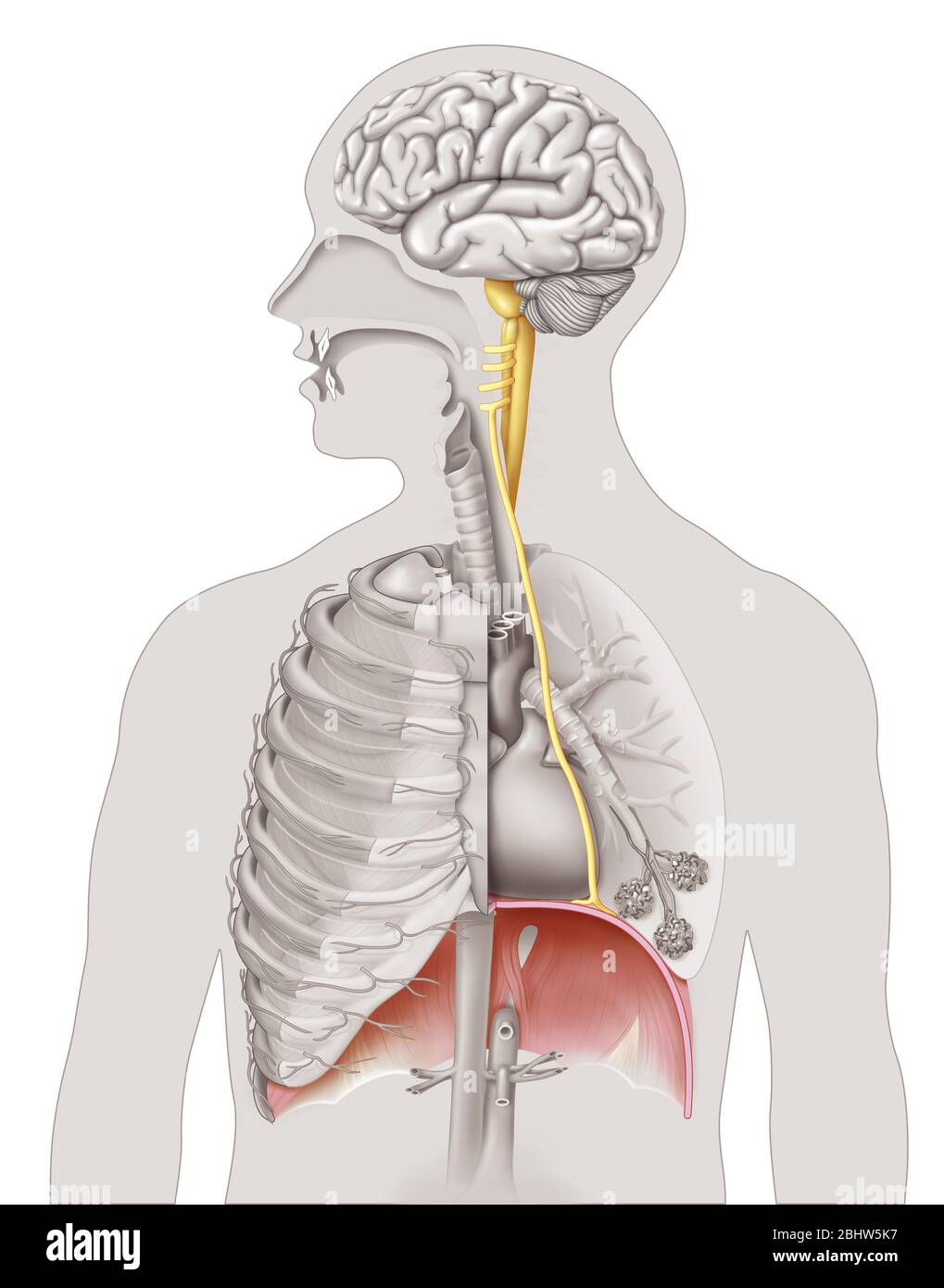 Medical illustration highlighting the phrenic nerve responsible for the innervation of the diaphragm that causes the main movements of the breath. The Stock Photohttps://www.alamy.com/image-license-details/?v=1https://www.alamy.com/medical-illustration-highlighting-the-phrenic-nerve-responsible-for-the-innervation-of-the-diaphragm-that-causes-the-main-movements-of-the-breath-the-image355209771.html
RM2BHW5K7–Medical illustration highlighting the phrenic nerve responsible for the innervation of the diaphragm that causes the main movements of the breath. The
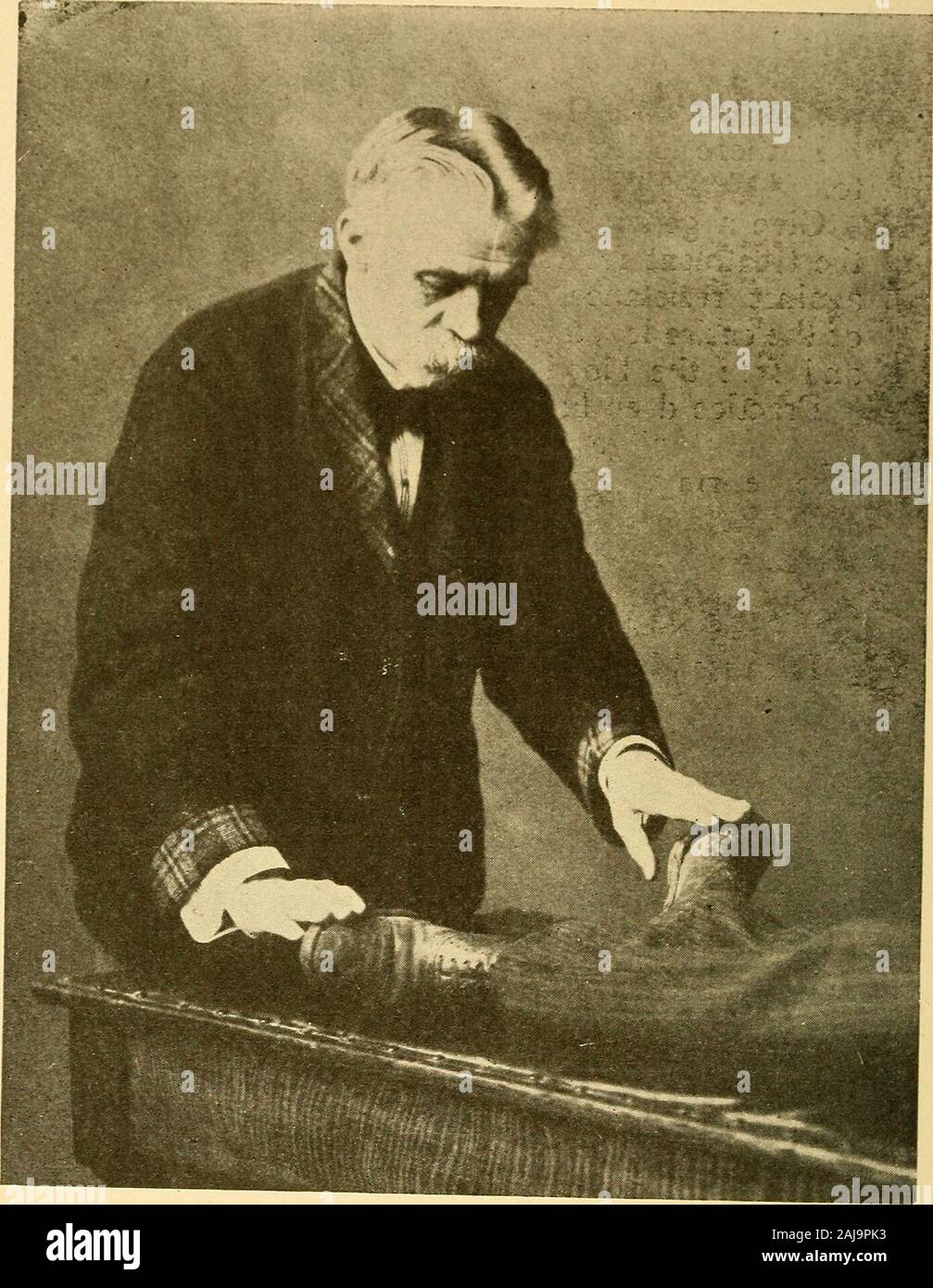
 Andrew Ure (May 18, 1778 - January 2, 1857) was a Scottish doctor, scholar, chemist, Scriptural geologist and early business theorist. In 1818 Ure revealed experiments he had been carrying out on a murderer/thief named Matthew Clydesdale, after the man's execution by hanging. He claimed that, by stimulating the phrenic nerve, life could be restored in cases of suffocation, drowning or hanging. Stock Photohttps://www.alamy.com/image-license-details/?v=1https://www.alamy.com/andrew-ure-may-18-1778-january-2-1857-was-a-scottish-doctor-scholar-chemist-scriptural-geologist-and-early-business-theorist-in-1818-ure-revealed-experiments-he-had-been-carrying-out-on-a-murdererthief-named-matthew-clydesdale-after-the-mans-execution-by-hanging-he-claimed-that-by-stimulating-the-phrenic-nerve-life-could-be-restored-in-cases-of-suffocation-drowning-or-hanging-image246588233.html
RMT951PH–Andrew Ure (May 18, 1778 - January 2, 1857) was a Scottish doctor, scholar, chemist, Scriptural geologist and early business theorist. In 1818 Ure revealed experiments he had been carrying out on a murderer/thief named Matthew Clydesdale, after the man's execution by hanging. He claimed that, by stimulating the phrenic nerve, life could be restored in cases of suffocation, drowning or hanging.
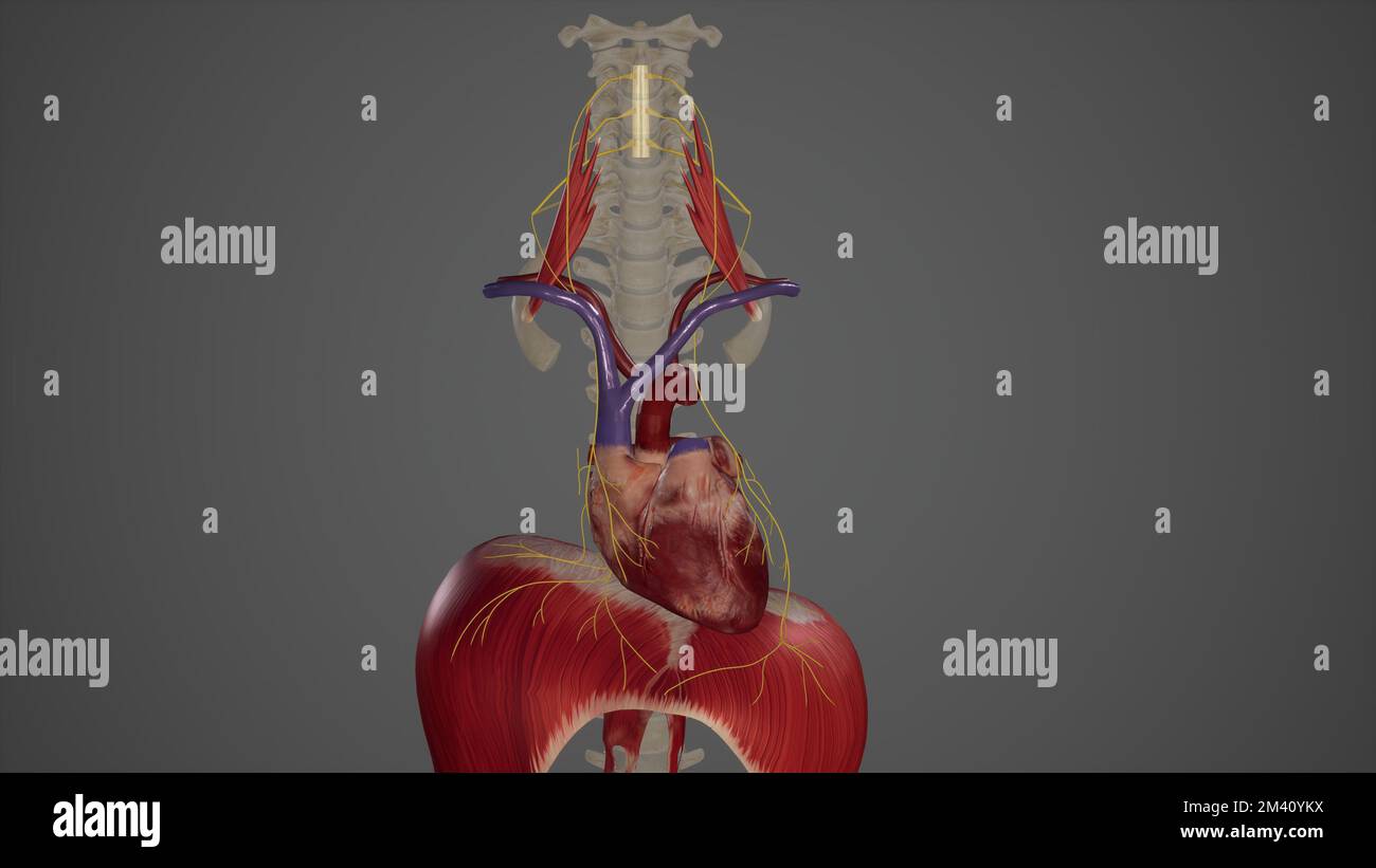 Phrenic Nerves Stock Photohttps://www.alamy.com/image-license-details/?v=1https://www.alamy.com/phrenic-nerves-image501581022.html
RF2M40YKX–Phrenic Nerves
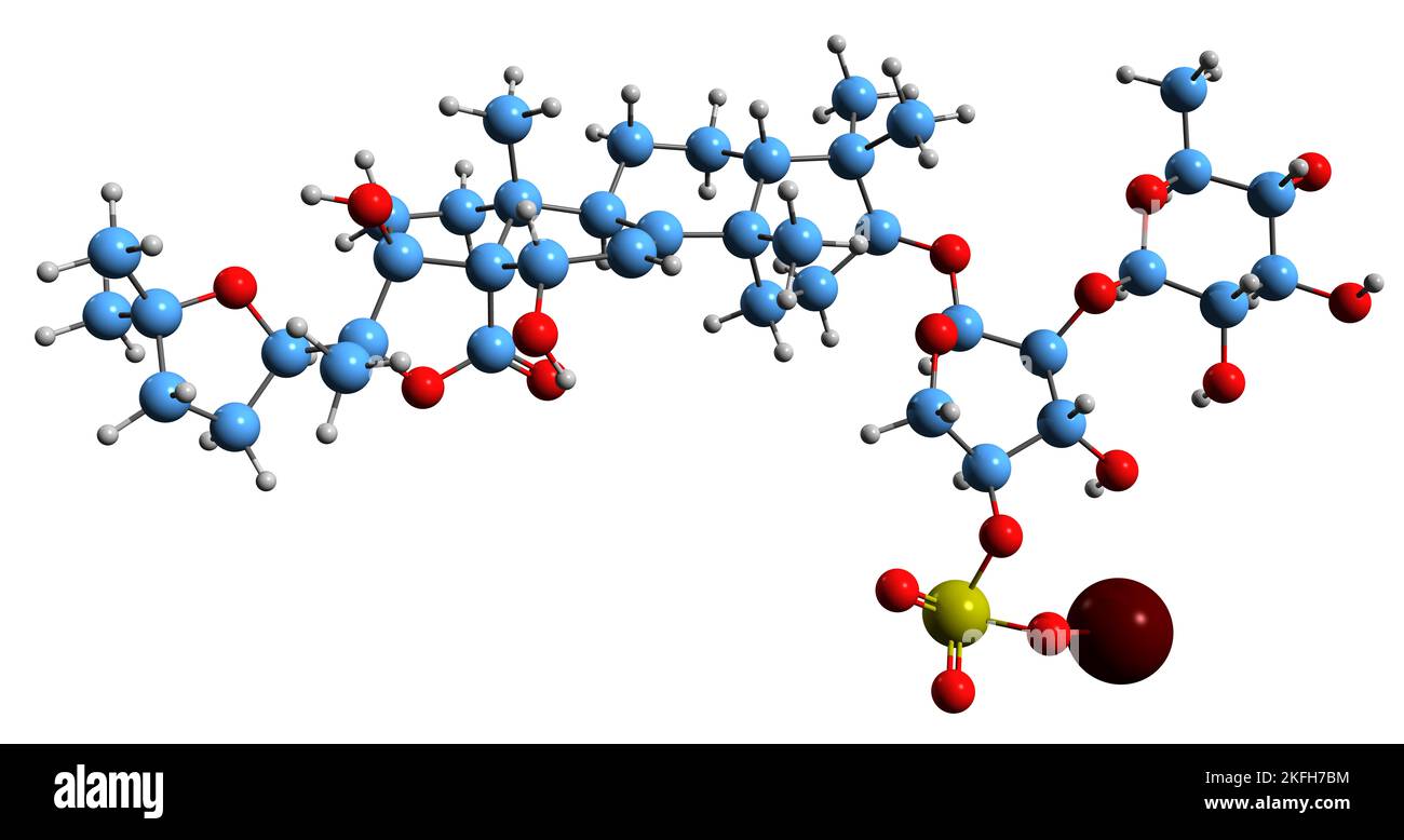 3D image of Holothurin B skeletal formula - molecular chemical structure of triterpene glycoside isolated on white background Stock Photohttps://www.alamy.com/image-license-details/?v=1https://www.alamy.com/3d-image-of-holothurin-b-skeletal-formula-molecular-chemical-structure-of-triterpene-glycoside-isolated-on-white-background-image491489144.html
RF2KFH7BM–3D image of Holothurin B skeletal formula - molecular chemical structure of triterpene glycoside isolated on white background
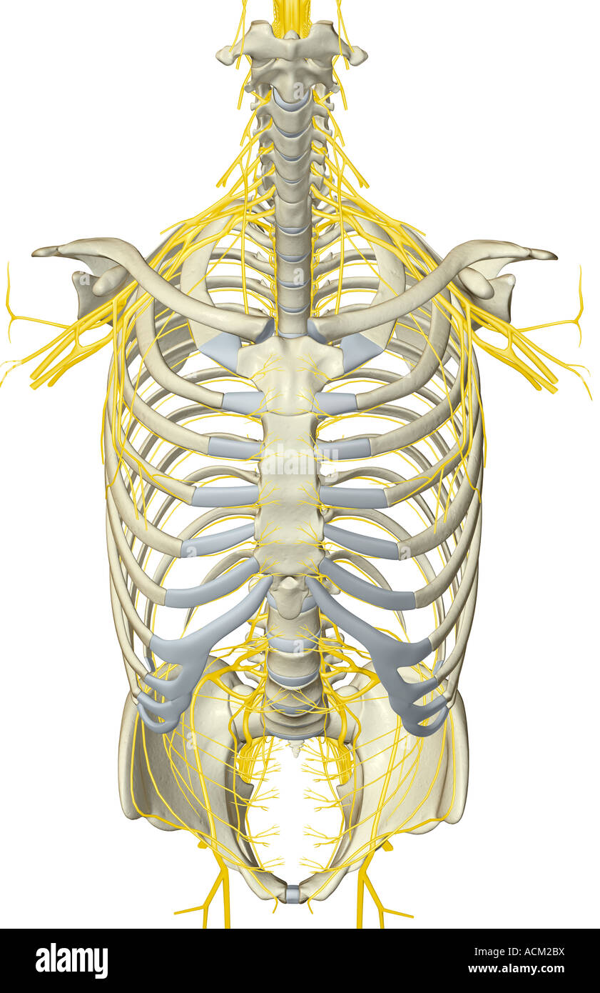
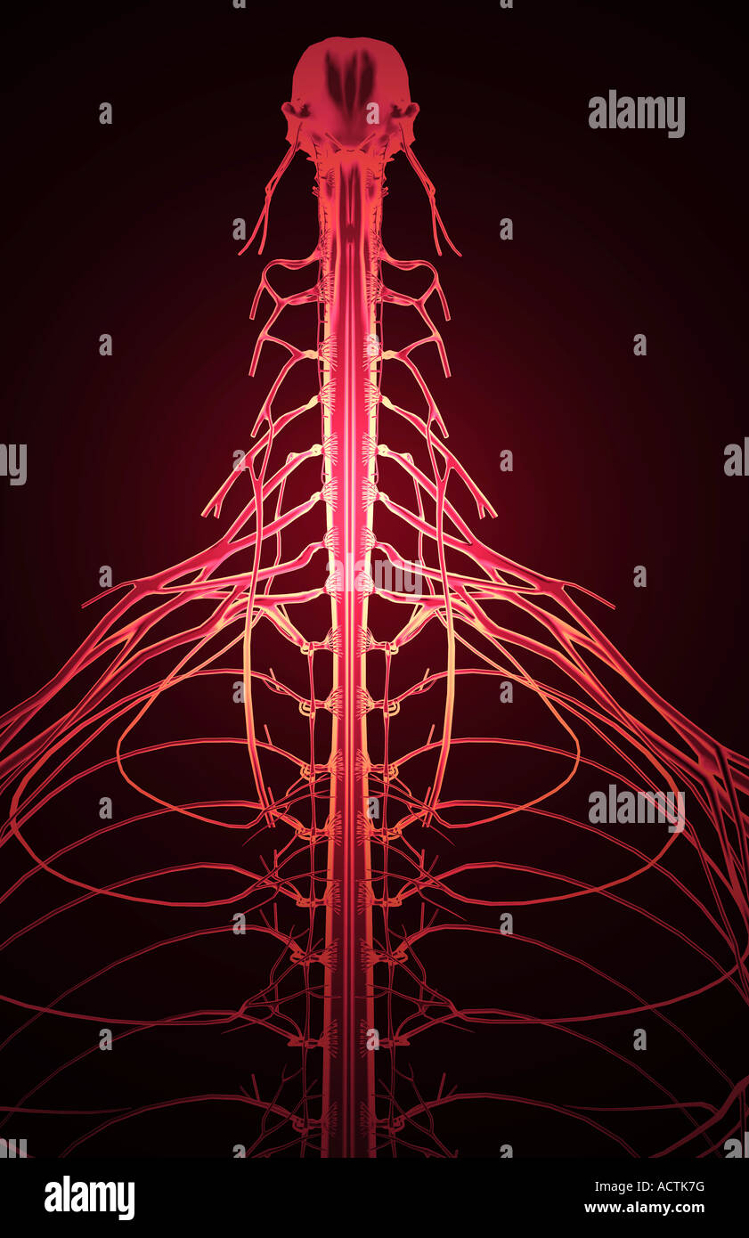 Nerve supply of the upper body Stock Photohttps://www.alamy.com/image-license-details/?v=1https://www.alamy.com/stock-photo-nerve-supply-of-the-upper-body-13227843.html
RFACTK7G–Nerve supply of the upper body
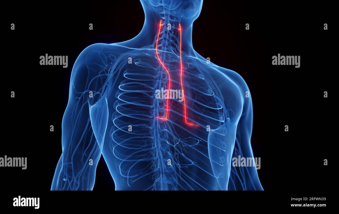 Phrenic nerves, illustration Stock Photohttps://www.alamy.com/image-license-details/?v=1https://www.alamy.com/phrenic-nerves-illustration-image560516973.html
RF2RFWN39–Phrenic nerves, illustration
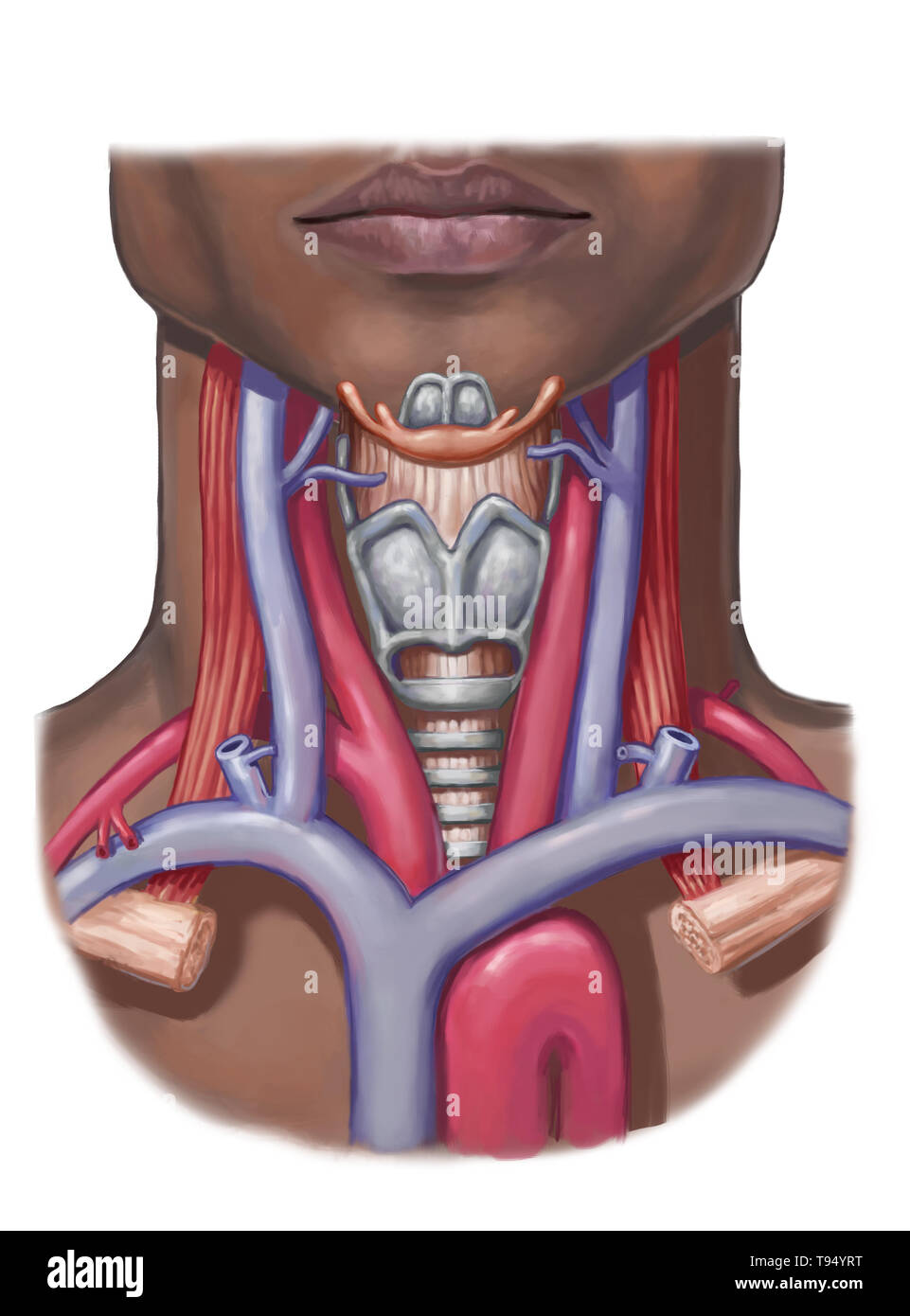 Neck anatomy, illustration. Stock Photohttps://www.alamy.com/image-license-details/?v=1https://www.alamy.com/neck-anatomy-illustration-image246586700.html
RMT94YRT–Neck anatomy, illustration.
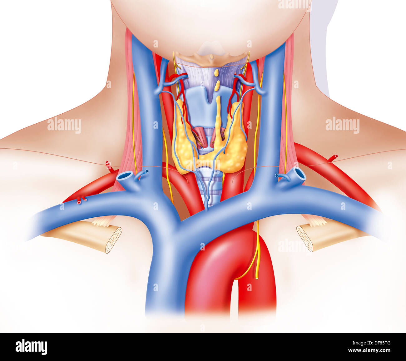 NECK ANATOMY Stock Photohttps://www.alamy.com/image-license-details/?v=1https://www.alamy.com/neck-anatomy-image61031168.html
RMDF85TG–NECK ANATOMY
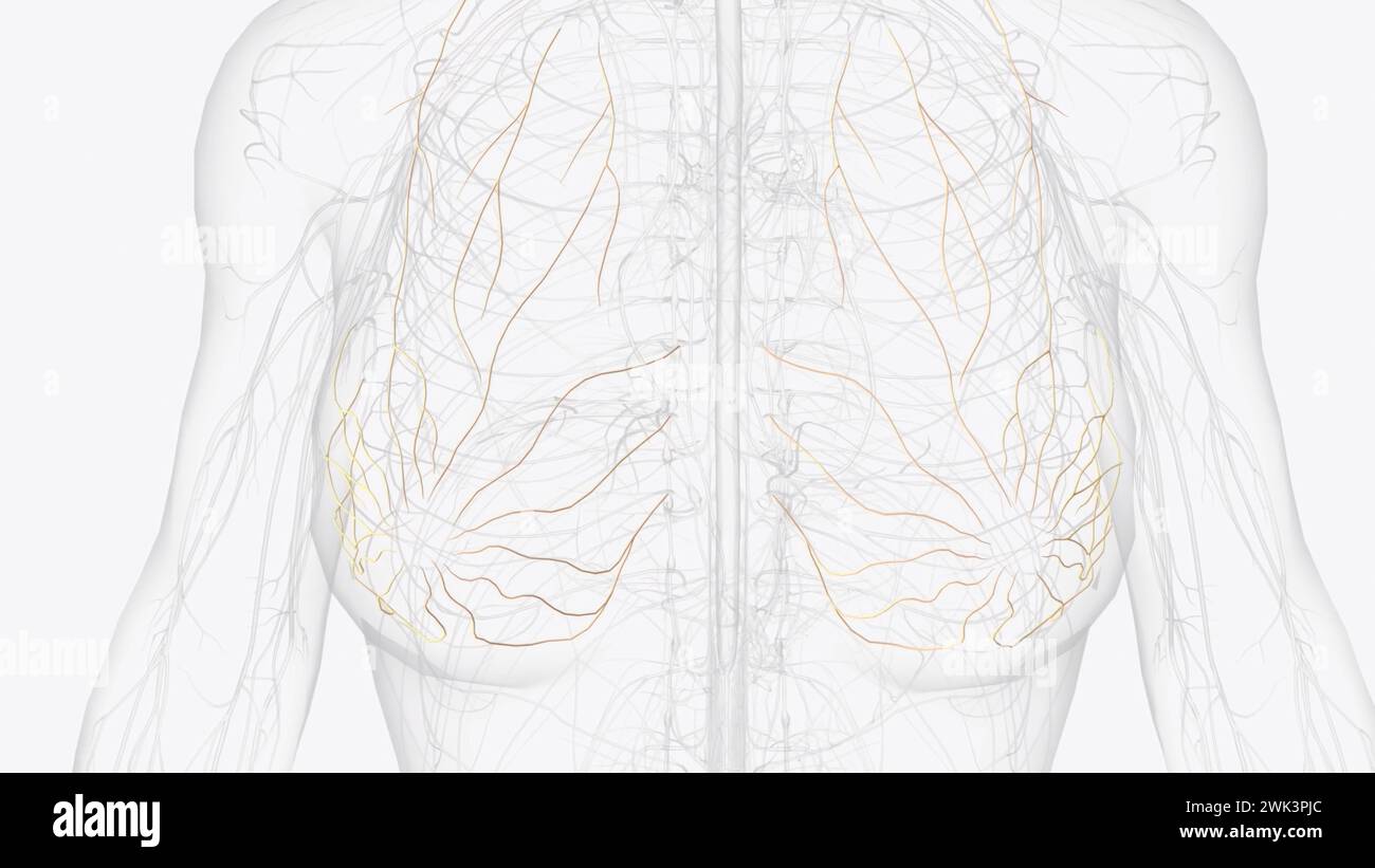 The internal thoracic (mammary) nerve is formed by contributions from the subclavianplexus and the phrenic nerve 3d illustration Stock Photohttps://www.alamy.com/image-license-details/?v=1https://www.alamy.com/the-internal-thoracic-mammary-nerve-is-formed-by-contributions-from-the-subclavianplexus-and-the-phrenic-nerve-3d-illustration-image596914596.html
RF2WK3PJC–The internal thoracic (mammary) nerve is formed by contributions from the subclavianplexus and the phrenic nerve 3d illustration
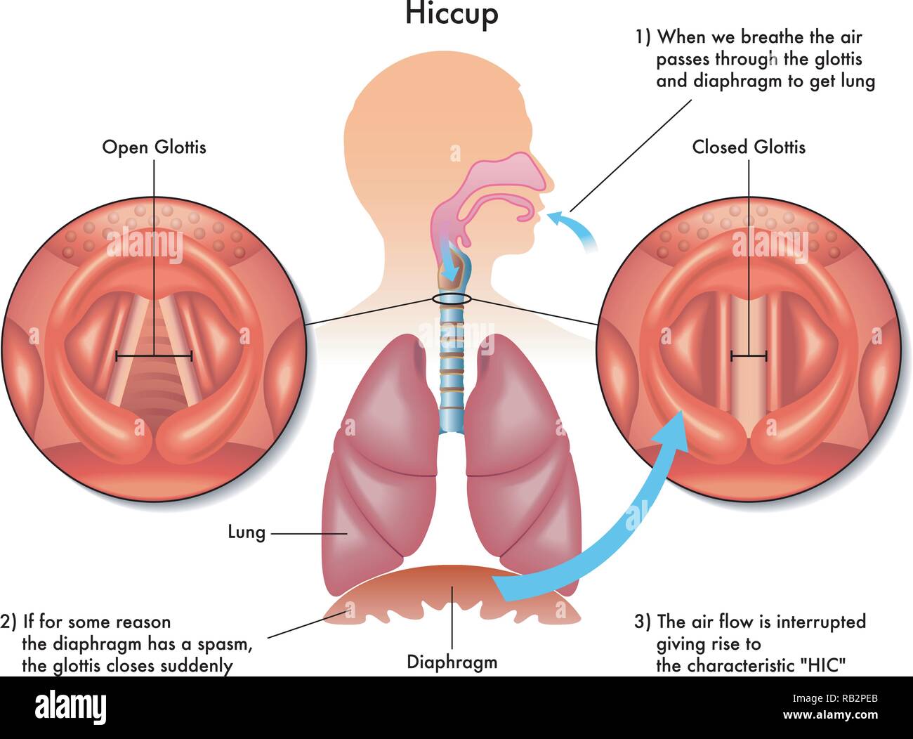 Medical illustration of the symptoms and causes of hiccup Stock Vectorhttps://www.alamy.com/image-license-details/?v=1https://www.alamy.com/medical-illustration-of-the-symptoms-and-causes-of-hiccup-image230557555.html
RFRB2PEB–Medical illustration of the symptoms and causes of hiccup
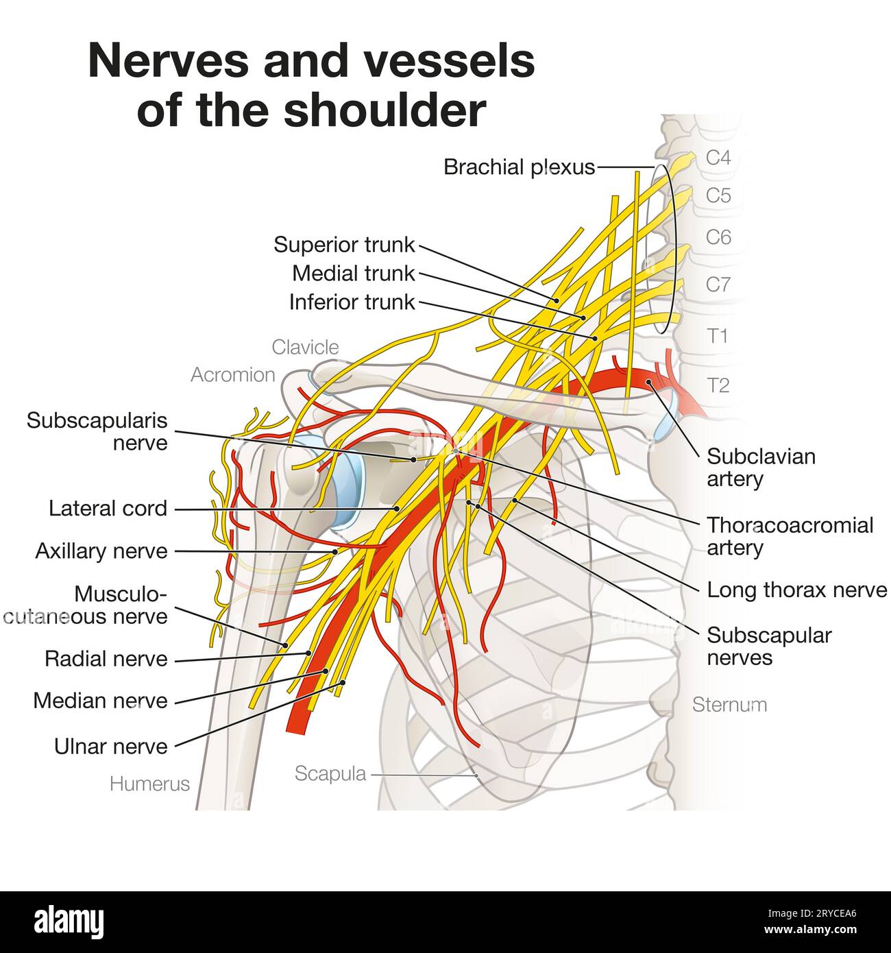 The shoulder region houses a complex network of nerves and vessels, including the brachial plexus, arteries, and veins, essential for limb innervation Stock Photohttps://www.alamy.com/image-license-details/?v=1https://www.alamy.com/the-shoulder-region-houses-a-complex-network-of-nerves-and-vessels-including-the-brachial-plexus-arteries-and-veins-essential-for-limb-innervation-image567602174.html
RF2RYCEA6–The shoulder region houses a complex network of nerves and vessels, including the brachial plexus, arteries, and veins, essential for limb innervation
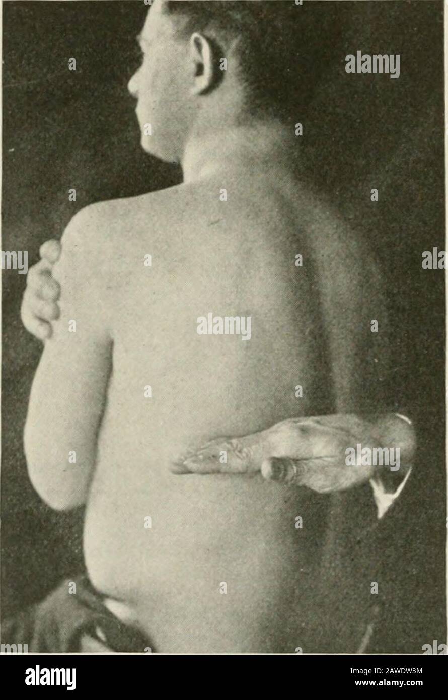
![. The only osteopractic method of treating diseases at home. dthe false ribs. The next two, only connect with thespina] column and are designated as floating ribs. 128 OSTEOPRACTIC METHOD DESCRIPTION OF CUT 38.Holding the Phrenic Nerve, This nerve contracts the diaphragm, and pressureon it breaks the nerve wave to that muscle and slowsits action. How to Hold it Stand behind the patient (see Cut 38), and placethe fingers upon the transverse process of the third,fourth and fifth cervical vertebrae, press the musclesforward and slip the fingers down in front of the trans-verse process, where a pr Stock Photo . The only osteopractic method of treating diseases at home. dthe false ribs. The next two, only connect with thespina] column and are designated as floating ribs. 128 OSTEOPRACTIC METHOD DESCRIPTION OF CUT 38.Holding the Phrenic Nerve, This nerve contracts the diaphragm, and pressureon it breaks the nerve wave to that muscle and slowsits action. How to Hold it Stand behind the patient (see Cut 38), and placethe fingers upon the transverse process of the third,fourth and fifth cervical vertebrae, press the musclesforward and slip the fingers down in front of the trans-verse process, where a pr Stock Photo](https://c8.alamy.com/comp/2AGA7FW/the-only-osteopractic-method-of-treating-diseases-at-home-dthe-false-ribs-the-next-two-only-connect-with-thespina-column-and-are-designated-as-floating-ribs-128-osteopractic-method-description-of-cut-38holding-the-phrenic-nerve-this-nerve-contracts-the-diaphragm-and-pressureon-it-breaks-the-nerve-wave-to-that-muscle-and-slowsits-action-how-to-hold-it-stand-behind-the-patient-see-cut-38-and-placethe-fingers-upon-the-transverse-process-of-the-thirdfourth-and-fifth-cervical-vertebrae-press-the-musclesforward-and-slip-the-fingers-down-in-front-of-the-trans-verse-process-where-a-pr-2AGA7FW.jpg) . The only osteopractic method of treating diseases at home. dthe false ribs. The next two, only connect with thespina] column and are designated as floating ribs. 128 OSTEOPRACTIC METHOD DESCRIPTION OF CUT 38.Holding the Phrenic Nerve, This nerve contracts the diaphragm, and pressureon it breaks the nerve wave to that muscle and slowsits action. How to Hold it Stand behind the patient (see Cut 38), and placethe fingers upon the transverse process of the third,fourth and fifth cervical vertebrae, press the musclesforward and slip the fingers down in front of the trans-verse process, where a pr Stock Photohttps://www.alamy.com/image-license-details/?v=1https://www.alamy.com/the-only-osteopractic-method-of-treating-diseases-at-home-dthe-false-ribs-the-next-two-only-connect-with-thespina-column-and-are-designated-as-floating-ribs-128-osteopractic-method-description-of-cut-38holding-the-phrenic-nerve-this-nerve-contracts-the-diaphragm-and-pressureon-it-breaks-the-nerve-wave-to-that-muscle-and-slowsits-action-how-to-hold-it-stand-behind-the-patient-see-cut-38-and-placethe-fingers-upon-the-transverse-process-of-the-thirdfourth-and-fifth-cervical-vertebrae-press-the-musclesforward-and-slip-the-fingers-down-in-front-of-the-trans-verse-process-where-a-pr-image337056941.html
. The only osteopractic method of treating diseases at home. dthe false ribs. The next two, only connect with thespina] column and are designated as floating ribs. 128 OSTEOPRACTIC METHOD DESCRIPTION OF CUT 38.Holding the Phrenic Nerve, This nerve contracts the diaphragm, and pressureon it breaks the nerve wave to that muscle and slowsits action. How to Hold it Stand behind the patient (see Cut 38), and placethe fingers upon the transverse process of the third,fourth and fifth cervical vertebrae, press the musclesforward and slip the fingers down in front of the trans-verse process, where a pr Stock Photohttps://www.alamy.com/image-license-details/?v=1https://www.alamy.com/the-only-osteopractic-method-of-treating-diseases-at-home-dthe-false-ribs-the-next-two-only-connect-with-thespina-column-and-are-designated-as-floating-ribs-128-osteopractic-method-description-of-cut-38holding-the-phrenic-nerve-this-nerve-contracts-the-diaphragm-and-pressureon-it-breaks-the-nerve-wave-to-that-muscle-and-slowsits-action-how-to-hold-it-stand-behind-the-patient-see-cut-38-and-placethe-fingers-upon-the-transverse-process-of-the-thirdfourth-and-fifth-cervical-vertebrae-press-the-musclesforward-and-slip-the-fingers-down-in-front-of-the-trans-verse-process-where-a-pr-image337056941.htmlRM2AGA7FW–. The only osteopractic method of treating diseases at home. dthe false ribs. The next two, only connect with thespina] column and are designated as floating ribs. 128 OSTEOPRACTIC METHOD DESCRIPTION OF CUT 38.Holding the Phrenic Nerve, This nerve contracts the diaphragm, and pressureon it breaks the nerve wave to that muscle and slowsits action. How to Hold it Stand behind the patient (see Cut 38), and placethe fingers upon the transverse process of the third,fourth and fifth cervical vertebrae, press the musclesforward and slip the fingers down in front of the trans-verse process, where a pr
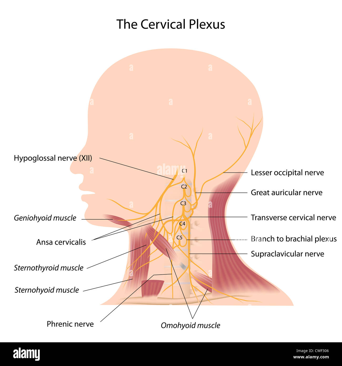 The cervical plexus Stock Photohttps://www.alamy.com/image-license-details/?v=1https://www.alamy.com/stock-photo-the-cervical-plexus-50118774.html
RFCWF306–The cervical plexus
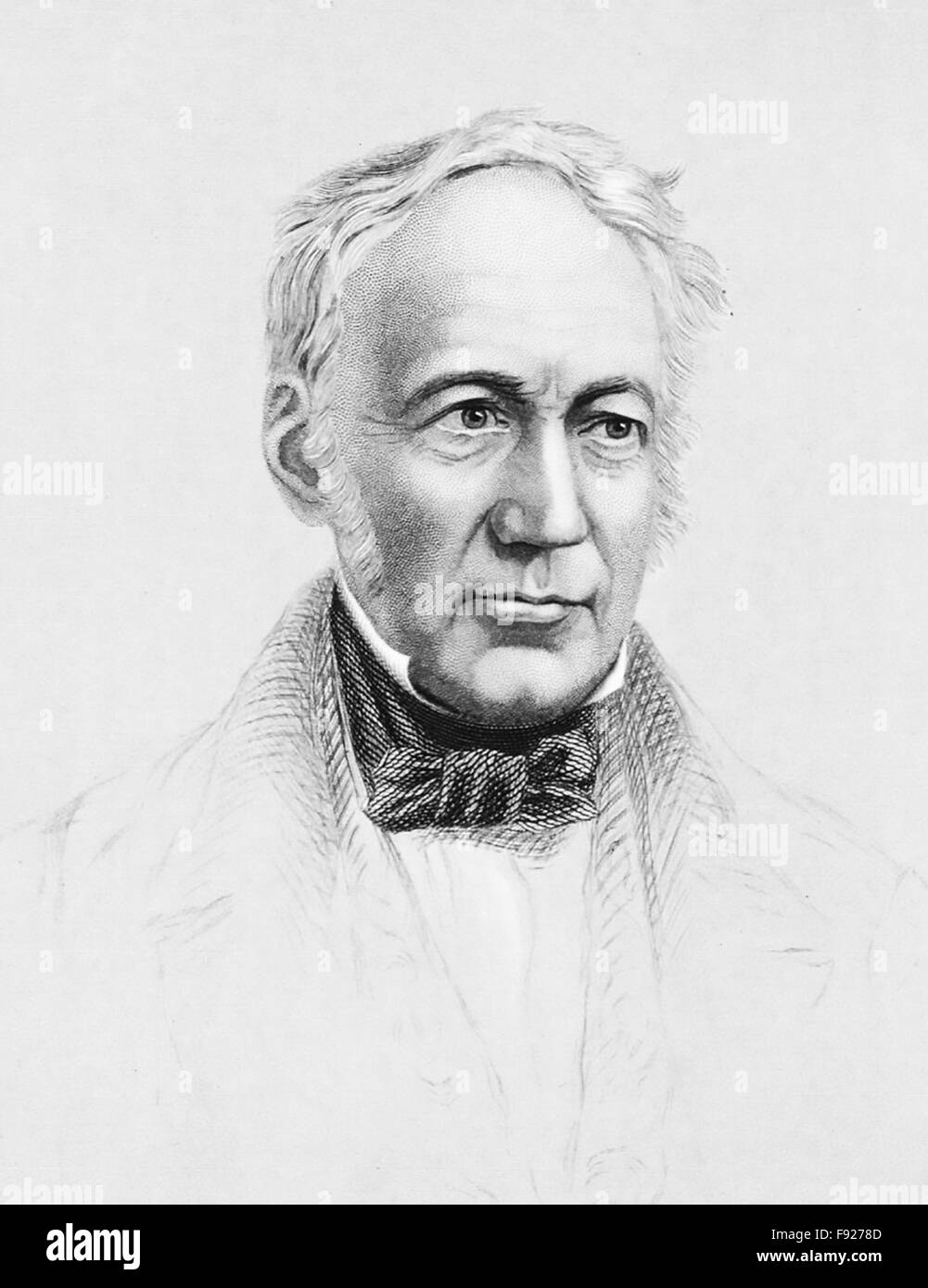 ANDREW URE (1778-1857) Scottish doctor and chemist about 1842. Engraving by William Jackman Stock Photohttps://www.alamy.com/image-license-details/?v=1https://www.alamy.com/stock-photo-andrew-ure-1778-1857-scottish-doctor-and-chemist-about-1842-engraving-91633373.html
RMF9278D–ANDREW URE (1778-1857) Scottish doctor and chemist about 1842. Engraving by William Jackman
 Galvanic Experiments on Human Subject, 1818 Stock Photohttps://www.alamy.com/image-license-details/?v=1https://www.alamy.com/stock-photo-galvanic-experiments-on-human-subject-1818-135097442.html
RMHRP64J–Galvanic Experiments on Human Subject, 1818
 ANDREW URE (1778-1857) Scottish doctor in an 1867 engraving of his electrical experiments on the corpse of Matthew Clydsdale in 1818. From: Louis Figuier's 1891 book Les Merveilles de la Science Stock Photohttps://www.alamy.com/image-license-details/?v=1https://www.alamy.com/stock-photo-andrew-ure-1778-1857-scottish-doctor-in-an-1867-engraving-of-his-electrical-91516809.html
RMF8TXHD–ANDREW URE (1778-1857) Scottish doctor in an 1867 engraving of his electrical experiments on the corpse of Matthew Clydsdale in 1818. From: Louis Figuier's 1891 book Les Merveilles de la Science
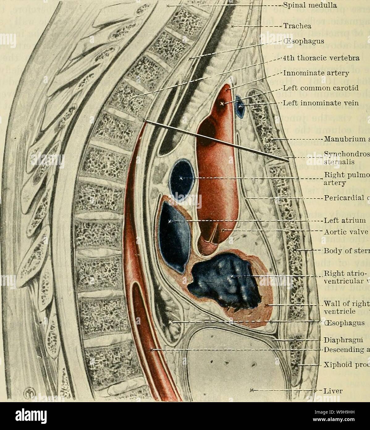 Archive image from page 1123 of Cunningham's Text-book of anatomy (1914). Cunningham's Text-book of anatomy cunninghamstextb00cunn Year: 1914 ( 1090 THE KESPIRATOKY SYSTEM. great arteries which spring from it; (2) the innominate veins and part of the vena cava superior; (3) the trachea, oesophagus, and thoracic duct; (4) the phrenic, vagi, and cardiac nerves, and the left recurrent nerve; (5) the thymus. The middle mediastinum is the wide part of the area which contains the pericardium, and lies below the superior mediastinum. In addition to the pericardium and its contents the middle mediast Stock Photohttps://www.alamy.com/image-license-details/?v=1https://www.alamy.com/archive-image-from-page-1123-of-cunninghams-text-book-of-anatomy-1914-cunninghams-text-book-of-anatomy-cunninghamstextb00cunn-year-1914-1090-the-kespiratoky-system-great-arteries-which-spring-from-it-2-the-innominate-veins-and-part-of-the-vena-cava-superior-3-the-trachea-oesophagus-and-thoracic-duct-4-the-phrenic-vagi-and-cardiac-nerves-and-the-left-recurrent-nerve-5-the-thymus-the-middle-mediastinum-is-the-wide-part-of-the-area-which-contains-the-pericardium-and-lies-below-the-superior-mediastinum-in-addition-to-the-pericardium-and-its-contents-the-middle-mediast-image264068157.html
RMW9H9HH–Archive image from page 1123 of Cunningham's Text-book of anatomy (1914). Cunningham's Text-book of anatomy cunninghamstextb00cunn Year: 1914 ( 1090 THE KESPIRATOKY SYSTEM. great arteries which spring from it; (2) the innominate veins and part of the vena cava superior; (3) the trachea, oesophagus, and thoracic duct; (4) the phrenic, vagi, and cardiac nerves, and the left recurrent nerve; (5) the thymus. The middle mediastinum is the wide part of the area which contains the pericardium, and lies below the superior mediastinum. In addition to the pericardium and its contents the middle mediast
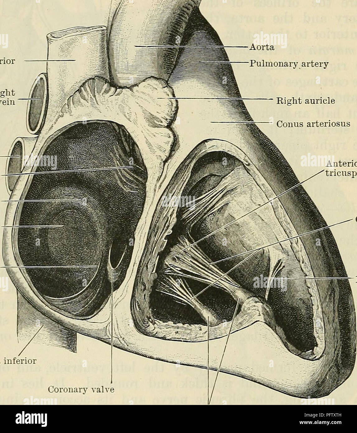 . Cunningham's Text-book of anatomy. Anatomy. 874 THE VASCULAK SYSTEM. aperture. Above and anteriorly it is in relation with the ascending aorta, and from the junction of this aspect with the right lateral boundary the right auricle is prolonged anteriorly and to the left. Its right side forms the right margin of the heart, and is in relation with the right phrenic nerve and its accom- panying vessels, and with the right pleura and lung, the pericardium intervening. On the left this atrium is limited by the oblique septum which separates it from the left atrium. The sulcus terminalis is a shal Stock Photohttps://www.alamy.com/image-license-details/?v=1https://www.alamy.com/cunninghams-text-book-of-anatomy-anatomy-874-the-vasculak-system-aperture-above-and-anteriorly-it-is-in-relation-with-the-ascending-aorta-and-from-the-junction-of-this-aspect-with-the-right-lateral-boundary-the-right-auricle-is-prolonged-anteriorly-and-to-the-left-its-right-side-forms-the-right-margin-of-the-heart-and-is-in-relation-with-the-right-phrenic-nerve-and-its-accom-panying-vessels-and-with-the-right-pleura-and-lung-the-pericardium-intervening-on-the-left-this-atrium-is-limited-by-the-oblique-septum-which-separates-it-from-the-left-atrium-the-sulcus-terminalis-is-a-shal-image216292177.html
RMPFTXTH–. Cunningham's Text-book of anatomy. Anatomy. 874 THE VASCULAK SYSTEM. aperture. Above and anteriorly it is in relation with the ascending aorta, and from the junction of this aspect with the right lateral boundary the right auricle is prolonged anteriorly and to the left. Its right side forms the right margin of the heart, and is in relation with the right phrenic nerve and its accom- panying vessels, and with the right pleura and lung, the pericardium intervening. On the left this atrium is limited by the oblique septum which separates it from the left atrium. The sulcus terminalis is a shal
 . Fig. 7. Nervous relations of the thoracic rete. The rete is dotted. Part of the posterior thoracic vein has been omitted in order to show the nerves underneath. /, Pneumo-gastric nerve k, Retial ganglion g, Cervical sympathetic chord /, Phrenic nerve //, Thoracic sympathetic chord /', Middle cervical sympathetic ganglion j, Posterior cervical sympathetic ganglion a, Dorsal aorta b, Carotid artery c, Jugular vein d, Posterior thoracic vein e, Pulmonary artery cr3B, Cervical spinal nerves tr1'5, Thoracic spinal nerves Stock Photohttps://www.alamy.com/image-license-details/?v=1https://www.alamy.com/fig-7-nervous-relations-of-the-thoracic-rete-the-rete-is-dotted-part-of-the-posterior-thoracic-vein-has-been-omitted-in-order-to-show-the-nerves-underneath-pneumo-gastric-nerve-k-retial-ganglion-g-cervical-sympathetic-chord-phrenic-nerve-thoracic-sympathetic-chord-middle-cervical-sympathetic-ganglion-j-posterior-cervical-sympathetic-ganglion-a-dorsal-aorta-b-carotid-artery-c-jugular-vein-d-posterior-thoracic-vein-e-pulmonary-artery-cr3b-cervical-spinal-nerves-tr15-thoracic-spinal-nerves-image180025800.html
RMMCTTMT–. Fig. 7. Nervous relations of the thoracic rete. The rete is dotted. Part of the posterior thoracic vein has been omitted in order to show the nerves underneath. /, Pneumo-gastric nerve k, Retial ganglion g, Cervical sympathetic chord /, Phrenic nerve //, Thoracic sympathetic chord /', Middle cervical sympathetic ganglion j, Posterior cervical sympathetic ganglion a, Dorsal aorta b, Carotid artery c, Jugular vein d, Posterior thoracic vein e, Pulmonary artery cr3B, Cervical spinal nerves tr1'5, Thoracic spinal nerves
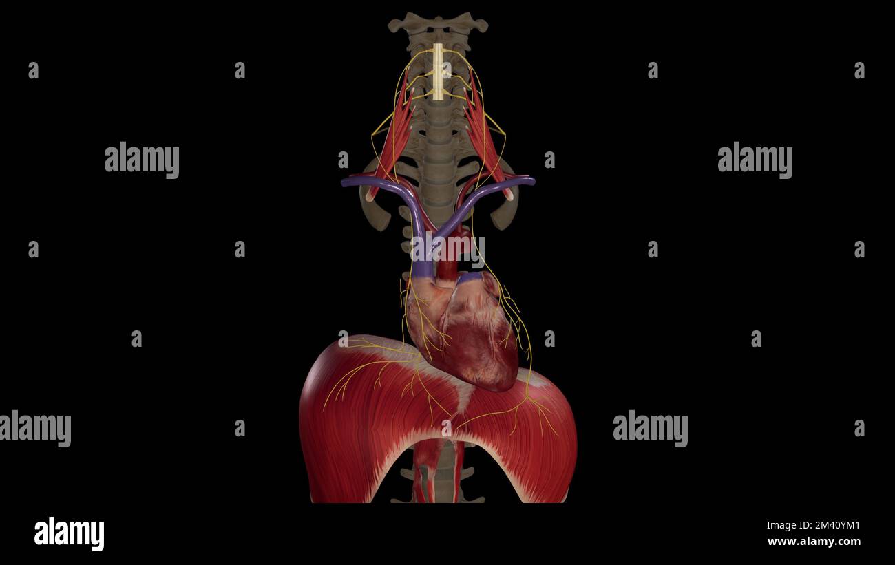 Phrenic Nerves on Black Background Stock Photohttps://www.alamy.com/image-license-details/?v=1https://www.alamy.com/phrenic-nerves-on-black-background-image501581025.html
RF2M40YM1–Phrenic Nerves on Black Background
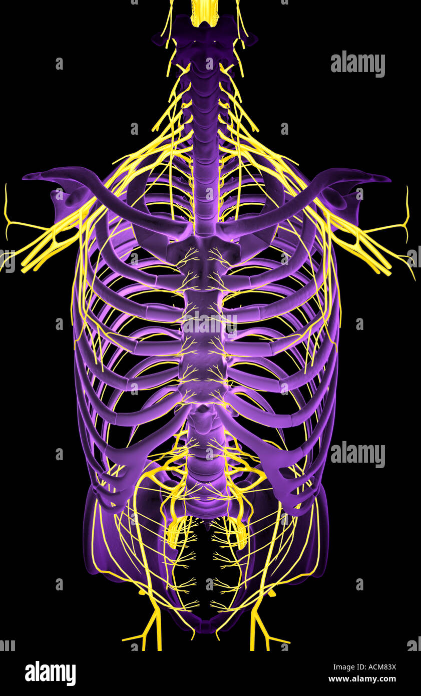 Nerves of the upper body Stock Photohttps://www.alamy.com/image-license-details/?v=1https://www.alamy.com/stock-photo-nerves-of-the-upper-body-13186477.html
RFACM83X–Nerves of the upper body
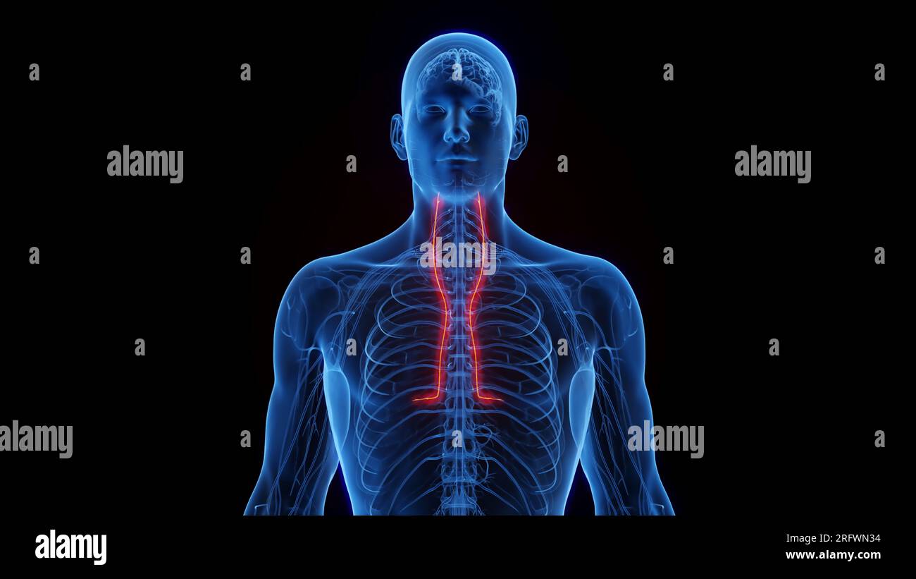 Phrenic nerves, illustration Stock Photohttps://www.alamy.com/image-license-details/?v=1https://www.alamy.com/phrenic-nerves-illustration-image560516968.html
RF2RFWN34–Phrenic nerves, illustration
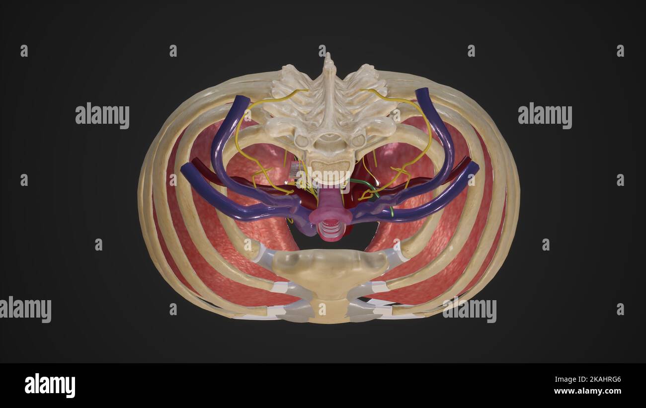 Superior Thoracic Aperture Stock Photohttps://www.alamy.com/image-license-details/?v=1https://www.alamy.com/superior-thoracic-aperture-image488428534.html
RF2KAHRG6–Superior Thoracic Aperture
 The internal thoracic (mammary) nerve is formed by contributions from the subclavianplexus and the phrenic nerve 3d illustration Stock Photohttps://www.alamy.com/image-license-details/?v=1https://www.alamy.com/the-internal-thoracic-mammary-nerve-is-formed-by-contributions-from-the-subclavianplexus-and-the-phrenic-nerve-3d-illustration-image596914530.html
RF2WK3PG2–The internal thoracic (mammary) nerve is formed by contributions from the subclavianplexus and the phrenic nerve 3d illustration
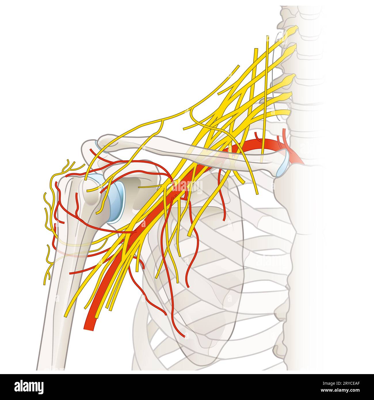 The shoulder region houses a complex network of nerves and vessels, including the brachial plexus, arteries, and veins, essential for limb innervation Stock Photohttps://www.alamy.com/image-license-details/?v=1https://www.alamy.com/the-shoulder-region-houses-a-complex-network-of-nerves-and-vessels-including-the-brachial-plexus-arteries-and-veins-essential-for-limb-innervation-image567602183.html
RF2RYCEAF–The shoulder region houses a complex network of nerves and vessels, including the brachial plexus, arteries, and veins, essential for limb innervation
 . The only osteopractic method of treating diseases at home. CUT 38.Holding the Phrenic Nerve 131. Stock Photohttps://www.alamy.com/image-license-details/?v=1https://www.alamy.com/the-only-osteopractic-method-of-treating-diseases-at-home-cut-38holding-the-phrenic-nerve-131-image337056301.html
RM2AGA6N1–. The only osteopractic method of treating diseases at home. CUT 38.Holding the Phrenic Nerve 131.
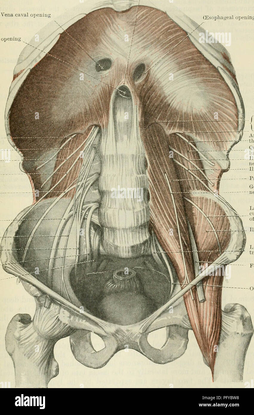 . Cunningham's Text-book of anatomy. Anatomy. THE MUSCLES OF THE THOEAX. 473 quadratum) in the right lobe of the central tendon transmits the inferior vena cava, and small branches of the right phrenic nerve. The hiatus ozsophageus (oesophageal opening) is in the muscular substance of the diaphragm, posterior to the central tendon, and is surrounded by a sphincter-like arrangement of the crural fibres. Besides the oesophagus, this opening transmits the two vagi nerves. Middle arcuate ligament Vena caval opening (Esophageal opening in diaphragm Aortic openin Anterior ramus of twelfth thoracic n Stock Photohttps://www.alamy.com/image-license-details/?v=1https://www.alamy.com/cunninghams-text-book-of-anatomy-anatomy-the-muscles-of-the-thoeax-473-quadratum-in-the-right-lobe-of-the-central-tendon-transmits-the-inferior-vena-cava-and-small-branches-of-the-right-phrenic-nerve-the-hiatus-ozsophageus-oesophageal-opening-is-in-the-muscular-substance-of-the-diaphragm-posterior-to-the-central-tendon-and-is-surrounded-by-a-sphincter-like-arrangement-of-the-crural-fibres-besides-the-oesophagus-this-opening-transmits-the-two-vagi-nerves-middle-arcuate-ligament-vena-caval-opening-esophageal-opening-in-diaphragm-aortic-openin-anterior-ramus-of-twelfth-thoracic-n-image216346292.html
RMPFYBW8–. Cunningham's Text-book of anatomy. Anatomy. THE MUSCLES OF THE THOEAX. 473 quadratum) in the right lobe of the central tendon transmits the inferior vena cava, and small branches of the right phrenic nerve. The hiatus ozsophageus (oesophageal opening) is in the muscular substance of the diaphragm, posterior to the central tendon, and is surrounded by a sphincter-like arrangement of the crural fibres. Besides the oesophagus, this opening transmits the two vagi nerves. Middle arcuate ligament Vena caval opening (Esophageal opening in diaphragm Aortic openin Anterior ramus of twelfth thoracic n
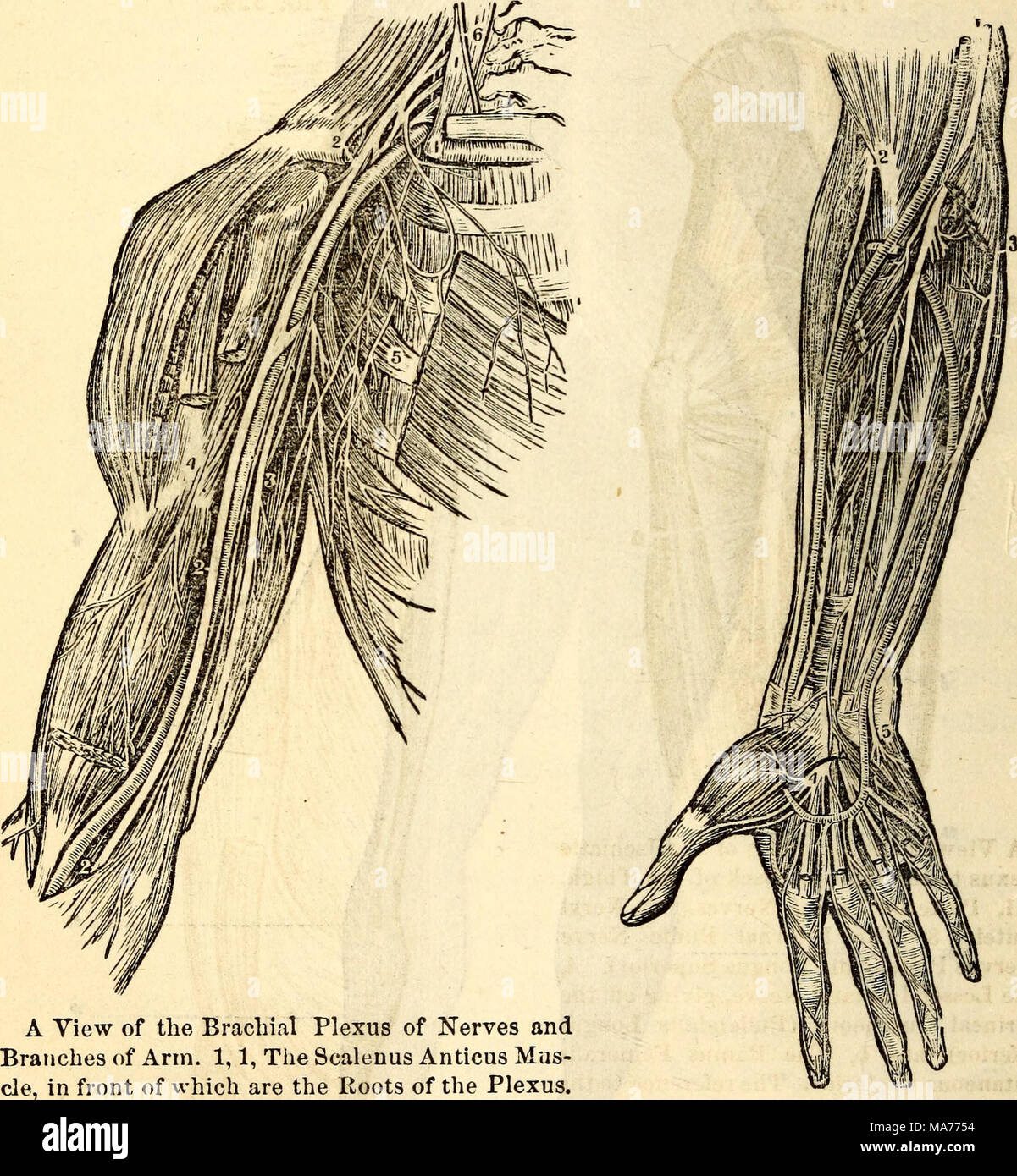 . Elementary anatomy and physiology : for colleges, academies, and other schools . 2, 2, The Median Nerve. 3, The Ulnar Nerve. 4, Nerves of Front of Fore-Arm. 1, The Branch to the Biceps Muscle. 5, The Nerves Median Nerve. 2, Anterior Branch of Wrisbersr. 6, The Phrenic Nerve from the of Musculo-Spiral or Radial Nerve. Third and Fourth Cervical. 3, Ulnar Nerve. 4, Division of Me- dian Nerve in the Palm to the Thumb, First, Second, and Radial Side of Third Finder. 5, Division of Ulnar Nerve to Ulnar Side of Third and both Sides of Fourth Finger. 600. What nerves go to make up the cervical and b Stock Photohttps://www.alamy.com/image-license-details/?v=1https://www.alamy.com/elementary-anatomy-and-physiology-for-colleges-academies-and-other-schools-2-2-the-median-nerve-3-the-ulnar-nerve-4-nerves-of-front-of-fore-arm-1-the-branch-to-the-biceps-muscle-5-the-nerves-median-nerve-2-anterior-branch-of-wrisbersr-6-the-phrenic-nerve-from-the-of-musculo-spiral-or-radial-nerve-third-and-fourth-cervical-3-ulnar-nerve-4-division-of-me-dian-nerve-in-the-palm-to-the-thumb-first-second-and-radial-side-of-third-finder-5-division-of-ulnar-nerve-to-ulnar-side-of-third-and-both-sides-of-fourth-finger-600-what-nerves-go-to-make-up-the-cervical-and-b-image178409536.html
RMMA7754–. Elementary anatomy and physiology : for colleges, academies, and other schools . 2, 2, The Median Nerve. 3, The Ulnar Nerve. 4, Nerves of Front of Fore-Arm. 1, The Branch to the Biceps Muscle. 5, The Nerves Median Nerve. 2, Anterior Branch of Wrisbersr. 6, The Phrenic Nerve from the of Musculo-Spiral or Radial Nerve. Third and Fourth Cervical. 3, Ulnar Nerve. 4, Division of Me- dian Nerve in the Palm to the Thumb, First, Second, and Radial Side of Third Finder. 5, Division of Ulnar Nerve to Ulnar Side of Third and both Sides of Fourth Finger. 600. What nerves go to make up the cervical and b
 Nerves of the upper body Stock Photohttps://www.alamy.com/image-license-details/?v=1https://www.alamy.com/stock-photo-nerves-of-the-upper-body-13249379.html
RFACXY9T–Nerves of the upper body
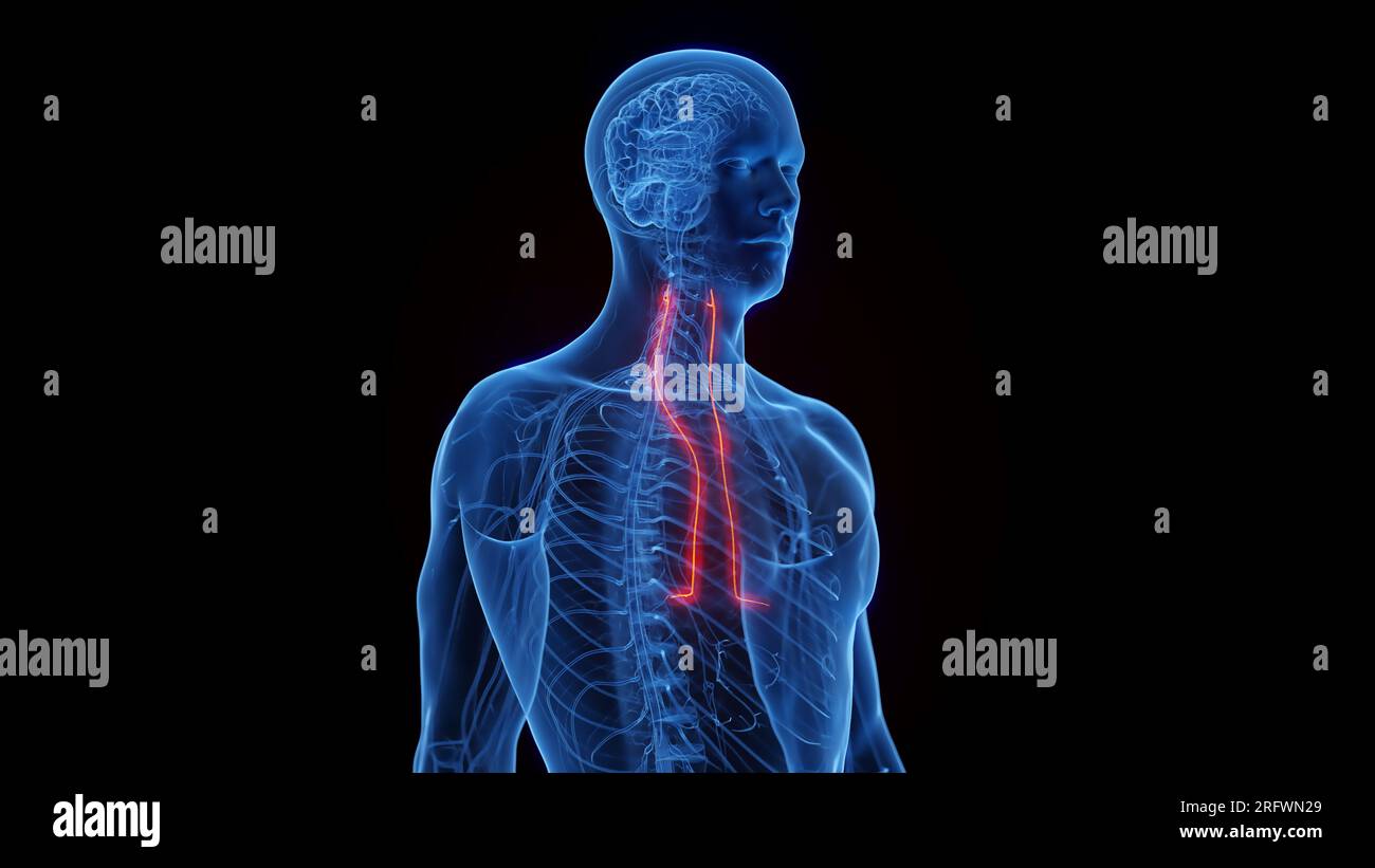 Phrenic nerves, illustration Stock Photohttps://www.alamy.com/image-license-details/?v=1https://www.alamy.com/phrenic-nerves-illustration-image560516945.html
RF2RFWN29–Phrenic nerves, illustration
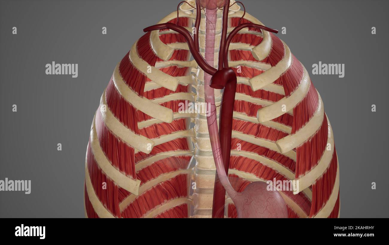 Medical Ilustration of Bood Supply of Esophagus Stock Photohttps://www.alamy.com/image-license-details/?v=1https://www.alamy.com/medical-ilustration-of-bood-supply-of-esophagus-image488428583.html
RF2KAHRHY–Medical Ilustration of Bood Supply of Esophagus
 The internal thoracic (mammary) nerve is formed by contributions from the subclavianplexus and the phrenic nerve 3d illustration Stock Photohttps://www.alamy.com/image-license-details/?v=1https://www.alamy.com/the-internal-thoracic-mammary-nerve-is-formed-by-contributions-from-the-subclavianplexus-and-the-phrenic-nerve-3d-illustration-image596914356.html
RF2WK3P9T–The internal thoracic (mammary) nerve is formed by contributions from the subclavianplexus and the phrenic nerve 3d illustration
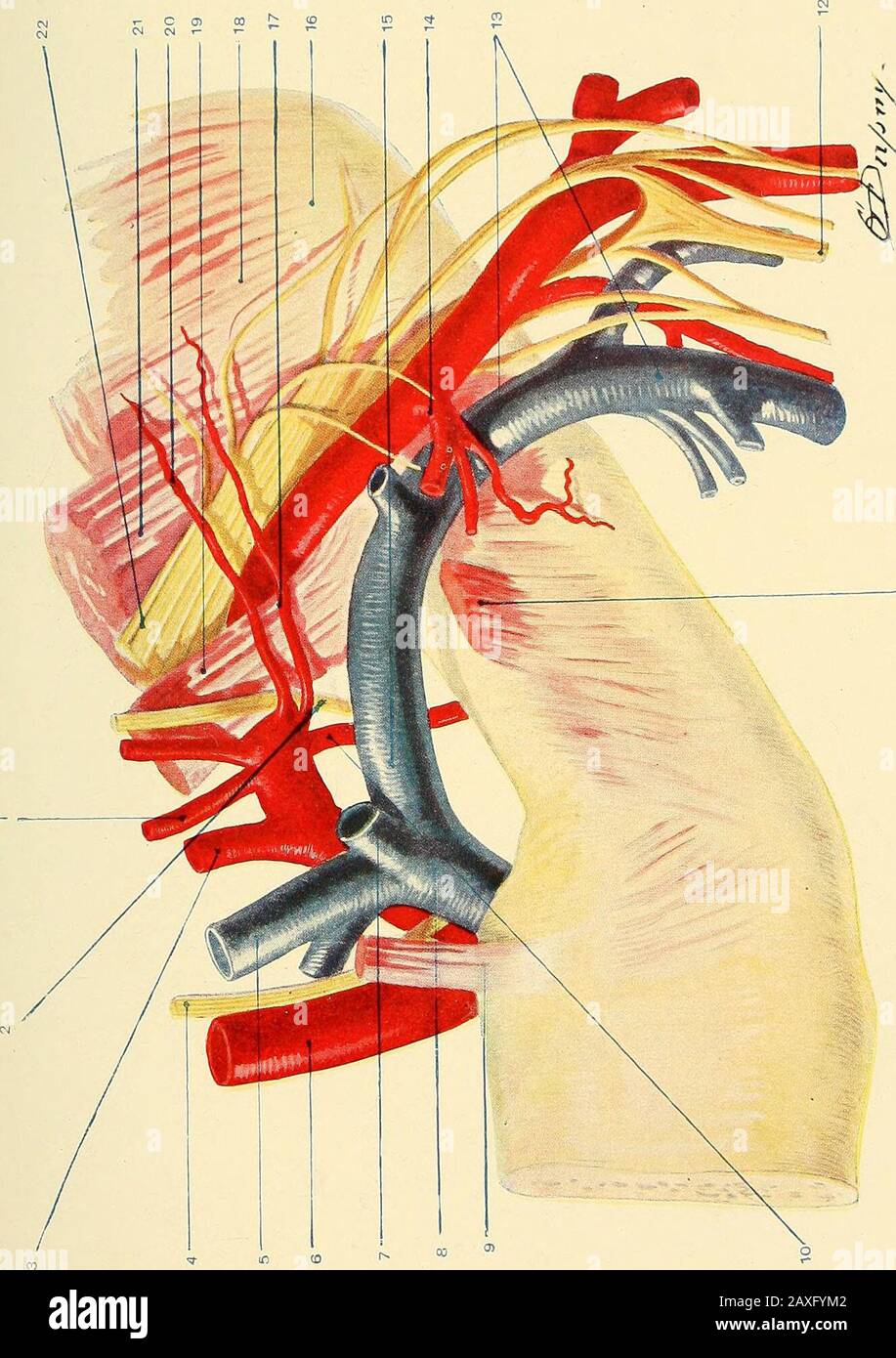 A manual of operative surgery . roid muscles, and thecervical fascia. It is near to the trachea, is in contact with thepleura below and behind, and is in close relation with the innominate^internal jugular, and vertebral veins, the vagus, recurrent laryngeal,cardiac, and sympathetic nerves. The left subclavian is also in relationwith the thoracic duct and the phrenic nerve. From this part of theartery arise the vertebral, the internal mammary, and the thyroidaxis. The second part of the artery reaches highest in the neck, and liesbetween the anterior and middle scalene muscles. It is still in Stock Photohttps://www.alamy.com/image-license-details/?v=1https://www.alamy.com/a-manual-of-operative-surgery-roid-muscles-and-thecervical-fascia-it-is-near-to-the-trachea-is-in-contact-with-thepleura-below-and-behind-and-is-in-close-relation-with-the-innominateinternal-jugular-and-vertebral-veins-the-vagus-recurrent-laryngealcardiac-and-sympathetic-nerves-the-left-subclavian-is-also-in-relationwith-the-thoracic-duct-and-the-phrenic-nerve-from-this-part-of-theartery-arise-the-vertebral-the-internal-mammary-and-the-thyroidaxis-the-second-part-of-the-artery-reaches-highest-in-the-neck-and-liesbetween-the-anterior-and-middle-scalene-muscles-it-is-still-in-image343329058.html
RM2AXFYM2–A manual of operative surgery . roid muscles, and thecervical fascia. It is near to the trachea, is in contact with thepleura below and behind, and is in close relation with the innominate^internal jugular, and vertebral veins, the vagus, recurrent laryngeal,cardiac, and sympathetic nerves. The left subclavian is also in relationwith the thoracic duct and the phrenic nerve. From this part of theartery arise the vertebral, the internal mammary, and the thyroidaxis. The second part of the artery reaches highest in the neck, and liesbetween the anterior and middle scalene muscles. It is still in
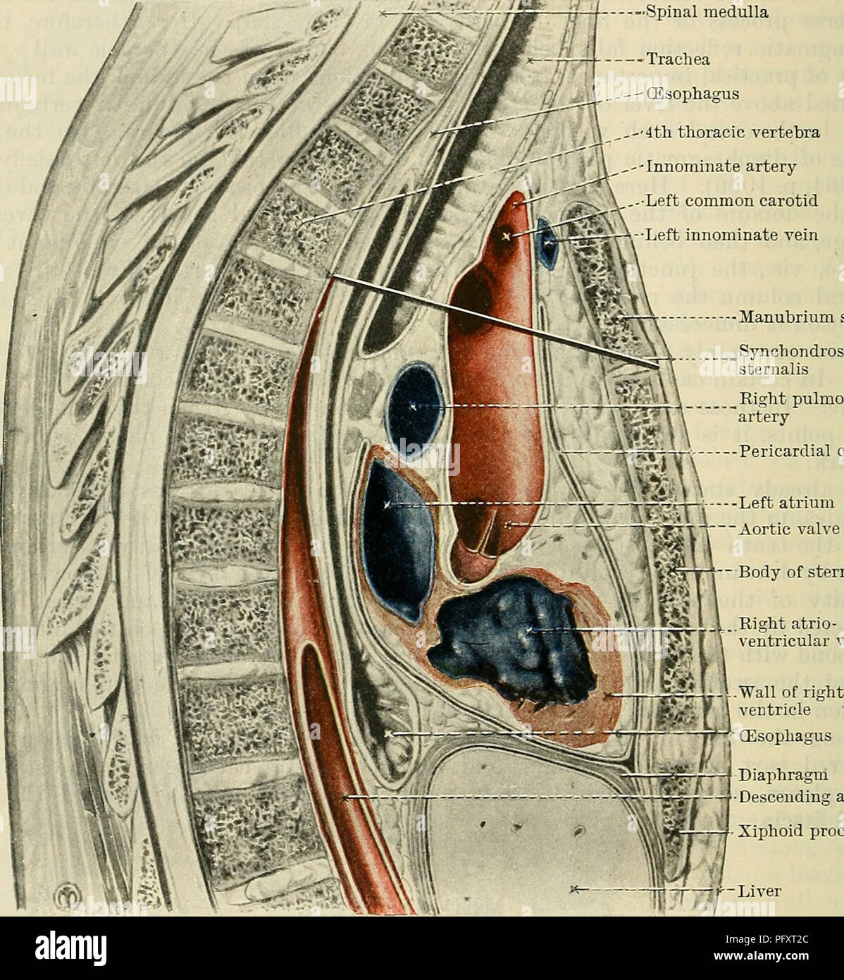 . Cunningham's Text-book of anatomy. Anatomy. 1090 THE KESPIRATOKY SYSTEM. great arteries which spring from it; (2) the innominate veins and part of the vena cava superior; (3) the trachea, oesophagus, and thoracic duct; (4) the phrenic, vagi, and cardiac nerves, and the left recurrent nerve; (5) the thymus. The middle mediastinum is the wide part of the area which contains the pericardium, and lies below the superior mediastinum. In addition to the pericardium and its contents the middle mediastinum contains the phrenic nerves and their accompanying vessels. The ventral mediastinum is that pa Stock Photohttps://www.alamy.com/image-license-details/?v=1https://www.alamy.com/cunninghams-text-book-of-anatomy-anatomy-1090-the-kespiratoky-system-great-arteries-which-spring-from-it-2-the-innominate-veins-and-part-of-the-vena-cava-superior-3-the-trachea-oesophagus-and-thoracic-duct-4-the-phrenic-vagi-and-cardiac-nerves-and-the-left-recurrent-nerve-5-the-thymus-the-middle-mediastinum-is-the-wide-part-of-the-area-which-contains-the-pericardium-and-lies-below-the-superior-mediastinum-in-addition-to-the-pericardium-and-its-contents-the-middle-mediastinum-contains-the-phrenic-nerves-and-their-accompanying-vessels-the-ventral-mediastinum-is-that-pa-image216333892.html
RMPFXT2C–. Cunningham's Text-book of anatomy. Anatomy. 1090 THE KESPIRATOKY SYSTEM. great arteries which spring from it; (2) the innominate veins and part of the vena cava superior; (3) the trachea, oesophagus, and thoracic duct; (4) the phrenic, vagi, and cardiac nerves, and the left recurrent nerve; (5) the thymus. The middle mediastinum is the wide part of the area which contains the pericardium, and lies below the superior mediastinum. In addition to the pericardium and its contents the middle mediastinum contains the phrenic nerves and their accompanying vessels. The ventral mediastinum is that pa
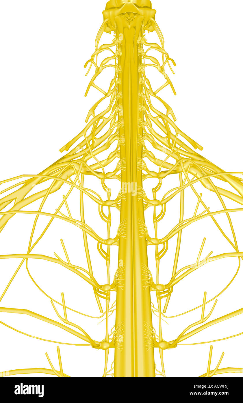 Nerves of the upper body Stock Photohttps://www.alamy.com/image-license-details/?v=1https://www.alamy.com/stock-photo-nerves-of-the-upper-body-13235933.html
RFACWF9J–Nerves of the upper body
 Phrenic nerves, illustration Stock Photohttps://www.alamy.com/image-license-details/?v=1https://www.alamy.com/phrenic-nerves-illustration-image560516954.html
RF2RFWN2J–Phrenic nerves, illustration
 The phrenic nerve is a bilateral, mixed nerve that originates in the neck and descends through the thorax to reach the diaphragm 3d illustration Stock Photohttps://www.alamy.com/image-license-details/?v=1https://www.alamy.com/the-phrenic-nerve-is-a-bilateral-mixed-nerve-that-originates-in-the-neck-and-descends-through-the-thorax-to-reach-the-diaphragm-3d-illustration-image596913279.html
RF2WK3MYB–The phrenic nerve is a bilateral, mixed nerve that originates in the neck and descends through the thorax to reach the diaphragm 3d illustration
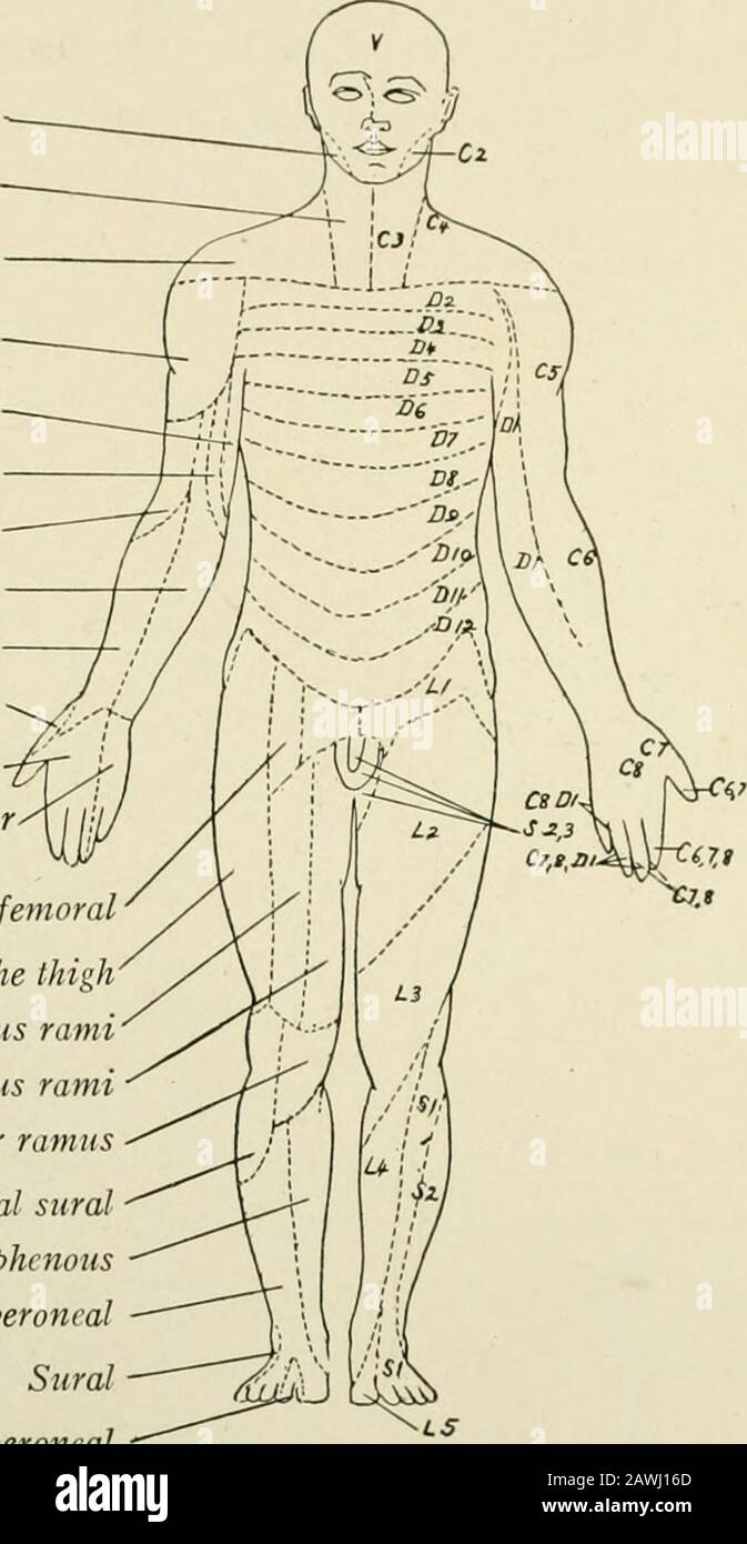 The anatomy of the nervous system, from the standpoint of development and function . ine the level of a lesion of thespinal cord or nerve roots within the vertebral canal. In the same way the shifting of muscles during embryonic development hasbeen accompanied by corresponding changes in the spacial distribution of themotor fibers. A familiar example is furnished by the diaphragm, the musculatureof which is derived from the cervical myotomes and which in its descent carrieswith it the phrenic nerve. This explains the origin of the phrenic from thethird, fourth, and fifth cervical nerves. 6o TH Stock Photohttps://www.alamy.com/image-license-details/?v=1https://www.alamy.com/the-anatomy-of-the-nervous-system-from-the-standpoint-of-development-and-function-ine-the-level-of-a-lesion-of-thespinal-cord-or-nerve-roots-within-the-vertebral-canal-in-the-same-way-the-shifting-of-muscles-during-embryonic-development-hasbeen-accompanied-by-corresponding-changes-in-the-spacial-distribution-of-themotor-fibers-a-familiar-example-is-furnished-by-the-diaphragm-the-musculatureof-which-is-derived-from-the-cervical-myotomes-and-which-in-its-descent-carrieswith-it-the-phrenic-nerve-this-explains-the-origin-of-the-phrenic-from-thethird-fourth-and-fifth-cervical-nerves-6o-th-image342759493.html
RM2AWJ16D–The anatomy of the nervous system, from the standpoint of development and function . ine the level of a lesion of thespinal cord or nerve roots within the vertebral canal. In the same way the shifting of muscles during embryonic development hasbeen accompanied by corresponding changes in the spacial distribution of themotor fibers. A familiar example is furnished by the diaphragm, the musculatureof which is derived from the cervical myotomes and which in its descent carrieswith it the phrenic nerve. This explains the origin of the phrenic from thethird, fourth, and fifth cervical nerves. 6o TH
 . Cunningham's Text-book of anatomy. Anatomy. 702 THE NEEVOUS SYSTEM. important to realise the position of origin of certain nerves. The nerves to the prevertebral muscles, the communication with the phrenic, the dorsal scapular, and long thoracic nerves, arise from the anterior rami of the nerves involved in the plexus. The supra-scapular and the nerve to the subclavius arise at the level of formation of the secondary cords; and the anterior thoracic, subscapular, and thoraco - dorsal nerves arise from the secondary cords, prior to their ultimate subdivision into the nerves of distribution fo Stock Photohttps://www.alamy.com/image-license-details/?v=1https://www.alamy.com/cunninghams-text-book-of-anatomy-anatomy-702-the-neevous-system-important-to-realise-the-position-of-origin-of-certain-nerves-the-nerves-to-the-prevertebral-muscles-the-communication-with-the-phrenic-the-dorsal-scapular-and-long-thoracic-nerves-arise-from-the-anterior-rami-of-the-nerves-involved-in-the-plexus-the-supra-scapular-and-the-nerve-to-the-subclavius-arise-at-the-level-of-formation-of-the-secondary-cords-and-the-anterior-thoracic-subscapular-and-thoraco-dorsal-nerves-arise-from-the-secondary-cords-prior-to-their-ultimate-subdivision-into-the-nerves-of-distribution-fo-image216345386.html
RMPFYAMX–. Cunningham's Text-book of anatomy. Anatomy. 702 THE NEEVOUS SYSTEM. important to realise the position of origin of certain nerves. The nerves to the prevertebral muscles, the communication with the phrenic, the dorsal scapular, and long thoracic nerves, arise from the anterior rami of the nerves involved in the plexus. The supra-scapular and the nerve to the subclavius arise at the level of formation of the secondary cords; and the anterior thoracic, subscapular, and thoraco - dorsal nerves arise from the secondary cords, prior to their ultimate subdivision into the nerves of distribution fo
 Nerves of the upper body Stock Photohttps://www.alamy.com/image-license-details/?v=1https://www.alamy.com/stock-photo-nerves-of-the-upper-body-13263555.html
RFAD0DFG–Nerves of the upper body
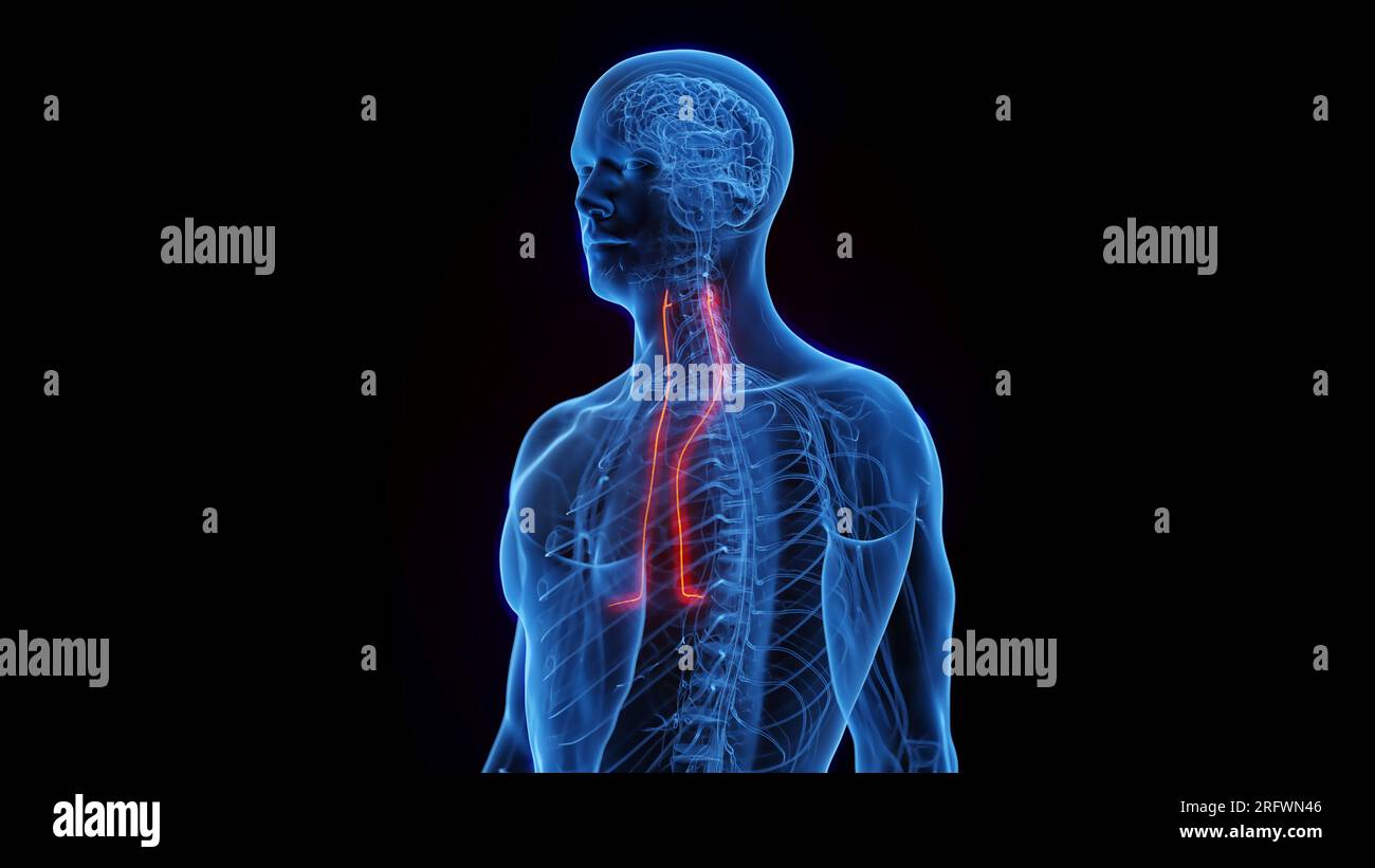 Phrenic nerves, illustration Stock Photohttps://www.alamy.com/image-license-details/?v=1https://www.alamy.com/phrenic-nerves-illustration-image560516998.html
RF2RFWN46–Phrenic nerves, illustration
 The phrenic nerve is a bilateral, mixed nerve that originates in the neck and descends through the thorax to reach the diaphragm 3d illustration Stock Photohttps://www.alamy.com/image-license-details/?v=1https://www.alamy.com/the-phrenic-nerve-is-a-bilateral-mixed-nerve-that-originates-in-the-neck-and-descends-through-the-thorax-to-reach-the-diaphragm-3d-illustration-image596913817.html
RF2WK3NJH–The phrenic nerve is a bilateral, mixed nerve that originates in the neck and descends through the thorax to reach the diaphragm 3d illustration
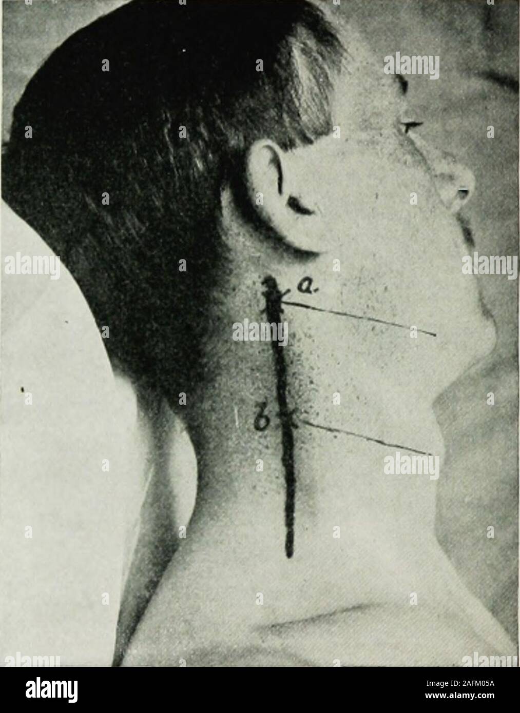 . Local and regional anesthesia : with chapters on spinal, epidural, paravertebral, and parasacral analgesia, and on other applications of local and regional anesthesia to the surgery of the eye, ear, nose and throat, and to dental practice. ebral injections in such regions as the neck mustreceive further experimental study before it can be popularized; thelikelihood of the solution reaching the phrenic nerve in effectivequantity should not be lost sight of; its origin from the third, fourth,and fifth cervical is practically the center of the field, and after forma-tion its course is more supe Stock Photohttps://www.alamy.com/image-license-details/?v=1https://www.alamy.com/local-and-regional-anesthesia-with-chapters-on-spinal-epidural-paravertebral-and-parasacral-analgesia-and-on-other-applications-of-local-and-regional-anesthesia-to-the-surgery-of-the-eye-ear-nose-and-throat-and-to-dental-practice-ebral-injections-in-such-regions-as-the-neck-mustreceive-further-experimental-study-before-it-can-be-popularized-thelikelihood-of-the-solution-reaching-the-phrenic-nerve-in-effectivequantity-should-not-be-lost-sight-of-its-origin-from-the-third-fourthand-fifth-cervical-is-practically-the-center-of-the-field-and-after-forma-tion-its-course-is-more-supe-image336656022.html
RM2AFM05A–. Local and regional anesthesia : with chapters on spinal, epidural, paravertebral, and parasacral analgesia, and on other applications of local and regional anesthesia to the surgery of the eye, ear, nose and throat, and to dental practice. ebral injections in such regions as the neck mustreceive further experimental study before it can be popularized; thelikelihood of the solution reaching the phrenic nerve in effectivequantity should not be lost sight of; its origin from the third, fourth,and fifth cervical is practically the center of the field, and after forma-tion its course is more supe
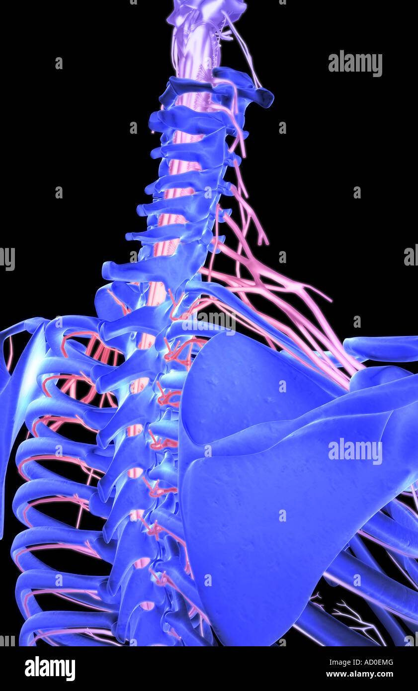 Nerves of the upper body Stock Photohttps://www.alamy.com/image-license-details/?v=1https://www.alamy.com/stock-photo-nerves-of-the-upper-body-13263951.html
RFAD0EMG–Nerves of the upper body
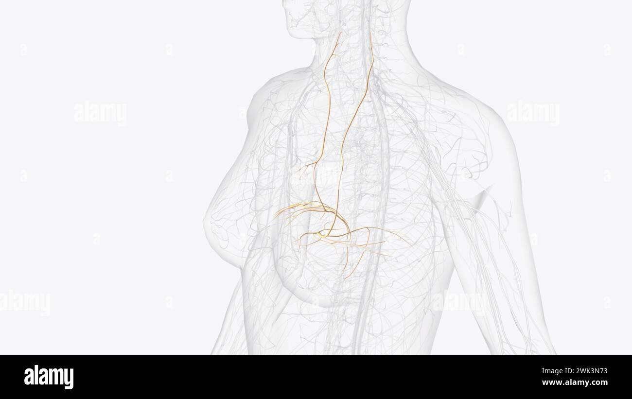 The phrenic nerve is a bilateral, mixed nerve that originates in the neck and descends through the thorax to reach the diaphragm 3d illustration Stock Photohttps://www.alamy.com/image-license-details/?v=1https://www.alamy.com/the-phrenic-nerve-is-a-bilateral-mixed-nerve-that-originates-in-the-neck-and-descends-through-the-thorax-to-reach-the-diaphragm-3d-illustration-image596913495.html
RF2WK3N73–The phrenic nerve is a bilateral, mixed nerve that originates in the neck and descends through the thorax to reach the diaphragm 3d illustration
 Atlas and text-book of topographic and applied anatomy . pon the superior vena cava andrunning upon the pericardium to the diaphragm), the comes nervi phrenici, the vena azygosmajor and its termination in the superior vena cava, the right intercostal vessels, the esophagus,the vagus nerve, the right sympathetic and splanchnic nerves, and the lymphatic glands at theroot of the lung. Upon the lejl side the visible structures are: the heart with the left phrenic nerve, the comesnervi phrenici, the descending aorta, the left subclavian artery, the left internal mammary artery,the left innominate v Stock Photohttps://www.alamy.com/image-license-details/?v=1https://www.alamy.com/atlas-and-text-book-of-topographic-and-applied-anatomy-pon-the-superior-vena-cava-andrunning-upon-the-pericardium-to-the-diaphragm-the-comes-nervi-phrenici-the-vena-azygosmajor-and-its-termination-in-the-superior-vena-cava-the-right-intercostal-vessels-the-esophagusthe-vagus-nerve-the-right-sympathetic-and-splanchnic-nerves-and-the-lymphatic-glands-at-theroot-of-the-lung-upon-the-lejl-side-the-visible-structures-are-the-heart-with-the-left-phrenic-nerve-the-comesnervi-phrenici-the-descending-aorta-the-left-subclavian-artery-the-left-internal-mammary-arterythe-left-innominate-v-image338259871.html
RM2AJ91WK–Atlas and text-book of topographic and applied anatomy . pon the superior vena cava andrunning upon the pericardium to the diaphragm), the comes nervi phrenici, the vena azygosmajor and its termination in the superior vena cava, the right intercostal vessels, the esophagus,the vagus nerve, the right sympathetic and splanchnic nerves, and the lymphatic glands at theroot of the lung. Upon the lejl side the visible structures are: the heart with the left phrenic nerve, the comesnervi phrenici, the descending aorta, the left subclavian artery, the left internal mammary artery,the left innominate v
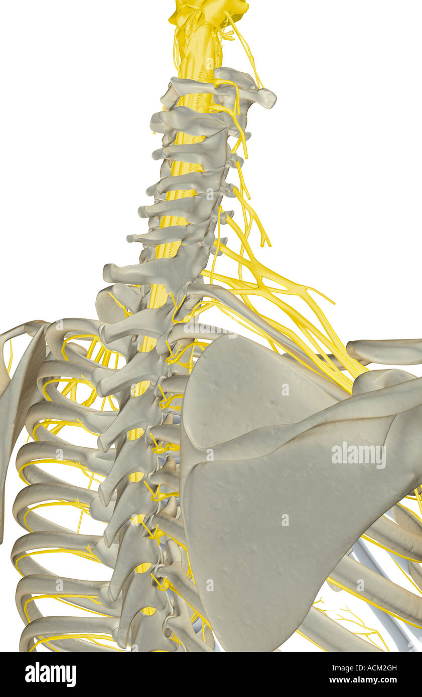 Nerves of the upper body Stock Photohttps://www.alamy.com/image-license-details/?v=1https://www.alamy.com/stock-photo-nerves-of-the-upper-body-13184608.html
RFACM2GH–Nerves of the upper body
 Human anatomy, including structure and development and practical considerations . tlie nerve to the subclavius. A branch of comnumication with the external anteriorthoracic and a l)rant:h to the clavicular head of tin- sterno-cleido-mastoiil have been noted. 5. The communicating branch to the phrenic nerve (Fir. 1090) arisesusually from the tifth ccrxical nerve, sometimes from the fifth and si.xth. Originatiniq;at the outer margin of the scalenus anticus it passes inward and joins the phrenic. Ifthis nere is not present the nerve to the subclavius usually supplies the deficiency. II. The Inf Stock Photohttps://www.alamy.com/image-license-details/?v=1https://www.alamy.com/human-anatomy-including-structure-and-development-and-practical-considerations-tlie-nerve-to-the-subclavius-a-branch-of-comnumication-with-the-external-anteriorthoracic-and-a-lranth-to-the-clavicular-head-of-tin-sterno-cleido-mastoiil-have-been-noted-5-the-communicating-branch-to-the-phrenic-nerve-fir-1090-arisesusually-from-the-tifth-ccrxical-nerve-sometimes-from-the-fifth-and-sixth-originatiniqat-the-outer-margin-of-the-scalenus-anticus-it-passes-inward-and-joins-the-phrenic-ifthis-nere-is-not-present-the-nerve-to-the-subclavius-usually-supplies-the-deficiency-ii-the-inf-image340296229.html
RM2ANHR8N–Human anatomy, including structure and development and practical considerations . tlie nerve to the subclavius. A branch of comnumication with the external anteriorthoracic and a l)rant:h to the clavicular head of tin- sterno-cleido-mastoiil have been noted. 5. The communicating branch to the phrenic nerve (Fir. 1090) arisesusually from the tifth ccrxical nerve, sometimes from the fifth and si.xth. Originatiniq;at the outer margin of the scalenus anticus it passes inward and joins the phrenic. Ifthis nere is not present the nerve to the subclavius usually supplies the deficiency. II. The Inf
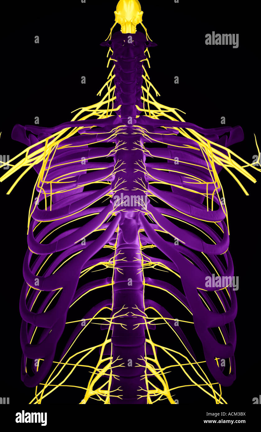 Nerves of the upper body Stock Photohttps://www.alamy.com/image-license-details/?v=1https://www.alamy.com/stock-photo-nerves-of-the-upper-body-13184893.html
RFACM3BX–Nerves of the upper body
 Surgical anatomy : a treatise on human anatomy in its application to the practice of medicine and surgery . es to the subclavius, rhomboidei,scaleni, and longus colli muscles, the posterior or long thoracic nerve (the externalrespiratory nerve of Bell), communicating, and supra-scapular nerves. The nerve to the subclavius muscle arises from the trunk formed by thefifth and sixth cervical nerves, and passes downward over the third portion of thesubclavian artery to the under surface of the subclavius muscle. It is frequentlyconnected with the phrenic nerve at the lower part of the neck by a fil Stock Photohttps://www.alamy.com/image-license-details/?v=1https://www.alamy.com/surgical-anatomy-a-treatise-on-human-anatomy-in-its-application-to-the-practice-of-medicine-and-surgery-es-to-the-subclavius-rhomboideiscaleni-and-longus-colli-muscles-the-posterior-or-long-thoracic-nerve-the-externalrespiratory-nerve-of-bell-communicating-and-supra-scapular-nerves-the-nerve-to-the-subclavius-muscle-arises-from-the-trunk-formed-by-thefifth-and-sixth-cervical-nerves-and-passes-downward-over-the-third-portion-of-thesubclavian-artery-to-the-under-surface-of-the-subclavius-muscle-it-is-frequentlyconnected-with-the-phrenic-nerve-at-the-lower-part-of-the-neck-by-a-fil-image339023782.html
RM2AKFT86–Surgical anatomy : a treatise on human anatomy in its application to the practice of medicine and surgery . es to the subclavius, rhomboidei,scaleni, and longus colli muscles, the posterior or long thoracic nerve (the externalrespiratory nerve of Bell), communicating, and supra-scapular nerves. The nerve to the subclavius muscle arises from the trunk formed by thefifth and sixth cervical nerves, and passes downward over the third portion of thesubclavian artery to the under surface of the subclavius muscle. It is frequentlyconnected with the phrenic nerve at the lower part of the neck by a fil
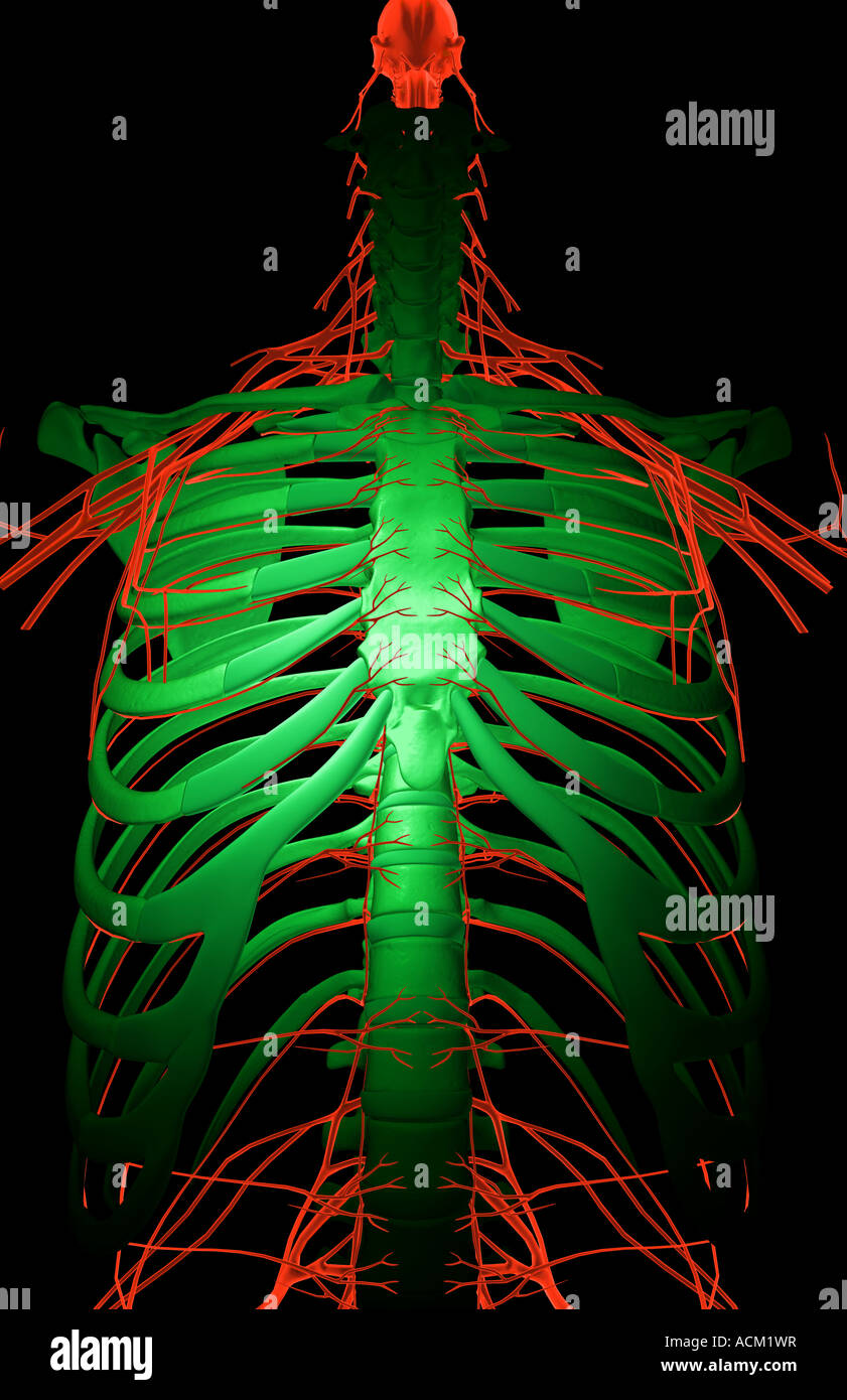 Nerves of the upper body Stock Photohttps://www.alamy.com/image-license-details/?v=1https://www.alamy.com/stock-photo-nerves-of-the-upper-body-13184386.html
RFACM1WR–Nerves of the upper body
 . A treatise on the nervous diseases of children, for physicians and students. ( Region of Central( Convolutions Speech AreaTemporal Facial Trunk of Facial Nerve ( Posterior Auricular I Nerve Facial Lower BranchSplenius Sternocleido MastoidSpinal Accessory NerveLevator Anguli Scapulae TrapeziusScapular Branch Circumflex NervePosterior ThoracicNerve Phrenic Nerve Erbs Point Brachial Plexus Fig. 7.—Motor Points of Face. (Erb.). FlG. 8.—Patient with Hypertrophy of Infra and Supra Spinati, Showing Rotation ofRight Scapula and Deep Groove along Inner Margin of Scapula (Atrophy ofRhomboids and Sligh Stock Photohttps://www.alamy.com/image-license-details/?v=1https://www.alamy.com/a-treatise-on-the-nervous-diseases-of-children-for-physicians-and-students-region-of-central-convolutions-speech-areatemporal-facial-trunk-of-facial-nerve-posterior-auricular-i-nerve-facial-lower-branchsplenius-sternocleido-mastoidspinal-accessory-nervelevator-anguli-scapulae-trapeziusscapular-branch-circumflex-nerveposterior-thoracicnerve-phrenic-nerve-erbs-point-brachial-plexus-fig-7motor-points-of-face-erb-flg-8patient-with-hypertrophy-of-infra-and-supra-spinati-showing-rotation-ofright-scapula-and-deep-groove-along-inner-margin-of-scapula-atrophy-ofrhomboids-and-sligh-image336825426.html
RM2AFYM7E–. A treatise on the nervous diseases of children, for physicians and students. ( Region of Central( Convolutions Speech AreaTemporal Facial Trunk of Facial Nerve ( Posterior Auricular I Nerve Facial Lower BranchSplenius Sternocleido MastoidSpinal Accessory NerveLevator Anguli Scapulae TrapeziusScapular Branch Circumflex NervePosterior ThoracicNerve Phrenic Nerve Erbs Point Brachial Plexus Fig. 7.—Motor Points of Face. (Erb.). FlG. 8.—Patient with Hypertrophy of Infra and Supra Spinati, Showing Rotation ofRight Scapula and Deep Groove along Inner Margin of Scapula (Atrophy ofRhomboids and Sligh
 Nerves of the upper body Stock Photohttps://www.alamy.com/image-license-details/?v=1https://www.alamy.com/stock-photo-nerves-of-the-upper-body-13261613.html
RFAD07NJ–Nerves of the upper body
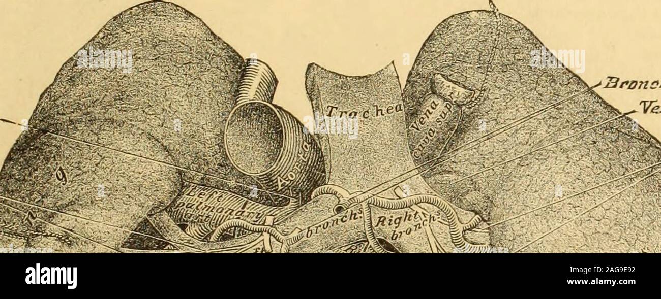 . A Reference handbook of the medical sciences : embracing the entire range of scientific and practical medicine and allied science. luded the su-perior vena cava. In front and externally the superior vena cava is cov-ered by the pleura. The right phrenic nerve lies uponits outer side, and at about one inch and a half from itstermination it enters the pericardium, from which it de-rives a serous covering in front and at the sides, but be-hind it has no covering where it is in contact with theright pulmonary artery and the upper right pulmonaryvein. The relation by its inner side with the ascen Stock Photohttps://www.alamy.com/image-license-details/?v=1https://www.alamy.com/a-reference-handbook-of-the-medical-sciences-embracing-the-entire-range-of-scientific-and-practical-medicine-and-allied-science-luded-the-su-perior-vena-cava-in-front-and-externally-the-superior-vena-cava-is-cov-ered-by-the-pleura-the-right-phrenic-nerve-lies-uponits-outer-side-and-at-about-one-inch-and-a-half-from-itstermination-it-enters-the-pericardium-from-which-it-de-rives-a-serous-covering-in-front-and-at-the-sides-but-be-hind-it-has-no-covering-where-it-is-in-contact-with-theright-pulmonary-artery-and-the-upper-right-pulmonaryvein-the-relation-by-its-inner-side-with-the-ascen-image337040286.html
RM2AG9E92–. A Reference handbook of the medical sciences : embracing the entire range of scientific and practical medicine and allied science. luded the su-perior vena cava. In front and externally the superior vena cava is cov-ered by the pleura. The right phrenic nerve lies uponits outer side, and at about one inch and a half from itstermination it enters the pericardium, from which it de-rives a serous covering in front and at the sides, but be-hind it has no covering where it is in contact with theright pulmonary artery and the upper right pulmonaryvein. The relation by its inner side with the ascen
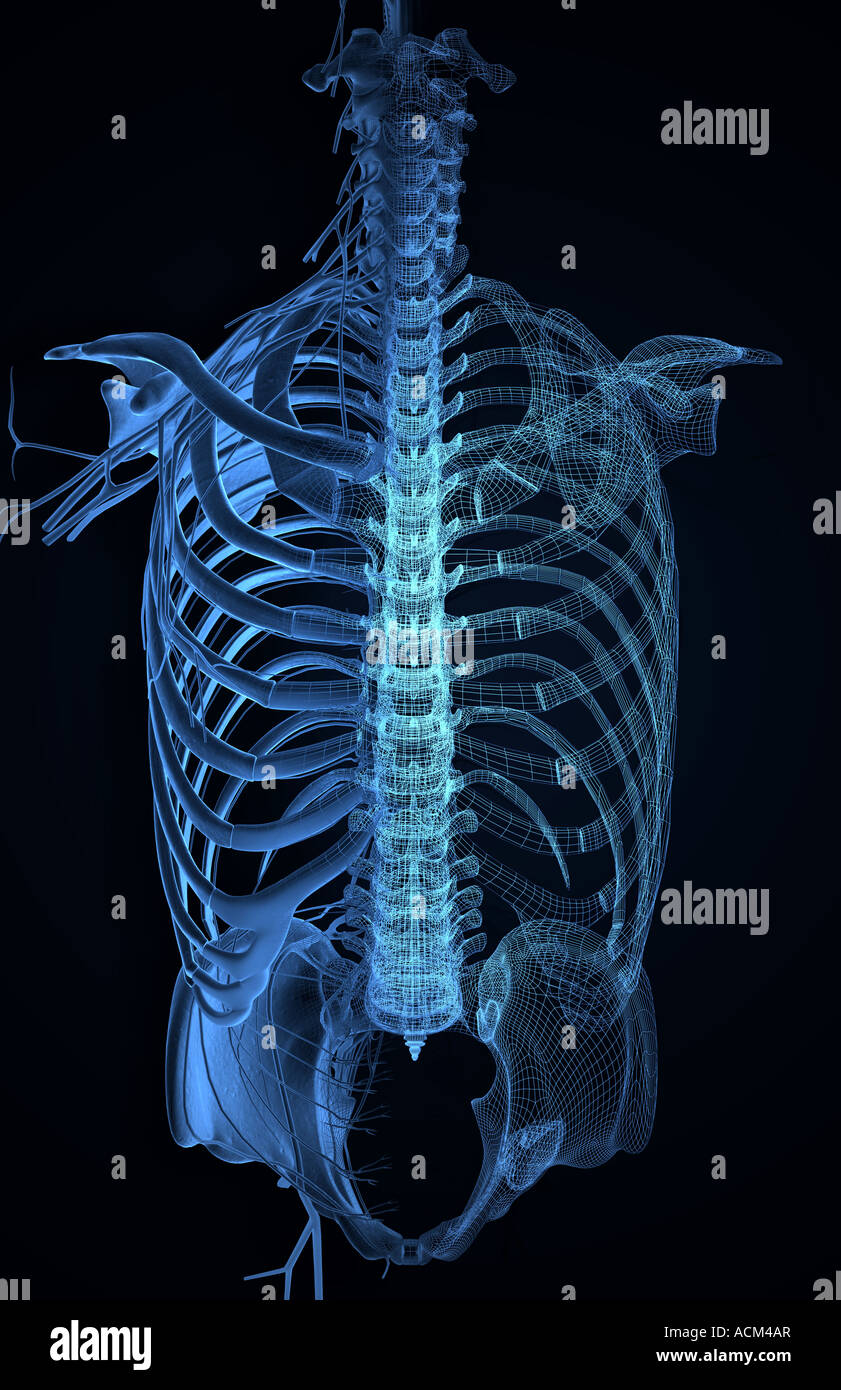 Nerves of the upper body Stock Photohttps://www.alamy.com/image-license-details/?v=1https://www.alamy.com/stock-photo-nerves-of-the-upper-body-13185214.html
RFACM4AR–Nerves of the upper body
 Diseases of the chest and the principles of physical diagnosis . tral diaphragmatic pleura is supi)lied by the phrenic nerve. Irrita-tion of this region produces pain and tenderness in the neck, especiallyalong the ridge of the trapezius muscle, often with a surrounding zoneof hyperesthesia (Fig. 41). This is a true referred pain—by way of thethird and fourth spinal segments. The pericardial pleura when irritatedmay produce similar pain, because its nerve supply is mainly if not en-tirely phrenic in origin (Capps). Pleural pain may be referred to the abdominal wall and lack of acareful examina Stock Photohttps://www.alamy.com/image-license-details/?v=1https://www.alamy.com/diseases-of-the-chest-and-the-principles-of-physical-diagnosis-tral-diaphragmatic-pleura-is-supilied-by-the-phrenic-nerve-irrita-tion-of-this-region-produces-pain-and-tenderness-in-the-neck-especiallyalong-the-ridge-of-the-trapezius-muscle-often-with-a-surrounding-zoneof-hyperesthesia-fig-41-this-is-a-true-referred-painby-way-of-thethird-and-fourth-spinal-segments-the-pericardial-pleura-when-irritatedmay-produce-similar-pain-because-its-nerve-supply-is-mainly-if-not-en-tirely-phrenic-in-origin-capps-pleural-pain-may-be-referred-to-the-abdominal-wall-and-lack-of-acareful-examina-image342668472.html
RM2AWDW3M–Diseases of the chest and the principles of physical diagnosis . tral diaphragmatic pleura is supi)lied by the phrenic nerve. Irrita-tion of this region produces pain and tenderness in the neck, especiallyalong the ridge of the trapezius muscle, often with a surrounding zoneof hyperesthesia (Fig. 41). This is a true referred pain—by way of thethird and fourth spinal segments. The pericardial pleura when irritatedmay produce similar pain, because its nerve supply is mainly if not en-tirely phrenic in origin (Capps). Pleural pain may be referred to the abdominal wall and lack of acareful examina
 Nerves of the upper body Stock Photohttps://www.alamy.com/image-license-details/?v=1https://www.alamy.com/stock-photo-nerves-of-the-upper-body-13184557.html
RFACM2BX–Nerves of the upper body
 Archives of internal medicine . persistence of this rela-tively high positive pressure even when the animal was under deep narcosis. Thispressure was evidently harmful, as artificial respiration had to be resorted tobecause of the limited respiratory excursion permitted. Experiment 9.—Jan. 5, 1905: Large male rabbit. Ether narcosis, tracheotomy.Right phrenic nerve exposed in the neck, and esophagus cut and tube passed tostomach and tied in place. Abdominal and pleural cannulas. Dorsal position.The usual record of low positive or negative abdominal pressure was obtainedduring deep narcosis and Stock Photohttps://www.alamy.com/image-license-details/?v=1https://www.alamy.com/archives-of-internal-medicine-persistence-of-this-rela-tively-high-positive-pressure-even-when-the-animal-was-under-deep-narcosis-thispressure-was-evidently-harmful-as-artificial-respiration-had-to-be-resorted-tobecause-of-the-limited-respiratory-excursion-permitted-experiment-9jan-5-1905-large-male-rabbit-ether-narcosis-tracheotomyright-phrenic-nerve-exposed-in-the-neck-and-esophagus-cut-and-tube-passed-tostomach-and-tied-in-place-abdominal-and-pleural-cannulas-dorsal-positionthe-usual-record-of-low-positive-or-negative-abdominal-pressure-was-obtainedduring-deep-narcosis-and-image340134738.html
RM2ANAD96–Archives of internal medicine . persistence of this rela-tively high positive pressure even when the animal was under deep narcosis. Thispressure was evidently harmful, as artificial respiration had to be resorted tobecause of the limited respiratory excursion permitted. Experiment 9.—Jan. 5, 1905: Large male rabbit. Ether narcosis, tracheotomy.Right phrenic nerve exposed in the neck, and esophagus cut and tube passed tostomach and tied in place. Abdominal and pleural cannulas. Dorsal position.The usual record of low positive or negative abdominal pressure was obtainedduring deep narcosis and
 Osteopathy Complete . TREATMENT. 1. See Acute Pleurisy (page 60). 2. Standing behind the patient, place the fingers uponthe transverse processes of the third, fourth, and fifth cervi-j^al vertebrae; press the muscles forward and slip the fingersdown in front of the transverse processes, where a pressurecan be exerted upon the phrenic nerve, near its origin (cut12). This nerve controls the diaphragm, and a pressure atthis point breaks the nerve-wave to this muscle, and conse-quently slows its action, as is fully explained under the headof Hiccoughs. The phrenic nerve should be held abouttwo min Stock Photohttps://www.alamy.com/image-license-details/?v=1https://www.alamy.com/osteopathy-complete-treatment-1-see-acute-pleurisy-page-60-2-standing-behind-the-patient-place-the-fingers-uponthe-transverse-processes-of-the-third-fourth-and-fifth-cervi-jal-vertebrae-press-the-muscles-forward-and-slip-the-fingersdown-in-front-of-the-transverse-processes-where-a-pressurecan-be-exerted-upon-the-phrenic-nerve-near-its-origin-cut12-this-nerve-controls-the-diaphragm-and-a-pressure-atthis-point-breaks-the-nerve-wave-to-this-muscle-and-conse-quently-slows-its-action-as-is-fully-explained-under-the-headof-hiccoughs-the-phrenic-nerve-should-be-held-abouttwo-min-image342676250.html
RM2AWE71E–Osteopathy Complete . TREATMENT. 1. See Acute Pleurisy (page 60). 2. Standing behind the patient, place the fingers uponthe transverse processes of the third, fourth, and fifth cervi-j^al vertebrae; press the muscles forward and slip the fingersdown in front of the transverse processes, where a pressurecan be exerted upon the phrenic nerve, near its origin (cut12). This nerve controls the diaphragm, and a pressure atthis point breaks the nerve-wave to this muscle, and conse-quently slows its action, as is fully explained under the headof Hiccoughs. The phrenic nerve should be held abouttwo min
 Physical diagnosis, including diseases of the thoracic and abdominal organs : a manual for students and physicians .. . Bronchi (child) filled with shot. No. 10. Diameter, .03S inch. Lateral view. Above it is the vena azygons major, arching forward tojoin the superior vena cava, and in front of the bronchusis the right pulmonary artery, with the superior venacava and the right phrenic nerve. (Fig. 7.) The left primary bronchus is given off at the bifnrca- 26 ANATOMICAL. tion of the trachea (Fig. 3). It immediately passesunder the arch of the aorta and goes obliquely down-wards and to the left Stock Photohttps://www.alamy.com/image-license-details/?v=1https://www.alamy.com/physical-diagnosis-including-diseases-of-the-thoracic-and-abdominal-organs-a-manual-for-students-and-physicians-bronchi-child-filled-with-shot-no-10-diameter-03s-inch-lateral-view-above-it-is-the-vena-azygons-major-arching-forward-tojoin-the-superior-vena-cava-and-in-front-of-the-bronchusis-the-right-pulmonary-artery-with-the-superior-venacava-and-the-right-phrenic-nerve-fig-7-the-left-primary-bronchus-is-given-off-at-the-bifnrca-26-anatomical-tion-of-the-trachea-fig-3-it-immediately-passesunder-the-arch-of-the-aorta-and-goes-obliquely-down-wards-and-to-the-left-image342856632.html
RM2AWPD3M–Physical diagnosis, including diseases of the thoracic and abdominal organs : a manual for students and physicians .. . Bronchi (child) filled with shot. No. 10. Diameter, .03S inch. Lateral view. Above it is the vena azygons major, arching forward tojoin the superior vena cava, and in front of the bronchusis the right pulmonary artery, with the superior venacava and the right phrenic nerve. (Fig. 7.) The left primary bronchus is given off at the bifnrca- 26 ANATOMICAL. tion of the trachea (Fig. 3). It immediately passesunder the arch of the aorta and goes obliquely down-wards and to the left
 . A treatise on the nervous diseases of children : for physicians and students. ( Region of Central( Convolutions Speech AreaTemporal Facial Trunk of Facial Nerve ( Posterior Auricular ( Nerve Facial Lower BranchSplenius Sternocleido MastoidSpinal Accessory NerveLevator Anguli Scapulae TrapeziusScapular Branch Circumflex NervePosterior Thoracic Nerve Phrenic Nerve Erbs Point Brachial Plexus Fig. 7.—Motor Points of Face. (Erb.). Fig. 8.—Patient with Hypertrophy of Infra and Supra Spinati, Showing Rotation ofRight Scapula and Deep Groove along Inner Margin of Scapula (Atrophy ofRhomboids and Shg Stock Photohttps://www.alamy.com/image-license-details/?v=1https://www.alamy.com/a-treatise-on-the-nervous-diseases-of-children-for-physicians-and-students-region-of-central-convolutions-speech-areatemporal-facial-trunk-of-facial-nerve-posterior-auricular-nerve-facial-lower-branchsplenius-sternocleido-mastoidspinal-accessory-nervelevator-anguli-scapulae-trapeziusscapular-branch-circumflex-nerveposterior-thoracic-nerve-phrenic-nerve-erbs-point-brachial-plexus-fig-7motor-points-of-face-erb-fig-8patient-with-hypertrophy-of-infra-and-supra-spinati-showing-rotation-ofright-scapula-and-deep-groove-along-inner-margin-of-scapula-atrophy-ofrhomboids-and-shg-image337080665.html
RM2AGB9R5–. A treatise on the nervous diseases of children : for physicians and students. ( Region of Central( Convolutions Speech AreaTemporal Facial Trunk of Facial Nerve ( Posterior Auricular ( Nerve Facial Lower BranchSplenius Sternocleido MastoidSpinal Accessory NerveLevator Anguli Scapulae TrapeziusScapular Branch Circumflex NervePosterior Thoracic Nerve Phrenic Nerve Erbs Point Brachial Plexus Fig. 7.—Motor Points of Face. (Erb.). Fig. 8.—Patient with Hypertrophy of Infra and Supra Spinati, Showing Rotation ofRight Scapula and Deep Groove along Inner Margin of Scapula (Atrophy ofRhomboids and Shg
 Osteopathic first aids to the sick : written for the sick people . theother ribs, fig. 47; spread the ribs, fig. 28; in-hibit the phrenic nerve, fig. 14; and knead theabdomen, fig. 53. Treat twice each day. Eat nothing the first day and very little at atime afterward. Drink as little as possible. Seethat bowels and kidneys are active. Have thefeet and legs warm, and avoid all possibility oftaking cold. Steamy atmosphere from boilingwater to which some carbolic acid has been add-ed is soothing . MUMPS. Parotitis. Symptoms. For several days there may beheadache, restlessness, loss of appetite, p Stock Photohttps://www.alamy.com/image-license-details/?v=1https://www.alamy.com/osteopathic-first-aids-to-the-sick-written-for-the-sick-people-theother-ribs-fig-47-spread-the-ribs-fig-28-in-hibit-the-phrenic-nerve-fig-14-and-knead-theabdomen-fig-53-treat-twice-each-day-eat-nothing-the-first-day-and-very-little-at-atime-afterward-drink-as-little-as-possible-seethat-bowels-and-kidneys-are-active-have-thefeet-and-legs-warm-and-avoid-all-possibility-oftaking-cold-steamy-atmosphere-from-boilingwater-to-which-some-carbolic-acid-has-been-add-ed-is-soothing-mumps-parotitis-symptoms-for-several-days-there-may-beheadache-restlessness-loss-of-appetite-p-image338303106.html
RM2AJB11P–Osteopathic first aids to the sick : written for the sick people . theother ribs, fig. 47; spread the ribs, fig. 28; in-hibit the phrenic nerve, fig. 14; and knead theabdomen, fig. 53. Treat twice each day. Eat nothing the first day and very little at atime afterward. Drink as little as possible. Seethat bowels and kidneys are active. Have thefeet and legs warm, and avoid all possibility oftaking cold. Steamy atmosphere from boilingwater to which some carbolic acid has been add-ed is soothing . MUMPS. Parotitis. Symptoms. For several days there may beheadache, restlessness, loss of appetite, p
 Diseases of the nervous system : a text-book of neurology and psychiatry . Fig. 35.—Scheme of innervationof breathing: D, diaphragm; nf,phrenic nerve; X, sensory vagusbranches to the lungs; nr, respira-tory nucleus in medulla; nXs, sen-sory nucleus of the vagus; nrs,respiratory center in midbrainregion. (Bechterew.) VASCULAR APPARATUS 99 of the third ventricle is thought to be a higher coordinating switch-board—the nucleus dorsalis vagi, an end station. Through this portionof the mechanism, psychical influences are switched in, modifying thetonus through emotions, pain, and local stimuli.. Fig Stock Photohttps://www.alamy.com/image-license-details/?v=1https://www.alamy.com/diseases-of-the-nervous-system-a-text-book-of-neurology-and-psychiatry-fig-35scheme-of-innervationof-breathing-d-diaphragm-nfphrenic-nerve-x-sensory-vagusbranches-to-the-lungs-nr-respira-tory-nucleus-in-medulla-nxs-sen-sory-nucleus-of-the-vagus-nrsrespiratory-center-in-midbrainregion-bechterew-vascular-apparatus-99-of-the-third-ventricle-is-thought-to-be-a-higher-coordinating-switch-boardthe-nucleus-dorsalis-vagi-an-end-station-through-this-portionof-the-mechanism-psychical-influences-are-switched-in-modifying-thetonus-through-emotions-pain-and-local-stimuli-fig-image339065165.html
RM2AKHN25–Diseases of the nervous system : a text-book of neurology and psychiatry . Fig. 35.—Scheme of innervationof breathing: D, diaphragm; nf,phrenic nerve; X, sensory vagusbranches to the lungs; nr, respira-tory nucleus in medulla; nXs, sen-sory nucleus of the vagus; nrs,respiratory center in midbrainregion. (Bechterew.) VASCULAR APPARATUS 99 of the third ventricle is thought to be a higher coordinating switch-board—the nucleus dorsalis vagi, an end station. Through this portionof the mechanism, psychical influences are switched in, modifying thetonus through emotions, pain, and local stimuli.. Fig
![A treatise on the nervous diseases of children, for physicians and students . I Region of Central( Convolutions Speech Area Temporal Trunk of Facial Nerve< Posterior Auricular] Nerve Facial Lower BranchSplenius Stemocteido MastoidSpinal Accessory NerveLevator Anguli Scapulae TrapeziusScapular Branch Circumflex Nerve FosteriorThoracic Nerve Phrenic Nerve Erbs Point Brachial Plexus Fig. 7.—Motor Points of Face. {Erb.). Fig. 8.—Patient with Hypertrophy of Infra and Supra Spinati, Showing Rotation ofRight Scapula and Deep Groove along Inner Margin of Scapula (Atrophy ofRhomboids and Slight Atro Stock Photo A treatise on the nervous diseases of children, for physicians and students . I Region of Central( Convolutions Speech Area Temporal Trunk of Facial Nerve< Posterior Auricular] Nerve Facial Lower BranchSplenius Stemocteido MastoidSpinal Accessory NerveLevator Anguli Scapulae TrapeziusScapular Branch Circumflex Nerve FosteriorThoracic Nerve Phrenic Nerve Erbs Point Brachial Plexus Fig. 7.—Motor Points of Face. {Erb.). Fig. 8.—Patient with Hypertrophy of Infra and Supra Spinati, Showing Rotation ofRight Scapula and Deep Groove along Inner Margin of Scapula (Atrophy ofRhomboids and Slight Atro Stock Photo](https://c8.alamy.com/comp/2AXBMPY/a-treatise-on-the-nervous-diseases-of-children-for-physicians-and-students-i-region-of-central-convolutions-speech-area-temporal-trunk-of-facial-nervelt-posterior-auricular-nerve-facial-lower-branchsplenius-stemocteido-mastoidspinal-accessory-nervelevator-anguli-scapulae-trapeziusscapular-branch-circumflex-nerve-fosteriorthoracic-nerve-phrenic-nerve-erbs-point-brachial-plexus-fig-7motor-points-of-face-erb-fig-8patient-with-hypertrophy-of-infra-and-supra-spinati-showing-rotation-ofright-scapula-and-deep-groove-along-inner-margin-of-scapula-atrophy-ofrhomboids-and-slight-atro-2AXBMPY.jpg) A treatise on the nervous diseases of children, for physicians and students . I Region of Central( Convolutions Speech Area Temporal Trunk of Facial Nerve< Posterior Auricular] Nerve Facial Lower BranchSplenius Stemocteido MastoidSpinal Accessory NerveLevator Anguli Scapulae TrapeziusScapular Branch Circumflex Nerve FosteriorThoracic Nerve Phrenic Nerve Erbs Point Brachial Plexus Fig. 7.—Motor Points of Face. {Erb.). Fig. 8.—Patient with Hypertrophy of Infra and Supra Spinati, Showing Rotation ofRight Scapula and Deep Groove along Inner Margin of Scapula (Atrophy ofRhomboids and Slight Atro Stock Photohttps://www.alamy.com/image-license-details/?v=1https://www.alamy.com/a-treatise-on-the-nervous-diseases-of-children-for-physicians-and-students-i-region-of-central-convolutions-speech-area-temporal-trunk-of-facial-nervelt-posterior-auricular-nerve-facial-lower-branchsplenius-stemocteido-mastoidspinal-accessory-nervelevator-anguli-scapulae-trapeziusscapular-branch-circumflex-nerve-fosteriorthoracic-nerve-phrenic-nerve-erbs-point-brachial-plexus-fig-7motor-points-of-face-erb-fig-8patient-with-hypertrophy-of-infra-and-supra-spinati-showing-rotation-ofright-scapula-and-deep-groove-along-inner-margin-of-scapula-atrophy-ofrhomboids-and-slight-atro-image343235843.html
A treatise on the nervous diseases of children, for physicians and students . I Region of Central( Convolutions Speech Area Temporal Trunk of Facial Nerve< Posterior Auricular] Nerve Facial Lower BranchSplenius Stemocteido MastoidSpinal Accessory NerveLevator Anguli Scapulae TrapeziusScapular Branch Circumflex Nerve FosteriorThoracic Nerve Phrenic Nerve Erbs Point Brachial Plexus Fig. 7.—Motor Points of Face. {Erb.). Fig. 8.—Patient with Hypertrophy of Infra and Supra Spinati, Showing Rotation ofRight Scapula and Deep Groove along Inner Margin of Scapula (Atrophy ofRhomboids and Slight Atro Stock Photohttps://www.alamy.com/image-license-details/?v=1https://www.alamy.com/a-treatise-on-the-nervous-diseases-of-children-for-physicians-and-students-i-region-of-central-convolutions-speech-area-temporal-trunk-of-facial-nervelt-posterior-auricular-nerve-facial-lower-branchsplenius-stemocteido-mastoidspinal-accessory-nervelevator-anguli-scapulae-trapeziusscapular-branch-circumflex-nerve-fosteriorthoracic-nerve-phrenic-nerve-erbs-point-brachial-plexus-fig-7motor-points-of-face-erb-fig-8patient-with-hypertrophy-of-infra-and-supra-spinati-showing-rotation-ofright-scapula-and-deep-groove-along-inner-margin-of-scapula-atrophy-ofrhomboids-and-slight-atro-image343235843.htmlRM2AXBMPY–A treatise on the nervous diseases of children, for physicians and students . I Region of Central( Convolutions Speech Area Temporal Trunk of Facial Nerve< Posterior Auricular] Nerve Facial Lower BranchSplenius Stemocteido MastoidSpinal Accessory NerveLevator Anguli Scapulae TrapeziusScapular Branch Circumflex Nerve FosteriorThoracic Nerve Phrenic Nerve Erbs Point Brachial Plexus Fig. 7.—Motor Points of Face. {Erb.). Fig. 8.—Patient with Hypertrophy of Infra and Supra Spinati, Showing Rotation ofRight Scapula and Deep Groove along Inner Margin of Scapula (Atrophy ofRhomboids and Slight Atro
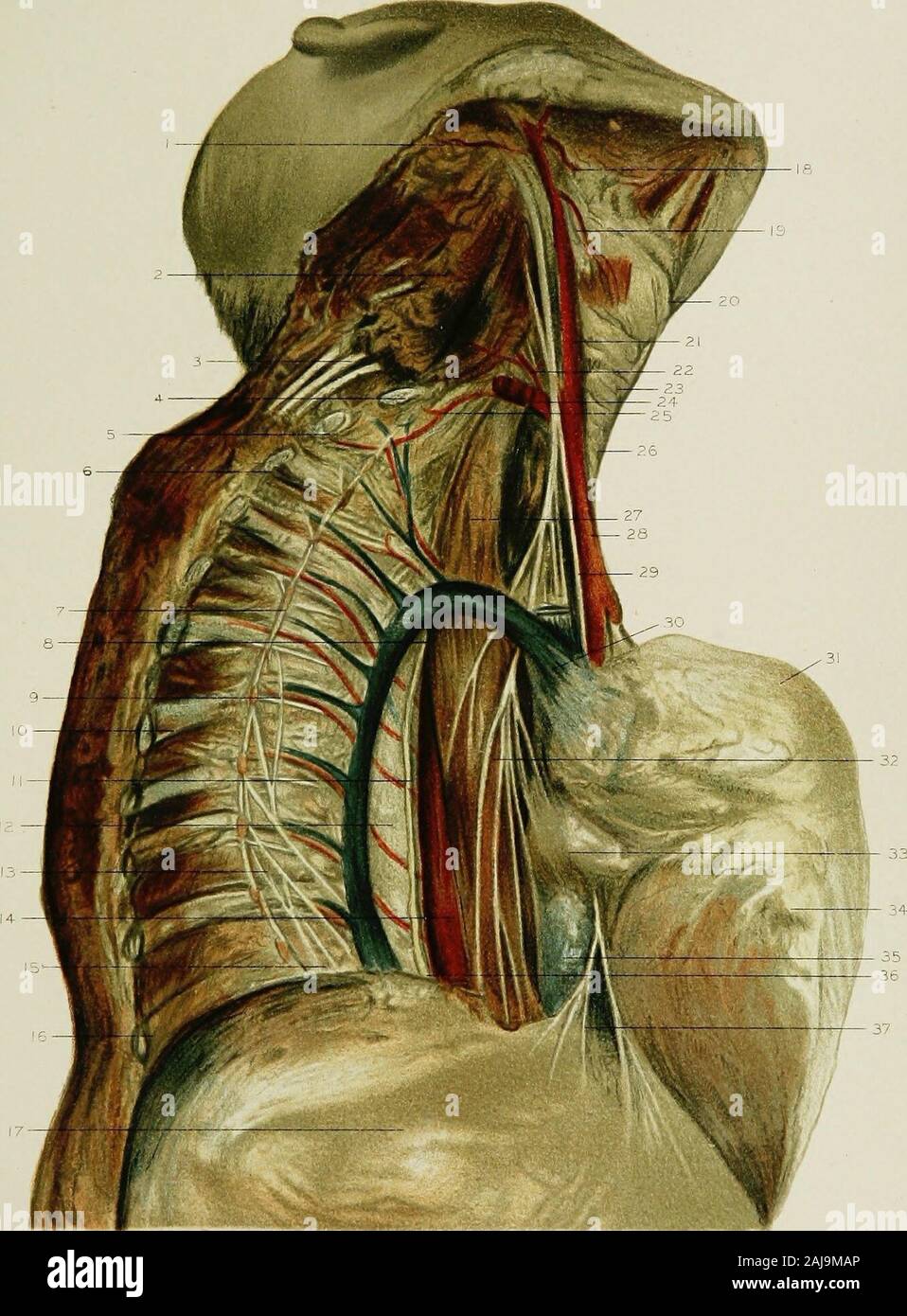 Regional anatomy in its relation to medicine and surgery . g thoracic aorta. 15. The lesser splanchnic nerve. 16. Section of the eleventh rib. 17. The hepatic arch of the diaphragm. 18. The lingual artery, 19. The descending thyroid arterj, and superior laryngeal 20. The notch in the thyroid cartUage. 21. The common carotid artery. 22. The thyroid axis. 23. The cricoid cartilage. 24. The right subclavian artery. 25. The first intercostal artery. 26. The trachea. 27. The cesophagus. 28. The innominate artery. 29. The right phrenic nerve. 30. The vena azygos major vein, entering into the superio Stock Photohttps://www.alamy.com/image-license-details/?v=1https://www.alamy.com/regional-anatomy-in-its-relation-to-medicine-and-surgery-g-thoracic-aorta-15-the-lesser-splanchnic-nerve-16-section-of-the-eleventh-rib-17-the-hepatic-arch-of-the-diaphragm-18-the-lingual-artery-19-the-descending-thyroid-arterj-and-superior-laryngeal-20-the-notch-in-the-thyroid-cartuage-21-the-common-carotid-artery-22-the-thyroid-axis-23-the-cricoid-cartilage-24-the-right-subclavian-artery-25-the-first-intercostal-artery-26-the-trachea-27-the-cesophagus-28-the-innominate-artery-29-the-right-phrenic-nerve-30-the-vena-azygos-major-vein-entering-into-the-superio-image338274350.html
RM2AJ9MAP–Regional anatomy in its relation to medicine and surgery . g thoracic aorta. 15. The lesser splanchnic nerve. 16. Section of the eleventh rib. 17. The hepatic arch of the diaphragm. 18. The lingual artery, 19. The descending thyroid arterj, and superior laryngeal 20. The notch in the thyroid cartUage. 21. The common carotid artery. 22. The thyroid axis. 23. The cricoid cartilage. 24. The right subclavian artery. 25. The first intercostal artery. 26. The trachea. 27. The cesophagus. 28. The innominate artery. 29. The right phrenic nerve. 30. The vena azygos major vein, entering into the superio
 Regional anatomy in its relation to medicine and surgery . ightventricle of the heart within the pericardium. 25. The left superior thyroid artery. 26. The left thyro-hyoid muscle. 27. The crico-thyroid membrane. 28. The cricoid cartilage. 29. The left internal jugular vein. 30. The left common carotid artery. 31. The left pneumogastric nerve. S2. The left brachial plexus of nerves. 33. The left recurrent laryngeal nerve. 34. The left scalenus anticus muscle. 35. The trachea. 36. The left subclavian artery. 37. The left subclavian vein. 38. The left phrenic nerve. 39. The left innominate vein. Stock Photohttps://www.alamy.com/image-license-details/?v=1https://www.alamy.com/regional-anatomy-in-its-relation-to-medicine-and-surgery-ightventricle-of-the-heart-within-the-pericardium-25-the-left-superior-thyroid-artery-26-the-left-thyro-hyoid-muscle-27-the-crico-thyroid-membrane-28-the-cricoid-cartilage-29-the-left-internal-jugular-vein-30-the-left-common-carotid-artery-31-the-left-pneumogastric-nerve-s2-the-left-brachial-plexus-of-nerves-33-the-left-recurrent-laryngeal-nerve-34-the-left-scalenus-anticus-muscle-35-the-trachea-36-the-left-subclavian-artery-37-the-left-subclavian-vein-38-the-left-phrenic-nerve-39-the-left-innominate-vein-image338276904.html
RM2AJ9RJ0–Regional anatomy in its relation to medicine and surgery . ightventricle of the heart within the pericardium. 25. The left superior thyroid artery. 26. The left thyro-hyoid muscle. 27. The crico-thyroid membrane. 28. The cricoid cartilage. 29. The left internal jugular vein. 30. The left common carotid artery. 31. The left pneumogastric nerve. S2. The left brachial plexus of nerves. 33. The left recurrent laryngeal nerve. 34. The left scalenus anticus muscle. 35. The trachea. 36. The left subclavian artery. 37. The left subclavian vein. 38. The left phrenic nerve. 39. The left innominate vein.
 A text-book of clinical anatomy : for students and practitioners . is normal, due to the mode of prepa-ration (formalin). The black spaces indicating the pleural and the pericardial cavitiesdo not exist during life, as was explained in Fig. 46. LL, Left lower lobe. RL, Rightlower lobe. P, Pleural cavity; under normal conditions, this does not exist during life.RV, Right ventricle. LV, Left ventricle. LA, Left auricle. Between the two is seenone of the mitral valves. RA, Right auricle. 1, Phrenic nerve, placed in connectivetissue between mediastinal pleura and parietal pericardium. 2, Descendin Stock Photohttps://www.alamy.com/image-license-details/?v=1https://www.alamy.com/a-text-book-of-clinical-anatomy-for-students-and-practitioners-is-normal-due-to-the-mode-of-prepa-ration-formalin-the-black-spaces-indicating-the-pleural-and-the-pericardial-cavitiesdo-not-exist-during-life-as-was-explained-in-fig-46-ll-left-lower-lobe-rl-rightlower-lobe-p-pleural-cavity-under-normal-conditions-this-does-not-exist-during-liferv-right-ventricle-lv-left-ventricle-la-left-auricle-between-the-two-is-seenone-of-the-mitral-valves-ra-right-auricle-1-phrenic-nerve-placed-in-connectivetissue-between-mediastinal-pleura-and-parietal-pericardium-2-descendin-image340224966.html
RM2ANEGBJ–A text-book of clinical anatomy : for students and practitioners . is normal, due to the mode of prepa-ration (formalin). The black spaces indicating the pleural and the pericardial cavitiesdo not exist during life, as was explained in Fig. 46. LL, Left lower lobe. RL, Rightlower lobe. P, Pleural cavity; under normal conditions, this does not exist during life.RV, Right ventricle. LV, Left ventricle. LA, Left auricle. Between the two is seenone of the mitral valves. RA, Right auricle. 1, Phrenic nerve, placed in connectivetissue between mediastinal pleura and parietal pericardium. 2, Descendin
 Osteopathic first aids to the sick : written for the sick people . If there is heart trouble, it must be attendedto. Give a general spinal treatment, fig. 37; givethe treatment for the legs, fig. 77; spread kneesagainst resistance, fig. 71 ; knead the musclesof the entire leg, fig. 70; treat the ankles, fig. 79;and free the blood vessels to the legs, fig. 7S. Practice deep breathing. Walk rapidly at ev-ery opportunity. Bathe feet in warm water be-fore retiring. HICCOUGH. Is caused by an irritation of the phrenic nerve. Spread the ribs, fig. 28 and inhibit the phrenicnerve, fig. 14. Repeat seve Stock Photohttps://www.alamy.com/image-license-details/?v=1https://www.alamy.com/osteopathic-first-aids-to-the-sick-written-for-the-sick-people-if-there-is-heart-trouble-it-must-be-attendedto-give-a-general-spinal-treatment-fig-37-givethe-treatment-for-the-legs-fig-77-spread-kneesagainst-resistance-fig-71-knead-the-musclesof-the-entire-leg-fig-70-treat-the-ankles-fig-79and-free-the-blood-vessels-to-the-legs-fig-7s-practice-deep-breathing-walk-rapidly-at-ev-ery-opportunity-bathe-feet-in-warm-water-be-fore-retiring-hiccough-is-caused-by-an-irritation-of-the-phrenic-nerve-spread-the-ribs-fig-28-and-inhibit-the-phrenicnerve-fig-14-repeat-seve-image338276151.html
RM2AJ9PK3–Osteopathic first aids to the sick : written for the sick people . If there is heart trouble, it must be attendedto. Give a general spinal treatment, fig. 37; givethe treatment for the legs, fig. 77; spread kneesagainst resistance, fig. 71 ; knead the musclesof the entire leg, fig. 70; treat the ankles, fig. 79;and free the blood vessels to the legs, fig. 7S. Practice deep breathing. Walk rapidly at ev-ery opportunity. Bathe feet in warm water be-fore retiring. HICCOUGH. Is caused by an irritation of the phrenic nerve. Spread the ribs, fig. 28 and inhibit the phrenicnerve, fig. 14. Repeat seve
 Regional anatomy in its relation to medicine and surgery . the right fourth rib. 9. The right phrenic nerve. 10. The middle lobe of the right lung. 11. Section through the right fifth rib. 12. The oesophagus. 13. The vena azygos major. 14. The inferior lobe of the right lung. 15. Section through the right sixth rib. 16. Section through the right seventh rib. 17. The right eighth rib. 18. Spine of the eighth thoracic vertebra. 19. The left internal mammary vessels. 20. The anterior margin of the superior lobe of the left lung. 21. The superior lobe of the left lung. 22. The left ventricle of th Stock Photohttps://www.alamy.com/image-license-details/?v=1https://www.alamy.com/regional-anatomy-in-its-relation-to-medicine-and-surgery-the-right-fourth-rib-9-the-right-phrenic-nerve-10-the-middle-lobe-of-the-right-lung-11-section-through-the-right-fifth-rib-12-the-oesophagus-13-the-vena-azygos-major-14-the-inferior-lobe-of-the-right-lung-15-section-through-the-right-sixth-rib-16-section-through-the-right-seventh-rib-17-the-right-eighth-rib-18-spine-of-the-eighth-thoracic-vertebra-19-the-left-internal-mammary-vessels-20-the-anterior-margin-of-the-superior-lobe-of-the-left-lung-21-the-superior-lobe-of-the-left-lung-22-the-left-ventricle-of-th-image338272161.html
RM2AJ9HGH–Regional anatomy in its relation to medicine and surgery . the right fourth rib. 9. The right phrenic nerve. 10. The middle lobe of the right lung. 11. Section through the right fifth rib. 12. The oesophagus. 13. The vena azygos major. 14. The inferior lobe of the right lung. 15. Section through the right sixth rib. 16. Section through the right seventh rib. 17. The right eighth rib. 18. Spine of the eighth thoracic vertebra. 19. The left internal mammary vessels. 20. The anterior margin of the superior lobe of the left lung. 21. The superior lobe of the left lung. 22. The left ventricle of th
 . Annals of surgery. Showing fascia dissected from the lower angle. Internal jugular vein shown !>ing beneath thestemomastoid muscle. The phrenic nerve, lying on the scalenus anticus muscle and branching fromthe cervical plexus, is exposed. Fig. 3.. Shows dissection completed. The spinal accessory nerve passes diagonally across from the upper partof the stemomastoid muscle to the inner surface of J;rapezius. Fig. Stock Photohttps://www.alamy.com/image-license-details/?v=1https://www.alamy.com/annals-of-surgery-showing-fascia-dissected-from-the-lower-angle-internal-jugular-vein-shown-!gting-beneath-thestemomastoid-muscle-the-phrenic-nerve-lying-on-the-scalenus-anticus-muscle-and-branching-fromthe-cervical-plexus-is-exposed-fig-3-shows-dissection-completed-the-spinal-accessory-nerve-passes-diagonally-across-from-the-upper-partof-the-stemomastoid-muscle-to-the-inner-surface-of-jrapezius-fig-image370642739.html
RM2CF06GK–. Annals of surgery. Showing fascia dissected from the lower angle. Internal jugular vein shown !>ing beneath thestemomastoid muscle. The phrenic nerve, lying on the scalenus anticus muscle and branching fromthe cervical plexus, is exposed. Fig. 3.. Shows dissection completed. The spinal accessory nerve passes diagonally across from the upper partof the stemomastoid muscle to the inner surface of J;rapezius. Fig.
 . Röntgen rays and electro-therapeutics : with chapters on radium and phototherapy . so as to excite the bronchial and laryngeal muscles;following this it acts centripetally upon the phrenic nerve, and uponthe great sympathetic. The excitation of the latter is able to modifythe vaso-motor activity of the vessels of the medulla oblongata and therespiratory centres. The results were found to be very favorable inessential asthma. II. Otology. Auditory-Nerve Deafness. This is best treated by the bifurcated electrode and the battery cur-rent, using the cathode to the ears. Gradually vary the curren Stock Photohttps://www.alamy.com/image-license-details/?v=1https://www.alamy.com/rntgen-rays-and-electro-therapeutics-with-chapters-on-radium-and-phototherapy-so-as-to-excite-the-bronchial-and-laryngeal-musclesfollowing-this-it-acts-centripetally-upon-the-phrenic-nerve-and-uponthe-great-sympathetic-the-excitation-of-the-latter-is-able-to-modifythe-vaso-motor-activity-of-the-vessels-of-the-medulla-oblongata-and-therespiratory-centres-the-results-were-found-to-be-very-favorable-inessential-asthma-ii-otology-auditory-nerve-deafness-this-is-best-treated-by-the-bifurcated-electrode-and-the-battery-cur-rent-using-the-cathode-to-the-ears-gradually-vary-the-curren-image376003500.html
RM2CRMC8C–. Röntgen rays and electro-therapeutics : with chapters on radium and phototherapy . so as to excite the bronchial and laryngeal muscles;following this it acts centripetally upon the phrenic nerve, and uponthe great sympathetic. The excitation of the latter is able to modifythe vaso-motor activity of the vessels of the medulla oblongata and therespiratory centres. The results were found to be very favorable inessential asthma. II. Otology. Auditory-Nerve Deafness. This is best treated by the bifurcated electrode and the battery cur-rent, using the cathode to the ears. Gradually vary the curren
 . Medical and surgical therapy . )art of the wound the sternal extremity of the firstrib is seen, upon which lies the subclavian vein and,inmiediately internal to it, the junction of the jugularwith the subclavian vein. Close to this point is thevcitcbral vein, running from above downwards andfrom behind forwards. Immediately outside this vein,and separating it from the subclavian artery, is thescalenus anticus muscle. The anterior surface oftliis nuiscle is crossed oy tue transverse scapular arteryand the phrenic nerve. Immediately internal to the WOUNDS OF VESSELS OF THE NECK 193 muscle the Stock Photohttps://www.alamy.com/image-license-details/?v=1https://www.alamy.com/medical-and-surgical-therapy-art-of-the-wound-the-sternal-extremity-of-the-firstrib-is-seen-upon-which-lies-the-subclavian-vein-andinmiediately-internal-to-it-the-junction-of-the-jugularwith-the-subclavian-vein-close-to-this-point-is-thevcitcbral-vein-running-from-above-downwards-andfrom-behind-forwards-immediately-outside-this-veinand-separating-it-from-the-subclavian-artery-is-thescalenus-anticus-muscle-the-anterior-surface-oftliis-nuiscle-is-crossed-oy-tue-transverse-scapular-arteryand-the-phrenic-nerve-immediately-internal-to-the-wounds-of-vessels-of-the-neck-193-muscle-the-image369624109.html
RM2CD9R91–. Medical and surgical therapy . )art of the wound the sternal extremity of the firstrib is seen, upon which lies the subclavian vein and,inmiediately internal to it, the junction of the jugularwith the subclavian vein. Close to this point is thevcitcbral vein, running from above downwards andfrom behind forwards. Immediately outside this vein,and separating it from the subclavian artery, is thescalenus anticus muscle. The anterior surface oftliis nuiscle is crossed oy tue transverse scapular arteryand the phrenic nerve. Immediately internal to the WOUNDS OF VESSELS OF THE NECK 193 muscle the
 . A compendium of the anatomy of the human body : intended principally for the use of students. The trunk of the phrenic nerve, which is here turnedafide, arifing from, or connected with, the third andfourth cervicals. /, The fourth, g, The fifth, h, The fixth, and, *, The Seventh cervical nerve. h% The firft dorfal nerve, joined to the feventh cervical.From the four inferior cervical nerves and firft dorfal,the Axillary plexus is formed, which fends off thefollowing nerves to the fuperior extremity, viz. /, The fcapularis. m, The articularis. A, The cut trunk of the cutaneus. o> oy The muf Stock Photohttps://www.alamy.com/image-license-details/?v=1https://www.alamy.com/a-compendium-of-the-anatomy-of-the-human-body-intended-principally-for-the-use-of-students-the-trunk-of-the-phrenic-nerve-which-is-here-turnedafide-arifing-from-or-connected-with-the-third-andfourth-cervicals-the-fourth-g-the-fifth-h-the-fixth-and-the-seventh-cervical-nerve-h-the-firft-dorfal-nerve-joined-to-the-feventh-cervicalfrom-the-four-inferior-cervical-nerves-and-firft-dorfalthe-axillary-plexus-is-formed-which-fends-off-thefollowing-nerves-to-the-fuperior-extremity-viz-the-fcapularis-m-the-articularis-a-the-cut-trunk-of-the-cutaneus-ogt-oy-the-muf-image370350482.html
RM2CEEWPX–. A compendium of the anatomy of the human body : intended principally for the use of students. The trunk of the phrenic nerve, which is here turnedafide, arifing from, or connected with, the third andfourth cervicals. /, The fourth, g, The fifth, h, The fixth, and, *, The Seventh cervical nerve. h% The firft dorfal nerve, joined to the feventh cervical.From the four inferior cervical nerves and firft dorfal,the Axillary plexus is formed, which fends off thefollowing nerves to the fuperior extremity, viz. /, The fcapularis. m, The articularis. A, The cut trunk of the cutaneus. o> oy The muf
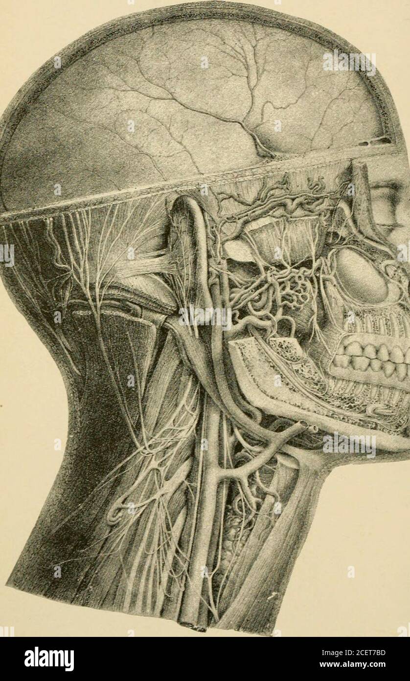 . Essentials of physiology, arranged in the form of questions and answers, prepared especially for students of medicine. pinalis colli and multifidus spinje muscles, andgoes beneath and within the complexus muscle, pierces thebiventer, and sends a cutaneous branch to the neck. (XVI.)—Fourth cervical nerve. 77. Anterior division. 78. Princi-pal root of the phrenic nerve. 79. The external branch ofthe posterior division divided. 80. Internal branch of theposterior division. 81. Muscular branches. 82. Descendingsuperficial cervical nerve. (XVII.—XIX.)—The fifth, sixth, and seventh cervical nerves Stock Photohttps://www.alamy.com/image-license-details/?v=1https://www.alamy.com/essentials-of-physiology-arranged-in-the-form-of-questions-and-answers-prepared-especially-for-students-of-medicine-pinalis-colli-and-multifidus-spinje-muscles-andgoes-beneath-and-within-the-complexus-muscle-pierces-thebiventer-and-sends-a-cutaneous-branch-to-the-neck-xvifourth-cervical-nerve-77-anterior-division-78-princi-pal-root-of-the-phrenic-nerve-79-the-external-branch-ofthe-posterior-division-divided-80-internal-branch-of-theposterior-division-81-muscular-branches-82-descendingsuperficial-cervical-nerve-xviixixthe-fifth-sixth-and-seventh-cervical-nerves-image370555569.html
RM2CET7BD–. Essentials of physiology, arranged in the form of questions and answers, prepared especially for students of medicine. pinalis colli and multifidus spinje muscles, andgoes beneath and within the complexus muscle, pierces thebiventer, and sends a cutaneous branch to the neck. (XVI.)—Fourth cervical nerve. 77. Anterior division. 78. Princi-pal root of the phrenic nerve. 79. The external branch ofthe posterior division divided. 80. Internal branch of theposterior division. 81. Muscular branches. 82. Descendingsuperficial cervical nerve. (XVII.—XIX.)—The fifth, sixth, and seventh cervical nerves
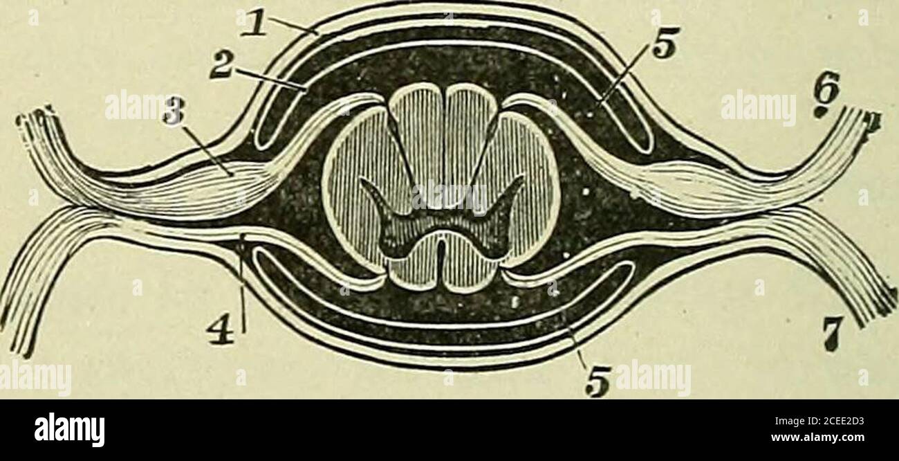 . Text-book of anatomy and physiology for nurses. Fig. 167.—Cauda Equina(Morris). Fig. 168.—Showing Division of Nerve.I, Dura mater; 2, arachnoid; 3, ganglionof post, root; 4, ant. root; 5, space contain-ing spinal fluid; 6, post, division of nerve(Holden). The cervical plexus.—Most of the branches of this plexussupply muscles of the neck (front and side). One exception is the * The communicating branches to sympathetic ganglia are of great importance,serving to connect the cerebro-spinal and sympathetic division into one great nervesvstem. THE PHRENIC NERVE. 237 great auricular {auricularis m Stock Photohttps://www.alamy.com/image-license-details/?v=1https://www.alamy.com/text-book-of-anatomy-and-physiology-for-nurses-fig-167cauda-equinamorris-fig-168showing-division-of-nervei-dura-mater-2-arachnoid-3-ganglionof-post-root-4-ant-root-5-space-contain-ing-spinal-fluid-6-post-division-of-nerveholden-the-cervical-plexusmost-of-the-branches-of-this-plexussupply-muscles-of-the-neck-front-and-side-one-exception-is-the-the-communicating-branches-to-sympathetic-ganglia-are-of-great-importanceserving-to-connect-the-cerebro-spinal-and-sympathetic-division-into-one-great-nervesvstem-the-phrenic-nerve-237-great-auricular-auricularis-m-image370332175.html
RM2CEE2D3–. Text-book of anatomy and physiology for nurses. Fig. 167.—Cauda Equina(Morris). Fig. 168.—Showing Division of Nerve.I, Dura mater; 2, arachnoid; 3, ganglionof post, root; 4, ant. root; 5, space contain-ing spinal fluid; 6, post, division of nerve(Holden). The cervical plexus.—Most of the branches of this plexussupply muscles of the neck (front and side). One exception is the * The communicating branches to sympathetic ganglia are of great importance,serving to connect the cerebro-spinal and sympathetic division into one great nervesvstem. THE PHRENIC NERVE. 237 great auricular {auricularis m
 . Text-book of anatomy and physiology for nurses. Fig. 167.—Cauda Equina(Morris). Fig. 168.—Showing Division of Nerve.I, Dura mater; 2, arachnoid; 3, ganglionof post, root; 4, ant. root; 5, space contain-ing spinal fluid; 6, post, division of nerve(Holden). The cervical plexus.—Most of the branches of this plexussupply muscles of the neck (front and side). One exception is the * The communicating branches to sympathetic ganglia are of great importance,serving to connect the cerebro-spinal and sympathetic division into one great nervesvstem. THE PHRENIC NERVE. 237 great auricular {auricularis m Stock Photohttps://www.alamy.com/image-license-details/?v=1https://www.alamy.com/text-book-of-anatomy-and-physiology-for-nurses-fig-167cauda-equinamorris-fig-168showing-division-of-nervei-dura-mater-2-arachnoid-3-ganglionof-post-root-4-ant-root-5-space-contain-ing-spinal-fluid-6-post-division-of-nerveholden-the-cervical-plexusmost-of-the-branches-of-this-plexussupply-muscles-of-the-neck-front-and-side-one-exception-is-the-the-communicating-branches-to-sympathetic-ganglia-are-of-great-importanceserving-to-connect-the-cerebro-spinal-and-sympathetic-division-into-one-great-nervesvstem-the-phrenic-nerve-237-great-auricular-auricularis-m-image370332086.html
RM2CEE29X–. Text-book of anatomy and physiology for nurses. Fig. 167.—Cauda Equina(Morris). Fig. 168.—Showing Division of Nerve.I, Dura mater; 2, arachnoid; 3, ganglionof post, root; 4, ant. root; 5, space contain-ing spinal fluid; 6, post, division of nerve(Holden). The cervical plexus.—Most of the branches of this plexussupply muscles of the neck (front and side). One exception is the * The communicating branches to sympathetic ganglia are of great importance,serving to connect the cerebro-spinal and sympathetic division into one great nervesvstem. THE PHRENIC NERVE. 237 great auricular {auricularis m
 . Annals of surgery. Supra.cla.vicular N. L Showing the skin and platysma dissected forward and backward. Retractors under posteriorborder of stemomastoid, exposing glands lying in gland-bearing fascia. The omohyoid marks the lowerangle of the dissection, and the mastoid process, the upper. The spinal accessory nerve is partly dis-sected from among the glands. Fig. 2.. Showing fascia dissected from the lower angle. Internal jugular vein shown !>ing beneath thestemomastoid muscle. The phrenic nerve, lying on the scalenus anticus muscle and branching fromthe cervical plexus, is exposed. Fig. Stock Photohttps://www.alamy.com/image-license-details/?v=1https://www.alamy.com/annals-of-surgery-supraclavicular-n-l-showing-the-skin-and-platysma-dissected-forward-and-backward-retractors-under-posteriorborder-of-stemomastoid-exposing-glands-lying-in-gland-bearing-fascia-the-omohyoid-marks-the-lowerangle-of-the-dissection-and-the-mastoid-process-the-upper-the-spinal-accessory-nerve-is-partly-dis-sected-from-among-the-glands-fig-2-showing-fascia-dissected-from-the-lower-angle-internal-jugular-vein-shown-!gting-beneath-thestemomastoid-muscle-the-phrenic-nerve-lying-on-the-scalenus-anticus-muscle-and-branching-fromthe-cervical-plexus-is-exposed-fig-image370642851.html
RM2CF06MK–. Annals of surgery. Supra.cla.vicular N. L Showing the skin and platysma dissected forward and backward. Retractors under posteriorborder of stemomastoid, exposing glands lying in gland-bearing fascia. The omohyoid marks the lowerangle of the dissection, and the mastoid process, the upper. The spinal accessory nerve is partly dis-sected from among the glands. Fig. 2.. Showing fascia dissected from the lower angle. Internal jugular vein shown !>ing beneath thestemomastoid muscle. The phrenic nerve, lying on the scalenus anticus muscle and branching fromthe cervical plexus, is exposed. Fig.
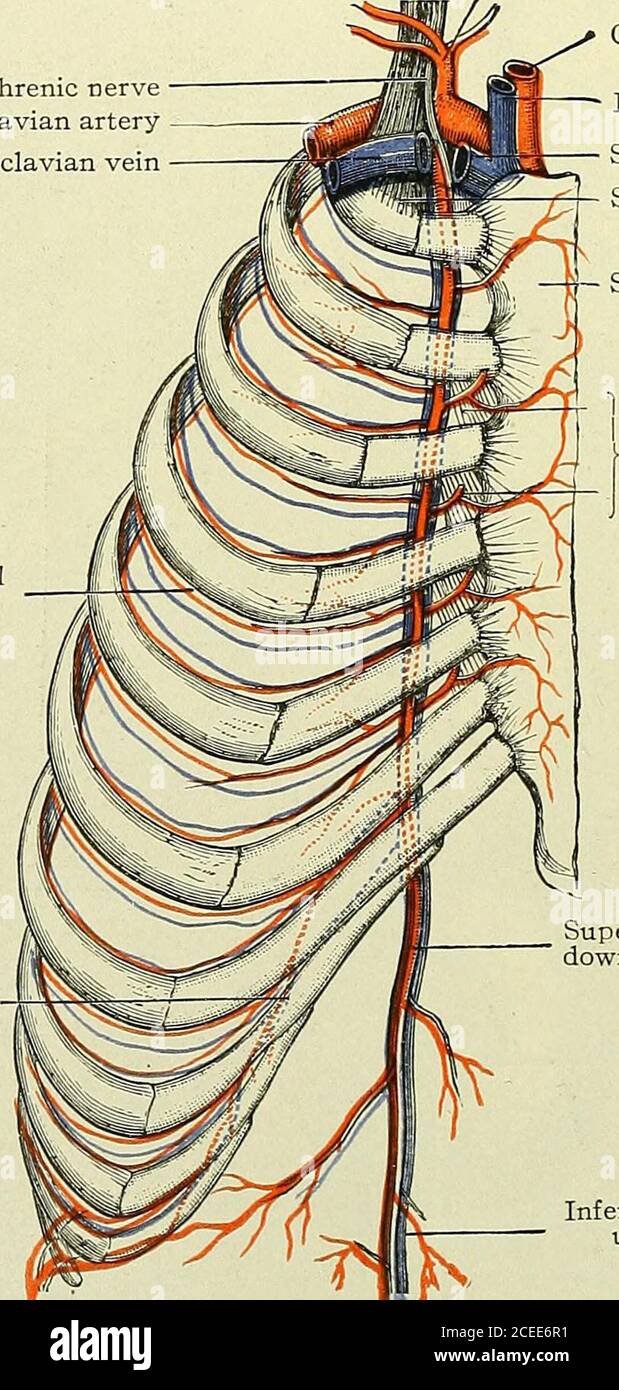 . Text-book of anatomy and physiology for nurses. Fig. 121.—The Aorta, showing the Three Portions (Morris). lyo ANATOMY AND PHYSIOLOGY FOR NURSES. Branches of the arch in their order: Two coronary (right and left) to heart muscle (Fig. 113). Right subclavian to right One anonyma, ij inches long. . < upper extremity.Right common carotid to[ right head and neck. One left common carotid to left head and neck. One left subclavian to left upper extremity. Phrenic nerve Subclavian artery Subclavian vein Anterior intercostalbranch Branch of mammary. Common carotid Internal jugular veinSubclavian v Stock Photohttps://www.alamy.com/image-license-details/?v=1https://www.alamy.com/text-book-of-anatomy-and-physiology-for-nurses-fig-121the-aorta-showing-the-three-portions-morris-lyo-anatomy-and-physiology-for-nurses-branches-of-the-arch-in-their-order-two-coronary-right-and-left-to-heart-muscle-fig-113-right-subclavian-to-right-one-anonyma-ij-inches-long-lt-upper-extremityright-common-carotid-to-right-head-and-neck-one-left-common-carotid-to-left-head-and-neck-one-left-subclavian-to-left-upper-extremity-phrenic-nerve-subclavian-artery-subclavian-vein-anterior-intercostalbranch-branch-of-mammary-common-carotid-internal-jugular-veinsubclavian-v-image370335589.html
RM2CEE6R1–. Text-book of anatomy and physiology for nurses. Fig. 121.—The Aorta, showing the Three Portions (Morris). lyo ANATOMY AND PHYSIOLOGY FOR NURSES. Branches of the arch in their order: Two coronary (right and left) to heart muscle (Fig. 113). Right subclavian to right One anonyma, ij inches long. . < upper extremity.Right common carotid to[ right head and neck. One left common carotid to left head and neck. One left subclavian to left upper extremity. Phrenic nerve Subclavian artery Subclavian vein Anterior intercostalbranch Branch of mammary. Common carotid Internal jugular veinSubclavian v
 . Buffalo Medical Journal. al nerve: inferior branch. 8, Hypoglossalnerve. 9. Accessorious nerve. 10. Erbs point (supraclavicular point). 11. Phrenic nerve.12. Brachial plexus. 13. Axillary nerve. MUSCLES. a. Frontalis, b. Corrugator supercilii. c. Orbicularis palpebrarum, d. Nasal mus-cles, e. Zygomatic muscles, f. Orbicularis oris. g. Massetor. h. Levater menti. i. Quad-ratus mcnti (depressor labii inferioris). k. Platysina myoidcs. 1. Hyoid muscles.m Sterno-clcido-mastoid. n. Omo-hyoid. o. Splenicus. p. Trapezius, r. Levator anguliscapull. 6. Triangularis menti (depressor anguli oris), t. S Stock Photohttps://www.alamy.com/image-license-details/?v=1https://www.alamy.com/buffalo-medical-journal-al-nerve-inferior-branch-8-hypoglossalnerve-9-accessorious-nerve-10-erbs-point-supraclavicular-point-11-phrenic-nerve12-brachial-plexus-13-axillary-nerve-muscles-a-frontalis-b-corrugator-supercilii-c-orbicularis-palpebrarum-d-nasal-mus-cles-e-zygomatic-muscles-f-orbicularis-oris-g-massetor-h-levater-menti-i-quad-ratus-mcnti-depressor-labii-inferioris-k-platysina-myoidcs-1-hyoid-musclesm-sterno-clcido-mastoid-n-omo-hyoid-o-splenicus-p-trapezius-r-levator-anguliscapull-6-triangularis-menti-depressor-anguli-oris-t-s-image370442070.html
RM2CEK2HX–. Buffalo Medical Journal. al nerve: inferior branch. 8, Hypoglossalnerve. 9. Accessorious nerve. 10. Erbs point (supraclavicular point). 11. Phrenic nerve.12. Brachial plexus. 13. Axillary nerve. MUSCLES. a. Frontalis, b. Corrugator supercilii. c. Orbicularis palpebrarum, d. Nasal mus-cles, e. Zygomatic muscles, f. Orbicularis oris. g. Massetor. h. Levater menti. i. Quad-ratus mcnti (depressor labii inferioris). k. Platysina myoidcs. 1. Hyoid muscles.m Sterno-clcido-mastoid. n. Omo-hyoid. o. Splenicus. p. Trapezius, r. Levator anguliscapull. 6. Triangularis menti (depressor anguli oris), t. S
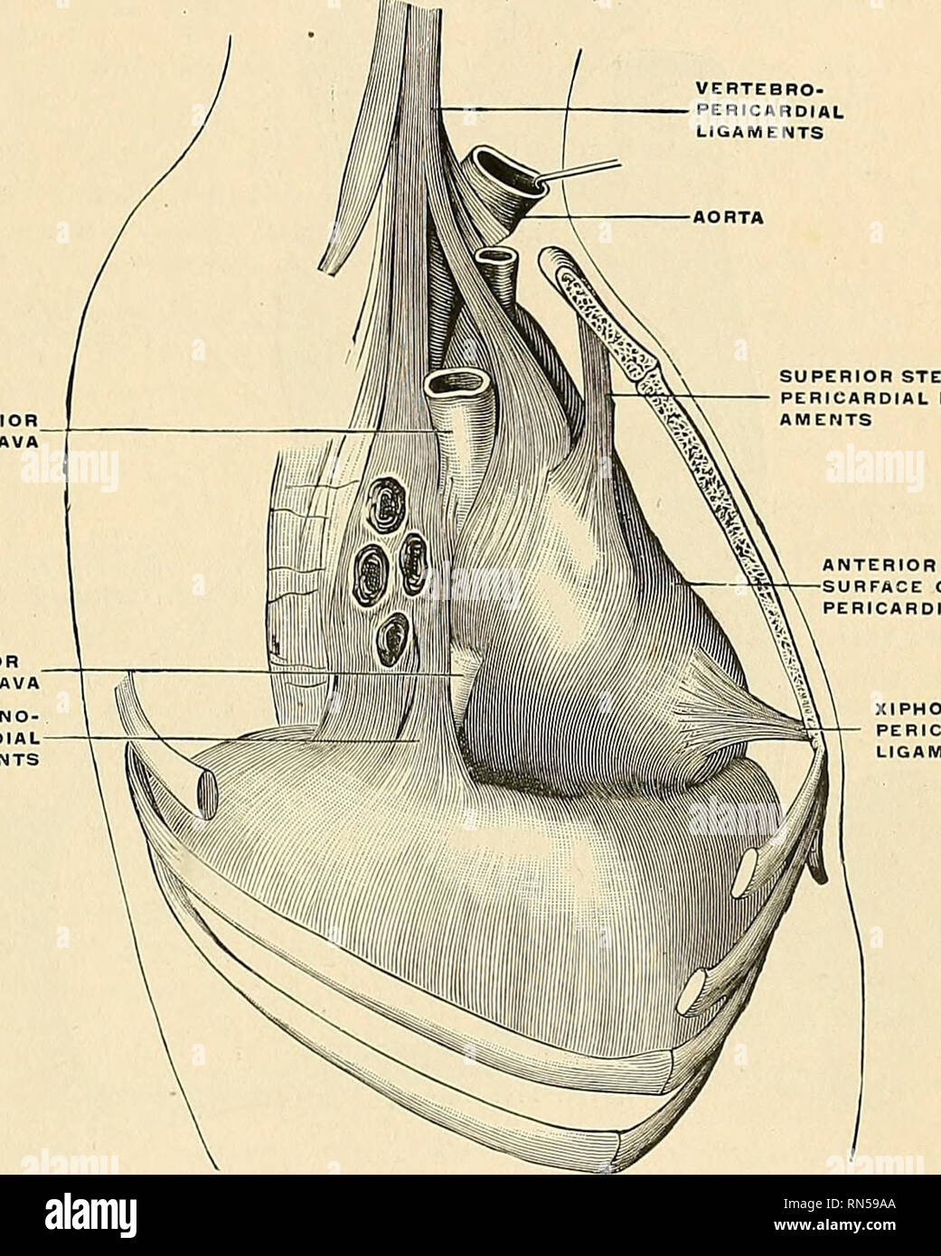 . Anatomy, descriptive and applied. Anatomy. THE PEBTCARDIUM 549 the upper passing to the manubrium, and the lower to the ensiform cartilage. Behind, it rests upon the bronchi, the oesophagus, and the descending aorta. Laterally, it is covered by the pleurse, and is in relation to the inner surface of the lungs; the phrenic nerve with its accompanying vessels descends between the pericardium and pleura on either side (Fig. 409). The vessels receiving fibrous prolongation from this membrane are the aorta, the superior vena cava, the right and left pulmonary arteries, the four pulmonary veins, a Stock Photohttps://www.alamy.com/image-license-details/?v=1https://www.alamy.com/anatomy-descriptive-and-applied-anatomy-the-pebtcardium-549-the-upper-passing-to-the-manubrium-and-the-lower-to-the-ensiform-cartilage-behind-it-rests-upon-the-bronchi-the-oesophagus-and-the-descending-aorta-laterally-it-is-covered-by-the-pleurse-and-is-in-relation-to-the-inner-surface-of-the-lungs-the-phrenic-nerve-with-its-accompanying-vessels-descends-between-the-pericardium-and-pleura-on-either-side-fig-409-the-vessels-receiving-fibrous-prolongation-from-this-membrane-are-the-aorta-the-superior-vena-cava-the-right-and-left-pulmonary-arteries-the-four-pulmonary-veins-a-image236759666.html
RMRN59AA–. Anatomy, descriptive and applied. Anatomy. THE PEBTCARDIUM 549 the upper passing to the manubrium, and the lower to the ensiform cartilage. Behind, it rests upon the bronchi, the oesophagus, and the descending aorta. Laterally, it is covered by the pleurse, and is in relation to the inner surface of the lungs; the phrenic nerve with its accompanying vessels descends between the pericardium and pleura on either side (Fig. 409). The vessels receiving fibrous prolongation from this membrane are the aorta, the superior vena cava, the right and left pulmonary arteries, the four pulmonary veins, a
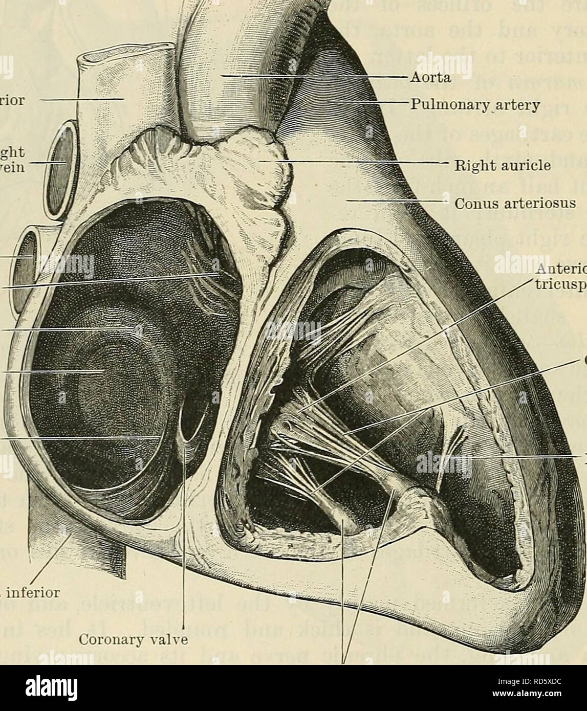 . Cunningham's Text-book of anatomy. Anatomy. 874 THE VASCULAK SYSTEM. aperture. Above and anteriorly it is in relation with the ascending aorta, and from the junction of this aspect with the right lateral boundary the right auricle is prolonged anteriorly and to the left. Its right side forms the right margin of the heart, and is in relation with the right phrenic nerve and its accom- panying vessels, and with the right pleura and lung, the pericardium intervening. On the left this atrium is limited by the oblique septum which separates it from the left atrium. The sulcus terminalis is a shal Stock Photohttps://www.alamy.com/image-license-details/?v=1https://www.alamy.com/cunninghams-text-book-of-anatomy-anatomy-874-the-vasculak-system-aperture-above-and-anteriorly-it-is-in-relation-with-the-ascending-aorta-and-from-the-junction-of-this-aspect-with-the-right-lateral-boundary-the-right-auricle-is-prolonged-anteriorly-and-to-the-left-its-right-side-forms-the-right-margin-of-the-heart-and-is-in-relation-with-the-right-phrenic-nerve-and-its-accom-panying-vessels-and-with-the-right-pleura-and-lung-the-pericardium-intervening-on-the-left-this-atrium-is-limited-by-the-oblique-septum-which-separates-it-from-the-left-atrium-the-sulcus-terminalis-is-a-shal-image231855832.html
RMRD5XDC–. Cunningham's Text-book of anatomy. Anatomy. 874 THE VASCULAK SYSTEM. aperture. Above and anteriorly it is in relation with the ascending aorta, and from the junction of this aspect with the right lateral boundary the right auricle is prolonged anteriorly and to the left. Its right side forms the right margin of the heart, and is in relation with the right phrenic nerve and its accom- panying vessels, and with the right pleura and lung, the pericardium intervening. On the left this atrium is limited by the oblique septum which separates it from the left atrium. The sulcus terminalis is a shal
 . The anatomy of the human body. Human anatomy; Anatomy. 778 NEUROLOGY. branches, viz., the great mastoid (y), the small mastoid, and the great auricular (q); and of descending branches, subdivided into the deep and the superficial ; the deep ones consisting of the internal descending branch (before s), the phrenic nerve (l), and the branch- es for the trapezius, levator anguli scapula, and rhomboideus; the superficial descending branches are the supra-clavicular and the acromial (m). According to their distribution, they may also be divided into muscular and cutaneous branches ; the muscular Stock Photohttps://www.alamy.com/image-license-details/?v=1https://www.alamy.com/the-anatomy-of-the-human-body-human-anatomy-anatomy-778-neurology-branches-viz-the-great-mastoid-y-the-small-mastoid-and-the-great-auricular-q-and-of-descending-branches-subdivided-into-the-deep-and-the-superficial-the-deep-ones-consisting-of-the-internal-descending-branch-before-s-the-phrenic-nerve-l-and-the-branch-es-for-the-trapezius-levator-anguli-scapula-and-rhomboideus-the-superficial-descending-branches-are-the-supra-clavicular-and-the-acromial-m-according-to-their-distribution-they-may-also-be-divided-into-muscular-and-cutaneous-branches-the-muscular-image236793812.html
RMRN6TWT–. The anatomy of the human body. Human anatomy; Anatomy. 778 NEUROLOGY. branches, viz., the great mastoid (y), the small mastoid, and the great auricular (q); and of descending branches, subdivided into the deep and the superficial ; the deep ones consisting of the internal descending branch (before s), the phrenic nerve (l), and the branch- es for the trapezius, levator anguli scapula, and rhomboideus; the superficial descending branches are the supra-clavicular and the acromial (m). According to their distribution, they may also be divided into muscular and cutaneous branches ; the muscular
 . Anatomy, descriptive and applied. Anatomy. THE CERVICAL PLEXUS 1025 The left phrenic nerve is rather longer than the right, from the inclination of the heart to the left side, and from the Diaphragm being lower on this than on the opposite side. It enters the thorax behind the left innominate vein, and crosses in front of the vagus and the arch of the aorta and the root of the hmg. Each nerve supplies filaments to the pericardium and pleura, and near the thorax is joined by a filament from the sympathetic, and, occasionally, by one from the ansa cervicalis. Branches have been described as pa Stock Photohttps://www.alamy.com/image-license-details/?v=1https://www.alamy.com/anatomy-descriptive-and-applied-anatomy-the-cervical-plexus-1025-the-left-phrenic-nerve-is-rather-longer-than-the-right-from-the-inclination-of-the-heart-to-the-left-side-and-from-the-diaphragm-being-lower-on-this-than-on-the-opposite-side-it-enters-the-thorax-behind-the-left-innominate-vein-and-crosses-in-front-of-the-vagus-and-the-arch-of-the-aorta-and-the-root-of-the-hmg-each-nerve-supplies-filaments-to-the-pericardium-and-pleura-and-near-the-thorax-is-joined-by-a-filament-from-the-sympathetic-and-occasionally-by-one-from-the-ansa-cervicalis-branches-have-been-described-as-pa-image236769841.html
RMRN5P9N–. Anatomy, descriptive and applied. Anatomy. THE CERVICAL PLEXUS 1025 The left phrenic nerve is rather longer than the right, from the inclination of the heart to the left side, and from the Diaphragm being lower on this than on the opposite side. It enters the thorax behind the left innominate vein, and crosses in front of the vagus and the arch of the aorta and the root of the hmg. Each nerve supplies filaments to the pericardium and pleura, and near the thorax is joined by a filament from the sympathetic, and, occasionally, by one from the ansa cervicalis. Branches have been described as pa
 . Biology of the vertebrates : a comparative study of man and his animal allies. Vertebrates; Vertebrates -- Anatomy; Anatomy, Comparative. 6^8 Biology of the Vertebrates vicissitudes, just as a faithful dog, trotting behind its master, serves to identify him, regardless of the different costumes or disguises which the master may assume. A striking illustration of the constancy of nerves to transforming muscles is furnished by the phrenic nerve that supplies the diaphragm, which is a migratory muscle laid down originally far anterior in the neck region. With. Please note that these images are Stock Photohttps://www.alamy.com/image-license-details/?v=1https://www.alamy.com/biology-of-the-vertebrates-a-comparative-study-of-man-and-his-animal-allies-vertebrates-vertebrates-anatomy-anatomy-comparative-68-biology-of-the-vertebrates-vicissitudes-just-as-a-faithful-dog-trotting-behind-its-master-serves-to-identify-him-regardless-of-the-different-costumes-or-disguises-which-the-master-may-assume-a-striking-illustration-of-the-constancy-of-nerves-to-transforming-muscles-is-furnished-by-the-phrenic-nerve-that-supplies-the-diaphragm-which-is-a-migratory-muscle-laid-down-originally-far-anterior-in-the-neck-region-with-please-note-that-these-images-are-image234594116.html
RMRHJK58–. Biology of the vertebrates : a comparative study of man and his animal allies. Vertebrates; Vertebrates -- Anatomy; Anatomy, Comparative. 6^8 Biology of the Vertebrates vicissitudes, just as a faithful dog, trotting behind its master, serves to identify him, regardless of the different costumes or disguises which the master may assume. A striking illustration of the constancy of nerves to transforming muscles is furnished by the phrenic nerve that supplies the diaphragm, which is a migratory muscle laid down originally far anterior in the neck region. With. Please note that these images are
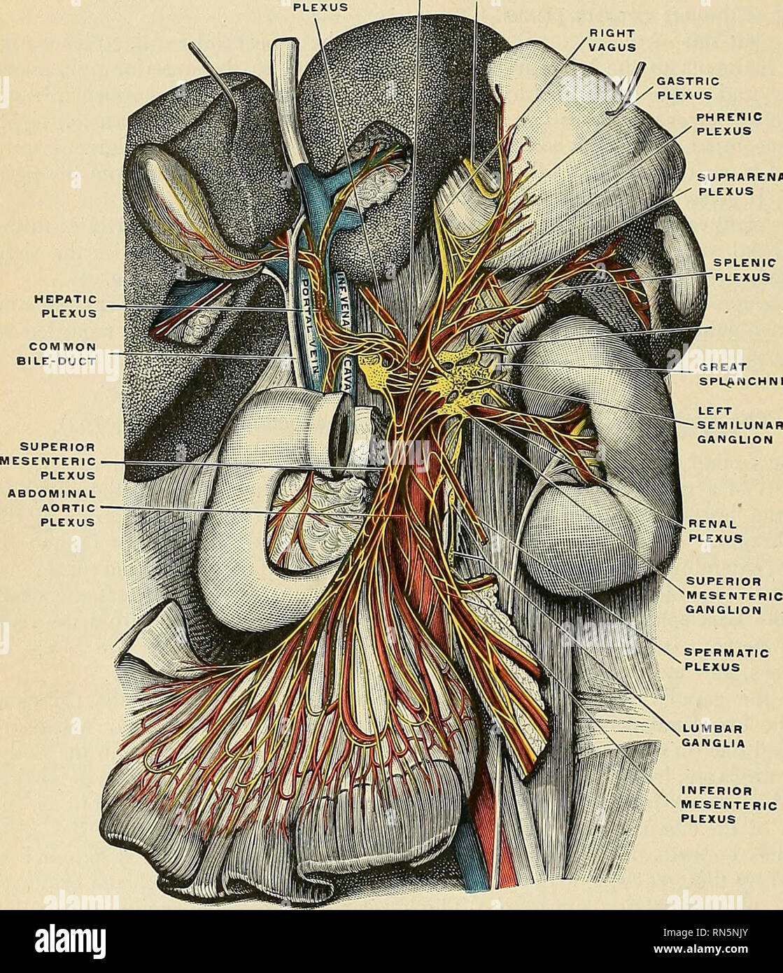 . Anatomy, descriptive and applied. Anatomy. 1074 THE NERVE SYSTEM The Phrenic Plexus {plexus phrenicus) (Fig. 792) accompanies the inferior phrenic artery to the Diaphragm, some filaments passing to the suprarenal gland. It arises from the upper part of the semilunar ganglion, and is larger on the right than on the left side. It receives one or two branches from the phrenic nerve. At the point of junction with the phrenic nerve is a small ganglion, the phrenic ganglion (ganglion phrenicum) (Fig. 793), which lies on the under surface of the Diaphragm, near the right suprarenal. Its branches ar Stock Photohttps://www.alamy.com/image-license-details/?v=1https://www.alamy.com/anatomy-descriptive-and-applied-anatomy-1074-the-nerve-system-the-phrenic-plexus-plexus-phrenicus-fig-792-accompanies-the-inferior-phrenic-artery-to-the-diaphragm-some-filaments-passing-to-the-suprarenal-gland-it-arises-from-the-upper-part-of-the-semilunar-ganglion-and-is-larger-on-the-right-than-on-the-left-side-it-receives-one-or-two-branches-from-the-phrenic-nerve-at-the-point-of-junction-with-the-phrenic-nerve-is-a-small-ganglion-the-phrenic-ganglion-ganglion-phrenicum-fig-793-which-lies-on-the-under-surface-of-the-diaphragm-near-the-right-suprarenal-its-branches-ar-image236769315.html
RMRN5NJY–. Anatomy, descriptive and applied. Anatomy. 1074 THE NERVE SYSTEM The Phrenic Plexus {plexus phrenicus) (Fig. 792) accompanies the inferior phrenic artery to the Diaphragm, some filaments passing to the suprarenal gland. It arises from the upper part of the semilunar ganglion, and is larger on the right than on the left side. It receives one or two branches from the phrenic nerve. At the point of junction with the phrenic nerve is a small ganglion, the phrenic ganglion (ganglion phrenicum) (Fig. 793), which lies on the under surface of the Diaphragm, near the right suprarenal. Its branches ar
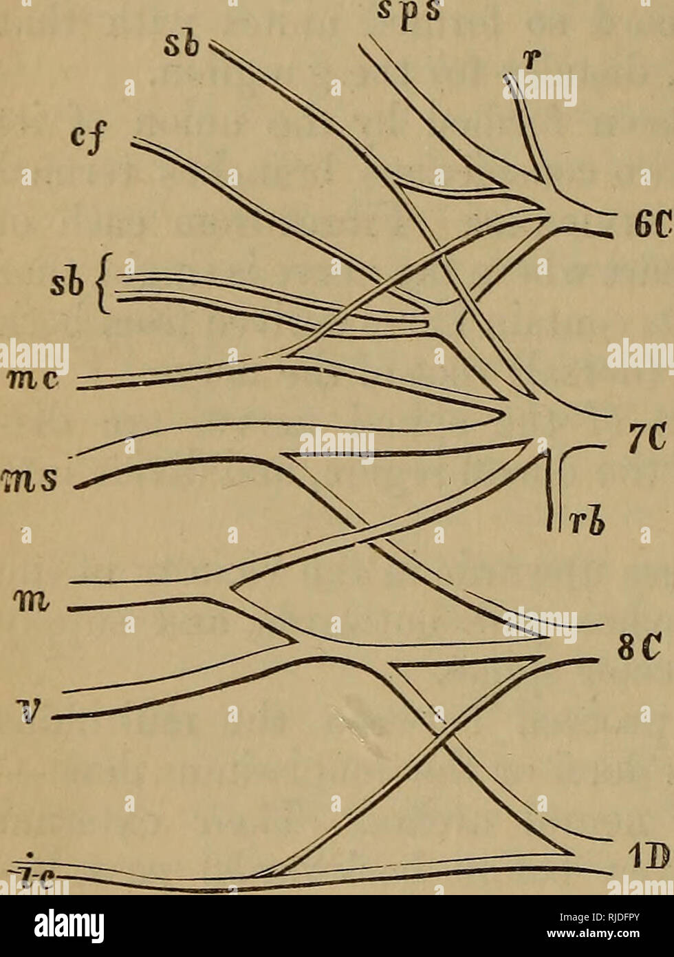 . The cat : an introduction to the study of backboned animals, especially mammals. Cats; Anatomy, Comparative. 278 TEE CAT. [CHAP. IX. plexus, placed opposite the first four vertebrae below the sterno- mastoid muscle, and connected, near the skull, with the pneumo- gastric, hypoglossal, and sympathetic nerves. The fifth and sixth cervical nerves give off a branch called the phrenic nerve, which passes backAvards between the pleura and the pericardium, and is distributed to the diaphragm. § 18. The three posterior cervical nerves unite to form an inter- lacement called the brachial plexus, whic Stock Photohttps://www.alamy.com/image-license-details/?v=1https://www.alamy.com/the-cat-an-introduction-to-the-study-of-backboned-animals-especially-mammals-cats-anatomy-comparative-278-tee-cat-chap-ix-plexus-placed-opposite-the-first-four-vertebrae-below-the-sterno-mastoid-muscle-and-connected-near-the-skull-with-the-pneumo-gastric-hypoglossal-and-sympathetic-nerves-the-fifth-and-sixth-cervical-nerves-give-off-a-branch-called-the-phrenic-nerve-which-passes-backavards-between-the-pleura-and-the-pericardium-and-is-distributed-to-the-diaphragm-18-the-three-posterior-cervical-nerves-unite-to-form-an-inter-lacement-called-the-brachial-plexus-whic-image235096371.html
RMRJDFPY–. The cat : an introduction to the study of backboned animals, especially mammals. Cats; Anatomy, Comparative. 278 TEE CAT. [CHAP. IX. plexus, placed opposite the first four vertebrae below the sterno- mastoid muscle, and connected, near the skull, with the pneumo- gastric, hypoglossal, and sympathetic nerves. The fifth and sixth cervical nerves give off a branch called the phrenic nerve, which passes backAvards between the pleura and the pericardium, and is distributed to the diaphragm. § 18. The three posterior cervical nerves unite to form an inter- lacement called the brachial plexus, whic
 . Biology of the vertebrates : a comparative study of man and his animal allies. Vertebrates; Vertebrates -- Anatomy; Anatomy, Comparative. Fig. 580. Superficial muscles of the back. (After Morris.) Fig. 581. The tendon of Achilles, in black, showing how the work which muscles do may be applied at a point some distance from the mus- cle itself. the backward shifting of the heart the diaphragm finally assumes an abdominal position remote from the neck, yet the phrenic nerve, although made up from the third, fourth, and fifth cervical nerves, goes out of its way to retain connection with it and Stock Photohttps://www.alamy.com/image-license-details/?v=1https://www.alamy.com/biology-of-the-vertebrates-a-comparative-study-of-man-and-his-animal-allies-vertebrates-vertebrates-anatomy-anatomy-comparative-fig-580-superficial-muscles-of-the-back-after-morris-fig-581-the-tendon-of-achilles-in-black-showing-how-the-work-which-muscles-do-may-be-applied-at-a-point-some-distance-from-the-mus-cle-itself-the-backward-shifting-of-the-heart-the-diaphragm-finally-assumes-an-abdominal-position-remote-from-the-neck-yet-the-phrenic-nerve-although-made-up-from-the-third-fourth-and-fifth-cervical-nerves-goes-out-of-its-way-to-retain-connection-with-it-and-image234594102.html
RMRHJK4P–. Biology of the vertebrates : a comparative study of man and his animal allies. Vertebrates; Vertebrates -- Anatomy; Anatomy, Comparative. Fig. 580. Superficial muscles of the back. (After Morris.) Fig. 581. The tendon of Achilles, in black, showing how the work which muscles do may be applied at a point some distance from the mus- cle itself. the backward shifting of the heart the diaphragm finally assumes an abdominal position remote from the neck, yet the phrenic nerve, although made up from the third, fourth, and fifth cervical nerves, goes out of its way to retain connection with it and
 . Elementary text-book of zoology. LEPUS. 391 A description of the nerves cannot be entered into here, but a few of the more important are to be seen in the neck. (Plate XIII.) In this region we have already noticed the carotid arteries, the internal and external jugular veins, the oesophagus, trachea and phrenic veins. Just internal to the phrenic nerve and close beside the carotid artery runs Fig. 279.âRabbit's Brain. A, Dorsal View. â Olfactory Lobe. Position of Corpus Callosuni Pineal Body, Cerebellum.. Cerebral Hemispbere. Corpora Quadrigemina. Flocculus of Cerebellum. Medulla Oblongata. Stock Photohttps://www.alamy.com/image-license-details/?v=1https://www.alamy.com/elementary-text-book-of-zoology-lepus-391-a-description-of-the-nerves-cannot-be-entered-into-here-but-a-few-of-the-more-important-are-to-be-seen-in-the-neck-plate-xiii-in-this-region-we-have-already-noticed-the-carotid-arteries-the-internal-and-external-jugular-veins-the-oesophagus-trachea-and-phrenic-veins-just-internal-to-the-phrenic-nerve-and-close-beside-the-carotid-artery-runs-fig-279rabbits-brain-a-dorsal-view-olfactory-lobe-position-of-corpus-callosuni-pineal-body-cerebellum-cerebral-hemispbere-corpora-quadrigemina-flocculus-of-cerebellum-medulla-oblongata-image232088481.html
RMRDGF69–. Elementary text-book of zoology. LEPUS. 391 A description of the nerves cannot be entered into here, but a few of the more important are to be seen in the neck. (Plate XIII.) In this region we have already noticed the carotid arteries, the internal and external jugular veins, the oesophagus, trachea and phrenic veins. Just internal to the phrenic nerve and close beside the carotid artery runs Fig. 279.âRabbit's Brain. A, Dorsal View. â Olfactory Lobe. Position of Corpus Callosuni Pineal Body, Cerebellum.. Cerebral Hemispbere. Corpora Quadrigemina. Flocculus of Cerebellum. Medulla Oblongata.
 . Cunningham's Text-book of anatomy. Anatomy. THE MUSCLES OF THE THOEAX. 473 quadratum) in the right lobe of the central tendon transmits the inferior vena cava, and small branches of the right phrenic nerve. The hiatus ozsophageus (oesophageal opening) is in the muscular substance of the diaphragm, posterior to the central tendon, and is surrounded by a sphincter-like arrangement of the crural fibres. Besides the oesophagus, this opening transmits the two vagi nerves. Middle arcuate ligament Vena caval opening (Esophageal opening in diaphragm Aortic openin Anterior ramus of twelfth thoracic n Stock Photohttps://www.alamy.com/image-license-details/?v=1https://www.alamy.com/cunninghams-text-book-of-anatomy-anatomy-the-muscles-of-the-thoeax-473-quadratum-in-the-right-lobe-of-the-central-tendon-transmits-the-inferior-vena-cava-and-small-branches-of-the-right-phrenic-nerve-the-hiatus-ozsophageus-oesophageal-opening-is-in-the-muscular-substance-of-the-diaphragm-posterior-to-the-central-tendon-and-is-surrounded-by-a-sphincter-like-arrangement-of-the-crural-fibres-besides-the-oesophagus-this-opening-transmits-the-two-vagi-nerves-middle-arcuate-ligament-vena-caval-opening-esophageal-opening-in-diaphragm-aortic-openin-anterior-ramus-of-twelfth-thoracic-n-image231850256.html
RMRD5KA8–. Cunningham's Text-book of anatomy. Anatomy. THE MUSCLES OF THE THOEAX. 473 quadratum) in the right lobe of the central tendon transmits the inferior vena cava, and small branches of the right phrenic nerve. The hiatus ozsophageus (oesophageal opening) is in the muscular substance of the diaphragm, posterior to the central tendon, and is surrounded by a sphincter-like arrangement of the crural fibres. Besides the oesophagus, this opening transmits the two vagi nerves. Middle arcuate ligament Vena caval opening (Esophageal opening in diaphragm Aortic openin Anterior ramus of twelfth thoracic n
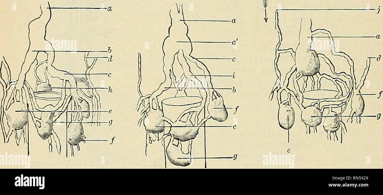 . Anatomy, descriptive and applied. Anatomy. 772 THE VASCULAR SYSTEMS subclavian artery and in front of tiie Scalenus anticus muscle and the phrenic- nerve, so as to form an arch; it terminates in the angle of junction of the left subclavian vein with the left internal jugular vein. It usually opens at the apex of the angle in the superior and outer surface, but may open on the posterior surface. Sometuiies it terminates by two or more branches. Figs. 552 and 554 show the termination of the thoracic duct. The thoracic duct, at its commence- ment, is about 2 to 3 mm. in diameter, diminishes con Stock Photohttps://www.alamy.com/image-license-details/?v=1https://www.alamy.com/anatomy-descriptive-and-applied-anatomy-772-the-vascular-systems-subclavian-artery-and-in-front-of-tiie-scalenus-anticus-muscle-and-the-phrenic-nerve-so-as-to-form-an-arch-it-terminates-in-the-angle-of-junction-of-the-left-subclavian-vein-with-the-left-internal-jugular-vein-it-usually-opens-at-the-apex-of-the-angle-in-the-superior-and-outer-surface-but-may-open-on-the-posterior-surface-sometuiies-it-terminates-by-two-or-more-branches-figs-552-and-554-show-the-termination-of-the-thoracic-duct-the-thoracic-duct-at-its-commence-ment-is-about-2-to-3-mm-in-diameter-diminishes-con-image236772769.html
RMRN5X29–. Anatomy, descriptive and applied. Anatomy. 772 THE VASCULAR SYSTEMS subclavian artery and in front of tiie Scalenus anticus muscle and the phrenic- nerve, so as to form an arch; it terminates in the angle of junction of the left subclavian vein with the left internal jugular vein. It usually opens at the apex of the angle in the superior and outer surface, but may open on the posterior surface. Sometuiies it terminates by two or more branches. Figs. 552 and 554 show the termination of the thoracic duct. The thoracic duct, at its commence- ment, is about 2 to 3 mm. in diameter, diminishes con
 . A manual of elementary zoology . Zoology. THE RABBIT 457 laryngeal and passes backwards beside the main vagus, and a recurrent or inferior laryngeal branch, which loops forward round an artery and runs beside the trachea to the muscles of the larynx; behind this the vagus passes backwards along the oesophagus; (3) the cervical sympathetic, lying beside the vagus and depressor; (4) the spinal nerves, of which the third gives a great auricular branch to the ear and the fourth and fifth give off branches which join to form the phrenic nerve to the diaphragm. The vagus bears its Kar'iL. Fig. 333 Stock Photohttps://www.alamy.com/image-license-details/?v=1https://www.alamy.com/a-manual-of-elementary-zoology-zoology-the-rabbit-457-laryngeal-and-passes-backwards-beside-the-main-vagus-and-a-recurrent-or-inferior-laryngeal-branch-which-loops-forward-round-an-artery-and-runs-beside-the-trachea-to-the-muscles-of-the-larynx-behind-this-the-vagus-passes-backwards-along-the-oesophagus-3-the-cervical-sympathetic-lying-beside-the-vagus-and-depressor-4-the-spinal-nerves-of-which-the-third-gives-a-great-auricular-branch-to-the-ear-and-the-fourth-and-fifth-give-off-branches-which-join-to-form-the-phrenic-nerve-to-the-diaphragm-the-vagus-bears-its-karil-fig-333-image232107882.html
RMRDHBY6–. A manual of elementary zoology . Zoology. THE RABBIT 457 laryngeal and passes backwards beside the main vagus, and a recurrent or inferior laryngeal branch, which loops forward round an artery and runs beside the trachea to the muscles of the larynx; behind this the vagus passes backwards along the oesophagus; (3) the cervical sympathetic, lying beside the vagus and depressor; (4) the spinal nerves, of which the third gives a great auricular branch to the ear and the fourth and fifth give off branches which join to form the phrenic nerve to the diaphragm. The vagus bears its Kar'iL. Fig. 333
 . A text-book of comparative physiology for students and practitioners of comparative (veterinary) medicine. Physiology, Comparative. 394 COMPARATIVE PHTSIOLOaY. Brain above medulla from which Impulses modifying respiration may proceed. 'acial muscles. Respiratory centre ill the medvllu. Cutaneous surface from which afferent impiUses proceed di., recfly to prain. Thoracic resp. muscles.. Spinal cord.—p- Respiratory tract. Diaphragm with phrenic nerve. Cutaneous sur- face from which impulses reach res- piratory centre by spinal cord. Fig. 309.—^Diagram intended to illustrate nervous mechanism o Stock Photohttps://www.alamy.com/image-license-details/?v=1https://www.alamy.com/a-text-book-of-comparative-physiology-for-students-and-practitioners-of-comparative-veterinary-medicine-physiology-comparative-394-comparative-phtsioloay-brain-above-medulla-from-which-impulses-modifying-respiration-may-proceed-acial-muscles-respiratory-centre-ill-the-medvllu-cutaneous-surface-from-which-afferent-impiuses-proceed-di-recfly-to-prain-thoracic-resp-muscles-spinal-cordp-respiratory-tract-diaphragm-with-phrenic-nerve-cutaneous-sur-face-from-which-impulses-reach-res-piratory-centre-by-spinal-cord-fig-309diagram-intended-to-illustrate-nervous-mechanism-o-image232319472.html
RMRDY1T0–. A text-book of comparative physiology for students and practitioners of comparative (veterinary) medicine. Physiology, Comparative. 394 COMPARATIVE PHTSIOLOaY. Brain above medulla from which Impulses modifying respiration may proceed. 'acial muscles. Respiratory centre ill the medvllu. Cutaneous surface from which afferent impiUses proceed di., recfly to prain. Thoracic resp. muscles.. Spinal cord.—p- Respiratory tract. Diaphragm with phrenic nerve. Cutaneous sur- face from which impulses reach res- piratory centre by spinal cord. Fig. 309.—^Diagram intended to illustrate nervous mechanism o
 . A laboratory manual and text-book of embryology. Embryology. 192 THE ENTODERMAL CANAL AND ITS DERIVATIVES Dorsally the pleural and peritoneal cavities are permanently partitioned length- wise by the dorsal mesentery. The septum transversum in 2 mm. embryos occupies a transverse position in the middle cervical region (Fig. 185, 2). According to Mall, it migrates caudally, its ventral portion at first moving more rapidly so that its position becomes oblique. In 5 mm. embryos (Fig. 185, 5) it is opposite the fifth cervical segment, at which level it receives the phrenic nerve. In stages later t Stock Photohttps://www.alamy.com/image-license-details/?v=1https://www.alamy.com/a-laboratory-manual-and-text-book-of-embryology-embryology-192-the-entodermal-canal-and-its-derivatives-dorsally-the-pleural-and-peritoneal-cavities-are-permanently-partitioned-length-wise-by-the-dorsal-mesentery-the-septum-transversum-in-2-mm-embryos-occupies-a-transverse-position-in-the-middle-cervical-region-fig-185-2-according-to-mall-it-migrates-caudally-its-ventral-portion-at-first-moving-more-rapidly-so-that-its-position-becomes-oblique-in-5-mm-embryos-fig-185-5-it-is-opposite-the-fifth-cervical-segment-at-which-level-it-receives-the-phrenic-nerve-in-stages-later-t-image232320650.html
RMRDY3A2–. A laboratory manual and text-book of embryology. Embryology. 192 THE ENTODERMAL CANAL AND ITS DERIVATIVES Dorsally the pleural and peritoneal cavities are permanently partitioned length- wise by the dorsal mesentery. The septum transversum in 2 mm. embryos occupies a transverse position in the middle cervical region (Fig. 185, 2). According to Mall, it migrates caudally, its ventral portion at first moving more rapidly so that its position becomes oblique. In 5 mm. embryos (Fig. 185, 5) it is opposite the fifth cervical segment, at which level it receives the phrenic nerve. In stages later t
 . A laboratory manual and text-book of embryology. Embryology. 196 THE ENTODERMAL CANAL AND ITS DERIVATIVES from (4) the dorsal mesentery. In addition to these, the striated muscle of the diaphragm, according to Bardeen, takes its origin from a pair of pre-muscle masses which in 9 mm. embryos lie one on each side opposite the fifth cervical segment. This is the level at which the phrenic nerve enters the septum trans- EsophaauS Common cardinal vein Septum trans vers urn Pleura-pericardial canal , Lung Pericardial cai/ffy. Pleural cavity * Pleura-peritoneal membrcuie 'Pleural cavity {Pericardia Stock Photohttps://www.alamy.com/image-license-details/?v=1https://www.alamy.com/a-laboratory-manual-and-text-book-of-embryology-embryology-196-the-entodermal-canal-and-its-derivatives-from-4-the-dorsal-mesentery-in-addition-to-these-the-striated-muscle-of-the-diaphragm-according-to-bardeen-takes-its-origin-from-a-pair-of-pre-muscle-masses-which-in-9-mm-embryos-lie-one-on-each-side-opposite-the-fifth-cervical-segment-this-is-the-level-at-which-the-phrenic-nerve-enters-the-septum-trans-esophaaus-common-cardinal-vein-septum-trans-vers-urn-pleura-pericardial-canal-lung-pericardial-caiffy-pleural-cavity-pleura-peritoneal-membrcuie-pleural-cavity-pericardia-image232320628.html
RMRDY398–. A laboratory manual and text-book of embryology. Embryology. 196 THE ENTODERMAL CANAL AND ITS DERIVATIVES from (4) the dorsal mesentery. In addition to these, the striated muscle of the diaphragm, according to Bardeen, takes its origin from a pair of pre-muscle masses which in 9 mm. embryos lie one on each side opposite the fifth cervical segment. This is the level at which the phrenic nerve enters the septum trans- EsophaauS Common cardinal vein Septum trans vers urn Pleura-pericardial canal , Lung Pericardial cai/ffy. Pleural cavity * Pleura-peritoneal membrcuie 'Pleural cavity {Pericardia
 . The anatomical record. Anatomy; Anatomy. I"i|iurc 1. Left |)lcural cavity viewed from tlieloft side, witli the left luiitt re- moverl. The larne foramen in the mediastinal jileura, in front of the root of the left luiiK opens direeti}' into the jjcricardial sae, exposing the heart; /, areh of aorta an^tal pleiu'a. Through the opening could be seen the j)ul- monary artery and left auriculai' ai)))eii(lage, as sjiown in figure 1. The left phrenic nerve pass(>d between the two layers of the anterior edge of the foramen. Aside from the large opening in it, the pericardium was normal. The Stock Photohttps://www.alamy.com/image-license-details/?v=1https://www.alamy.com/the-anatomical-record-anatomy-anatomy-iquotiiurc-1-left-lcural-cavity-viewed-from-tlieloft-side-witli-the-left-luiitt-re-moverl-the-larne-foramen-in-the-mediastinal-jileura-in-front-of-the-root-of-the-left-luiik-opens-direeti-into-the-jjcricardial-sae-exposing-the-heart-areh-of-aorta-antal-pleiua-through-the-opening-could-be-seen-the-jul-monary-artery-and-left-auriculai-aieiilage-as-sjiown-in-figure-1-the-left-phrenic-nerve-passgtd-between-the-two-layers-of-the-anterior-edge-of-the-foramen-aside-from-the-large-opening-in-it-the-pericardium-was-normal-the-image236850950.html
RMRN9DPE–. The anatomical record. Anatomy; Anatomy. I"i|iurc 1. Left |)lcural cavity viewed from tlieloft side, witli the left luiitt re- moverl. The larne foramen in the mediastinal jileura, in front of the root of the left luiiK opens direeti}' into the jjcricardial sae, exposing the heart; /, areh of aorta an^tal pleiu'a. Through the opening could be seen the j)ul- monary artery and left auriculai' ai)))eii(lage, as sjiown in figure 1. The left phrenic nerve pass(>d between the two layers of the anterior edge of the foramen. Aside from the large opening in it, the pericardium was normal. The
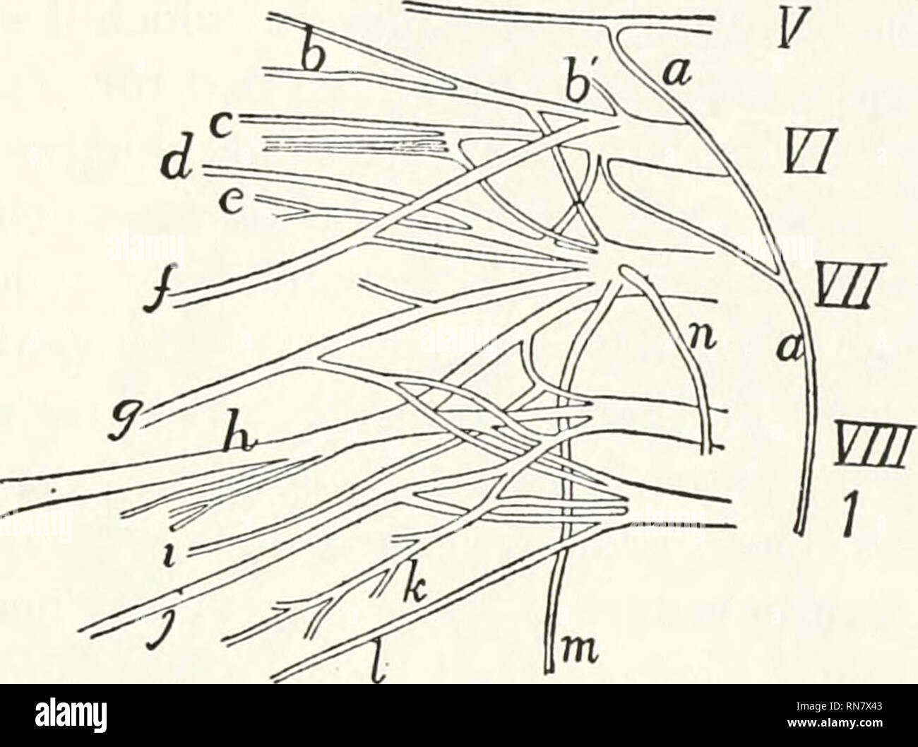 . Anatomy of the cat. Cats; Mammals. THE PERIPHERAL NERVOUS SYSTEM. 387 or both of the anterior thoracic nerves (^k and ;/), of the pos- terior thoracic {in), the three subscapular nerves (r, c, and 2), the axillary {d), musculocutaneus (/), radial {h), and median {g) nerves. The eighth cervical (VIII) supplies parts of one V. Fig. 159.—Diagram ok the Right Brachial Plexus. V, VI, VII, VIII, the fifth to eighth cervical nerves, i, the first thoracic nerve, a, phrenic nerve; /', sujirascapular; h', nerve to serralus anterior and levator scapulas muscles; c, first or cranial subscapular nerve; d Stock Photohttps://www.alamy.com/image-license-details/?v=1https://www.alamy.com/anatomy-of-the-cat-cats-mammals-the-peripheral-nervous-system-387-or-both-of-the-anterior-thoracic-nerves-k-and-of-the-pos-terior-thoracic-in-the-three-subscapular-nerves-r-c-and-2-the-axillary-d-musculocutaneus-radial-h-and-median-g-nerves-the-eighth-cervical-viii-supplies-parts-of-one-v-fig-159diagram-ok-the-right-brachial-plexus-v-vi-vii-viii-the-fifth-to-eighth-cervical-nerves-i-the-first-thoracic-nerve-a-phrenic-nerve-sujirascapular-h-nerve-to-serralus-anterior-and-levator-scapulas-muscles-c-first-or-cranial-subscapular-nerve-d-image236816723.html
RMRN7X43–. Anatomy of the cat. Cats; Mammals. THE PERIPHERAL NERVOUS SYSTEM. 387 or both of the anterior thoracic nerves (^k and ;/), of the pos- terior thoracic {in), the three subscapular nerves (r, c, and 2), the axillary {d), musculocutaneus (/), radial {h), and median {g) nerves. The eighth cervical (VIII) supplies parts of one V. Fig. 159.—Diagram ok the Right Brachial Plexus. V, VI, VII, VIII, the fifth to eighth cervical nerves, i, the first thoracic nerve, a, phrenic nerve; /', sujirascapular; h', nerve to serralus anterior and levator scapulas muscles; c, first or cranial subscapular nerve; d
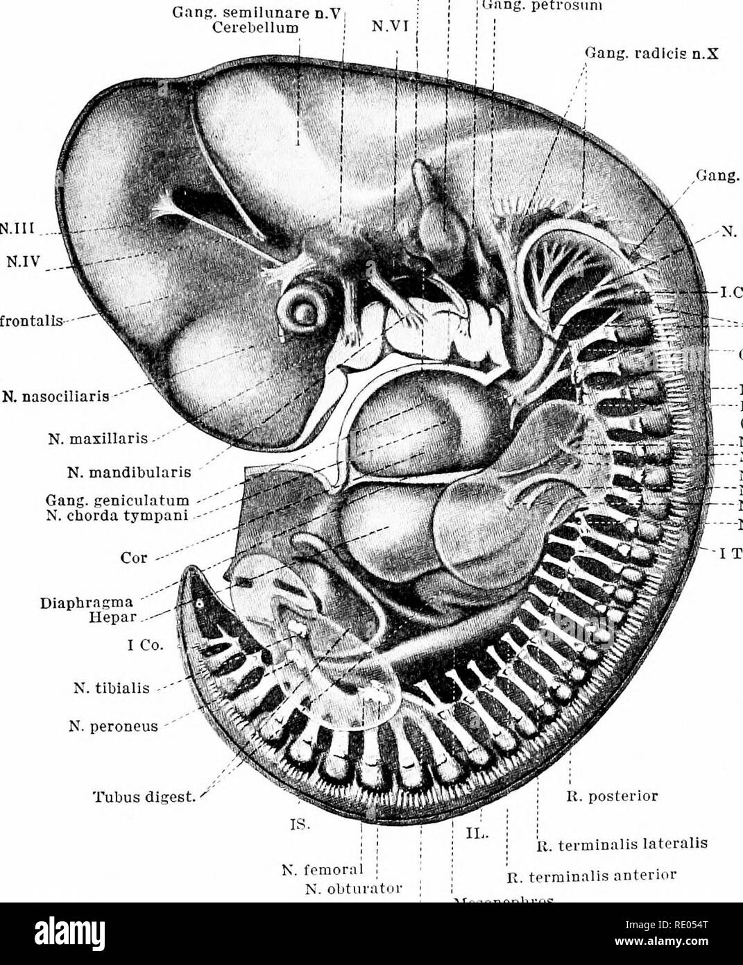 . A laboratory manual and text-book of embryology. Embryology. SPINAL NERVES 353 At the points where the anterior and lateral terminal rami arise, connecting loops may extend from one spinal nerve to another. Thus in the cervical region superficial and deep nerve plexuses are formed. The deep cervical plexus forms the ansa hypoglossi and the phrenic nerve. GftHg. semilunare d.V; Cerebellum i Vesicula auditiva Gang, acusticumj Gang, radieis n IX Gftng. petrosuni Gang, radieis n.X N. frontalis--"". Gang. Proriep , N. hyppglossus Gang, nodos. -N. desc. cerv. -Rami byoid. (Ansa hypogloss Stock Photohttps://www.alamy.com/image-license-details/?v=1https://www.alamy.com/a-laboratory-manual-and-text-book-of-embryology-embryology-spinal-nerves-353-at-the-points-where-the-anterior-and-lateral-terminal-rami-arise-connecting-loops-may-extend-from-one-spinal-nerve-to-another-thus-in-the-cervical-region-superficial-and-deep-nerve-plexuses-are-formed-the-deep-cervical-plexus-forms-the-ansa-hypoglossi-and-the-phrenic-nerve-gfthg-semilunare-dv-cerebellum-i-vesicula-auditiva-gang-acusticumj-gang-radieis-n-ix-gftng-petrosuni-gang-radieis-nx-n-frontalis-quotquot-gang-proriep-n-hyppglossus-gang-nodos-n-desc-cerv-rami-byoid-ansa-hypogloss-image232344024.html
RMRE054T–. A laboratory manual and text-book of embryology. Embryology. SPINAL NERVES 353 At the points where the anterior and lateral terminal rami arise, connecting loops may extend from one spinal nerve to another. Thus in the cervical region superficial and deep nerve plexuses are formed. The deep cervical plexus forms the ansa hypoglossi and the phrenic nerve. GftHg. semilunare d.V; Cerebellum i Vesicula auditiva Gang, acusticumj Gang, radieis n IX Gftng. petrosuni Gang, radieis n.X N. frontalis--"". Gang. Proriep , N. hyppglossus Gang, nodos. -N. desc. cerv. -Rami byoid. (Ansa hypogloss
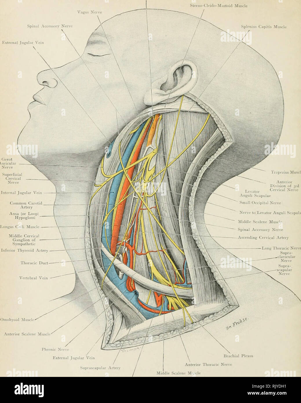 . Atlas of applied (topographical) human anatomy for students and practitioners. Anatomy. Upper Cfrvic;il Ganglion of Sympathetic Stciiuf-Cli'i<ln-M;ist()i(l Muscle Vagus Jferve Spinal Accessory Nerv Splonius Cajjitis Muscle I'"xternal lni^ul.ii 'cin . Omohyoid MnscI Anterior Scalene Miiscl Phrenic Nerve External Jugular Vein Suprascapular Artery Subclavian Aitery Brachial Plexus Anterior Thoracic Nerve Middle Scalene Muscle Fig. 68. Outer Region of Neck. Upper Cervical Ganglia of Sympathetic. Va JSfat. Size. RiOnnau Limited, Lomloii. Rebman Compauy, New Ymlc.. Please note that these Stock Photohttps://www.alamy.com/image-license-details/?v=1https://www.alamy.com/atlas-of-applied-topographical-human-anatomy-for-students-and-practitioners-anatomy-upper-cfrvicil-ganglion-of-sympathetic-stciiuf-cliiltln-mistil-muscle-vagus-jferve-spinal-accessory-nerv-splonius-cajjitis-muscle-iquotxternal-lniulii-cin-omohyoid-mnsci-anterior-scalene-miiscl-phrenic-nerve-external-jugular-vein-suprascapular-artery-subclavian-aitery-brachial-plexus-anterior-thoracic-nerve-middle-scalene-muscle-fig-68-outer-region-of-neck-upper-cervical-ganglia-of-sympathetic-va-jsfat-size-rionnau-limited-lomloii-rebman-compauy-new-ymlc-please-note-that-these-image235401965.html
RMRJYDH1–. Atlas of applied (topographical) human anatomy for students and practitioners. Anatomy. Upper Cfrvic;il Ganglion of Sympathetic Stciiuf-Cli'i<ln-M;ist()i(l Muscle Vagus Jferve Spinal Accessory Nerv Splonius Cajjitis Muscle I'"xternal lni^ul.ii 'cin . Omohyoid MnscI Anterior Scalene Miiscl Phrenic Nerve External Jugular Vein Suprascapular Artery Subclavian Aitery Brachial Plexus Anterior Thoracic Nerve Middle Scalene Muscle Fig. 68. Outer Region of Neck. Upper Cervical Ganglia of Sympathetic. Va JSfat. Size. RiOnnau Limited, Lomloii. Rebman Compauy, New Ymlc.. Please note that these
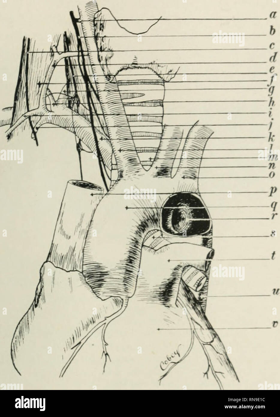 . The anatomical record. Anatomy; Anatomy. ANOMALOUS RIGHT SUBCLAVIAN ARTERY 17. Fig. 1 Drawing from a dissection of the ventral aspect of the nwk anil thorax showing origin, course, and relations of the anomalous right subclavian artorj-. A window is cut into the aorta to show point of origin of anomalous vessel. a, recurrent (inferior) laryngeal nerve b, vagus nerve r, phrenic nerve (/, inferior thyroid artery c, sympathetic cord /, anterior scalene muscle (J, vertebral artery h, transverse cervical artery i, suprascapular artery j, internal mammary artery A', anomalous right subclavian arte Stock Photohttps://www.alamy.com/image-license-details/?v=1https://www.alamy.com/the-anatomical-record-anatomy-anatomy-anomalous-right-subclavian-artery-17-fig-1-drawing-from-a-dissection-of-the-ventral-aspect-of-the-nwk-anil-thorax-showing-origin-course-and-relations-of-the-anomalous-right-subclavian-artorj-a-window-is-cut-into-the-aorta-to-show-point-of-origin-of-anomalous-vessel-a-recurrent-inferior-laryngeal-nerve-b-vagus-nerve-r-phrenic-nerve-inferior-thyroid-artery-c-sympathetic-cord-anterior-scalene-muscle-j-vertebral-artery-h-transverse-cervical-artery-i-suprascapular-artery-j-internal-mammary-artery-a-anomalous-right-subclavian-arte-image236851144.html
RMRN9E1C–. The anatomical record. Anatomy; Anatomy. ANOMALOUS RIGHT SUBCLAVIAN ARTERY 17. Fig. 1 Drawing from a dissection of the ventral aspect of the nwk anil thorax showing origin, course, and relations of the anomalous right subclavian artorj-. A window is cut into the aorta to show point of origin of anomalous vessel. a, recurrent (inferior) laryngeal nerve b, vagus nerve r, phrenic nerve (/, inferior thyroid artery c, sympathetic cord /, anterior scalene muscle (J, vertebral artery h, transverse cervical artery i, suprascapular artery j, internal mammary artery A', anomalous right subclavian arte
 . Elementary text-book of zoology. lii.â-i«i».o i^iooiti^iiuN or Thorax and Neck or a Rabbit from the Ventral Side. (Ad nat.) Internal Carotii Hypoglossal Anterior Facial, Posterior Facial. External Care Thyroid Cartilage (L Anterior Laryngei Thyroid I. ^.Phrer Ni Ths ventral wall of the thorax is removed, the heart is thrown over to the rabbit s right, and the left lung is also drawn over to the right under the left phrenic nerve. The sympathetic nerve and internal jugular veins are omitted in order not branches have been removed. â ⢠â â - ⢠ins are all blue and the arteries. Please note Stock Photohttps://www.alamy.com/image-license-details/?v=1https://www.alamy.com/elementary-text-book-of-zoology-lii-iio-iiooitiiiun-or-thorax-and-neck-or-a-rabbit-from-the-ventral-side-ad-nat-internal-carotii-hypoglossal-anterior-facial-posterior-facial-external-care-thyroid-cartilage-l-anterior-laryngei-thyroid-i-phrer-ni-ths-ventral-wall-of-the-thorax-is-removed-the-heart-is-thrown-over-to-the-rabbit-s-right-and-the-left-lung-is-also-drawn-over-to-the-right-under-the-left-phrenic-nerve-the-sympathetic-nerve-and-internal-jugular-veins-are-omitted-in-order-not-branches-have-been-removed-ins-are-all-blue-and-the-arteries-please-note-image232088490.html
RMRDGF6J–. Elementary text-book of zoology. lii.â-i«i».o i^iooiti^iiuN or Thorax and Neck or a Rabbit from the Ventral Side. (Ad nat.) Internal Carotii Hypoglossal Anterior Facial, Posterior Facial. External Care Thyroid Cartilage (L Anterior Laryngei Thyroid I. ^.Phrer Ni Ths ventral wall of the thorax is removed, the heart is thrown over to the rabbit s right, and the left lung is also drawn over to the right under the left phrenic nerve. The sympathetic nerve and internal jugular veins are omitted in order not branches have been removed. â ⢠â â - ⢠ins are all blue and the arteries. Please note