Quick filters:
Placental attachment Stock Photos and Images
 A manual of obstetrics . h reversal of thedirection of rotation. Meclianism of the Third Stage of Labor.—There are variousviews advanced as to the manner in which the placenta be-comes detached from the uterine wall. The most probableone, and that which is now generally accepted, is that ofdiminution in the area of placental attachment. Accordingto this view the elastic placental tissue follows up the con-tracting uterus to a certain extent, probably to one-half ofits transverse diameter, and then, further condensation of itstissues becoming impossible, with the succeeding pain it is 156 A MAN Stock Photohttps://www.alamy.com/image-license-details/?v=1https://www.alamy.com/a-manual-of-obstetrics-h-reversal-of-thedirection-of-rotation-meclianism-of-the-third-stage-of-laborthere-are-variousviews-advanced-as-to-the-manner-in-which-the-placenta-be-comes-detached-from-the-uterine-wall-the-most-probableone-and-that-which-is-now-generally-accepted-is-that-ofdiminution-in-the-area-of-placental-attachment-accordingto-this-view-the-elastic-placental-tissue-follows-up-the-con-tracting-uterus-to-a-certain-extent-probably-to-one-half-ofits-transverse-diameter-and-then-further-condensation-of-itstissues-becoming-impossible-with-the-succeeding-pain-it-is-156-a-man-image338183297.html
RM2AJ5G6W–A manual of obstetrics . h reversal of thedirection of rotation. Meclianism of the Third Stage of Labor.—There are variousviews advanced as to the manner in which the placenta be-comes detached from the uterine wall. The most probableone, and that which is now generally accepted, is that ofdiminution in the area of placental attachment. Accordingto this view the elastic placental tissue follows up the con-tracting uterus to a certain extent, probably to one-half ofits transverse diameter, and then, further condensation of itstissues becoming impossible, with the succeeding pain it is 156 A MAN
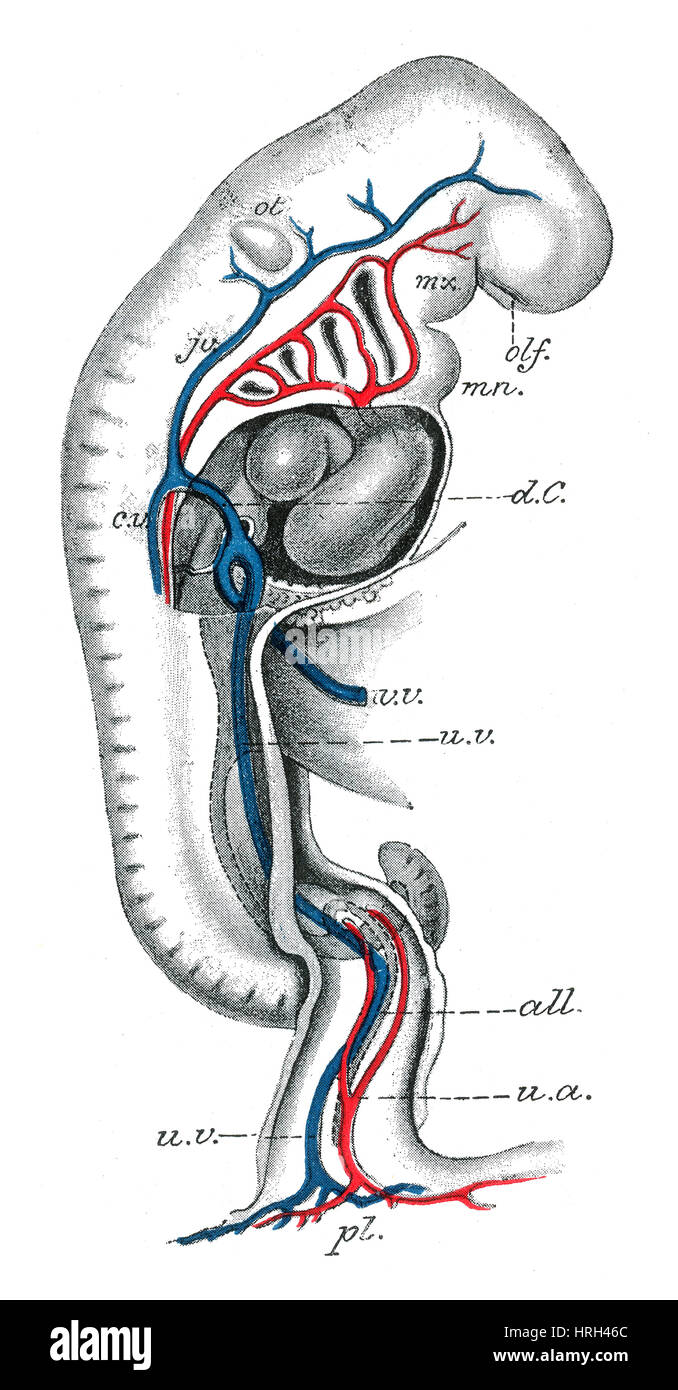 Human Embryo, 3 Weeks Stock Photohttps://www.alamy.com/image-license-details/?v=1https://www.alamy.com/stock-photo-human-embryo-3-weeks-134986164.html
RMHRH46C–Human Embryo, 3 Weeks
 The female Bush Hyrax has a strong attachment to her young and when she has to go off to feed the young instinctively group together in creches Stock Photohttps://www.alamy.com/image-license-details/?v=1https://www.alamy.com/the-female-bush-hyrax-has-a-strong-attachment-to-her-young-and-when-she-has-to-go-off-to-feed-the-young-instinctively-group-together-in-creches-image401455753.html
RM2E93TWD–The female Bush Hyrax has a strong attachment to her young and when she has to go off to feed the young instinctively group together in creches
 A manual of obstetrics . FiG. 71.—Breech presentation,R. S. P. Fig. 72.—Breech presentation,L. S. P. bilicus and toward the back. Fetal Diameters Involved.—The same as in the first position. Steps of the Meehanism.—The same as in the third position, with reversal of thedirection of rotation. Meclianism of the Third Stage of Labor.—There are variousviews advanced as to the manner in which the placenta be-comes detached from the uterine wall. The most probableone, and that which is now generally accepted, is that ofdiminution in the area of placental attachment. Accordingto this view the elastic Stock Photohttps://www.alamy.com/image-license-details/?v=1https://www.alamy.com/a-manual-of-obstetrics-fig-71breech-presentationr-s-p-fig-72breech-presentationl-s-p-bilicus-and-toward-the-back-fetal-diameters-involvedthe-same-as-in-the-first-position-steps-of-the-meehanismthe-same-as-in-the-third-position-with-reversal-of-thedirection-of-rotation-meclianism-of-the-third-stage-of-laborthere-are-variousviews-advanced-as-to-the-manner-in-which-the-placenta-be-comes-detached-from-the-uterine-wall-the-most-probableone-and-that-which-is-now-generally-accepted-is-that-ofdiminution-in-the-area-of-placental-attachment-accordingto-this-view-the-elastic-image338183397.html
RM2AJ5GAD–A manual of obstetrics . FiG. 71.—Breech presentation,R. S. P. Fig. 72.—Breech presentation,L. S. P. bilicus and toward the back. Fetal Diameters Involved.—The same as in the first position. Steps of the Meehanism.—The same as in the third position, with reversal of thedirection of rotation. Meclianism of the Third Stage of Labor.—There are variousviews advanced as to the manner in which the placenta be-comes detached from the uterine wall. The most probableone, and that which is now generally accepted, is that ofdiminution in the area of placental attachment. Accordingto this view the elastic
 A nurse's handbook of obstetrics, for use in training-schools . Fig. 35.—Placental attachment. A, normal attachment at the fundus; B, lateral placentapraevia; C, marginal placenta praevia; D, complete, or central, placenta praevia. Hemorrhage due to the detachment of a normally situatedplacenta may show itself externally or it may be entirely con-cealed, the blood remaining in the uterus and finding room foritself by collecting between the fetal sac and the uterine wall (seeFig. 103). In such a case the only symptoms would be those ofsevere internal hemorrhage already described, together withe Stock Photohttps://www.alamy.com/image-license-details/?v=1https://www.alamy.com/a-nurses-handbook-of-obstetrics-for-use-in-training-schools-fig-35placental-attachment-a-normal-attachment-at-the-fundus-b-lateral-placentapraevia-c-marginal-placenta-praevia-d-complete-or-central-placenta-praevia-hemorrhage-due-to-the-detachment-of-a-normally-situatedplacenta-may-show-itself-externally-or-it-may-be-entirely-con-cealed-the-blood-remaining-in-the-uterus-and-finding-room-foritself-by-collecting-between-the-fetal-sac-and-the-uterine-wall-seefig-103-in-such-a-case-the-only-symptoms-would-be-those-ofsevere-internal-hemorrhage-already-described-together-withe-image342740041.html
RM2AWH4BN–A nurse's handbook of obstetrics, for use in training-schools . Fig. 35.—Placental attachment. A, normal attachment at the fundus; B, lateral placentapraevia; C, marginal placenta praevia; D, complete, or central, placenta praevia. Hemorrhage due to the detachment of a normally situatedplacenta may show itself externally or it may be entirely con-cealed, the blood remaining in the uterus and finding room foritself by collecting between the fetal sac and the uterine wall (seeFig. 103). In such a case the only symptoms would be those ofsevere internal hemorrhage already described, together withe
 A nurse's handbook of obstetrics, for use in training-schools . Fig. 35.—Placental attachment. A, normal attachment at the fundus; B, lateral placentapraevia; C, marginal placenta praevia; D, complete, or central, placenta praevia. Hemorrhage due to the detachment of a normally situatedplacenta may show itself externally or it may be entirely con-cealed, the blood remaining in the uterus and finding room foritself by collecting between the fetal sac and the uterine wall (seeFig. 103). In such a case the only symptoms would be those ofsevere internal hemorrhage already described, together withe Stock Photohttps://www.alamy.com/image-license-details/?v=1https://www.alamy.com/a-nurses-handbook-of-obstetrics-for-use-in-training-schools-fig-35placental-attachment-a-normal-attachment-at-the-fundus-b-lateral-placentapraevia-c-marginal-placenta-praevia-d-complete-or-central-placenta-praevia-hemorrhage-due-to-the-detachment-of-a-normally-situatedplacenta-may-show-itself-externally-or-it-may-be-entirely-con-cealed-the-blood-remaining-in-the-uterus-and-finding-room-foritself-by-collecting-between-the-fetal-sac-and-the-uterine-wall-seefig-103-in-such-a-case-the-only-symptoms-would-be-those-ofsevere-internal-hemorrhage-already-described-together-withe-image339085688.html
RM2AKJK74–A nurse's handbook of obstetrics, for use in training-schools . Fig. 35.—Placental attachment. A, normal attachment at the fundus; B, lateral placentapraevia; C, marginal placenta praevia; D, complete, or central, placenta praevia. Hemorrhage due to the detachment of a normally situatedplacenta may show itself externally or it may be entirely con-cealed, the blood remaining in the uterus and finding room foritself by collecting between the fetal sac and the uterine wall (seeFig. 103). In such a case the only symptoms would be those ofsevere internal hemorrhage already described, together withe
 Fishes . himdcB,or Galeida), a modem offshoot from the Lamnoid type, andespecially characterized by the presence of a third eyelid, thenictitating membrane, which can be drawn across the eye from 19B The True Sharks below. The heterocercal tail has no keel; -the end is bent up-ward; both dorsal fins are present, and the first is well in frontof the ventral fins; the last gill-opening over the base of thepectoral, the head normally formed; these sharks are ovovivipa-rous, the young being hatched* in a sort of uterus, with orwithout placental attachment. Some of these sharks are small, blunt-too Stock Photohttps://www.alamy.com/image-license-details/?v=1https://www.alamy.com/fishes-himdcbor-galeida-a-modem-offshoot-from-the-lamnoid-type-andespecially-characterized-by-the-presence-of-a-third-eyelid-thenictitating-membrane-which-can-be-drawn-across-the-eye-from-19b-the-true-sharks-below-the-heterocercal-tail-has-no-keel-the-end-is-bent-up-ward-both-dorsal-fins-are-present-and-the-first-is-well-in-frontof-the-ventral-fins-the-last-gill-opening-over-the-base-of-thepectoral-the-head-normally-formed-these-sharks-are-ovovivipa-rous-the-young-being-hatched-in-a-sort-of-uterus-with-orwithout-placental-attachment-some-of-these-sharks-are-small-blunt-too-image343056142.html
RM2AX3FH2–Fishes . himdcB,or Galeida), a modem offshoot from the Lamnoid type, andespecially characterized by the presence of a third eyelid, thenictitating membrane, which can be drawn across the eye from 19B The True Sharks below. The heterocercal tail has no keel; -the end is bent up-ward; both dorsal fins are present, and the first is well in frontof the ventral fins; the last gill-opening over the base of thepectoral, the head normally formed; these sharks are ovovivipa-rous, the young being hatched* in a sort of uterus, with orwithout placental attachment. Some of these sharks are small, blunt-too
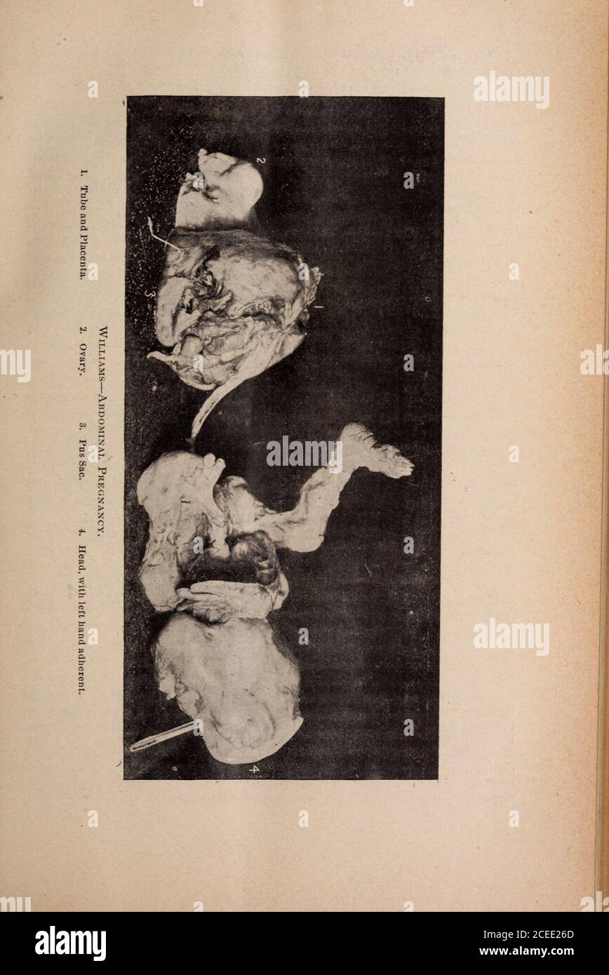 . Tri-State medical journal. * Read before the Central New York Medical Association, Buffalo, N. Y., October16, 1894. From the Buffalo Medical Journal, condensed.. 126 Original Articles. The placenta, which is of good size, was found in ihe expanded Fal-lopian tube, to the right of upper part of fundus of uterus. The umbilicalcord passed through an opening in the tube to the umbilicus of the child. Apart of the tube near the placental attachment was sacculated and containedseveral ounces of pus, which burst during removal, and some of the pus es-caped into the abdominal cavity ; the ovary on t Stock Photohttps://www.alamy.com/image-license-details/?v=1https://www.alamy.com/tri-state-medical-journal-read-before-the-central-new-york-medical-association-buffalo-n-y-october16-1894-from-the-buffalo-medical-journal-condensed-126-original-articles-the-placenta-which-is-of-good-size-was-found-in-ihe-expanded-fal-lopian-tube-to-the-right-of-upper-part-of-fundus-of-uterus-the-umbilicalcord-passed-through-an-opening-in-the-tube-to-the-umbilicus-of-the-child-apart-of-the-tube-near-the-placental-attachment-was-sacculated-and-containedseveral-ounces-of-pus-which-burst-during-removal-and-some-of-the-pus-es-caped-into-the-abdominal-cavity-the-ovary-on-t-image370331989.html
RM2CEE26D–. Tri-State medical journal. * Read before the Central New York Medical Association, Buffalo, N. Y., October16, 1894. From the Buffalo Medical Journal, condensed.. 126 Original Articles. The placenta, which is of good size, was found in ihe expanded Fal-lopian tube, to the right of upper part of fundus of uterus. The umbilicalcord passed through an opening in the tube to the umbilicus of the child. Apart of the tube near the placental attachment was sacculated and containedseveral ounces of pus, which burst during removal, and some of the pus es-caped into the abdominal cavity ; the ovary on t
 . Fishes. Fishes. 198 The True Sharks below. The heterocercal tail has no keel; the end is bent up- ward ; both dorsal fins are present, and the first is well in front of the ventral fins; the last gill-opening over the base of the pectoral, the head normally formed ; these sharks are ovovivipa- rous, the young being hatched in a sort of uterus, with or without placental attachment. Some of these sharks are small, blunt-toothed, and innocuous. Others reach a very large size and are surpassed in voracity only by the various Lammdcs. The genera Cynias and Mustelus, comprising the soft-mouthed or Stock Photohttps://www.alamy.com/image-license-details/?v=1https://www.alamy.com/fishes-fishes-198-the-true-sharks-below-the-heterocercal-tail-has-no-keel-the-end-is-bent-up-ward-both-dorsal-fins-are-present-and-the-first-is-well-in-front-of-the-ventral-fins-the-last-gill-opening-over-the-base-of-the-pectoral-the-head-normally-formed-these-sharks-are-ovovivipa-rous-the-young-being-hatched-in-a-sort-of-uterus-with-or-without-placental-attachment-some-of-these-sharks-are-small-blunt-toothed-and-innocuous-others-reach-a-very-large-size-and-are-surpassed-in-voracity-only-by-the-various-lammdcs-the-genera-cynias-and-mustelus-comprising-the-soft-mouthed-or-image232218942.html
RMRDPDHJ–. Fishes. Fishes. 198 The True Sharks below. The heterocercal tail has no keel; the end is bent up- ward ; both dorsal fins are present, and the first is well in front of the ventral fins; the last gill-opening over the base of the pectoral, the head normally formed ; these sharks are ovovivipa- rous, the young being hatched in a sort of uterus, with or without placental attachment. Some of these sharks are small, blunt-toothed, and innocuous. Others reach a very large size and are surpassed in voracity only by the various Lammdcs. The genera Cynias and Mustelus, comprising the soft-mouthed or
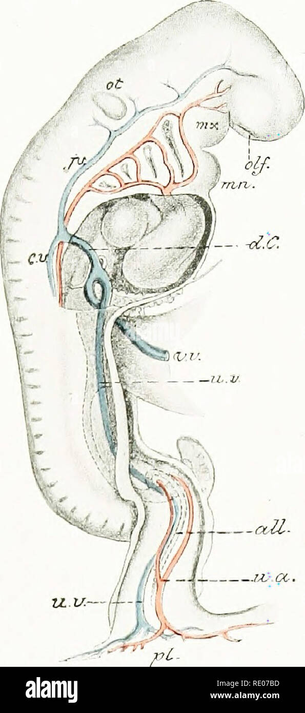 . A laboratory manual and text-book of embryology. Embryology. y Aortic arches 7-4 A It in m Vitello-umbilical vein L. umbilical vein If' Fig. 83.—Ventral reconstruction of a 3.2 mm. embryo, showing vessels (His).. Fig. 84.—Lateral view of human embryo of 4.2 mm., showing aortic arches and venous trunks (His). mx, Maxillary process; mn, mandibular arch; d.C, common cardinal vein; p., anterior cardinal vein; ex., posterior cardinal vein; v.v., vitelline vein; u.a., umbilical artery; n.v , umbilical vein; all., allantois; pi, placental attachment of body-stalk; «//., olfactory pit; ot, otocyst.. Stock Photohttps://www.alamy.com/image-license-details/?v=1https://www.alamy.com/a-laboratory-manual-and-text-book-of-embryology-embryology-y-aortic-arches-7-4-a-it-in-m-vitello-umbilical-vein-l-umbilical-vein-if-fig-83ventral-reconstruction-of-a-32-mm-embryo-showing-vessels-his-fig-84lateral-view-of-human-embryo-of-42-mm-showing-aortic-arches-and-venous-trunks-his-mx-maxillary-process-mn-mandibular-arch-dc-common-cardinal-vein-p-anterior-cardinal-vein-ex-posterior-cardinal-vein-vv-vitelline-vein-ua-umbilical-artery-nv-umbilical-vein-all-allantois-pi-placental-attachment-of-body-stalk-olfactory-pit-ot-otocyst-image232345777.html
RMRE07BD–. A laboratory manual and text-book of embryology. Embryology. y Aortic arches 7-4 A It in m Vitello-umbilical vein L. umbilical vein If' Fig. 83.—Ventral reconstruction of a 3.2 mm. embryo, showing vessels (His).. Fig. 84.—Lateral view of human embryo of 4.2 mm., showing aortic arches and venous trunks (His). mx, Maxillary process; mn, mandibular arch; d.C, common cardinal vein; p., anterior cardinal vein; ex., posterior cardinal vein; v.v., vitelline vein; u.a., umbilical artery; n.v , umbilical vein; all., allantois; pi, placental attachment of body-stalk; «//., olfactory pit; ot, otocyst..
 Veterinary obstetrics, including the diseases of breeding animals and of the new-born . Fig. 79. Cotyledons of a cow, according to Colin. u, Uterus. Ch, Chorion. C, Maternal, C^ fetal portion of cotjdedon.Fetal and maternal portions are partly separated from each other.(Bonnet.) The area, or areas, in the mucosa of the uterus at which elabo-rate changes take place for the attachment and nutrition of thefetus, is known as the maternal placenta and the correspondingportion or portions of the chorion which sends capillary tuftsinto the placental area of the uterus, constitute the fetal placenta. Stock Photohttps://www.alamy.com/image-license-details/?v=1https://www.alamy.com/veterinary-obstetrics-including-the-diseases-of-breeding-animals-and-of-the-new-born-fig-79-cotyledons-of-a-cow-according-to-colin-u-uterus-ch-chorion-c-maternal-c-fetal-portion-of-cotjdedonfetal-and-maternal-portions-are-partly-separated-from-each-otherbonnet-the-area-or-areas-in-the-mucosa-of-the-uterus-at-which-elabo-rate-changes-take-place-for-the-attachment-and-nutrition-of-thefetus-is-known-as-the-maternal-placenta-and-the-correspondingportion-or-portions-of-the-chorion-which-sends-capillary-tuftsinto-the-placental-area-of-the-uterus-constitute-the-fetal-placenta-image340309815.html
RM2ANJCHY–Veterinary obstetrics, including the diseases of breeding animals and of the new-born . Fig. 79. Cotyledons of a cow, according to Colin. u, Uterus. Ch, Chorion. C, Maternal, C^ fetal portion of cotjdedon.Fetal and maternal portions are partly separated from each other.(Bonnet.) The area, or areas, in the mucosa of the uterus at which elabo-rate changes take place for the attachment and nutrition of thefetus, is known as the maternal placenta and the correspondingportion or portions of the chorion which sends capillary tuftsinto the placental area of the uterus, constitute the fetal placenta.
 A textbook of obstetrics . j<». PLATE 2.. Anomalies of the Placenta: I, Placenta with irregular lobes (Auvard); 2, placenta intwo unequal lobes (Auvard); J, irregular placenta (Auvard); 4, small accessory placenta(Ribemont-Lepage); 5, placenta succenturiata (Ribemont-Lepage); 6. placenta, oval (Auvard); 7. placenta with velamentous attachLepage) ; 8, placenta with two equal lobes (Ribemont Lepage). attachment of cord (Ribemont- THE PLACENTA. 12 1 normal in this disease and the placenta may continue to performits physiological functions. Degeneration of the Placental Villi.—The morbid proces Stock Photohttps://www.alamy.com/image-license-details/?v=1https://www.alamy.com/a-textbook-of-obstetrics-jlt-plate-2-anomalies-of-the-placenta-i-placenta-with-irregular-lobes-auvard-2-placenta-intwo-unequal-lobes-auvard-j-irregular-placenta-auvard-4-small-accessory-placentaribemont-lepage-5-placenta-succenturiata-ribemont-lepage-6-placenta-oval-auvard-7-placenta-with-velamentous-attachlepage-8-placenta-with-two-equal-lobes-ribemont-lepage-attachment-of-cord-ribemont-the-placenta-12-1-normal-in-this-disease-and-the-placenta-may-continue-to-performits-physiological-functions-degeneration-of-the-placental-villithe-morbid-proces-image343078471.html
RM2AX4G2F–A textbook of obstetrics . j<». PLATE 2.. Anomalies of the Placenta: I, Placenta with irregular lobes (Auvard); 2, placenta intwo unequal lobes (Auvard); J, irregular placenta (Auvard); 4, small accessory placenta(Ribemont-Lepage); 5, placenta succenturiata (Ribemont-Lepage); 6. placenta, oval (Auvard); 7. placenta with velamentous attachLepage) ; 8, placenta with two equal lobes (Ribemont Lepage). attachment of cord (Ribemont- THE PLACENTA. 12 1 normal in this disease and the placenta may continue to performits physiological functions. Degeneration of the Placental Villi.—The morbid proces
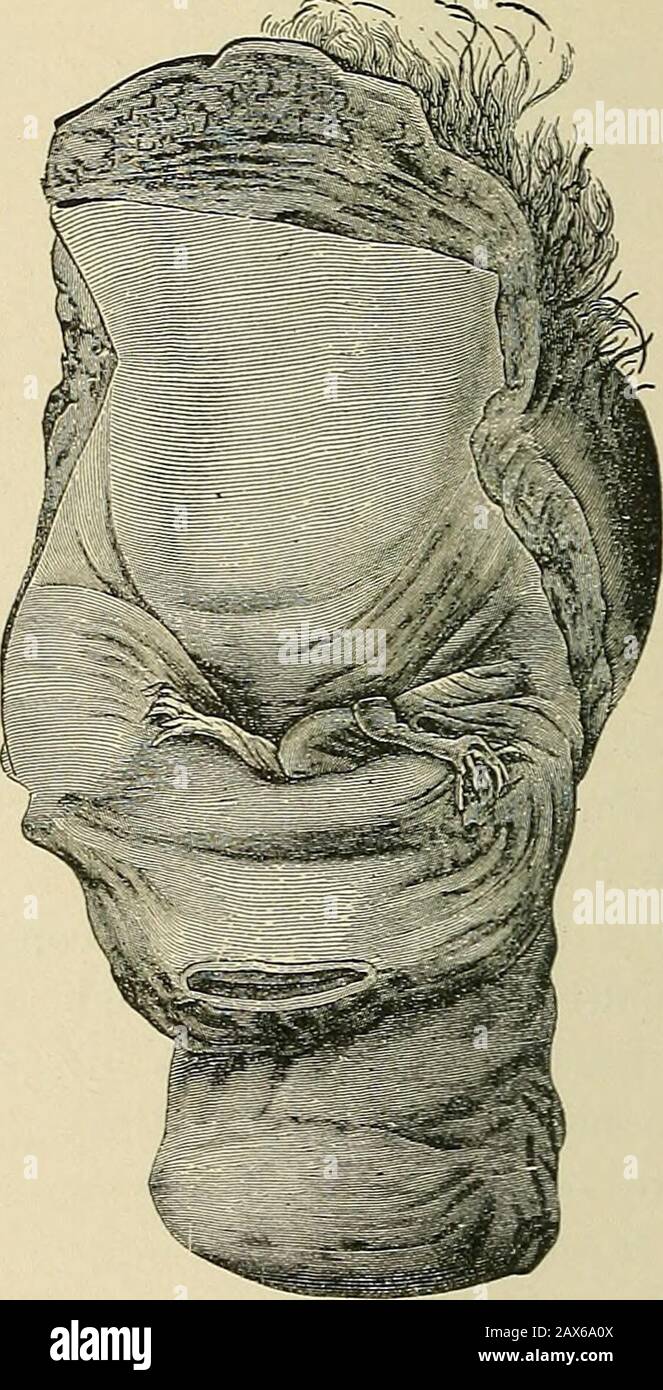 The American text-book of obstetrics for practitioners and students . Fig. 116.—Inversion of uterus: drawing from anold specimen in alcohol. The atonic chief site ofplacental attachment (c) is shrunken by the alco-hol, and thus its lessening is explained; 6, contrac-tion-ring ; o, external os uteri (after J. Veit). Fig. 117.—Inversion of the uterus. The lumenof the rectum is seen, and also the inversion fun-nel in which are the tubes and an ovary (after J.Veit). placental site existing, simply the weight of the placenta may cause sinkingof that portion of the uterus in the cavity. Such occurre Stock Photohttps://www.alamy.com/image-license-details/?v=1https://www.alamy.com/the-american-text-book-of-obstetrics-for-practitioners-and-students-fig-116inversion-of-uterus-drawing-from-anold-specimen-in-alcohol-the-atonic-chief-site-ofplacental-attachment-c-is-shrunken-by-the-alco-hol-and-thus-its-lessening-is-explained-6-contrac-tion-ring-o-external-os-uteri-after-j-veit-fig-117inversion-of-the-uterus-the-lumenof-the-rectum-is-seen-and-also-the-inversion-fun-nel-in-which-are-the-tubes-and-an-ovary-after-jveit-placental-site-existing-simply-the-weight-of-the-placenta-may-cause-sinkingof-that-portion-of-the-uterus-in-the-cavity-such-occurre-image343117626.html
RM2AX6A0X–The American text-book of obstetrics for practitioners and students . Fig. 116.—Inversion of uterus: drawing from anold specimen in alcohol. The atonic chief site ofplacental attachment (c) is shrunken by the alco-hol, and thus its lessening is explained; 6, contrac-tion-ring ; o, external os uteri (after J. Veit). Fig. 117.—Inversion of the uterus. The lumenof the rectum is seen, and also the inversion fun-nel in which are the tubes and an ovary (after J.Veit). placental site existing, simply the weight of the placenta may cause sinkingof that portion of the uterus in the cavity. Such occurre
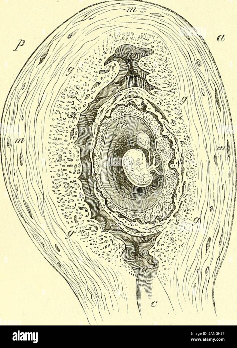 Quain's elements of anatomy . placenta, thus including a placental and a uterinedecidua serotina. Fig- 673. Fig. 673. — Semi-diagram- matic OUTLINE OF ANANTERO-POSTERIOK, SECTIONOF THE GRAVID UTERUSAND OVUM OP FIVE WEEKS (A.T.). This drawing is taken froma very perfect specimen ofthe titerus obtained from thebody of a woman who died ofcholera in 1849. a, anterior uterine wall in-side which was situated theplacental attachment of theovum; p, posterior uterinewall (the accessory parts beingomitted) ; m, muscular sub-stance of the wall; v,thickened lining membraneforming decidua vera, andshowing Stock Photohttps://www.alamy.com/image-license-details/?v=1https://www.alamy.com/quains-elements-of-anatomy-placenta-thus-including-a-placental-and-a-uterinedecidua-serotina-fig-673-fig-673-semi-diagram-matic-outline-of-anantero-posteriok-sectionof-the-gravid-uterusand-ovum-op-five-weeks-at-this-drawing-is-taken-froma-very-perfect-specimen-ofthe-titerus-obtained-from-thebody-of-a-woman-who-died-ofcholera-in-1849-a-anterior-uterine-wall-in-side-which-was-situated-theplacental-attachment-of-theovum-p-posterior-uterinewall-the-accessory-parts-beingomitted-m-muscular-sub-stance-of-the-wall-vthickened-lining-membraneforming-decidua-vera-andshowing-image340269419.html
RM2ANGH37–Quain's elements of anatomy . placenta, thus including a placental and a uterinedecidua serotina. Fig- 673. Fig. 673. — Semi-diagram- matic OUTLINE OF ANANTERO-POSTERIOK, SECTIONOF THE GRAVID UTERUSAND OVUM OP FIVE WEEKS (A.T.). This drawing is taken froma very perfect specimen ofthe titerus obtained from thebody of a woman who died ofcholera in 1849. a, anterior uterine wall in-side which was situated theplacental attachment of theovum; p, posterior uterinewall (the accessory parts beingomitted) ; m, muscular sub-stance of the wall; v,thickened lining membraneforming decidua vera, andshowing
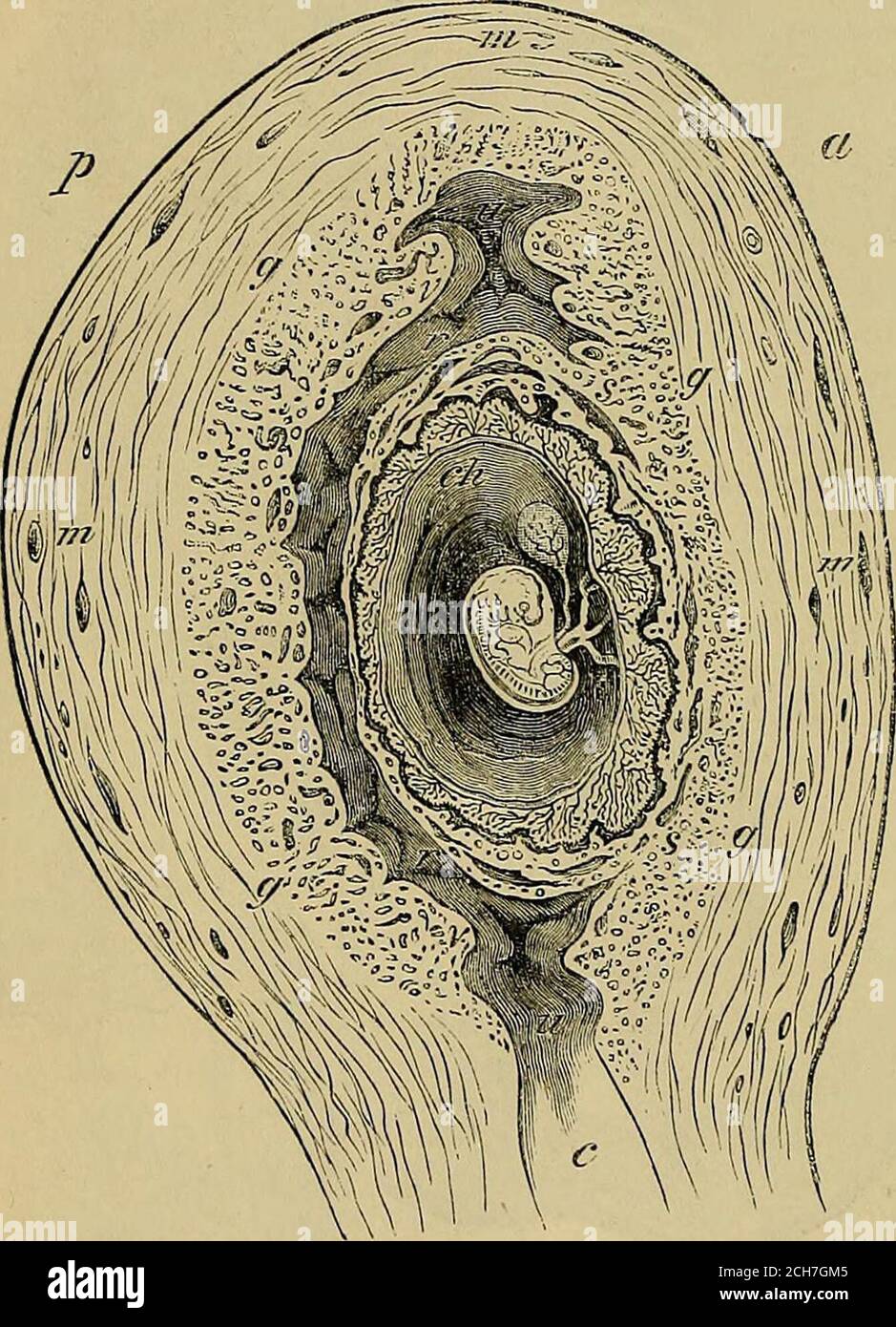 . Quain's elements of anatomy . acenta, thus including a placental and a uterinedecidua serotina. Fig. 673. Fig. 673. — Semi-diagram- matic OUTLINE OF ANANTEEO-POSTERIOR SECTIONOP THE GRAVID UTERUS■AND OVUM OF FIVE -WEEKS (A.T.). This drawing is taken froma very perfect specimen ofthe uterus obtained from tliebody of a woman who died ofcholera in 1849. a, anterior uterine wall in-side which was situated theplacental attachment of theovum; p, posterior uterinewall (the accessory parts beingomitted) ; m, muscular sub-stance of the wall; r,thickened lining membraneforming decidua vera, andshowing Stock Photohttps://www.alamy.com/image-license-details/?v=1https://www.alamy.com/quains-elements-of-anatomy-acenta-thus-including-a-placental-and-a-uterinedecidua-serotina-fig-673-fig-673-semi-diagram-matic-outline-of-ananteeo-posterior-sectionop-the-gravid-uterusand-ovum-of-five-weeks-at-this-drawing-is-taken-froma-very-perfect-specimen-ofthe-uterus-obtained-from-tliebody-of-a-woman-who-died-ofcholera-in-1849-a-anterior-uterine-wall-in-side-which-was-situated-theplacental-attachment-of-theovum-p-posterior-uterinewall-the-accessory-parts-beingomitted-m-muscular-sub-stance-of-the-wall-rthickened-lining-membraneforming-decidua-vera-andshowing-image372033653.html
RM2CH7GM5–. Quain's elements of anatomy . acenta, thus including a placental and a uterinedecidua serotina. Fig. 673. Fig. 673. — Semi-diagram- matic OUTLINE OF ANANTEEO-POSTERIOR SECTIONOP THE GRAVID UTERUS■AND OVUM OF FIVE -WEEKS (A.T.). This drawing is taken froma very perfect specimen ofthe uterus obtained from tliebody of a woman who died ofcholera in 1849. a, anterior uterine wall in-side which was situated theplacental attachment of theovum; p, posterior uterinewall (the accessory parts beingomitted) ; m, muscular sub-stance of the wall; r,thickened lining membraneforming decidua vera, andshowing
 . The physiology of reproduction. Reproduction. 394 THE PHYSIOLOGY OF REPRODUCTION III. The Placenta in Indeciduata In the placental Mammals, an attachment takes place be- tween maternal and foetal tissues in the uterus, and the tropho- blast is vascularised, except in the Primates, by the allantois. The method of attachment varies in different orders, and sometimes in different groups of an order. In the Indeciduata,. Fig. 87.—Portion of the injected chorion of the pig. The figure shows a minute circular spot, h, enclosed by a vascular ring from which villous ridges (r, r) radiate (Turner). ( Stock Photohttps://www.alamy.com/image-license-details/?v=1https://www.alamy.com/the-physiology-of-reproduction-reproduction-394-the-physiology-of-reproduction-iii-the-placenta-in-indeciduata-in-the-placental-mammals-an-attachment-takes-place-be-tween-maternal-and-foetal-tissues-in-the-uterus-and-the-tropho-blast-is-vascularised-except-in-the-primates-by-the-allantois-the-method-of-attachment-varies-in-different-orders-and-sometimes-in-different-groups-of-an-order-in-the-indeciduata-fig-87portion-of-the-injected-chorion-of-the-pig-the-figure-shows-a-minute-circular-spot-h-enclosed-by-a-vascular-ring-from-which-villous-ridges-r-r-radiate-turner-image232371949.html
RMRE1CP5–. The physiology of reproduction. Reproduction. 394 THE PHYSIOLOGY OF REPRODUCTION III. The Placenta in Indeciduata In the placental Mammals, an attachment takes place be- tween maternal and foetal tissues in the uterus, and the tropho- blast is vascularised, except in the Primates, by the allantois. The method of attachment varies in different orders, and sometimes in different groups of an order. In the Indeciduata,. Fig. 87.—Portion of the injected chorion of the pig. The figure shows a minute circular spot, h, enclosed by a vascular ring from which villous ridges (r, r) radiate (Turner). (
 . Anatomischer Anzeiger. Anatomy, Comparative; Anatomy, Comparative. blast at the animal pole continues to remain distinct from the under- lying layers (Fig. 2). Upon reaching the placental zone, the vesicle, which has now attained a diameter of about 430 mm, becomes attached to the uterine mucosa^of this area. At first the attachment could be more properly spoken of as simply an adhesion. There is here a slight break in my series, and I am therefore not able to speak with con- fidence concerning the changes that immediately follow the attachment of the vesicle; but from what takes place later Stock Photohttps://www.alamy.com/image-license-details/?v=1https://www.alamy.com/anatomischer-anzeiger-anatomy-comparative-anatomy-comparative-blast-at-the-animal-pole-continues-to-remain-distinct-from-the-under-lying-layers-fig-2-upon-reaching-the-placental-zone-the-vesicle-which-has-now-attained-a-diameter-of-about-430-mm-becomes-attached-to-the-uterine-mucosaof-this-area-at-first-the-attachment-could-be-more-properly-spoken-of-as-simply-an-adhesion-there-is-here-a-slight-break-in-my-series-and-i-am-therefore-not-able-to-speak-with-con-fidence-concerning-the-changes-that-immediately-follow-the-attachment-of-the-vesicle-but-from-what-takes-place-later-image236807043.html
RMRN7DPB–. Anatomischer Anzeiger. Anatomy, Comparative; Anatomy, Comparative. blast at the animal pole continues to remain distinct from the under- lying layers (Fig. 2). Upon reaching the placental zone, the vesicle, which has now attained a diameter of about 430 mm, becomes attached to the uterine mucosa^of this area. At first the attachment could be more properly spoken of as simply an adhesion. There is here a slight break in my series, and I am therefore not able to speak with con- fidence concerning the changes that immediately follow the attachment of the vesicle; but from what takes place later
 . Anatomischer Anzeiger. Anatomy, Comparative; Anatomy, Comparative. 371. blast at the animal pole continues to remain distinct from the under- lying layers (Fig. 2). Upon reaching the placental zone, the vesicle, which has now attained a diameter of about 430 mm, becomes attached to the uterine mucosa^of this area. At first the attachment could be more properly spoken of as simply an adhesion. There is here a slight break in my series, and I am therefore not able to speak with con- fidence concerning the changes that immediately follow the attachment of the vesicle; but from what takes place Stock Photohttps://www.alamy.com/image-license-details/?v=1https://www.alamy.com/anatomischer-anzeiger-anatomy-comparative-anatomy-comparative-371-blast-at-the-animal-pole-continues-to-remain-distinct-from-the-under-lying-layers-fig-2-upon-reaching-the-placental-zone-the-vesicle-which-has-now-attained-a-diameter-of-about-430-mm-becomes-attached-to-the-uterine-mucosaof-this-area-at-first-the-attachment-could-be-more-properly-spoken-of-as-simply-an-adhesion-there-is-here-a-slight-break-in-my-series-and-i-am-therefore-not-able-to-speak-with-con-fidence-concerning-the-changes-that-immediately-follow-the-attachment-of-the-vesicle-but-from-what-takes-place-image236807051.html
RMRN7DPK–. Anatomischer Anzeiger. Anatomy, Comparative; Anatomy, Comparative. 371. blast at the animal pole continues to remain distinct from the under- lying layers (Fig. 2). Upon reaching the placental zone, the vesicle, which has now attained a diameter of about 430 mm, becomes attached to the uterine mucosa^of this area. At first the attachment could be more properly spoken of as simply an adhesion. There is here a slight break in my series, and I am therefore not able to speak with con- fidence concerning the changes that immediately follow the attachment of the vesicle; but from what takes place