Quick filters:
Plant cell microscope Stock Photos and Images
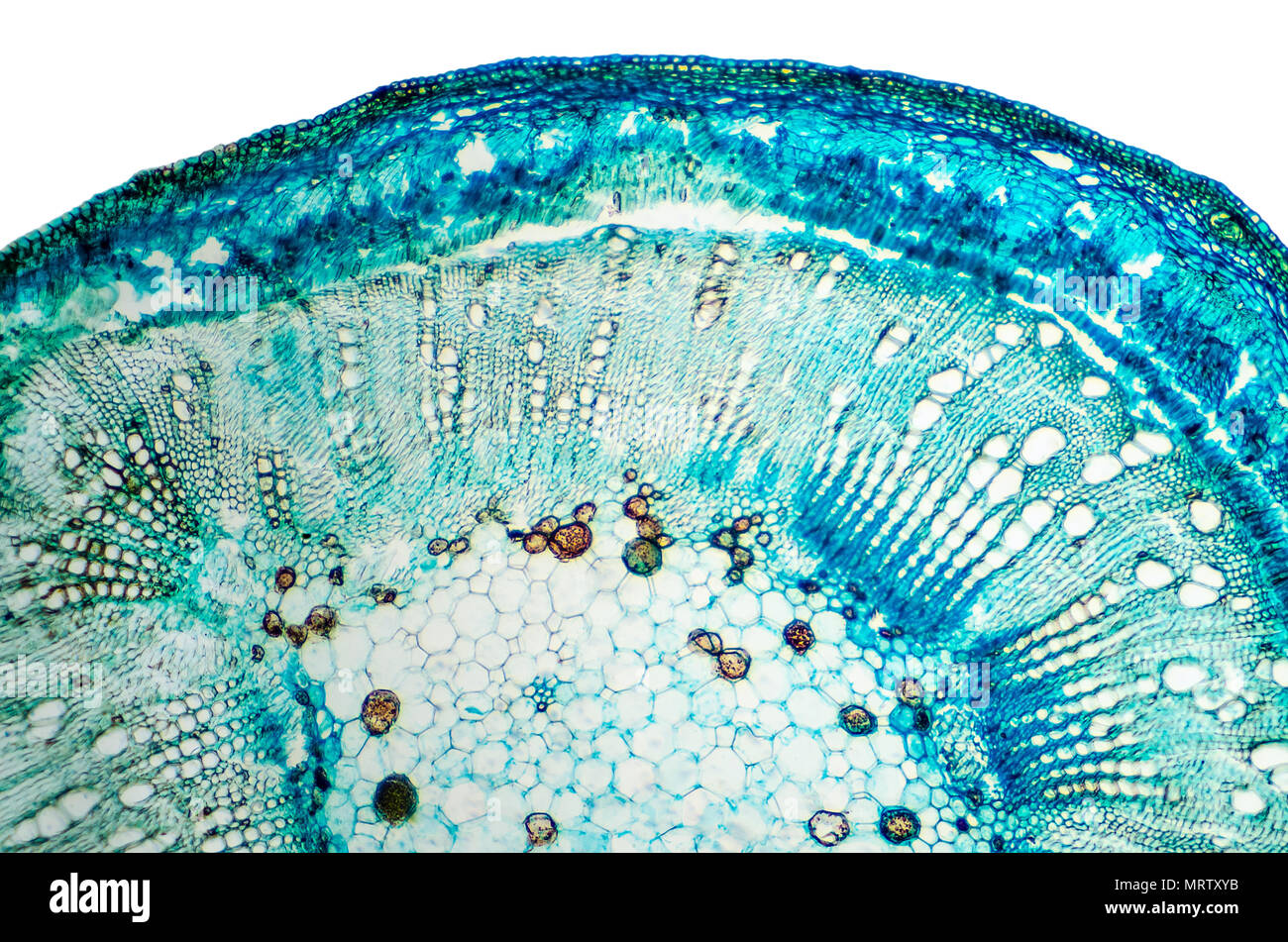 Stem of cotton cross section. Light microscope slide with microsection of plants of the genus Gossypium in the mallow family Malvaceae. Plant anatomy. Stock Photohttps://www.alamy.com/image-license-details/?v=1https://www.alamy.com/stem-of-cotton-cross-section-light-microscope-slide-with-microsection-of-plants-of-the-genus-gossypium-in-the-mallow-family-malvaceae-plant-anatomy-image186788767.html
RFMRTXYB–Stem of cotton cross section. Light microscope slide with microsection of plants of the genus Gossypium in the mallow family Malvaceae. Plant anatomy.
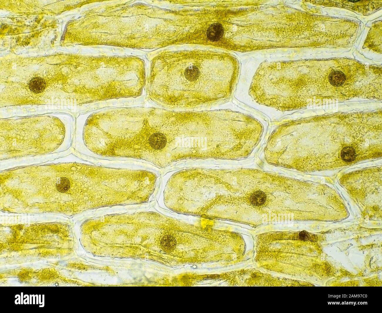 Onion skin cells under the microscope, horizontal field of view is about 0.61 mm Stock Photohttps://www.alamy.com/image-license-details/?v=1https://www.alamy.com/onion-skin-cells-under-the-microscope-horizontal-field-of-view-is-about-061-mm-image339493504.html
RM2AM97C0–Onion skin cells under the microscope, horizontal field of view is about 0.61 mm
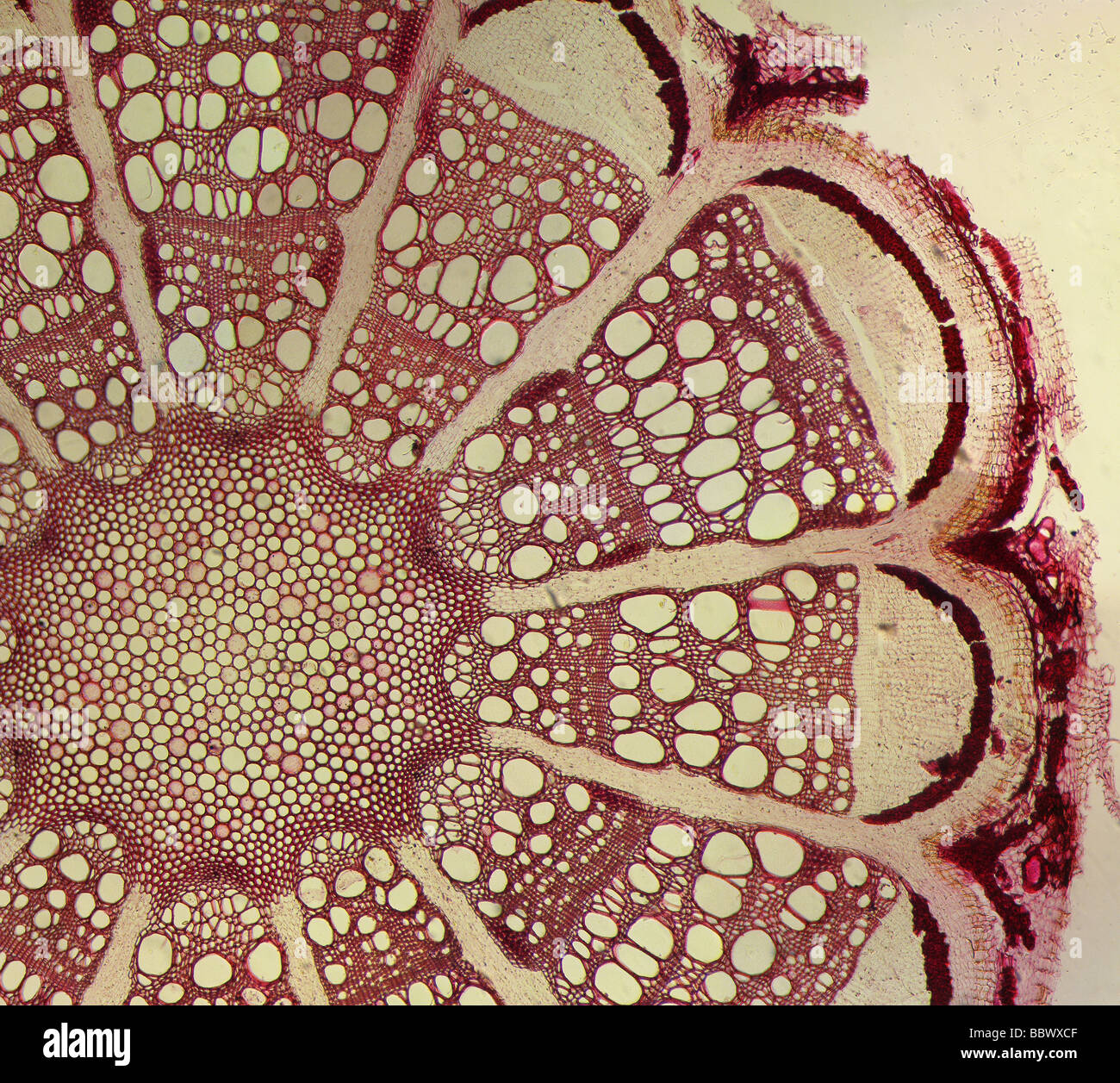 microphotograph of a stained clematis plant stem cross section slide through a microscope Stock Photohttps://www.alamy.com/image-license-details/?v=1https://www.alamy.com/stock-photo-microphotograph-of-a-stained-clematis-plant-stem-cross-section-slide-24541119.html
RFBBWXCF–microphotograph of a stained clematis plant stem cross section slide through a microscope
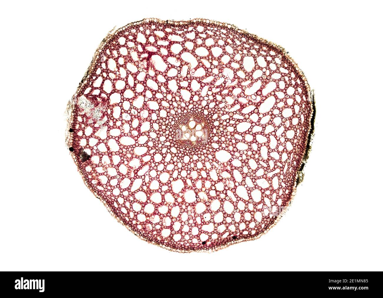 cross section cut under the microscope – microscopic view of plant cells for botanic education Stock Photohttps://www.alamy.com/image-license-details/?v=1https://www.alamy.com/cross-section-cut-under-the-microscope-microscopic-view-of-plant-cells-for-botanic-education-image396908853.html
RF2E1MN85–cross section cut under the microscope – microscopic view of plant cells for botanic education
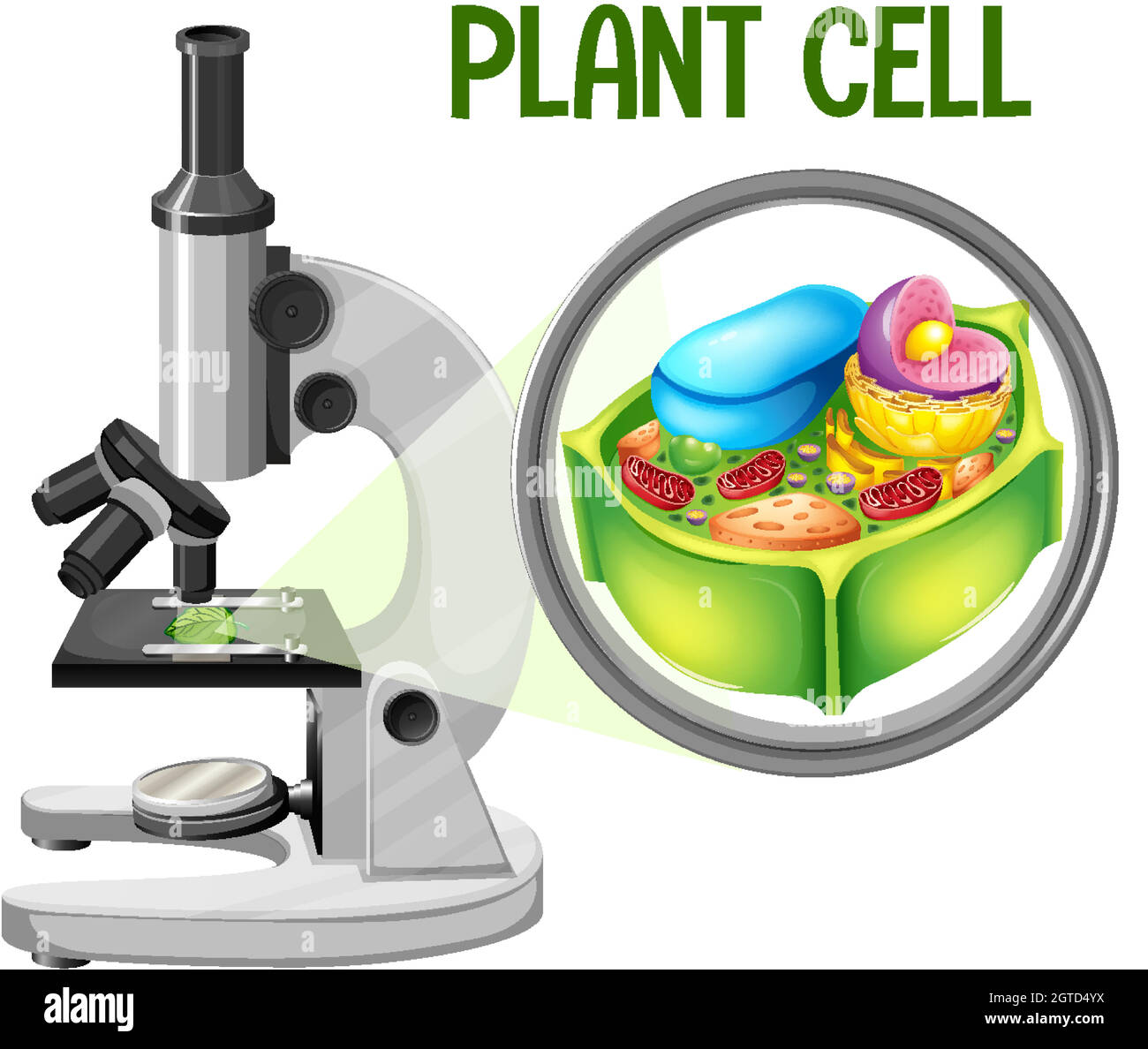 Microscope with plant cell diagram Stock Vectorhttps://www.alamy.com/image-license-details/?v=1https://www.alamy.com/microscope-with-plant-cell-diagram-image445300238.html
RF2GTD4YX–Microscope with plant cell diagram
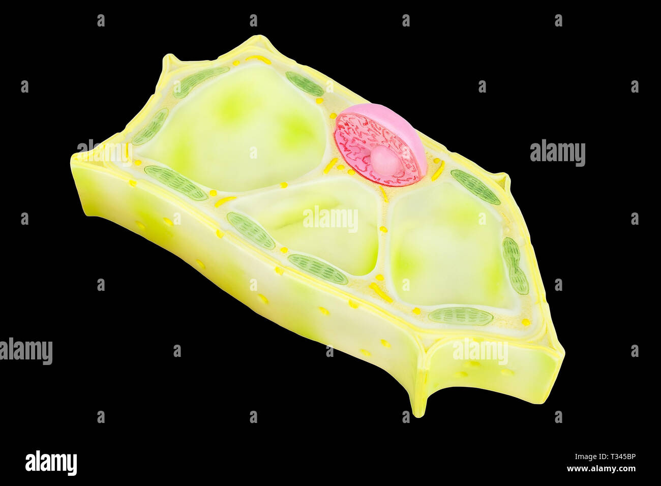 Plant cell model for education isolated on black background Stock Photohttps://www.alamy.com/image-license-details/?v=1https://www.alamy.com/plant-cell-model-for-education-isolated-on-black-background-image242881178.html
RFT345BP–Plant cell model for education isolated on black background
 Research into cytoplasmic streaming. Cells of aquatic plant Chara coralline in Petri dish beneath microscope objective. UK Stock Photohttps://www.alamy.com/image-license-details/?v=1https://www.alamy.com/research-into-cytoplasmic-streaming-cells-of-aquatic-plant-chara-coralline-image62268373.html
RMDH8FXD–Research into cytoplasmic streaming. Cells of aquatic plant Chara coralline in Petri dish beneath microscope objective. UK
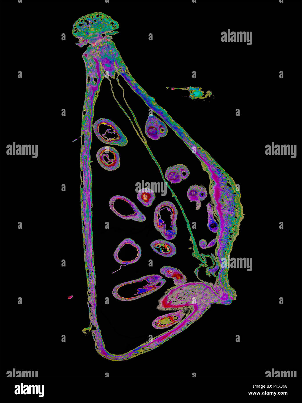 Micro Photography of Shepherd's Purse Mature Embryo Capsella Stock Photohttps://www.alamy.com/image-license-details/?v=1https://www.alamy.com/micro-photography-of-shepherds-purse-mature-embryo-capsella-image218776160.html
RMPKX368–Micro Photography of Shepherd's Purse Mature Embryo Capsella
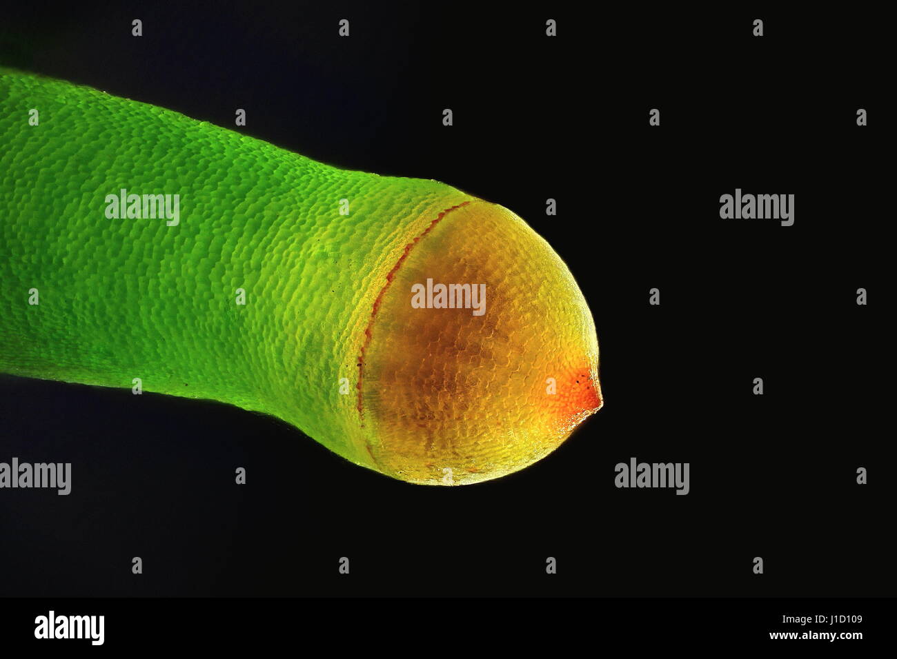 Spore capsule of Plagiomnium affine, Many-fruited Thyme-moss, microscope image Stock Photohttps://www.alamy.com/image-license-details/?v=1https://www.alamy.com/stock-photo-spore-capsule-of-plagiomnium-affine-many-fruited-thyme-moss-microscope-138583769.html
RFJ1D109–Spore capsule of Plagiomnium affine, Many-fruited Thyme-moss, microscope image
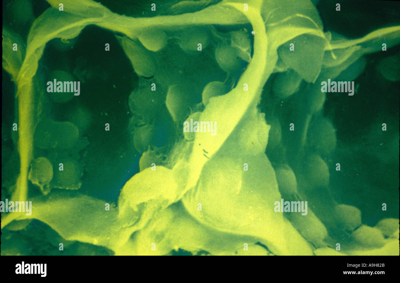 Scientific SEM of plant cell shows chloroplasts and cell walls mag x 2190 1 95 9 33 Stock Photohttps://www.alamy.com/image-license-details/?v=1https://www.alamy.com/scientific-sem-of-plant-cell-shows-chloroplasts-and-cell-walls-mag-image2283562.html
RMA9H82B–Scientific SEM of plant cell shows chloroplasts and cell walls mag x 2190 1 95 9 33
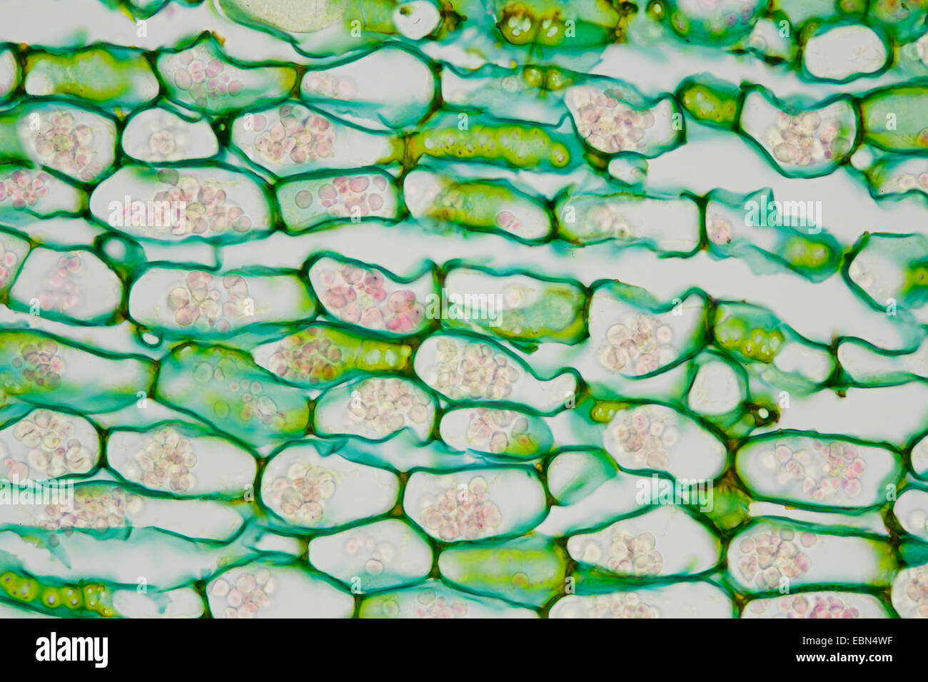 horse-radish (Armoracia rusticana), microscopical cut of a root with starch grains Stock Photohttps://www.alamy.com/image-license-details/?v=1https://www.alamy.com/stock-photo-horse-radish-armoracia-rusticana-microscopical-cut-of-a-root-with-76067531.html
RMEBN4WF–horse-radish (Armoracia rusticana), microscopical cut of a root with starch grains
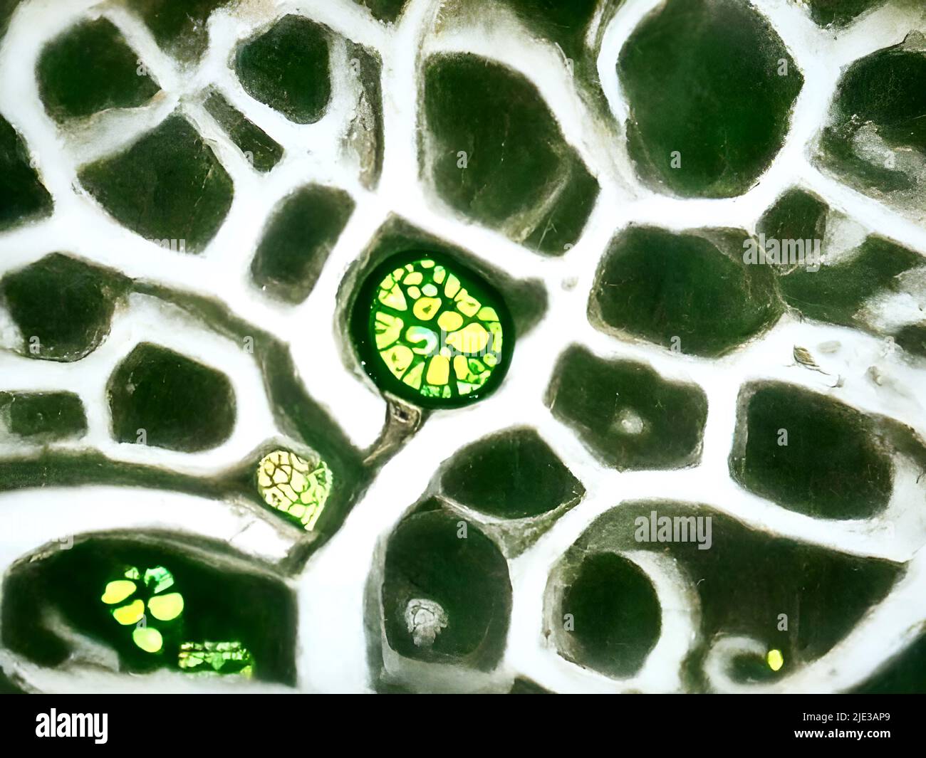 Plant cells under the microscope Stock Photohttps://www.alamy.com/image-license-details/?v=1https://www.alamy.com/plant-cells-under-the-microscope-image473359441.html
RM2JE3AP9–Plant cells under the microscope
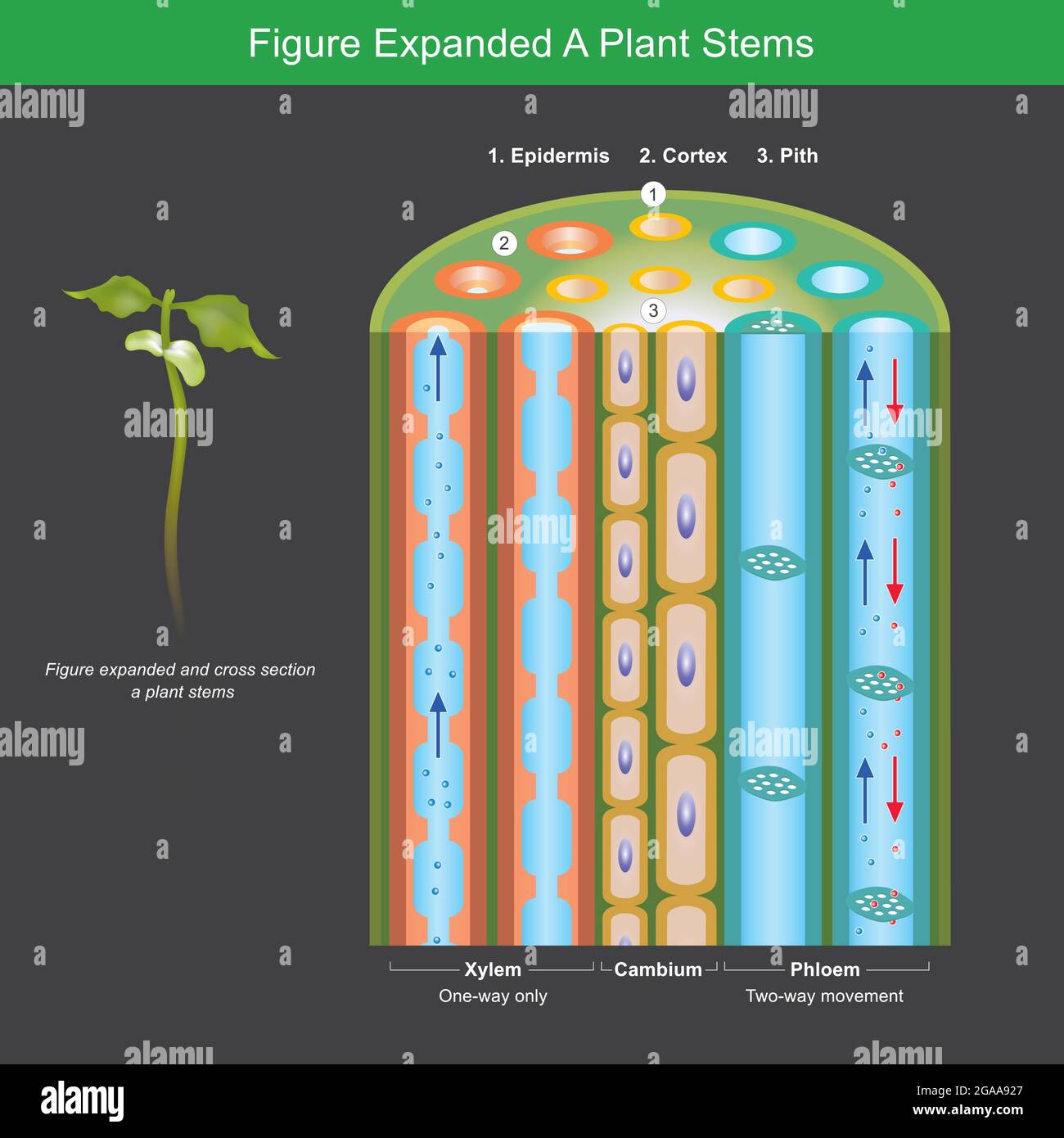 Figure Expanded A Plant Stems. Figure expanded for explain a plants transport nutrient and water in stems. Illustration. Stock Vectorhttps://www.alamy.com/image-license-details/?v=1https://www.alamy.com/figure-expanded-a-plant-stems-figure-expanded-for-explain-a-plants-transport-nutrient-and-water-in-stems-illustration-image436632399.html
RF2GAA927–Figure Expanded A Plant Stems. Figure expanded for explain a plants transport nutrient and water in stems. Illustration.
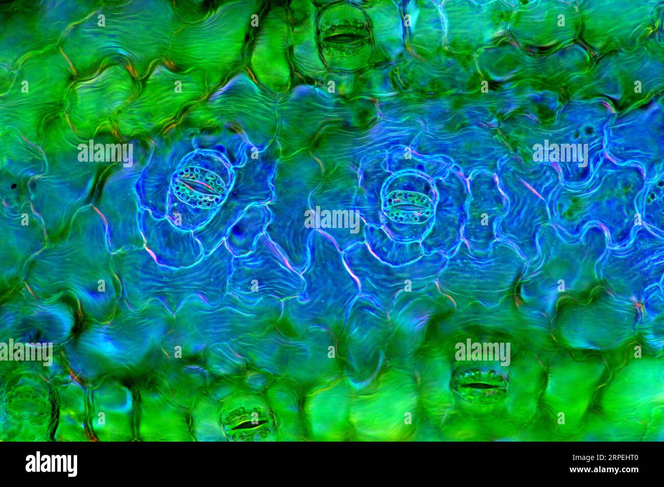 The image presents stomata in Spathiphyllum leaf epidermis, photographed through the microscope in polarized light at a magnification of 100X Stock Photohttps://www.alamy.com/image-license-details/?v=1https://www.alamy.com/the-image-presents-stomata-in-spathiphyllum-leaf-epidermis-photographed-through-the-microscope-in-polarized-light-at-a-magnification-of-100x-image564575536.html
RM2RPEHT0–The image presents stomata in Spathiphyllum leaf epidermis, photographed through the microscope in polarized light at a magnification of 100X
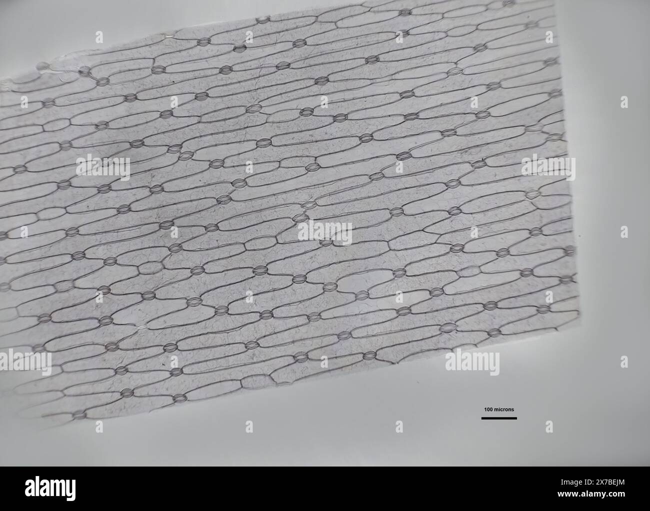 monocot leaf under a microscope Stock Photohttps://www.alamy.com/image-license-details/?v=1https://www.alamy.com/monocot-leaf-under-a-microscope-image606918444.html
RF2X7BEJM–monocot leaf under a microscope
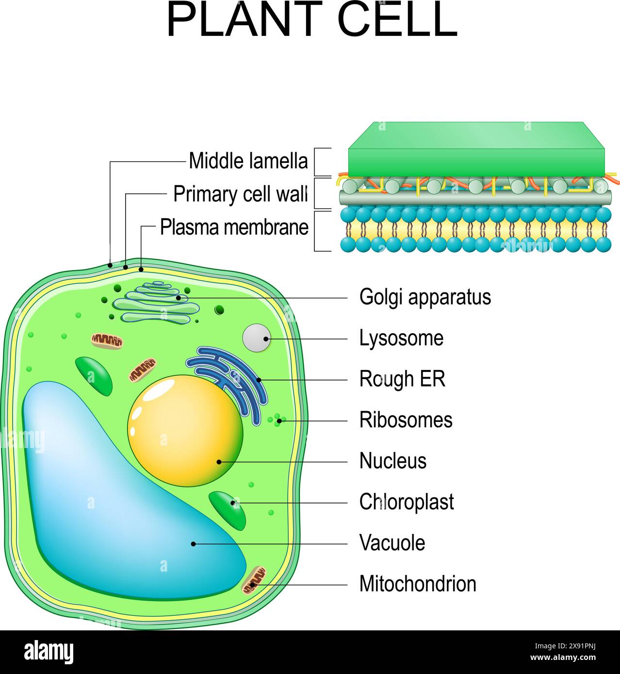 Plant cell. Structure and Anatomy. Close-up of layers of a cell wall. Vector illustration Stock Vectorhttps://www.alamy.com/image-license-details/?v=1https://www.alamy.com/plant-cell-structure-and-anatomy-close-up-of-layers-of-a-cell-wall-vector-illustration-image607934590.html
RF2X91PNJ–Plant cell. Structure and Anatomy. Close-up of layers of a cell wall. Vector illustration
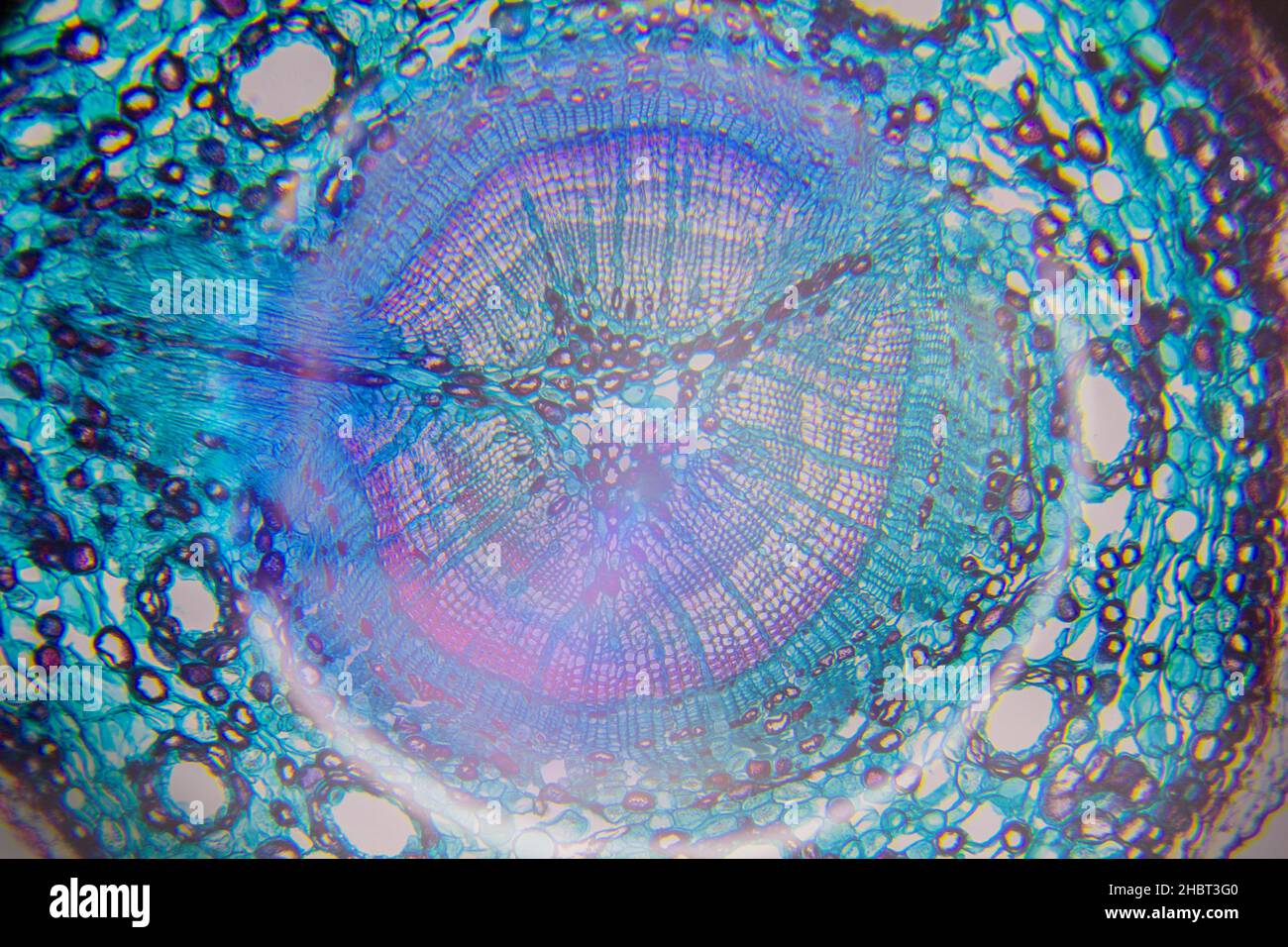 Microscopy of a tree cell Stock Photohttps://www.alamy.com/image-license-details/?v=1https://www.alamy.com/microscopy-of-a-tree-cell-image454760432.html
RF2HBT3G0–Microscopy of a tree cell
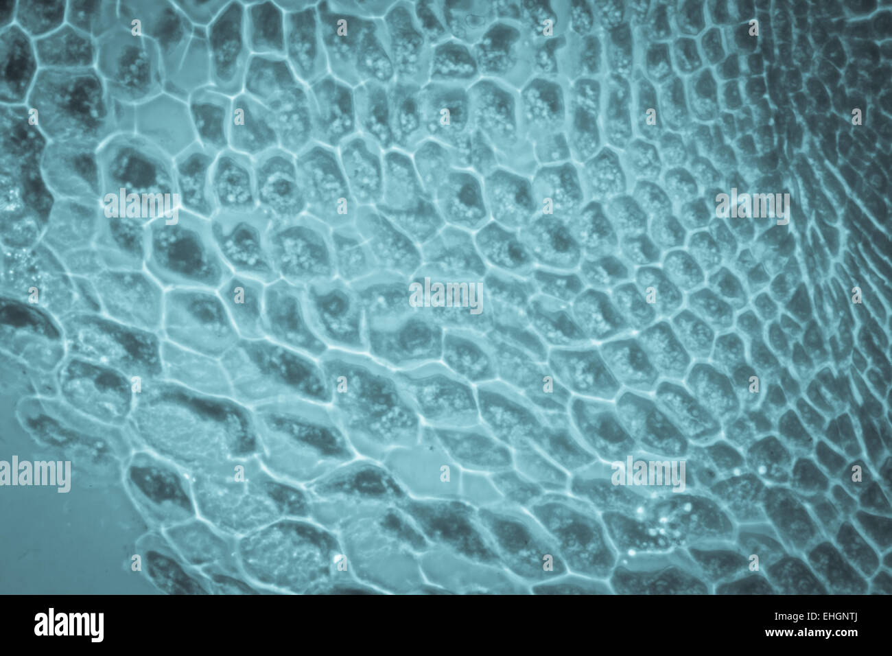 micrograph plant root tip tissue cell Stock Photohttps://www.alamy.com/image-license-details/?v=1https://www.alamy.com/stock-photo-micrograph-plant-root-tip-tissue-cell-79659010.html
RFEHGNTJ–micrograph plant root tip tissue cell
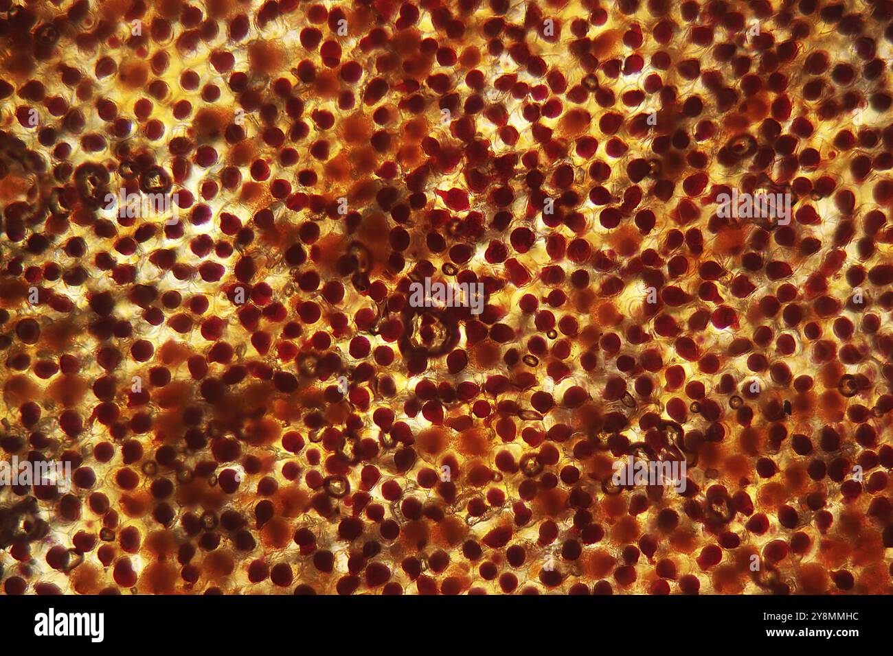 Cells of Flower leaf of a tagetes blossom under the microscope Stock Photohttps://www.alamy.com/image-license-details/?v=1https://www.alamy.com/cells-of-flower-leaf-of-a-tagetes-blossom-under-the-microscope-image624945704.html
RF2Y8MMHC–Cells of Flower leaf of a tagetes blossom under the microscope
 Sunflower stem, cross section, 8X light micrograph. Plant stem of Helianthus annuus, under the light microscope. Two individual shots combined. Stock Photohttps://www.alamy.com/image-license-details/?v=1https://www.alamy.com/sunflower-stem-cross-section-8x-light-micrograph-plant-stem-of-helianthus-annuus-under-the-light-microscope-two-individual-shots-combined-image595268505.html
RF2WGCR1D–Sunflower stem, cross section, 8X light micrograph. Plant stem of Helianthus annuus, under the light microscope. Two individual shots combined.
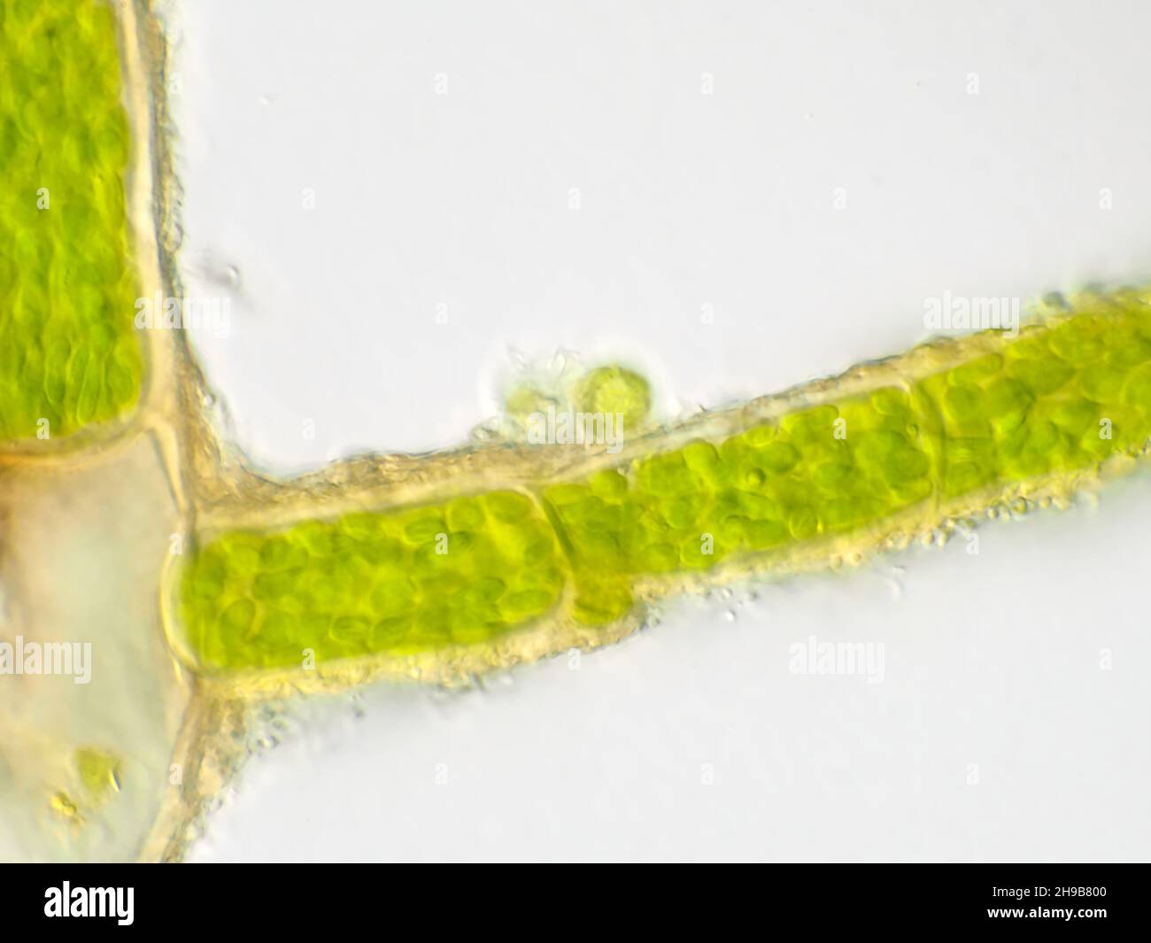 Plant cells under the microscope, showing chlorophyll content Stock Photohttps://www.alamy.com/image-license-details/?v=1https://www.alamy.com/plant-cells-under-the-microscope-showing-chlorophyll-content-image453249216.html
RM2H9B800–Plant cells under the microscope, showing chlorophyll content
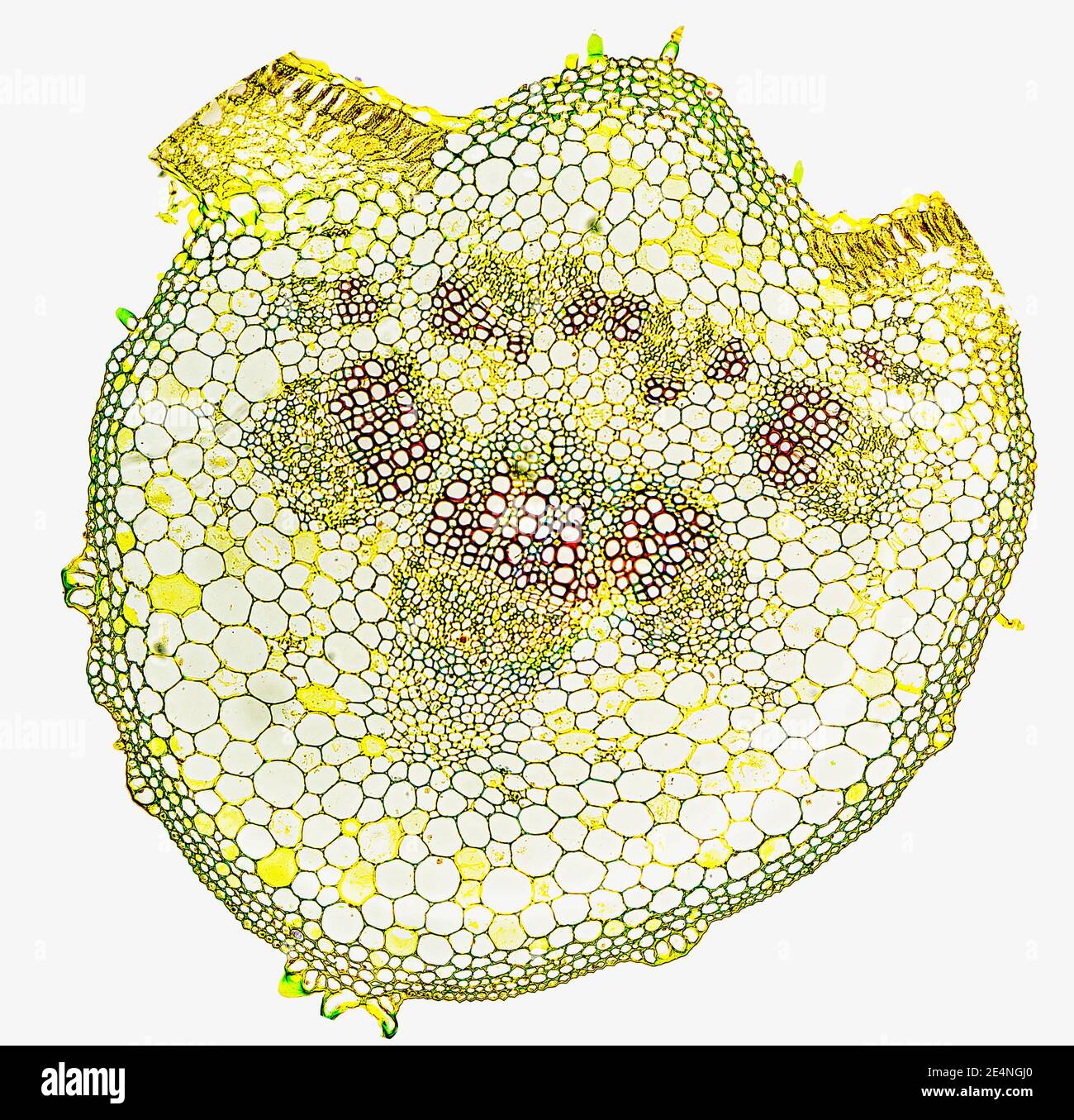 Helianthus (sunflower) leaf midrib showing cell structure, t.s. x100 Stock Photohttps://www.alamy.com/image-license-details/?v=1https://www.alamy.com/helianthus-sunflower-leaf-midrib-showing-cell-structure-ts-x100-image398771128.html
RM2E4NGJ0–Helianthus (sunflower) leaf midrib showing cell structure, t.s. x100
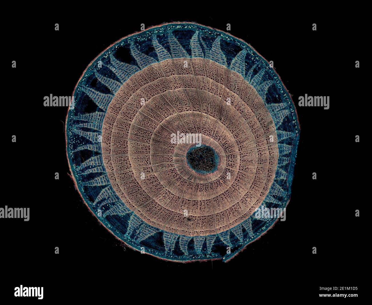 cross section cut under the microscope – microscopic view of plant cells for botanic education Stock Photohttps://www.alamy.com/image-license-details/?v=1https://www.alamy.com/cross-section-cut-under-the-microscope-microscopic-view-of-plant-cells-for-botanic-education-image396893313.html
RF2E1M1D5–cross section cut under the microscope – microscopic view of plant cells for botanic education
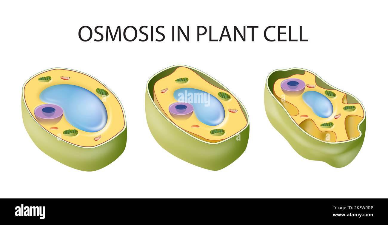 Diagram showing osmosis in plant cell Stock Photohttps://www.alamy.com/image-license-details/?v=1https://www.alamy.com/diagram-showing-osmosis-in-plant-cell-image491677642.html
RF2KFWRRP–Diagram showing osmosis in plant cell
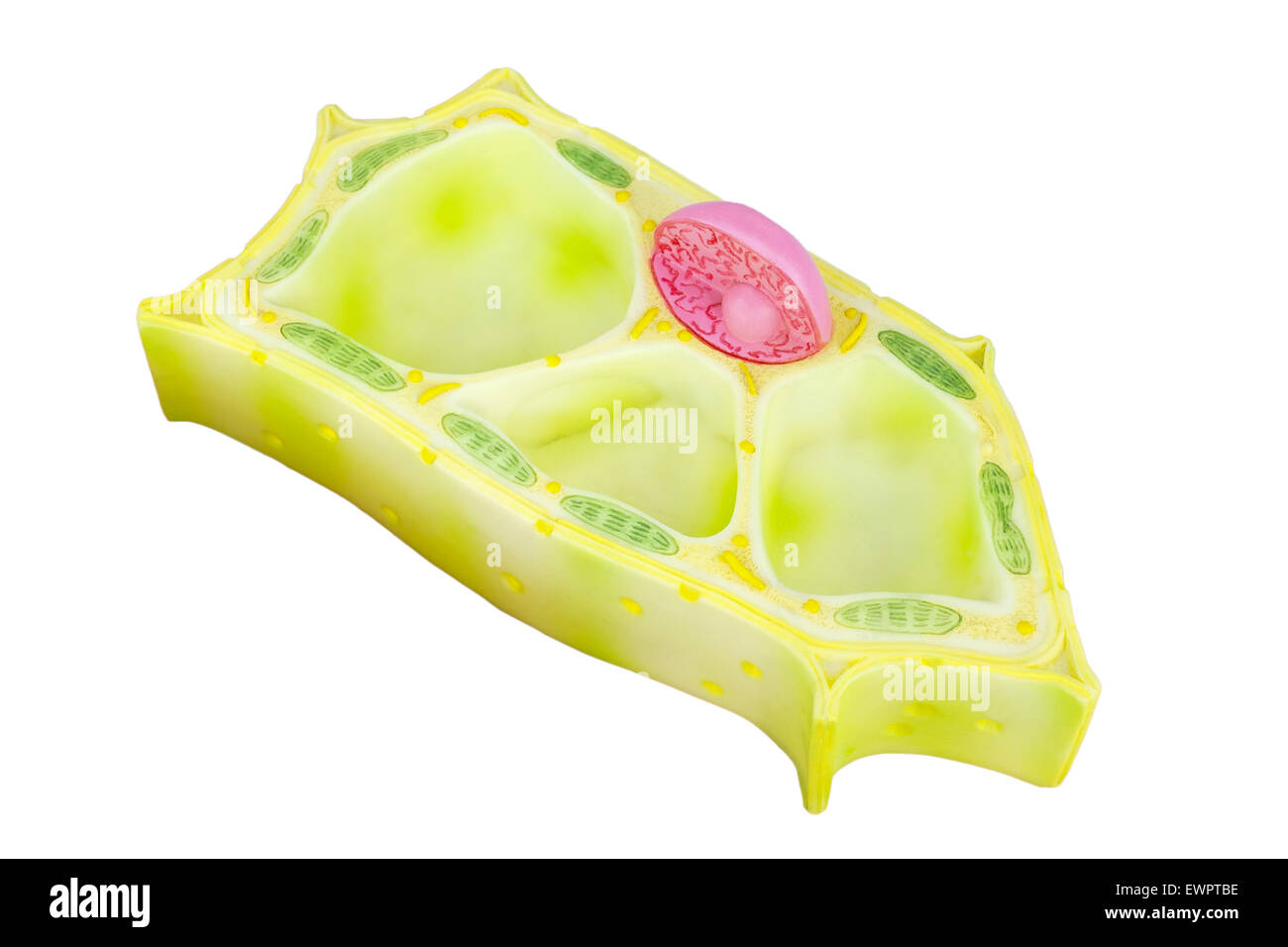 Artificial Plant cell for education isolated on white background Stock Photohttps://www.alamy.com/image-license-details/?v=1https://www.alamy.com/stock-photo-artificial-plant-cell-for-education-isolated-on-white-background-84709954.html
RFEWPTBE–Artificial Plant cell for education isolated on white background
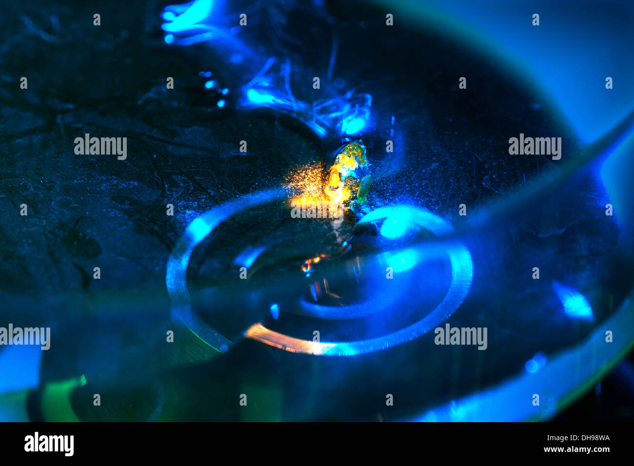 Research into cytoplasmic streaming. Cells of aquatic plant Chara coralline in Petri dish beneath microscope objective. UK Stock Photohttps://www.alamy.com/image-license-details/?v=1https://www.alamy.com/research-into-cytoplasmic-streaming-cells-of-aquatic-plant-chara-coralline-image62284806.html
RMDH98WA–Research into cytoplasmic streaming. Cells of aquatic plant Chara coralline in Petri dish beneath microscope objective. UK
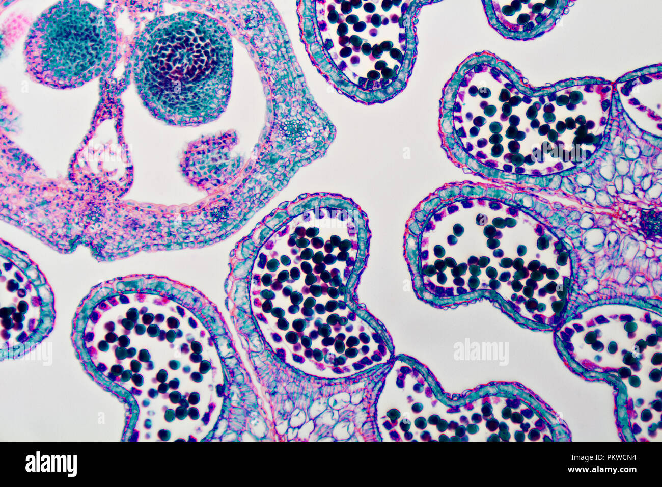 Micro Photography of Plant matter Stock Photohttps://www.alamy.com/image-license-details/?v=1https://www.alamy.com/micro-photography-of-plant-matter-image218761680.html
RMPKWCN4–Micro Photography of Plant matter
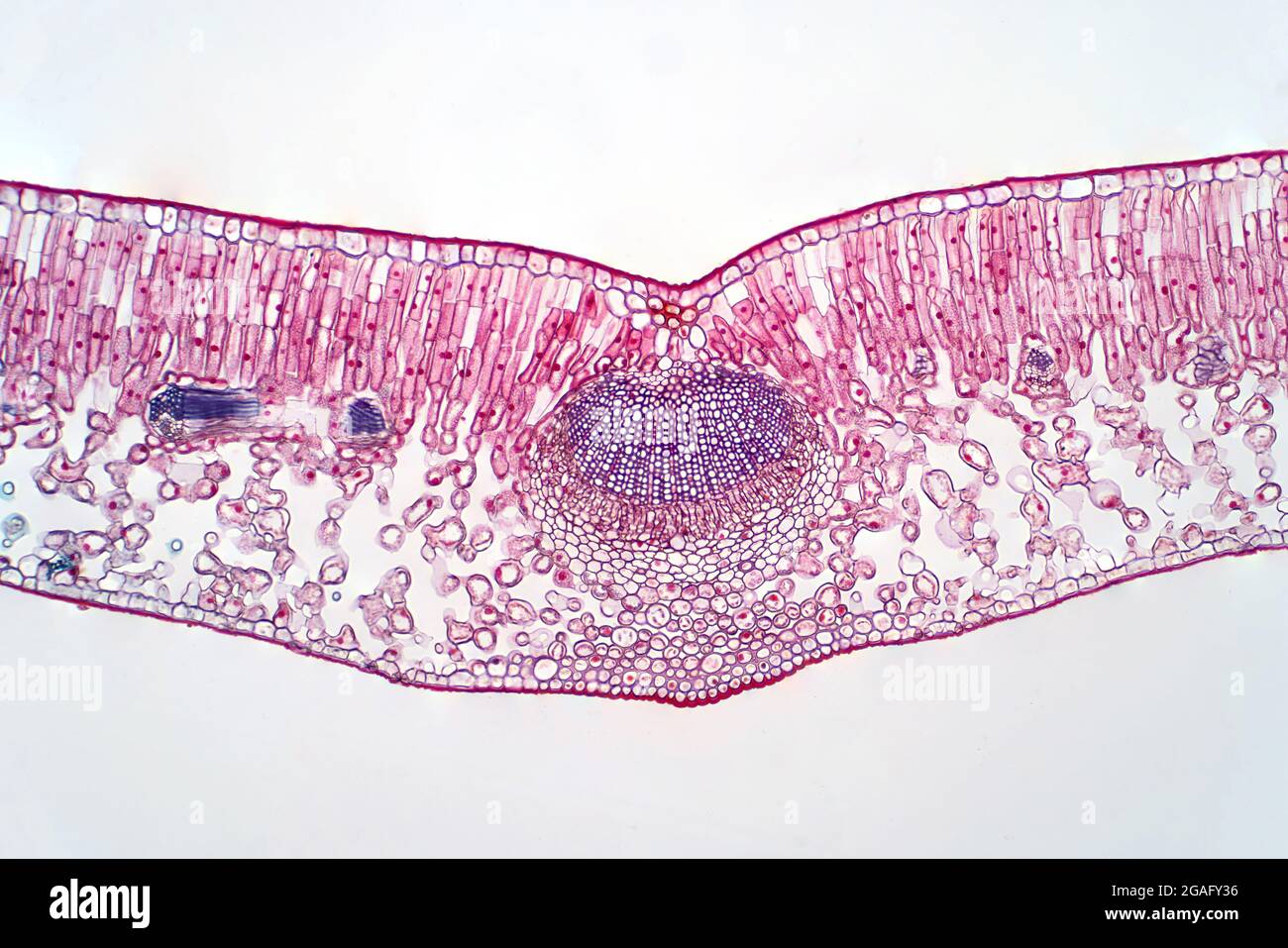 Cross section of a leaf, light micrograph Stock Photohttps://www.alamy.com/image-license-details/?v=1https://www.alamy.com/cross-section-of-a-leaf-light-micrograph-image436756298.html
RF2GAFY36–Cross section of a leaf, light micrograph
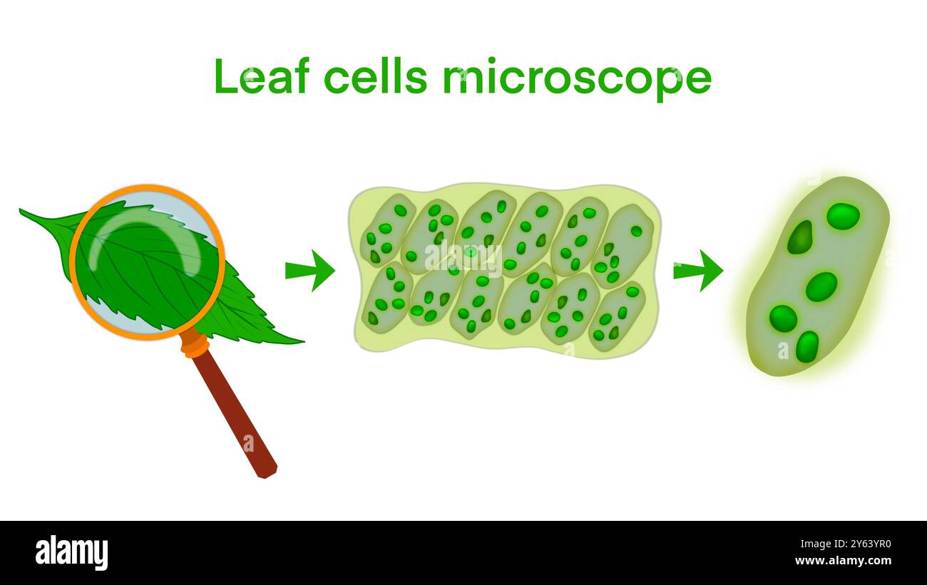 Leaf cells microscope magnification, plant leaf microscopic structure, Water plant leaf cells with chloroplasts, chlorophyll or chloroplast biotech Stock Photohttps://www.alamy.com/image-license-details/?v=1https://www.alamy.com/leaf-cells-microscope-magnification-plant-leaf-microscopic-structure-water-plant-leaf-cells-with-chloroplasts-chlorophyll-or-chloroplast-biotech-image623348852.html
RF2Y63YR0–Leaf cells microscope magnification, plant leaf microscopic structure, Water plant leaf cells with chloroplasts, chlorophyll or chloroplast biotech
 Indian corn, maize (Zea mays), Vascular bundles in a sprout of maize Stock Photohttps://www.alamy.com/image-license-details/?v=1https://www.alamy.com/stock-photo-indian-corn-maize-zea-mays-vascular-bundles-in-a-sprout-of-maize-47950390.html
RMCP095X–Indian corn, maize (Zea mays), Vascular bundles in a sprout of maize
 Abstract Science Colorful Pattern Background Stock Photohttps://www.alamy.com/image-license-details/?v=1https://www.alamy.com/abstract-science-colorful-pattern-background-image483512342.html
RF2K2HTWX–Abstract Science Colorful Pattern Background
 Abstract Science Colorful Pattern Background Stock Photohttps://www.alamy.com/image-license-details/?v=1https://www.alamy.com/abstract-science-colorful-pattern-background-image483508744.html
RF2K2HM9C–Abstract Science Colorful Pattern Background
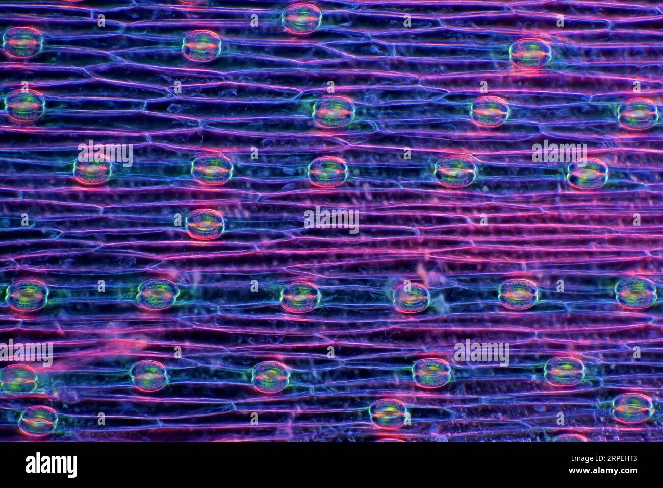 The image presents stomata in hyacinth leaf epidermis, photographed through the microscope in polarized light at a magnification of 100X Stock Photohttps://www.alamy.com/image-license-details/?v=1https://www.alamy.com/the-image-presents-stomata-in-hyacinth-leaf-epidermis-photographed-through-the-microscope-in-polarized-light-at-a-magnification-of-100x-image564575539.html
RM2RPEHT3–The image presents stomata in hyacinth leaf epidermis, photographed through the microscope in polarized light at a magnification of 100X
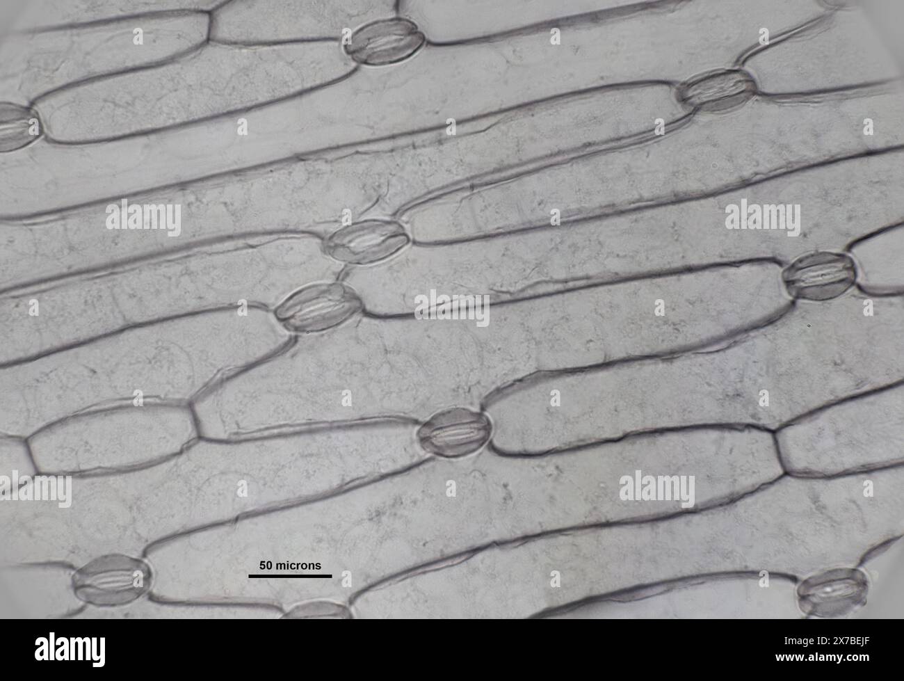 monocot leaf under a microscope Stock Photohttps://www.alamy.com/image-license-details/?v=1https://www.alamy.com/monocot-leaf-under-a-microscope-image606918439.html
RF2X7BEJF–monocot leaf under a microscope
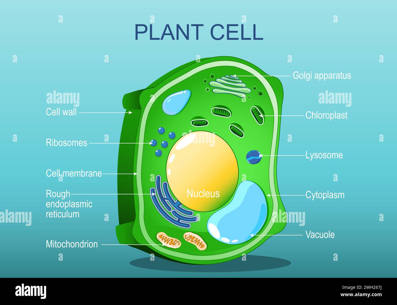 Plant cell structure. Anatomy of a cell of tree leaf. Green plant. Isometric flat vector illustration Stock Vectorhttps://www.alamy.com/image-license-details/?v=1https://www.alamy.com/plant-cell-structure-anatomy-of-a-cell-of-tree-leaf-green-plant-isometric-flat-vector-illustration-image595650486.html
RF2WH267J–Plant cell structure. Anatomy of a cell of tree leaf. Green plant. Isometric flat vector illustration
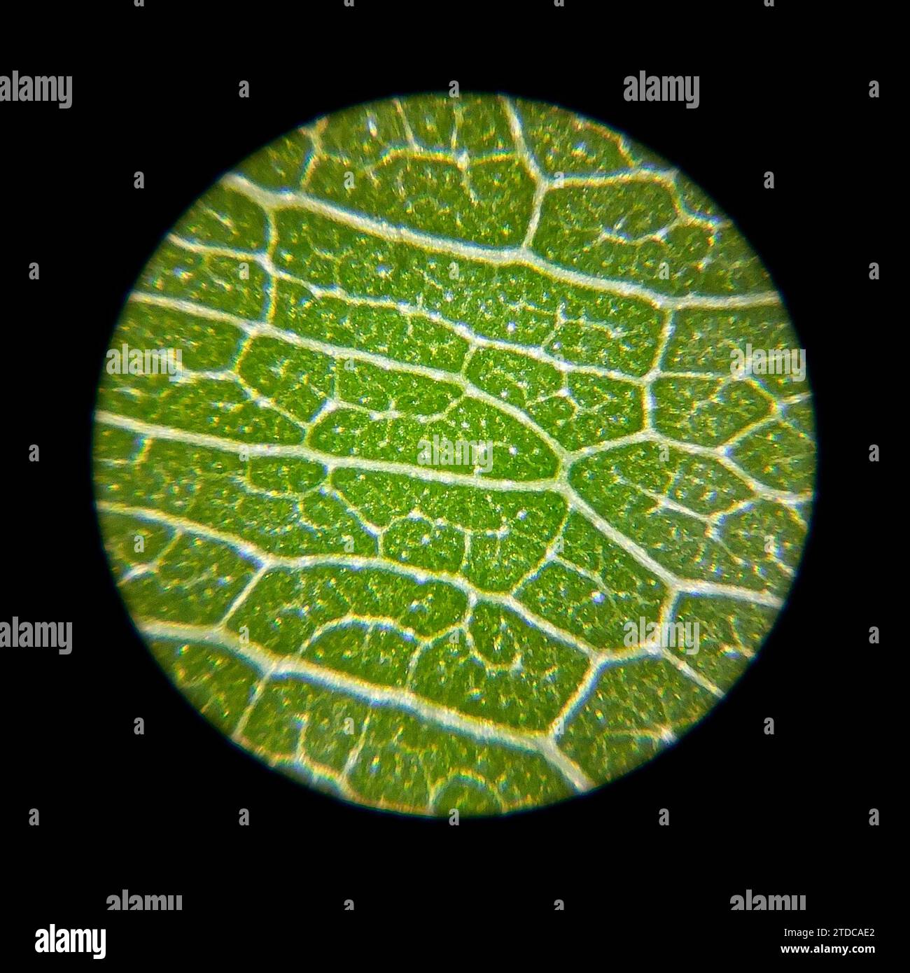 Green plant leaf surface viewed under a microscope Stock Photohttps://www.alamy.com/image-license-details/?v=1https://www.alamy.com/green-plant-leaf-surface-viewed-under-a-microscope-image576204330.html
RF2TDCAE2–Green plant leaf surface viewed under a microscope
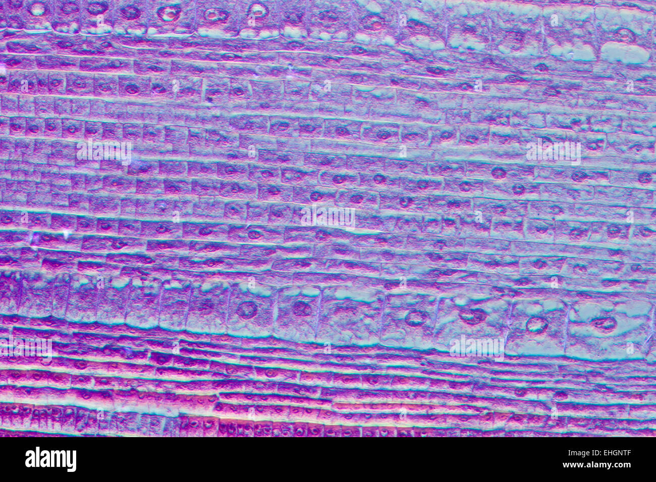 micrograph plant root tip tissue cell Stock Photohttps://www.alamy.com/image-license-details/?v=1https://www.alamy.com/stock-photo-micrograph-plant-root-tip-tissue-cell-79659007.html
RFEHGNTF–micrograph plant root tip tissue cell
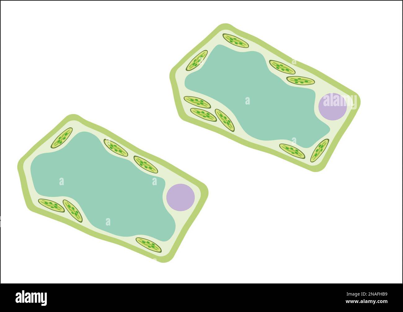 Leaf cell Stock Photohttps://www.alamy.com/image-license-details/?v=1https://www.alamy.com/leaf-cell-image522800525.html
RM2NAFHB9–Leaf cell
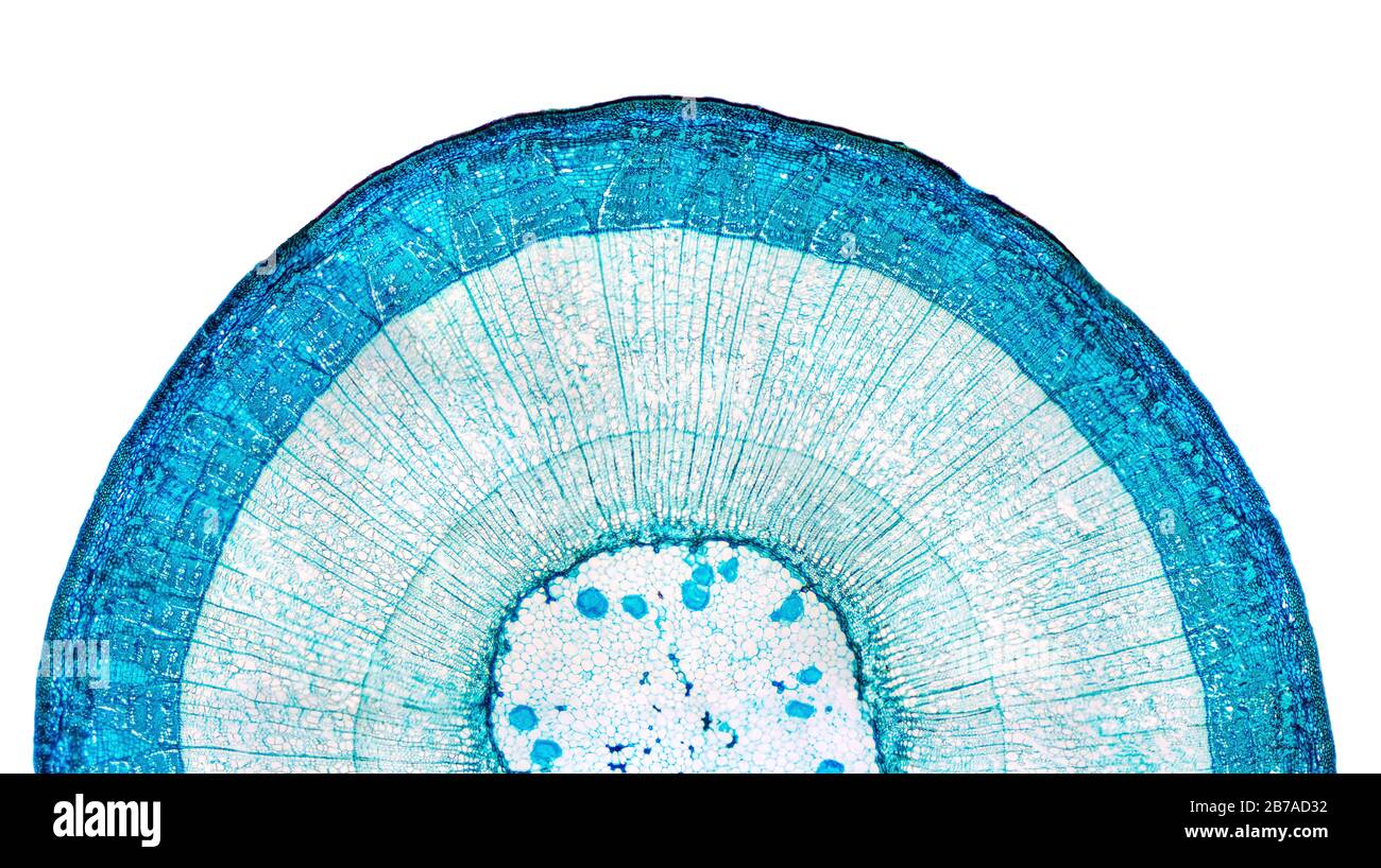 Stem of wood dicotyledon, half cross section under microscope. Light microscope slide with the microsection of a wood stem with vascular bundles. Stock Photohttps://www.alamy.com/image-license-details/?v=1https://www.alamy.com/stem-of-wood-dicotyledon-half-cross-section-under-microscope-light-microscope-slide-with-the-microsection-of-a-wood-stem-with-vascular-bundles-image348739750.html
RF2B7AD32–Stem of wood dicotyledon, half cross section under microscope. Light microscope slide with the microsection of a wood stem with vascular bundles.
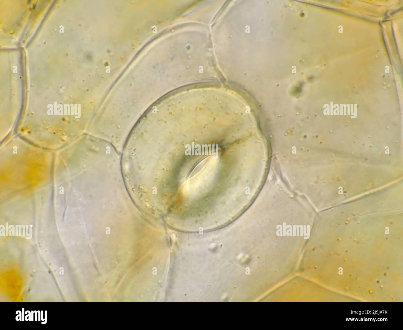 Stoma on orchid petal under the microscope, horizontal field of view is about 121 microns Stock Photohttps://www.alamy.com/image-license-details/?v=1https://www.alamy.com/stoma-on-orchid-petal-under-the-microscope-horizontal-field-of-view-is-about-121-microns-image470627575.html
RM2J9JX7K–Stoma on orchid petal under the microscope, horizontal field of view is about 121 microns
 Cell abstract concept. Under microscope. 3d render illustration Stock Photohttps://www.alamy.com/image-license-details/?v=1https://www.alamy.com/cell-abstract-concept-under-microscope-3d-render-illustration-image226757801.html
RFR4WKTW–Cell abstract concept. Under microscope. 3d render illustration
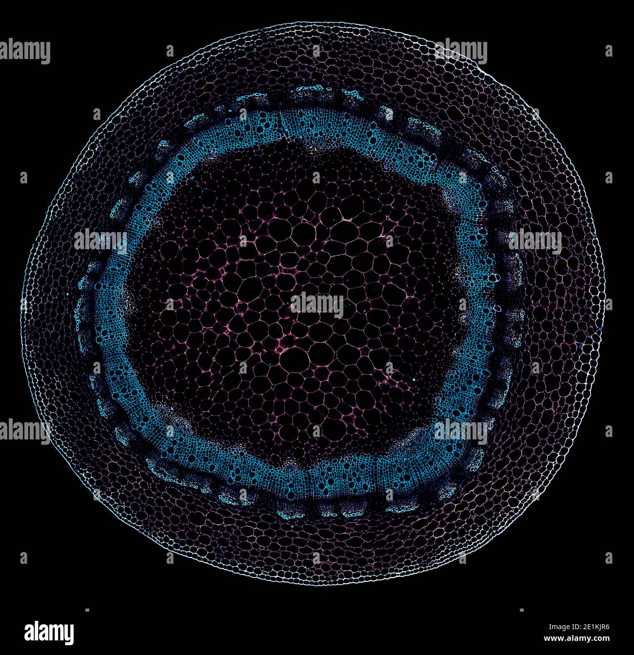 cross section cut under the microscope – microscopic view of plant cells for botanic education Stock Photohttps://www.alamy.com/image-license-details/?v=1https://www.alamy.com/cross-section-cut-under-the-microscope-microscopic-view-of-plant-cells-for-botanic-education-image396884970.html
RF2E1KJR6–cross section cut under the microscope – microscopic view of plant cells for botanic education
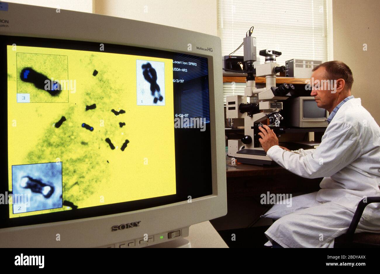 Plant Hybrid Research Stock Photohttps://www.alamy.com/image-license-details/?v=1https://www.alamy.com/plant-hybrid-research-image352799186.html
RM2BDYAXX–Plant Hybrid Research
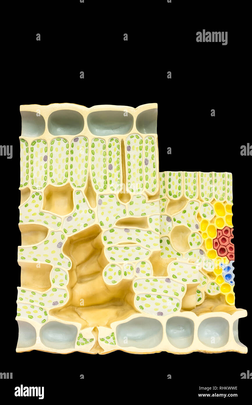 Model leaf with plant cells chloroplasts chlorophyll isolated on black background Stock Photohttps://www.alamy.com/image-license-details/?v=1https://www.alamy.com/model-leaf-with-plant-cells-chloroplasts-chlorophyll-isolated-on-black-background-image234621338.html
RFRHKWWE–Model leaf with plant cells chloroplasts chlorophyll isolated on black background
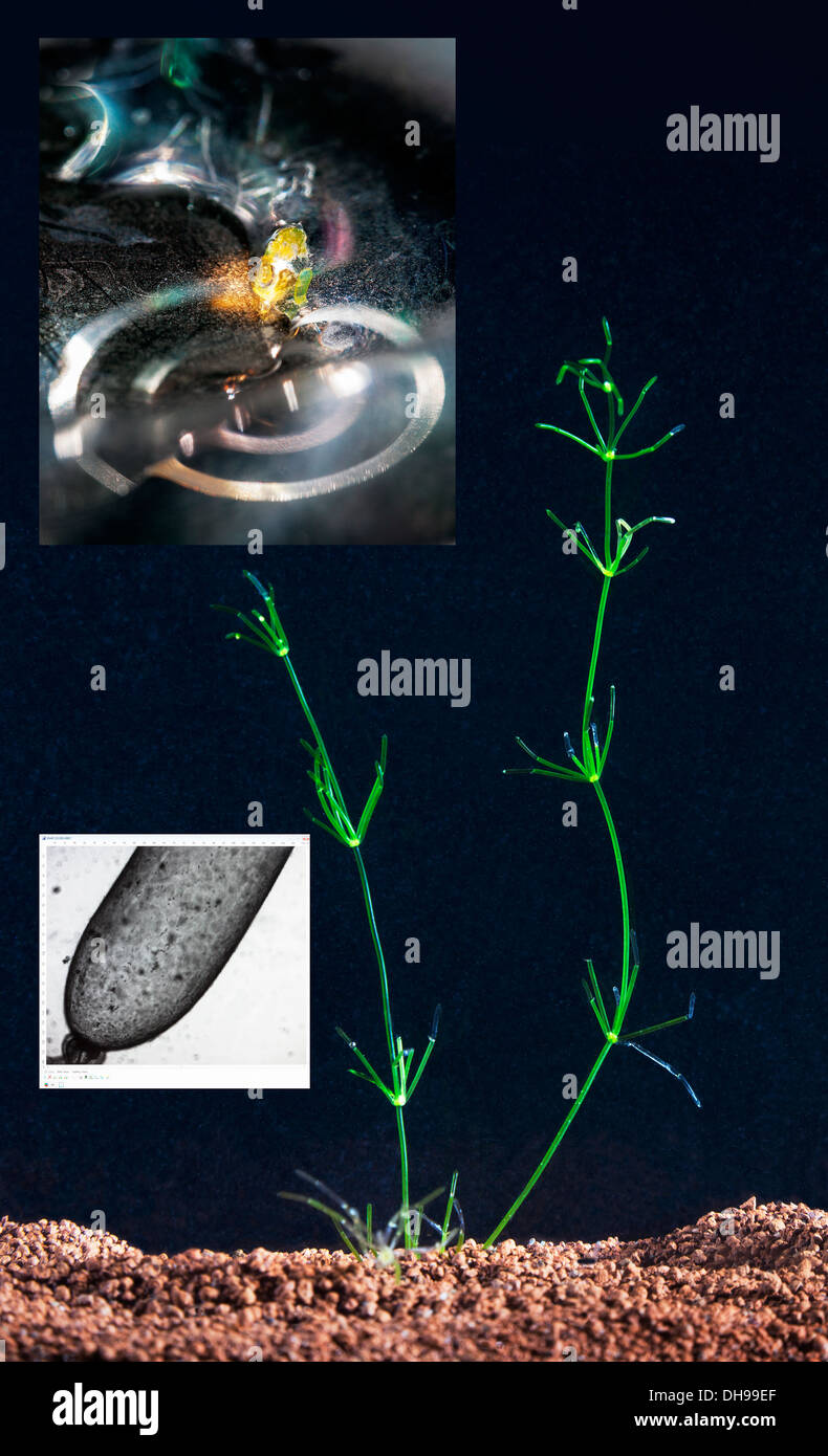 Composite. Research into cytoplasmic streaming using aquatic fresh water giant cell algae Chara coralline in close up and on microscope. Medical use Stock Photohttps://www.alamy.com/image-license-details/?v=1https://www.alamy.com/composite-research-into-cytoplasmic-streaming-using-aquatic-fresh-image62285287.html
RMDH99EF–Composite. Research into cytoplasmic streaming using aquatic fresh water giant cell algae Chara coralline in close up and on microscope. Medical use
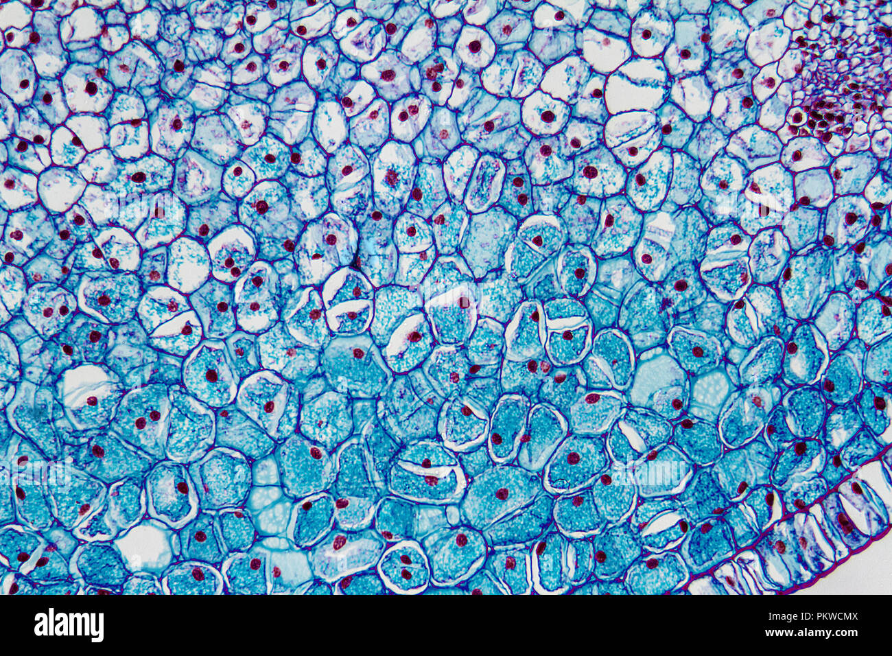 Micro Photography of Plant matter (Lily Ovulary) Stock Photohttps://www.alamy.com/image-license-details/?v=1https://www.alamy.com/micro-photography-of-plant-matter-lily-ovulary-image218761674.html
RMPKWCMX–Micro Photography of Plant matter (Lily Ovulary)
 3d rendering of a microscopic view of a plant cell. The cell is green, which is because it contains chloroplasts, Stock Photohttps://www.alamy.com/image-license-details/?v=1https://www.alamy.com/3d-rendering-of-a-microscopic-view-of-a-plant-cell-the-cell-is-green-which-is-because-it-contains-chloroplasts-image592728020.html
RF2WC92HT–3d rendering of a microscopic view of a plant cell. The cell is green, which is because it contains chloroplasts,
 Abstract illustration of green chlorophyll plant cells as pattern seamless Stock Photohttps://www.alamy.com/image-license-details/?v=1https://www.alamy.com/abstract-illustration-of-green-chlorophyll-plant-cells-as-pattern-seamless-image238712927.html
RFRTA8NK–Abstract illustration of green chlorophyll plant cells as pattern seamless
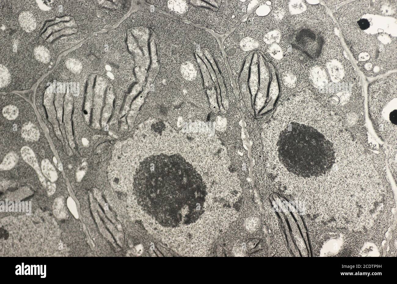 Vegetal cell showing nucleus, cell wall, nucleoli, chloroplast and starch. A ultrathin section of tobacco leaf mesophyll cells showing chloroplast str Stock Photohttps://www.alamy.com/image-license-details/?v=1https://www.alamy.com/vegetal-cell-showing-nucleus-cell-wall-nucleoli-chloroplast-and-starch-a-ultrathin-section-of-tobacco-leaf-mesophyll-cells-showing-chloroplast-str-image369952621.html
RM2CDTP9H–Vegetal cell showing nucleus, cell wall, nucleoli, chloroplast and starch. A ultrathin section of tobacco leaf mesophyll cells showing chloroplast str
 Abstract Science Colorful Pattern Background Stock Photohttps://www.alamy.com/image-license-details/?v=1https://www.alamy.com/abstract-science-colorful-pattern-background-image483508972.html
RF2K2HMHG–Abstract Science Colorful Pattern Background
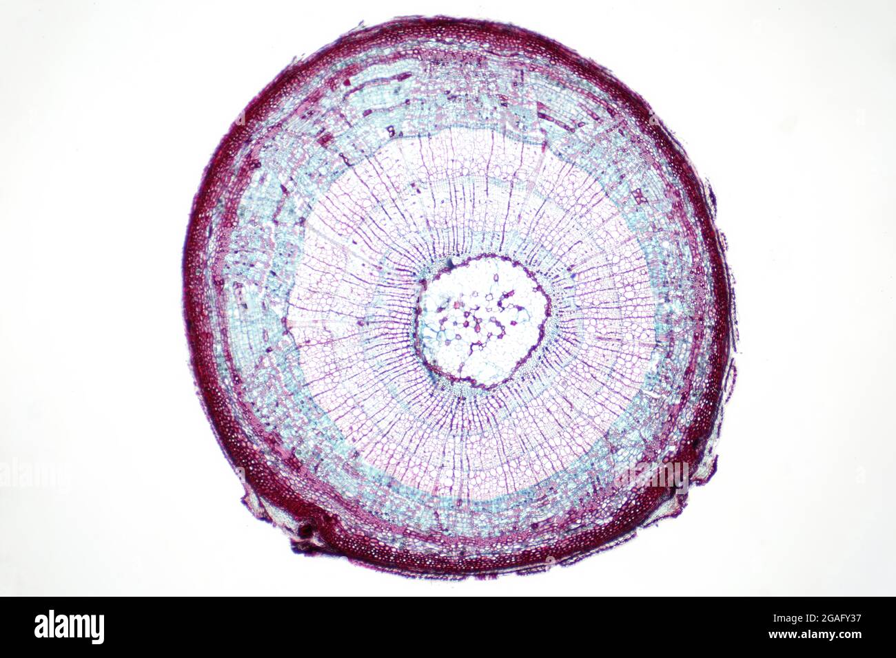 Plant stem, light micrograph Stock Photohttps://www.alamy.com/image-license-details/?v=1https://www.alamy.com/plant-stem-light-micrograph-image436756299.html
RF2GAFY37–Plant stem, light micrograph
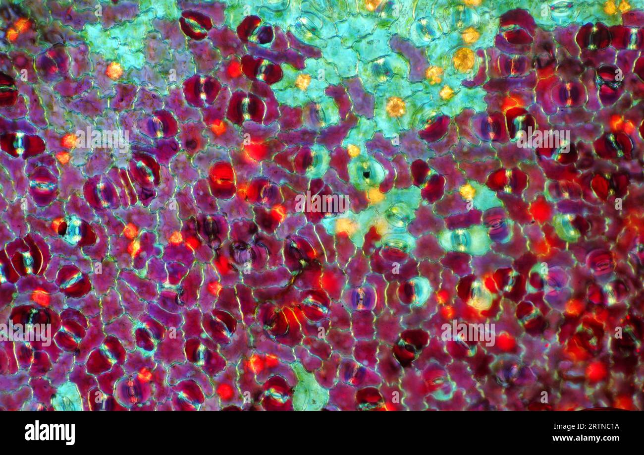 The image presents stomata in Croton leaf epidermis, photographed through the microscope in polarized light at a magnification of 100X Stock Photohttps://www.alamy.com/image-license-details/?v=1https://www.alamy.com/the-image-presents-stomata-in-croton-leaf-epidermis-photographed-through-the-microscope-in-polarized-light-at-a-magnification-of-100x-image565953958.html
RM2RTNC1A–The image presents stomata in Croton leaf epidermis, photographed through the microscope in polarized light at a magnification of 100X
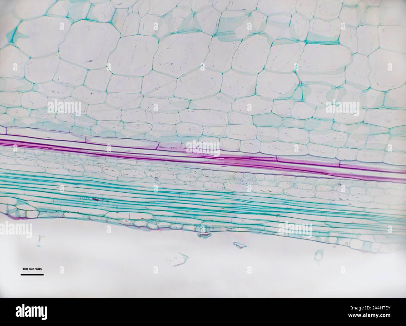 Cucurbis under a light microscope Stock Photohttps://www.alamy.com/image-license-details/?v=1https://www.alamy.com/cucurbis-under-a-light-microscope-image605213923.html
RF2X4HTEY–Cucurbis under a light microscope
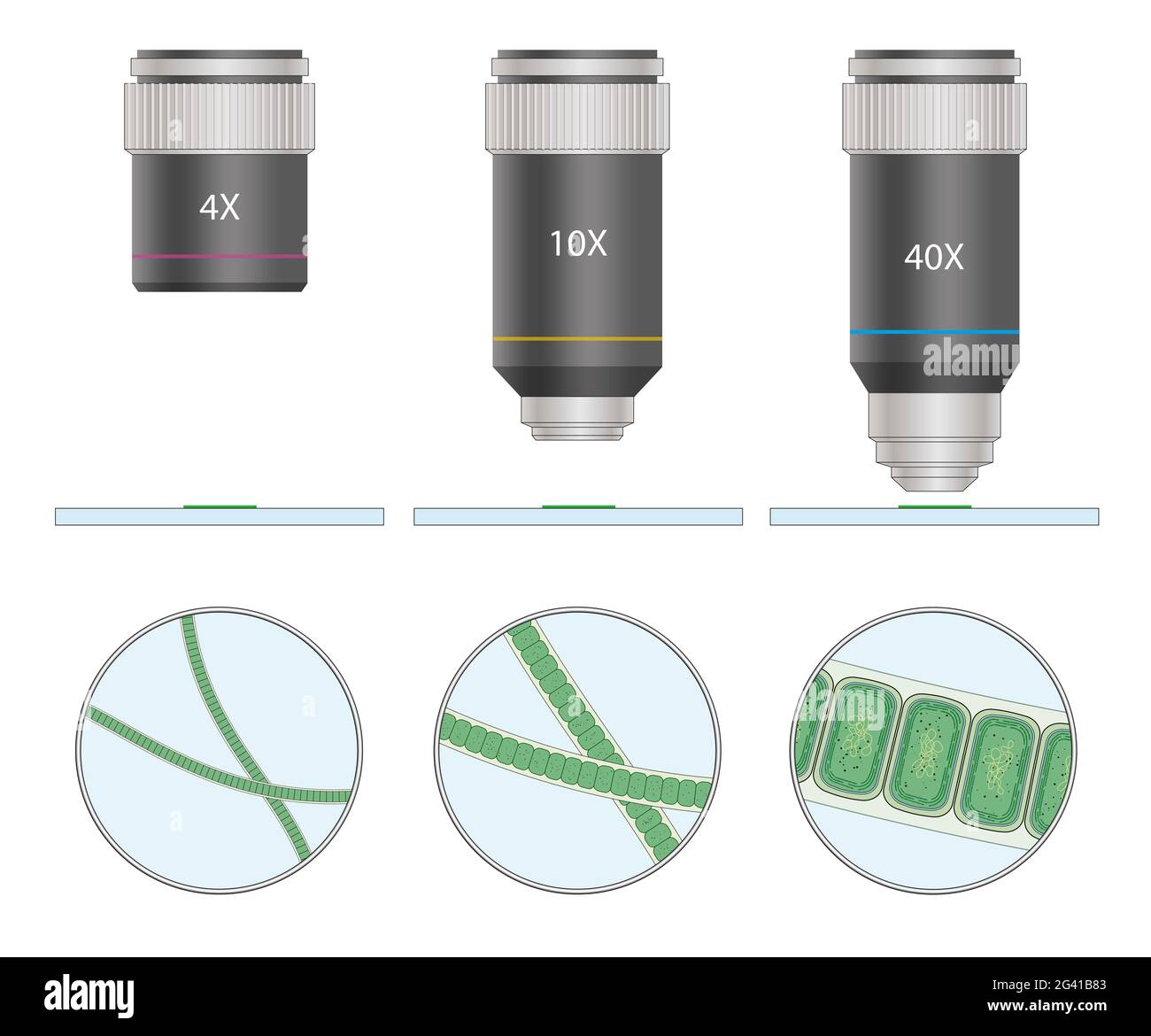 Microscope view of cyanobacteria or Cyanophyta Stock Photohttps://www.alamy.com/image-license-details/?v=1https://www.alamy.com/microscope-view-of-cyanobacteria-or-cyanophyta-image432748627.html
RF2G41B83–Microscope view of cyanobacteria or Cyanophyta
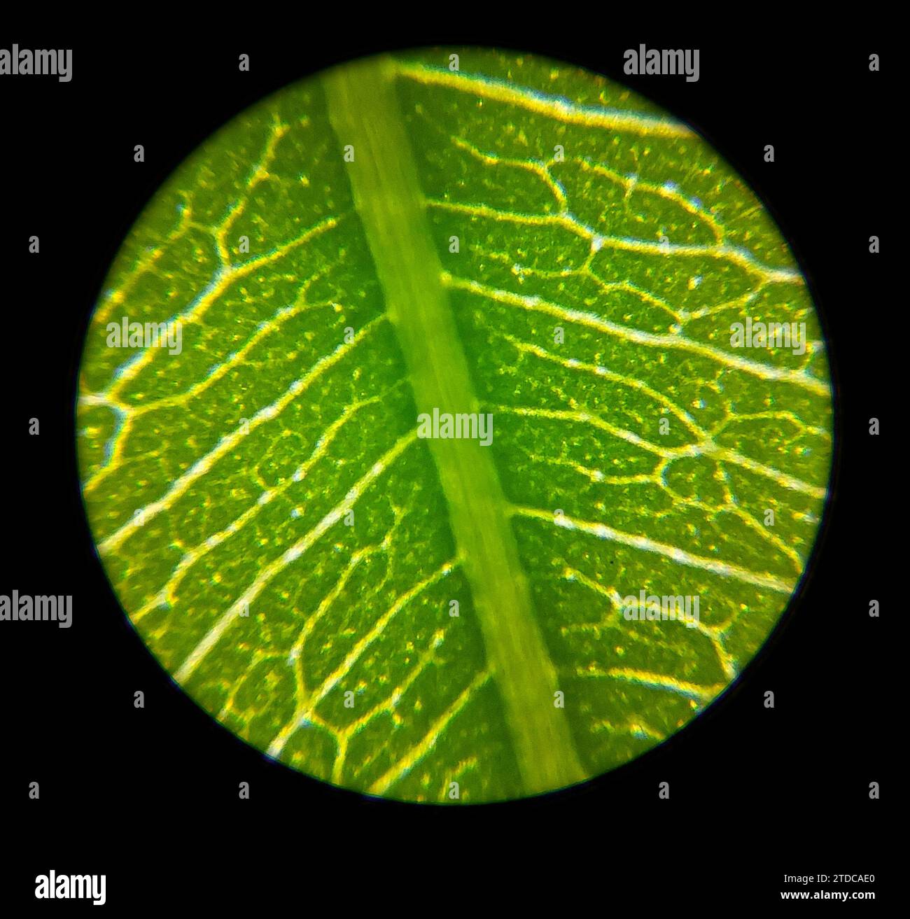 Green plant leaf surface viewed under a microscope Stock Photohttps://www.alamy.com/image-license-details/?v=1https://www.alamy.com/green-plant-leaf-surface-viewed-under-a-microscope-image576204328.html
RF2TDCAE0–Green plant leaf surface viewed under a microscope
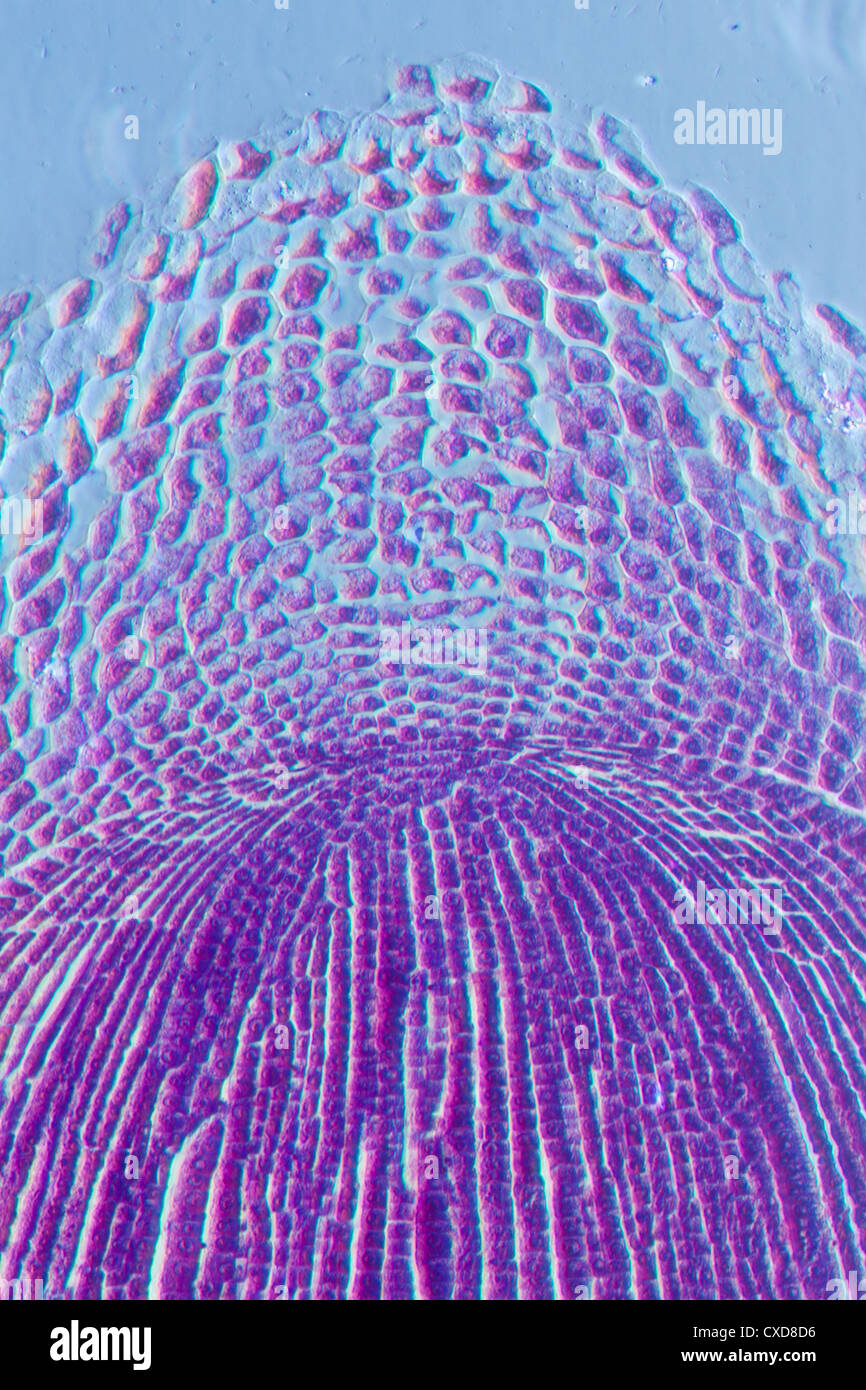 micrograph plant root tip tissue cell Stock Photohttps://www.alamy.com/image-license-details/?v=1https://www.alamy.com/stock-photo-micrograph-plant-root-tip-tissue-cell-50693810.html
RFCXD8D6–micrograph plant root tip tissue cell
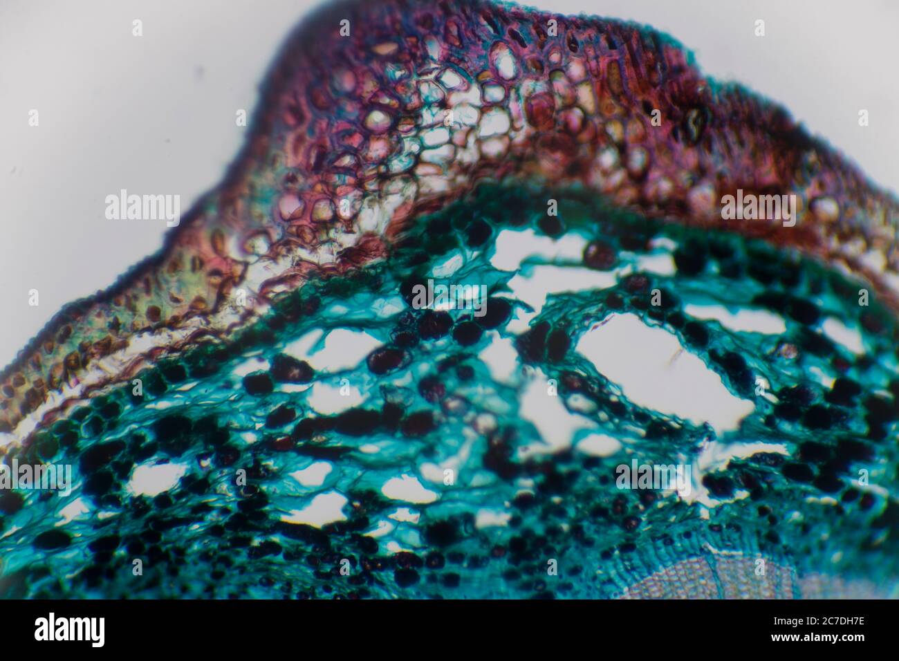 Microscopic photo of a plant cell with red and green colors and cell textures. Stock Photohttps://www.alamy.com/image-license-details/?v=1https://www.alamy.com/microscopic-photo-of-a-plant-cell-with-red-and-green-colors-and-cell-textures-image366019234.html
RF2C7DH7E–Microscopic photo of a plant cell with red and green colors and cell textures.
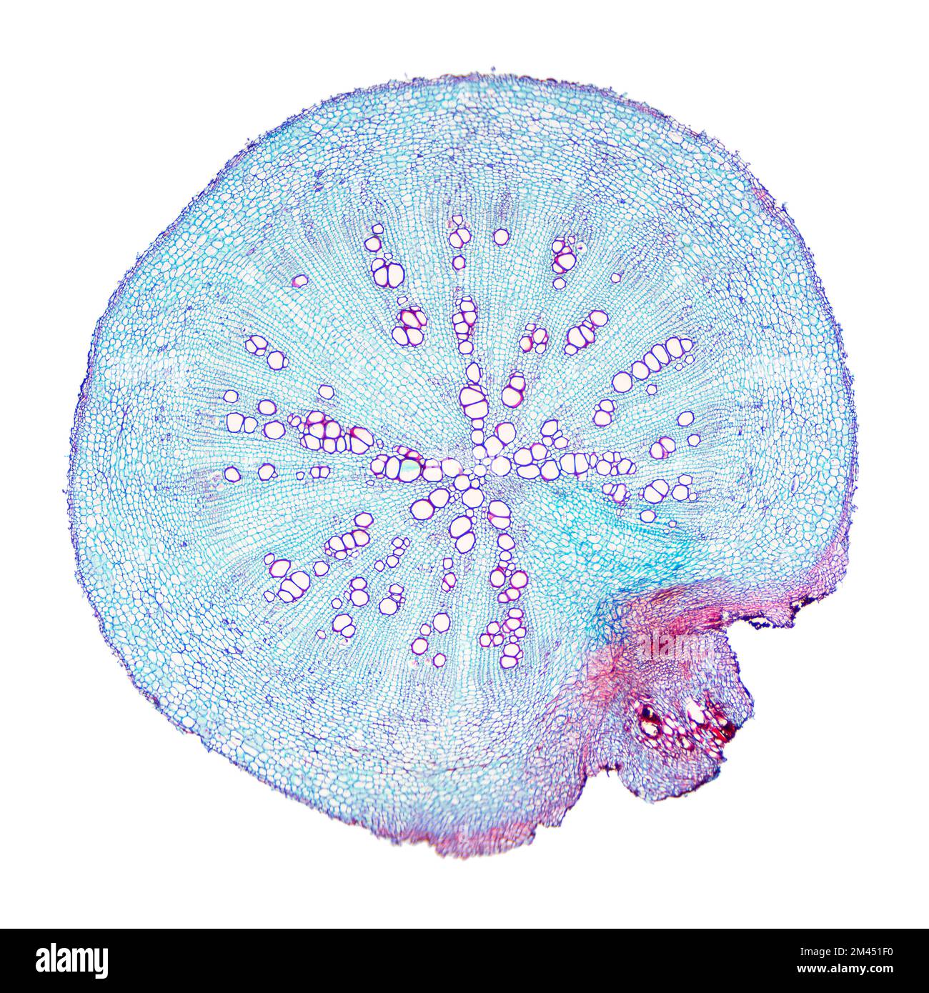 Radish root, cross section under light microscope. Transverse section through the root of the Raphanus sativus plant. Micrograph at 8X magnification. Stock Photohttps://www.alamy.com/image-license-details/?v=1https://www.alamy.com/radish-root-cross-section-under-light-microscope-transverse-section-through-the-root-of-the-raphanus-sativus-plant-micrograph-at-8x-magnification-image501670260.html
RF2M451F0–Radish root, cross section under light microscope. Transverse section through the root of the Raphanus sativus plant. Micrograph at 8X magnification.
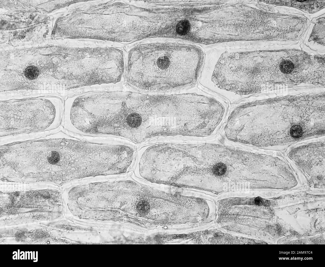 Onion skin cells under the microscope, horizontal field of view is about 0.61 mm Stock Photohttps://www.alamy.com/image-license-details/?v=1https://www.alamy.com/onion-skin-cells-under-the-microscope-horizontal-field-of-view-is-about-061-mm-image339493508.html
RM2AM97C4–Onion skin cells under the microscope, horizontal field of view is about 0.61 mm
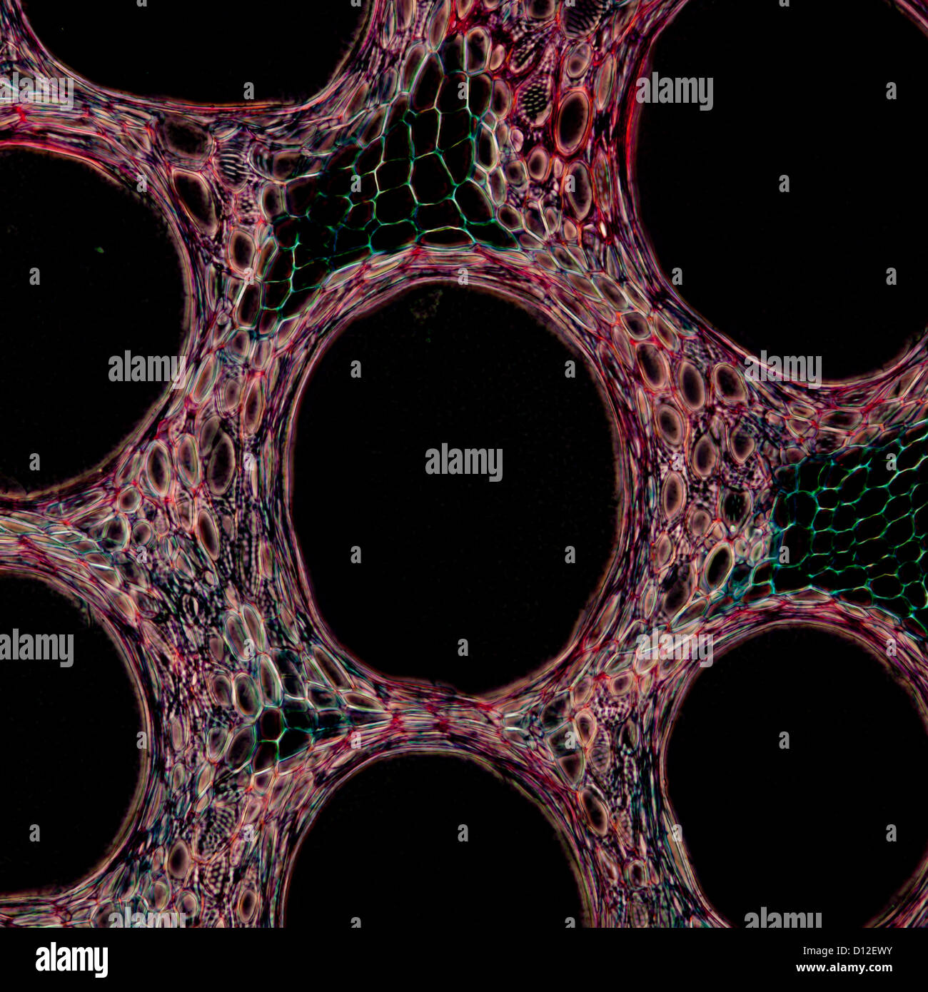 micrograph plant tissue cell , stem of pumpkin Stock Photohttps://www.alamy.com/image-license-details/?v=1https://www.alamy.com/stock-photo-micrograph-plant-tissue-cell-stem-of-pumpkin-52301367.html
RFD12EWY–micrograph plant tissue cell , stem of pumpkin
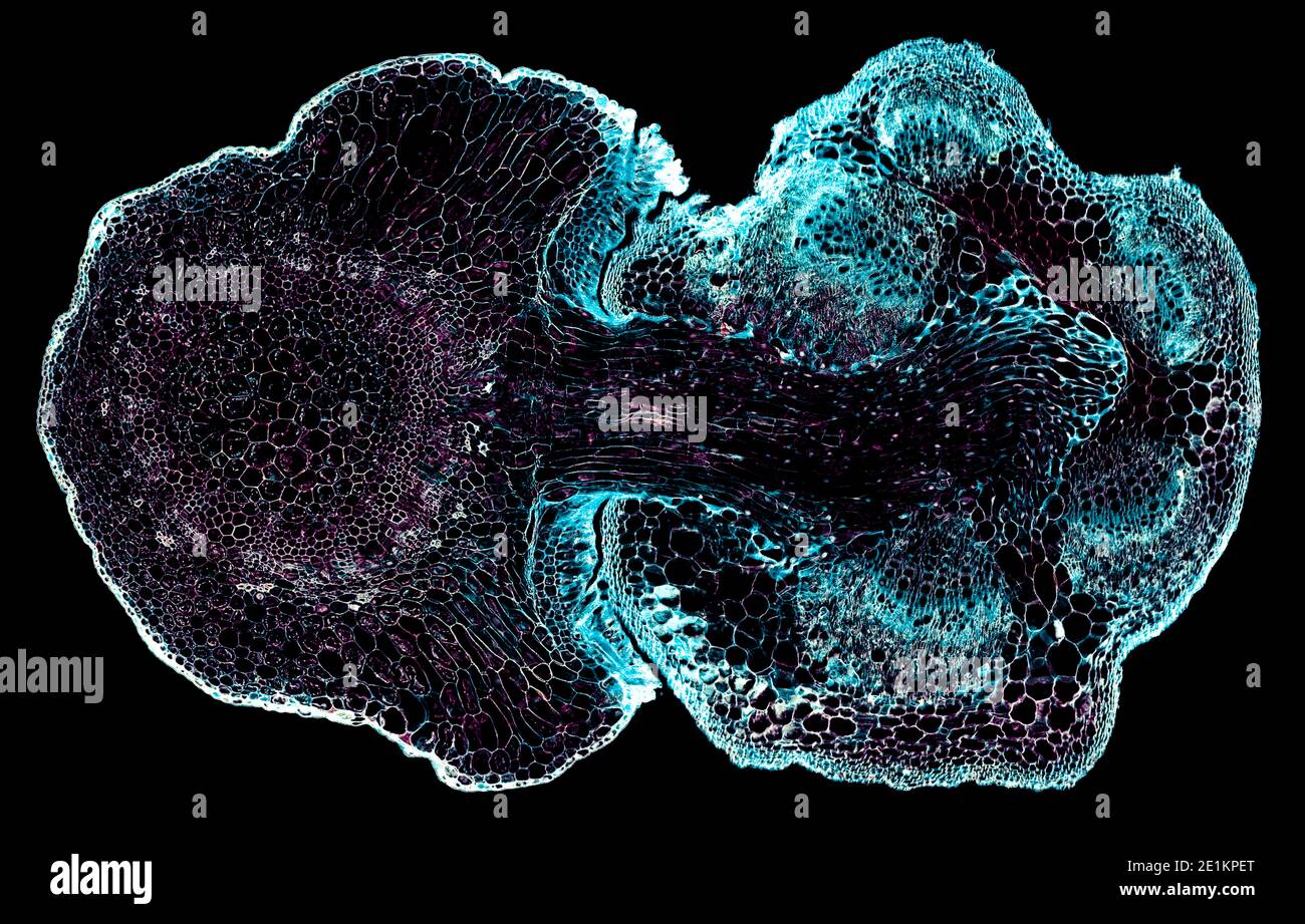 cross section cut under the microscope – microscopic view of plant cells for botanic education Stock Photohttps://www.alamy.com/image-license-details/?v=1https://www.alamy.com/cross-section-cut-under-the-microscope-microscopic-view-of-plant-cells-for-botanic-education-image396887872.html
RF2E1KPET–cross section cut under the microscope – microscopic view of plant cells for botanic education
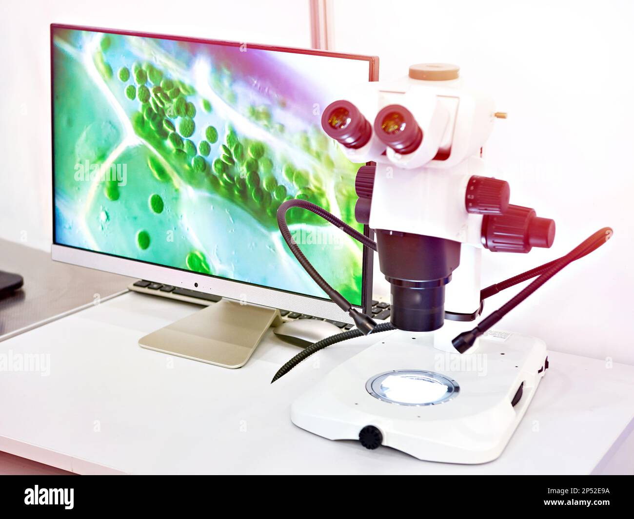 Research stereo microscope with computers monitor Stock Photohttps://www.alamy.com/image-license-details/?v=1https://www.alamy.com/research-stereo-microscope-with-computers-monitor-image536649830.html
RF2P52E9A–Research stereo microscope with computers monitor
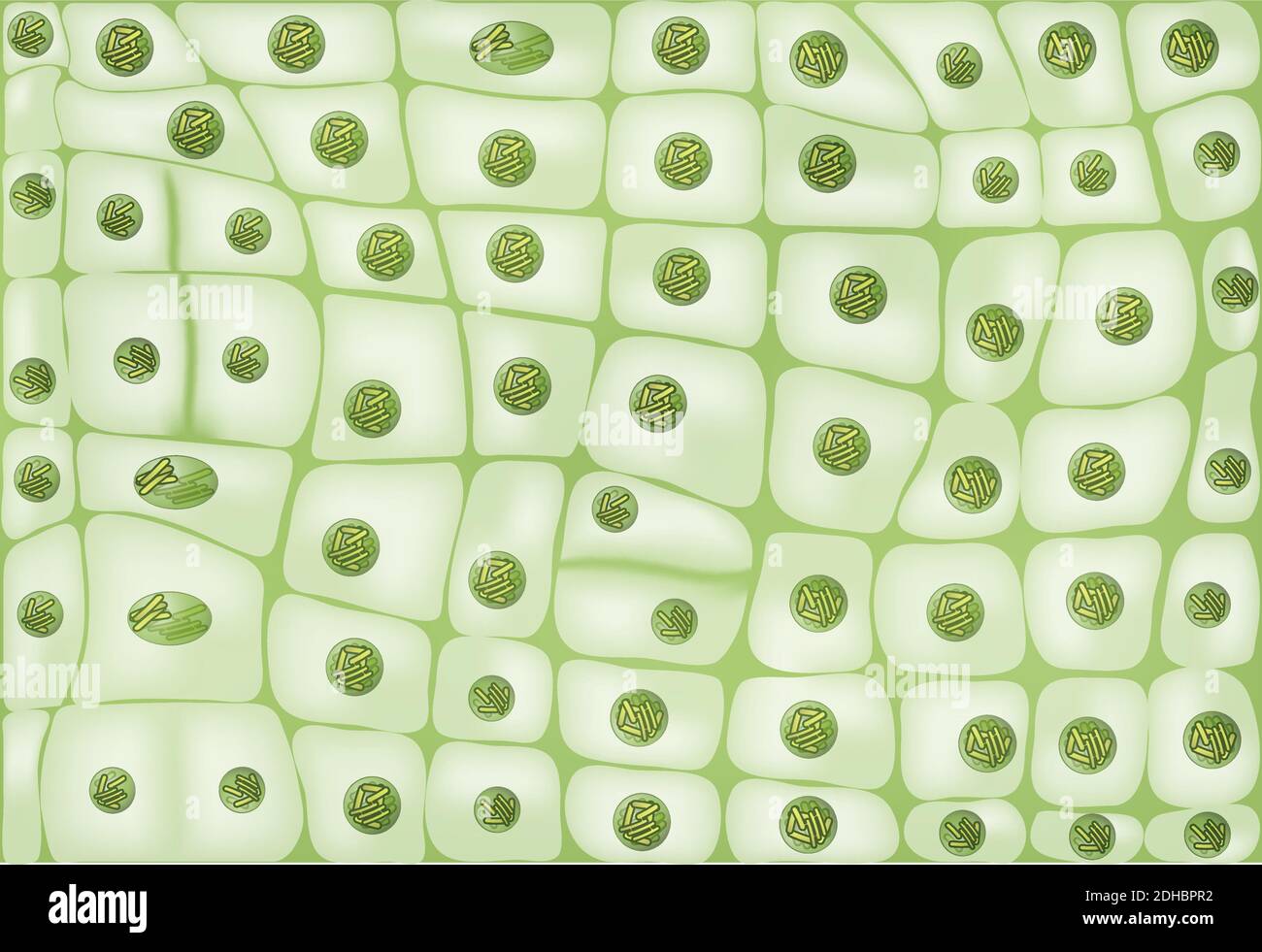 Cell division background Stock Vectorhttps://www.alamy.com/image-license-details/?v=1https://www.alamy.com/cell-division-background-image389336614.html
RF2DHBPR2–Cell division background
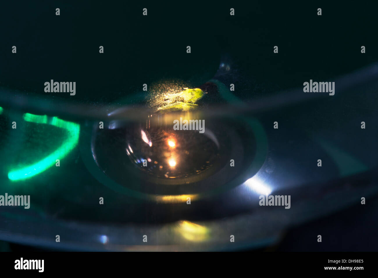 Research into cytoplasmic streaming. Cells of aquatic plant Chara coralline in Petri dish beneath microscope objective. UK Stock Photohttps://www.alamy.com/image-license-details/?v=1https://www.alamy.com/research-into-cytoplasmic-streaming-cells-of-aquatic-plant-chara-coralline-image62284493.html
RMDH98E5–Research into cytoplasmic streaming. Cells of aquatic plant Chara coralline in Petri dish beneath microscope objective. UK
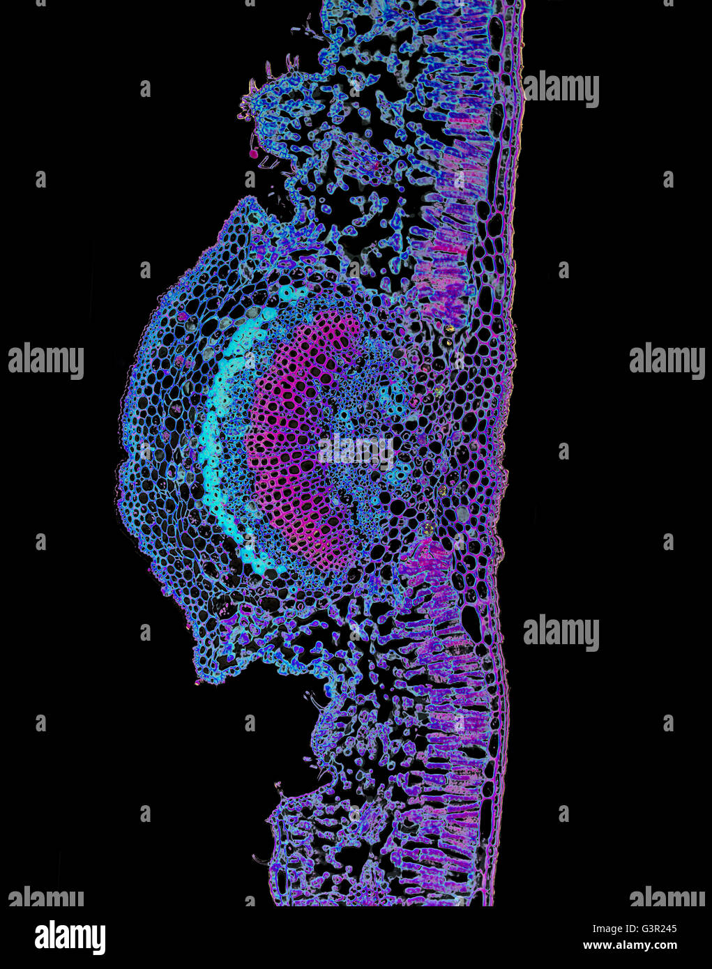 Micro photo of a Nerium Oleander Leaf Stock Photohttps://www.alamy.com/image-license-details/?v=1https://www.alamy.com/stock-photo-micro-photo-of-a-nerium-oleander-leaf-105612757.html
RMG3R245–Micro photo of a Nerium Oleander Leaf
 3d rendering of a microscopic view of a plant cell. The cell is green, which is because it contains chloroplasts, Stock Photohttps://www.alamy.com/image-license-details/?v=1https://www.alamy.com/3d-rendering-of-a-microscopic-view-of-a-plant-cell-the-cell-is-green-which-is-because-it-contains-chloroplasts-image592728019.html
RF2WC92HR–3d rendering of a microscopic view of a plant cell. The cell is green, which is because it contains chloroplasts,
 Fungal Parasitic Mold Spore Growth on the Plant. High Magnification Microscopic Photo for Science or Medicine Concept Background Stock Photohttps://www.alamy.com/image-license-details/?v=1https://www.alamy.com/fungal-parasitic-mold-spore-growth-on-the-plant-high-magnification-microscopic-photo-for-science-or-medicine-concept-background-image556022362.html
RF2R8H05E–Fungal Parasitic Mold Spore Growth on the Plant. High Magnification Microscopic Photo for Science or Medicine Concept Background
 Microscopic view of a potato tuber cross-section with starch grains (amyloplasts) in plant cell. Polarized light, crossed polarizers. Stock Photohttps://www.alamy.com/image-license-details/?v=1https://www.alamy.com/stock-photo-microscopic-view-of-a-potato-tuber-cross-section-with-starch-grains-140927366.html
RFJ57P86–Microscopic view of a potato tuber cross-section with starch grains (amyloplasts) in plant cell. Polarized light, crossed polarizers.
 Abstract Science Colorful Pattern Background Stock Photohttps://www.alamy.com/image-license-details/?v=1https://www.alamy.com/abstract-science-colorful-pattern-background-image483508955.html
RF2K2HMGY–Abstract Science Colorful Pattern Background
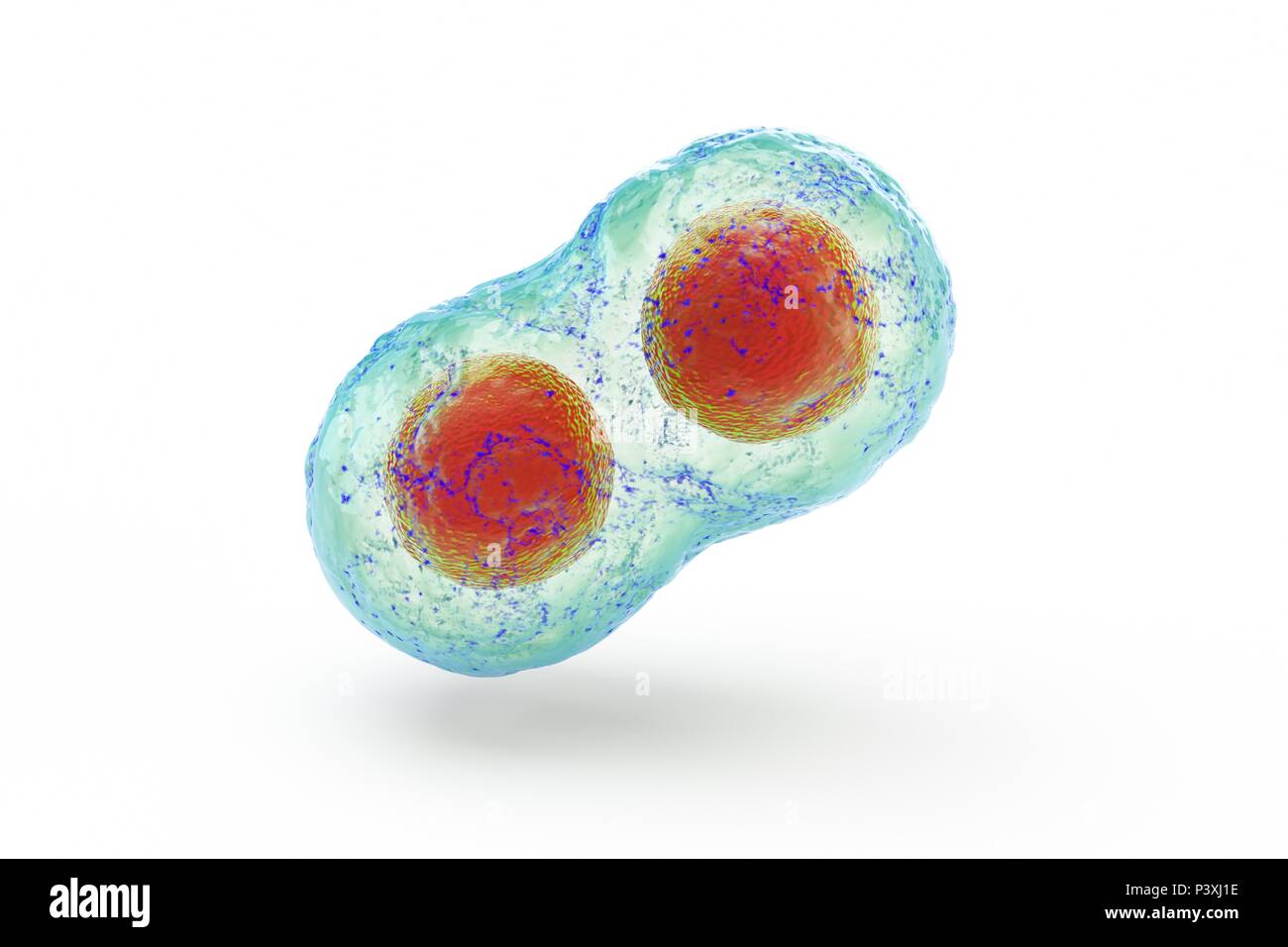 biological cell isolated on whithe background microscope 3D Illustration Stock Photohttps://www.alamy.com/image-license-details/?v=1https://www.alamy.com/biological-cell-isolated-on-whithe-background-microscope-3d-illustration-image208953290.html
RFP3XJ1E–biological cell isolated on whithe background microscope 3D Illustration
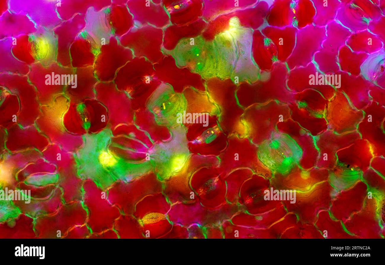 The image presents stomata in Croton leaf epidermis, photographed through the microscope in polarized light at a magnification of 200X Stock Photohttps://www.alamy.com/image-license-details/?v=1https://www.alamy.com/the-image-presents-stomata-in-croton-leaf-epidermis-photographed-through-the-microscope-in-polarized-light-at-a-magnification-of-200x-image565953986.html
RM2RTNC2A–The image presents stomata in Croton leaf epidermis, photographed through the microscope in polarized light at a magnification of 200X
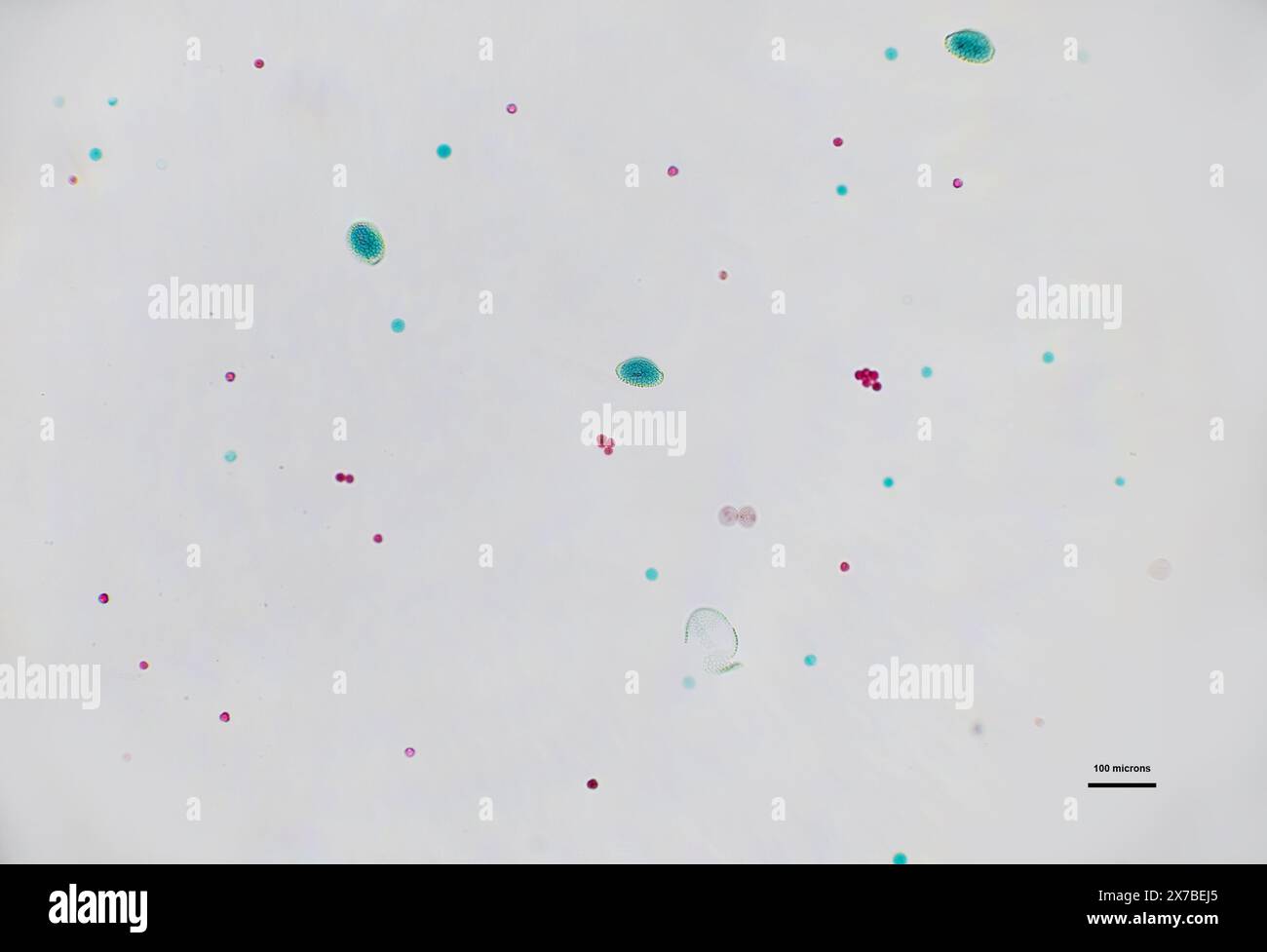 mixed pollen under a microscope at 10 times magnification Stock Photohttps://www.alamy.com/image-license-details/?v=1https://www.alamy.com/mixed-pollen-under-a-microscope-at-10-times-magnification-image606918429.html
RF2X7BEJ5–mixed pollen under a microscope at 10 times magnification
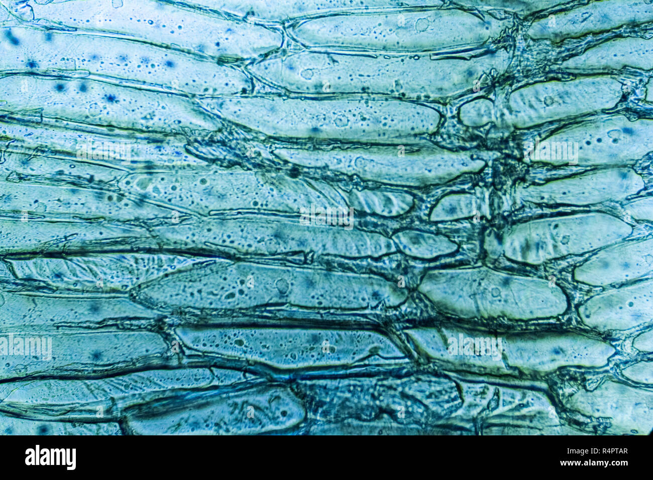 Microscope view of onion cells Stock Photohttps://www.alamy.com/image-license-details/?v=1https://www.alamy.com/microscope-view-of-onion-cells-image226695471.html
RFR4PTAR–Microscope view of onion cells
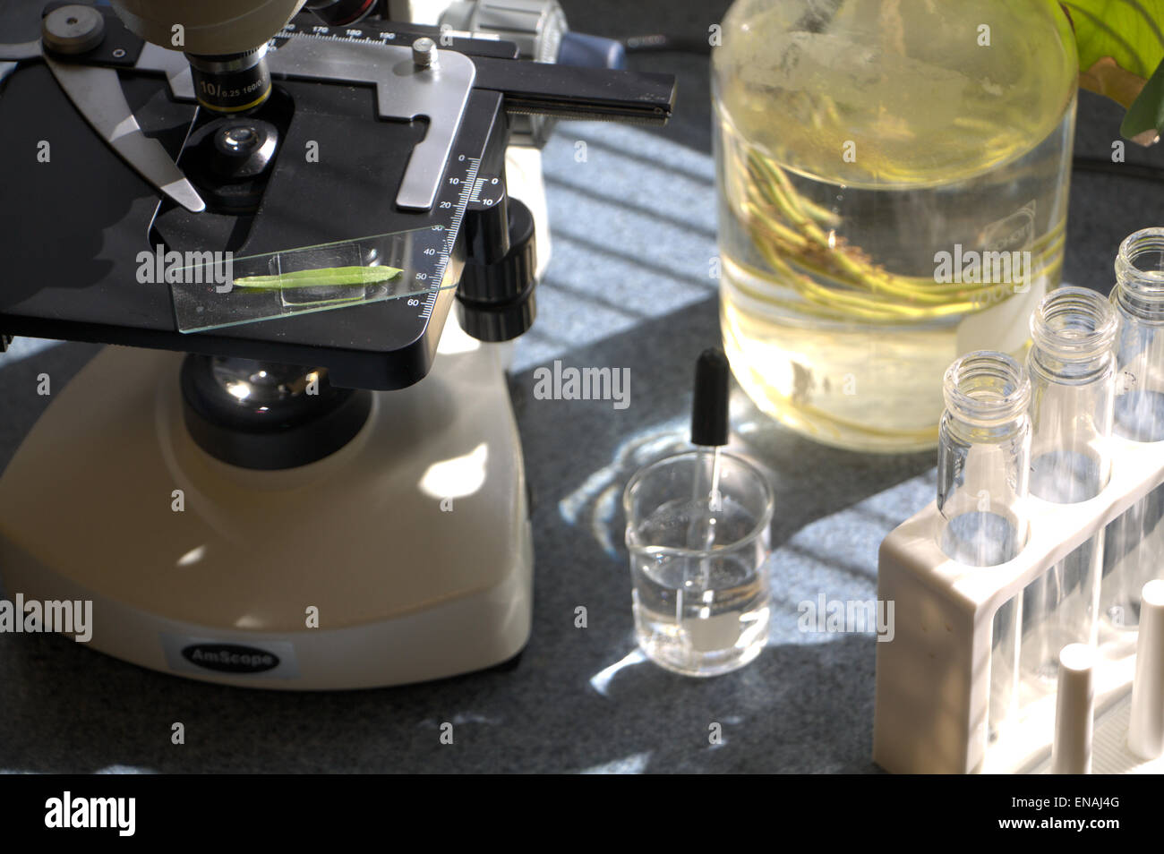 Examining a leaf under a microscope Stock Photohttps://www.alamy.com/image-license-details/?v=1https://www.alamy.com/stock-photo-examining-a-leaf-under-a-microscope-81983008.html
RFENAJ4G–Examining a leaf under a microscope
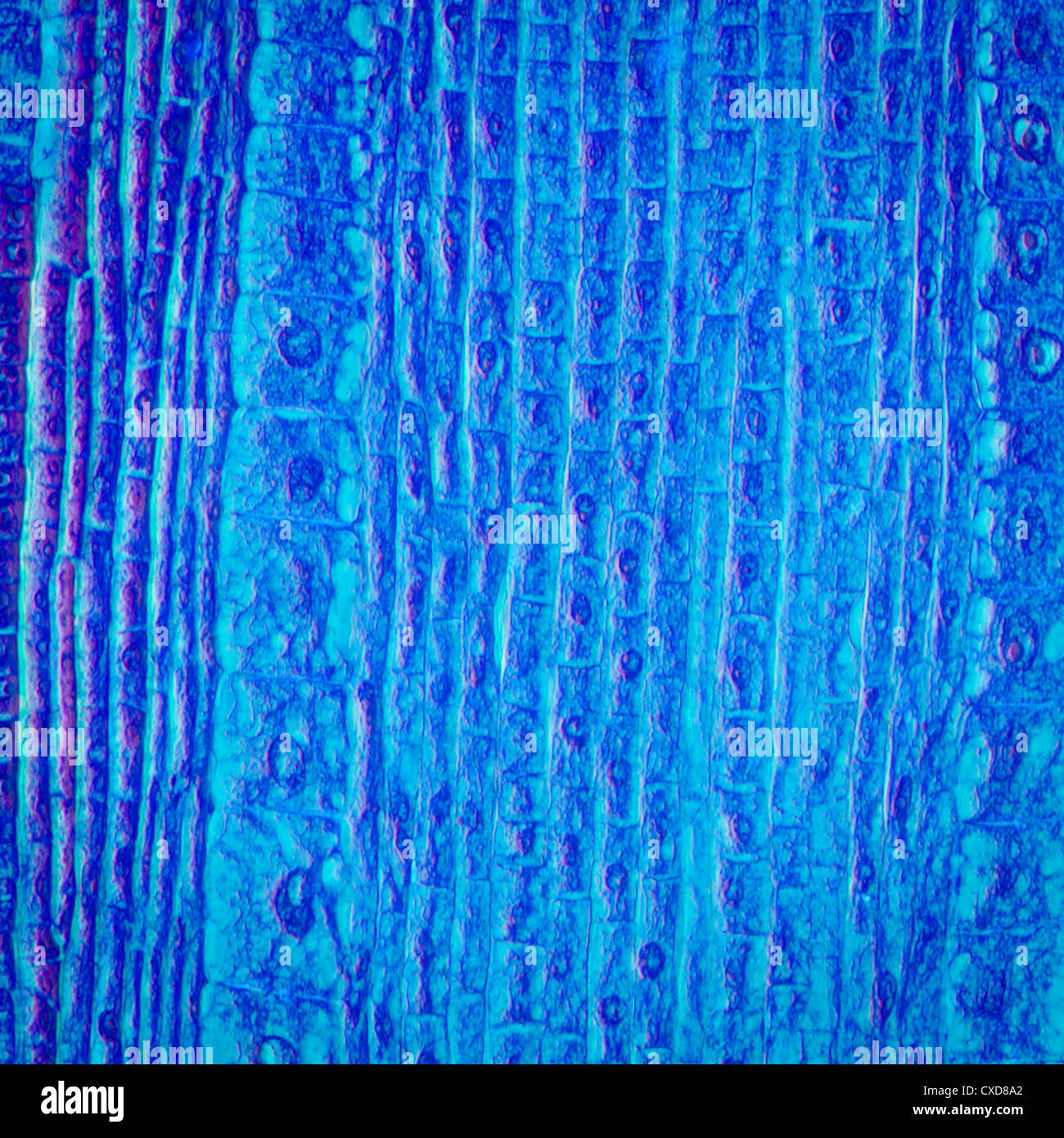 micrograph plant root tip tissue cell Stock Photohttps://www.alamy.com/image-license-details/?v=1https://www.alamy.com/stock-photo-micrograph-plant-root-tip-tissue-cell-50693722.html
RFCXD8A2–micrograph plant root tip tissue cell
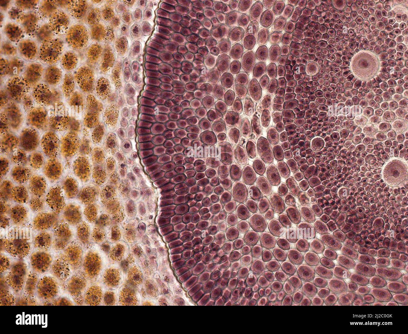 Corn grain. An interesting photo taken with a microscope. Cross section through corn grain (Zea mays). Stock Photohttps://www.alamy.com/image-license-details/?v=1https://www.alamy.com/corn-grain-an-interesting-photo-taken-with-a-microscope-cross-section-through-corn-grain-zea-mays-image466173139.html
RF2J2C0GK–Corn grain. An interesting photo taken with a microscope. Cross section through corn grain (Zea mays).
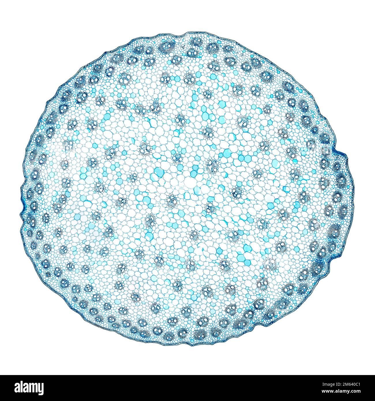 Zea stem, maize stem, cross section, 20X light micrograph. Stem of the plant Zea mays, under the light microscope. Stock Photohttps://www.alamy.com/image-license-details/?v=1https://www.alamy.com/zea-stem-maize-stem-cross-section-20x-light-micrograph-stem-of-the-plant-zea-mays-under-the-light-microscope-image502876753.html
RF2M640C1–Zea stem, maize stem, cross section, 20X light micrograph. Stem of the plant Zea mays, under the light microscope.
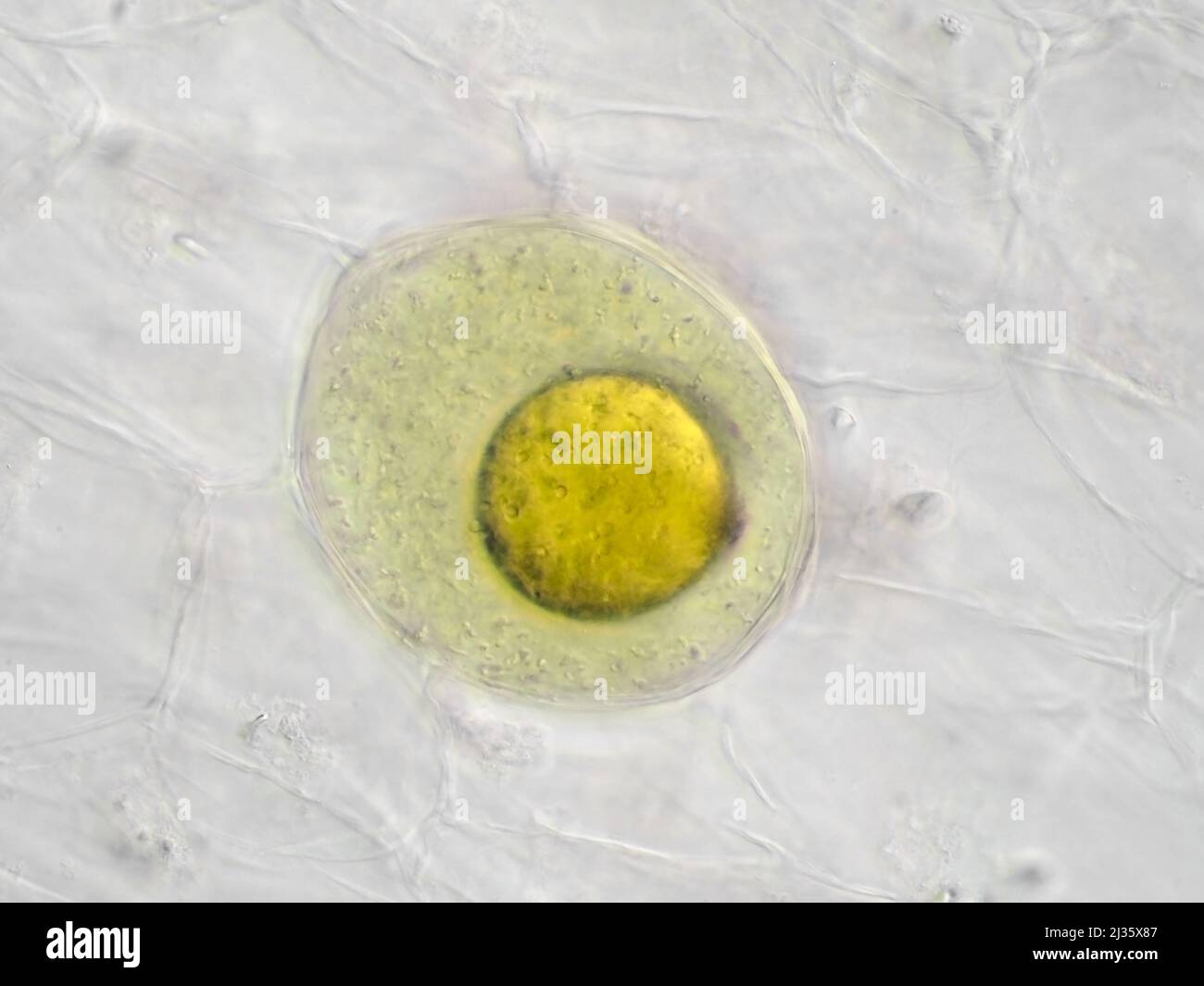 Pickled sushi ginger under the microscope, showing a secretory cell with an oil globule Stock Photohttps://www.alamy.com/image-license-details/?v=1https://www.alamy.com/pickled-sushi-ginger-under-the-microscope-showing-a-secretory-cell-with-an-oil-globule-image466654279.html
RM2J35X87–Pickled sushi ginger under the microscope, showing a secretory cell with an oil globule
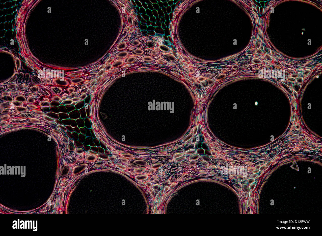 micrograph plant tissue cell , stem of pumpkin Stock Photohttps://www.alamy.com/image-license-details/?v=1https://www.alamy.com/stock-photo-micrograph-plant-tissue-cell-stem-of-pumpkin-52301365.html
RFD12EWW–micrograph plant tissue cell , stem of pumpkin
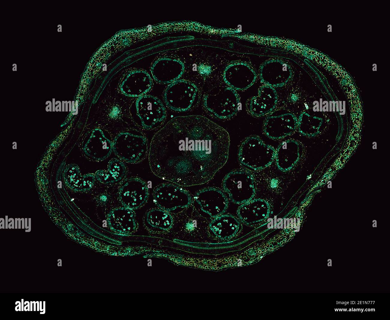 cross section cut under the microscope – microscopic view of plant cells for botanic education Stock Photohttps://www.alamy.com/image-license-details/?v=1https://www.alamy.com/cross-section-cut-under-the-microscope-microscopic-view-of-plant-cells-for-botanic-education-image396919803.html
RF2E1N777–cross section cut under the microscope – microscopic view of plant cells for botanic education
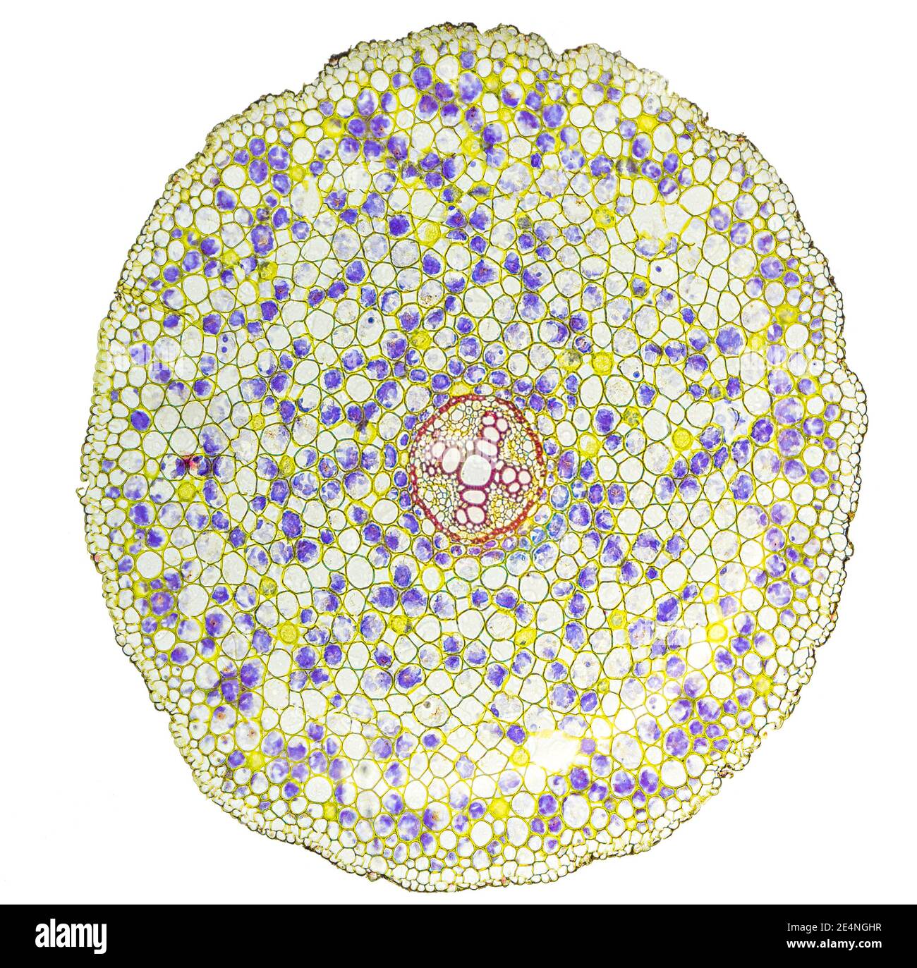 Ranunculus root t.s. metaxylem x100 Stock Photohttps://www.alamy.com/image-license-details/?v=1https://www.alamy.com/ranunculus-root-ts-metaxylem-x100-image398771123.html
RM2E4NGHR–Ranunculus root t.s. metaxylem x100
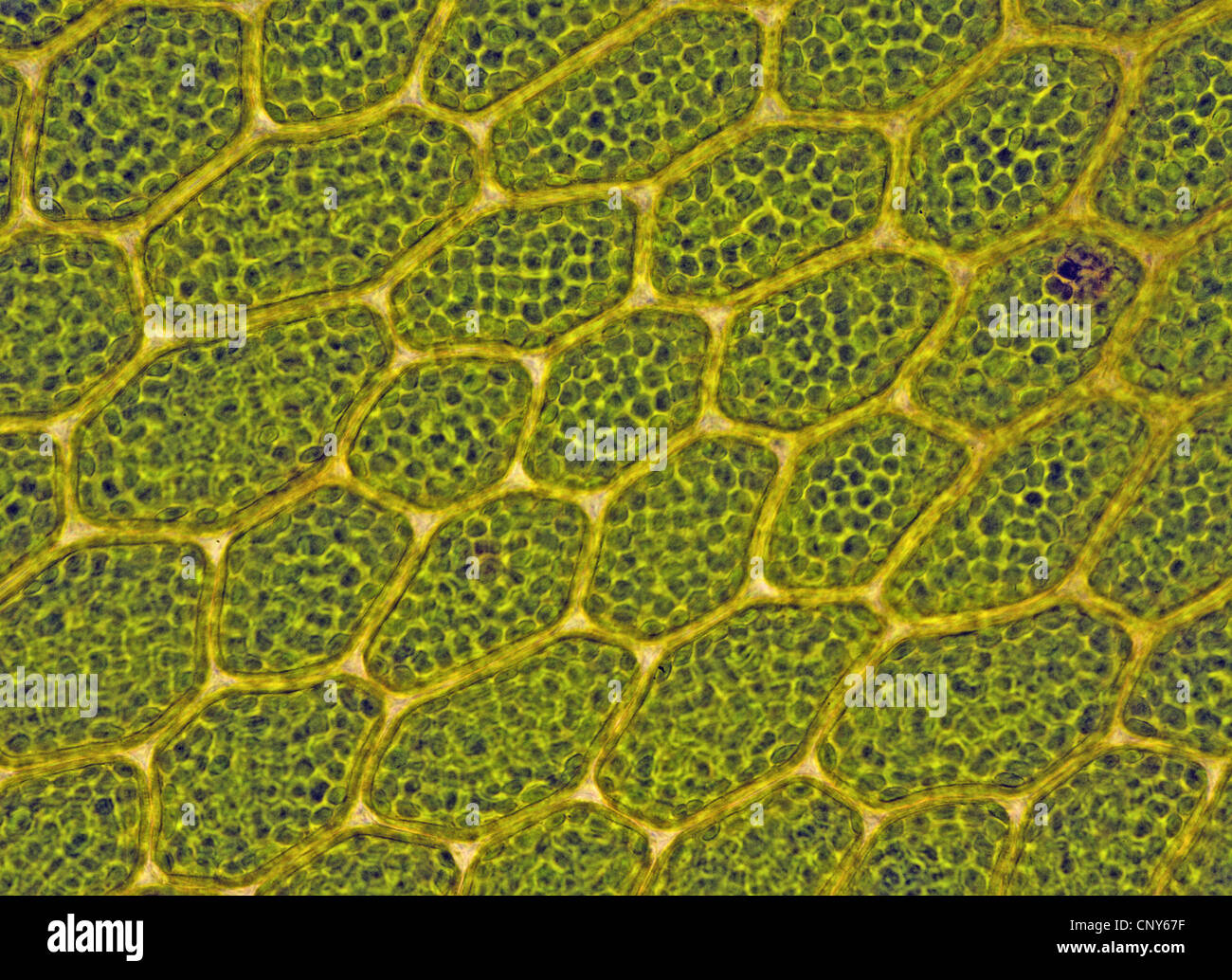 Cells with chloroplasts in Bryophytes (Mnium sp.), Norway Stock Photohttps://www.alamy.com/image-license-details/?v=1https://www.alamy.com/stock-photo-cells-with-chloroplasts-in-bryophytes-mnium-sp-norway-47926131.html
RMCNY67F–Cells with chloroplasts in Bryophytes (Mnium sp.), Norway
 Stoma. Asparagus stem. 16x Stock Photohttps://www.alamy.com/image-license-details/?v=1https://www.alamy.com/stoma-asparagus-stem-16x-image188662503.html
RFMXX8XF–Stoma. Asparagus stem. 16x
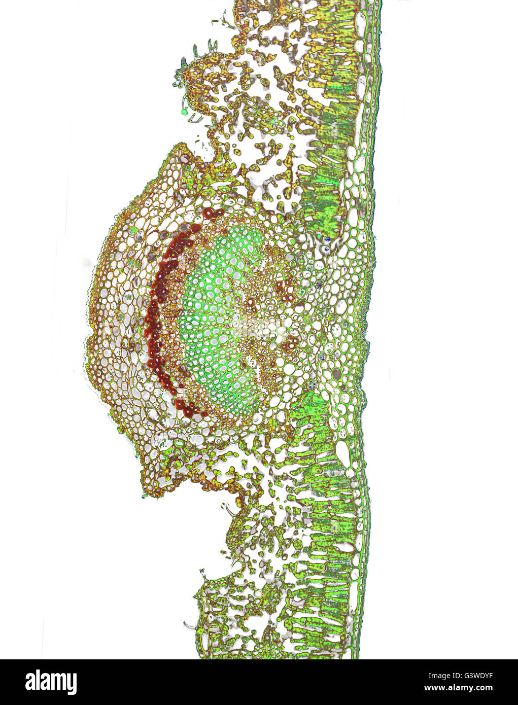 Micro photo of a Nerium Oleander Leaf Stock Photohttps://www.alamy.com/image-license-details/?v=1https://www.alamy.com/stock-photo-micro-photo-of-a-nerium-oleander-leaf-105665939.html
RMG3WDYF–Micro photo of a Nerium Oleander Leaf
 3d rendering of a microscopic view of a plant cell. The cell is green, which is because it contains chloroplasts, Stock Photohttps://www.alamy.com/image-license-details/?v=1https://www.alamy.com/3d-rendering-of-a-microscopic-view-of-a-plant-cell-the-cell-is-green-which-is-because-it-contains-chloroplasts-image592728021.html
RF2WC92HW–3d rendering of a microscopic view of a plant cell. The cell is green, which is because it contains chloroplasts,
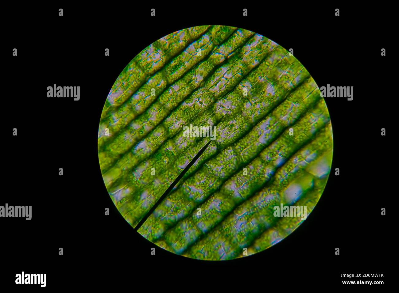 Green leaf grains also known as chloroplasts in cells of a waterweed seen through a microscope. Biology experiment. Stock Photohttps://www.alamy.com/image-license-details/?v=1https://www.alamy.com/green-leaf-grains-also-known-as-chloroplasts-in-cells-of-a-waterweed-seen-through-a-microscope-biology-experiment-image382774719.html
RF2D6MW1K–Green leaf grains also known as chloroplasts in cells of a waterweed seen through a microscope. Biology experiment.
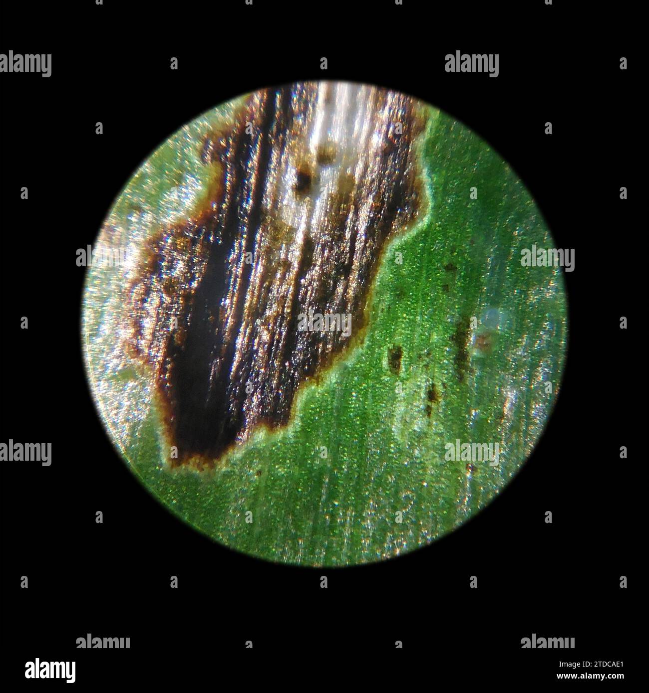 the surface of leaves of green plants affected by pests observed under a microscope Stock Photohttps://www.alamy.com/image-license-details/?v=1https://www.alamy.com/the-surface-of-leaves-of-green-plants-affected-by-pests-observed-under-a-microscope-image576204329.html
RF2TDCAE1–the surface of leaves of green plants affected by pests observed under a microscope
 Abstract Science Colorful Pattern Background Stock Photohttps://www.alamy.com/image-license-details/?v=1https://www.alamy.com/abstract-science-colorful-pattern-background-image483508906.html
RF2K2HMF6–Abstract Science Colorful Pattern Background
 biological cell isolated on whithe background microscope 3D Illustration Stock Photohttps://www.alamy.com/image-license-details/?v=1https://www.alamy.com/biological-cell-isolated-on-whithe-background-microscope-3d-illustration-image208953338.html
RFP3XJ36–biological cell isolated on whithe background microscope 3D Illustration
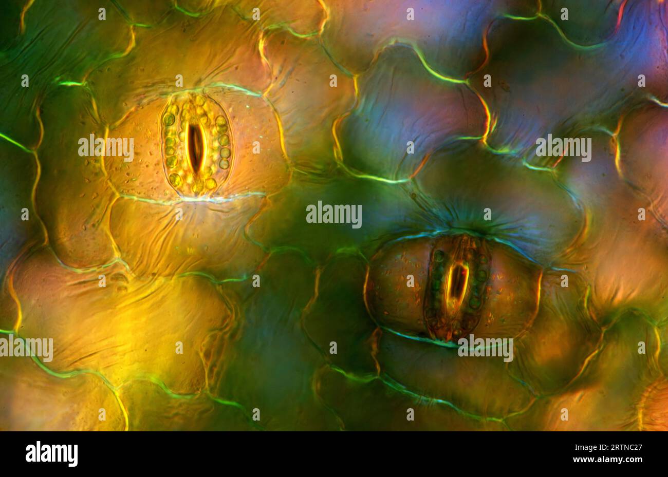 The image presents stomata in Spathiphyllum leaf epidermis, photographed through the microscope in polarized light at a magnification of 200X Stock Photohttps://www.alamy.com/image-license-details/?v=1https://www.alamy.com/the-image-presents-stomata-in-spathiphyllum-leaf-epidermis-photographed-through-the-microscope-in-polarized-light-at-a-magnification-of-200x-image565953983.html
RM2RTNC27–The image presents stomata in Spathiphyllum leaf epidermis, photographed through the microscope in polarized light at a magnification of 200X
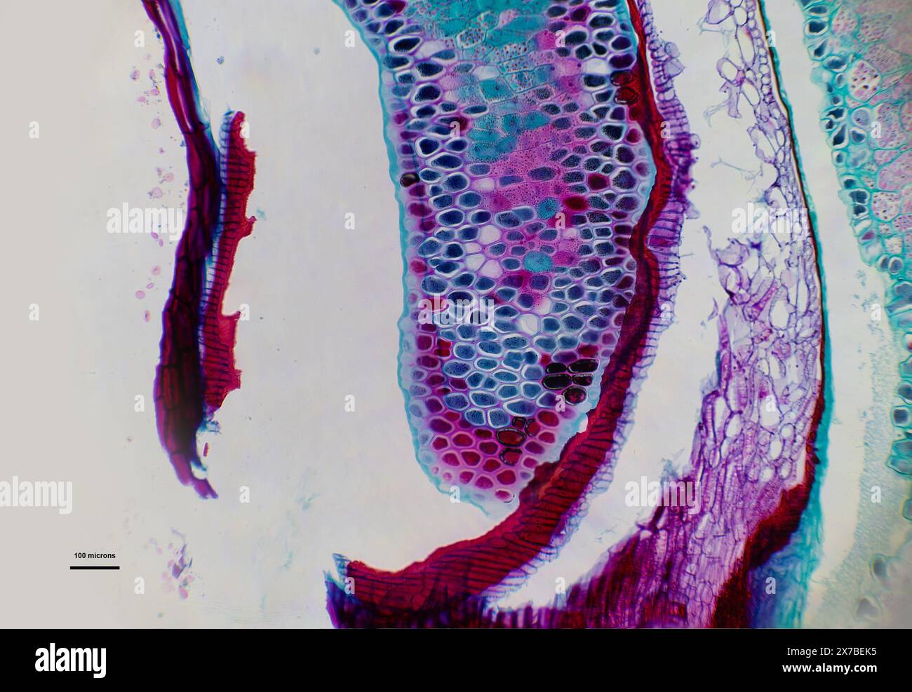 Triticum seed under a microscope at 10 times magnification Stock Photohttps://www.alamy.com/image-license-details/?v=1https://www.alamy.com/triticum-seed-under-a-microscope-at-10-times-magnification-image606918457.html
RF2X7BEK5–Triticum seed under a microscope at 10 times magnification
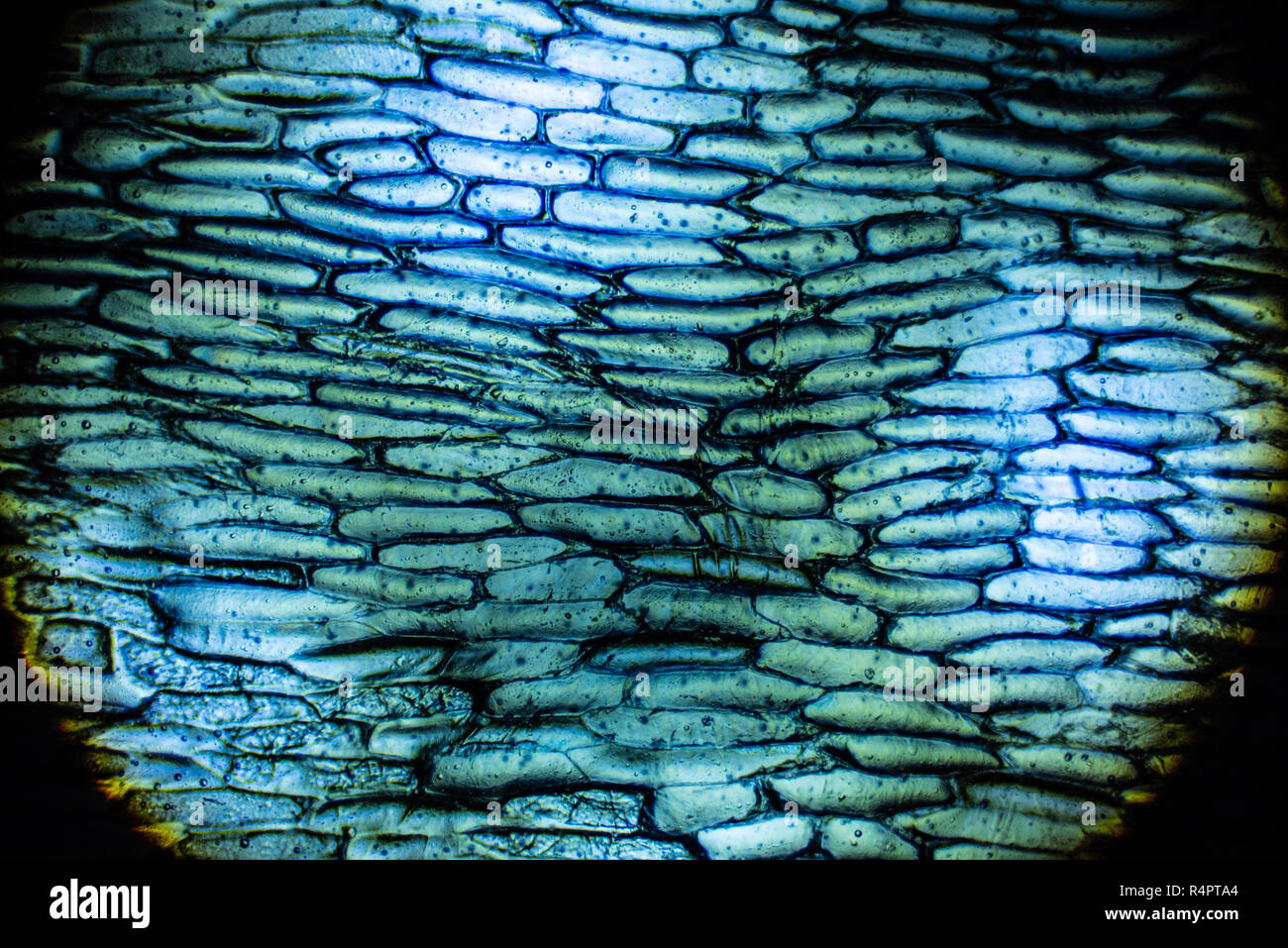 Onion cells under the microscope Stock Photohttps://www.alamy.com/image-license-details/?v=1https://www.alamy.com/onion-cells-under-the-microscope-image226695452.html
RFR4PTA4–Onion cells under the microscope
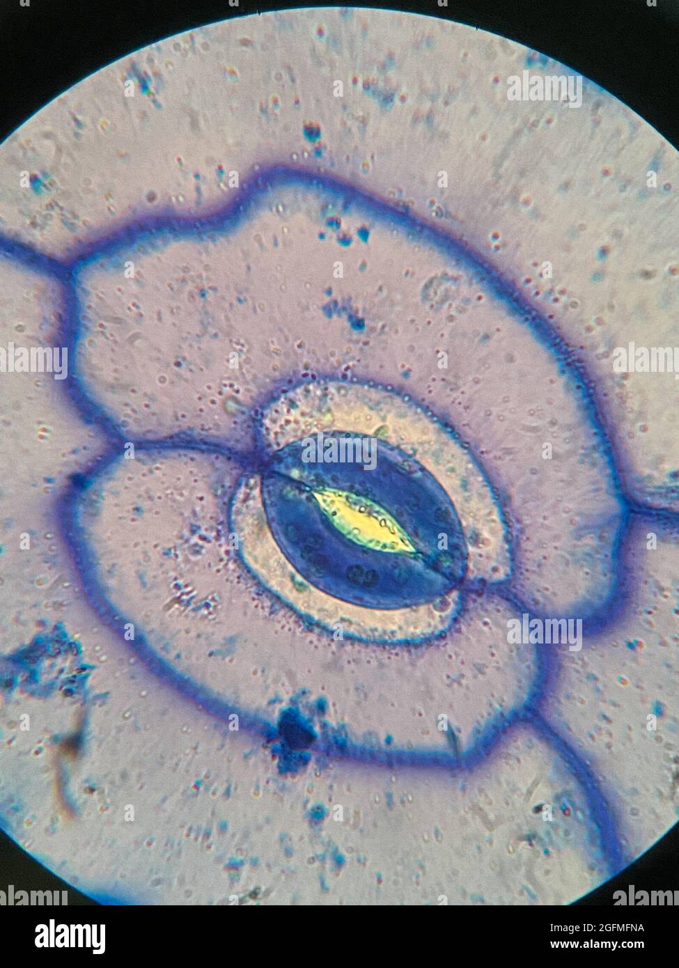 microscopic photo of stomata on the leaf of Portulaca oleracea plant Stock Photohttps://www.alamy.com/image-license-details/?v=1https://www.alamy.com/microscopic-photo-of-stomata-on-the-leaf-of-portulaca-oleracea-plant-image439930438.html
RF2GFMFNA–microscopic photo of stomata on the leaf of Portulaca oleracea plant
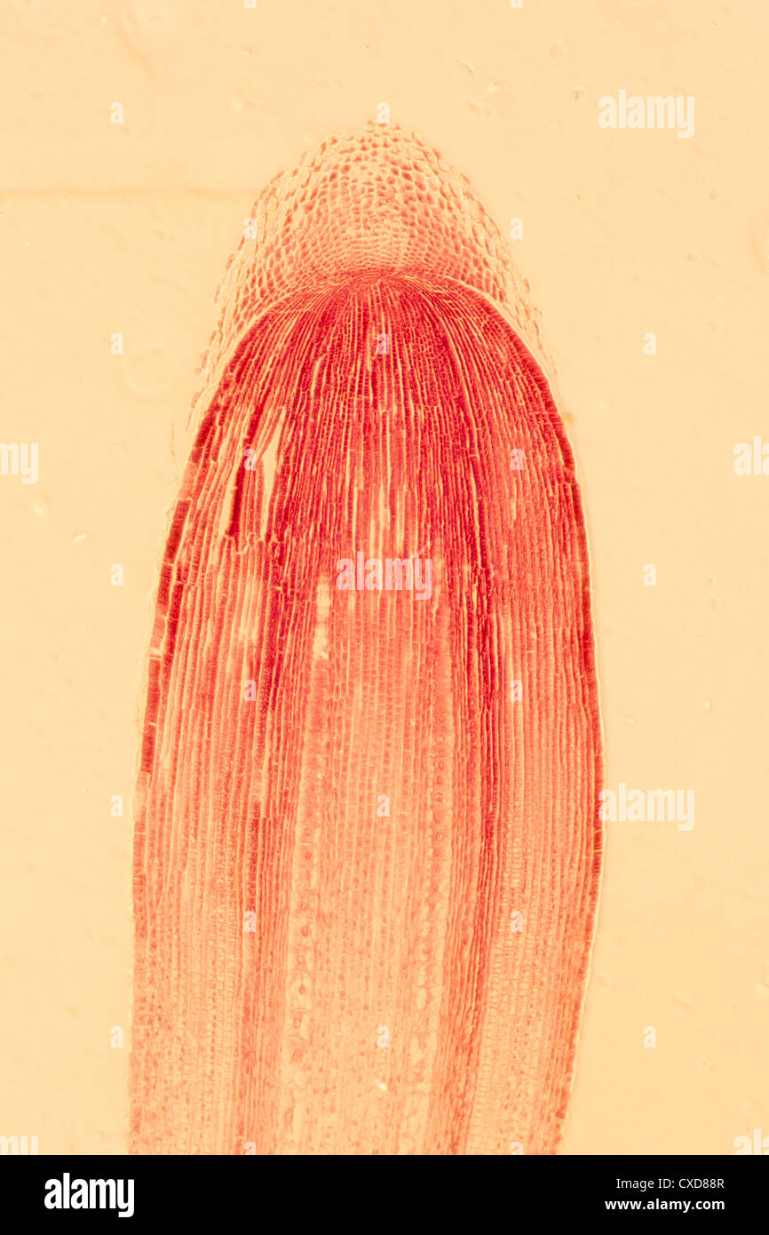 micrograph plant root tip tissue cell Stock Photohttps://www.alamy.com/image-license-details/?v=1https://www.alamy.com/stock-photo-micrograph-plant-root-tip-tissue-cell-50693687.html
RFCXD88R–micrograph plant root tip tissue cell
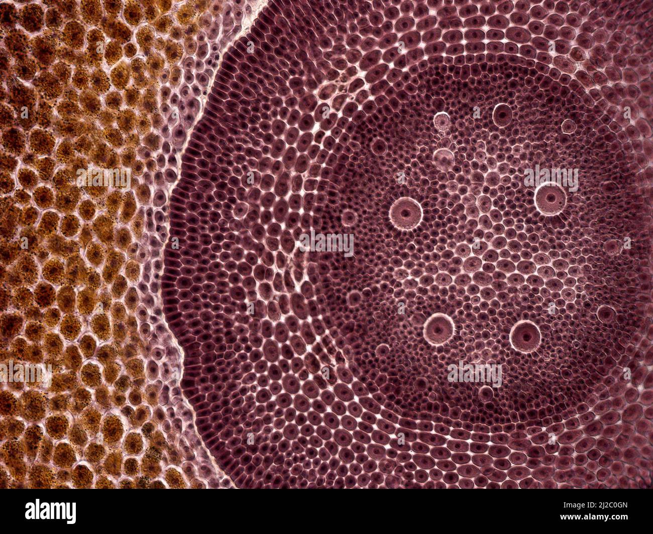 Corn grain. An interesting photo taken with a microscope. Cross section through corn grain (Zea mays). Stock Photohttps://www.alamy.com/image-license-details/?v=1https://www.alamy.com/corn-grain-an-interesting-photo-taken-with-a-microscope-cross-section-through-corn-grain-zea-mays-image466173141.html
RF2J2C0GN–Corn grain. An interesting photo taken with a microscope. Cross section through corn grain (Zea mays).
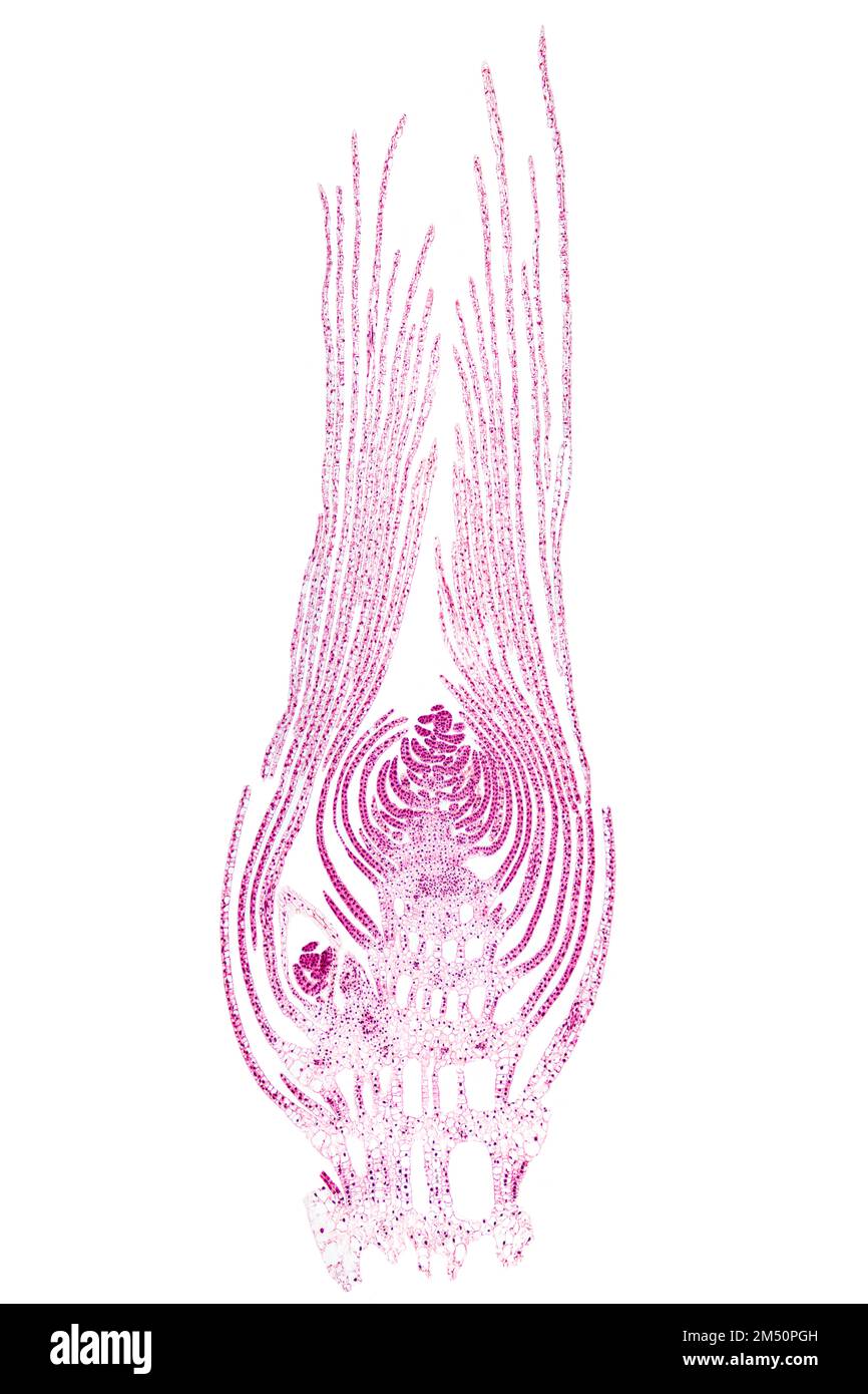 Apical bud of an aquatic plant, longitudinal section, 20X light micrograph. Whole bud, under the light microscope, stained for better visualization. Stock Photohttps://www.alamy.com/image-license-details/?v=1https://www.alamy.com/apical-bud-of-an-aquatic-plant-longitudinal-section-20x-light-micrograph-whole-bud-under-the-light-microscope-stained-for-better-visualization-image502191665.html
RF2M50PGH–Apical bud of an aquatic plant, longitudinal section, 20X light micrograph. Whole bud, under the light microscope, stained for better visualization.
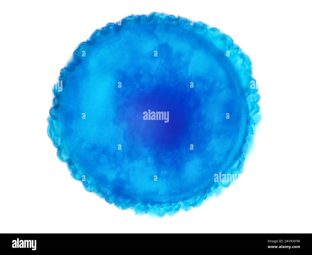 Relatively large (about 80 microns in diameter) spore, likely club-moss spore, under the microscope Stock Photohttps://www.alamy.com/image-license-details/?v=1https://www.alamy.com/relatively-large-about-80-microns-in-diameter-spore-likely-club-moss-spore-under-the-microscope-image344023901.html
RM2AYKHYW–Relatively large (about 80 microns in diameter) spore, likely club-moss spore, under the microscope
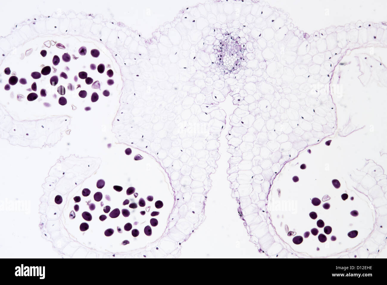 anther of lily flower, Lilium brownii, science plant cell micrograph Stock Photohttps://www.alamy.com/image-license-details/?v=1https://www.alamy.com/stock-photo-anther-of-lily-flower-lilium-brownii-science-plant-cell-micrograph-52301130.html
RFD12EHE–anther of lily flower, Lilium brownii, science plant cell micrograph
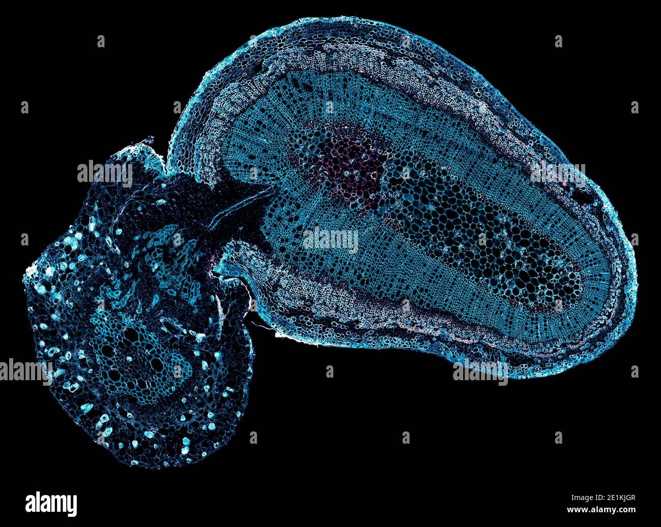 cross section cut under the microscope – microscopic view of plant cells for botanic education Stock Photohttps://www.alamy.com/image-license-details/?v=1https://www.alamy.com/cross-section-cut-under-the-microscope-microscopic-view-of-plant-cells-for-botanic-education-image396884791.html
RF2E1KJGR–cross section cut under the microscope – microscopic view of plant cells for botanic education
