Quick filters:
Plant cell mitosis Stock Photos and Images
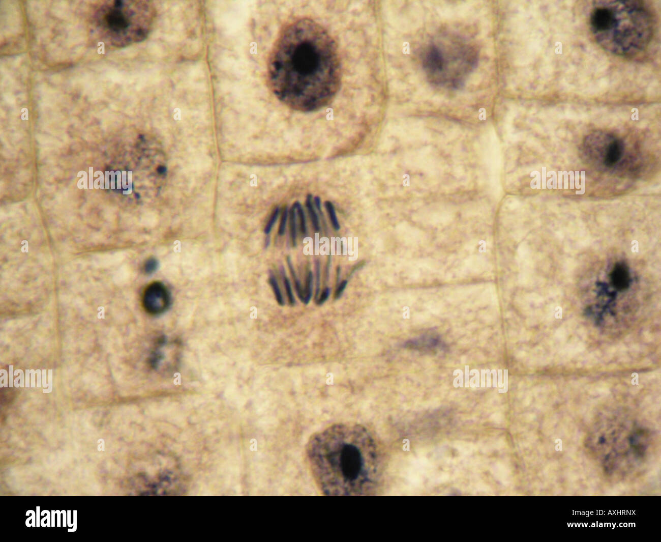 Mitosis (Anaphase) in onion tissue at 1000x under optical microscope. inmersion oil objetive 100x Stock Photohttps://www.alamy.com/image-license-details/?v=1https://www.alamy.com/stock-photo-mitosis-anaphase-in-onion-tissue-at-1000x-under-optical-microscope-16851445.html
RMAXHRNX–Mitosis (Anaphase) in onion tissue at 1000x under optical microscope. inmersion oil objetive 100x
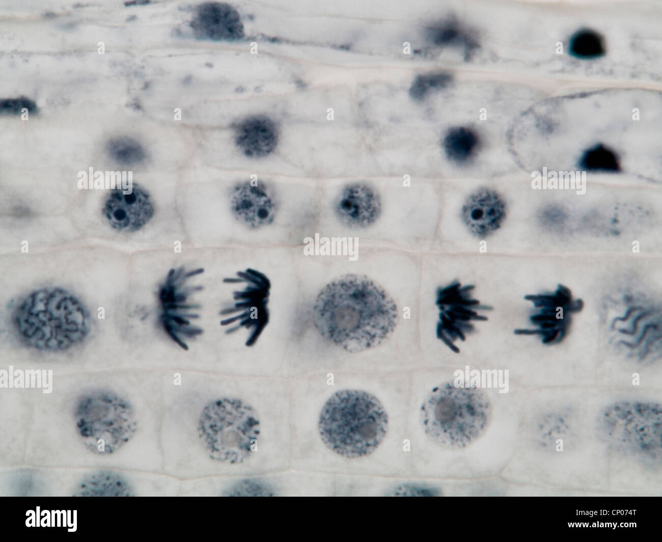 mitosis in a onion root, anaphasis Stock Photohttps://www.alamy.com/image-license-details/?v=1https://www.alamy.com/stock-photo-mitosis-in-a-onion-root-anaphasis-47948792.html
RMCP074T–mitosis in a onion root, anaphasis
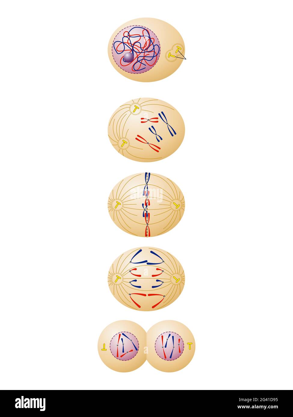 Animal and plant somatic cell mitosis Stock Photohttps://www.alamy.com/image-license-details/?v=1https://www.alamy.com/animal-and-plant-somatic-cell-mitosis-image432750225.html
RF2G41D95–Animal and plant somatic cell mitosis
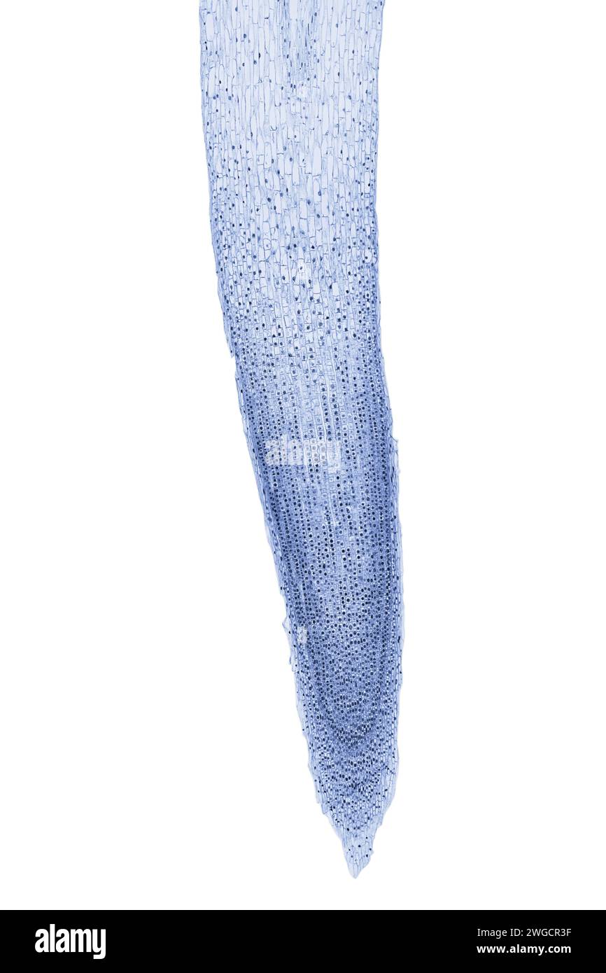 Onion root tip, Allium cepa, showing all stages of mitosis, longitudinal section, 20X light micrograph. Lengthwise cut root tip of an onion. Stock Photohttps://www.alamy.com/image-license-details/?v=1https://www.alamy.com/onion-root-tip-allium-cepa-showing-all-stages-of-mitosis-longitudinal-section-20x-light-micrograph-lengthwise-cut-root-tip-of-an-onion-image595268563.html
RF2WGCR3F–Onion root tip, Allium cepa, showing all stages of mitosis, longitudinal section, 20X light micrograph. Lengthwise cut root tip of an onion.
 Abstract Science Colorful Pattern Background Stock Photohttps://www.alamy.com/image-license-details/?v=1https://www.alamy.com/abstract-science-colorful-pattern-background-image483512342.html
RF2K2HTWX–Abstract Science Colorful Pattern Background
 Abstract Science Colorful Pattern Background Stock Photohttps://www.alamy.com/image-license-details/?v=1https://www.alamy.com/abstract-science-colorful-pattern-background-image483508744.html
RF2K2HM9C–Abstract Science Colorful Pattern Background
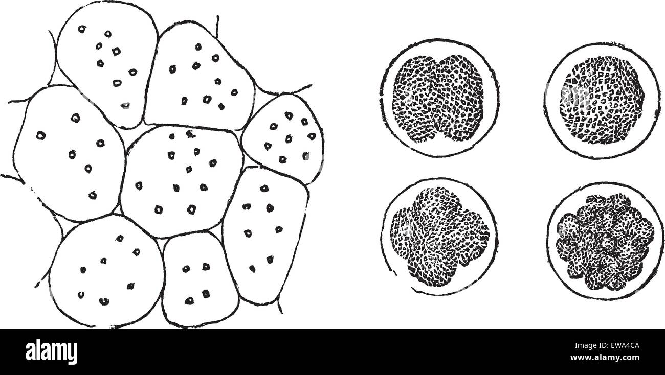 Cell Division in plants (left) and in animals (right), vintage engraved illustration. Trousset encyclopedia (1886 - 1891). Stock Vectorhttps://www.alamy.com/image-license-details/?v=1https://www.alamy.com/stock-photo-cell-division-in-plants-left-and-in-animals-right-vintage-engraved-84430874.html
RFEWA4CA–Cell Division in plants (left) and in animals (right), vintage engraved illustration. Trousset encyclopedia (1886 - 1891).
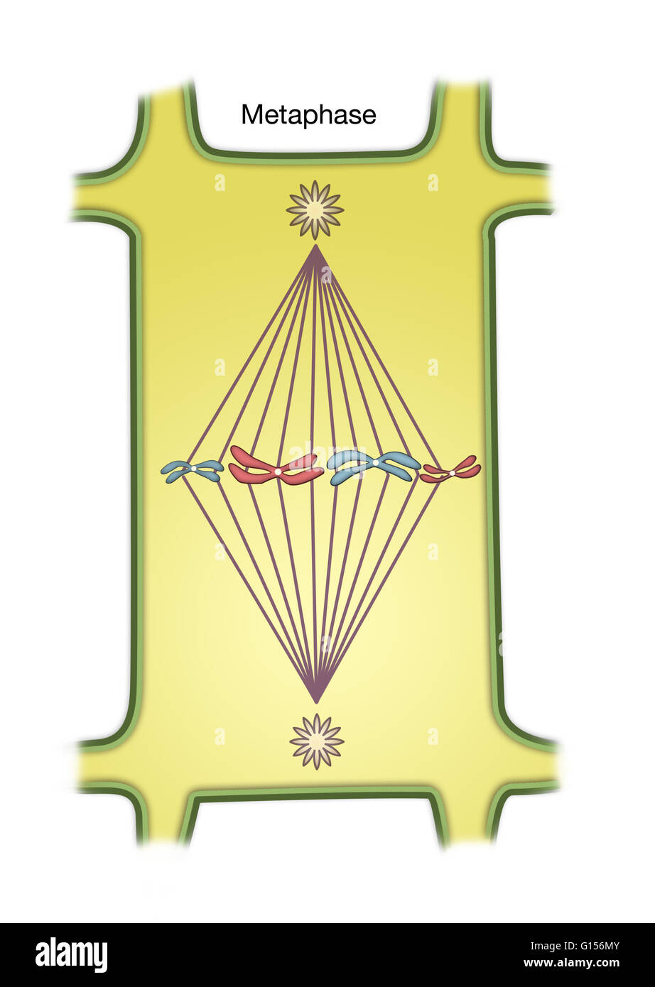 Diagram of Metaphase of Mitosis in a plant cell. Stock Photohttps://www.alamy.com/image-license-details/?v=1https://www.alamy.com/stock-photo-diagram-of-metaphase-of-mitosis-in-a-plant-cell-103991915.html
RMG156MY–Diagram of Metaphase of Mitosis in a plant cell.
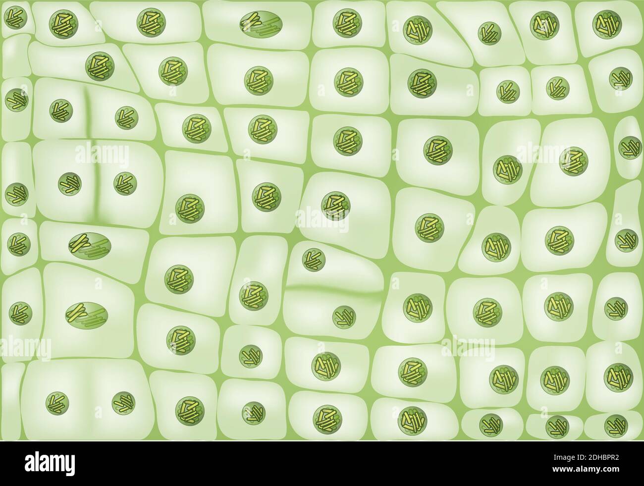 Cell division background Stock Vectorhttps://www.alamy.com/image-license-details/?v=1https://www.alamy.com/cell-division-background-image389336614.html
RF2DHBPR2–Cell division background
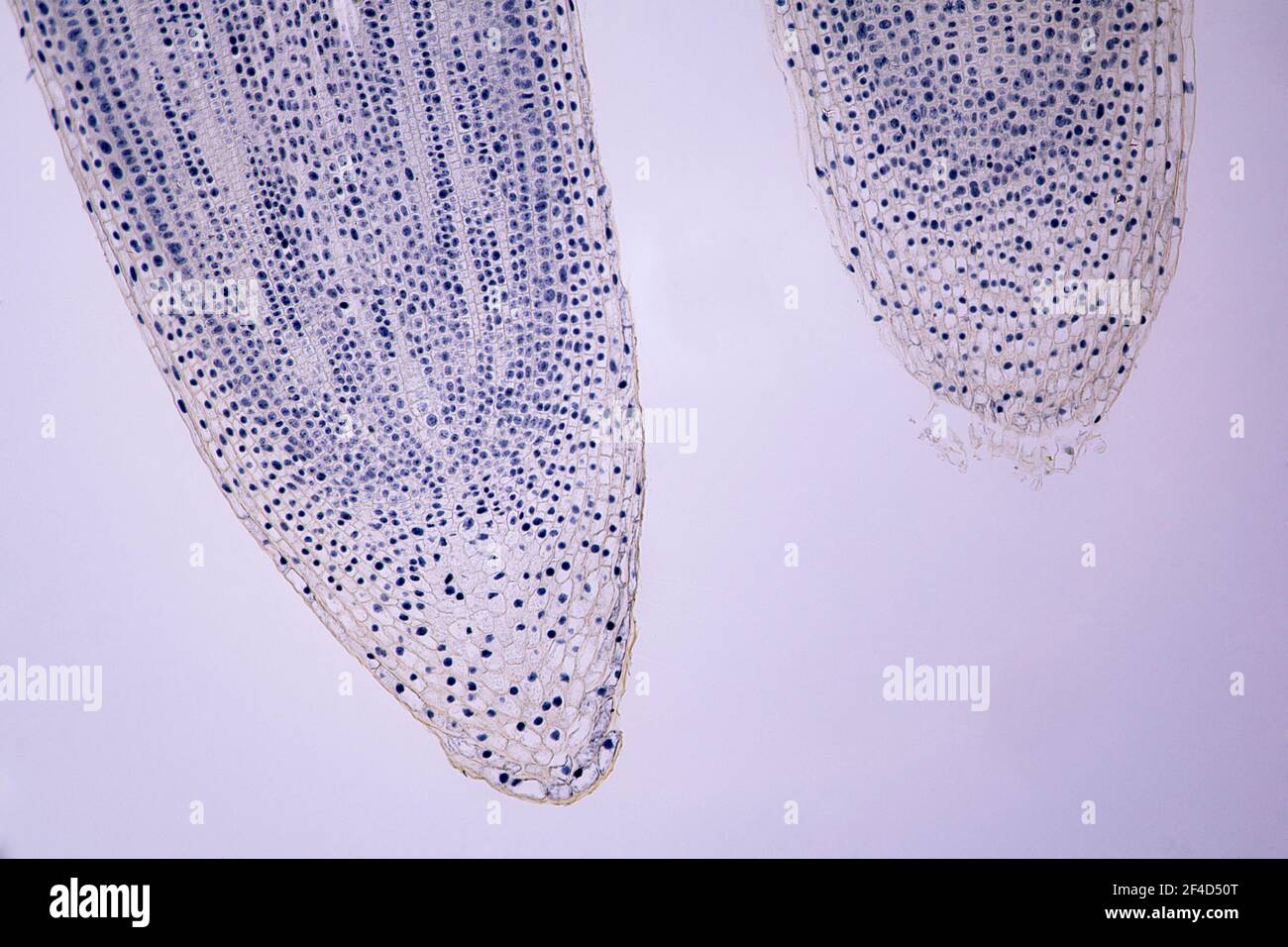 mitosis in an Allium root tip Longitudinal section under a light microscope at 10 times magnification. Stock Photohttps://www.alamy.com/image-license-details/?v=1https://www.alamy.com/mitosis-in-an-allium-root-tip-longitudinal-section-under-a-light-microscope-at-10-times-magnification-image415796776.html
RF2F4D50T–mitosis in an Allium root tip Longitudinal section under a light microscope at 10 times magnification.
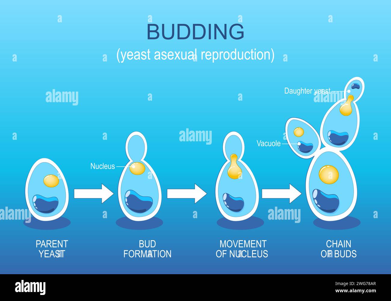 Budding. Yeast Asexual reproduction. Fungi growth. Cell division. Vector diagram. Stock Vectorhttps://www.alamy.com/image-license-details/?v=1https://www.alamy.com/budding-yeast-asexual-reproduction-fungi-growth-cell-division-vector-diagram-image595147247.html
RF2WG78AR–Budding. Yeast Asexual reproduction. Fungi growth. Cell division. Vector diagram.
 Microphotography of root mitosis of an onion. Stock Photohttps://www.alamy.com/image-license-details/?v=1https://www.alamy.com/microphotography-of-root-mitosis-of-an-onion-image365985237.html
RF2C7C1W9–Microphotography of root mitosis of an onion.
 Vicia Fabia root tip mitosis transverse section Stock Photohttps://www.alamy.com/image-license-details/?v=1https://www.alamy.com/vicia-fabia-root-tip-mitosis-transverse-section-image363642002.html
RM2C3H92A–Vicia Fabia root tip mitosis transverse section
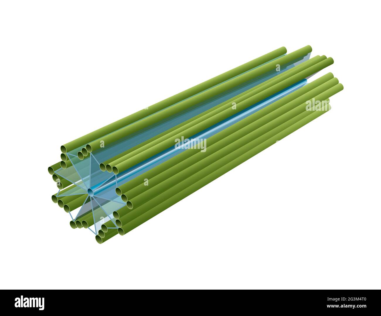 Centrioles in eukaryotic cell. Structure Stock Photohttps://www.alamy.com/image-license-details/?v=1https://www.alamy.com/centrioles-in-eukaryotic-cell-structure-image432546016.html
RF2G3M4T0–Centrioles in eukaryotic cell. Structure
 Embryonic bright green stem cells colony under a microscope 3D illustration Stock Photohttps://www.alamy.com/image-license-details/?v=1https://www.alamy.com/embryonic-bright-green-stem-cells-colony-under-a-microscope-3d-illustration-image234777302.html
RFRHY0RJ–Embryonic bright green stem cells colony under a microscope 3D illustration
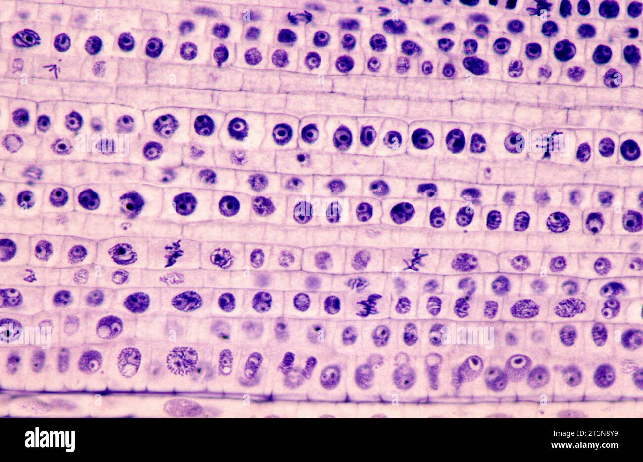 Root apical meristem showing cell divisions (mitosis). Onion root photomicrograph. Stock Photohttps://www.alamy.com/image-license-details/?v=1https://www.alamy.com/root-apical-meristem-showing-cell-divisions-mitosis-onion-root-photomicrograph-image578244669.html
RF2TGN8Y9–Root apical meristem showing cell divisions (mitosis). Onion root photomicrograph.
 Extreme magnification - Pelargonium, Glandular hairs and tector - section through stem at 20x Stock Photohttps://www.alamy.com/image-license-details/?v=1https://www.alamy.com/stock-photo-extreme-magnification-pelargonium-glandular-hairs-and-tector-section-135789601.html
RMHTWN0H–Extreme magnification - Pelargonium, Glandular hairs and tector - section through stem at 20x
 Dividing Cancer Cell Oncology Research Concept 3d illustration proteins with lymphocytes gene editing , t cells or cancer cells, meiosis, mitosis 3d Stock Photohttps://www.alamy.com/image-license-details/?v=1https://www.alamy.com/dividing-cancer-cell-oncology-research-concept-3d-illustration-proteins-with-lymphocytes-gene-editing-t-cells-or-cancer-cells-meiosis-mitosis-3d-image247428305.html
RFTAF995–Dividing Cancer Cell Oncology Research Concept 3d illustration proteins with lymphocytes gene editing , t cells or cancer cells, meiosis, mitosis 3d
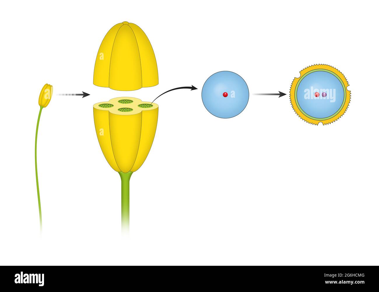 Structure of stamens of flowering plants Stock Photohttps://www.alamy.com/image-license-details/?v=1https://www.alamy.com/structure-of-stamens-of-flowering-plants-image434330304.html
RF2G6HCMG–Structure of stamens of flowering plants
 Mitosis Horse Ascaris egg under the microscope, background (Ascaris lumbricoides) Stock Photohttps://www.alamy.com/image-license-details/?v=1https://www.alamy.com/stock-photo-mitosis-horse-ascaris-egg-under-the-microscope-background-ascaris-51118851.html
RFCY4JH7–Mitosis Horse Ascaris egg under the microscope, background (Ascaris lumbricoides)
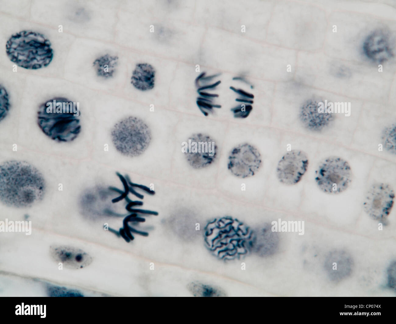 mitosis in a onion root, prophasis and anaphasis Stock Photohttps://www.alamy.com/image-license-details/?v=1https://www.alamy.com/stock-photo-mitosis-in-a-onion-root-prophasis-and-anaphasis-47948794.html
RMCP074X–mitosis in a onion root, prophasis and anaphasis
 A centriole is made of nine sets of microtubules, each in groups of three known as triplet microtubulesCell structure of centriole. Stock Photohttps://www.alamy.com/image-license-details/?v=1https://www.alamy.com/a-centriole-is-made-of-nine-sets-of-microtubules-each-in-groups-of-three-known-as-triplet-microtubulescell-structure-of-centriole-image432546102.html
RF2G3M4Y2–A centriole is made of nine sets of microtubules, each in groups of three known as triplet microtubulesCell structure of centriole.
 Organic cell, 3d rendering Stock Photohttps://www.alamy.com/image-license-details/?v=1https://www.alamy.com/organic-cell-3d-rendering-image352686135.html
RF2BDP6NB–Organic cell, 3d rendering
 Abstract Science Colorful Pattern Background Stock Photohttps://www.alamy.com/image-license-details/?v=1https://www.alamy.com/abstract-science-colorful-pattern-background-image483508972.html
RF2K2HMHG–Abstract Science Colorful Pattern Background
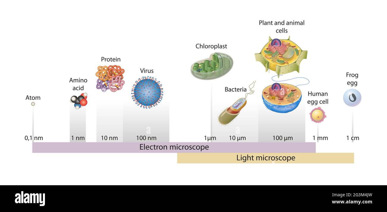 Sizes of cells drawn on a logarithmic scale, indicating the range of readily resolvable objects in the light and electron microscope Stock Photohttps://www.alamy.com/image-license-details/?v=1https://www.alamy.com/sizes-of-cells-drawn-on-a-logarithmic-scale-indicating-the-range-of-readily-resolvable-objects-in-the-light-and-electron-microscope-image432545873.html
RF2G3M4JW–Sizes of cells drawn on a logarithmic scale, indicating the range of readily resolvable objects in the light and electron microscope
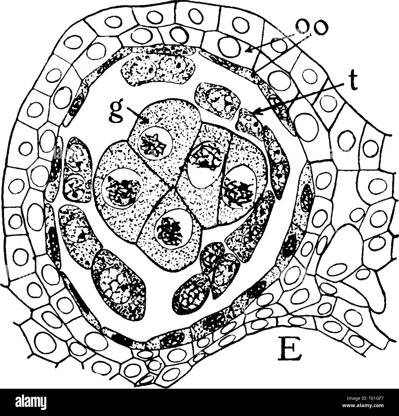 This is Microspore Anther Lobe view. In flowering plants microspore mother cells are formed within the pollen sacs of the anthers by mitosis, the micr Stock Vectorhttps://www.alamy.com/image-license-details/?v=1https://www.alamy.com/this-is-microspore-anther-lobe-view-in-flowering-plants-microspore-mother-cells-are-formed-within-the-pollen-sacs-of-the-anthers-by-mitosis-the-micr-image244668011.html
RFT61GF7–This is Microspore Anther Lobe view. In flowering plants microspore mother cells are formed within the pollen sacs of the anthers by mitosis, the micr
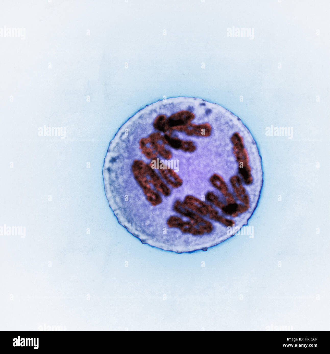 Anaphase of Mitosis in Trillium Cell Stock Photohttps://www.alamy.com/image-license-details/?v=1https://www.alamy.com/stock-photo-anaphase-of-mitosis-in-trillium-cell-135017534.html
RMHRJG6P–Anaphase of Mitosis in Trillium Cell
 Cells mitosis micrograph Stock Photohttps://www.alamy.com/image-license-details/?v=1https://www.alamy.com/stock-photo-cells-mitosis-micrograph-93443612.html
RFFC0M7T–Cells mitosis micrograph
 leaf stoma close up for photosynthesis. Chlorophyll is the molecule in leaves that uses the energy in sunlight to turn water and Stock Photohttps://www.alamy.com/image-license-details/?v=1https://www.alamy.com/stock-photo-leaf-stoma-close-up-for-photosynthesis-chlorophyll-is-the-molecule-84821819.html
RFEWYY2K–leaf stoma close up for photosynthesis. Chlorophyll is the molecule in leaves that uses the energy in sunlight to turn water and
 Cells mitosis micrograph Stock Photohttps://www.alamy.com/image-license-details/?v=1https://www.alamy.com/stock-photo-cells-mitosis-micrograph-80171363.html
RFEJC3AY–Cells mitosis micrograph
RF2XY145A–Biology icons collection is a vector illustration with editable stroke.
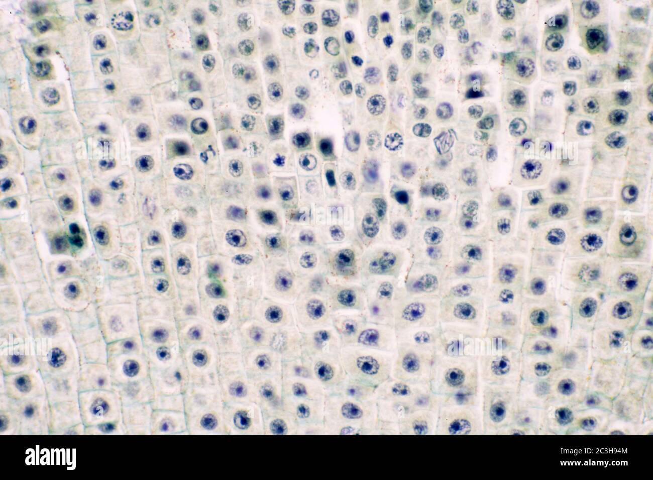 Vicia Fabia root tip mitosis transverse section Stock Photohttps://www.alamy.com/image-license-details/?v=1https://www.alamy.com/vicia-fabia-root-tip-mitosis-transverse-section-image363642068.html
RM2C3H94M–Vicia Fabia root tip mitosis transverse section
RF2XXY3G1–Biology icons collection is a vector illustration with editable stroke.
 Cells mitosis micrograph Stock Photohttps://www.alamy.com/image-license-details/?v=1https://www.alamy.com/stock-photo-cells-mitosis-micrograph-103150242.html
RMFYPW56–Cells mitosis micrograph
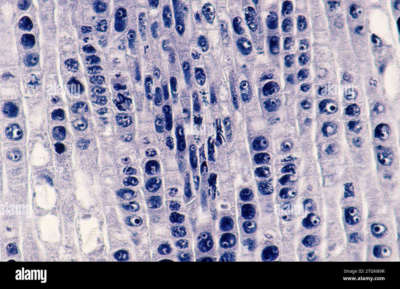 Root apical meristem showing cell divisions (mitosis). Onion root photomicrograph. Stock Photohttps://www.alamy.com/image-license-details/?v=1https://www.alamy.com/root-apical-meristem-showing-cell-divisions-mitosis-onion-root-photomicrograph-image578244179.html
RF2TGN89R–Root apical meristem showing cell divisions (mitosis). Onion root photomicrograph.
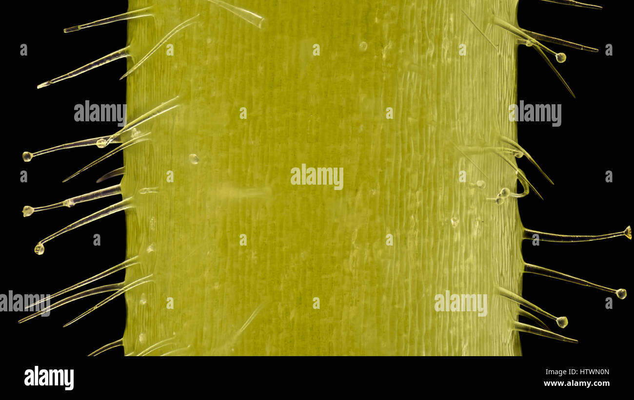 Extreme magnification - Pelargonium, Glandular hairs and tector - section through stem at 20x Stock Photohttps://www.alamy.com/image-license-details/?v=1https://www.alamy.com/stock-photo-extreme-magnification-pelargonium-glandular-hairs-and-tector-section-135789605.html
RMHTWN0N–Extreme magnification - Pelargonium, Glandular hairs and tector - section through stem at 20x
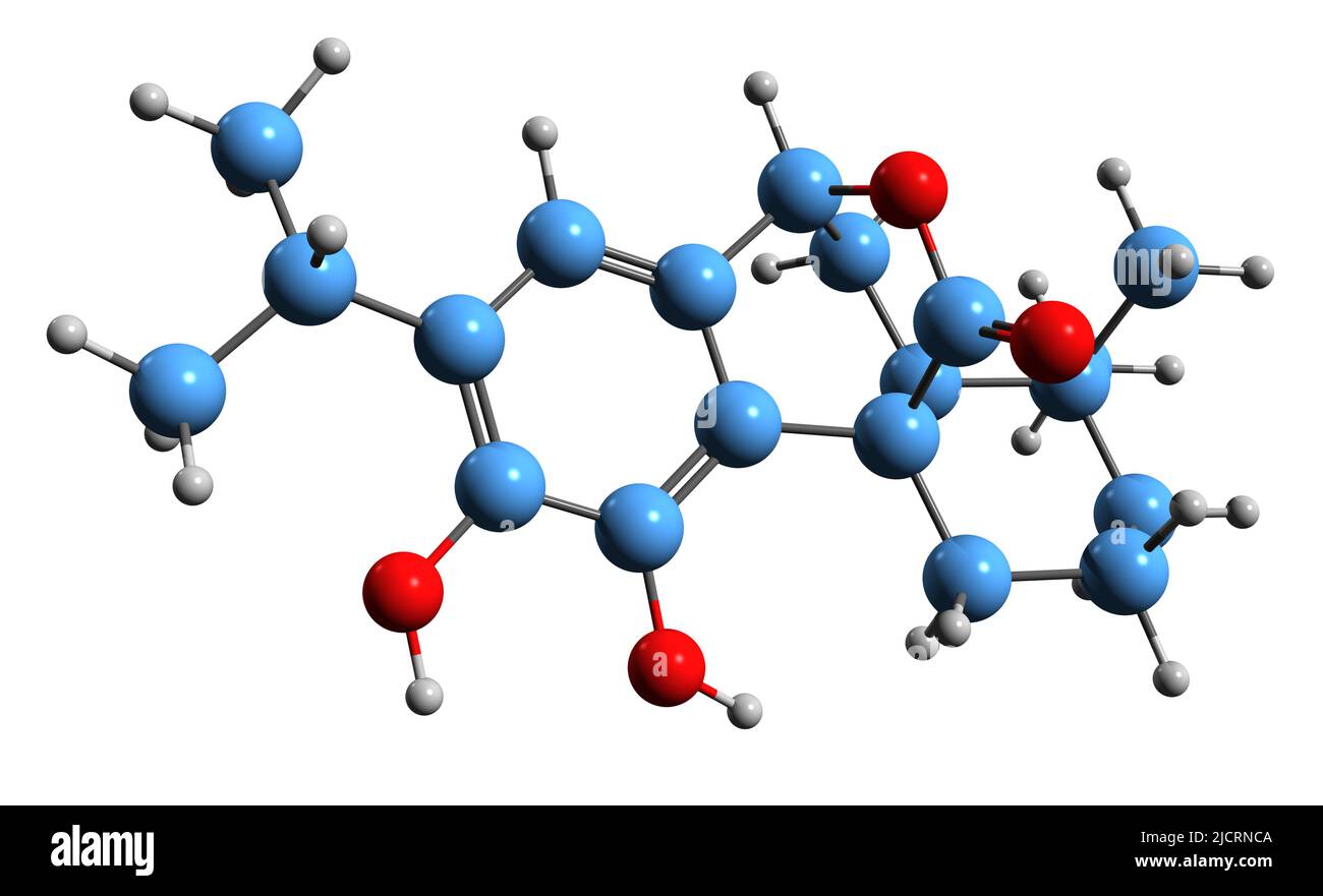 3D image of Carnosol skeletal formula - molecular chemical structure of phenolic diterpene isolated on white background Stock Photohttps://www.alamy.com/image-license-details/?v=1https://www.alamy.com/3d-image-of-carnosol-skeletal-formula-molecular-chemical-structure-of-phenolic-diterpene-isolated-on-white-background-image472577514.html
RF2JCRNCA–3D image of Carnosol skeletal formula - molecular chemical structure of phenolic diterpene isolated on white background
 Abstract cell of virus or pathogenic bacterium under microscope, biofilm illustration. Stock Photohttps://www.alamy.com/image-license-details/?v=1https://www.alamy.com/abstract-cell-of-virus-or-pathogenic-bacterium-under-microscope-biofilm-illustration-image501669985.html
RF2M45155–Abstract cell of virus or pathogenic bacterium under microscope, biofilm illustration.
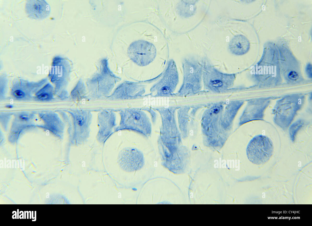 Mitosis Horse Ascaris egg under the microscope, background (Ascaris lumbricoides) Stock Photohttps://www.alamy.com/image-license-details/?v=1https://www.alamy.com/stock-photo-mitosis-horse-ascaris-egg-under-the-microscope-background-ascaris-51118856.html
RFCY4JHC–Mitosis Horse Ascaris egg under the microscope, background (Ascaris lumbricoides)
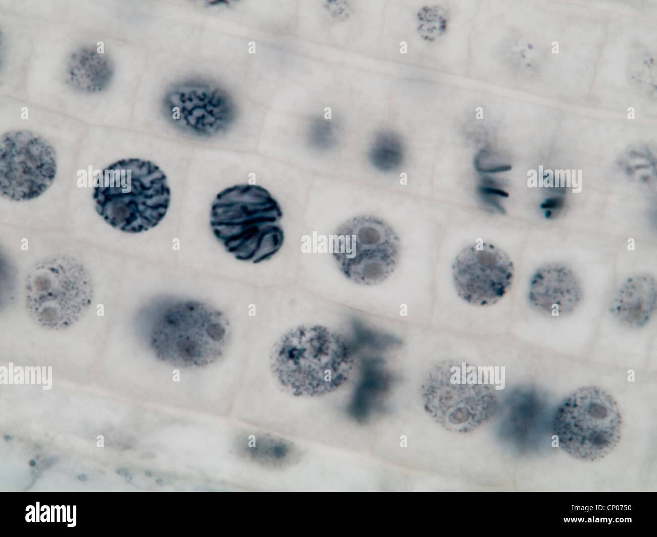 mitosis in a onion root, prophasis and anaphasis Stock Photohttps://www.alamy.com/image-license-details/?v=1https://www.alamy.com/stock-photo-mitosis-in-a-onion-root-prophasis-and-anaphasis-47948796.html
RMCP0750–mitosis in a onion root, prophasis and anaphasis
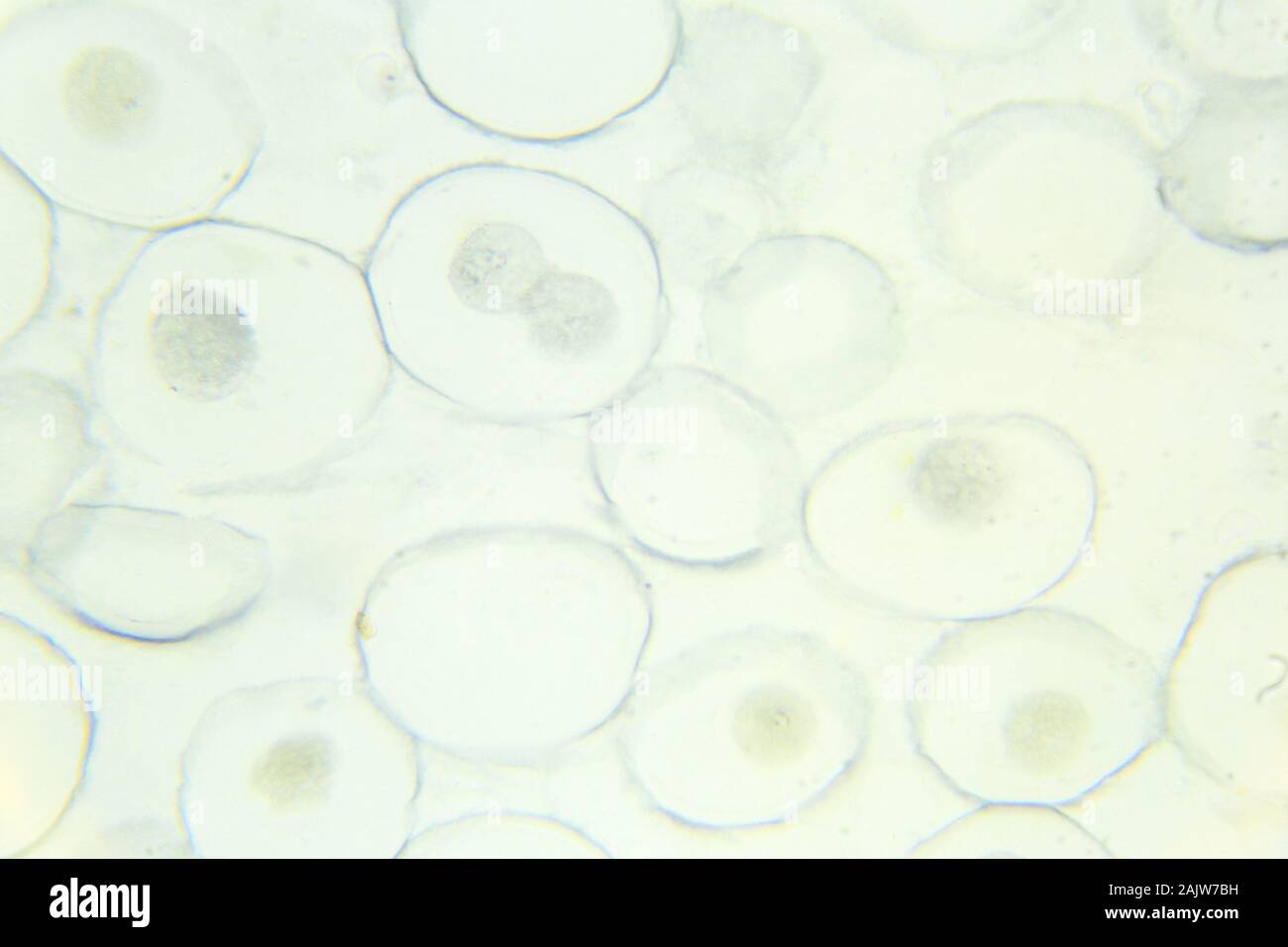 Mitosis Horse Ascaris egg under the microscope, background (Ascaris lumbricoides) Stock Photohttps://www.alamy.com/image-license-details/?v=1https://www.alamy.com/mitosis-horse-ascaris-egg-under-the-microscope-background-ascaris-lumbricoides-image338615413.html
RF2AJW7BH–Mitosis Horse Ascaris egg under the microscope, background (Ascaris lumbricoides)
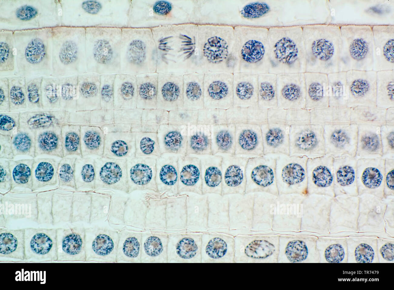 Garden onion, Bulb Onion, Common Onion (Allium cepa), cell tissue of a garden onion with dyed chromosomes, light microscopy, x 120, Germany Stock Photohttps://www.alamy.com/image-license-details/?v=1https://www.alamy.com/garden-onion-bulb-onion-common-onion-allium-cepa-cell-tissue-of-a-garden-onion-with-dyed-chromosomes-light-microscopy-x-120-germany-image255239245.html
RMTR7479–Garden onion, Bulb Onion, Common Onion (Allium cepa), cell tissue of a garden onion with dyed chromosomes, light microscopy, x 120, Germany
 Abstract Science Colorful Pattern Background Stock Photohttps://www.alamy.com/image-license-details/?v=1https://www.alamy.com/abstract-science-colorful-pattern-background-image483508955.html
RF2K2HMGY–Abstract Science Colorful Pattern Background
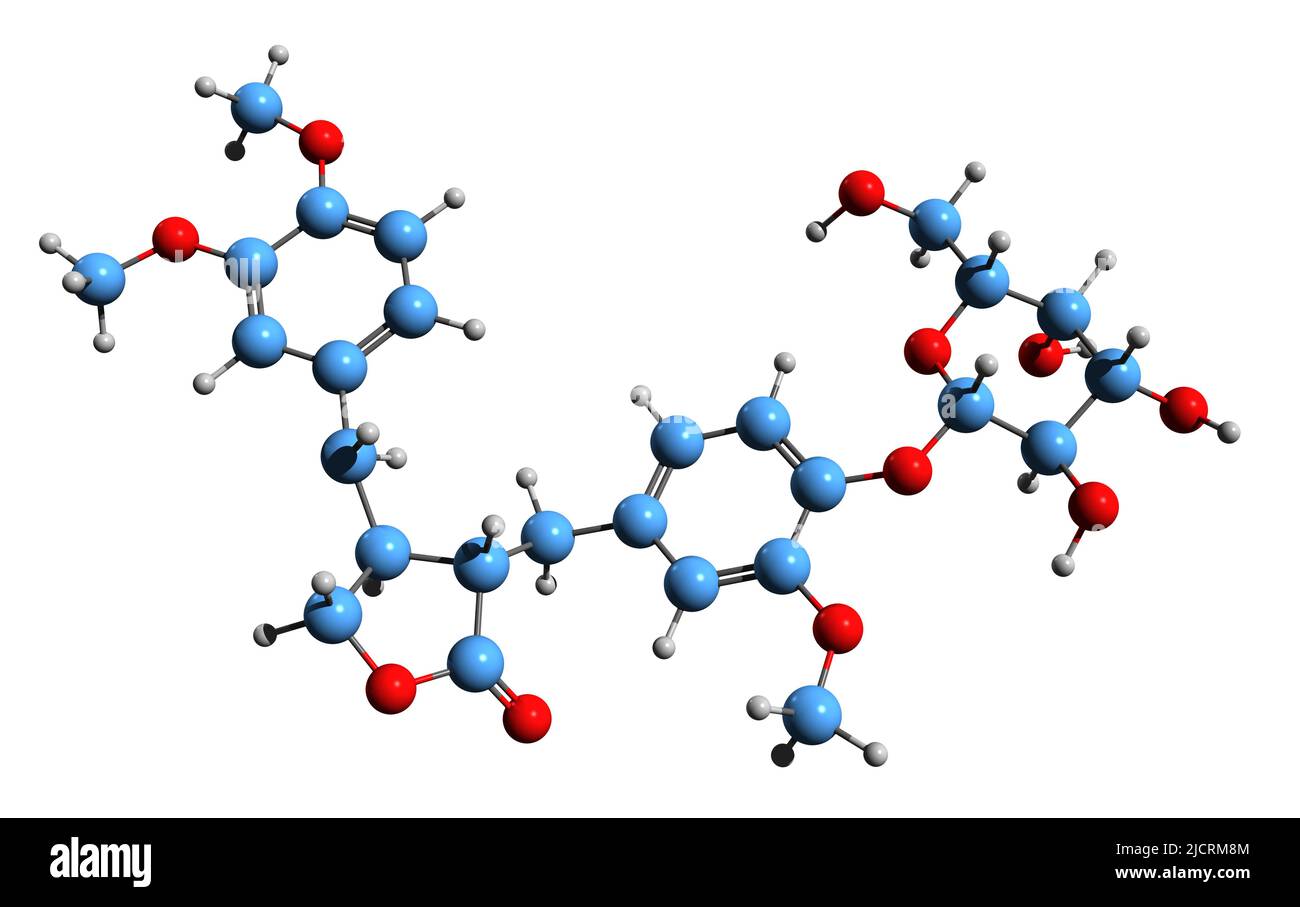 3D image of Arctiin skeletal formula - molecular chemical structure of lignan isolated on white background Stock Photohttps://www.alamy.com/image-license-details/?v=1https://www.alamy.com/3d-image-of-arctiin-skeletal-formula-molecular-chemical-structure-of-lignan-isolated-on-white-background-image472576628.html
RF2JCRM8M–3D image of Arctiin skeletal formula - molecular chemical structure of lignan isolated on white background
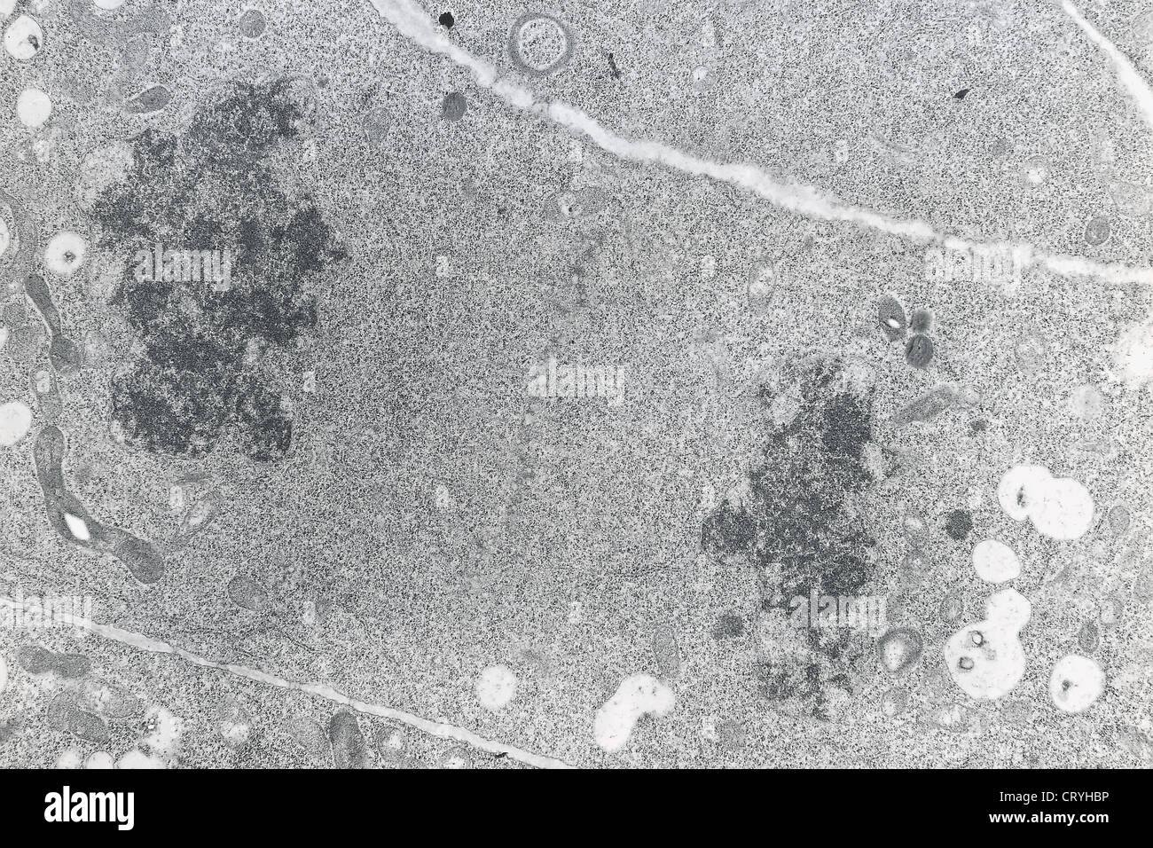 MITOSIS TELOPHASE Stock Photohttps://www.alamy.com/image-license-details/?v=1https://www.alamy.com/stock-photo-mitosis-telophase-49164186.html
RMCRYHBP–MITOSIS TELOPHASE
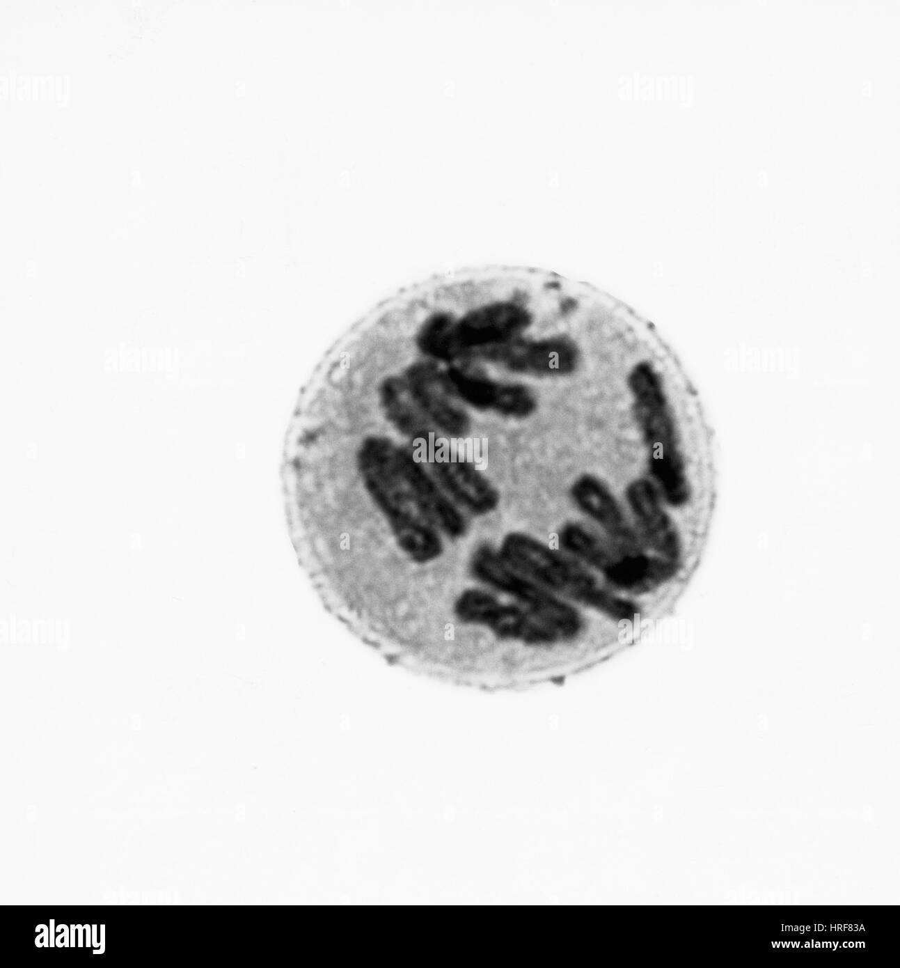 Anaphase of Mitosis in Trillium Cell Stock Photohttps://www.alamy.com/image-license-details/?v=1https://www.alamy.com/stock-photo-anaphase-of-mitosis-in-trillium-cell-134945310.html
RMHRF83A–Anaphase of Mitosis in Trillium Cell
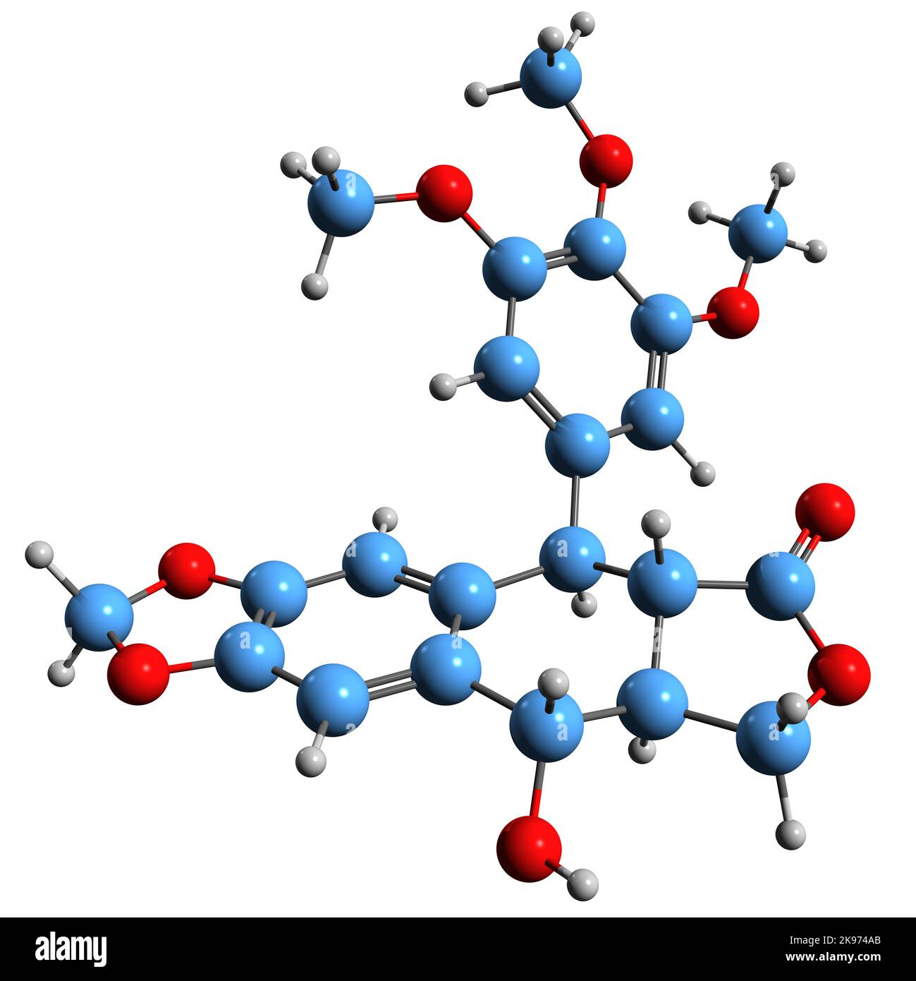 3D image of Podophyllotoxin skeletal formula - molecular chemical structure of nonalkaloid toxin antimitotic isolated on white background Stock Photohttps://www.alamy.com/image-license-details/?v=1https://www.alamy.com/3d-image-of-podophyllotoxin-skeletal-formula-molecular-chemical-structure-of-nonalkaloid-toxin-antimitotic-isolated-on-white-background-image487579299.html
RF2K974AB–3D image of Podophyllotoxin skeletal formula - molecular chemical structure of nonalkaloid toxin antimitotic isolated on white background
RF2PP8R8M–Biology science linear icons set. Photosynthesis, Mitosis, DNA, Ecosystem, Mutation, Evolution, Ecology line vector and concept signs. Genetics
 Chlorophyll is the molecule in leaves that uses the energy in sunlight to turn water and carbon dioxide gas into sugar and oxyge Stock Photohttps://www.alamy.com/image-license-details/?v=1https://www.alamy.com/stock-photo-chlorophyll-is-the-molecule-in-leaves-that-uses-the-energy-in-sunlight-88650114.html
RFF46A3E–Chlorophyll is the molecule in leaves that uses the energy in sunlight to turn water and carbon dioxide gas into sugar and oxyge
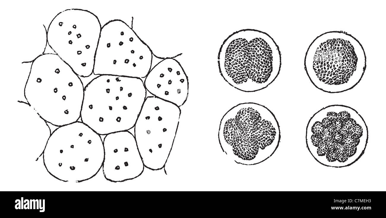 Cell Division in plants (left) and in animals (right), vintage engraved illustration. Trousset encyclopedia (1886 - 1891). Stock Photohttps://www.alamy.com/image-license-details/?v=1https://www.alamy.com/stock-photo-cell-division-in-plants-left-and-in-animals-right-vintage-engraved-39173823.html
RFC7MEH3–Cell Division in plants (left) and in animals (right), vintage engraved illustration. Trousset encyclopedia (1886 - 1891).
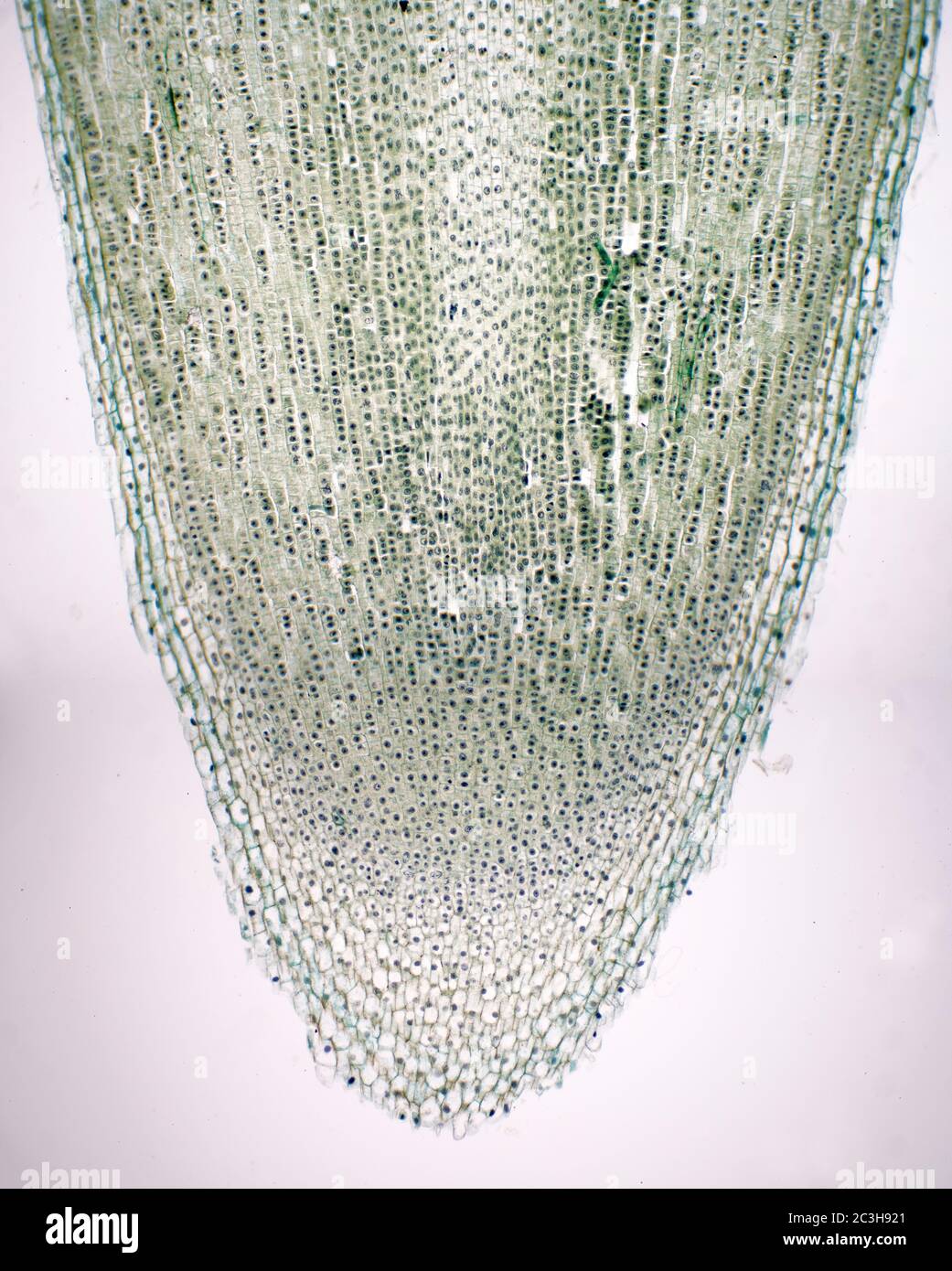 Vicia Fabia root tip mitosis transverse section Stock Photohttps://www.alamy.com/image-license-details/?v=1https://www.alamy.com/vicia-fabia-root-tip-mitosis-transverse-section-image363641993.html
RM2C3H921–Vicia Fabia root tip mitosis transverse section
 mycelium, fungus spores, Mold spores, fungus on the leather surface, landscape of microworld, colony of fungi Stock Photohttps://www.alamy.com/image-license-details/?v=1https://www.alamy.com/mycelium-fungus-spores-mold-spores-fungus-on-the-leather-surface-landscape-of-microworld-colony-of-fungi-image271351624.html
RFWND3NC–mycelium, fungus spores, Mold spores, fungus on the leather surface, landscape of microworld, colony of fungi
 3D illustration of HOMOLOGOUS CHROMOSOMES script with DNA double helix , isolated on pale green gradient. Stock Photohttps://www.alamy.com/image-license-details/?v=1https://www.alamy.com/3d-illustration-of-homologous-chromosomes-script-with-dna-double-helix-isolated-on-pale-green-gradient-image210926920.html
RFP74FC8–3D illustration of HOMOLOGOUS CHROMOSOMES script with DNA double helix , isolated on pale green gradient.
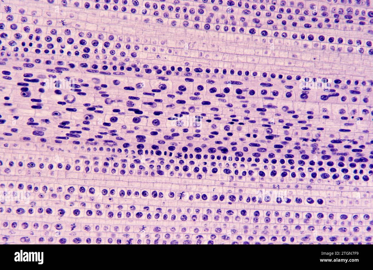 Root apical meristem showing cell divisions (mitosis). Onion root photomicrograph. Stock Photohttps://www.alamy.com/image-license-details/?v=1https://www.alamy.com/root-apical-meristem-showing-cell-divisions-mitosis-onion-root-photomicrograph-image578243549.html
RF2TGN7F9–Root apical meristem showing cell divisions (mitosis). Onion root photomicrograph.
 mycelium, fungus spores, Mold spores, fungus on the leather surface, landscape of microworld, colony of fungi Stock Photohttps://www.alamy.com/image-license-details/?v=1https://www.alamy.com/mycelium-fungus-spores-mold-spores-fungus-on-the-leather-surface-landscape-of-microworld-colony-of-fungi-image271352024.html
RFWND47M–mycelium, fungus spores, Mold spores, fungus on the leather surface, landscape of microworld, colony of fungi
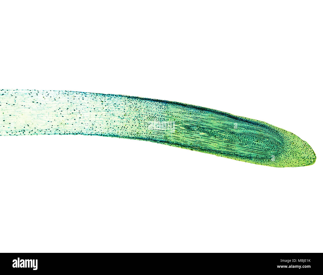 Light photomicrograph of Mitosis of onion root tip cells seen through microscope Stock Photohttps://www.alamy.com/image-license-details/?v=1https://www.alamy.com/light-photomicrograph-of-mitosis-of-onion-root-tip-cells-seen-through-microscope-image179271055.html
RFMBJE1K–Light photomicrograph of Mitosis of onion root tip cells seen through microscope
 Abstract cell of virus or pathogenic bacterium under microscope, biofilm illustration. Stock Photohttps://www.alamy.com/image-license-details/?v=1https://www.alamy.com/abstract-cell-of-virus-or-pathogenic-bacterium-under-microscope-biofilm-illustration-image501669975.html
RF2M4514R–Abstract cell of virus or pathogenic bacterium under microscope, biofilm illustration.
 Mold spores, fungus on the leather surface, landscape of microworld, colony of fungi Stock Photohttps://www.alamy.com/image-license-details/?v=1https://www.alamy.com/mold-spores-fungus-on-the-leather-surface-landscape-of-microworld-colony-of-fungi-image271351741.html
RFWND3WH–Mold spores, fungus on the leather surface, landscape of microworld, colony of fungi
 The cell in development and inheritance . Fig. 25. — Diagrams showing the prophases of mitosis.A. Resting cell with reticular nucleus and true nucleolus ; at c the attraction-sphere containingtwo centrosomes. B. Early prophase ; the chromatin forming a continuous spireme, nucleolus stillpresent; above, the amphiaster (a). CD. Two different types of later prophases. C. Disappear-ance of the primary spindle, divergence of the centrosomes to opposite poles of the nucleus (exam-ples, some plant-cells, cleavage-stages of many eggs). D. Persistence of the primary spindle (toform in some cases the ce Stock Photohttps://www.alamy.com/image-license-details/?v=1https://www.alamy.com/the-cell-in-development-and-inheritance-fig-25-diagrams-showing-the-prophases-of-mitosisa-resting-cell-with-reticular-nucleus-and-true-nucleolus-at-c-the-attraction-sphere-containingtwo-centrosomes-b-early-prophase-the-chromatin-forming-a-continuous-spireme-nucleolus-stillpresent-above-the-amphiaster-a-cd-two-different-types-of-later-prophases-c-disappear-ance-of-the-primary-spindle-divergence-of-the-centrosomes-to-opposite-poles-of-the-nucleus-exam-ples-some-plant-cells-cleavage-stages-of-many-eggs-d-persistence-of-the-primary-spindle-toform-in-some-cases-the-ce-image338432634.html
RM2AJGX7P–The cell in development and inheritance . Fig. 25. — Diagrams showing the prophases of mitosis.A. Resting cell with reticular nucleus and true nucleolus ; at c the attraction-sphere containingtwo centrosomes. B. Early prophase ; the chromatin forming a continuous spireme, nucleolus stillpresent; above, the amphiaster (a). CD. Two different types of later prophases. C. Disappear-ance of the primary spindle, divergence of the centrosomes to opposite poles of the nucleus (exam-ples, some plant-cells, cleavage-stages of many eggs). D. Persistence of the primary spindle (toform in some cases the ce
 fungus on the leather surface, landscape of microworld, colony of fungi Stock Photohttps://www.alamy.com/image-license-details/?v=1https://www.alamy.com/fungus-on-the-leather-surface-landscape-of-microworld-colony-of-fungi-image271352079.html
RFWND49K–fungus on the leather surface, landscape of microworld, colony of fungi
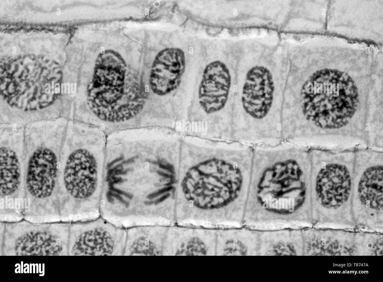 Garden onion, Bulb Onion, Common Onion (Allium cepa), cell tissue of a garden onion with dyed chromosomes, light microscopy, x 200, Germany Stock Photohttps://www.alamy.com/image-license-details/?v=1https://www.alamy.com/garden-onion-bulb-onion-common-onion-allium-cepa-cell-tissue-of-a-garden-onion-with-dyed-chromosomes-light-microscopy-x-200-germany-image255239246.html
RMTR747A–Garden onion, Bulb Onion, Common Onion (Allium cepa), cell tissue of a garden onion with dyed chromosomes, light microscopy, x 200, Germany
 Abstract Science Colorful Pattern Background Stock Photohttps://www.alamy.com/image-license-details/?v=1https://www.alamy.com/abstract-science-colorful-pattern-background-image483508906.html
RF2K2HMF6–Abstract Science Colorful Pattern Background
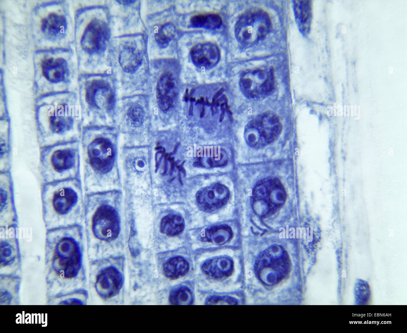 daffodil (Narcissus spec.), mitosis in celles of the root apex of a daffodill, 1000 x Stock Photohttps://www.alamy.com/image-license-details/?v=1https://www.alamy.com/stock-photo-daffodil-narcissus-spec-mitosis-in-celles-of-the-root-apex-of-a-daffodill-76068681.html
RMEBN6AH–daffodil (Narcissus spec.), mitosis in celles of the root apex of a daffodill, 1000 x
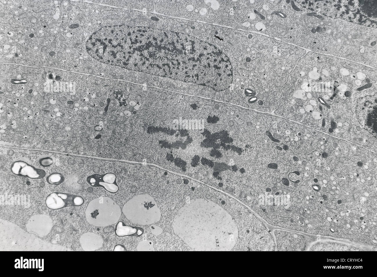 MITOSIS ANAPHASE Stock Photohttps://www.alamy.com/image-license-details/?v=1https://www.alamy.com/stock-photo-mitosis-anaphase-49164196.html
RMCRYHC4–MITOSIS ANAPHASE
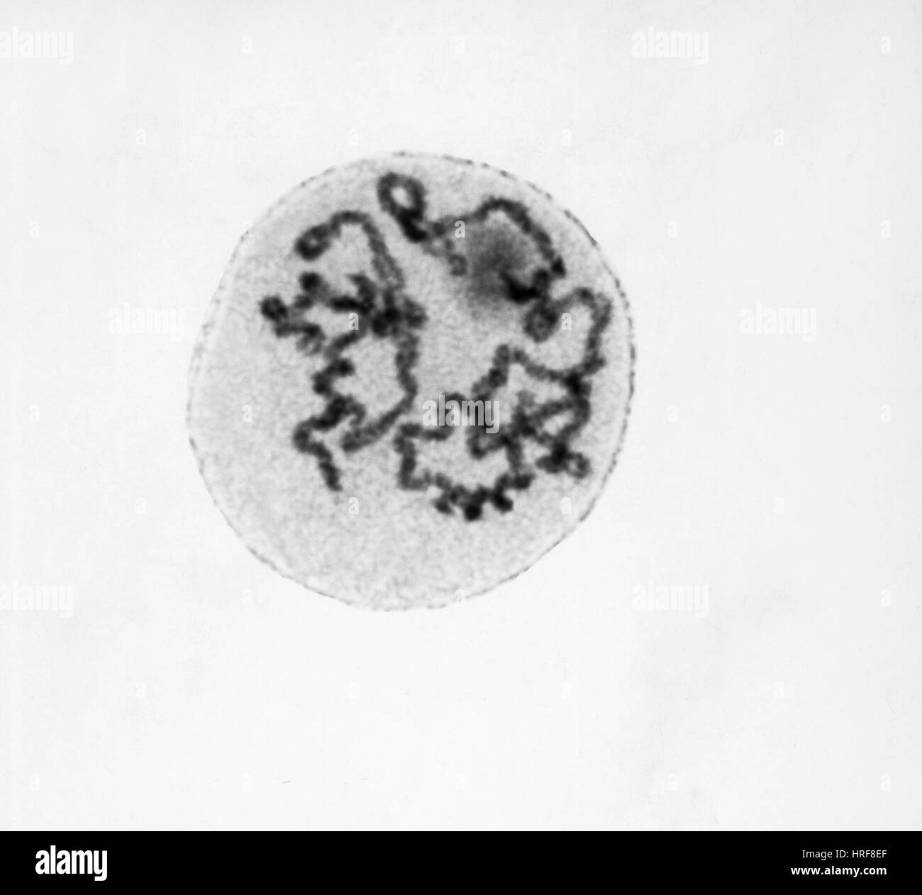 Prophase of Mitosis in Trillium Cell Stock Photohttps://www.alamy.com/image-license-details/?v=1https://www.alamy.com/stock-photo-prophase-of-mitosis-in-trillium-cell-134945623.html
RMHRF8EF–Prophase of Mitosis in Trillium Cell
 Chloroplast eukaryotic cell animation under the microscope. Motion. Abstract visualization of microscopic formation in a plant cell, biology science Stock Photohttps://www.alamy.com/image-license-details/?v=1https://www.alamy.com/chloroplast-eukaryotic-cell-animation-under-the-microscope-motion-abstract-visualization-of-microscopic-formation-in-a-plant-cell-biology-science-image467555380.html
RF2J4JYJC–Chloroplast eukaryotic cell animation under the microscope. Motion. Abstract visualization of microscopic formation in a plant cell, biology science
RF2PP8N76–Biology science linear icons set. Photosynthesis, Mitosis, DNA, Ecosystem, Mutation, Evolution, Ecology line vector and concept signs. Genetics
 Chlorophyll is the molecule in leaves that uses the energy in sunlight to turn water and carbon dioxide gas into sugar and oxyge Stock Photohttps://www.alamy.com/image-license-details/?v=1https://www.alamy.com/stock-photo-chlorophyll-is-the-molecule-in-leaves-that-uses-the-energy-in-sunlight-88650115.html
RFF46A3F–Chlorophyll is the molecule in leaves that uses the energy in sunlight to turn water and carbon dioxide gas into sugar and oxyge
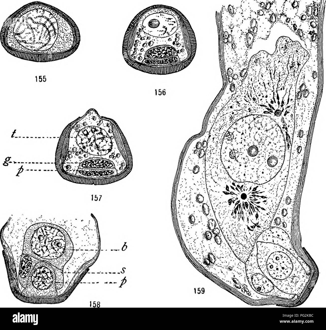 . Morphology of gymnosperms. Gymnosperms; Plant morphology. CYCADALES 141 It is generally accepted that the cycads are wind-pollinated. Pearson (47), however, observed insects dusted with the pollen of Encephalartos villosus, and believes it is probable that they effect. Figs. 155-159.—Dioon edule'; the germination of the microspore; fig. 155, the nucleus in early prophase o£ the first mitosis, exine and inline sharply differentiated (August 14, 1905); fig. 156, the shedding stage (September 1906); fig. 157,'beginning of the pollen tube; /, tube nucleus; g, generative cell; p, prothallial cell Stock Photohttps://www.alamy.com/image-license-details/?v=1https://www.alamy.com/morphology-of-gymnosperms-gymnosperms-plant-morphology-cycadales-141-it-is-generally-accepted-that-the-cycads-are-wind-pollinated-pearson-47-however-observed-insects-dusted-with-the-pollen-of-encephalartos-villosus-and-believes-it-is-probable-that-they-effect-figs-155-159dioon-edule-the-germination-of-the-microspore-fig-155-the-nucleus-in-early-prophase-o-the-first-mitosis-exine-and-inline-sharply-differentiated-august-14-1905-fig-156-the-shedding-stage-september-1906-fig-157beginning-of-the-pollen-tube-tube-nucleus-g-generative-cell-p-prothallial-cell-image216418032.html
RMPG2KBC–. Morphology of gymnosperms. Gymnosperms; Plant morphology. CYCADALES 141 It is generally accepted that the cycads are wind-pollinated. Pearson (47), however, observed insects dusted with the pollen of Encephalartos villosus, and believes it is probable that they effect. Figs. 155-159.—Dioon edule'; the germination of the microspore; fig. 155, the nucleus in early prophase o£ the first mitosis, exine and inline sharply differentiated (August 14, 1905); fig. 156, the shedding stage (September 1906); fig. 157,'beginning of the pollen tube; /, tube nucleus; g, generative cell; p, prothallial cell
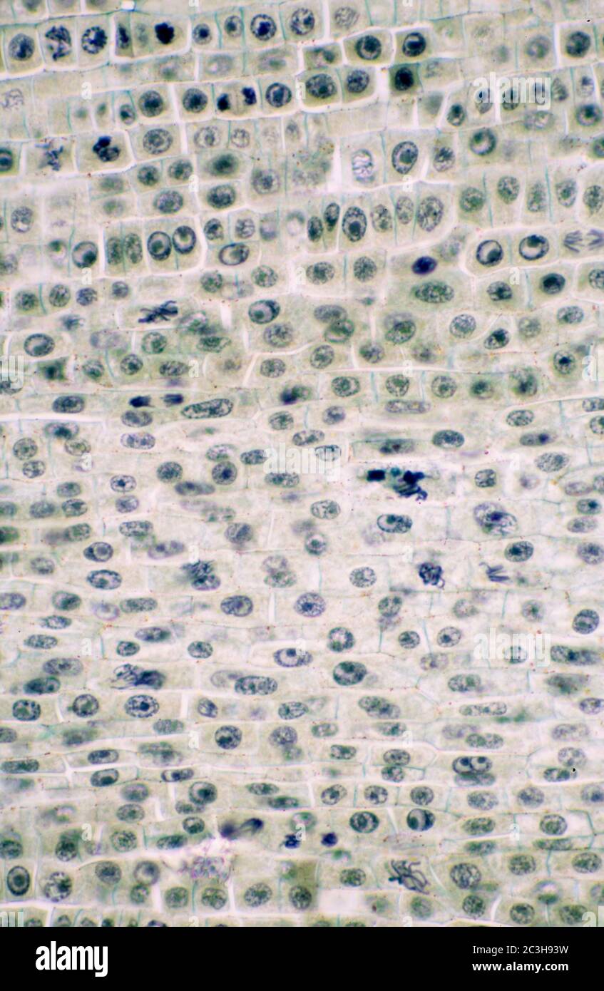 Vicia Fabia root tip mitosis transverse section Stock Photohttps://www.alamy.com/image-license-details/?v=1https://www.alamy.com/vicia-fabia-root-tip-mitosis-transverse-section-image363642045.html
RM2C3H93W–Vicia Fabia root tip mitosis transverse section
 Abstract Science Colorful Pattern Background Stock Photohttps://www.alamy.com/image-license-details/?v=1https://www.alamy.com/abstract-science-colorful-pattern-background-image441678280.html
RF2GJG548–Abstract Science Colorful Pattern Background
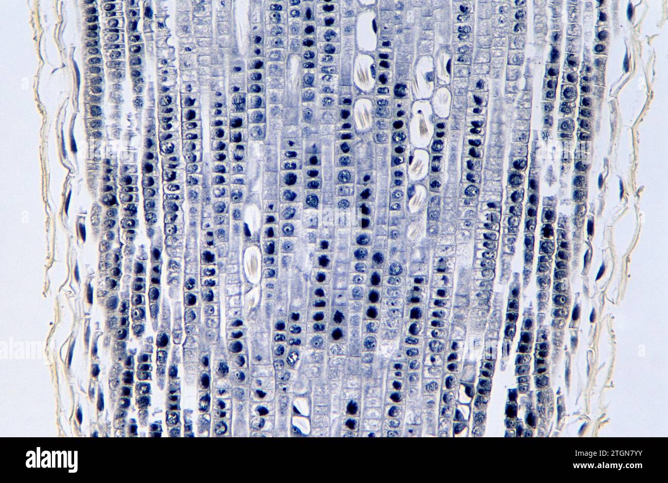 Root apical meristem showing cell divisions (mitosis) and raphides. Onion root photomicrograph. Stock Photohttps://www.alamy.com/image-license-details/?v=1https://www.alamy.com/root-apical-meristem-showing-cell-divisions-mitosis-and-raphides-onion-root-photomicrograph-image578243903.html
RF2TGN7YY–Root apical meristem showing cell divisions (mitosis) and raphides. Onion root photomicrograph.
 Light photomicrograph of Mitosis of onion root tip cells seen through microscope Stock Photohttps://www.alamy.com/image-license-details/?v=1https://www.alamy.com/stock-photo-light-photomicrograph-of-mitosis-of-onion-root-tip-cells-seen-through-75812190.html
RFEB9F66–Light photomicrograph of Mitosis of onion root tip cells seen through microscope
 Abstract cell of virus or pathogenic bacterium under microscope, biofilm illustration. Stock Photohttps://www.alamy.com/image-license-details/?v=1https://www.alamy.com/abstract-cell-of-virus-or-pathogenic-bacterium-under-microscope-biofilm-illustration-image501669995.html
RF2M4515F–Abstract cell of virus or pathogenic bacterium under microscope, biofilm illustration.
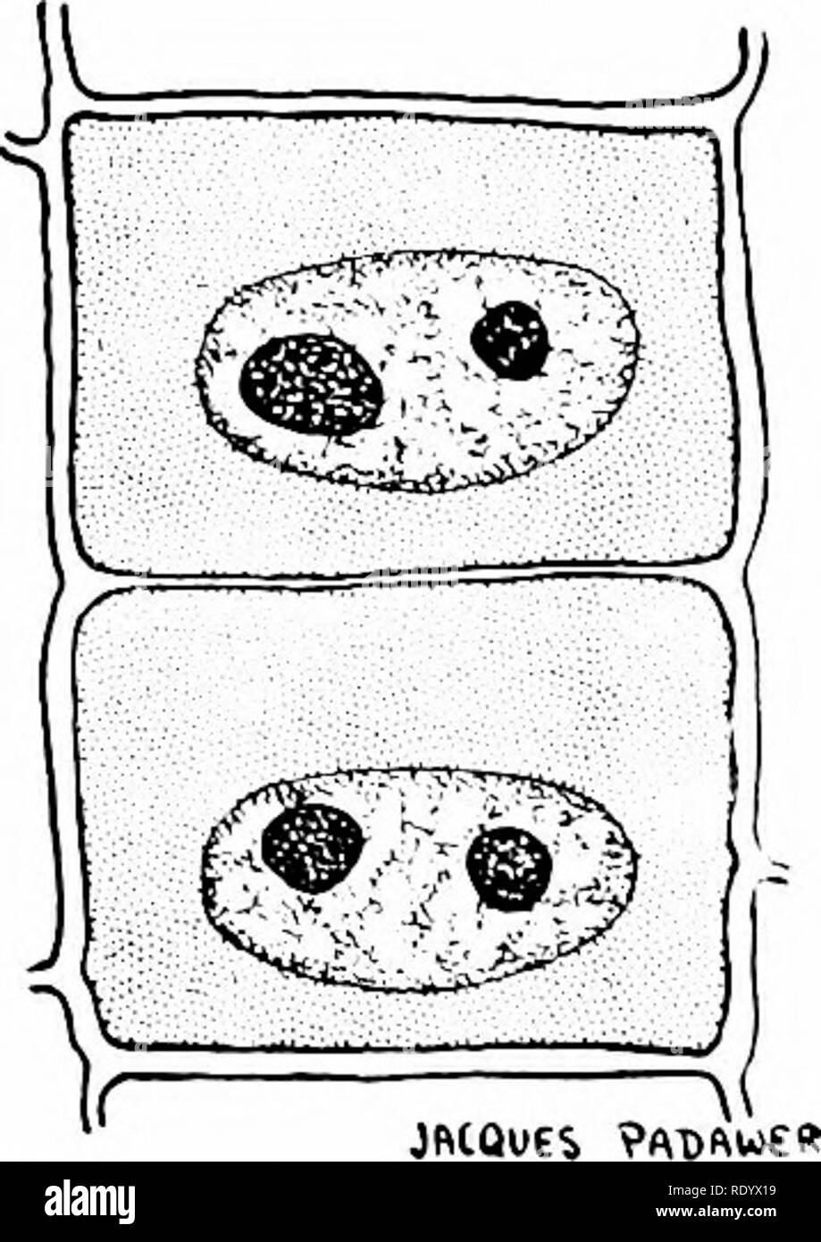 . Principles of modern biology. Biology. L. TELOPHASES Fig. 3-1. Mitosis in a typical plant cell. DAUGHTER CELLS. Please note that these images are extracted from scanned page images that may have been digitally enhanced for readability - coloration and appearance of these illustrations may not perfectly resemble the original work.. Marsland, Douglas, 1899-. New York, Holt, Rinehart and Winston Stock Photohttps://www.alamy.com/image-license-details/?v=1https://www.alamy.com/principles-of-modern-biology-biology-l-telophases-fig-3-1-mitosis-in-a-typical-plant-cell-daughter-cells-please-note-that-these-images-are-extracted-from-scanned-page-images-that-may-have-been-digitally-enhanced-for-readability-coloration-and-appearance-of-these-illustrations-may-not-perfectly-resemble-the-original-work-marsland-douglas-1899-new-york-holt-rinehart-and-winston-image232338437.html
RMRDYX19–. Principles of modern biology. Biology. L. TELOPHASES Fig. 3-1. Mitosis in a typical plant cell. DAUGHTER CELLS. Please note that these images are extracted from scanned page images that may have been digitally enhanced for readability - coloration and appearance of these illustrations may not perfectly resemble the original work.. Marsland, Douglas, 1899-. New York, Holt, Rinehart and Winston
 Mold spores, fungus on the leather surface, landscape of microworld, colony of fungi Stock Photohttps://www.alamy.com/image-license-details/?v=1https://www.alamy.com/mold-spores-fungus-on-the-leather-surface-landscape-of-microworld-colony-of-fungi-image271351908.html
RFWND43G–Mold spores, fungus on the leather surface, landscape of microworld, colony of fungi
 Abstract Science Colorful Pattern Background Stock Photohttps://www.alamy.com/image-license-details/?v=1https://www.alamy.com/abstract-science-colorful-pattern-background-image483508975.html
RF2K2HMHK–Abstract Science Colorful Pattern Background
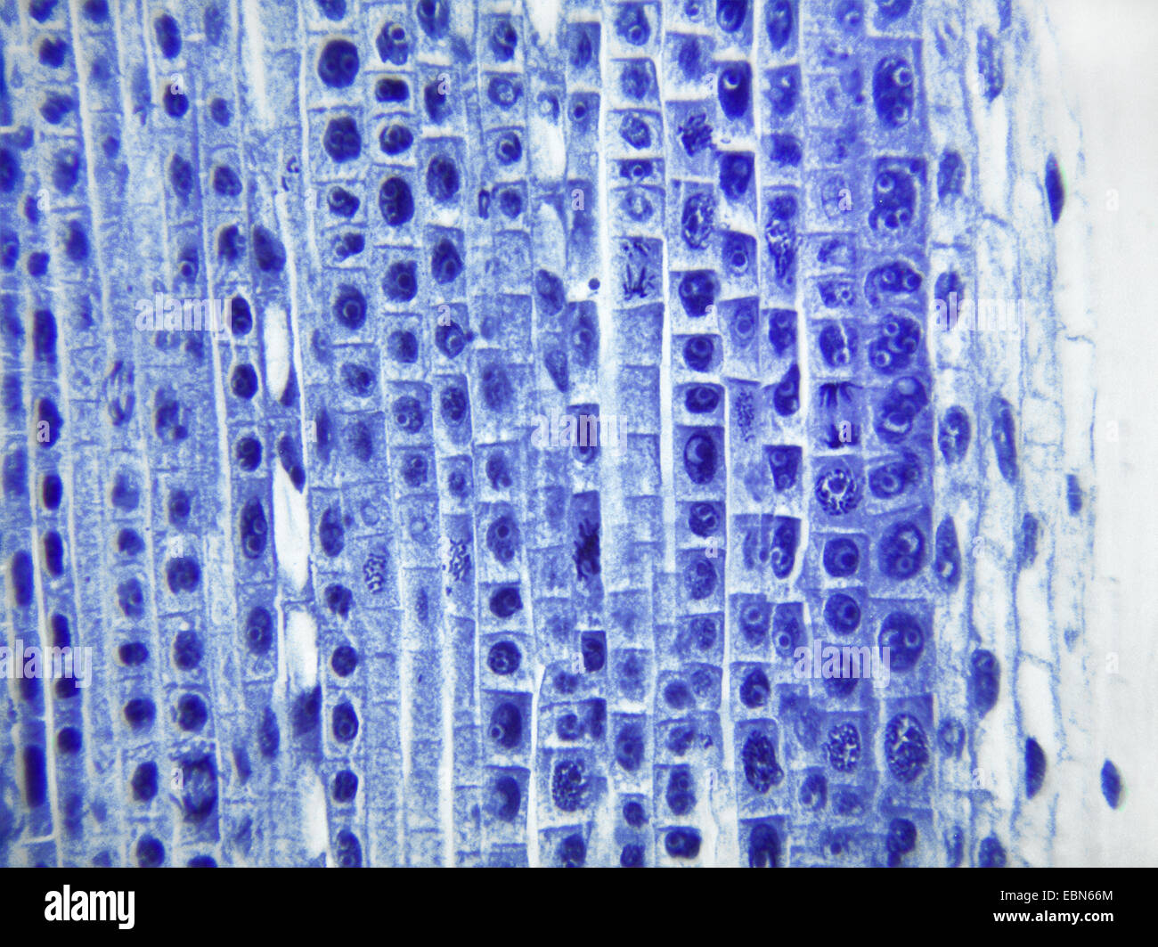 daffodil (Narcissus spec.), mitosis in celles of the root apex of a daffodill, 400 x Stock Photohttps://www.alamy.com/image-license-details/?v=1https://www.alamy.com/stock-photo-daffodil-narcissus-spec-mitosis-in-celles-of-the-root-apex-of-a-daffodill-76068572.html
RMEBN66M–daffodil (Narcissus spec.), mitosis in celles of the root apex of a daffodill, 400 x
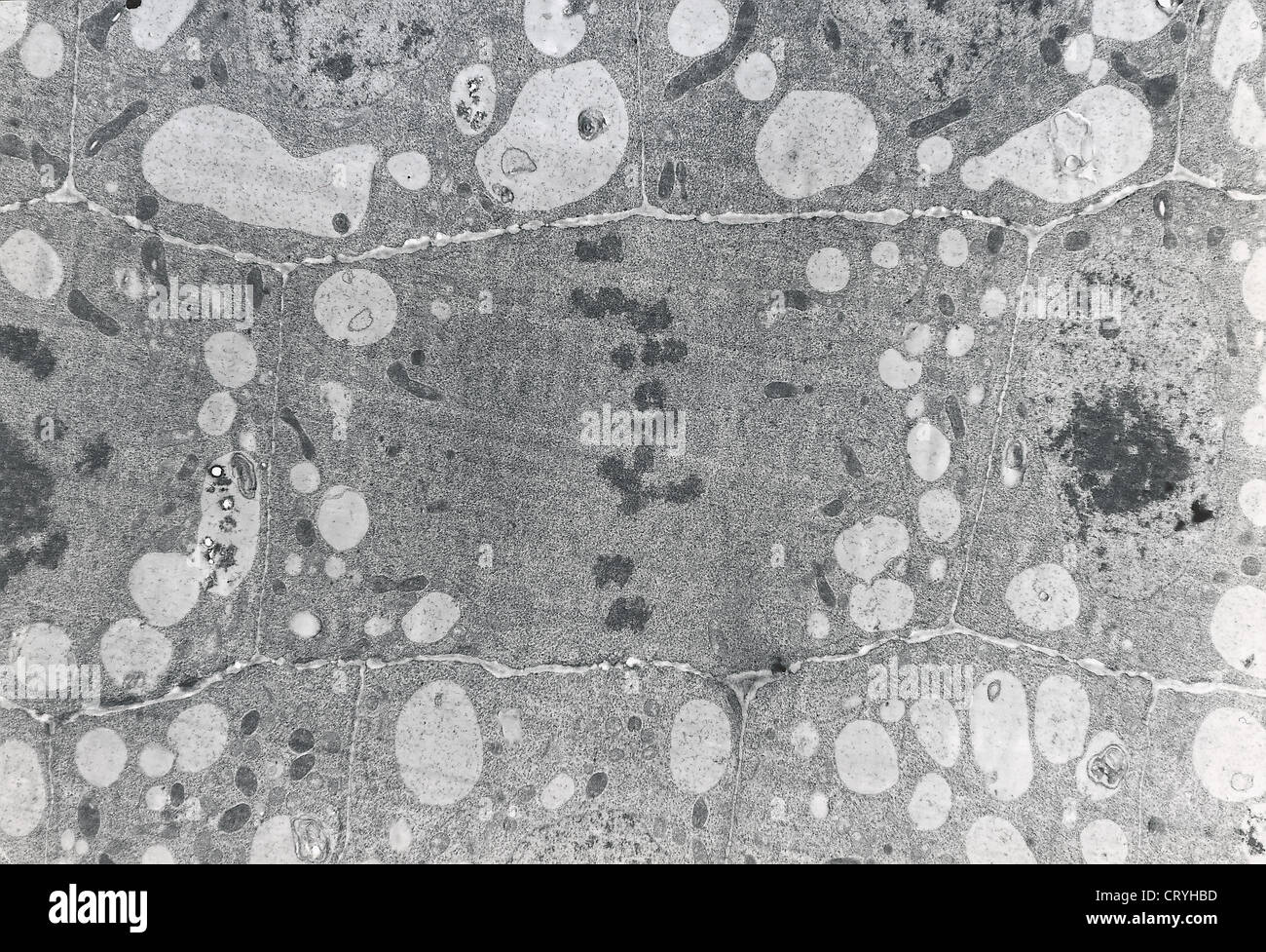 MITOSIS METAPHASE Stock Photohttps://www.alamy.com/image-license-details/?v=1https://www.alamy.com/stock-photo-mitosis-metaphase-49164177.html
RMCRYHBD–MITOSIS METAPHASE
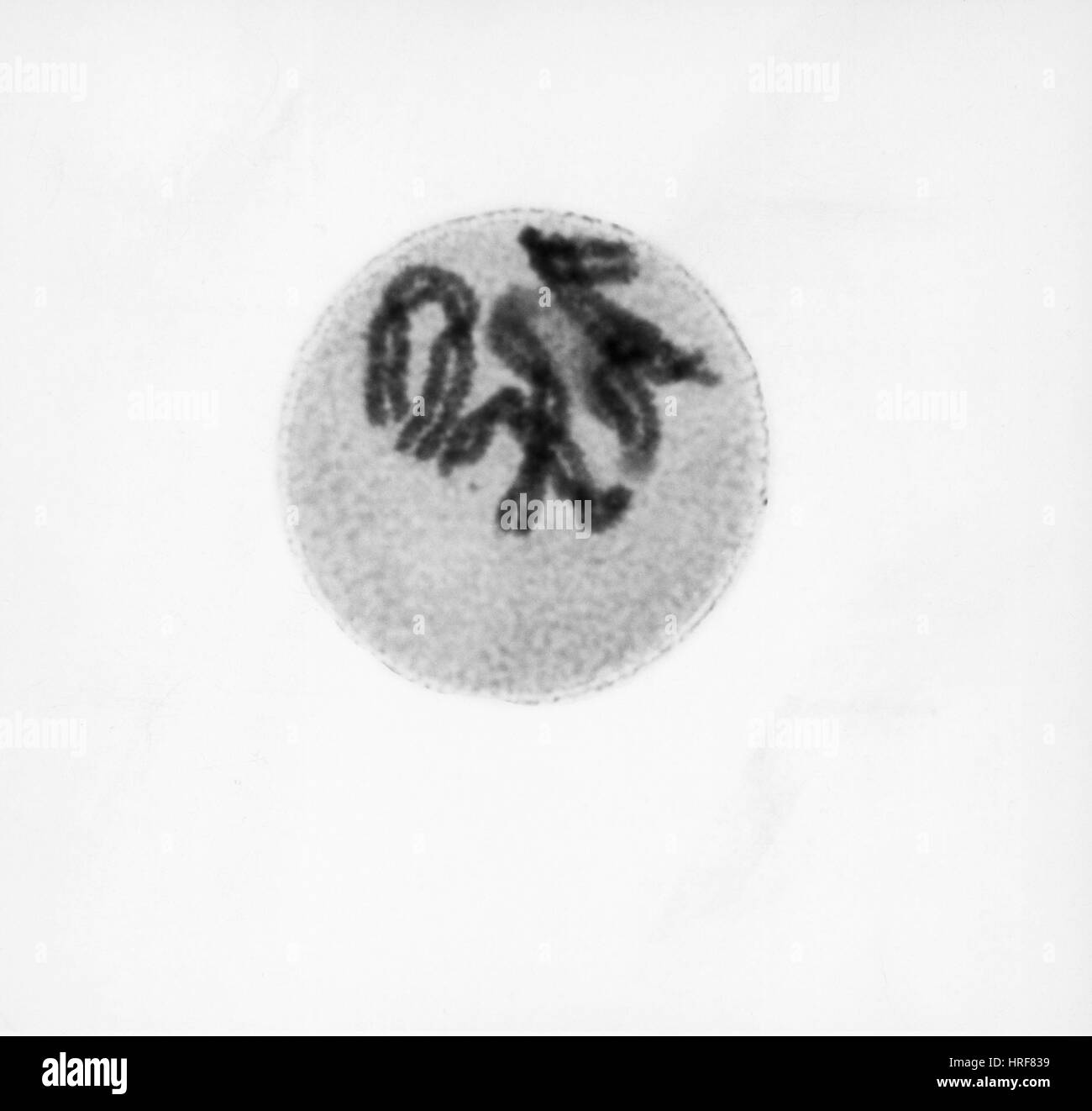 Prophase of Mitosis in Trillium Cell Stock Photohttps://www.alamy.com/image-license-details/?v=1https://www.alamy.com/stock-photo-prophase-of-mitosis-in-trillium-cell-134945309.html
RMHRF839–Prophase of Mitosis in Trillium Cell
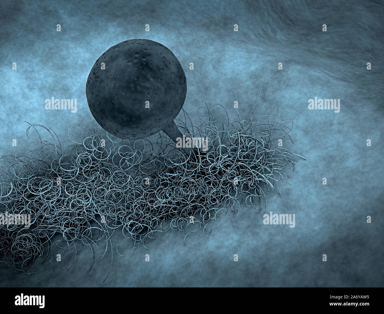 reproduction of fungi, fungus growing, growth of the fungus Stock Photohttps://www.alamy.com/image-license-details/?v=1https://www.alamy.com/reproduction-of-fungi-fungus-growing-growth-of-the-fungus-image331286177.html
RF2A6YAW5–reproduction of fungi, fungus growing, growth of the fungus
RF2PP87YF–Biology science linear icons set. Photosynthesis, Mitosis, DNA, Ecosystem, Mutation, Evolution, Ecology vector symbols and line concept signs
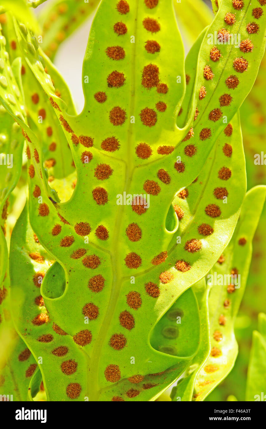 Chlorophyll is the molecule in leaves that uses the energy in sunlight to turn water and carbon dioxide gas into sugar and oxyge Stock Photohttps://www.alamy.com/image-license-details/?v=1https://www.alamy.com/stock-photo-chlorophyll-is-the-molecule-in-leaves-that-uses-the-energy-in-sunlight-88650124.html
RFF46A3T–Chlorophyll is the molecule in leaves that uses the energy in sunlight to turn water and carbon dioxide gas into sugar and oxyge
 reproduction of fungi, fungus growing, growth of the fungus Stock Photohttps://www.alamy.com/image-license-details/?v=1https://www.alamy.com/reproduction-of-fungi-fungus-growing-growth-of-the-fungus-image331286171.html
RF2A6YATY–reproduction of fungi, fungus growing, growth of the fungus
 Chloroplast eukaryotic cell animation under the microscope. Motion. Abstract visualization of microscopic formation in a plant cell, biology science Stock Photohttps://www.alamy.com/image-license-details/?v=1https://www.alamy.com/chloroplast-eukaryotic-cell-animation-under-the-microscope-motion-abstract-visualization-of-microscopic-formation-in-a-plant-cell-biology-science-image467583496.html
RF2J4M7EG–Chloroplast eukaryotic cell animation under the microscope. Motion. Abstract visualization of microscopic formation in a plant cell, biology science
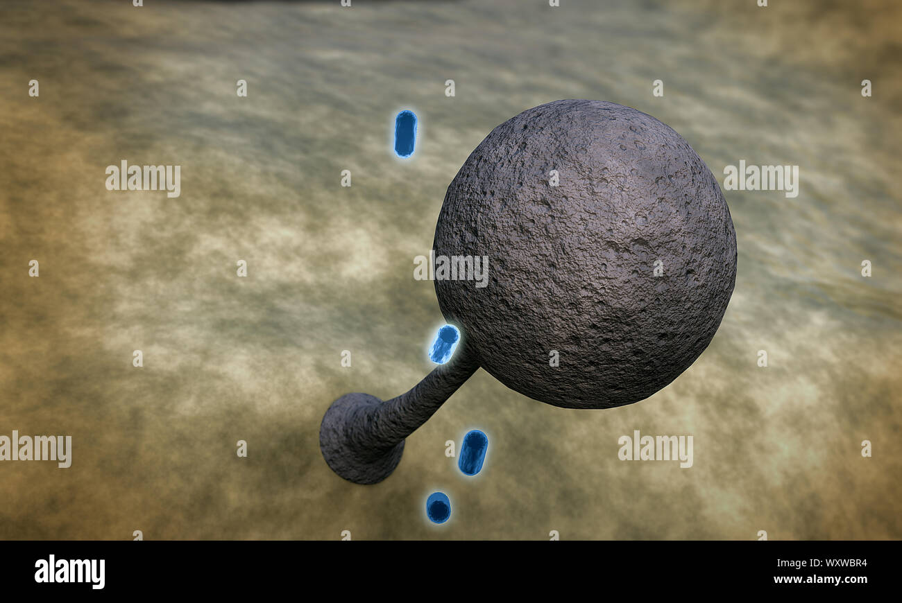 antibiotic kills the mildew, action of medicine Stock Photohttps://www.alamy.com/image-license-details/?v=1https://www.alamy.com/antibiotic-kills-the-mildew-action-of-medicine-image274694648.html
RFWXWBR4–antibiotic kills the mildew, action of medicine
 . A text-book of mycology and plant pathology . Plant diseases; Fungi in agriculture; Plant diseases; Fungi. PATHOLOGIC PLANT ANATOMY 373 149). Four nuclei in one cell is the most we have seen, but it is prob- able that larger numbers occur. It would seem from the studies of Erwin F. Smith, which, however, are incomplete, that most of the cell divisions in crown gall are by mitosis. Frequently, however, there have been found nuclei variously lobed and in process of amitotic division, and this is probably the way in which several nuclei are formed in one cell (Fig. 149).. Fig. 149.—Nuclear divi Stock Photohttps://www.alamy.com/image-license-details/?v=1https://www.alamy.com/a-text-book-of-mycology-and-plant-pathology-plant-diseases-fungi-in-agriculture-plant-diseases-fungi-pathologic-plant-anatomy-373-149-four-nuclei-in-one-cell-is-the-most-we-have-seen-but-it-is-prob-able-that-larger-numbers-occur-it-would-seem-from-the-studies-of-erwin-f-smith-which-however-are-incomplete-that-most-of-the-cell-divisions-in-crown-gall-are-by-mitosis-frequently-however-there-have-been-found-nuclei-variously-lobed-and-in-process-of-amitotic-division-and-this-is-probably-the-way-in-which-several-nuclei-are-formed-in-one-cell-fig-149-fig-149nuclear-divi-image216450380.html
RMPG44JM–. A text-book of mycology and plant pathology . Plant diseases; Fungi in agriculture; Plant diseases; Fungi. PATHOLOGIC PLANT ANATOMY 373 149). Four nuclei in one cell is the most we have seen, but it is prob- able that larger numbers occur. It would seem from the studies of Erwin F. Smith, which, however, are incomplete, that most of the cell divisions in crown gall are by mitosis. Frequently, however, there have been found nuclei variously lobed and in process of amitotic division, and this is probably the way in which several nuclei are formed in one cell (Fig. 149).. Fig. 149.—Nuclear divi
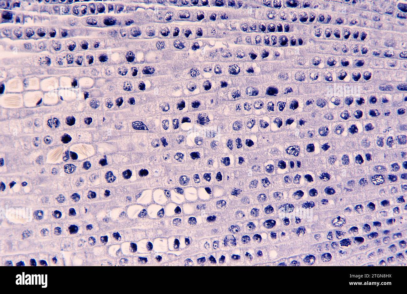 Root apical meristem showing cell divisions (mitosis) and raphides. Onion root photomicrograph. Stock Photohttps://www.alamy.com/image-license-details/?v=1https://www.alamy.com/root-apical-meristem-showing-cell-divisions-mitosis-and-raphides-onion-root-photomicrograph-image578244406.html
RF2TGN8HX–Root apical meristem showing cell divisions (mitosis) and raphides. Onion root photomicrograph.
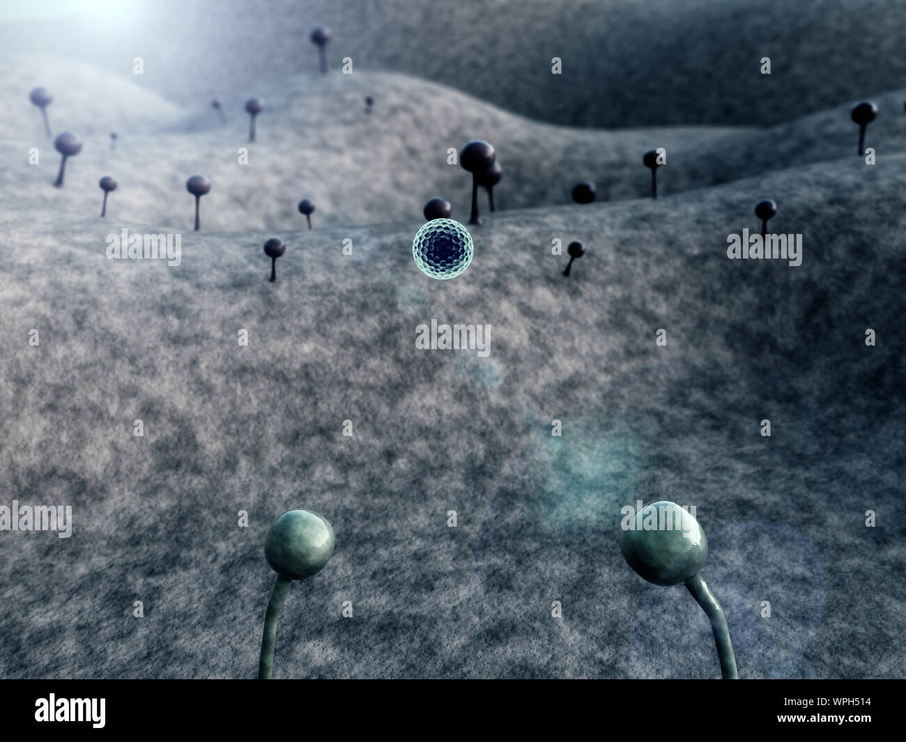 fungus, fungus on the leather surface, landscape of microworld, reproduction of fungi, fungus growing Stock Photohttps://www.alamy.com/image-license-details/?v=1https://www.alamy.com/fungus-fungus-on-the-leather-surface-landscape-of-microworld-reproduction-of-fungi-fungus-growing-image272055088.html
RFWPH514–fungus, fungus on the leather surface, landscape of microworld, reproduction of fungi, fungus growing
 Abstract Science Colorful Pattern Background Stock Photohttps://www.alamy.com/image-license-details/?v=1https://www.alamy.com/abstract-science-colorful-pattern-background-image438826069.html
RF2GDX73H–Abstract Science Colorful Pattern Background
 Abstract cell of virus or pathogenic bacterium under microscope, biofilm illustration. Stock Photohttps://www.alamy.com/image-license-details/?v=1https://www.alamy.com/abstract-cell-of-virus-or-pathogenic-bacterium-under-microscope-biofilm-illustration-image501669981.html
RF2M45151–Abstract cell of virus or pathogenic bacterium under microscope, biofilm illustration.
 . Morphology of angiosperms (Morphology of spermatophytes. Part II). Angiosperms; Plant morphology. THE FEMALE GAMETOPHYTE 73 reduced number of chromosomes, and this is true whether a row of two, three, or four megaspores is to be produced, or the mother-cell is to develop directly into the embryo-sac, as in Lilium. In such forms as Lilium the second mitosis also corre- sponds in all essential details with the second division that is to result in a row of four megaspores. The third mitosis differs. Please note that these images are extracted from scanned page images that may have been digitall Stock Photohttps://www.alamy.com/image-license-details/?v=1https://www.alamy.com/morphology-of-angiosperms-morphology-of-spermatophytes-part-ii-angiosperms-plant-morphology-the-female-gametophyte-73-reduced-number-of-chromosomes-and-this-is-true-whether-a-row-of-two-three-or-four-megaspores-is-to-be-produced-or-the-mother-cell-is-to-develop-directly-into-the-embryo-sac-as-in-lilium-in-such-forms-as-lilium-the-second-mitosis-also-corre-sponds-in-all-essential-details-with-the-second-division-that-is-to-result-in-a-row-of-four-megaspores-the-third-mitosis-differs-please-note-that-these-images-are-extracted-from-scanned-page-images-that-may-have-been-digitall-image232315824.html
RMRDXW5M–. Morphology of angiosperms (Morphology of spermatophytes. Part II). Angiosperms; Plant morphology. THE FEMALE GAMETOPHYTE 73 reduced number of chromosomes, and this is true whether a row of two, three, or four megaspores is to be produced, or the mother-cell is to develop directly into the embryo-sac, as in Lilium. In such forms as Lilium the second mitosis also corre- sponds in all essential details with the second division that is to result in a row of four megaspores. The third mitosis differs. Please note that these images are extracted from scanned page images that may have been digitall
 Abstract Science Colorful Pattern Background Stock Photohttps://www.alamy.com/image-license-details/?v=1https://www.alamy.com/abstract-science-colorful-pattern-background-image483512361.html
RF2K2HTXH–Abstract Science Colorful Pattern Background
 daffodil (Narcissus spec.), mitosis in celles of the root apex of a daffodill, 1000 x Stock Photohttps://www.alamy.com/image-license-details/?v=1https://www.alamy.com/stock-photo-daffodil-narcissus-spec-mitosis-in-celles-of-the-root-apex-of-a-daffodill-76068680.html
RMEBN6AG–daffodil (Narcissus spec.), mitosis in celles of the root apex of a daffodill, 1000 x
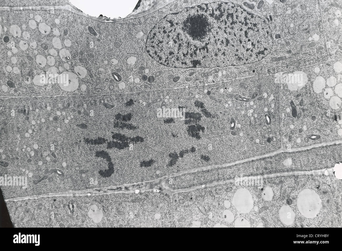 MITOSIS ANAPHASE Stock Photohttps://www.alamy.com/image-license-details/?v=1https://www.alamy.com/stock-photo-mitosis-anaphase-49164191.html
RMCRYHBY–MITOSIS ANAPHASE
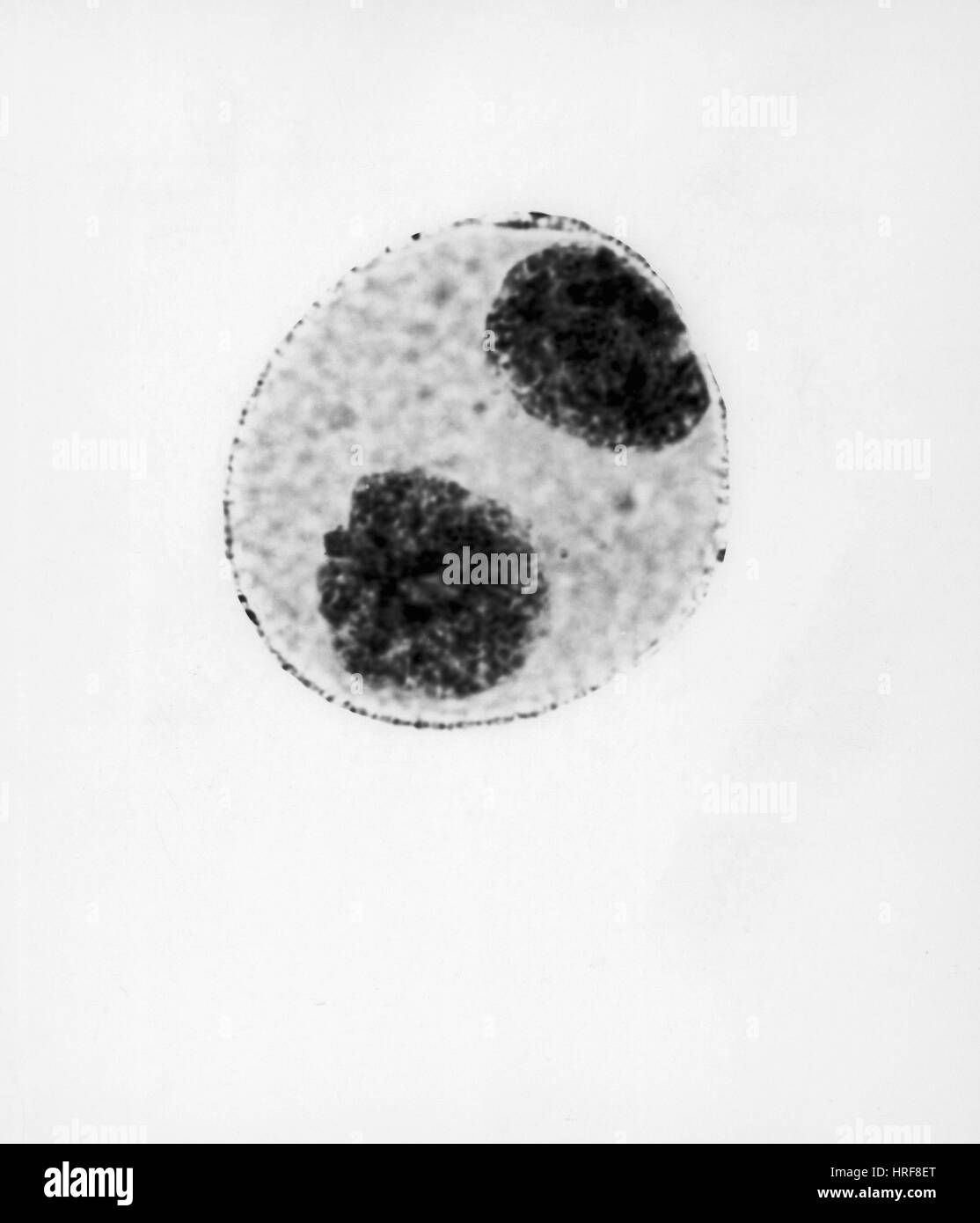 Telophase of Mitosis in Trillium Cell Stock Photohttps://www.alamy.com/image-license-details/?v=1https://www.alamy.com/stock-photo-telophase-of-mitosis-in-trillium-cell-134945632.html
RMHRF8ET–Telophase of Mitosis in Trillium Cell
 Chlorophyll is the molecule in leaves that uses the energy in sunlight to turn water and carbon dioxide gas into sugar and oxyge Stock Photohttps://www.alamy.com/image-license-details/?v=1https://www.alamy.com/stock-photo-chlorophyll-is-the-molecule-in-leaves-that-uses-the-energy-in-sunlight-88650111.html
RFF46A3B–Chlorophyll is the molecule in leaves that uses the energy in sunlight to turn water and carbon dioxide gas into sugar and oxyge
 New simple form of life. Design. Visualization of the origins of life. Stock Photohttps://www.alamy.com/image-license-details/?v=1https://www.alamy.com/new-simple-form-of-life-design-visualization-of-the-origins-of-life-image607765874.html
RF2X8P3G2–New simple form of life. Design. Visualization of the origins of life.
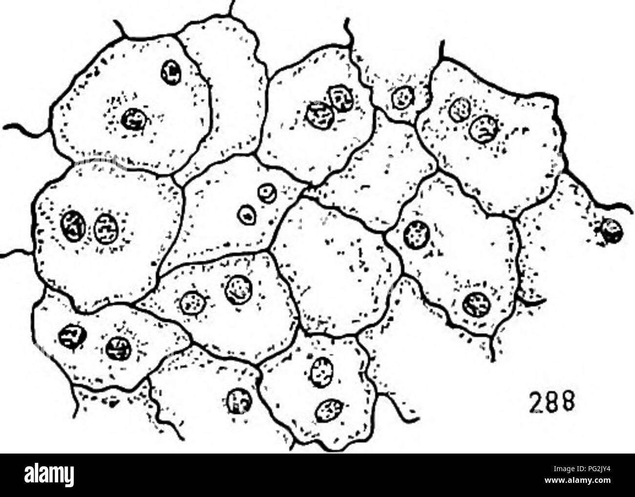 . Morphology of gymnosperms. Gymnosperms; Plant morphology. 286 287. Figs. 285-288.—Cryptomeria japonica: development of the endosperm; fig. 285, longitudinal section of upper portion of endosperm showing multinucleate condition and also that the walls are incomplete; May 26, 1902; X23S; fig. 286, telophase of the mitosis which is to result in the formation of a binucleate cell like those shown in fig. 288; May 29, 1903; XijOoo; fig. 287, a later stage in the same mitosis, the two daughter nuclei completely inclosed by the kinoplasmic fibrils; X 1,000; fig. 288, portion of the endosperm soon a Stock Photohttps://www.alamy.com/image-license-details/?v=1https://www.alamy.com/morphology-of-gymnosperms-gymnosperms-plant-morphology-286-287-figs-285-288cryptomeria-japonica-development-of-the-endosperm-fig-285-longitudinal-section-of-upper-portion-of-endosperm-showing-multinucleate-condition-and-also-that-the-walls-are-incomplete-may-26-1902-x23s-fig-286-telophase-of-the-mitosis-which-is-to-result-in-the-formation-of-a-binucleate-cell-like-those-shown-in-fig-288-may-29-1903-xijooo-fig-287-a-later-stage-in-the-same-mitosis-the-two-daughter-nuclei-completely-inclosed-by-the-kinoplasmic-fibrils-x-1000-fig-288-portion-of-the-endosperm-soon-a-image216417688.html
RMPG2JY4–. Morphology of gymnosperms. Gymnosperms; Plant morphology. 286 287. Figs. 285-288.—Cryptomeria japonica: development of the endosperm; fig. 285, longitudinal section of upper portion of endosperm showing multinucleate condition and also that the walls are incomplete; May 26, 1902; X23S; fig. 286, telophase of the mitosis which is to result in the formation of a binucleate cell like those shown in fig. 288; May 29, 1903; XijOoo; fig. 287, a later stage in the same mitosis, the two daughter nuclei completely inclosed by the kinoplasmic fibrils; X 1,000; fig. 288, portion of the endosperm soon a
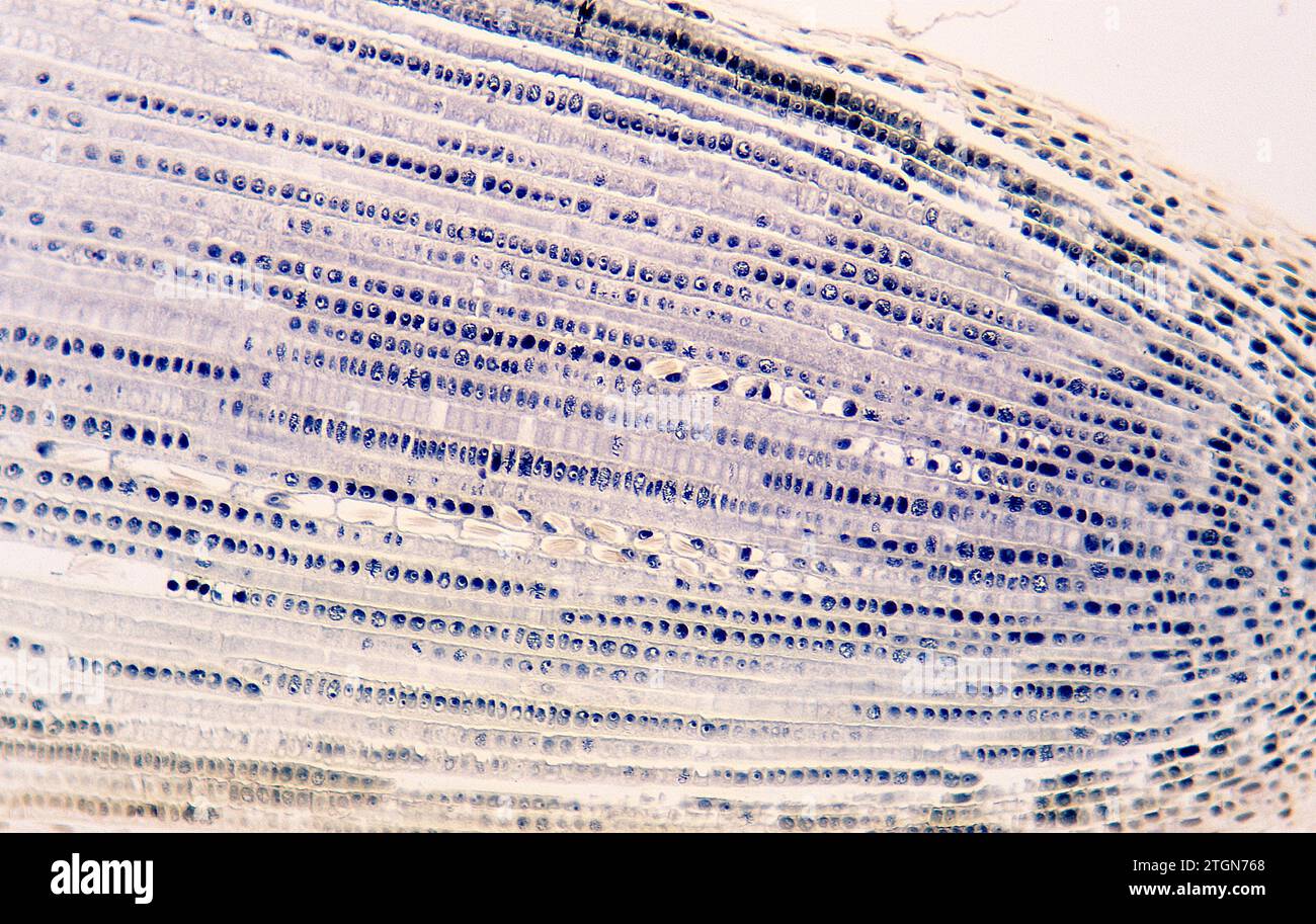 Root apical meristem showing cell divisions (mitosis) and raphides. Onion root photomicrograph. Stock Photohttps://www.alamy.com/image-license-details/?v=1https://www.alamy.com/root-apical-meristem-showing-cell-divisions-mitosis-and-raphides-onion-root-photomicrograph-image578243296.html
RF2TGN768–Root apical meristem showing cell divisions (mitosis) and raphides. Onion root photomicrograph.