Polyclonal Stock Photos and Images
 antibody, immunoglobulin, antibody molecule logo vector Stock Vectorhttps://www.alamy.com/image-license-details/?v=1https://www.alamy.com/antibody-immunoglobulin-antibody-molecule-logo-vector-image182256451.html
RFMGEDXY–antibody, immunoglobulin, antibody molecule logo vector
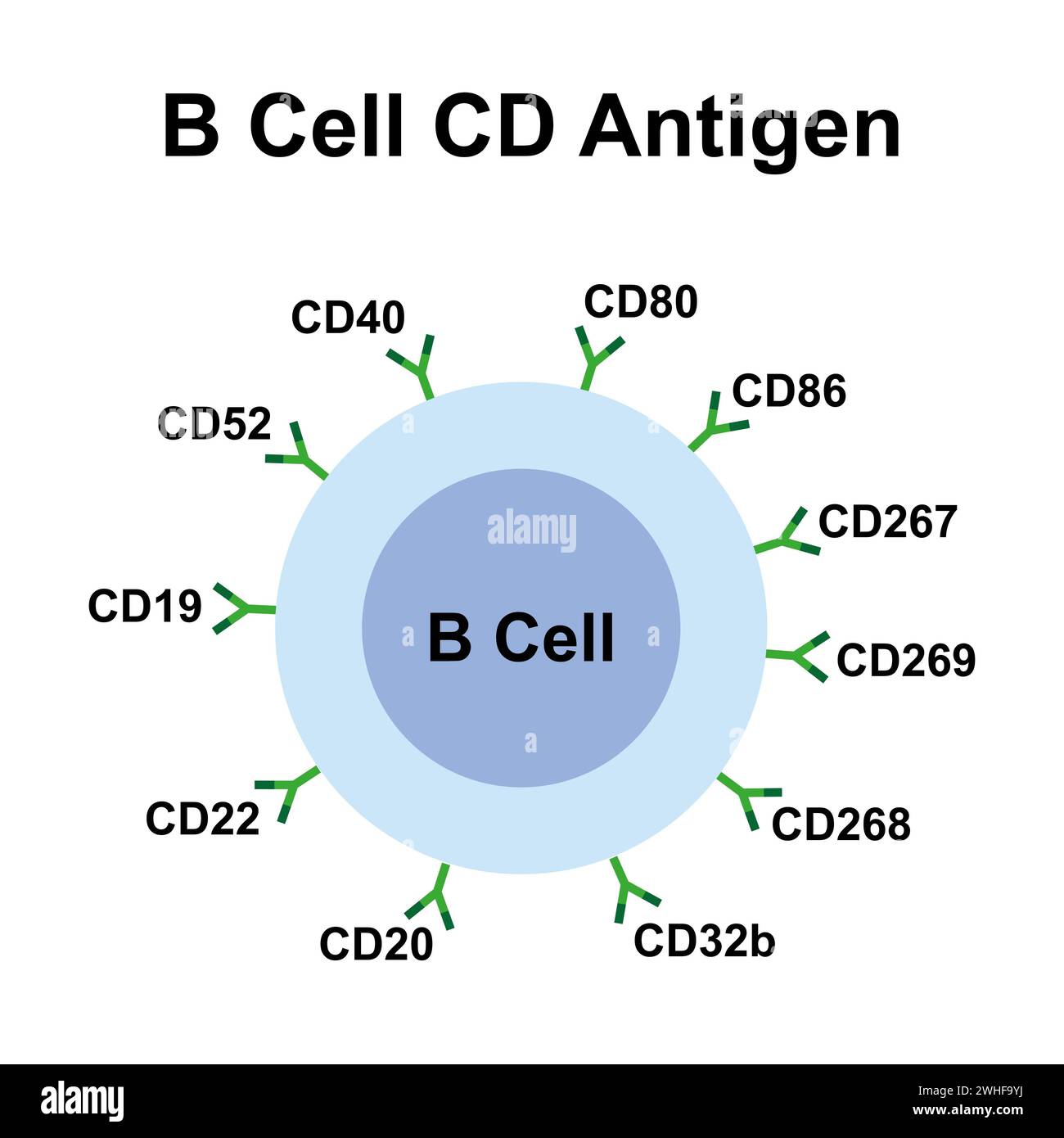 B cell CD antigen, illustration Stock Photohttps://www.alamy.com/image-license-details/?v=1https://www.alamy.com/b-cell-cd-antigen-illustration-image595938774.html
RF2WHF9YJ–B cell CD antigen, illustration
 Monoclonal antibody (Immunoglobulin) molecule, chemical structure. Most current biotech drugs are monoclonal antibodies. Stock Photohttps://www.alamy.com/image-license-details/?v=1https://www.alamy.com/stock-photo-monoclonal-antibody-immunoglobulin-molecule-chemical-structure-most-100699299.html
RFFRR6YF–Monoclonal antibody (Immunoglobulin) molecule, chemical structure. Most current biotech drugs are monoclonal antibodies.
 3D illustration of 'ANTIBODY PRODUCTION' title on medical documents. Medicial concept. Stock Photohttps://www.alamy.com/image-license-details/?v=1https://www.alamy.com/3d-illustration-of-antibody-production-title-on-medical-documents-image155759028.html
RFK1BC6C–3D illustration of 'ANTIBODY PRODUCTION' title on medical documents. Medicial concept.
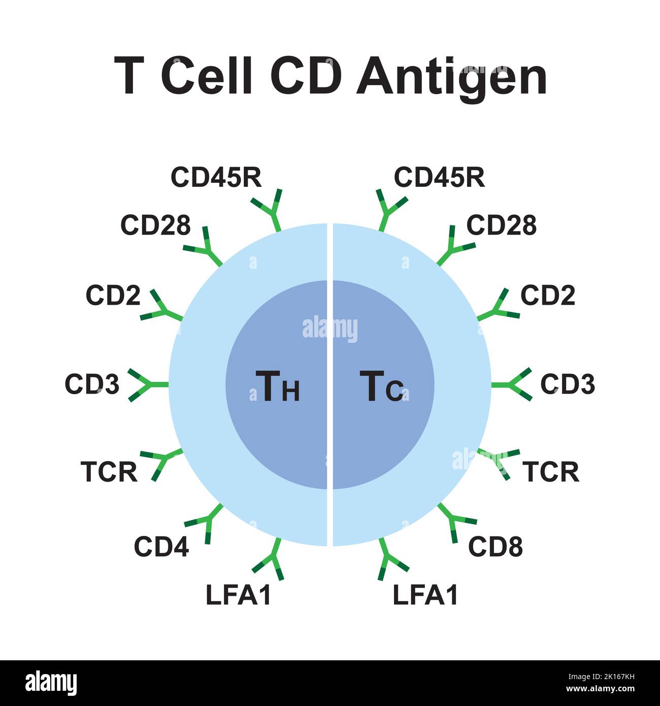 Scientific Designing of T Cell CD Antigen. Colorful Symbols. Vector Illustration. Stock Vectorhttps://www.alamy.com/image-license-details/?v=1https://www.alamy.com/scientific-designing-of-t-cell-cd-antigen-colorful-symbols-vector-illustration-image482642709.html
RF2K167KH–Scientific Designing of T Cell CD Antigen. Colorful Symbols. Vector Illustration.
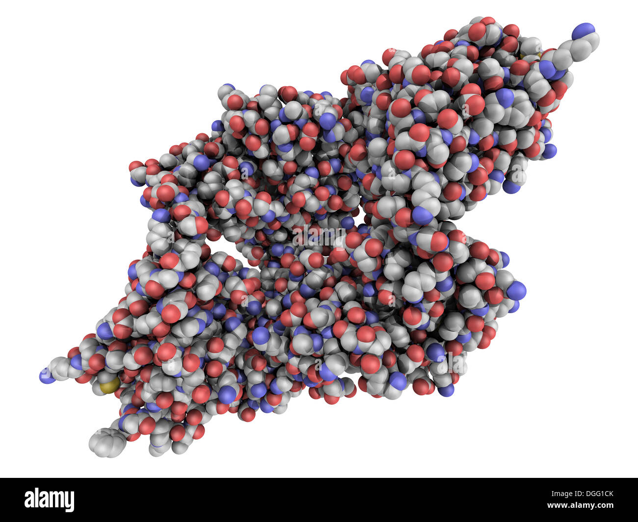 Monoclonal antibody (Immunoglobulin) molecule, chemical structure. Most current biotech drugs are monoclonal antibodies. Stock Photohttps://www.alamy.com/image-license-details/?v=1https://www.alamy.com/monoclonal-antibody-immunoglobulin-molecule-chemical-structure-most-image61817971.html
RFDGG1CK–Monoclonal antibody (Immunoglobulin) molecule, chemical structure. Most current biotech drugs are monoclonal antibodies.
 small antibody molecule logo Stock Vectorhttps://www.alamy.com/image-license-details/?v=1https://www.alamy.com/small-antibody-molecule-logo-image223768682.html
RFR01F6J–small antibody molecule logo
 . The Biological bulletin. Biology; Zoology; Biology; Marine Biology. LOCALIZATION OF CHLOROPEROXIDASE 219. Figure 2. Immunofluorescence labeling of cross sections of the tail of Notomastus lobalus. Polyclonal antisera to heme protein and flavoprotein subunits of chloroperoxidase from N- hhalus were used; binding was visualized by fluorescein-conjugated anti-rabbit antibody. Labeling with (A) heme protein antibody; (B) flavoprotein antibody; (C) 1:1 mixture of heme protein and flavoprotein antibodies; (D) preimmune rabbit serum. (E) Neighboring section stained with hemotoxylin and eosin and ex Stock Photohttps://www.alamy.com/image-license-details/?v=1https://www.alamy.com/the-biological-bulletin-biology-zoology-biology-marine-biology-localization-of-chloroperoxidase-219-figure-2-immunofluorescence-labeling-of-cross-sections-of-the-tail-of-notomastus-lobalus-polyclonal-antisera-to-heme-protein-and-flavoprotein-subunits-of-chloroperoxidase-from-n-hhalus-were-used-binding-was-visualized-by-fluorescein-conjugated-anti-rabbit-antibody-labeling-with-a-heme-protein-antibody-b-flavoprotein-antibody-c-11-mixture-of-heme-protein-and-flavoprotein-antibodies-d-preimmune-rabbit-serum-e-neighboring-section-stained-with-hemotoxylin-and-eosin-and-ex-image234631262.html
RMRHMAFX–. The Biological bulletin. Biology; Zoology; Biology; Marine Biology. LOCALIZATION OF CHLOROPEROXIDASE 219. Figure 2. Immunofluorescence labeling of cross sections of the tail of Notomastus lobalus. Polyclonal antisera to heme protein and flavoprotein subunits of chloroperoxidase from N- hhalus were used; binding was visualized by fluorescein-conjugated anti-rabbit antibody. Labeling with (A) heme protein antibody; (B) flavoprotein antibody; (C) 1:1 mixture of heme protein and flavoprotein antibodies; (D) preimmune rabbit serum. (E) Neighboring section stained with hemotoxylin and eosin and ex
 small antibody molecule logo Stock Vectorhttps://www.alamy.com/image-license-details/?v=1https://www.alamy.com/small-antibody-molecule-logo-image223768676.html
RFR01F6C–small antibody molecule logo
 . The Biological bulletin. Biology; Zoology; Biology; Marine Biology. K. S. YOON ET AL. Figure 3. (A) Immunofluorescence labeling of gut epithelium (e) in cross sections of the tail of Notomuslus lohatus. A mixture ot polyclonal antibodies to the heme protein and davoprotein subunits of chloroper- oxidase was used; binding was visualized by FITC-conjugated anti-rabbit antibody. (B) Neighboring section stained with hemotoxylin and eosin and examined with light microscopy (e—gut epithelium). Scale bar = 72 jtm. highest enzyme activity was detected in extracts from the tail region, with much less Stock Photohttps://www.alamy.com/image-license-details/?v=1https://www.alamy.com/the-biological-bulletin-biology-zoology-biology-marine-biology-k-s-yoon-et-al-figure-3-a-immunofluorescence-labeling-of-gut-epithelium-e-in-cross-sections-of-the-tail-of-notomuslus-lohatus-a-mixture-ot-polyclonal-antibodies-to-the-heme-protein-and-davoprotein-subunits-of-chloroper-oxidase-was-used-binding-was-visualized-by-fitc-conjugated-anti-rabbit-antibody-b-neighboring-section-stained-with-hemotoxylin-and-eosin-and-examined-with-light-microscopy-egut-epithelium-scale-bar-=-72-jtm-highest-enzyme-activity-was-detected-in-extracts-from-the-tail-region-with-much-less-image234634393.html
RMRHMEFN–. The Biological bulletin. Biology; Zoology; Biology; Marine Biology. K. S. YOON ET AL. Figure 3. (A) Immunofluorescence labeling of gut epithelium (e) in cross sections of the tail of Notomuslus lohatus. A mixture ot polyclonal antibodies to the heme protein and davoprotein subunits of chloroper- oxidase was used; binding was visualized by FITC-conjugated anti-rabbit antibody. (B) Neighboring section stained with hemotoxylin and eosin and examined with light microscopy (e—gut epithelium). Scale bar = 72 jtm. highest enzyme activity was detected in extracts from the tail region, with much less
 small antibody molecule logo Stock Vectorhttps://www.alamy.com/image-license-details/?v=1https://www.alamy.com/small-antibody-molecule-logo-image223768687.html
RFR01F6R–small antibody molecule logo
 . The Biological bulletin. Biology; Zoology; Biology; Marine Biology. Figures 4-9. ImmunohistochemicaJ localization of vacuolar-type ATPase (V-ATPase) in whole accessory boring organ (ABO) of Nucellu lamellosa. Figure 4. This section was probed with polyclonal antibody to the V-ATPase d (39-kDa) subunit and alkaline-phosphatase-conjugated secondary antibody. Immunoreactive sites were visualized with BCIP/NBT. Immunostaining is exclusive to the brush border of the ABO (b). Immunostaining is absent in the foot epithelium (arrowheads), e, epithelial cells; s, sinus. Scale bar: 100 fim. Figure 5. Stock Photohttps://www.alamy.com/image-license-details/?v=1https://www.alamy.com/the-biological-bulletin-biology-zoology-biology-marine-biology-figures-4-9-immunohistochemicaj-localization-of-vacuolar-type-atpase-v-atpase-in-whole-accessory-boring-organ-abo-of-nucellu-lamellosa-figure-4-this-section-was-probed-with-polyclonal-antibody-to-the-v-atpase-d-39-kda-subunit-and-alkaline-phosphatase-conjugated-secondary-antibody-immunoreactive-sites-were-visualized-with-bcipnbt-immunostaining-is-exclusive-to-the-brush-border-of-the-abo-b-immunostaining-is-absent-in-the-foot-epithelium-arrowheads-e-epithelial-cells-s-sinus-scale-bar-100-fim-figure-5-image234634261.html
RMRHMEB1–. The Biological bulletin. Biology; Zoology; Biology; Marine Biology. Figures 4-9. ImmunohistochemicaJ localization of vacuolar-type ATPase (V-ATPase) in whole accessory boring organ (ABO) of Nucellu lamellosa. Figure 4. This section was probed with polyclonal antibody to the V-ATPase d (39-kDa) subunit and alkaline-phosphatase-conjugated secondary antibody. Immunoreactive sites were visualized with BCIP/NBT. Immunostaining is exclusive to the brush border of the ABO (b). Immunostaining is absent in the foot epithelium (arrowheads), e, epithelial cells; s, sinus. Scale bar: 100 fim. Figure 5.
 small antibody molecule logo Stock Vectorhttps://www.alamy.com/image-license-details/?v=1https://www.alamy.com/small-antibody-molecule-logo-image223768681.html
RFR01F6H–small antibody molecule logo
 . The Biological bulletin. Biology; Zoology; Biology; Marine Biology. Figure 1. Light microscopy. Cross-section of a tentacle of Condylactis gigantea showing specific nickel-enhanced immunostain with polyclonal NF 200 antisera. The product is localized to an area above the muscle layer and mesoglea that contains the neural plexus. Reaction product is also seen in the gastrodermis. The scale bar is 10 ^m. E, epidermis; M, mesoglea; G. gastrodermis. of nerve cell bodies and processes (C. DellaCorte, C. Fu- lenwider. D. Hessinger. and W.O. McClure. manuscript in prep). Immunolocalization was also Stock Photohttps://www.alamy.com/image-license-details/?v=1https://www.alamy.com/the-biological-bulletin-biology-zoology-biology-marine-biology-figure-1-light-microscopy-cross-section-of-a-tentacle-of-condylactis-gigantea-showing-specific-nickel-enhanced-immunostain-with-polyclonal-nf-200-antisera-the-product-is-localized-to-an-area-above-the-muscle-layer-and-mesoglea-that-contains-the-neural-plexus-reaction-product-is-also-seen-in-the-gastrodermis-the-scale-bar-is-10-m-e-epidermis-m-mesoglea-g-gastrodermis-of-nerve-cell-bodies-and-processes-c-dellacorte-c-fu-lenwider-d-hessinger-and-wo-mcclure-manuscript-in-prep-immunolocalization-was-also-image234631312.html
RMRHMAHM–. The Biological bulletin. Biology; Zoology; Biology; Marine Biology. Figure 1. Light microscopy. Cross-section of a tentacle of Condylactis gigantea showing specific nickel-enhanced immunostain with polyclonal NF 200 antisera. The product is localized to an area above the muscle layer and mesoglea that contains the neural plexus. Reaction product is also seen in the gastrodermis. The scale bar is 10 ^m. E, epidermis; M, mesoglea; G. gastrodermis. of nerve cell bodies and processes (C. DellaCorte, C. Fu- lenwider. D. Hessinger. and W.O. McClure. manuscript in prep). Immunolocalization was also
 . The Biological bulletin. Biology; Zoology; Biology; Marine Biology. 2 34567 8 9 * Figure 5. Specificity of the polyclonal antiserum raised against OHSS. (A) SDS-PAGE of fractions eluted from molecular-sieve chromatography. The polyacrylamide gel was transblotted onto a PVDF membrane and stained with Coomassie brilliant blue. (B) Immunostaining of the PVDF membrane with a polyclonal antiserum raised to OHSS. Numbers to the left show the molecular masses of the standards shown in Figure 2B. The intensely immunostained bands in fractions 6-8 have masses of about 32 kDa and 30 kDa (arrows to the Stock Photohttps://www.alamy.com/image-license-details/?v=1https://www.alamy.com/the-biological-bulletin-biology-zoology-biology-marine-biology-2-34567-8-9-figure-5-specificity-of-the-polyclonal-antiserum-raised-against-ohss-a-sds-page-of-fractions-eluted-from-molecular-sieve-chromatography-the-polyacrylamide-gel-was-transblotted-onto-a-pvdf-membrane-and-stained-with-coomassie-brilliant-blue-b-immunostaining-of-the-pvdf-membrane-with-a-polyclonal-antiserum-raised-to-ohss-numbers-to-the-left-show-the-molecular-masses-of-the-standards-shown-in-figure-2b-the-intensely-immunostained-bands-in-fractions-6-8-have-masses-of-about-32-kda-and-30-kda-arrows-to-the-image234633820.html
RMRHMDR8–. The Biological bulletin. Biology; Zoology; Biology; Marine Biology. 2 34567 8 9 * Figure 5. Specificity of the polyclonal antiserum raised against OHSS. (A) SDS-PAGE of fractions eluted from molecular-sieve chromatography. The polyacrylamide gel was transblotted onto a PVDF membrane and stained with Coomassie brilliant blue. (B) Immunostaining of the PVDF membrane with a polyclonal antiserum raised to OHSS. Numbers to the left show the molecular masses of the standards shown in Figure 2B. The intensely immunostained bands in fractions 6-8 have masses of about 32 kDa and 30 kDa (arrows to the
 . The Biological bulletin. Biology; Zoology; Biology; Marine Biology. NEUROFILAMENT-LIKE PROTEINS IN CNIDARIA 203. Figure 1. Light microscopy. Cross-section of a tentacle of Condylactis gigantea showing specific nickel-enhanced immunostain with polyclonal NF 200 antisera. The product is localized to an area above the muscle layer and mesoglea that contains the neural plexus. Reaction product is also seen in the gastrodermis. The scale bar is 10 ^m. E, epidermis; M, mesoglea; G. gastrodermis. of nerve cell bodies and processes (C. DellaCorte, C. Fu- lenwider. D. Hessinger. and W.O. McClure. man Stock Photohttps://www.alamy.com/image-license-details/?v=1https://www.alamy.com/the-biological-bulletin-biology-zoology-biology-marine-biology-neurofilament-like-proteins-in-cnidaria-203-figure-1-light-microscopy-cross-section-of-a-tentacle-of-condylactis-gigantea-showing-specific-nickel-enhanced-immunostain-with-polyclonal-nf-200-antisera-the-product-is-localized-to-an-area-above-the-muscle-layer-and-mesoglea-that-contains-the-neural-plexus-reaction-product-is-also-seen-in-the-gastrodermis-the-scale-bar-is-10-m-e-epidermis-m-mesoglea-g-gastrodermis-of-nerve-cell-bodies-and-processes-c-dellacorte-c-fu-lenwider-d-hessinger-and-wo-mcclure-man-image234631326.html
RMRHMAJ6–. The Biological bulletin. Biology; Zoology; Biology; Marine Biology. NEUROFILAMENT-LIKE PROTEINS IN CNIDARIA 203. Figure 1. Light microscopy. Cross-section of a tentacle of Condylactis gigantea showing specific nickel-enhanced immunostain with polyclonal NF 200 antisera. The product is localized to an area above the muscle layer and mesoglea that contains the neural plexus. Reaction product is also seen in the gastrodermis. The scale bar is 10 ^m. E, epidermis; M, mesoglea; G. gastrodermis. of nerve cell bodies and processes (C. DellaCorte, C. Fu- lenwider. D. Hessinger. and W.O. McClure. man
 . The Biological bulletin. Biology; Zoology; Biology; Marine Biology. PROSOBRANCH REPRODUCTIVE PEPTIDES 311 50% identical to the ELH sequence (Chiu ct til.. 1979). Tests of positive staining of CDC neurons and cerebral commissure were used to determine whether the anti- ELH antibody had a sufficiently broad specificity that it would bind to CDCH, a homolog of A. californica ELH in a species from a different subclass of gastropods. The primary anti-a-CDCP is a polyclonal rabbit serum obtained from J. Van Minnen (Vrije Universiteit, Amster- dam) and characterized in previous publications on L. s Stock Photohttps://www.alamy.com/image-license-details/?v=1https://www.alamy.com/the-biological-bulletin-biology-zoology-biology-marine-biology-prosobranch-reproductive-peptides-311-50-identical-to-the-elh-sequence-chiu-ct-til-1979-tests-of-positive-staining-of-cdc-neurons-and-cerebral-commissure-were-used-to-determine-whether-the-anti-elh-antibody-had-a-sufficiently-broad-specificity-that-it-would-bind-to-cdch-a-homolog-of-a-californica-elh-in-a-species-from-a-different-subclass-of-gastropods-the-primary-anti-a-cdcp-is-a-polyclonal-rabbit-serum-obtained-from-j-van-minnen-vrije-universiteit-amster-dam-and-characterized-in-previous-publications-on-l-s-image234630960.html
RMRHMA54–. The Biological bulletin. Biology; Zoology; Biology; Marine Biology. PROSOBRANCH REPRODUCTIVE PEPTIDES 311 50% identical to the ELH sequence (Chiu ct til.. 1979). Tests of positive staining of CDC neurons and cerebral commissure were used to determine whether the anti- ELH antibody had a sufficiently broad specificity that it would bind to CDCH, a homolog of A. californica ELH in a species from a different subclass of gastropods. The primary anti-a-CDCP is a polyclonal rabbit serum obtained from J. Van Minnen (Vrije Universiteit, Amster- dam) and characterized in previous publications on L. s
 . The Biological bulletin. Biology; Zoology; Biology; Marine Biology. ACTIVE SUBSTANCE IN CRAB HATCH WATER 181 Fraction number 23456789 B Fraction number 556 392 266 — 143- kDa. 2 34567 8 9 * Figure 5. Specificity of the polyclonal antiserum raised against OHSS. (A) SDS-PAGE of fractions eluted from molecular-sieve chromatography. The polyacrylamide gel was transblotted onto a PVDF membrane and stained with Coomassie brilliant blue. (B) Immunostaining of the PVDF membrane with a polyclonal antiserum raised to OHSS. Numbers to the left show the molecular masses of the standards shown in Figure Stock Photohttps://www.alamy.com/image-license-details/?v=1https://www.alamy.com/the-biological-bulletin-biology-zoology-biology-marine-biology-active-substance-in-crab-hatch-water-181-fraction-number-23456789-b-fraction-number-556-392-266-143-kda-2-34567-8-9-figure-5-specificity-of-the-polyclonal-antiserum-raised-against-ohss-a-sds-page-of-fractions-eluted-from-molecular-sieve-chromatography-the-polyacrylamide-gel-was-transblotted-onto-a-pvdf-membrane-and-stained-with-coomassie-brilliant-blue-b-immunostaining-of-the-pvdf-membrane-with-a-polyclonal-antiserum-raised-to-ohss-numbers-to-the-left-show-the-molecular-masses-of-the-standards-shown-in-figure-image234633837.html
RMRHMDRW–. The Biological bulletin. Biology; Zoology; Biology; Marine Biology. ACTIVE SUBSTANCE IN CRAB HATCH WATER 181 Fraction number 23456789 B Fraction number 556 392 266 — 143- kDa. 2 34567 8 9 * Figure 5. Specificity of the polyclonal antiserum raised against OHSS. (A) SDS-PAGE of fractions eluted from molecular-sieve chromatography. The polyacrylamide gel was transblotted onto a PVDF membrane and stained with Coomassie brilliant blue. (B) Immunostaining of the PVDF membrane with a polyclonal antiserum raised to OHSS. Numbers to the left show the molecular masses of the standards shown in Figure
![. The Biological bulletin. Biology; Zoology; Biology; Marine Biology. Figure 7. Immunochemica] staining of the structures remaining attached to a female's ovigerous hairs after hatching. Left: the iminunoblot of an extract of the remnants subjected to SDS-PAGE. Arrowhead: 32-kDa protein band. Numbers to the left show the molecular masses of the standard molecules (as in Fig. 2B). Right: the remnants stained with polyclonal FITC-conjugated OHSS antiserum. ci1: broken egg case:/: funiculus; pc: prezoeal cuticle; oh: female ovigerous hair (see fig. 2 in Saigusa. 1994). Scale: 100 /nl.. Please not Stock Photo . The Biological bulletin. Biology; Zoology; Biology; Marine Biology. Figure 7. Immunochemica] staining of the structures remaining attached to a female's ovigerous hairs after hatching. Left: the iminunoblot of an extract of the remnants subjected to SDS-PAGE. Arrowhead: 32-kDa protein band. Numbers to the left show the molecular masses of the standard molecules (as in Fig. 2B). Right: the remnants stained with polyclonal FITC-conjugated OHSS antiserum. ci1: broken egg case:/: funiculus; pc: prezoeal cuticle; oh: female ovigerous hair (see fig. 2 in Saigusa. 1994). Scale: 100 /nl.. Please not Stock Photo](https://c8.alamy.com/comp/RHMDNG/the-biological-bulletin-biology-zoology-biology-marine-biology-figure-7-immunochemica-staining-of-the-structures-remaining-attached-to-a-females-ovigerous-hairs-after-hatching-left-the-iminunoblot-of-an-extract-of-the-remnants-subjected-to-sds-page-arrowhead-32-kda-protein-band-numbers-to-the-left-show-the-molecular-masses-of-the-standard-molecules-as-in-fig-2b-right-the-remnants-stained-with-polyclonal-fitc-conjugated-ohss-antiserum-ci1-broken-egg-case-funiculus-pc-prezoeal-cuticle-oh-female-ovigerous-hair-see-fig-2-in-saigusa-1994-scale-100-nl-please-not-RHMDNG.jpg) . The Biological bulletin. Biology; Zoology; Biology; Marine Biology. Figure 7. Immunochemica] staining of the structures remaining attached to a female's ovigerous hairs after hatching. Left: the iminunoblot of an extract of the remnants subjected to SDS-PAGE. Arrowhead: 32-kDa protein band. Numbers to the left show the molecular masses of the standard molecules (as in Fig. 2B). Right: the remnants stained with polyclonal FITC-conjugated OHSS antiserum. ci1: broken egg case:/: funiculus; pc: prezoeal cuticle; oh: female ovigerous hair (see fig. 2 in Saigusa. 1994). Scale: 100 /nl.. Please not Stock Photohttps://www.alamy.com/image-license-details/?v=1https://www.alamy.com/the-biological-bulletin-biology-zoology-biology-marine-biology-figure-7-immunochemica-staining-of-the-structures-remaining-attached-to-a-females-ovigerous-hairs-after-hatching-left-the-iminunoblot-of-an-extract-of-the-remnants-subjected-to-sds-page-arrowhead-32-kda-protein-band-numbers-to-the-left-show-the-molecular-masses-of-the-standard-molecules-as-in-fig-2b-right-the-remnants-stained-with-polyclonal-fitc-conjugated-ohss-antiserum-ci1-broken-egg-case-funiculus-pc-prezoeal-cuticle-oh-female-ovigerous-hair-see-fig-2-in-saigusa-1994-scale-100-nl-please-not-image234633772.html
. The Biological bulletin. Biology; Zoology; Biology; Marine Biology. Figure 7. Immunochemica] staining of the structures remaining attached to a female's ovigerous hairs after hatching. Left: the iminunoblot of an extract of the remnants subjected to SDS-PAGE. Arrowhead: 32-kDa protein band. Numbers to the left show the molecular masses of the standard molecules (as in Fig. 2B). Right: the remnants stained with polyclonal FITC-conjugated OHSS antiserum. ci1: broken egg case:/: funiculus; pc: prezoeal cuticle; oh: female ovigerous hair (see fig. 2 in Saigusa. 1994). Scale: 100 /nl.. Please not Stock Photohttps://www.alamy.com/image-license-details/?v=1https://www.alamy.com/the-biological-bulletin-biology-zoology-biology-marine-biology-figure-7-immunochemica-staining-of-the-structures-remaining-attached-to-a-females-ovigerous-hairs-after-hatching-left-the-iminunoblot-of-an-extract-of-the-remnants-subjected-to-sds-page-arrowhead-32-kda-protein-band-numbers-to-the-left-show-the-molecular-masses-of-the-standard-molecules-as-in-fig-2b-right-the-remnants-stained-with-polyclonal-fitc-conjugated-ohss-antiserum-ci1-broken-egg-case-funiculus-pc-prezoeal-cuticle-oh-female-ovigerous-hair-see-fig-2-in-saigusa-1994-scale-100-nl-please-not-image234633772.htmlRMRHMDNG–. The Biological bulletin. Biology; Zoology; Biology; Marine Biology. Figure 7. Immunochemica] staining of the structures remaining attached to a female's ovigerous hairs after hatching. Left: the iminunoblot of an extract of the remnants subjected to SDS-PAGE. Arrowhead: 32-kDa protein band. Numbers to the left show the molecular masses of the standard molecules (as in Fig. 2B). Right: the remnants stained with polyclonal FITC-conjugated OHSS antiserum. ci1: broken egg case:/: funiculus; pc: prezoeal cuticle; oh: female ovigerous hair (see fig. 2 in Saigusa. 1994). Scale: 100 /nl.. Please not
 . The Biological bulletin. Biology; Zoology; Biology; Marine Biology. SHRIMP CORTICAL GRANULE cDNA AND ANTIBODY 347 1234 1234 MrXlO-3 205- 116- 97- 66- • I ! B Figure 3. Pcnacits vannamei polypeptides were separated by SDS- PAGE, and stained with Coomassie Blue R (Panel A), or blotted onto nitrocellulose and incubated with a polyclonal antibody to the geneti- cally engineered fusion polypeptide (Panel B). Samples were from an animal with mid-cycle ovaries (oocyte diameter 150 ^m). Lane 1, ovary: 2, hemolymph; 3, muscle; 4, hepatopancreas. Positions of mo- lecular weight markers are indicated Stock Photohttps://www.alamy.com/image-license-details/?v=1https://www.alamy.com/the-biological-bulletin-biology-zoology-biology-marine-biology-shrimp-cortical-granule-cdna-and-antibody-347-1234-1234-mrxlo-3-205-116-97-66-i-!-b-figure-3-pcnacits-vannamei-polypeptides-were-separated-by-sds-page-and-stained-with-coomassie-blue-r-panel-a-or-blotted-onto-nitrocellulose-and-incubated-with-a-polyclonal-antibody-to-the-geneti-cally-engineered-fusion-polypeptide-panel-b-samples-were-from-an-animal-with-mid-cycle-ovaries-oocyte-diameter-150-m-lane-1-ovary-2-hemolymph-3-muscle-4-hepatopancreas-positions-of-mo-lecular-weight-markers-are-indicated-image234644723.html
RMRHMYMK–. The Biological bulletin. Biology; Zoology; Biology; Marine Biology. SHRIMP CORTICAL GRANULE cDNA AND ANTIBODY 347 1234 1234 MrXlO-3 205- 116- 97- 66- • I ! B Figure 3. Pcnacits vannamei polypeptides were separated by SDS- PAGE, and stained with Coomassie Blue R (Panel A), or blotted onto nitrocellulose and incubated with a polyclonal antibody to the geneti- cally engineered fusion polypeptide (Panel B). Samples were from an animal with mid-cycle ovaries (oocyte diameter 150 ^m). Lane 1, ovary: 2, hemolymph; 3, muscle; 4, hepatopancreas. Positions of mo- lecular weight markers are indicated
 . The Biological bulletin. Biology; Zoology; Biology; Marine Biology. B 55.6 39.2 26.6 14.3 kDa i a. Figure 6. Immunostaining with antibodies affinity purified from SDS-PAGE bands that had OHSS activity. SDS-PAGE of the proteins from molecular-sieve fractions 6 and 7 (see Fig. 2A) was carried out for either 3.5 h (A) or 9h (B), the longer electrophoresis producing the greater separation between the active bands at 32 kDa and 30 kDa. The bands were cut out of the PVDF membranes, incubated with a polyclonal antiserum raised to OHSS, and the bound antibodies were eluted (see Methods). These antib Stock Photohttps://www.alamy.com/image-license-details/?v=1https://www.alamy.com/the-biological-bulletin-biology-zoology-biology-marine-biology-b-556-392-266-143-kda-i-a-figure-6-immunostaining-with-antibodies-affinity-purified-from-sds-page-bands-that-had-ohss-activity-sds-page-of-the-proteins-from-molecular-sieve-fractions-6-and-7-see-fig-2a-was-carried-out-for-either-35-h-a-or-9h-b-the-longer-electrophoresis-producing-the-greater-separation-between-the-active-bands-at-32-kda-and-30-kda-the-bands-were-cut-out-of-the-pvdf-membranes-incubated-with-a-polyclonal-antiserum-raised-to-ohss-and-the-bound-antibodies-were-eluted-see-methods-these-antib-image234633804.html
RMRHMDPM–. The Biological bulletin. Biology; Zoology; Biology; Marine Biology. B 55.6 39.2 26.6 14.3 kDa i a. Figure 6. Immunostaining with antibodies affinity purified from SDS-PAGE bands that had OHSS activity. SDS-PAGE of the proteins from molecular-sieve fractions 6 and 7 (see Fig. 2A) was carried out for either 3.5 h (A) or 9h (B), the longer electrophoresis producing the greater separation between the active bands at 32 kDa and 30 kDa. The bands were cut out of the PVDF membranes, incubated with a polyclonal antiserum raised to OHSS, and the bound antibodies were eluted (see Methods). These antib
 . The Biological bulletin. Biology; Zoology; Marine biology. 408 K. KAMINO ET AL. 16.9—. Figure 5. Immunoblotting analysis using polyclonal antibody against CB-2. Lane 1: CB-peptides. Lane 2: primary cement treated with CNBr. Number on side of lane 1 indicates molecular mass (kDa) of molecular weight peptide standards (Pharmacia LKB). protein play a role in removing the weak boundary-water layer and in spreading the protein onto the surface of the substratum in the process of adhesion. The hydroxylated side-chains of proteins in SFl and SF2 may be similarly important for underwater adhesion. T Stock Photohttps://www.alamy.com/image-license-details/?v=1https://www.alamy.com/the-biological-bulletin-biology-zoology-marine-biology-408-k-kamino-et-al-169-figure-5-immunoblotting-analysis-using-polyclonal-antibody-against-cb-2-lane-1-cb-peptides-lane-2-primary-cement-treated-with-cnbr-number-on-side-of-lane-1-indicates-molecular-mass-kda-of-molecular-weight-peptide-standards-pharmacia-lkb-protein-play-a-role-in-removing-the-weak-boundary-water-layer-and-in-spreading-the-protein-onto-the-surface-of-the-substratum-in-the-process-of-adhesion-the-hydroxylated-side-chains-of-proteins-in-sfl-and-sf2-may-be-similarly-important-for-underwater-adhesion-t-image234625723.html
RMRHM3E3–. The Biological bulletin. Biology; Zoology; Marine biology. 408 K. KAMINO ET AL. 16.9—. Figure 5. Immunoblotting analysis using polyclonal antibody against CB-2. Lane 1: CB-peptides. Lane 2: primary cement treated with CNBr. Number on side of lane 1 indicates molecular mass (kDa) of molecular weight peptide standards (Pharmacia LKB). protein play a role in removing the weak boundary-water layer and in spreading the protein onto the surface of the substratum in the process of adhesion. The hydroxylated side-chains of proteins in SFl and SF2 may be similarly important for underwater adhesion. T
 . The Biological bulletin. Biology; Zoology; Biology; Marine Biology. 184 M. SAIGLISA AND H. IWASAKI Days before hatching 14d 10d 6d 4d 2d 1d 4h Z REM 55.6 39.2 26.6 — 32 kDa 14.3 -> kDa Figure 8. The appearance of immunoreactive OHSS in developing crab embryos. Embryo clusters (one- third of an ovigerous seta) were detached from a single female, crushed, and then denatured with the lysis buffer. The extracts (supernatant) were subjected to SDS-PAGE, and blots were stained immunochemically with the polyclonal OHSS antiserum. d (or /?): days (or hours) before hatching. Z: post-hatched embryo Stock Photohttps://www.alamy.com/image-license-details/?v=1https://www.alamy.com/the-biological-bulletin-biology-zoology-biology-marine-biology-184-m-saiglisa-and-h-iwasaki-days-before-hatching-14d-10d-6d-4d-2d-1d-4h-z-rem-556-392-266-32-kda-143-gt-kda-figure-8-the-appearance-of-immunoreactive-ohss-in-developing-crab-embryos-embryo-clusters-one-third-of-an-ovigerous-seta-were-detached-from-a-single-female-crushed-and-then-denatured-with-the-lysis-buffer-the-extracts-supernatant-were-subjected-to-sds-page-and-blots-were-stained-immunochemically-with-the-polyclonal-ohss-antiserum-d-or-days-or-hours-before-hatching-z-post-hatched-embryo-image234633759.html
RMRHMDN3–. The Biological bulletin. Biology; Zoology; Biology; Marine Biology. 184 M. SAIGLISA AND H. IWASAKI Days before hatching 14d 10d 6d 4d 2d 1d 4h Z REM 55.6 39.2 26.6 — 32 kDa 14.3 -> kDa Figure 8. The appearance of immunoreactive OHSS in developing crab embryos. Embryo clusters (one- third of an ovigerous seta) were detached from a single female, crushed, and then denatured with the lysis buffer. The extracts (supernatant) were subjected to SDS-PAGE, and blots were stained immunochemically with the polyclonal OHSS antiserum. d (or /?): days (or hours) before hatching. Z: post-hatched embryo