Quick filters:
Polyp removal Stock Photos and Images
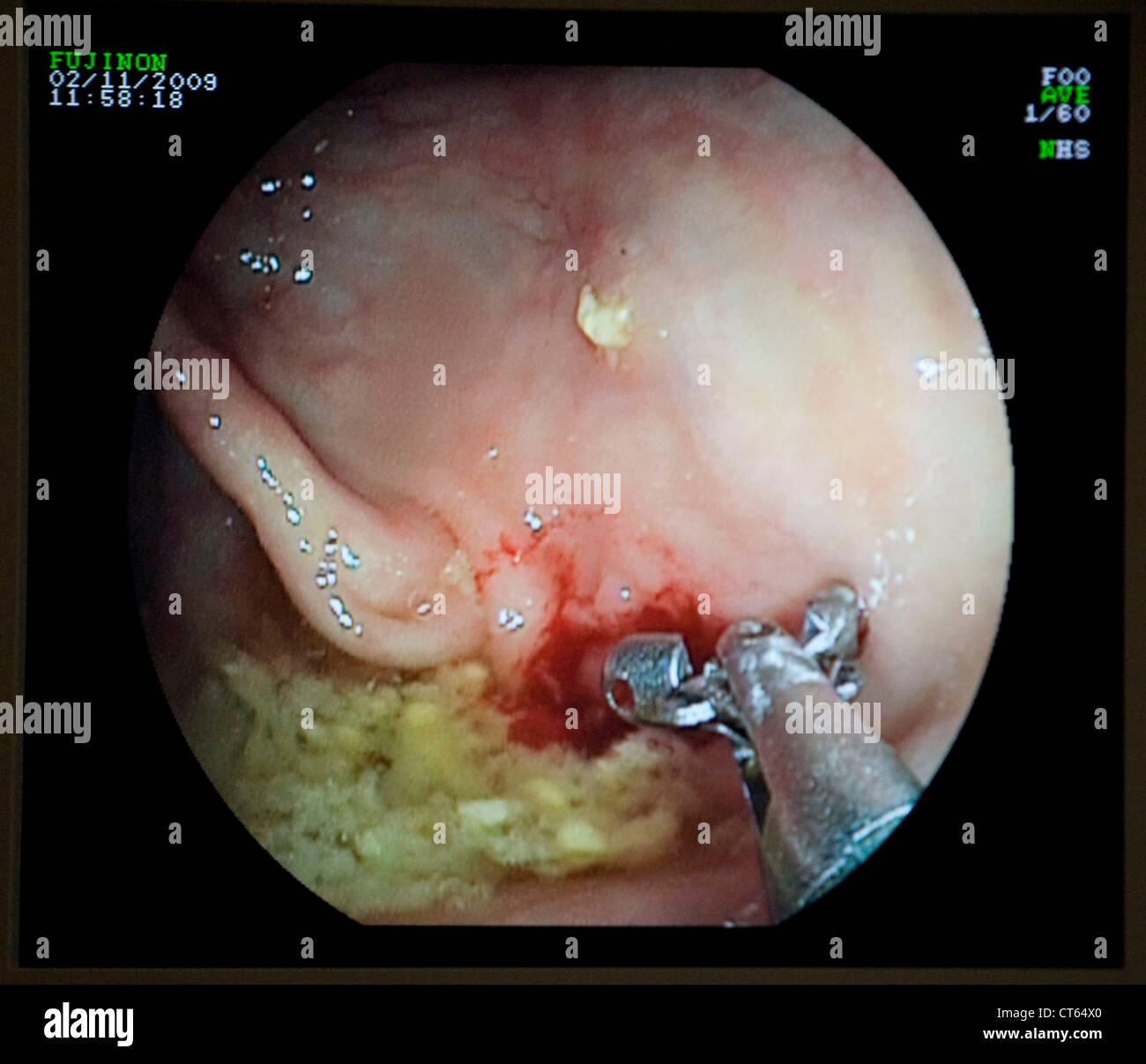 COLON POLYP REMOVAL, ENDOSCOPY Stock Photohttps://www.alamy.com/image-license-details/?v=1https://www.alamy.com/stock-photo-colon-polyp-removal-endoscopy-49308056.html
RMCT64X0–COLON POLYP REMOVAL, ENDOSCOPY
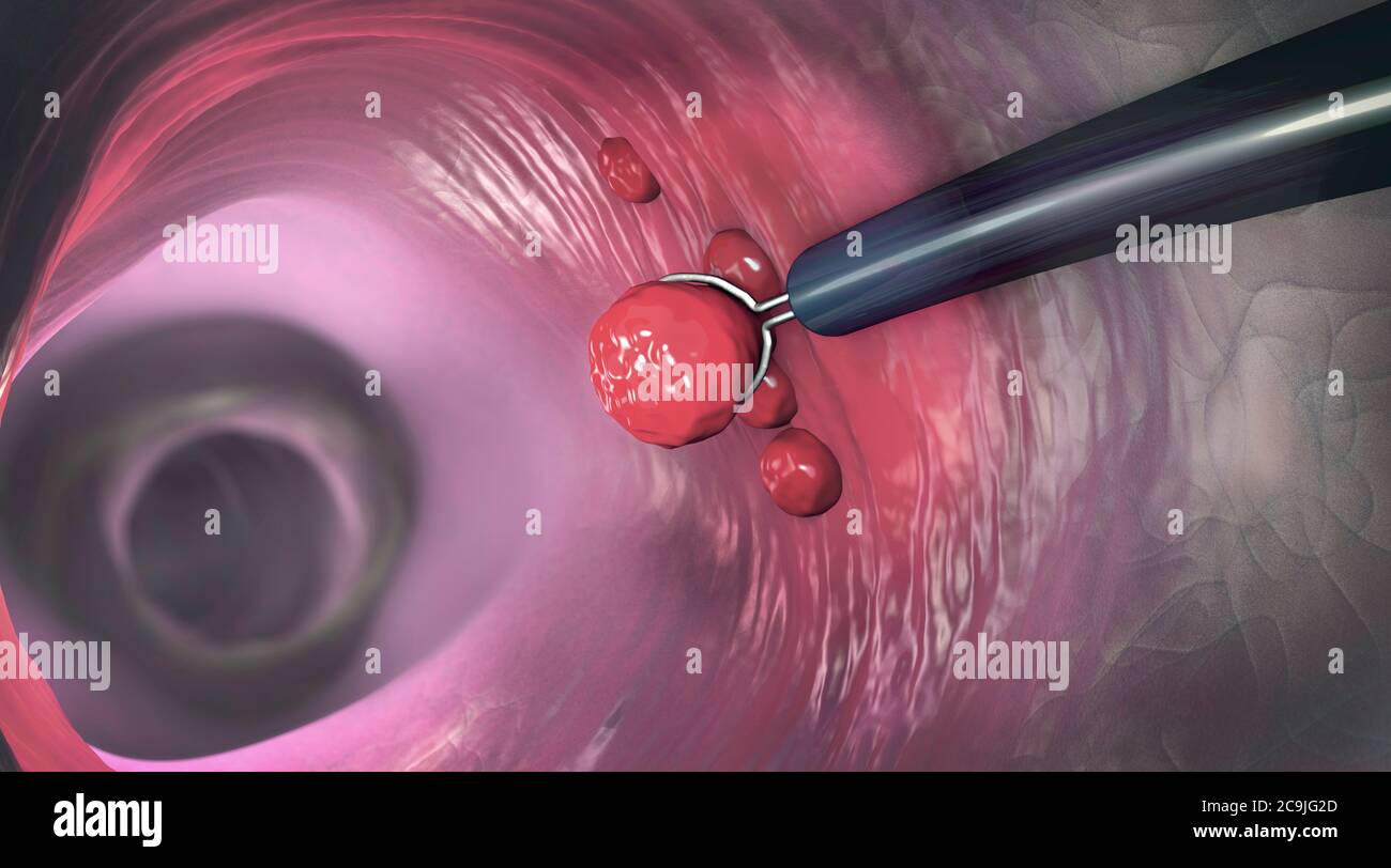 Polyp removal, illustration. Removal of a colonic polyp with an electrical wire loop during a colonoscopy. Stock Photohttps://www.alamy.com/image-license-details/?v=1https://www.alamy.com/polyp-removal-illustration-removal-of-a-colonic-polyp-with-an-electrical-wire-loop-during-a-colonoscopy-image367357381.html
RF2C9JG2D–Polyp removal, illustration. Removal of a colonic polyp with an electrical wire loop during a colonoscopy.
 COLON POLYP REMOVAL, ENDOSCOPY Stock Photohttps://www.alamy.com/image-license-details/?v=1https://www.alamy.com/stock-photo-colon-polyp-removal-endoscopy-49272287.html
RMCT4F8F–COLON POLYP REMOVAL, ENDOSCOPY
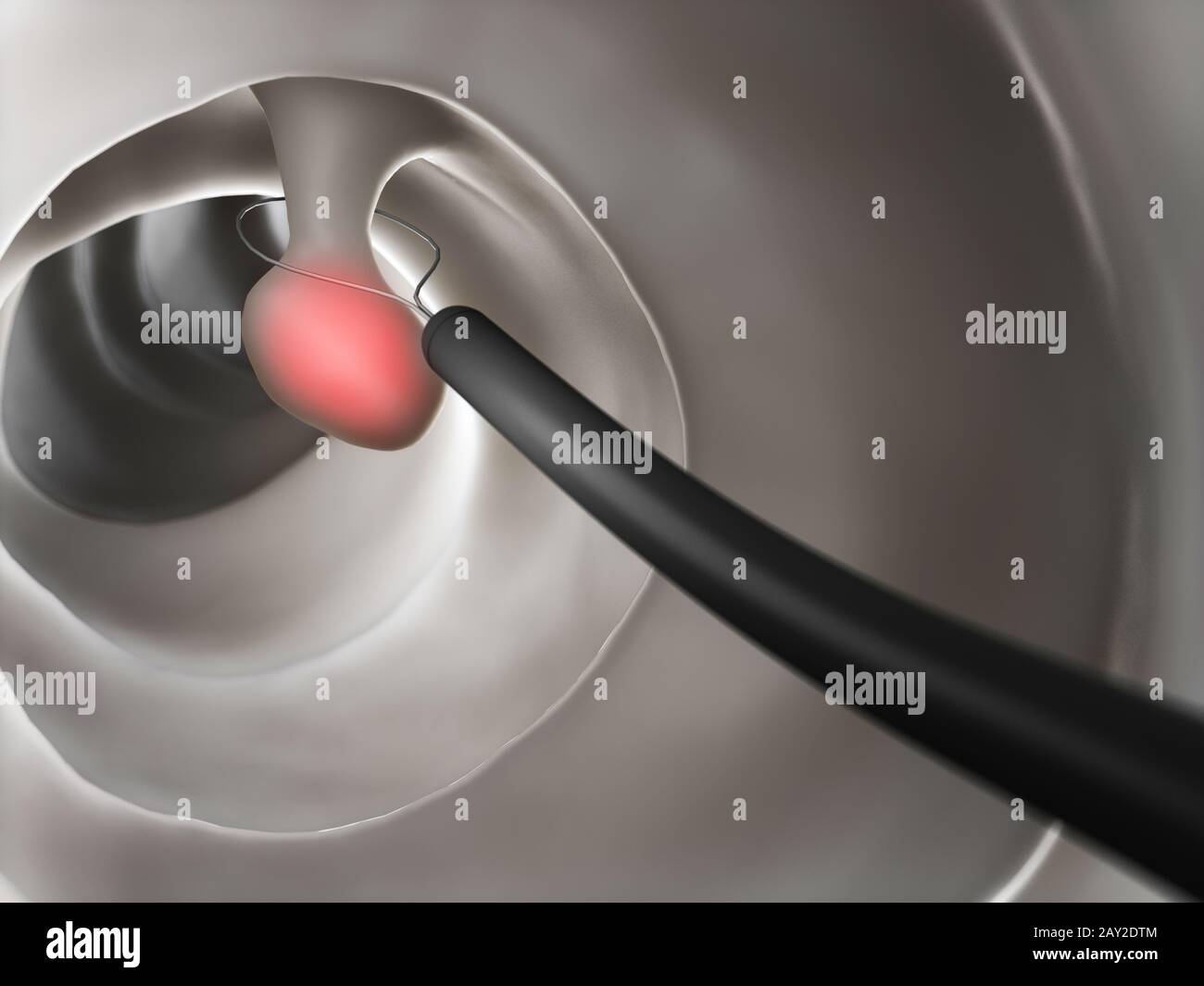 3d rendered illustration of a polyp removal Stock Photohttps://www.alamy.com/image-license-details/?v=1https://www.alamy.com/3d-rendered-illustration-of-a-polyp-removal-image343647492.html
RM2AY2DTM–3d rendered illustration of a polyp removal
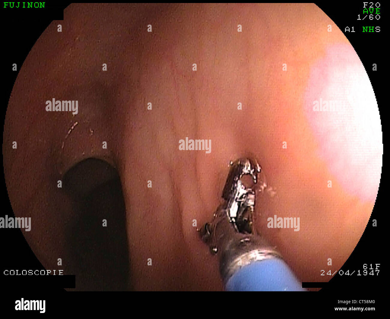 COLON POLYP REMOVAL, ENDOSCOPY Stock Photohttps://www.alamy.com/image-license-details/?v=1https://www.alamy.com/stock-photo-colon-polyp-removal-endoscopy-49289072.html
RMCT58M0–COLON POLYP REMOVAL, ENDOSCOPY
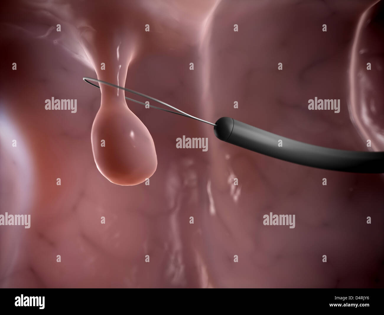 Polyp removal Stock Photohttps://www.alamy.com/image-license-details/?v=1https://www.alamy.com/stock-photo-polyp-removal-54609498.html
RFD4RJY6–Polyp removal
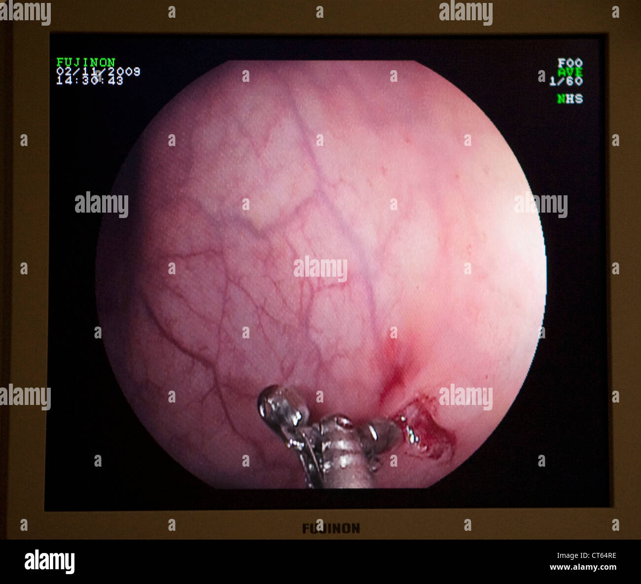 COLON POLYP REMOVAL, ENDOSCOPY Stock Photohttps://www.alamy.com/image-license-details/?v=1https://www.alamy.com/stock-photo-colon-polyp-removal-endoscopy-49307986.html
RMCT64RE–COLON POLYP REMOVAL, ENDOSCOPY
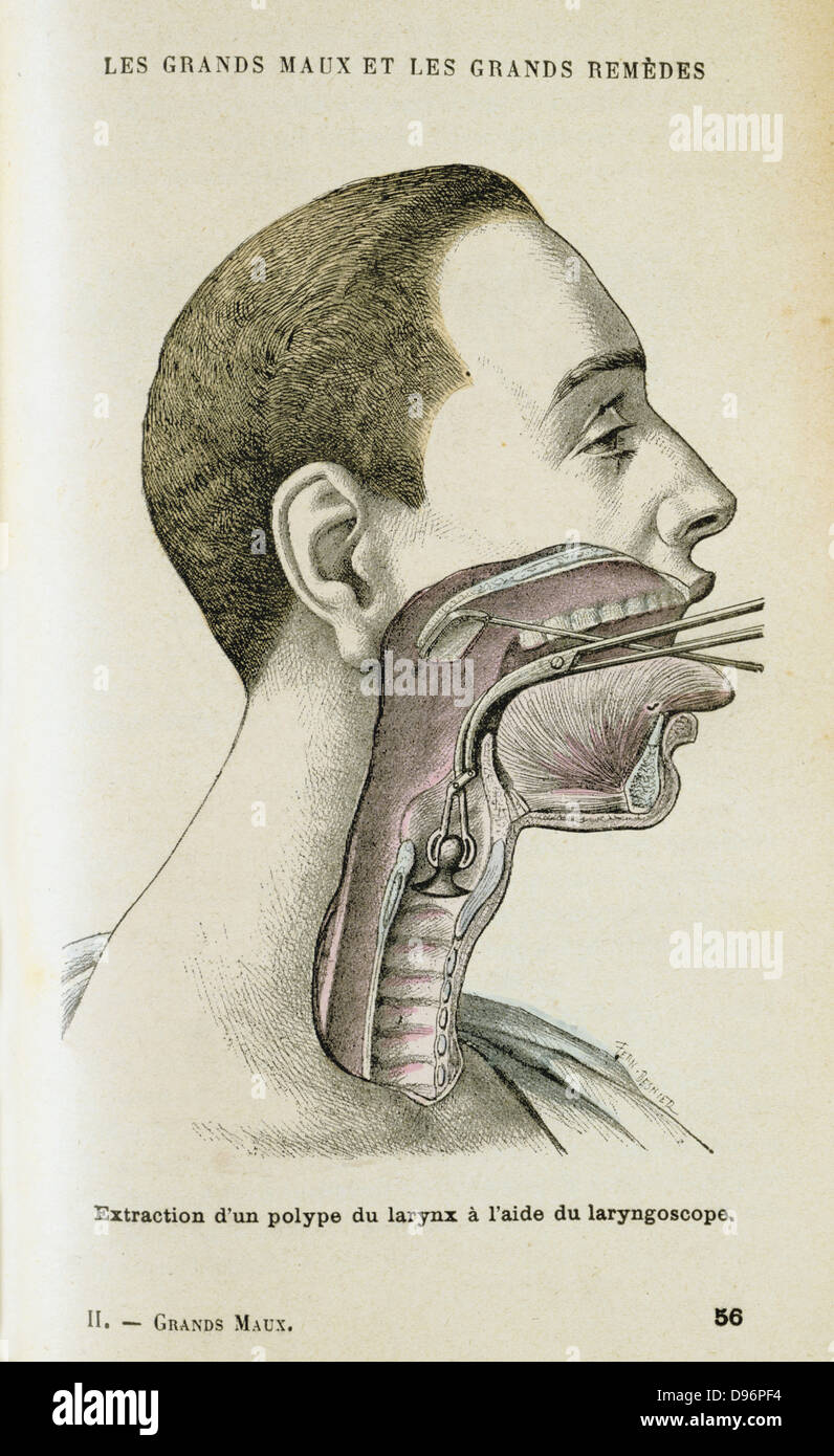 Using a laryngoscope to aid a surgeon in the removal of a polyp from the throat, c1890. A small mirror on a long metal handle was invented in 1854 by a Spanish singing teacher Manuel Garcia (1805)- 1906) to study the vocal chords. In 1858 Ludwig Turck (1810-1868) and Johann Czermak (1828-1873) both published papers on use of the instrument, but Czermak invented a headband mirror to throw light on Laryngoscope mirror, an so on the throat. From 'Les Grands Maux et les Grands Remedes' ('The Principal Illnesses and Their Remedies'), Jules Rengade, (Paris, c1890). Stock Photohttps://www.alamy.com/image-license-details/?v=1https://www.alamy.com/stock-photo-using-a-laryngoscope-to-aid-a-surgeon-in-the-removal-of-a-polyp-from-57312392.html
RMD96PF4–Using a laryngoscope to aid a surgeon in the removal of a polyp from the throat, c1890. A small mirror on a long metal handle was invented in 1854 by a Spanish singing teacher Manuel Garcia (1805)- 1906) to study the vocal chords. In 1858 Ludwig Turck (1810-1868) and Johann Czermak (1828-1873) both published papers on use of the instrument, but Czermak invented a headband mirror to throw light on Laryngoscope mirror, an so on the throat. From 'Les Grands Maux et les Grands Remedes' ('The Principal Illnesses and Their Remedies'), Jules Rengade, (Paris, c1890).
 U.S. President Lyndon Johnson meeting with the press following throat polyp removal surgery, National Naval Medical Center, Bethesda, Maryland, USA, Robert Knudsen, November 16, 1966 Stock Photohttps://www.alamy.com/image-license-details/?v=1https://www.alamy.com/us-president-lyndon-johnson-meeting-with-the-press-following-throat-polyp-removal-surgery-national-naval-medical-center-bethesda-maryland-usa-robert-knudsen-november-16-1966-image617554766.html
RM2XTM1BA–U.S. President Lyndon Johnson meeting with the press following throat polyp removal surgery, National Naval Medical Center, Bethesda, Maryland, USA, Robert Knudsen, November 16, 1966
 Removal of a colonic polyp with a electrical wire loop during a colonoscopy - 3d illustration Stock Photohttps://www.alamy.com/image-license-details/?v=1https://www.alamy.com/removal-of-a-colonic-polyp-with-a-electrical-wire-loop-during-a-colonoscopy-3d-illustration-image362157694.html
RF2C15KRA–Removal of a colonic polyp with a electrical wire loop during a colonoscopy - 3d illustration
 Colorectal cancer, malignant tumor in intestine, Endoscope inside colonoscopy, gut intestine, Colon polyp removal, colonic polyps search, Polypectomy, Stock Photohttps://www.alamy.com/image-license-details/?v=1https://www.alamy.com/colorectal-cancer-malignant-tumor-in-intestine-endoscope-inside-colonoscopy-gut-intestine-colon-polyp-removal-colonic-polyps-search-polypectomy-image571217628.html
RM2T595WG–Colorectal cancer, malignant tumor in intestine, Endoscope inside colonoscopy, gut intestine, Colon polyp removal, colonic polyps search, Polypectomy,
 Removal of a colonic polyp with a electrical wire loop during a colonoscopy - 3d illustration Stock Photohttps://www.alamy.com/image-license-details/?v=1https://www.alamy.com/removal-of-a-colonic-polyp-with-a-electrical-wire-loop-during-a-colonoscopy-3d-illustration-image362157690.html
RF2C15KR6–Removal of a colonic polyp with a electrical wire loop during a colonoscopy - 3d illustration
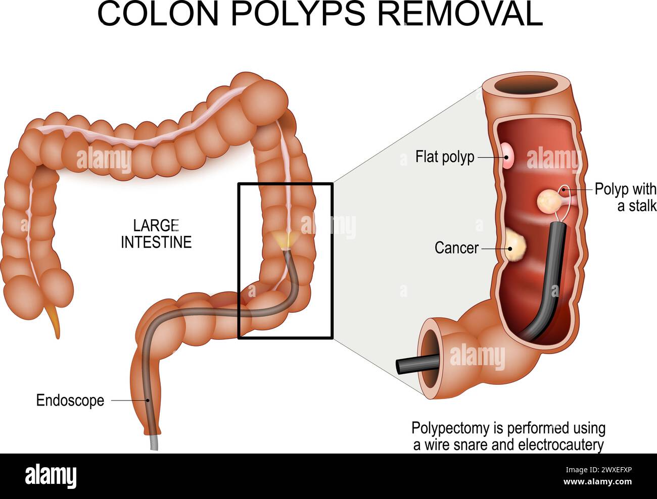 Colon polyps removal. Colonoscopy and Polypectomy. Colon cancer prevention. Human large intestine with Endoscope. Close-up of a cross section part of Stock Vectorhttps://www.alamy.com/image-license-details/?v=1https://www.alamy.com/colon-polyps-removal-colonoscopy-and-polypectomy-colon-cancer-prevention-human-large-intestine-with-endoscope-close-up-of-a-cross-section-part-of-image601453406.html
RF2WXEFXP–Colon polyps removal. Colonoscopy and Polypectomy. Colon cancer prevention. Human large intestine with Endoscope. Close-up of a cross section part of
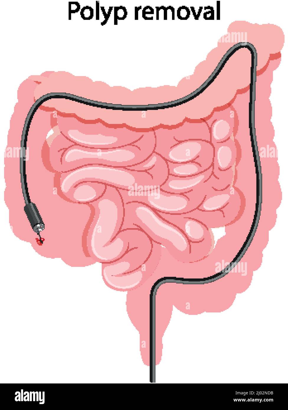 Diagram showing polyp removal illustration Stock Vectorhttps://www.alamy.com/image-license-details/?v=1https://www.alamy.com/diagram-showing-polyp-removal-illustration-image464740679.html
RF2J02NDB–Diagram showing polyp removal illustration
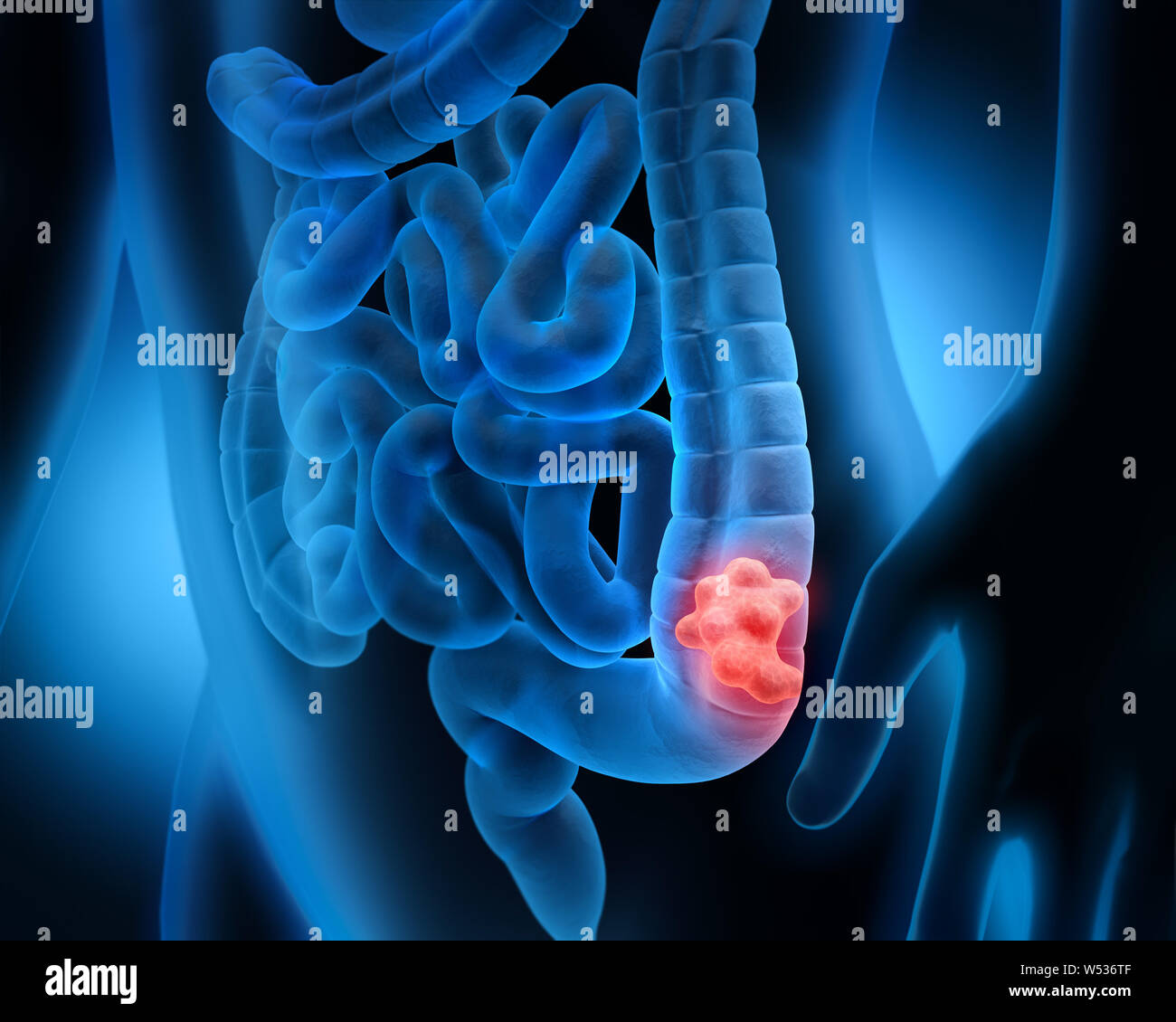 Body with intestinal polyp - 3D illustration Stock Photohttps://www.alamy.com/image-license-details/?v=1https://www.alamy.com/body-with-intestinal-polyp-3d-illustration-image261300047.html
RFW536TF–Body with intestinal polyp - 3D illustration
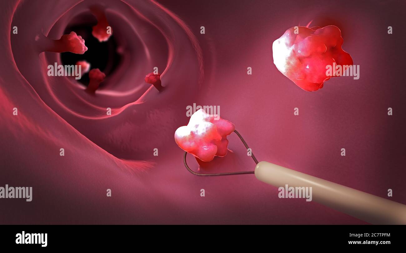 Removal of a colonic polyp with a electrical wire loop during a colonoscopy - 3d illustration Stock Photohttps://www.alamy.com/image-license-details/?v=1https://www.alamy.com/removal-of-a-colonic-polyp-with-a-electrical-wire-loop-during-a-colonoscopy-3d-illustration-image366264856.html
RF2C7TPFM–Removal of a colonic polyp with a electrical wire loop during a colonoscopy - 3d illustration
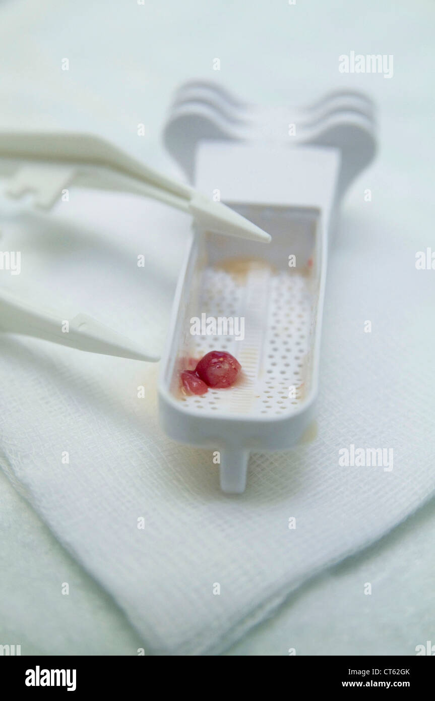 POLYP IN THE COLON, ANATOMY Stock Photohttps://www.alamy.com/image-license-details/?v=1https://www.alamy.com/stock-photo-polyp-in-the-colon-anatomy-49306227.html
RMCT62GK–POLYP IN THE COLON, ANATOMY
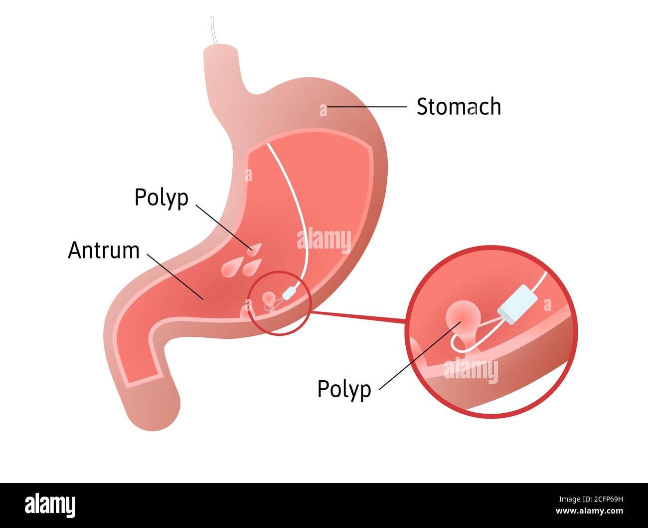 Removal of gastric polyps, masses of cells inside stomach. pedunculated and flat-based polyp. Antrum. Medical vector illustration marked with lines. Stock Vectorhttps://www.alamy.com/image-license-details/?v=1https://www.alamy.com/removal-of-gastric-polyps-masses-of-cells-inside-stomach-pedunculated-and-flat-based-polyp-antrum-medical-vector-illustration-marked-with-lines-image371125485.html
RF2CFP69H–Removal of gastric polyps, masses of cells inside stomach. pedunculated and flat-based polyp. Antrum. Medical vector illustration marked with lines.
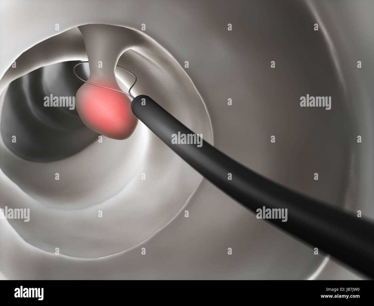 3d rendered illustration of a polyp removal Stock Photohttps://www.alamy.com/image-license-details/?v=1https://www.alamy.com/stock-photo-3d-rendered-illustration-of-a-polyp-removal-144612636.html
RFJB7JW0–3d rendered illustration of a polyp removal
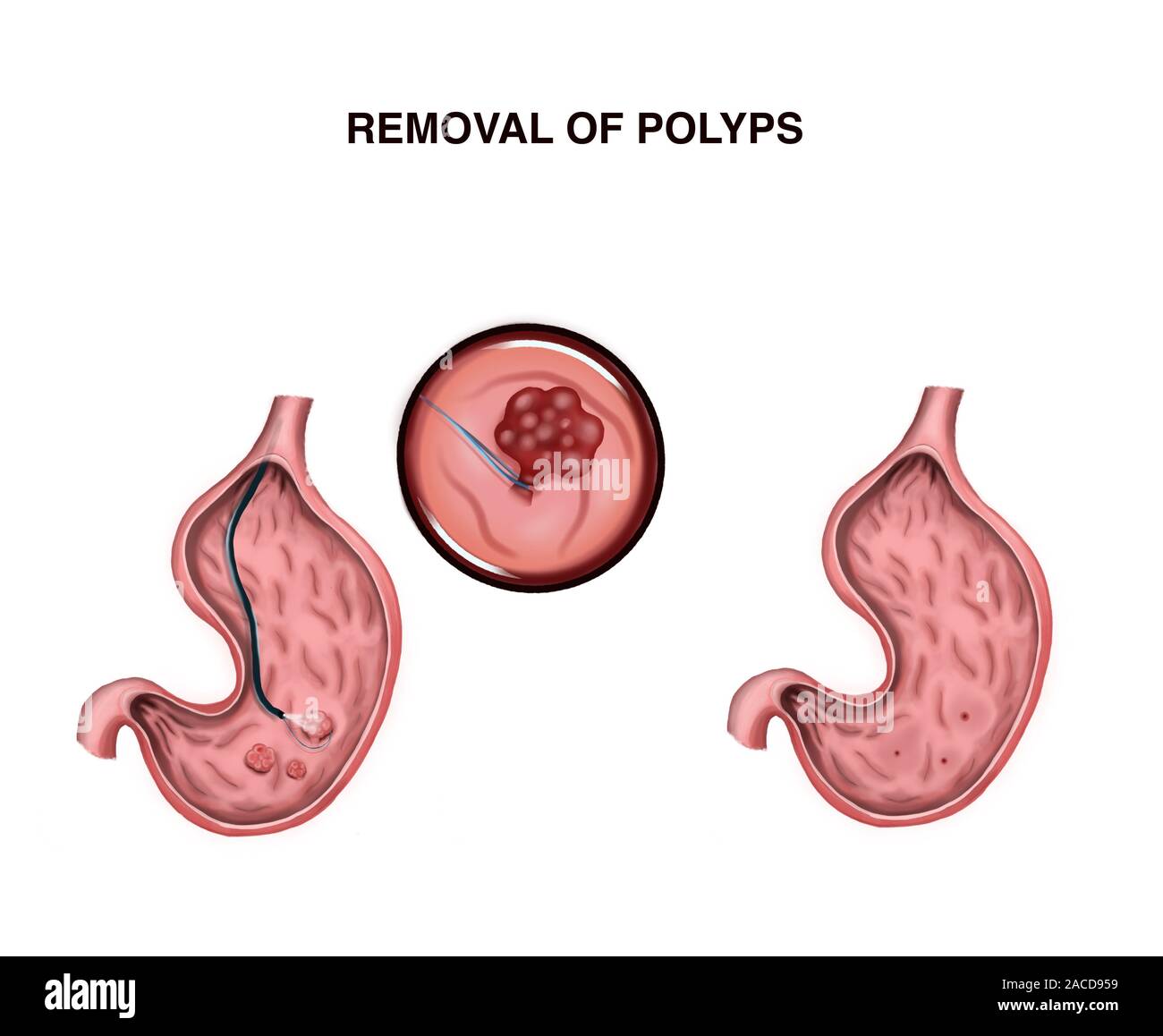 Illustration of removal of stomach polyps Stock Photohttps://www.alamy.com/image-license-details/?v=1https://www.alamy.com/illustration-of-removal-of-stomach-polyps-image334665445.html
RF2ACD959–Illustration of removal of stomach polyps
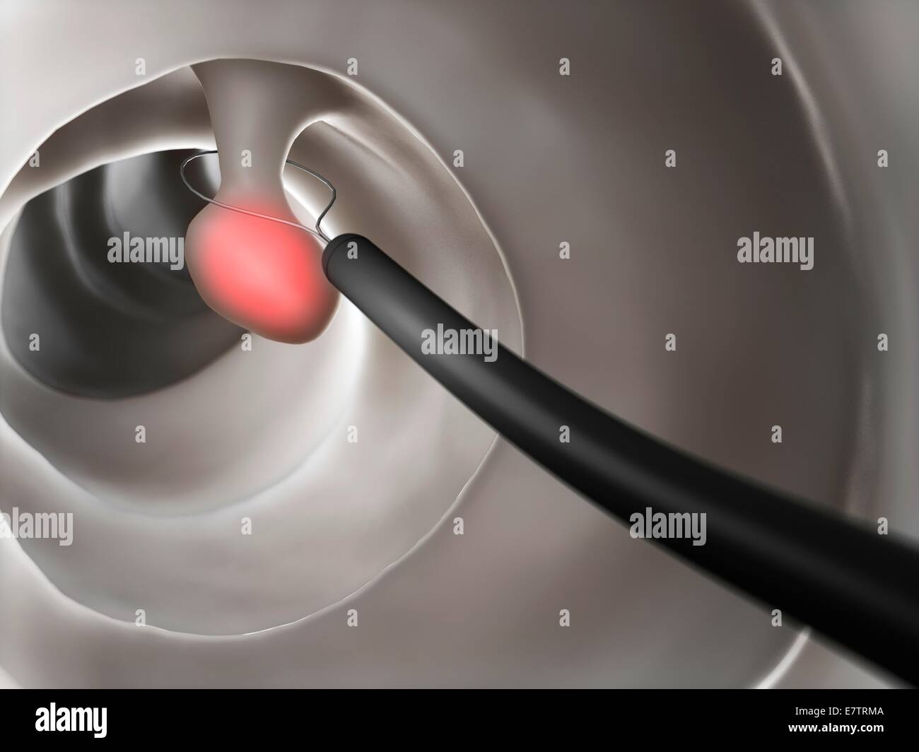
 Polyp removal, illustration. Removal of a colonic polyp with an electrical wire loop during a colonoscopy. Stock Photohttps://www.alamy.com/image-license-details/?v=1https://www.alamy.com/polyp-removal-illustration-removal-of-a-colonic-polyp-with-an-electrical-wire-loop-during-a-colonoscopy-image367357546.html
RF2C9JG8A–Polyp removal, illustration. Removal of a colonic polyp with an electrical wire loop during a colonoscopy.
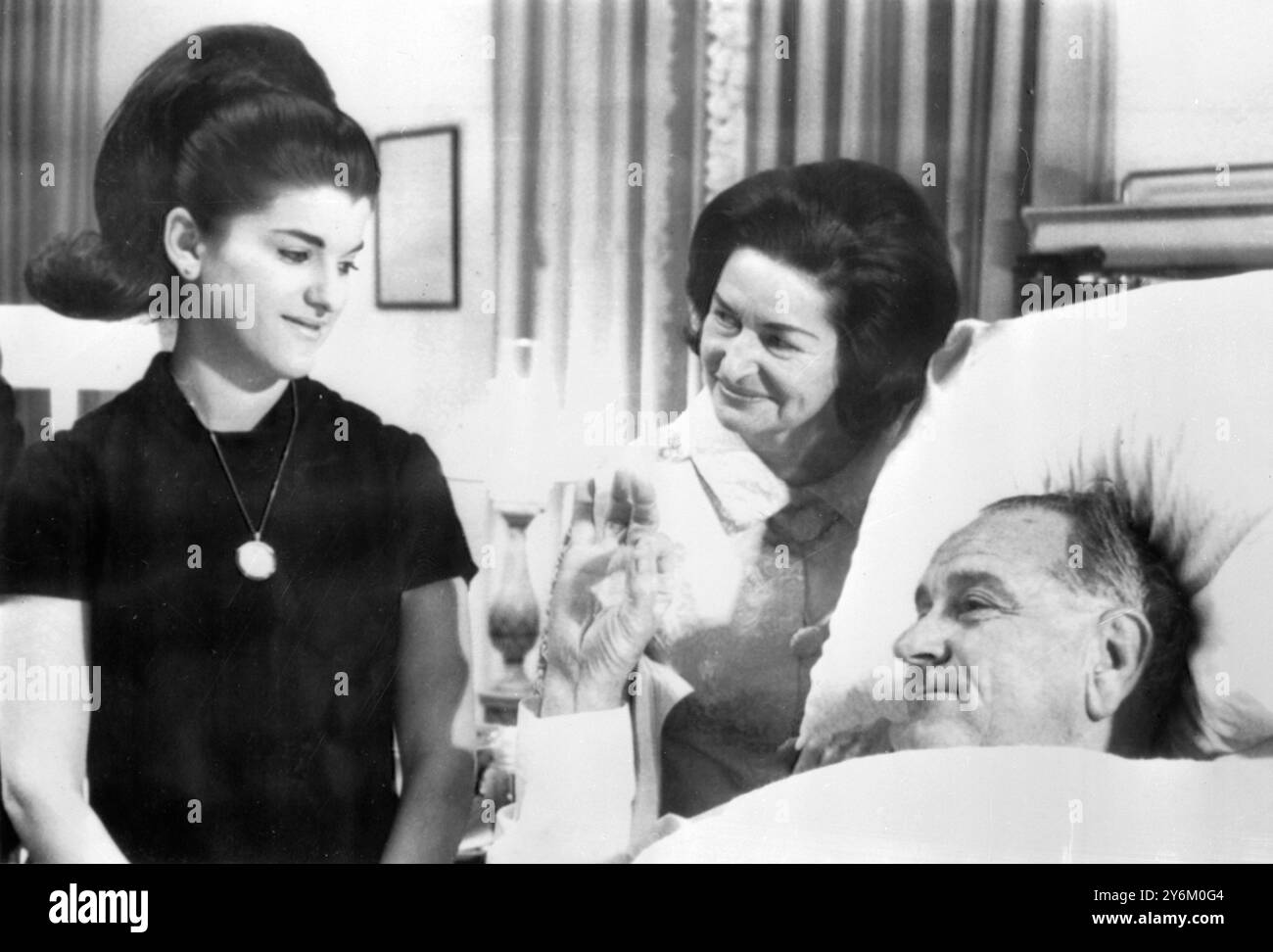 Bethesda Naval Hospital, Washington: The United States President Lyndon B. Johnson gives the 'O.K.' sign to this wife Mrs Claudia 'Lady Bird' Johnson (centre) and daughter Luci Baines Johnson after he was successfully operated on today, November 16, for the removal of a non-cancerous polyp in his throat, and correction of an abdominal hernia. The President, who is 58, is expected to be allowed to get out of bed and walk in the immediate future. 16 November 1966 Stock Photohttps://www.alamy.com/image-license-details/?v=1https://www.alamy.com/bethesda-naval-hospital-washington-the-united-states-president-lyndon-b-johnson-gives-the-ok-sign-to-this-wife-mrs-claudia-lady-bird-johnson-centre-and-daughter-luci-baines-johnson-after-he-was-successfully-operated-on-today-november-16-for-the-removal-of-a-non-cancerous-polyp-in-his-throat-and-correction-of-an-abdominal-hernia-the-president-who-is-58-is-expected-to-be-allowed-to-get-out-of-bed-and-walk-in-the-immediate-future-16-november-1966-image623700676.html
RM2Y6M0G4–Bethesda Naval Hospital, Washington: The United States President Lyndon B. Johnson gives the 'O.K.' sign to this wife Mrs Claudia 'Lady Bird' Johnson (centre) and daughter Luci Baines Johnson after he was successfully operated on today, November 16, for the removal of a non-cancerous polyp in his throat, and correction of an abdominal hernia. The President, who is 58, is expected to be allowed to get out of bed and walk in the immediate future. 16 November 1966
 3d rendered illustration of a polyp removal Stock Photohttps://www.alamy.com/image-license-details/?v=1https://www.alamy.com/3d-rendered-illustration-of-a-polyp-removal-image343647699.html
RM2AY2E43–3d rendered illustration of a polyp removal
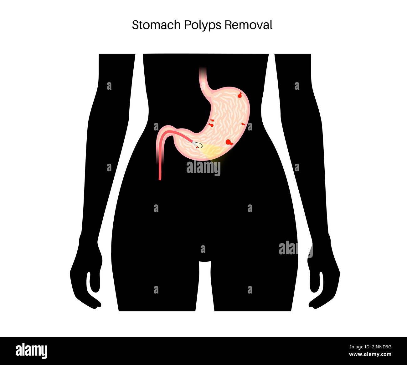 Stomach polyp removal, illustration Stock Photohttps://www.alamy.com/image-license-details/?v=1https://www.alamy.com/stomach-polyp-removal-illustration-image478058996.html
RF2JNND3G–Stomach polyp removal, illustration
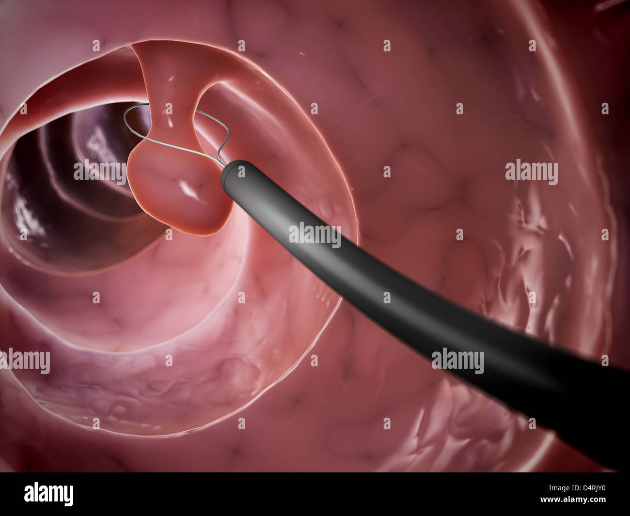 Polyp removal Stock Photohttps://www.alamy.com/image-license-details/?v=1https://www.alamy.com/stock-photo-polyp-removal-54609492.html
RFD4RJY0–Polyp removal
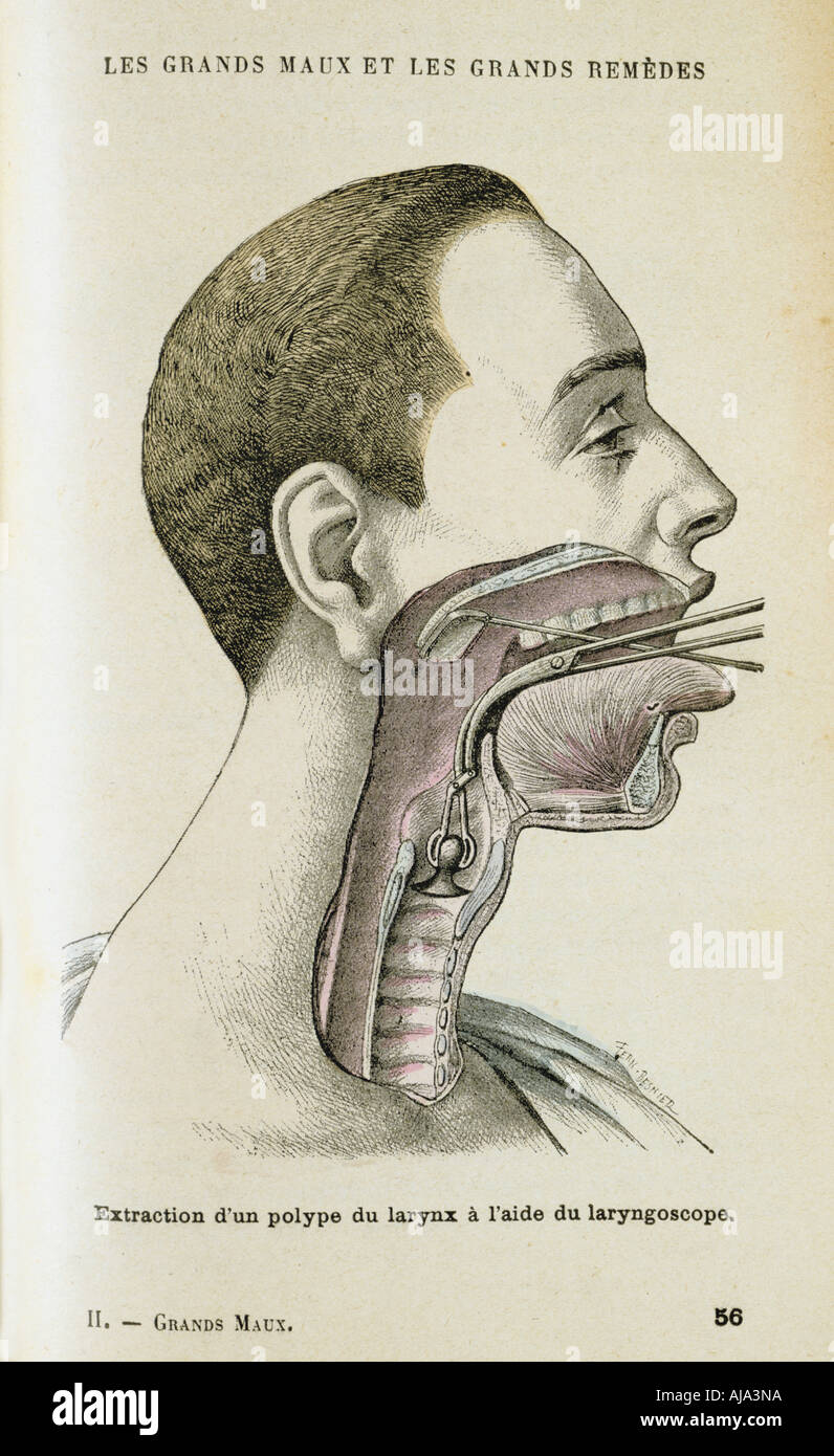 Using a laryngoscope to aid the removal of a polyp from the throat, c1890. Artist: Unknown Stock Photohttps://www.alamy.com/image-license-details/?v=1https://www.alamy.com/using-a-laryngoscope-to-aid-the-removal-of-a-polyp-from-the-throat-image8383641.html
RMAJA3NA–Using a laryngoscope to aid the removal of a polyp from the throat, c1890. Artist: Unknown
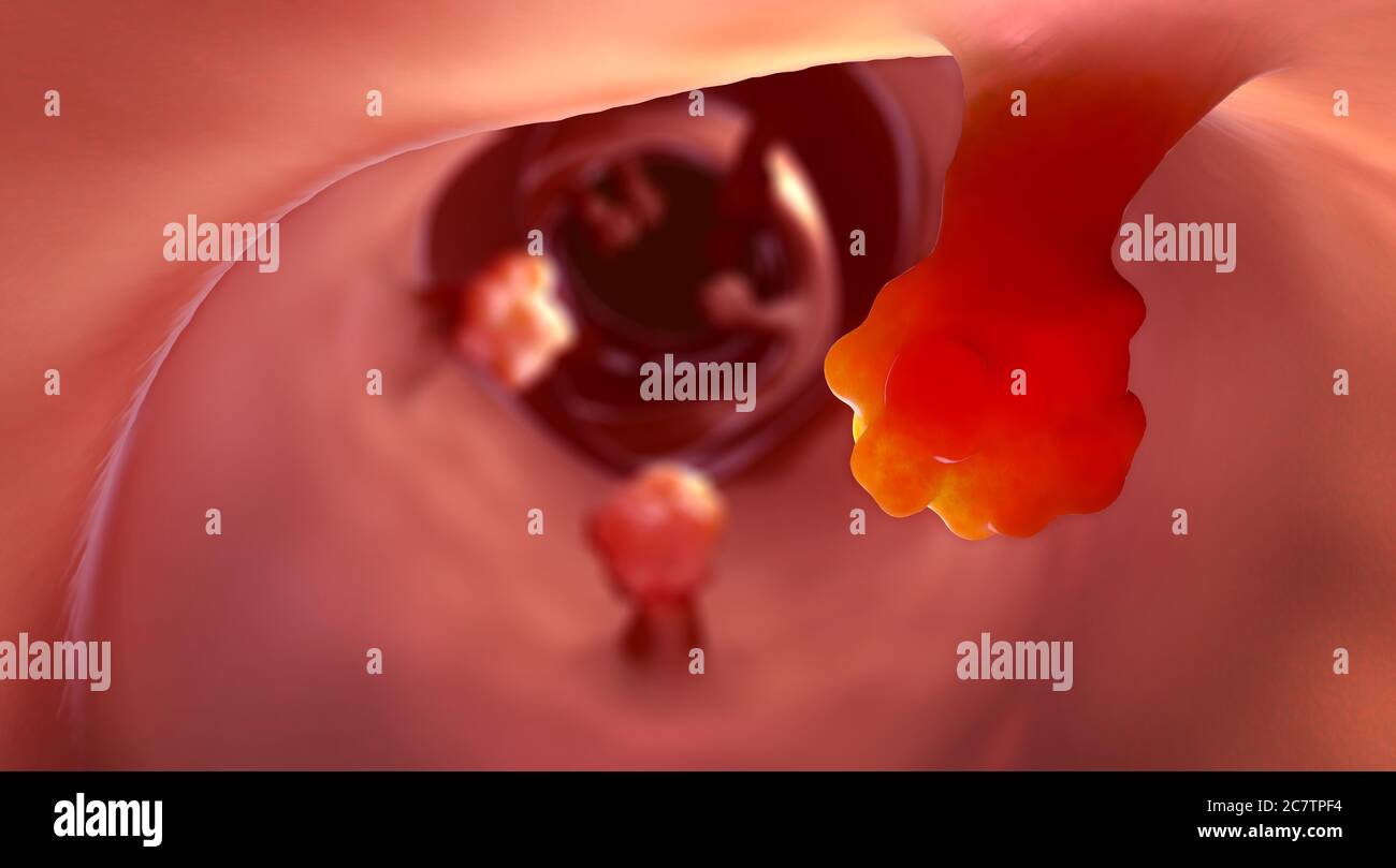 Close-up view of intestinal polyps and diseased intestinal tissue that can cause cancer - 3d illustration Stock Photohttps://www.alamy.com/image-license-details/?v=1https://www.alamy.com/close-up-view-of-intestinal-polyps-and-diseased-intestinal-tissue-that-can-cause-cancer-3d-illustration-image366264840.html
RF2C7TPF4–Close-up view of intestinal polyps and diseased intestinal tissue that can cause cancer - 3d illustration
 Paul Glynn looking on as U.S. President Lyndon Johnson giving OK sign from his hospital bed following throat polyp removal surgery, National Naval Medical Center, Bethesda, Maryland, USA, Robert Knudsen, November 16, 1966 Stock Photohttps://www.alamy.com/image-license-details/?v=1https://www.alamy.com/paul-glynn-looking-on-as-us-president-lyndon-johnson-giving-ok-sign-from-his-hospital-bed-following-throat-polyp-removal-surgery-national-naval-medical-center-bethesda-maryland-usa-robert-knudsen-november-16-1966-image617554765.html
RM2XTM1B9–Paul Glynn looking on as U.S. President Lyndon Johnson giving OK sign from his hospital bed following throat polyp removal surgery, National Naval Medical Center, Bethesda, Maryland, USA, Robert Knudsen, November 16, 1966
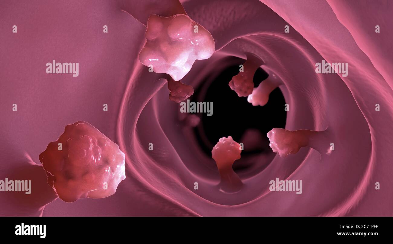 Close-up view of intestinal polyps and diseased intestinal tissue that can cause cancer Stock Photohttps://www.alamy.com/image-license-details/?v=1https://www.alamy.com/close-up-view-of-intestinal-polyps-and-diseased-intestinal-tissue-that-can-cause-cancer-image366264851.html
RF2C7TPFF–Close-up view of intestinal polyps and diseased intestinal tissue that can cause cancer
 Colorectal cancer, malignant tumor in intestine, Endoscope inside colonoscopy, gut intestine, Colon polyp removal, colonic polyps search, Polypectomy, Stock Photohttps://www.alamy.com/image-license-details/?v=1https://www.alamy.com/colorectal-cancer-malignant-tumor-in-intestine-endoscope-inside-colonoscopy-gut-intestine-colon-polyp-removal-colonic-polyps-search-polypectomy-image571218093.html
RM2T596E5–Colorectal cancer, malignant tumor in intestine, Endoscope inside colonoscopy, gut intestine, Colon polyp removal, colonic polyps search, Polypectomy,
 Photo before and after removal of large mole on woman's skin. Mole removal concept Stock Photohttps://www.alamy.com/image-license-details/?v=1https://www.alamy.com/photo-before-and-after-removal-of-large-mole-on-womans-skin-mole-removal-concept-image466772852.html
RF2J3B9F0–Photo before and after removal of large mole on woman's skin. Mole removal concept
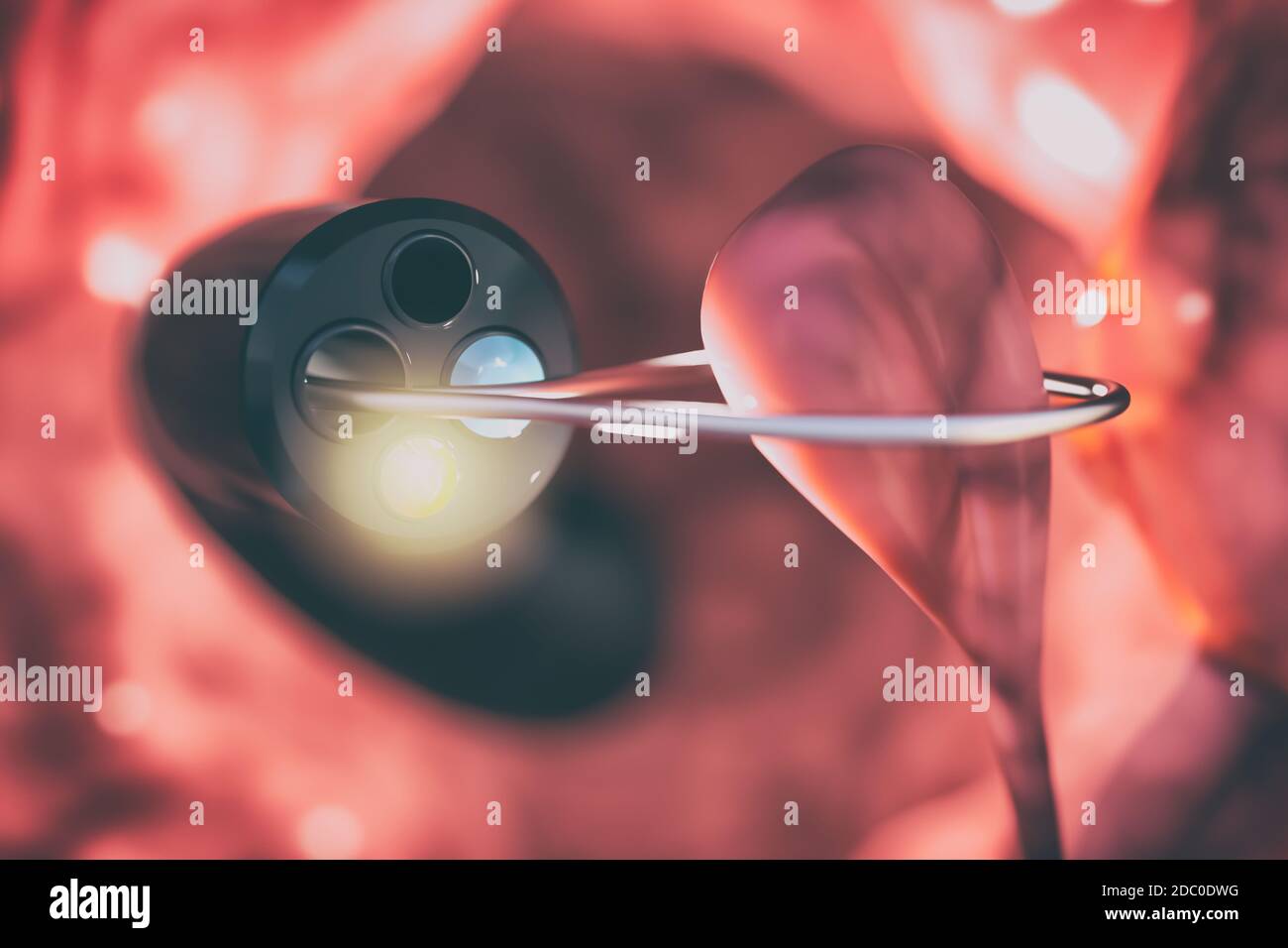 Removal of the polyp from the intestinal wall with a colonoscope. Medical device to check the condition of the intestines and detect gastrointestinal Stock Photohttps://www.alamy.com/image-license-details/?v=1https://www.alamy.com/removal-of-the-polyp-from-the-intestinal-wall-with-a-colonoscope-medical-device-to-check-the-condition-of-the-intestines-and-detect-gastrointestinal-image386014876.html
RF2DC0DWG–Removal of the polyp from the intestinal wall with a colonoscope. Medical device to check the condition of the intestines and detect gastrointestinal
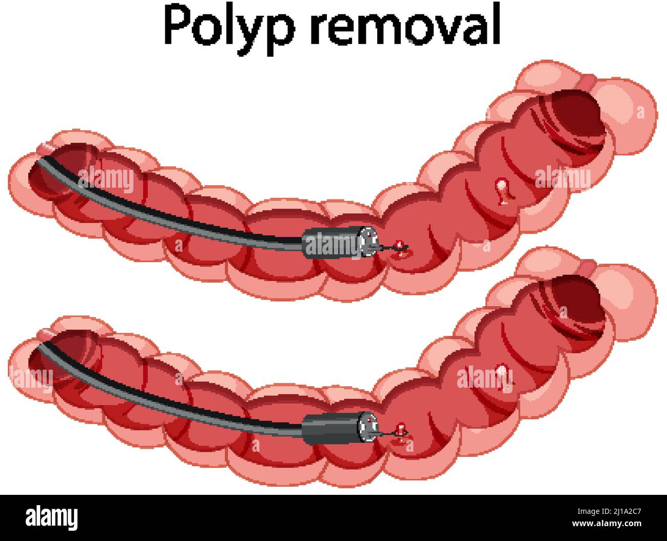 Diagram showing polyp removal illustration Stock Vectorhttps://www.alamy.com/image-license-details/?v=1https://www.alamy.com/diagram-showing-polyp-removal-illustration-image465516023.html
RF2J1A2C7–Diagram showing polyp removal illustration
 Photo before and after removal of large mole on woman's skin. Selective focus. Mole removal concept Stock Photohttps://www.alamy.com/image-license-details/?v=1https://www.alamy.com/photo-before-and-after-removal-of-large-mole-on-womans-skin-selective-focus-mole-removal-concept-image466772857.html
RF2J3B9F5–Photo before and after removal of large mole on woman's skin. Selective focus. Mole removal concept
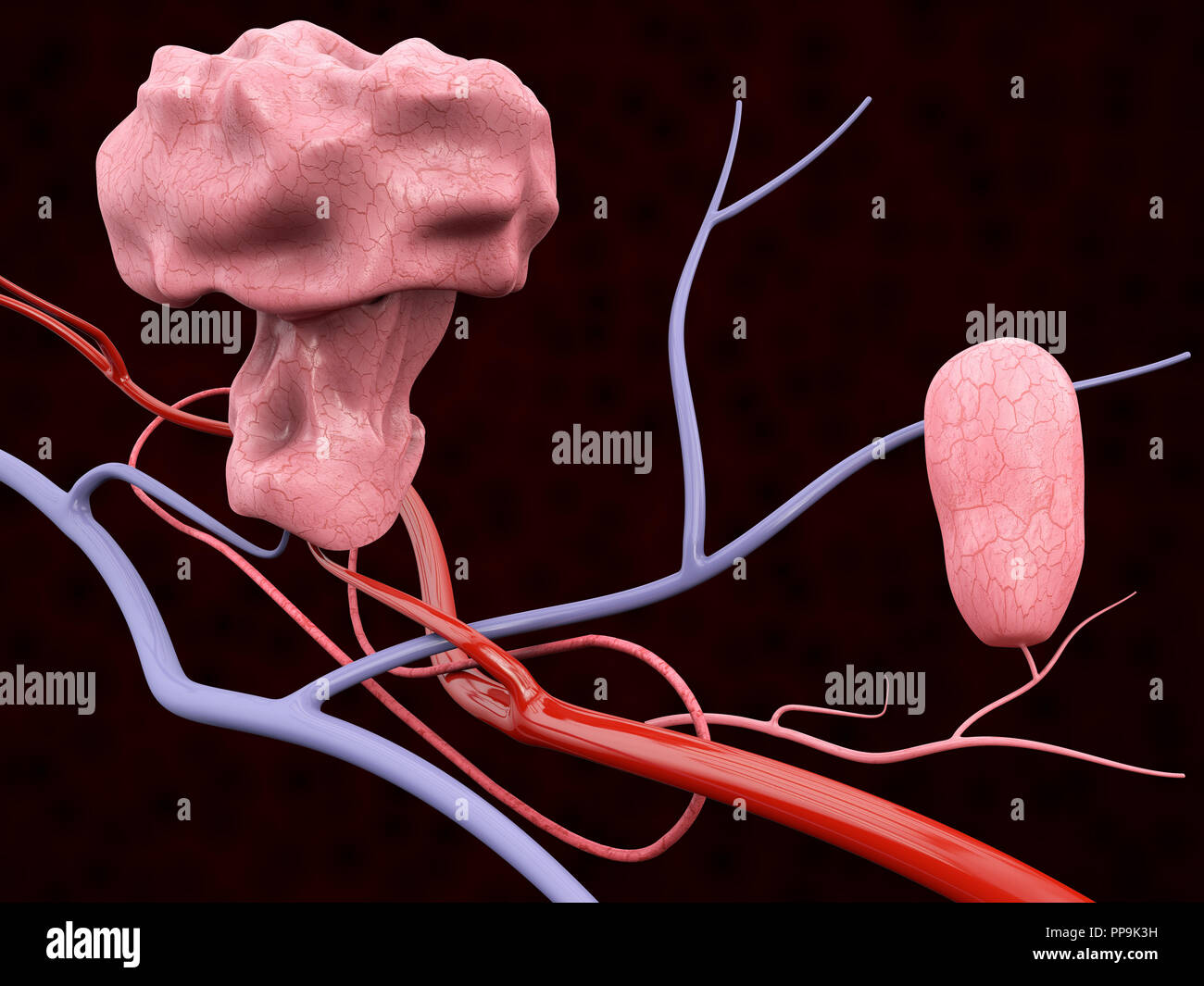 3d Illustration of Polyp with Blood Veins, isolated black background Stock Photohttps://www.alamy.com/image-license-details/?v=1https://www.alamy.com/3d-illustration-of-polyp-with-blood-veins-isolated-black-background-image220259413.html
RFPP9K3H–3d Illustration of Polyp with Blood Veins, isolated black background
 Photo Before and After removal of Large Mole on woman skin. Mole removal concept. Selective focus Stock Photohttps://www.alamy.com/image-license-details/?v=1https://www.alamy.com/photo-before-and-after-removal-of-large-mole-on-woman-skin-mole-removal-concept-selective-focus-image466772854.html
RF2J3B9F2–Photo Before and After removal of Large Mole on woman skin. Mole removal concept. Selective focus
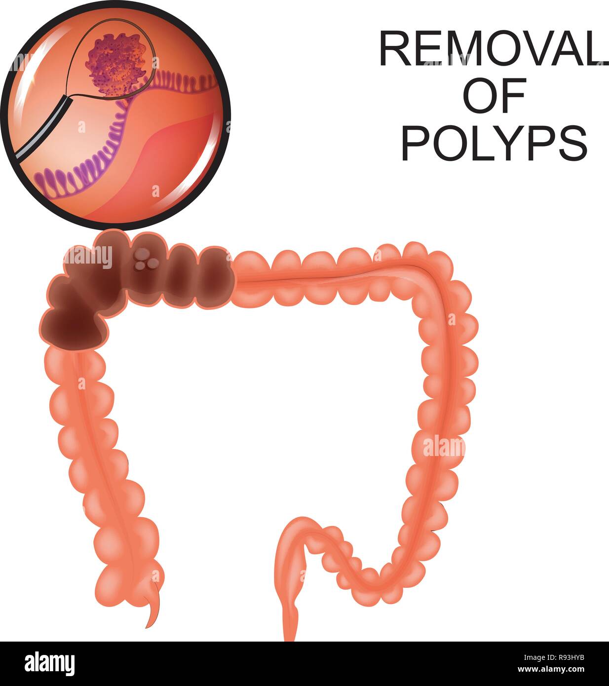 vector illustration of polyps in the colon. removal of polyps Stock Vectorhttps://www.alamy.com/image-license-details/?v=1https://www.alamy.com/vector-illustration-of-polyps-in-the-colon-removal-of-polyps-image229346639.html
RFR93HYB–vector illustration of polyps in the colon. removal of polyps
 3d rendered illustration of a polyp removal Stock Photohttps://www.alamy.com/image-license-details/?v=1https://www.alamy.com/stock-photo-3d-rendered-illustration-of-a-polyp-removal-144612638.html
RFJB7JW2–3d rendered illustration of a polyp removal
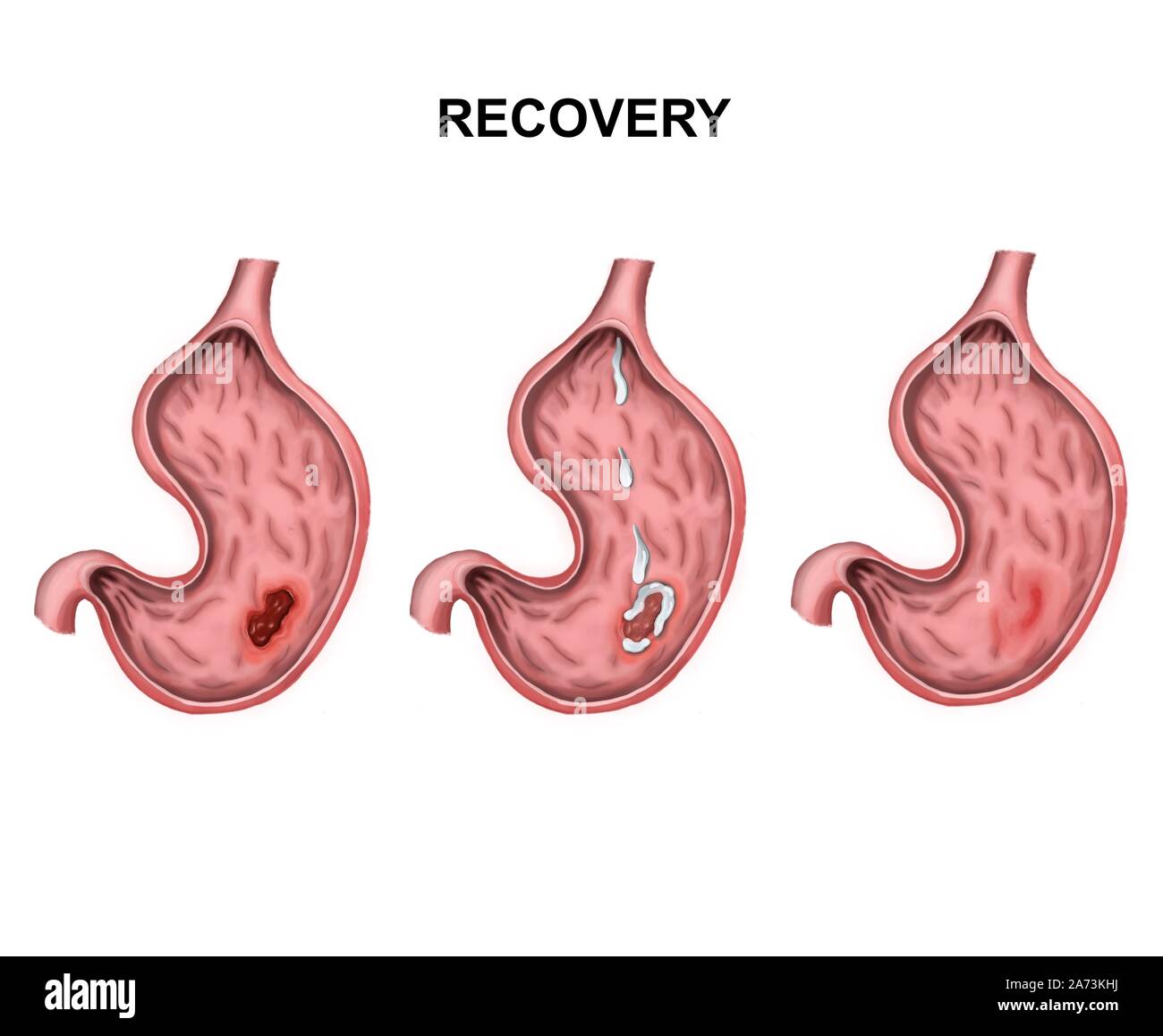 Recovery and healing of stomach ulcers Stock Photohttps://www.alamy.com/image-license-details/?v=1https://www.alamy.com/recovery-and-healing-of-stomach-ulcers-image331380830.html
RF2A73KHJ–Recovery and healing of stomach ulcers
 3d rendered illustration of a polyp removal Stock Photohttps://www.alamy.com/image-license-details/?v=1https://www.alamy.com/stock-photo-3d-rendered-illustration-of-a-polyp-removal-144604979.html
RFJB793F–3d rendered illustration of a polyp removal
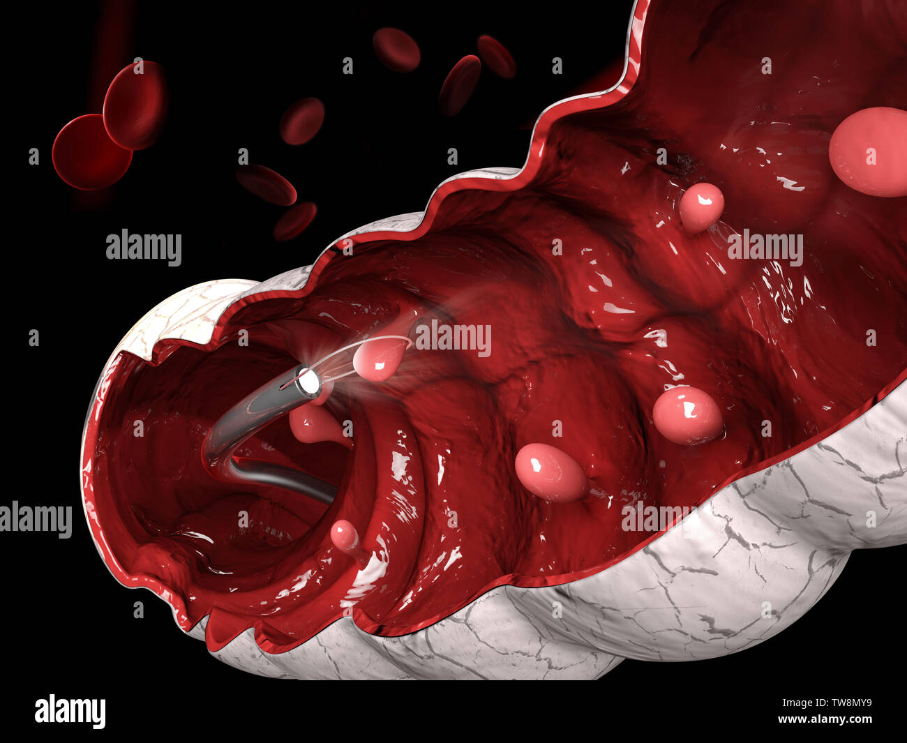 3d illustration of Removal of a colonic polyp with a electrical wire loop during a colonoscopy Stock Photohttps://www.alamy.com/image-license-details/?v=1https://www.alamy.com/3d-illustration-of-removal-of-a-colonic-polyp-with-a-electrical-wire-loop-during-a-colonoscopy-image256503613.html
RFTW8MY9–3d illustration of Removal of a colonic polyp with a electrical wire loop during a colonoscopy
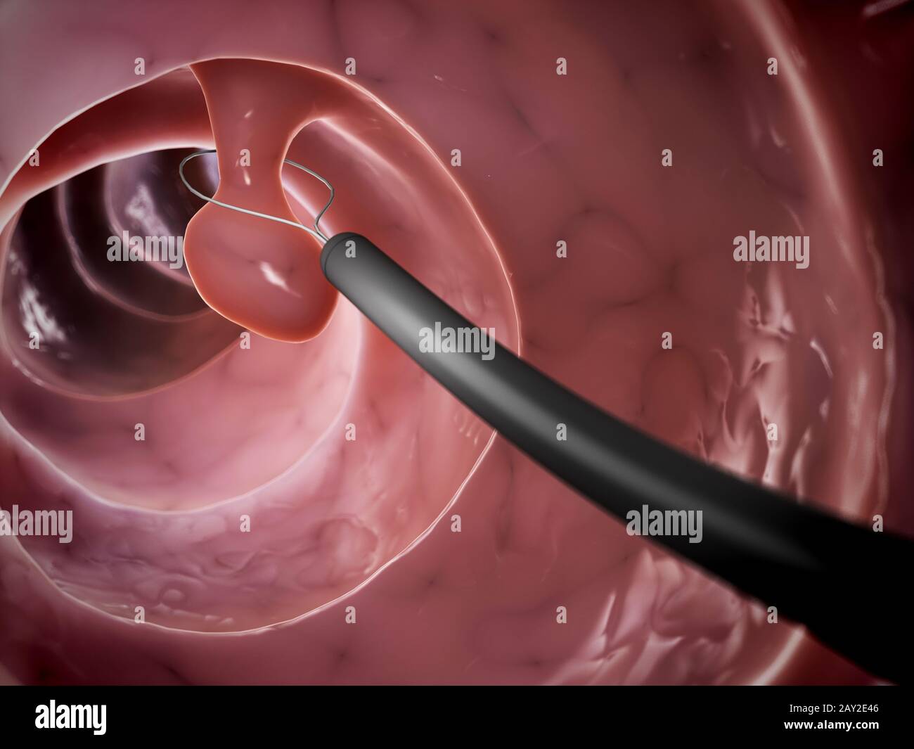 3d rendered illustration of a polyp removal Stock Photohttps://www.alamy.com/image-license-details/?v=1https://www.alamy.com/3d-rendered-illustration-of-a-polyp-removal-image343647702.html
RM2AY2E46–3d rendered illustration of a polyp removal
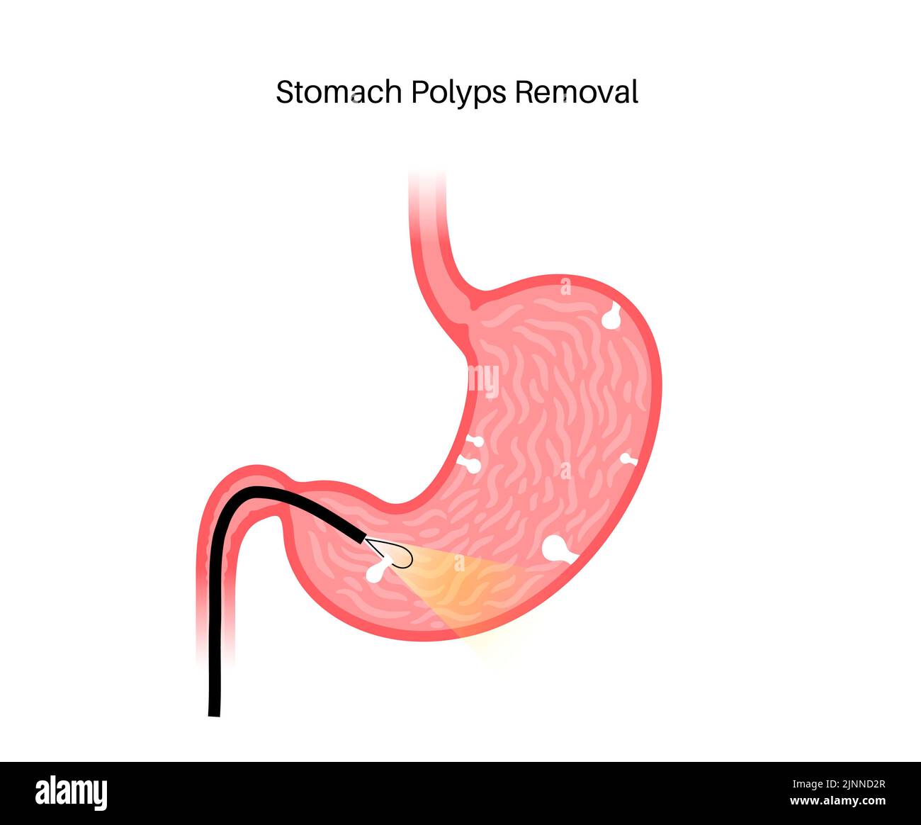 Stomach polyp removal, illustration Stock Photohttps://www.alamy.com/image-license-details/?v=1https://www.alamy.com/stomach-polyp-removal-illustration-image478058975.html
RF2JNND2R–Stomach polyp removal, illustration
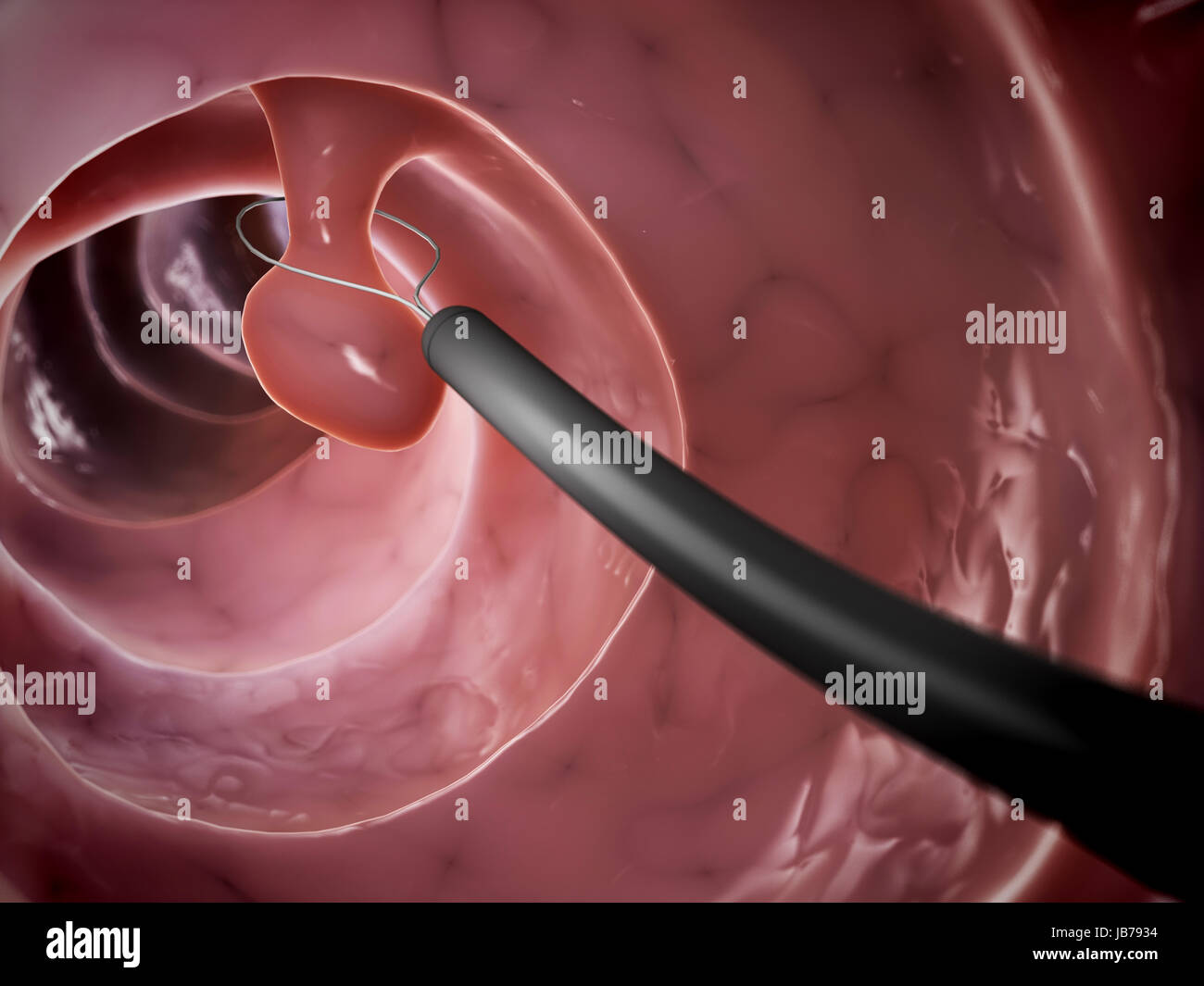 3d rendered illustration of a polyp removal Stock Photohttps://www.alamy.com/image-license-details/?v=1https://www.alamy.com/stock-photo-3d-rendered-illustration-of-a-polyp-removal-144604968.html
RFJB7934–3d rendered illustration of a polyp removal
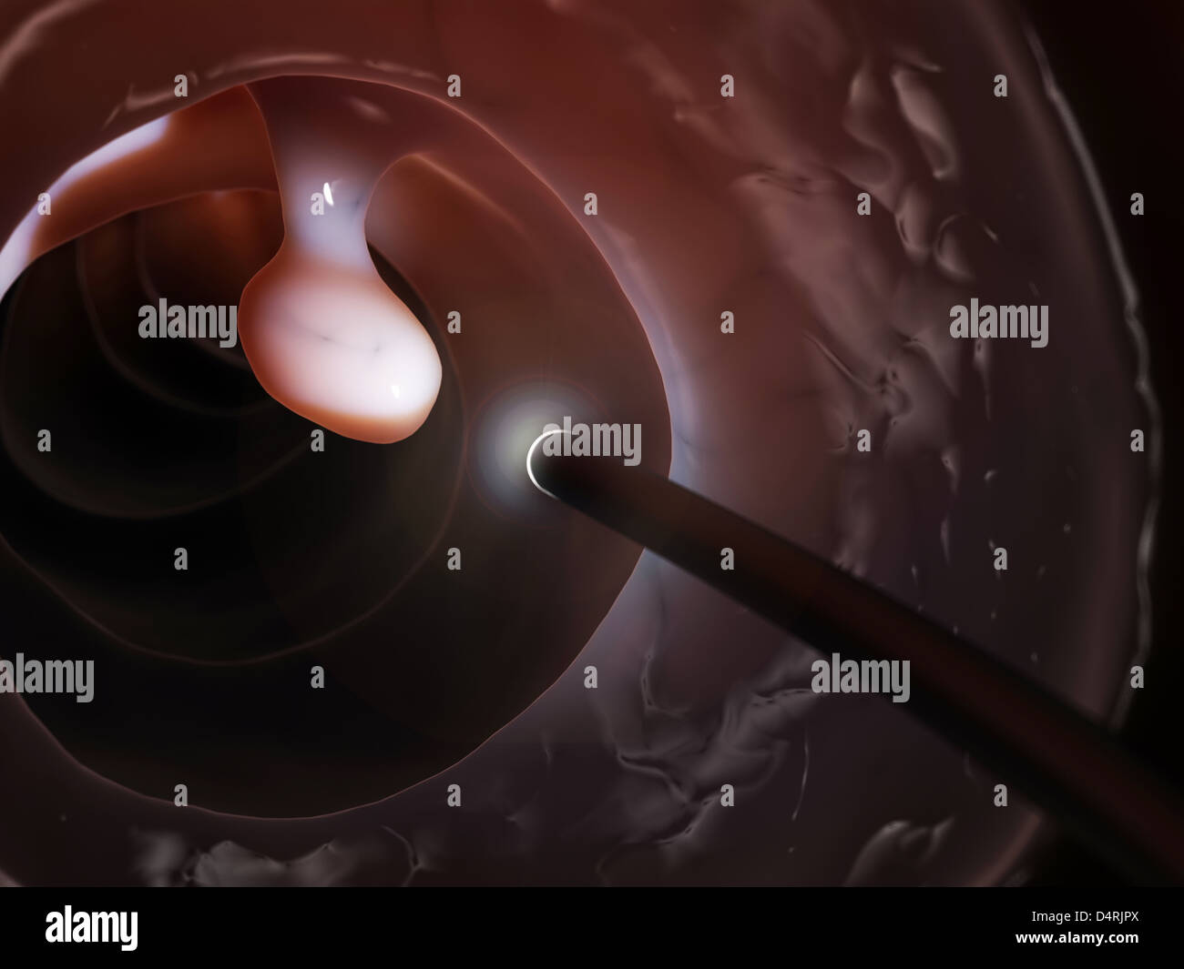 Colonoscopy Stock Photohttps://www.alamy.com/image-license-details/?v=1https://www.alamy.com/stock-photo-colonoscopy-54609378.html
RFD4RJPX–Colonoscopy
 Skin Tag, Acrochordon on Female's Head. Stock Photohttps://www.alamy.com/image-license-details/?v=1https://www.alamy.com/skin-tag-acrochordon-on-females-head-image398135461.html
RF2E3MHRH–Skin Tag, Acrochordon on Female's Head.
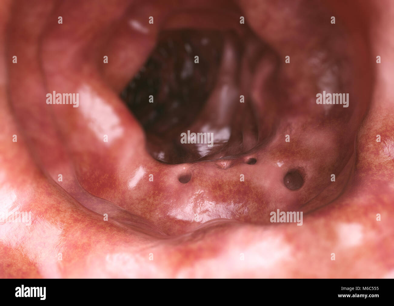 Intestine diverticula closeup - 3D Rendering Stock Photohttps://www.alamy.com/image-license-details/?v=1https://www.alamy.com/stock-photo-intestine-diverticula-closeup-3d-rendering-176059105.html
RFM6C555–Intestine diverticula closeup - 3D Rendering
 Photo Collage of different moles on human skin. Stock Photohttps://www.alamy.com/image-license-details/?v=1https://www.alamy.com/photo-collage-of-different-moles-on-human-skin-image453971935.html
RF2HAG5RB–Photo Collage of different moles on human skin.
 Colorectal cancer, malignant tumor in intestine, Endoscope inside colonoscopy, gut intestine, Colon polyp removal, colonic polyps search, Polypectomy, Stock Photohttps://www.alamy.com/image-license-details/?v=1https://www.alamy.com/colorectal-cancer-malignant-tumor-in-intestine-endoscope-inside-colonoscopy-gut-intestine-colon-polyp-removal-colonic-polyps-search-polypectomy-image571217884.html
RM2T5966M–Colorectal cancer, malignant tumor in intestine, Endoscope inside colonoscopy, gut intestine, Colon polyp removal, colonic polyps search, Polypectomy,
 Dentist doctor hand checking inside mouth polyps removal stitches or harmless spot Stock Photohttps://www.alamy.com/image-license-details/?v=1https://www.alamy.com/stock-image-dentist-doctor-hand-checking-inside-mouth-polyps-removal-stitches-166107204.html
RFKJ6RC4–Dentist doctor hand checking inside mouth polyps removal stitches or harmless spot
 Doctor surgeon with a scalpel in his hand before bowel surgery in a patient, close-up. Small intestine part removal concept and bowel disease treatmen Stock Photohttps://www.alamy.com/image-license-details/?v=1https://www.alamy.com/doctor-surgeon-with-a-scalpel-in-his-hand-before-bowel-surgery-in-a-patient-close-up-small-intestine-part-removal-concept-and-bowel-disease-treatmen-image441135261.html
RF2GHKCEN–Doctor surgeon with a scalpel in his hand before bowel surgery in a patient, close-up. Small intestine part removal concept and bowel disease treatmen
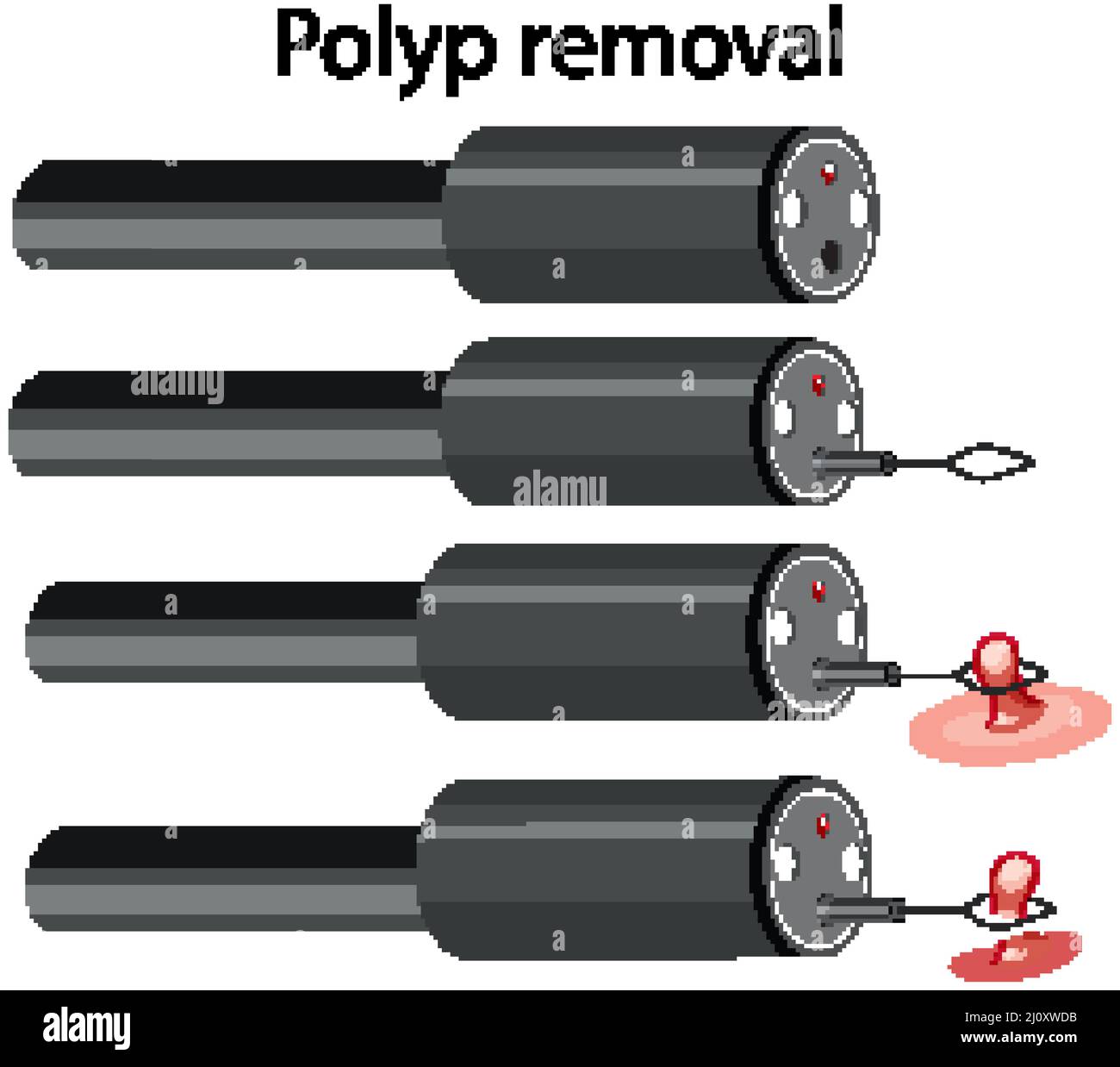 Diagram showing process of polyp removal illustration Stock Vectorhttps://www.alamy.com/image-license-details/?v=1https://www.alamy.com/diagram-showing-process-of-polyp-removal-illustration-image465270663.html
RF2J0XWDB–Diagram showing process of polyp removal illustration
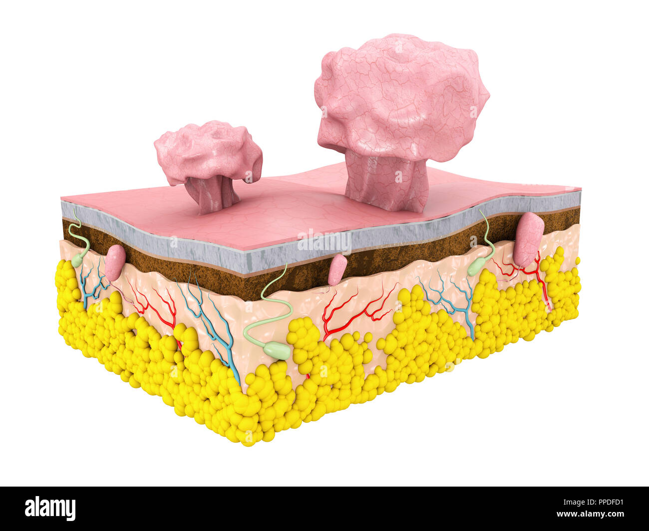 3d Illustration of Polype with Skin structure, isolated white background Stock Photohttps://www.alamy.com/image-license-details/?v=1https://www.alamy.com/3d-illustration-of-polype-with-skin-structure-isolated-white-background-image220344349.html
RFPPDFD1–3d Illustration of Polype with Skin structure, isolated white background
 The practice of surgery . Fig. 180.—Removal of myomata. mean shelling out the myomata, one by one, from the uterus. Theoperation is so easy in appropriate cases that nothing more than theillustrations are needed to demonstrate it. Open the abdomen; throw. Fig. 181.—Uterine polyp removed -u-ith scissors. the patient in the Trendelenburg position; wall off the uterus; pull itto the fore with vulsellum forceps, and enucleate the tumors individuallywith knife, scissors, and fingers. In properly selected cases the opera- 300 FEMALE OKGAXS OF GENERATION tion is cxtrcMnely easy, aiul the hemorrhagic Stock Photohttps://www.alamy.com/image-license-details/?v=1https://www.alamy.com/the-practice-of-surgery-fig-180removal-of-myomata-mean-shelling-out-the-myomata-one-by-one-from-the-uterus-theoperation-is-so-easy-in-appropriate-cases-that-nothing-more-than-theillustrations-are-needed-to-demonstrate-it-open-the-abdomen-throw-fig-181uterine-polyp-removed-u-ith-scissors-the-patient-in-the-trendelenburg-position-wall-off-the-uterus-pull-itto-the-fore-with-vulsellum-forceps-and-enucleate-the-tumors-individuallywith-knife-scissors-and-fingers-in-properly-selected-cases-the-opera-300-female-okgaxs-of-generation-tion-is-cxtrcmnely-easy-aiul-the-hemorrhagic-image342749608.html
RM2AWHGHC–The practice of surgery . Fig. 180.—Removal of myomata. mean shelling out the myomata, one by one, from the uterus. Theoperation is so easy in appropriate cases that nothing more than theillustrations are needed to demonstrate it. Open the abdomen; throw. Fig. 181.—Uterine polyp removed -u-ith scissors. the patient in the Trendelenburg position; wall off the uterus; pull itto the fore with vulsellum forceps, and enucleate the tumors individuallywith knife, scissors, and fingers. In properly selected cases the opera- 300 FEMALE OKGAXS OF GENERATION tion is cxtrcMnely easy, aiul the hemorrhagic
 Female ovary close-up on a blue background of a medical mockup. The concept of ovary removal about cancer of the female reproductive system. Diseases Stock Photohttps://www.alamy.com/image-license-details/?v=1https://www.alamy.com/female-ovary-close-up-on-a-blue-background-of-a-medical-mockup-the-concept-of-ovary-removal-about-cancer-of-the-female-reproductive-system-diseases-image576255109.html
RF2TDEK7H–Female ovary close-up on a blue background of a medical mockup. The concept of ovary removal about cancer of the female reproductive system. Diseases
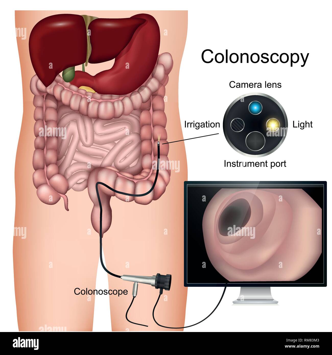 Colonoscopy procedure labeled 3d vector diagram on white background Stock Vectorhttps://www.alamy.com/image-license-details/?v=1https://www.alamy.com/colonoscopy-procedure-labeled-3d-vector-diagram-on-white-background-image236206435.html
RFRM83M3–Colonoscopy procedure labeled 3d vector diagram on white background
 Diagnosis - Colon Cancer. Medical Concept. Stock Photohttps://www.alamy.com/image-license-details/?v=1https://www.alamy.com/stock-photo-diagnosis-colon-cancer-medical-concept-83048048.html
RFER34HM–Diagnosis - Colon Cancer. Medical Concept.
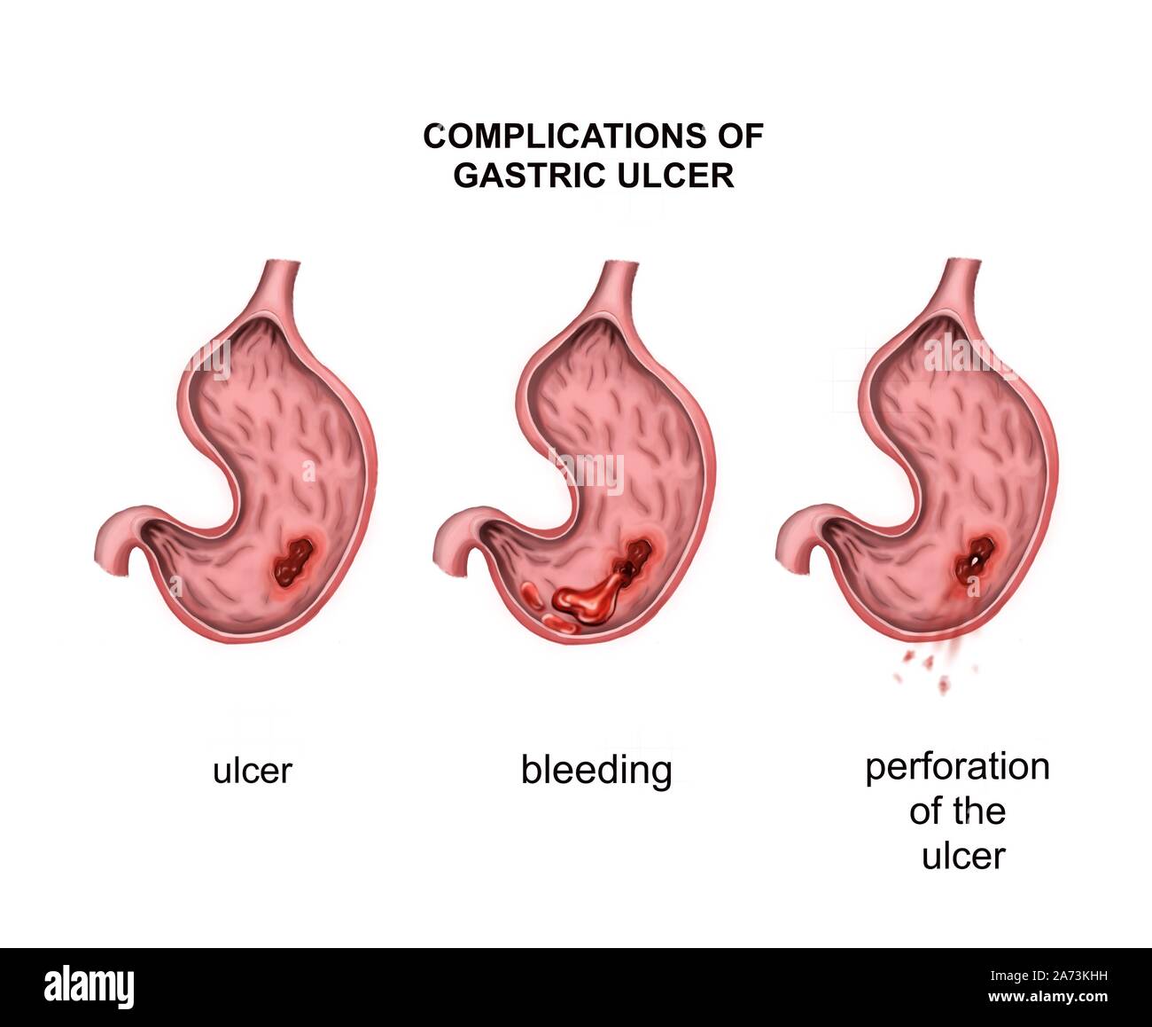 Complications of gastric ulcer. Bleeding and perforation Stock Photohttps://www.alamy.com/image-license-details/?v=1https://www.alamy.com/complications-of-gastric-ulcer-bleeding-and-perforation-image331380829.html
RF2A73KHH–Complications of gastric ulcer. Bleeding and perforation
 Diagnosis - Colon Cancer. Medicine Concept. 3D Illustration. Stock Photohttps://www.alamy.com/image-license-details/?v=1https://www.alamy.com/stock-photo-diagnosis-colon-cancer-medicine-concept-3d-illustration-134006210.html
RFHP0E82–Diagnosis - Colon Cancer. Medicine Concept. 3D Illustration.
 Human intestines with For Resection hanging sign, 3D rendering isolated on white background Stock Photohttps://www.alamy.com/image-license-details/?v=1https://www.alamy.com/human-intestines-with-for-resection-hanging-sign-3d-rendering-isolated-on-white-background-image429296234.html
RF2FXC3MA–Human intestines with For Resection hanging sign, 3D rendering isolated on white background
 3d rendered illustration of a polyp removal Stock Photohttps://www.alamy.com/image-license-details/?v=1https://www.alamy.com/3d-rendered-illustration-of-a-polyp-removal-image343647290.html
RM2AY2DHE–3d rendered illustration of a polyp removal
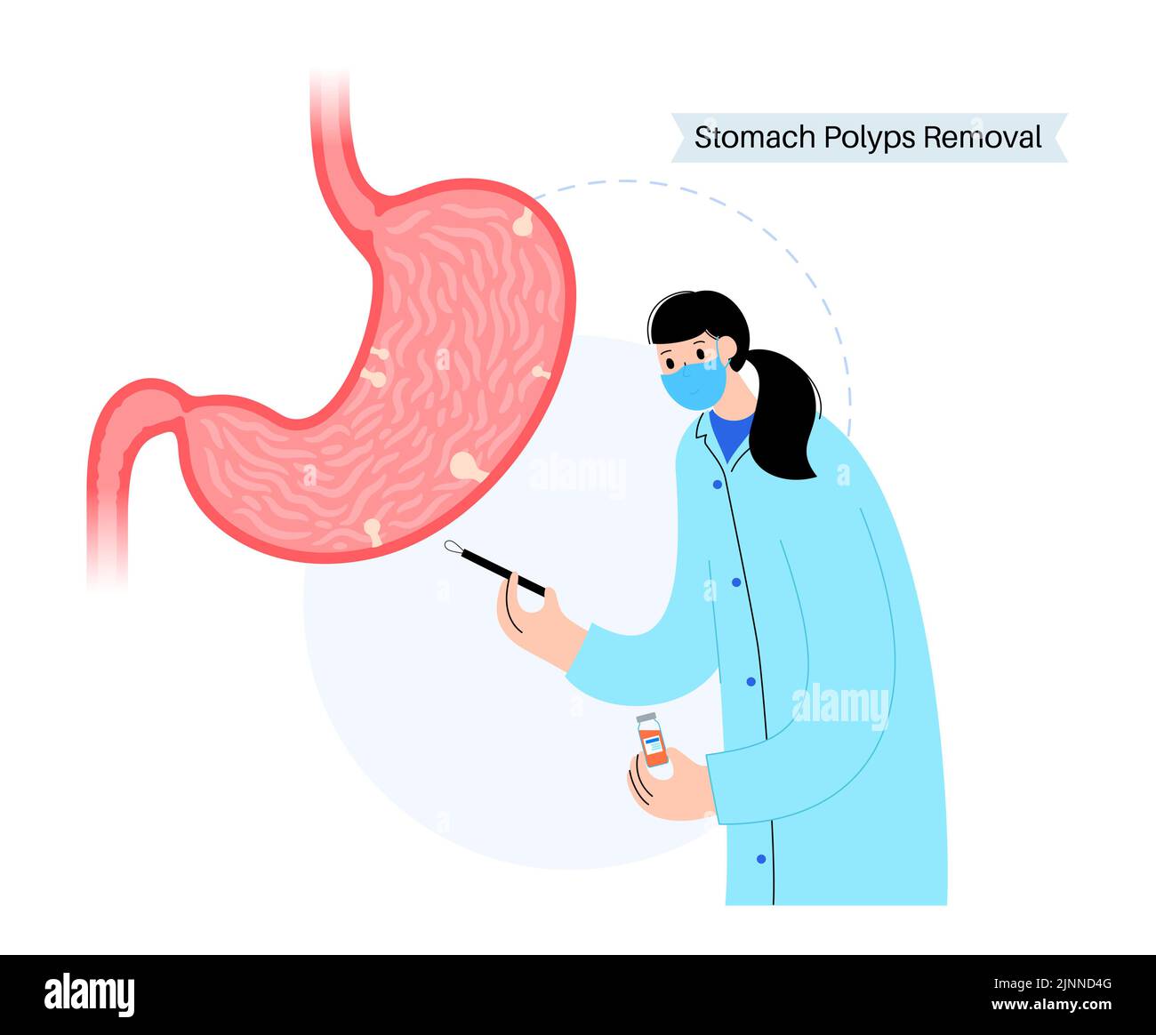 Stomach polyp removal, illustration Stock Photohttps://www.alamy.com/image-license-details/?v=1https://www.alamy.com/stomach-polyp-removal-illustration-image478059024.html
RF2JNND4G–Stomach polyp removal, illustration
 Colon Cancer Inscription in Extract From the History of Disease. Close Up View of Medicine Concept. 3D Illustration. Stock Photohttps://www.alamy.com/image-license-details/?v=1https://www.alamy.com/stock-photo-colon-cancer-inscription-in-extract-from-the-history-of-disease-close-135580871.html
RFHTG6NY–Colon Cancer Inscription in Extract From the History of Disease. Close Up View of Medicine Concept. 3D Illustration.
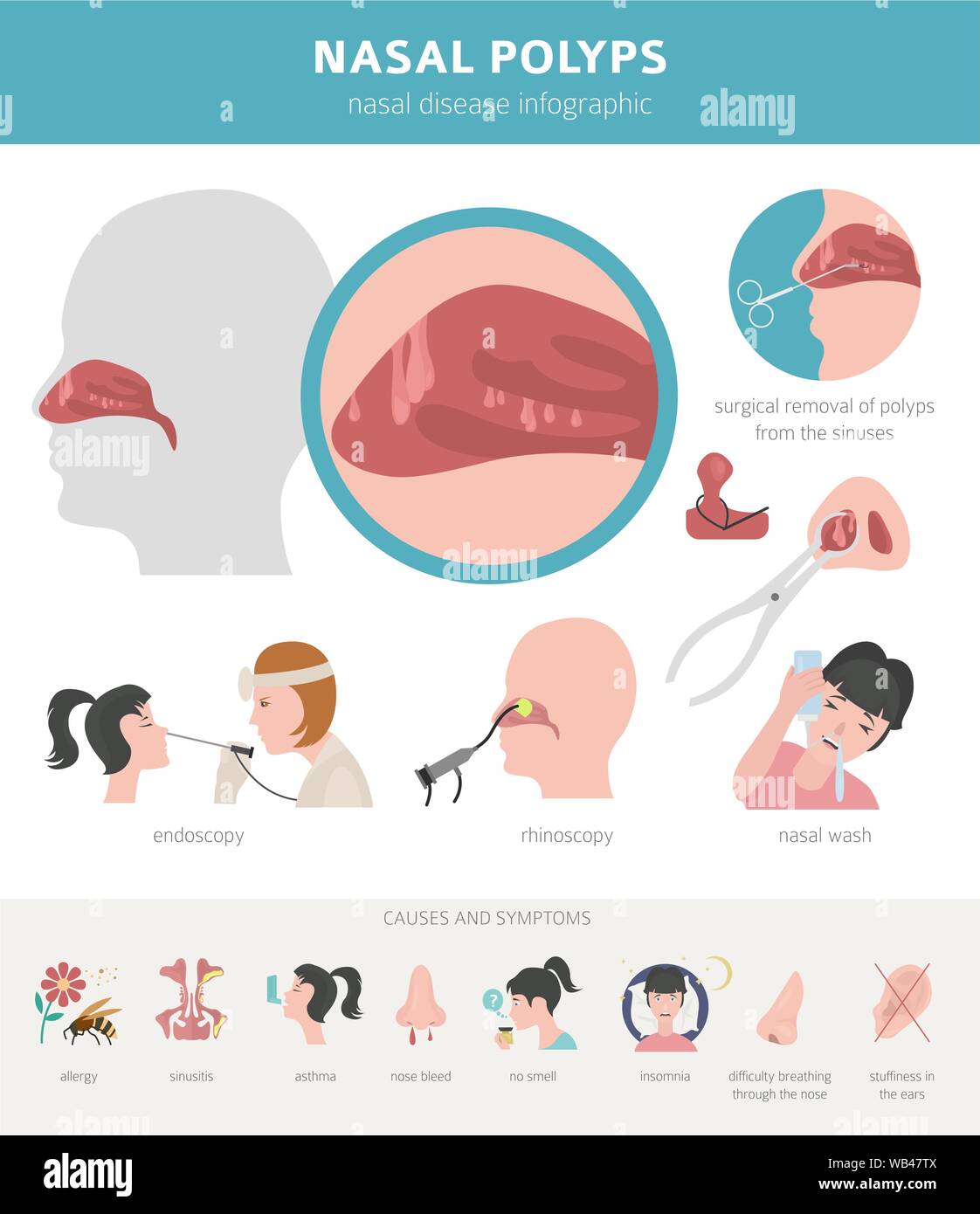 Nasal diseases. Nasal polyps causes, diagnosis and treatment medical infographic design. Vector illustration Stock Vectorhttps://www.alamy.com/image-license-details/?v=1https://www.alamy.com/nasal-diseases-nasal-polyps-causes-diagnosis-and-treatment-medical-infographic-design-vector-illustration-image265010730.html
RFWB47TX–Nasal diseases. Nasal polyps causes, diagnosis and treatment medical infographic design. Vector illustration
 Chalkboard with Colon Cancer. 3D Illustration. Stock Photohttps://www.alamy.com/image-license-details/?v=1https://www.alamy.com/stock-photo-chalkboard-with-colon-cancer-3d-illustration-125829871.html
RFH8M17Y–Chalkboard with Colon Cancer. 3D Illustration.
 Intestinal polyps closeup - 3D Rendering Stock Photohttps://www.alamy.com/image-license-details/?v=1https://www.alamy.com/stock-photo-intestinal-polyps-closeup-3d-rendering-176059103.html
RFM6C553–Intestinal polyps closeup - 3D Rendering
 Nasal diseases. Nasal polyps causes, diagnosis and treatment medical infographic design. Vector illustration Stock Vectorhttps://www.alamy.com/image-license-details/?v=1https://www.alamy.com/nasal-diseases-nasal-polyps-causes-diagnosis-and-treatment-medical-infographic-design-vector-illustration-image265010900.html
RFWB4830–Nasal diseases. Nasal polyps causes, diagnosis and treatment medical infographic design. Vector illustration
 Colorectal cancer, malignant tumor in intestine, Endoscope inside colonoscopy, gut intestine, Colon polyp removal, colonic polyps search, Polypectomy, Stock Photohttps://www.alamy.com/image-license-details/?v=1https://www.alamy.com/colorectal-cancer-malignant-tumor-in-intestine-endoscope-inside-colonoscopy-gut-intestine-colon-polyp-removal-colonic-polyps-search-polypectomy-image571217705.html
RM2T59609–Colorectal cancer, malignant tumor in intestine, Endoscope inside colonoscopy, gut intestine, Colon polyp removal, colonic polyps search, Polypectomy,
 Colon Cancer Diagnosis. Medical Concept. Stock Photohttps://www.alamy.com/image-license-details/?v=1https://www.alamy.com/stock-photo-colon-cancer-diagnosis-medical-concept-105190143.html
RFG33R2R–Colon Cancer Diagnosis. Medical Concept.
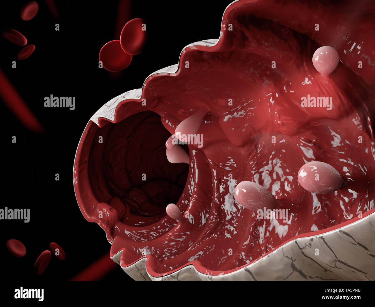 Colon polyps. 3d illustration, Polyp in the intestine Stock Photohttps://www.alamy.com/image-license-details/?v=1https://www.alamy.com/colon-polyps-3d-illustration-polyp-in-the-intestine-image247219319.html
RFTA5PNB–Colon polyps. 3d illustration, Polyp in the intestine
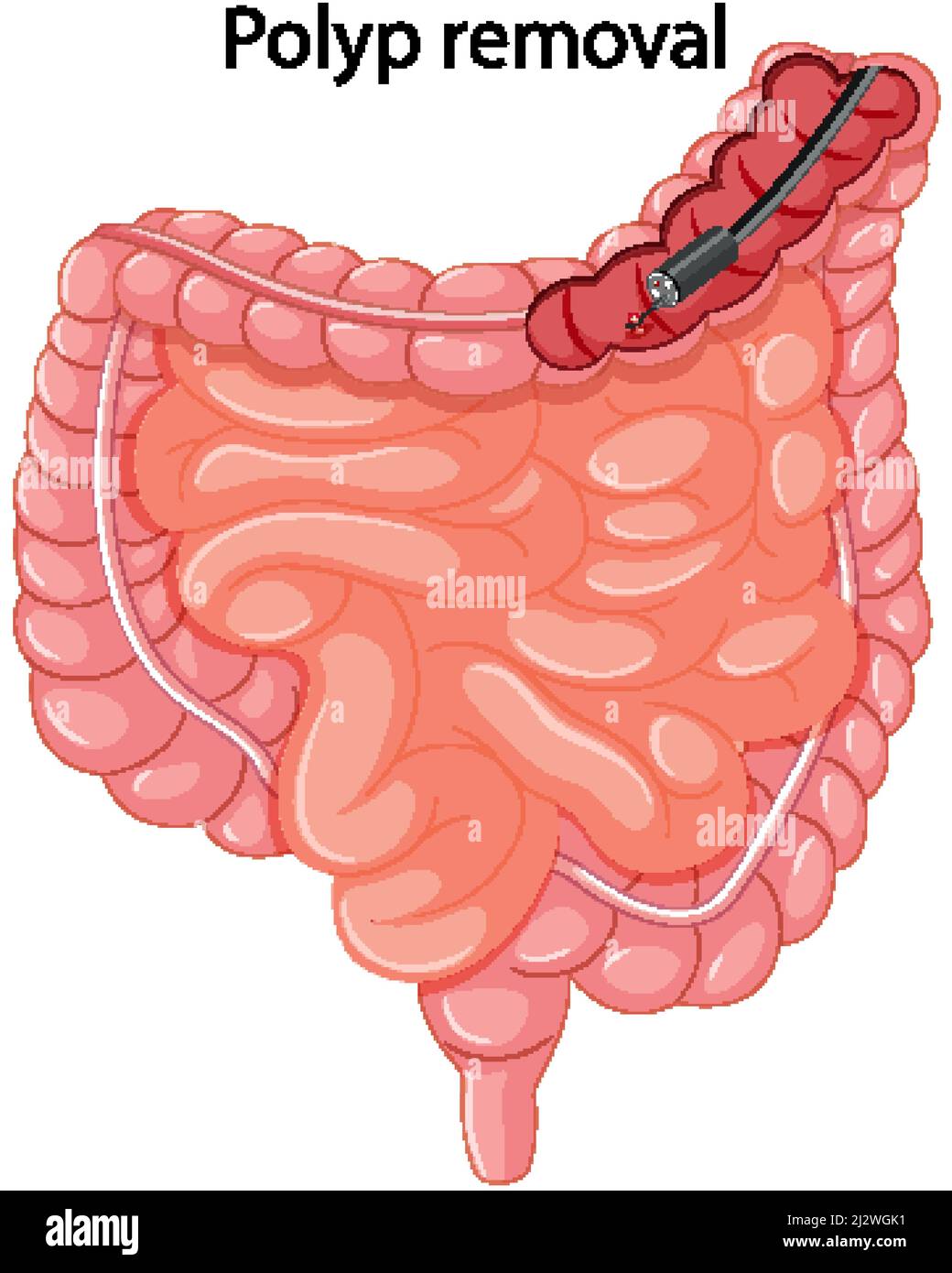 Diagram showing polyp removal illustration Stock Vectorhttps://www.alamy.com/image-license-details/?v=1https://www.alamy.com/diagram-showing-polyp-removal-illustration-image466471125.html
RF2J2WGK1–Diagram showing polyp removal illustration
 Colon cancer - Printed Diagnosis on Red Background. Stock Photohttps://www.alamy.com/image-license-details/?v=1https://www.alamy.com/stock-photo-colon-cancer-printed-diagnosis-on-red-background-89584648.html
RFF5MX3M–Colon cancer - Printed Diagnosis on Red Background.
 Transactions . .?^ ?«^*-.,-,. PLATE 3, POLYPOID ANGIOMA OF THE EAR. RICHARDS. Transverse section near outer extremity. POLYPOID ANGIOMA OF THE EAR. 205 tumor lost but little of their dark dull red color alter removal.Tlie childs hearing is now excellent, although both drum-membranes are depressed and are both of the dark grayishred color already mentioned. I have urged treatment ofthe nasal catarrh at the hands of a throat specialist. Theremoved polyp was pronounced by Dr. Freeborn to bebeyond question an angioma, and the accompanying micro-photographs abundantly prove the accuracy of this opi Stock Photohttps://www.alamy.com/image-license-details/?v=1https://www.alamy.com/transactions-plate-3-polypoid-angioma-of-the-ear-richards-transverse-section-near-outer-extremity-polypoid-angioma-of-the-ear-205-tumor-lost-but-little-of-their-dark-dull-red-color-alter-removaltlie-childs-hearing-is-now-excellent-although-both-drum-membranes-are-depressed-and-are-both-of-the-dark-grayishred-color-already-mentioned-i-have-urged-treatment-ofthe-nasal-catarrh-at-the-hands-of-a-throat-specialist-theremoved-polyp-was-pronounced-by-dr-freeborn-to-bebeyond-question-an-angioma-and-the-accompanying-micro-photographs-abundantly-prove-the-accuracy-of-this-opi-image338418932.html
RM2AJG8PC–Transactions . .?^ ?«^*-.,-,. PLATE 3, POLYPOID ANGIOMA OF THE EAR. RICHARDS. Transverse section near outer extremity. POLYPOID ANGIOMA OF THE EAR. 205 tumor lost but little of their dark dull red color alter removal.Tlie childs hearing is now excellent, although both drum-membranes are depressed and are both of the dark grayishred color already mentioned. I have urged treatment ofthe nasal catarrh at the hands of a throat specialist. Theremoved polyp was pronounced by Dr. Freeborn to bebeyond question an angioma, and the accompanying micro-photographs abundantly prove the accuracy of this opi
 Colon Cancer Diagnosis. Medical Concept. Stock Photohttps://www.alamy.com/image-license-details/?v=1https://www.alamy.com/stock-photo-colon-cancer-diagnosis-medical-concept-89584582.html
RFF5MX1A–Colon Cancer Diagnosis. Medical Concept.
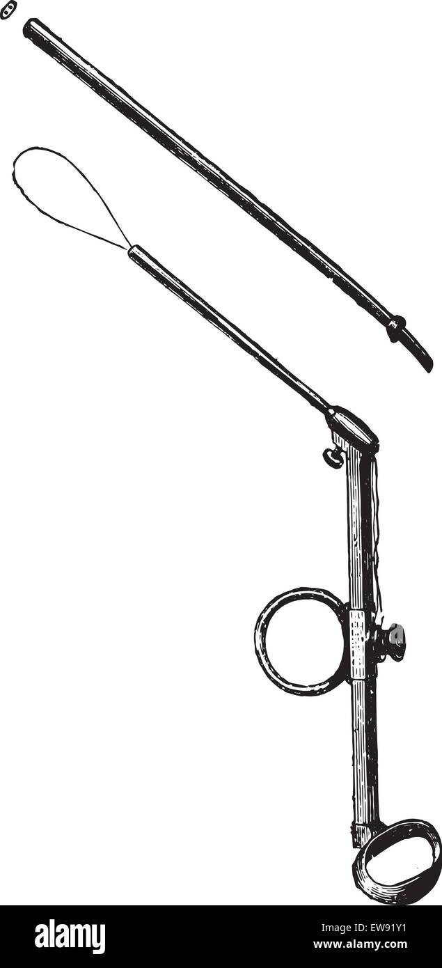 Polypectomy for polyps ear, vintage engraved illustration. Usual Medicine Dictionary - Paul Labarthe - 1885. Stock Vectorhttps://www.alamy.com/image-license-details/?v=1https://www.alamy.com/stock-photo-polypectomy-for-polyps-ear-vintage-engraved-illustration-usual-medicine-84406981.html
RFEW91Y1–Polypectomy for polyps ear, vintage engraved illustration. Usual Medicine Dictionary - Paul Labarthe - 1885.
 Papillomas on the skin - one red dying off in the axillary region and several small neoplasms. Human viral infections Stock Photohttps://www.alamy.com/image-license-details/?v=1https://www.alamy.com/papillomas-on-the-skin-one-red-dying-off-in-the-axillary-region-and-several-small-neoplasms-human-viral-infections-image598887695.html
RF2WP9KA7–Papillomas on the skin - one red dying off in the axillary region and several small neoplasms. Human viral infections
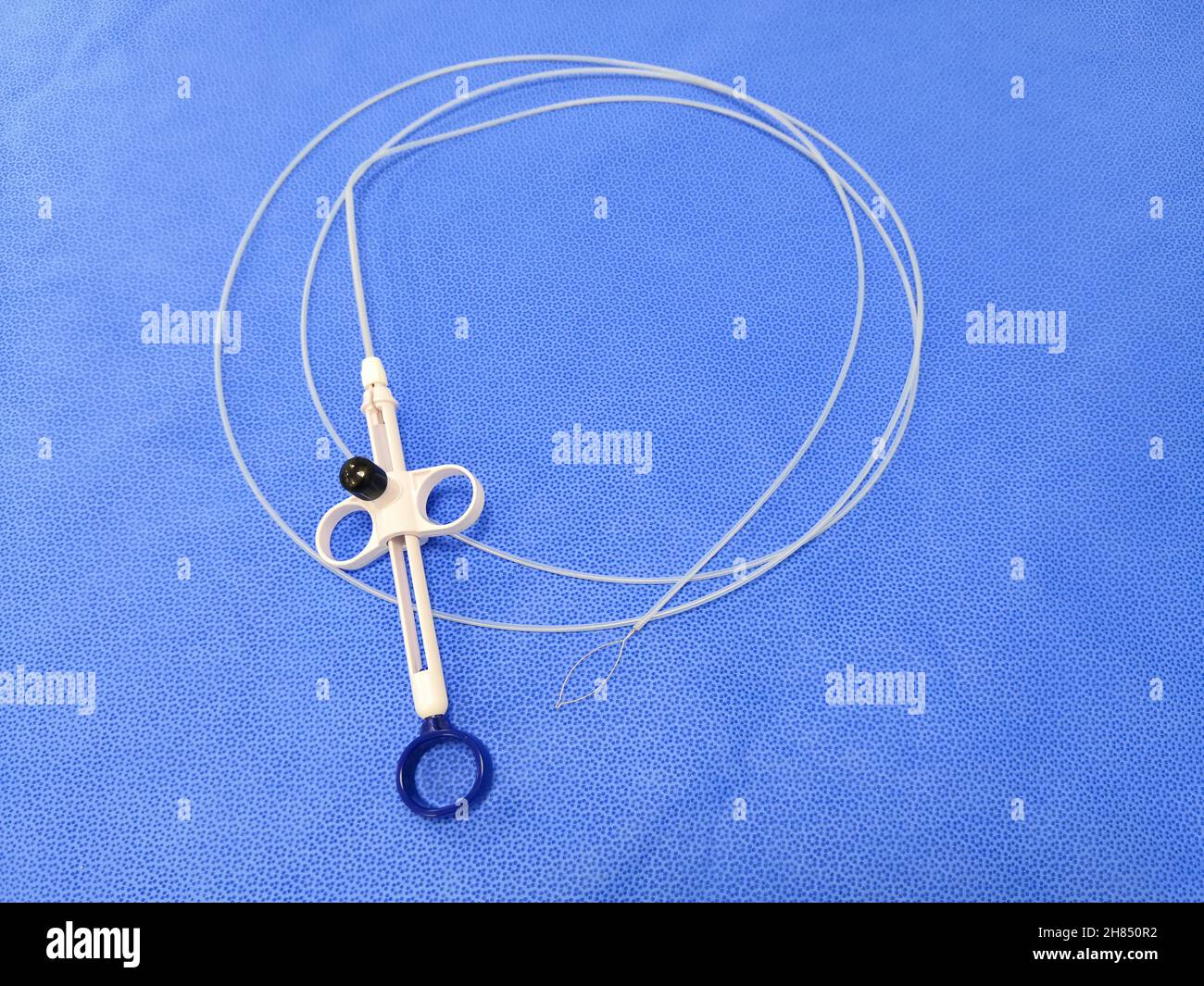 Closeup Image Of Endoscopic Instrument Polypectomy Snare In Blue Background. Selective Focus Stock Photohttps://www.alamy.com/image-license-details/?v=1https://www.alamy.com/closeup-image-of-endoscopic-instrument-polypectomy-snare-in-blue-background-selective-focus-image452497222.html
RF2H850R2–Closeup Image Of Endoscopic Instrument Polypectomy Snare In Blue Background. Selective Focus
 External skin diseases. Rash on the skin of a man in the form of moles, warts and papillomas Stock Photohttps://www.alamy.com/image-license-details/?v=1https://www.alamy.com/external-skin-diseases-rash-on-the-skin-of-a-man-in-the-form-of-moles-warts-and-papillomas-image458590427.html
RF2HJ2GNF–External skin diseases. Rash on the skin of a man in the form of moles, warts and papillomas
 ACROCHORDON, ABLATION Stock Photohttps://www.alamy.com/image-license-details/?v=1https://www.alamy.com/stock-photo-acrochordon-ablation-49277151.html
RMCT4NE7–ACROCHORDON, ABLATION
RFMGXD9X–Icons For Medical Specialties. Enterology And Intestine Concept. Vector Illustration With Hand Drawn Medicine Doodle.
 Stomach polyp removal, illustration Stock Photohttps://www.alamy.com/image-license-details/?v=1https://www.alamy.com/stomach-polyp-removal-illustration-image478059026.html
RF2JNND4J–Stomach polyp removal, illustration
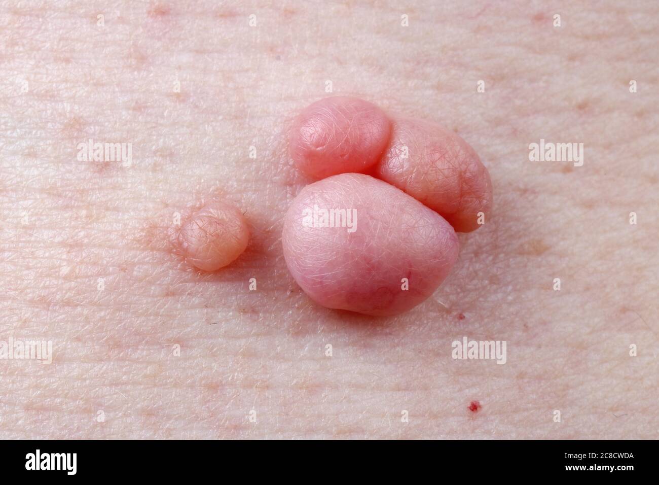 Warts, polyps and moles grow on human skin all their life macro. Stock Photohttps://www.alamy.com/image-license-details/?v=1https://www.alamy.com/warts-polyps-and-moles-grow-on-human-skin-all-their-life-macro-image366618374.html
RF2C8CWDA–Warts, polyps and moles grow on human skin all their life macro.
 Removal of a polyp in the human colon, computer artwork. Stock Photohttps://www.alamy.com/image-license-details/?v=1https://www.alamy.com/stock-photo-removal-of-a-polyp-in-the-human-colon-computer-artwork-73689515.html
RFE7TRMB–Removal of a polyp in the human colon, computer artwork.
 Melanoma word cloud and hand with marker concept on white background. Stock Photohttps://www.alamy.com/image-license-details/?v=1https://www.alamy.com/melanoma-word-cloud-and-hand-with-marker-concept-on-white-background-image225215316.html
RFR2BCC4–Melanoma word cloud and hand with marker concept on white background.
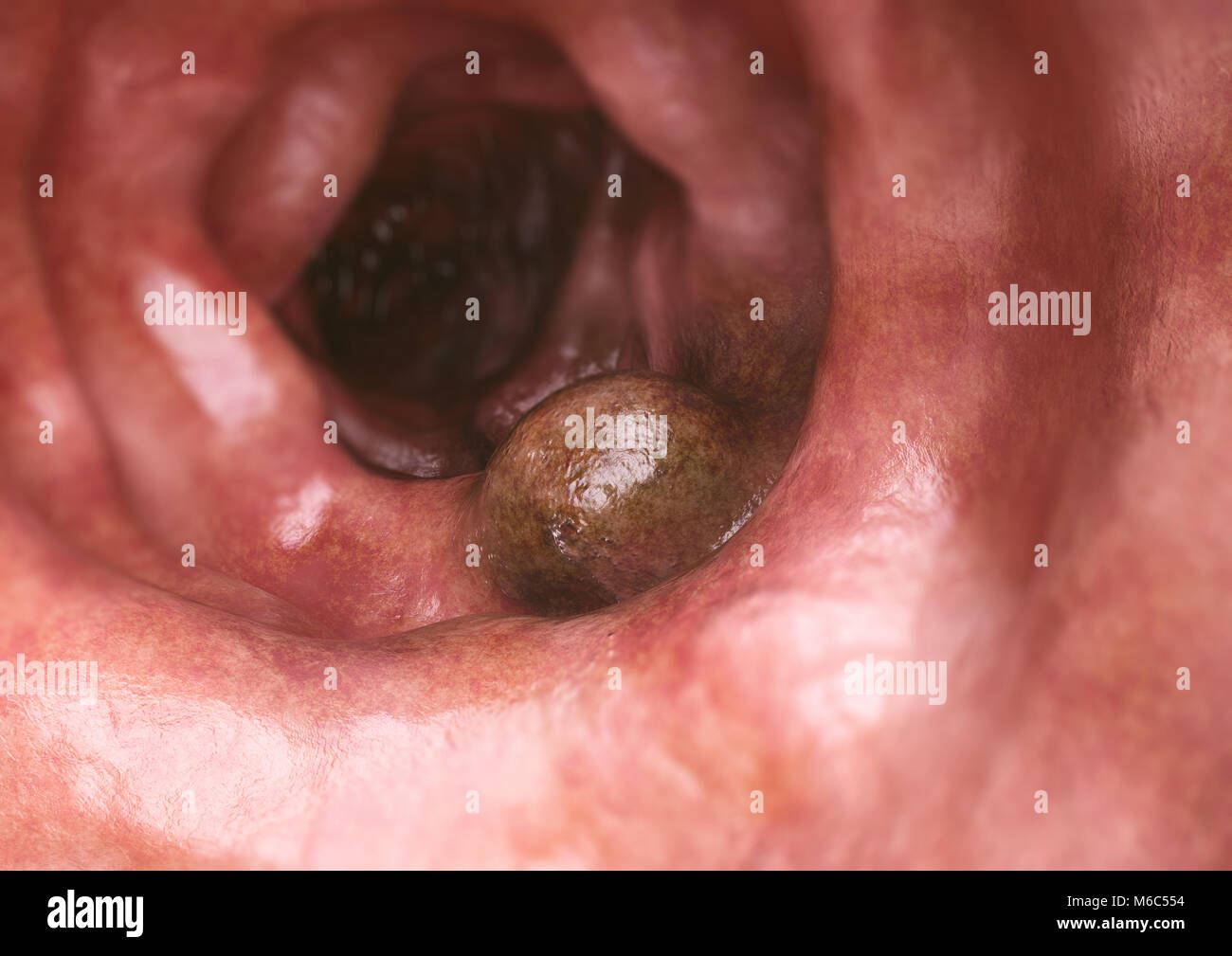 Colon cancer closeup - 3D Rendering Stock Photohttps://www.alamy.com/image-license-details/?v=1https://www.alamy.com/stock-photo-colon-cancer-closeup-3d-rendering-176059104.html
RFM6C554–Colon cancer closeup - 3D Rendering
 Nasal diseases. Nasal polyps causes, diagnosis and treatment medical infographic design. Vector illustration Stock Vectorhttps://www.alamy.com/image-license-details/?v=1https://www.alamy.com/nasal-diseases-nasal-polyps-causes-diagnosis-and-treatment-medical-infographic-design-vector-illustration-image265010744.html
RFWB47WC–Nasal diseases. Nasal polyps causes, diagnosis and treatment medical infographic design. Vector illustration
 Colorectal cancer, malignant tumor in intestine, Endoscope inside colonoscopy, gut intestine, Colon polyp removal, colonic polyps search, Polypectomy, Stock Photohttps://www.alamy.com/image-license-details/?v=1https://www.alamy.com/colorectal-cancer-malignant-tumor-in-intestine-endoscope-inside-colonoscopy-gut-intestine-colon-polyp-removal-colonic-polyps-search-polypectomy-image571217483.html
RM2T595MB–Colorectal cancer, malignant tumor in intestine, Endoscope inside colonoscopy, gut intestine, Colon polyp removal, colonic polyps search, Polypectomy,
 Bile ducts and gall bladder from plasticine on a blue background. Gallbladder disease concept and laparoscopic methods for removing gallstones. Chroni Stock Photohttps://www.alamy.com/image-license-details/?v=1https://www.alamy.com/bile-ducts-and-gall-bladder-from-plasticine-on-a-blue-background-gallbladder-disease-concept-and-laparoscopic-methods-for-removing-gallstones-chroni-image328261508.html
RF2A21GW8–Bile ducts and gall bladder from plasticine on a blue background. Gallbladder disease concept and laparoscopic methods for removing gallstones. Chroni
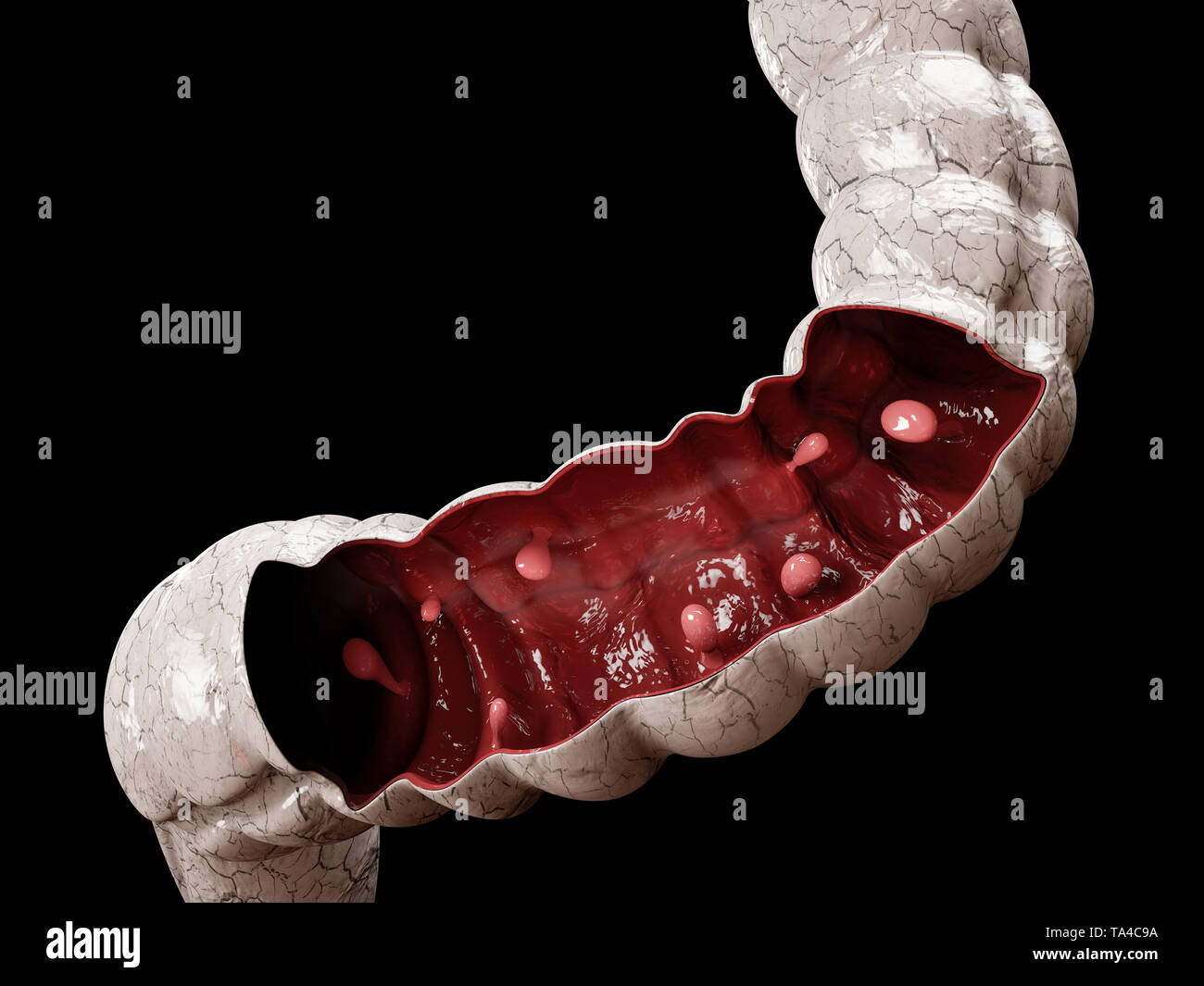 Colon polyps. 3d illustration, Polyp in the intestine Stock Photohttps://www.alamy.com/image-license-details/?v=1https://www.alamy.com/colon-polyps-3d-illustration-polyp-in-the-intestine-image247189190.html
RFTA4C9A–Colon polyps. 3d illustration, Polyp in the intestine
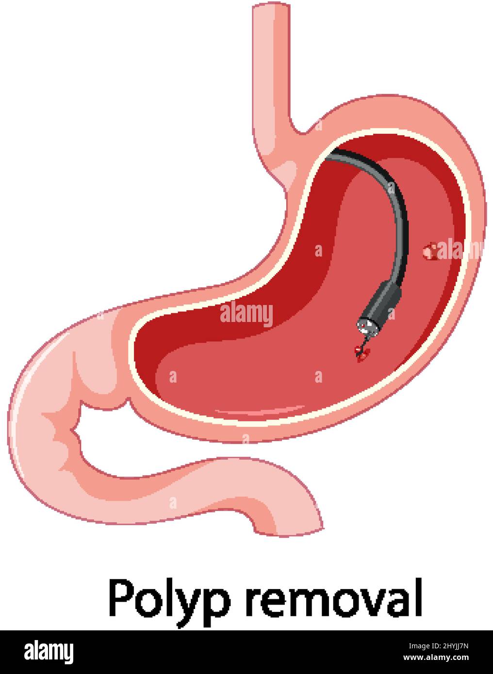 Diagram showing polyp removal in human illustration Stock Vectorhttps://www.alamy.com/image-license-details/?v=1https://www.alamy.com/diagram-showing-polyp-removal-in-human-illustration-image464474745.html
RF2HYJJ7N–Diagram showing polyp removal in human illustration
 Miliums on the face, removal of hardened and mature milia by a beautician. new Stock Photohttps://www.alamy.com/image-license-details/?v=1https://www.alamy.com/miliums-on-the-face-removal-of-hardened-and-mature-milia-by-a-beautician-new-image419229232.html
RF2FA1F4G–Miliums on the face, removal of hardened and mature milia by a beautician. new
 . Diseases of the ear; a text-book for practitioners and students of medicine. Fig. 112.—Removal of aural polyp with the snare. REMOVAL OF AURAL POLYPS. *7. ?Fig. 113. ?Removal of aural polyp withthe sharp curette. below the growth, and then raised so that the ring of the in-strument will encircle it; by moving the curette delicately it canbe carried upward along the pedicle to its point of attachment;then, by pressing the instru-ment firmly against the wallof the canal, and at the sametime drawing it outward, themass is removed. This procedure is notpainful if care is taken not totouch the wa Stock Photohttps://www.alamy.com/image-license-details/?v=1https://www.alamy.com/diseases-of-the-ear-a-text-book-for-practitioners-and-students-of-medicine-fig-112removal-of-aural-polyp-with-the-snare-removal-of-aural-polyps-7-fig-113-removal-of-aural-polyp-withthe-sharp-curette-below-the-growth-and-then-raised-so-that-the-ring-of-the-in-strument-will-encircle-it-by-moving-the-curette-delicately-it-canbe-carried-upward-along-the-pedicle-to-its-point-of-attachmentthen-by-pressing-the-instru-ment-firmly-against-the-wallof-the-canal-and-at-the-sametime-drawing-it-outward-themass-is-removed-this-procedure-is-notpainful-if-care-is-taken-not-totouch-the-wa-image370534678.html
RM2CER8NA–. Diseases of the ear; a text-book for practitioners and students of medicine. Fig. 112.—Removal of aural polyp with the snare. REMOVAL OF AURAL POLYPS. *7. ?Fig. 113. ?Removal of aural polyp withthe sharp curette. below the growth, and then raised so that the ring of the in-strument will encircle it; by moving the curette delicately it canbe carried upward along the pedicle to its point of attachment;then, by pressing the instru-ment firmly against the wallof the canal, and at the sametime drawing it outward, themass is removed. This procedure is notpainful if care is taken not totouch the wa
RF2YWMJ8K–Colonoscopy line black icon. Sign for web page, mobile app, button, logo. Vector isolated button. Editable stroke.
 Polypectomy for polyps ear, vintage engraved illustration. Usual Medicine Dictionary - Paul Labarthe - 1885. Stock Vectorhttps://www.alamy.com/image-license-details/?v=1https://www.alamy.com/stock-photo-polypectomy-for-polyps-ear-vintage-engraved-illustration-usual-medicine-84419399.html
RFEW9HPF–Polypectomy for polyps ear, vintage engraved illustration. Usual Medicine Dictionary - Paul Labarthe - 1885.
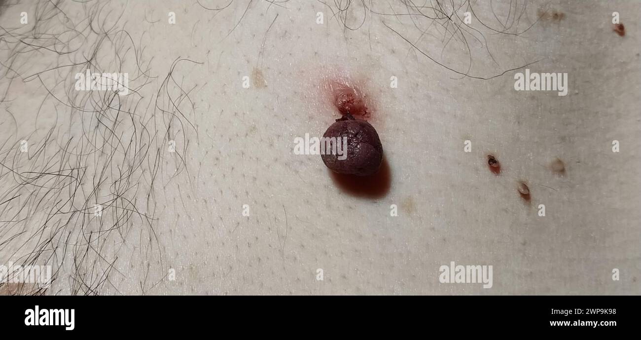 Papillomas on the skin - one red dying off and several small neoplasms. Therapy for skin diseases. Human viral infections. Stock Photohttps://www.alamy.com/image-license-details/?v=1https://www.alamy.com/papillomas-on-the-skin-one-red-dying-off-and-several-small-neoplasms-therapy-for-skin-diseases-human-viral-infections-image598887668.html
RF2WP9K98–Papillomas on the skin - one red dying off and several small neoplasms. Therapy for skin diseases. Human viral infections.
RF2YWMGCR–Colonoscopy line black icon. Sign for web page, mobile app, button, logo. Vector isolated button. Editable stroke.
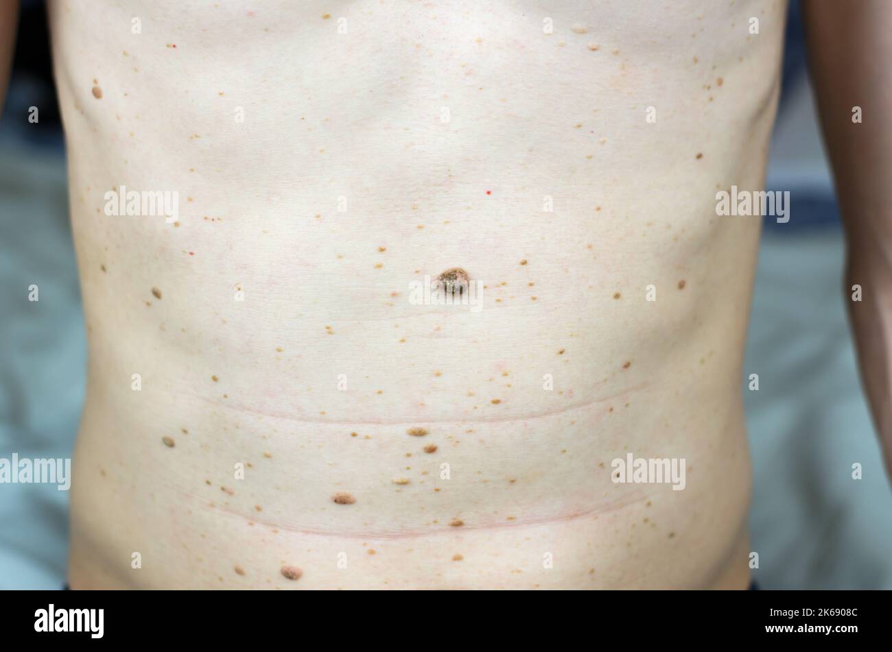 Moles, warts and papillomas on human skin. Diseases of the skin Stock Photohttps://www.alamy.com/image-license-details/?v=1https://www.alamy.com/moles-warts-and-papillomas-on-human-skin-diseases-of-the-skin-image485776044.html
RF2K6908C–Moles, warts and papillomas on human skin. Diseases of the skin
 ACROCHORDON, ABLATION Stock Photohttps://www.alamy.com/image-license-details/?v=1https://www.alamy.com/stock-photo-acrochordon-ablation-49277162.html
RMCT4NEJ–ACROCHORDON, ABLATION
 . /^JGONOZOOID if PALPON JELLY POLYP Text-fig. 25. Physalia physalis. A young gonodendron after removal of three branches. Specimen, Lanzarote no. 2. The numerals refer to the number of subterminal, nectophore-bearing sections (not shown) on each end branch, x 9. sub-terminal section has a long-stalked nectophore («) in this position. The terminal palpons with their jelly-polyps are visible in the earliest growth-stages of the gonodendra (PI. XXII, fig. 4); in well- developed specimens the oldest terminal palpons at the bases of the larger gonodendral branches reach a considerable size (Text-f Stock Photohttps://www.alamy.com/image-license-details/?v=1https://www.alamy.com/jgonozooid-if-palpon-jelly-polyp-text-fig-25-physalia-physalis-a-young-gonodendron-after-removal-of-three-branches-specimen-lanzarote-no-2-the-numerals-refer-to-the-number-of-subterminal-nectophore-bearing-sections-not-shown-on-each-end-branch-x-9-sub-terminal-section-has-a-long-stalked-nectophore-in-this-position-the-terminal-palpons-with-their-jelly-polyps-are-visible-in-the-earliest-growth-stages-of-the-gonodendra-pi-xxii-fig-4-in-well-developed-specimens-the-oldest-terminal-palpons-at-the-bases-of-the-larger-gonodendral-branches-reach-a-considerable-size-text-f-image179954452.html
RMMCNHMM–. /^JGONOZOOID if PALPON JELLY POLYP Text-fig. 25. Physalia physalis. A young gonodendron after removal of three branches. Specimen, Lanzarote no. 2. The numerals refer to the number of subterminal, nectophore-bearing sections (not shown) on each end branch, x 9. sub-terminal section has a long-stalked nectophore («) in this position. The terminal palpons with their jelly-polyps are visible in the earliest growth-stages of the gonodendra (PI. XXII, fig. 4); in well- developed specimens the oldest terminal palpons at the bases of the larger gonodendral branches reach a considerable size (Text-f
