Quick filters:
Popliteal Stock Photos and Images
 First aid, pressure applied to popliteal artery Stock Photohttps://www.alamy.com/image-license-details/?v=1https://www.alamy.com/stock-image-first-aid-pressure-applied-to-popliteal-artery-169313675.html
RMKRCW8Y–First aid, pressure applied to popliteal artery
 Popliteal aneurysm, illustration. An aneurysm is a blood-filled dilation in a blood vessel. Stock Photohttps://www.alamy.com/image-license-details/?v=1https://www.alamy.com/popliteal-aneurysm-illustration-an-aneurysm-is-a-blood-filled-dilation-in-a-blood-vessel-image367362509.html
RF2C9JPHH–Popliteal aneurysm, illustration. An aneurysm is a blood-filled dilation in a blood vessel.
 Popliteal cyst Stock Vectorhttps://www.alamy.com/image-license-details/?v=1https://www.alamy.com/popliteal-cyst-image549588680.html
RF2PX3WY4–Popliteal cyst
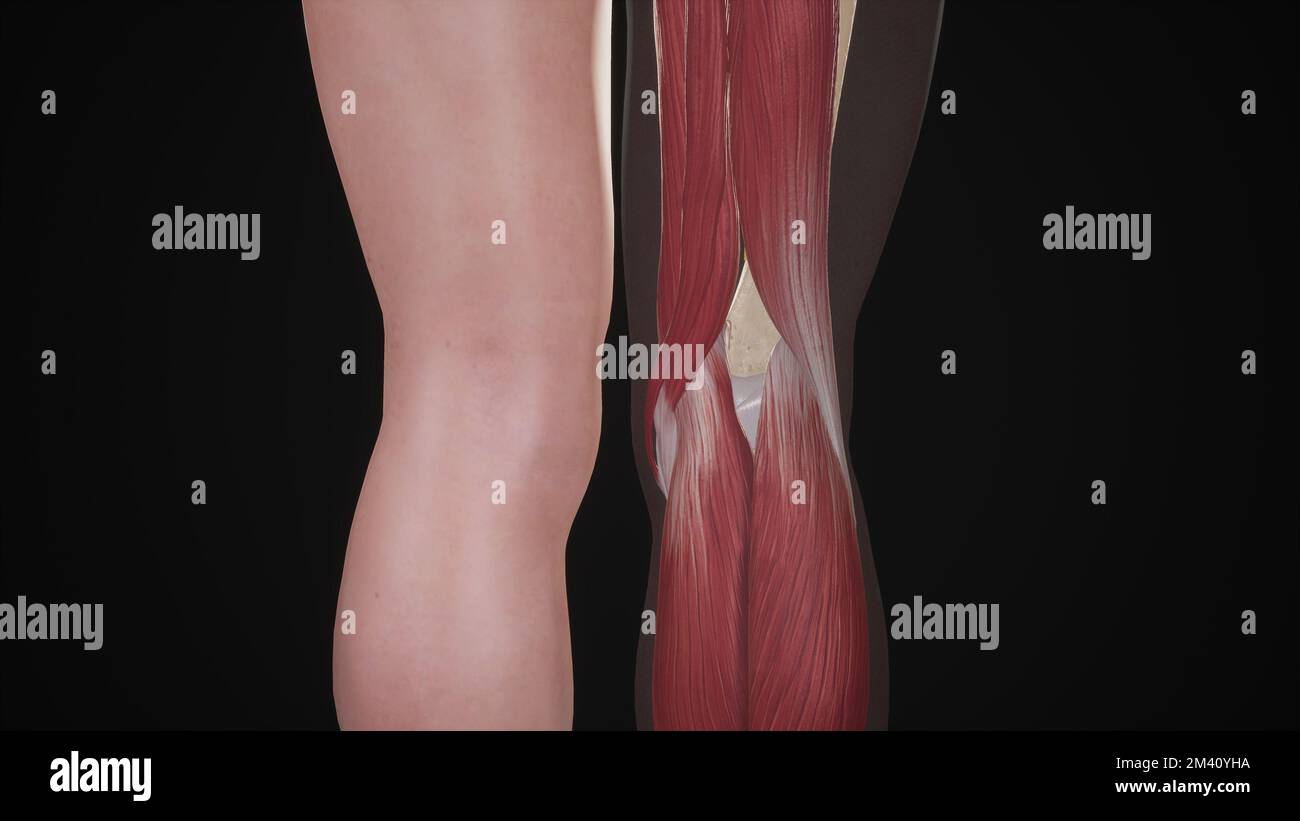 Boundaries of Popliteal Fossa Stock Photohttps://www.alamy.com/image-license-details/?v=1https://www.alamy.com/boundaries-of-popliteal-fossa-image501580950.html
RF2M40YHA–Boundaries of Popliteal Fossa
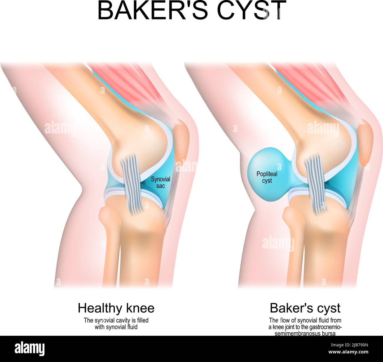 Baker's cyst. Normal knee, and joint with Popliteal cyst. The flow of synovial fluid from the knee joint to the gastrocnemio-semimembranosus bursa Stock Vectorhttps://www.alamy.com/image-license-details/?v=1https://www.alamy.com/bakers-cyst-normal-knee-and-joint-with-popliteal-cyst-the-flow-of-synovial-fluid-from-the-knee-joint-to-the-gastrocnemio-semimembranosus-bursa-image471601893.html
RF2JB790N–Baker's cyst. Normal knee, and joint with Popliteal cyst. The flow of synovial fluid from the knee joint to the gastrocnemio-semimembranosus bursa
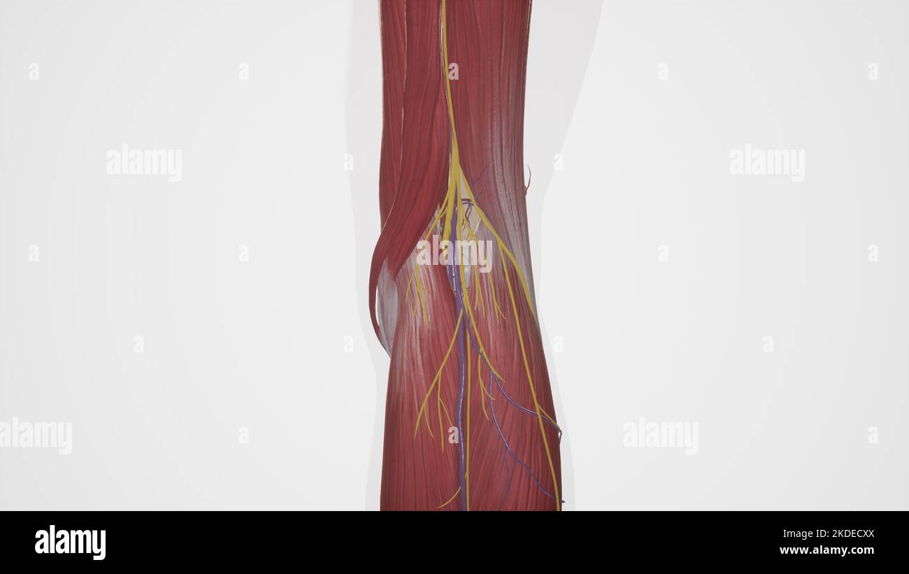 Anatomical Illustration of Popliteal Fossa Stock Photohttps://www.alamy.com/image-license-details/?v=1https://www.alamy.com/anatomical-illustration-of-popliteal-fossa-image490198322.html
RF2KDECXX–Anatomical Illustration of Popliteal Fossa
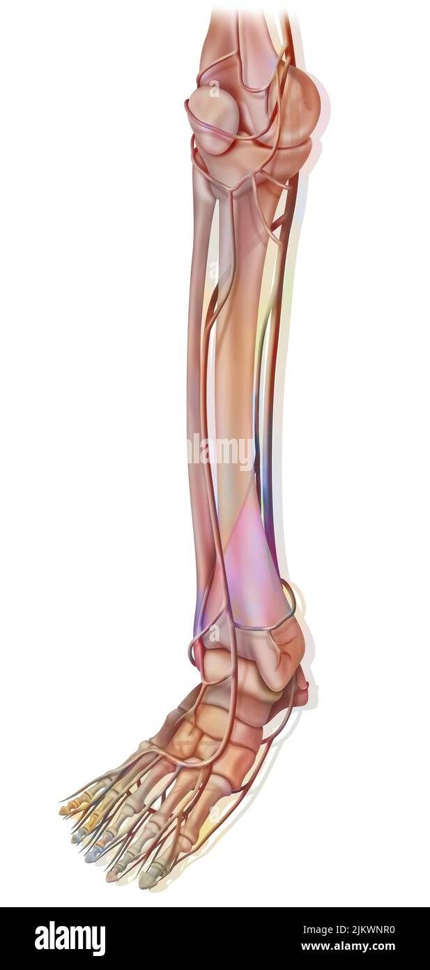 The arteries of the lower part of the lower extremity. Stock Photohttps://www.alamy.com/image-license-details/?v=1https://www.alamy.com/the-arteries-of-the-lower-part-of-the-lower-extremity-image476924308.html
RF2JKWNR0–The arteries of the lower part of the lower extremity.
 Popliteal fossa. Back view of male's knees on plain background. Graphic resource of anatomy and structure of the human body. Stock Photohttps://www.alamy.com/image-license-details/?v=1https://www.alamy.com/popliteal-fossa-back-view-of-males-knees-on-plain-background-graphic-resource-of-anatomy-and-structure-of-the-human-body-image607805592.html
RF2X8RX6G–Popliteal fossa. Back view of male's knees on plain background. Graphic resource of anatomy and structure of the human body.
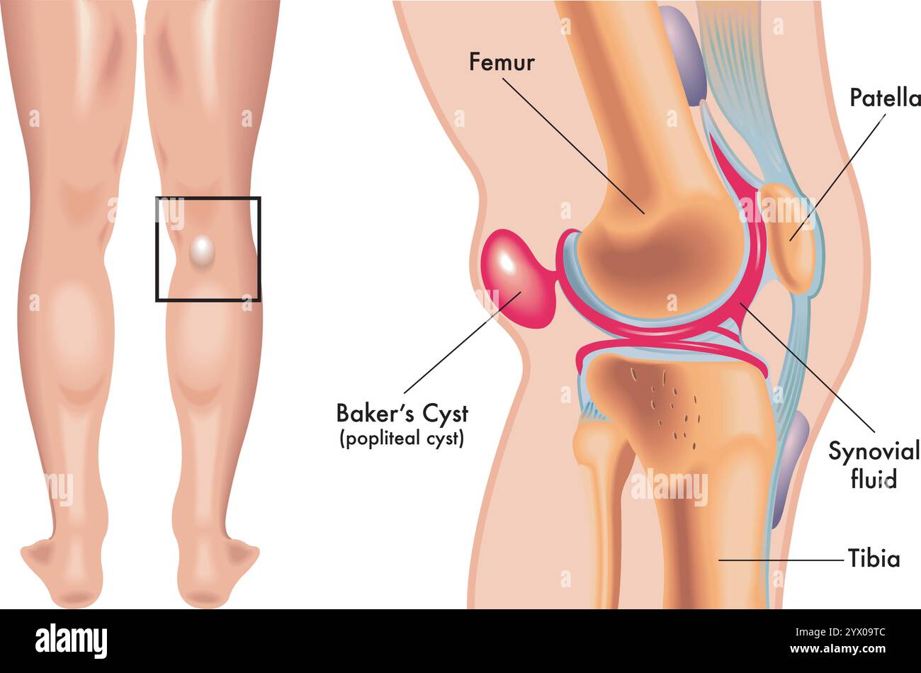 Medical illustration of baker cyst, with annotation. Stock Vectorhttps://www.alamy.com/image-license-details/?v=1https://www.alamy.com/medical-illustration-of-baker-cyst-with-annotation-image635562044.html
RF2YX09TC–Medical illustration of baker cyst, with annotation.
 Hock support bandage on a dog's hind leg. Stock Photohttps://www.alamy.com/image-license-details/?v=1https://www.alamy.com/hock-support-bandage-on-a-dogs-hind-leg-image470537419.html
RF2J9ER7R–Hock support bandage on a dog's hind leg.
![Acu-moxa point chart, showing the weizhong (Middle of the Crook) point, from Chuanwu lingji lu (Record of Sovereign Teachings), by Zhang Youheng, a treatise on acu-moxa in two volumes. This work survives only in a manuscript draft, completed in 1869 (8th year of the Tongzhi reign period of the Qing dynasty). The weizhong point is located at the back of the knee joint, in the middle of the popliteal crease. It can be needled to a depth of 5 fen (1 fen [0.1 cun/Chinese proportional inch] = c. 0. Stock Photo Acu-moxa point chart, showing the weizhong (Middle of the Crook) point, from Chuanwu lingji lu (Record of Sovereign Teachings), by Zhang Youheng, a treatise on acu-moxa in two volumes. This work survives only in a manuscript draft, completed in 1869 (8th year of the Tongzhi reign period of the Qing dynasty). The weizhong point is located at the back of the knee joint, in the middle of the popliteal crease. It can be needled to a depth of 5 fen (1 fen [0.1 cun/Chinese proportional inch] = c. 0. Stock Photo](https://c8.alamy.com/comp/T95083/acu-moxa-point-chart-showing-the-weizhong-middle-of-the-crook-point-from-chuanwu-lingji-lu-record-of-sovereign-teachings-by-zhang-youheng-a-treatise-on-acu-moxa-in-two-volumes-this-work-survives-only-in-a-manuscript-draft-completed-in-1869-8th-year-of-the-tongzhi-reign-period-of-the-qing-dynasty-the-weizhong-point-is-located-at-the-back-of-the-knee-joint-in-the-middle-of-the-popliteal-crease-it-can-be-needled-to-a-depth-of-5-fen-1-fen-01-cunchinese-proportional-inch-=-c-0-T95083.jpg) Acu-moxa point chart, showing the weizhong (Middle of the Crook) point, from Chuanwu lingji lu (Record of Sovereign Teachings), by Zhang Youheng, a treatise on acu-moxa in two volumes. This work survives only in a manuscript draft, completed in 1869 (8th year of the Tongzhi reign period of the Qing dynasty). The weizhong point is located at the back of the knee joint, in the middle of the popliteal crease. It can be needled to a depth of 5 fen (1 fen [0.1 cun/Chinese proportional inch] = c. 0. Stock Photohttps://www.alamy.com/image-license-details/?v=1https://www.alamy.com/acu-moxa-point-chart-showing-the-weizhong-middle-of-the-crook-point-from-chuanwu-lingji-lu-record-of-sovereign-teachings-by-zhang-youheng-a-treatise-on-acu-moxa-in-two-volumes-this-work-survives-only-in-a-manuscript-draft-completed-in-1869-8th-year-of-the-tongzhi-reign-period-of-the-qing-dynasty-the-weizhong-point-is-located-at-the-back-of-the-knee-joint-in-the-middle-of-the-popliteal-crease-it-can-be-needled-to-a-depth-of-5-fen-1-fen-01-cunchinese-proportional-inch-=-c-0-image246587043.html
Acu-moxa point chart, showing the weizhong (Middle of the Crook) point, from Chuanwu lingji lu (Record of Sovereign Teachings), by Zhang Youheng, a treatise on acu-moxa in two volumes. This work survives only in a manuscript draft, completed in 1869 (8th year of the Tongzhi reign period of the Qing dynasty). The weizhong point is located at the back of the knee joint, in the middle of the popliteal crease. It can be needled to a depth of 5 fen (1 fen [0.1 cun/Chinese proportional inch] = c. 0. Stock Photohttps://www.alamy.com/image-license-details/?v=1https://www.alamy.com/acu-moxa-point-chart-showing-the-weizhong-middle-of-the-crook-point-from-chuanwu-lingji-lu-record-of-sovereign-teachings-by-zhang-youheng-a-treatise-on-acu-moxa-in-two-volumes-this-work-survives-only-in-a-manuscript-draft-completed-in-1869-8th-year-of-the-tongzhi-reign-period-of-the-qing-dynasty-the-weizhong-point-is-located-at-the-back-of-the-knee-joint-in-the-middle-of-the-popliteal-crease-it-can-be-needled-to-a-depth-of-5-fen-1-fen-01-cunchinese-proportional-inch-=-c-0-image246587043.htmlRMT95083–Acu-moxa point chart, showing the weizhong (Middle of the Crook) point, from Chuanwu lingji lu (Record of Sovereign Teachings), by Zhang Youheng, a treatise on acu-moxa in two volumes. This work survives only in a manuscript draft, completed in 1869 (8th year of the Tongzhi reign period of the Qing dynasty). The weizhong point is located at the back of the knee joint, in the middle of the popliteal crease. It can be needled to a depth of 5 fen (1 fen [0.1 cun/Chinese proportional inch] = c. 0.
 Aneurysm being a complex subject, related to other important topics. Stock Photohttps://www.alamy.com/image-license-details/?v=1https://www.alamy.com/aneurysm-being-a-complex-subject-related-to-other-important-topics-image619012314.html
RF2XY2CEJ–Aneurysm being a complex subject, related to other important topics.
 Popliteal region (left), vintage engraved illustration. Usual Medicine Dictionary by Dr Labarthe - 1885. Stock Vectorhttps://www.alamy.com/image-license-details/?v=1https://www.alamy.com/stock-photo-popliteal-region-left-vintage-engraved-illustration-usual-medicine-84407935.html
RFEW9353–Popliteal region (left), vintage engraved illustration. Usual Medicine Dictionary by Dr Labarthe - 1885.
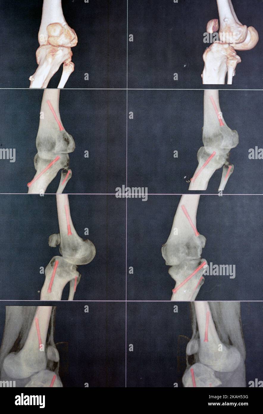 CT angiography of the left knee 3D view showing patent popliteal artery and its bifurcations tibial and peroneal arteries on a patient with bilateral Stock Photohttps://www.alamy.com/image-license-details/?v=1https://www.alamy.com/ct-angiography-of-the-left-knee-3d-view-showing-patent-popliteal-artery-and-its-bifurcations-tibial-and-peroneal-arteries-on-a-patient-with-bilateral-image488414068.html
RF2KAH53G–CT angiography of the left knee 3D view showing patent popliteal artery and its bifurcations tibial and peroneal arteries on a patient with bilateral
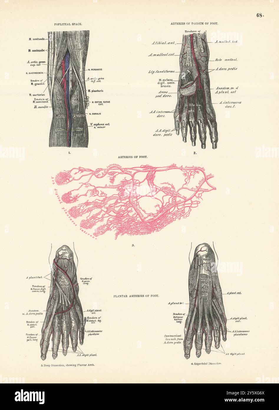 Anatomy. Popliteal Space, Dorsum & Foot Arteries 1880 old antique print Stock Photohttps://www.alamy.com/image-license-details/?v=1https://www.alamy.com/anatomy-popliteal-space-dorsum-foot-arteries-1880-old-antique-print-image623230018.html
RF2Y5XG6X–Anatomy. Popliteal Space, Dorsum & Foot Arteries 1880 old antique print
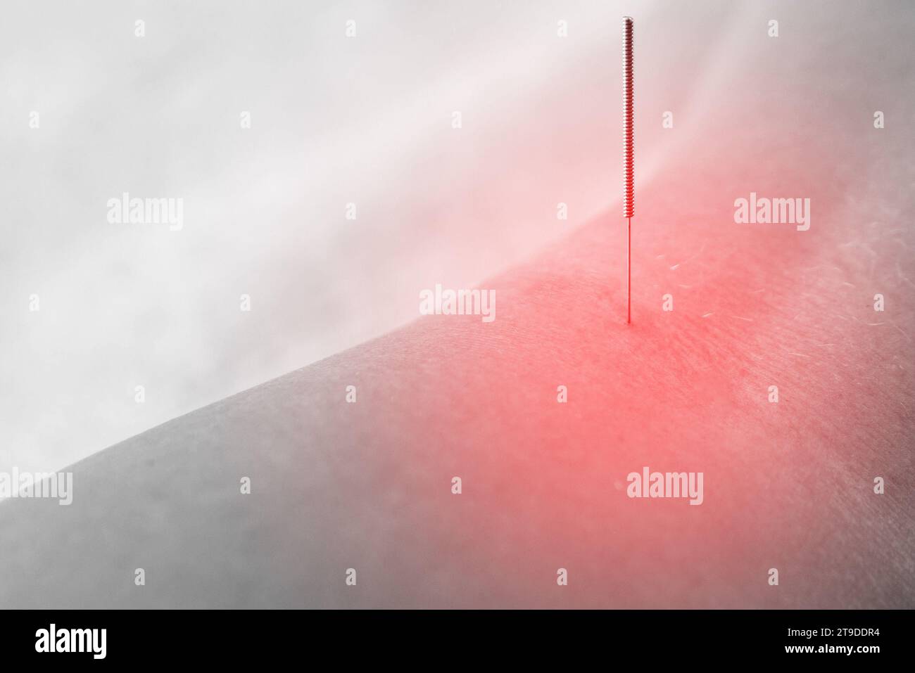 Alternative medicine. Close-up of female popliteal fossa with steel needles inserted during procedure of acupuncture therapy. Stock Photohttps://www.alamy.com/image-license-details/?v=1https://www.alamy.com/alternative-medicine-close-up-of-female-popliteal-fossa-with-steel-needles-inserted-during-procedure-of-acupuncture-therapy-image573770264.html
RF2T9DDR4–Alternative medicine. Close-up of female popliteal fossa with steel needles inserted during procedure of acupuncture therapy.
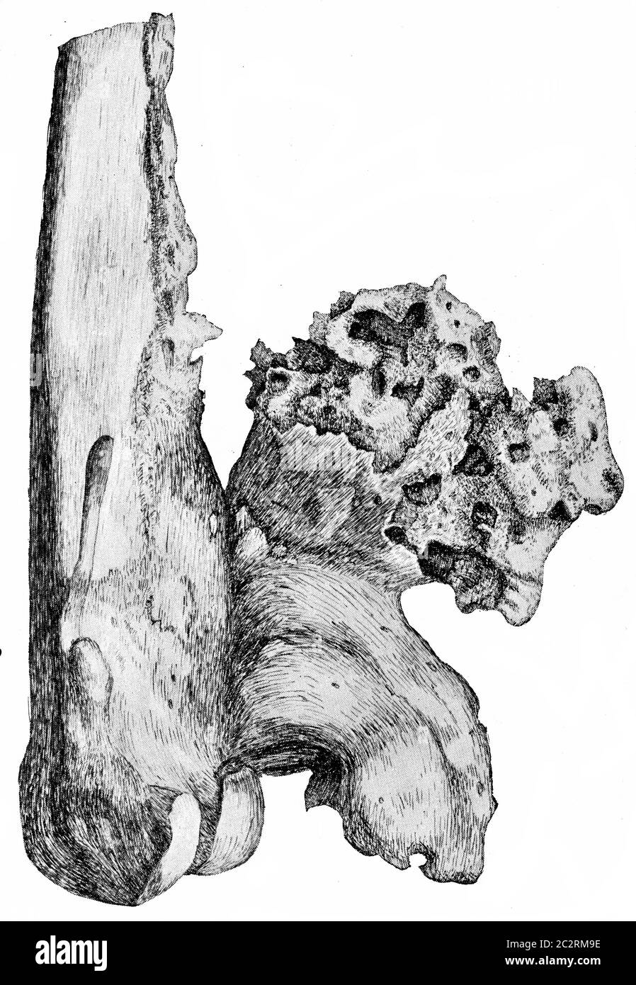 Osteophytes on the popliteal aspect of the lower end of the femur, vintage engraved illustration. Stock Photohttps://www.alamy.com/image-license-details/?v=1https://www.alamy.com/osteophytes-on-the-popliteal-aspect-of-the-lower-end-of-the-femur-vintage-engraved-illustration-image363167882.html
RF2C2RM9E–Osteophytes on the popliteal aspect of the lower end of the femur, vintage engraved illustration.
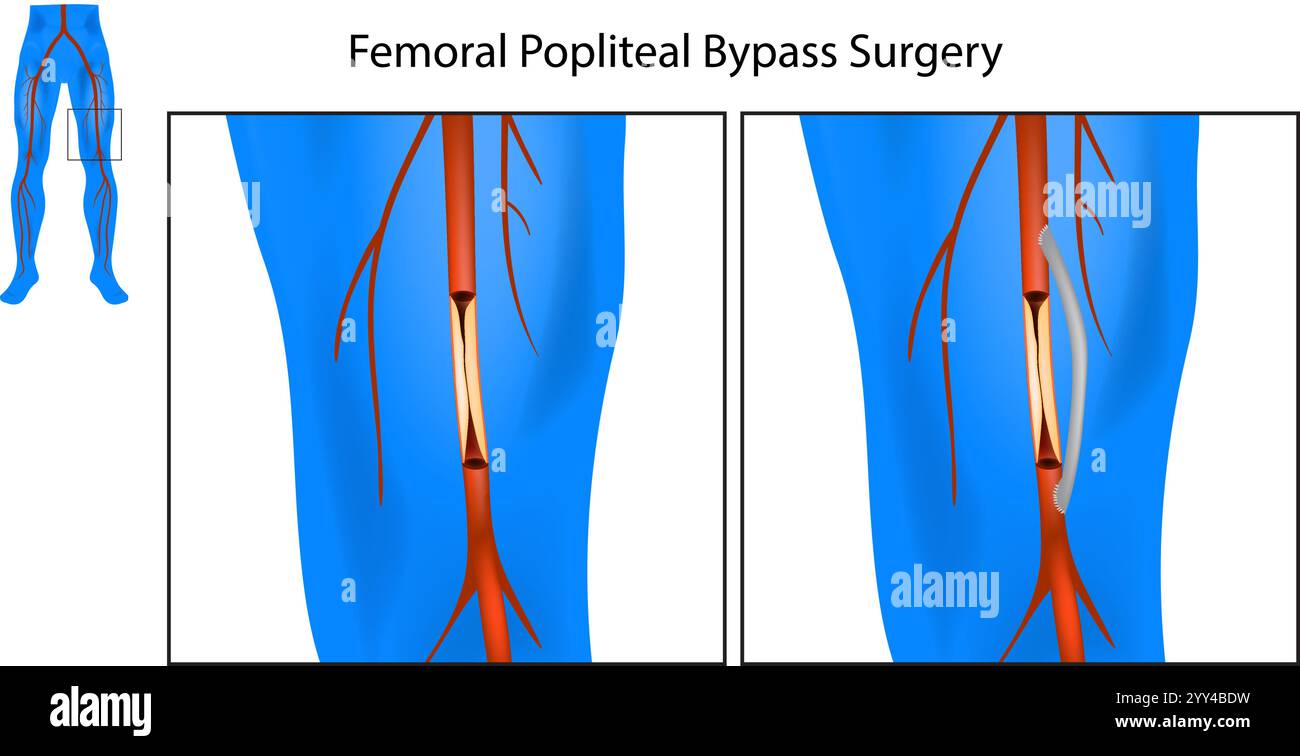 Femoral Popliteal Bypass Surgery. Leg Artery Bypass Surgery Diagram. Medical Chart of the Procedure for Leg Artery Blockage Stock Vectorhttps://www.alamy.com/image-license-details/?v=1https://www.alamy.com/femoral-popliteal-bypass-surgery-leg-artery-bypass-surgery-diagram-medical-chart-of-the-procedure-for-leg-artery-blockage-image636265781.html
RF2YY4BDW–Femoral Popliteal Bypass Surgery. Leg Artery Bypass Surgery Diagram. Medical Chart of the Procedure for Leg Artery Blockage
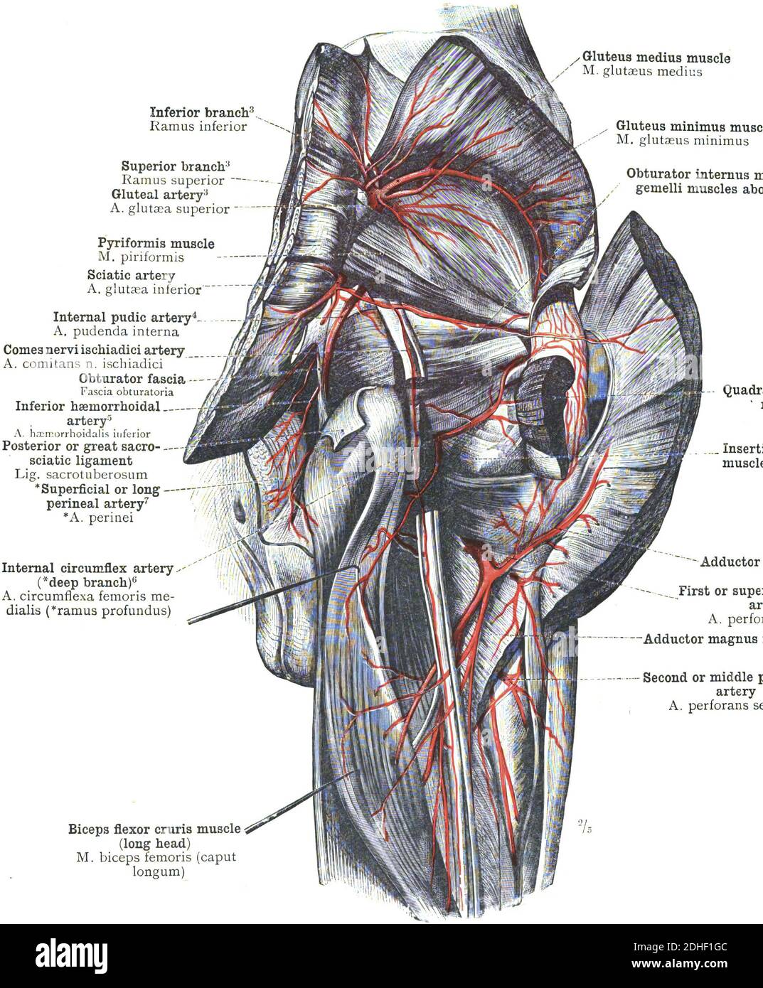 The anatomy of popliteal artery on a white background Stock Photohttps://www.alamy.com/image-license-details/?v=1https://www.alamy.com/the-anatomy-of-popliteal-artery-on-a-white-background-image389407772.html
RF2DHF1GC–The anatomy of popliteal artery on a white background
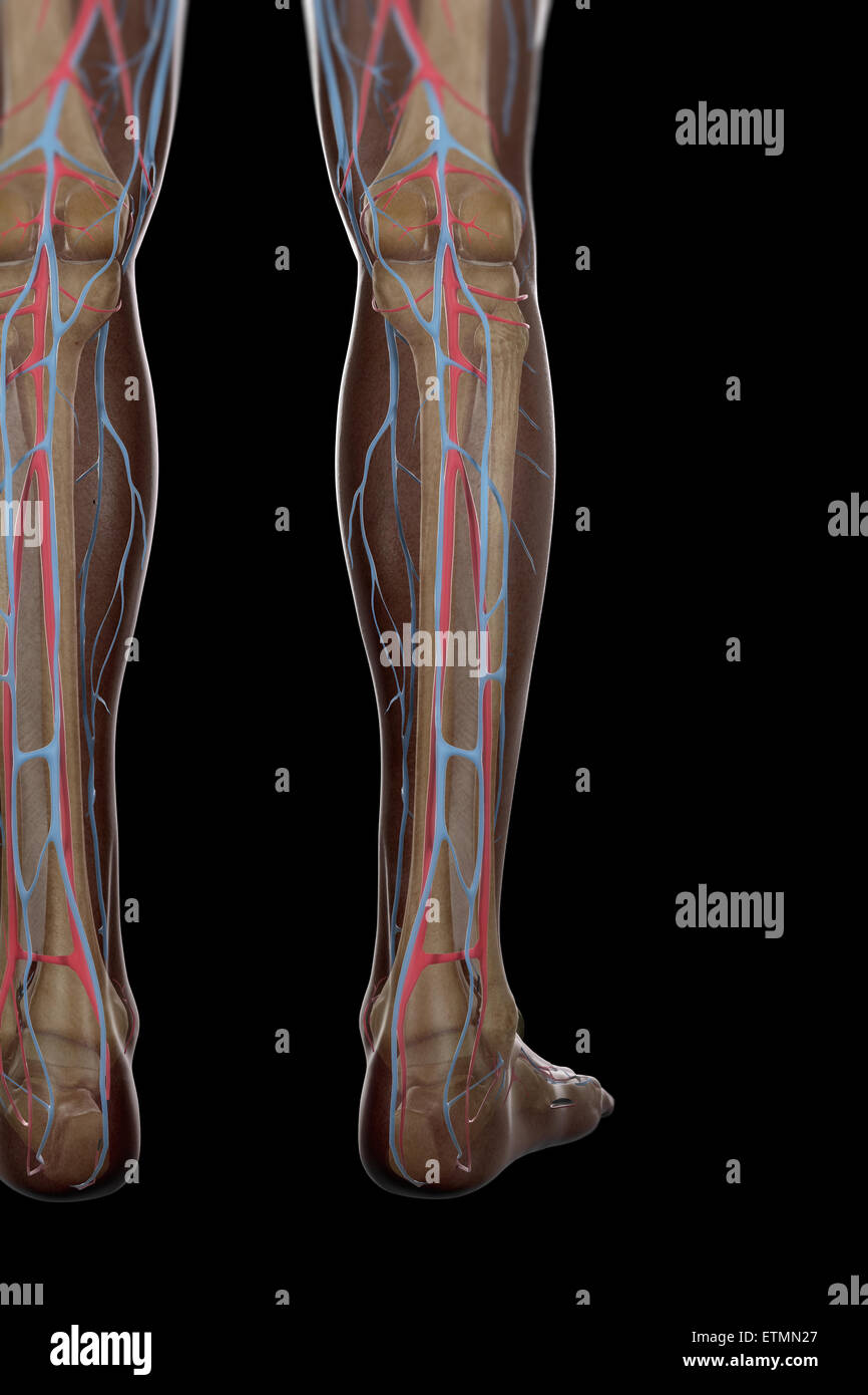 Illustration of the blood supply and skeletal structure of the lower legs, visible through skin. Stock Photohttps://www.alamy.com/image-license-details/?v=1https://www.alamy.com/stock-photo-illustration-of-the-blood-supply-and-skeletal-structure-of-the-lower-84048783.html
RMETMN27–Illustration of the blood supply and skeletal structure of the lower legs, visible through skin.
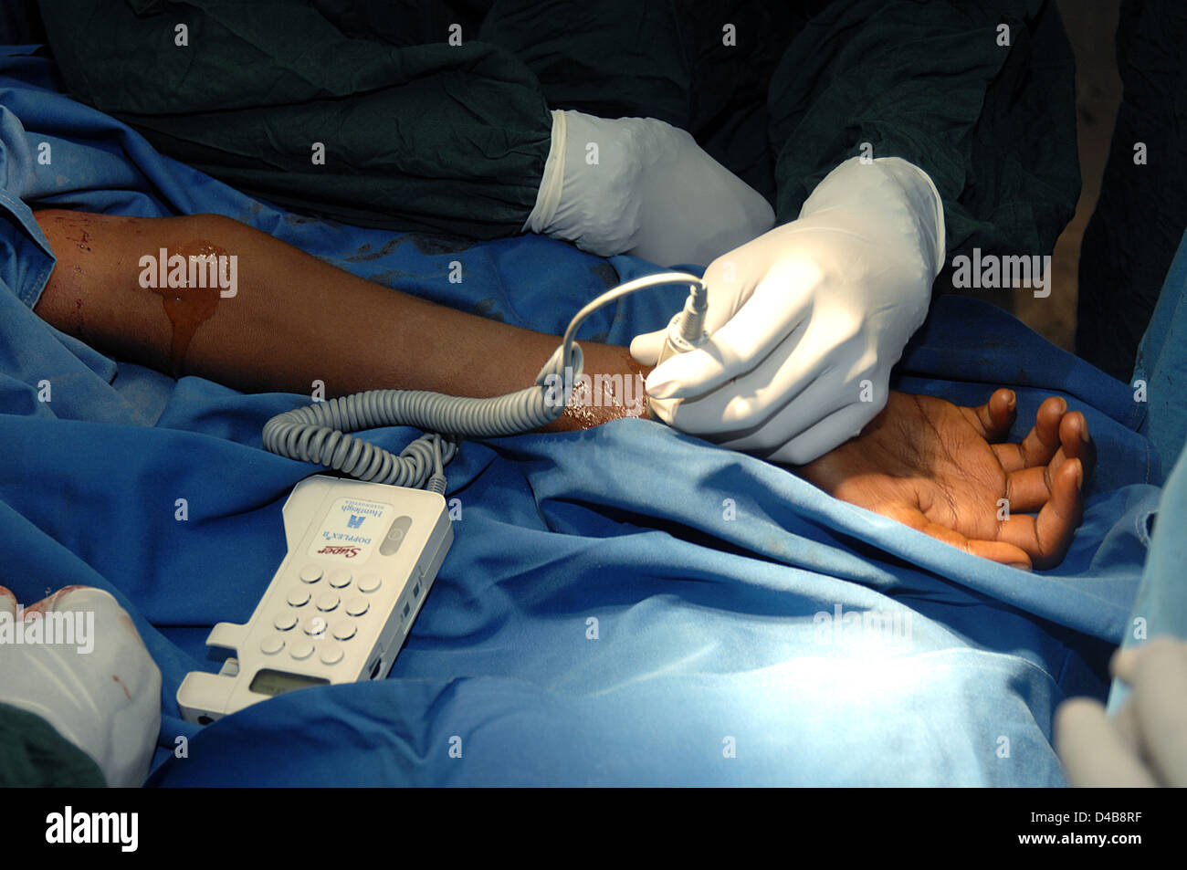 Surgeons use a carotid Doppler machine to check for a pulse on the arm of a patient, during surgery. Sudan, Africa. Stock Photohttps://www.alamy.com/image-license-details/?v=1https://www.alamy.com/stock-photo-surgeons-use-a-carotid-doppler-machine-to-check-for-a-pulse-on-the-54338131.html
RMD4B8RF–Surgeons use a carotid Doppler machine to check for a pulse on the arm of a patient, during surgery. Sudan, Africa.
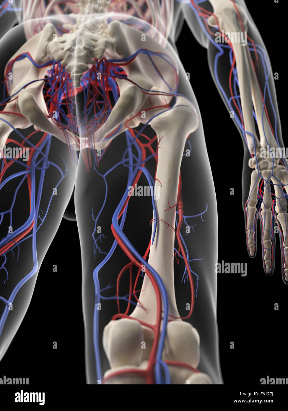 medically accurate illustration of the leg blood supply Stock Photohttps://www.alamy.com/image-license-details/?v=1https://www.alamy.com/stock-photo-medically-accurate-illustration-of-the-leg-blood-supply-89767430.html
RFF6177J–medically accurate illustration of the leg blood supply
 Anatomy of female body with arteries and veins. Stock Photohttps://www.alamy.com/image-license-details/?v=1https://www.alamy.com/stock-photo-anatomy-of-female-body-with-arteries-and-veins-59361030.html
RFDCG3GP–Anatomy of female body with arteries and veins.
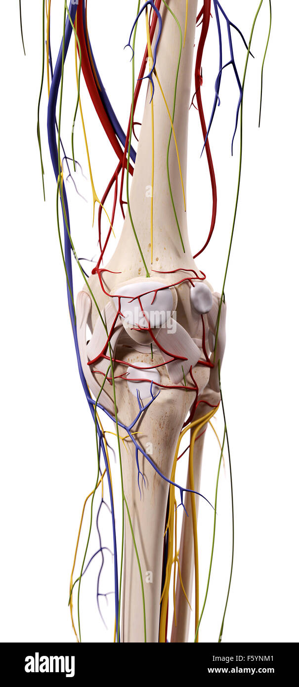 medical accurate illustration of the knee anatomy Stock Photohttps://www.alamy.com/image-license-details/?v=1https://www.alamy.com/stock-photo-medical-accurate-illustration-of-the-knee-anatomy-89734849.html
RFF5YNM1–medical accurate illustration of the knee anatomy
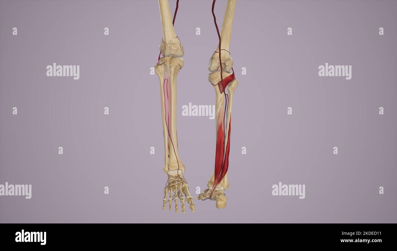 Arterial Supply to the Anterior and Posterior Leg Via Popliteal Artery and Its Branches Stock Photohttps://www.alamy.com/image-license-details/?v=1https://www.alamy.com/arterial-supply-to-the-anterior-and-posterior-leg-via-popliteal-artery-and-its-branches-image490198381.html
RF2KDED11–Arterial Supply to the Anterior and Posterior Leg Via Popliteal Artery and Its Branches
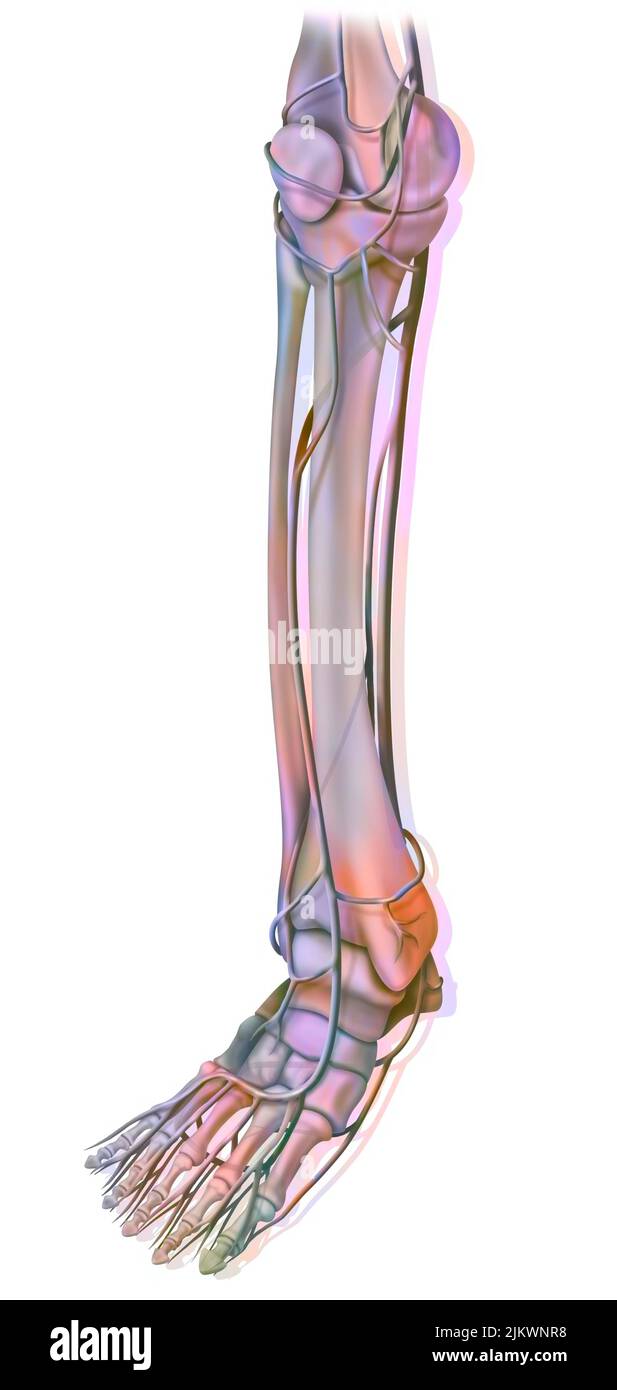 The arteries of the lower part of the lower extremity. Stock Photohttps://www.alamy.com/image-license-details/?v=1https://www.alamy.com/the-arteries-of-the-lower-part-of-the-lower-extremity-image476924316.html
RF2JKWNR8–The arteries of the lower part of the lower extremity.
 Sciatic Nerve on Black Background.3d rendering Stock Photohttps://www.alamy.com/image-license-details/?v=1https://www.alamy.com/sciatic-nerve-on-black-background3d-rendering-image501580952.html
RF2M40YHC–Sciatic Nerve on Black Background.3d rendering
 ANEURYSM POPLITEAL ARTERY Stock Photohttps://www.alamy.com/image-license-details/?v=1https://www.alamy.com/stock-photo-aneurysm-popliteal-artery-49173208.html
RMCT00X0–ANEURYSM POPLITEAL ARTERY
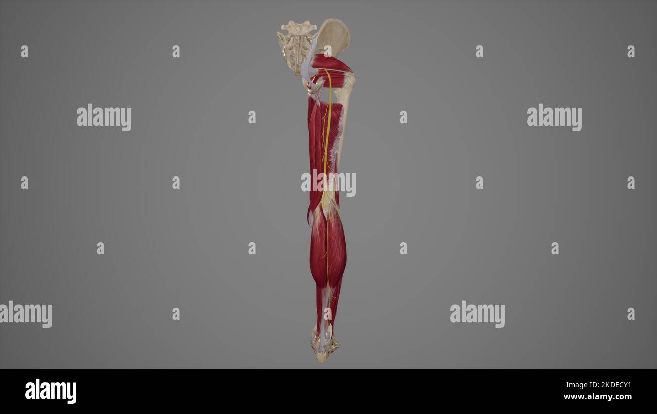 Anatomical Illustration of Sciatic Nerve Stock Photohttps://www.alamy.com/image-license-details/?v=1https://www.alamy.com/anatomical-illustration-of-sciatic-nerve-image490198325.html
RF2KDECY1–Anatomical Illustration of Sciatic Nerve
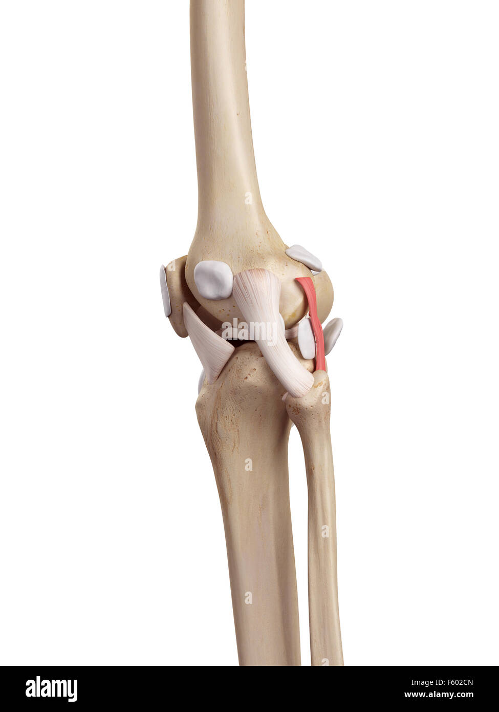 medical accurate illustration of the arcuate politeal ligament Stock Photohttps://www.alamy.com/image-license-details/?v=1https://www.alamy.com/stock-photo-medical-accurate-illustration-of-the-arcuate-politeal-ligament-89741701.html
RFF602CN–medical accurate illustration of the arcuate politeal ligament
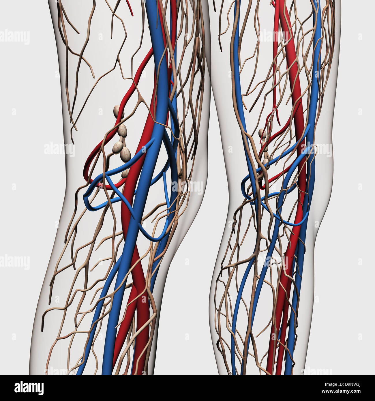 Medical illustration of arteries, veins and lymphatic system in human legs. Stock Photohttps://www.alamy.com/image-license-details/?v=1https://www.alamy.com/stock-photo-medical-illustration-of-arteries-veins-and-lymphatic-system-in-human-57643702.html
RFD9NW3J–Medical illustration of arteries, veins and lymphatic system in human legs.
 Popliteal region (left), vintage engraved illustration. Usual Medicine Dictionary by Dr Labarthe - 1885. Stock Vectorhttps://www.alamy.com/image-license-details/?v=1https://www.alamy.com/stock-photo-popliteal-region-left-vintage-engraved-illustration-usual-medicine-84419777.html
RFEW9J81–Popliteal region (left), vintage engraved illustration. Usual Medicine Dictionary by Dr Labarthe - 1885.
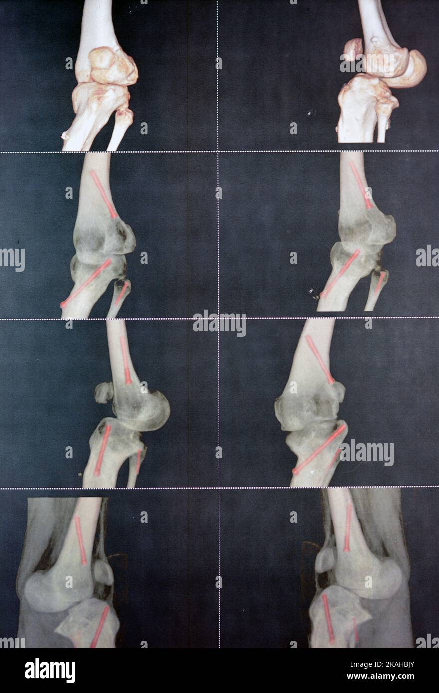 CT angiography of the left knee 3D view showing patent popliteal artery and its bifurcations tibial and peroneal arteries on a patient with bilateral Stock Photohttps://www.alamy.com/image-license-details/?v=1https://www.alamy.com/ct-angiography-of-the-left-knee-3d-view-showing-patent-popliteal-artery-and-its-bifurcations-tibial-and-peroneal-arteries-on-a-patient-with-bilateral-image488419203.html
RF2KAHBJY–CT angiography of the left knee 3D view showing patent popliteal artery and its bifurcations tibial and peroneal arteries on a patient with bilateral
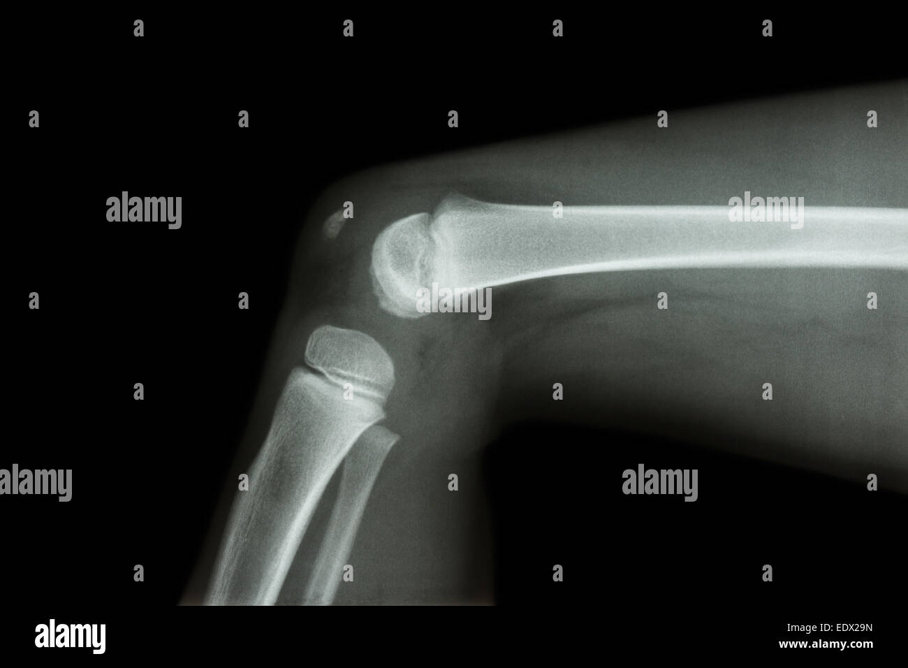 film x-ray knee lateral of child Stock Photohttps://www.alamy.com/image-license-details/?v=1https://www.alamy.com/stock-photo-film-x-ray-knee-lateral-of-child-77404593.html
RFEDX29N–film x-ray knee lateral of child
 Alternative medicine. Close-up of female popliteal fossa with steel needles inserted during procedure of acupuncture therapy. Stock Photohttps://www.alamy.com/image-license-details/?v=1https://www.alamy.com/alternative-medicine-close-up-of-female-popliteal-fossa-with-steel-needles-inserted-during-procedure-of-acupuncture-therapy-image573770395.html
RF2T9DDYR–Alternative medicine. Close-up of female popliteal fossa with steel needles inserted during procedure of acupuncture therapy.
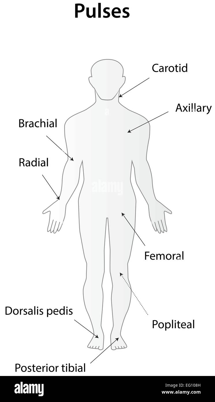 Pulses Stock Vectorhttps://www.alamy.com/image-license-details/?v=1https://www.alamy.com/stock-photo-pulses-78698161.html
RFEG108H–Pulses
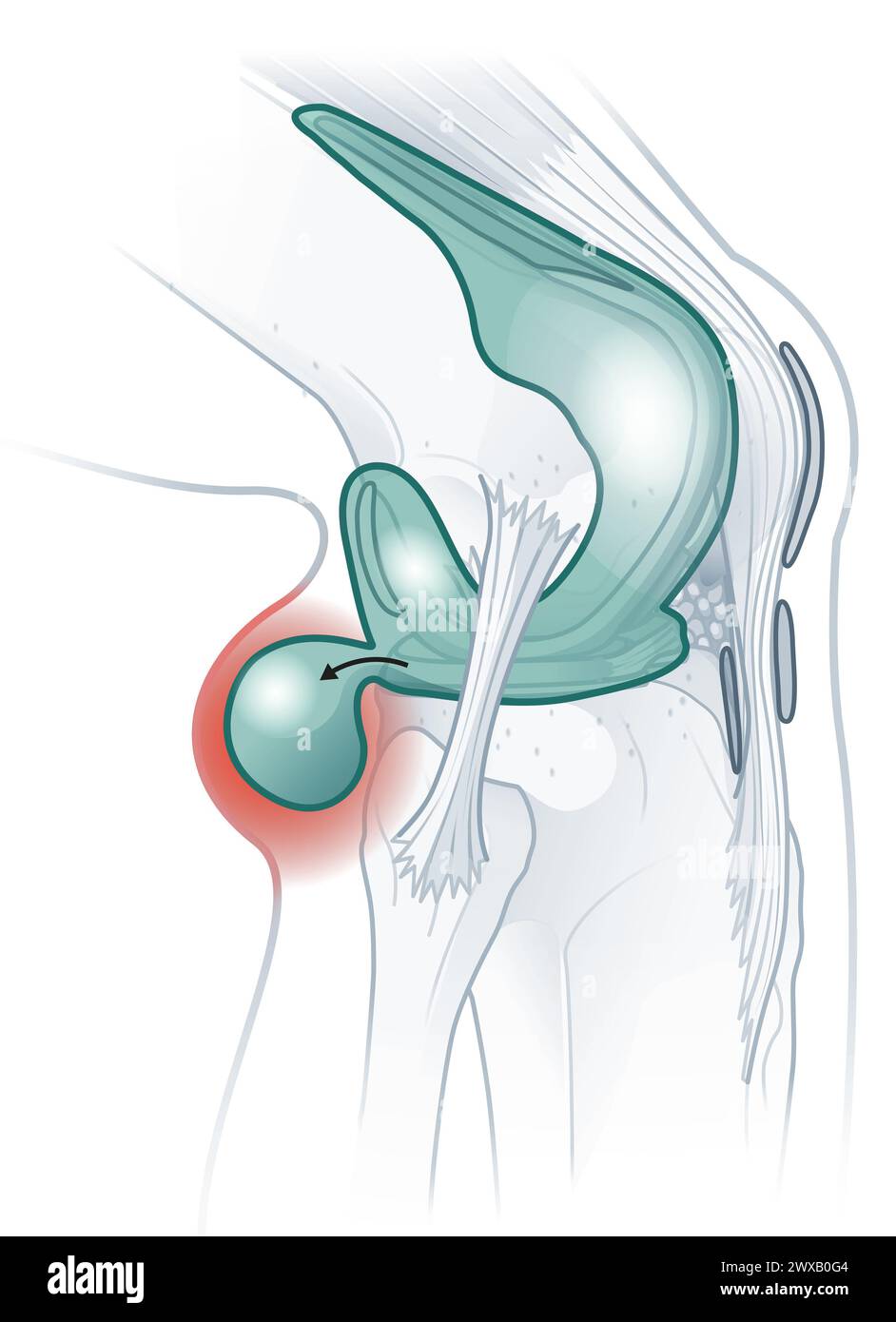 Baker's cyst: fluid-filled swelling behind the knee due to underlying knee issues, causing pain, stiffness, may require treatment. Stock Photohttps://www.alamy.com/image-license-details/?v=1https://www.alamy.com/bakers-cyst-fluid-filled-swelling-behind-the-knee-due-to-underlying-knee-issues-causing-pain-stiffness-may-require-treatment-image601375492.html
RF2WXB0G4–Baker's cyst: fluid-filled swelling behind the knee due to underlying knee issues, causing pain, stiffness, may require treatment.
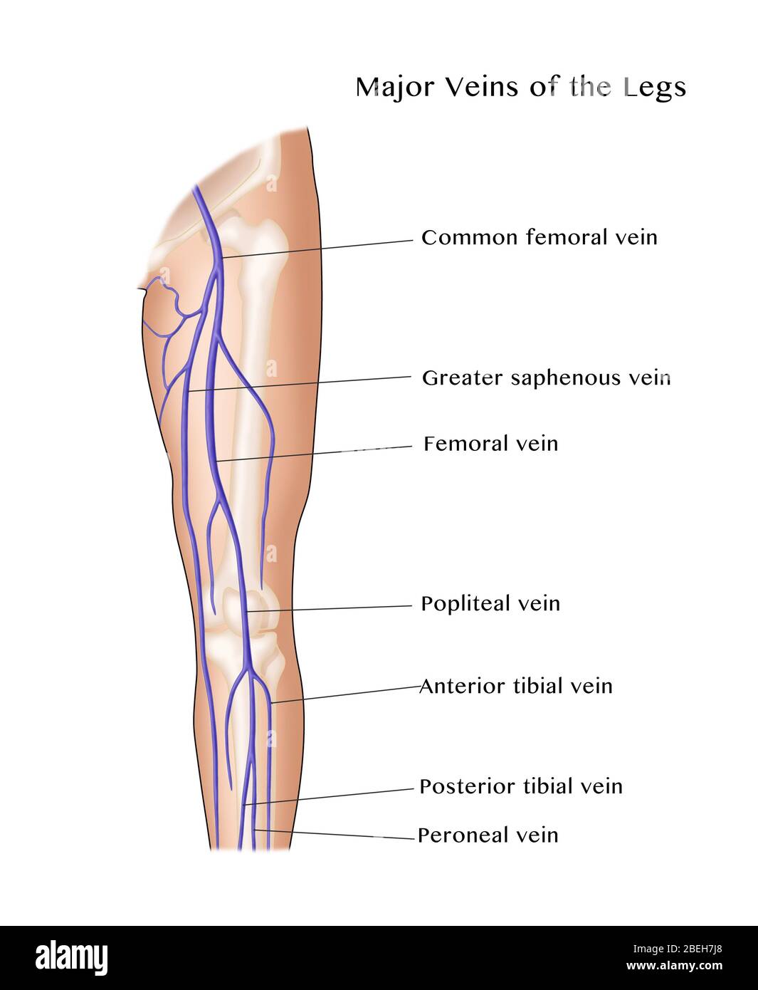 Major Veins of the Leg Stock Photohttps://www.alamy.com/image-license-details/?v=1https://www.alamy.com/major-veins-of-the-leg-image353191728.html
RM2BEH7J8–Major Veins of the Leg
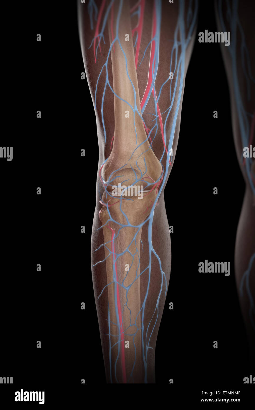 Illustration of the blood supply and skeletal system of the lower legs, visible through skin. Stock Photohttps://www.alamy.com/image-license-details/?v=1https://www.alamy.com/stock-photo-illustration-of-the-blood-supply-and-skeletal-system-of-the-lower-84049295.html
RMETMNMF–Illustration of the blood supply and skeletal system of the lower legs, visible through skin.
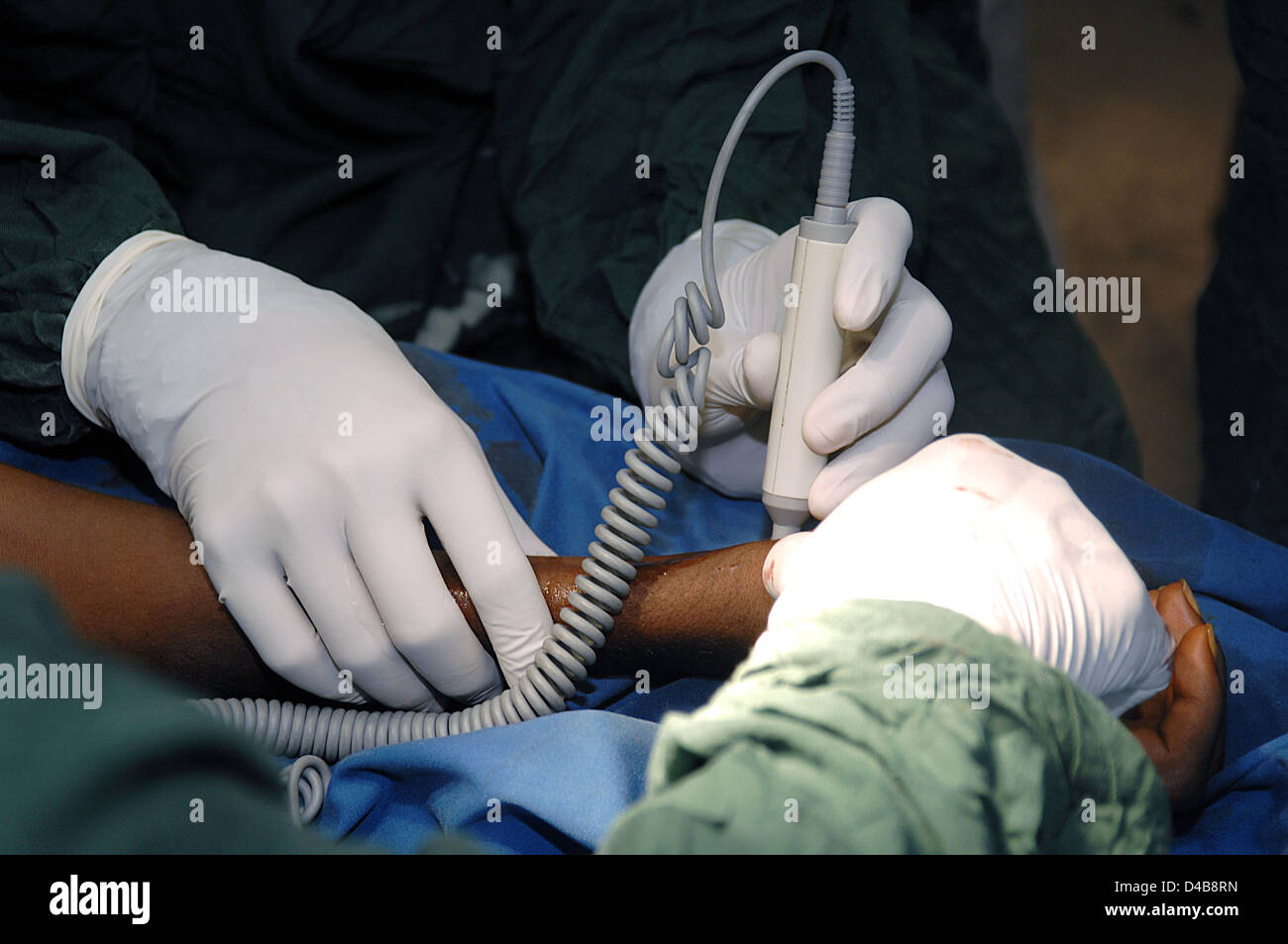 Surgeons use a carotid Doppler machine to check for a pulse on the arm of a patient, during surgery. Sudan, Africa. Stock Photohttps://www.alamy.com/image-license-details/?v=1https://www.alamy.com/stock-photo-surgeons-use-a-carotid-doppler-machine-to-check-for-a-pulse-on-the-54338137.html
RMD4B8RN–Surgeons use a carotid Doppler machine to check for a pulse on the arm of a patient, during surgery. Sudan, Africa.
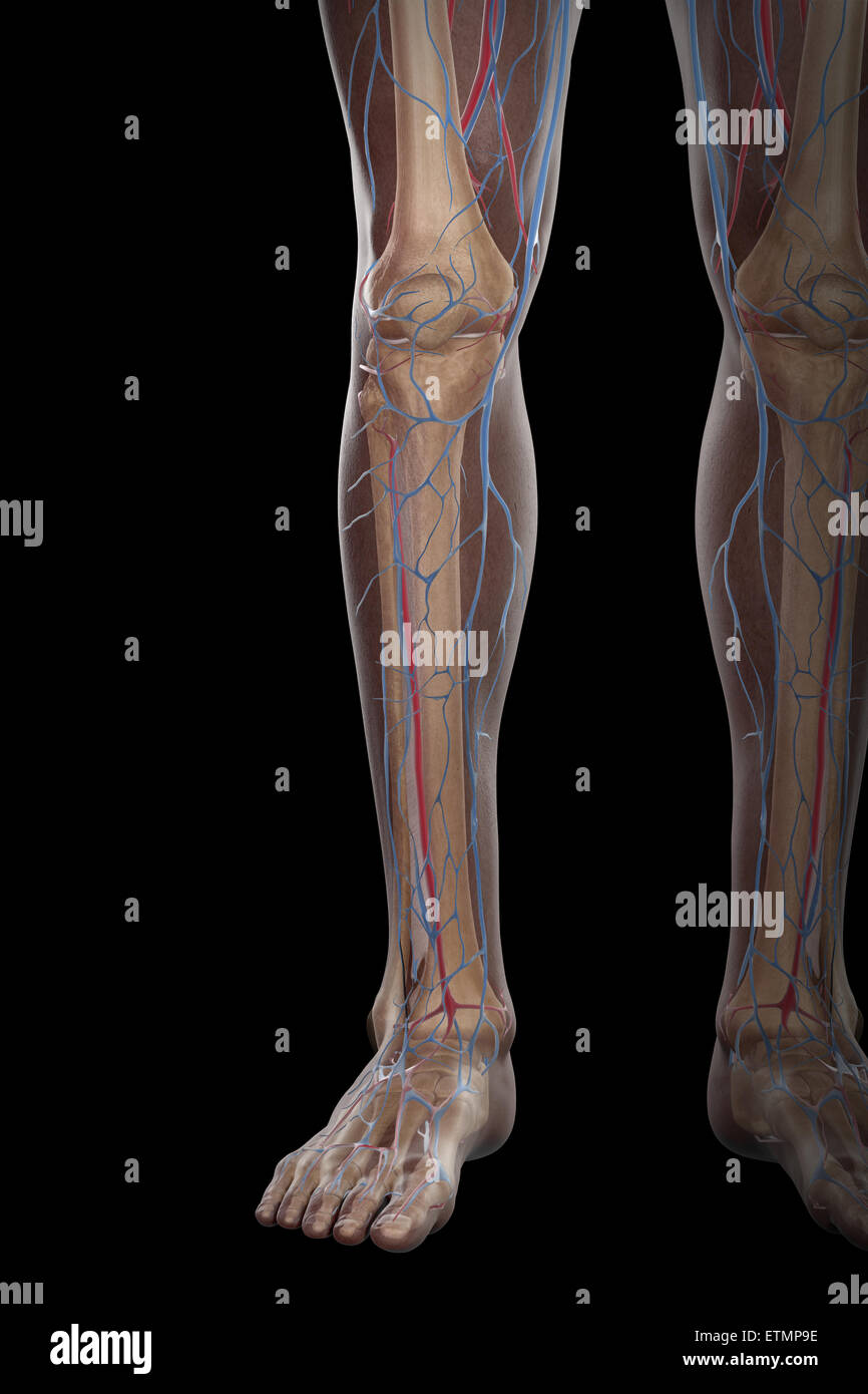 Illustration of the blood supply and skeletal structure of the lower legs, visible through skin. Stock Photohttps://www.alamy.com/image-license-details/?v=1https://www.alamy.com/stock-photo-illustration-of-the-blood-supply-and-skeletal-structure-of-the-lower-84049770.html
RMETMP9E–Illustration of the blood supply and skeletal structure of the lower legs, visible through skin.
 Vintage illustrations of Nerves of the Human Body 1900s Stock Photohttps://www.alamy.com/image-license-details/?v=1https://www.alamy.com/vintage-illustrations-of-nerves-of-the-human-body-1900s-image350211994.html
RF2B9NEY6–Vintage illustrations of Nerves of the Human Body 1900s
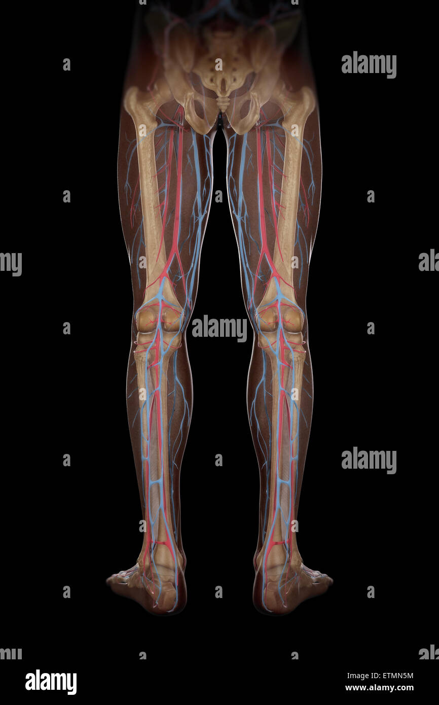 Illustration of the blood supply and skeletal structure of the legs, visible through skin. Stock Photohttps://www.alamy.com/image-license-details/?v=1https://www.alamy.com/stock-photo-illustration-of-the-blood-supply-and-skeletal-structure-of-the-legs-84048880.html
RMETMN5M–Illustration of the blood supply and skeletal structure of the legs, visible through skin.
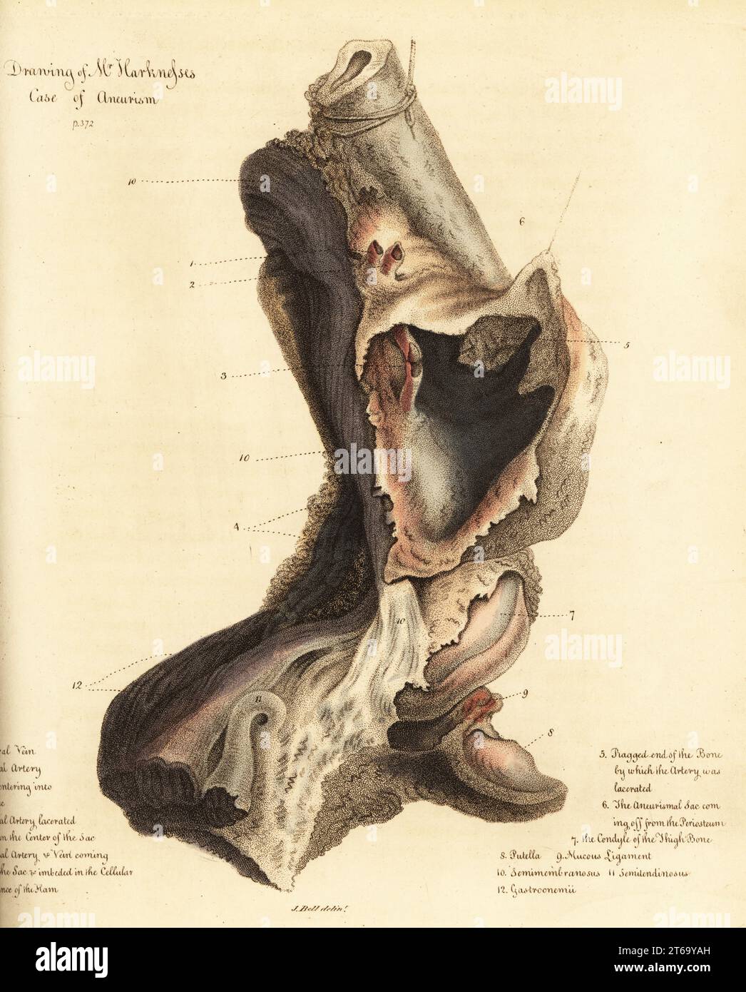 Fatal aneurysmal sac from the broken leg of a man, 1815. The sailor died of gangrene before the leg could be amputated. Drawing of Mr. Harkness's case of aneurism. Femoral vein 1, femoral artery 2, lacerated femoral artery 3, popliteal artery 4, ragged end of the bone 5, aneurysmal sac 6, condyle of the thigh bone 7, putella 8, mucous ligament 9, semimembranosus 10, semilendinosus 11, gastroenemii 12. Handcoloured copperplate engraving by after an illustration by John Bell from his own Principles of Surgery, as they Relate to Wounds, Ulcers and Fistulas, Longman, Hurst, Rees, Orme and Brown, L Stock Photohttps://www.alamy.com/image-license-details/?v=1https://www.alamy.com/fatal-aneurysmal-sac-from-the-broken-leg-of-a-man-1815-the-sailor-died-of-gangrene-before-the-leg-could-be-amputated-drawing-of-mr-harknesss-case-of-aneurism-femoral-vein-1-femoral-artery-2-lacerated-femoral-artery-3-popliteal-artery-4-ragged-end-of-the-bone-5-aneurysmal-sac-6-condyle-of-the-thigh-bone-7-putella-8-mucous-ligament-9-semimembranosus-10-semilendinosus-11-gastroenemii-12-handcoloured-copperplate-engraving-by-after-an-illustration-by-john-bell-from-his-own-principles-of-surgery-as-they-relate-to-wounds-ulcers-and-fistulas-longman-hurst-rees-orme-and-brown-l-image571849113.html
RM2T69YAH–Fatal aneurysmal sac from the broken leg of a man, 1815. The sailor died of gangrene before the leg could be amputated. Drawing of Mr. Harkness's case of aneurism. Femoral vein 1, femoral artery 2, lacerated femoral artery 3, popliteal artery 4, ragged end of the bone 5, aneurysmal sac 6, condyle of the thigh bone 7, putella 8, mucous ligament 9, semimembranosus 10, semilendinosus 11, gastroenemii 12. Handcoloured copperplate engraving by after an illustration by John Bell from his own Principles of Surgery, as they Relate to Wounds, Ulcers and Fistulas, Longman, Hurst, Rees, Orme and Brown, L
 popliteal fossa, back of the knee joint illustration Stock Photohttps://www.alamy.com/image-license-details/?v=1https://www.alamy.com/stock-photo-popliteal-fossa-back-of-the-knee-joint-illustration-39555737.html
RFC89WMW–popliteal fossa, back of the knee joint illustration
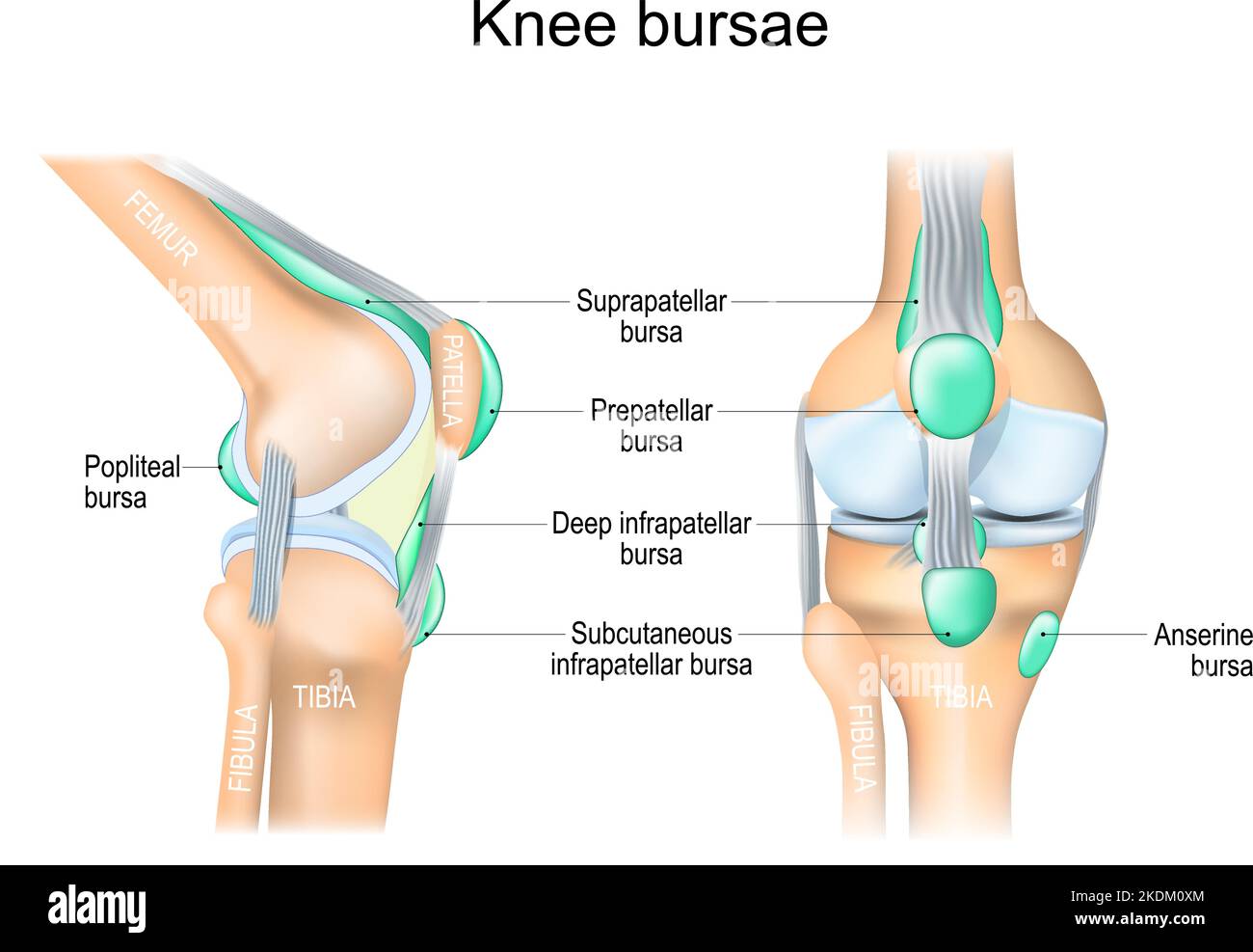 Knee bursae. synovial pockets or sacs that surround the knee joint cavity. Synovial joint anatomy. Frontal and side view of human knee joint. Vector Stock Vectorhttps://www.alamy.com/image-license-details/?v=1https://www.alamy.com/knee-bursae-synovial-pockets-or-sacs-that-surround-the-knee-joint-cavity-synovial-joint-anatomy-frontal-and-side-view-of-human-knee-joint-vector-image490320620.html
RF2KDM0XM–Knee bursae. synovial pockets or sacs that surround the knee joint cavity. Synovial joint anatomy. Frontal and side view of human knee joint. Vector
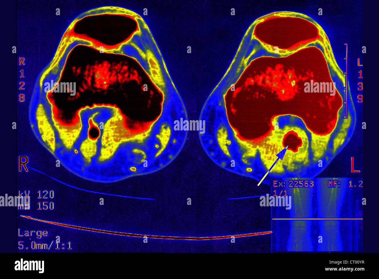 ANEURYSM POPLITEAL ARTERY Stock Photohttps://www.alamy.com/image-license-details/?v=1https://www.alamy.com/stock-photo-aneurysm-popliteal-artery-49173259.html
RMCT00YR–ANEURYSM POPLITEAL ARTERY
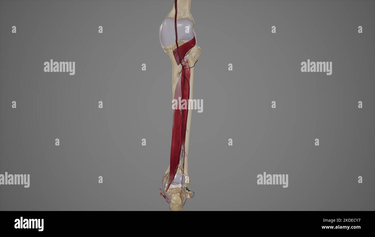 Anatomical Illustration of Tibial Arteries Stock Photohttps://www.alamy.com/image-license-details/?v=1https://www.alamy.com/anatomical-illustration-of-tibial-arteries-image490198331.html
RF2KDECY7–Anatomical Illustration of Tibial Arteries
 POPLITEAL ARTERY STENOSIS, ANGIO Stock Photohttps://www.alamy.com/image-license-details/?v=1https://www.alamy.com/stock-photo-popliteal-artery-stenosis-angio-49256825.html
RMCT3RG9–POPLITEAL ARTERY STENOSIS, ANGIO
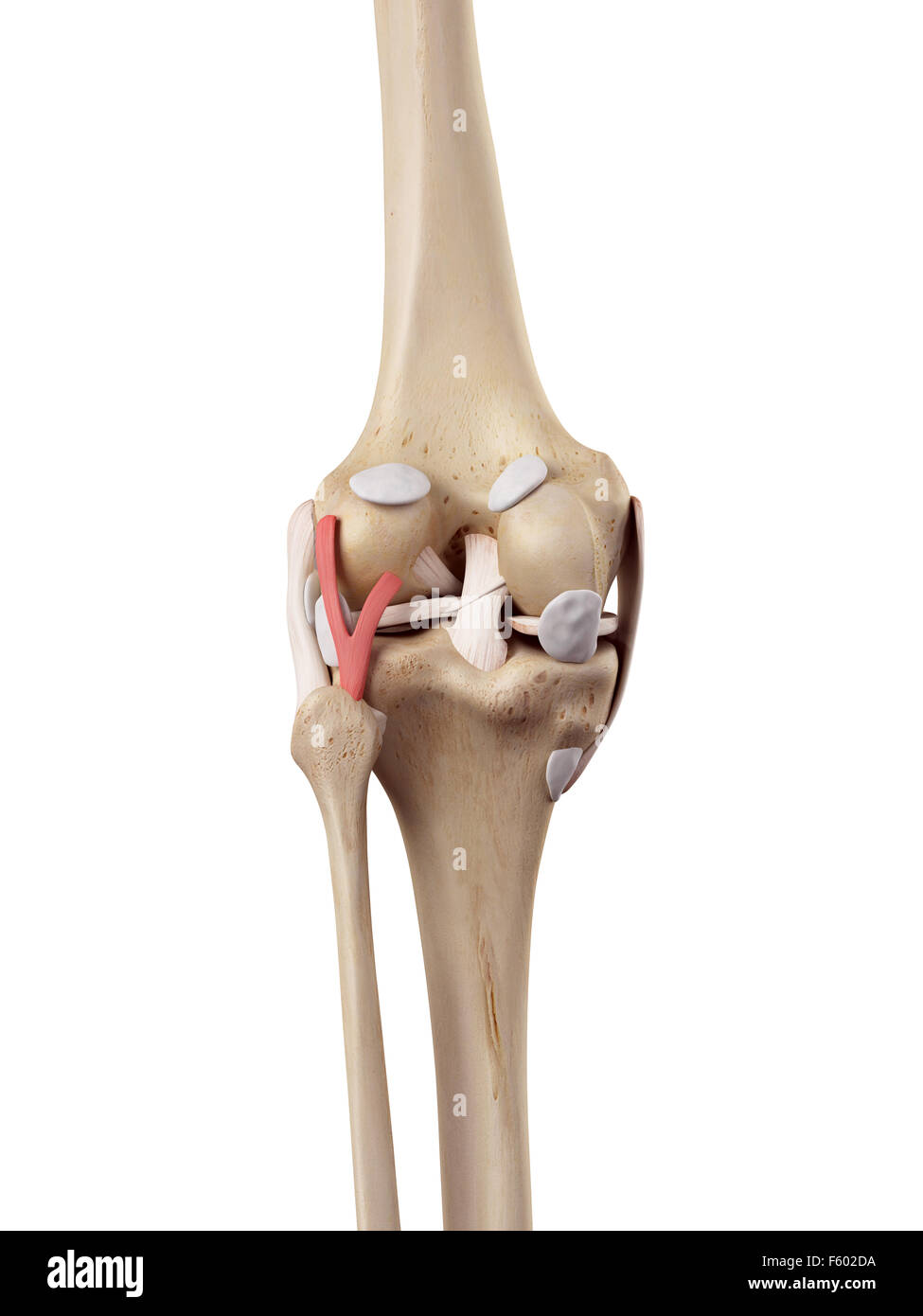 medical accurate illustration of the arcuate politeal ligament Stock Photohttps://www.alamy.com/image-license-details/?v=1https://www.alamy.com/stock-photo-medical-accurate-illustration-of-the-arcuate-politeal-ligament-89741718.html
RFF602DA–medical accurate illustration of the arcuate politeal ligament
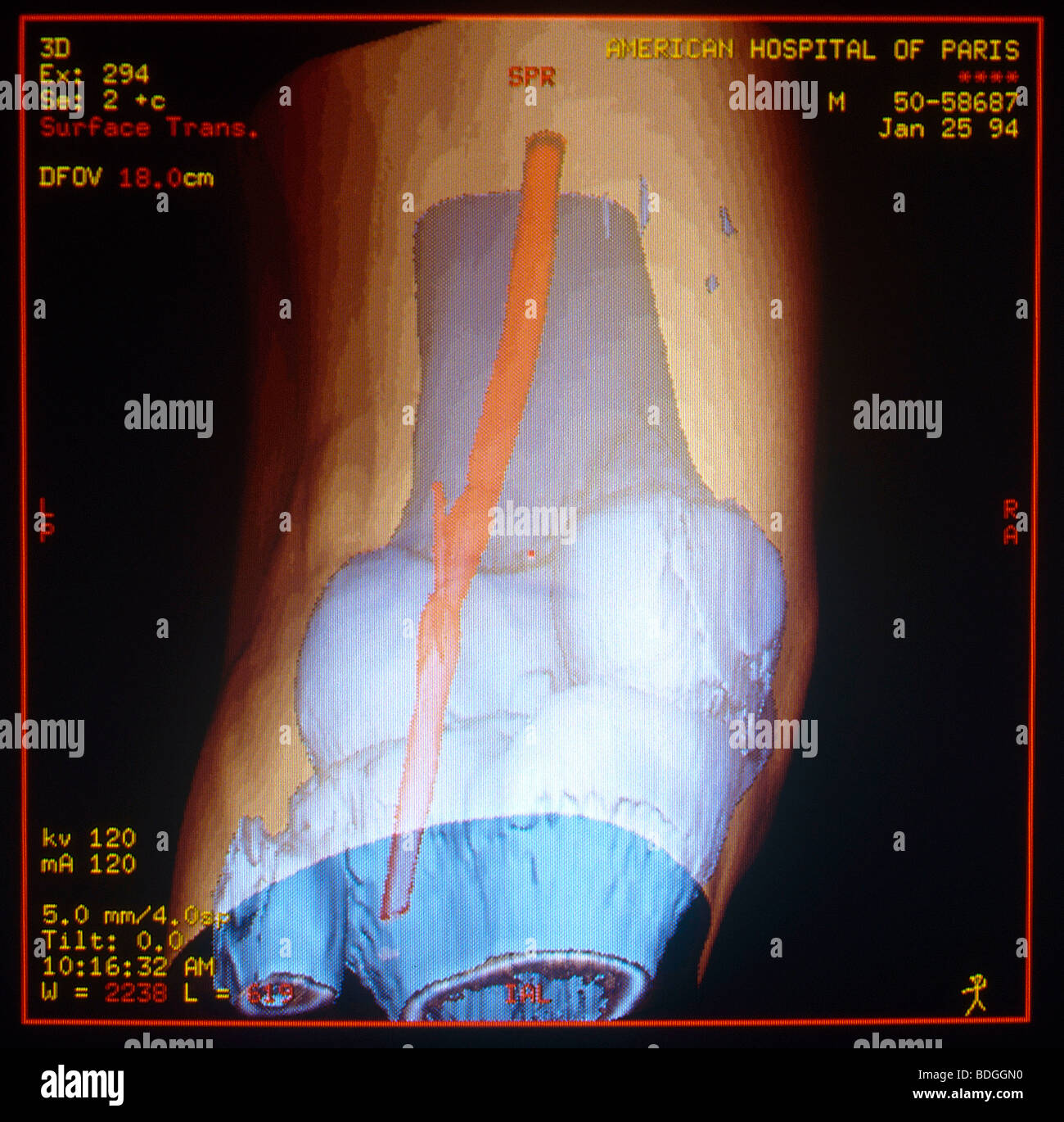 ARTERIAL STENOSIS, 3D SCAN Popliteal artery. Stock Photohttps://www.alamy.com/image-license-details/?v=1https://www.alamy.com/stock-photo-arterial-stenosis-3d-scan-popliteal-artery-25565260.html
RFBDGGN0–ARTERIAL STENOSIS, 3D SCAN Popliteal artery.
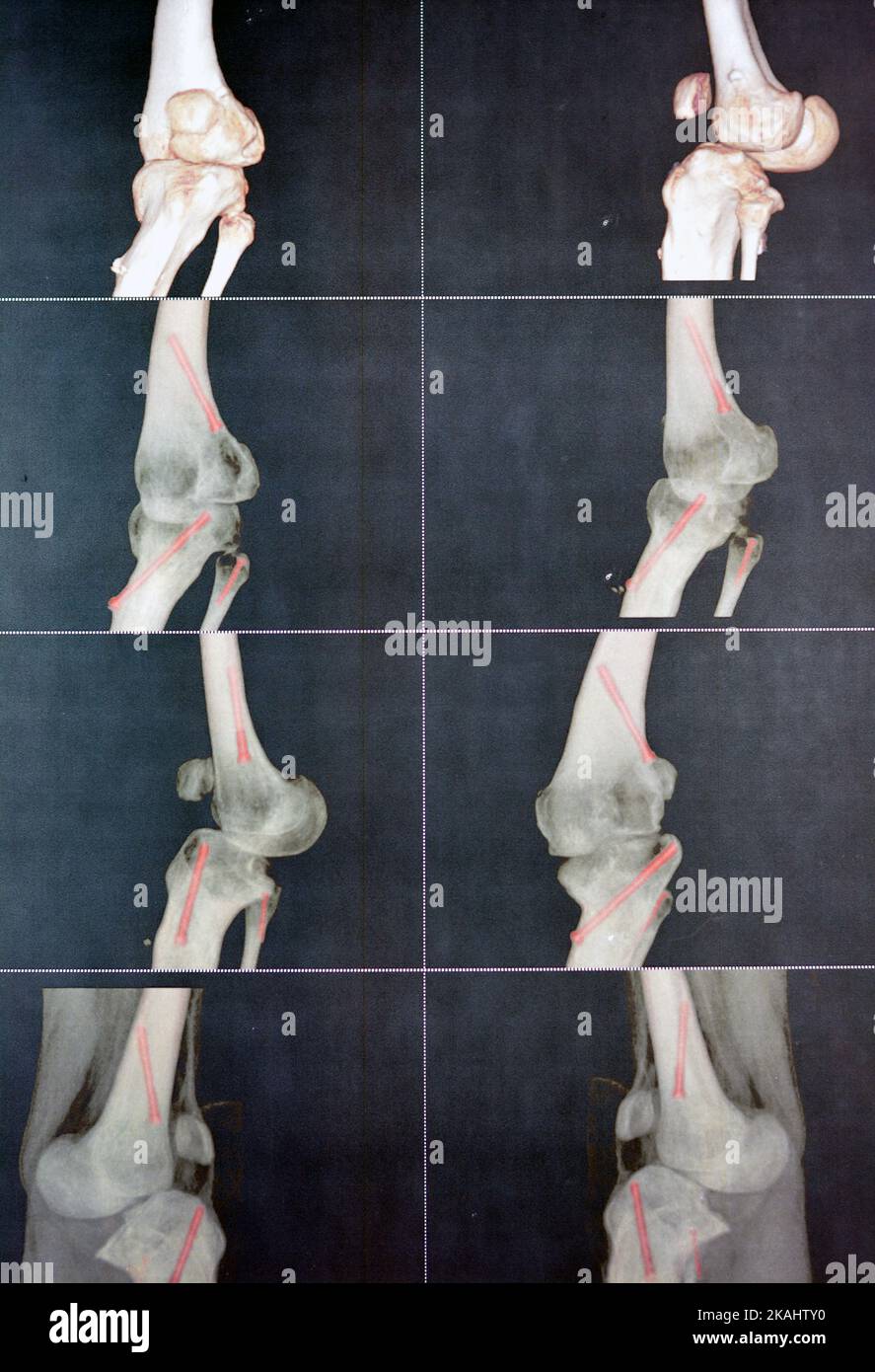 CT angiography of the left knee 3D view showing patent popliteal artery and its bifurcations tibial and peroneal arteries on a patient with bilateral Stock Photohttps://www.alamy.com/image-license-details/?v=1https://www.alamy.com/ct-angiography-of-the-left-knee-3d-view-showing-patent-popliteal-artery-and-its-bifurcations-tibial-and-peroneal-arteries-on-a-patient-with-bilateral-image488429620.html
RF2KAHTY0–CT angiography of the left knee 3D view showing patent popliteal artery and its bifurcations tibial and peroneal arteries on a patient with bilateral
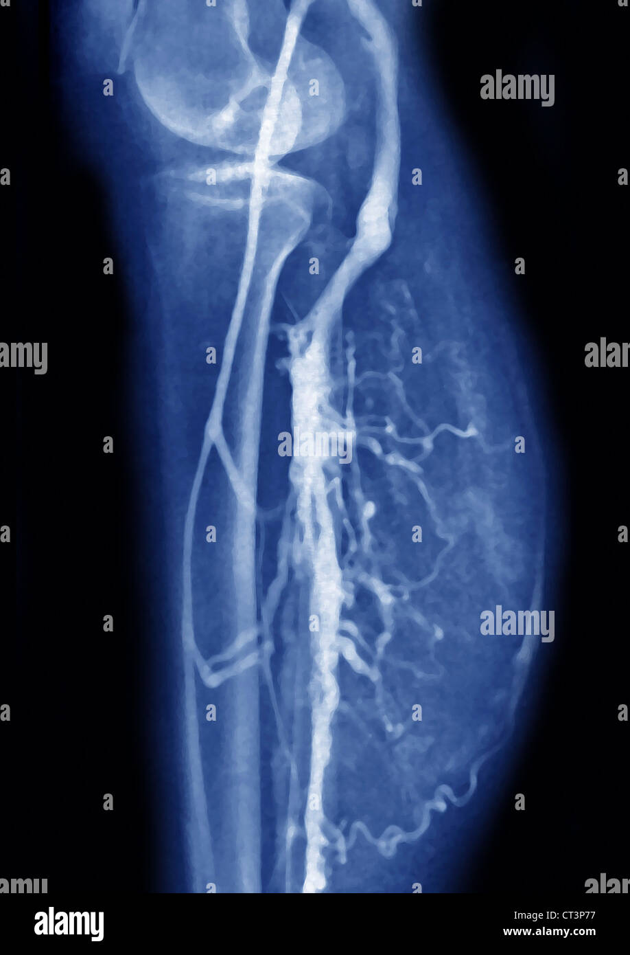 LOWER LIMB, ANGIOGRAPHY Stock Photohttps://www.alamy.com/image-license-details/?v=1https://www.alamy.com/stock-photo-lower-limb-angiography-49255787.html
RMCT3P77–LOWER LIMB, ANGIOGRAPHY
 medically accurate illustration of the arteries and veins of the knee Stock Photohttps://www.alamy.com/image-license-details/?v=1https://www.alamy.com/stock-photo-medically-accurate-illustration-of-the-arteries-and-veins-of-the-knee-89767432.html
RFF6177M–medically accurate illustration of the arteries and veins of the knee
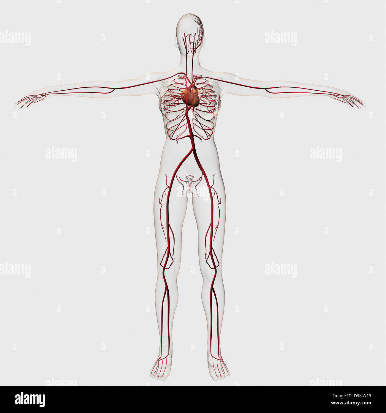 Medical illustration of female circulatory system with heart and arteries visible, full body view. Stock Photohttps://www.alamy.com/image-license-details/?v=1https://www.alamy.com/stock-photo-medical-illustration-of-female-circulatory-system-with-heart-and-arteries-57643661.html
RFD9NW25–Medical illustration of female circulatory system with heart and arteries visible, full body view.
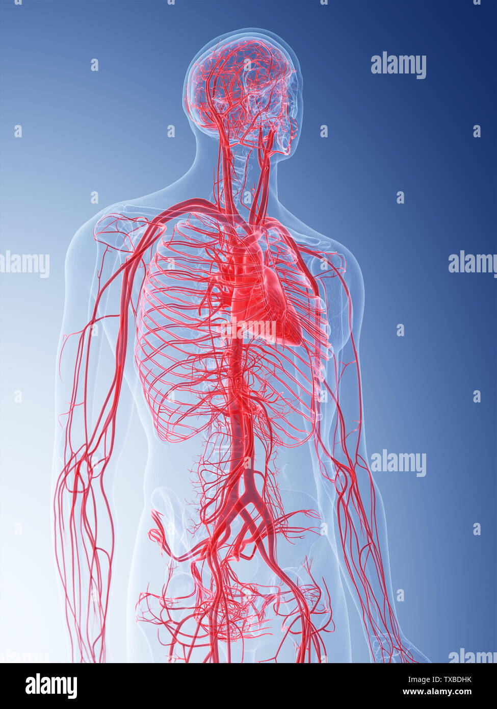 3d rendered medically accurate illustration of the vascular system Stock Photohttps://www.alamy.com/image-license-details/?v=1https://www.alamy.com/3d-rendered-medically-accurate-illustration-of-the-vascular-system-image257178367.html
RFTXBDHK–3d rendered medically accurate illustration of the vascular system
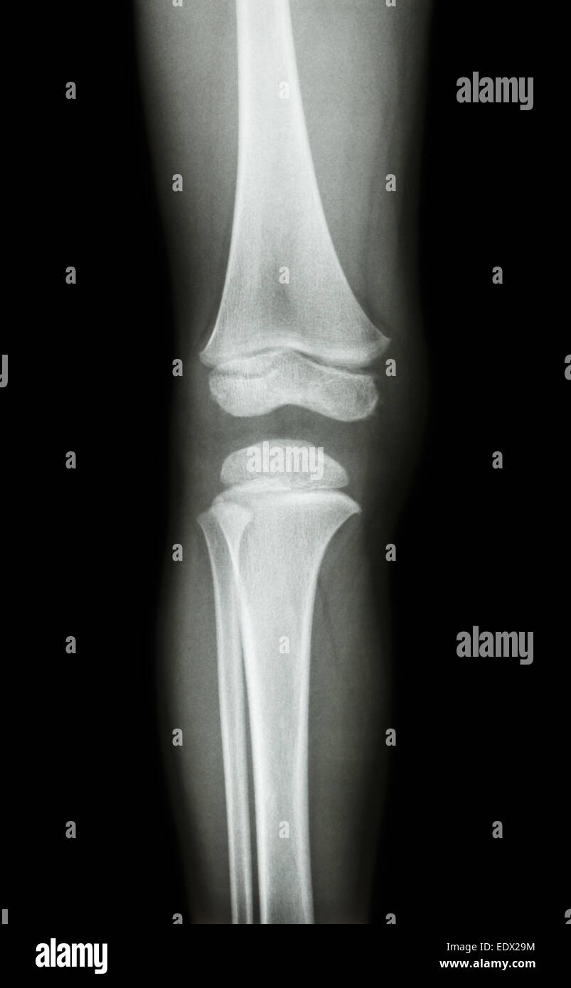 film x-ray knee AP(antero-posterior) of child Stock Photohttps://www.alamy.com/image-license-details/?v=1https://www.alamy.com/stock-photo-film-x-ray-knee-apantero-posterior-of-child-77404592.html
RFEDX29M–film x-ray knee AP(antero-posterior) of child
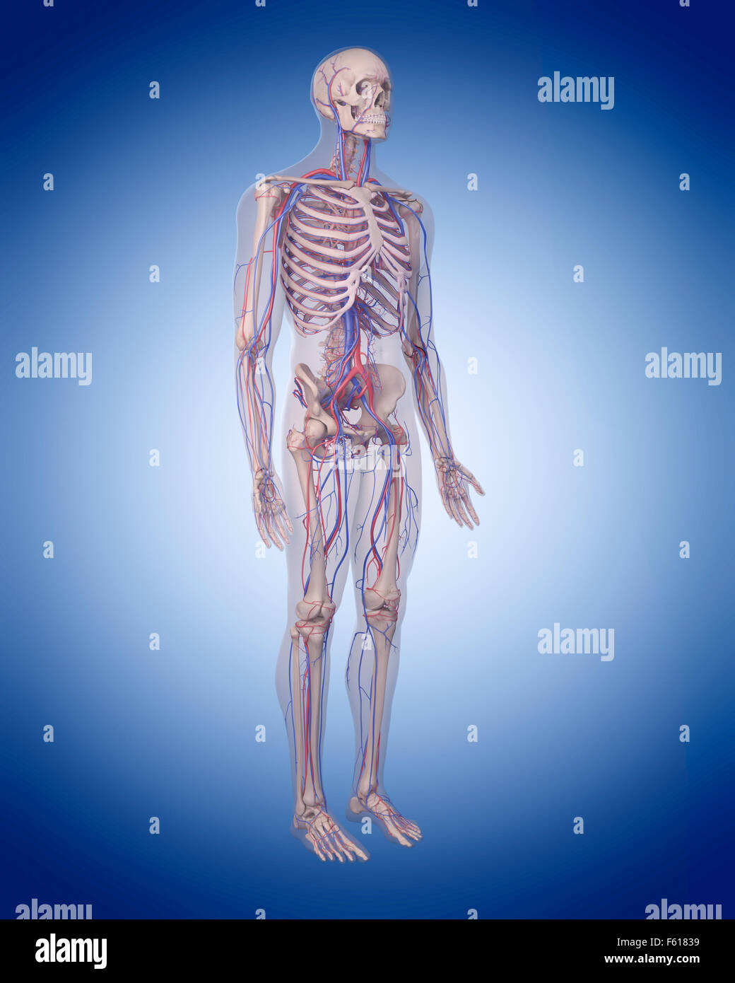 medically accurate illustration of the circulatory system Stock Photohttps://www.alamy.com/image-license-details/?v=1https://www.alamy.com/stock-photo-medically-accurate-illustration-of-the-circulatory-system-89768093.html
RFF61839–medically accurate illustration of the circulatory system
 Close up surgeons performing Axillary Artery Aneurysm Repair on arm teenage boy after stab wound shoulder. Sudan Africa. Stock Photohttps://www.alamy.com/image-license-details/?v=1https://www.alamy.com/stock-photo-close-up-surgeons-performing-axillary-artery-aneurysm-repair-on-arm-54338108.html
RMD4B8PM–Close up surgeons performing Axillary Artery Aneurysm Repair on arm teenage boy after stab wound shoulder. Sudan Africa.
 Baker's cyst: fluid-filled swelling behind the knee due to underlying knee issues, causing pain, stiffness, may require treatment. Stock Photohttps://www.alamy.com/image-license-details/?v=1https://www.alamy.com/bakers-cyst-fluid-filled-swelling-behind-the-knee-due-to-underlying-knee-issues-causing-pain-stiffness-may-require-treatment-image601375499.html
RF2WXB0GB–Baker's cyst: fluid-filled swelling behind the knee due to underlying knee issues, causing pain, stiffness, may require treatment.
 Major Veins of the Leg Stock Photohttps://www.alamy.com/image-license-details/?v=1https://www.alamy.com/major-veins-of-the-leg-image353191698.html
RM2BEH7H6–Major Veins of the Leg
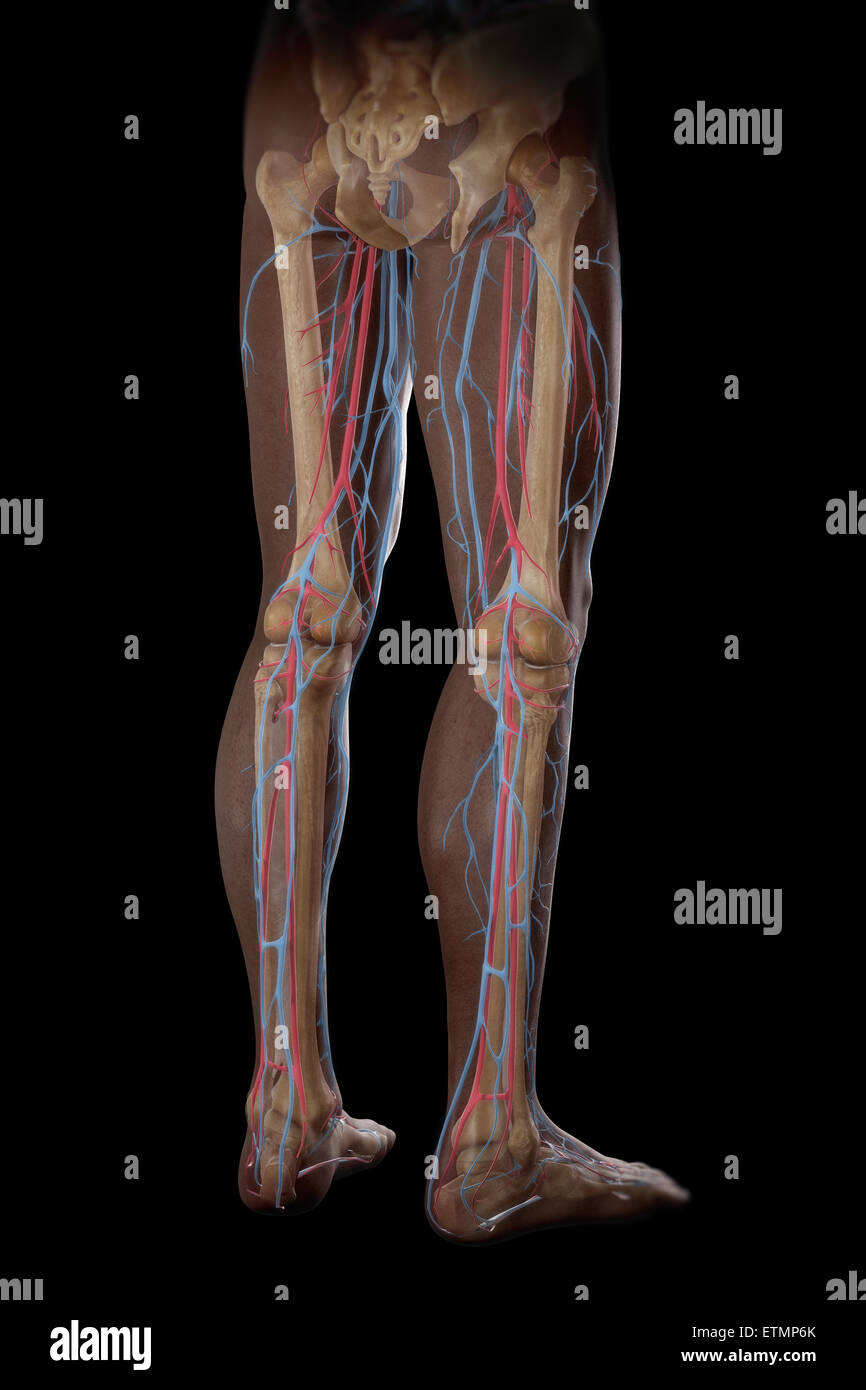 Illustration of the blood supply and skeletal structure of the legs, visible through skin. Stock Photohttps://www.alamy.com/image-license-details/?v=1https://www.alamy.com/stock-photo-illustration-of-the-blood-supply-and-skeletal-structure-of-the-legs-84049691.html
RMETMP6K–Illustration of the blood supply and skeletal structure of the legs, visible through skin.
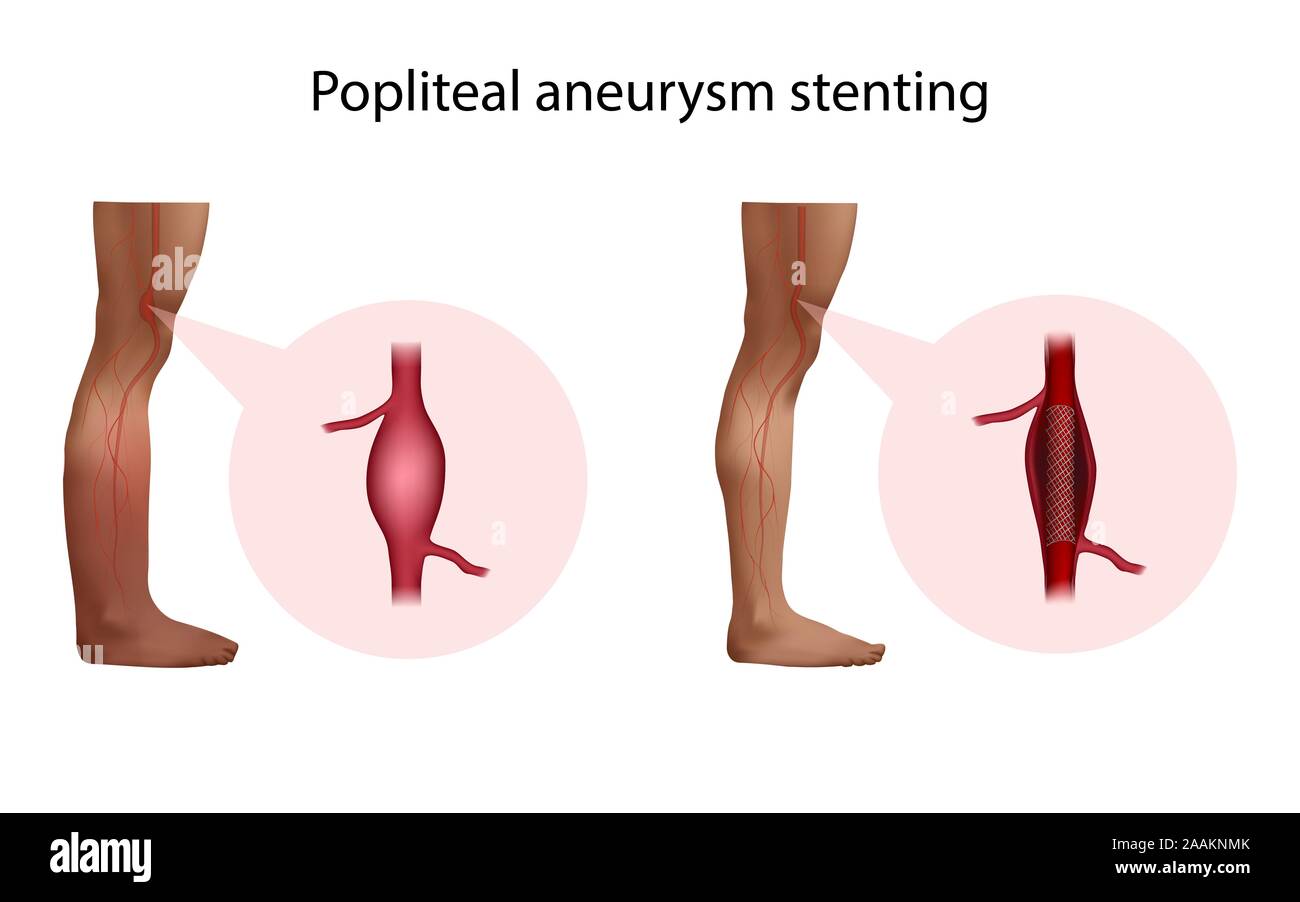 Popliteal aneurysm stenting, illustration. Before and after surgery. Stock Photohttps://www.alamy.com/image-license-details/?v=1https://www.alamy.com/popliteal-aneurysm-stenting-illustration-before-and-after-surgery-image333577683.html
RF2AAKNMK–Popliteal aneurysm stenting, illustration. Before and after surgery.
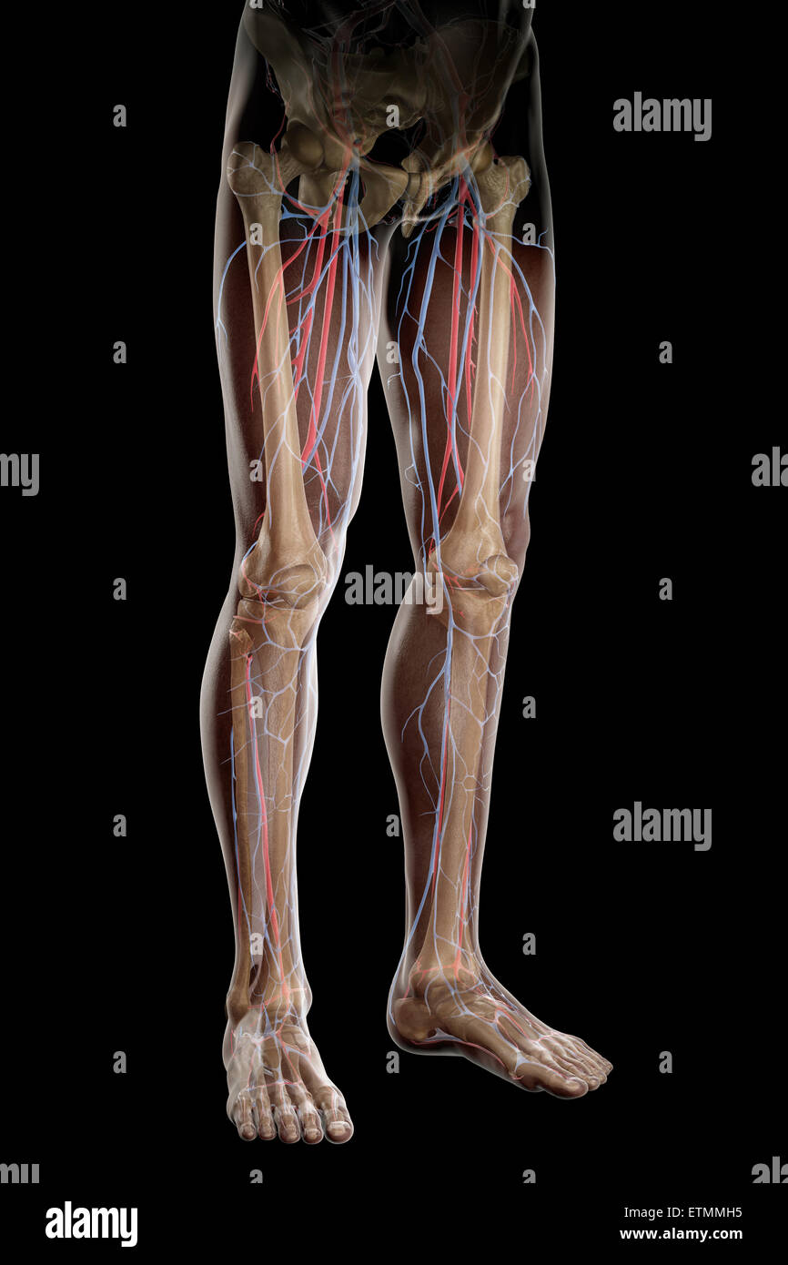 Illustration of the blood supply and skeletal structure of the legs, visible through skin. Stock Photohttps://www.alamy.com/image-license-details/?v=1https://www.alamy.com/stock-photo-illustration-of-the-blood-supply-and-skeletal-structure-of-the-legs-84048417.html
RMETMMH5–Illustration of the blood supply and skeletal structure of the legs, visible through skin.
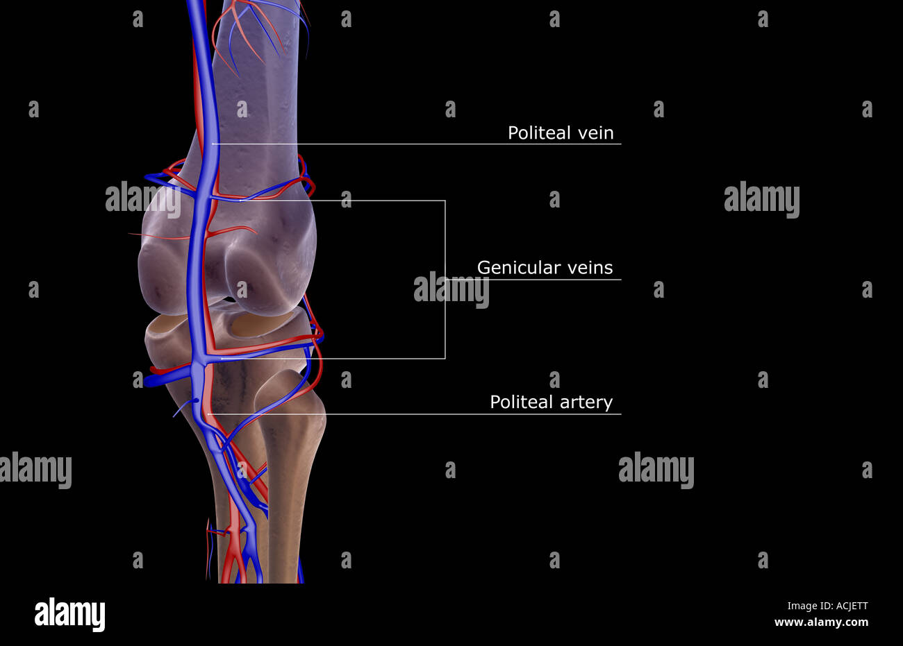 The blood supply of the knee Stock Photohttps://www.alamy.com/image-license-details/?v=1https://www.alamy.com/stock-photo-the-blood-supply-of-the-knee-13169927.html
RFACJETT–The blood supply of the knee
 Illustration showing the blood supply and skeletal structure of the legs, visible through skin. Stock Photohttps://www.alamy.com/image-license-details/?v=1https://www.alamy.com/stock-photo-illustration-showing-the-blood-supply-and-skeletal-structure-of-the-84048818.html
RMETMN3E–Illustration showing the blood supply and skeletal structure of the legs, visible through skin.
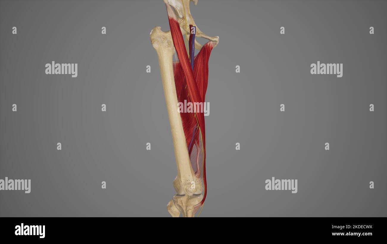 Anatomical Illustration of Adductor Canal Stock Photohttps://www.alamy.com/image-license-details/?v=1https://www.alamy.com/anatomical-illustration-of-adductor-canal-image490198294.html
RF2KDECWX–Anatomical Illustration of Adductor Canal
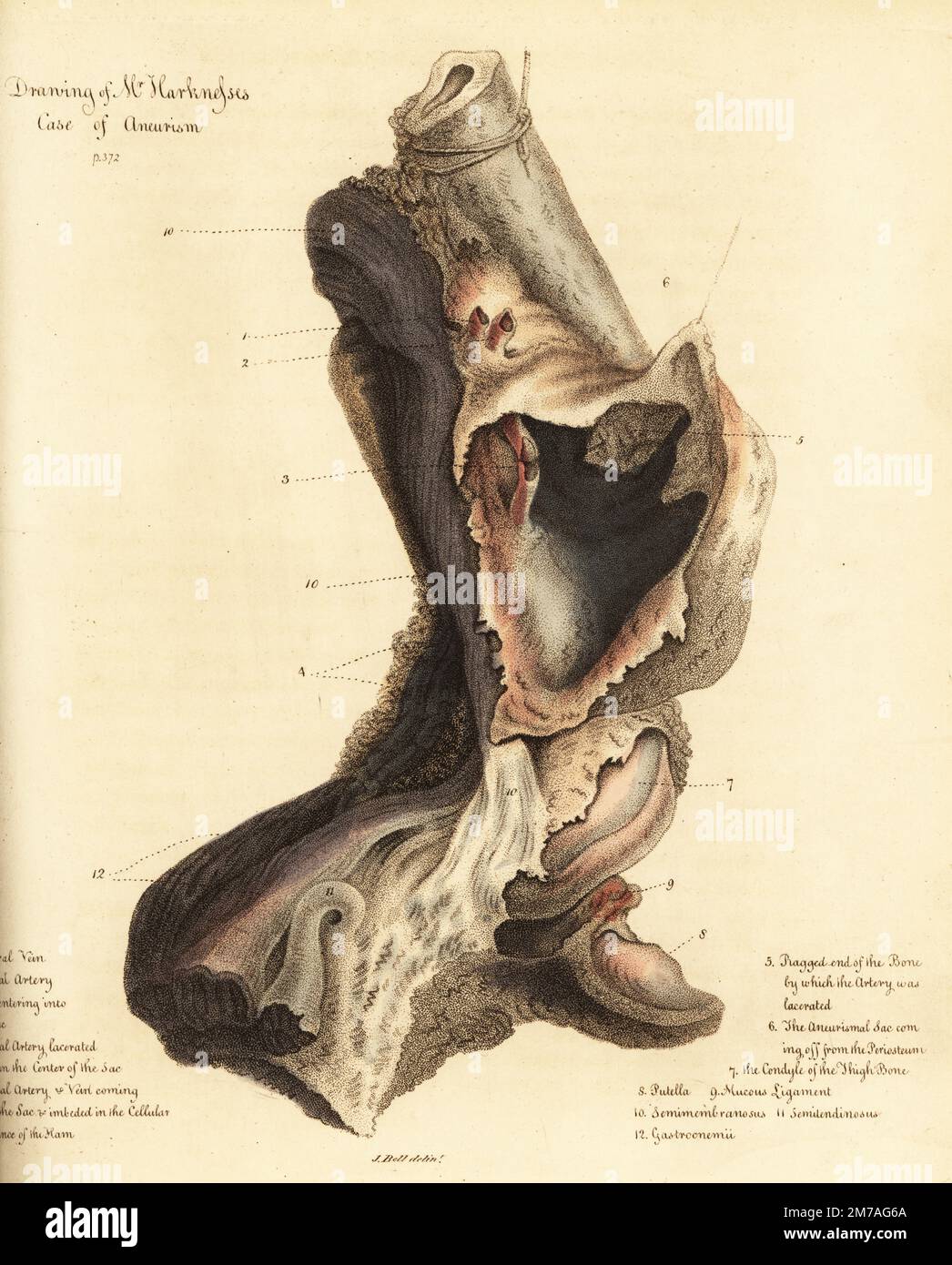 Fatal aneurysmal sac from the broken leg of a man, 1815. The sailor died of gangrene before the leg could be amputated. Drawing of Mr. Harkness's case of aneurism. Femoral vein 1, femoral artery 2, lacerated femoral artery 3, popliteal artery 4, ragged end of the bone 5, aneurysmal sac 6, condyle of the thigh bone 7, putella 8, mucous ligament 9, semimembranosus 10, semilendinosus 11, gastroenemii 12. Handcoloured copperplate engraving by after an illustration by John Bell from his own Principles of Surgery, as they Relate to Wounds, Ulcers and Fistulas, Longman, Hurst, Rees, Orme and Brown, Stock Photohttps://www.alamy.com/image-license-details/?v=1https://www.alamy.com/fatal-aneurysmal-sac-from-the-broken-leg-of-a-man-1815-the-sailor-died-of-gangrene-before-the-leg-could-be-amputated-drawing-of-mr-harknesss-case-of-aneurism-femoral-vein-1-femoral-artery-2-lacerated-femoral-artery-3-popliteal-artery-4-ragged-end-of-the-bone-5-aneurysmal-sac-6-condyle-of-the-thigh-bone-7-putella-8-mucous-ligament-9-semimembranosus-10-semilendinosus-11-gastroenemii-12-handcoloured-copperplate-engraving-by-after-an-illustration-by-john-bell-from-his-own-principles-of-surgery-as-they-relate-to-wounds-ulcers-and-fistulas-longman-hurst-rees-orme-and-brown-image503635506.html
RM2M7AG6A–Fatal aneurysmal sac from the broken leg of a man, 1815. The sailor died of gangrene before the leg could be amputated. Drawing of Mr. Harkness's case of aneurism. Femoral vein 1, femoral artery 2, lacerated femoral artery 3, popliteal artery 4, ragged end of the bone 5, aneurysmal sac 6, condyle of the thigh bone 7, putella 8, mucous ligament 9, semimembranosus 10, semilendinosus 11, gastroenemii 12. Handcoloured copperplate engraving by after an illustration by John Bell from his own Principles of Surgery, as they Relate to Wounds, Ulcers and Fistulas, Longman, Hurst, Rees, Orme and Brown,
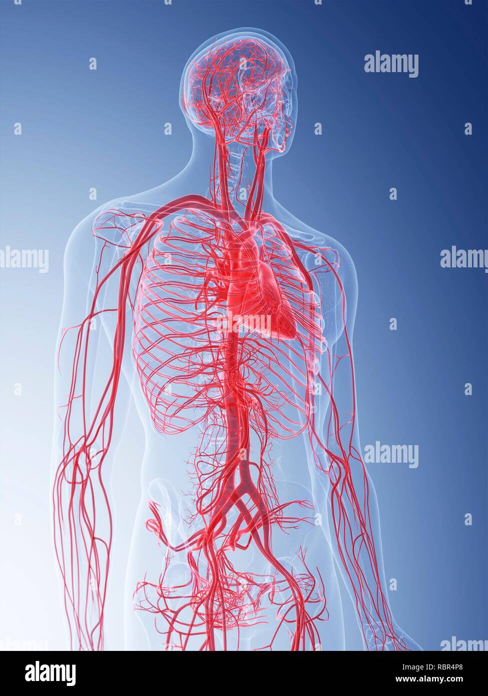 Illustration of the vascular system. Stock Photohttps://www.alamy.com/image-license-details/?v=1https://www.alamy.com/illustration-of-the-vascular-system-image231004656.html
RFRBR4P8–Illustration of the vascular system.
 Man doing sports indoors complaining with knee pain sitting on a mat. Side view. Stock Photohttps://www.alamy.com/image-license-details/?v=1https://www.alamy.com/man-doing-sports-indoors-complaining-with-knee-pain-sitting-on-a-mat-side-view-image501344327.html
RF2M3J5PF–Man doing sports indoors complaining with knee pain sitting on a mat. Side view.
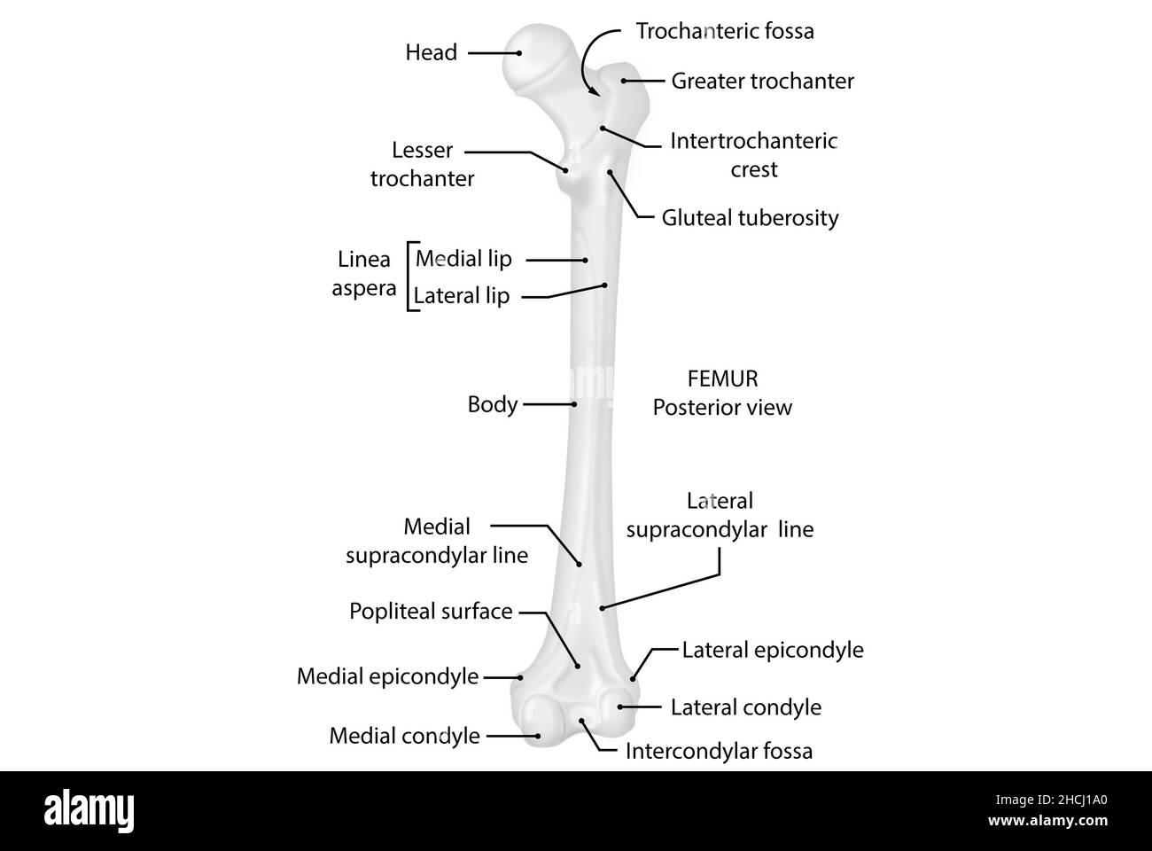 Femur, posterior view, anatomy, human body Stock Photohttps://www.alamy.com/image-license-details/?v=1https://www.alamy.com/femur-posterior-view-anatomy-human-body-image455241640.html
RF2HCJ1A0–Femur, posterior view, anatomy, human body
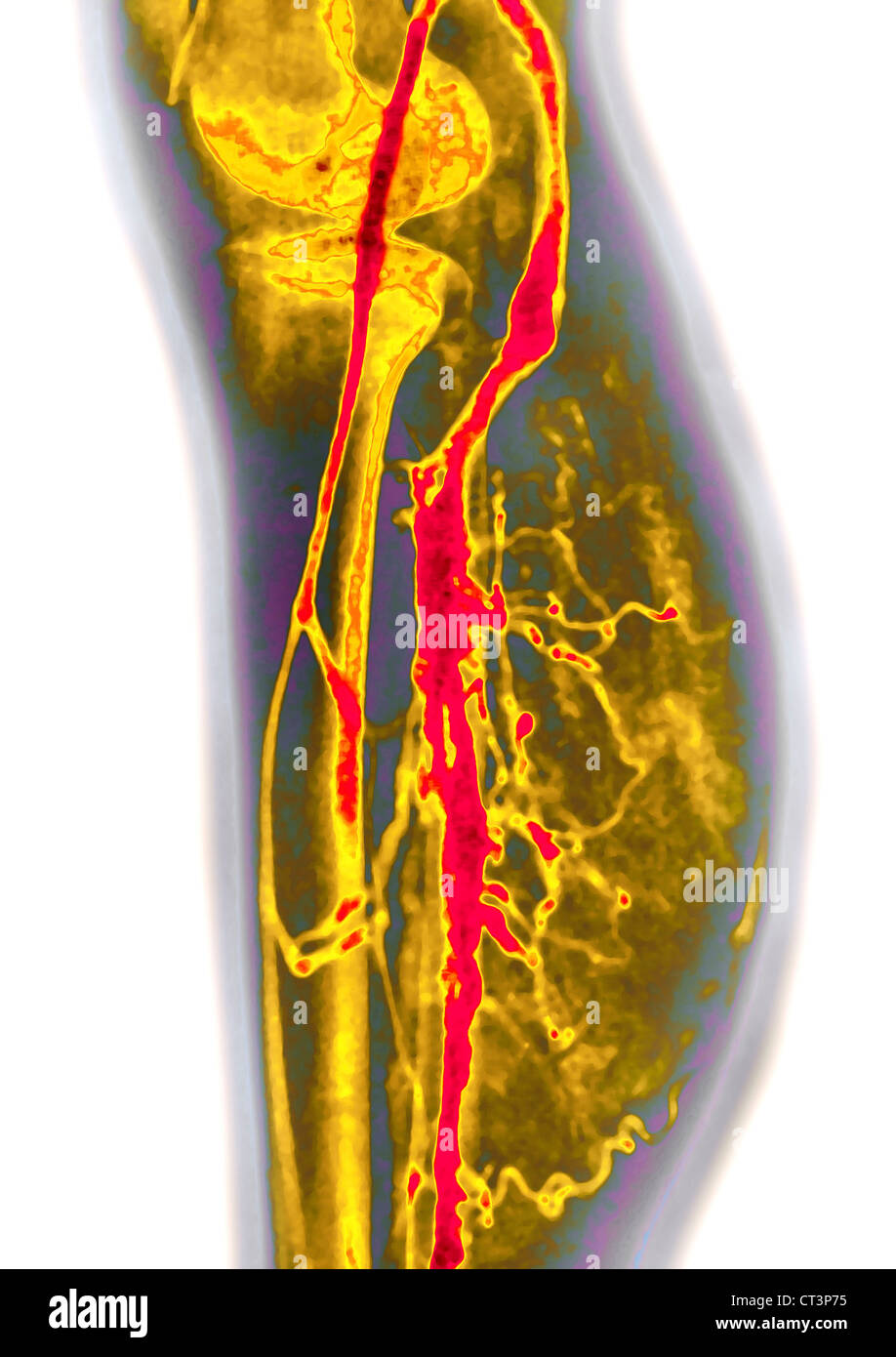 LOWER LIMB, ANGIOGRAPHY Stock Photohttps://www.alamy.com/image-license-details/?v=1https://www.alamy.com/stock-photo-lower-limb-angiography-49255785.html
RMCT3P75–LOWER LIMB, ANGIOGRAPHY
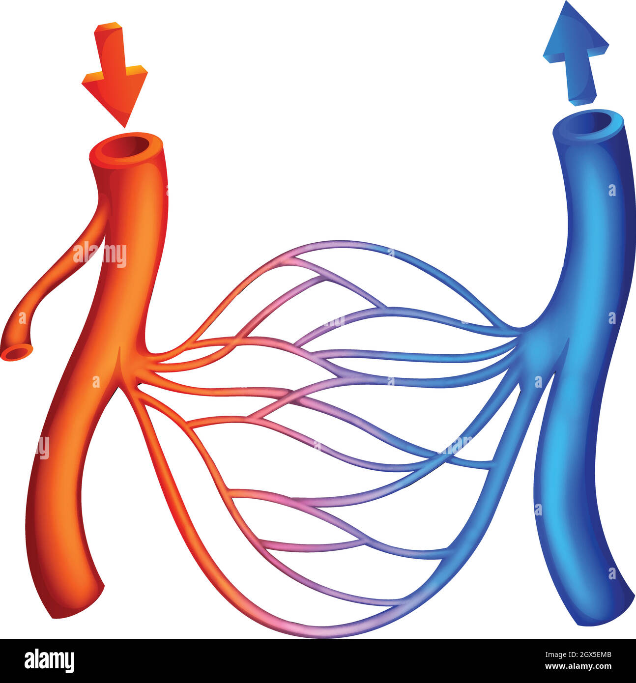 Blood Circulation Stock Vectorhttps://www.alamy.com/image-license-details/?v=1https://www.alamy.com/blood-circulation-image446361563.html
RF2GX5EMB–Blood Circulation
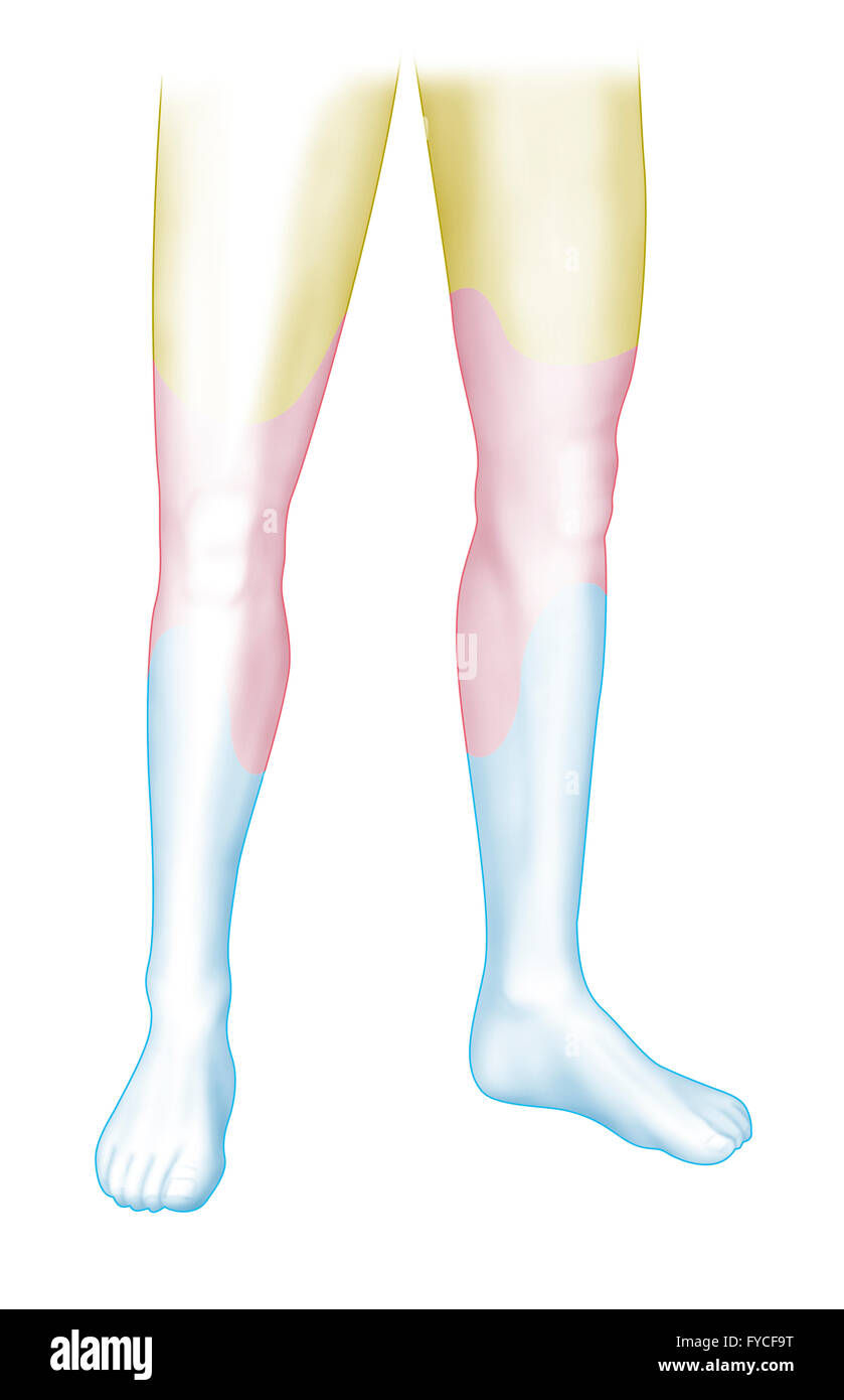 ARTERIAL STENOSIS Stock Photohttps://www.alamy.com/image-license-details/?v=1https://www.alamy.com/stock-photo-arterial-stenosis-102923012.html
RMFYCF9T–ARTERIAL STENOSIS
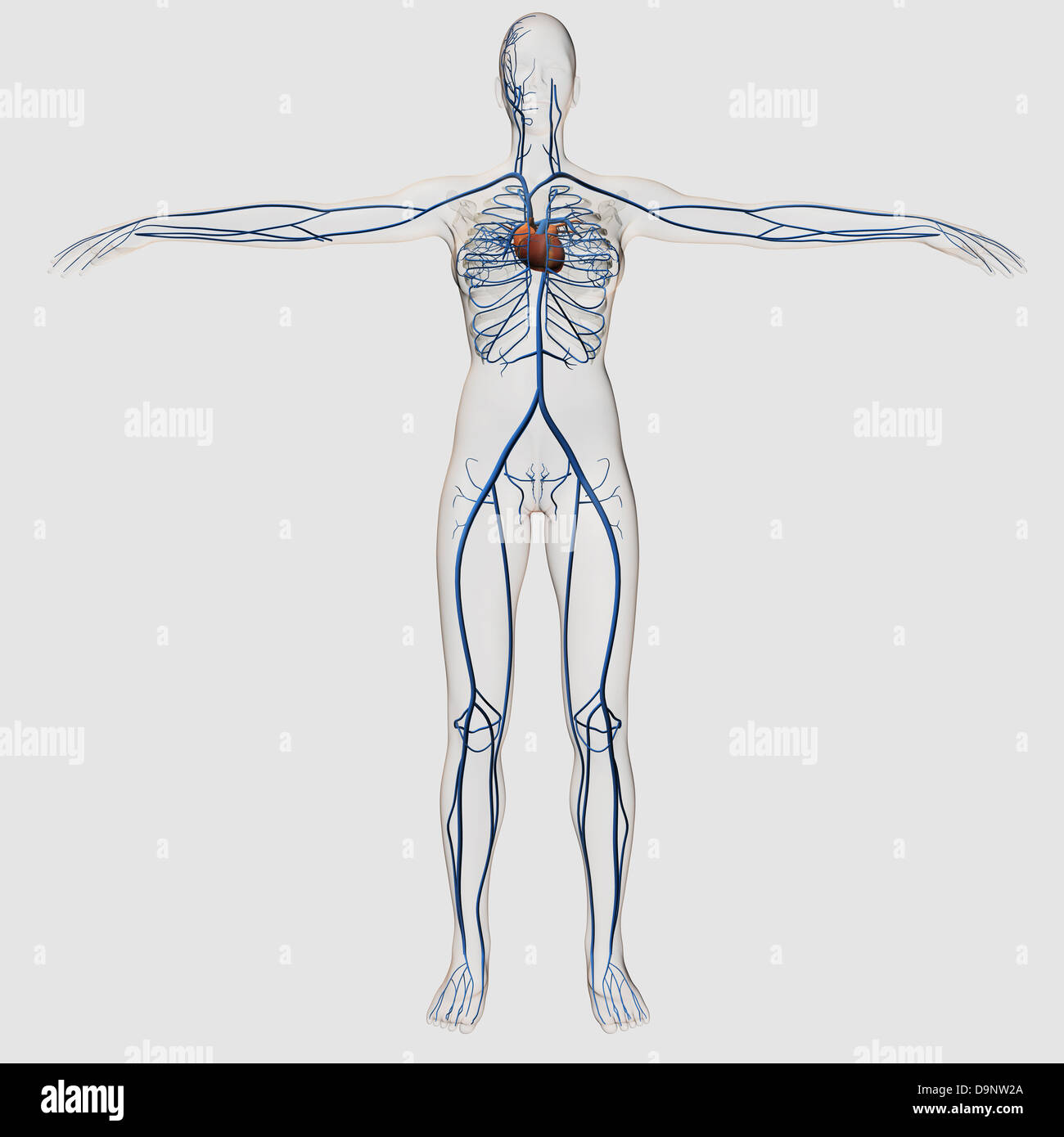 Medical illustration of female circulatory system with heart and veins visible, full body view. Stock Photohttps://www.alamy.com/image-license-details/?v=1https://www.alamy.com/stock-photo-medical-illustration-of-female-circulatory-system-with-heart-and-veins-57643666.html
RFD9NW2A–Medical illustration of female circulatory system with heart and veins visible, full body view.
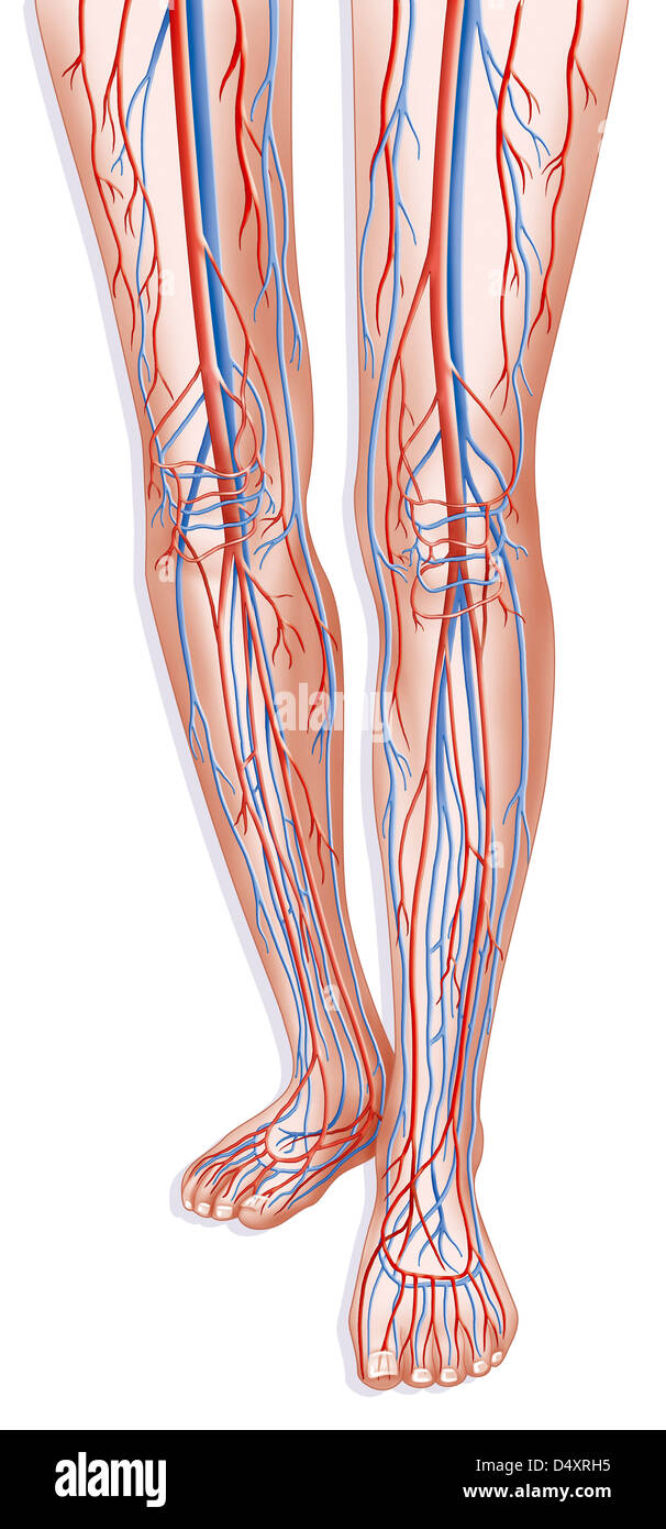 LOWER LIMB, BLOOD CIRCULATION Stock Photohttps://www.alamy.com/image-license-details/?v=1https://www.alamy.com/stock-photo-lower-limb-blood-circulation-54678993.html
RMD4XRH5–LOWER LIMB, BLOOD CIRCULATION
 medically accurate illustration of the circulatory system Stock Photohttps://www.alamy.com/image-license-details/?v=1https://www.alamy.com/stock-photo-medically-accurate-illustration-of-the-circulatory-system-89768092.html
RFF61838–medically accurate illustration of the circulatory system
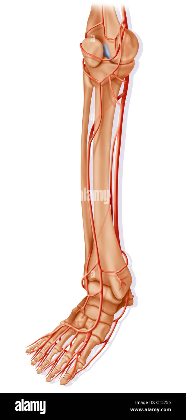 LOWER LIMB, BLOOD CIRCULATION Stock Photohttps://www.alamy.com/image-license-details/?v=1https://www.alamy.com/stock-photo-lower-limb-blood-circulation-49287873.html
RMCT5755–LOWER LIMB, BLOOD CIRCULATION
 Close up surgeons performing Axillary Artery Aneurysm Repair on arm teenage boy after stab wound shoulder. Sudan Africa. Stock Photohttps://www.alamy.com/image-license-details/?v=1https://www.alamy.com/stock-photo-close-up-surgeons-performing-axillary-artery-aneurysm-repair-on-arm-54338113.html
RMD4B8PW–Close up surgeons performing Axillary Artery Aneurysm Repair on arm teenage boy after stab wound shoulder. Sudan Africa.
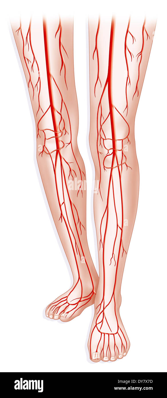 Lower limb, blood circulation Stock Photohttps://www.alamy.com/image-license-details/?v=1https://www.alamy.com/lower-limb-blood-circulation-image68401073.html
RMDY7X7D–Lower limb, blood circulation
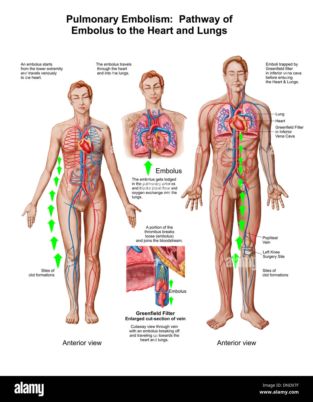
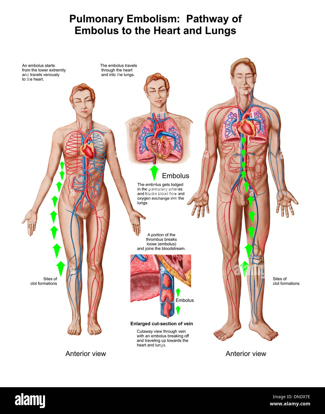 Pulmonary embolism, pathway of embolus to the heart and lungs. Stock Photohttps://www.alamy.com/image-license-details/?v=1https://www.alamy.com/pulmonary-embolism-pathway-of-embolus-to-the-heart-and-lungs-image64844850.html
RFDNDX7E–Pulmonary embolism, pathway of embolus to the heart and lungs.
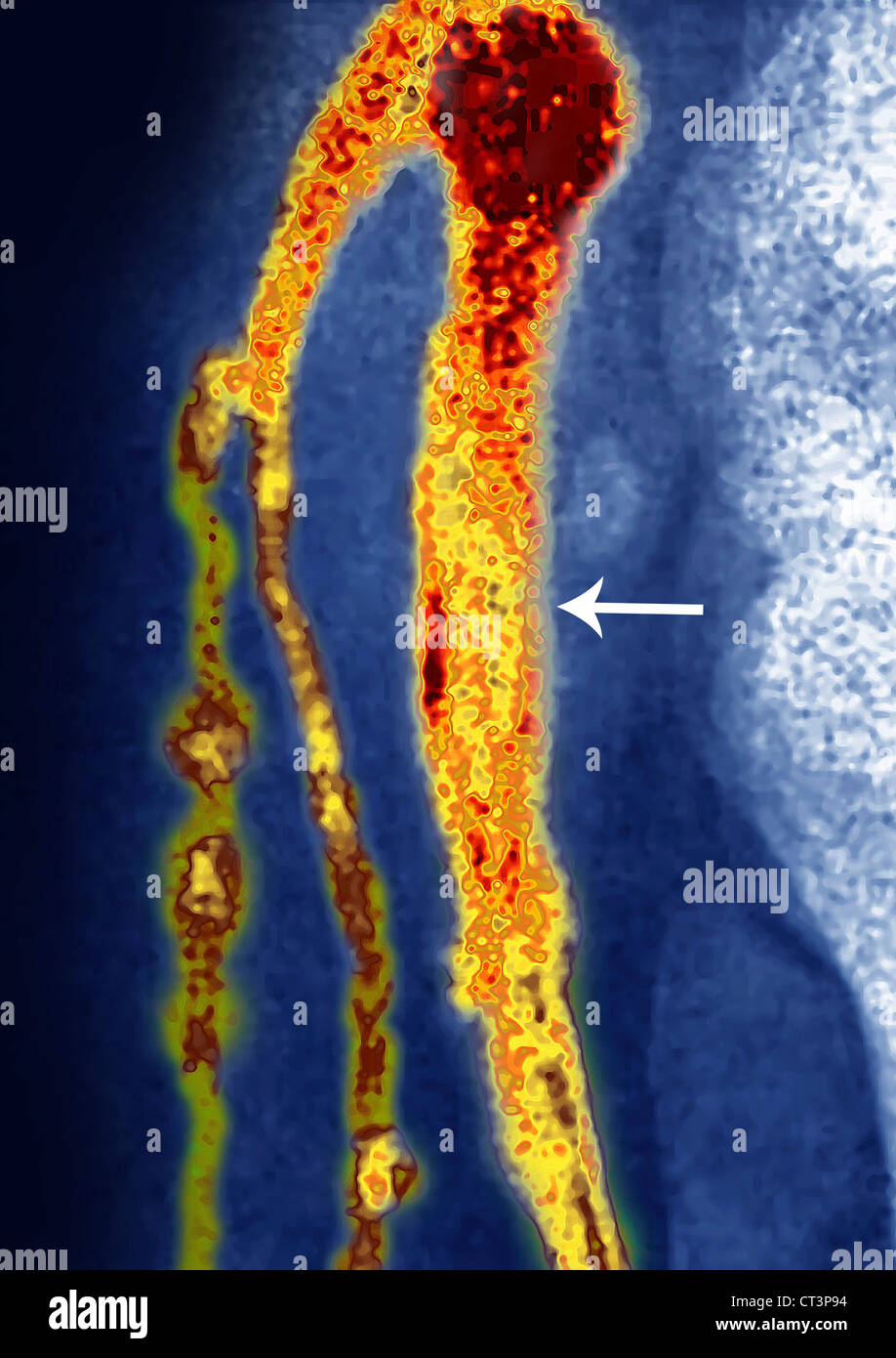 THOMBOSED VEIN, ANGIOGRAPHY Stock Photohttps://www.alamy.com/image-license-details/?v=1https://www.alamy.com/stock-photo-thombosed-vein-angiography-49255840.html
RMCT3P94–THOMBOSED VEIN, ANGIOGRAPHY
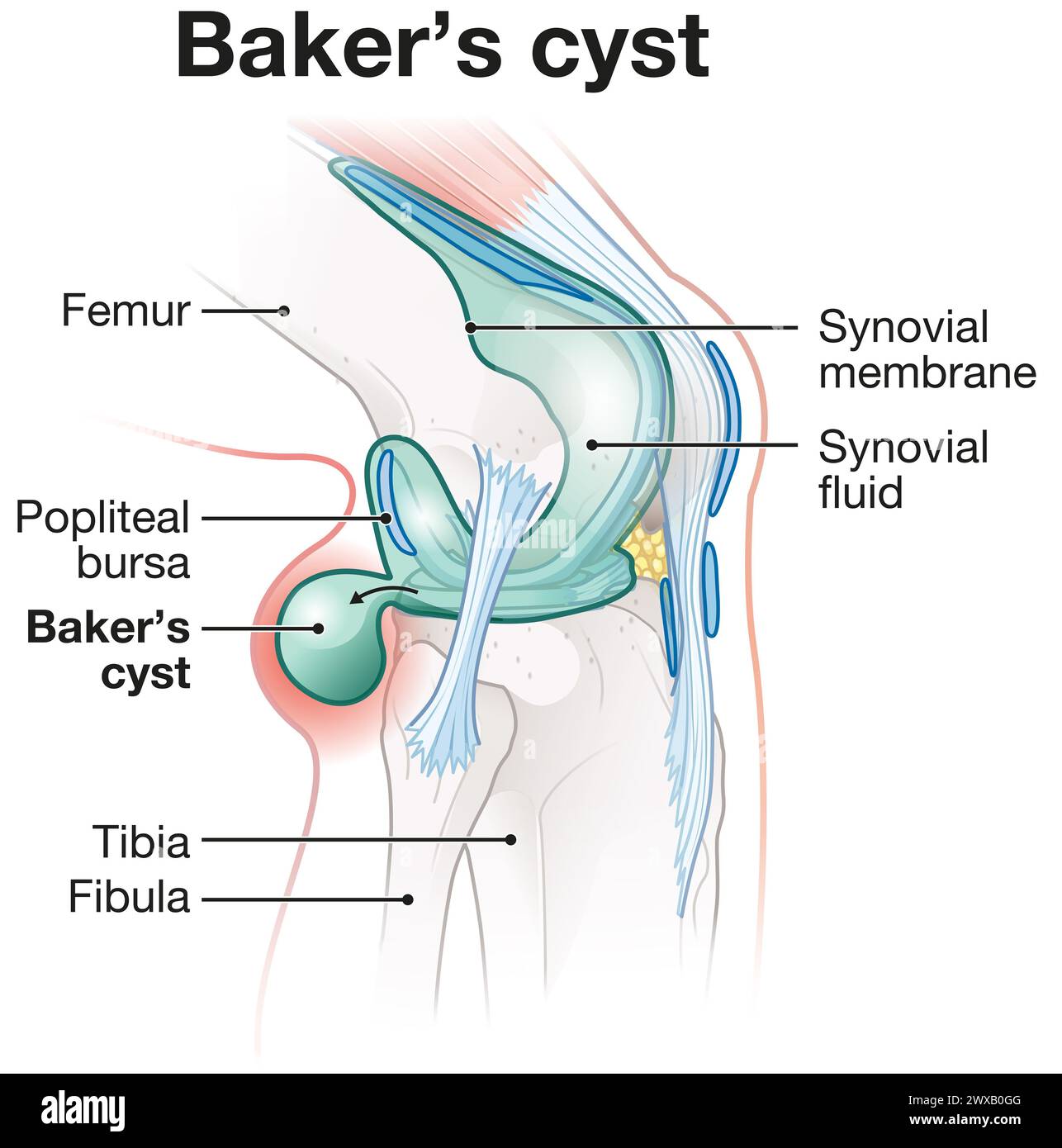 Baker's cyst: fluid-filled swelling behind the knee due to underlying knee issues, causing pain, stiffness, may require treatment. Stock Photohttps://www.alamy.com/image-license-details/?v=1https://www.alamy.com/bakers-cyst-fluid-filled-swelling-behind-the-knee-due-to-underlying-knee-issues-causing-pain-stiffness-may-require-treatment-image601375504.html
RF2WXB0GG–Baker's cyst: fluid-filled swelling behind the knee due to underlying knee issues, causing pain, stiffness, may require treatment.
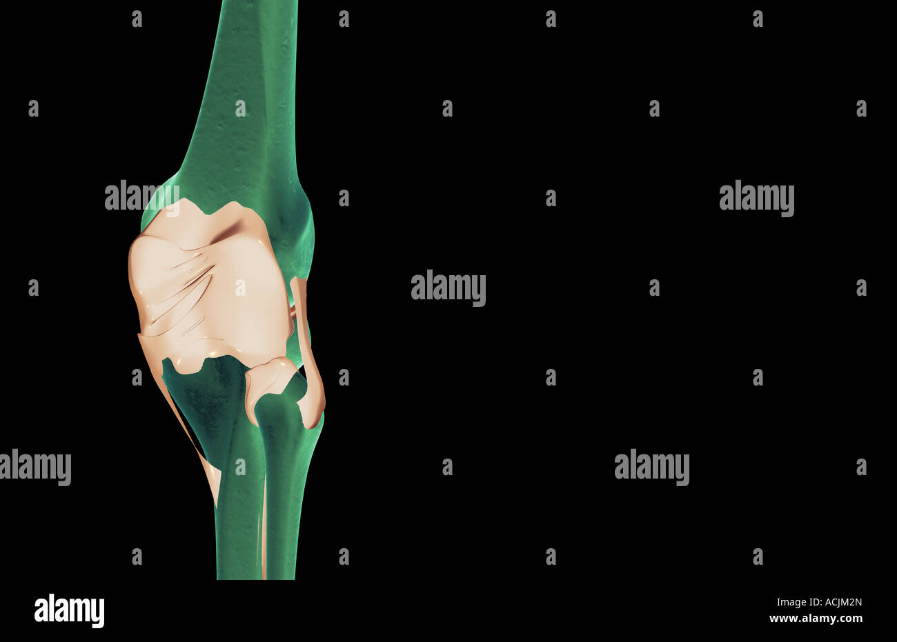 The ligaments of the knee Stock Photohttps://www.alamy.com/image-license-details/?v=1https://www.alamy.com/stock-photo-the-ligaments-of-the-knee-13171676.html
RFACJM2N–The ligaments of the knee
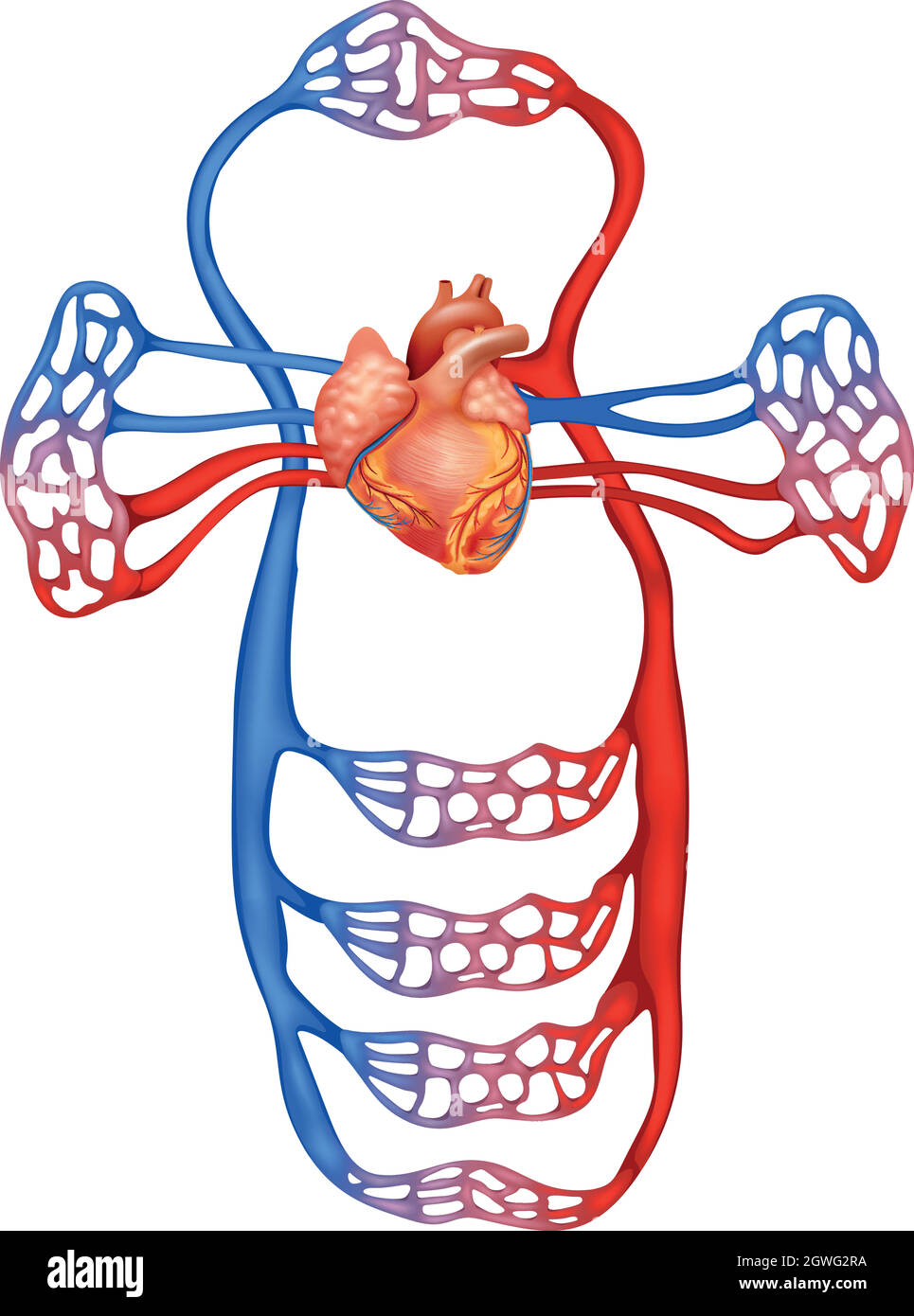 Circulatory system Stock Vectorhttps://www.alamy.com/image-license-details/?v=1https://www.alamy.com/circulatory-system-image445979054.html
RF2GWG2RA–Circulatory system
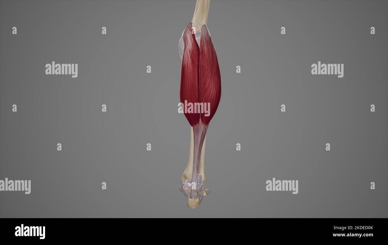 Medical Illustration of Gastrocnemius Stock Photohttps://www.alamy.com/image-license-details/?v=1https://www.alamy.com/medical-illustration-of-gastrocnemius-image490198371.html
RF2KDED0K–Medical Illustration of Gastrocnemius
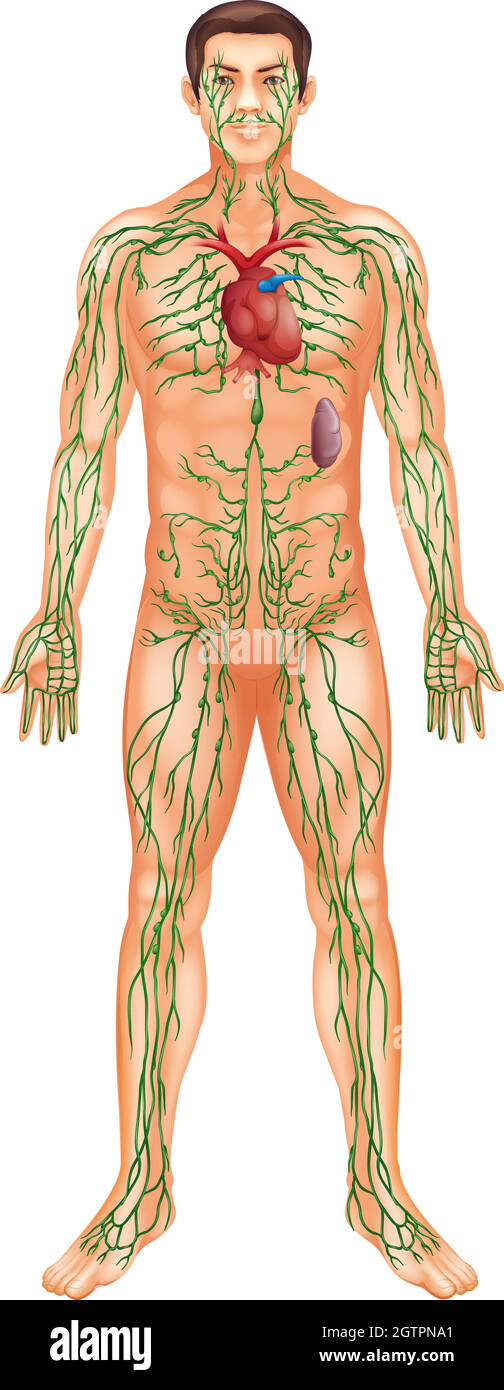 Lymphatic System Stock Vectorhttps://www.alamy.com/image-license-details/?v=1https://www.alamy.com/lymphatic-system-image445510633.html
RF2GTPNA1–Lymphatic System
RF2XBP3K4–Icon for knee,suffocation
 Detail of man doing sports with knee pain holding himself with his hand sitting on a mat. Side view. Stock Photohttps://www.alamy.com/image-license-details/?v=1https://www.alamy.com/detail-of-man-doing-sports-with-knee-pain-holding-himself-with-his-hand-sitting-on-a-mat-side-view-image504033010.html
RF2M80K6X–Detail of man doing sports with knee pain holding himself with his hand sitting on a mat. Side view.
 Female circulatory system Stock Vectorhttps://www.alamy.com/image-license-details/?v=1https://www.alamy.com/female-circulatory-system-image444687057.html
RF2GRD6TH–Female circulatory system
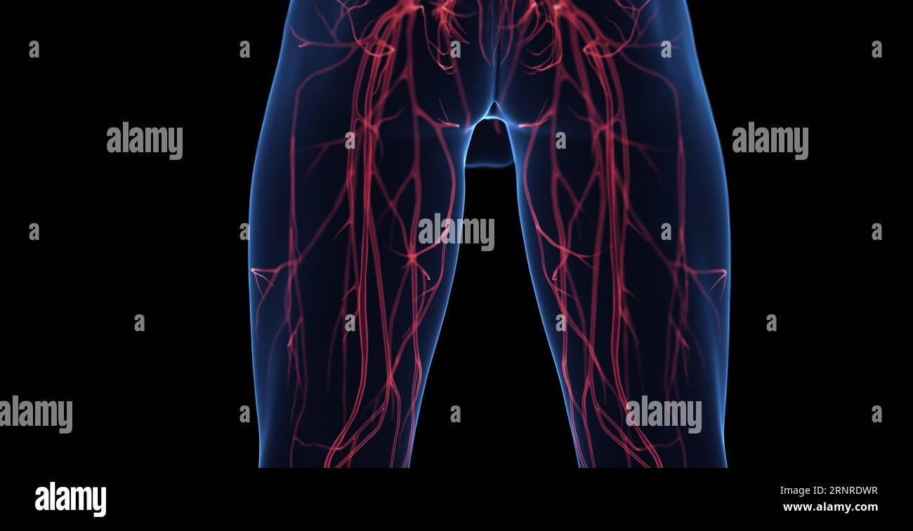 Male leg veins, illustration Stock Photohttps://www.alamy.com/image-license-details/?v=1https://www.alamy.com/male-leg-veins-illustration-image564155363.html
RF2RNRDWR–Male leg veins, illustration
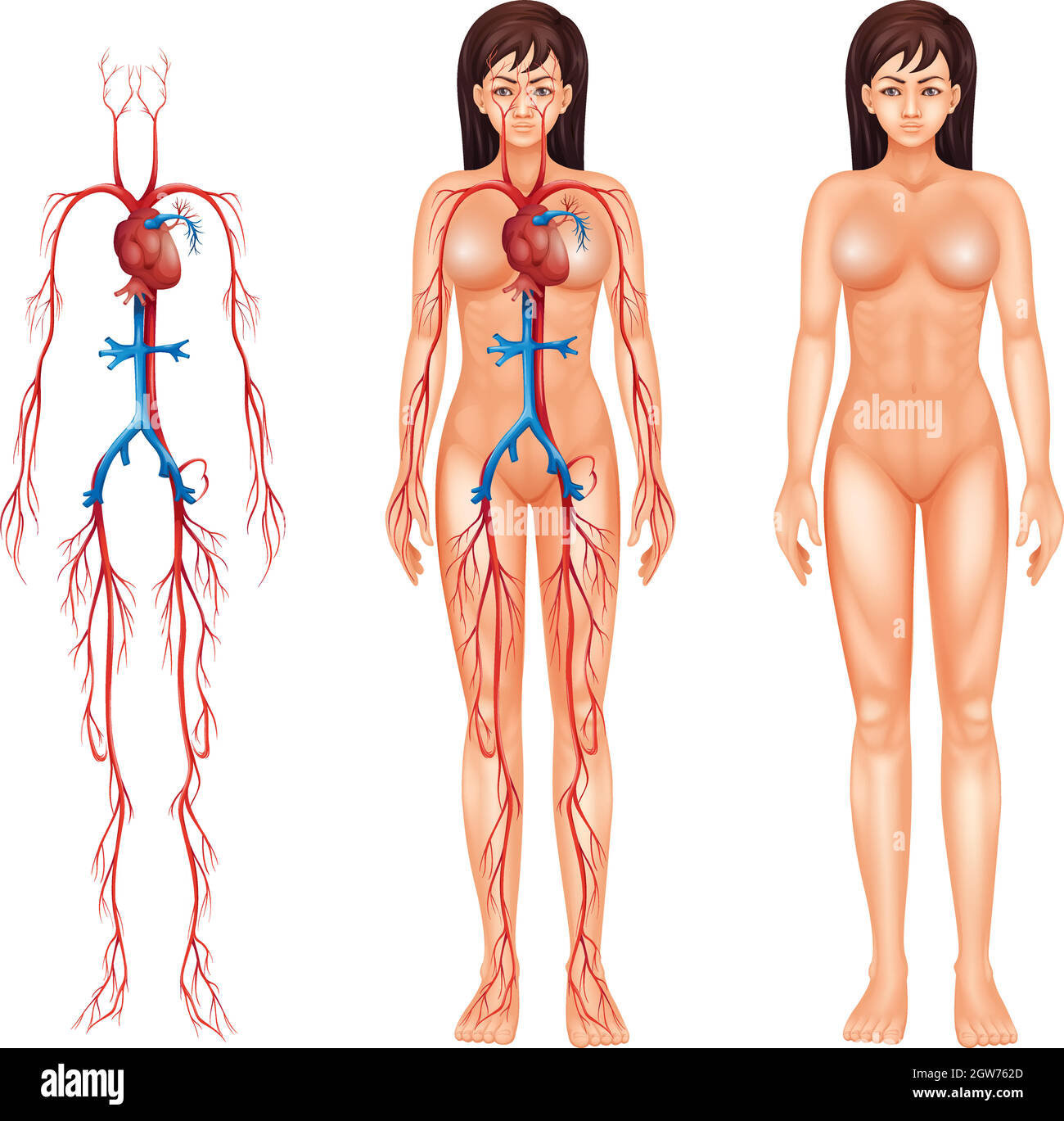 Female circulatory system Stock Vectorhttps://www.alamy.com/image-license-details/?v=1https://www.alamy.com/female-circulatory-system-image445784037.html
RF2GW762D–Female circulatory system
 Popliteal, tibial and peroneal arteries, MRI (Magnetic Resonance Imaging), medical imaging for diagnosis. Hospital Policlinica Gipuzkoa, San Sebastian, Donostia, Euskadi, Spain. Stock Photohttps://www.alamy.com/image-license-details/?v=1https://www.alamy.com/popliteal-tibial-and-peroneal-arteries-mri-magnetic-resonance-imaging-medical-imaging-for-diagnosis-hospital-policlinica-gipuzkoa-san-sebastian-donostia-euskadi-spain-image602325606.html
RM2WYX8CP–Popliteal, tibial and peroneal arteries, MRI (Magnetic Resonance Imaging), medical imaging for diagnosis. Hospital Policlinica Gipuzkoa, San Sebastian, Donostia, Euskadi, Spain.
 Femoral Endovascular Procedure. Illustration depicting the femoral arteries and popliteal arteries. Angioplasty femoral artery Stock Vectorhttps://www.alamy.com/image-license-details/?v=1https://www.alamy.com/femoral-endovascular-procedure-illustration-depicting-the-femoral-arteries-and-popliteal-arteries-angioplasty-femoral-artery-image641326141.html
RF2S7AX11–Femoral Endovascular Procedure. Illustration depicting the femoral arteries and popliteal arteries. Angioplasty femoral artery
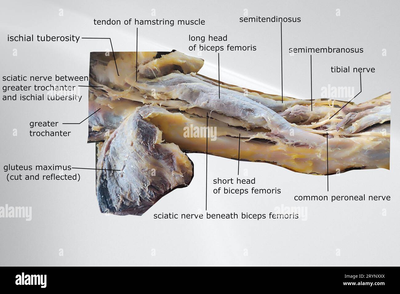 dissection image of the muscle of the thigh with showing sciatic nerve, hamstring muscle, gluteal muscles and popliteal fossa Stock Photohttps://www.alamy.com/image-license-details/?v=1https://www.alamy.com/dissection-image-of-the-muscle-of-the-thigh-with-showing-sciatic-nerve-hamstring-muscle-gluteal-muscles-and-popliteal-fossa-image567809618.html
RF2RYNXXX–dissection image of the muscle of the thigh with showing sciatic nerve, hamstring muscle, gluteal muscles and popliteal fossa
 Branches of right popliteal artery 3d illustration Stock Photohttps://www.alamy.com/image-license-details/?v=1https://www.alamy.com/branches-of-right-popliteal-artery-3d-illustration-image596578621.html
RF2WJGE39–Branches of right popliteal artery 3d illustration
 Popliteal area with disease. Watercolour by Mabel Green, 1900. Stock Photohttps://www.alamy.com/image-license-details/?v=1https://www.alamy.com/popliteal-area-with-disease-watercolour-by-mabel-green-1900-image450077200.html
RM2H46P1M–Popliteal area with disease. Watercolour by Mabel Green, 1900.
 Close up surgeons performing Axillary Artery Aneurysm Repair on arm teenage boy after stab wound shoulder. Sudan Africa. Stock Photohttps://www.alamy.com/image-license-details/?v=1https://www.alamy.com/stock-photo-close-up-surgeons-performing-axillary-artery-aneurysm-repair-on-arm-54338117.html
RMD4B8R1–Close up surgeons performing Axillary Artery Aneurysm Repair on arm teenage boy after stab wound shoulder. Sudan Africa.
 Popliteal, tibial and peroneal arteries, MRI (Magnetic Resonance Imaging), medical imaging for diagnosis. Hospital Policlinica Gipuzkoa, San Sebastian Stock Photohttps://www.alamy.com/image-license-details/?v=1https://www.alamy.com/popliteal-tibial-and-peroneal-arteries-mri-magnetic-resonance-imaging-medical-imaging-for-diagnosis-hospital-policlinica-gipuzkoa-san-sebastian-image604759662.html
RF2X3W53A–Popliteal, tibial and peroneal arteries, MRI (Magnetic Resonance Imaging), medical imaging for diagnosis. Hospital Policlinica Gipuzkoa, San Sebastian
