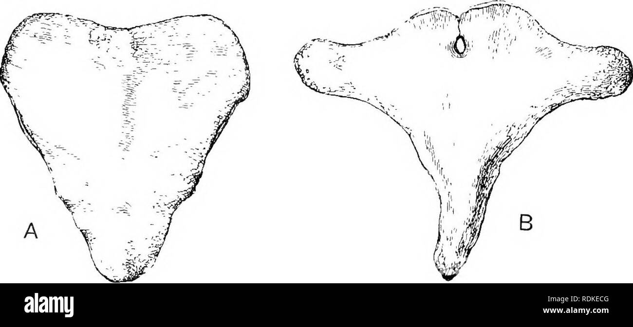
Posterior arch of atlas Cut Out Stock Images
 . Human physiology. Fig. 23.—The Atlas,»or First CervicalVertebra; viewed from above. i, the anterior arch; 2, the posterior arch ;3, spinal cavity ; 4, lateral processes ; 5 marksthe position of the odontoid peg of the axis :6, concave surfaces which articulate with theoccipital bone. The dotted line marks theposition of the ligament which secures thepeg. The body is absent, but is representedby the odontoid peg of the axis.. Fig. 24.—The Axis, or Second Cervical Vertebra. a, viewed from above and behind ; B, viewed from the right side ; t, odontoidprocess ; 2, body ; 3, spinal cavity ; 4, la Stock Photohttps://www.alamy.com/image-license-details/?v=1https://www.alamy.com/human-physiology-fig-23the-atlasor-first-cervicalvertebra-viewed-from-above-i-the-anterior-arch-2-the-posterior-arch-3-spinal-cavity-4-lateral-processes-5-marksthe-position-of-the-odontoid-peg-of-the-axis-6-concave-surfaces-which-articulate-with-theoccipital-bone-the-dotted-line-marks-theposition-of-the-ligament-which-secures-thepeg-the-body-is-absent-but-is-representedby-the-odontoid-peg-of-the-axis-fig-24the-axis-or-second-cervical-vertebra-a-viewed-from-above-and-behind-b-viewed-from-the-right-side-t-odontoidprocess-2-body-3-spinal-cavity-4-la-image370649756.html
RM2CF0FF8–. Human physiology. Fig. 23.—The Atlas,»or First CervicalVertebra; viewed from above. i, the anterior arch; 2, the posterior arch ;3, spinal cavity ; 4, lateral processes ; 5 marksthe position of the odontoid peg of the axis :6, concave surfaces which articulate with theoccipital bone. The dotted line marks theposition of the ligament which secures thepeg. The body is absent, but is representedby the odontoid peg of the axis.. Fig. 24.—The Axis, or Second Cervical Vertebra. a, viewed from above and behind ; B, viewed from the right side ; t, odontoidprocess ; 2, body ; 3, spinal cavity ; 4, la
 . The Cambridge natural history. Zoology. Fig. 186.âSection tlirougli iiiiddlB line of united cervical vertebrae of Greenland liight 'liale {Bal- aena â mysticetus). x -^. a. Arti- cular surface for occipital condyle; e, ejiipli3'sis on posterior end of body of seventh cervical vertebia ; 571, foramen in aich of atlas ior first spinal nerve ; 1, nrcli of atlas ; 2, 3, 4, 5, 6, conjoined arches of the axis and four followin"; verte- brae ; 7, arch of seventh vertebra. (From Flower's Oxteulagy.) as there is only a rudimentary pelvis, not attached to the vertebral column, no sacral region Stock Photohttps://www.alamy.com/image-license-details/?v=1https://www.alamy.com/the-cambridge-natural-history-zoology-fig-186section-tlirougli-iiiiddlb-line-of-united-cervical-vertebrae-of-greenland-liight-liale-bal-aena-mysticetus-x-a-arti-cular-surface-for-occipital-condyle-e-ejiipli3sis-on-posterior-end-of-body-of-seventh-cervical-vertebia-571-foramen-in-aich-of-atlas-ior-first-spinal-nerve-1-nrcli-of-atlas-2-3-4-5-6-conjoined-arches-of-the-axis-and-four-followinquot-verte-brae-7-arch-of-seventh-vertebra-from-flowers-oxteulagy-as-there-is-only-a-rudimentary-pelvis-not-attached-to-the-vertebral-column-no-sacral-region-image232153728.html
RMRDKECG–. The Cambridge natural history. Zoology. Fig. 186.âSection tlirougli iiiiddlB line of united cervical vertebrae of Greenland liight 'liale {Bal- aena â mysticetus). x -^. a. Arti- cular surface for occipital condyle; e, ejiipli3'sis on posterior end of body of seventh cervical vertebia ; 571, foramen in aich of atlas ior first spinal nerve ; 1, nrcli of atlas ; 2, 3, 4, 5, 6, conjoined arches of the axis and four followin"; verte- brae ; 7, arch of seventh vertebra. (From Flower's Oxteulagy.) as there is only a rudimentary pelvis, not attached to the vertebral column, no sacral region