Posterior parietal cortex Stock Photos and Images
(27)See posterior parietal cortex stock video clipsQuick filters:
Posterior parietal cortex Stock Photos and Images
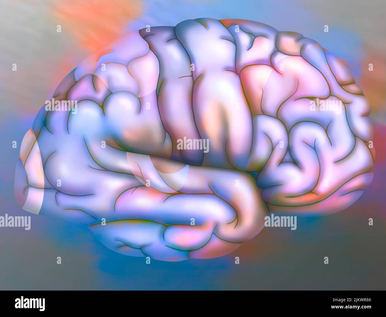 Areas of the brain of the right hemisphere: cortex (visual, parietal, sensory, motor, premotor) and auditory area. Stock Photohttps://www.alamy.com/image-license-details/?v=1https://www.alamy.com/areas-of-the-brain-of-the-right-hemisphere-cortex-visual-parietal-sensory-motor-premotor-and-auditory-area-image476925406.html
RF2JKWR66–Areas of the brain of the right hemisphere: cortex (visual, parietal, sensory, motor, premotor) and auditory area.
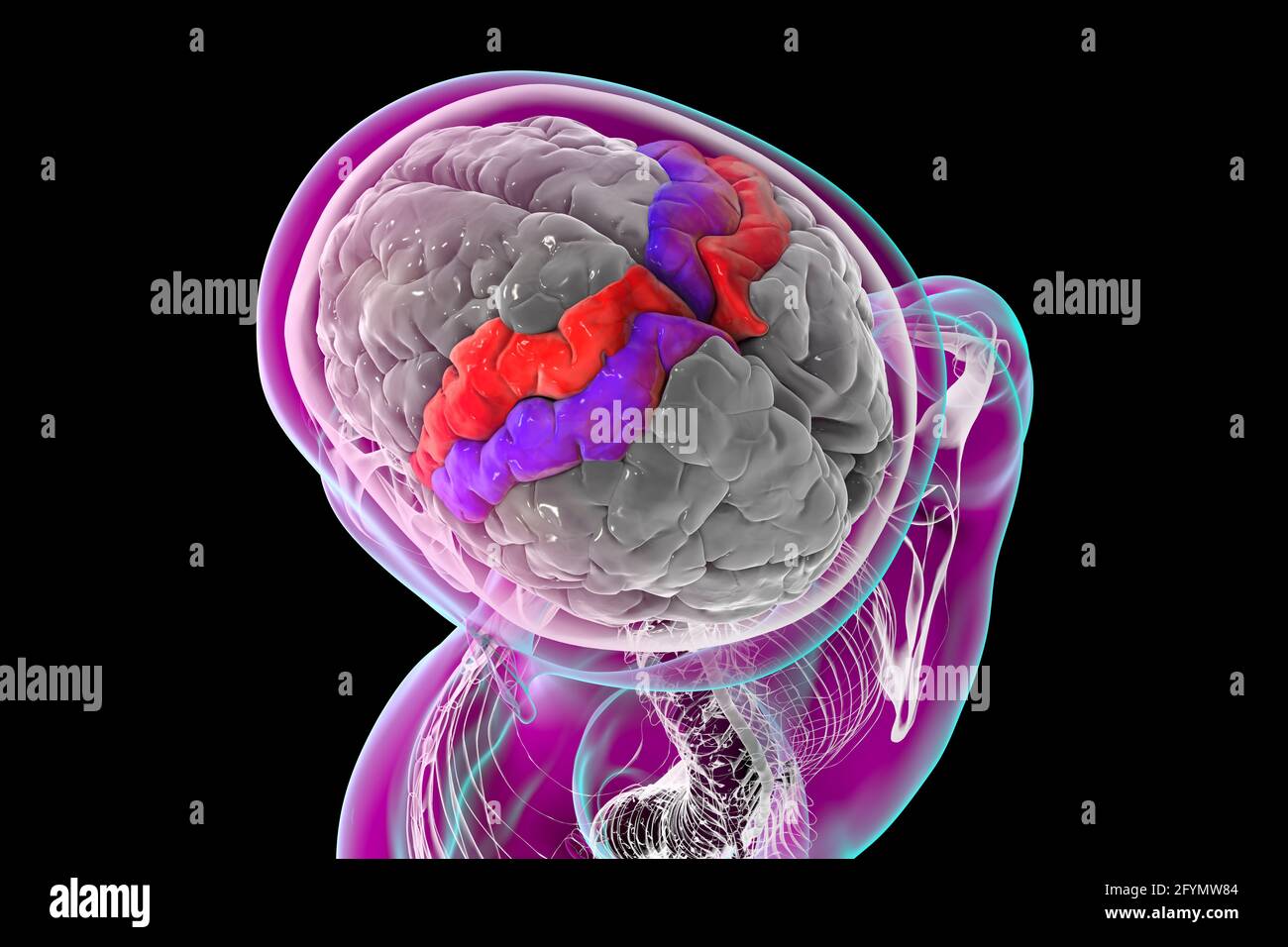 Precentral and postcentral gyri of the brain, illustration Stock Photohttps://www.alamy.com/image-license-details/?v=1https://www.alamy.com/precentral-and-postcentral-gyri-of-the-brain-illustration-image430103412.html
RF2FYMW84–Precentral and postcentral gyri of the brain, illustration
 The Journal of nervous and mental disease . radiating fibrespass out of the thalamus to become intermingled with thefibres of the internal capsule, and to be distributed to thecerebral cortex. Those from the front of the ganglion passto the frontal lobe; those from the middle are distributedto the posterior part of the frontal and to the parietal andtemporo-sphenoidal lobes ; those from the posterior partcan be traced to the temporo-sphenoidal and occipital lobes.From the region of the pulvinar, or posterior tubercle,fibres can be traced into the optic tract. 1 The Corpus Striatum, Journal of Stock Photohttps://www.alamy.com/image-license-details/?v=1https://www.alamy.com/the-journal-of-nervous-and-mental-disease-radiating-fibrespass-out-of-the-thalamus-to-become-intermingled-with-thefibres-of-the-internal-capsule-and-to-be-distributed-to-thecerebral-cortex-those-from-the-front-of-the-ganglion-passto-the-frontal-lobe-those-from-the-middle-are-distributedto-the-posterior-part-of-the-frontal-and-to-the-parietal-andtemporo-sphenoidal-lobes-those-from-the-posterior-partcan-be-traced-to-the-temporo-sphenoidal-and-occipital-lobesfrom-the-region-of-the-pulvinar-or-posterior-tuberclefibres-can-be-traced-into-the-optic-tract-1-the-corpus-striatum-journal-of-image340073246.html
RM2AN7JW2–The Journal of nervous and mental disease . radiating fibrespass out of the thalamus to become intermingled with thefibres of the internal capsule, and to be distributed to thecerebral cortex. Those from the front of the ganglion passto the frontal lobe; those from the middle are distributedto the posterior part of the frontal and to the parietal andtemporo-sphenoidal lobes ; those from the posterior partcan be traced to the temporo-sphenoidal and occipital lobes.From the region of the pulvinar, or posterior tubercle,fibres can be traced into the optic tract. 1 The Corpus Striatum, Journal of
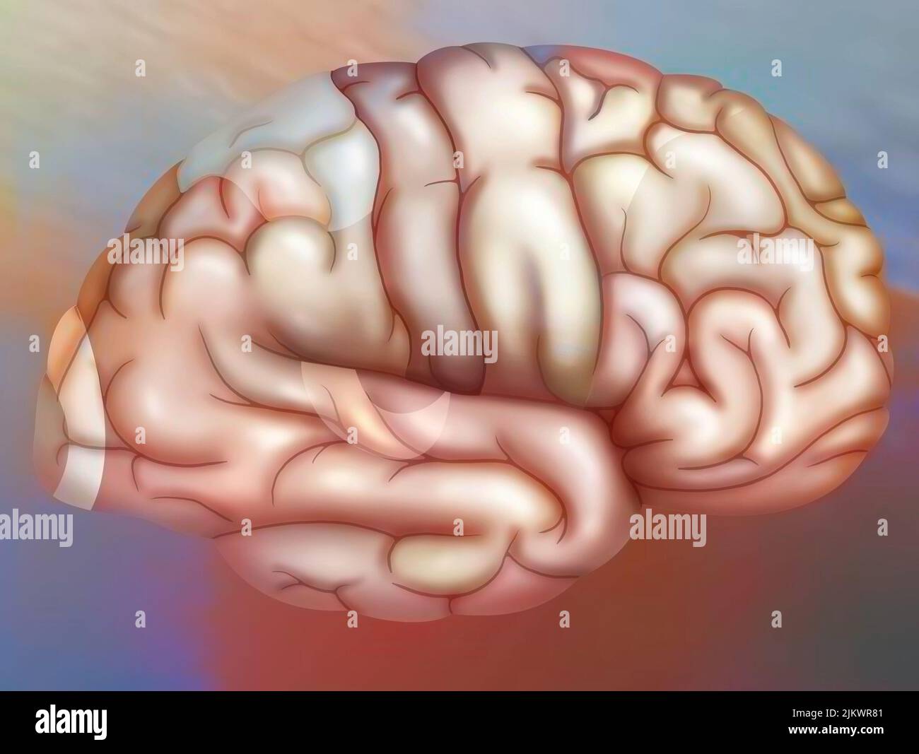 Areas of the brain of the right hemisphere: cortex (visual, parietal, sensory, motor, premotor) and auditory area. Stock Photohttps://www.alamy.com/image-license-details/?v=1https://www.alamy.com/areas-of-the-brain-of-the-right-hemisphere-cortex-visual-parietal-sensory-motor-premotor-and-auditory-area-image476925457.html
RF2JKWR81–Areas of the brain of the right hemisphere: cortex (visual, parietal, sensory, motor, premotor) and auditory area.
 Precentral and postcentral gyri of the brain, illustration Stock Photohttps://www.alamy.com/image-license-details/?v=1https://www.alamy.com/precentral-and-postcentral-gyri-of-the-brain-illustration-image430103405.html
RF2FYMW7W–Precentral and postcentral gyri of the brain, illustration
 Diseases of the nervous system .. . 61 research combined with the findings at autopsies, we know that the motorcerebral cortex is situated in the posterior and lower portion of the frontallobe, and in the anterior portion of the parietal Johe, especially in the anteriorand posterior ( ?) central com^olutions. Here also there are isolated fields forcertain motor regions. Thus, the center for the lower extremities is situated in the central con-volutions on both sides of the upper portion of the central furrow (Figs. Fig. 55.—Coxvkxity of the Brain from Above. (After Toldt.) 55-57); those for th Stock Photohttps://www.alamy.com/image-license-details/?v=1https://www.alamy.com/diseases-of-the-nervous-system-61-research-combined-with-the-findings-at-autopsies-we-know-that-the-motorcerebral-cortex-is-situated-in-the-posterior-and-lower-portion-of-the-frontallobe-and-in-the-anterior-portion-of-the-parietal-johe-especially-in-the-anteriorand-posterior-central-comolutions-here-also-there-are-isolated-fields-forcertain-motor-regions-thus-the-center-for-the-lower-extremities-is-situated-in-the-central-con-volutions-on-both-sides-of-the-upper-portion-of-the-central-furrow-figs-fig-55coxvkxity-of-the-brain-from-above-after-toldt-55-57-those-for-th-image342720332.html
RM2AWG77T–Diseases of the nervous system .. . 61 research combined with the findings at autopsies, we know that the motorcerebral cortex is situated in the posterior and lower portion of the frontallobe, and in the anterior portion of the parietal Johe, especially in the anteriorand posterior ( ?) central com^olutions. Here also there are isolated fields forcertain motor regions. Thus, the center for the lower extremities is situated in the central con-volutions on both sides of the upper portion of the central furrow (Figs. Fig. 55.—Coxvkxity of the Brain from Above. (After Toldt.) 55-57); those for th
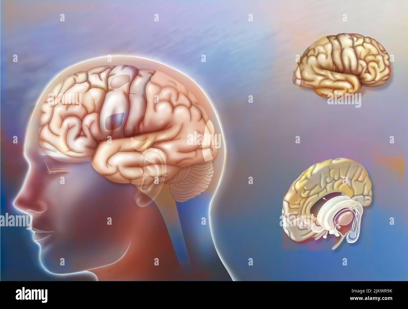 Left and right hemisphere brain areas and midline structures. Stock Photohttps://www.alamy.com/image-license-details/?v=1https://www.alamy.com/left-and-right-hemisphere-brain-areas-and-midline-structures-image476925503.html
RF2JKWR9K–Left and right hemisphere brain areas and midline structures.
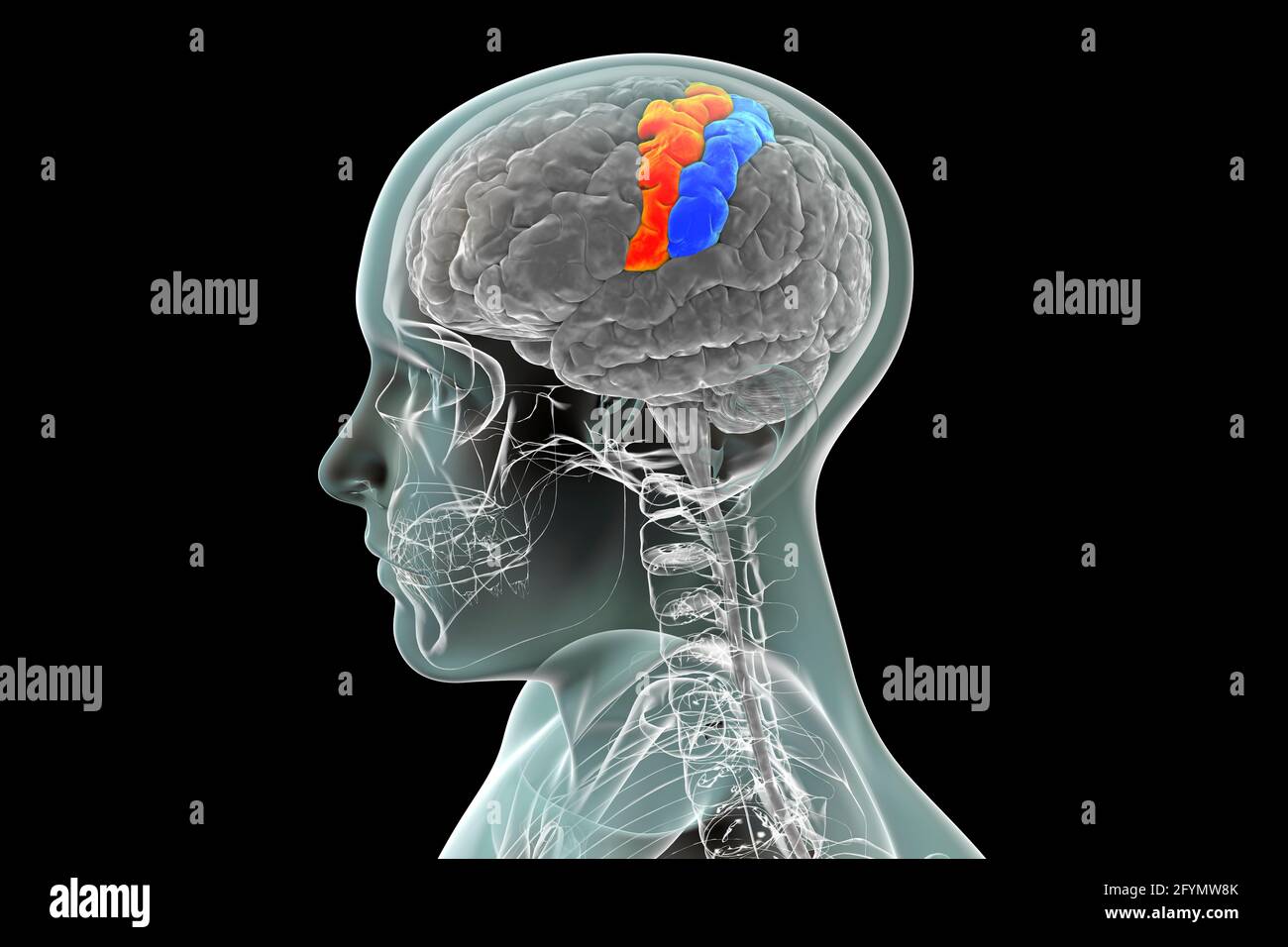 Precentral and postcentral gyri of the brain, illustration Stock Photohttps://www.alamy.com/image-license-details/?v=1https://www.alamy.com/precentral-and-postcentral-gyri-of-the-brain-illustration-image430103427.html
RF2FYMW8K–Precentral and postcentral gyri of the brain, illustration
 Organic and functional nervous diseases; a text-book of neurology . Fia OTa The Vascular Supply of the Cerebral Cortex. (Dejerine.)The regions supplied by different arterial branches are shown in different colors The median surface.—Branches of the anterior cerebral artery. Fia. Anterior frontal. Fim.Middle frontal. Ftp. Posterior frontal. Branches of the posterior cerebral artery. OTa. Temporo-occipital anterior. OTm. Temporo-occipital median. K. Calcarine. The lateral surface.—Branches of the middle.cerebral artery. Fi. Inferior frontal. Fa. Ascend-inK frontal. Pa. Ai^cending parietal. Pi. I Stock Photohttps://www.alamy.com/image-license-details/?v=1https://www.alamy.com/organic-and-functional-nervous-diseases-a-text-book-of-neurology-fia-ota-the-vascular-supply-of-the-cerebral-cortex-dejerinethe-regions-supplied-by-different-arterial-branches-are-shown-in-different-colors-the-median-surfacebranches-of-the-anterior-cerebral-artery-fia-anterior-frontal-fimmiddle-frontal-ftp-posterior-frontal-branches-of-the-posterior-cerebral-artery-ota-temporo-occipital-anterior-otm-temporo-occipital-median-k-calcarine-the-lateral-surfacebranches-of-the-middlecerebral-artery-fi-inferior-frontal-fa-ascend-ink-frontal-pa-aicending-parietal-pi-i-image339934212.html
RM2AN19FG–Organic and functional nervous diseases; a text-book of neurology . Fia OTa The Vascular Supply of the Cerebral Cortex. (Dejerine.)The regions supplied by different arterial branches are shown in different colors The median surface.—Branches of the anterior cerebral artery. Fia. Anterior frontal. Fim.Middle frontal. Ftp. Posterior frontal. Branches of the posterior cerebral artery. OTa. Temporo-occipital anterior. OTm. Temporo-occipital median. K. Calcarine. The lateral surface.—Branches of the middle.cerebral artery. Fi. Inferior frontal. Fa. Ascend-inK frontal. Pa. Ai^cending parietal. Pi. I
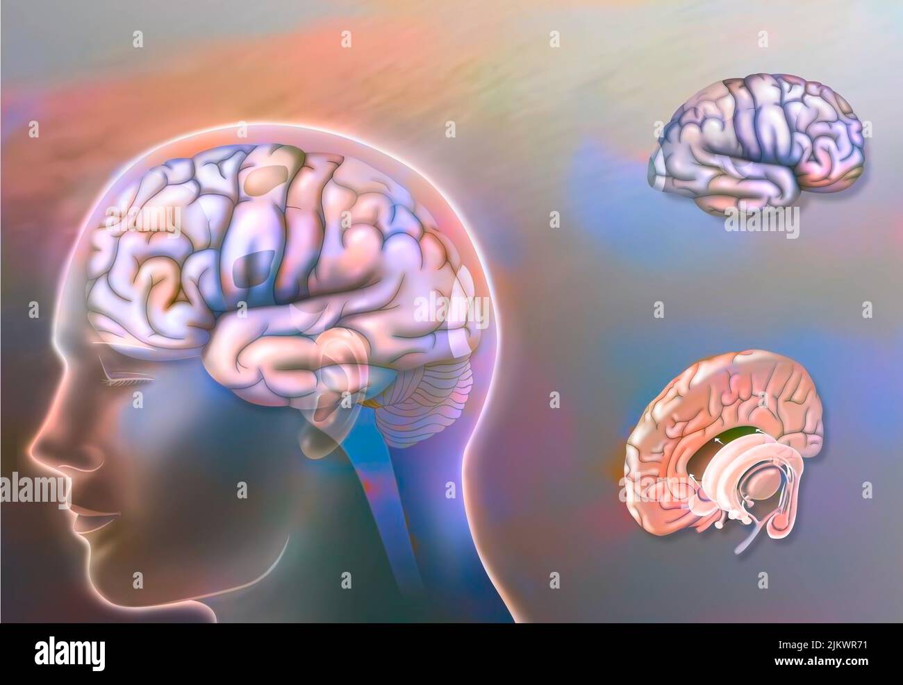 Left and right hemisphere brain areas and midline structures. Stock Photohttps://www.alamy.com/image-license-details/?v=1https://www.alamy.com/left-and-right-hemisphere-brain-areas-and-midline-structures-image476925429.html
RF2JKWR71–Left and right hemisphere brain areas and midline structures.
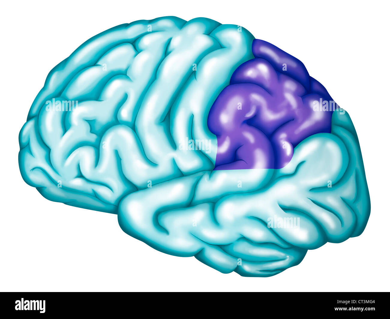 SOMATOSENSORY CORTEX Stock Photohttps://www.alamy.com/image-license-details/?v=1https://www.alamy.com/stock-photo-somatosensory-cortex-49254468.html
RMCT3MG4–SOMATOSENSORY CORTEX
 Organic and functional nervous diseases; a text-book of neurology . Fia OTa The Vascular Supply of the Cerebral Cortex. (Dejerine.)The regions supplied by different arterial branches are shown in different colors The median surface.—Branches of the anterior cerebral artery. Fia. Anterior frontal. Fim.Middle frontal. Ftp. Posterior frontal. Branches of the posterior cerebral artery. OTa. Temporo-occipital anterior. OTm. Temporo-occipital median. K. Calcarine. The lateral surface.—Branches of the middle.cerebral artery. Fi. Inferior frontal. Fa. Ascend-inK frontal. Pa. Ai^cending parietal. Pi. I Stock Photohttps://www.alamy.com/image-license-details/?v=1https://www.alamy.com/organic-and-functional-nervous-diseases-a-text-book-of-neurology-fia-ota-the-vascular-supply-of-the-cerebral-cortex-dejerinethe-regions-supplied-by-different-arterial-branches-are-shown-in-different-colors-the-median-surfacebranches-of-the-anterior-cerebral-artery-fia-anterior-frontal-fimmiddle-frontal-ftp-posterior-frontal-branches-of-the-posterior-cerebral-artery-ota-temporo-occipital-anterior-otm-temporo-occipital-median-k-calcarine-the-lateral-surfacebranches-of-the-middlecerebral-artery-fi-inferior-frontal-fa-ascend-ink-frontal-pa-aicending-parietal-pi-i-image339933862.html
RM2AN1932–Organic and functional nervous diseases; a text-book of neurology . Fia OTa The Vascular Supply of the Cerebral Cortex. (Dejerine.)The regions supplied by different arterial branches are shown in different colors The median surface.—Branches of the anterior cerebral artery. Fia. Anterior frontal. Fim.Middle frontal. Ftp. Posterior frontal. Branches of the posterior cerebral artery. OTa. Temporo-occipital anterior. OTm. Temporo-occipital median. K. Calcarine. The lateral surface.—Branches of the middle.cerebral artery. Fi. Inferior frontal. Fa. Ascend-inK frontal. Pa. Ai^cending parietal. Pi. I
 SOMATOSENSORY CORTEX Stock Photohttps://www.alamy.com/image-license-details/?v=1https://www.alamy.com/stock-photo-somatosensory-cortex-49254408.html
RMCT3ME0–SOMATOSENSORY CORTEX
 . Diseases of infancy and childhood . Top View of tlio Fcvtal Skull, showiuo- tho :nitiMior fontnnollo and thefrontal, coronal, and sagittal suturos. ((^randin A: .larinan.) PLATE XXXVI. Posterior View of the Fcptal Skull, showing the posterior fontanelle and thelauibdoidal and sagittal sutures. (Grandin & Jarnian.) REFLEXES. 737 due to an increase in tlie thickness of the cortex and in the size of thecortical constituents. Difference Beiwe&n Infantile and Adult Brain.—The fissure of Sylviusin its relation to the spherio-parietal and squamous sutures occupies ahigher position in childhood than Stock Photohttps://www.alamy.com/image-license-details/?v=1https://www.alamy.com/diseases-of-infancy-and-childhood-top-view-of-tlio-fcvtal-skull-showiuo-tho-nitimior-fontnnollo-and-thefrontal-coronal-and-sagittal-suturos-randin-a-larinan-plate-xxxvi-posterior-view-of-the-fcptal-skull-showing-the-posterior-fontanelle-and-thelauibdoidal-and-sagittal-sutures-grandin-jarnian-reflexes-737-due-to-an-increase-in-tlie-thickness-of-the-cortex-and-in-the-size-of-thecortical-constituents-difference-beiwen-infantile-and-adult-brainthe-fissure-of-sylviusin-its-relation-to-the-spherio-parietal-and-squamous-sutures-occupies-ahigher-position-in-childhood-than-image370082261.html
RM2CE2KKH–. Diseases of infancy and childhood . Top View of tlio Fcvtal Skull, showiuo- tho :nitiMior fontnnollo and thefrontal, coronal, and sagittal suturos. ((^randin A: .larinan.) PLATE XXXVI. Posterior View of the Fcptal Skull, showing the posterior fontanelle and thelauibdoidal and sagittal sutures. (Grandin & Jarnian.) REFLEXES. 737 due to an increase in tlie thickness of the cortex and in the size of thecortical constituents. Difference Beiwe&n Infantile and Adult Brain.—The fissure of Sylviusin its relation to the spherio-parietal and squamous sutures occupies ahigher position in childhood than
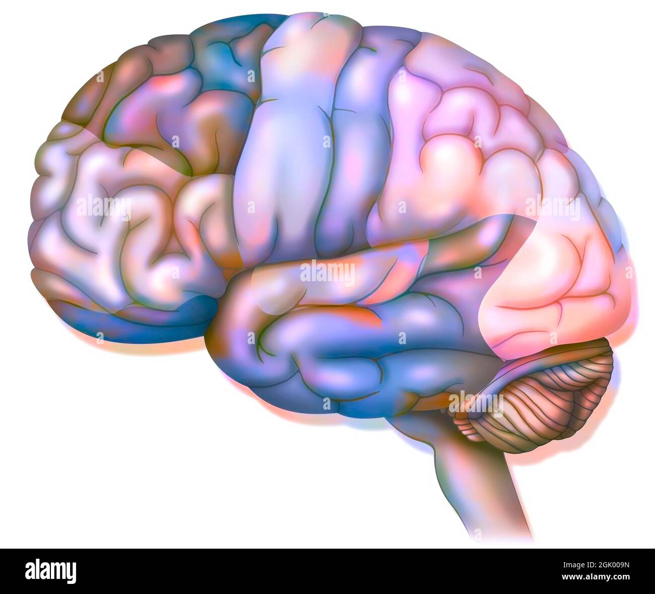 Areas of the brain: prefrontal cortex, premotor cortex and oculomotor cortex. . Stock Photohttps://www.alamy.com/image-license-details/?v=1https://www.alamy.com/areas-of-the-brain-prefrontal-cortex-premotor-cortex-and-oculomotor-cortex-image441937937.html
RF2GK009N–Areas of the brain: prefrontal cortex, premotor cortex and oculomotor cortex. .
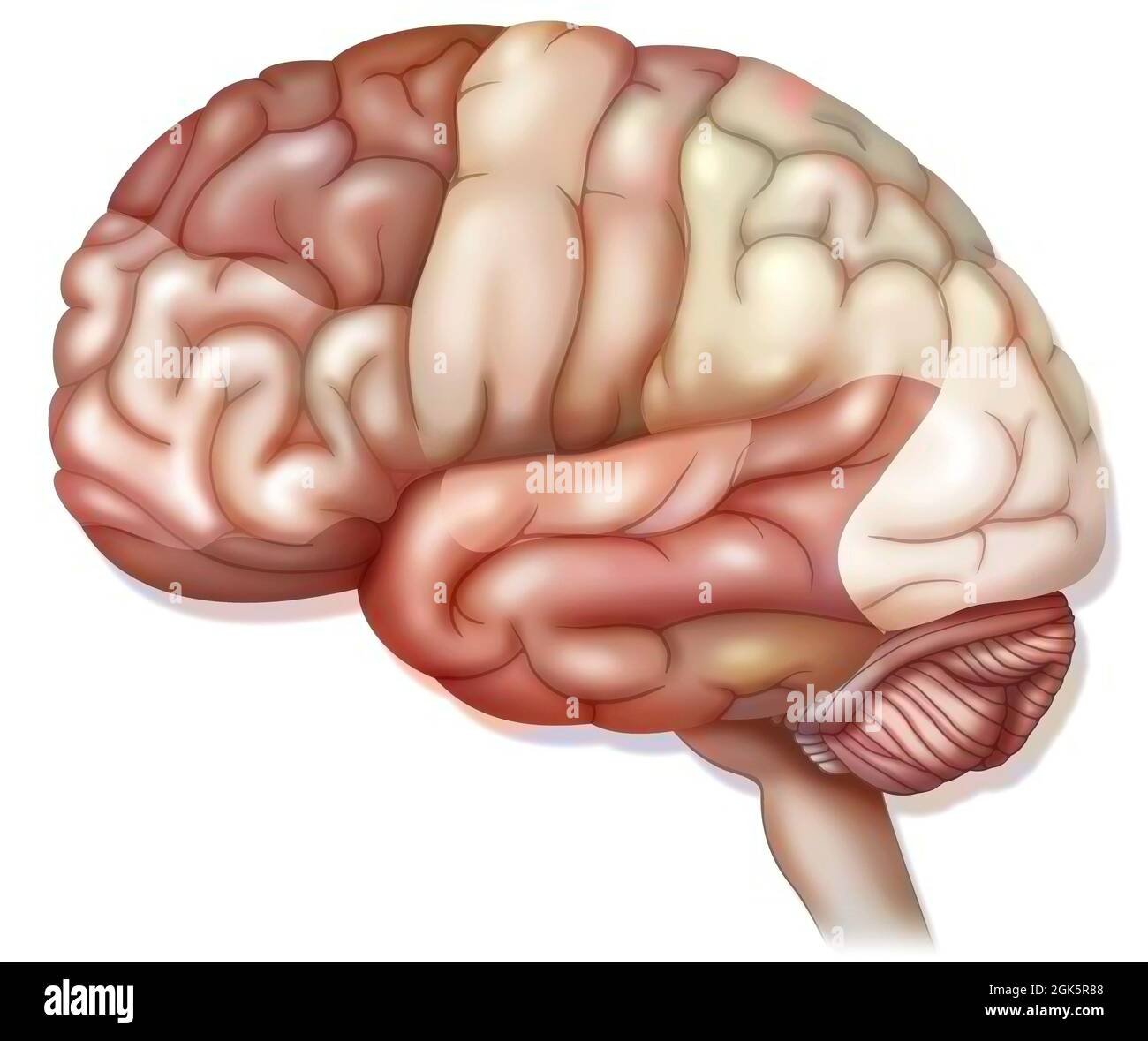 Areas of the brain: prefrontal cortex, premotor cortex and oculomotor cortex. . Stock Photohttps://www.alamy.com/image-license-details/?v=1https://www.alamy.com/areas-of-the-brain-prefrontal-cortex-premotor-cortex-and-oculomotor-cortex-image442065688.html
RF2GK5R88–Areas of the brain: prefrontal cortex, premotor cortex and oculomotor cortex. .
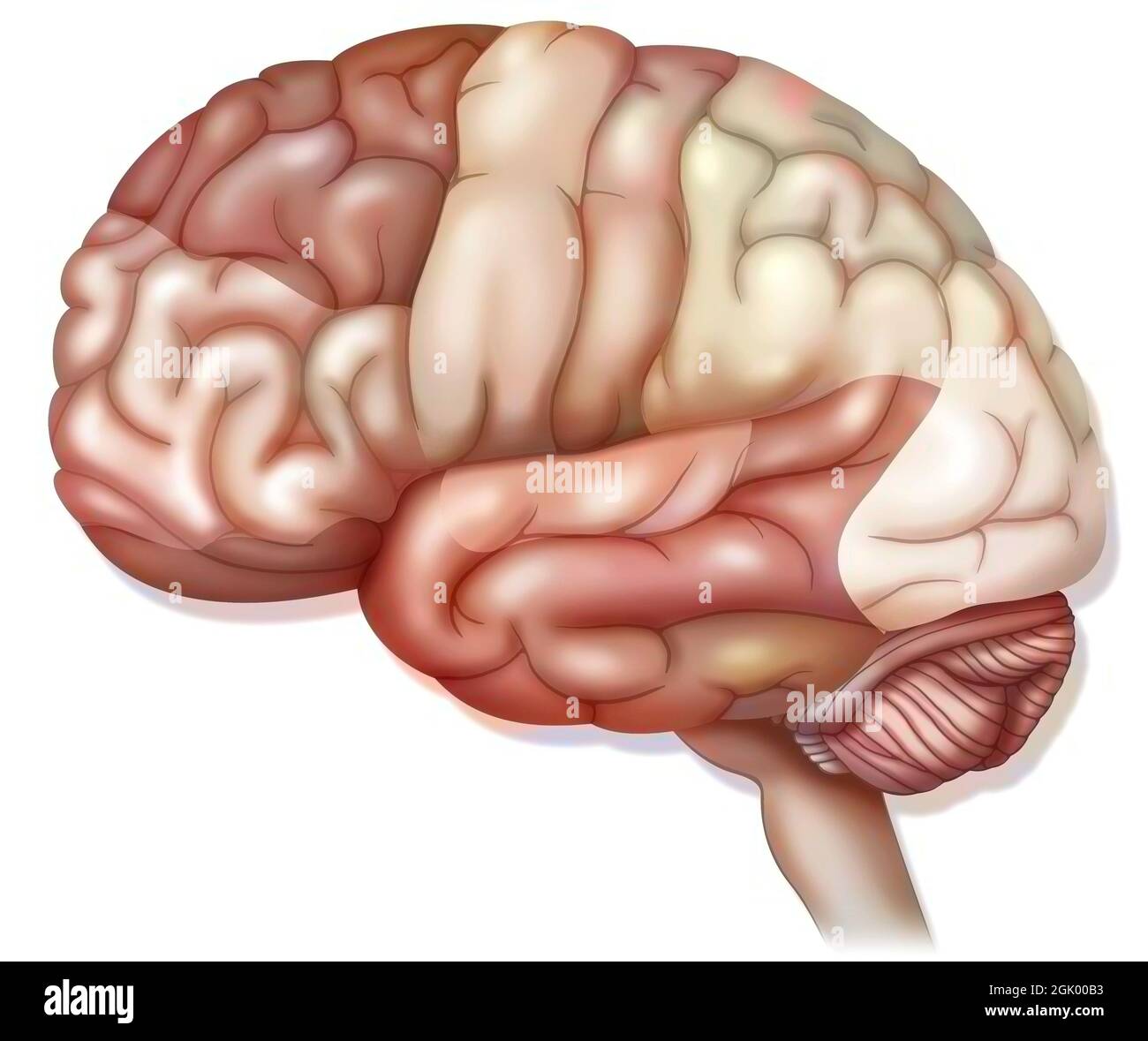 Areas of the brain: prefrontal cortex, premotor cortex and oculomotor cortex. . Stock Photohttps://www.alamy.com/image-license-details/?v=1https://www.alamy.com/areas-of-the-brain-prefrontal-cortex-premotor-cortex-and-oculomotor-cortex-image441937975.html
RF2GK00B3–Areas of the brain: prefrontal cortex, premotor cortex and oculomotor cortex. .
 Areas of the brain: prefrontal cortex, premotor cortex and oculomotor cortex. . Stock Photohttps://www.alamy.com/image-license-details/?v=1https://www.alamy.com/areas-of-the-brain-prefrontal-cortex-premotor-cortex-and-oculomotor-cortex-image442065683.html
RF2GK5R83–Areas of the brain: prefrontal cortex, premotor cortex and oculomotor cortex. .
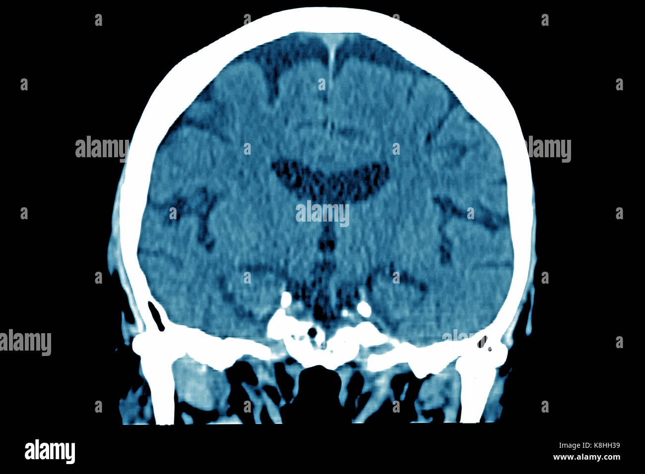
 CEREBRAL ATROPHY, CT SCAN Stock Photohttps://www.alamy.com/image-license-details/?v=1https://www.alamy.com/stock-image-cerebral-atrophy-ct-scan-160197172.html
RMK8HH3G–CEREBRAL ATROPHY, CT SCAN
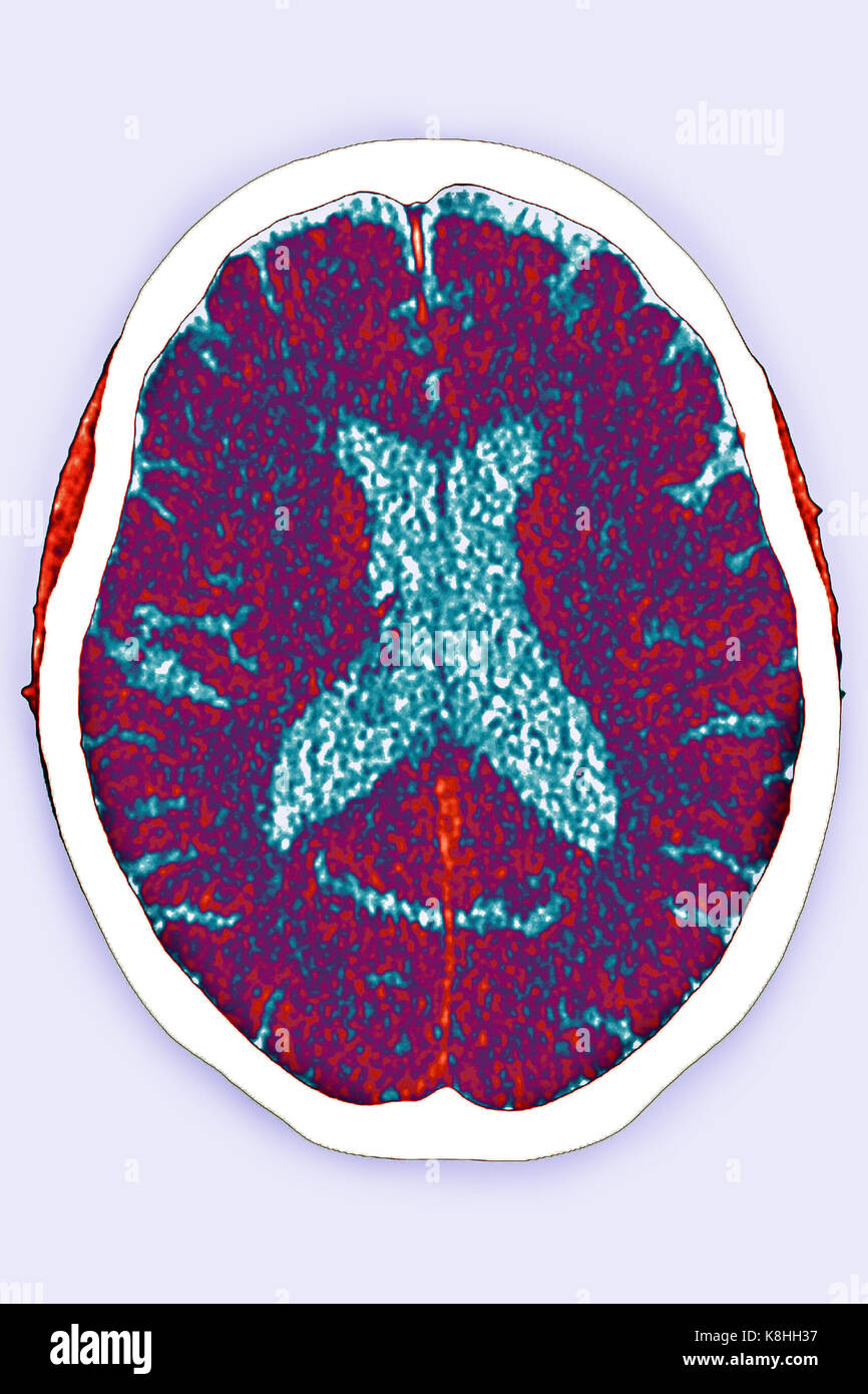 CEREBRAL ATROPHY, CT SCAN Stock Photohttps://www.alamy.com/image-license-details/?v=1https://www.alamy.com/stock-image-cerebral-atrophy-ct-scan-160197163.html
RMK8HH37–CEREBRAL ATROPHY, CT SCAN
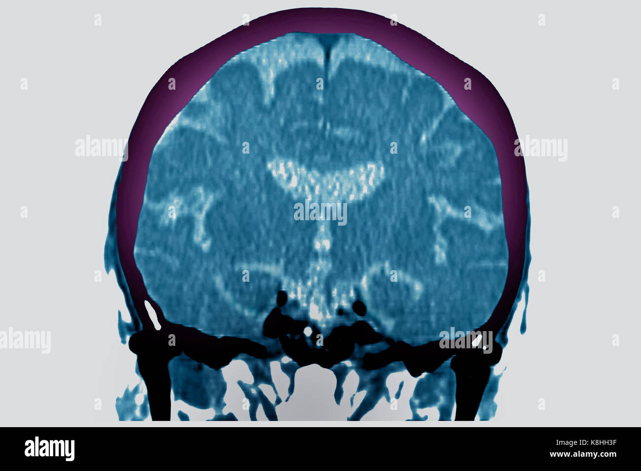 CEREBRAL ATROPHY, CT SCAN Stock Photohttps://www.alamy.com/image-license-details/?v=1https://www.alamy.com/stock-image-cerebral-atrophy-ct-scan-160197171.html
RMK8HH3F–CEREBRAL ATROPHY, CT SCAN
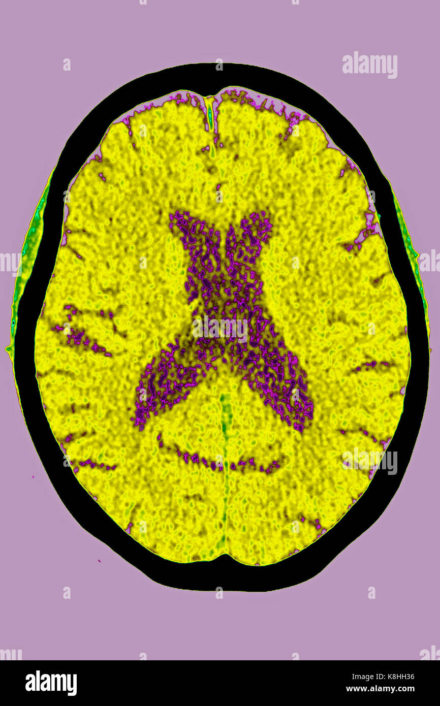 CEREBRAL ATROPHY, CT SCAN Stock Photohttps://www.alamy.com/image-license-details/?v=1https://www.alamy.com/stock-image-cerebral-atrophy-ct-scan-160197162.html
RMK8HH36–CEREBRAL ATROPHY, CT SCAN
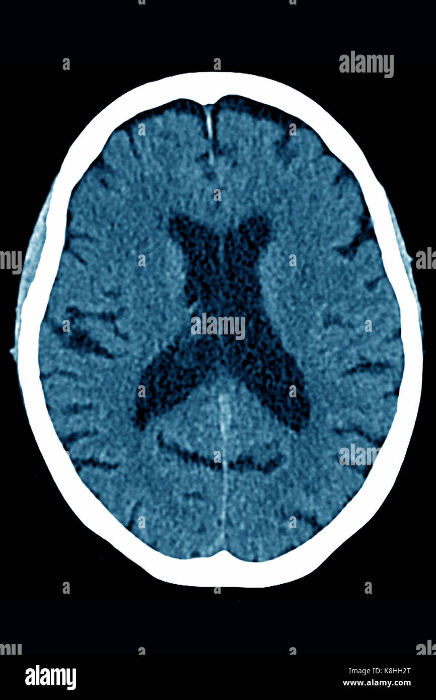 CEREBRAL ATROPHY, CT SCAN Stock Photohttps://www.alamy.com/image-license-details/?v=1https://www.alamy.com/stock-image-cerebral-atrophy-ct-scan-160197152.html
RMK8HH2T–CEREBRAL ATROPHY, CT SCAN
 CEREBRAL ATROPHY, CT SCAN Stock Photohttps://www.alamy.com/image-license-details/?v=1https://www.alamy.com/stock-image-cerebral-atrophy-ct-scan-160197175.html
RMK8HH3K–CEREBRAL ATROPHY, CT SCAN
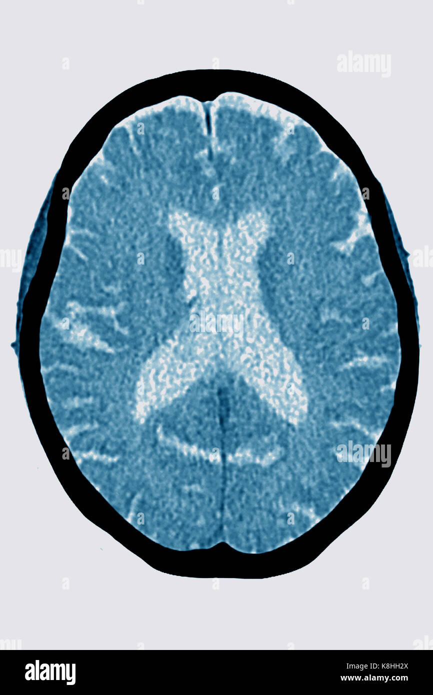 CEREBRAL ATROPHY, CT SCAN Stock Photohttps://www.alamy.com/image-license-details/?v=1https://www.alamy.com/stock-image-cerebral-atrophy-ct-scan-160197154.html
RMK8HH2X–CEREBRAL ATROPHY, CT SCAN
