Quick filters:
Prokaryote Stock Photos and Images
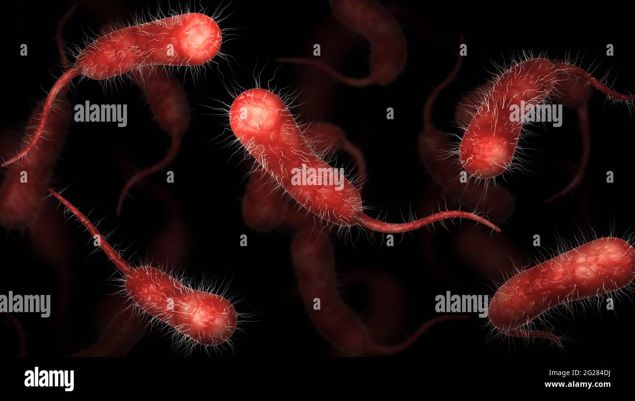 Biomedical illustration of vibrio vulnificus bacteria on black background. Stock Photohttps://www.alamy.com/image-license-details/?v=1https://www.alamy.com/biomedical-illustration-of-vibrio-vulnificus-bacteria-on-black-background-image431667646.html
RF2G284DJ–Biomedical illustration of vibrio vulnificus bacteria on black background.
 Tularaemia bacteria (Francisella tularensis), illustration. F. tularensis is Gram-negative, coccobacillus prokaryote. A zoonotic microorganism that causes tularaemia, a disease of wild rodents and rabbits that can be transmitted to humans and domesticated pets. Stock Photohttps://www.alamy.com/image-license-details/?v=1https://www.alamy.com/stock-photo-tularaemia-bacteria-francisella-tularensis-illustration-f-tularensis-136521058.html
RFHX3202–Tularaemia bacteria (Francisella tularensis), illustration. F. tularensis is Gram-negative, coccobacillus prokaryote. A zoonotic microorganism that causes tularaemia, a disease of wild rodents and rabbits that can be transmitted to humans and domesticated pets.
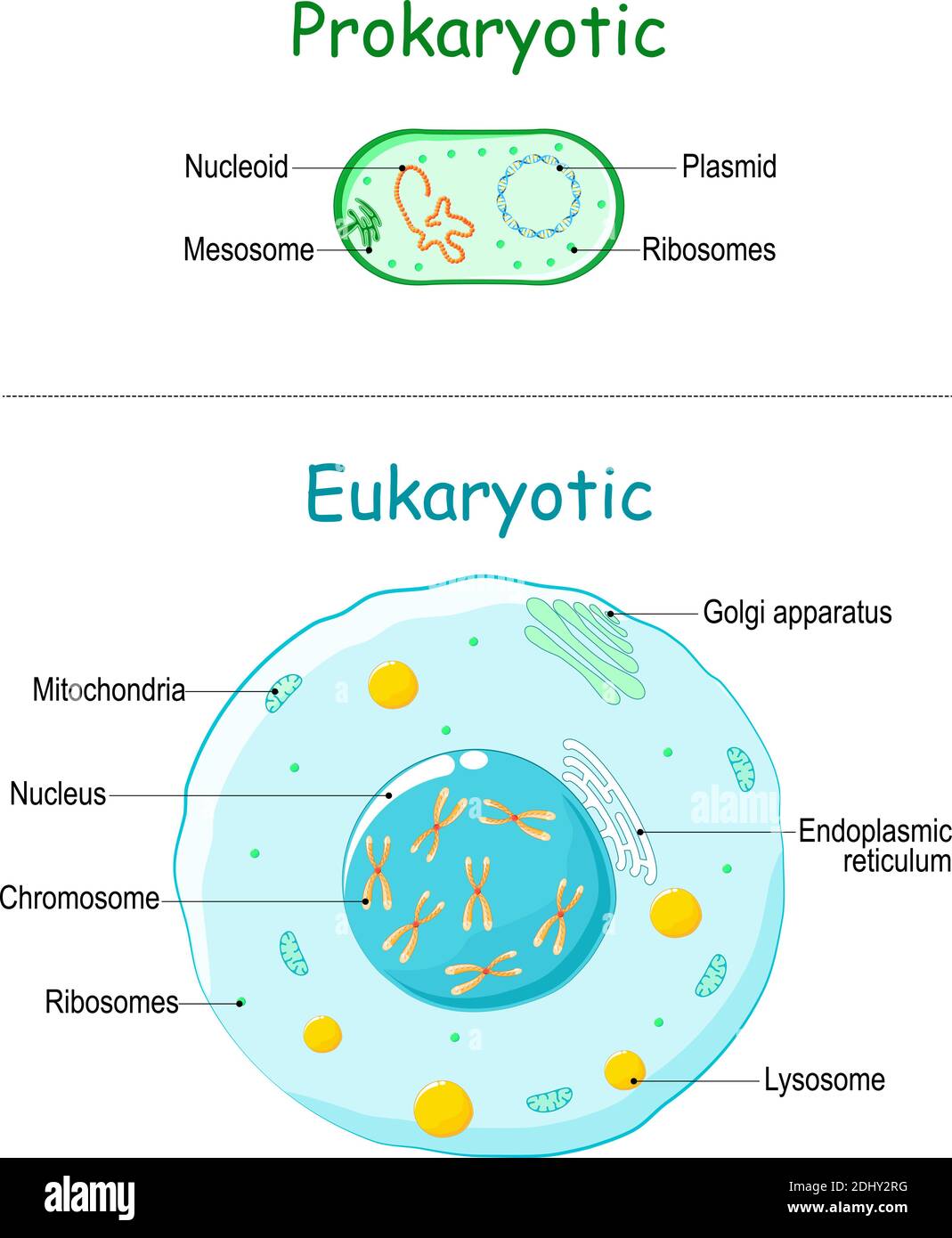 Prokaryote vs Eukaryote. illustration of eukaryotic and prokaryotic cell with text. Differences between Prokaryotic and Eukaryotic cells. vector Stock Vectorhttps://www.alamy.com/image-license-details/?v=1https://www.alamy.com/prokaryote-vs-eukaryote-illustration-of-eukaryotic-and-prokaryotic-cell-with-text-differences-between-prokaryotic-and-eukaryotic-cells-vector-image389672180.html
RF2DHY2RG–Prokaryote vs Eukaryote. illustration of eukaryotic and prokaryotic cell with text. Differences between Prokaryotic and Eukaryotic cells. vector
 3D Rendering of a cross-section of a bacterial cell, showing its organelles. it's prokaryote bacteria cell. Stock Photohttps://www.alamy.com/image-license-details/?v=1https://www.alamy.com/3d-rendering-of-a-cross-section-of-a-bacterial-cell-showing-its-organelles-its-prokaryote-bacteria-cell-image620483809.html
RF2Y1DDC1–3D Rendering of a cross-section of a bacterial cell, showing its organelles. it's prokaryote bacteria cell.
 00166142.tif Oscillatoria intermedia var phormidioides Hansg. Stock Photohttps://www.alamy.com/image-license-details/?v=1https://www.alamy.com/00166142tif-oscillatoria-intermedia-var-phormidioides-hansg-image360479980.html
RM2BXD7W0–00166142.tif Oscillatoria intermedia var phormidioides Hansg.
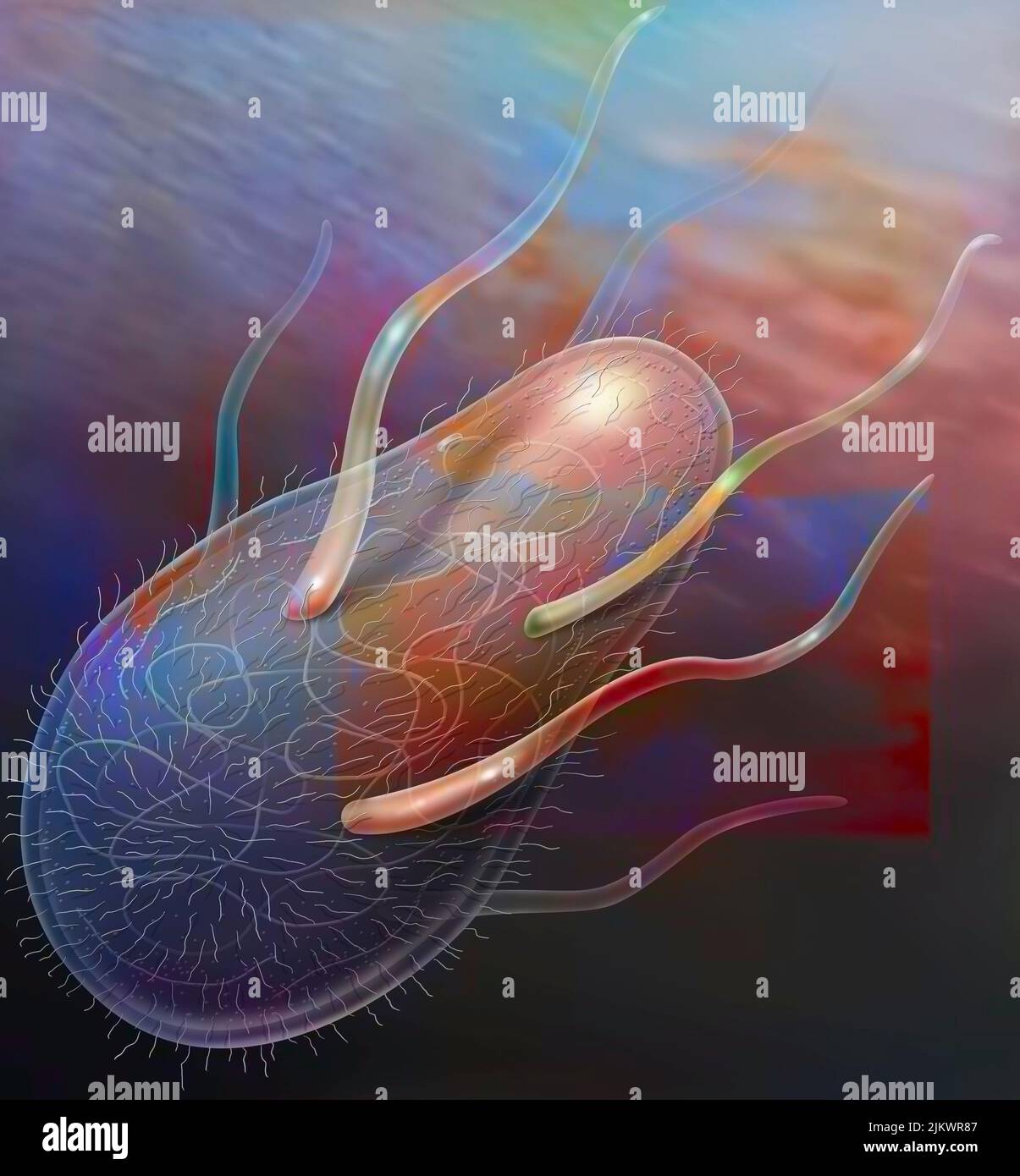 Representation of a bacterium (prokaryote) with its organelles in transparency. Stock Photohttps://www.alamy.com/image-license-details/?v=1https://www.alamy.com/representation-of-a-bacterium-prokaryote-with-its-organelles-in-transparency-image476925463.html
RF2JKWR87–Representation of a bacterium (prokaryote) with its organelles in transparency.
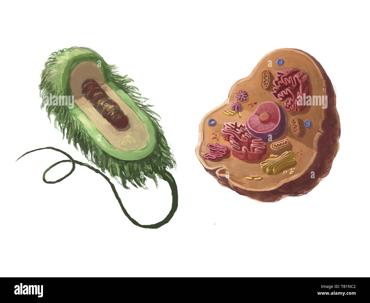 Prokaryotic and Eukaryotic Cells, Illustration Stock Photohttps://www.alamy.com/image-license-details/?v=1https://www.alamy.com/prokaryotic-and-eukaryotic-cells-illustration-image245901154.html
RMT81NC2–Prokaryotic and Eukaryotic Cells, Illustration
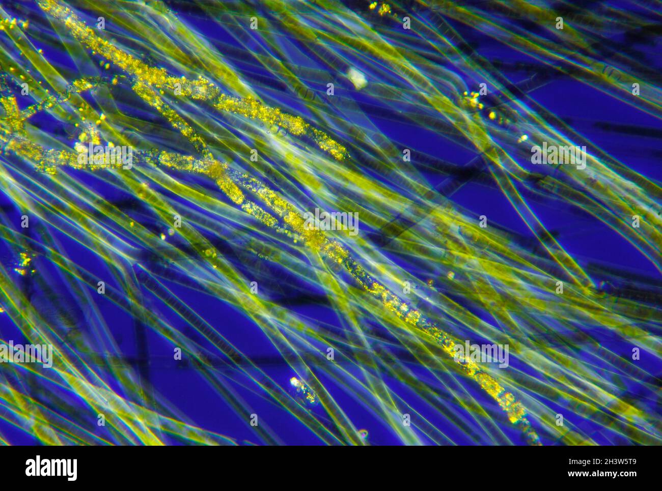 Microscopic view of a cyanobacteria (blue-green algae, Oscillatoria) filaments. Polarized light with crossed polarizers. Stock Photohttps://www.alamy.com/image-license-details/?v=1https://www.alamy.com/microscopic-view-of-a-cyanobacteria-blue-green-algae-oscillatoria-filaments-polarized-light-with-crossed-polarizers-image449866937.html
RF2H3W5T9–Microscopic view of a cyanobacteria (blue-green algae, Oscillatoria) filaments. Polarized light with crossed polarizers.
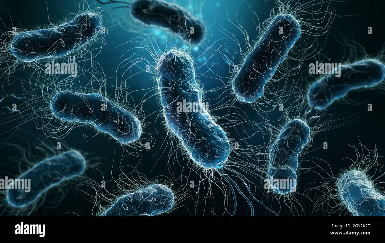 Colony of bacteria close-up 3D rendering illustration on blue background. Microbiology, medical, biology, bacteriology, science, medicine, infection, Stock Photohttps://www.alamy.com/image-license-details/?v=1https://www.alamy.com/colony-of-bacteria-close-up-3d-rendering-illustration-on-blue-background-microbiology-medical-biology-bacteriology-science-medicine-infection-image446913792.html
RF2GY2K2T–Colony of bacteria close-up 3D rendering illustration on blue background. Microbiology, medical, biology, bacteriology, science, medicine, infection,
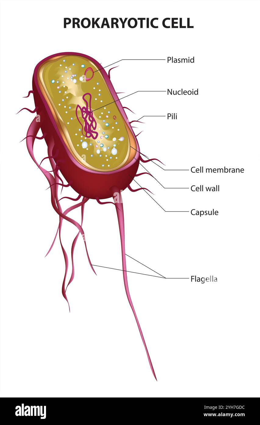 Prokaryotic Cell Structure Chart, vector medical illustration, online education material. English translation text Stock Vectorhttps://www.alamy.com/image-license-details/?v=1https://www.alamy.com/prokaryotic-cell-structure-chart-vector-medical-illustration-online-education-material-english-translation-text-image630188984.html
RF2YH7GDC–Prokaryotic Cell Structure Chart, vector medical illustration, online education material. English translation text
 Diagram of Eukaryotic flagella.Vector illustration. Stock Vectorhttps://www.alamy.com/image-license-details/?v=1https://www.alamy.com/diagram-of-eukaryotic-flagellavector-illustration-image576063310.html
RF2TD5XHJ–Diagram of Eukaryotic flagella.Vector illustration.
 Diagram of microorganism optimal temperature range - Psychrophile, Mesophile, Thremophile and Hyperthermophile growth rates with example bacteria. Stock Vectorhttps://www.alamy.com/image-license-details/?v=1https://www.alamy.com/diagram-of-microorganism-optimal-temperature-range-psychrophile-mesophile-thremophile-and-hyperthermophile-growth-rates-with-example-bacteria-image479922825.html
RF2JTPACW–Diagram of microorganism optimal temperature range - Psychrophile, Mesophile, Thremophile and Hyperthermophile growth rates with example bacteria.
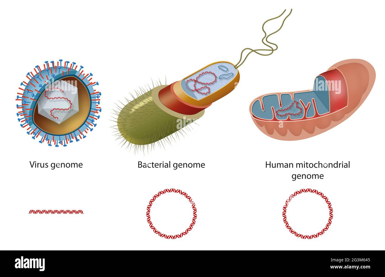 Types of genome in virus, bacteria and human mitochondria. Diagram of closed circular DNA and linear DNA Stock Photohttps://www.alamy.com/image-license-details/?v=1https://www.alamy.com/types-of-genome-in-virus-bacteria-and-human-mitochondria-diagram-of-closed-circular-dna-and-linear-dna-image432547029.html
RF2G3M645–Types of genome in virus, bacteria and human mitochondria. Diagram of closed circular DNA and linear DNA
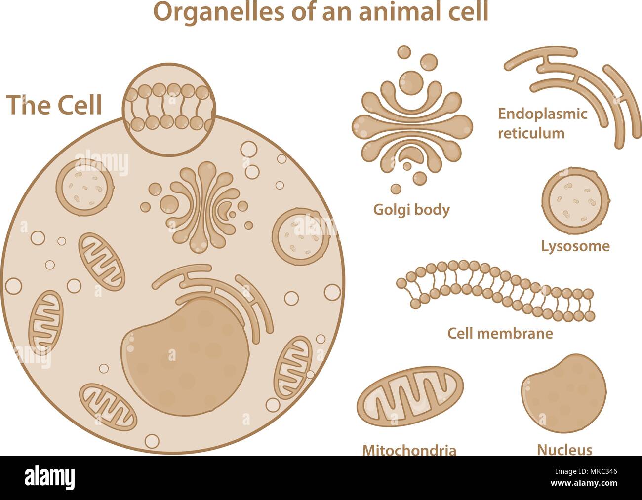 Organelles and major components of an animal cell. Stock Vectorhttps://www.alamy.com/image-license-details/?v=1https://www.alamy.com/organelles-and-major-components-of-an-animal-cell-image184048038.html
RFMKC346–Organelles and major components of an animal cell.
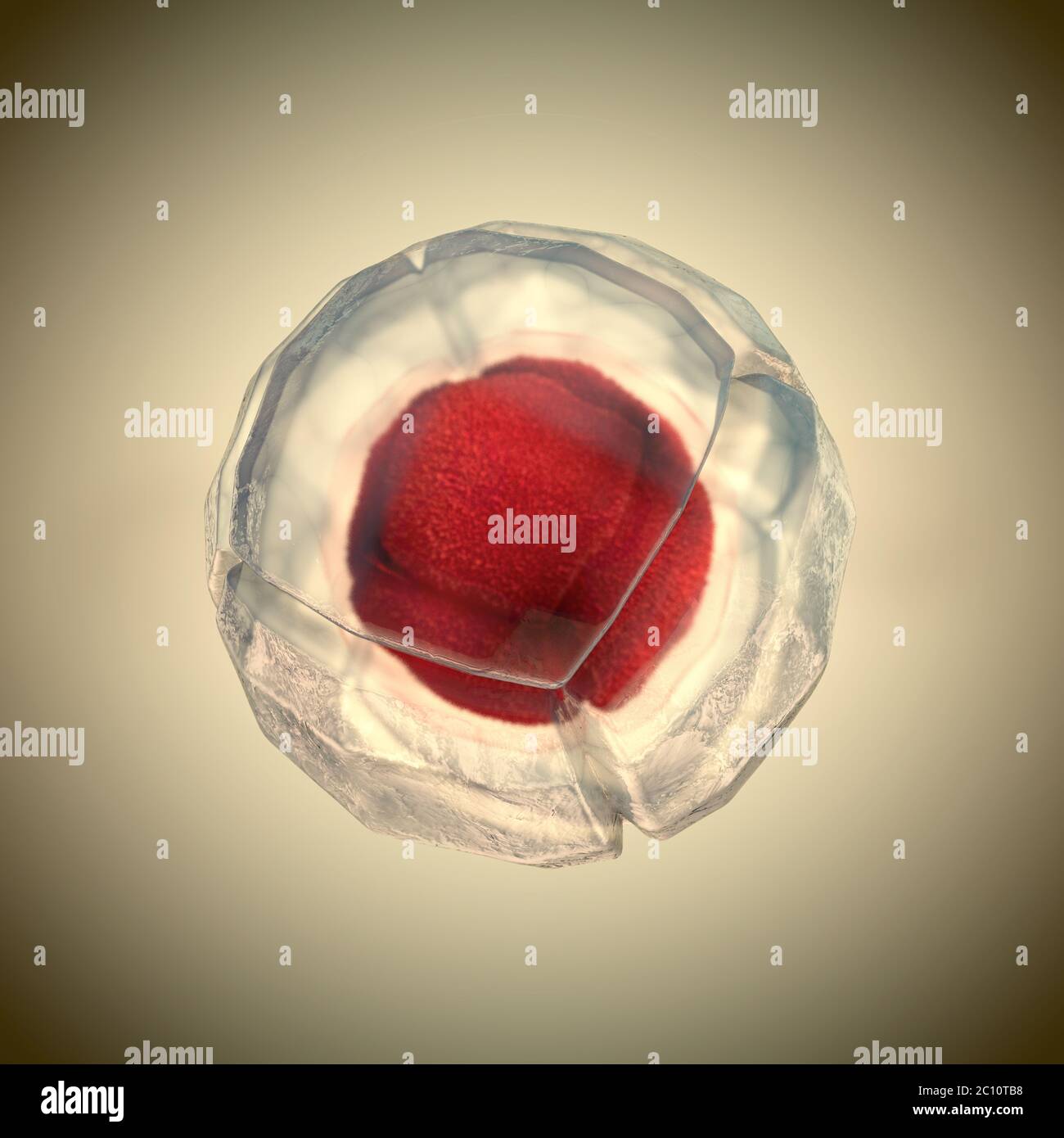 3d illustration of cell division, cell membrane and a splitting red nucleus Stock Photohttps://www.alamy.com/image-license-details/?v=1https://www.alamy.com/3d-illustration-of-cell-division-cell-membrane-and-a-splitting-red-nucleus-image362051516.html
RF2C10TB8–3d illustration of cell division, cell membrane and a splitting red nucleus
 Peritrichous Bacteria with lot of flagellum, harmful bacillus with long tails, prokaryote, viruses probiotics and pathogen, H. pylori, microbe flagell Stock Photohttps://www.alamy.com/image-license-details/?v=1https://www.alamy.com/peritrichous-bacteria-with-lot-of-flagellum-harmful-bacillus-with-long-tails-prokaryote-viruses-probiotics-and-pathogen-h-pylori-microbe-flagell-image622012602.html
RM2Y3Y3BP–Peritrichous Bacteria with lot of flagellum, harmful bacillus with long tails, prokaryote, viruses probiotics and pathogen, H. pylori, microbe flagell
RF3CFB4XK–Icon for bacterium, prokaryote
 Prokaryote word cloud concept Stock Photohttps://www.alamy.com/image-license-details/?v=1https://www.alamy.com/prokaryote-word-cloud-concept-image262839121.html
RFW7H9YD–Prokaryote word cloud concept
 Bacteria culture plates petri dishes with blue transformation colonies in incubator Stock Photohttps://www.alamy.com/image-license-details/?v=1https://www.alamy.com/bacteria-culture-plates-petri-dishes-with-blue-transformation-colonies-image6349431.html
RMA4RMR8–Bacteria culture plates petri dishes with blue transformation colonies in incubator
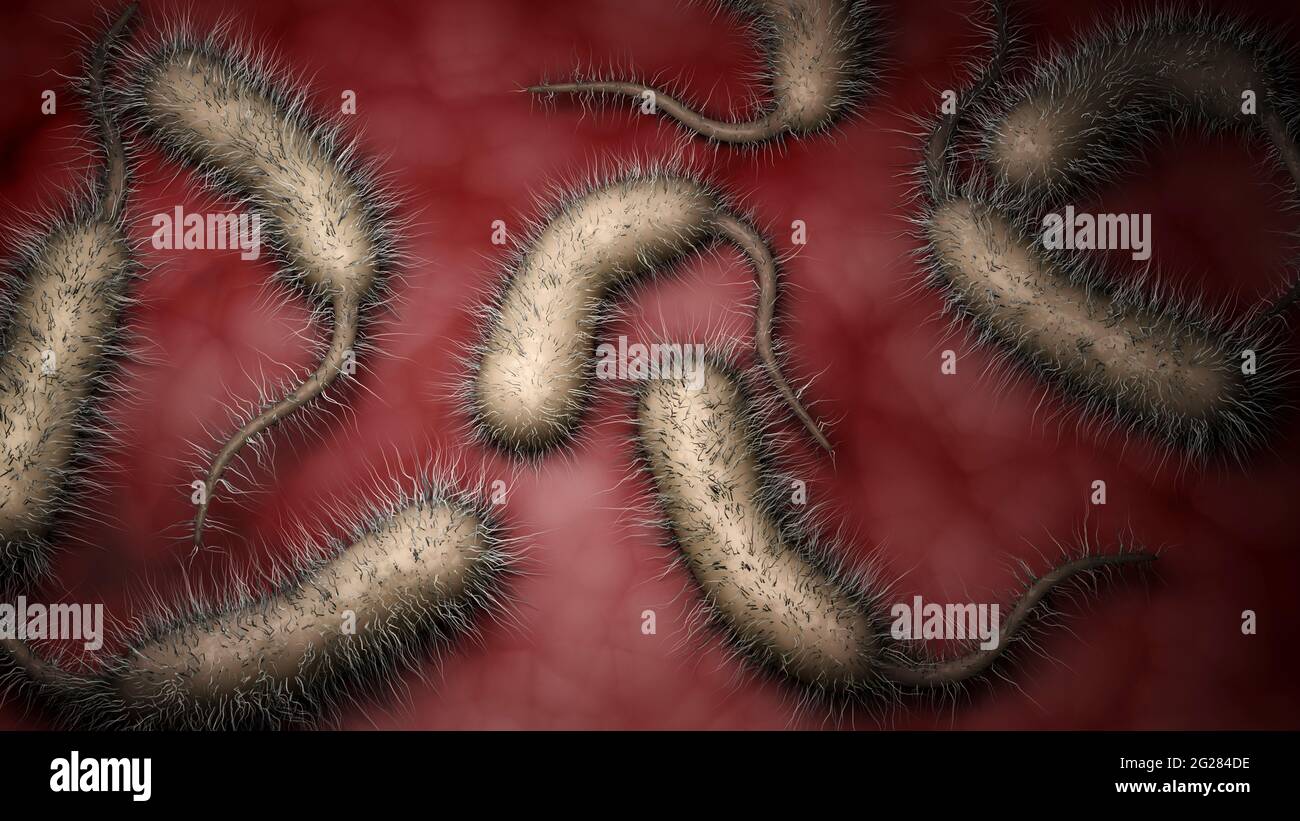 Biomedical illustration of vibrio vulnificus bacteria inside human body. Stock Photohttps://www.alamy.com/image-license-details/?v=1https://www.alamy.com/biomedical-illustration-of-vibrio-vulnificus-bacteria-inside-human-body-image431667642.html
RF2G284DE–Biomedical illustration of vibrio vulnificus bacteria inside human body.
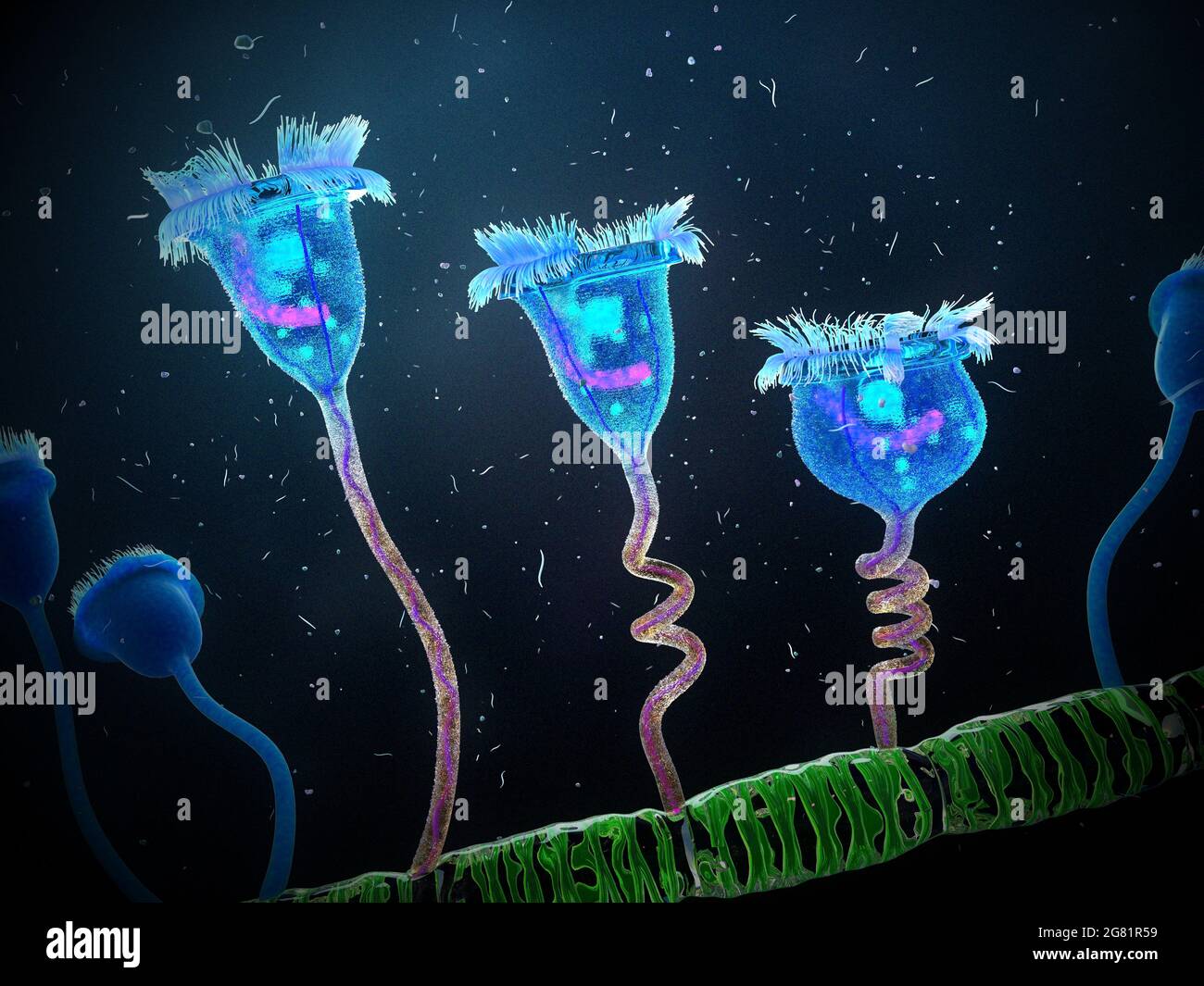 Vorticella protozoa attached to algae, illustration Stock Photohttps://www.alamy.com/image-license-details/?v=1https://www.alamy.com/vorticella-protozoa-attached-to-algae-illustration-image435216581.html
RF2G81R59–Vorticella protozoa attached to algae, illustration
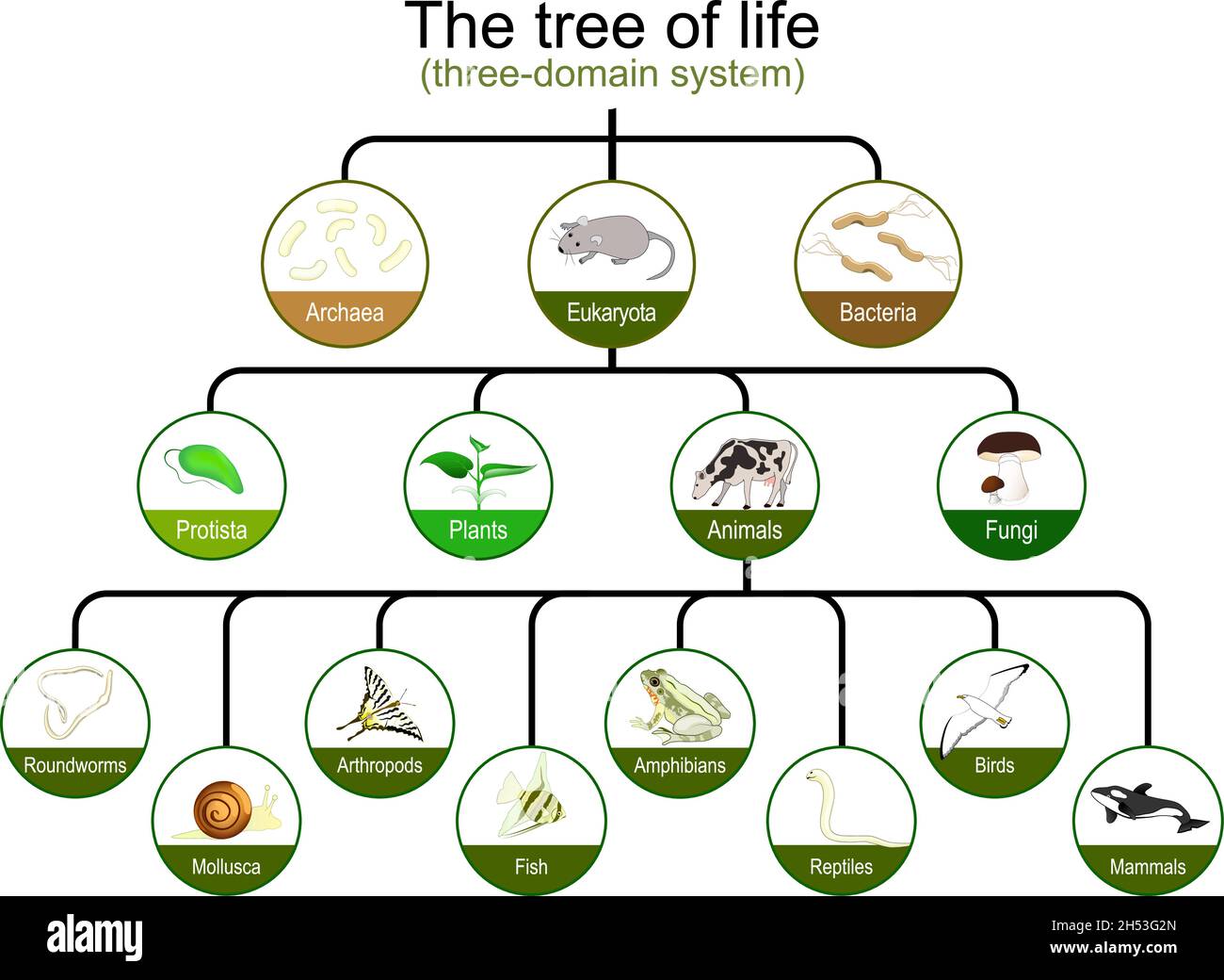 Taxonomy. classification of tree of life. three-domain system. Phylogenetic and symbiogenetic tree of living organisms. origins of Archaea, eukaryotes Stock Vectorhttps://www.alamy.com/image-license-details/?v=1https://www.alamy.com/taxonomy-classification-of-tree-of-life-three-domain-system-phylogenetic-and-symbiogenetic-tree-of-living-organisms-origins-of-archaea-eukaryotes-image450621325.html
RF2H53G2N–Taxonomy. classification of tree of life. three-domain system. Phylogenetic and symbiogenetic tree of living organisms. origins of Archaea, eukaryotes
 3D Rendering of a cross-section of a bacterial cell, showing its organelles. it's prokaryote bacteria cell. Stock Photohttps://www.alamy.com/image-license-details/?v=1https://www.alamy.com/3d-rendering-of-a-cross-section-of-a-bacterial-cell-showing-its-organelles-its-prokaryote-bacteria-cell-image620483796.html
RF2Y1DDBG–3D Rendering of a cross-section of a bacterial cell, showing its organelles. it's prokaryote bacteria cell.
 3d rendered medically accurate illustration of a human cell Stock Photohttps://www.alamy.com/image-license-details/?v=1https://www.alamy.com/3d-rendered-medically-accurate-illustration-of-a-human-cell-image257161372.html
RFTXAKXM–3d rendered medically accurate illustration of a human cell
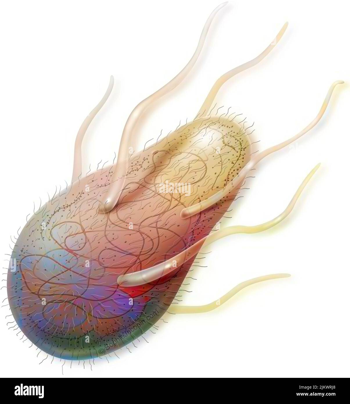 Bacterium (prokaryote) with its organelles in transparency. Stock Photohttps://www.alamy.com/image-license-details/?v=1https://www.alamy.com/bacterium-prokaryote-with-its-organelles-in-transparency-image476925744.html
RF2JKWRJ8–Bacterium (prokaryote) with its organelles in transparency.
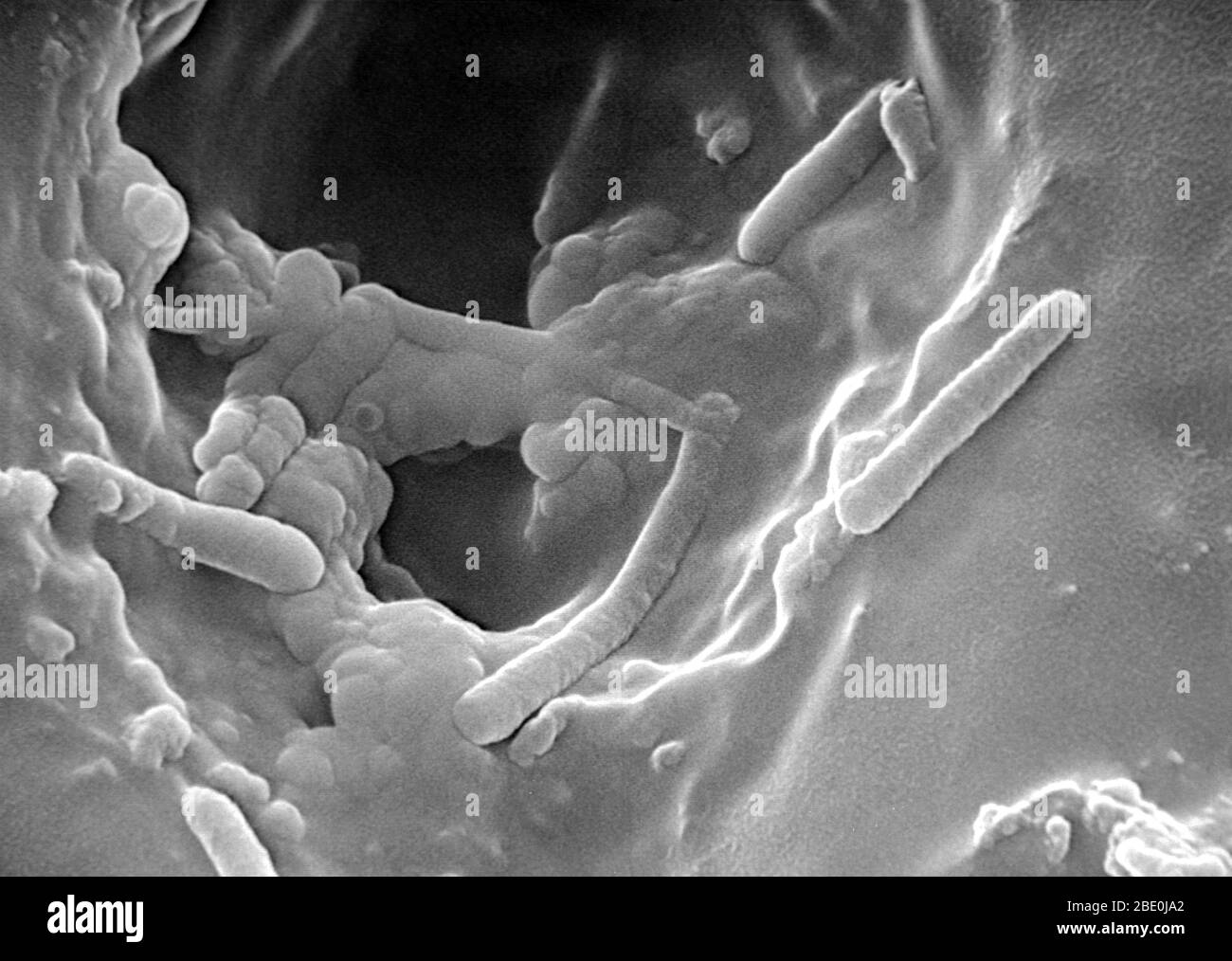 Scanning Elctron Micrograph (SEM) of Pseudomonas aeruginosa, a versatile gram-negative bacterium that grows in soil, marshes, and coastal marine habitats, as well as on plant and animal tissues. Pseudomonas aeruginosa is rod-shaped bacterium that can cause disease in plants and animals, including humans. A species of considerable medical importance, P. aeruginosa is a multidrug resistant pathogen recognised for its ubiquity, its intrinsically advanced antibiotic resistance mechanisms, and its association with serious illnesses, hospital-acquired infections such as ventilator-associated pneumon Stock Photohttps://www.alamy.com/image-license-details/?v=1https://www.alamy.com/scanning-elctron-micrograph-sem-of-pseudomonas-aeruginosa-a-versatile-gram-negative-bacterium-that-grows-in-soil-marshes-and-coastal-marine-habitats-as-well-as-on-plant-and-animal-tissues-pseudomonas-aeruginosa-is-rod-shaped-bacterium-that-can-cause-disease-in-plants-and-animals-including-humans-a-species-of-considerable-medical-importance-p-aeruginosa-is-a-multidrug-resistant-pathogen-recognised-for-its-ubiquity-its-intrinsically-advanced-antibiotic-resistance-mechanisms-and-its-association-with-serious-illnesses-hospital-acquired-infections-such-as-ventilator-associated-pneumon-image352826938.html
RM2BE0JA2–Scanning Elctron Micrograph (SEM) of Pseudomonas aeruginosa, a versatile gram-negative bacterium that grows in soil, marshes, and coastal marine habitats, as well as on plant and animal tissues. Pseudomonas aeruginosa is rod-shaped bacterium that can cause disease in plants and animals, including humans. A species of considerable medical importance, P. aeruginosa is a multidrug resistant pathogen recognised for its ubiquity, its intrinsically advanced antibiotic resistance mechanisms, and its association with serious illnesses, hospital-acquired infections such as ventilator-associated pneumon
RMP23TKJ–. Bacteria 25 Bacteria - Print - Iconographia Zoologica - Special Collections University of Amsterdam - UBAINV0274 065 04 0003
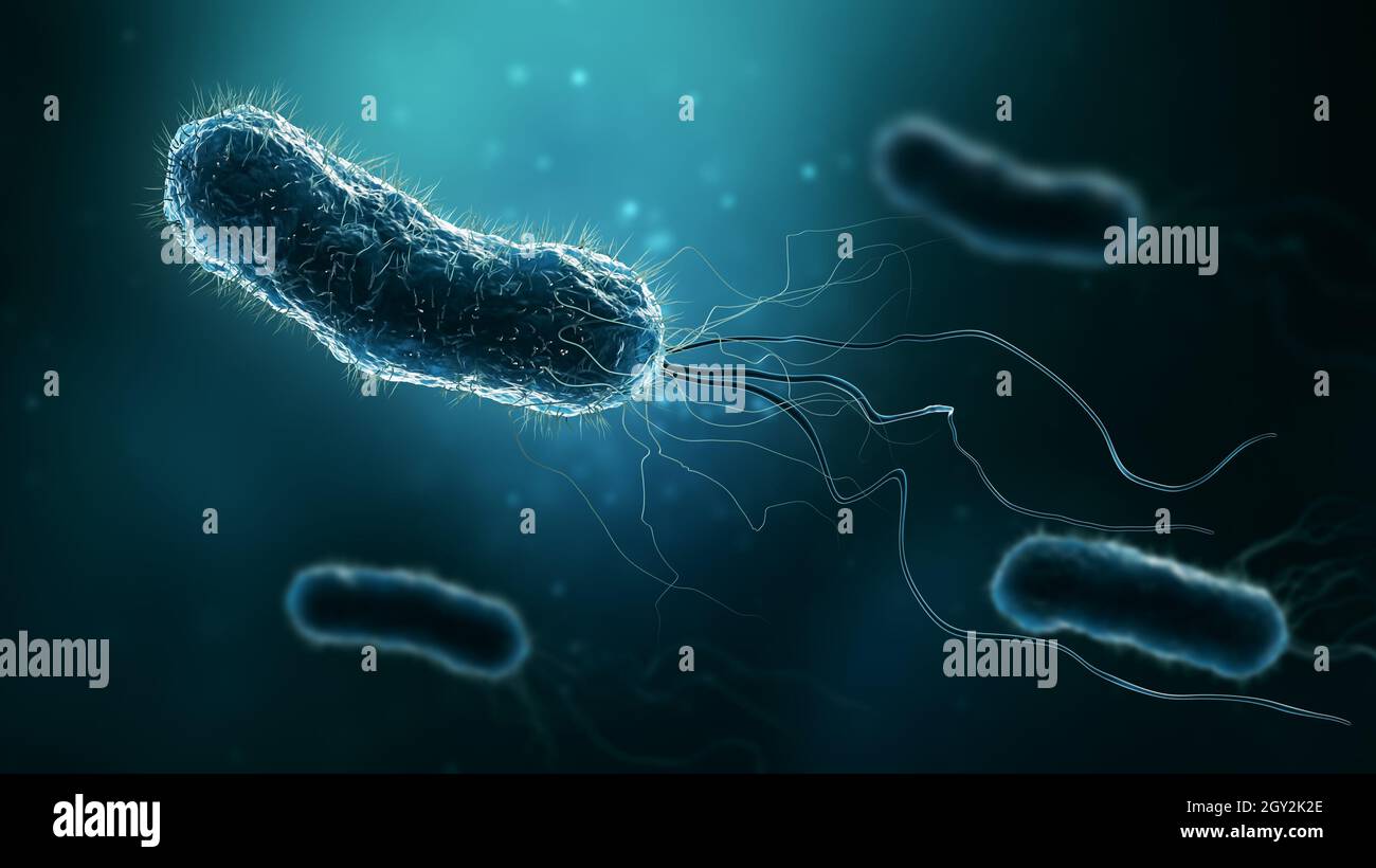 Group of bacteria such as Escherichia coli, Helicobacter pylori or salmonella 3D rendering illustration. Microbiology, medical, bacteriology, biology, Stock Photohttps://www.alamy.com/image-license-details/?v=1https://www.alamy.com/group-of-bacteria-such-as-escherichia-coli-helicobacter-pylori-or-salmonella-3d-rendering-illustration-microbiology-medical-bacteriology-biology-image446913782.html
RF2GY2K2E–Group of bacteria such as Escherichia coli, Helicobacter pylori or salmonella 3D rendering illustration. Microbiology, medical, bacteriology, biology,
 Desulforudis audaxviator Stock Photohttps://www.alamy.com/image-license-details/?v=1https://www.alamy.com/stock-image-desulforudis-audaxviator-169414560.html
RMKRHE00–Desulforudis audaxviator
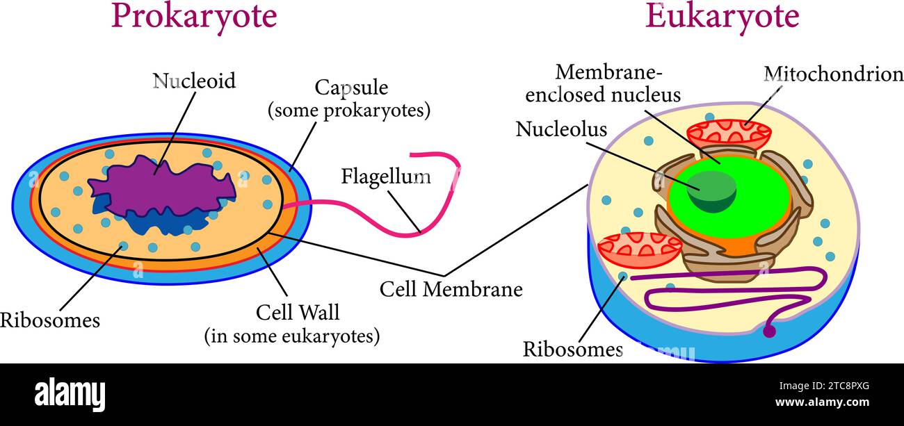 Comparison between eukaryotes and prokaryotes .Vector illustration. Stock Vectorhttps://www.alamy.com/image-license-details/?v=1https://www.alamy.com/comparison-between-eukaryotes-and-prokaryotes-vector-illustration-image575511624.html
RF2TC8PXG–Comparison between eukaryotes and prokaryotes .Vector illustration.
RF2JTPAFM–Set of 3 petri dish icons. Colorful simple illustration with bacterial cells.
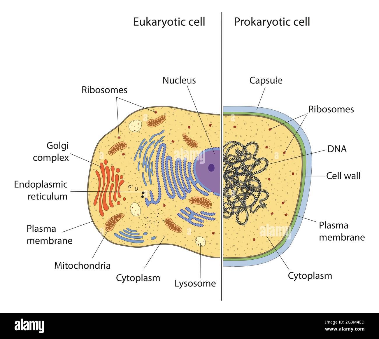 lllustration of eukaryotic and prokaryotic cell with text Stock Photohttps://www.alamy.com/image-license-details/?v=1https://www.alamy.com/lllustration-of-eukaryotic-and-prokaryotic-cell-with-text-image432545749.html
RF2G3M4ED–lllustration of eukaryotic and prokaryotic cell with text
 Anacystis montana Anacystis montana. Stock Photohttps://www.alamy.com/image-license-details/?v=1https://www.alamy.com/anacystis-montana-anacystis-montana-image362500583.html
RM2C1N95B–Anacystis montana Anacystis montana.
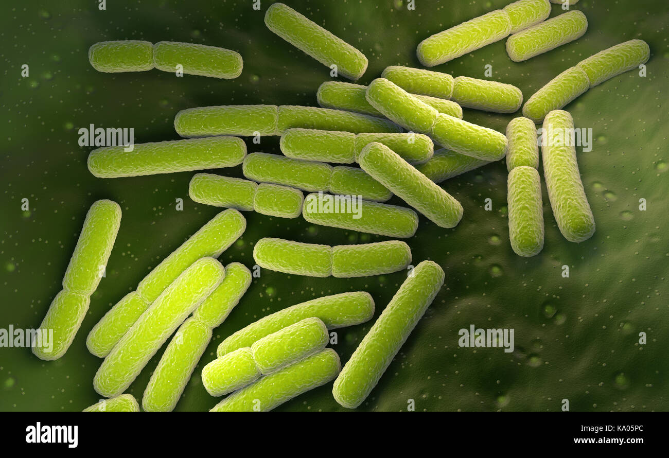 E. coli. Escherichia coli bacteria cells. 3D illustration Stock Photohttps://www.alamy.com/image-license-details/?v=1https://www.alamy.com/stock-image-e-coli-escherichia-coli-bacteria-cells-3d-illustration-161044420.html
RFKA05PC–E. coli. Escherichia coli bacteria cells. 3D illustration
 Peritrichous Bacteria with lot of flagellum, harmful bacillus with long tails, prokaryote, viruses probiotics and pathogen, H. pylori, microbe flagell Stock Photohttps://www.alamy.com/image-license-details/?v=1https://www.alamy.com/peritrichous-bacteria-with-lot-of-flagellum-harmful-bacillus-with-long-tails-prokaryote-viruses-probiotics-and-pathogen-h-pylori-microbe-flagell-image622012917.html
RM2Y3Y3R1–Peritrichous Bacteria with lot of flagellum, harmful bacillus with long tails, prokaryote, viruses probiotics and pathogen, H. pylori, microbe flagell
RF3C05AF0–Icon for bacterium, prokaryote
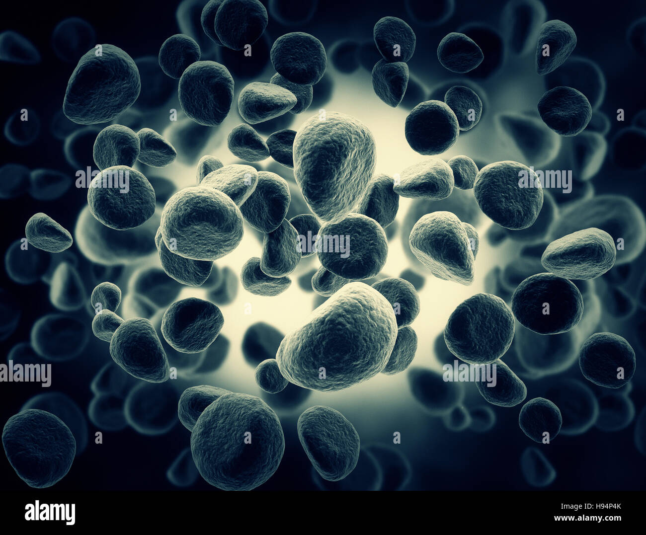 High resolution Illustration of cells Stock Photohttps://www.alamy.com/image-license-details/?v=1https://www.alamy.com/stock-photo-high-resolution-illustration-of-cells-126109667.html
RFH94P4K–High resolution Illustration of cells
 A scientist holds a tube containing a starter culture of Serratia marcescens on agar for research use Stock Photohttps://www.alamy.com/image-license-details/?v=1https://www.alamy.com/stock-photo-a-scientist-holds-a-tube-containing-a-starter-culture-of-serratia-16393289.html
RMATW46J–A scientist holds a tube containing a starter culture of Serratia marcescens on agar for research use
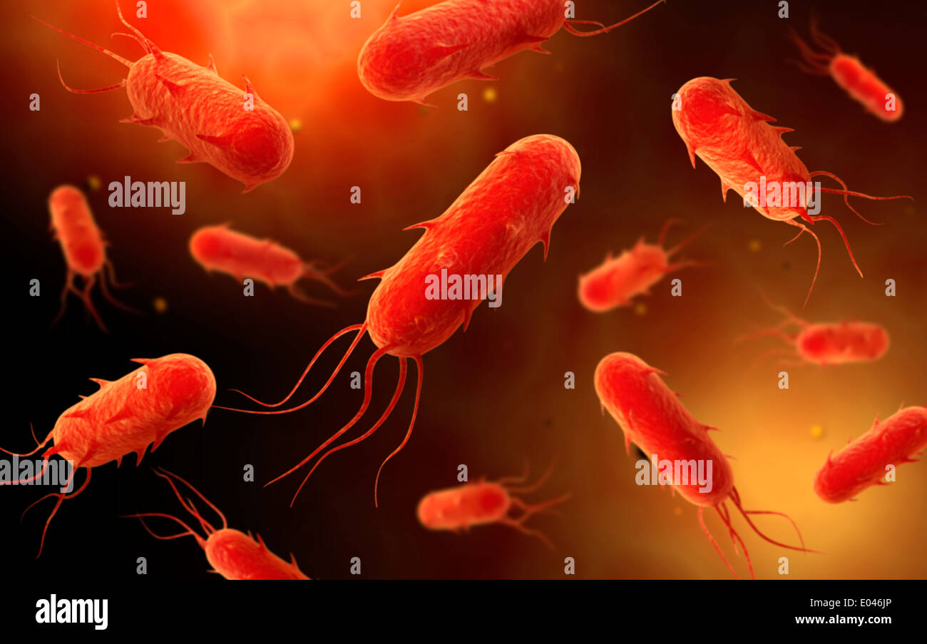 Conceptual image of flagellate bacterium. Stock Photohttps://www.alamy.com/image-license-details/?v=1https://www.alamy.com/conceptual-image-of-flagellate-bacterium-image68934510.html
RFE046JP–Conceptual image of flagellate bacterium.
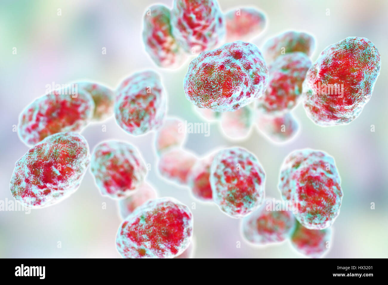 Tularaemia bacteria (Francisella tularensis), illustration. F. tularensis is Gram-negative, coccobacillus prokaryote. A zoonotic microorganism that causes tularaemia, a disease of wild rodents and rabbits that can be transmitted to humans and domesticated pets. Stock Photohttps://www.alamy.com/image-license-details/?v=1https://www.alamy.com/stock-photo-tularaemia-bacteria-francisella-tularensis-illustration-f-tularensis-136521057.html
RFHX3201–Tularaemia bacteria (Francisella tularensis), illustration. F. tularensis is Gram-negative, coccobacillus prokaryote. A zoonotic microorganism that causes tularaemia, a disease of wild rodents and rabbits that can be transmitted to humans and domesticated pets.
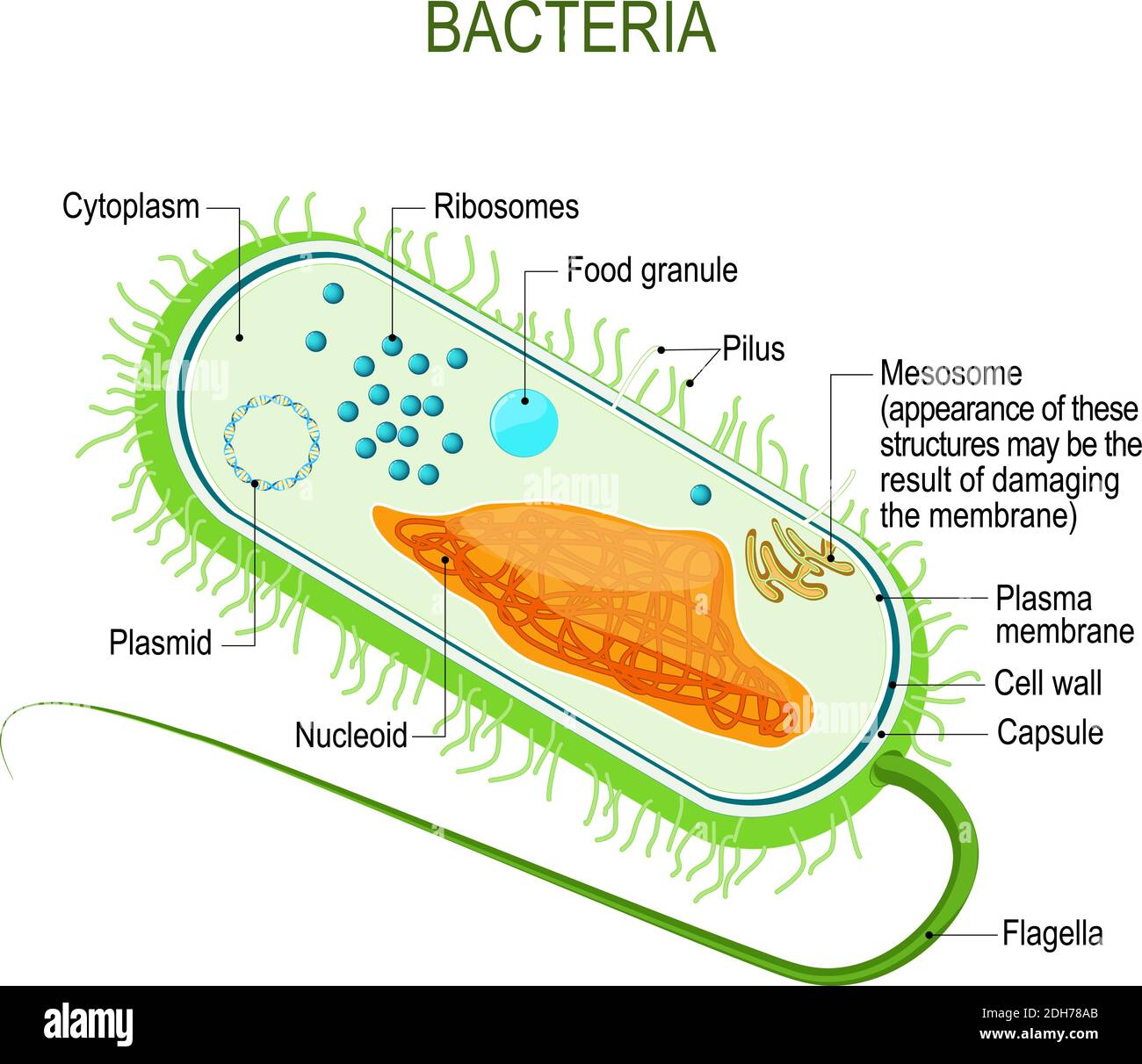 Structure of a bacterial cell. Anatomy of the prokaryote. unicellular organism. Vector diagram for your design, educational, medical, biological use Stock Vectorhttps://www.alamy.com/image-license-details/?v=1https://www.alamy.com/structure-of-a-bacterial-cell-anatomy-of-the-prokaryote-unicellular-organism-vector-diagram-for-your-design-educational-medical-biological-use-image389237475.html
RF2DH78AB–Structure of a bacterial cell. Anatomy of the prokaryote. unicellular organism. Vector diagram for your design, educational, medical, biological use
 3D Rendering of a cross-section of a bacterial cell, showing its organelles. it's prokaryote bacteria cell. Stock Photohttps://www.alamy.com/image-license-details/?v=1https://www.alamy.com/3d-rendering-of-a-cross-section-of-a-bacterial-cell-showing-its-organelles-its-prokaryote-bacteria-cell-image620483811.html
RF2Y1DDC3–3D Rendering of a cross-section of a bacterial cell, showing its organelles. it's prokaryote bacteria cell.
 3d rendered medically accurate illustration of a human cell Stock Photohttps://www.alamy.com/image-license-details/?v=1https://www.alamy.com/3d-rendered-medically-accurate-illustration-of-a-human-cell-image257161355.html
RFTXAKX3–3d rendered medically accurate illustration of a human cell
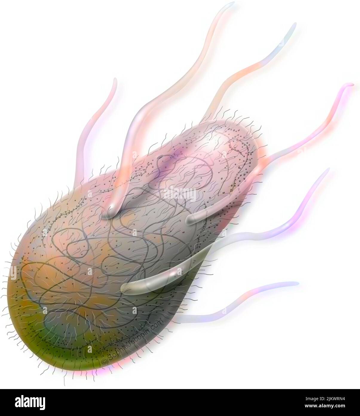 Bacterium (prokaryote) with its organelles in transparency. Stock Photohttps://www.alamy.com/image-license-details/?v=1https://www.alamy.com/bacterium-prokaryote-with-its-organelles-in-transparency-image476925824.html
RF2JKWRN4–Bacterium (prokaryote) with its organelles in transparency.
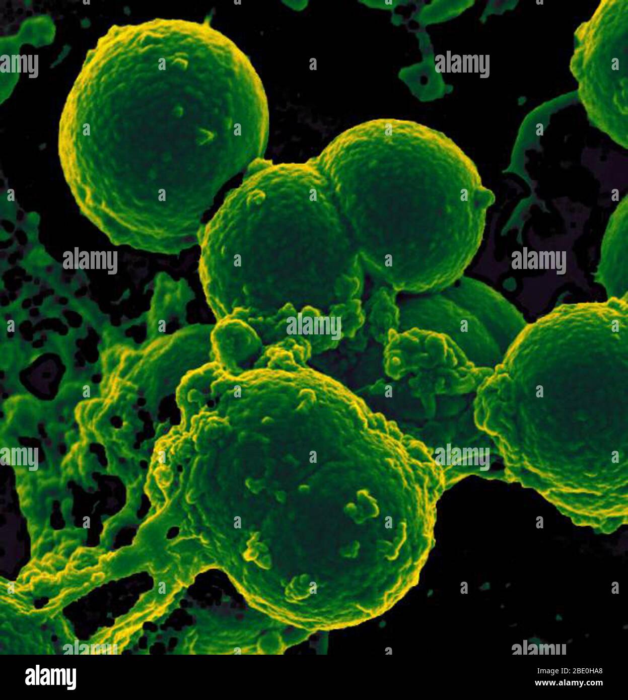 Scanning electron micrograph of neutrophil ingesting methicillin-resistant Staphylococcus aureus bacteria. Methicillin-resistant Staphylococcus aureus (MRSA) is a bacterium responsible for several difficult-to-treat infections in humans. It is also called multidrug-resistant Staphylococcus aureus and oxacillin-resistant Staphylococcus aureus (ORSA). MRSA is any strain of Staphylococcus aureus that has developed resistance to beta-lactam antibiotics, which include penicillins and cephalosporins. Strains unable to resist these antibiotics are classified as methicillin-sensitive Staphylococcus au Stock Photohttps://www.alamy.com/image-license-details/?v=1https://www.alamy.com/scanning-electron-micrograph-of-neutrophil-ingesting-methicillin-resistant-staphylococcus-aureus-bacteria-methicillin-resistant-staphylococcus-aureus-mrsa-is-a-bacterium-responsible-for-several-difficult-to-treat-infections-in-humans-it-is-also-called-multidrug-resistant-staphylococcus-aureus-and-oxacillin-resistant-staphylococcus-aureus-orsa-mrsa-is-any-strain-of-staphylococcus-aureus-that-has-developed-resistance-to-beta-lactam-antibiotics-which-include-penicillins-and-cephalosporins-strains-unable-to-resist-these-antibiotics-are-classified-as-methicillin-sensitive-staphylococcus-au-image352826160.html
RM2BE0HA8–Scanning electron micrograph of neutrophil ingesting methicillin-resistant Staphylococcus aureus bacteria. Methicillin-resistant Staphylococcus aureus (MRSA) is a bacterium responsible for several difficult-to-treat infections in humans. It is also called multidrug-resistant Staphylococcus aureus and oxacillin-resistant Staphylococcus aureus (ORSA). MRSA is any strain of Staphylococcus aureus that has developed resistance to beta-lactam antibiotics, which include penicillins and cephalosporins. Strains unable to resist these antibiotics are classified as methicillin-sensitive Staphylococcus au
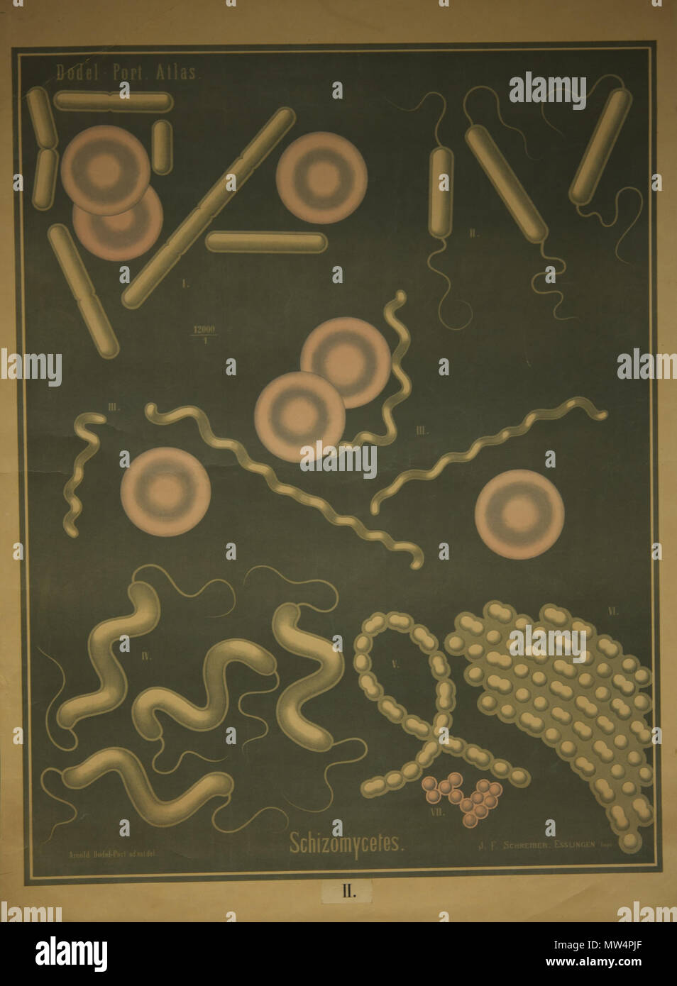 165 Dodel-Port Atlas Schizomycètes II Stock Photohttps://www.alamy.com/image-license-details/?v=1https://www.alamy.com/165-dodel-port-atlas-schizomyctes-ii-image187575655.html
RMMW4PJF–165 Dodel-Port Atlas Schizomycètes II
 Cells division Stock Photohttps://www.alamy.com/image-license-details/?v=1https://www.alamy.com/cells-division-image232258929.html
RFRDT8HN–Cells division
 Transparent cells in the blood, 3d rendering. Computer digital drawing. Stock Photohttps://www.alamy.com/image-license-details/?v=1https://www.alamy.com/transparent-cells-in-the-blood-3d-rendering-computer-digital-drawing-image356591566.html
RF2BM4452–Transparent cells in the blood, 3d rendering. Computer digital drawing.
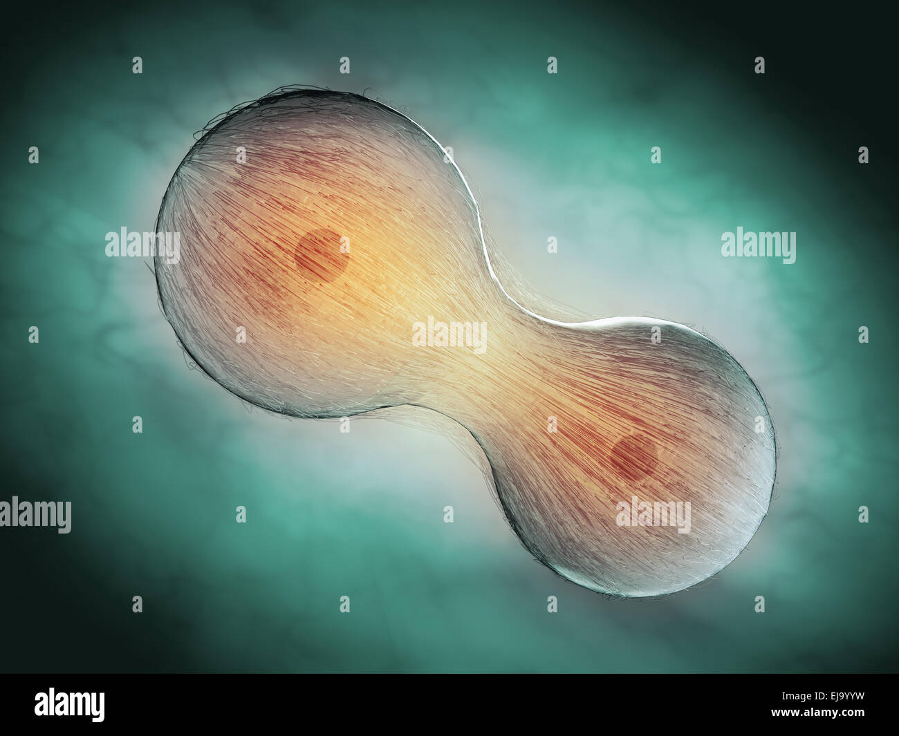 Cell division through mitosis - scientific illustration Stock Photohttps://www.alamy.com/image-license-details/?v=1https://www.alamy.com/stock-photo-cell-division-through-mitosis-scientific-illustration-80124797.html
RFEJ9YYW–Cell division through mitosis - scientific illustration
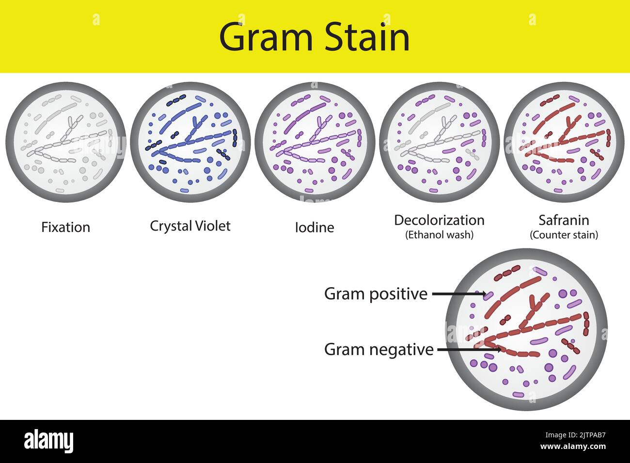 Diagram showing gram staining microbiology lab technique steps - microbiology laboratory using Crystal violet and Safranin Stock Vectorhttps://www.alamy.com/image-license-details/?v=1https://www.alamy.com/diagram-showing-gram-staining-microbiology-lab-technique-steps-microbiology-laboratory-using-crystal-violet-and-safranin-image479922779.html
RF2JTPAB7–Diagram showing gram staining microbiology lab technique steps - microbiology laboratory using Crystal violet and Safranin
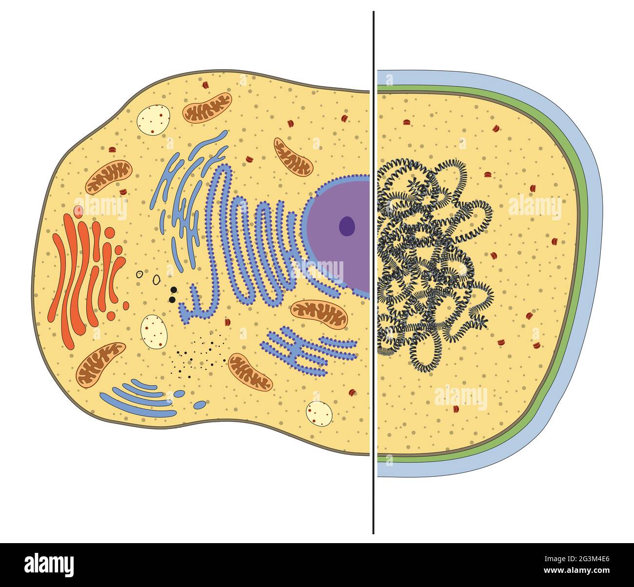 Illustration of eukaryotic and prokaryotic cells. Differences Stock Photohttps://www.alamy.com/image-license-details/?v=1https://www.alamy.com/illustration-of-eukaryotic-and-prokaryotic-cells-differences-image432545742.html
RF2G3M4E6–Illustration of eukaryotic and prokaryotic cells. Differences
 Anacystis cyanea Anacystis cyanea. Stock Photohttps://www.alamy.com/image-license-details/?v=1https://www.alamy.com/anacystis-cyanea-anacystis-cyanea-image362500367.html
RM2C1N8WK–Anacystis cyanea Anacystis cyanea.
 Abstract illustration of cells in mitosis or multiplication of cells Stock Photohttps://www.alamy.com/image-license-details/?v=1https://www.alamy.com/stock-photo-abstract-illustration-of-cells-in-mitosis-or-multiplication-of-cells-89099344.html
RFF4XR3C–Abstract illustration of cells in mitosis or multiplication of cells
 Peritrichous Bacteria with lot of flagellum, harmful bacillus with long tails, prokaryote, viruses probiotics and pathogen, H. pylori, microbe flagell Stock Photohttps://www.alamy.com/image-license-details/?v=1https://www.alamy.com/peritrichous-bacteria-with-lot-of-flagellum-harmful-bacillus-with-long-tails-prokaryote-viruses-probiotics-and-pathogen-h-pylori-microbe-flagell-image622013908.html
RM2Y3Y52C–Peritrichous Bacteria with lot of flagellum, harmful bacillus with long tails, prokaryote, viruses probiotics and pathogen, H. pylori, microbe flagell
RF3CAJ050–Icon for bacterium, prokaryote
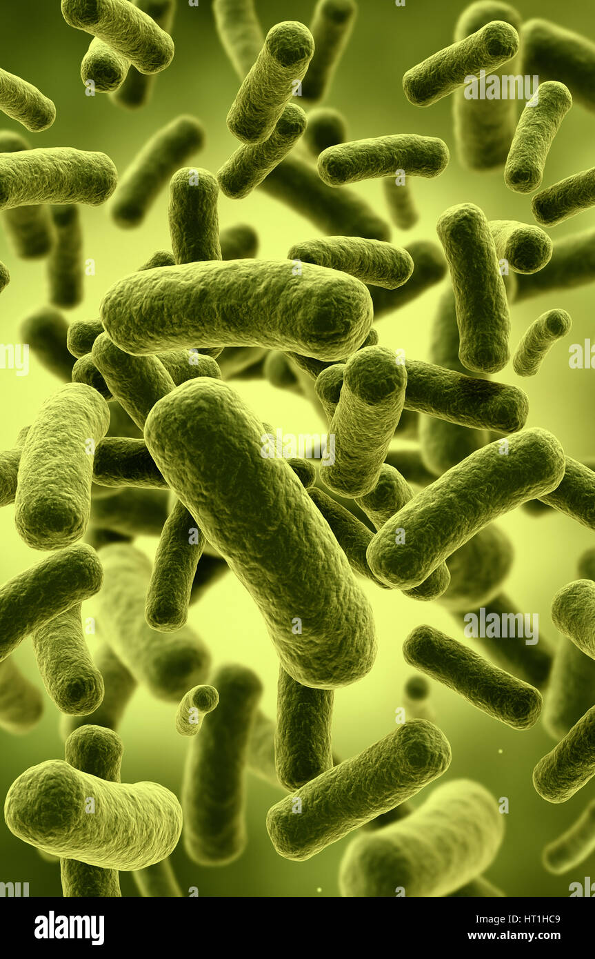 Illustration of bacteria cells Stock Photohttps://www.alamy.com/image-license-details/?v=1https://www.alamy.com/stock-photo-illustration-of-bacteria-cells-135259945.html
RFHT1HC9–Illustration of bacteria cells
 hand washing Stock Photohttps://www.alamy.com/image-license-details/?v=1https://www.alamy.com/stock-photo-hand-washing-35103112.html
RFC132AG–hand washing
 Microscopic view of bacteria. Stock Photohttps://www.alamy.com/image-license-details/?v=1https://www.alamy.com/microscopic-view-of-bacteria-image68934396.html
RFE046EM–Microscopic view of bacteria.
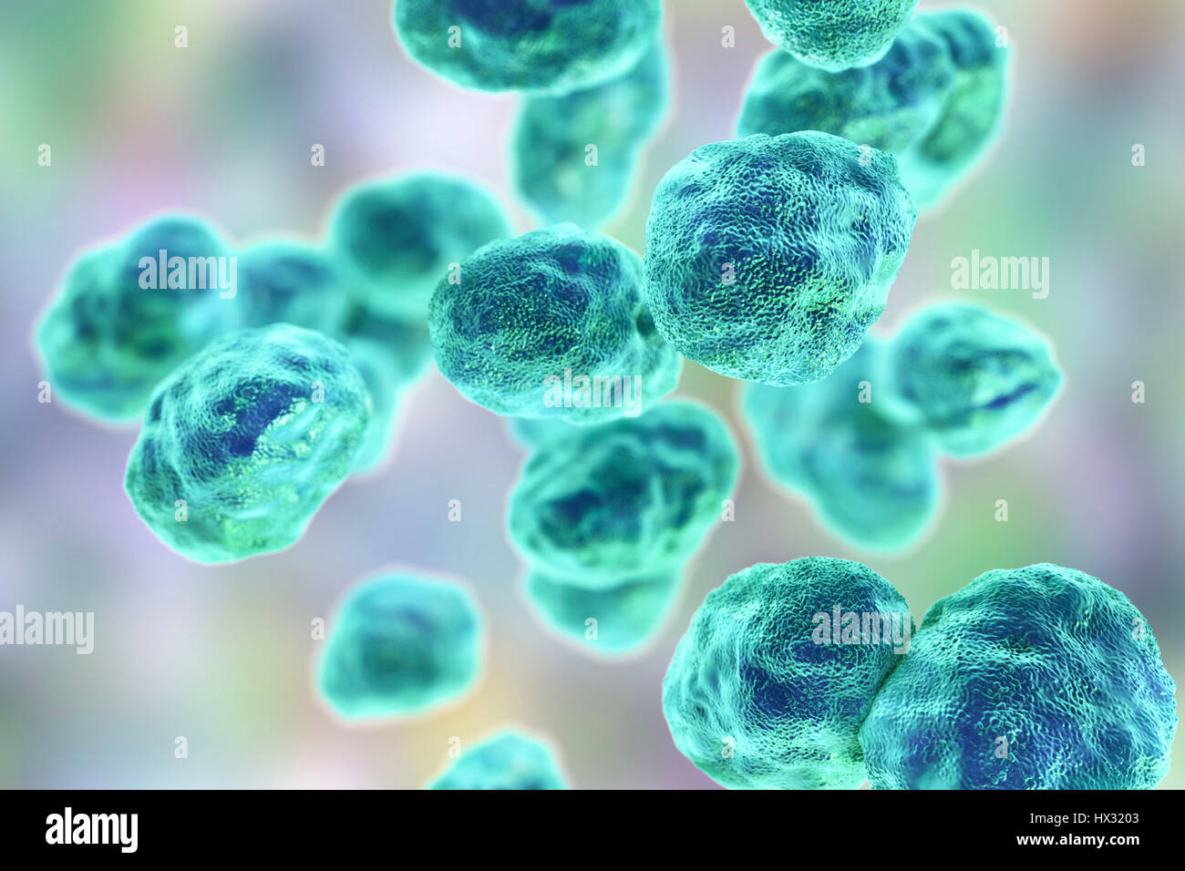 Tularaemia bacteria (Francisella tularensis), illustration. F. tularensis is Gram-negative, coccobacillus prokaryote. A zoonotic microorganism that causes tularaemia, a disease of wild rodents and rabbits that can be transmitted to humans and domesticated pets. Stock Photohttps://www.alamy.com/image-license-details/?v=1https://www.alamy.com/stock-photo-tularaemia-bacteria-francisella-tularensis-illustration-f-tularensis-136521059.html
RFHX3203–Tularaemia bacteria (Francisella tularensis), illustration. F. tularensis is Gram-negative, coccobacillus prokaryote. A zoonotic microorganism that causes tularaemia, a disease of wild rodents and rabbits that can be transmitted to humans and domesticated pets.
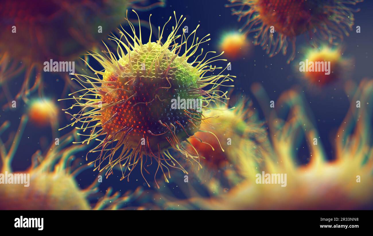 Mimiviruses are part of the Mimiviridae family and among the largest viruses. Giant viruses can be larger than prokaryotes (bacteria and archaea) Stock Photohttps://www.alamy.com/image-license-details/?v=1https://www.alamy.com/mimiviruses-are-part-of-the-mimiviridae-family-and-among-the-largest-viruses-giant-viruses-can-be-larger-than-prokaryotes-bacteria-and-archaea-image552658660.html
RF2R33NN8–Mimiviruses are part of the Mimiviridae family and among the largest viruses. Giant viruses can be larger than prokaryotes (bacteria and archaea)
 Agar slant tube culture of Kocuria rhizophila (Micrococcus luteus). Stock Photohttps://www.alamy.com/image-license-details/?v=1https://www.alamy.com/stock-photo-agar-slant-tube-culture-of-kocuria-rhizophila-micrococcus-luteus-16492490.html
RMAW7KCY–Agar slant tube culture of Kocuria rhizophila (Micrococcus luteus).
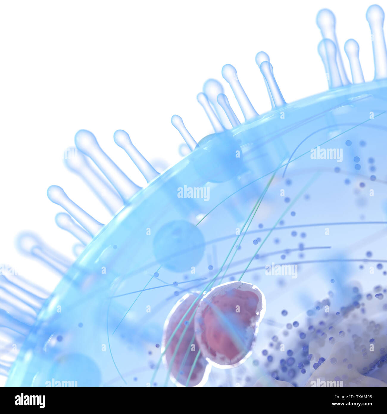 3d rendered medically accurate illustration of a human cell Stock Photohttps://www.alamy.com/image-license-details/?v=1https://www.alamy.com/3d-rendered-medically-accurate-illustration-of-a-human-cell-image257161668.html
RFTXAM98–3d rendered medically accurate illustration of a human cell
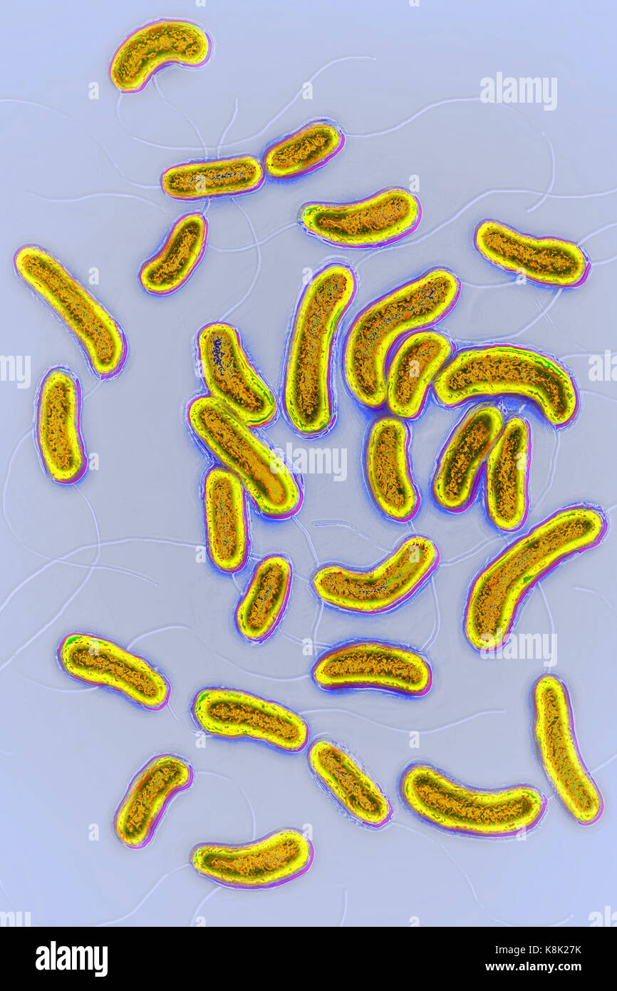 VIBRIO CHOLERAE Stock Photohttps://www.alamy.com/image-license-details/?v=1https://www.alamy.com/stock-image-vibrio-cholerae-160229431.html
RMK8K27K–VIBRIO CHOLERAE
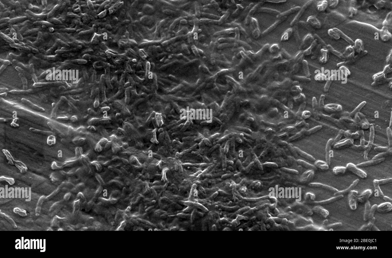 SEM of a laboratory-grown potable (drinking) water biofilm before the introduction of Vermamoeba vermiformis (Hartmanella) cysts. Aquatic bacteria were grown as biofilm on steel for one week. V. vermiformis were then added, and phagocytized the bacteria that multiplied within vesicles that became cysts in which the bacteria will live until they rupture. Vermamoeba vermiformis a free-living amoeba (FLA), is widespread in nature and has been isolated from soil, freshwater, air, and a variety of engineered water systems. Two distinct life cycle forms are known for Vermamoeba vermiformis, the trop Stock Photohttps://www.alamy.com/image-license-details/?v=1https://www.alamy.com/sem-of-a-laboratory-grown-potable-drinking-water-biofilm-before-the-introduction-of-vermamoeba-vermiformis-hartmanella-cysts-aquatic-bacteria-were-grown-as-biofilm-on-steel-for-one-week-v-vermiformis-were-then-added-and-phagocytized-the-bacteria-that-multiplied-within-vesicles-that-became-cysts-in-which-the-bacteria-will-live-until-they-rupture-vermamoeba-vermiformis-a-free-living-amoeba-fla-is-widespread-in-nature-and-has-been-isolated-from-soil-freshwater-air-and-a-variety-of-engineered-water-systems-two-distinct-life-cycle-forms-are-known-for-vermamoeba-vermiformis-the-trop-image352826993.html
RM2BE0JC1–SEM of a laboratory-grown potable (drinking) water biofilm before the introduction of Vermamoeba vermiformis (Hartmanella) cysts. Aquatic bacteria were grown as biofilm on steel for one week. V. vermiformis were then added, and phagocytized the bacteria that multiplied within vesicles that became cysts in which the bacteria will live until they rupture. Vermamoeba vermiformis a free-living amoeba (FLA), is widespread in nature and has been isolated from soil, freshwater, air, and a variety of engineered water systems. Two distinct life cycle forms are known for Vermamoeba vermiformis, the trop
 cells Stock Photohttps://www.alamy.com/image-license-details/?v=1https://www.alamy.com/cells-image374717627.html
RM2CNHT4B–cells
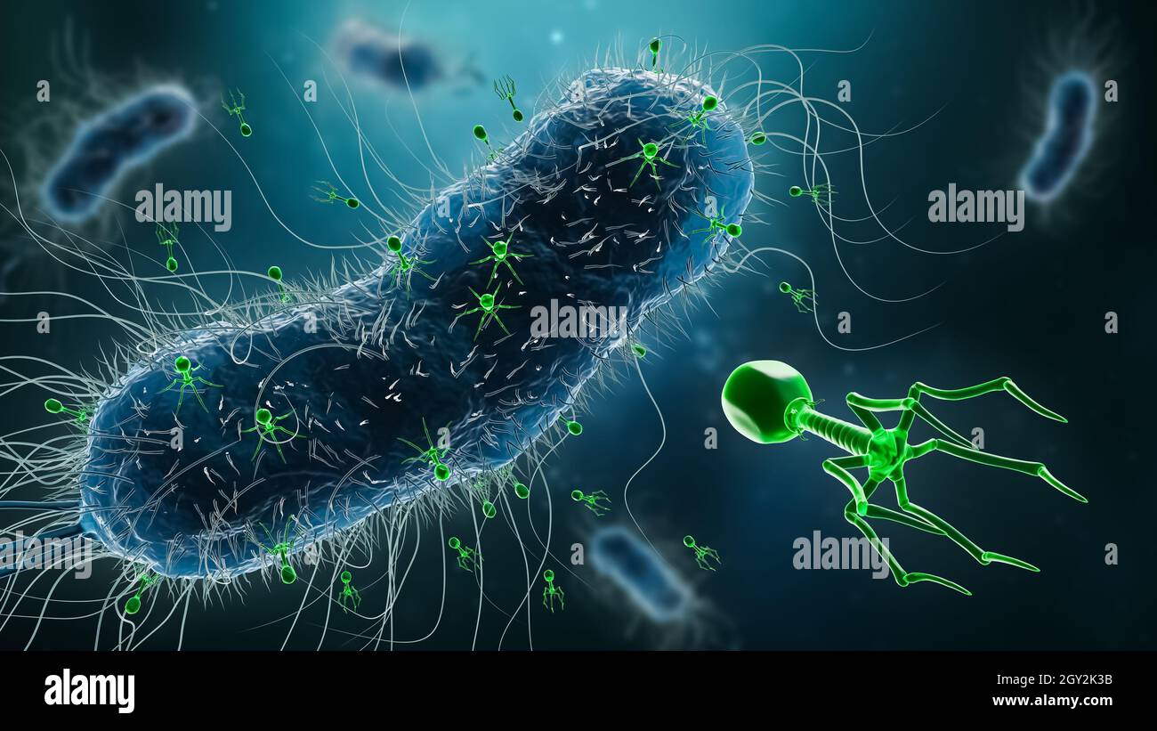 Group of phages or bacteriophages infecting bacteria 3D rendering illustration. Microbiology, science, medicine, biology, medical and healthcare conce Stock Photohttps://www.alamy.com/image-license-details/?v=1https://www.alamy.com/group-of-phages-or-bacteriophages-infecting-bacteria-3d-rendering-illustration-microbiology-science-medicine-biology-medical-and-healthcare-conce-image446913807.html
RF2GY2K3B–Group of phages or bacteriophages infecting bacteria 3D rendering illustration. Microbiology, science, medicine, biology, medical and healthcare conce
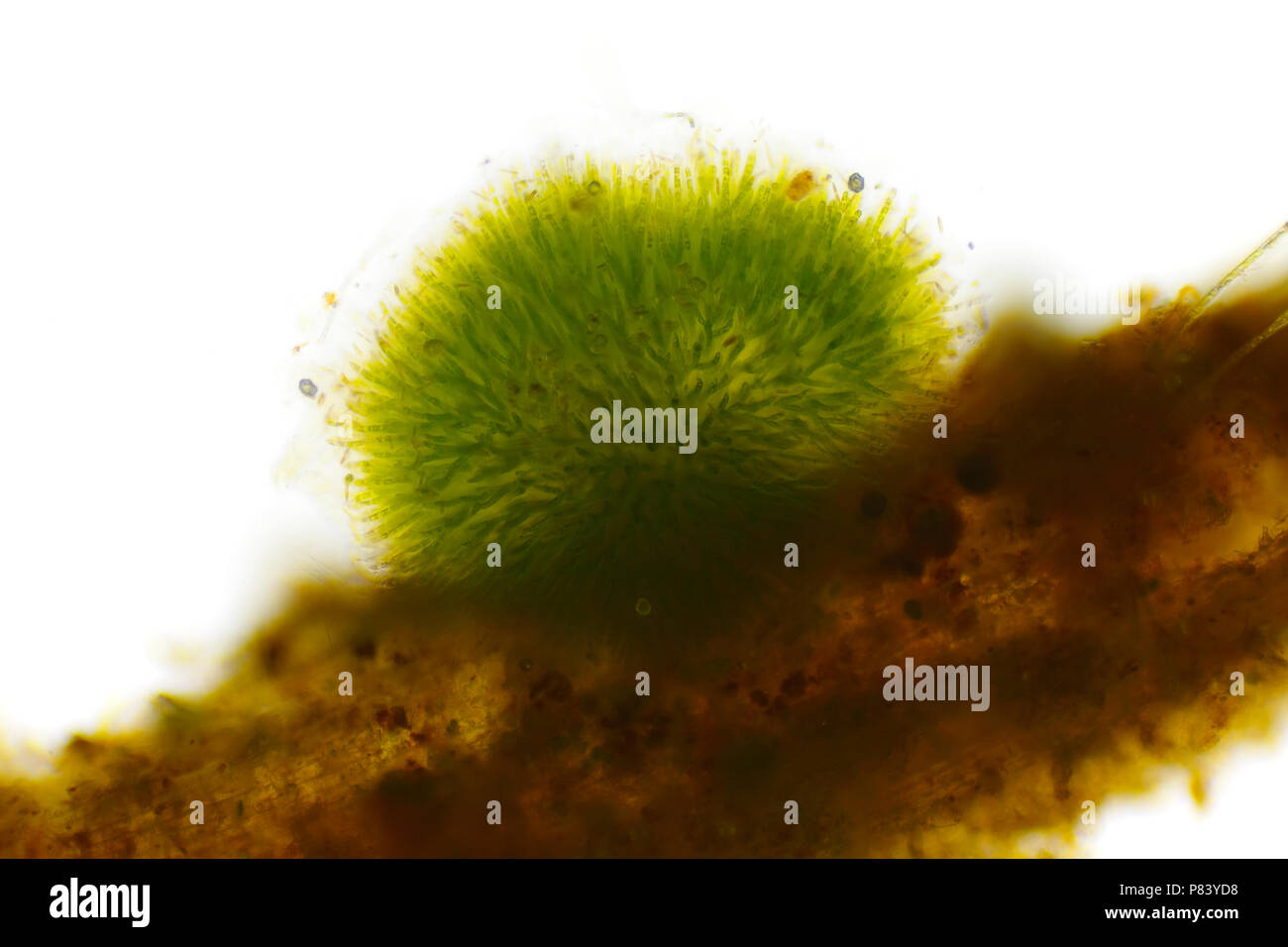 Spherical colony of cyanobacteria (Gleotrichia, blue-green algae). Side view, Brightfield illumination. Stock Photohttps://www.alamy.com/image-license-details/?v=1https://www.alamy.com/spherical-colony-of-cyanobacteria-gleotrichia-blue-green-algae-side-view-brightfield-illumination-image211529060.html
RFP83YD8–Spherical colony of cyanobacteria (Gleotrichia, blue-green algae). Side view, Brightfield illumination.
 Transparent cells in the blood, 3d rendering. Computer digital drawing. Stock Photohttps://www.alamy.com/image-license-details/?v=1https://www.alamy.com/transparent-cells-in-the-blood-3d-rendering-computer-digital-drawing-image356591523.html
RF2BM443F–Transparent cells in the blood, 3d rendering. Computer digital drawing.
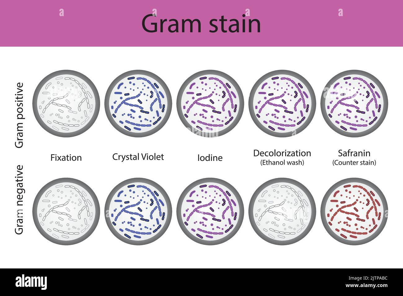 Diagram showing gram staining microbiology lab technique steps - microbiology laboratory using Crystal violet and Safranin Stock Vectorhttps://www.alamy.com/image-license-details/?v=1https://www.alamy.com/diagram-showing-gram-staining-microbiology-lab-technique-steps-microbiology-laboratory-using-crystal-violet-and-safranin-image479922784.html
RF2JTPABC–Diagram showing gram staining microbiology lab technique steps - microbiology laboratory using Crystal violet and Safranin
 foraminiferans, forams (Foraminiferida), Calcarina of Japan, foram on a matchstick head Stock Photohttps://www.alamy.com/image-license-details/?v=1https://www.alamy.com/foraminiferans-forams-foraminiferida-calcarina-of-japan-foram-on-a-matchstick-head-image255238322.html
RMTR732A–foraminiferans, forams (Foraminiferida), Calcarina of Japan, foram on a matchstick head
 Schizothrix tenerrima Schizothrix tenerrima. Stock Photohttps://www.alamy.com/image-license-details/?v=1https://www.alamy.com/schizothrix-tenerrima-schizothrix-tenerrima-image362499988.html
RM2C1N8C4–Schizothrix tenerrima Schizothrix tenerrima.
 Seamless high quality high resolution abstract background Stock Photohttps://www.alamy.com/image-license-details/?v=1https://www.alamy.com/stock-photo-seamless-high-quality-high-resolution-abstract-background-51387404.html
RFCYGW4C–Seamless high quality high resolution abstract background
 Peritrichous Bacteria with lot of flagellum, harmful bacillus with long tails, prokaryote, viruses probiotics and pathogen, H. pylori, microbe flagell Stock Photohttps://www.alamy.com/image-license-details/?v=1https://www.alamy.com/peritrichous-bacteria-with-lot-of-flagellum-harmful-bacillus-with-long-tails-prokaryote-viruses-probiotics-and-pathogen-h-pylori-microbe-flagell-image622010993.html
RM2Y3Y1A9–Peritrichous Bacteria with lot of flagellum, harmful bacillus with long tails, prokaryote, viruses probiotics and pathogen, H. pylori, microbe flagell
RF3BNY6TX–Icon for bacterium, prokaryote
 High resolution Illustration of cells Stock Photohttps://www.alamy.com/image-license-details/?v=1https://www.alamy.com/stock-photo-high-resolution-illustration-of-cells-147420414.html
RFJFRG6P–High resolution Illustration of cells
 Cell division on a dark blue background. 3d render. Stock Photohttps://www.alamy.com/image-license-details/?v=1https://www.alamy.com/cell-division-on-a-dark-blue-background-3d-render-image345308425.html
RF2B1P4BN–Cell division on a dark blue background. 3d render.
 Microscopic view of bacteria. Stock Photohttps://www.alamy.com/image-license-details/?v=1https://www.alamy.com/microscopic-view-of-bacteria-image68934394.html
RFE046EJ–Microscopic view of bacteria.
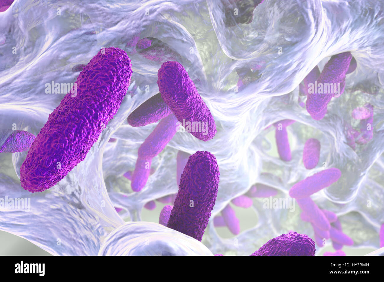 Klebsiella pneumoniae bacteria in biofilm,computer illustration.K.pneumoniae are Gram-negative,encapsulated,non-motile,enteric,rod prokaryote.This species causes Friedlander's pneumonia urinary tract infections.K.pneumoniae's pathogenicity can be attributed to its production of heat-stable Stock Photohttps://www.alamy.com/image-license-details/?v=1https://www.alamy.com/stock-photo-klebsiella-pneumoniae-bacteria-in-biofilmcomputer-illustrationkpneumoniae-137143349.html
RFHY3BMN–Klebsiella pneumoniae bacteria in biofilm,computer illustration.K.pneumoniae are Gram-negative,encapsulated,non-motile,enteric,rod prokaryote.This species causes Friedlander's pneumonia urinary tract infections.K.pneumoniae's pathogenicity can be attributed to its production of heat-stable
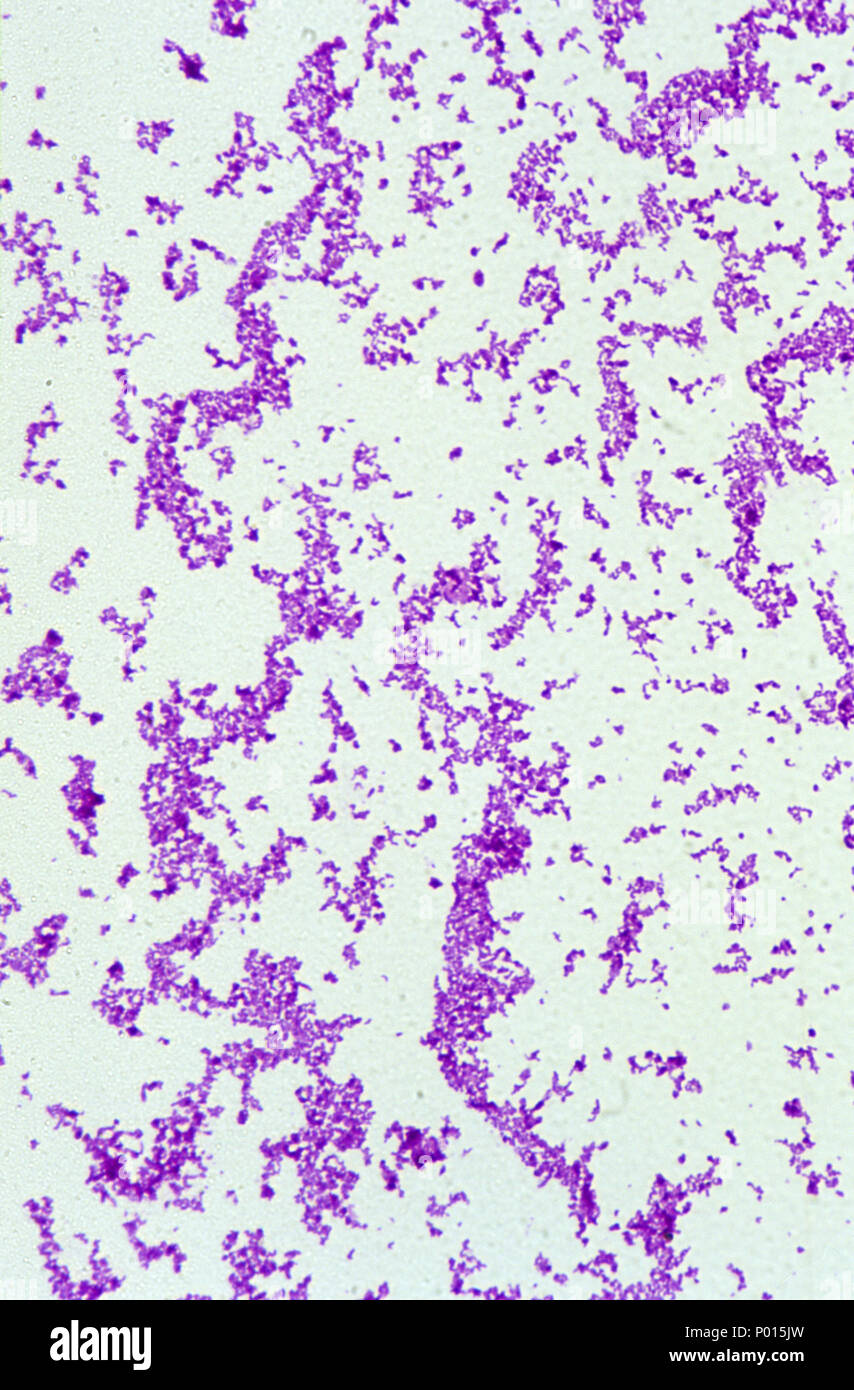 Corynebacterium diphtheriae bacteria Stock Photohttps://www.alamy.com/image-license-details/?v=1https://www.alamy.com/corynebacterium-diphtheriae-bacteria-image206550817.html
RFP015JW–Corynebacterium diphtheriae bacteria
 Magnifying glass and protozoa in the blob Stock Photohttps://www.alamy.com/image-license-details/?v=1https://www.alamy.com/stock-photo-magnifying-glass-and-protozoa-in-the-blob-80030696.html
RMEJ5KY4–Magnifying glass and protozoa in the blob
 3d rendered medically accurate illustration of a human cell Stock Photohttps://www.alamy.com/image-license-details/?v=1https://www.alamy.com/3d-rendered-medically-accurate-illustration-of-a-human-cell-image257161226.html
RFTXAKNE–3d rendered medically accurate illustration of a human cell
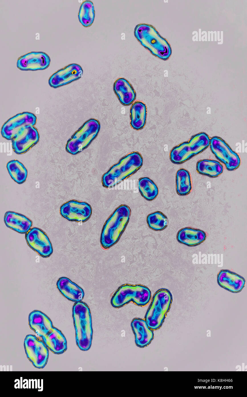 YERSINIA PESTIS Stock Photohttps://www.alamy.com/image-license-details/?v=1https://www.alamy.com/stock-image-yersinia-pestis-160197246.html
RMK8HH66–YERSINIA PESTIS
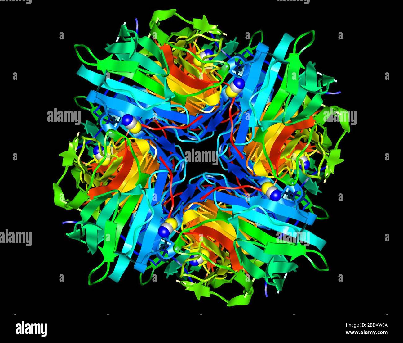 Pseudomonas aeruginosa Protein Stock Photohttps://www.alamy.com/image-license-details/?v=1https://www.alamy.com/pseudomonas-aeruginosa-protein-image352788502.html
RM2BDXW9A–Pseudomonas aeruginosa Protein
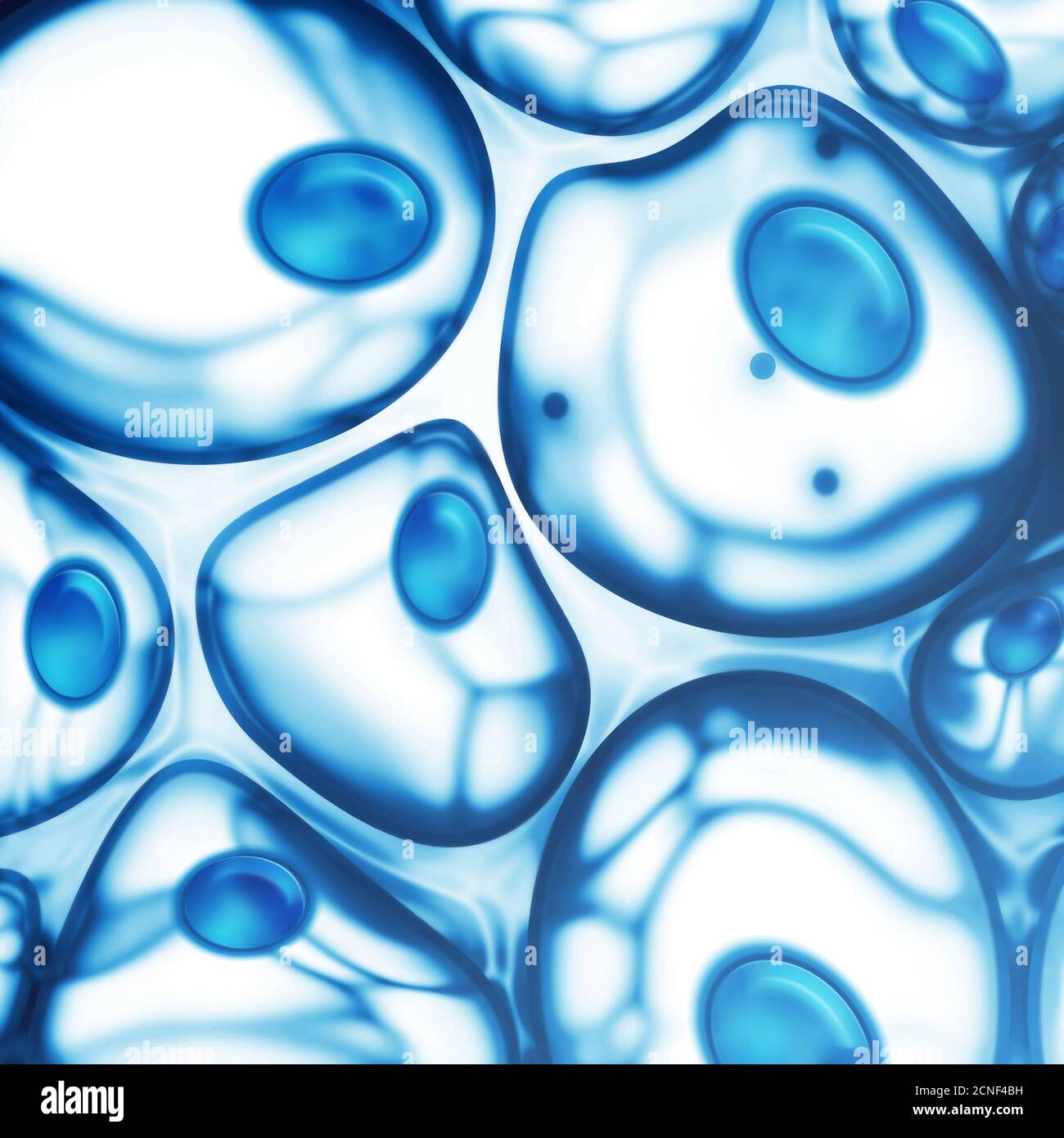 cells Stock Photohttps://www.alamy.com/image-license-details/?v=1https://www.alamy.com/cells-image374658245.html
RM2CNF4BH–cells
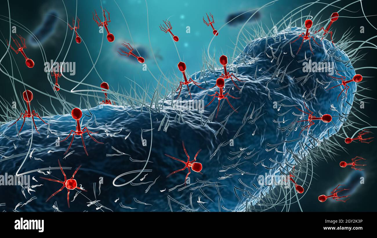 Generic bacteria such as Escherichia coli infected by group of phages or bacteriophages 3D rendering illustration. Microbiology, medicine, science, me Stock Photohttps://www.alamy.com/image-license-details/?v=1https://www.alamy.com/generic-bacteria-such-as-escherichia-coli-infected-by-group-of-phages-or-bacteriophages-3d-rendering-illustration-microbiology-medicine-science-me-image446913818.html
RF2GY2K3P–Generic bacteria such as Escherichia coli infected by group of phages or bacteriophages 3D rendering illustration. Microbiology, medicine, science, me
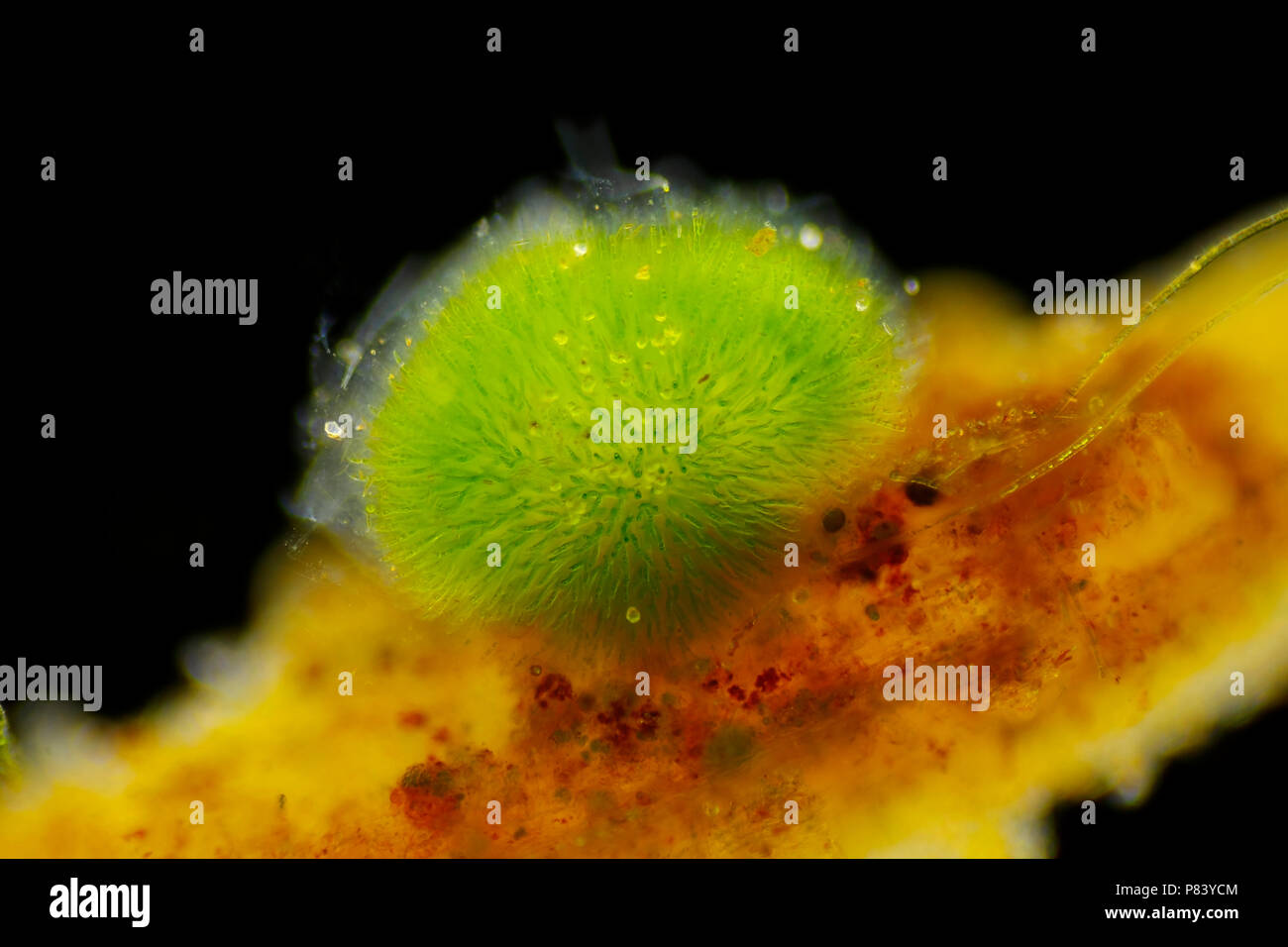 Spherical colony of cyanobacteria (Gleotrichia, blue-green algae). Side view, Darkfield illumination. Stock Photohttps://www.alamy.com/image-license-details/?v=1https://www.alamy.com/spherical-colony-of-cyanobacteria-gleotrichia-blue-green-algae-side-view-darkfield-illumination-image211529044.html
RFP83YCM–Spherical colony of cyanobacteria (Gleotrichia, blue-green algae). Side view, Darkfield illumination.
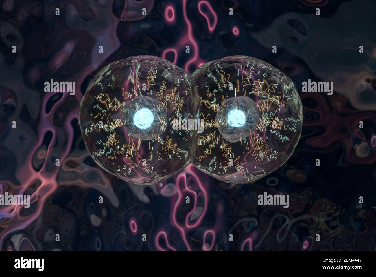 Transparent cells in the blood, 3d rendering. Computer digital drawing. Stock Photohttps://www.alamy.com/image-license-details/?v=1https://www.alamy.com/transparent-cells-in-the-blood-3d-rendering-computer-digital-drawing-image356591563.html
RF2BM444Y–Transparent cells in the blood, 3d rendering. Computer digital drawing.
RF2JTPABA–Set of 3 petri dish icons. Colorful simple illustration with bacterial cells.
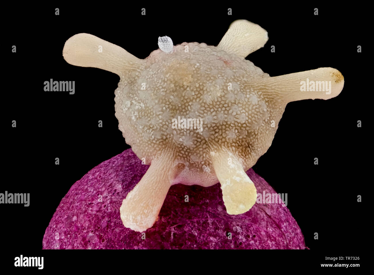 foraminiferans, forams (Foraminiferida), Calcarina of Japan, foram on a matchstick head and radiolarian Stock Photohttps://www.alamy.com/image-license-details/?v=1https://www.alamy.com/foraminiferans-forams-foraminiferida-calcarina-of-japan-foram-on-a-matchstick-head-and-radiolarian-image255238318.html
RMTR7326–foraminiferans, forams (Foraminiferida), Calcarina of Japan, foram on a matchstick head and radiolarian
 Anabaena oscillarioides Anabaena oscillarioides. Stock Photohttps://www.alamy.com/image-license-details/?v=1https://www.alamy.com/anabaena-oscillarioides-anabaena-oscillarioides-image362500338.html
RM2C1N8TJ–Anabaena oscillarioides Anabaena oscillarioides.
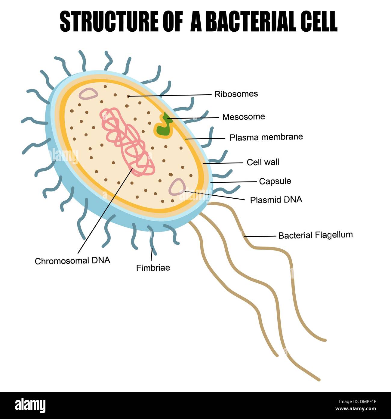 Structure of a bacterial cell Stock Vectorhttps://www.alamy.com/image-license-details/?v=1https://www.alamy.com/structure-of-a-bacterial-cell-image64419055.html
RFDMPF4F–Structure of a bacterial cell
 Peritrichous Bacteria with lot of flagellum, harmful bacillus with long tails, prokaryote, viruses probiotics and pathogen, H. pylori, microbe flagell Stock Photohttps://www.alamy.com/image-license-details/?v=1https://www.alamy.com/peritrichous-bacteria-with-lot-of-flagellum-harmful-bacillus-with-long-tails-prokaryote-viruses-probiotics-and-pathogen-h-pylori-microbe-flagell-image620982363.html
RM2Y2859F–Peritrichous Bacteria with lot of flagellum, harmful bacillus with long tails, prokaryote, viruses probiotics and pathogen, H. pylori, microbe flagell
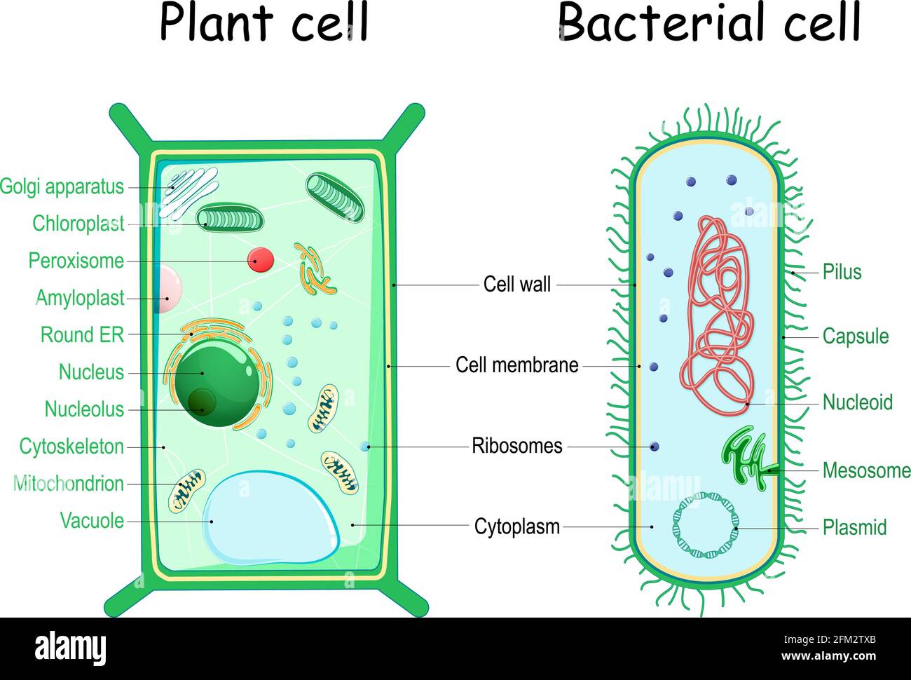 bacteria and plant cell. comparison of cell structure. Similarities and differences. cross section and anatomy of cell. Biology Chart. Vector Stock Vectorhttps://www.alamy.com/image-license-details/?v=1https://www.alamy.com/bacteria-and-plant-cell-comparison-of-cell-structure-similarities-and-differences-cross-section-and-anatomy-of-cell-biology-chart-vector-image425405411.html
RF2FM2TXB–bacteria and plant cell. comparison of cell structure. Similarities and differences. cross section and anatomy of cell. Biology Chart. Vector
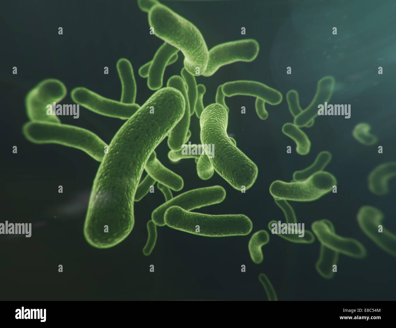 Flowing group of bacteria cells Stock Photohttps://www.alamy.com/image-license-details/?v=1https://www.alamy.com/stock-photo-flowing-group-of-bacteria-cells-74026196.html
RFE8C54M–Flowing group of bacteria cells
 Cell division on a dark blue background. 3d render. Stock Photohttps://www.alamy.com/image-license-details/?v=1https://www.alamy.com/cell-division-on-a-dark-blue-background-3d-render-image345308394.html
RF2B1P4AJ–Cell division on a dark blue background. 3d render.
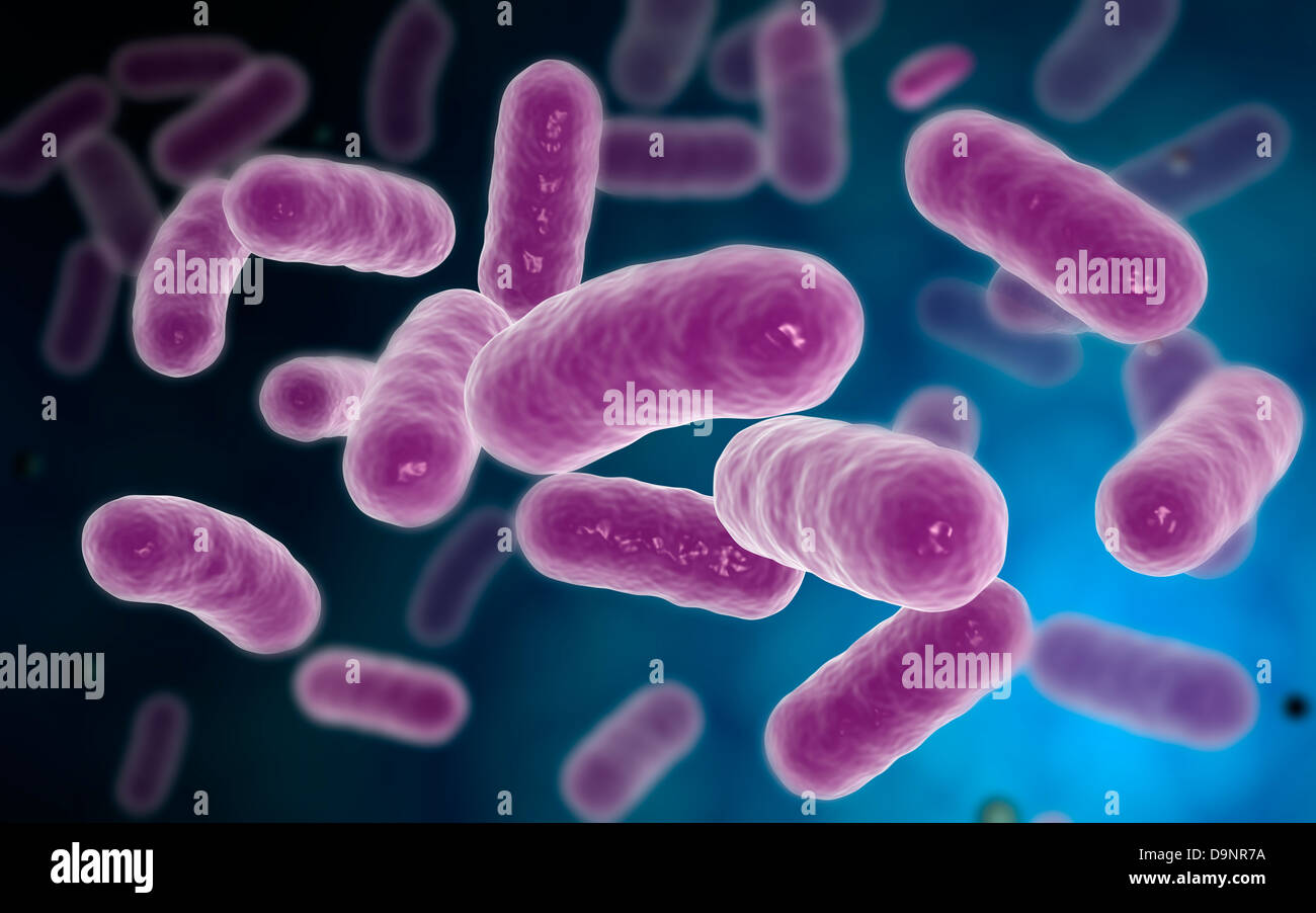 Conceptual image of bacteria. Stock Photohttps://www.alamy.com/image-license-details/?v=1https://www.alamy.com/stock-photo-conceptual-image-of-bacteria-57642238.html
RFD9NR7A–Conceptual image of bacteria.
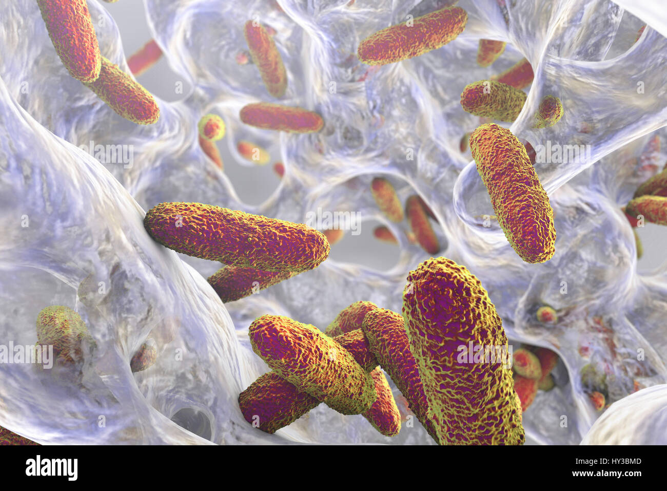 Klebsiella pneumoniae bacteria in biofilm,computer illustration.K.pneumoniae are Gram-negative,encapsulated,non-motile,enteric,rod prokaryote.This species causes Friedlander's pneumonia urinary tract infections.K.pneumoniae's pathogenicity can be attributed to its production of heat-stable Stock Photohttps://www.alamy.com/image-license-details/?v=1https://www.alamy.com/stock-photo-klebsiella-pneumoniae-bacteria-in-biofilmcomputer-illustrationkpneumoniae-137143341.html
RFHY3BMD–Klebsiella pneumoniae bacteria in biofilm,computer illustration.K.pneumoniae are Gram-negative,encapsulated,non-motile,enteric,rod prokaryote.This species causes Friedlander's pneumonia urinary tract infections.K.pneumoniae's pathogenicity can be attributed to its production of heat-stable
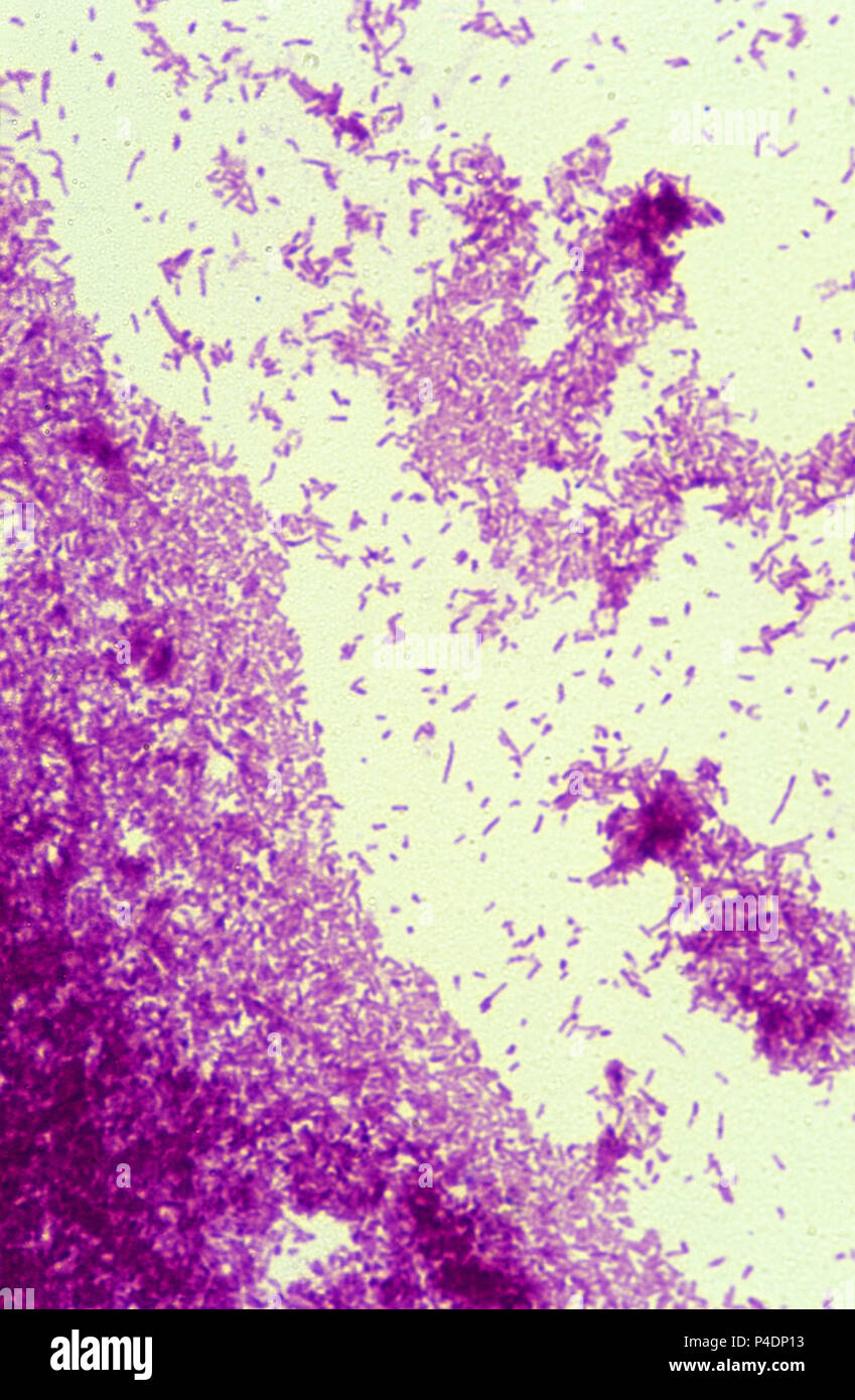 Mycobacterium tuberculosis bacteria Stock Photohttps://www.alamy.com/image-license-details/?v=1https://www.alamy.com/mycobacterium-tuberculosis-bacteria-image209285695.html
RFP4DP13–Mycobacterium tuberculosis bacteria
 Vector cartoon style circle composition of colorful funny bacteria characters. Good and bad flora microbes. Isolated on white background. Stock Vectorhttps://www.alamy.com/image-license-details/?v=1https://www.alamy.com/stock-image-vector-cartoon-style-circle-composition-of-colorful-funny-bacteria-165877719.html
RFKHTAM7–Vector cartoon style circle composition of colorful funny bacteria characters. Good and bad flora microbes. Isolated on white background.
