Quick filters:
Pseudomonadales Stock Photos and Images
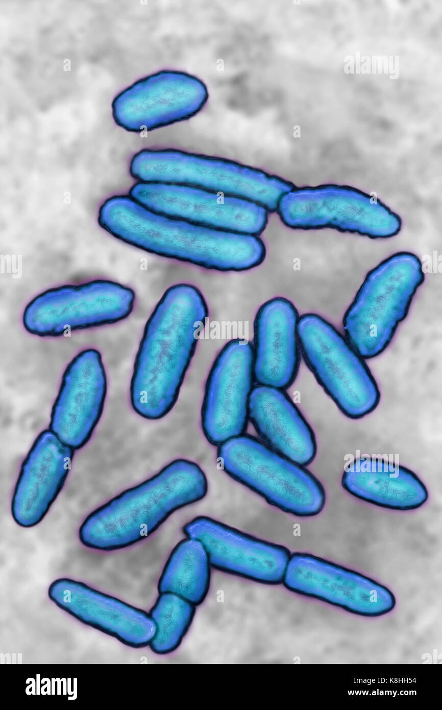 PSEUDOMONAS AERUGINOSA Stock Photohttps://www.alamy.com/image-license-details/?v=1https://www.alamy.com/stock-image-pseudomonas-aeruginosa-160197216.html
RMK8HH54–PSEUDOMONAS AERUGINOSA
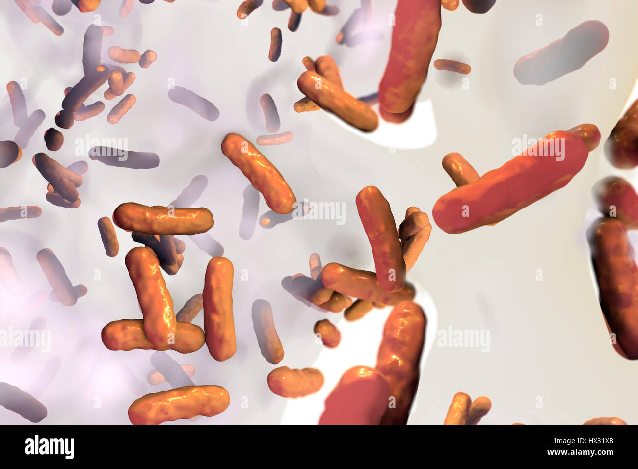 Pseudomonas aeruginosa bacteria inside biofilm,computer illustration.This is Gram-negative,aerobic,enteric,rod prokaryote.P.aeruginosa causes skin infections,urinary tract infections septicaemia.It produces blue-green pigment,pyocyanin,which characterizes bluish pus produced by infection.It is Stock Photohttps://www.alamy.com/image-license-details/?v=1https://www.alamy.com/stock-photo-pseudomonas-aeruginosa-bacteria-inside-biofilmcomputer-illustrationthis-136521011.html
RFHX31XB–Pseudomonas aeruginosa bacteria inside biofilm,computer illustration.This is Gram-negative,aerobic,enteric,rod prokaryote.P.aeruginosa causes skin infections,urinary tract infections septicaemia.It produces blue-green pigment,pyocyanin,which characterizes bluish pus produced by infection.It is
 Pseudomonas savastanoi gall on retama sphaerocarpa Stock Photohttps://www.alamy.com/image-license-details/?v=1https://www.alamy.com/pseudomonas-savastanoi-gall-on-retama-sphaerocarpa-image9441676.html
RMAWAXGD–Pseudomonas savastanoi gall on retama sphaerocarpa
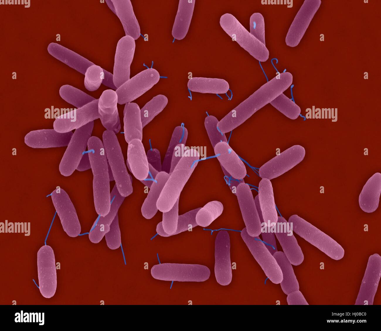 Coloured scanning electron micrograph (SEM) of Pseudomonas aeruginosa, Gram-negative, aerobic, enteric, rod prokaryote (dividing). This bacterium produces a blue, Green pigment, pyocyanin, which characterizes the bluish pus produced by the infection. It is found in soil, water, skin flora, and most man-made environments throughout the world. Magnification: x2, 400 when shortest axis printed at 25 millimetres. Stock Photohttps://www.alamy.com/image-license-details/?v=1https://www.alamy.com/stock-photo-coloured-scanning-electron-micrograph-sem-of-pseudomonas-aeruginosa-131545344.html
RFHJ0BC0–Coloured scanning electron micrograph (SEM) of Pseudomonas aeruginosa, Gram-negative, aerobic, enteric, rod prokaryote (dividing). This bacterium produces a blue, Green pigment, pyocyanin, which characterizes the bluish pus produced by the infection. It is found in soil, water, skin flora, and most man-made environments throughout the world. Magnification: x2, 400 when shortest axis printed at 25 millimetres.
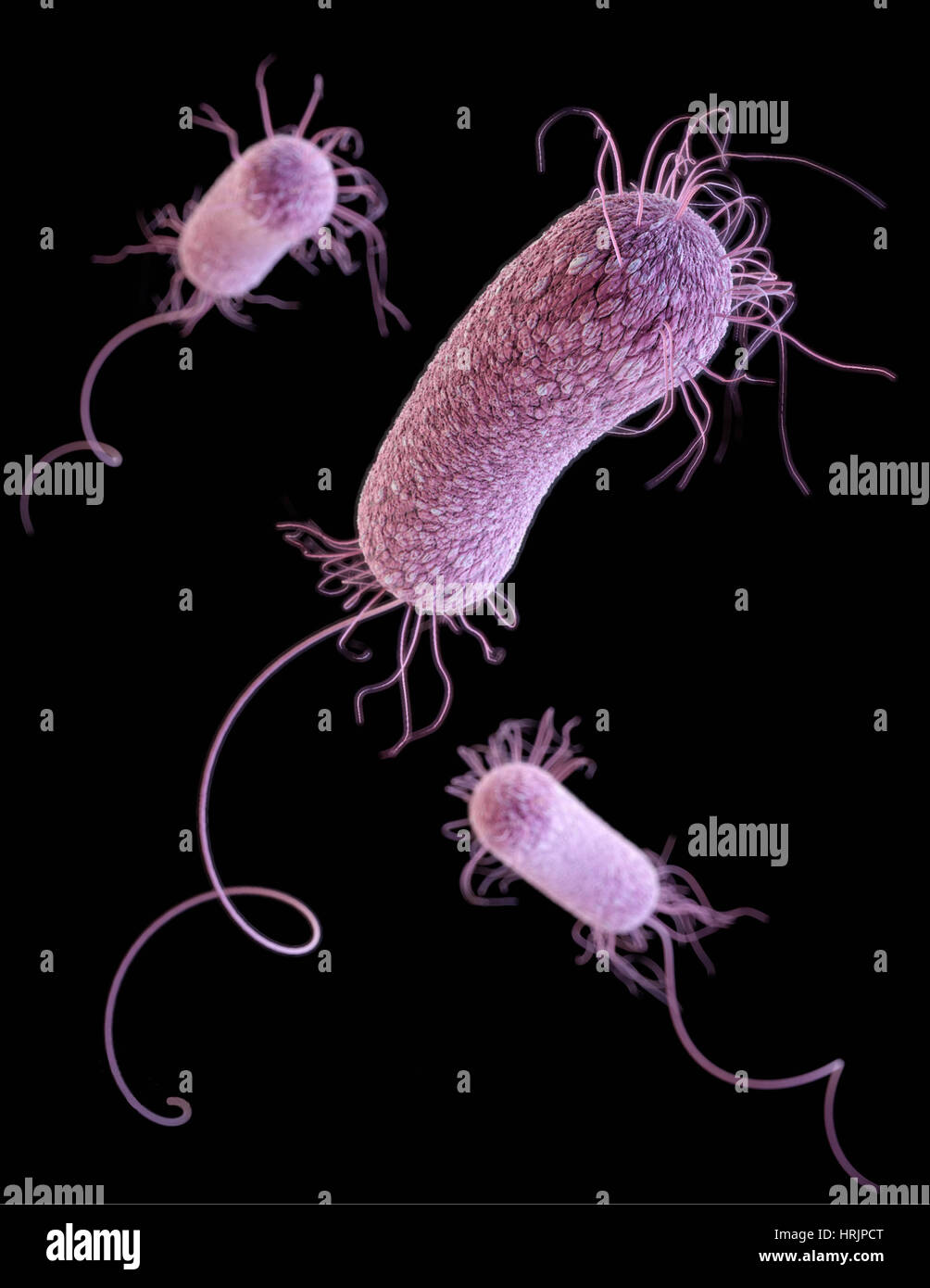 MDR Pathogen, Pseudomonas aeruginosa, 3D Model Stock Photohttps://www.alamy.com/image-license-details/?v=1https://www.alamy.com/stock-photo-mdr-pathogen-pseudomonas-aeruginosa-3d-model-135022408.html
RMHRJPCT–MDR Pathogen, Pseudomonas aeruginosa, 3D Model
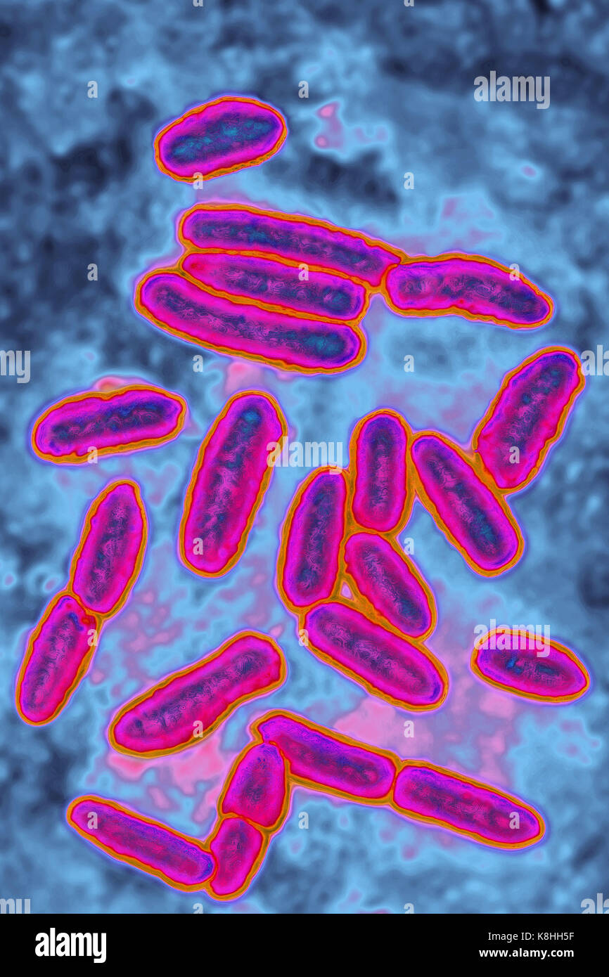 PSEUDOMONAS AERUGINOSA Stock Photohttps://www.alamy.com/image-license-details/?v=1https://www.alamy.com/stock-image-pseudomonas-aeruginosa-160197227.html
RMK8HH5F–PSEUDOMONAS AERUGINOSA
 Pseudomonas savastanoi gall on retama sphaerocarpa Stock Photohttps://www.alamy.com/image-license-details/?v=1https://www.alamy.com/stock-photo-pseudomonas-savastanoi-gall-on-retama-sphaerocarpa-10818436.html
RMA3MGBH–Pseudomonas savastanoi gall on retama sphaerocarpa
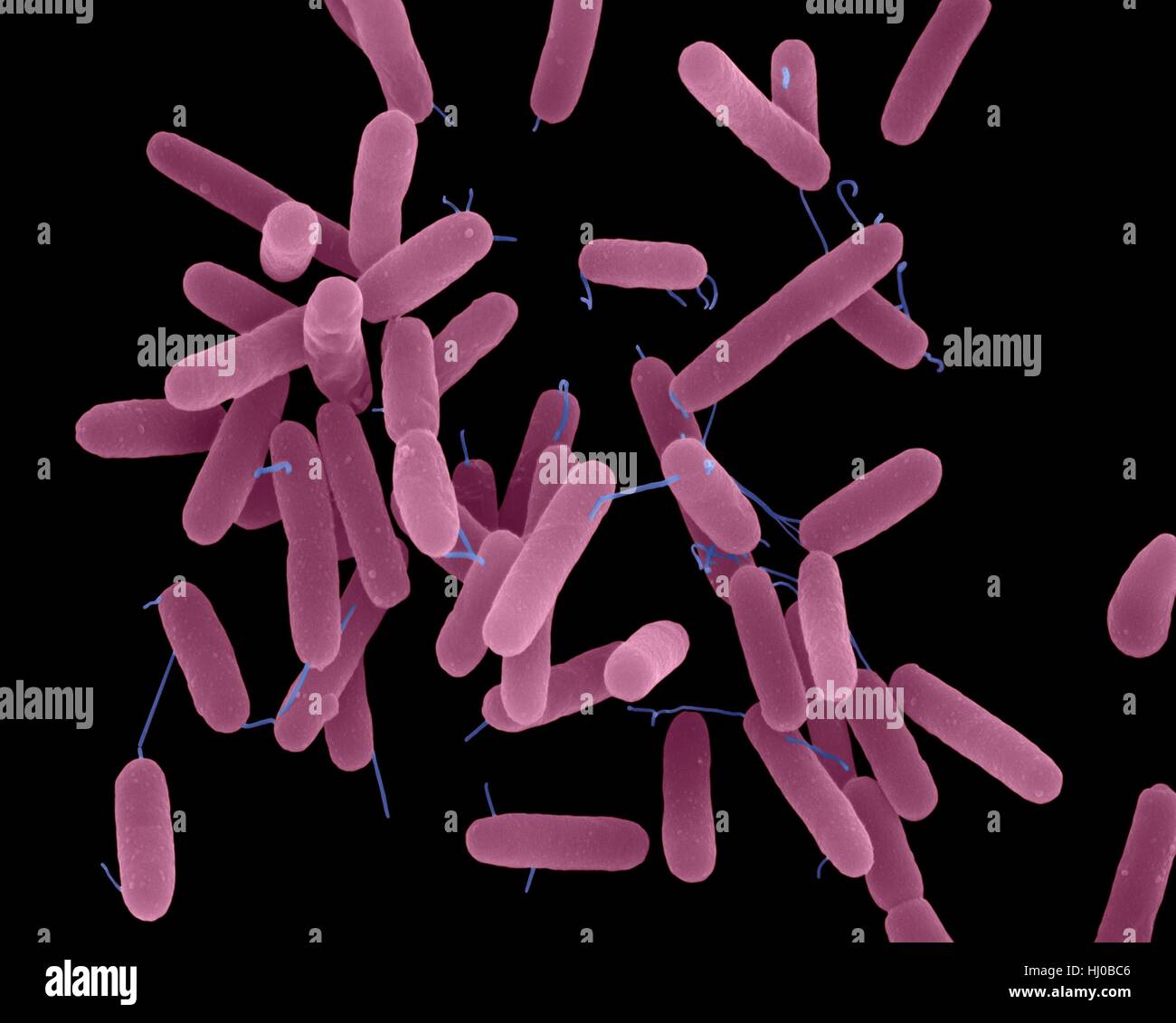 Coloured scanning electron micrograph (SEM) of Pseudomonas aeruginosa, Gram-negative, aerobic, enteric, rod prokaryote (dividing). This bacterium produces a blue, Green pigment, pyocyanin, which characterizes the bluish pus produced by the infection. It is found in soil, water, skin flora, and most man-made environments throughout the world. Magnification: x2, 400 when shortest axis printed at 25 millimetres. Stock Photohttps://www.alamy.com/image-license-details/?v=1https://www.alamy.com/stock-photo-coloured-scanning-electron-micrograph-sem-of-pseudomonas-aeruginosa-131545350.html
RFHJ0BC6–Coloured scanning electron micrograph (SEM) of Pseudomonas aeruginosa, Gram-negative, aerobic, enteric, rod prokaryote (dividing). This bacterium produces a blue, Green pigment, pyocyanin, which characterizes the bluish pus produced by the infection. It is found in soil, water, skin flora, and most man-made environments throughout the world. Magnification: x2, 400 when shortest axis printed at 25 millimetres.
 MDR Pathogen, Pseudomonas aeruginosa, SEM Stock Photohttps://www.alamy.com/image-license-details/?v=1https://www.alamy.com/stock-photo-mdr-pathogen-pseudomonas-aeruginosa-sem-135022402.html
RMHRJPCJ–MDR Pathogen, Pseudomonas aeruginosa, SEM
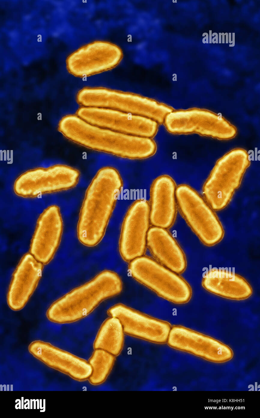 PSEUDOMONAS AERUGINOSA Stock Photohttps://www.alamy.com/image-license-details/?v=1https://www.alamy.com/stock-image-pseudomonas-aeruginosa-160197213.html
RMK8HH51–PSEUDOMONAS AERUGINOSA
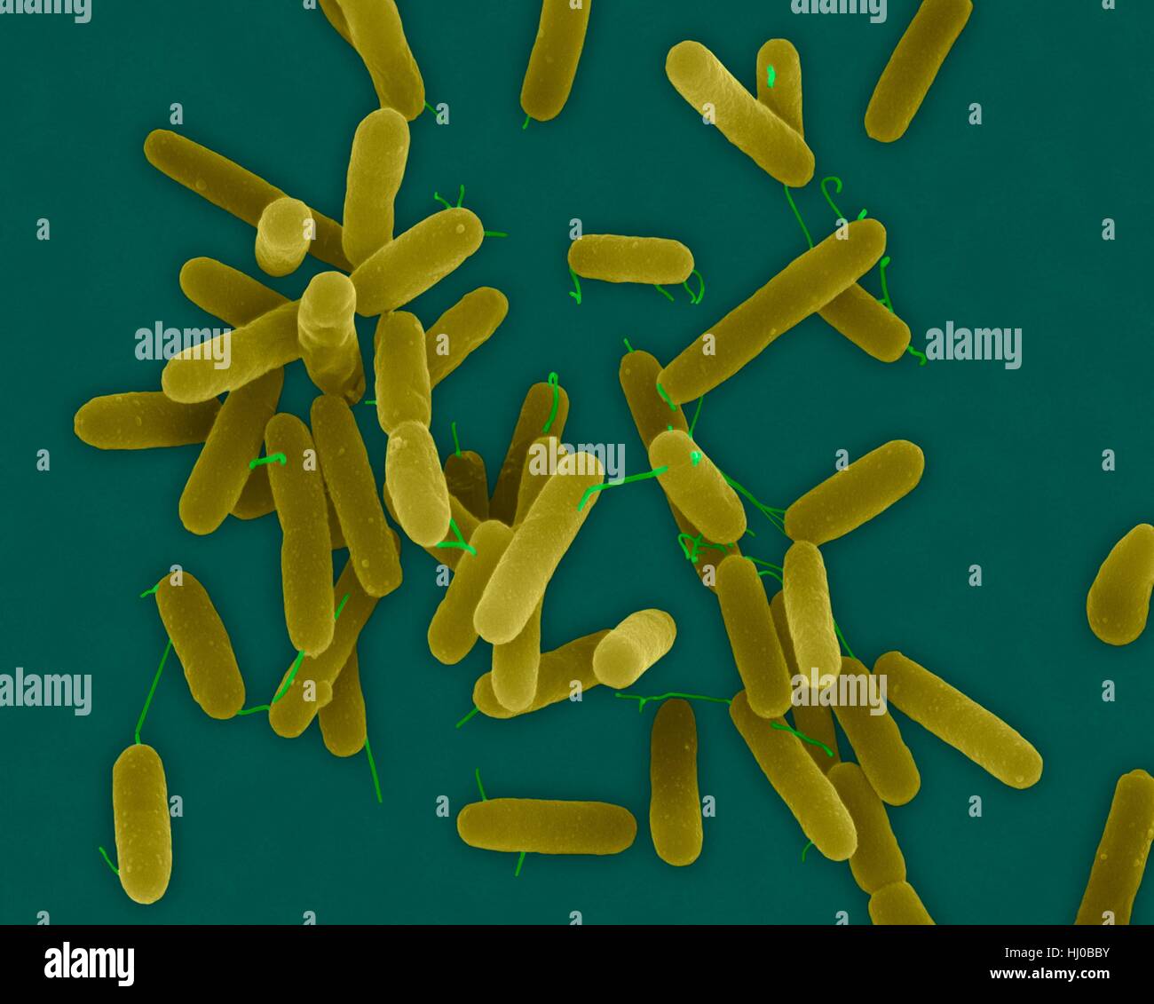 Coloured scanning electron micrograph (SEM) of Pseudomonas aeruginosa, Gram-negative, aerobic, enteric, rod prokaryote (dividing). This bacterium produces a blue, Green pigment, pyocyanin, which characterizes the bluish pus produced by the infection. It is found in soil, water, skin flora, and most man-made environments throughout the world. Magnification: x2, 400 when shortest axis printed at 25 millimetres. Stock Photohttps://www.alamy.com/image-license-details/?v=1https://www.alamy.com/stock-photo-coloured-scanning-electron-micrograph-sem-of-pseudomonas-aeruginosa-131545343.html
RFHJ0BBY–Coloured scanning electron micrograph (SEM) of Pseudomonas aeruginosa, Gram-negative, aerobic, enteric, rod prokaryote (dividing). This bacterium produces a blue, Green pigment, pyocyanin, which characterizes the bluish pus produced by the infection. It is found in soil, water, skin flora, and most man-made environments throughout the world. Magnification: x2, 400 when shortest axis printed at 25 millimetres.
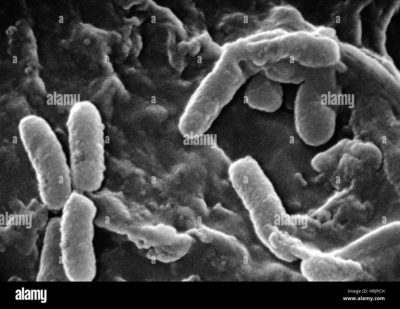 MDR Pathogen, Pseudomonas aeruginosa, SEM Stock Photohttps://www.alamy.com/image-license-details/?v=1https://www.alamy.com/stock-photo-mdr-pathogen-pseudomonas-aeruginosa-sem-135022401.html
RMHRJPCH–MDR Pathogen, Pseudomonas aeruginosa, SEM
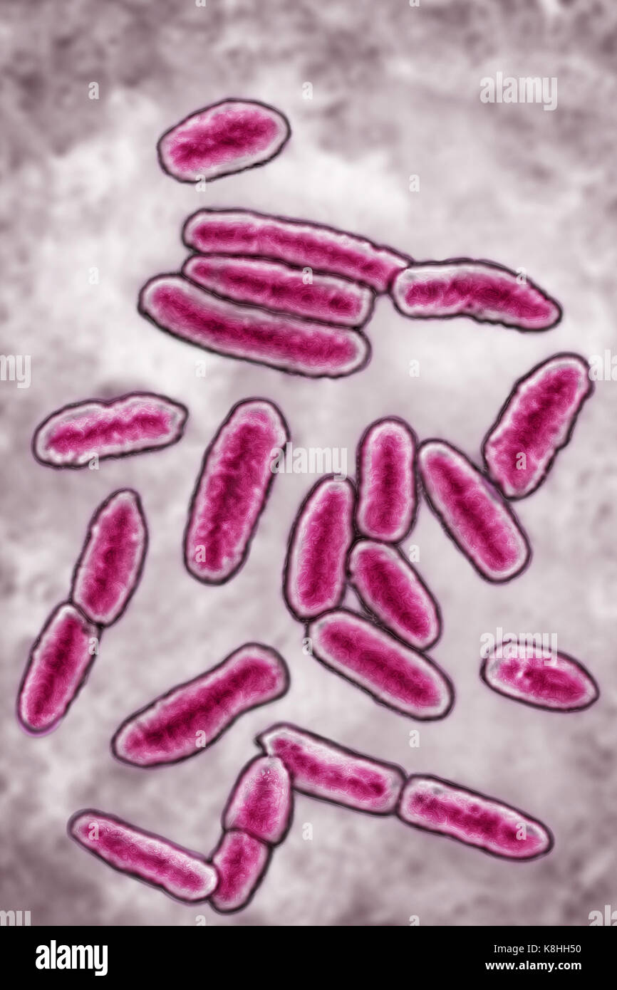 PSEUDOMONAS AERUGINOSA Stock Photohttps://www.alamy.com/image-license-details/?v=1https://www.alamy.com/stock-image-pseudomonas-aeruginosa-160197212.html
RMK8HH50–PSEUDOMONAS AERUGINOSA
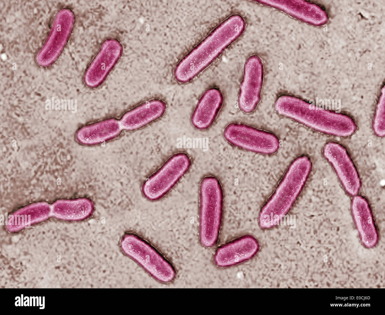 Pseudomonas aeruginosa Stock Photohttps://www.alamy.com/image-license-details/?v=1https://www.alamy.com/pseudomonas-aeruginosa-image69119189.html
RME0CJ6D–Pseudomonas aeruginosa
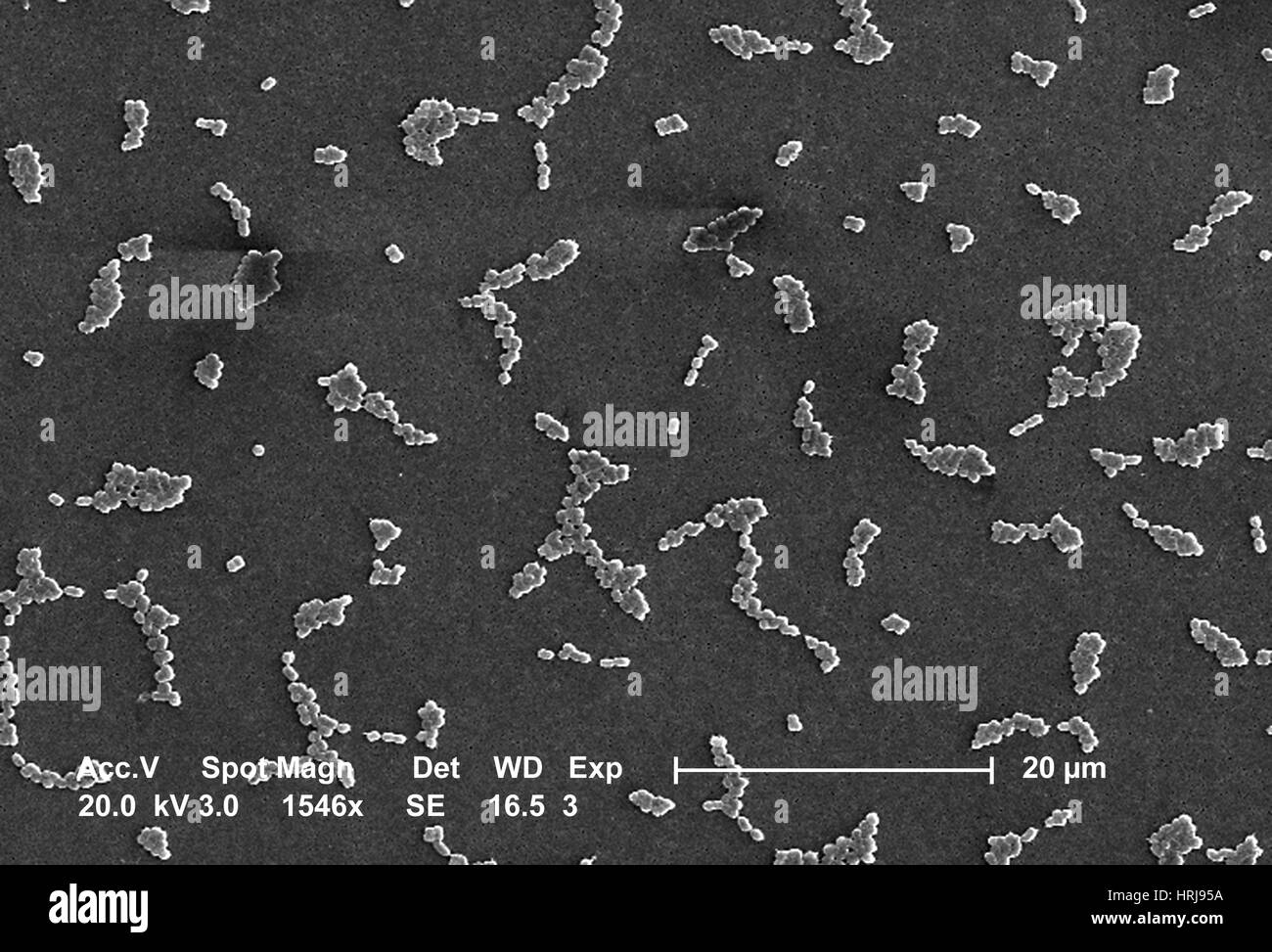 Acinetobacter baumannii Bacteria, SEM Stock Photohttps://www.alamy.com/image-license-details/?v=1https://www.alamy.com/stock-photo-acinetobacter-baumannii-bacteria-sem-135012006.html
RMHRJ95A–Acinetobacter baumannii Bacteria, SEM
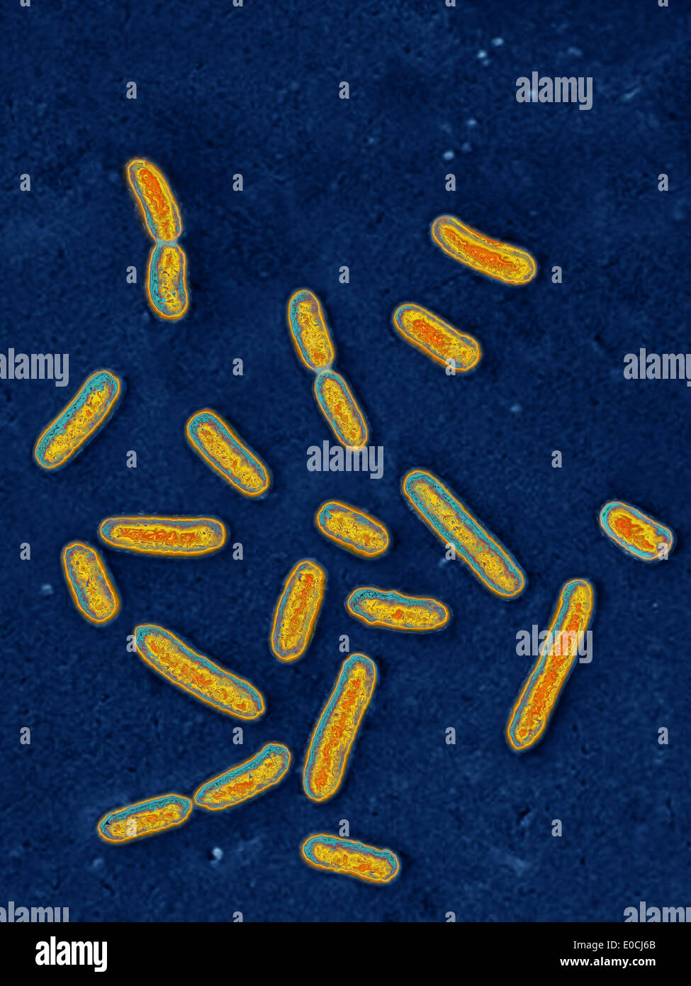 Pseudomonas aeruginosa Stock Photohttps://www.alamy.com/image-license-details/?v=1https://www.alamy.com/pseudomonas-aeruginosa-image69119187.html
RME0CJ6B–Pseudomonas aeruginosa
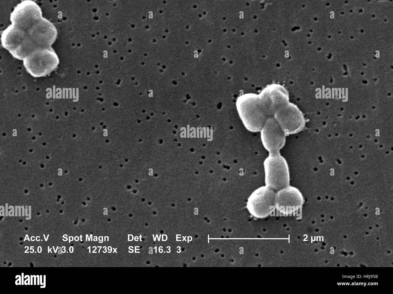 Acinetobacter baumannii Bacteria, SEM Stock Photohttps://www.alamy.com/image-license-details/?v=1https://www.alamy.com/stock-photo-acinetobacter-baumannii-bacteria-sem-135012007.html
RMHRJ95B–Acinetobacter baumannii Bacteria, SEM
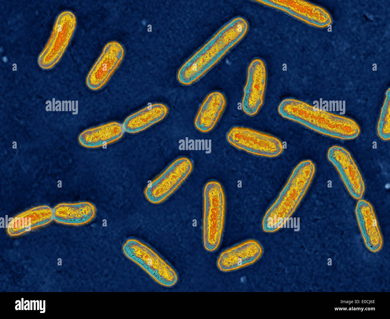 Pseudomonas aeruginosa Stock Photohttps://www.alamy.com/image-license-details/?v=1https://www.alamy.com/pseudomonas-aeruginosa-image69119190.html
RME0CJ6E–Pseudomonas aeruginosa
 Pseudomonas aeruginosa Stock Photohttps://www.alamy.com/image-license-details/?v=1https://www.alamy.com/pseudomonas-aeruginosa-image69119191.html
RME0CJ6F–Pseudomonas aeruginosa
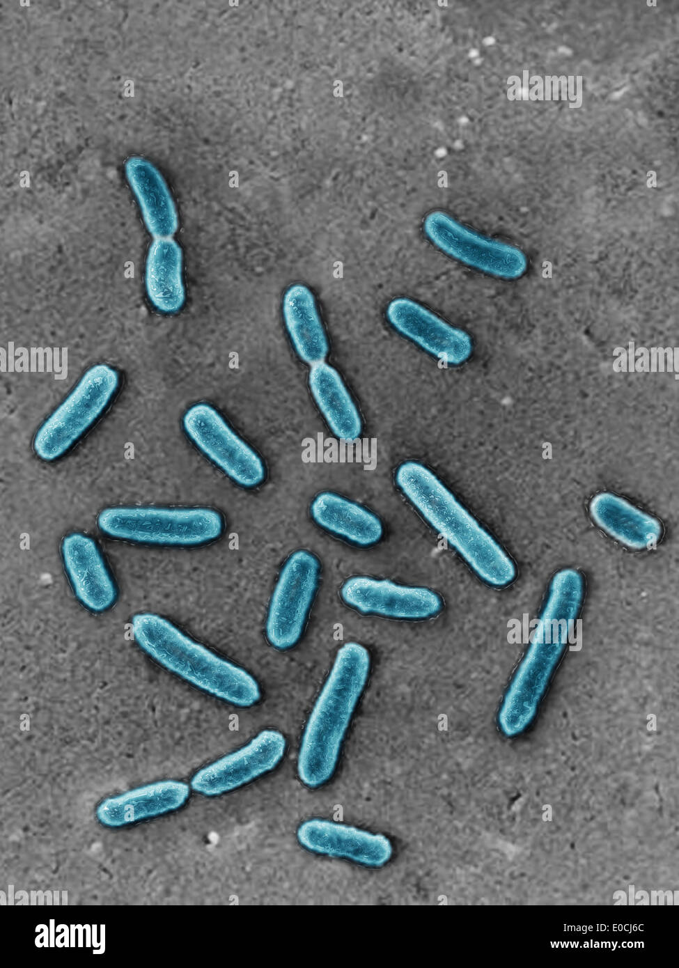 Pseudomonas aeruginosa Stock Photohttps://www.alamy.com/image-license-details/?v=1https://www.alamy.com/pseudomonas-aeruginosa-image69119188.html
RME0CJ6C–Pseudomonas aeruginosa
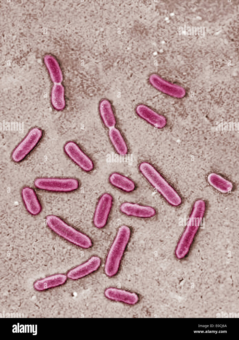 Pseudomonas aeruginosa Stock Photohttps://www.alamy.com/image-license-details/?v=1https://www.alamy.com/pseudomonas-aeruginosa-image69119186.html
RME0CJ6A–Pseudomonas aeruginosa
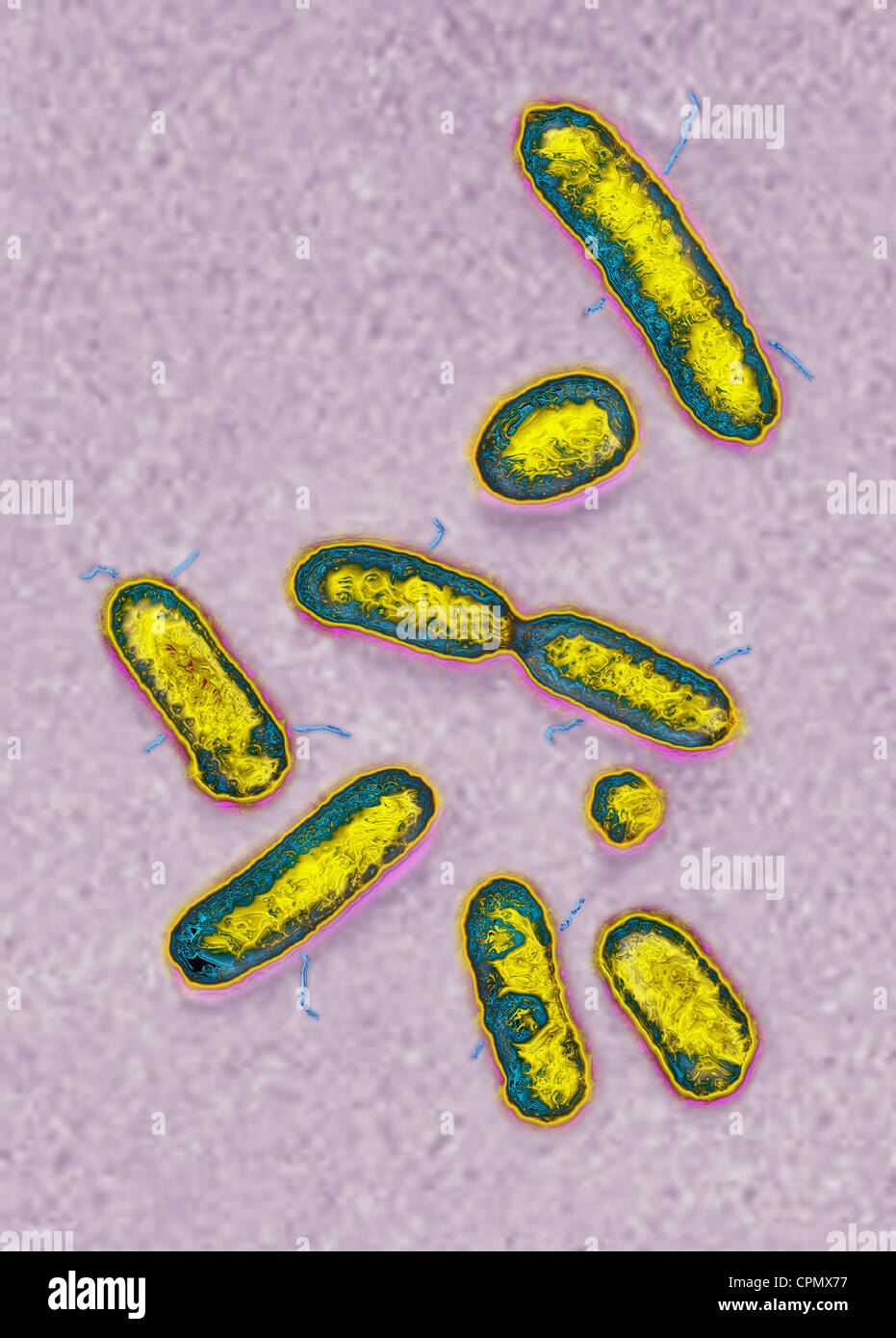 PSEUDOMONAS AERUGINOSA Stock Photohttps://www.alamy.com/image-license-details/?v=1https://www.alamy.com/stock-photo-pseudomonas-aeruginosa-48402795.html
RMCPMX77–PSEUDOMONAS AERUGINOSA
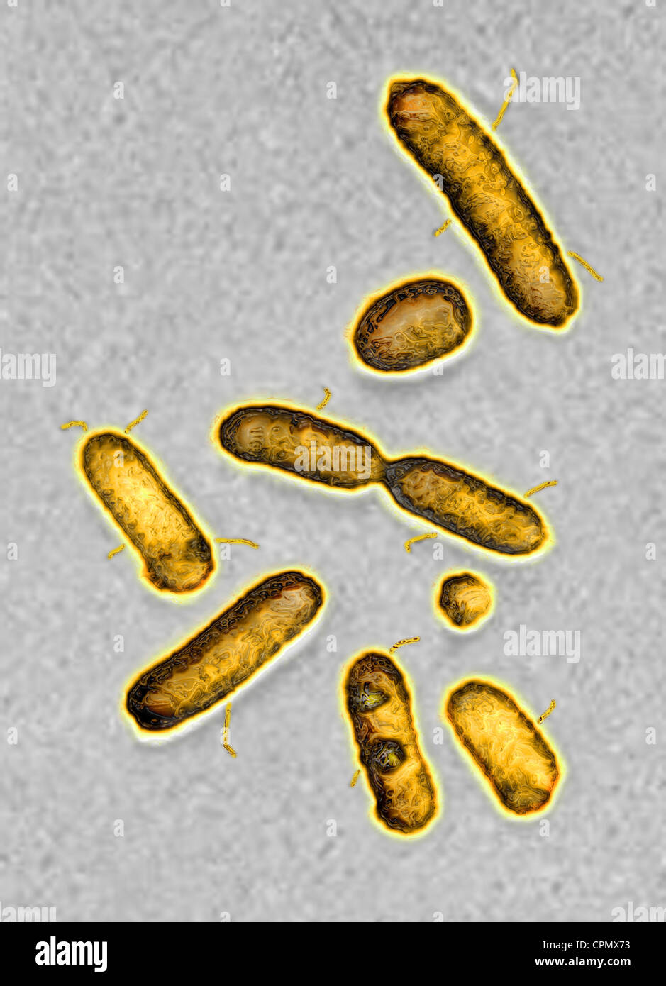 PSEUDOMONAS AERUGINOSA Stock Photohttps://www.alamy.com/image-license-details/?v=1https://www.alamy.com/stock-photo-pseudomonas-aeruginosa-48402791.html
RMCPMX73–PSEUDOMONAS AERUGINOSA
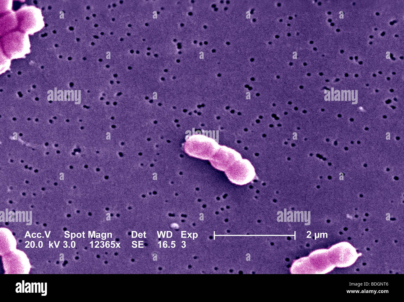 ACINETOBACTER BAUMANNII Stock Photohttps://www.alamy.com/image-license-details/?v=1https://www.alamy.com/stock-photo-acinetobacter-baumannii-25569270.html
RFBDGNT6–ACINETOBACTER BAUMANNII
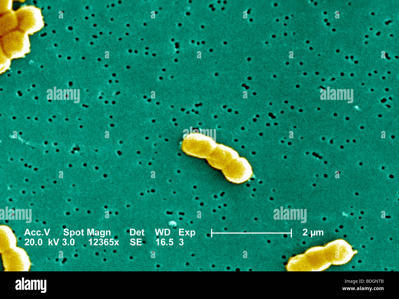 ACINETOBACTER BAUMANNII Stock Photohttps://www.alamy.com/image-license-details/?v=1https://www.alamy.com/stock-photo-acinetobacter-baumannii-25569275.html
RFBDGNTB–ACINETOBACTER BAUMANNII
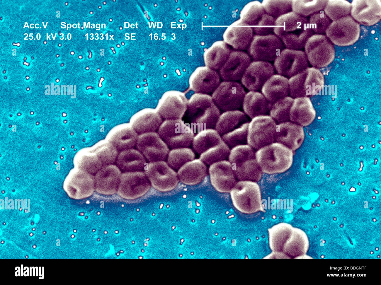 ACINETOBACTER BAUMANNII Stock Photohttps://www.alamy.com/image-license-details/?v=1https://www.alamy.com/stock-photo-acinetobacter-baumannii-25569279.html
RFBDGNTF–ACINETOBACTER BAUMANNII
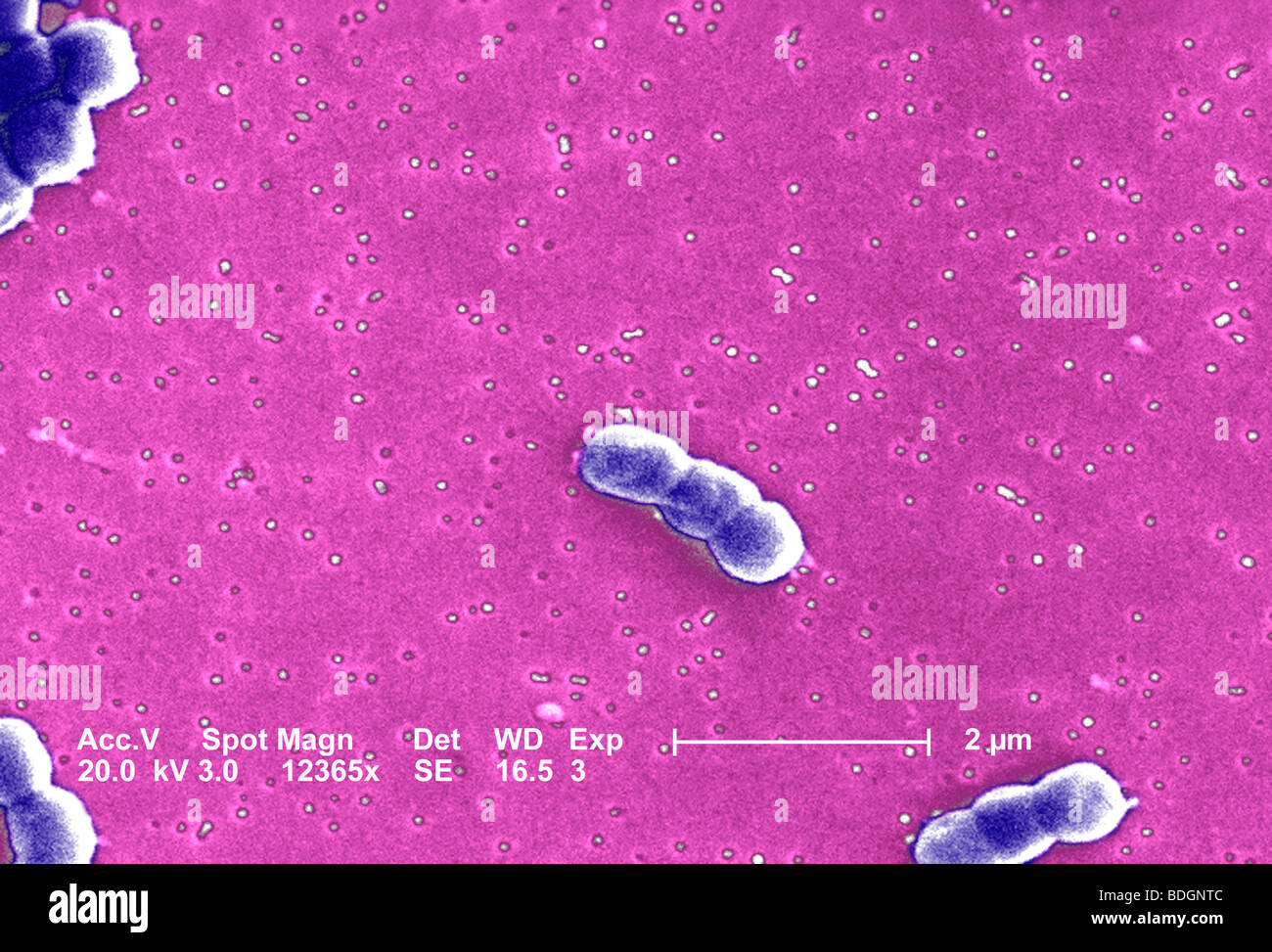 ACINETOBACTER BAUMANNII Stock Photohttps://www.alamy.com/image-license-details/?v=1https://www.alamy.com/stock-photo-acinetobacter-baumannii-25569276.html
RFBDGNTC–ACINETOBACTER BAUMANNII
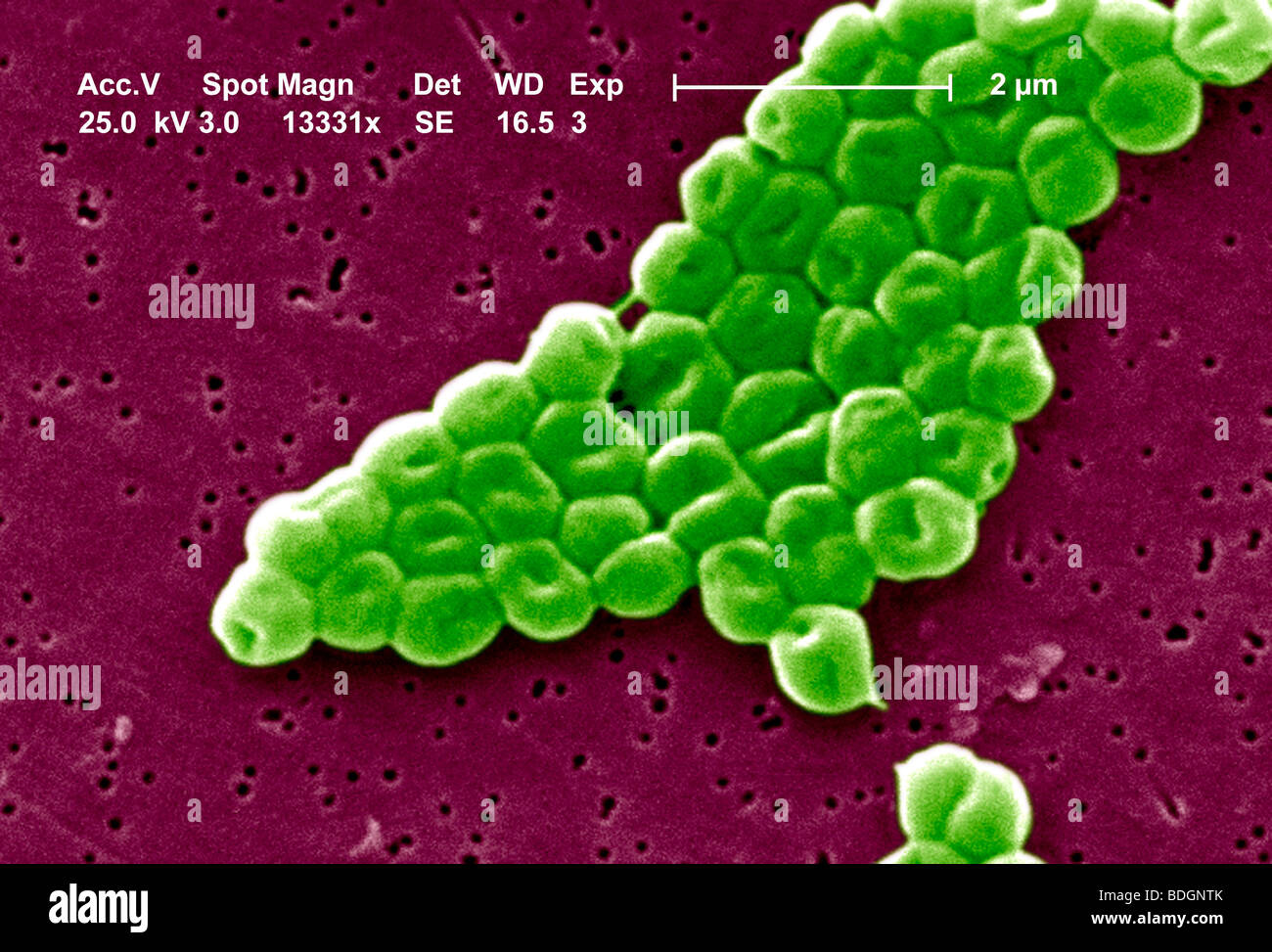 ACINETOBACTER BAUMANNII Stock Photohttps://www.alamy.com/image-license-details/?v=1https://www.alamy.com/stock-photo-acinetobacter-baumannii-25569283.html
RFBDGNTK–ACINETOBACTER BAUMANNII
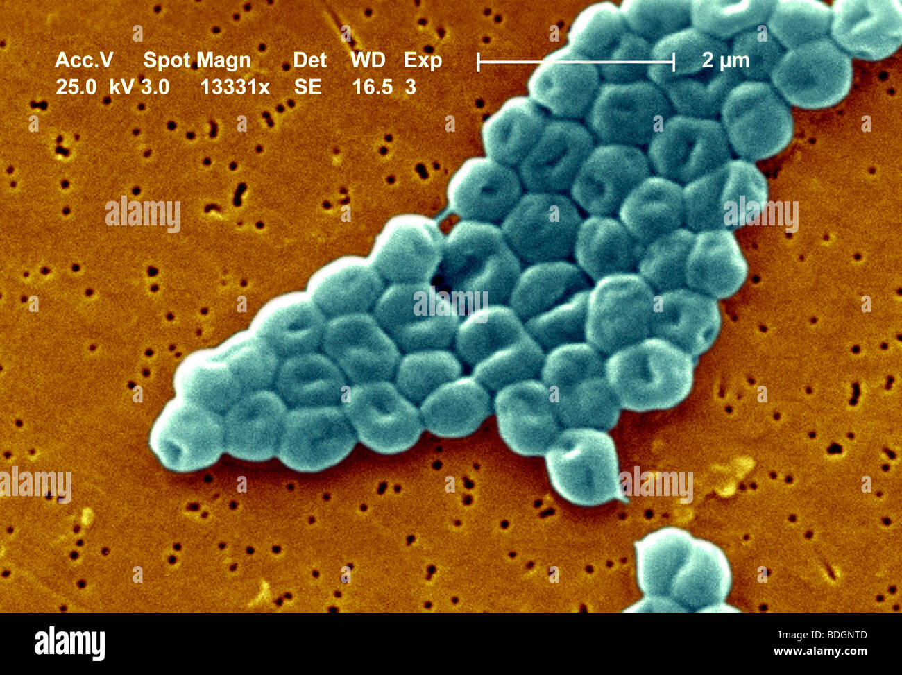 ACINETOBACTER BAUMANNII Stock Photohttps://www.alamy.com/image-license-details/?v=1https://www.alamy.com/stock-photo-acinetobacter-baumannii-25569277.html
RFBDGNTD–ACINETOBACTER BAUMANNII
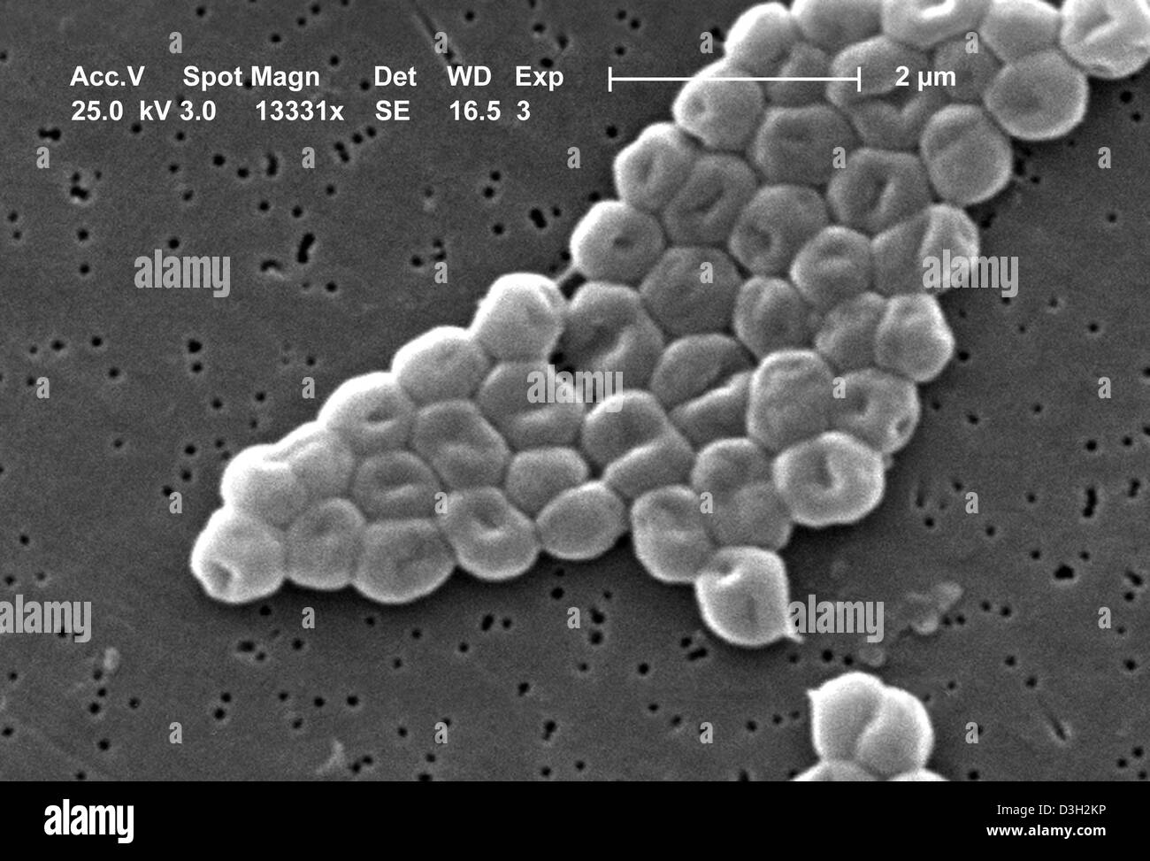 ACINETOBACTER BAUMANNII Stock Photohttps://www.alamy.com/image-license-details/?v=1https://www.alamy.com/stock-photo-acinetobacter-baumannii-53850378.html
RMD3H2KP–ACINETOBACTER BAUMANNII
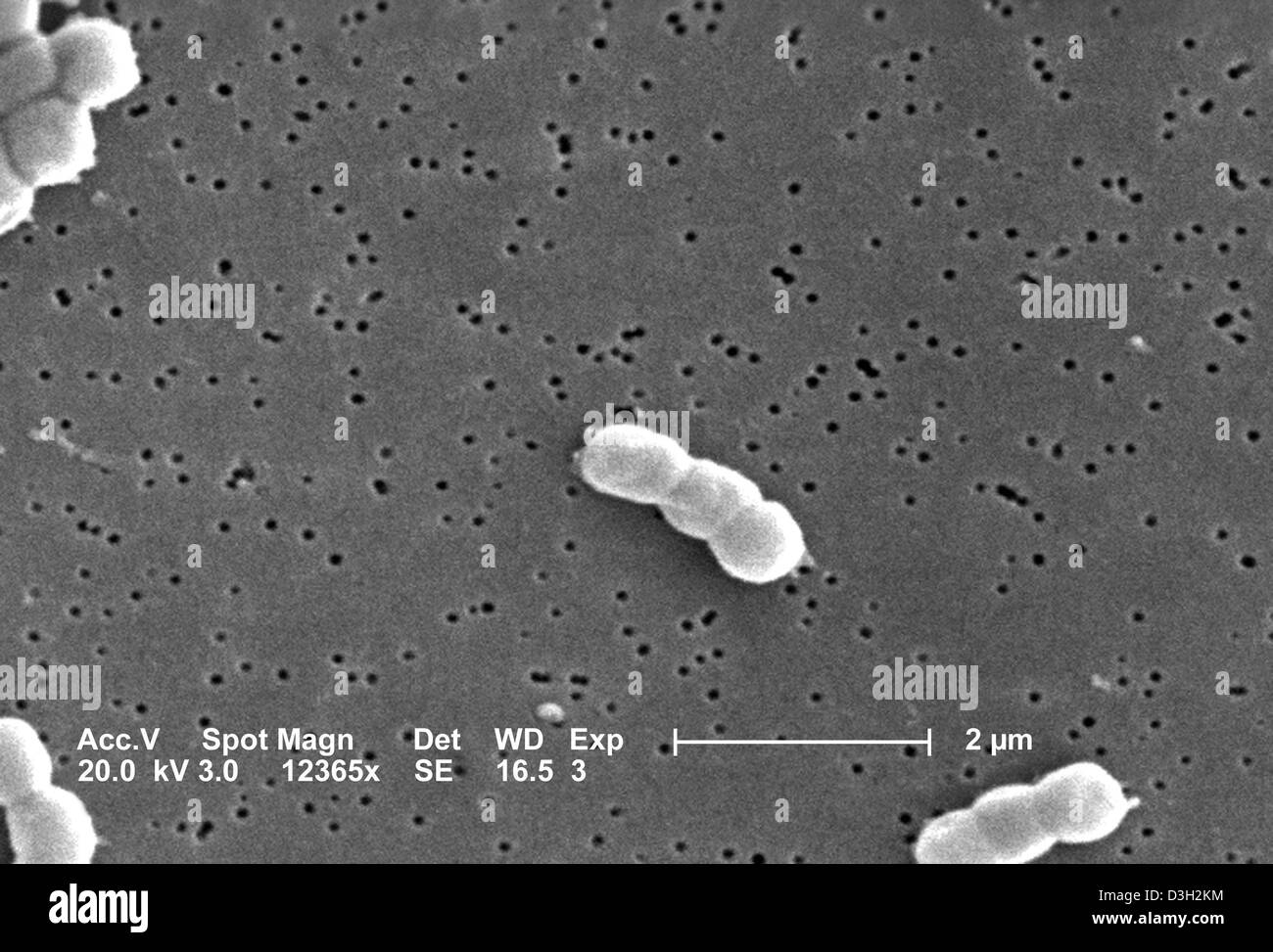 ACINETOBACTER BAUMANNII Stock Photohttps://www.alamy.com/image-license-details/?v=1https://www.alamy.com/stock-photo-acinetobacter-baumannii-53850376.html
RMD3H2KM–ACINETOBACTER BAUMANNII
 PSEUDOMONAS AERUGINOSA Stock Photohttps://www.alamy.com/image-license-details/?v=1https://www.alamy.com/stock-photo-pseudomonas-aeruginosa-53863177.html
RMD3HK0W–PSEUDOMONAS AERUGINOSA
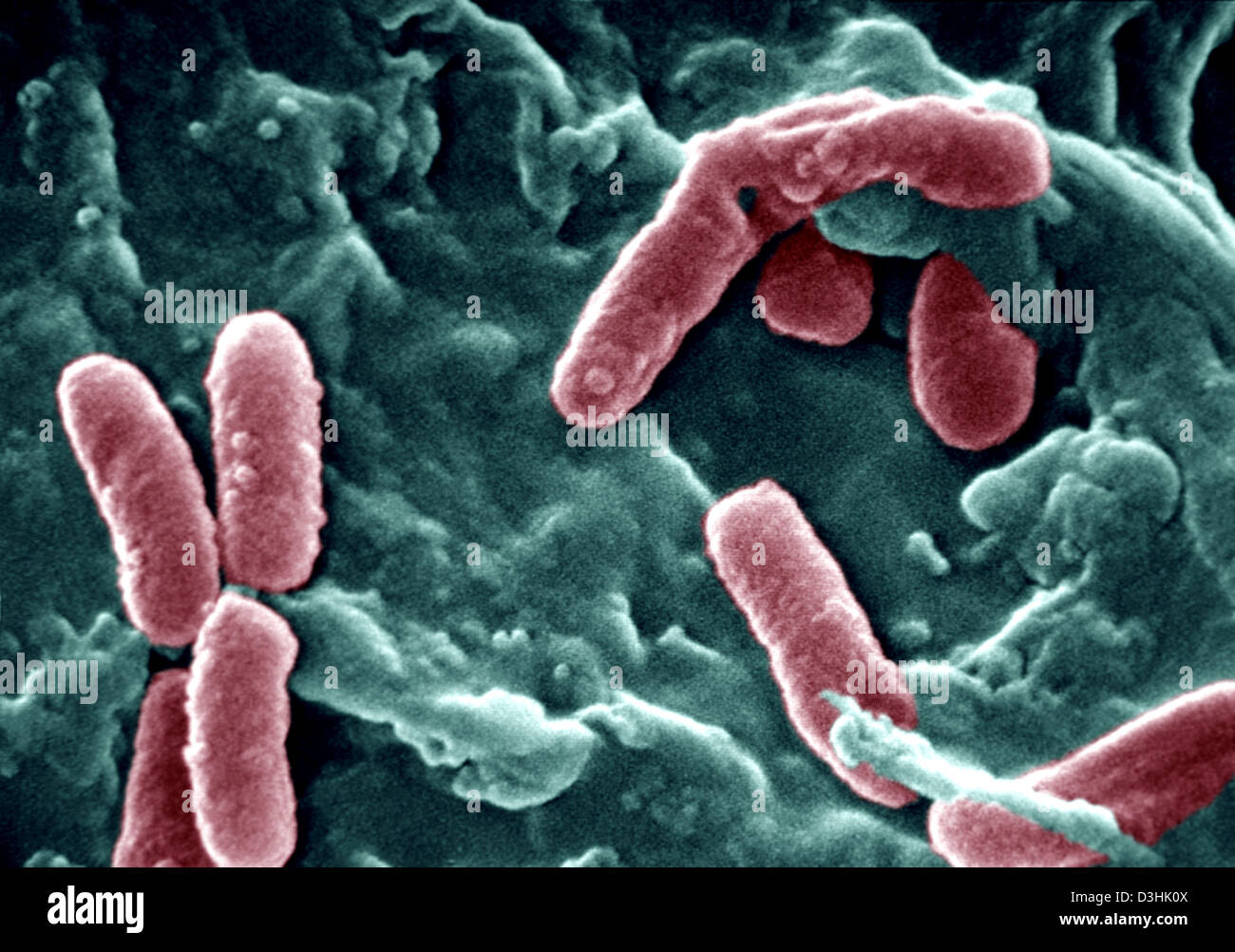 PSEUDOMONAS AERUGINOSA Stock Photohttps://www.alamy.com/image-license-details/?v=1https://www.alamy.com/stock-photo-pseudomonas-aeruginosa-53863178.html
RMD3HK0X–PSEUDOMONAS AERUGINOSA
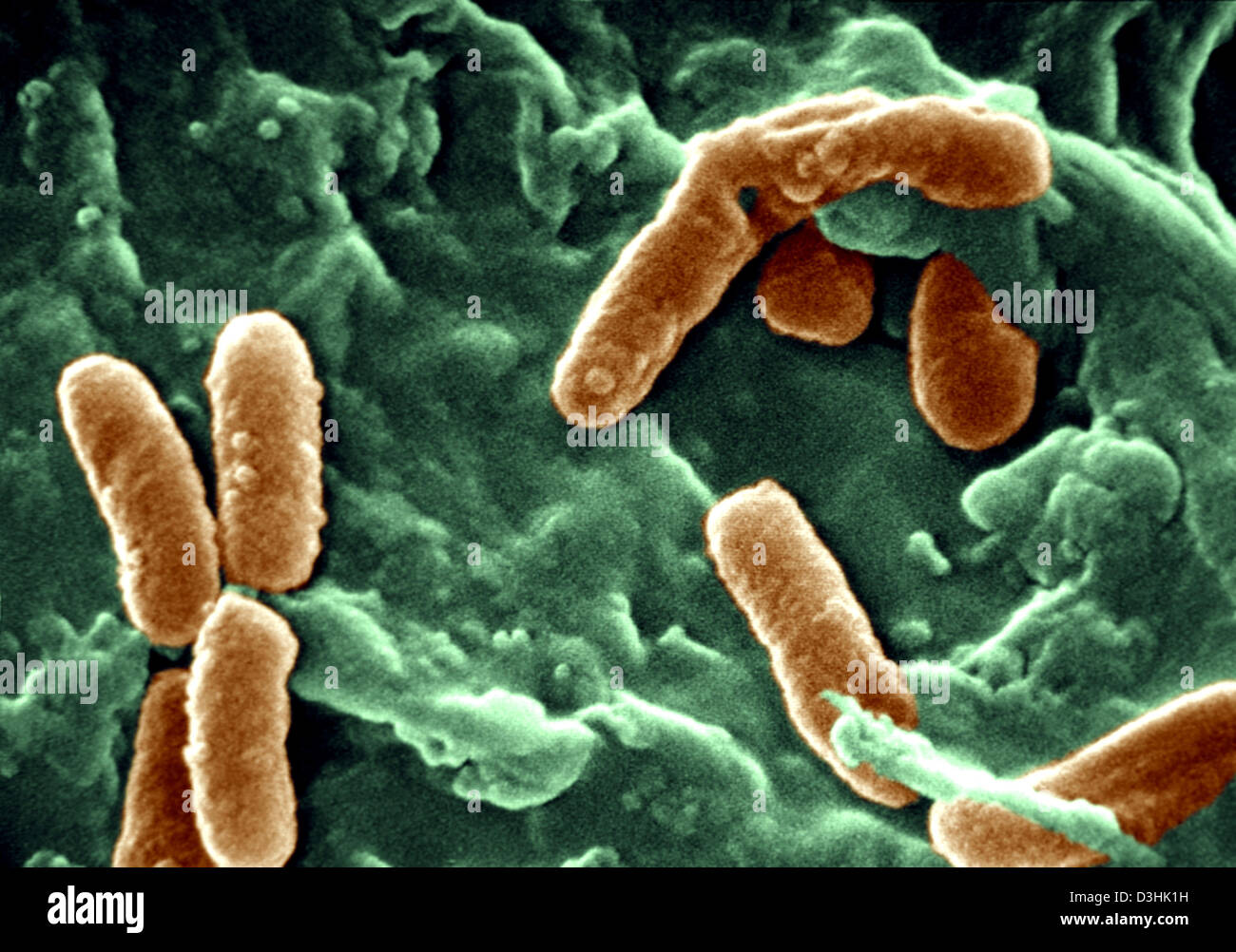 PSEUDOMONAS AERUGINOSA Stock Photohttps://www.alamy.com/image-license-details/?v=1https://www.alamy.com/stock-photo-pseudomonas-aeruginosa-53863197.html
RMD3HK1H–PSEUDOMONAS AERUGINOSA
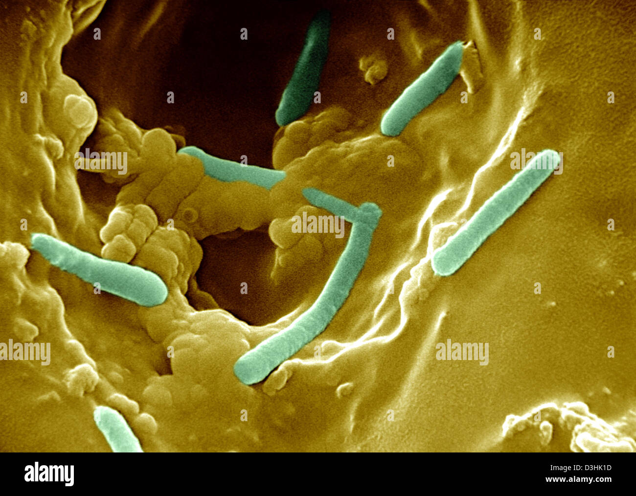 PSEUDOMONAS AERUGINOSA Stock Photohttps://www.alamy.com/image-license-details/?v=1https://www.alamy.com/stock-photo-pseudomonas-aeruginosa-53863193.html
RMD3HK1D–PSEUDOMONAS AERUGINOSA
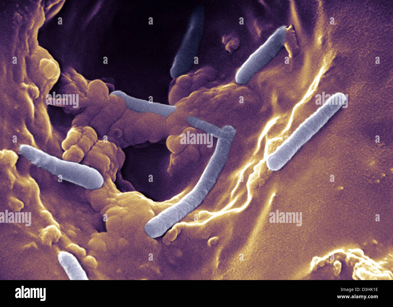 PSEUDOMONAS AERUGINOSA Stock Photohttps://www.alamy.com/image-license-details/?v=1https://www.alamy.com/stock-photo-pseudomonas-aeruginosa-53863194.html
RMD3HK1E–PSEUDOMONAS AERUGINOSA
 PSEUDOMONAS AERUGINOSA Stock Photohttps://www.alamy.com/image-license-details/?v=1https://www.alamy.com/stock-photo-pseudomonas-aeruginosa-53863174.html
RMD3HK0P–PSEUDOMONAS AERUGINOSA
 PSEUDOMONAS AERUGINOSA Stock Photohttps://www.alamy.com/image-license-details/?v=1https://www.alamy.com/stock-photo-pseudomonas-aeruginosa-49162721.html
RMCRYFFD–PSEUDOMONAS AERUGINOSA
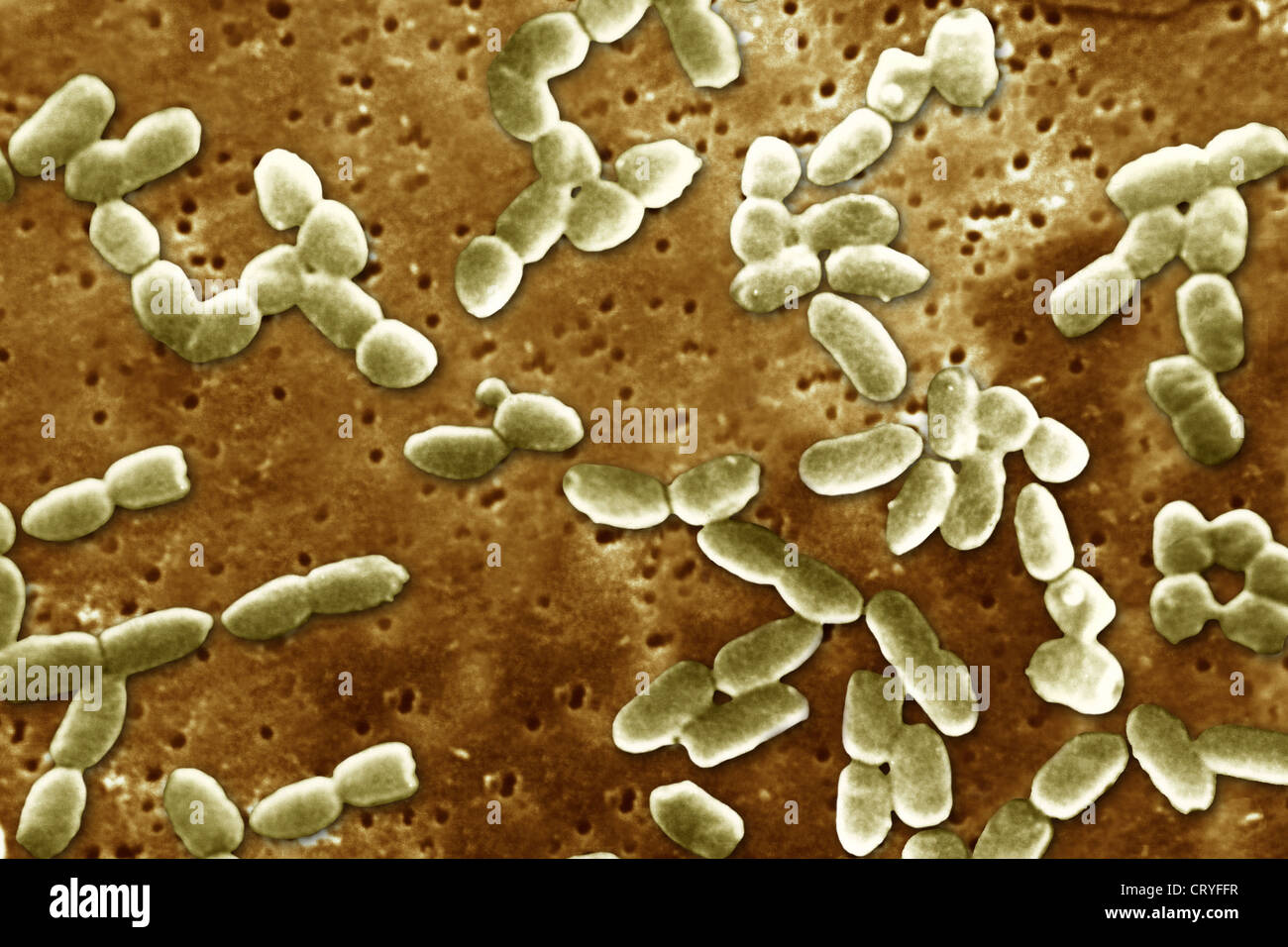 PSEUDOMONAS AERUGINOSA Stock Photohttps://www.alamy.com/image-license-details/?v=1https://www.alamy.com/stock-photo-pseudomonas-aeruginosa-49162731.html
RMCRYFFR–PSEUDOMONAS AERUGINOSA
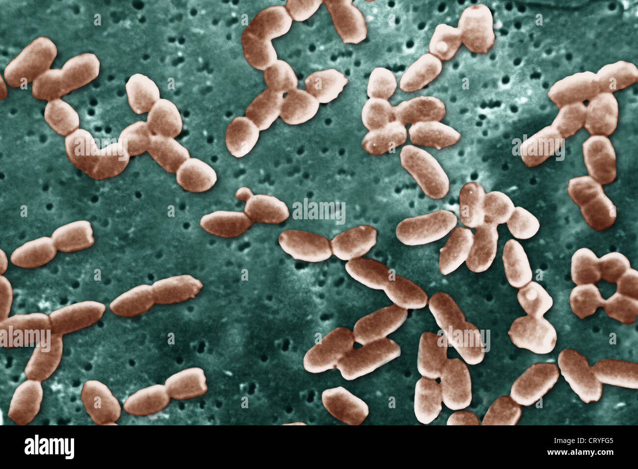 PSEUDOMONAS AERUGINOSA Stock Photohttps://www.alamy.com/image-license-details/?v=1https://www.alamy.com/stock-photo-pseudomonas-aeruginosa-49162741.html
RMCRYFG5–PSEUDOMONAS AERUGINOSA
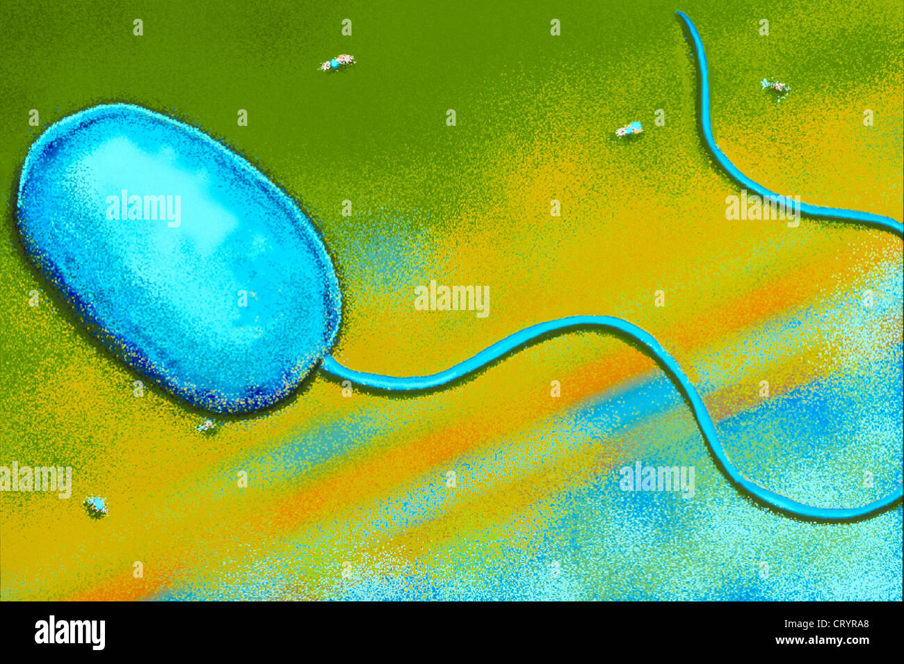 PSEUDOMONAS AERUGINOSA Stock Photohttps://www.alamy.com/image-license-details/?v=1https://www.alamy.com/stock-photo-pseudomonas-aeruginosa-49168848.html
RMCRYRA8–PSEUDOMONAS AERUGINOSA
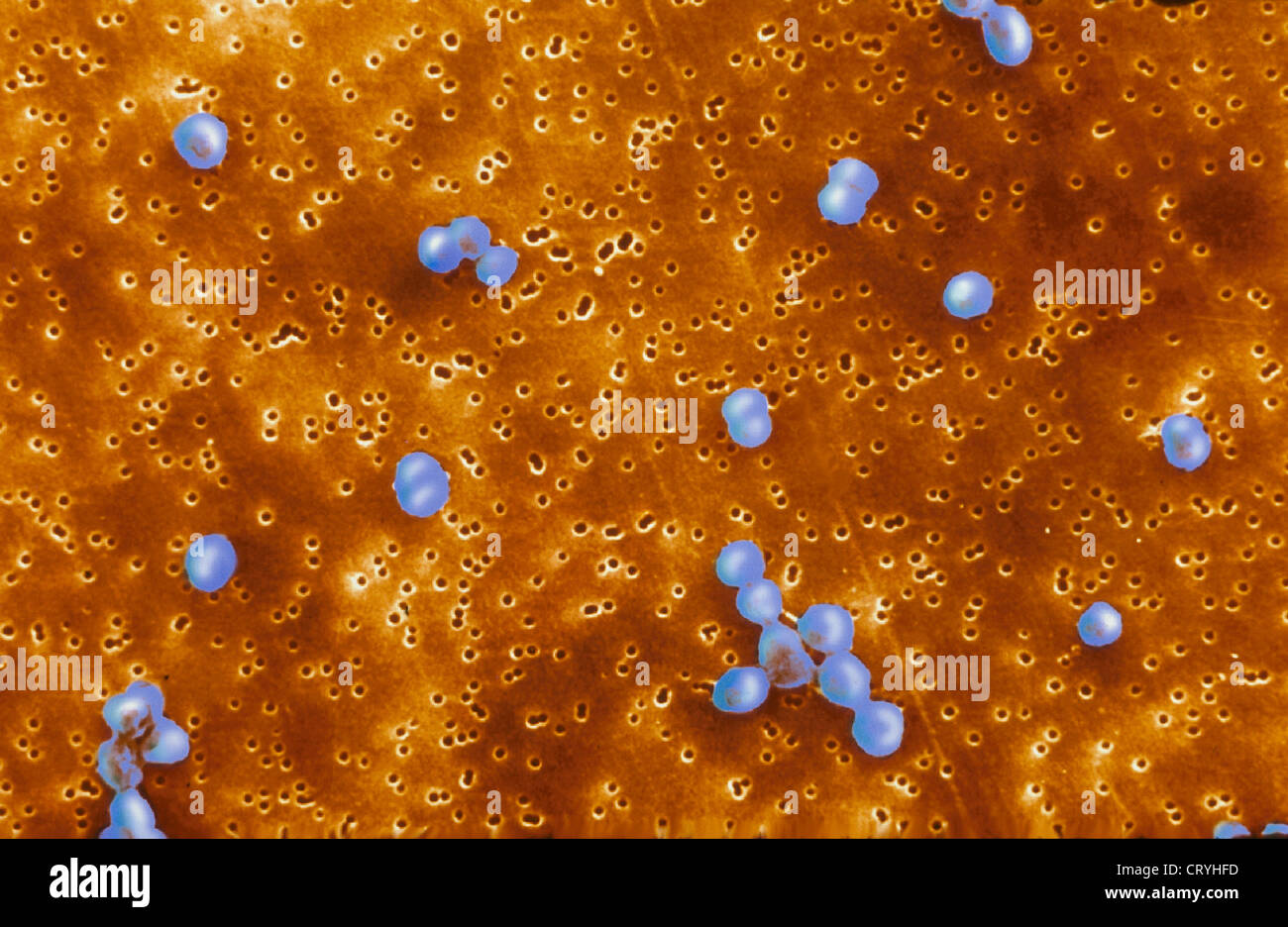 MORAXELLA CATARRHALIS Stock Photohttps://www.alamy.com/image-license-details/?v=1https://www.alamy.com/stock-photo-moraxella-catarrhalis-49164289.html
RMCRYHFD–MORAXELLA CATARRHALIS
