Quick filters:
Putamen Stock Photos and Images
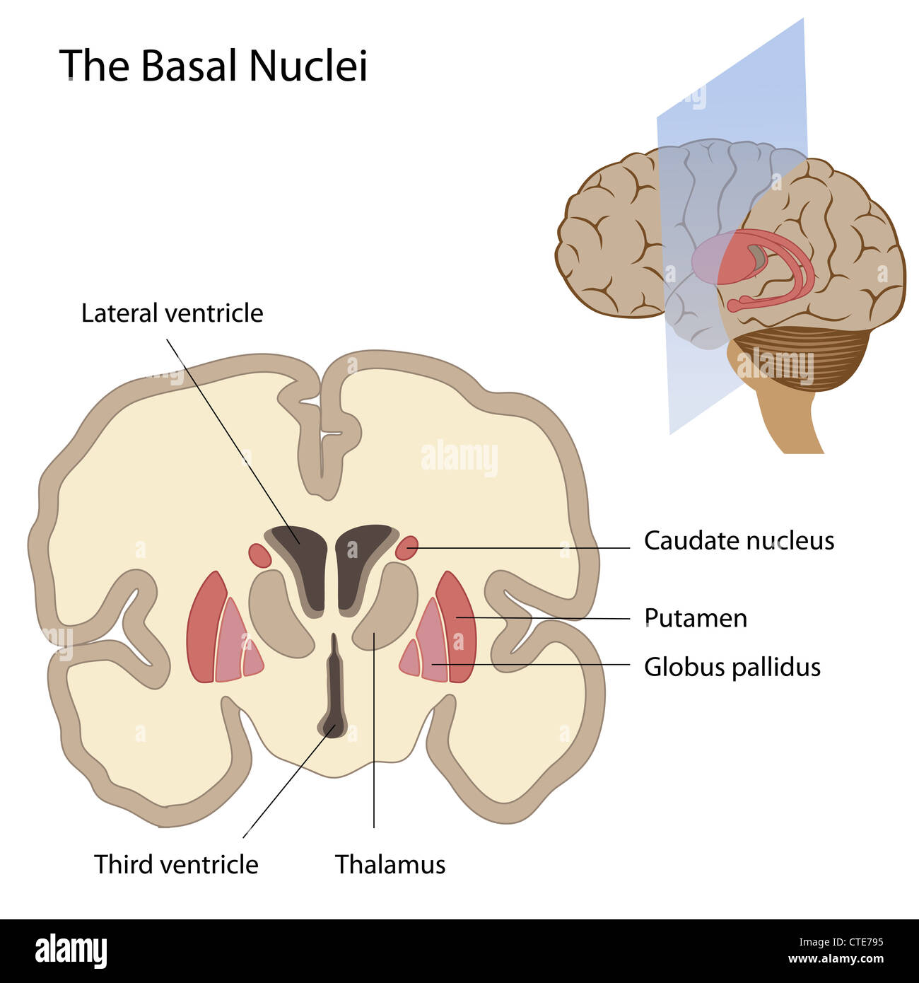
 Putamen, artwork Stock Photohttps://www.alamy.com/image-license-details/?v=1https://www.alamy.com/stock-photo-putamen-artwork-52732169.html
RFD1P4BN–Putamen, artwork
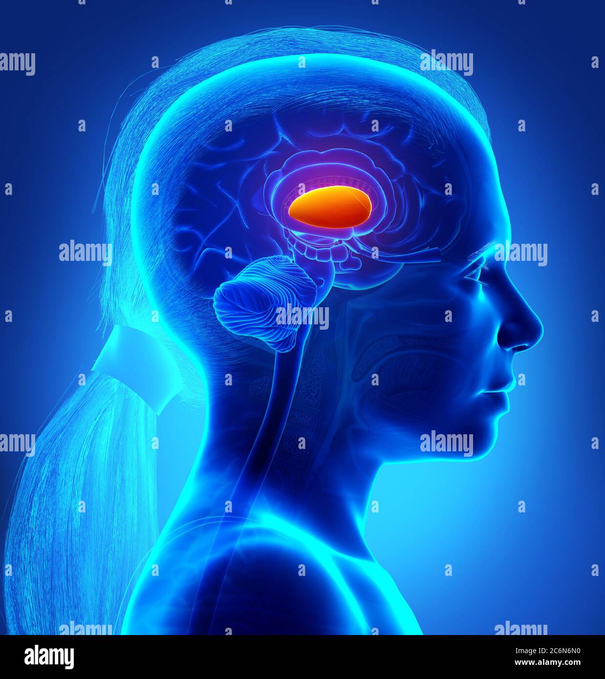 3d rendered medically accurate illustration of young girl brains anatomy- the putamen Stock Photohttps://www.alamy.com/image-license-details/?v=1https://www.alamy.com/3d-rendered-medically-accurate-illustration-of-young-girl-brains-anatomy-the-putamen-image365571948.html
RF2C6N6N0–3d rendered medically accurate illustration of young girl brains anatomy- the putamen
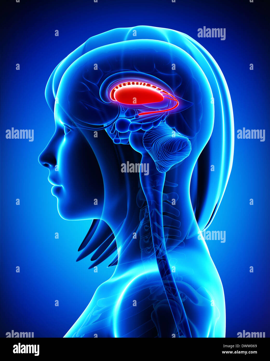
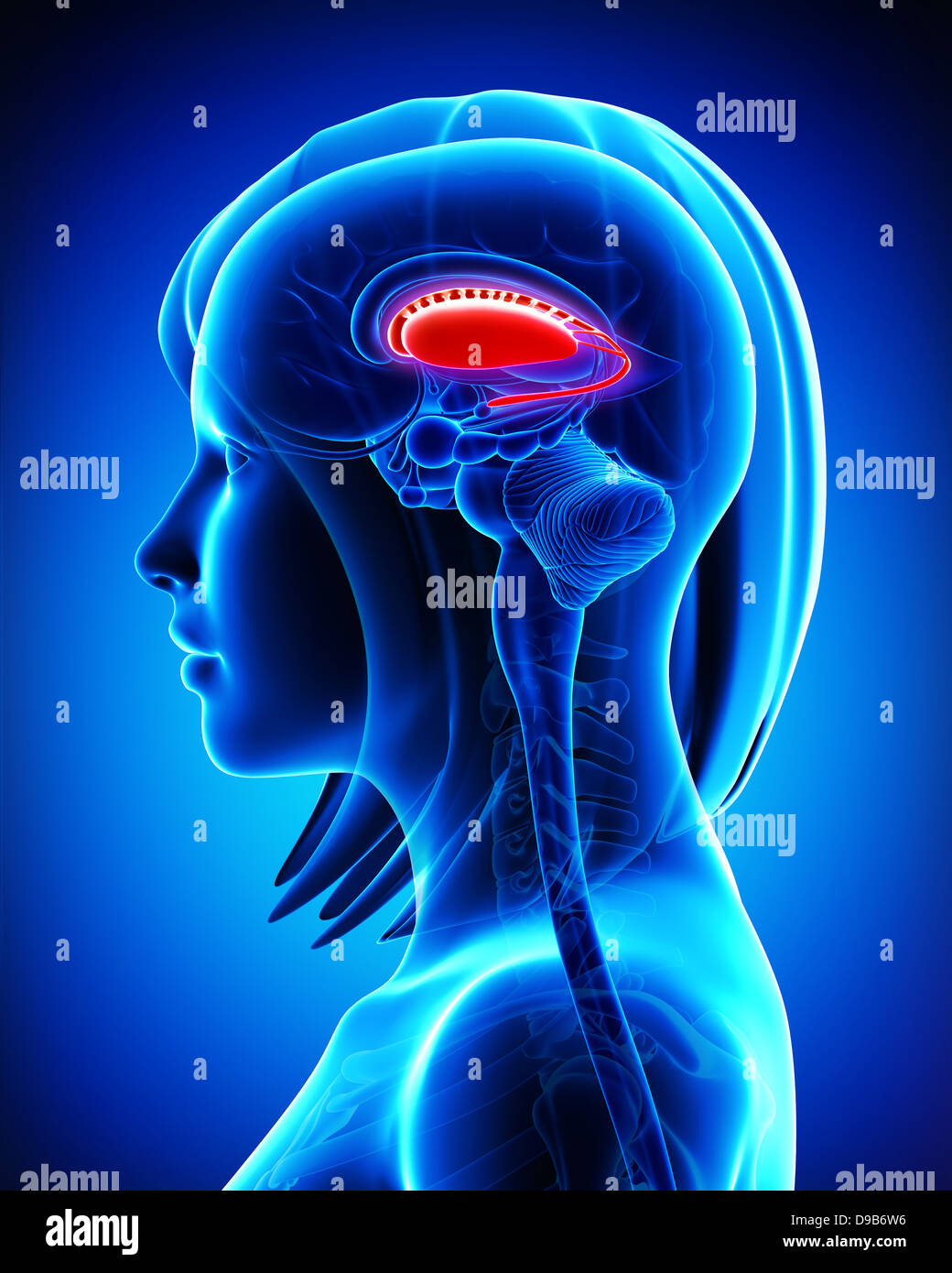 Anatomy of brain putamen and caudate nucleus- cross section Stock Photohttps://www.alamy.com/image-license-details/?v=1https://www.alamy.com/stock-photo-anatomy-of-brain-putamen-and-caudate-nucleus-cross-section-57409890.html
RFD9B6W6–Anatomy of brain putamen and caudate nucleus- cross section
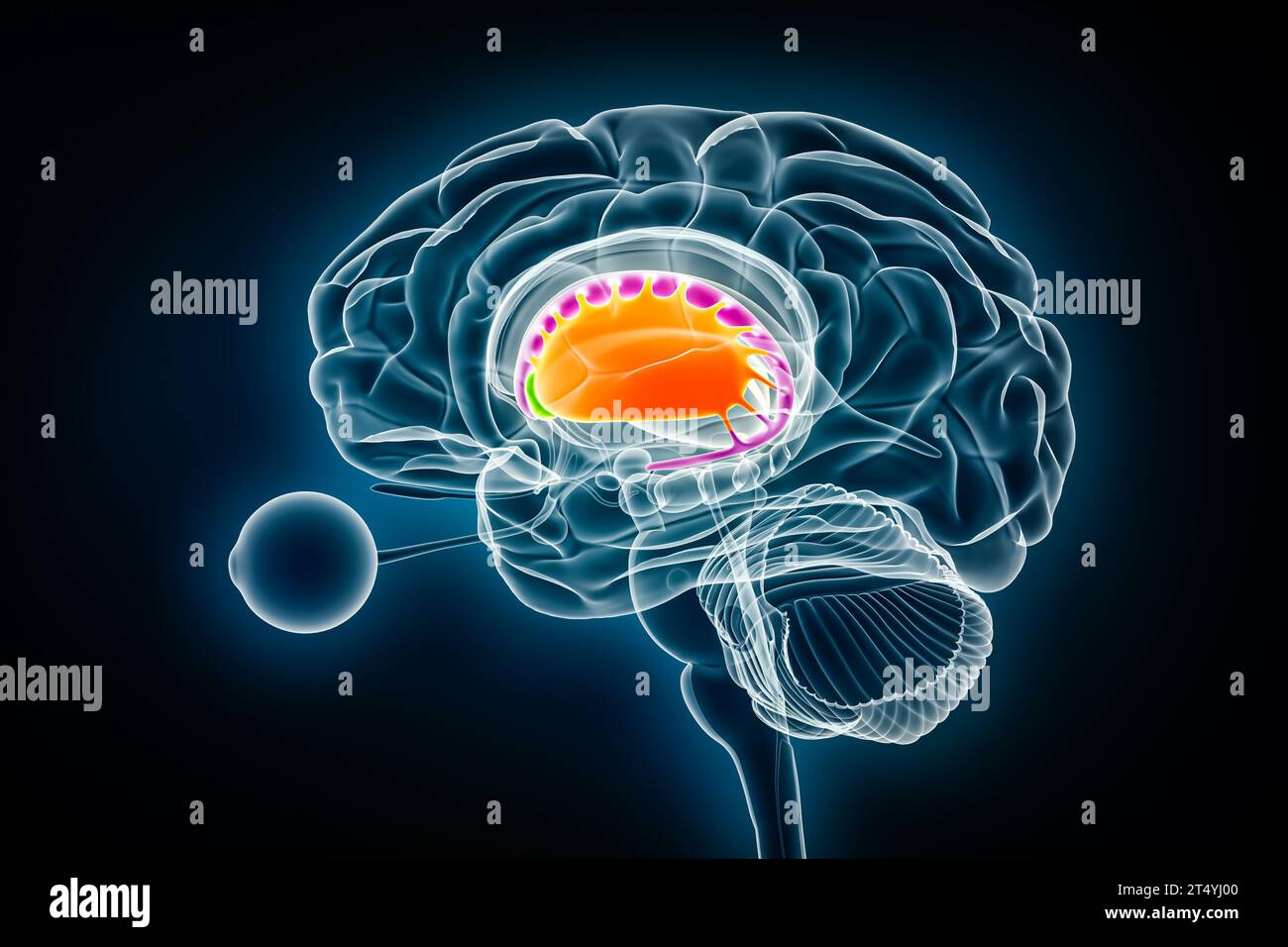 Putamen in orange, nucleus accumbens in green and caudate nucleus in purple 3D rendering illustration. Human brain, basal ganglia and corpus striatum Stock Photohttps://www.alamy.com/image-license-details/?v=1https://www.alamy.com/putamen-in-orange-nucleus-accumbens-in-green-and-caudate-nucleus-in-purple-3d-rendering-illustration-human-brain-basal-ganglia-and-corpus-striatum-image571007584.html
RF2T4YJ00–Putamen in orange, nucleus accumbens in green and caudate nucleus in purple 3D rendering illustration. Human brain, basal ganglia and corpus striatum
 Mango stone isolated Stock Photohttps://www.alamy.com/image-license-details/?v=1https://www.alamy.com/stock-image-mango-stone-isolated-167216168.html
RFKM19X0–Mango stone isolated
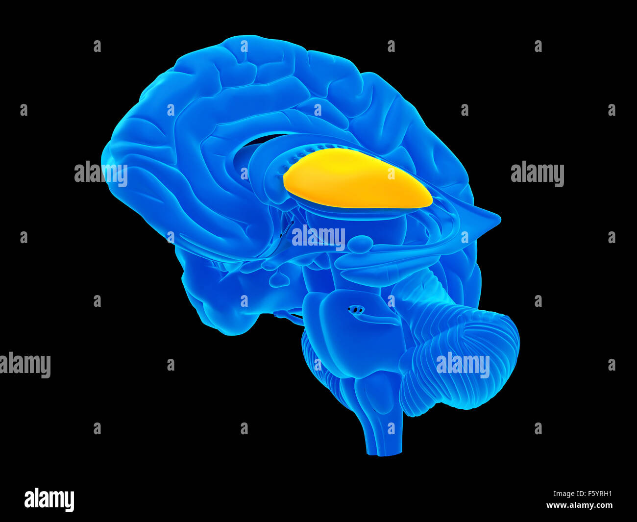 medically accurate illustration of the putamen Stock Photohttps://www.alamy.com/image-license-details/?v=1https://www.alamy.com/stock-photo-medically-accurate-illustration-of-the-putamen-89736333.html
RFF5YRH1–medically accurate illustration of the putamen
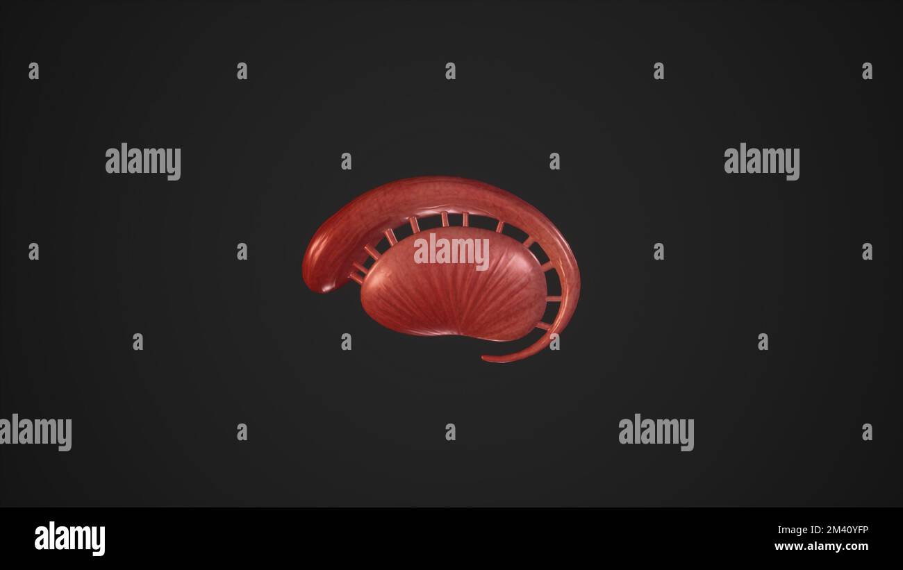 Anatomical Illustration of Corpus Striatum.3d rendering Stock Photohttps://www.alamy.com/image-license-details/?v=1https://www.alamy.com/anatomical-illustration-of-corpus-striatum3d-rendering-image501580906.html
RF2M40YFP–Anatomical Illustration of Corpus Striatum.3d rendering
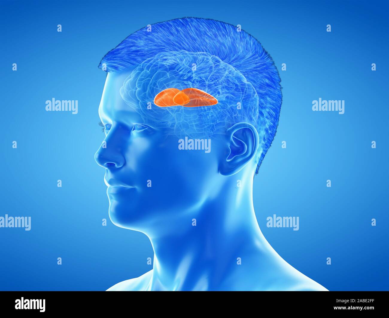 3d rendered medically accurate illustration of the brain anatomy - the putamen Stock Photohttps://www.alamy.com/image-license-details/?v=1https://www.alamy.com/3d-rendered-medically-accurate-illustration-of-the-brain-anatomy-the-putamen-image334067539.html
RF2ABE2FF–3d rendered medically accurate illustration of the brain anatomy - the putamen
 Ripe apricots with green leaves on a ceramic plate on a wooden table. Stock Photohttps://www.alamy.com/image-license-details/?v=1https://www.alamy.com/ripe-apricots-with-green-leaves-on-a-ceramic-plate-on-a-wooden-table-image366654900.html
RF2C8EG1T–Ripe apricots with green leaves on a ceramic plate on a wooden table.
 Connections between the substancia nigra and the putamen in a Parkinson's disease Stock Photohttps://www.alamy.com/image-license-details/?v=1https://www.alamy.com/stock-photo-connections-between-the-substancia-nigra-and-the-putamen-in-a-parkinsons-87046651.html
RFF1H8TY–Connections between the substancia nigra and the putamen in a Parkinson's disease
 Succinea putamen. 20 Jan 20161 Succinea putamen Stock Photohttps://www.alamy.com/image-license-details/?v=1https://www.alamy.com/succinea-putamen-20-jan-20161-succinea-putamen-image352204978.html
RM2BD0916–Succinea putamen. 20 Jan 20161 Succinea putamen
 Connections between putamen and frontal lobe in a Parkinson's patient brain Stock Photohttps://www.alamy.com/image-license-details/?v=1https://www.alamy.com/stock-photo-connections-between-putamen-and-frontal-lobe-in-a-parkinsons-patient-87046647.html
RFF1H8TR–Connections between putamen and frontal lobe in a Parkinson's patient brain
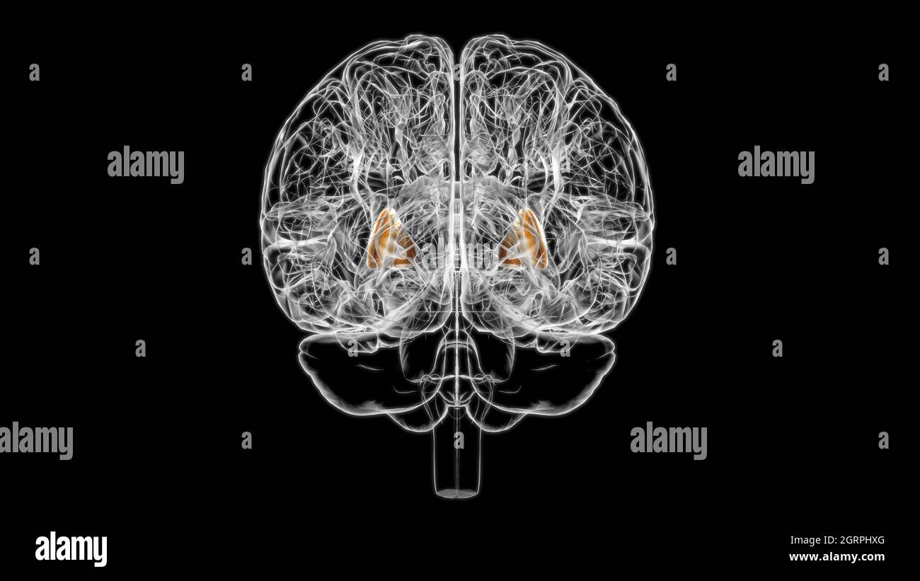 Brain putamen Anatomy For Medical Concept 3D Illustration Stock Photohttps://www.alamy.com/image-license-details/?v=1https://www.alamy.com/brain-putamen-anatomy-for-medical-concept-3d-illustration-image444893304.html
RF2GRPHXG–Brain putamen Anatomy For Medical Concept 3D Illustration
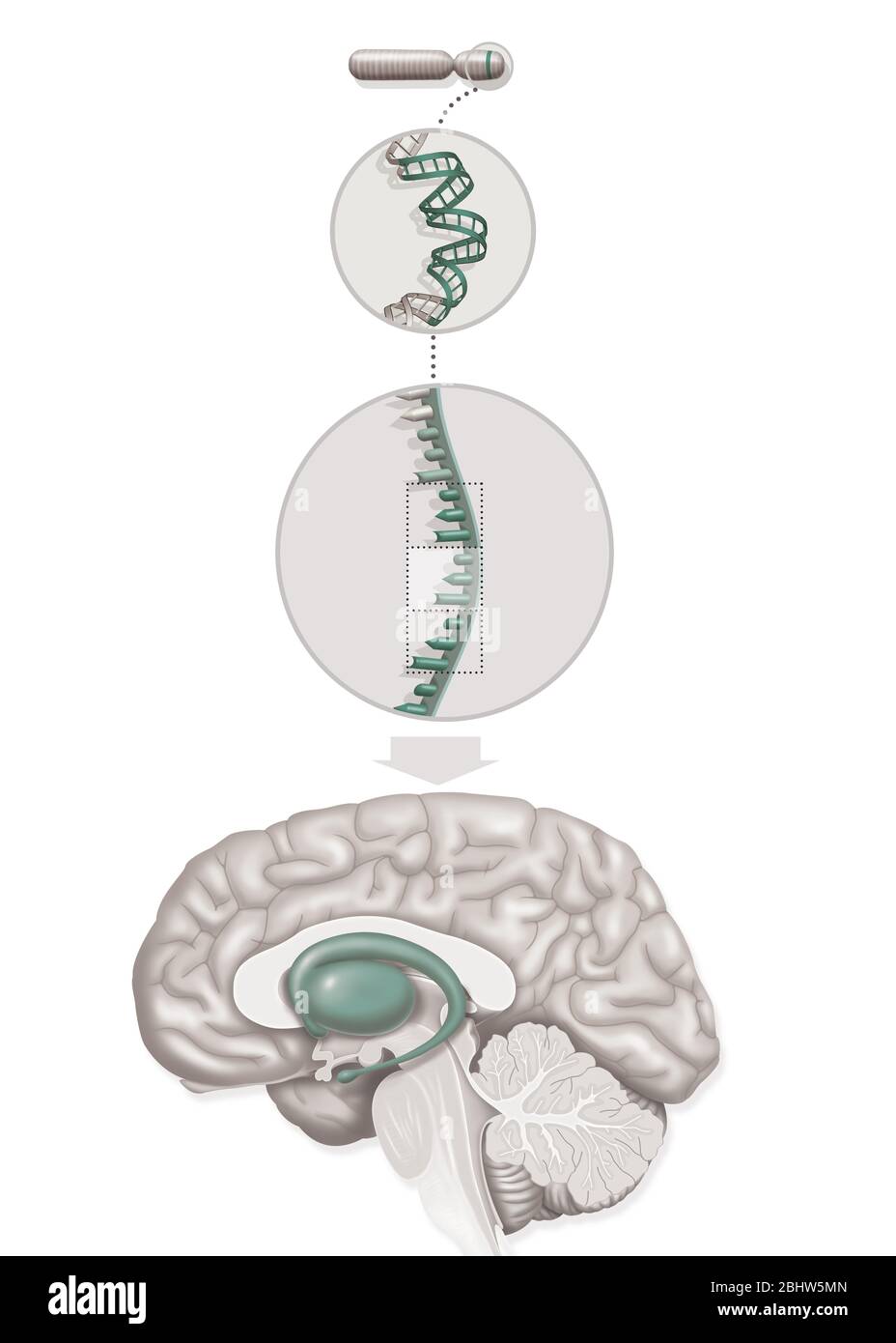 Medical illustration representing Huntington's disease, a genetic disorder involving the HD gene (Huntington Disease) of chromosome 4 with abnormal re Stock Photohttps://www.alamy.com/image-license-details/?v=1https://www.alamy.com/medical-illustration-representing-huntingtons-disease-a-genetic-disorder-involving-the-hd-gene-huntington-disease-of-chromosome-4-with-abnormal-re-image355209813.html
RM2BHW5MN–Medical illustration representing Huntington's disease, a genetic disorder involving the HD gene (Huntington Disease) of chromosome 4 with abnormal re
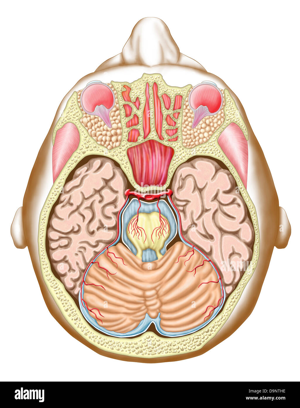 Transverse section of the midbrain. Stock Photohttps://www.alamy.com/image-license-details/?v=1https://www.alamy.com/stock-photo-transverse-section-of-the-midbrain-57643306.html
RFD9NTHE–Transverse section of the midbrain.
 Longan sweet tropical fruit whole and half. Stock Vectorhttps://www.alamy.com/image-license-details/?v=1https://www.alamy.com/longan-sweet-tropical-fruit-whole-and-half-image334901113.html
RF2ACT1P1–Longan sweet tropical fruit whole and half.
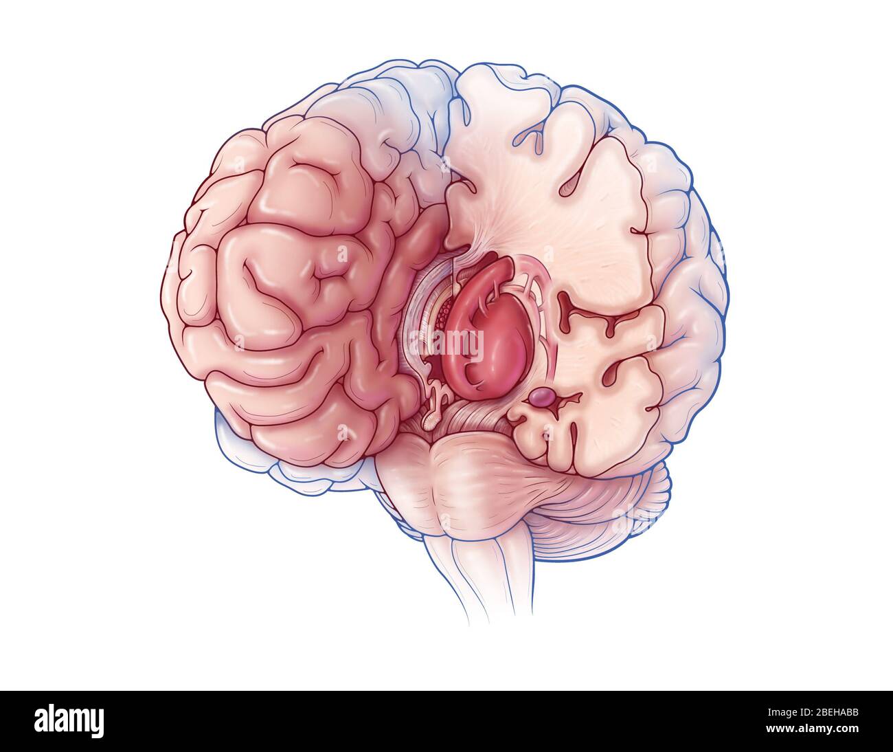 Limbic System, Illustration Stock Photohttps://www.alamy.com/image-license-details/?v=1https://www.alamy.com/limbic-system-illustration-image353193887.html
RM2BEHABB–Limbic System, Illustration
 Big egg standing in small straw nest Stock Photohttps://www.alamy.com/image-license-details/?v=1https://www.alamy.com/big-egg-standing-in-small-straw-nest-image363118938.html
RF2C2NDWE–Big egg standing in small straw nest
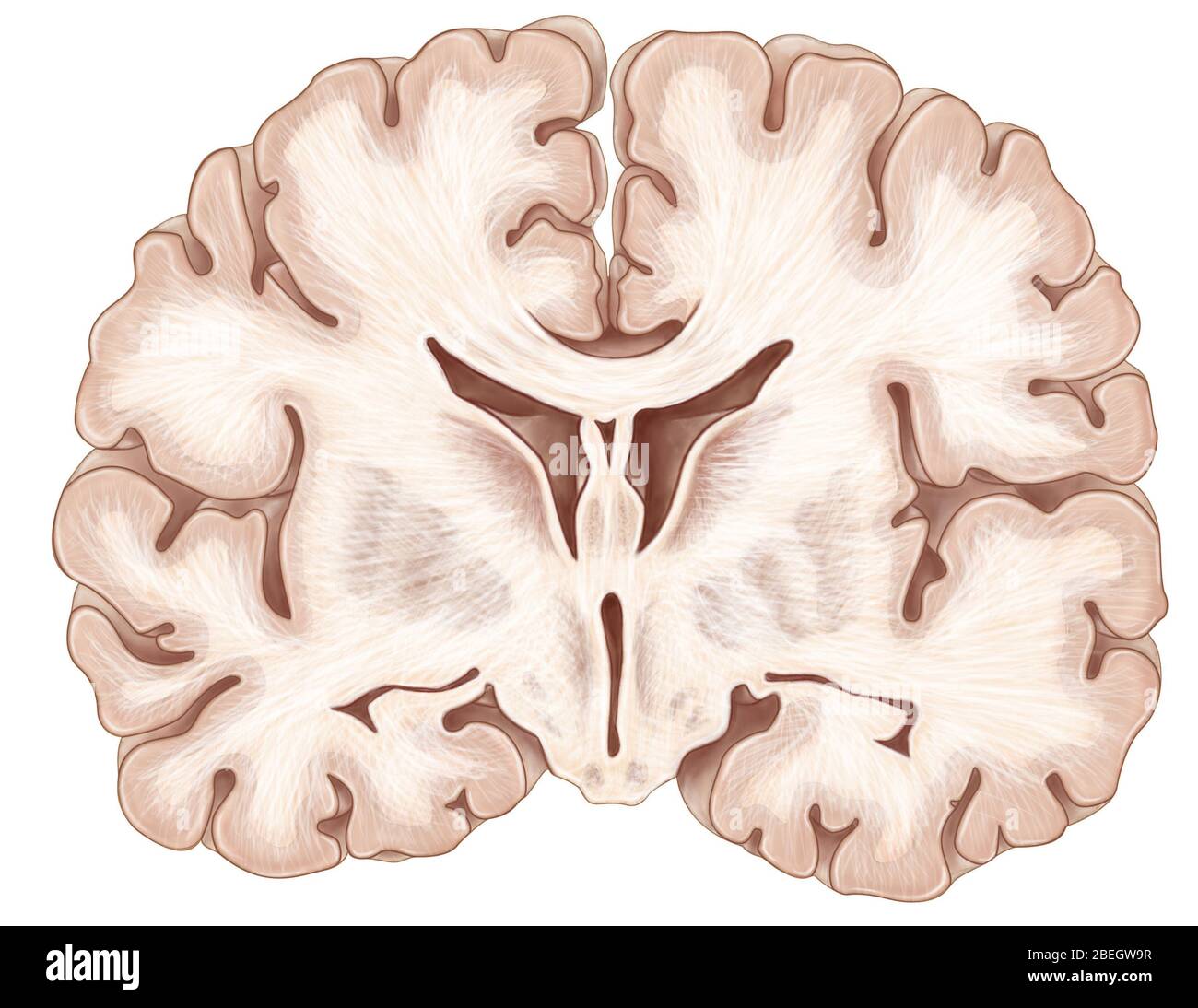 Brain, Coronal Section Stock Photohttps://www.alamy.com/image-license-details/?v=1https://www.alamy.com/brain-coronal-section-image353183651.html
RM2BEGW9R–Brain, Coronal Section
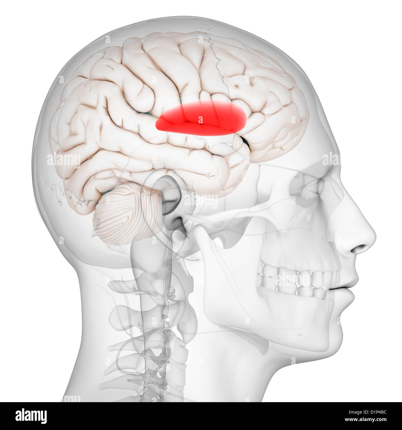 Putamen, artwork Stock Photohttps://www.alamy.com/image-license-details/?v=1https://www.alamy.com/stock-photo-putamen-artwork-52732160.html
RFD1P4BC–Putamen, artwork
 3d rendered medically accurate illustration of a female brains anatomy- the putamen Stock Photohttps://www.alamy.com/image-license-details/?v=1https://www.alamy.com/3d-rendered-medically-accurate-illustration-of-a-female-brains-anatomy-the-putamen-image365553328.html
RF2C6MB00–3d rendered medically accurate illustration of a female brains anatomy- the putamen
 Close-up of straw nest. Background or texture Stock Photohttps://www.alamy.com/image-license-details/?v=1https://www.alamy.com/close-up-of-straw-nest-background-or-texture-image363120840.html
RF2C2NG9C–Close-up of straw nest. Background or texture
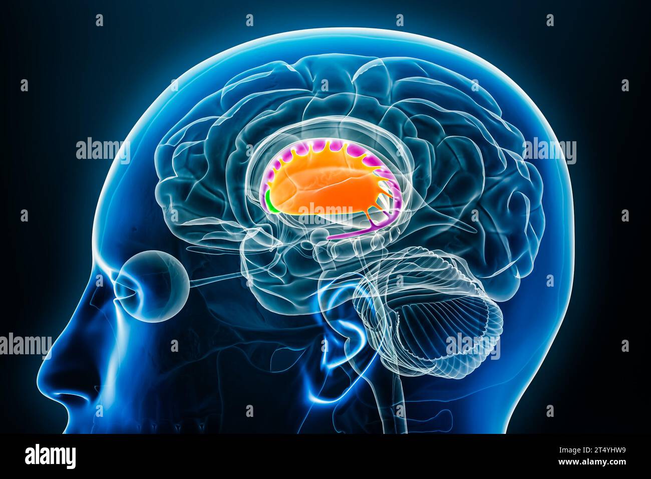 Putamen in orange, nucleus accumbens in green and caudate nucleus in purple 3D rendering illustration with body contours. Human brain, basal ganglia a Stock Photohttps://www.alamy.com/image-license-details/?v=1https://www.alamy.com/putamen-in-orange-nucleus-accumbens-in-green-and-caudate-nucleus-in-purple-3d-rendering-illustration-with-body-contours-human-brain-basal-ganglia-a-image571007509.html
RF2T4YHW9–Putamen in orange, nucleus accumbens in green and caudate nucleus in purple 3D rendering illustration with body contours. Human brain, basal ganglia a
 Mango stone isolated Stock Photohttps://www.alamy.com/image-license-details/?v=1https://www.alamy.com/stock-image-mango-stone-isolated-166713949.html
RFKK6D9H–Mango stone isolated
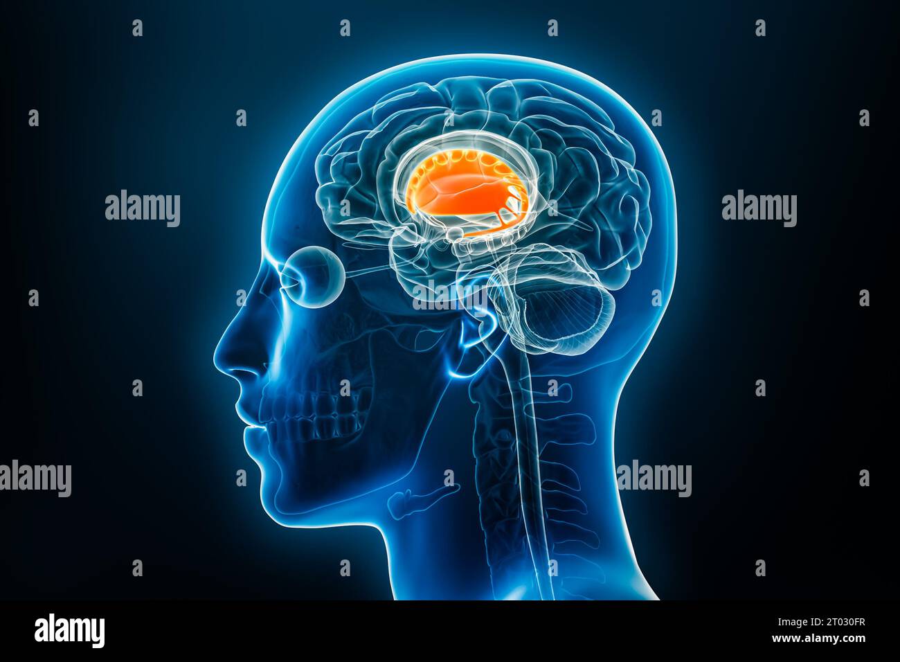 Dorsal striatum with putamen and caudate nucleus or basal ganglia 3D rendering illustration. Human brain and body anatomy, medical, biology, science, Stock Photohttps://www.alamy.com/image-license-details/?v=1https://www.alamy.com/dorsal-striatum-with-putamen-and-caudate-nucleus-or-basal-ganglia-3d-rendering-illustration-human-brain-and-body-anatomy-medical-biology-science-image568008443.html
RF2T030FR–Dorsal striatum with putamen and caudate nucleus or basal ganglia 3D rendering illustration. Human brain and body anatomy, medical, biology, science,
RF2G66890–Nuts icons line outline vector illustration set. Shapes and cross-sections
 Xray lateral or profile view of the putamen 3D rendering illustration with male body contours. Human brain anatomy, medical, biology, science, neurosc Stock Photohttps://www.alamy.com/image-license-details/?v=1https://www.alamy.com/xray-lateral-or-profile-view-of-the-putamen-3d-rendering-illustration-with-male-body-contours-human-brain-anatomy-medical-biology-science-neurosc-image567602760.html
RF2RYCF34–Xray lateral or profile view of the putamen 3D rendering illustration with male body contours. Human brain anatomy, medical, biology, science, neurosc
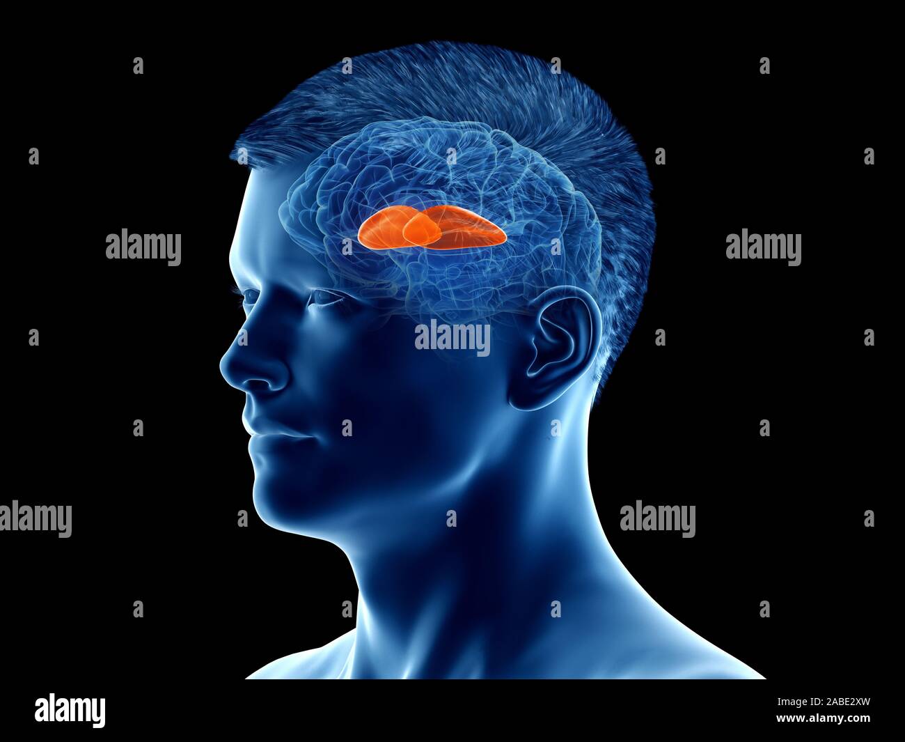 3d rendered medically accurate illustration of the brain anatomy - the putamen Stock Photohttps://www.alamy.com/image-license-details/?v=1https://www.alamy.com/3d-rendered-medically-accurate-illustration-of-the-brain-anatomy-the-putamen-image334067857.html
RF2ABE2XW–3d rendered medically accurate illustration of the brain anatomy - the putamen
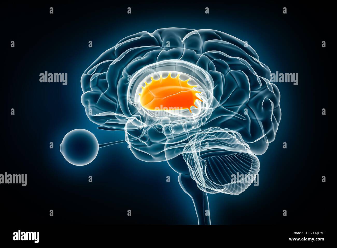 Putamen lateral x-ray view 3D rendering illustration. Human brain and basal ganglia anatomy, medical, healthcare, biology, science, neuroscience, neur Stock Photohttps://www.alamy.com/image-license-details/?v=1https://www.alamy.com/putamen-lateral-x-ray-view-3d-rendering-illustration-human-brain-and-basal-ganglia-anatomy-medical-healthcare-biology-science-neuroscience-neur-image570806083.html
RF2T4JCYF–Putamen lateral x-ray view 3D rendering illustration. Human brain and basal ganglia anatomy, medical, healthcare, biology, science, neuroscience, neur
 3d rendered medically accurate illustration of the putamen Stock Photohttps://www.alamy.com/image-license-details/?v=1https://www.alamy.com/3d-rendered-medically-accurate-illustration-of-the-putamen-image257800884.html
RFTYBRJC–3d rendered medically accurate illustration of the putamen
 Fresh sweet apricots on the wooden table. Stock Photohttps://www.alamy.com/image-license-details/?v=1https://www.alamy.com/fresh-sweet-apricots-on-the-wooden-table-image366485521.html
RF2C86T0H–Fresh sweet apricots on the wooden table.
 Brain precentral gyrus Anatomy For Medical Concept 3D Illustration Stock Photohttps://www.alamy.com/image-license-details/?v=1https://www.alamy.com/brain-precentral-gyrus-anatomy-for-medical-concept-3d-illustration-image444893318.html
RF2GRPHY2–Brain precentral gyrus Anatomy For Medical Concept 3D Illustration
 Medical illustration representing Huntington's disease, a genetic disorder involving the HD gene (Huntington Disease) of chromosome 4 with abnormal re Stock Photohttps://www.alamy.com/image-license-details/?v=1https://www.alamy.com/medical-illustration-representing-huntingtons-disease-a-genetic-disorder-involving-the-hd-gene-huntington-disease-of-chromosome-4-with-abnormal-re-image355209799.html
RM2BHW5M7–Medical illustration representing Huntington's disease, a genetic disorder involving the HD gene (Huntington Disease) of chromosome 4 with abnormal re
 Three fresh plums on white background - one opened Stock Photohttps://www.alamy.com/image-license-details/?v=1https://www.alamy.com/stock-photo-three-fresh-plums-on-white-background-one-opened-32911796.html
RFBWF798–Three fresh plums on white background - one opened
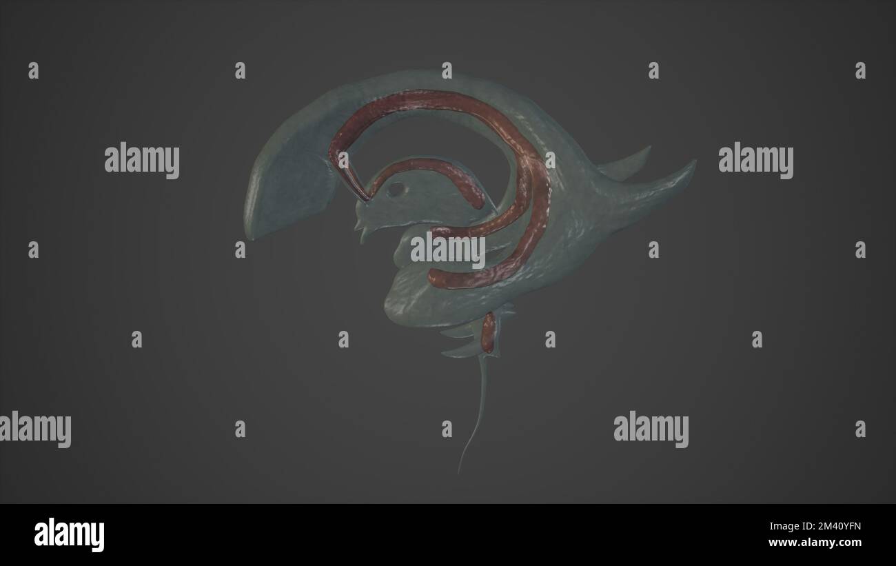 Anatomical Illustration of Choroid Plexuses.3d rendering Stock Photohttps://www.alamy.com/image-license-details/?v=1https://www.alamy.com/anatomical-illustration-of-choroid-plexuses3d-rendering-image501580905.html
RF2M40YFN–Anatomical Illustration of Choroid Plexuses.3d rendering
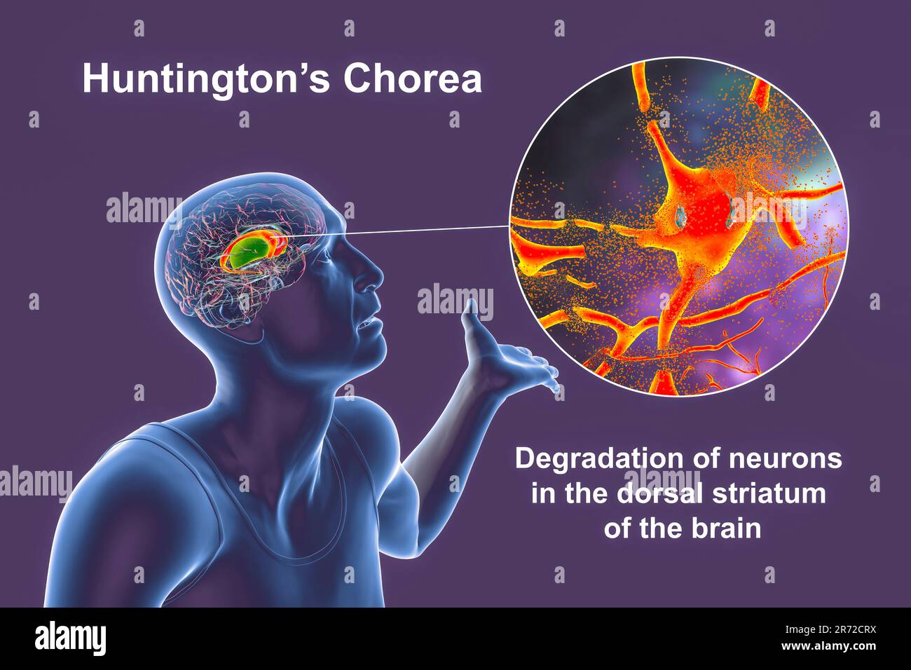 Dorsal striatum, caudate nucleus and putamen, highlighted in the brain of a person with Huntington's disease and close-up view of neuronal degradation Stock Photohttps://www.alamy.com/image-license-details/?v=1https://www.alamy.com/dorsal-striatum-caudate-nucleus-and-putamen-highlighted-in-the-brain-of-a-person-with-huntingtons-disease-and-close-up-view-of-neuronal-degradation-image555088350.html
RF2R72CRX–Dorsal striatum, caudate nucleus and putamen, highlighted in the brain of a person with Huntington's disease and close-up view of neuronal degradation
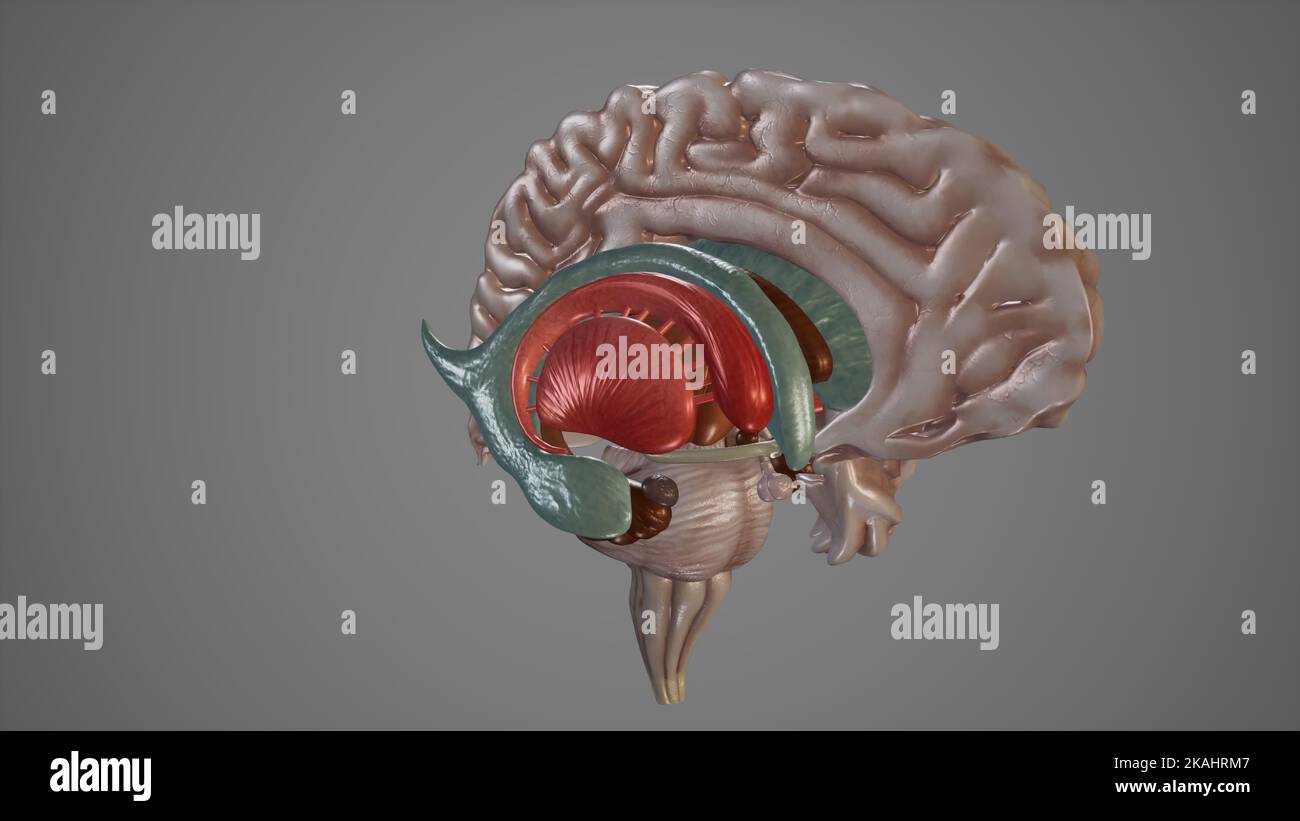 Medical Illustration of Deep Structures of Brain Stock Photohttps://www.alamy.com/image-license-details/?v=1https://www.alamy.com/medical-illustration-of-deep-structures-of-brain-image488428647.html
RF2KAHRM7–Medical Illustration of Deep Structures of Brain
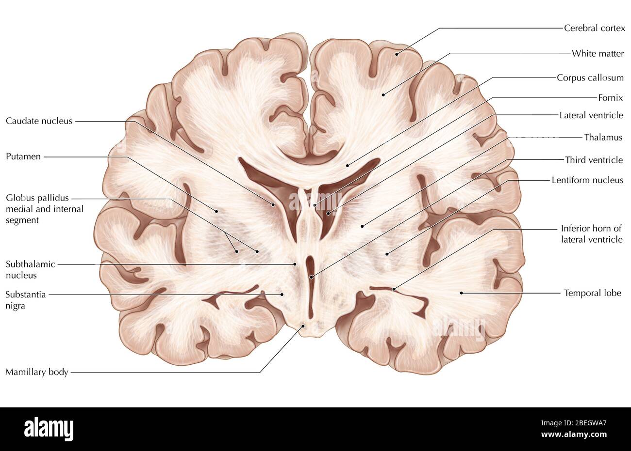 Brain, Coronal Section Stock Photohttps://www.alamy.com/image-license-details/?v=1https://www.alamy.com/brain-coronal-section-image353183663.html
RM2BEGWA7–Brain, Coronal Section
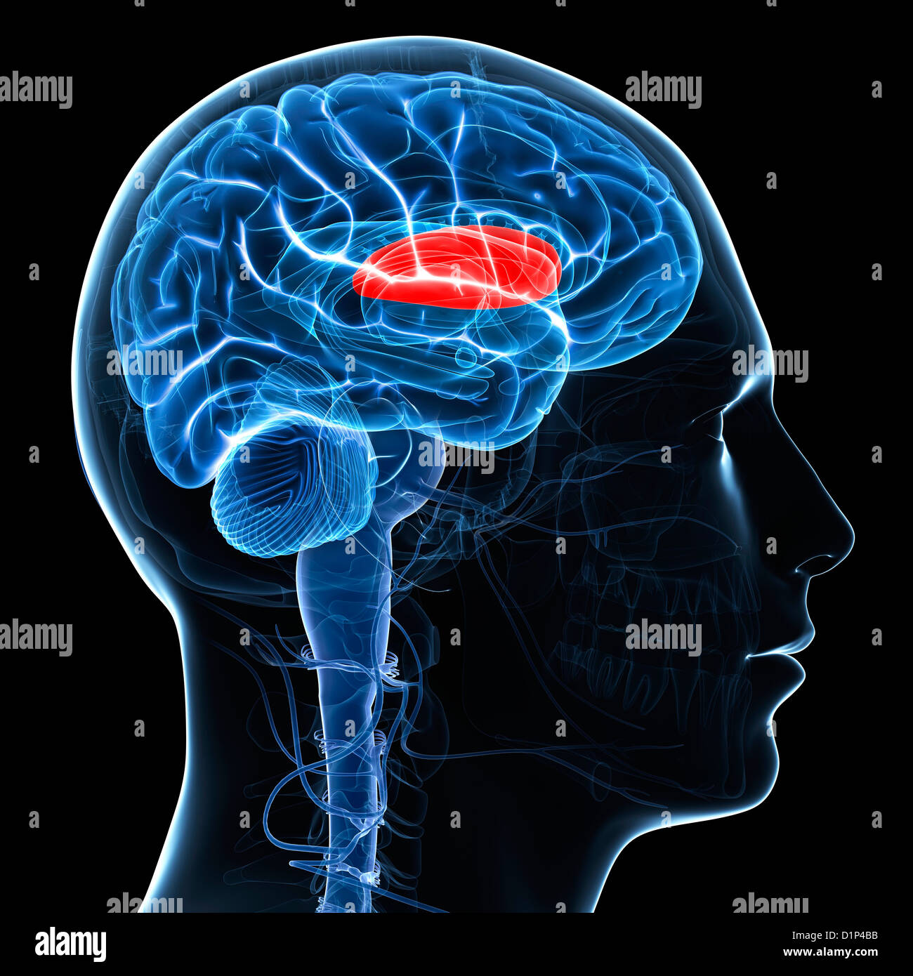 Putamen, artwork Stock Photohttps://www.alamy.com/image-license-details/?v=1https://www.alamy.com/stock-photo-putamen-artwork-52732159.html
RFD1P4BB–Putamen, artwork
 3d rendered medically accurate illustration of young boy brains anatomy- the putamen Stock Photohttps://www.alamy.com/image-license-details/?v=1https://www.alamy.com/3d-rendered-medically-accurate-illustration-of-young-boy-brains-anatomy-the-putamen-image366007939.html
RF2C7D2T3–3d rendered medically accurate illustration of young boy brains anatomy- the putamen
 Big egg in small nest. Top view Stock Photohttps://www.alamy.com/image-license-details/?v=1https://www.alamy.com/stock-photo-big-egg-in-small-nest-top-view-89845978.html
RFF64RCX–Big egg in small nest. Top view
 3d rendered medically accurate illustration of a female brains anatomy- the putamen Stock Photohttps://www.alamy.com/image-license-details/?v=1https://www.alamy.com/3d-rendered-medically-accurate-illustration-of-a-female-brains-anatomy-the-putamen-image365927895.html
RF2C79CNB–3d rendered medically accurate illustration of a female brains anatomy- the putamen
 Mango stone isolated on the white background with clipping path Stock Photohttps://www.alamy.com/image-license-details/?v=1https://www.alamy.com/stock-image-mango-stone-isolated-on-the-white-background-with-clipping-path-165938410.html
RFKHY43P–Mango stone isolated on the white background with clipping path
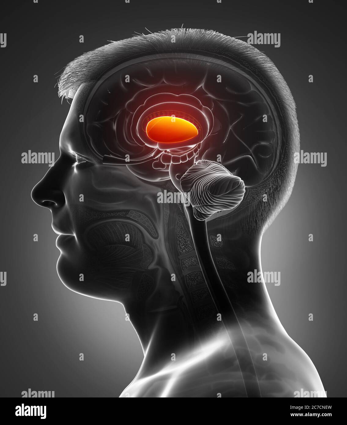 3d rendered medically accurate illustration of a male brains anatomy- the putamen Stock Photohttps://www.alamy.com/image-license-details/?v=1https://www.alamy.com/3d-rendered-medically-accurate-illustration-of-a-male-brains-anatomy-the-putamen-image366000625.html
RF2C7CNEW–3d rendered medically accurate illustration of a male brains anatomy- the putamen
 Big egg standing in small straw nest Stock Photohttps://www.alamy.com/image-license-details/?v=1https://www.alamy.com/stock-photo-big-egg-standing-in-small-straw-nest-89877929.html
RFF66861–Big egg standing in small straw nest
 Illustrative image depicting a hypertensive hemorrhage in the basal ganglia region of the brain. Stock Vectorhttps://www.alamy.com/image-license-details/?v=1https://www.alamy.com/illustrative-image-depicting-a-hypertensive-hemorrhage-in-the-basal-ganglia-region-of-the-brain-image602701310.html
RF2X0FBJP–Illustrative image depicting a hypertensive hemorrhage in the basal ganglia region of the brain.
 Close-up of straw nest. Background or texture Stock Photohttps://www.alamy.com/image-license-details/?v=1https://www.alamy.com/stock-photo-close-up-of-straw-nest-background-or-texture-89586261.html
RFF5N059–Close-up of straw nest. Background or texture
 Putamen x-ray profile close-up view 3D rendering illustration with body contours. Human brain and basal ganglia anatomy, medical, biology, science, ne Stock Photohttps://www.alamy.com/image-license-details/?v=1https://www.alamy.com/putamen-x-ray-profile-close-up-view-3d-rendering-illustration-with-body-contours-human-brain-and-basal-ganglia-anatomy-medical-biology-science-ne-image570806103.html
RF2T4JD07–Putamen x-ray profile close-up view 3D rendering illustration with body contours. Human brain and basal ganglia anatomy, medical, biology, science, ne
 3d rendered medically accurate illustration of the putamen Stock Photohttps://www.alamy.com/image-license-details/?v=1https://www.alamy.com/3d-rendered-medically-accurate-illustration-of-the-putamen-image257800087.html
RFTYBPHY–3d rendered medically accurate illustration of the putamen
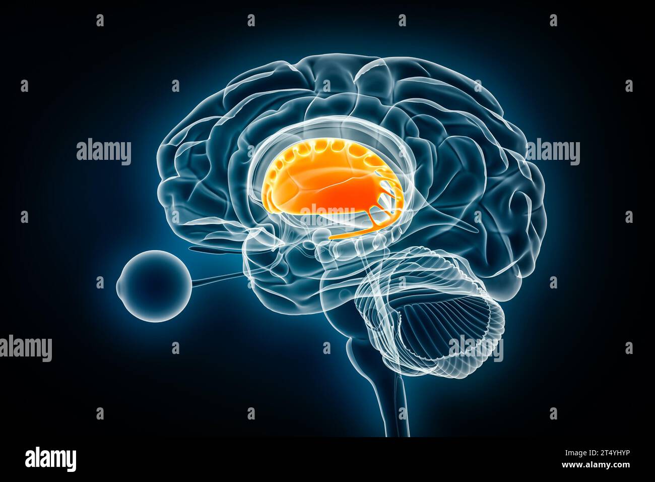 Corpus striatum profile x-ray view 3D rendering illustration. Human brain and basal ganglia anatomy, medical, healthcare, biology, science, neuroscien Stock Photohttps://www.alamy.com/image-license-details/?v=1https://www.alamy.com/corpus-striatum-profile-x-ray-view-3d-rendering-illustration-human-brain-and-basal-ganglia-anatomy-medical-healthcare-biology-science-neuroscien-image571007578.html
RF2T4YHYP–Corpus striatum profile x-ray view 3D rendering illustration. Human brain and basal ganglia anatomy, medical, healthcare, biology, science, neuroscien
 Ripe apricots with green leaves on a ceramic plate on a wooden table. Stock Photohttps://www.alamy.com/image-license-details/?v=1https://www.alamy.com/ripe-apricots-with-green-leaves-on-a-ceramic-plate-on-a-wooden-table-image366485513.html
RF2C86T09–Ripe apricots with green leaves on a ceramic plate on a wooden table.
 Medical illustration representing Huntington's disease, a genetic disorder involving the HD gene (Huntington Disease) of chromosome 4 with abnormal re Stock Photohttps://www.alamy.com/image-license-details/?v=1https://www.alamy.com/medical-illustration-representing-huntingtons-disease-a-genetic-disorder-involving-the-hd-gene-huntington-disease-of-chromosome-4-with-abnormal-re-image355209820.html
RM2BHW5N0–Medical illustration representing Huntington's disease, a genetic disorder involving the HD gene (Huntington Disease) of chromosome 4 with abnormal re
 Brain occipital lobe Anatomy For Medical Concept 3D Illustration Stock Photohttps://www.alamy.com/image-license-details/?v=1https://www.alamy.com/brain-occipital-lobe-anatomy-for-medical-concept-3d-illustration-image444893305.html
RF2GRPHXH–Brain occipital lobe Anatomy For Medical Concept 3D Illustration
 Three fresh plums on white background - one opened Stock Photohttps://www.alamy.com/image-license-details/?v=1https://www.alamy.com/stock-photo-three-fresh-plums-on-white-background-one-opened-32911713.html
RFBWF769–Three fresh plums on white background - one opened
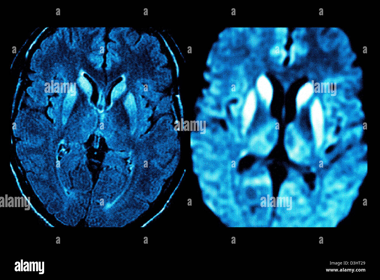 CREUTZFELDT JAKOB DISEASE, MRI Stock Photohttps://www.alamy.com/image-license-details/?v=1https://www.alamy.com/stock-photo-creutzfeldt-jakob-disease-mri-53867137.html
RFD3HT29–CREUTZFELDT JAKOB DISEASE, MRI
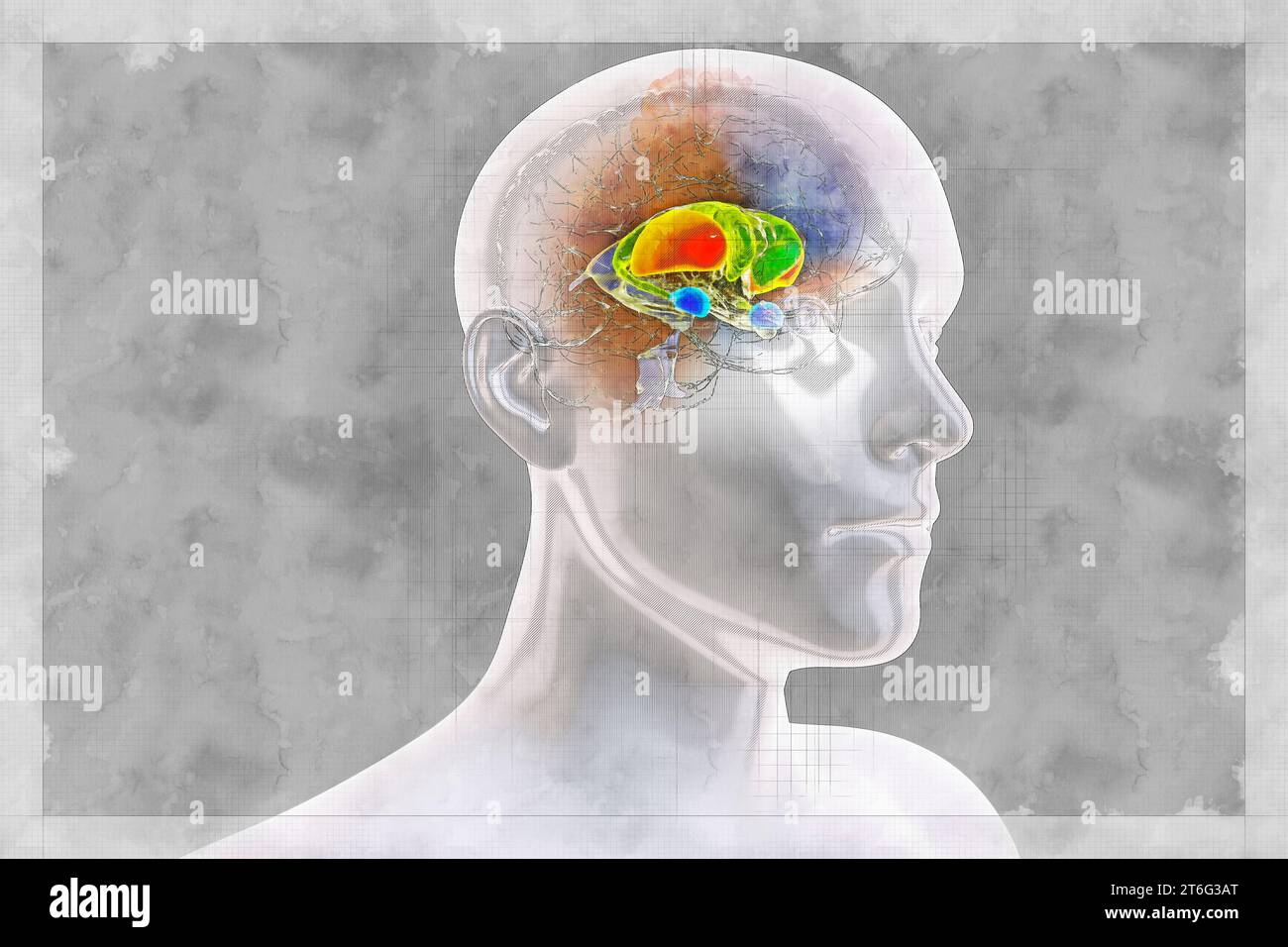 Brain dorsal striatum anatomy, 3D illustration in sketch style. The dorsal striatum consists of the caudate nucleus (green) and the putamen (orange). Stock Photohttps://www.alamy.com/image-license-details/?v=1https://www.alamy.com/brain-dorsal-striatum-anatomy-3d-illustration-in-sketch-style-the-dorsal-striatum-consists-of-the-caudate-nucleus-green-and-the-putamen-orange-image571983968.html
RF2T6G3AT–Brain dorsal striatum anatomy, 3D illustration in sketch style. The dorsal striatum consists of the caudate nucleus (green) and the putamen (orange).
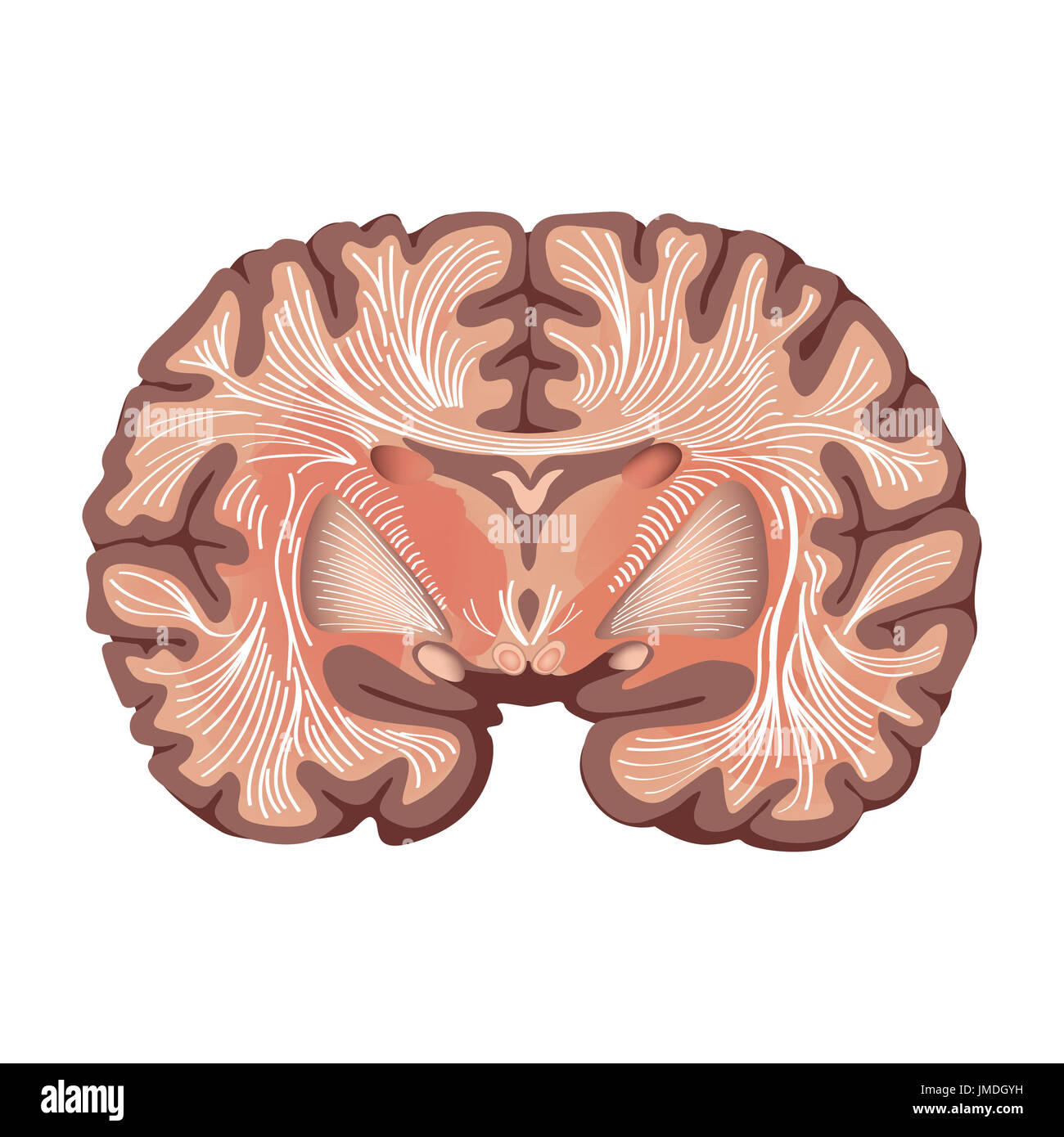 Brain anatomy. Brain showing the basal ganglia and thalamic nuclei isolated on white background. Stock Photohttps://www.alamy.com/image-license-details/?v=1https://www.alamy.com/brain-anatomy-brain-showing-the-basal-ganglia-and-thalamic-nuclei-image150274757.html
RFJMDGYH–Brain anatomy. Brain showing the basal ganglia and thalamic nuclei isolated on white background.
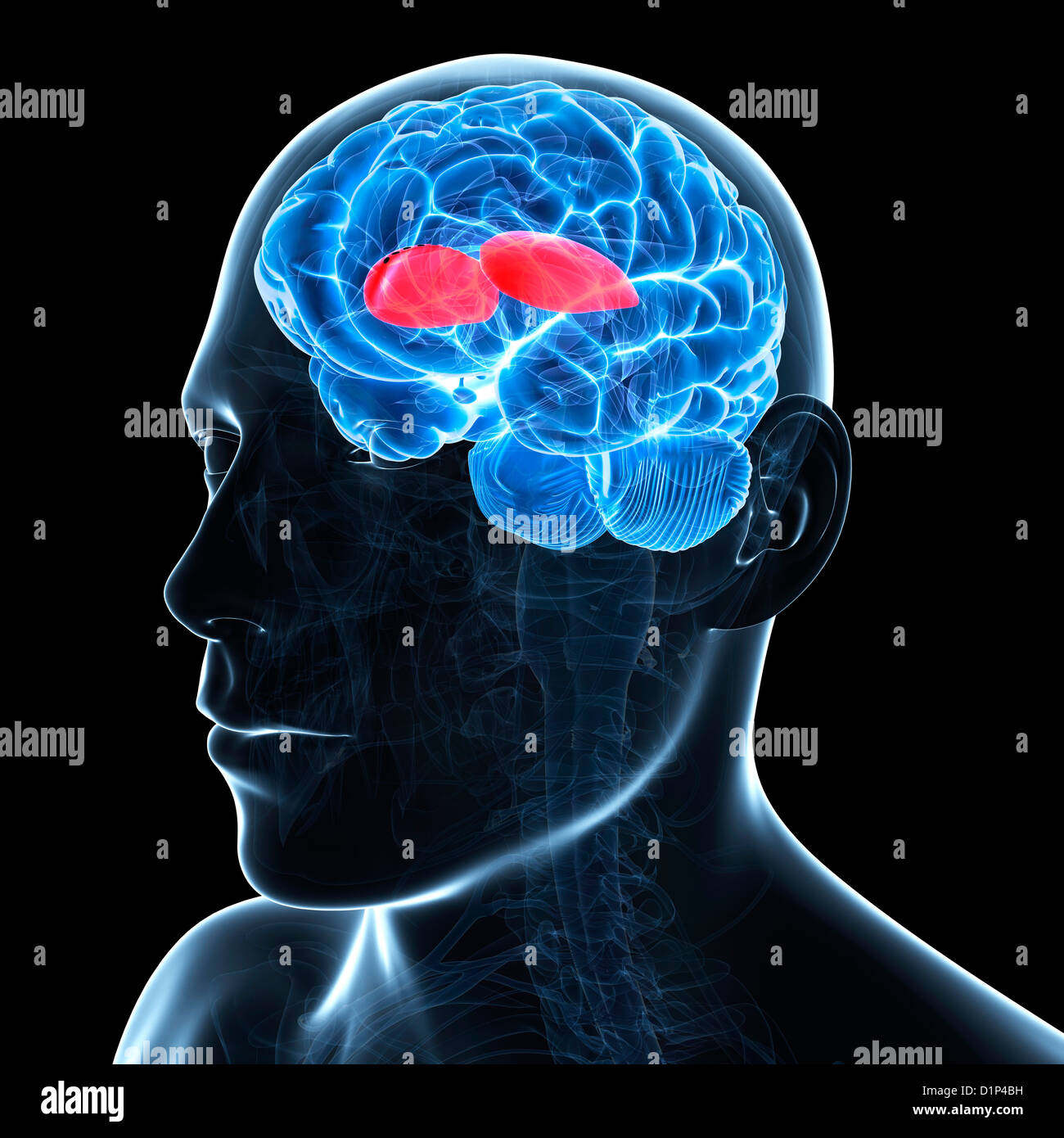 Putamen, artwork Stock Photohttps://www.alamy.com/image-license-details/?v=1https://www.alamy.com/stock-photo-putamen-artwork-52732165.html
RFD1P4BH–Putamen, artwork
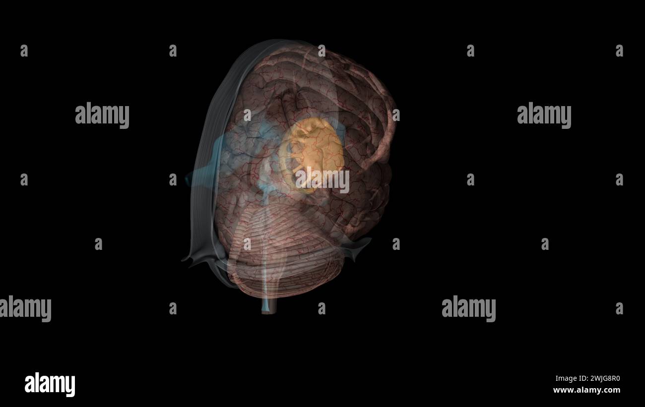 The forebrain structures include the caudate nucleus, the putamen, the nucleus accumbens (or ventral striatum) and the globus pallidus Stock Photohttps://www.alamy.com/image-license-details/?v=1https://www.alamy.com/the-forebrain-structures-include-the-caudate-nucleus-the-putamen-the-nucleus-accumbens-or-ventral-striatum-and-the-globus-pallidus-image596574468.html
RF2WJG8R0–The forebrain structures include the caudate nucleus, the putamen, the nucleus accumbens (or ventral striatum) and the globus pallidus
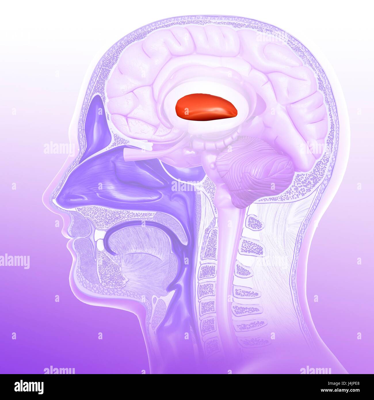 Illustration of the putamen of the human brain. Stock Photohttps://www.alamy.com/image-license-details/?v=1https://www.alamy.com/stock-photo-illustration-of-the-putamen-of-the-human-brain-140554352.html
RFJ4JPE8–Illustration of the putamen of the human brain.
 A brood of Eider (Somateria mollissima) swims away. Two additional females as caregivers (eider creche) Stock Photohttps://www.alamy.com/image-license-details/?v=1https://www.alamy.com/a-brood-of-eider-somateria-mollissima-swims-away-two-additional-females-as-caregivers-eider-creche-image471221260.html
RF2JAHYEM–A brood of Eider (Somateria mollissima) swims away. Two additional females as caregivers (eider creche)
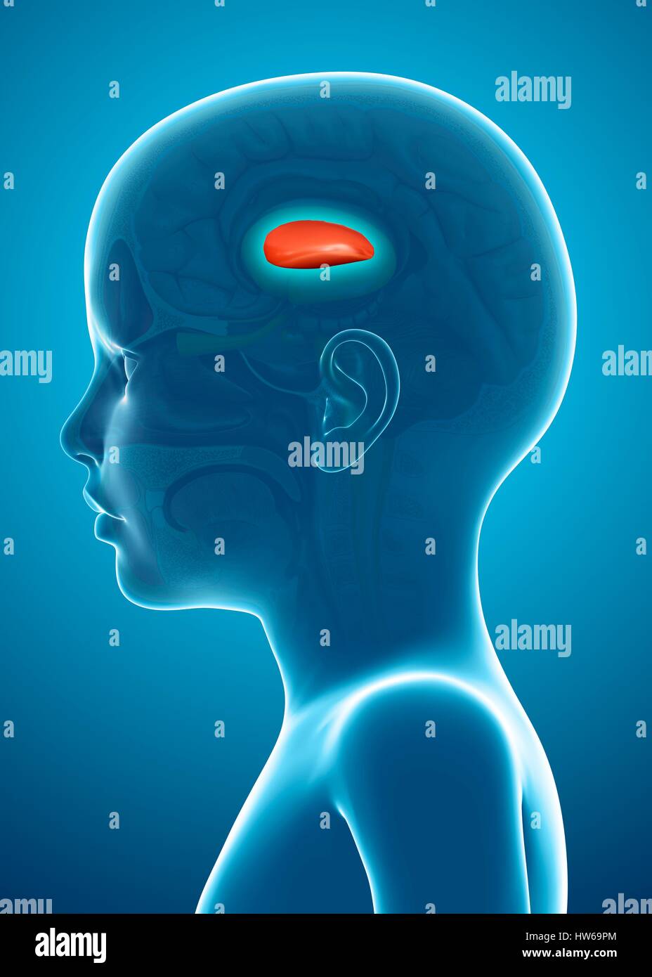 Illustration of the putamen in a child's brain. Stock Photohttps://www.alamy.com/image-license-details/?v=1https://www.alamy.com/stock-photo-illustration-of-the-putamen-in-a-childs-brain-135978380.html
RFHW69PM–Illustration of the putamen in a child's brain.
 3d rendered medically accurate illustration of a female brains anatomy- the putamen Stock Photohttps://www.alamy.com/image-license-details/?v=1https://www.alamy.com/3d-rendered-medically-accurate-illustration-of-a-female-brains-anatomy-the-putamen-image365974704.html
RF2C7BGD4–3d rendered medically accurate illustration of a female brains anatomy- the putamen
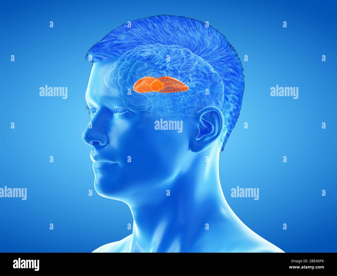 Putamen of the brain, illustration. Stock Photohttps://www.alamy.com/image-license-details/?v=1https://www.alamy.com/putamen-of-the-brain-illustration-image352900791.html
RF2BE40FK–Putamen of the brain, illustration.
 Common Eider chick in the hands of an ornithologist. It will be ringed for dispersion and migration study Stock Photohttps://www.alamy.com/image-license-details/?v=1https://www.alamy.com/common-eider-chick-in-the-hands-of-an-ornithologist-it-will-be-ringed-for-dispersion-and-migration-study-image558403163.html
RF2RCDCX3–Common Eider chick in the hands of an ornithologist. It will be ringed for dispersion and migration study
![Archive image from page 682 of Cunningham's Text-book of anatomy (1914). Cunningham's Text-book of anatomy cunninghamstextb00cunn Year: 1914 ( Caudate nucleus asciculus occipito- frontalis [superior] Internal capsule Putamen Fasciculus longi- tudinals superior Globus / pallidus I— Claustrum Superior operculum Insula Fasciculus occipito- frontalis [inferior] Temporal operculum Anterior commissure Fasciculus uncinatus Fig. 576.—Two Frontal Sections through the Cerebral Hemispheres of an Orang, in the Plane of the Anterior Commissure. A, Section through the left hemisphere in a plane a short Stock Photo Archive image from page 682 of Cunningham's Text-book of anatomy (1914). Cunningham's Text-book of anatomy cunninghamstextb00cunn Year: 1914 ( Caudate nucleus asciculus occipito- frontalis [superior] Internal capsule Putamen Fasciculus longi- tudinals superior Globus / pallidus I— Claustrum Superior operculum Insula Fasciculus occipito- frontalis [inferior] Temporal operculum Anterior commissure Fasciculus uncinatus Fig. 576.—Two Frontal Sections through the Cerebral Hemispheres of an Orang, in the Plane of the Anterior Commissure. A, Section through the left hemisphere in a plane a short Stock Photo](https://c8.alamy.com/comp/W9H6BK/archive-image-from-page-682-of-cunninghams-text-book-of-anatomy-1914-cunninghams-text-book-of-anatomy-cunninghamstextb00cunn-year-1914-caudate-nucleus-asciculus-occipito-frontalis-superior-internal-capsule-putamen-fasciculus-longi-tudinals-superior-globus-pallidus-i-claustrum-superior-operculum-insula-fasciculus-occipito-frontalis-inferior-temporal-operculum-anterior-commissure-fasciculus-uncinatus-fig-576two-frontal-sections-through-the-cerebral-hemispheres-of-an-orang-in-the-plane-of-the-anterior-commissure-a-section-through-the-left-hemisphere-in-a-plane-a-short-W9H6BK.jpg) Archive image from page 682 of Cunningham's Text-book of anatomy (1914). Cunningham's Text-book of anatomy cunninghamstextb00cunn Year: 1914 ( Caudate nucleus asciculus occipito- frontalis [superior] Internal capsule Putamen Fasciculus longi- tudinals superior Globus / pallidus I— Claustrum Superior operculum Insula Fasciculus occipito- frontalis [inferior] Temporal operculum Anterior commissure Fasciculus uncinatus Fig. 576.—Two Frontal Sections through the Cerebral Hemispheres of an Orang, in the Plane of the Anterior Commissure. A, Section through the left hemisphere in a plane a short Stock Photohttps://www.alamy.com/image-license-details/?v=1https://www.alamy.com/archive-image-from-page-682-of-cunninghams-text-book-of-anatomy-1914-cunninghams-text-book-of-anatomy-cunninghamstextb00cunn-year-1914-caudate-nucleus-asciculus-occipito-frontalis-superior-internal-capsule-putamen-fasciculus-longi-tudinals-superior-globus-pallidus-i-claustrum-superior-operculum-insula-fasciculus-occipito-frontalis-inferior-temporal-operculum-anterior-commissure-fasciculus-uncinatus-fig-576two-frontal-sections-through-the-cerebral-hemispheres-of-an-orang-in-the-plane-of-the-anterior-commissure-a-section-through-the-left-hemisphere-in-a-plane-a-short-image264065639.html
Archive image from page 682 of Cunningham's Text-book of anatomy (1914). Cunningham's Text-book of anatomy cunninghamstextb00cunn Year: 1914 ( Caudate nucleus asciculus occipito- frontalis [superior] Internal capsule Putamen Fasciculus longi- tudinals superior Globus / pallidus I— Claustrum Superior operculum Insula Fasciculus occipito- frontalis [inferior] Temporal operculum Anterior commissure Fasciculus uncinatus Fig. 576.—Two Frontal Sections through the Cerebral Hemispheres of an Orang, in the Plane of the Anterior Commissure. A, Section through the left hemisphere in a plane a short Stock Photohttps://www.alamy.com/image-license-details/?v=1https://www.alamy.com/archive-image-from-page-682-of-cunninghams-text-book-of-anatomy-1914-cunninghams-text-book-of-anatomy-cunninghamstextb00cunn-year-1914-caudate-nucleus-asciculus-occipito-frontalis-superior-internal-capsule-putamen-fasciculus-longi-tudinals-superior-globus-pallidus-i-claustrum-superior-operculum-insula-fasciculus-occipito-frontalis-inferior-temporal-operculum-anterior-commissure-fasciculus-uncinatus-fig-576two-frontal-sections-through-the-cerebral-hemispheres-of-an-orang-in-the-plane-of-the-anterior-commissure-a-section-through-the-left-hemisphere-in-a-plane-a-short-image264065639.htmlRMW9H6BK–Archive image from page 682 of Cunningham's Text-book of anatomy (1914). Cunningham's Text-book of anatomy cunninghamstextb00cunn Year: 1914 ( Caudate nucleus asciculus occipito- frontalis [superior] Internal capsule Putamen Fasciculus longi- tudinals superior Globus / pallidus I— Claustrum Superior operculum Insula Fasciculus occipito- frontalis [inferior] Temporal operculum Anterior commissure Fasciculus uncinatus Fig. 576.—Two Frontal Sections through the Cerebral Hemispheres of an Orang, in the Plane of the Anterior Commissure. A, Section through the left hemisphere in a plane a short
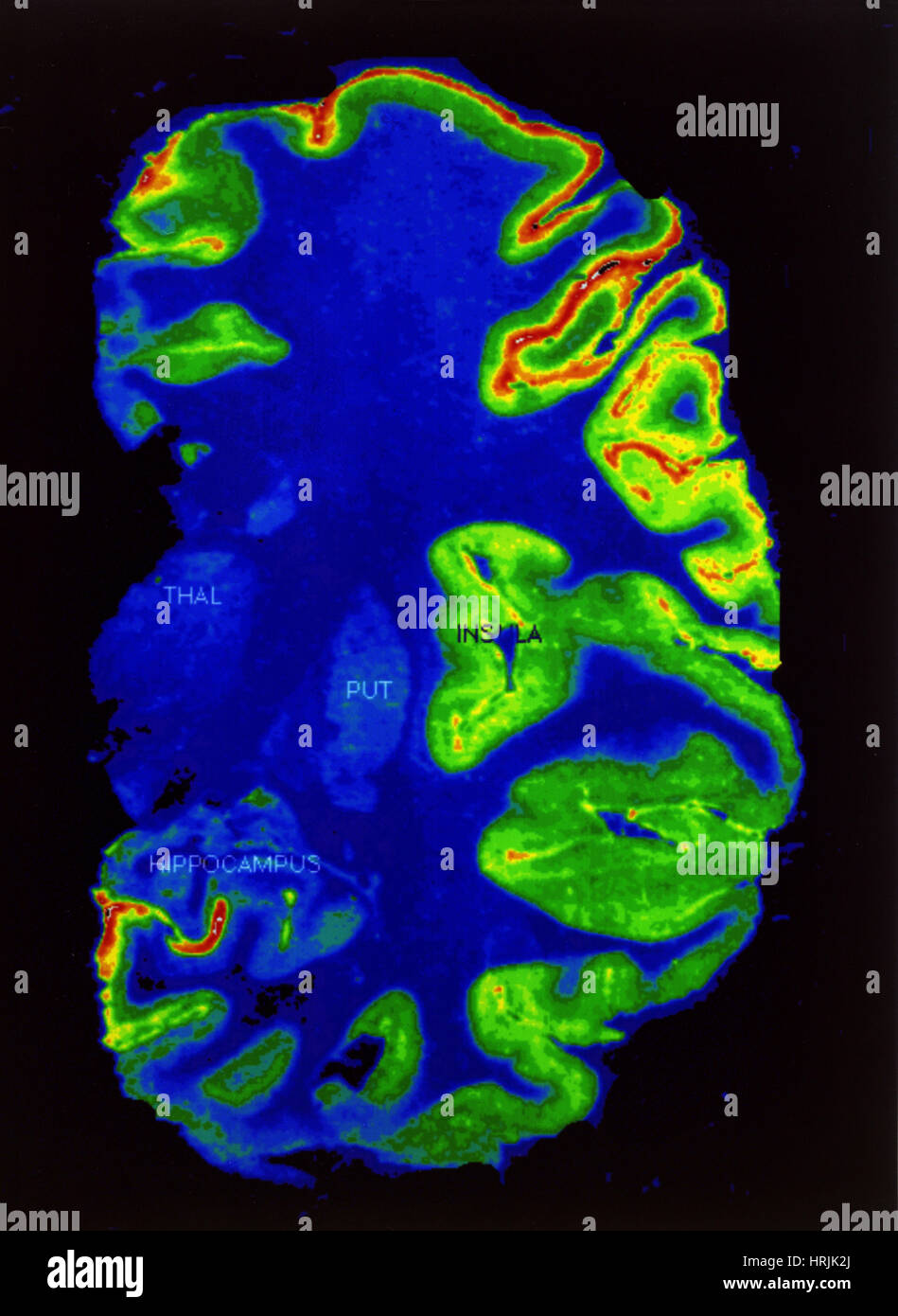 PET Scan, Healthy Brain Stock Photohttps://www.alamy.com/image-license-details/?v=1https://www.alamy.com/stock-photo-pet-scan-healthy-brain-135019770.html
RMHRJK2J–PET Scan, Healthy Brain
 3d rendered medically accurate illustration of the putamen Stock Photohttps://www.alamy.com/image-license-details/?v=1https://www.alamy.com/3d-rendered-medically-accurate-illustration-of-the-putamen-image257799674.html
RFTYBP36–3d rendered medically accurate illustration of the putamen
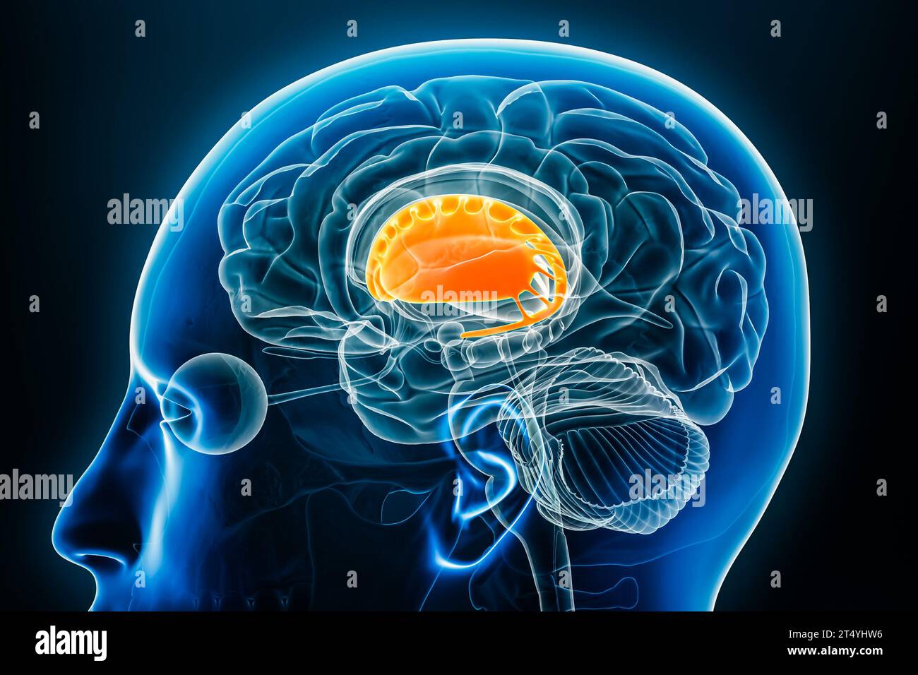 Corpus striatum x-ray profile close-up view 3D rendering illustration with body contours. Human brain and basal ganglia anatomy, medical, biology, sci Stock Photohttps://www.alamy.com/image-license-details/?v=1https://www.alamy.com/corpus-striatum-x-ray-profile-close-up-view-3d-rendering-illustration-with-body-contours-human-brain-and-basal-ganglia-anatomy-medical-biology-sci-image571007506.html
RF2T4YHW6–Corpus striatum x-ray profile close-up view 3D rendering illustration with body contours. Human brain and basal ganglia anatomy, medical, biology, sci
 Fresh sweet apricots on the wooden table. Stock Photohttps://www.alamy.com/image-license-details/?v=1https://www.alamy.com/fresh-sweet-apricots-on-the-wooden-table-image366485570.html
RF2C86T2A–Fresh sweet apricots on the wooden table.
 peach Stock Photohttps://www.alamy.com/image-license-details/?v=1https://www.alamy.com/peach-image226154036.html
RFR3X5NT–peach
 Ripe apricots with green leaves on a ceramic plate on a wooden table. Stock Photohttps://www.alamy.com/image-license-details/?v=1https://www.alamy.com/ripe-apricots-with-green-leaves-on-a-ceramic-plate-on-a-wooden-table-image366485499.html
RF2C86RYR–Ripe apricots with green leaves on a ceramic plate on a wooden table.
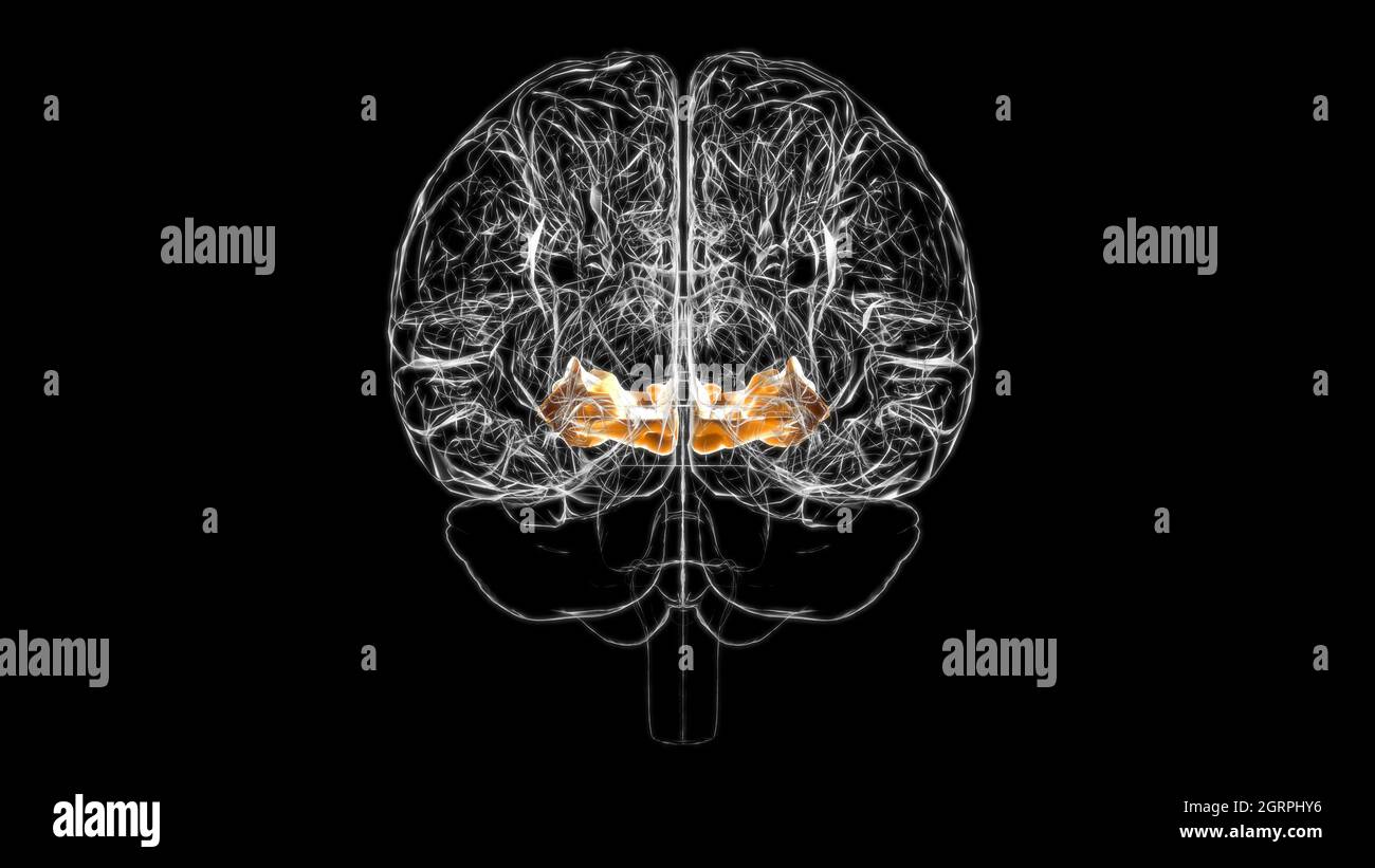 Brain Orbital gyrus Anatomy For Medical Concept 3D Illustration Stock Photohttps://www.alamy.com/image-license-details/?v=1https://www.alamy.com/brain-orbital-gyrus-anatomy-for-medical-concept-3d-illustration-image444893322.html
RF2GRPHY6–Brain Orbital gyrus Anatomy For Medical Concept 3D Illustration
 Three fresh plums on white background - one opened Stock Photohttps://www.alamy.com/image-license-details/?v=1https://www.alamy.com/stock-photo-three-fresh-plums-on-white-background-one-opened-32911756.html
RFBWF77T–Three fresh plums on white background - one opened
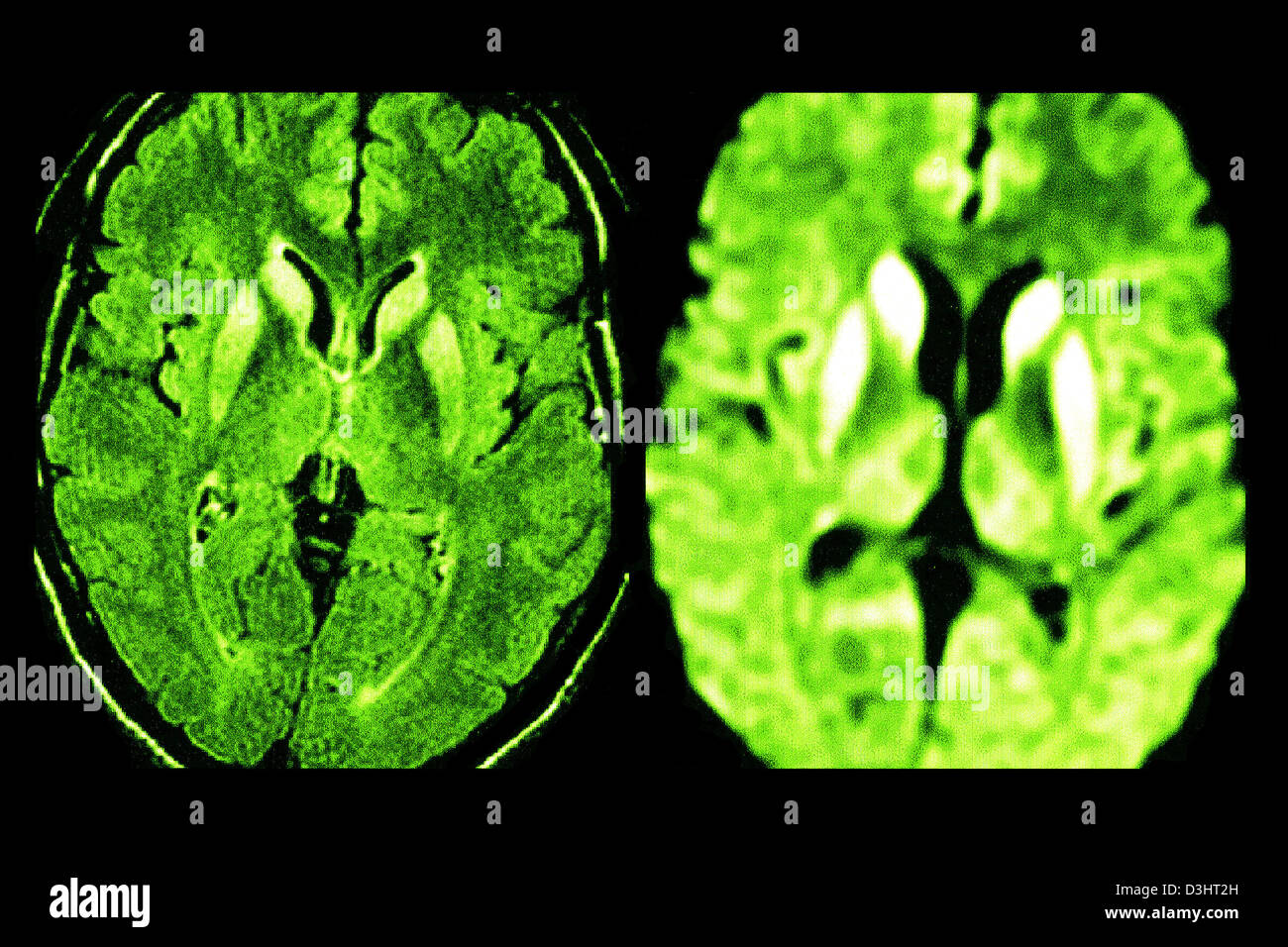 CREUTZFELDT JAKOB DISEASE, MRI Stock Photohttps://www.alamy.com/image-license-details/?v=1https://www.alamy.com/stock-photo-creutzfeldt-jakob-disease-mri-53867145.html
RFD3HT2H–CREUTZFELDT JAKOB DISEASE, MRI
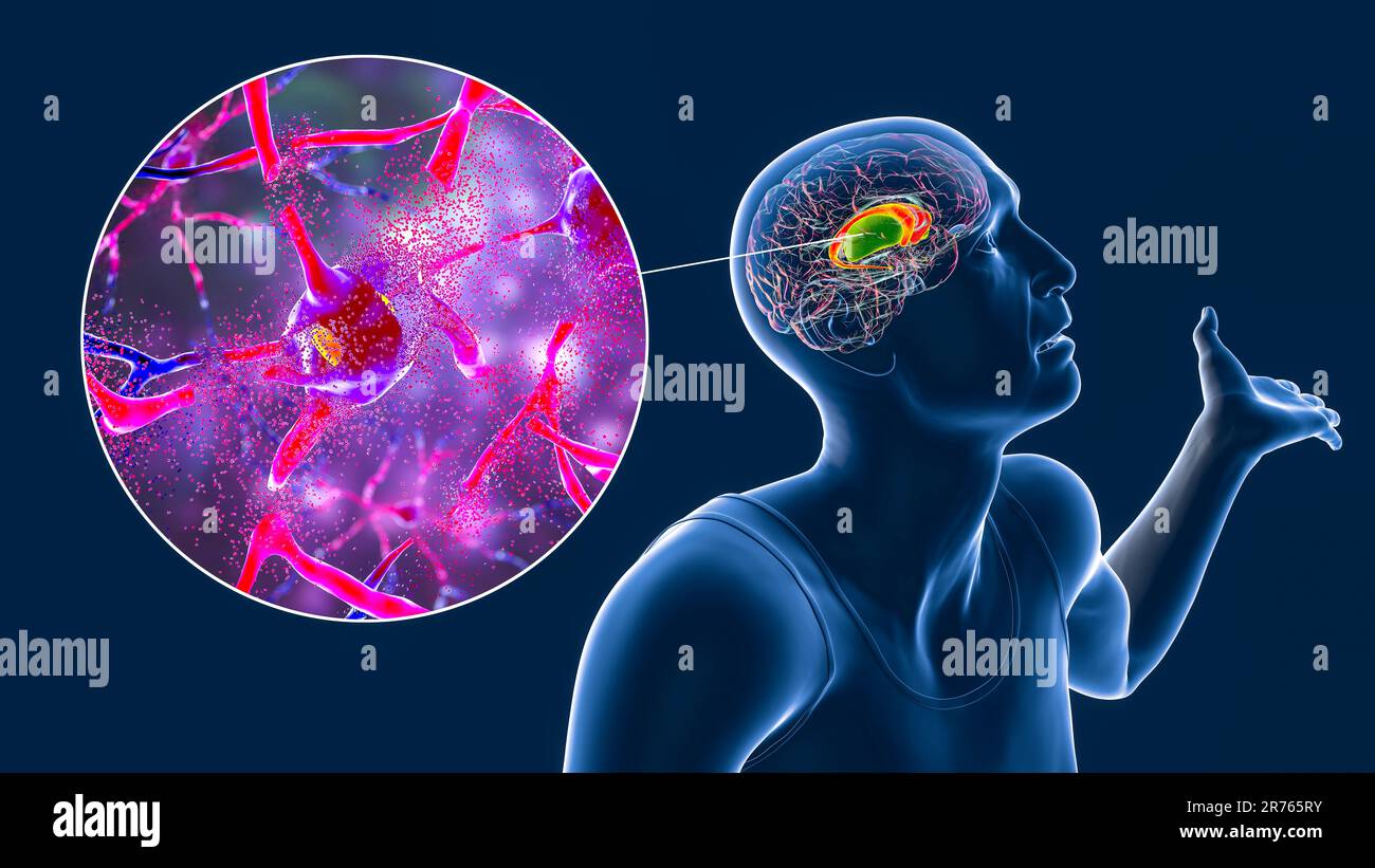 Dorsal striatum, caudate nucleus and putamen, highlighted in the brain of a person with Huntington's disease with involuntary movements, and close-up Stock Photohttps://www.alamy.com/image-license-details/?v=1https://www.alamy.com/dorsal-striatum-caudate-nucleus-and-putamen-highlighted-in-the-brain-of-a-person-with-huntingtons-disease-with-involuntary-movements-and-close-up-image555170671.html
RF2R765RY–Dorsal striatum, caudate nucleus and putamen, highlighted in the brain of a person with Huntington's disease with involuntary movements, and close-up
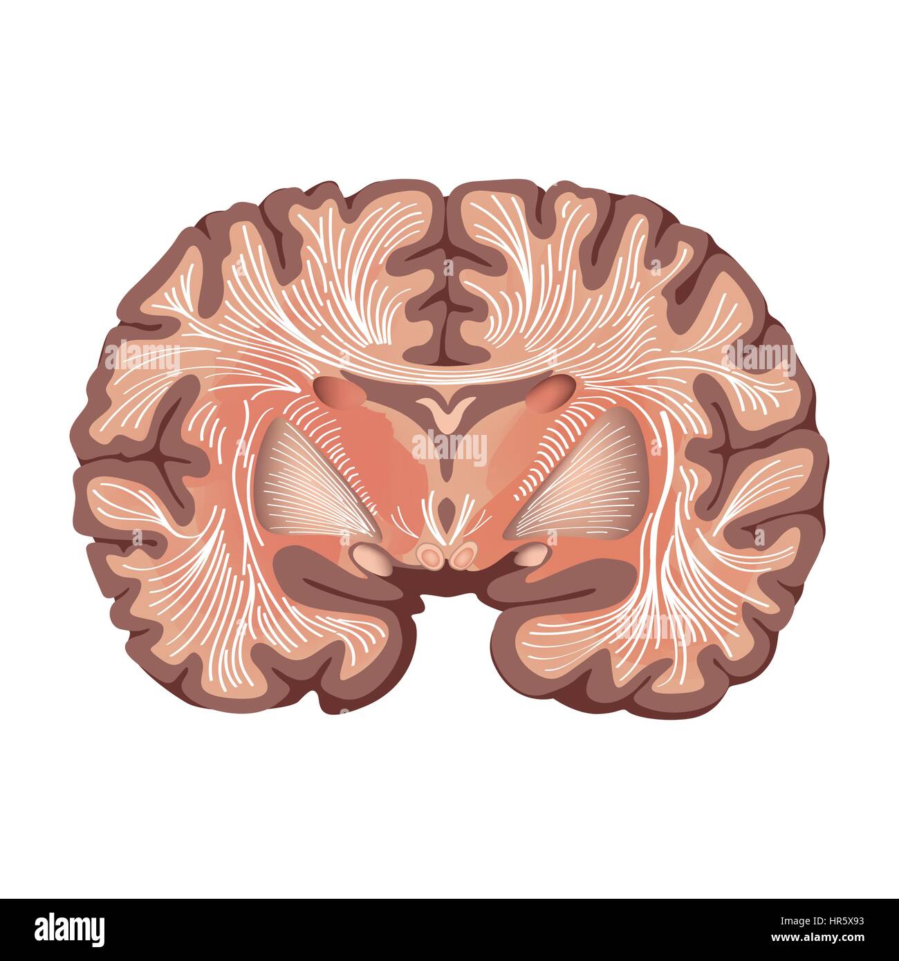 Brain anatomy. Brain showing the basal ganglia and thalamic nuclei isolated on white background. Stock Vectorhttps://www.alamy.com/image-license-details/?v=1https://www.alamy.com/stock-photo-brain-anatomy-brain-showing-the-basal-ganglia-and-thalamic-nuclei-134740063.html
RFHR5X93–Brain anatomy. Brain showing the basal ganglia and thalamic nuclei isolated on white background.
 Caudate nucleus, Putamen 3d medical Stock Photohttps://www.alamy.com/image-license-details/?v=1https://www.alamy.com/caudate-nucleus-putamen-3d-medical-image596573383.html
RF2WJG7C7–Caudate nucleus, Putamen 3d medical
 Medical Concept, Illustration of Brain and Nerve Diseases with Prevention and Medical Treatments. Stock Vectorhttps://www.alamy.com/image-license-details/?v=1https://www.alamy.com/stock-photo-medical-concept-illustration-of-brain-and-nerve-diseases-with-prevention-92882895.html
RFFB3527–Medical Concept, Illustration of Brain and Nerve Diseases with Prevention and Medical Treatments.
![Infographic about the areas of the memory and the different functions implied. [Adobe InDesign (.indd)]. Stock Photo Infographic about the areas of the memory and the different functions implied. [Adobe InDesign (.indd)]. Stock Photo](https://c8.alamy.com/comp/2NEBJGG/infographic-about-the-areas-of-the-memory-and-the-different-functions-implied-adobe-indesign-indd-2NEBJGG.jpg) Infographic about the areas of the memory and the different functions implied. [Adobe InDesign (.indd)]. Stock Photohttps://www.alamy.com/image-license-details/?v=1https://www.alamy.com/infographic-about-the-areas-of-the-memory-and-the-different-functions-implied-adobe-indesign-indd-image525172272.html
Infographic about the areas of the memory and the different functions implied. [Adobe InDesign (.indd)]. Stock Photohttps://www.alamy.com/image-license-details/?v=1https://www.alamy.com/infographic-about-the-areas-of-the-memory-and-the-different-functions-implied-adobe-indesign-indd-image525172272.htmlRM2NEBJGG–Infographic about the areas of the memory and the different functions implied. [Adobe InDesign (.indd)].
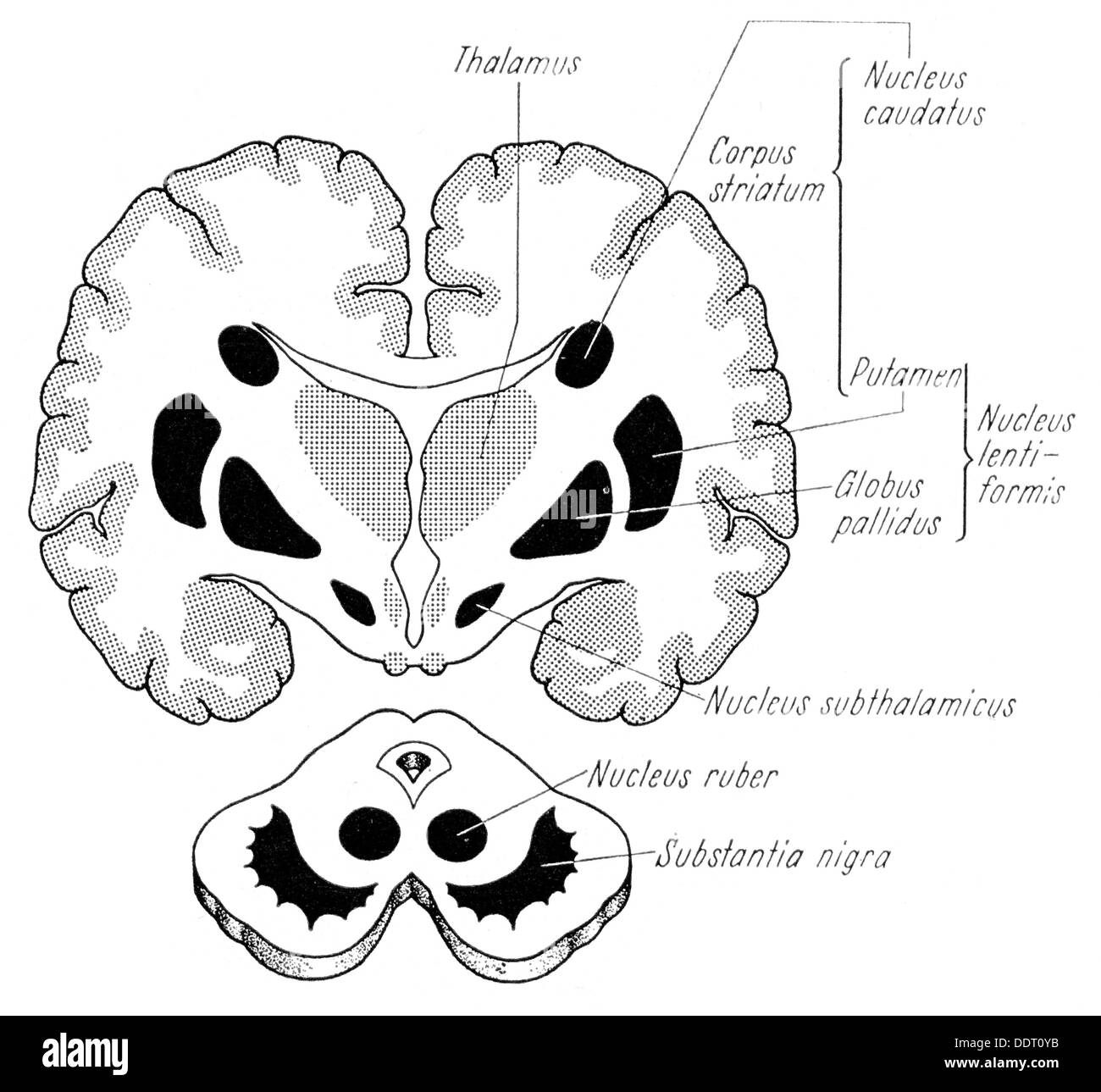 medicine, anatomy, cerebric / cranium, core areas of the extrapyramidal system, drawing, from: Werner Scheid, 'Lehrbuch der Neurologie', Stuttgart, 1968, Additional-Rights-Clearences-Not Available Stock Photohttps://www.alamy.com/image-license-details/?v=1https://www.alamy.com/medicine-anatomy-cerebric-cranium-core-areas-of-the-extrapyramidal-image60149247.html
RMDDT0YB–medicine, anatomy, cerebric / cranium, core areas of the extrapyramidal system, drawing, from: Werner Scheid, 'Lehrbuch der Neurologie', Stuttgart, 1968, Additional-Rights-Clearences-Not Available
 3d rendered medically accurate illustration of young boy brains anatomy- the putamen Stock Photohttps://www.alamy.com/image-license-details/?v=1https://www.alamy.com/3d-rendered-medically-accurate-illustration-of-young-boy-brains-anatomy-the-putamen-image365554084.html
RF2C6MBY0–3d rendered medically accurate illustration of young boy brains anatomy- the putamen
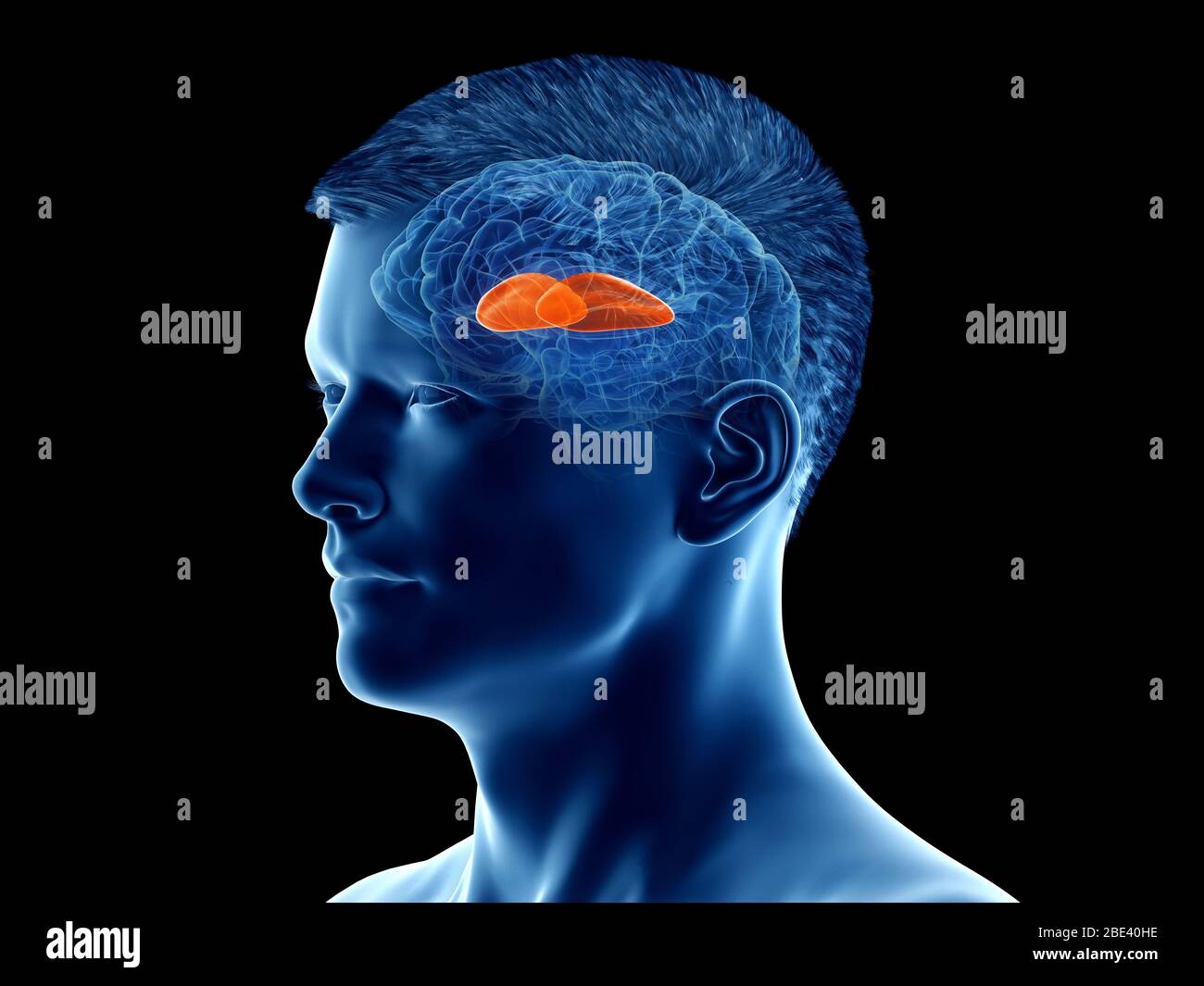 Putamen of the brain, illustration. Stock Photohttps://www.alamy.com/image-license-details/?v=1https://www.alamy.com/putamen-of-the-brain-illustration-image352900842.html
RF2BE40HE–Putamen of the brain, illustration.
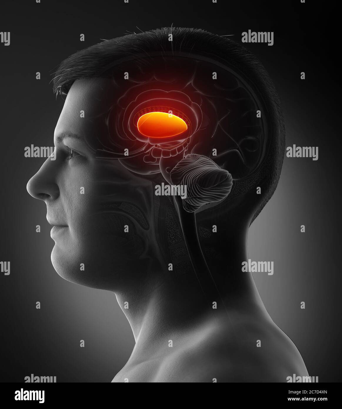 3d rendered medically accurate illustration of a male brains anatomy- the putamen Stock Photohttps://www.alamy.com/image-license-details/?v=1https://www.alamy.com/3d-rendered-medically-accurate-illustration-of-a-male-brains-anatomy-the-putamen-image366009581.html
RF2C7D4XN–3d rendered medically accurate illustration of a male brains anatomy- the putamen
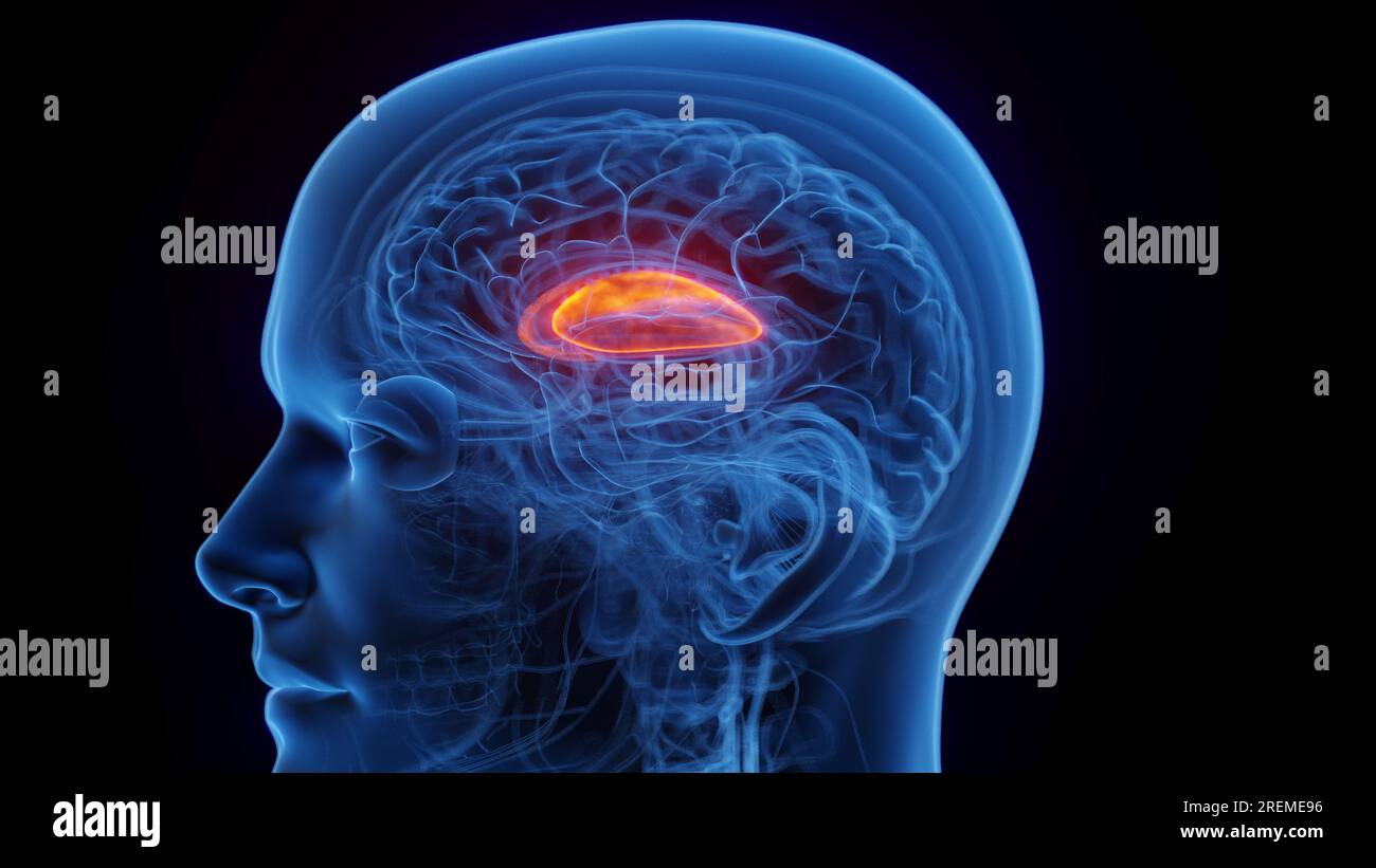 The putamen, illustration. Stock Photohttps://www.alamy.com/image-license-details/?v=1https://www.alamy.com/the-putamen-illustration-image559787234.html
RF2REME96–The putamen, illustration.
 Eider's (somateria mollissima) nest. These ducklings are two days old. Gulf of Finland of the Baltic Sea Stock Photohttps://www.alamy.com/image-license-details/?v=1https://www.alamy.com/eiders-somateria-mollissima-nest-these-ducklings-are-two-days-old-gulf-of-finland-of-the-baltic-sea-image574365626.html
RF2TACH62–Eider's (somateria mollissima) nest. These ducklings are two days old. Gulf of Finland of the Baltic Sea
 3d rendered medically accurate illustration of the putamen Stock Photohttps://www.alamy.com/image-license-details/?v=1https://www.alamy.com/3d-rendered-medically-accurate-illustration-of-the-putamen-image257798797.html
RFTYBMYW–3d rendered medically accurate illustration of the putamen
 Archive image from page 104 of Denkschriften der Medicinisch-Naturwissenschaftlichen Gesellschaft zu. Denkschriften der Medicinisch-Naturwissenschaftlichen Gesellschaft zu Jena denkschriftender09medi Year: 1879 Zur Erforschung der Hirnfaflerung, posterior capsulae internaposterioris zwischen Nucleus caudatus (N. caud.) und Putamen (l'u/., hindurch, weiterhin ventral von der Stria terminalis (ßtr.term.) bis nahe an die Capsula ventralia corporis geniculati lateralis verfolgen. Unmittelbar ventral von diesen Fasern sehen wir eine von markhaltigen Fasern fast freie Region, unsere Begio ansäe le Stock Photohttps://www.alamy.com/image-license-details/?v=1https://www.alamy.com/archive-image-from-page-104-of-denkschriften-der-medicinisch-naturwissenschaftlichen-gesellschaft-zu-denkschriften-der-medicinisch-naturwissenschaftlichen-gesellschaft-zu-jena-denkschriftender09medi-year-1879-zur-erforschung-der-hirnfaflerung-posterior-capsulae-internaposterioris-zwischen-nucleus-caudatus-n-caud-und-putamen-lu-hindurch-weiterhin-ventral-von-der-stria-terminalis-trterm-bis-nahe-an-die-capsula-ventralia-corporis-geniculati-lateralis-verfolgen-unmittelbar-ventral-von-diesen-fasern-sehen-wir-eine-von-markhaltigen-fasern-fast-freie-region-unsere-begio-anse-le-image259446312.html
RMW22PBM–Archive image from page 104 of Denkschriften der Medicinisch-Naturwissenschaftlichen Gesellschaft zu. Denkschriften der Medicinisch-Naturwissenschaftlichen Gesellschaft zu Jena denkschriftender09medi Year: 1879 Zur Erforschung der Hirnfaflerung, posterior capsulae internaposterioris zwischen Nucleus caudatus (N. caud.) und Putamen (l'u/., hindurch, weiterhin ventral von der Stria terminalis (ßtr.term.) bis nahe an die Capsula ventralia corporis geniculati lateralis verfolgen. Unmittelbar ventral von diesen Fasern sehen wir eine von markhaltigen Fasern fast freie Region, unsere Begio ansäe le
 3d rendered medically accurate illustration of the brain anatomy - the putamen Stock Photohttps://www.alamy.com/image-license-details/?v=1https://www.alamy.com/3d-rendered-medically-accurate-illustration-of-the-brain-anatomy-the-putamen-image334038068.html
RF2ABCMY0–3d rendered medically accurate illustration of the brain anatomy - the putamen
 peach Stock Photohttps://www.alamy.com/image-license-details/?v=1https://www.alamy.com/peach-image226154031.html
RFR3X5NK–peach
 Human anatomy, including structure and development and practical considerations . d by numerousstrands of nerve-fibres which break the continuity of the gray substance and jjroducean appearance of ladial striation. The structure of the corpus striatum varies in its several ijarts, that of thecaudate nucleus and the putamen being almost identical, whilst that of the globuspallidus, although similar in both zones, differs from the histological make up of theother parts. The close resemblance of the caudate nucleus and the putamen corre-sponds to their early common origin, since at first they con Stock Photohttps://www.alamy.com/image-license-details/?v=1https://www.alamy.com/human-anatomy-including-structure-and-development-and-practical-considerations-d-by-numerousstrands-of-nerve-fibres-which-break-the-continuity-of-the-gray-substance-and-jjroducean-appearance-of-ladial-striation-the-structure-of-the-corpus-striatum-varies-in-its-several-ijarts-that-of-thecaudate-nucleus-and-the-putamen-being-almost-identical-whilst-that-of-the-globuspallidus-although-similar-in-both-zones-differs-from-the-histological-make-up-of-theother-parts-the-close-resemblance-of-the-caudate-nucleus-and-the-putamen-corre-sponds-to-their-early-common-origin-since-at-first-they-con-image342666684.html
RM2AWDPRT–Human anatomy, including structure and development and practical considerations . d by numerousstrands of nerve-fibres which break the continuity of the gray substance and jjroducean appearance of ladial striation. The structure of the corpus striatum varies in its several ijarts, that of thecaudate nucleus and the putamen being almost identical, whilst that of the globuspallidus, although similar in both zones, differs from the histological make up of theother parts. The close resemblance of the caudate nucleus and the putamen corre-sponds to their early common origin, since at first they con
 Brain Pituitary gland Anatomy For Medical Concept 3D Illustration Stock Photohttps://www.alamy.com/image-license-details/?v=1https://www.alamy.com/brain-pituitary-gland-anatomy-for-medical-concept-3d-illustration-image444893311.html
RF2GRPHXR–Brain Pituitary gland Anatomy For Medical Concept 3D Illustration
 Three fresh plums on white background - one opened Stock Photohttps://www.alamy.com/image-license-details/?v=1https://www.alamy.com/stock-photo-three-fresh-plums-on-white-background-one-opened-32911731.html
RFBWF76Y–Three fresh plums on white background - one opened
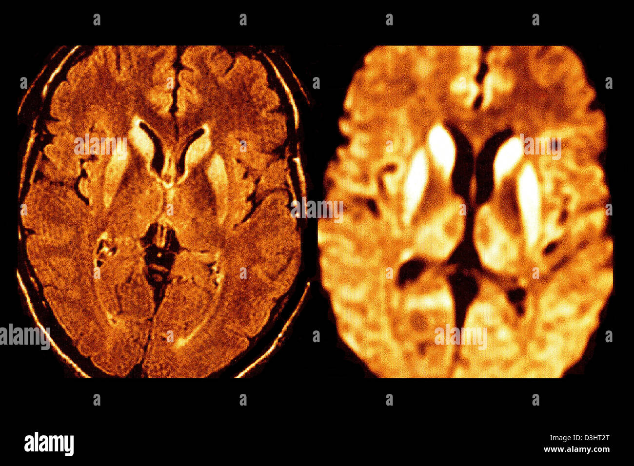 CREUTZFELDT JAKOB DISEASE, MRI Stock Photohttps://www.alamy.com/image-license-details/?v=1https://www.alamy.com/stock-photo-creutzfeldt-jakob-disease-mri-53867152.html
RFD3HT2T–CREUTZFELDT JAKOB DISEASE, MRI
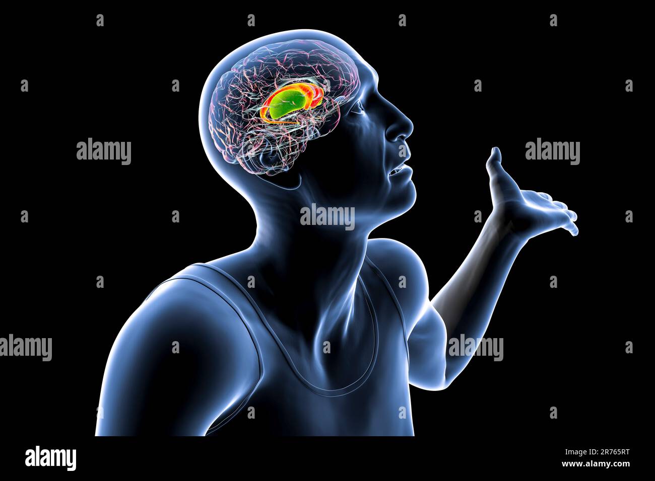 Dorsal striatum, caudate nucleus and putamen, highlighted in the brain of a person with Huntington's disease with involuntary movements, conceptual co Stock Photohttps://www.alamy.com/image-license-details/?v=1https://www.alamy.com/dorsal-striatum-caudate-nucleus-and-putamen-highlighted-in-the-brain-of-a-person-with-huntingtons-disease-with-involuntary-movements-conceptual-co-image555170668.html
RF2R765RT–Dorsal striatum, caudate nucleus and putamen, highlighted in the brain of a person with Huntington's disease with involuntary movements, conceptual co
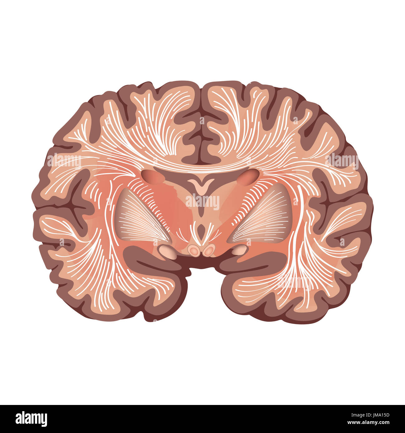 Brain anatomy. Brain showing the basal ganglia and thalamic nuclei isolated on white background. Stock Photohttps://www.alamy.com/image-license-details/?v=1https://www.alamy.com/brain-anatomy-brain-showing-the-basal-ganglia-and-thalamic-nuclei-image150196521.html
RFJMA15D–Brain anatomy. Brain showing the basal ganglia and thalamic nuclei isolated on white background.
 Caudate nucleus, Putamen 3d medical Stock Photohttps://www.alamy.com/image-license-details/?v=1https://www.alamy.com/caudate-nucleus-putamen-3d-medical-image596591258.html
RF2WJH26J–Caudate nucleus, Putamen 3d medical
 Feijoa sweet tropical berry whole and cut. Stock Vectorhttps://www.alamy.com/image-license-details/?v=1https://www.alamy.com/feijoa-sweet-tropical-berry-whole-and-cut-image334904039.html
RF2ACT5EF–Feijoa sweet tropical berry whole and cut.
