Quick filters:
Radiocarpal Stock Photos and Images
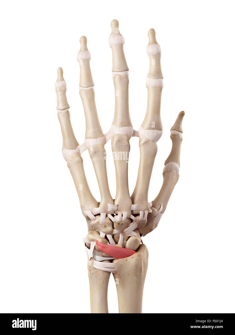 medical accurate illustration of the dorsal radiocarpal ligaments Stock Photohttps://www.alamy.com/image-license-details/?v=1https://www.alamy.com/stock-photo-medical-accurate-illustration-of-the-dorsal-radiocarpal-ligaments-89741068.html
RFF601J4–medical accurate illustration of the dorsal radiocarpal ligaments
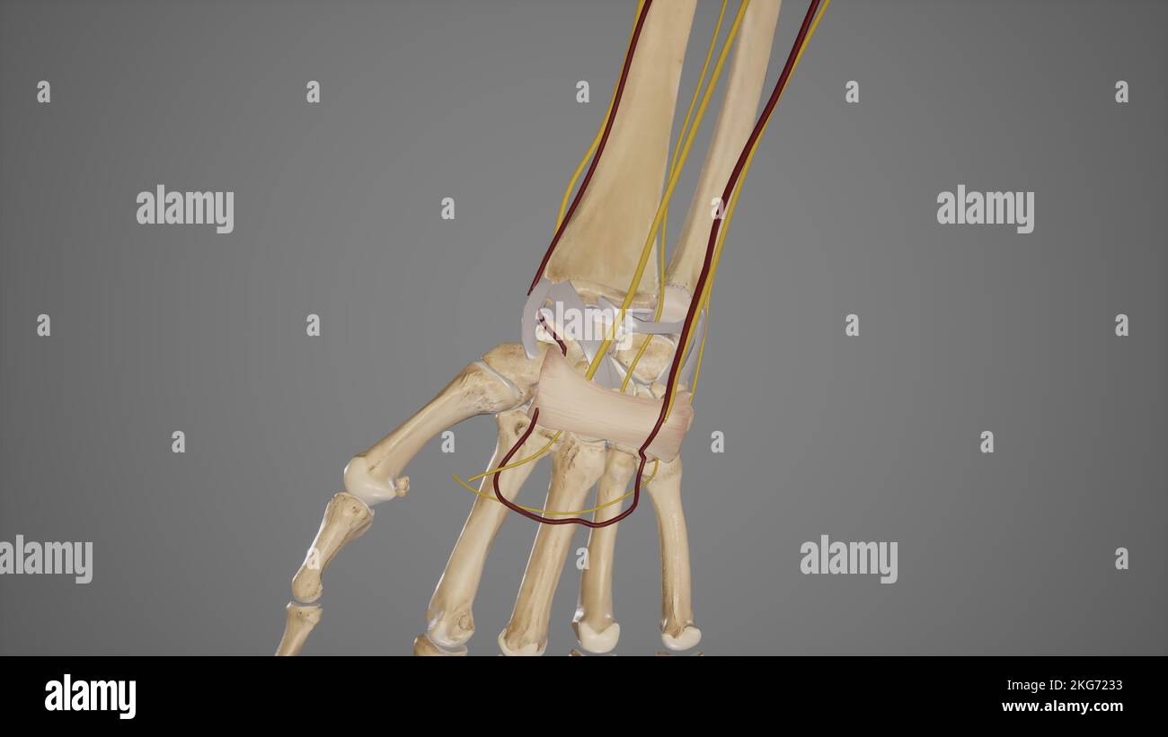 Wrist Joint Stock Photohttps://www.alamy.com/image-license-details/?v=1https://www.alamy.com/wrist-joint-image491880119.html
RF2KG7233–Wrist Joint
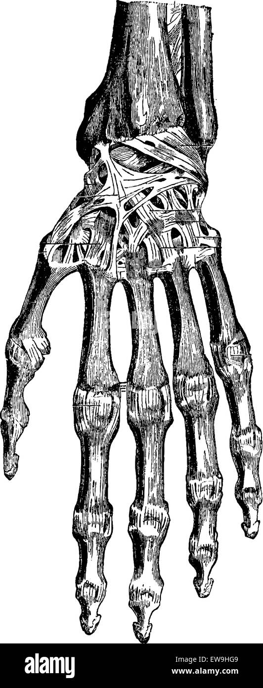 Radiocarpal joint, carpal bones them, carpometacarpal and hand (dorsal), vintage engraved illustration. Usual Medicine Dictionar Stock Vectorhttps://www.alamy.com/image-license-details/?v=1https://www.alamy.com/stock-photo-radiocarpal-joint-carpal-bones-them-carpometacarpal-and-hand-dorsal-84419225.html
RFEW9HG9–Radiocarpal joint, carpal bones them, carpometacarpal and hand (dorsal), vintage engraved illustration. Usual Medicine Dictionar
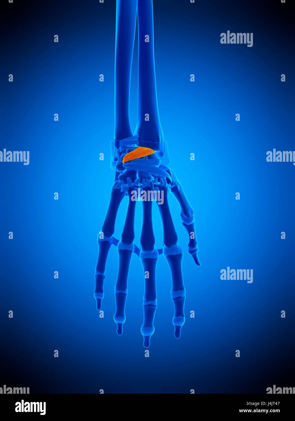 Illustration of the dorsal radiocarpal ligaments. Stock Photohttps://www.alamy.com/image-license-details/?v=1https://www.alamy.com/stock-photo-illustration-of-the-dorsal-radiocarpal-ligaments-140555639.html
RFJ4JT47–Illustration of the dorsal radiocarpal ligaments.
 Neon hand bone red color vector illustration image flat style light Stock Vectorhttps://www.alamy.com/image-license-details/?v=1https://www.alamy.com/neon-hand-bone-red-color-vector-illustration-image-flat-style-light-image456247630.html
RF2HE7TE6–Neon hand bone red color vector illustration image flat style light
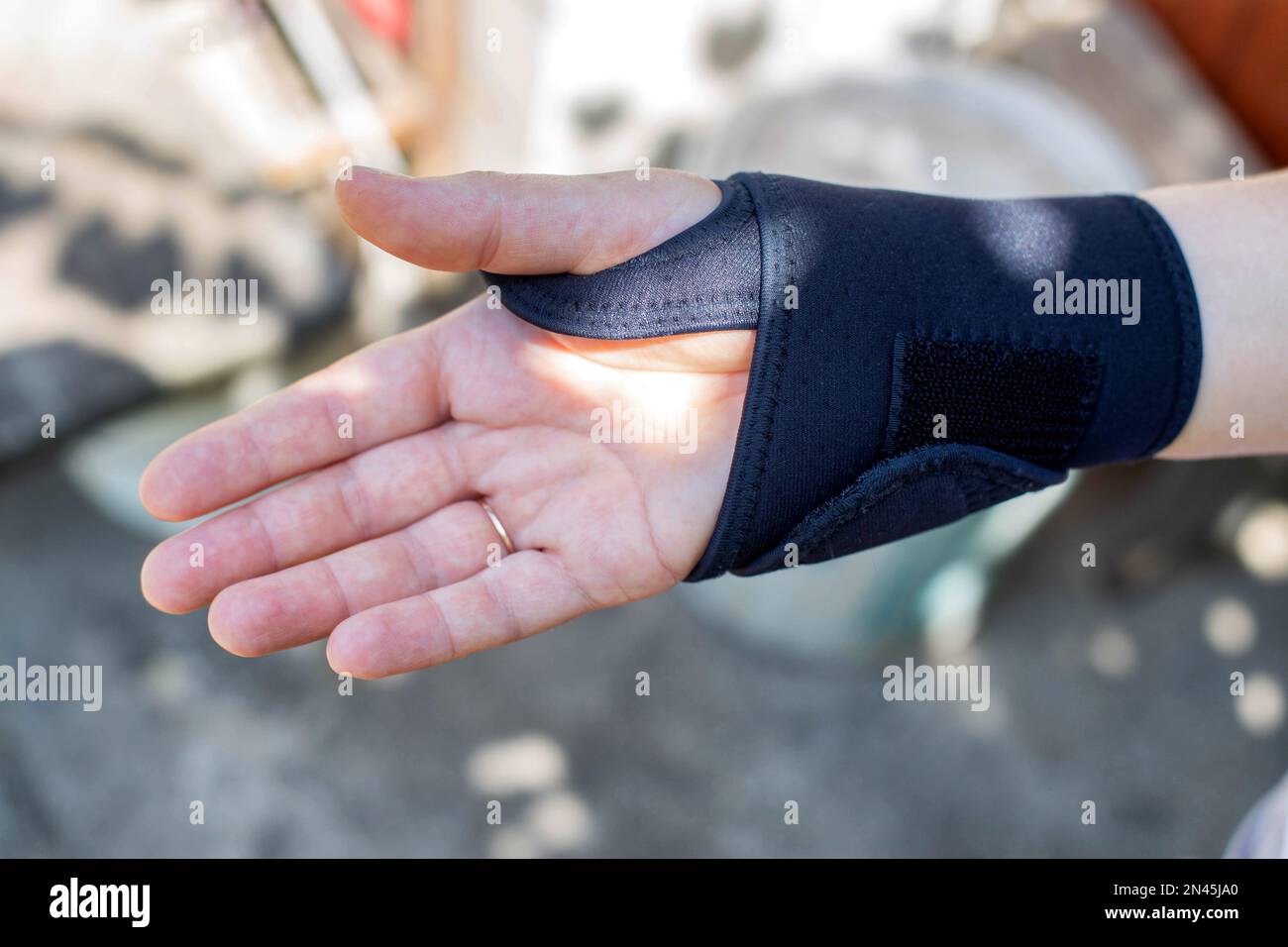 Universal fixator of the wrist joint on the patient's hand. Bandage for fracture and sprain of the radiocarpal joint, pain in the arm. Stock Photohttps://www.alamy.com/image-license-details/?v=1https://www.alamy.com/universal-fixator-of-the-wrist-joint-on-the-patients-hand-bandage-for-fracture-and-sprain-of-the-radiocarpal-joint-pain-in-the-arm-image518893816.html
RF2N45JA0–Universal fixator of the wrist joint on the patient's hand. Bandage for fracture and sprain of the radiocarpal joint, pain in the arm.
RF2R7329Y–Types of joints. Biomechanical and anatomical classification. Set icons with human elbow, knee, wrist joints, and Articulations of a foot. bones and j
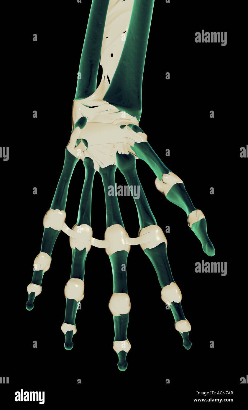 The ligaments of the hand Stock Photohttps://www.alamy.com/image-license-details/?v=1https://www.alamy.com/stock-photo-the-ligaments-of-the-hand-13195630.html
RFACN7AR–The ligaments of the hand
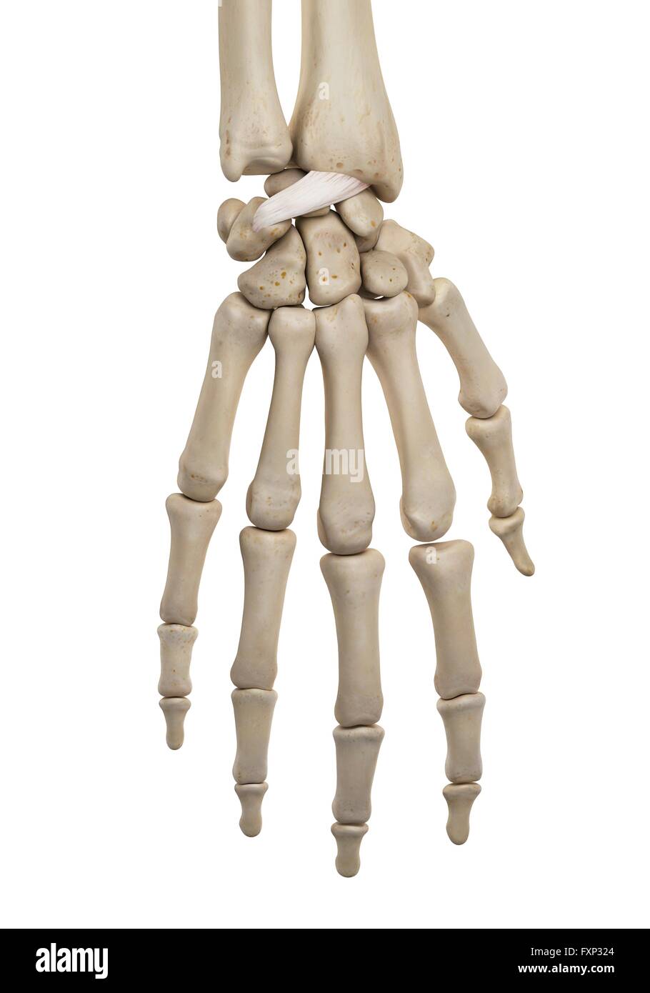 Human hand ligaments, computer illustration. Stock Photohttps://www.alamy.com/image-license-details/?v=1https://www.alamy.com/stock-photo-human-hand-ligaments-computer-illustration-102518252.html
RFFXP324–Human hand ligaments, computer illustration.
 Healthcare specialist examining injured wrist, alternative medicine treatment Stock Photohttps://www.alamy.com/image-license-details/?v=1https://www.alamy.com/healthcare-specialist-examining-injured-wrist-alternative-medicine-treatment-image339796718.html
RF2AMR252–Healthcare specialist examining injured wrist, alternative medicine treatment
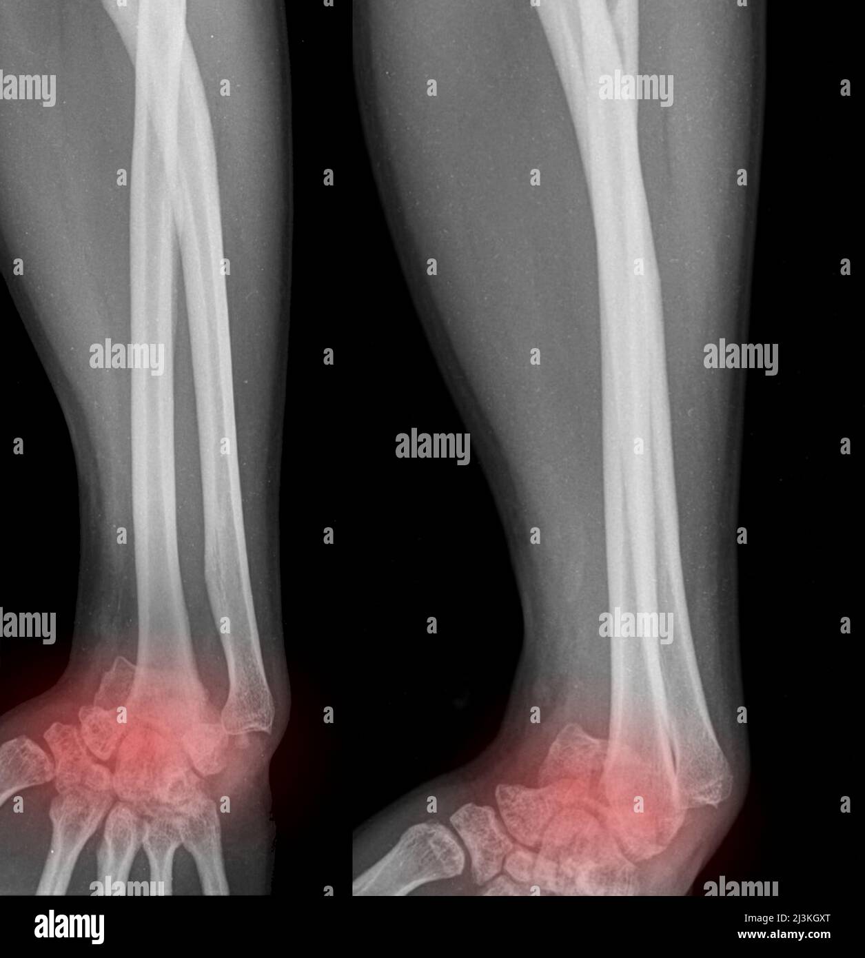 Dislocated wrists, X-ray Stock Photohttps://www.alamy.com/image-license-details/?v=1https://www.alamy.com/dislocated-wrists-x-ray-image466954288.html
RF2J3KGXT–Dislocated wrists, X-ray
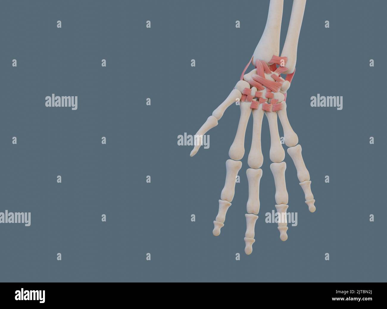 Close view of wrist joint, with ligaments and bones. Stock Photohttps://www.alamy.com/image-license-details/?v=1https://www.alamy.com/close-view-of-wrist-joint-with-ligaments-and-bones-image479689690.html
RF2JTBN2J–Close view of wrist joint, with ligaments and bones.
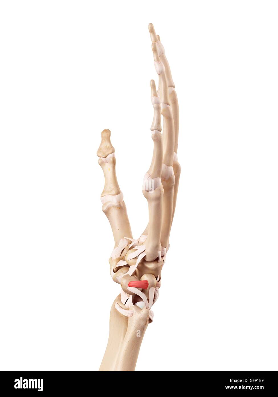 Human hand anatomy, illustration. Stock Photohttps://www.alamy.com/image-license-details/?v=1https://www.alamy.com/stock-photo-human-hand-anatomy-illustration-112680801.html
RFGF91E9–Human hand anatomy, illustration.
RF2AH3M5N–Wrist brace color icon. Hand orthosis. Radiocarpal joint bandage. Wrist support. Hand splint. Isolated vector illustration
 Antique Medical Illustration of Dislocations of the Wrist circa 1881 Stock Photohttps://www.alamy.com/image-license-details/?v=1https://www.alamy.com/stock-photo-antique-medical-illustration-of-dislocations-of-the-wrist-circa-1881-37154041.html
RFC4CEA1–Antique Medical Illustration of Dislocations of the Wrist circa 1881
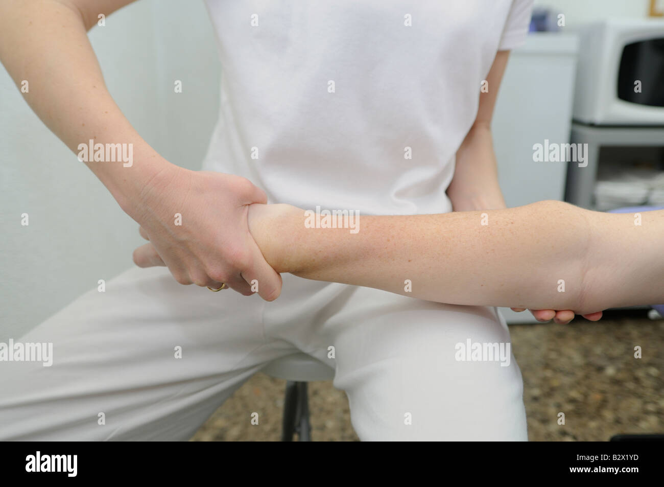 Osteopathic manipulation of lower extremity of radius bone. Osteopathic medicine. Osteopathic therapy. Stock Photohttps://www.alamy.com/image-license-details/?v=1https://www.alamy.com/stock-photo-osteopathic-manipulation-of-lower-extremity-of-radius-bone-osteopathic-19011985.html
RMB2X1YD–Osteopathic manipulation of lower extremity of radius bone. Osteopathic medicine. Osteopathic therapy.
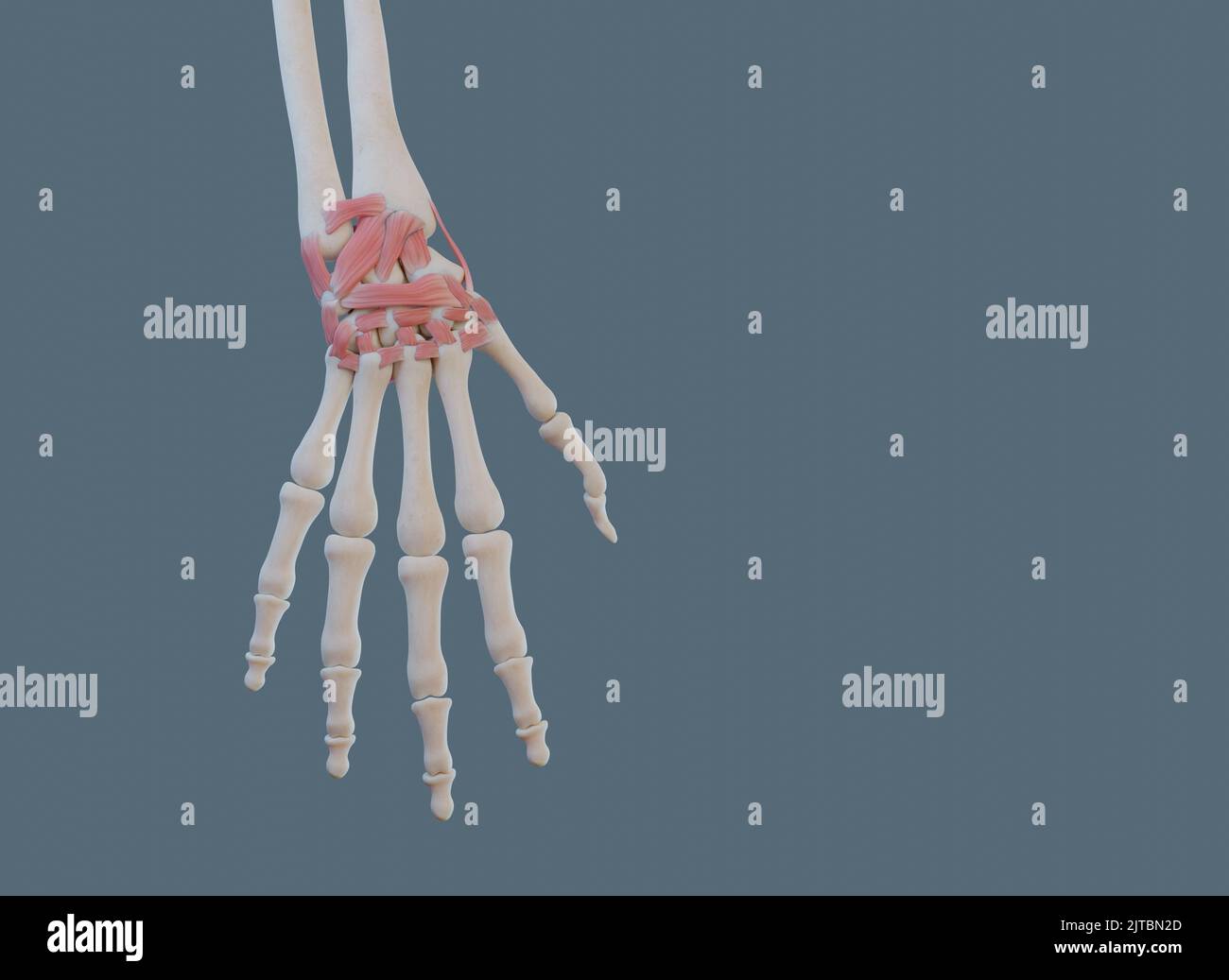 Close view of carpal (wrist) joint, with ligaments and bones. Stock Photohttps://www.alamy.com/image-license-details/?v=1https://www.alamy.com/close-view-of-carpal-wrist-joint-with-ligaments-and-bones-image479689685.html
RF2JTBN2D–Close view of carpal (wrist) joint, with ligaments and bones.
 . The topographical anatomy of the limbs of the horse. Horses; Physiology. 74 TOPOGRAPHICAL ANATOMY OF The synovial lining of the joint capsule is divided into three parts in agreement with the three articulations over which the capsule extends. Its radiocarpal part is most extensive, lubricates the radio- carpal joint, and is continuous with the articulations of the accessory carpal (pisiform) bone. It also lubricates the articulations between the neighbouring bones of the proximal row above the interosseous ligaments. The intercarpal part of the synovial lining lubricates the Kadial carpal b Stock Photohttps://www.alamy.com/image-license-details/?v=1https://www.alamy.com/the-topographical-anatomy-of-the-limbs-of-the-horse-horses-physiology-74-topographical-anatomy-of-the-synovial-lining-of-the-joint-capsule-is-divided-into-three-parts-in-agreement-with-the-three-articulations-over-which-the-capsule-extends-its-radiocarpal-part-is-most-extensive-lubricates-the-radio-carpal-joint-and-is-continuous-with-the-articulations-of-the-accessory-carpal-pisiform-bone-it-also-lubricates-the-articulations-between-the-neighbouring-bones-of-the-proximal-row-above-the-interosseous-ligaments-the-intercarpal-part-of-the-synovial-lining-lubricates-the-kadial-carpal-b-image232418140.html
RMRE3FKT–. The topographical anatomy of the limbs of the horse. Horses; Physiology. 74 TOPOGRAPHICAL ANATOMY OF The synovial lining of the joint capsule is divided into three parts in agreement with the three articulations over which the capsule extends. Its radiocarpal part is most extensive, lubricates the radio- carpal joint, and is continuous with the articulations of the accessory carpal (pisiform) bone. It also lubricates the articulations between the neighbouring bones of the proximal row above the interosseous ligaments. The intercarpal part of the synovial lining lubricates the Kadial carpal b
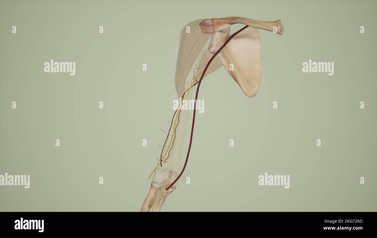 Deep Brachial Artery Stock Photohttps://www.alamy.com/image-license-details/?v=1https://www.alamy.com/deep-brachial-artery-image491880213.html
RF2KG726D–Deep Brachial Artery
 Radiocarpal joint, carpal bones them, carpometacarpal and hand (dorsal), vintage engraved illustration. Usual Medicine Dictionar Stock Vectorhttps://www.alamy.com/image-license-details/?v=1https://www.alamy.com/stock-photo-radiocarpal-joint-carpal-bones-them-carpometacarpal-and-hand-dorsal-84406908.html
RFEW91TC–Radiocarpal joint, carpal bones them, carpometacarpal and hand (dorsal), vintage engraved illustration. Usual Medicine Dictionar
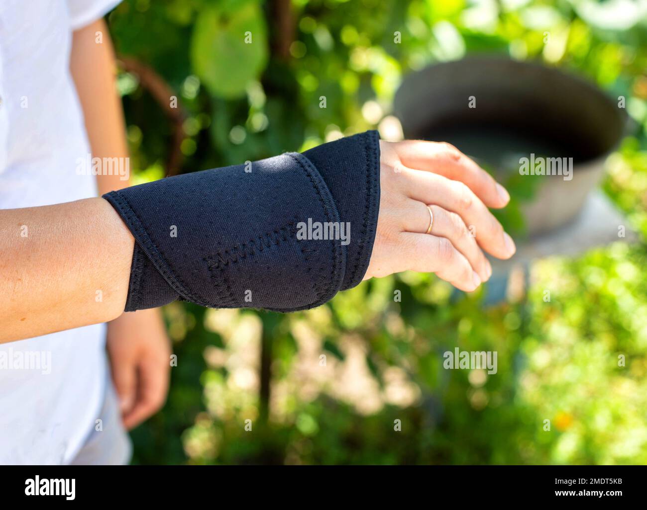 Universal fixator of the wrist joint on the patient's hand. Bandage for fracture and sprain of the radiocarpal joint, pain in the arm. Stock Photohttps://www.alamy.com/image-license-details/?v=1https://www.alamy.com/universal-fixator-of-the-wrist-joint-on-the-patients-hand-bandage-for-fracture-and-sprain-of-the-radiocarpal-joint-pain-in-the-arm-image507622511.html
RF2MDT5KB–Universal fixator of the wrist joint on the patient's hand. Bandage for fracture and sprain of the radiocarpal joint, pain in the arm.
 dotcore surgeon holds a scalpel near the radiocarpal joint on the girl s arm, the concept of operation and treatment of carpal tunnel syndrome and joi Stock Photohttps://www.alamy.com/image-license-details/?v=1https://www.alamy.com/dotcore-surgeon-holds-a-scalpel-near-the-radiocarpal-joint-on-the-girl-s-arm-the-concept-of-operation-and-treatment-of-carpal-tunnel-syndrome-and-joi-image261557527.html
RFW5EY87–dotcore surgeon holds a scalpel near the radiocarpal joint on the girl s arm, the concept of operation and treatment of carpal tunnel syndrome and joi
 The ligaments of the hand Stock Photohttps://www.alamy.com/image-license-details/?v=1https://www.alamy.com/stock-photo-the-ligaments-of-the-hand-13170321.html
RFACJG1P–The ligaments of the hand
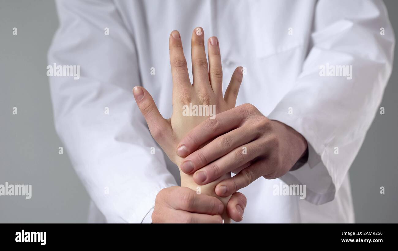 Masseur giving patient hand massage after injury, examining patients wrist Stock Photohttps://www.alamy.com/image-license-details/?v=1https://www.alamy.com/masseur-giving-patient-hand-massage-after-injury-examining-patients-wrist-image339796722.html
RF2AMR256–Masseur giving patient hand massage after injury, examining patients wrist
RF2AH3R6Y–Wrist brace color icon. Hand orthosis. Radiocarpal joint bandage. Wrist support. Hand splint. Isolated vector illustration
 Antique Medical Illustration of Dislocations of the Wrist circa 1881 Stock Photohttps://www.alamy.com/image-license-details/?v=1https://www.alamy.com/stock-photo-antique-medical-illustration-of-dislocations-of-the-wrist-circa-1881-37154048.html
RFC4CEA8–Antique Medical Illustration of Dislocations of the Wrist circa 1881
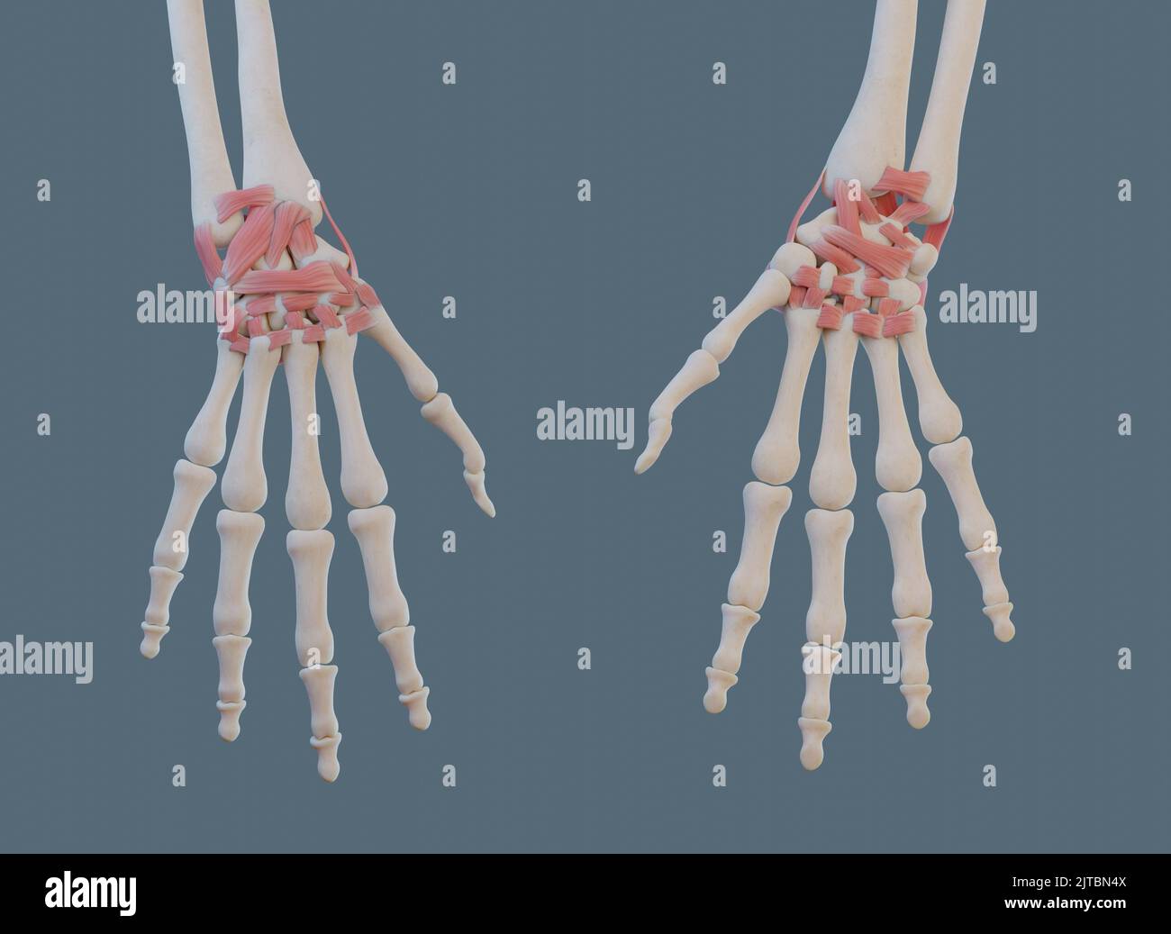 Anterior and posterior views of wrist joint, with ligaments and bones. Stock Photohttps://www.alamy.com/image-license-details/?v=1https://www.alamy.com/anterior-and-posterior-views-of-wrist-joint-with-ligaments-and-bones-image479689754.html
RF2JTBN4X–Anterior and posterior views of wrist joint, with ligaments and bones.
 . An atlas of human anatomy for students and physicians. Anatomy. THE ARTICULATIONS OF THE UPPER LIMB 215 Shaft of the radius Diaphysis radii Radiocarpal articulation Articulatio radiocarpea Lunar, or semilunar, bone Os lunatum Transverse carpal articulation Articulatio intercarpea Carpometacarpal articulation^ Articulatio carpometacarpea Anterior or palmar metacarpophalangeal ligament1 Lig. accessorium volare Metacarpophalangeal articulation Articulatio metacarpo-phalangea Ungual phalanx' 1'halanx III.. Epiphysial disc Synchondrosis epiphyseos Distal epiphysis of the radius Epiphysis distalis Stock Photohttps://www.alamy.com/image-license-details/?v=1https://www.alamy.com/an-atlas-of-human-anatomy-for-students-and-physicians-anatomy-the-articulations-of-the-upper-limb-215-shaft-of-the-radius-diaphysis-radii-radiocarpal-articulation-articulatio-radiocarpea-lunar-or-semilunar-bone-os-lunatum-transverse-carpal-articulation-articulatio-intercarpea-carpometacarpal-articulation-articulatio-carpometacarpea-anterior-or-palmar-metacarpophalangeal-ligament1-lig-accessorium-volare-metacarpophalangeal-articulation-articulatio-metacarpo-phalangea-ungual-phalanx-1halanx-iii-epiphysial-disc-synchondrosis-epiphyseos-distal-epiphysis-of-the-radius-epiphysis-distalis-image235395319.html
RMRJY53K–. An atlas of human anatomy for students and physicians. Anatomy. THE ARTICULATIONS OF THE UPPER LIMB 215 Shaft of the radius Diaphysis radii Radiocarpal articulation Articulatio radiocarpea Lunar, or semilunar, bone Os lunatum Transverse carpal articulation Articulatio intercarpea Carpometacarpal articulation^ Articulatio carpometacarpea Anterior or palmar metacarpophalangeal ligament1 Lig. accessorium volare Metacarpophalangeal articulation Articulatio metacarpo-phalangea Ungual phalanx' 1'halanx III.. Epiphysial disc Synchondrosis epiphyseos Distal epiphysis of the radius Epiphysis distalis
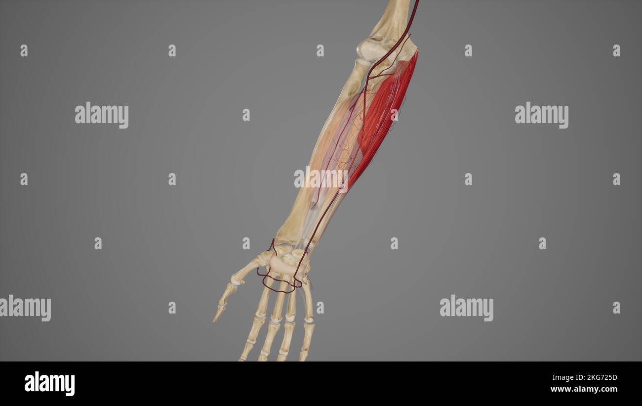 Main Branches of Ulnar Artery Stock Photohttps://www.alamy.com/image-license-details/?v=1https://www.alamy.com/main-branches-of-ulnar-artery-image491880185.html
RF2KG725D–Main Branches of Ulnar Artery
 Radiocarpal joint, carpal bones them, carpometacarpal and hand (palm side), vintage engraved illustration. Usual Medicine Dictio Stock Vectorhttps://www.alamy.com/image-license-details/?v=1https://www.alamy.com/stock-photo-radiocarpal-joint-carpal-bones-them-carpometacarpal-and-hand-palm-84406910.html
RFEW91TE–Radiocarpal joint, carpal bones them, carpometacarpal and hand (palm side), vintage engraved illustration. Usual Medicine Dictio
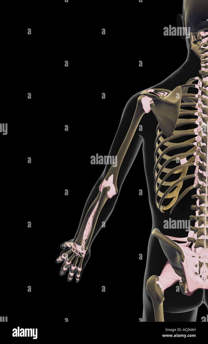 The ligaments of the upper limb Stock Photohttps://www.alamy.com/image-license-details/?v=1https://www.alamy.com/stock-photo-the-ligaments-of-the-upper-limb-13172104.html
RFACJNAH–The ligaments of the upper limb
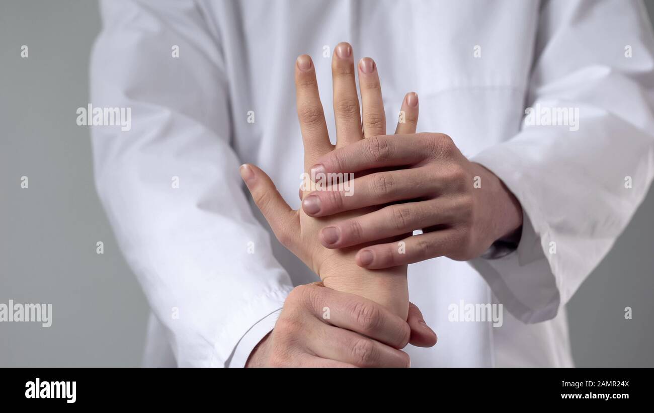 Doctor moving patient wrist, first aid in clinic, assessing severity of injury Stock Photohttps://www.alamy.com/image-license-details/?v=1https://www.alamy.com/doctor-moving-patient-wrist-first-aid-in-clinic-assessing-severity-of-injury-image339796714.html
RF2AMR24X–Doctor moving patient wrist, first aid in clinic, assessing severity of injury
RF2AH3NK2–Wrist brace chalk icon. Hand orthosis. Radiocarpal joint bandage. Wrist support. Hand splint. Isolated vector chalkboard illustration
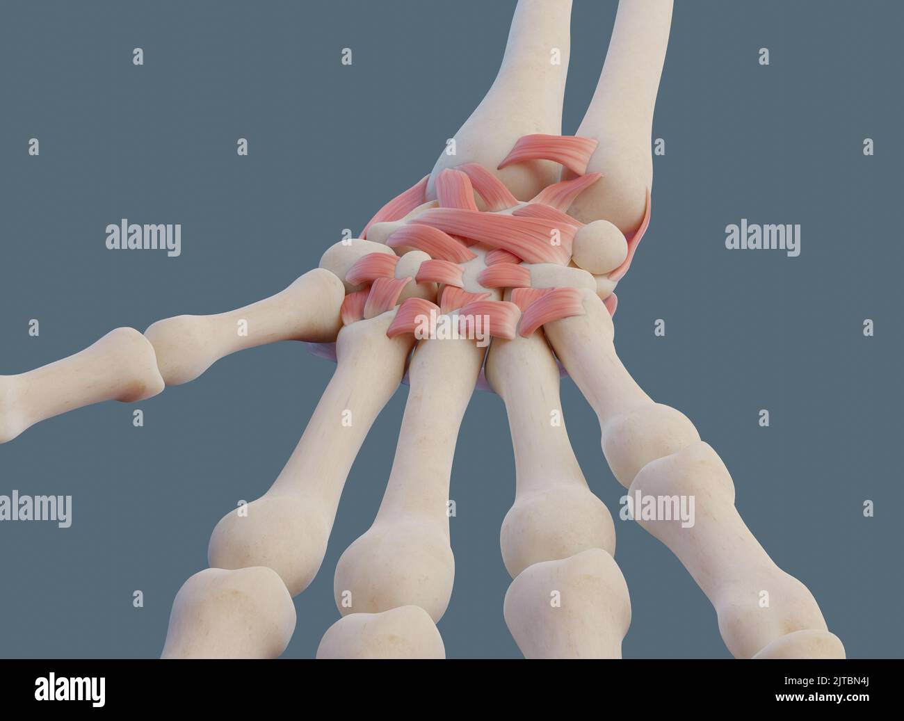 Close view of wrist joint, with ligaments and bones. Stock Photohttps://www.alamy.com/image-license-details/?v=1https://www.alamy.com/close-view-of-wrist-joint-with-ligaments-and-bones-image479689746.html
RF2JTBN4J–Close view of wrist joint, with ligaments and bones.
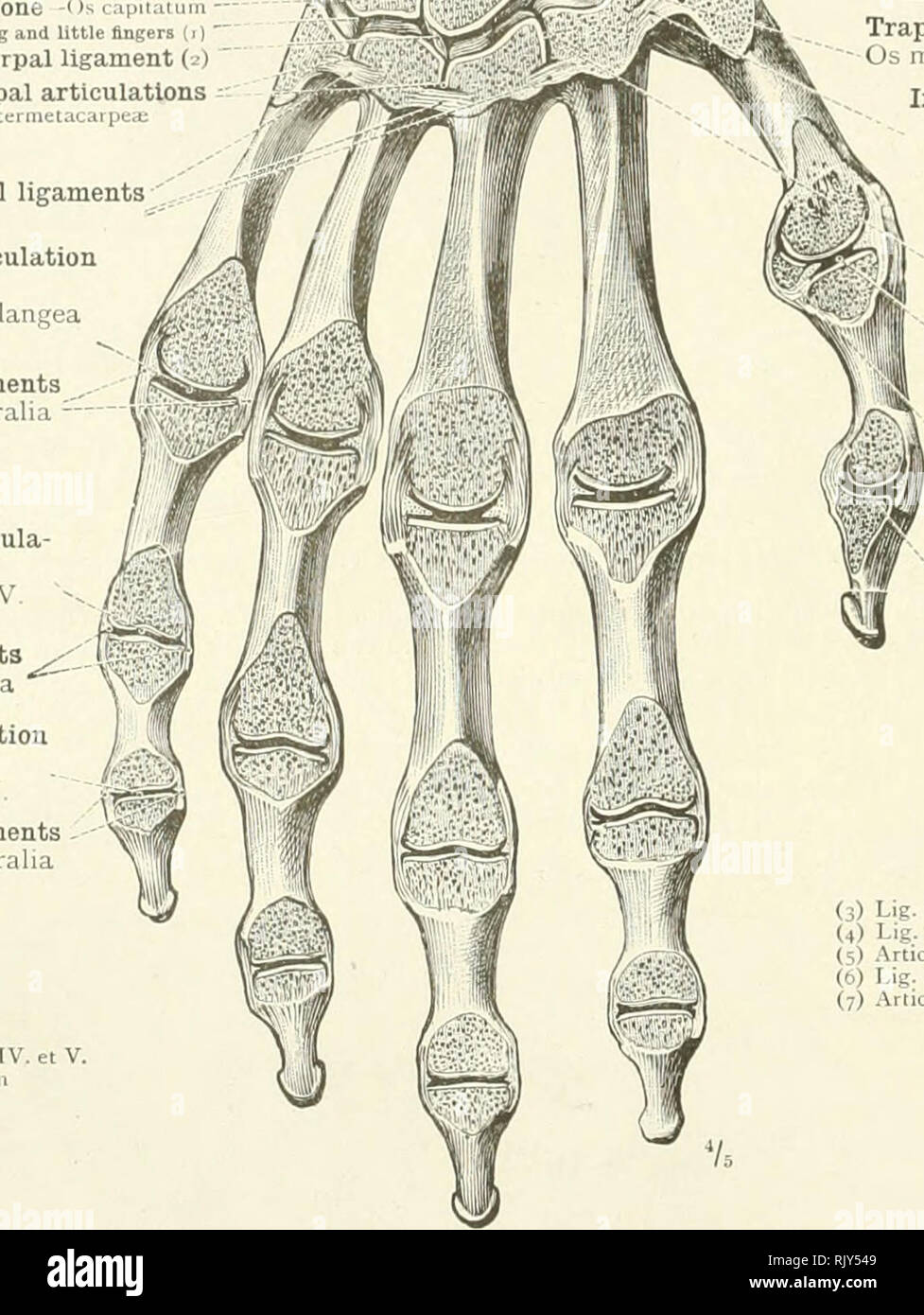 . An atlas of human anatomy for students and physicians. Anatomy. MM Lateral ligaments Ligg. collateralia Proximal interphalangeal articula- tion of the little finger Articulatio proximalis digiti V. Lateral ligaments ^S Ligg. collateralia Distal interphalangeal articulation of the little finger Articulatio distalis digiti V, Lateral ligaments Ligg. collateralia (1) Articulatio carpometacarpea dij (2) Lig. carpomelacarpeum interosseum. The radius Radiu i The radiocarpal articulation or wrist-joint Articulatio radii» arpea .Interosseous intercarpal ligament ( 0 Scaphoid bone-O* naviculare Ext Stock Photohttps://www.alamy.com/image-license-details/?v=1https://www.alamy.com/an-atlas-of-human-anatomy-for-students-and-physicians-anatomy-mm-lateral-ligaments-ligg-collateralia-proximal-interphalangeal-articula-tion-of-the-little-finger-articulatio-proximalis-digiti-v-lateral-ligaments-s-ligg-collateralia-distal-interphalangeal-articulation-of-the-little-finger-articulatio-distalis-digiti-v-lateral-ligaments-ligg-collateralia-1-articulatio-carpometacarpea-dij-2-lig-carpomelacarpeum-interosseum-the-radius-radiu-i-the-radiocarpal-articulation-or-wrist-joint-articulatio-radii-arpea-interosseous-intercarpal-ligament-0-scaphoid-bone-o-naviculare-ext-image235395337.html
RMRJY549–. An atlas of human anatomy for students and physicians. Anatomy. MM Lateral ligaments Ligg. collateralia Proximal interphalangeal articula- tion of the little finger Articulatio proximalis digiti V. Lateral ligaments ^S Ligg. collateralia Distal interphalangeal articulation of the little finger Articulatio distalis digiti V, Lateral ligaments Ligg. collateralia (1) Articulatio carpometacarpea dij (2) Lig. carpomelacarpeum interosseum. The radius Radiu i The radiocarpal articulation or wrist-joint Articulatio radii» arpea .Interosseous intercarpal ligament ( 0 Scaphoid bone-O* naviculare Ext
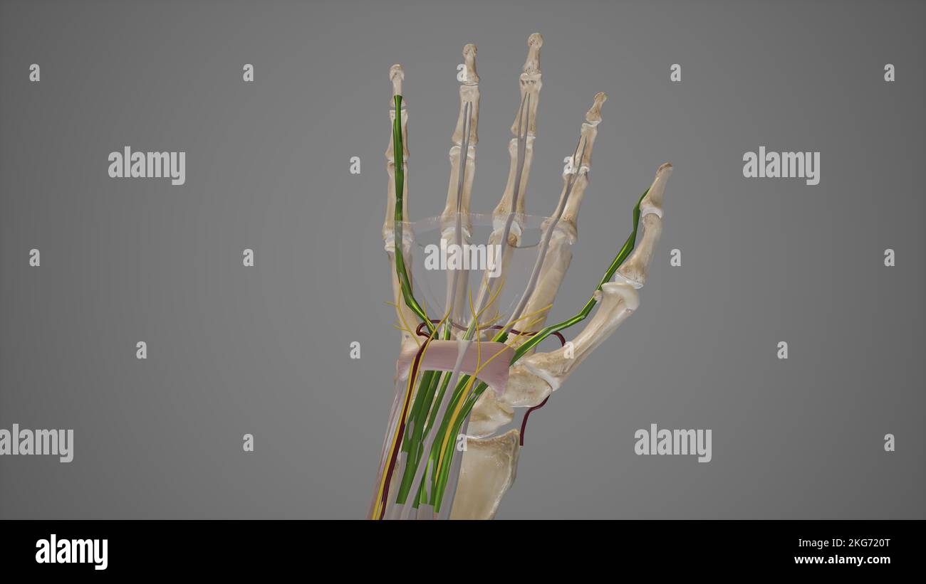 Carpal Tunnel Anatomy Stock Photohttps://www.alamy.com/image-license-details/?v=1https://www.alamy.com/carpal-tunnel-anatomy-image491880056.html
RF2KG720T–Carpal Tunnel Anatomy
 Radiocarpal joint, carpal bones them, carpometacarpal and hand (palm side), vintage engraved illustration. Usual Medicine Dictio Stock Vectorhttps://www.alamy.com/image-license-details/?v=1https://www.alamy.com/stock-photo-radiocarpal-joint-carpal-bones-them-carpometacarpal-and-hand-palm-84419227.html
RFEW9HGB–Radiocarpal joint, carpal bones them, carpometacarpal and hand (palm side), vintage engraved illustration. Usual Medicine Dictio
 The ligaments of the upper limb Stock Photohttps://www.alamy.com/image-license-details/?v=1https://www.alamy.com/stock-photo-the-ligaments-of-the-upper-limb-13174248.html
RFACJYMW–The ligaments of the upper limb
 Therapist looking at patient wrist, assessing severity of injury, closeup Stock Photohttps://www.alamy.com/image-license-details/?v=1https://www.alamy.com/therapist-looking-at-patient-wrist-assessing-severity-of-injury-closeup-image339796709.html
RF2AMR24N–Therapist looking at patient wrist, assessing severity of injury, closeup
RF2AH3XEG–Wrist brace neon light icon. Hand orthosis. Radiocarpal joint bandage. Glowing sign with alphabet, numbers and symbols. Wrist support. Hand splint. Ve
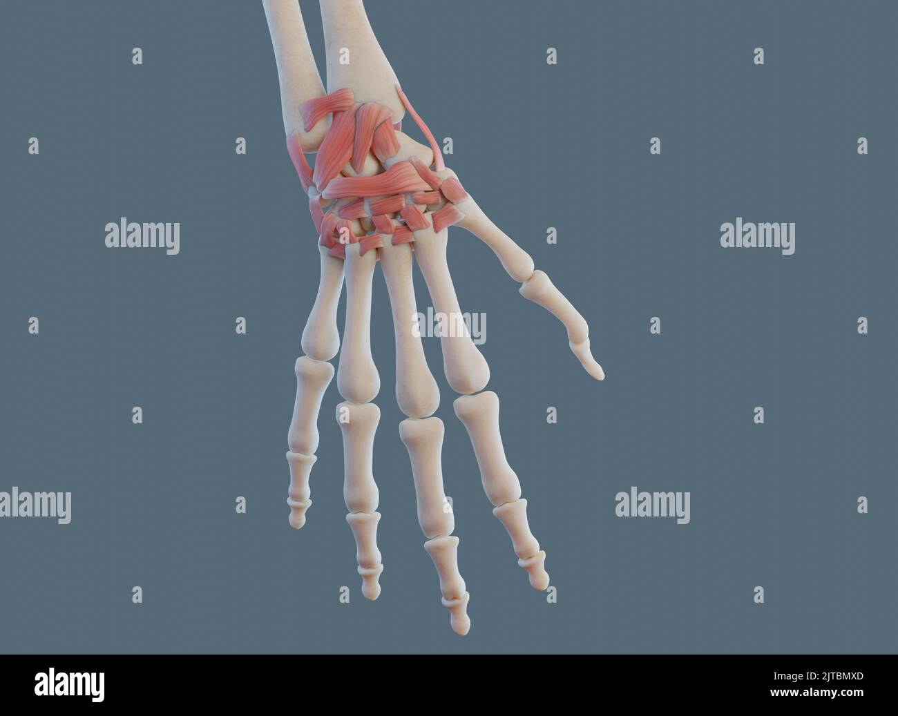 Close view of wrist joint, with ligaments and bones. Stock Photohttps://www.alamy.com/image-license-details/?v=1https://www.alamy.com/close-view-of-wrist-joint-with-ligaments-and-bones-image479689573.html
RF2JTBMXD–Close view of wrist joint, with ligaments and bones.
 . The topographical anatomy of the limbs of the horse. Horses; Physiology. THE LIMBS OF THE HOESE 47 for some distance proximal to the radiocarpal joint and downwards to near the middle of the metacarpus. Radial carpal boue (scaphoid). Third carpal bone (magnum). M. extensor carpi vadialis. Third metacarpal bone.. iladins. Accessory carpal bone (pisiform). M. extensor carpi ulnaris. M. flexor carpi ulnaris. Second carpal bone (trapezoid). First carpal bone (trapezium). Second metacarpal bone. Fig. 28.—Medial Asiiect of the Carpus, etc., with Areas of Muscular Attachment. Hadial carpal bone- (s Stock Photohttps://www.alamy.com/image-license-details/?v=1https://www.alamy.com/the-topographical-anatomy-of-the-limbs-of-the-horse-horses-physiology-the-limbs-of-the-hoese-47-for-some-distance-proximal-to-the-radiocarpal-joint-and-downwards-to-near-the-middle-of-the-metacarpus-radial-carpal-boue-scaphoid-third-carpal-bone-magnum-m-extensor-carpi-vadialis-third-metacarpal-bone-iladins-accessory-carpal-bone-pisiform-m-extensor-carpi-ulnaris-m-flexor-carpi-ulnaris-second-carpal-bone-trapezoid-first-carpal-bone-trapezium-second-metacarpal-bone-fig-28medial-asiiect-of-the-carpus-etc-with-areas-of-muscular-attachment-hadial-carpal-bone-s-image232418268.html
RMRE3FTC–. The topographical anatomy of the limbs of the horse. Horses; Physiology. THE LIMBS OF THE HOESE 47 for some distance proximal to the radiocarpal joint and downwards to near the middle of the metacarpus. Radial carpal boue (scaphoid). Third carpal bone (magnum). M. extensor carpi vadialis. Third metacarpal bone.. iladins. Accessory carpal bone (pisiform). M. extensor carpi ulnaris. M. flexor carpi ulnaris. Second carpal bone (trapezoid). First carpal bone (trapezium). Second metacarpal bone. Fig. 28.—Medial Asiiect of the Carpus, etc., with Areas of Muscular Attachment. Hadial carpal bone- (s
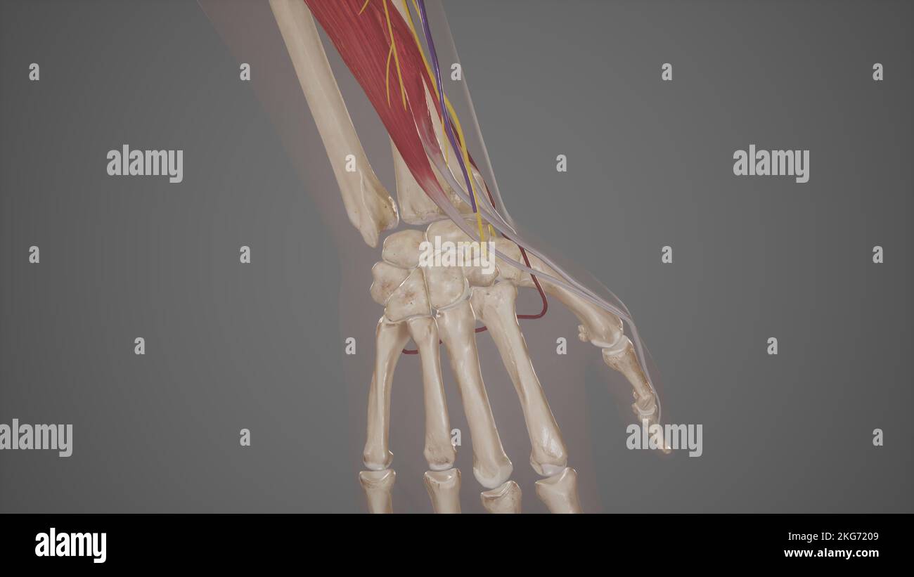 Anatomical Snuffbox Stock Photohttps://www.alamy.com/image-license-details/?v=1https://www.alamy.com/anatomical-snuffbox-image491880041.html
RF2KG7209–Anatomical Snuffbox
 The ligaments of the hand Stock Photohttps://www.alamy.com/image-license-details/?v=1https://www.alamy.com/stock-photo-the-ligaments-of-the-hand-13168718.html
RFACJB7Y–The ligaments of the hand
RF2AH407J–Wrist brace linear icon. Hand orthosis. Thin line illustration. Radiocarpal joint bandage. Wrist support. Hand splint. Contour symbol. Vector isolated
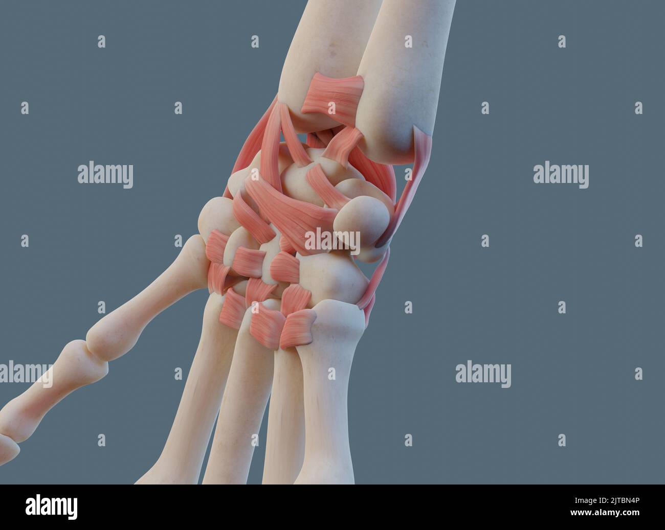 Close view of wrist joint, with ligaments and bones. Stock Photohttps://www.alamy.com/image-license-details/?v=1https://www.alamy.com/close-view-of-wrist-joint-with-ligaments-and-bones-image479689750.html
RF2JTBN4P–Close view of wrist joint, with ligaments and bones.
 . Atlas and text-book of human anatomy. Anatomy -- Atlases. Median vein Brachial cutery Ulna Radius Distal radio-ulnar articulation Articular disc Triquetrum Intercarpal articulation Hamatum Carpometacarpal articulation â f- I'). Lunate bone Radiocarpal articulation Naviailar bone Radial lateral ligament J vA Greater multangular bone ^v â V 'A -^ Cnrpometacarpal artiaUaiion of ihumb â¢',tw i^ ^ Metacarpal bone ⢠» of thumb Capitatum Metacarpal bones Ficr. 202. Lesser multangular bon£. Please note that these images are extracted from scanned page images that may have been dig Stock Photohttps://www.alamy.com/image-license-details/?v=1https://www.alamy.com/atlas-and-text-book-of-human-anatomy-anatomy-atlases-median-vein-brachial-cutery-ulna-radius-distal-radio-ulnar-articulation-articular-disc-triquetrum-intercarpal-articulation-hamatum-carpometacarpal-articulation-f-i-lunate-bone-radiocarpal-articulation-naviailar-bone-radial-lateral-ligament-j-va-greater-multangular-bone-v-v-a-cnrpometacarpal-artiauaiion-of-ihumb-tw-i-metacarpal-bone-of-thumb-capitatum-metacarpal-bones-ficr-202-lesser-multangular-bon-please-note-that-these-images-are-extracted-from-scanned-page-images-that-may-have-been-dig-image235395824.html
RMRJY5NM–. Atlas and text-book of human anatomy. Anatomy -- Atlases. Median vein Brachial cutery Ulna Radius Distal radio-ulnar articulation Articular disc Triquetrum Intercarpal articulation Hamatum Carpometacarpal articulation â f- I'). Lunate bone Radiocarpal articulation Naviailar bone Radial lateral ligament J vA Greater multangular bone ^v â V 'A -^ Cnrpometacarpal artiaUaiion of ihumb â¢',tw i^ ^ Metacarpal bone ⢠» of thumb Capitatum Metacarpal bones Ficr. 202. Lesser multangular bon£. Please note that these images are extracted from scanned page images that may have been dig
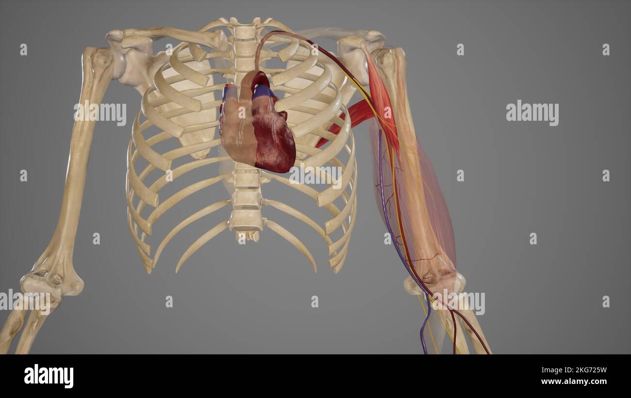 Brachial Artery-Relation and Branches Stock Photohttps://www.alamy.com/image-license-details/?v=1https://www.alamy.com/brachial-artery-relation-and-branches-image491880197.html
RF2KG725W–Brachial Artery-Relation and Branches
 The ligaments of the upper limb Stock Photohttps://www.alamy.com/image-license-details/?v=1https://www.alamy.com/stock-photo-the-ligaments-of-the-upper-limb-13166728.html
RFACJ5AH–The ligaments of the upper limb
RF2AH3X1E–Wrist brace glyph icon. Hand orthosis. Radiocarpal joint bandage. Silhouette symbol. Wrist support. Hand splint. Negative space. Vector isolated illus
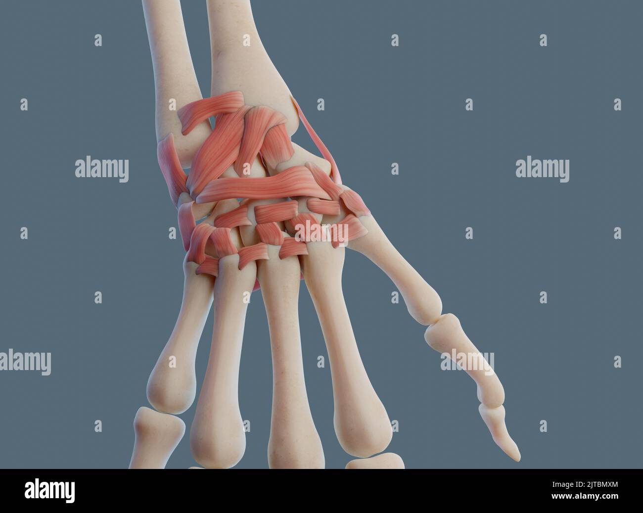 Close view of wrist joint, with ligaments and bones. Stock Photohttps://www.alamy.com/image-license-details/?v=1https://www.alamy.com/close-view-of-wrist-joint-with-ligaments-and-bones-image479689580.html
RF2JTBMXM–Close view of wrist joint, with ligaments and bones.
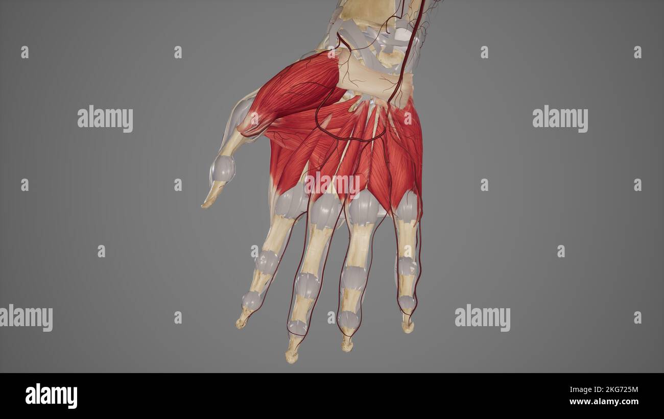 Superficial Palmar Arch Stock Photohttps://www.alamy.com/image-license-details/?v=1https://www.alamy.com/superficial-palmar-arch-image491880192.html
RF2KG725M–Superficial Palmar Arch
 The ligaments of the upper limb Stock Photohttps://www.alamy.com/image-license-details/?v=1https://www.alamy.com/stock-photo-the-ligaments-of-the-upper-limb-13224836.html
RFACTA8N–The ligaments of the upper limb
RF2AH3TE8–Wrist brace app icon. Hand orthosis. Radiocarpal joint bandage. UI/UX user interface. Wrist support. Hand splint. Web or mobile application. Vector is
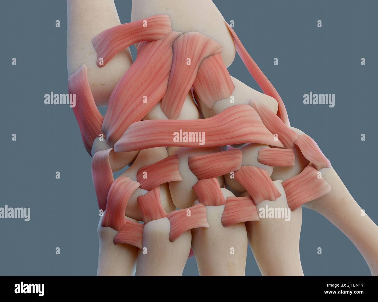 Close view of wrist joint, with ligaments and bones. Stock Photohttps://www.alamy.com/image-license-details/?v=1https://www.alamy.com/close-view-of-wrist-joint-with-ligaments-and-bones-image479689671.html
RF2JTBN1Y–Close view of wrist joint, with ligaments and bones.
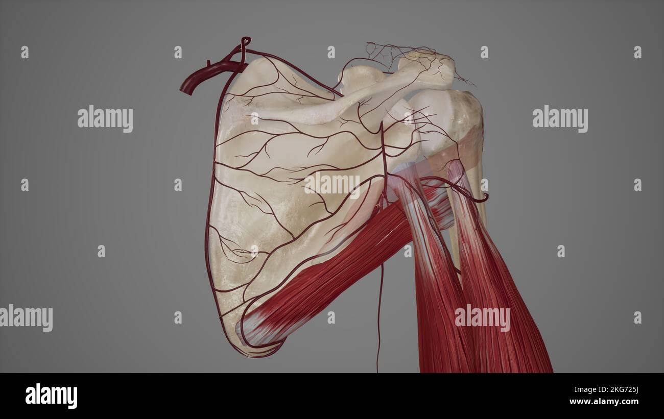 Scapular Anastomosis Stock Photohttps://www.alamy.com/image-license-details/?v=1https://www.alamy.com/scapular-anastomosis-image491880190.html
RF2KG725J–Scapular Anastomosis
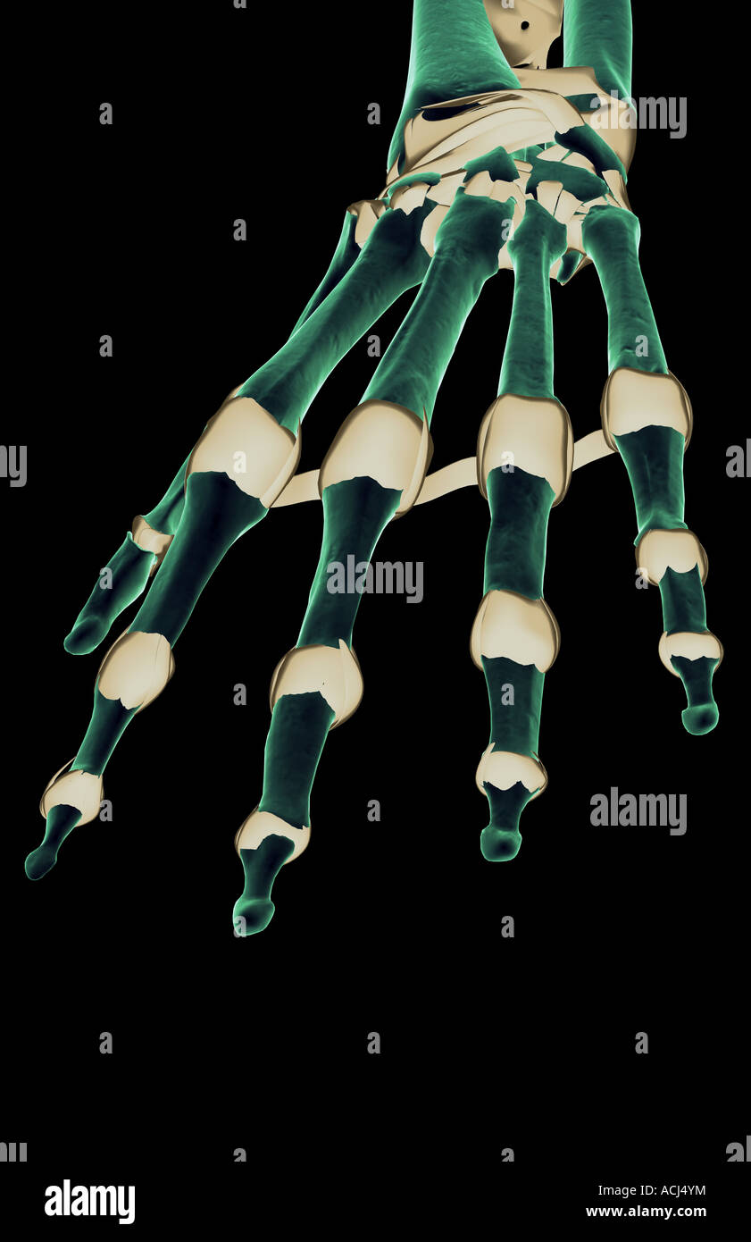 The ligaments of the hand Stock Photohttps://www.alamy.com/image-license-details/?v=1https://www.alamy.com/stock-photo-the-ligaments-of-the-hand-13166599.html
RFACJ4YM–The ligaments of the hand
RF2AH3N3R–Wrist brace app icon. Hand orthosis. Radiocarpal joint bandage. UI/UX user interface. Wrist support. Hand splint. Web or mobile application. Vector is
 Close view of wrist joint, with ligaments and bones. Stock Photohttps://www.alamy.com/image-license-details/?v=1https://www.alamy.com/close-view-of-wrist-joint-with-ligaments-and-bones-image479689577.html
RF2JTBMXH–Close view of wrist joint, with ligaments and bones.
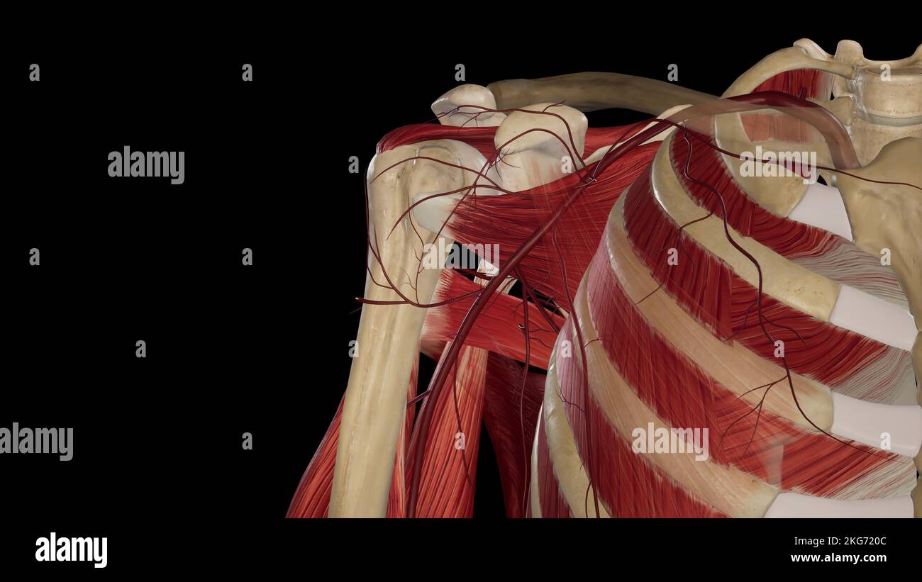 Axillary Artery Stock Photohttps://www.alamy.com/image-license-details/?v=1https://www.alamy.com/axillary-artery-image491880044.html
RF2KG720C–Axillary Artery
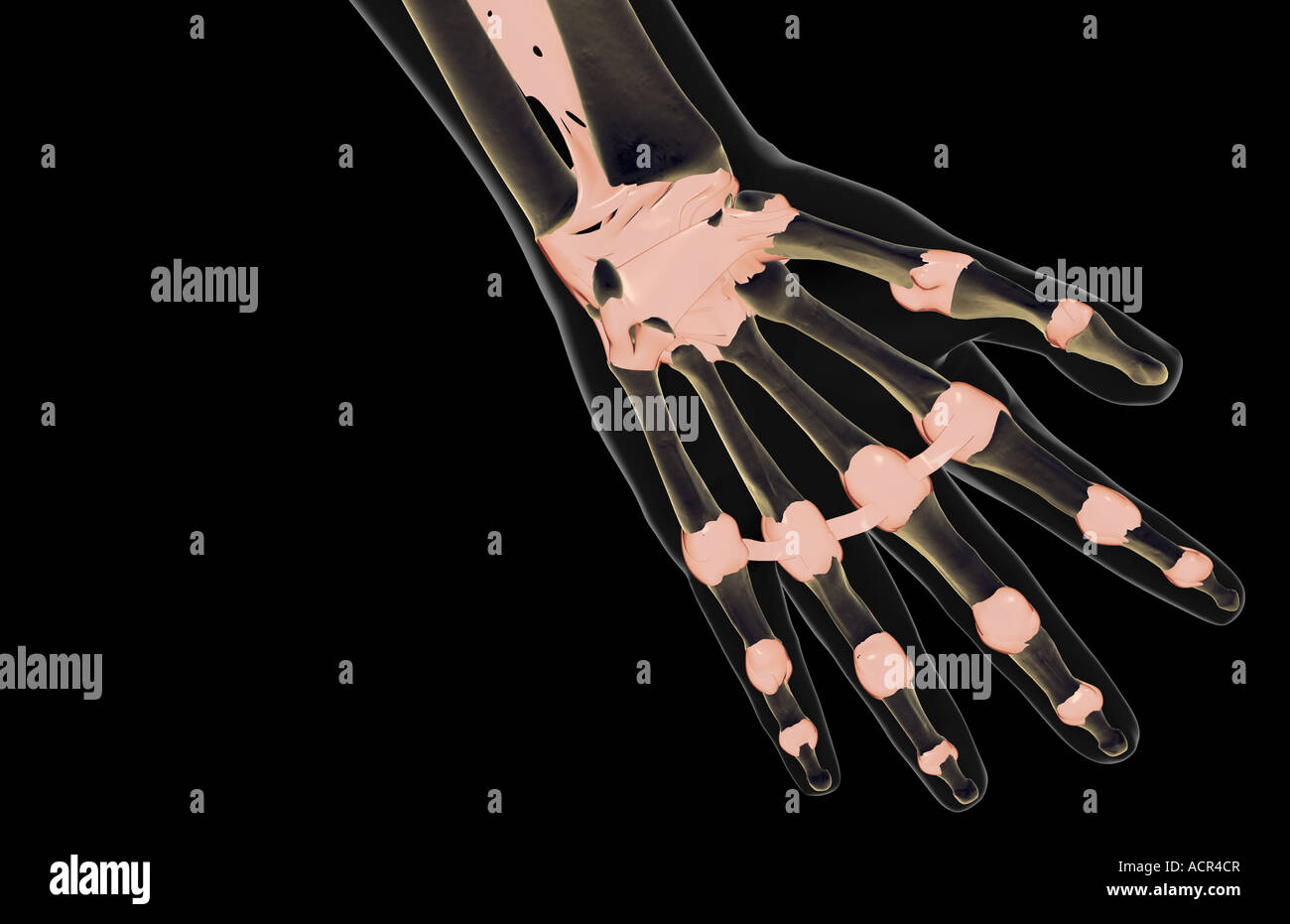 The ligaments of the hand Stock Photohttps://www.alamy.com/image-license-details/?v=1https://www.alamy.com/stock-photo-the-ligaments-of-the-hand-13213462.html
RFACR4CR–The ligaments of the hand
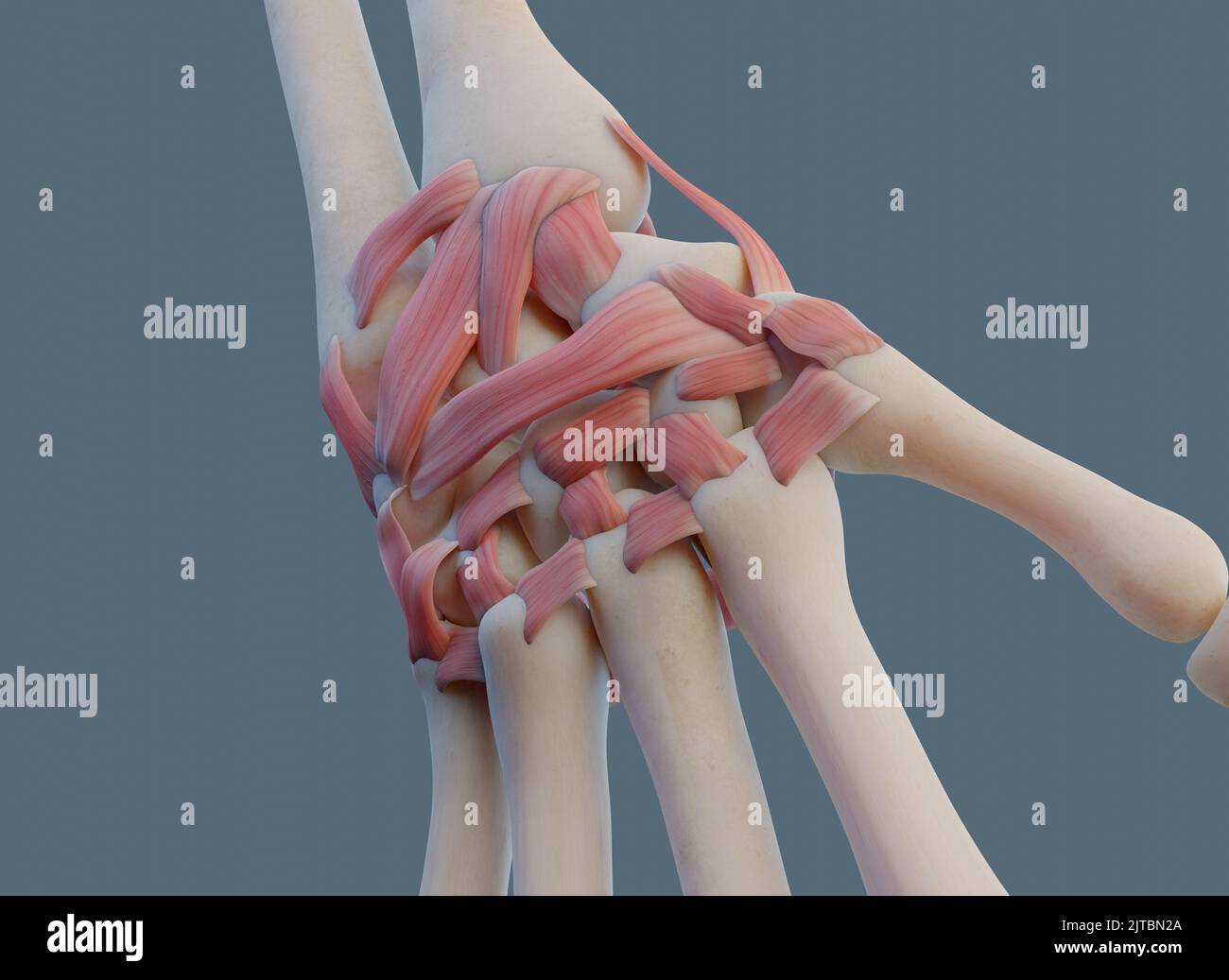 Close view of wrist joint, with ligaments and bones. Stock Photohttps://www.alamy.com/image-license-details/?v=1https://www.alamy.com/close-view-of-wrist-joint-with-ligaments-and-bones-image479689682.html
RF2JTBN2A–Close view of wrist joint, with ligaments and bones.
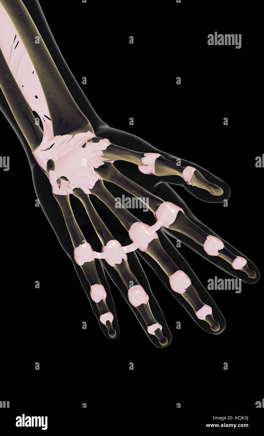 The ligaments of the hand Stock Photohttps://www.alamy.com/image-license-details/?v=1https://www.alamy.com/stock-photo-the-ligaments-of-the-hand-13171349.html
RFACJK3J–The ligaments of the hand
 Close view of wrist joint, with ligaments and bones. Stock Photohttps://www.alamy.com/image-license-details/?v=1https://www.alamy.com/close-view-of-wrist-joint-with-ligaments-and-bones-image479689677.html
RF2JTBN25–Close view of wrist joint, with ligaments and bones.
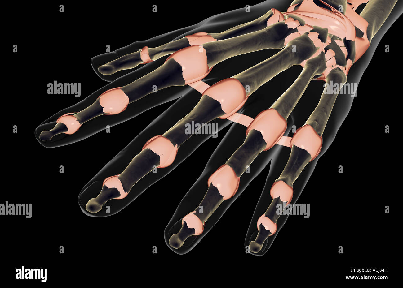 The ligaments of the hand Stock Photohttps://www.alamy.com/image-license-details/?v=1https://www.alamy.com/stock-photo-the-ligaments-of-the-hand-13167664.html
RFACJ84H–The ligaments of the hand
 Close view of wrist joint, with ligaments and bones. Stock Photohttps://www.alamy.com/image-license-details/?v=1https://www.alamy.com/close-view-of-wrist-joint-with-ligaments-and-bones-image479689672.html
RF2JTBN20–Close view of wrist joint, with ligaments and bones.
 The ligaments of the wrist Stock Photohttps://www.alamy.com/image-license-details/?v=1https://www.alamy.com/stock-photo-the-ligaments-of-the-wrist-13175993.html
RFACK4XJ–The ligaments of the wrist
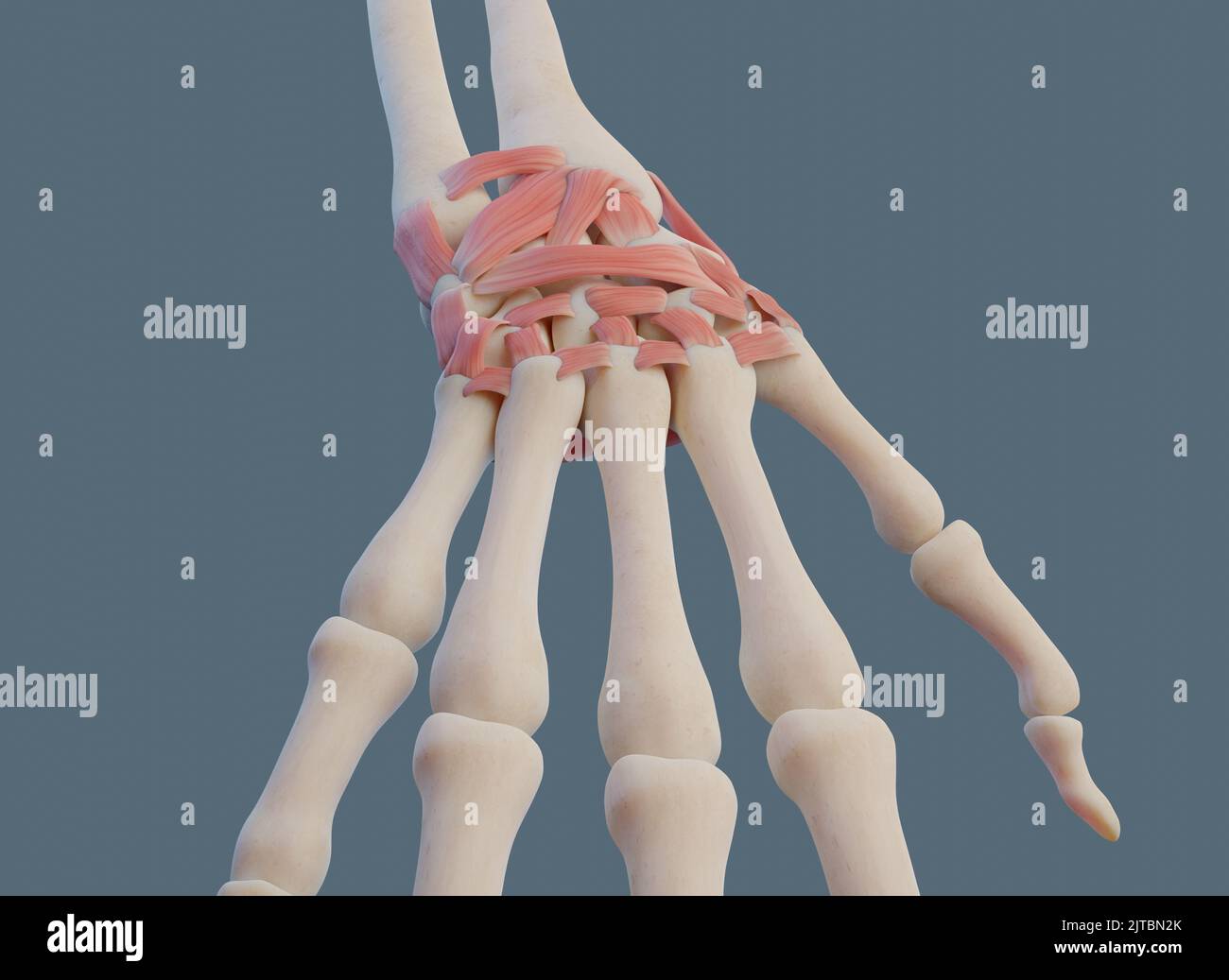 Close view of wrist joint, with ligaments and bones. Stock Photohttps://www.alamy.com/image-license-details/?v=1https://www.alamy.com/close-view-of-wrist-joint-with-ligaments-and-bones-image479689691.html
RF2JTBN2K–Close view of wrist joint, with ligaments and bones.
 The ligaments of the hand Stock Photohttps://www.alamy.com/image-license-details/?v=1https://www.alamy.com/stock-photo-the-ligaments-of-the-hand-13171965.html
RFACJMXP–The ligaments of the hand
 Close view of knee joint. With bones and ligaments, including meniscus. Stock Photohttps://www.alamy.com/image-license-details/?v=1https://www.alamy.com/close-view-of-knee-joint-with-bones-and-ligaments-including-meniscus-image479689565.html
RF2JTBMX5–Close view of knee joint. With bones and ligaments, including meniscus.
 The ligaments of the hand Stock Photohttps://www.alamy.com/image-license-details/?v=1https://www.alamy.com/stock-photo-the-ligaments-of-the-hand-13175043.html
RFACK23G–The ligaments of the hand
 Anterior view of knee joint. With bones and ligaments, including meniscus. Stock Photohttps://www.alamy.com/image-license-details/?v=1https://www.alamy.com/anterior-view-of-knee-joint-with-bones-and-ligaments-including-meniscus-image479689459.html
RF2JTBMPB–Anterior view of knee joint. With bones and ligaments, including meniscus.
 The ligaments of the forearm Stock Photohttps://www.alamy.com/image-license-details/?v=1https://www.alamy.com/stock-photo-the-ligaments-of-the-forearm-13172489.html
RFACJPEJ–The ligaments of the forearm
 Close view of knee joint. With bones and ligaments, including meniscus. Stock Photohttps://www.alamy.com/image-license-details/?v=1https://www.alamy.com/close-view-of-knee-joint-with-bones-and-ligaments-including-meniscus-image479689557.html
RF2JTBMWW–Close view of knee joint. With bones and ligaments, including meniscus.
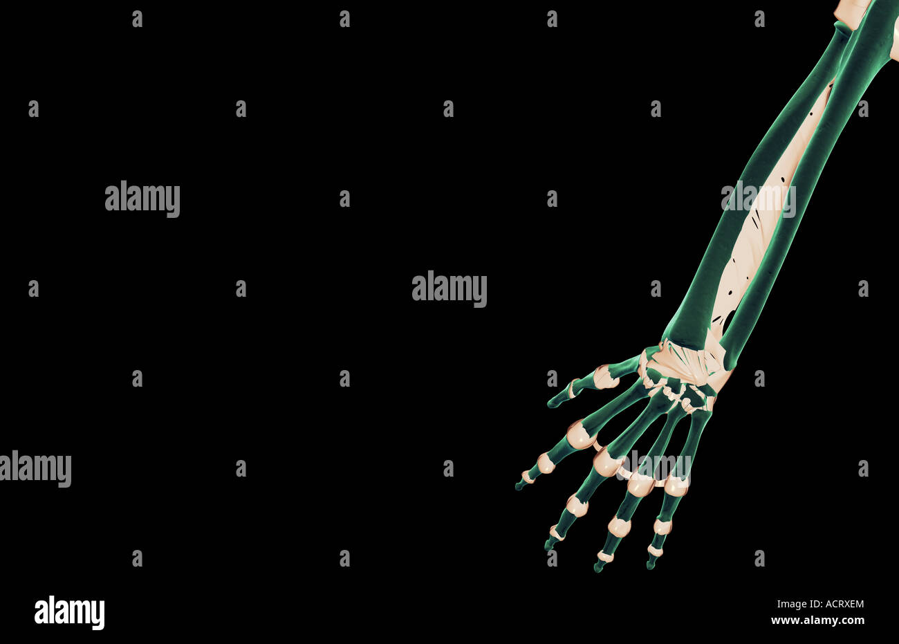 The ligaments of the forearm Stock Photohttps://www.alamy.com/image-license-details/?v=1https://www.alamy.com/stock-photo-the-ligaments-of-the-forearm-13220875.html
RFACRXEM–The ligaments of the forearm
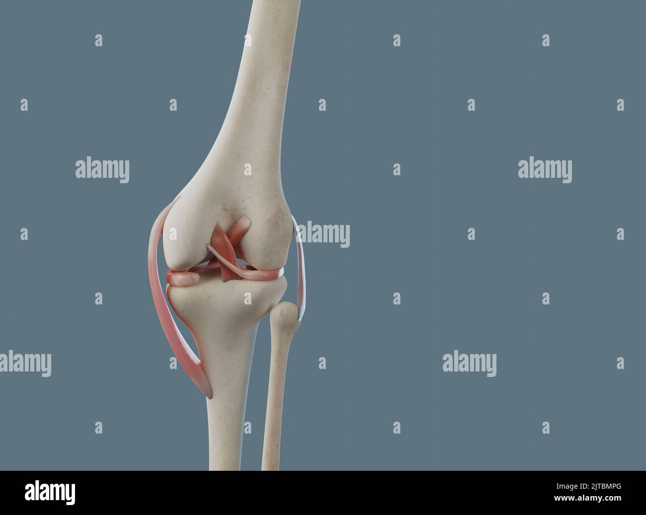 Posterior view of knee joint. With bones and ligaments, including meniscus. Stock Photohttps://www.alamy.com/image-license-details/?v=1https://www.alamy.com/posterior-view-of-knee-joint-with-bones-and-ligaments-including-meniscus-image479689464.html
RF2JTBMPG–Posterior view of knee joint. With bones and ligaments, including meniscus.
 The ligaments of the forearm Stock Photohttps://www.alamy.com/image-license-details/?v=1https://www.alamy.com/stock-photo-the-ligaments-of-the-forearm-13175943.html
RFACK4PG–The ligaments of the forearm
 Anterior view of knee joint. With bones and ligaments, including meniscus. Stock Photohttps://www.alamy.com/image-license-details/?v=1https://www.alamy.com/anterior-view-of-knee-joint-with-bones-and-ligaments-including-meniscus-image479689490.html
RF2JTBMRE–Anterior view of knee joint. With bones and ligaments, including meniscus.
 The ligaments of the upper limb Stock Photohttps://www.alamy.com/image-license-details/?v=1https://www.alamy.com/stock-photo-the-ligaments-of-the-upper-limb-13165563.html
RFACJ1WG–The ligaments of the upper limb
 Anterior and posterior views of knee joint. With bones and ligaments, including meniscus. Stock Photohttps://www.alamy.com/image-license-details/?v=1https://www.alamy.com/anterior-and-posterior-views-of-knee-joint-with-bones-and-ligaments-including-meniscus-image479689570.html
RF2JTBMXA–Anterior and posterior views of knee joint. With bones and ligaments, including meniscus.
 The ligaments of the upper limb Stock Photohttps://www.alamy.com/image-license-details/?v=1https://www.alamy.com/stock-photo-the-ligaments-of-the-upper-limb-13175162.html
RFACK2CY–The ligaments of the upper limb
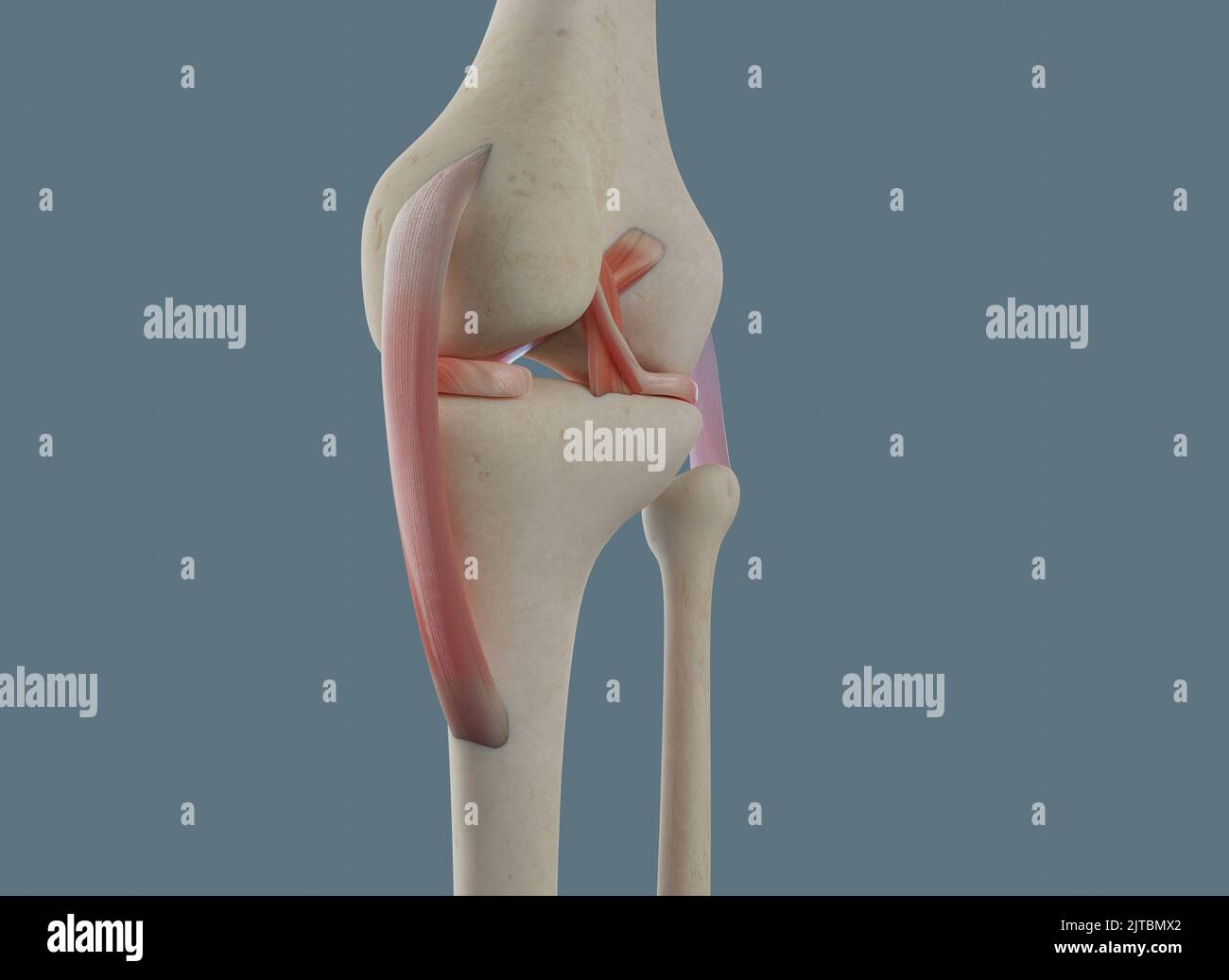 Close view of knee joint. With bones and ligaments, including meniscus. Stock Photohttps://www.alamy.com/image-license-details/?v=1https://www.alamy.com/close-view-of-knee-joint-with-bones-and-ligaments-including-meniscus-image479689562.html
RF2JTBMX2–Close view of knee joint. With bones and ligaments, including meniscus.
 The ligaments of the upper limb Stock Photohttps://www.alamy.com/image-license-details/?v=1https://www.alamy.com/stock-photo-the-ligaments-of-the-upper-limb-13168646.html
RFACJB1Y–The ligaments of the upper limb
 Posterior view of knee joint. With bones and ligaments, including meniscus. Stock Photohttps://www.alamy.com/image-license-details/?v=1https://www.alamy.com/posterior-view-of-knee-joint-with-bones-and-ligaments-including-meniscus-image479689560.html
RF2JTBMX0–Posterior view of knee joint. With bones and ligaments, including meniscus.
 The ligaments of the upper limb Stock Photohttps://www.alamy.com/image-license-details/?v=1https://www.alamy.com/stock-photo-the-ligaments-of-the-upper-limb-13174531.html
RFACK0GM–The ligaments of the upper limb
 Posterior view of knee joint. With bones and ligaments, including meniscus. Stock Photohttps://www.alamy.com/image-license-details/?v=1https://www.alamy.com/posterior-view-of-knee-joint-with-bones-and-ligaments-including-meniscus-image479689461.html
RF2JTBMPD–Posterior view of knee joint. With bones and ligaments, including meniscus.
 The ligaments of the upper limb Stock Photohttps://www.alamy.com/image-license-details/?v=1https://www.alamy.com/stock-photo-the-ligaments-of-the-upper-limb-13175573.html
RFACK3KJ–The ligaments of the upper limb
 Anterior view of knee joint. With bones and ligaments, including meniscus. Stock Photohttps://www.alamy.com/image-license-details/?v=1https://www.alamy.com/anterior-view-of-knee-joint-with-bones-and-ligaments-including-meniscus-image479689555.html
RF2JTBMWR–Anterior view of knee joint. With bones and ligaments, including meniscus.