Quick filters:
Renal fascia Stock Photos and Images
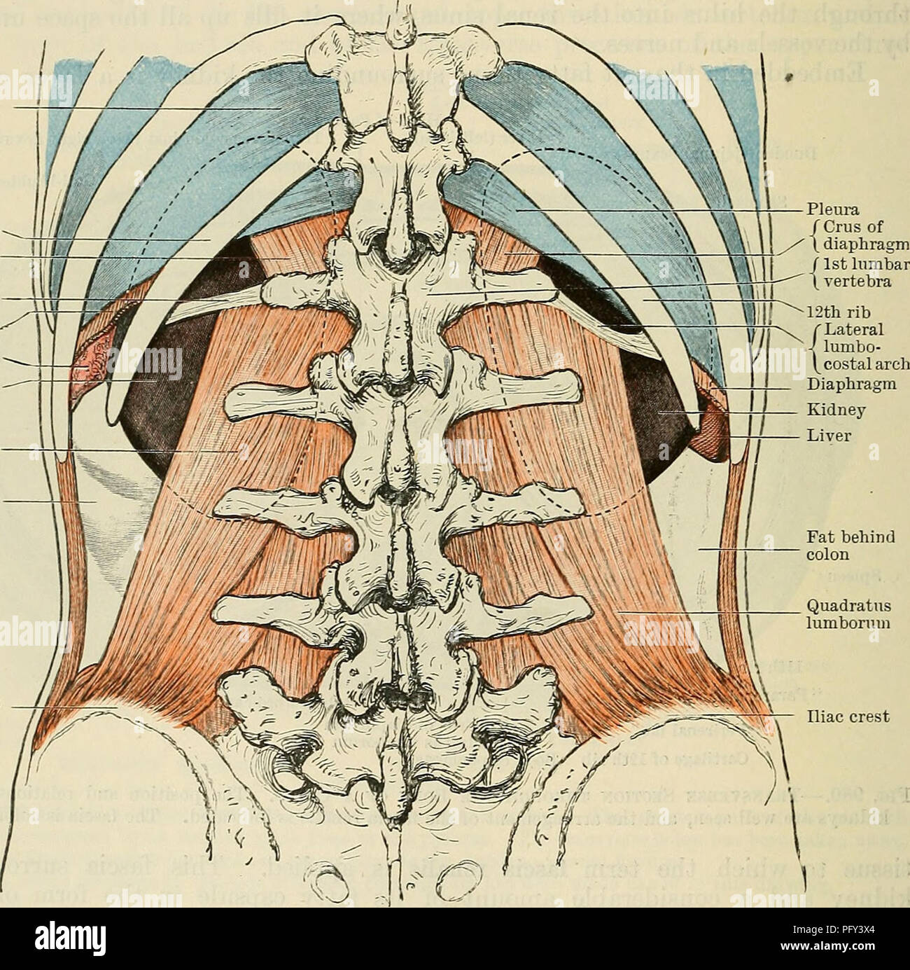
 . Cunningham's Text-book of anatomy. Anatomy. 1260 THE UKINO-GENITAL SYSTEM. the renal fascia by numerous connective tissue strands. These traverse the peri- renal fat and undoubtedly assist in fixing the kidney in its place. The pararenal fat is present in greatest quantity behind the inferior part of the kidney, and in this position the layer of fibrous tissue, separating the two masses of fat and forming the posterior layer of the sheath of renal fascia, is usually well marked. Fixation of the Kidney.—The kidney is not held in its place by any distinct ligaments, or special folds of periton Stock Photohttps://www.alamy.com/image-license-details/?v=1https://www.alamy.com/cunninghams-text-book-of-anatomy-anatomy-1260-the-ukino-genital-system-the-renal-fascia-by-numerous-connective-tissue-strands-these-traverse-the-peri-renal-fat-and-undoubtedly-assist-in-fixing-the-kidney-in-its-place-the-pararenal-fat-is-present-in-greatest-quantity-behind-the-inferior-part-of-the-kidney-and-in-this-position-the-layer-of-fibrous-tissue-separating-the-two-masses-of-fat-and-forming-the-posterior-layer-of-the-sheath-of-renal-fascia-is-usually-well-marked-fixation-of-the-kidneythe-kidney-is-not-held-in-its-place-by-any-distinct-ligaments-or-special-folds-of-periton-image231848861.html
RMRD5HGD–. Cunningham's Text-book of anatomy. Anatomy. 1260 THE UKINO-GENITAL SYSTEM. the renal fascia by numerous connective tissue strands. These traverse the peri- renal fat and undoubtedly assist in fixing the kidney in its place. The pararenal fat is present in greatest quantity behind the inferior part of the kidney, and in this position the layer of fibrous tissue, separating the two masses of fat and forming the posterior layer of the sheath of renal fascia, is usually well marked. Fixation of the Kidney.—The kidney is not held in its place by any distinct ligaments, or special folds of periton
 . Cunningham's Text-book of anatomy. Anatomy. 1260 THE UKINO-GENITAL SYSTEM. the renal fascia by numerous connective tissue strands. These traverse the peri- renal fat and undoubtedly assist in fixing the kidney in its place. The pararenal fat is present in greatest quantity behind the inferior part of the kidney, and in this position the layer of fibrous tissue, separating the two masses of fat and forming the posterior layer of the sheath of renal fascia, is usually well marked. Fixation of the Kidney.—The kidney is not held in its place by any distinct ligaments, or special folds of periton Stock Photohttps://www.alamy.com/image-license-details/?v=1https://www.alamy.com/cunninghams-text-book-of-anatomy-anatomy-1260-the-ukino-genital-system-the-renal-fascia-by-numerous-connective-tissue-strands-these-traverse-the-peri-renal-fat-and-undoubtedly-assist-in-fixing-the-kidney-in-its-place-the-pararenal-fat-is-present-in-greatest-quantity-behind-the-inferior-part-of-the-kidney-and-in-this-position-the-layer-of-fibrous-tissue-separating-the-two-masses-of-fat-and-forming-the-posterior-layer-of-the-sheath-of-renal-fascia-is-usually-well-marked-fixation-of-the-kidneythe-kidney-is-not-held-in-its-place-by-any-distinct-ligaments-or-special-folds-of-periton-image216340044.html
RMPFY3X4–. Cunningham's Text-book of anatomy. Anatomy. 1260 THE UKINO-GENITAL SYSTEM. the renal fascia by numerous connective tissue strands. These traverse the peri- renal fat and undoubtedly assist in fixing the kidney in its place. The pararenal fat is present in greatest quantity behind the inferior part of the kidney, and in this position the layer of fibrous tissue, separating the two masses of fat and forming the posterior layer of the sheath of renal fascia, is usually well marked. Fixation of the Kidney.—The kidney is not held in its place by any distinct ligaments, or special folds of periton
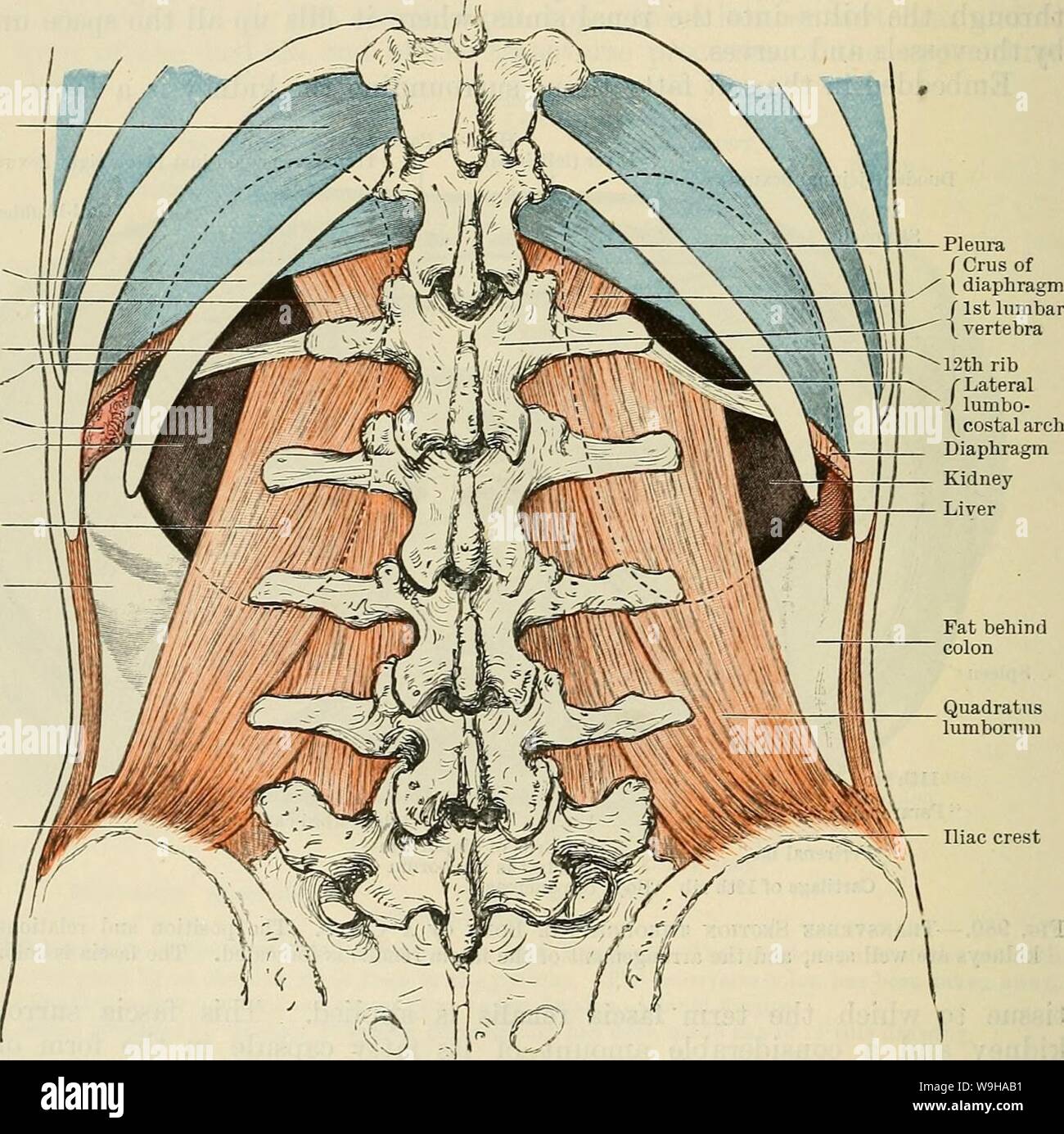 Archive image from page 1293 of Cunningham's Text-book of anatomy (1914). Cunningham's Text-book of anatomy cunninghamstextb00cunn Year: 1914 ( 1260 THE UKINO-GENITAL SYSTEM. the renal fascia by numerous connective tissue strands. These traverse the peri- renal fat and undoubtedly assist in fixing the kidney in its place. The pararenal fat is present in greatest quantity behind the inferior part of the kidney, and in this position the layer of fibrous tissue, separating the two masses of fat and forming the posterior layer of the sheath of renal fascia, is usually well marked. Fixation of the Stock Photohttps://www.alamy.com/image-license-details/?v=1https://www.alamy.com/archive-image-from-page-1293-of-cunninghams-text-book-of-anatomy-1914-cunninghams-text-book-of-anatomy-cunninghamstextb00cunn-year-1914-1260-the-ukino-genital-system-the-renal-fascia-by-numerous-connective-tissue-strands-these-traverse-the-peri-renal-fat-and-undoubtedly-assist-in-fixing-the-kidney-in-its-place-the-pararenal-fat-is-present-in-greatest-quantity-behind-the-inferior-part-of-the-kidney-and-in-this-position-the-layer-of-fibrous-tissue-separating-the-two-masses-of-fat-and-forming-the-posterior-layer-of-the-sheath-of-renal-fascia-is-usually-well-marked-fixation-of-the-image264068757.html
RMW9HAB1–Archive image from page 1293 of Cunningham's Text-book of anatomy (1914). Cunningham's Text-book of anatomy cunninghamstextb00cunn Year: 1914 ( 1260 THE UKINO-GENITAL SYSTEM. the renal fascia by numerous connective tissue strands. These traverse the peri- renal fat and undoubtedly assist in fixing the kidney in its place. The pararenal fat is present in greatest quantity behind the inferior part of the kidney, and in this position the layer of fibrous tissue, separating the two masses of fat and forming the posterior layer of the sheath of renal fascia, is usually well marked. Fixation of the
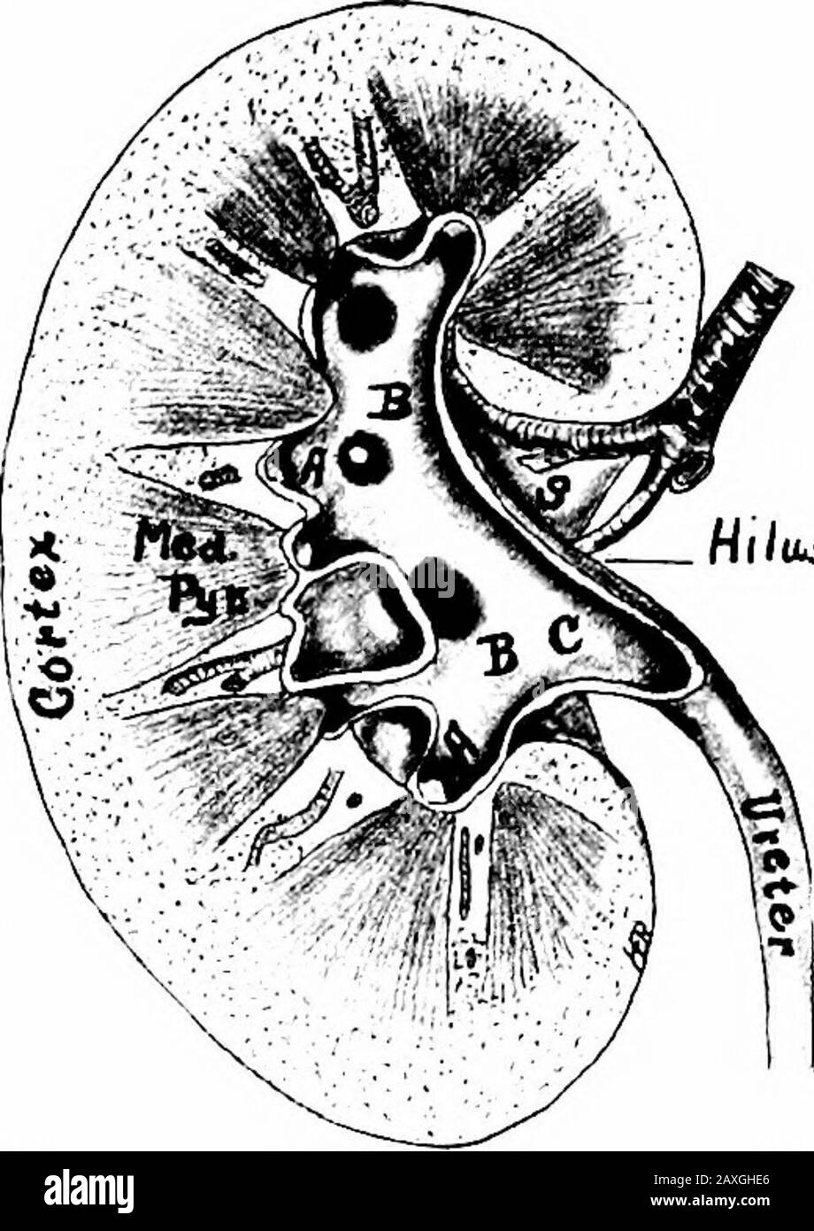 A manual of anatomy . Pararenal fat. The Organ is held inof the right kidney. A. A, Calyces position by the surrounding organs, minores; B, B, calyces majores; C, pelvis ! • n i i i i i of the ureter; 5, hilus of the kidney. ctiietty, and somcwhat by the renal fascia. The vessels of Ike kidney are the renal artery and the renal vein.The nerves are about fifteen in number and are derived from therenal plexus. This plexus is formed by branches from the solarplexus, celiac ganglion, aortic plexus and lesser and least splanchnicnerves. Branches from the tenth, eleventh and twelfth thoracicnerves a Stock Photohttps://www.alamy.com/image-license-details/?v=1https://www.alamy.com/a-manual-of-anatomy-pararenal-fat-the-organ-is-held-inof-the-right-kidney-a-a-calyces-position-by-the-surrounding-organs-minores-b-b-calyces-majores-c-pelvis-!-n-i-i-i-i-i-of-the-ureter-5-hilus-of-the-kidney-ctiietty-and-somcwhat-by-the-renal-fascia-the-vessels-of-ike-kidney-are-the-renal-artery-and-the-renal-veinthe-nerves-are-about-fifteen-in-number-and-are-derived-from-therenal-plexus-this-plexus-is-formed-by-branches-from-the-solarplexus-celiac-ganglion-aortic-plexus-and-lesser-and-least-splanchnicnerves-branches-from-the-tenth-eleventh-and-twelfth-thoracicnerves-a-image343343006.html
RM2AXGHE6–A manual of anatomy . Pararenal fat. The Organ is held inof the right kidney. A. A, Calyces position by the surrounding organs, minores; B, B, calyces majores; C, pelvis ! • n i i i i i of the ureter; 5, hilus of the kidney. ctiietty, and somcwhat by the renal fascia. The vessels of Ike kidney are the renal artery and the renal vein.The nerves are about fifteen in number and are derived from therenal plexus. This plexus is formed by branches from the solarplexus, celiac ganglion, aortic plexus and lesser and least splanchnicnerves. Branches from the tenth, eleventh and twelfth thoracicnerves a
 . Gynecology : . gers is seen a floor of fat of orange-yellow color. This fat layer,which varies greatly in thickness in different individuals, is continuous with theretroperitoneal fat of the iliac fossa and anterior abdominal wall. Between this retroperitoneal layer of fat and the kidney is another layer offat separated from it by a membrane, the retro renal fascia (Gerotas capsule).The last-named fat layer closely surrounds the kidney. In order to expose thekidney this layer must be opened. By gradual traction on the fatty capsule soo GYNECOLOGY with pressure forceps a movable kidney may be Stock Photohttps://www.alamy.com/image-license-details/?v=1https://www.alamy.com/gynecology-gers-is-seen-a-floor-of-fat-of-orange-yellow-color-this-fat-layerwhich-varies-greatly-in-thickness-in-different-individuals-is-continuous-with-theretroperitoneal-fat-of-the-iliac-fossa-and-anterior-abdominal-wall-between-this-retroperitoneal-layer-of-fat-and-the-kidney-is-another-layer-offat-separated-from-it-by-a-membrane-the-retro-renal-fascia-gerotas-capsulethe-last-named-fat-layer-closely-surrounds-the-kidney-in-order-to-expose-thekidney-this-layer-must-be-opened-by-gradual-traction-on-the-fatty-capsule-soo-gynecology-with-pressure-forceps-a-movable-kidney-may-be-image369769065.html
RM2CDGC61–. Gynecology : . gers is seen a floor of fat of orange-yellow color. This fat layer,which varies greatly in thickness in different individuals, is continuous with theretroperitoneal fat of the iliac fossa and anterior abdominal wall. Between this retroperitoneal layer of fat and the kidney is another layer offat separated from it by a membrane, the retro renal fascia (Gerotas capsule).The last-named fat layer closely surrounds the kidney. In order to expose thekidney this layer must be opened. By gradual traction on the fatty capsule soo GYNECOLOGY with pressure forceps a movable kidney may be
![. Comparative anatomy. Anatomy, Comparative. 4i6 COMPARATIVE ANATOMY with a slit-like aperture, the hilum. The kidneys are held in position ])oth by the peritoneum and by a connective tissue renal fascia. Only upper (anterior) and lower (posterior) surfaces of the kidneys are covered with peritoneum, the middle of the right kidney being crossed by the colon, that of the left by the pancreas. These relations, however, are subject to considerable variation. Structiire. A cross section of the human kidney in the region of the hilum shows that under its peritoneal and fatty investments the kidney. Stock Photo . Comparative anatomy. Anatomy, Comparative. 4i6 COMPARATIVE ANATOMY with a slit-like aperture, the hilum. The kidneys are held in position ])oth by the peritoneum and by a connective tissue renal fascia. Only upper (anterior) and lower (posterior) surfaces of the kidneys are covered with peritoneum, the middle of the right kidney being crossed by the colon, that of the left by the pancreas. These relations, however, are subject to considerable variation. Structiire. A cross section of the human kidney in the region of the hilum shows that under its peritoneal and fatty investments the kidney. Stock Photo](https://c8.alamy.com/comp/REF905/comparative-anatomy-anatomy-comparative-4i6-comparative-anatomy-with-a-slit-like-aperture-the-hilum-the-kidneys-are-held-in-position-oth-by-the-peritoneum-and-by-a-connective-tissue-renal-fascia-only-upper-anterior-and-lower-posterior-surfaces-of-the-kidneys-are-covered-with-peritoneum-the-middle-of-the-right-kidney-being-crossed-by-the-colon-that-of-the-left-by-the-pancreas-these-relations-however-are-subject-to-considerable-variation-structiire-a-cross-section-of-the-human-kidney-in-the-region-of-the-hilum-shows-that-under-its-peritoneal-and-fatty-investments-the-kidney-REF905.jpg) . Comparative anatomy. Anatomy, Comparative. 4i6 COMPARATIVE ANATOMY with a slit-like aperture, the hilum. The kidneys are held in position ])oth by the peritoneum and by a connective tissue renal fascia. Only upper (anterior) and lower (posterior) surfaces of the kidneys are covered with peritoneum, the middle of the right kidney being crossed by the colon, that of the left by the pancreas. These relations, however, are subject to considerable variation. Structiire. A cross section of the human kidney in the region of the hilum shows that under its peritoneal and fatty investments the kidney. Stock Photohttps://www.alamy.com/image-license-details/?v=1https://www.alamy.com/comparative-anatomy-anatomy-comparative-4i6-comparative-anatomy-with-a-slit-like-aperture-the-hilum-the-kidneys-are-held-in-position-oth-by-the-peritoneum-and-by-a-connective-tissue-renal-fascia-only-upper-anterior-and-lower-posterior-surfaces-of-the-kidneys-are-covered-with-peritoneum-the-middle-of-the-right-kidney-being-crossed-by-the-colon-that-of-the-left-by-the-pancreas-these-relations-however-are-subject-to-considerable-variation-structiire-a-cross-section-of-the-human-kidney-in-the-region-of-the-hilum-shows-that-under-its-peritoneal-and-fatty-investments-the-kidney-image232676309.html
. Comparative anatomy. Anatomy, Comparative. 4i6 COMPARATIVE ANATOMY with a slit-like aperture, the hilum. The kidneys are held in position ])oth by the peritoneum and by a connective tissue renal fascia. Only upper (anterior) and lower (posterior) surfaces of the kidneys are covered with peritoneum, the middle of the right kidney being crossed by the colon, that of the left by the pancreas. These relations, however, are subject to considerable variation. Structiire. A cross section of the human kidney in the region of the hilum shows that under its peritoneal and fatty investments the kidney. Stock Photohttps://www.alamy.com/image-license-details/?v=1https://www.alamy.com/comparative-anatomy-anatomy-comparative-4i6-comparative-anatomy-with-a-slit-like-aperture-the-hilum-the-kidneys-are-held-in-position-oth-by-the-peritoneum-and-by-a-connective-tissue-renal-fascia-only-upper-anterior-and-lower-posterior-surfaces-of-the-kidneys-are-covered-with-peritoneum-the-middle-of-the-right-kidney-being-crossed-by-the-colon-that-of-the-left-by-the-pancreas-these-relations-however-are-subject-to-considerable-variation-structiire-a-cross-section-of-the-human-kidney-in-the-region-of-the-hilum-shows-that-under-its-peritoneal-and-fatty-investments-the-kidney-image232676309.htmlRMREF905–. Comparative anatomy. Anatomy, Comparative. 4i6 COMPARATIVE ANATOMY with a slit-like aperture, the hilum. The kidneys are held in position ])oth by the peritoneum and by a connective tissue renal fascia. Only upper (anterior) and lower (posterior) surfaces of the kidneys are covered with peritoneum, the middle of the right kidney being crossed by the colon, that of the left by the pancreas. These relations, however, are subject to considerable variation. Structiire. A cross section of the human kidney in the region of the hilum shows that under its peritoneal and fatty investments the kidney.
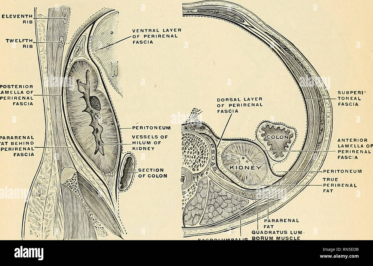 . Anatomy, descriptive and applied. Anatomy. Fig. 1110.—The posterior surfaces of the kidneys, showing areas of relation to the parietes.. Fig. 1111—Longitudinal section, showing the arrangement of the renal fascia. (After Gerota.) RARENAL FAT QUADRATUS LUM" SACROLUMBALIS BORUM MUSCUE GROUP Fig. 1112.—Transverse section, showing the relations of the renal fascia and the two layers of fat. (After Gerota.) Borders.—The external border {marcio lateralis) (Figs. 1107 and 1108) is convex, and is directed outward and backward, toward the posterolateral wall of the. Please note that these images Stock Photohttps://www.alamy.com/image-license-details/?v=1https://www.alamy.com/anatomy-descriptive-and-applied-anatomy-fig-1110the-posterior-surfaces-of-the-kidneys-showing-areas-of-relation-to-the-parietes-fig-1111longitudinal-section-showing-the-arrangement-of-the-renal-fascia-after-gerota-rarenal-fat-quadratus-lumquot-sacrolumbalis-borum-muscue-group-fig-1112transverse-section-showing-the-relations-of-the-renal-fascia-and-the-two-layers-of-fat-after-gerota-bordersthe-external-border-marcio-lateralis-figs-1107-and-1108-is-convex-and-is-directed-outward-and-backward-toward-the-posterolateral-wall-of-the-please-note-that-these-images-image236763671.html
RMRN5EDB–. Anatomy, descriptive and applied. Anatomy. Fig. 1110.—The posterior surfaces of the kidneys, showing areas of relation to the parietes.. Fig. 1111—Longitudinal section, showing the arrangement of the renal fascia. (After Gerota.) RARENAL FAT QUADRATUS LUM" SACROLUMBALIS BORUM MUSCUE GROUP Fig. 1112.—Transverse section, showing the relations of the renal fascia and the two layers of fat. (After Gerota.) Borders.—The external border {marcio lateralis) (Figs. 1107 and 1108) is convex, and is directed outward and backward, toward the posterolateral wall of the. Please note that these images
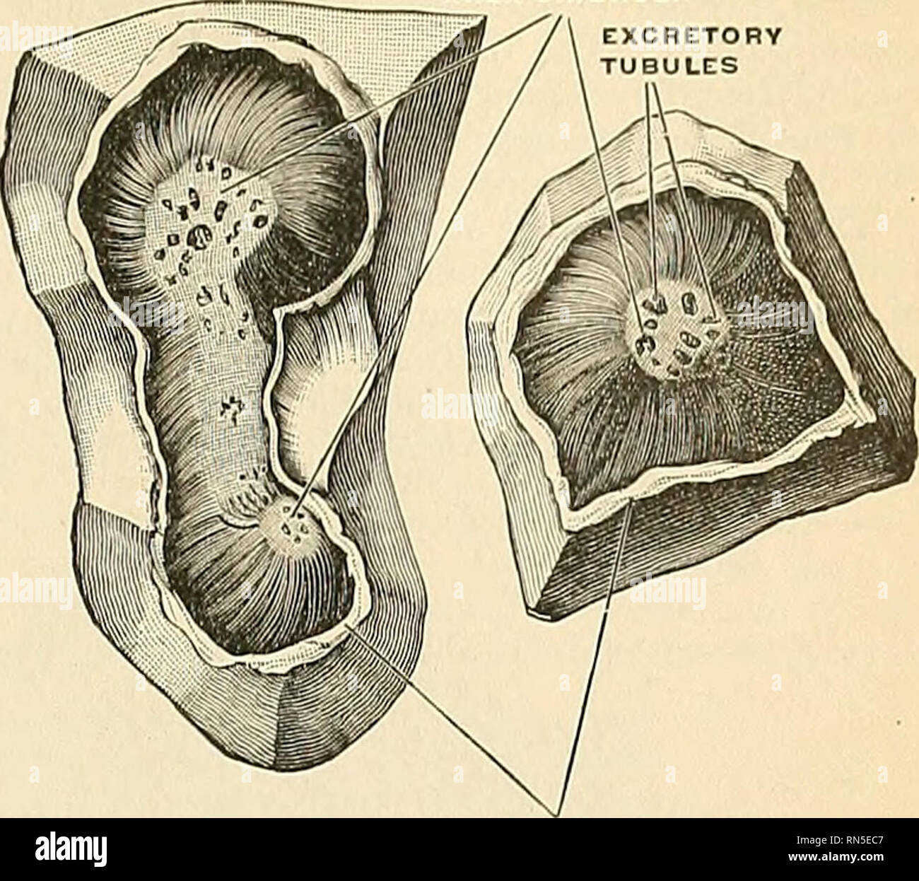 . Anatomy, descriptive and applied. Anatomy. THE KIDNEYS 1349. WALL OF RENAL CALIX Fig. 1114.—Area cribrosa of renal papilla. (Toldt.) pararenal or Transversalis fat. The kidney is held in position throngh the at- tachments of the fascia renahs and by the apposition of the neighboriiis viscera. General Structure of the Kidney.— ^„e:a chibhosa The Uiilncy is invested by a capsule of interlacing bundles of fibrous connective tissue (tunica fibrosa), which forms a firm, smooth covering for the organ. The cap- sule can be easily stripped off, but in doing so, numerous fine processes of connective Stock Photohttps://www.alamy.com/image-license-details/?v=1https://www.alamy.com/anatomy-descriptive-and-applied-anatomy-the-kidneys-1349-wall-of-renal-calix-fig-1114area-cribrosa-of-renal-papilla-toldt-pararenal-or-transversalis-fat-the-kidney-is-held-in-position-throngh-the-at-tachments-of-the-fascia-renahs-and-by-the-apposition-of-the-neighboriiis-viscera-general-structure-of-the-kidney-ea-chibhosa-the-uiilncy-is-invested-by-a-capsule-of-interlacing-bundles-of-fibrous-connective-tissue-tunica-fibrosa-which-forms-a-firm-smooth-covering-for-the-organ-the-cap-sule-can-be-easily-stripped-off-but-in-doing-so-numerous-fine-processes-of-connective-image236763639.html
RMRN5EC7–. Anatomy, descriptive and applied. Anatomy. THE KIDNEYS 1349. WALL OF RENAL CALIX Fig. 1114.—Area cribrosa of renal papilla. (Toldt.) pararenal or Transversalis fat. The kidney is held in position throngh the at- tachments of the fascia renahs and by the apposition of the neighboriiis viscera. General Structure of the Kidney.— ^„e:a chibhosa The Uiilncy is invested by a capsule of interlacing bundles of fibrous connective tissue (tunica fibrosa), which forms a firm, smooth covering for the organ. The cap- sule can be easily stripped off, but in doing so, numerous fine processes of connective
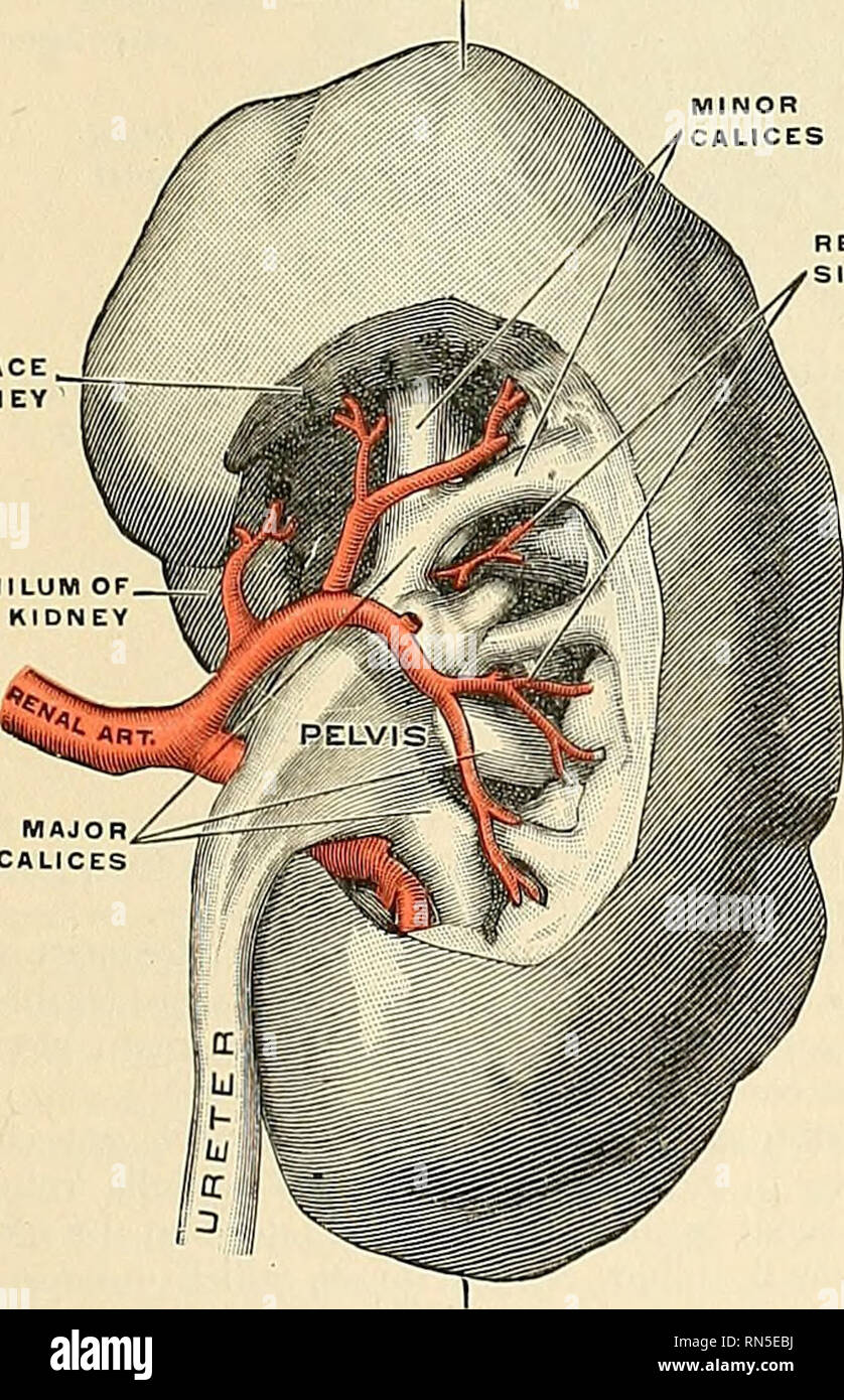 . Anatomy, descriptive and applied. Anatomy. WALL OF RENAL CALIX Fig. 1114.—Area cribrosa of renal papilla. (Toldt.) pararenal or Transversalis fat. The kidney is held in position throngh the at- tachments of the fascia renahs and by the apposition of the neighboriiis viscera. General Structure of the Kidney.— ^„e:a chibhosa The Uiilncy is invested by a capsule of interlacing bundles of fibrous connective tissue (tunica fibrosa), which forms a firm, smooth covering for the organ. The cap- sule can be easily stripped off, but in doing so, numerous fine processes of connective tissue and .small Stock Photohttps://www.alamy.com/image-license-details/?v=1https://www.alamy.com/anatomy-descriptive-and-applied-anatomy-wall-of-renal-calix-fig-1114area-cribrosa-of-renal-papilla-toldt-pararenal-or-transversalis-fat-the-kidney-is-held-in-position-throngh-the-at-tachments-of-the-fascia-renahs-and-by-the-apposition-of-the-neighboriiis-viscera-general-structure-of-the-kidney-ea-chibhosa-the-uiilncy-is-invested-by-a-capsule-of-interlacing-bundles-of-fibrous-connective-tissue-tunica-fibrosa-which-forms-a-firm-smooth-covering-for-the-organ-the-cap-sule-can-be-easily-stripped-off-but-in-doing-so-numerous-fine-processes-of-connective-tissue-and-small-image236763622.html
RMRN5EBJ–. Anatomy, descriptive and applied. Anatomy. WALL OF RENAL CALIX Fig. 1114.—Area cribrosa of renal papilla. (Toldt.) pararenal or Transversalis fat. The kidney is held in position throngh the at- tachments of the fascia renahs and by the apposition of the neighboriiis viscera. General Structure of the Kidney.— ^„e:a chibhosa The Uiilncy is invested by a capsule of interlacing bundles of fibrous connective tissue (tunica fibrosa), which forms a firm, smooth covering for the organ. The cap- sule can be easily stripped off, but in doing so, numerous fine processes of connective tissue and .small
 . The anatomy of the domestic animals . Veterinary anatomy. 556 THE URINARY ORGANS OF THE HORSE or three lumbar transverse processes. The dorsal surface is convex, and is related to the left crus of the diaphragm, the iliac fascia and psoas muscles, and the dorsal end Right adrenal Left adrenal. Left ureter Fig. 496.—Kidneys antj Adrenals of Horse; Dorsal ^'iew. Hardened in situ. Impression of seventeenth rib on right kidney is indicated by small cross. The left kidney was a little further forward in this subject than is usual. Ureters Renal vein. Please note that these images are extracted fr Stock Photohttps://www.alamy.com/image-license-details/?v=1https://www.alamy.com/the-anatomy-of-the-domestic-animals-veterinary-anatomy-556-the-urinary-organs-of-the-horse-or-three-lumbar-transverse-processes-the-dorsal-surface-is-convex-and-is-related-to-the-left-crus-of-the-diaphragm-the-iliac-fascia-and-psoas-muscles-and-the-dorsal-end-right-adrenal-left-adrenal-left-ureter-fig-496kidneys-antj-adrenals-of-horse-dorsal-iew-hardened-in-situ-impression-of-seventeenth-rib-on-right-kidney-is-indicated-by-small-cross-the-left-kidney-was-a-little-further-forward-in-this-subject-than-is-usual-ureters-renal-vein-please-note-that-these-images-are-extracted-fr-image232324237.html
RMRDY7X5–. The anatomy of the domestic animals . Veterinary anatomy. 556 THE URINARY ORGANS OF THE HORSE or three lumbar transverse processes. The dorsal surface is convex, and is related to the left crus of the diaphragm, the iliac fascia and psoas muscles, and the dorsal end Right adrenal Left adrenal. Left ureter Fig. 496.—Kidneys antj Adrenals of Horse; Dorsal ^'iew. Hardened in situ. Impression of seventeenth rib on right kidney is indicated by small cross. The left kidney was a little further forward in this subject than is usual. Ureters Renal vein. Please note that these images are extracted fr
 . Atlas of applied (topographical) human anatomy for students and practitioners. Anatomy. tissinius Dorsi Muscle Serratus Posticus Inferior Muscle Sacro Spinalis Muscle ;ih Rib Right Kidney^-^^g Inferior Vena Cava-— ^=' Renal Vessels—-^m Ureter a Ascending C nlon Quadratus Lumboruni Muscle Iliohypogastric Nerve Spermatic Vessels Iliu-inguinal Nerve Superficial Lamella of Lumbar Fascia Lateral Cutaneous Branch Gluteus Maxtmus Muscle. Gluteus Medius Muscle Origin of Transversalis Abdominis Muscle External Abdominal Oblique Muscle Internal Abdominal Oblique Muscle Fig. 134. Eight Kidney, Expose Stock Photohttps://www.alamy.com/image-license-details/?v=1https://www.alamy.com/atlas-of-applied-topographical-human-anatomy-for-students-and-practitioners-anatomy-tissinius-dorsi-muscle-serratus-posticus-inferior-muscle-sacro-spinalis-muscle-ih-rib-right-kidney-g-inferior-vena-cava-=-renal-vessels-m-ureter-a-ascending-c-nlon-quadratus-lumboruni-muscle-iliohypogastric-nerve-spermatic-vessels-iliu-inguinal-nerve-superficial-lamella-of-lumbar-fascia-lateral-cutaneous-branch-gluteus-maxtmus-muscle-gluteus-medius-muscle-origin-of-transversalis-abdominis-muscle-external-abdominal-oblique-muscle-internal-abdominal-oblique-muscle-fig-134-eight-kidney-expose-image235400954.html
RMRJYC8X–. Atlas of applied (topographical) human anatomy for students and practitioners. Anatomy. tissinius Dorsi Muscle Serratus Posticus Inferior Muscle Sacro Spinalis Muscle ;ih Rib Right Kidney^-^^g Inferior Vena Cava-— ^=' Renal Vessels—-^m Ureter a Ascending C nlon Quadratus Lumboruni Muscle Iliohypogastric Nerve Spermatic Vessels Iliu-inguinal Nerve Superficial Lamella of Lumbar Fascia Lateral Cutaneous Branch Gluteus Maxtmus Muscle. Gluteus Medius Muscle Origin of Transversalis Abdominis Muscle External Abdominal Oblique Muscle Internal Abdominal Oblique Muscle Fig. 134. Eight Kidney, Expose
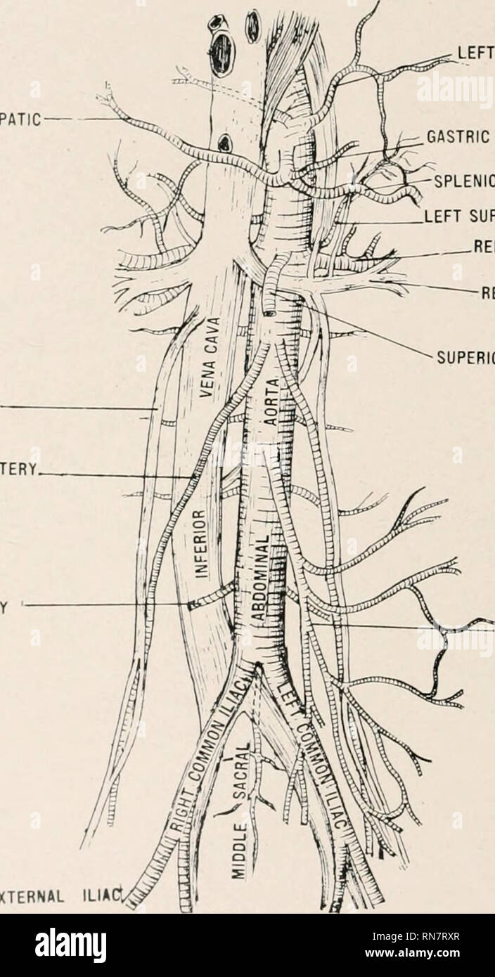 . Anatomy in a nutshell : a treatise on human anatomy in its relation to osteopathy. Human anatomy; Osteopathic medicine; Osteopathic Medicine; Anatomy. 228 ANATOMY IN A NUTSHELL. 112. WhyJ called popliteal? 113. Give contents. 114. Name the Ligaments of the ankle. 115. What forms the crucial anastomosis? 116. What muscles form the tendo Achillis? 117. Why is the Plantaris so called? 118. How many bones in the foot? Hi). Describe the fascia lata. PLATE XC. HEPATIC SPERMATIC VEIN SPERMATIC ARTERY RIGHT LUMBAR ARTERY. LEFT PHRENIC GASTRIC ARTERY -SPLENIC ARTERY LEFT SUPRARENAL RENAL ARTERY, RENA Stock Photohttps://www.alamy.com/image-license-details/?v=1https://www.alamy.com/anatomy-in-a-nutshell-a-treatise-on-human-anatomy-in-its-relation-to-osteopathy-human-anatomy-osteopathic-medicine-osteopathic-medicine-anatomy-228-anatomy-in-a-nutshell-112-whyj-called-popliteal-113-give-contents-114-name-the-ligaments-of-the-ankle-115-what-forms-the-crucial-anastomosis-116-what-muscles-form-the-tendo-achillis-117-why-is-the-plantaris-so-called-118-how-many-bones-in-the-foot-hi-describe-the-fascia-lata-plate-xc-hepatic-spermatic-vein-spermatic-artery-right-lumbar-artery-left-phrenic-gastric-artery-splenic-artery-left-suprarenal-renal-artery-rena-image236815007.html
RMRN7RXR–. Anatomy in a nutshell : a treatise on human anatomy in its relation to osteopathy. Human anatomy; Osteopathic medicine; Osteopathic Medicine; Anatomy. 228 ANATOMY IN A NUTSHELL. 112. WhyJ called popliteal? 113. Give contents. 114. Name the Ligaments of the ankle. 115. What forms the crucial anastomosis? 116. What muscles form the tendo Achillis? 117. Why is the Plantaris so called? 118. How many bones in the foot? Hi). Describe the fascia lata. PLATE XC. HEPATIC SPERMATIC VEIN SPERMATIC ARTERY RIGHT LUMBAR ARTERY. LEFT PHRENIC GASTRIC ARTERY -SPLENIC ARTERY LEFT SUPRARENAL RENAL ARTERY, RENA