Quick filters:
Renal pyramid kidney Stock Photos and Images
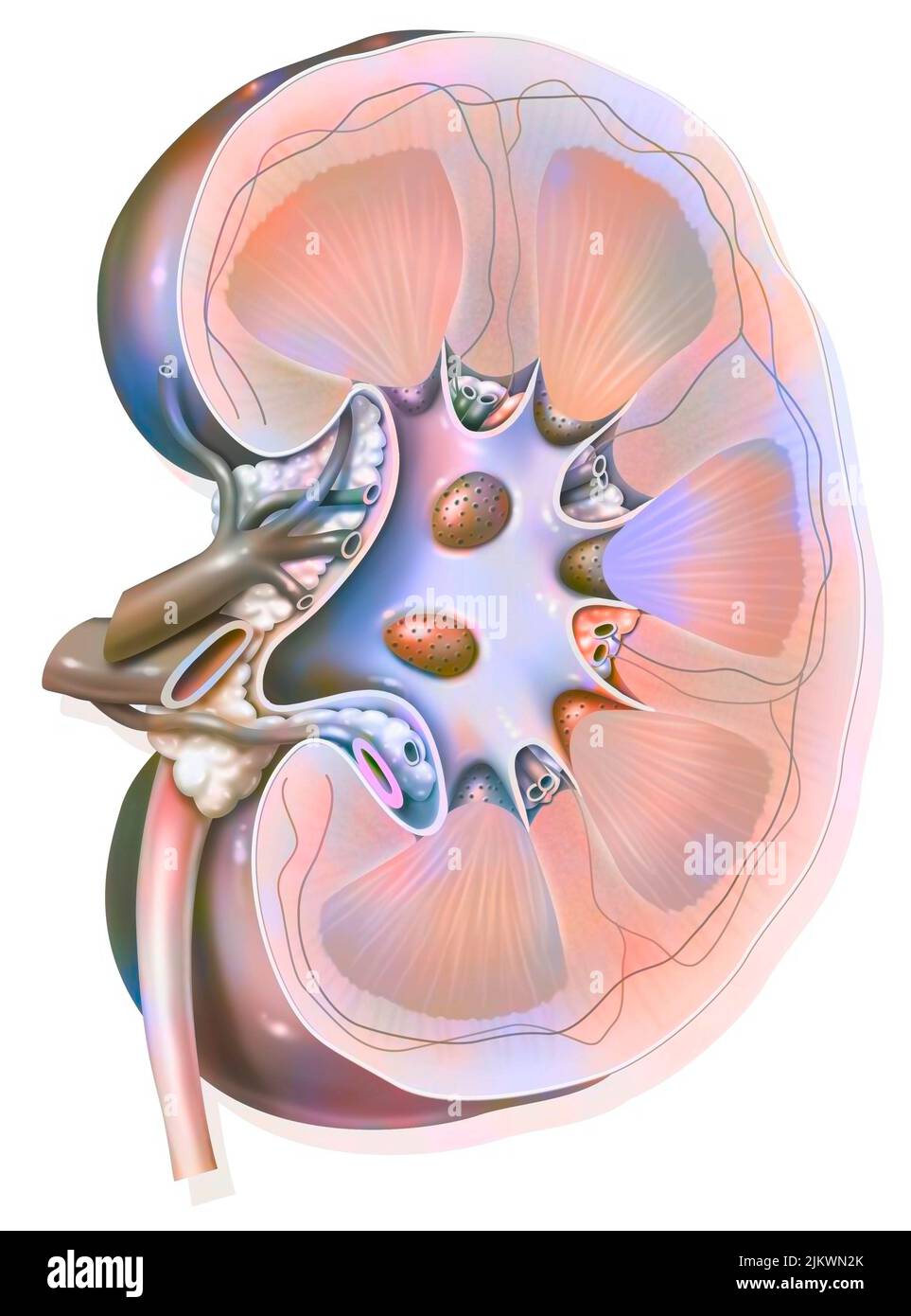 Sagittal section of the left kidney with the renal arteries and veins. Stock Photohttps://www.alamy.com/image-license-details/?v=1https://www.alamy.com/sagittal-section-of-the-left-kidney-with-the-renal-arteries-and-veins-image476923739.html
RF2JKWN2K–Sagittal section of the left kidney with the renal arteries and veins.
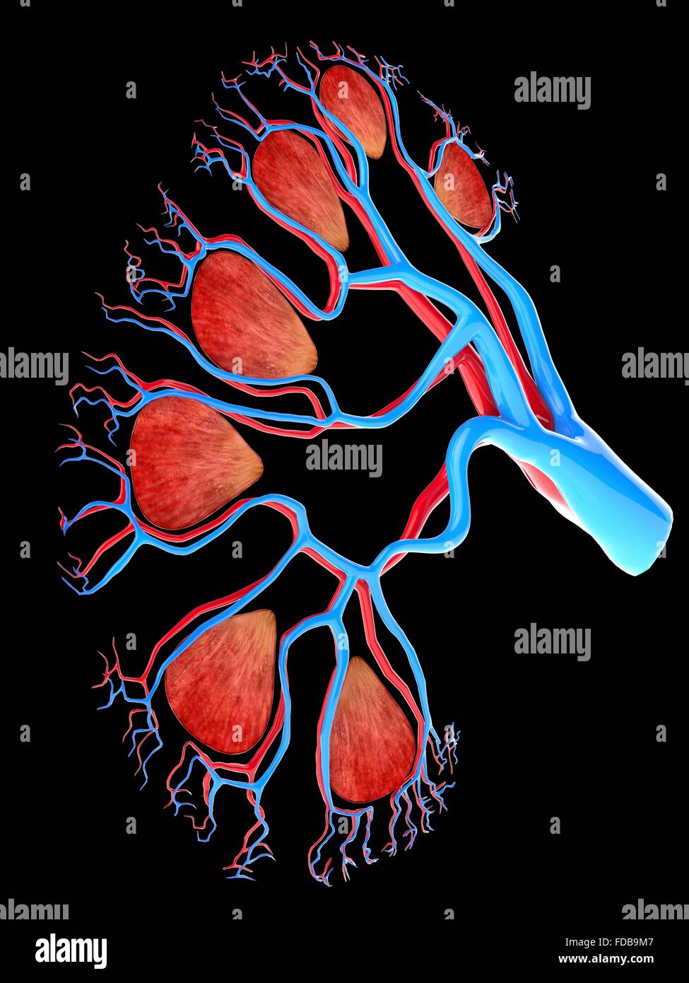 Kidney. Computer artwork showing the pyramid-shaped units of the inner medulla and the network of blood supply ending in capillaries in the cortex. Stock Photohttps://www.alamy.com/image-license-details/?v=1https://www.alamy.com/stock-photo-kidney-computer-artwork-showing-the-pyramid-shaped-units-of-the-inner-94291463.html
RFFDB9M7–Kidney. Computer artwork showing the pyramid-shaped units of the inner medulla and the network of blood supply ending in capillaries in the cortex.
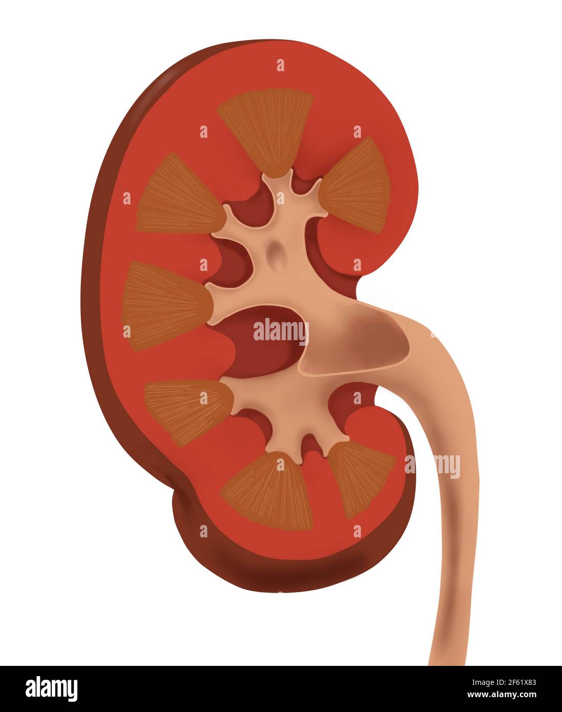 Normal Human Kidney Stock Photohttps://www.alamy.com/image-license-details/?v=1https://www.alamy.com/normal-human-kidney-image416779331.html
RM2F61X83–Normal Human Kidney
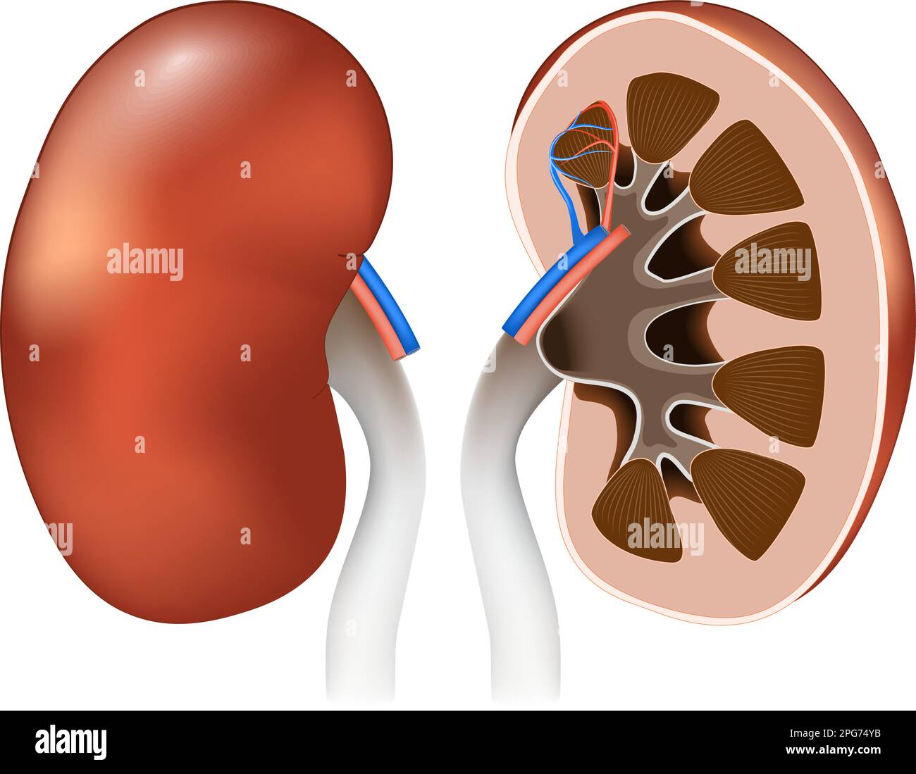 Kidney anatomy. Front view, and Cross section of human kidneys. Vector poster for education. Realistic illustration. Kidney Stock Vectorhttps://www.alamy.com/image-license-details/?v=1https://www.alamy.com/kidney-anatomy-front-view-and-cross-section-of-human-kidneys-vector-poster-for-education-realistic-illustration-kidney-image543513471.html
RF2PG74YB–Kidney anatomy. Front view, and Cross section of human kidneys. Vector poster for education. Realistic illustration. Kidney
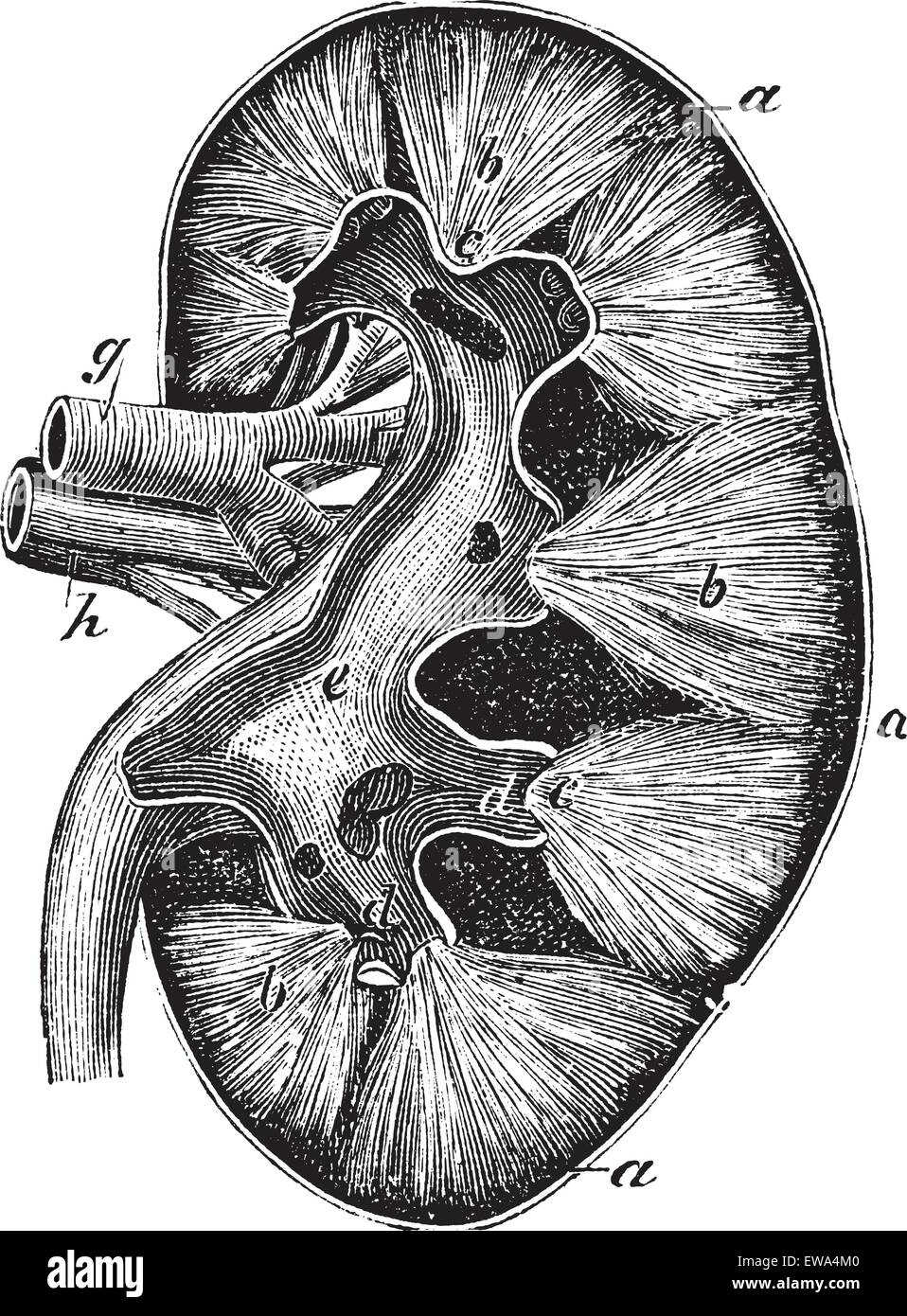 Human kidney, vintage engraving. Old engraved illustration of Human kidney structure with its functioning parts and their names. Stock Vectorhttps://www.alamy.com/image-license-details/?v=1https://www.alamy.com/stock-photo-human-kidney-vintage-engraving-old-engraved-illustration-of-human-84431088.html
RFEWA4M0–Human kidney, vintage engraving. Old engraved illustration of Human kidney structure with its functioning parts and their names.
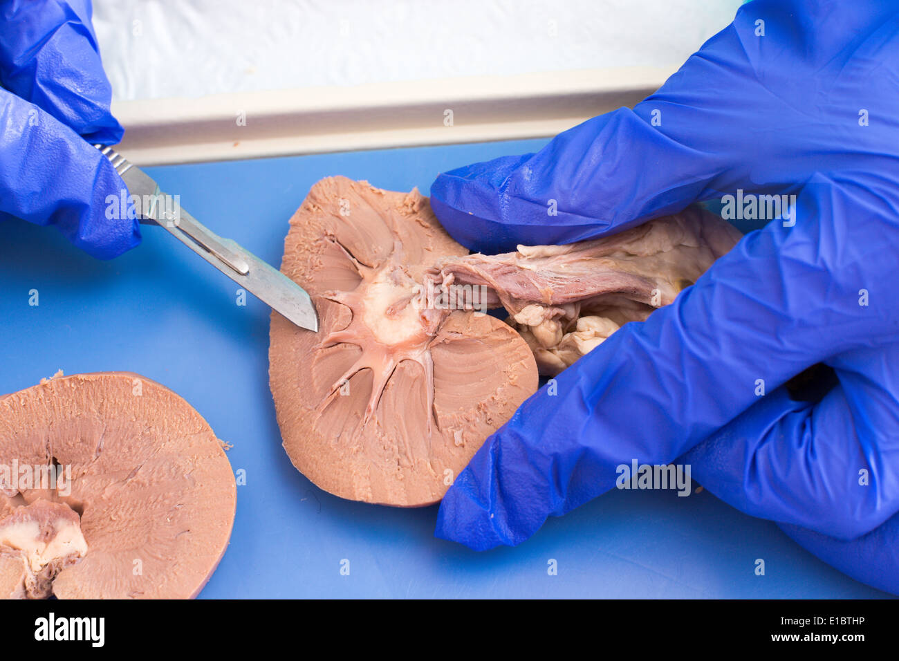 Medical student studying the internal structure of sheep kidney using a cross section to examine the tissue during anatomy class Stock Photohttps://www.alamy.com/image-license-details/?v=1https://www.alamy.com/medical-student-studying-the-internal-structure-of-sheep-kidney-using-image69716914.html
RFE1BTHP–Medical student studying the internal structure of sheep kidney using a cross section to examine the tissue during anatomy class
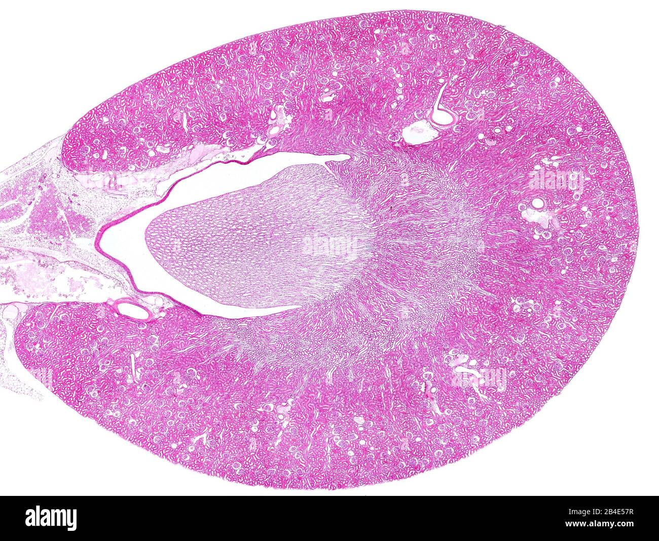 Light micrograph cross section of rat kidney stained with hematoxylin and eosin displaying the peripheral renal cortex (with many glomeruli) and, in t Stock Photohttps://www.alamy.com/image-license-details/?v=1https://www.alamy.com/light-micrograph-cross-section-of-rat-kidney-stained-with-hematoxylin-and-eosin-displaying-the-peripheral-renal-cortex-with-many-glomeruli-and-in-t-image346977451.html
RF2B4E57R–Light micrograph cross section of rat kidney stained with hematoxylin and eosin displaying the peripheral renal cortex (with many glomeruli) and, in t
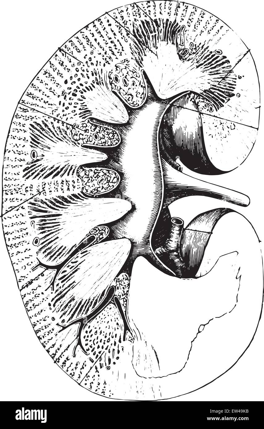 Longitudinal section of kidney, vintage engraved illustration. Stock Vectorhttps://www.alamy.com/image-license-details/?v=1https://www.alamy.com/stock-photo-longitudinal-section-of-kidney-vintage-engraved-illustration-84303279.html
RFEW49KB–Longitudinal section of kidney, vintage engraved illustration.
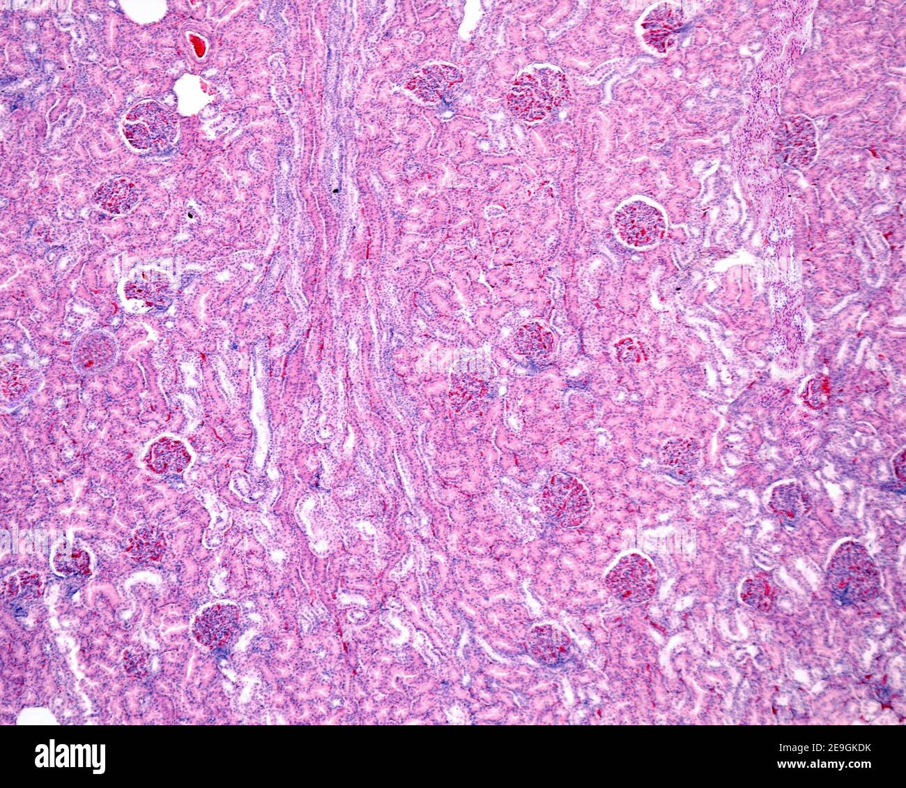 Low magnification micrograph showing several glomeruli and convoluted tubules in the cortex of a human kidney. In the center of the image there is a m Stock Photohttps://www.alamy.com/image-license-details/?v=1https://www.alamy.com/low-magnification-micrograph-showing-several-glomeruli-and-convoluted-tubules-in-the-cortex-of-a-human-kidney-in-the-center-of-the-image-there-is-a-m-image401736879.html
RF2E9GKDK–Low magnification micrograph showing several glomeruli and convoluted tubules in the cortex of a human kidney. In the center of the image there is a m
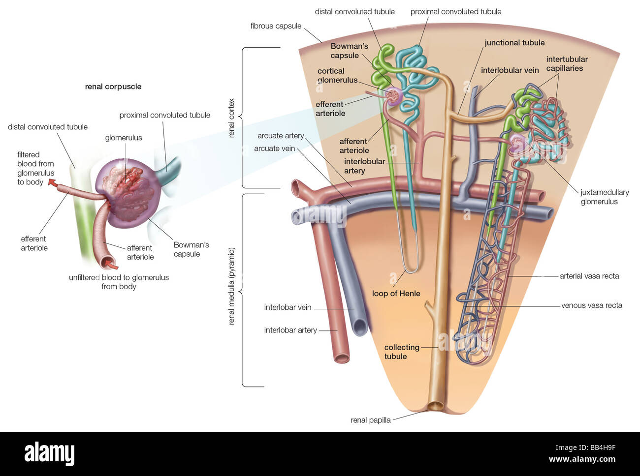 At one end of each nephron in the mammalian kidney exists a double-walled, cuplike structure called the renal corpuscle. Stock Photohttps://www.alamy.com/image-license-details/?v=1https://www.alamy.com/stock-photo-at-one-end-of-each-nephron-in-the-mammalian-kidney-exists-a-double-24072987.html
RMBB4H9F–At one end of each nephron in the mammalian kidney exists a double-walled, cuplike structure called the renal corpuscle.
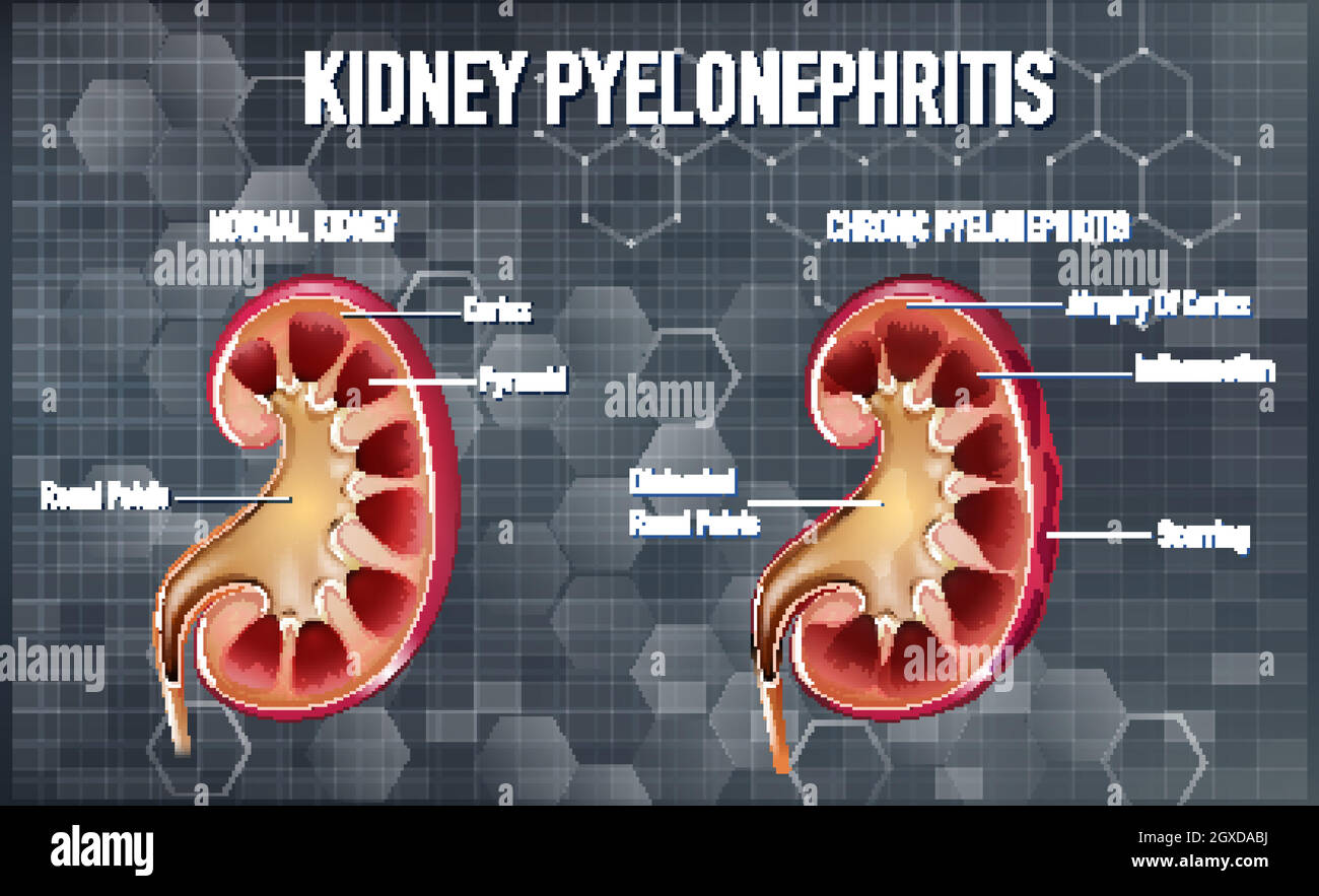 Informative illustration of Pyelonephritis Stock Vectorhttps://www.alamy.com/image-license-details/?v=1https://www.alamy.com/informative-illustration-of-pyelonephritis-image446533798.html
RF2GXDABJ–Informative illustration of Pyelonephritis
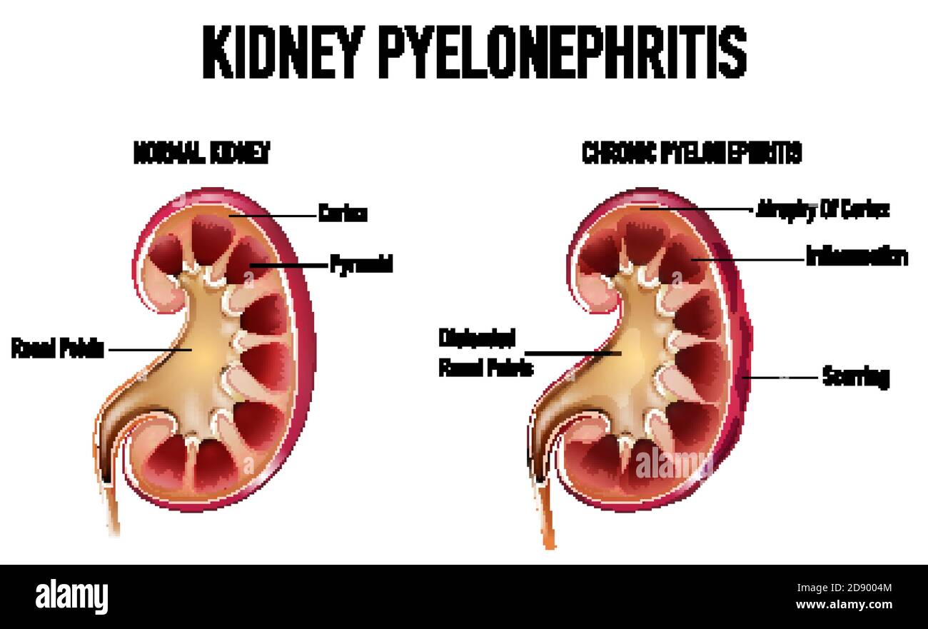 Informative illustration of Pyelonephritis illustration Stock Vectorhttps://www.alamy.com/image-license-details/?v=1https://www.alamy.com/informative-illustration-of-pyelonephritis-illustration-image384160132.html
RF2D9004M–Informative illustration of Pyelonephritis illustration
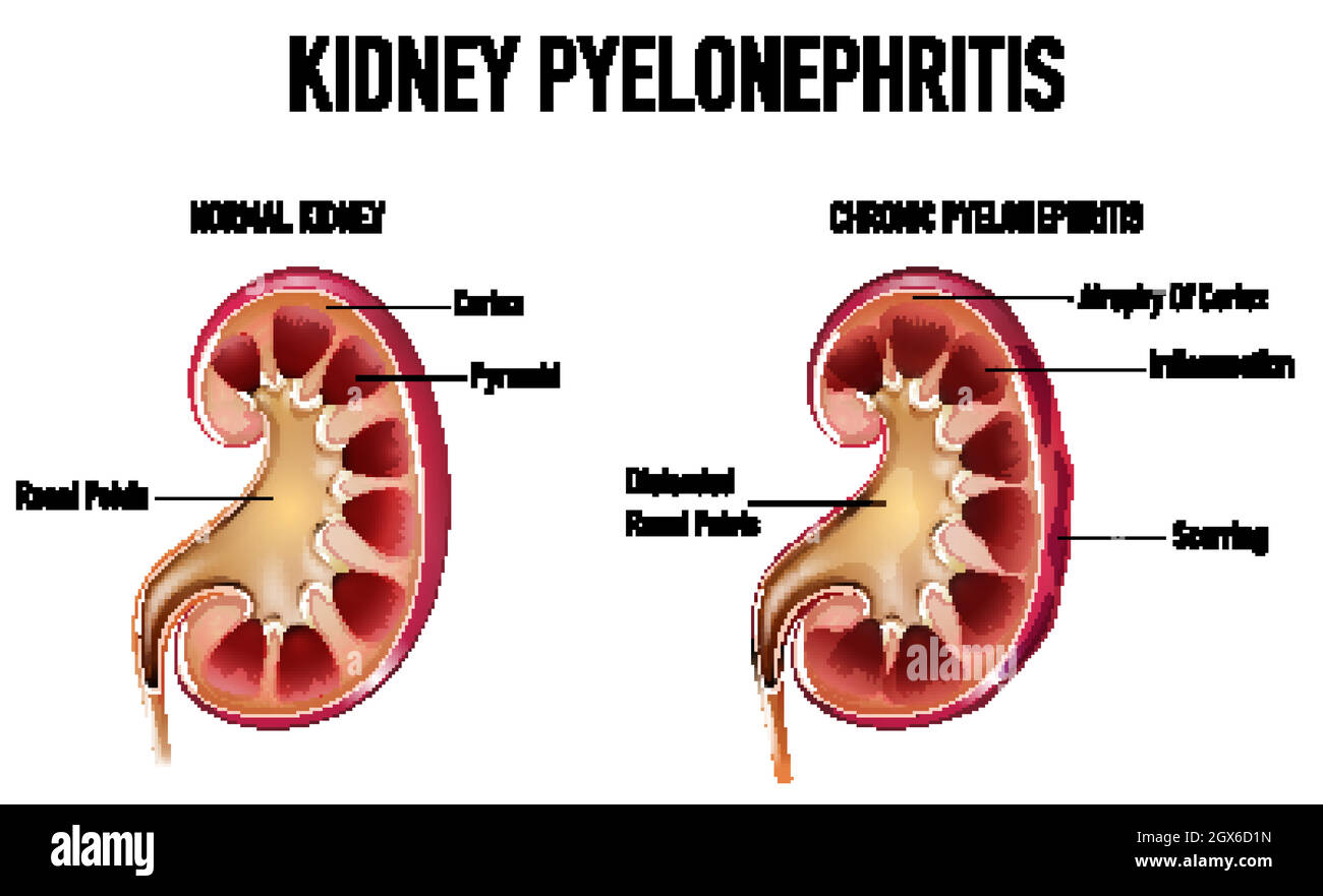 Informative illustration of Pyelonephritis Stock Vectorhttps://www.alamy.com/image-license-details/?v=1https://www.alamy.com/informative-illustration-of-pyelonephritis-image446382209.html
RF2GX6D1N–Informative illustration of Pyelonephritis
 Archive image from page 1302 of Cunningham's Text-book of anatomy (1914). Cunningham's Text-book of anatomy cunninghamstextb00cunn Year: 1914 ( THE DUCT OF THE KIDNEY. 1269 Cortical substance Basal part of pyramid' Interlobar . artery k Pyramid Papilla J-- Renal artery Calyx ureter, in most cases, lies across the external iliac; but this arrangement is by no means constant. The course and position occupied by the abdominal portion of the ureter is well seen in Fig. 983. In X-ray photographs, the shadow cast by the abdominal portion of the ureter when the latter has been rendered opaque, is se Stock Photohttps://www.alamy.com/image-license-details/?v=1https://www.alamy.com/archive-image-from-page-1302-of-cunninghams-text-book-of-anatomy-1914-cunninghams-text-book-of-anatomy-cunninghamstextb00cunn-year-1914-the-duct-of-the-kidney-1269-cortical-substance-basal-part-of-pyramid-interlobar-artery-k-pyramid-papilla-j-renal-artery-calyx-ureter-in-most-cases-lies-across-the-external-iliac-but-this-arrangement-is-by-no-means-constant-the-course-and-position-occupied-by-the-abdominal-portion-of-the-ureter-is-well-seen-in-fig-983-in-x-ray-photographs-the-shadow-cast-by-the-abdominal-portion-of-the-ureter-when-the-latter-has-been-rendered-opaque-is-se-image264068875.html
RMW9HAF7–Archive image from page 1302 of Cunningham's Text-book of anatomy (1914). Cunningham's Text-book of anatomy cunninghamstextb00cunn Year: 1914 ( THE DUCT OF THE KIDNEY. 1269 Cortical substance Basal part of pyramid' Interlobar . artery k Pyramid Papilla J-- Renal artery Calyx ureter, in most cases, lies across the external iliac; but this arrangement is by no means constant. The course and position occupied by the abdominal portion of the ureter is well seen in Fig. 983. In X-ray photographs, the shadow cast by the abdominal portion of the ureter when the latter has been rendered opaque, is se
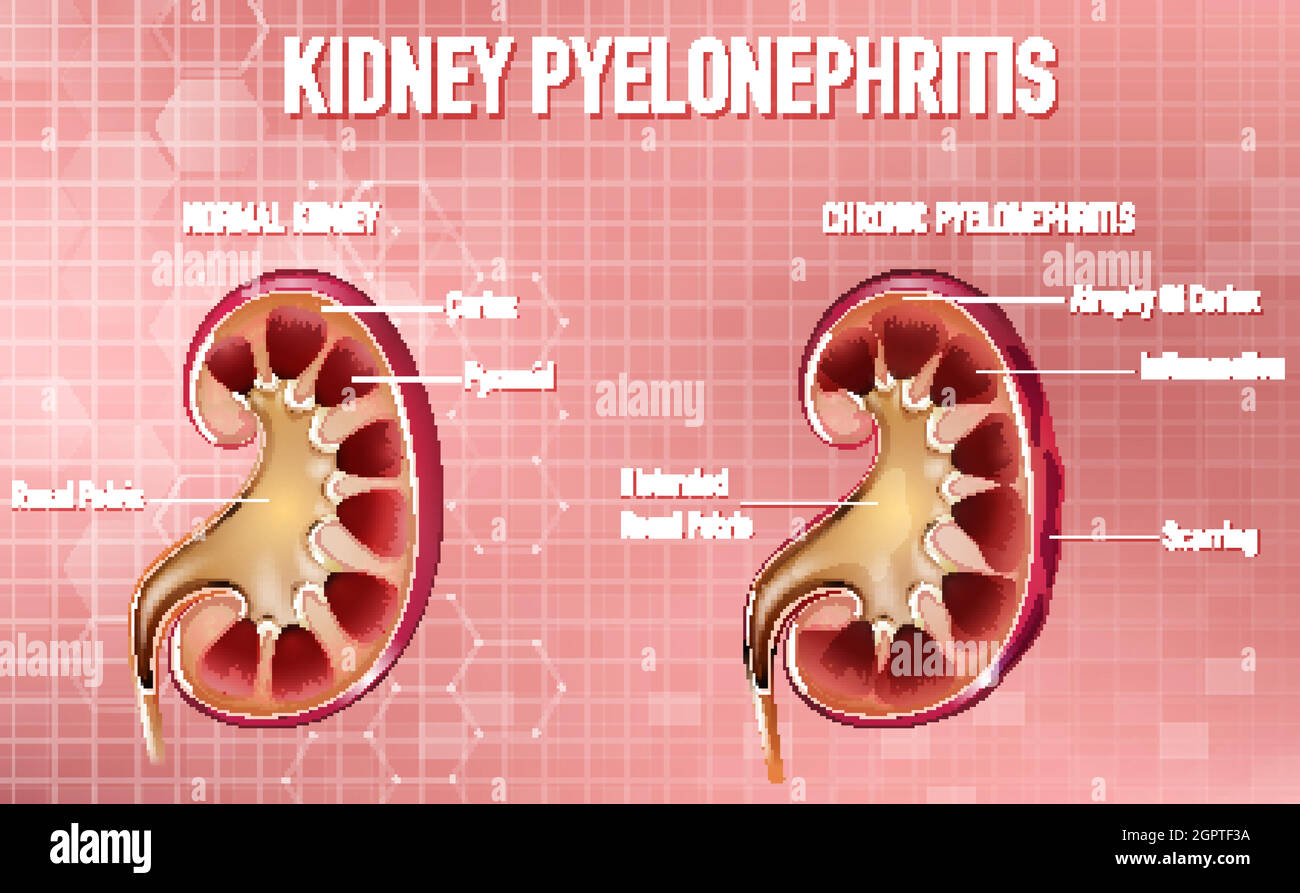 Informative illustration of Pyelonephritis Stock Vectorhttps://www.alamy.com/image-license-details/?v=1https://www.alamy.com/informative-illustration-of-pyelonephritis-image444320334.html
RF2GPTF3A–Informative illustration of Pyelonephritis
 An atlas of human anatomy for students and physicians . Renal papillse Papilla; renales ? Base of the pyramid Basis pyramidis . Sinus of the kidney 5/-^Sinus renalis-, / Pyramid of Malpighif Pyramis renalis (Malpighii) CortexSubstantia corticalis Columns of Eertin, or septularenum Columnae renales (Bertini) Fig. 829.—Sinus of the Kidney, displayedin the Coronally-bisected Kidney of anInfant aged Three Weeks Superior border Margo superior.Anterior surface Facies anteiioi Apex Ape. suprarenalis. Hilum Hilu Internal border Margo medialis Inferior suprarenal artery A. suprarenalis inferiorSuprar Stock Photohttps://www.alamy.com/image-license-details/?v=1https://www.alamy.com/an-atlas-of-human-anatomy-for-students-and-physicians-renal-papillse-papilla-renales-base-of-the-pyramid-basis-pyramidis-sinus-of-the-kidney-5-sinus-renalis-pyramid-of-malpighif-pyramis-renalis-malpighii-cortexsubstantia-corticalis-columns-of-eertin-or-septularenum-columnae-renales-bertini-fig-829sinus-of-the-kidney-displayedin-the-coronally-bisected-kidney-of-aninfant-aged-three-weeks-superior-border-margo-superioranterior-surface-facies-anteiioi-apex-ape-suprarenalis-hilum-hilu-internal-border-margo-medialis-inferior-suprarenal-artery-a-suprarenalis-inferiorsuprar-image338303561.html
RM2AJB1J1–An atlas of human anatomy for students and physicians . Renal papillse Papilla; renales ? Base of the pyramid Basis pyramidis . Sinus of the kidney 5/-^Sinus renalis-, / Pyramid of Malpighif Pyramis renalis (Malpighii) CortexSubstantia corticalis Columns of Eertin, or septularenum Columnae renales (Bertini) Fig. 829.—Sinus of the Kidney, displayedin the Coronally-bisected Kidney of anInfant aged Three Weeks Superior border Margo superior.Anterior surface Facies anteiioi Apex Ape. suprarenalis. Hilum Hilu Internal border Margo medialis Inferior suprarenal artery A. suprarenalis inferiorSuprar
 . Cunningham's Text-book of anatomy. Anatomy. THE DUCT OF THE KIDNEY. 1269 Cortical substance Basal part of pyramid" Interlobar^ . artery k Pyramid Papilla J-- Renal artery Calyx ureter, in most cases, lies across the external iliac; but this arrangement is by no means constant. The course and position occupied by the abdominal portion of the ureter is well seen in Fig. 983. In X-ray photographs, the shadow cast by the abdominal portion of the ureter when the latter has been rendered opaque, is seen to fall immediately in front of the tips of the transverse processes of the lower lumbar v Stock Photohttps://www.alamy.com/image-license-details/?v=1https://www.alamy.com/cunninghams-text-book-of-anatomy-anatomy-the-duct-of-the-kidney-1269-cortical-substance-basal-part-of-pyramidquot-interlobar-artery-k-pyramid-papilla-j-renal-artery-calyx-ureter-in-most-cases-lies-across-the-external-iliac-but-this-arrangement-is-by-no-means-constant-the-course-and-position-occupied-by-the-abdominal-portion-of-the-ureter-is-well-seen-in-fig-983-in-x-ray-photographs-the-shadow-cast-by-the-abdominal-portion-of-the-ureter-when-the-latter-has-been-rendered-opaque-is-seen-to-fall-immediately-in-front-of-the-tips-of-the-transverse-processes-of-the-lower-lumbar-v-image216340027.html
RMPFY3WF–. Cunningham's Text-book of anatomy. Anatomy. THE DUCT OF THE KIDNEY. 1269 Cortical substance Basal part of pyramid" Interlobar^ . artery k Pyramid Papilla J-- Renal artery Calyx ureter, in most cases, lies across the external iliac; but this arrangement is by no means constant. The course and position occupied by the abdominal portion of the ureter is well seen in Fig. 983. In X-ray photographs, the shadow cast by the abdominal portion of the ureter when the latter has been rendered opaque, is seen to fall immediately in front of the tips of the transverse processes of the lower lumbar v
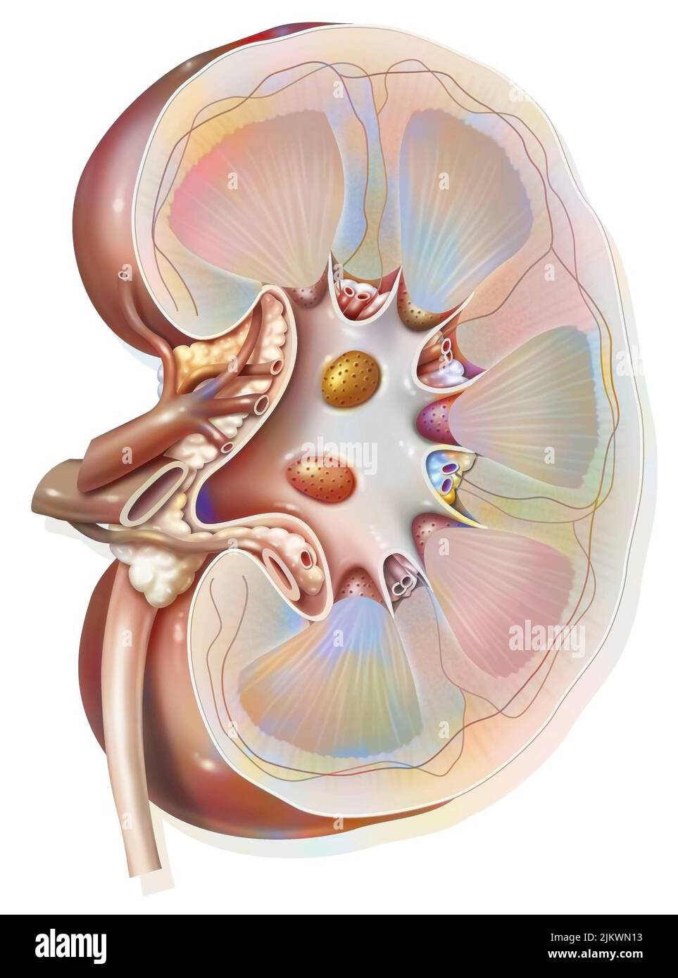 Sagittal section of the left kidney with the renal arteries and veins. Stock Photohttps://www.alamy.com/image-license-details/?v=1https://www.alamy.com/sagittal-section-of-the-left-kidney-with-the-renal-arteries-and-veins-image476923695.html
RF2JKWN13–Sagittal section of the left kidney with the renal arteries and veins.
 Kidney. Computer artwork showing the pyramid-shaped units of the inner medulla and the network of blood supply ending in capillaries in the cortex. Stock Photohttps://www.alamy.com/image-license-details/?v=1https://www.alamy.com/stock-photo-kidney-computer-artwork-showing-the-pyramid-shaped-units-of-the-inner-94291568.html
RFFDB9T0–Kidney. Computer artwork showing the pyramid-shaped units of the inner medulla and the network of blood supply ending in capillaries in the cortex.
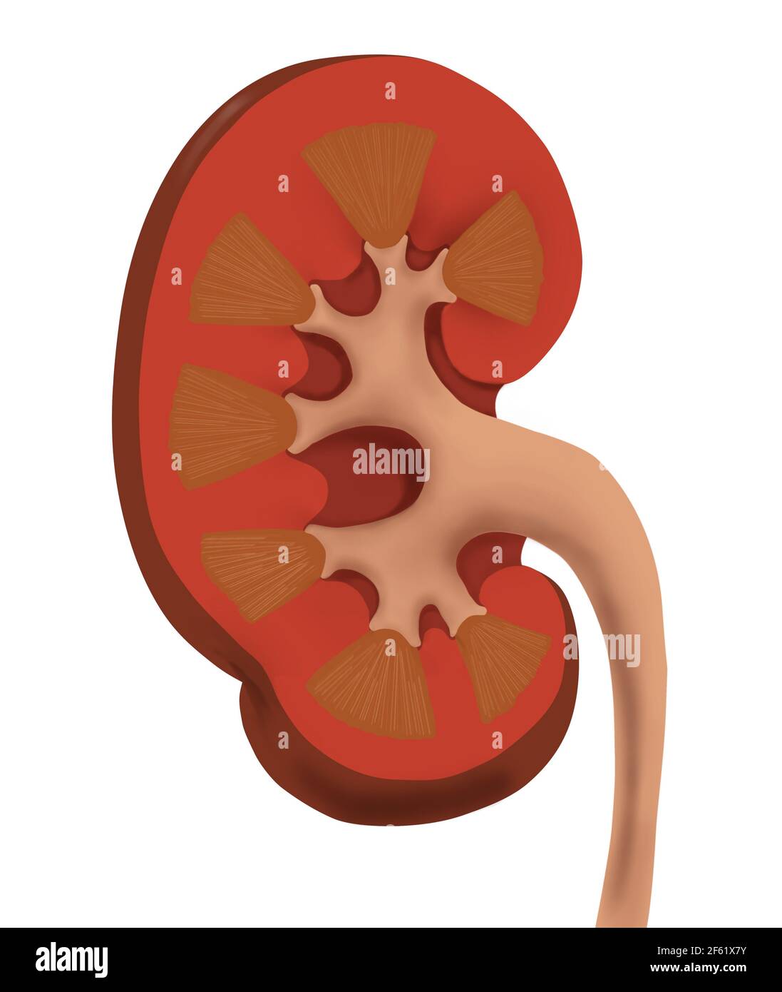 Normal Human Kidney Stock Photohttps://www.alamy.com/image-license-details/?v=1https://www.alamy.com/normal-human-kidney-image416779327.html
RM2F61X7Y–Normal Human Kidney
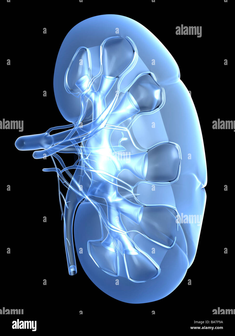 Kidney, computer artwork. Stock Photohttps://www.alamy.com/image-license-details/?v=1https://www.alamy.com/stock-photo-kidney-computer-artwork-23895798.html
RFBATF9A–Kidney, computer artwork.
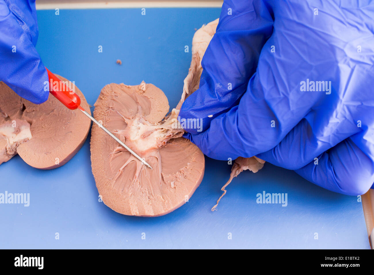 Student studying a dissected sheep kidney in medical school using a cross-section to examine the internal structure Stock Photohttps://www.alamy.com/image-license-details/?v=1https://www.alamy.com/student-studying-a-dissected-sheep-kidney-in-medical-school-using-image69716950.html
RFE1BTK2–Student studying a dissected sheep kidney in medical school using a cross-section to examine the internal structure
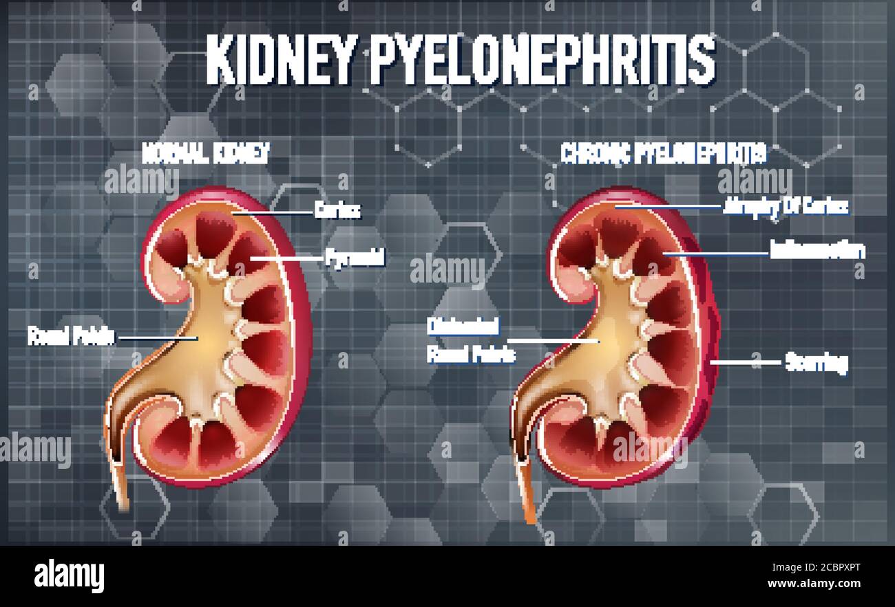 Informative illustration of Pyelonephritis illustration Stock Vectorhttps://www.alamy.com/image-license-details/?v=1https://www.alamy.com/informative-illustration-of-pyelonephritis-illustration-image368682912.html
RF2CBPXPT–Informative illustration of Pyelonephritis illustration
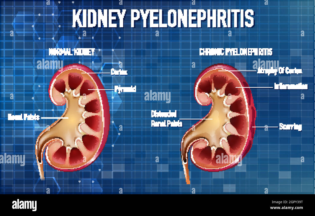 Informative illustration of Pyelonephritis Stock Vectorhttps://www.alamy.com/image-license-details/?v=1https://www.alamy.com/informative-illustration-of-pyelonephritis-image444376964.html
RF2GPY39T–Informative illustration of Pyelonephritis
 The physiology and hygiene of the house in which we live . the kidney theyare known as the uriniferous tubules, and if we follow up oneof these tubules from the kidney, from its opening on the surface of a pyramid, we shallfind that it terminates in a dila-tation not unlike those found inthe sweat-glands. The dilatationin the urinary tubule is knownas the Malpighian (from its dis-coverer, Malpighi) capsule (seefigure b), which incloses within ita network of capillaries (glomer-ulus) given off from the branchesof the renal artery. Through thistuft of capillaries the liquid ref-use of the blood Stock Photohttps://www.alamy.com/image-license-details/?v=1https://www.alamy.com/the-physiology-and-hygiene-of-the-house-in-which-we-live-the-kidney-theyare-known-as-the-uriniferous-tubules-and-if-we-follow-up-oneof-these-tubules-from-the-kidney-from-its-opening-on-the-surface-of-a-pyramid-we-shallfind-that-it-terminates-in-a-dila-tation-not-unlike-those-found-inthe-sweat-glands-the-dilatationin-the-urinary-tubule-is-knownas-the-malpighian-from-its-dis-coverer-malpighi-capsule-seefigure-b-which-incloses-within-ita-network-of-capillaries-glomer-ulus-given-off-from-the-branchesof-the-renal-artery-through-thistuft-of-capillaries-the-liquid-ref-use-of-the-blood-image339995148.html
RM2AN437T–The physiology and hygiene of the house in which we live . the kidney theyare known as the uriniferous tubules, and if we follow up oneof these tubules from the kidney, from its opening on the surface of a pyramid, we shallfind that it terminates in a dila-tation not unlike those found inthe sweat-glands. The dilatationin the urinary tubule is knownas the Malpighian (from its dis-coverer, Malpighi) capsule (seefigure b), which incloses within ita network of capillaries (glomer-ulus) given off from the branchesof the renal artery. Through thistuft of capillaries the liquid ref-use of the blood
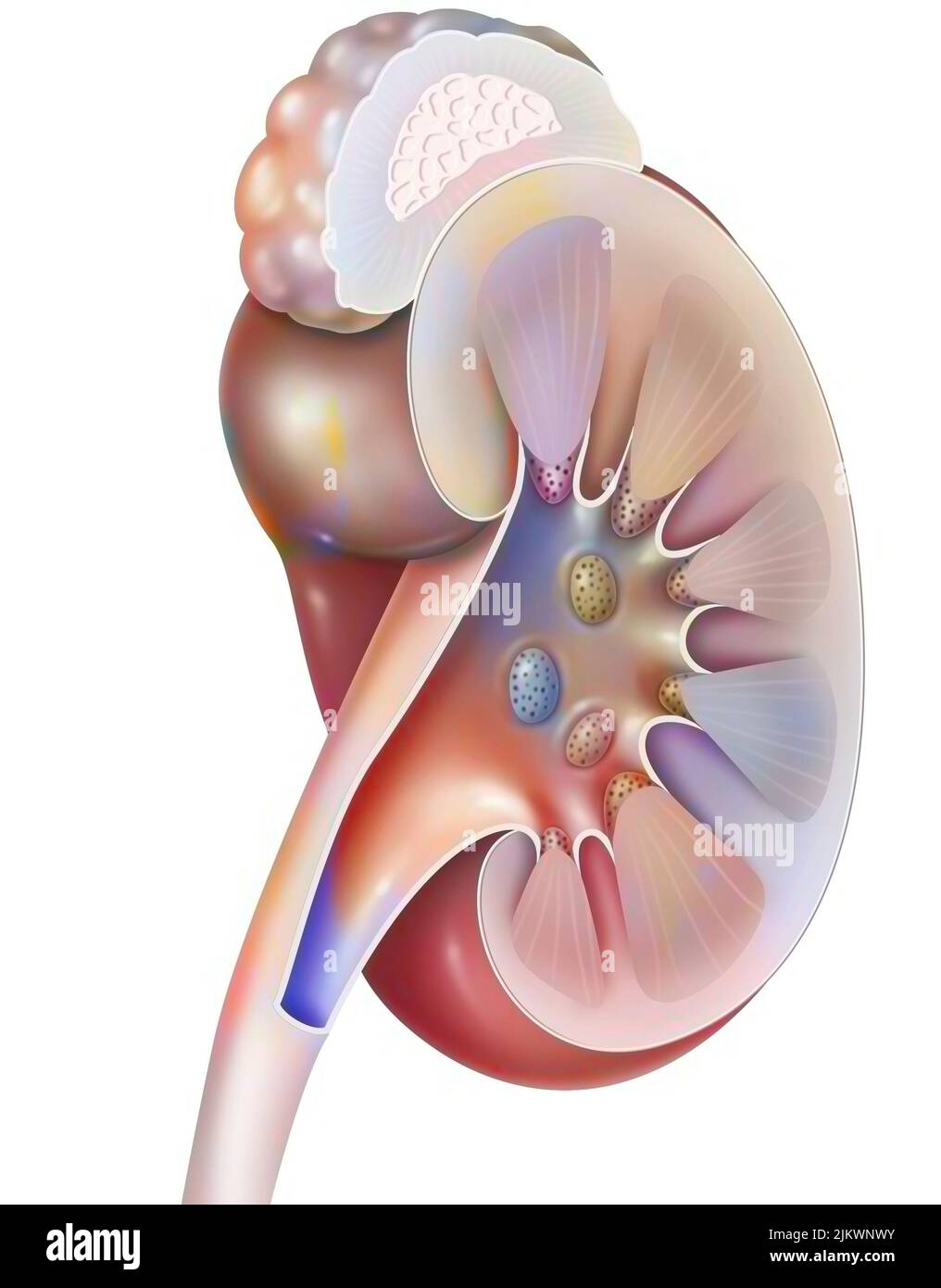 Structures of the kidney and left ureter in 3/4 views. Stock Photohttps://www.alamy.com/image-license-details/?v=1https://www.alamy.com/structures-of-the-kidney-and-left-ureter-in-34-views-image476924391.html
RF2JKWNWY–Structures of the kidney and left ureter in 3/4 views.
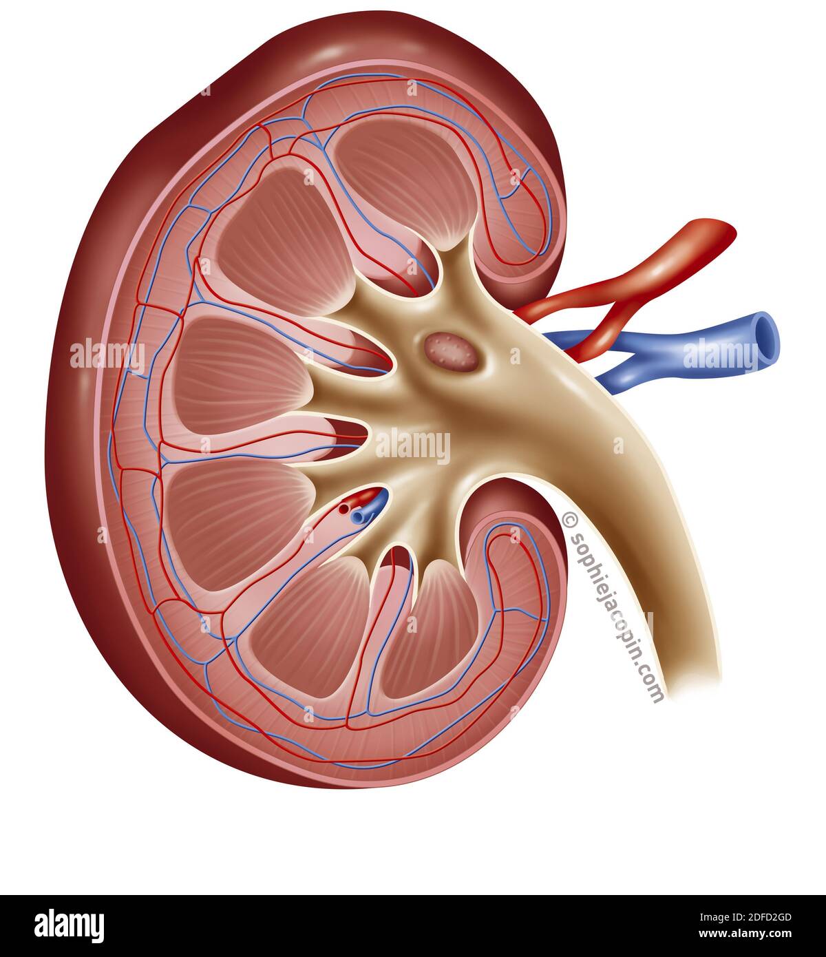 Kidney structure Stock Photohttps://www.alamy.com/image-license-details/?v=1https://www.alamy.com/kidney-structure-image388135341.html
RM2DFD2GD–Kidney structure
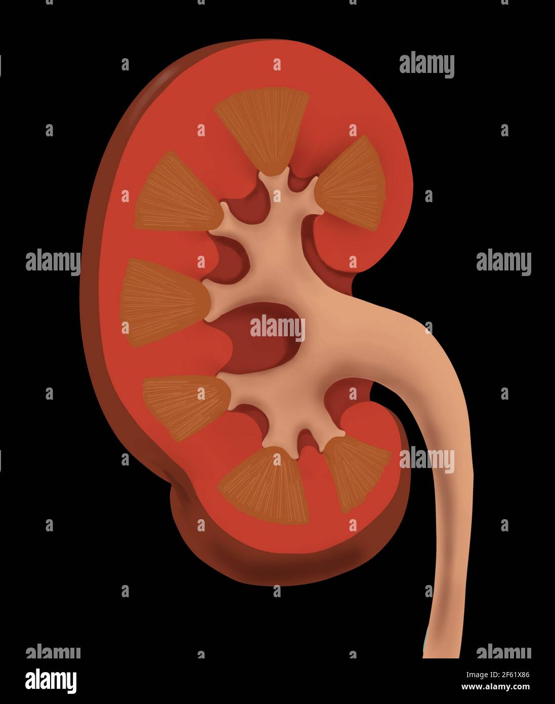 Normal Human Kidney Stock Photohttps://www.alamy.com/image-license-details/?v=1https://www.alamy.com/normal-human-kidney-image416779334.html
RM2F61X86–Normal Human Kidney
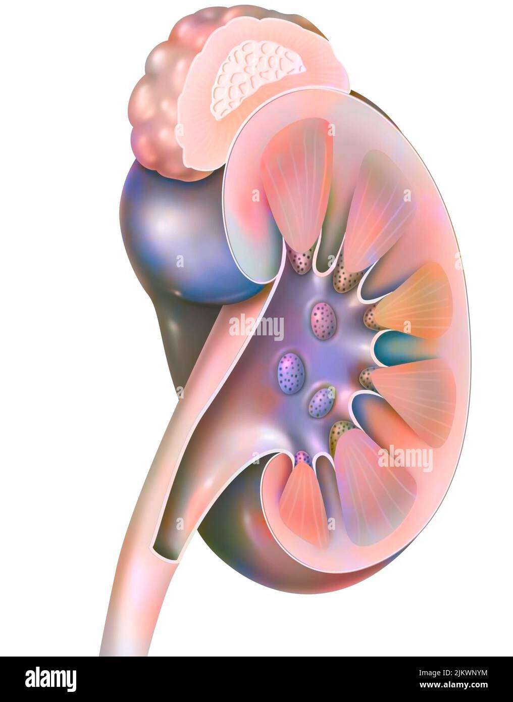 Structures of the kidney and left ureter in 3/4 views. Stock Photohttps://www.alamy.com/image-license-details/?v=1https://www.alamy.com/structures-of-the-kidney-and-left-ureter-in-34-views-image476924440.html
RF2JKWNYM–Structures of the kidney and left ureter in 3/4 views.
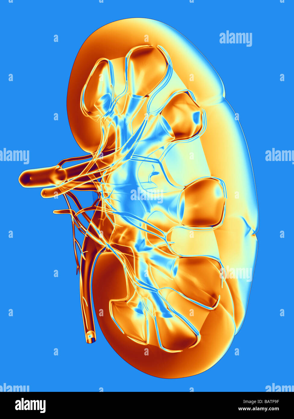 Kidney, computer artwork. Stock Photohttps://www.alamy.com/image-license-details/?v=1https://www.alamy.com/stock-photo-kidney-computer-artwork-23895803.html
RFBATF9F–Kidney, computer artwork.
 Kidney in section with close-up of the glomerulus. Stock Photohttps://www.alamy.com/image-license-details/?v=1https://www.alamy.com/kidney-in-section-with-close-up-of-the-glomerulus-image476923370.html
RF2JKWMHE–Kidney in section with close-up of the glomerulus.
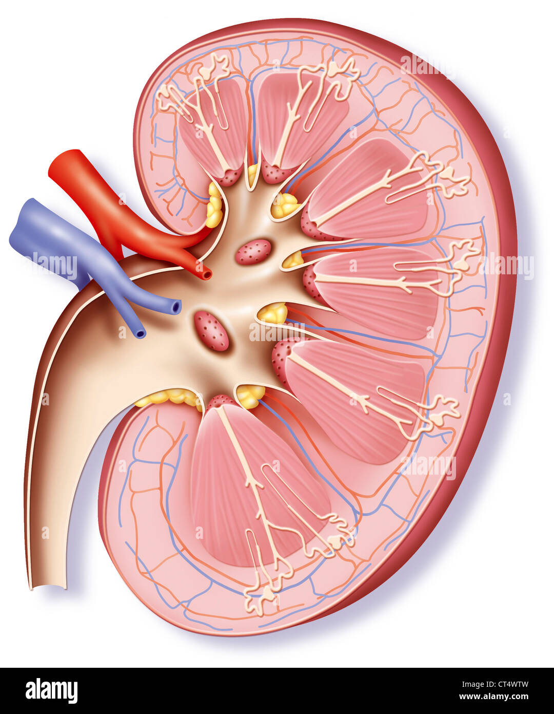 KIDNEY, DRAWING Stock Photohttps://www.alamy.com/image-license-details/?v=1https://www.alamy.com/stock-photo-kidney-drawing-49280585.html
RMCT4WTW–KIDNEY, DRAWING
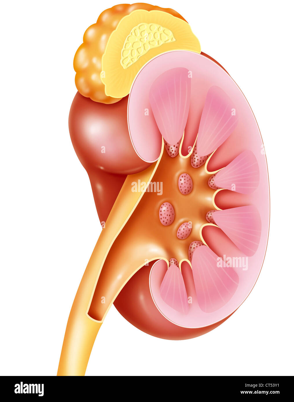 KIDNEY, DRAWING Stock Photohttps://www.alamy.com/image-license-details/?v=1https://www.alamy.com/stock-photo-kidney-drawing-49285349.html
RMCT53Y1–KIDNEY, DRAWING
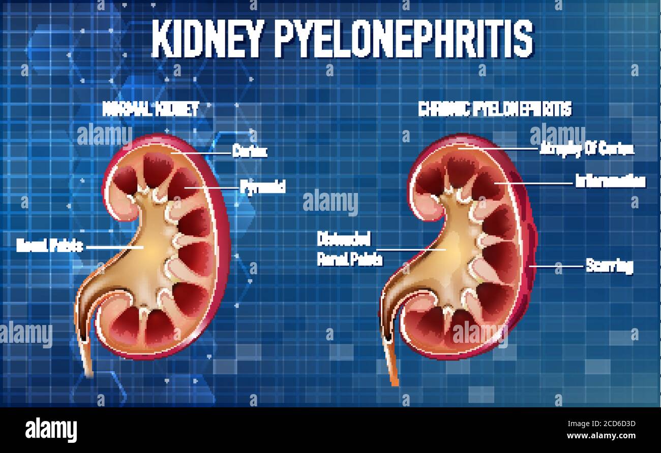 Informative illustration of Pyelonephritis illustration Stock Vectorhttps://www.alamy.com/image-license-details/?v=1https://www.alamy.com/informative-illustration-of-pyelonephritis-illustration-image369550257.html
RF2CD6D3D–Informative illustration of Pyelonephritis illustration
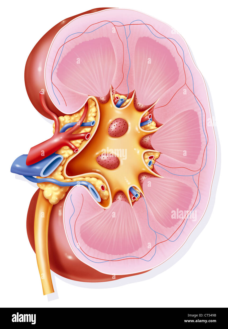 KIDNEY, DRAWING Stock Photohttps://www.alamy.com/image-license-details/?v=1https://www.alamy.com/stock-photo-kidney-drawing-49285639.html
RMCT549B–KIDNEY, DRAWING
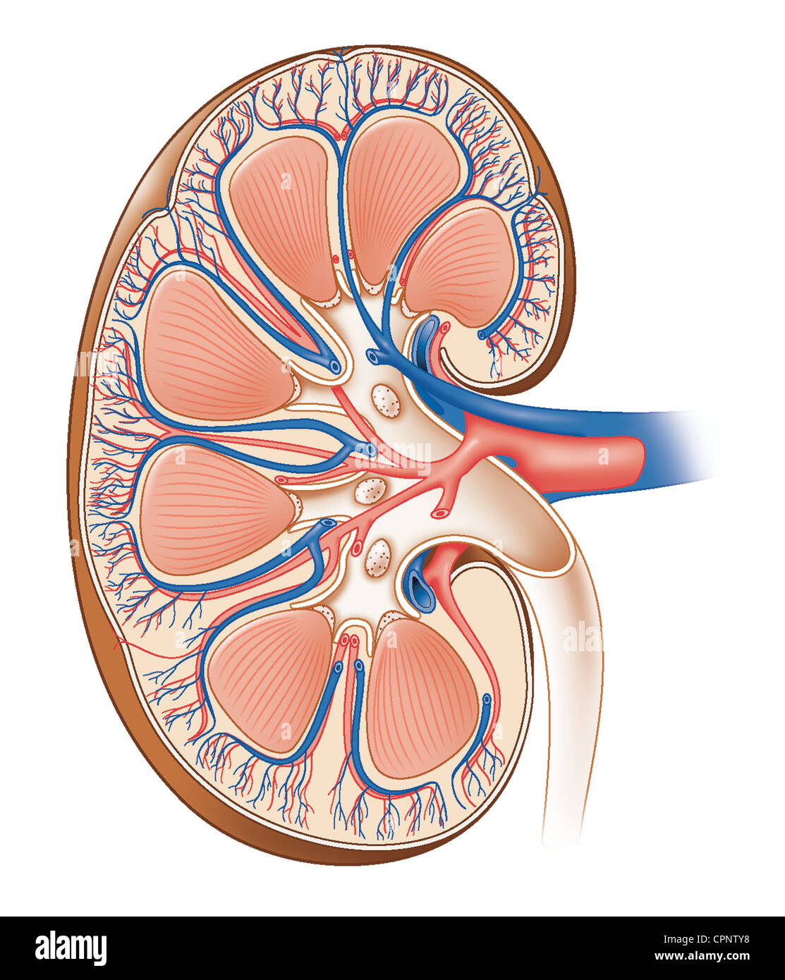 KIDNEY, ANATOMY Stock Photohttps://www.alamy.com/image-license-details/?v=1https://www.alamy.com/stock-photo-kidney-anatomy-48423740.html
RMCPNTY8–KIDNEY, ANATOMY
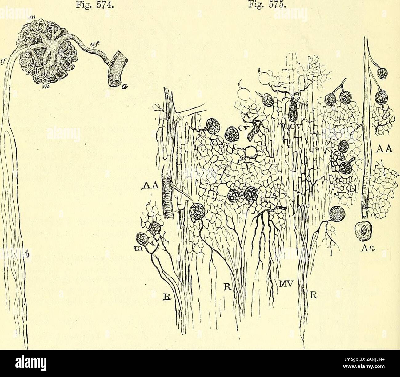 Quain's elements of anatomy . ferous tubules (fig. 573),the meshes of the network being poly-convoluted tubules and elongated amongst theBut the efferent vessels fiom the lower- gonal amongst th tubules of the medullary rays most glomeruli break up wholly into pencils of straight vessels (false Vasa recta) which pass directly into the bomidary layer of the medulla, and there supply the continuation downwards of the medullary rays into the pyramid. VOL. II, u u 658 THE KIDNEYS. The renal arteries give branches likewise to the capsule of the kidney whichanastomose with branches of the lumbar art Stock Photohttps://www.alamy.com/image-license-details/?v=1https://www.alamy.com/quains-elements-of-anatomy-ferous-tubules-fig-573the-meshes-of-the-network-being-poly-convoluted-tubules-and-elongated-amongst-thebut-the-efferent-vessels-fiom-the-lower-gonal-amongst-th-tubules-of-the-medullary-rays-most-glomeruli-break-up-wholly-into-pencils-of-straight-vessels-false-vasa-recta-which-pass-directly-into-the-bomidary-layer-of-the-medulla-and-there-supply-the-continuation-downwards-of-the-medullary-rays-into-the-pyramid-vol-ii-u-u-658-the-kidneys-the-renal-arteries-give-branches-likewise-to-the-capsule-of-the-kidney-whichanastomose-with-branches-of-the-lumbar-art-image340304416.html
RM2ANJ5N4–Quain's elements of anatomy . ferous tubules (fig. 573),the meshes of the network being poly-convoluted tubules and elongated amongst theBut the efferent vessels fiom the lower- gonal amongst th tubules of the medullary rays most glomeruli break up wholly into pencils of straight vessels (false Vasa recta) which pass directly into the bomidary layer of the medulla, and there supply the continuation downwards of the medullary rays into the pyramid. VOL. II, u u 658 THE KIDNEYS. The renal arteries give branches likewise to the capsule of the kidney whichanastomose with branches of the lumbar art
 KIDNEY, DRAWING Stock Photohttps://www.alamy.com/image-license-details/?v=1https://www.alamy.com/stock-photo-kidney-drawing-49290760.html
RMCT5AT8–KIDNEY, DRAWING
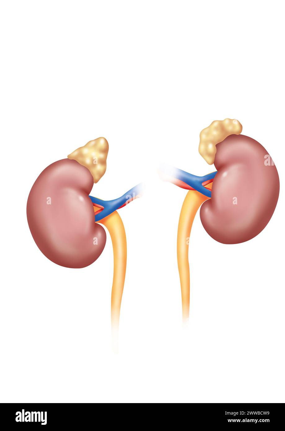 Kidneys in anterior view with adrenal glands ureters and renal arteries and veins. The left kidney is placed higher than the right kidney. Stock Photohttps://www.alamy.com/image-license-details/?v=1https://www.alamy.com/kidneys-in-anterior-view-with-adrenal-glands-ureters-and-renal-arteries-and-veins-the-left-kidney-is-placed-higher-than-the-right-kidney-image600770501.html
RM2WWBCW9–Kidneys in anterior view with adrenal glands ureters and renal arteries and veins. The left kidney is placed higher than the right kidney.
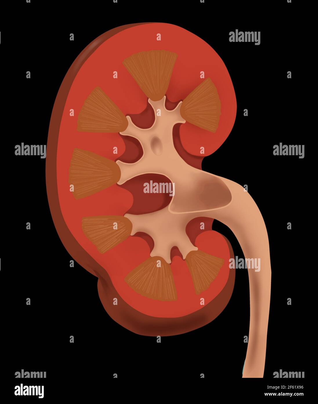 Normal Human Kidney Stock Photohttps://www.alamy.com/image-license-details/?v=1https://www.alamy.com/normal-human-kidney-image416779362.html
RM2F61X96–Normal Human Kidney
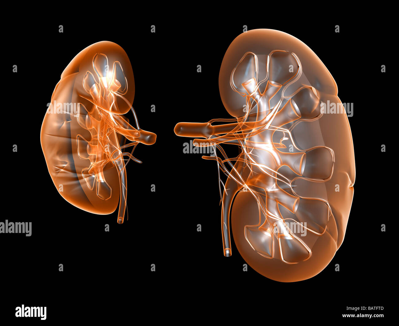 Kidneys, computer artwork. Stock Photohttps://www.alamy.com/image-license-details/?v=1https://www.alamy.com/stock-photo-kidneys-computer-artwork-23896221.html
RFBATFTD–Kidneys, computer artwork.
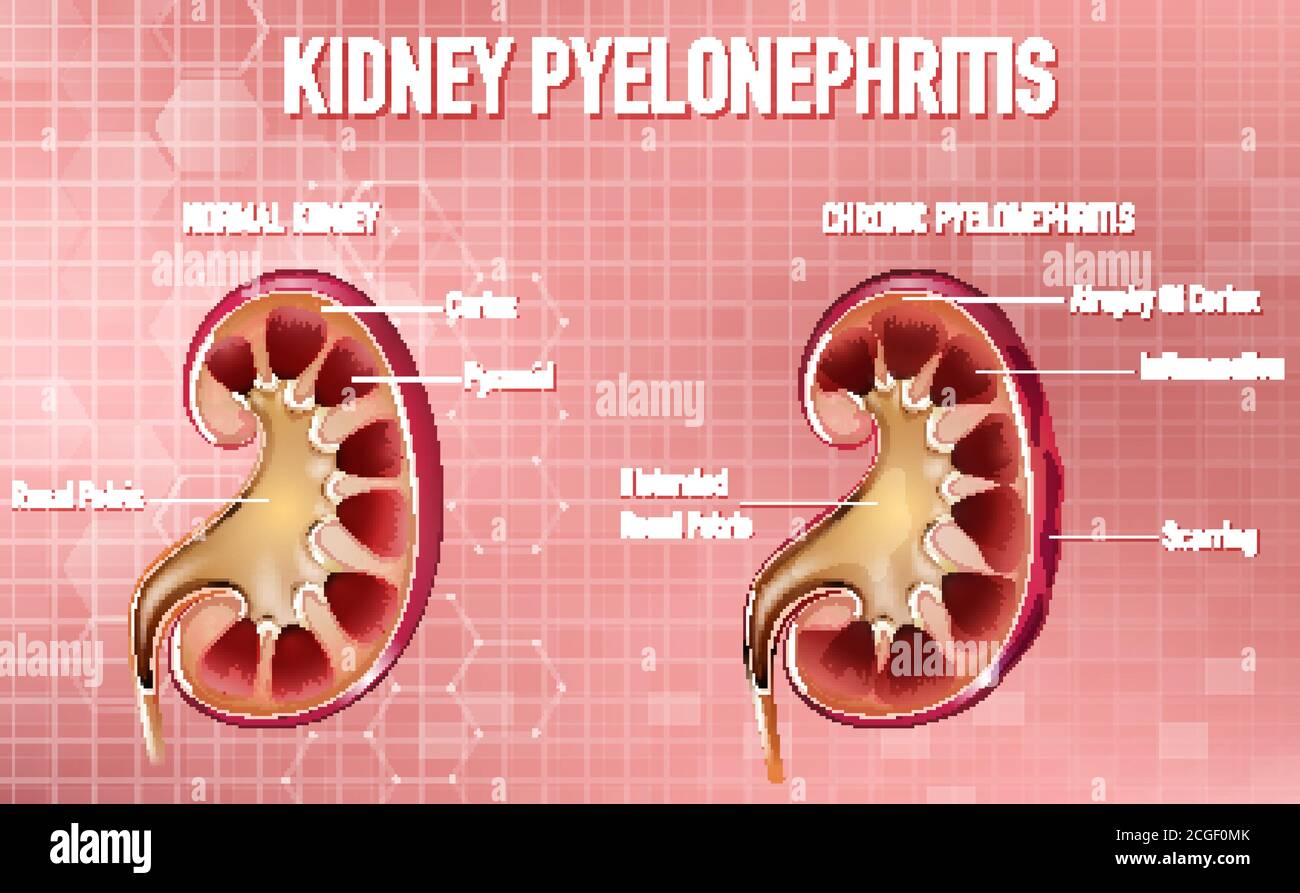 Informative illustration of Pyelonephritis illustration Stock Vectorhttps://www.alamy.com/image-license-details/?v=1https://www.alamy.com/informative-illustration-of-pyelonephritis-illustration-image371582083.html
RF2CGF0MK–Informative illustration of Pyelonephritis illustration
 An atlas of human anatomy for students and physicians . Base of the pyramidBasis pyiamidis Pyramid of Malpighi Py.inlir ml CM.ilpighii Renal papilla Papilla renahs Arterial archA. arciformis Renal papilla , Papilla renalis! Medulla /ISubstantia meduUarisCortexSubstantia corticalis Infundibulum Cal)x major Capsule, fibrous coat, or tunica/ albuginea of the kidney Tunica fibrosa Calices Calyces minores. Fig. 825.—Coronal Section through the RightKidney and the Renal Pelvis. Substantia Cor-ticalis, the Cortex ; Substantia Medullaris,the Medulla. ^ See Appendb I Base of the pyramidBasis pyramidis Stock Photohttps://www.alamy.com/image-license-details/?v=1https://www.alamy.com/an-atlas-of-human-anatomy-for-students-and-physicians-base-of-the-pyramidbasis-pyiamidis-pyramid-of-malpighi-pyinlir-ml-cmilpighii-renal-papilla-papilla-renahs-arterial-archa-arciformis-renal-papilla-papilla-renalis!-medulla-isubstantia-meduuariscortexsubstantia-corticalis-infundibulum-calx-major-capsule-fibrous-coat-or-tunica-albuginea-of-the-kidney-tunica-fibrosa-calices-calyces-minores-fig-825coronal-section-through-the-rightkidney-and-the-renal-pelvis-substantia-cor-ticalis-the-cortex-substantia-medullaristhe-medulla-see-appendb-i-base-of-the-pyramidbasis-pyramidis-image338304745.html
RM2AJB349–An atlas of human anatomy for students and physicians . Base of the pyramidBasis pyiamidis Pyramid of Malpighi Py.inlir ml CM.ilpighii Renal papilla Papilla renahs Arterial archA. arciformis Renal papilla , Papilla renalis! Medulla /ISubstantia meduUarisCortexSubstantia corticalis Infundibulum Cal)x major Capsule, fibrous coat, or tunica/ albuginea of the kidney Tunica fibrosa Calices Calyces minores. Fig. 825.—Coronal Section through the RightKidney and the Renal Pelvis. Substantia Cor-ticalis, the Cortex ; Substantia Medullaris,the Medulla. ^ See Appendb I Base of the pyramidBasis pyramidis
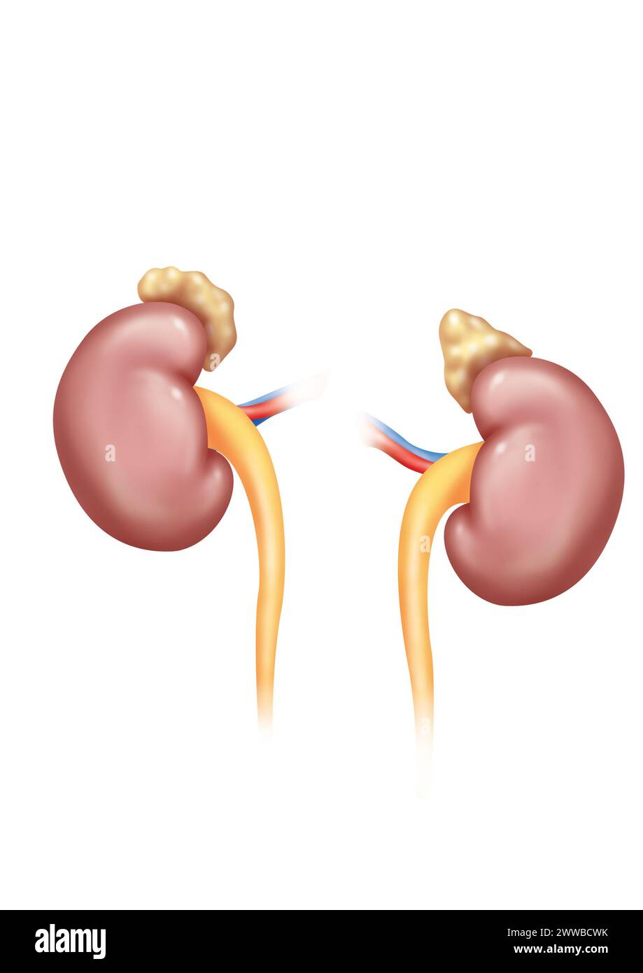 Kidneys in posterior view with adrenal glands ureters and renal arteries and veins. The left kidney is placed higher than the right kidney. Stock Photohttps://www.alamy.com/image-license-details/?v=1https://www.alamy.com/kidneys-in-posterior-view-with-adrenal-glands-ureters-and-renal-arteries-and-veins-the-left-kidney-is-placed-higher-than-the-right-kidney-image600770511.html
RM2WWBCWK–Kidneys in posterior view with adrenal glands ureters and renal arteries and veins. The left kidney is placed higher than the right kidney.
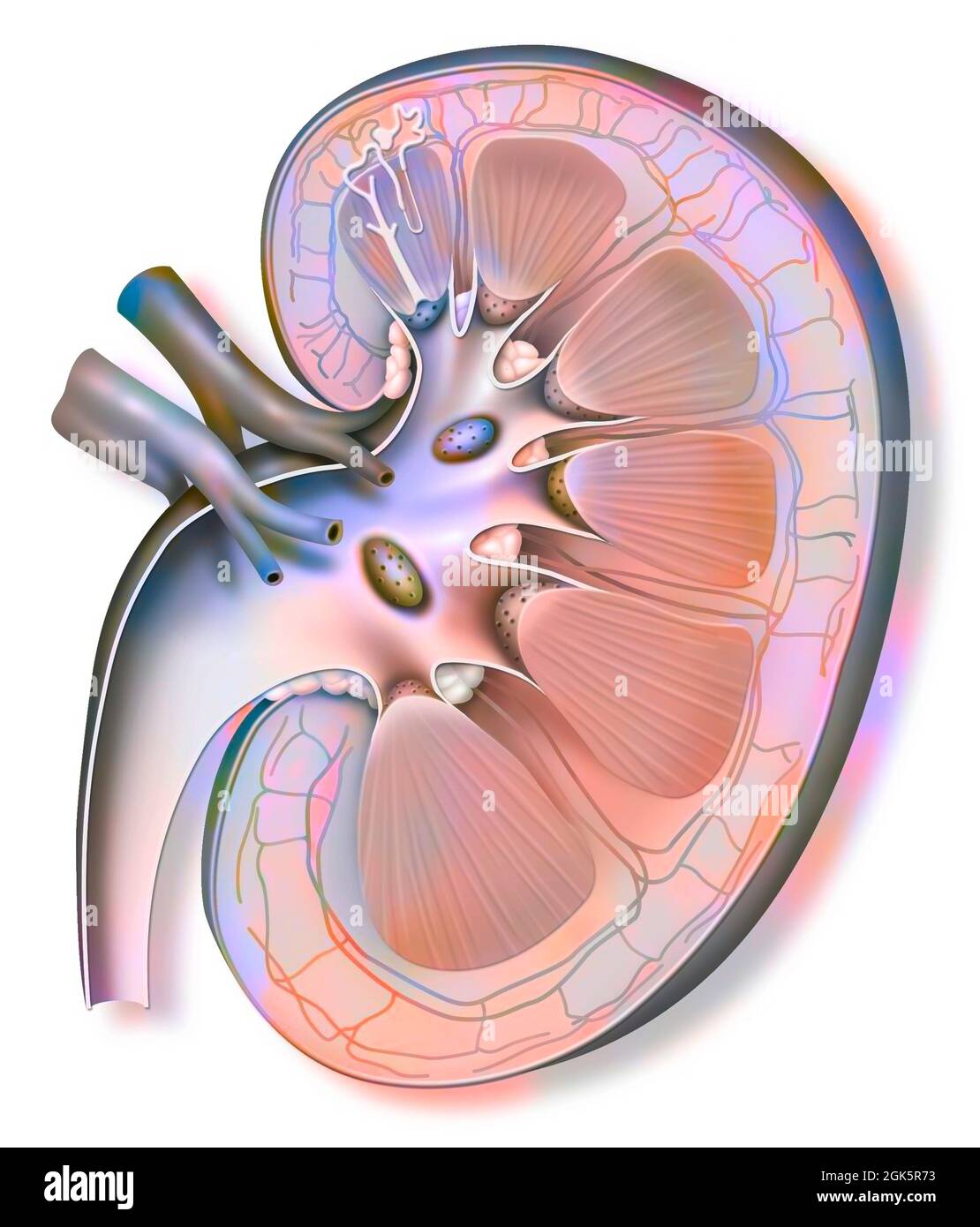 Kidney and left ureter with a nephron in enlarged size. Stock Photohttps://www.alamy.com/image-license-details/?v=1https://www.alamy.com/kidney-and-left-ureter-with-a-nephron-in-enlarged-size-image442065655.html
RF2GK5R73–Kidney and left ureter with a nephron in enlarged size.
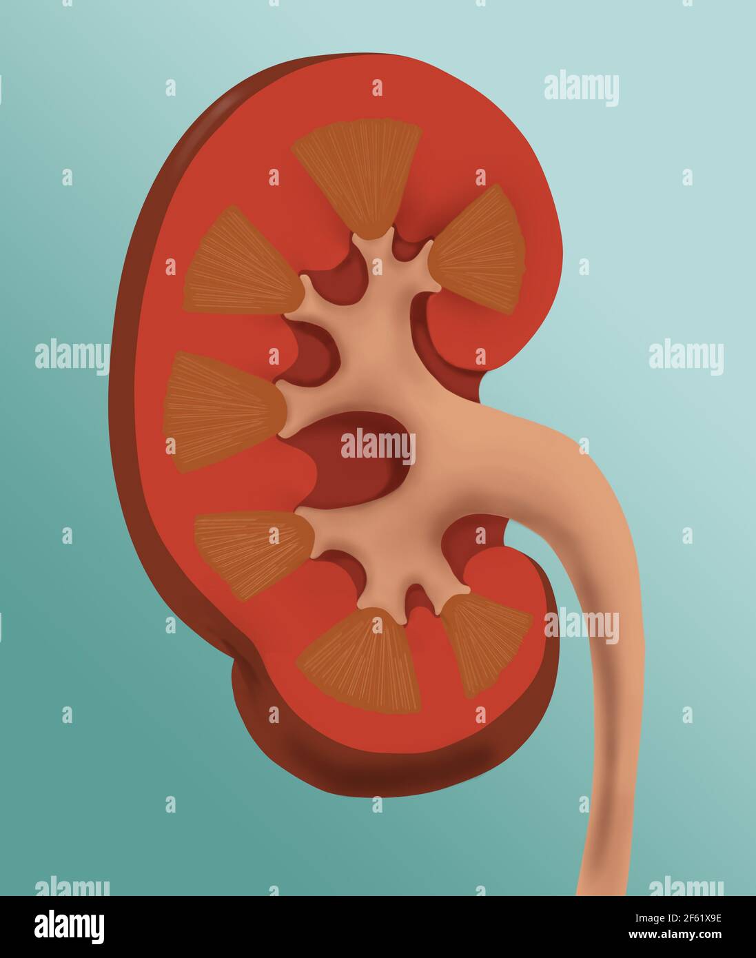 Normal Human Kidney Stock Photohttps://www.alamy.com/image-license-details/?v=1https://www.alamy.com/normal-human-kidney-image416779370.html
RM2F61X9E–Normal Human Kidney
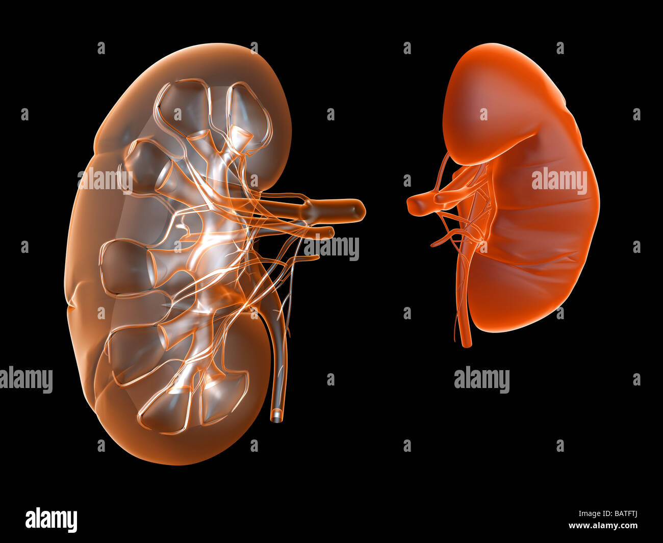 Kidneys, computer artwork. Stock Photohttps://www.alamy.com/image-license-details/?v=1https://www.alamy.com/stock-photo-kidneys-computer-artwork-23896226.html
RFBATFTJ–Kidneys, computer artwork.
 An atlas of human anatomy for students and physicians . Fig. 825.—Coronal Section through the RightKidney and the Renal Pelvis. Substantia Cor-ticalis, the Cortex ; Substantia Medullaris,the Medulla. ^ See Appendb I Base of the pyramidBasis pyramidis Renal papilla | Pyramid of MalpighiPapilla renalis Pyramis renalis (Malpighii) Fig. 826.—Sinus Renalis, the Sinus of theKidney, displayed in a Coronally-bisectedKidney by Removal of the Renal Pelvisand the Bloodvessels. Posterior Half. Ren—The kidney. 62- 492 URINARY ORGANS. *Area cnbrosa *Area cnbroba Orifices of the uriniferous tubules (excretor Stock Photohttps://www.alamy.com/image-license-details/?v=1https://www.alamy.com/an-atlas-of-human-anatomy-for-students-and-physicians-fig-825coronal-section-through-the-rightkidney-and-the-renal-pelvis-substantia-cor-ticalis-the-cortex-substantia-medullaristhe-medulla-see-appendb-i-base-of-the-pyramidbasis-pyramidis-renal-papilla-pyramid-of-malpighipapilla-renalis-pyramis-renalis-malpighii-fig-826sinus-renalis-the-sinus-of-thekidney-displayed-in-a-coronally-bisectedkidney-by-removal-of-the-renal-pelvisand-the-bloodvessels-posterior-half-renthe-kidney-62-492-urinary-organs-area-cnbrosa-area-cnbroba-orifices-of-the-uriniferous-tubules-excretor-image338304511.html
RM2AJB2RY–An atlas of human anatomy for students and physicians . Fig. 825.—Coronal Section through the RightKidney and the Renal Pelvis. Substantia Cor-ticalis, the Cortex ; Substantia Medullaris,the Medulla. ^ See Appendb I Base of the pyramidBasis pyramidis Renal papilla | Pyramid of MalpighiPapilla renalis Pyramis renalis (Malpighii) Fig. 826.—Sinus Renalis, the Sinus of theKidney, displayed in a Coronally-bisectedKidney by Removal of the Renal Pelvisand the Bloodvessels. Posterior Half. Ren—The kidney. 62- 492 URINARY ORGANS. *Area cnbrosa *Area cnbroba Orifices of the uriniferous tubules (excretor
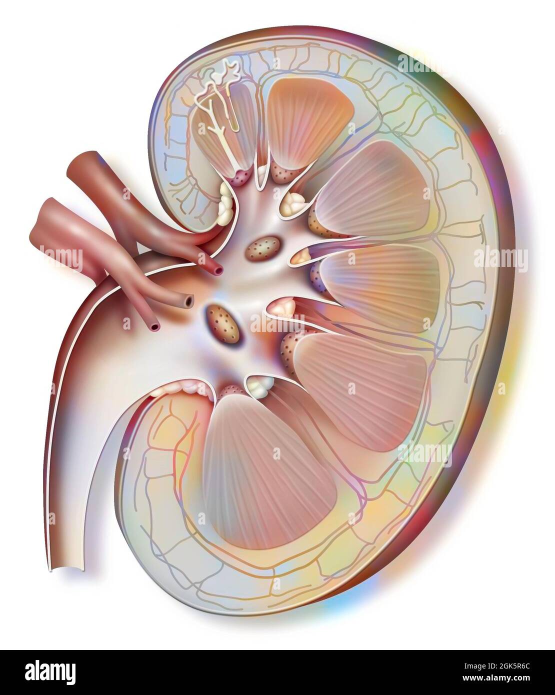 Kidney and left ureter with a nephron in enlarged size. Stock Photohttps://www.alamy.com/image-license-details/?v=1https://www.alamy.com/kidney-and-left-ureter-with-a-nephron-in-enlarged-size-image442065636.html
RF2GK5R6C–Kidney and left ureter with a nephron in enlarged size.
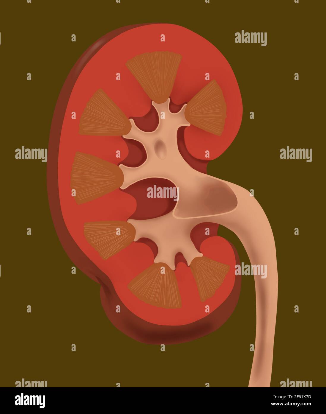 Normal Human Kidney Stock Photohttps://www.alamy.com/image-license-details/?v=1https://www.alamy.com/normal-human-kidney-image416779313.html
RM2F61X7D–Normal Human Kidney
 Kidneys, computer artwork. Stock Photohttps://www.alamy.com/image-license-details/?v=1https://www.alamy.com/stock-photo-kidneys-computer-artwork-23895802.html
RFBATF9E–Kidneys, computer artwork.
 An atlas of human anatomy for students and physicians . Lobules ofthe kidneyLobi renalesUreter Fig. 828.—Right Kidney and Suprarenal Cap-sule- FROM A Human Fi-Etus in the MiddleOF THE Seventh Month (Months of FourWeeks E.^ch). Seen from Behind. Lobules of the ^ *kidney •- Lobi renales Hilum of the ? kidneyHilus renal. Renal papillse Papilla; renales ? Base of the pyramid Basis pyramidis . Sinus of the kidney 5/-^Sinus renalis-, / Pyramid of Malpighif Pyramis renalis (Malpighii) CortexSubstantia corticalis Columns of Eertin, or septularenum Columnae renales (Bertini) Fig. 829.—Sinus of the Ki Stock Photohttps://www.alamy.com/image-license-details/?v=1https://www.alamy.com/an-atlas-of-human-anatomy-for-students-and-physicians-lobules-ofthe-kidneylobi-renalesureter-fig-828right-kidney-and-suprarenal-cap-sule-from-a-human-fi-etus-in-the-middleof-the-seventh-month-months-of-fourweeks-ech-seen-from-behind-lobules-of-the-kidney-lobi-renales-hilum-of-the-kidneyhilus-renal-renal-papillse-papilla-renales-base-of-the-pyramid-basis-pyramidis-sinus-of-the-kidney-5-sinus-renalis-pyramid-of-malpighif-pyramis-renalis-malpighii-cortexsubstantia-corticalis-columns-of-eertin-or-septularenum-columnae-renales-bertini-fig-829sinus-of-the-ki-image338303808.html
RM2AJB1XT–An atlas of human anatomy for students and physicians . Lobules ofthe kidneyLobi renalesUreter Fig. 828.—Right Kidney and Suprarenal Cap-sule- FROM A Human Fi-Etus in the MiddleOF THE Seventh Month (Months of FourWeeks E.^ch). Seen from Behind. Lobules of the ^ *kidney •- Lobi renales Hilum of the ? kidneyHilus renal. Renal papillse Papilla; renales ? Base of the pyramid Basis pyramidis . Sinus of the kidney 5/-^Sinus renalis-, / Pyramid of Malpighif Pyramis renalis (Malpighii) CortexSubstantia corticalis Columns of Eertin, or septularenum Columnae renales (Bertini) Fig. 829.—Sinus of the Ki
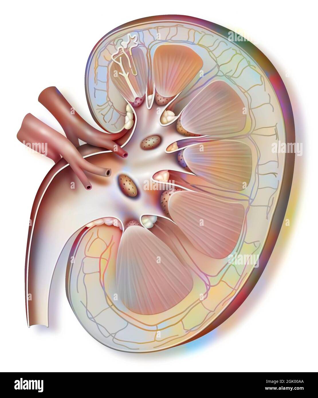 Kidney and left ureter with a nephron in enlarged size. Stock Photohttps://www.alamy.com/image-license-details/?v=1https://www.alamy.com/kidney-and-left-ureter-with-a-nephron-in-enlarged-size-image441937954.html
RF2GK00AA–Kidney and left ureter with a nephron in enlarged size.
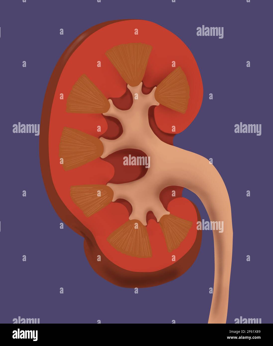 Normal Human Kidney Stock Photohttps://www.alamy.com/image-license-details/?v=1https://www.alamy.com/normal-human-kidney-image416779337.html
RM2F61X89–Normal Human Kidney
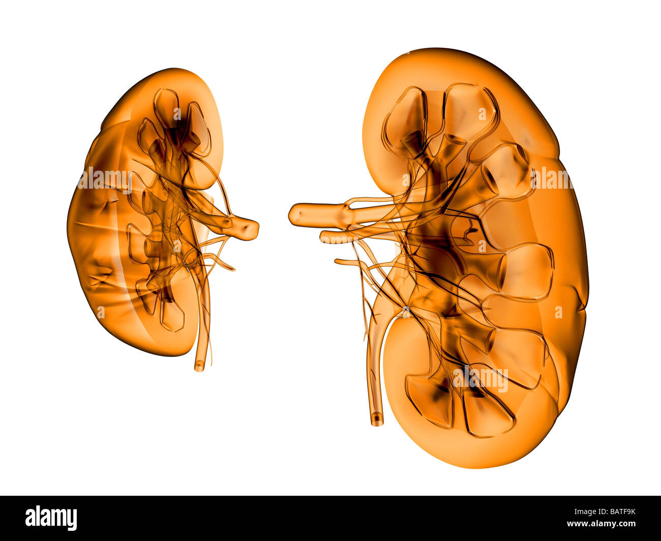 Kidneys, computer artwork. Stock Photohttps://www.alamy.com/image-license-details/?v=1https://www.alamy.com/stock-photo-kidneys-computer-artwork-23895807.html
RFBATF9K–Kidneys, computer artwork.
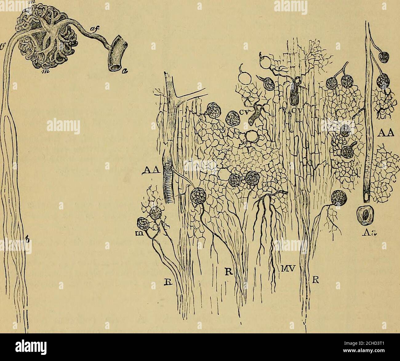 . Quain's elements of anatomy . (fig. 573),the meshes of the network being poly-gonal amongst the convoluted tubules and elongated amongst thetubules of the medullary rays. But the efferent vessels from the lower-most glomeruli break up wholly into pencils of straight vessels (falsevasa recta) which pass directly into the boundary layer of the medulla,and there supply the continuation downwards of the medullary rays intothe pyramid. VOL. II. • V V 658 THE KIDNEYS. The renal arteries give branches likewise to the capsule of the kidney -whichanastomose with branches of the lumbar arteries, and t Stock Photohttps://www.alamy.com/image-license-details/?v=1https://www.alamy.com/quains-elements-of-anatomy-fig-573the-meshes-of-the-network-being-poly-gonal-amongst-the-convoluted-tubules-and-elongated-amongst-thetubules-of-the-medullary-rays-but-the-efferent-vessels-from-the-lower-most-glomeruli-break-up-wholly-into-pencils-of-straight-vessels-falsevasa-recta-which-pass-directly-into-the-boundary-layer-of-the-medullaand-there-supply-the-continuation-downwards-of-the-medullary-rays-intothe-pyramid-vol-ii-v-v-658-the-kidneys-the-renal-arteries-give-branches-likewise-to-the-capsule-of-the-kidney-whichanastomose-with-branches-of-the-lumbar-arteries-and-t-image372155281.html
RM2CHD3T1–. Quain's elements of anatomy . (fig. 573),the meshes of the network being poly-gonal amongst the convoluted tubules and elongated amongst thetubules of the medullary rays. But the efferent vessels from the lower-most glomeruli break up wholly into pencils of straight vessels (falsevasa recta) which pass directly into the boundary layer of the medulla,and there supply the continuation downwards of the medullary rays intothe pyramid. VOL. II. • V V 658 THE KIDNEYS. The renal arteries give branches likewise to the capsule of the kidney -whichanastomose with branches of the lumbar arteries, and t
 Kidney and left ureter with a nephron in enlarged size. Stock Photohttps://www.alamy.com/image-license-details/?v=1https://www.alamy.com/kidney-and-left-ureter-with-a-nephron-in-enlarged-size-image441937939.html
RF2GK009R–Kidney and left ureter with a nephron in enlarged size.
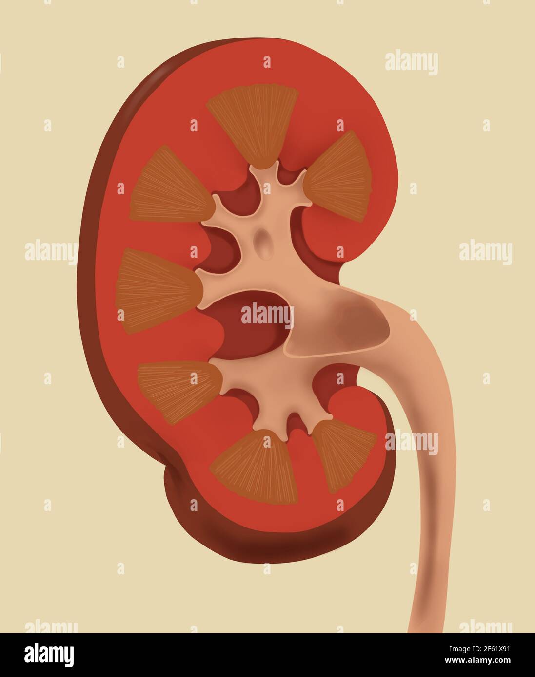 Normal Human Kidney Stock Photohttps://www.alamy.com/image-license-details/?v=1https://www.alamy.com/normal-human-kidney-image416779357.html
RM2F61X91–Normal Human Kidney
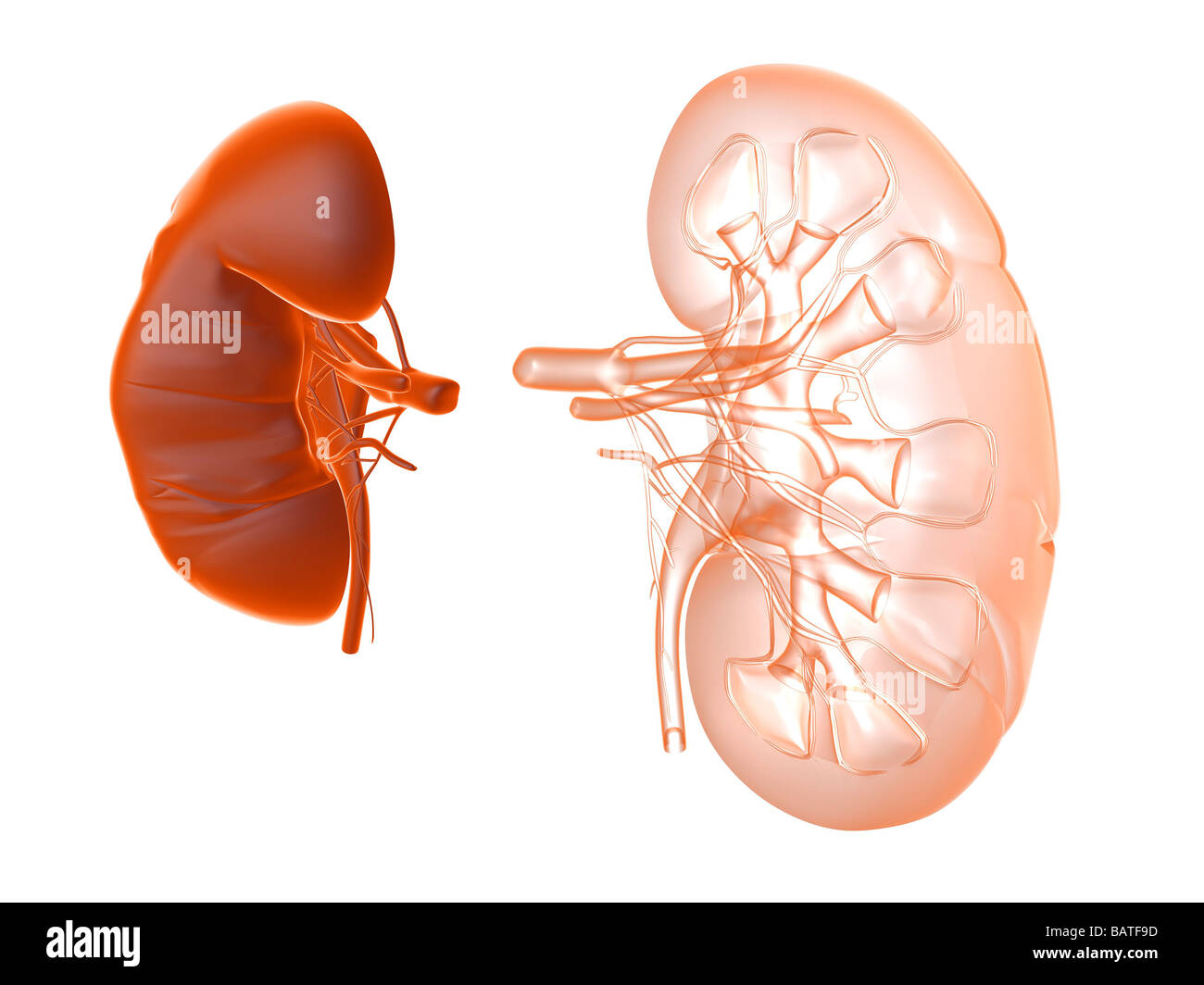 Kidneys, computer artwork. Stock Photohttps://www.alamy.com/image-license-details/?v=1https://www.alamy.com/stock-photo-kidneys-computer-artwork-23895801.html
RFBATF9D–Kidneys, computer artwork.
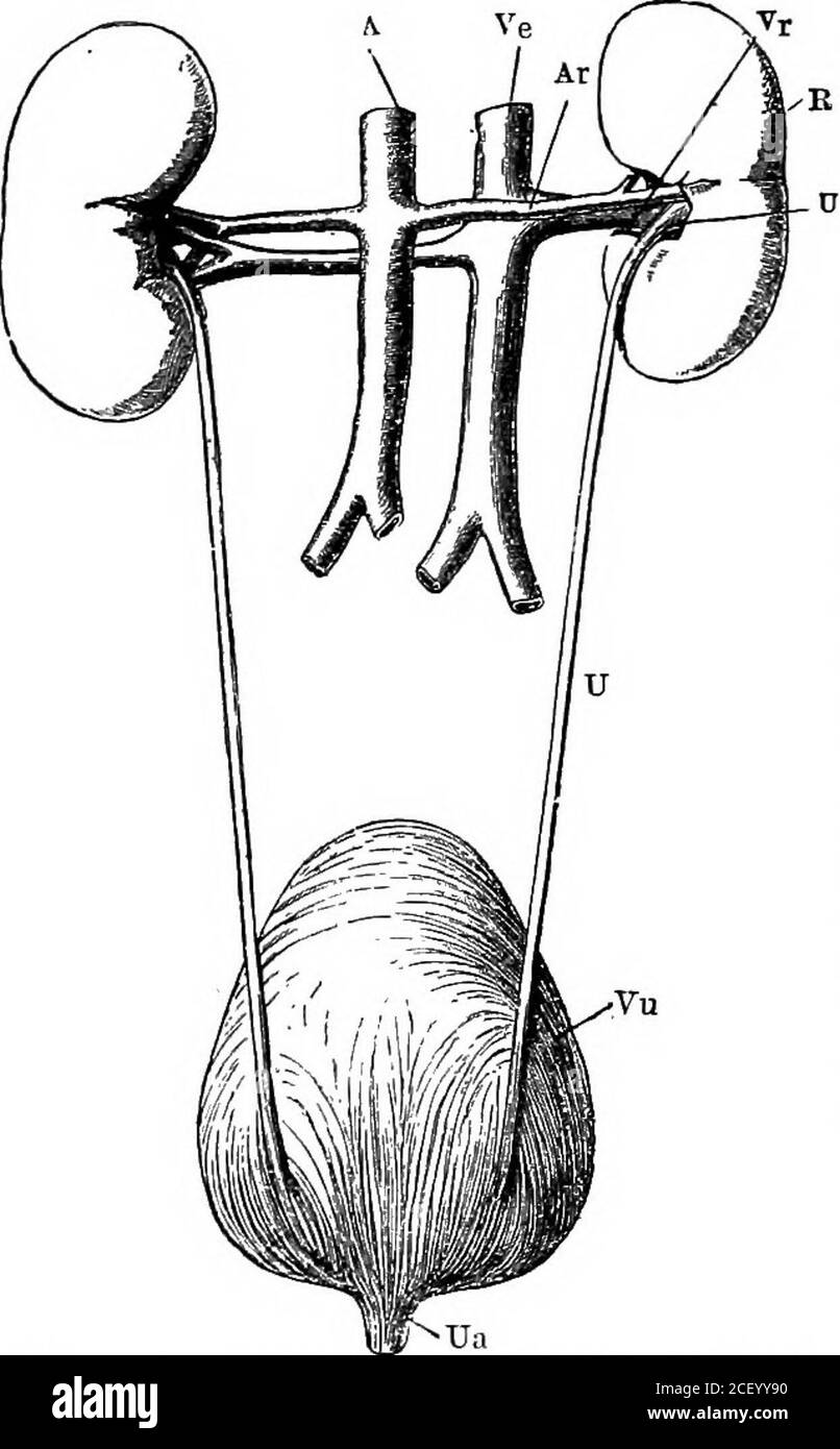 . Human physiology. number ofshort wide prolongations.The cavity is called thepelvis of the kidney, andits branches, calices (Lat.calyx, a cup). We alsoobserve that the solid por-tion of the kidney consistsof an inner medullarysubstance which has afibrous appearance, andan outer cortical sub-stance of a darker colour.The medullary sub-stance is not really fibrous,but consists of a multitude R, right kidney; U, ureter; A, aorta ; Ar, right renal of minute tubes arranged in separate conical massescalled the pyramids. Theapex or point of each pyramid is turned toward the pelvis of thekidney, into Stock Photohttps://www.alamy.com/image-license-details/?v=1https://www.alamy.com/human-physiology-number-ofshort-wide-prolongationsthe-cavity-is-called-thepelvis-of-the-kidney-andits-branches-calices-latcalyx-a-cup-we-alsoobserve-that-the-solid-por-tion-of-the-kidney-consistsof-an-inner-medullarysubstance-which-has-afibrous-appearance-andan-outer-cortical-sub-stance-of-a-darker-colourthe-medullary-sub-stance-is-not-really-fibrousbut-consists-of-a-multitude-r-right-kidney-u-ureter-a-aorta-ar-right-renal-of-minute-tubes-arranged-in-separate-conical-massescalled-the-pyramids-theapex-or-point-of-each-pyramid-is-turned-toward-the-pelvis-of-thekidney-into-image370637036.html
RM2CEYY90–. Human physiology. number ofshort wide prolongations.The cavity is called thepelvis of the kidney, andits branches, calices (Lat.calyx, a cup). We alsoobserve that the solid por-tion of the kidney consistsof an inner medullarysubstance which has afibrous appearance, andan outer cortical sub-stance of a darker colour.The medullary sub-stance is not really fibrous,but consists of a multitude R, right kidney; U, ureter; A, aorta ; Ar, right renal of minute tubes arranged in separate conical massescalled the pyramids. Theapex or point of each pyramid is turned toward the pelvis of thekidney, into
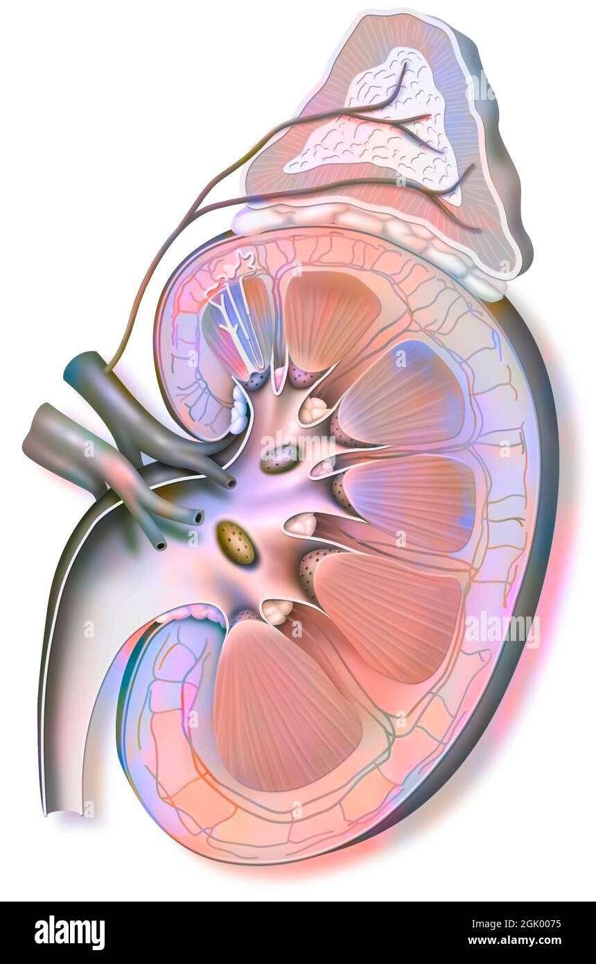 Left kidney (capped with its adrenal gland) and ureter. Stock Photohttps://www.alamy.com/image-license-details/?v=1https://www.alamy.com/left-kidney-capped-with-its-adrenal-gland-and-ureter-image441937865.html
RF2GK0075–Left kidney (capped with its adrenal gland) and ureter.
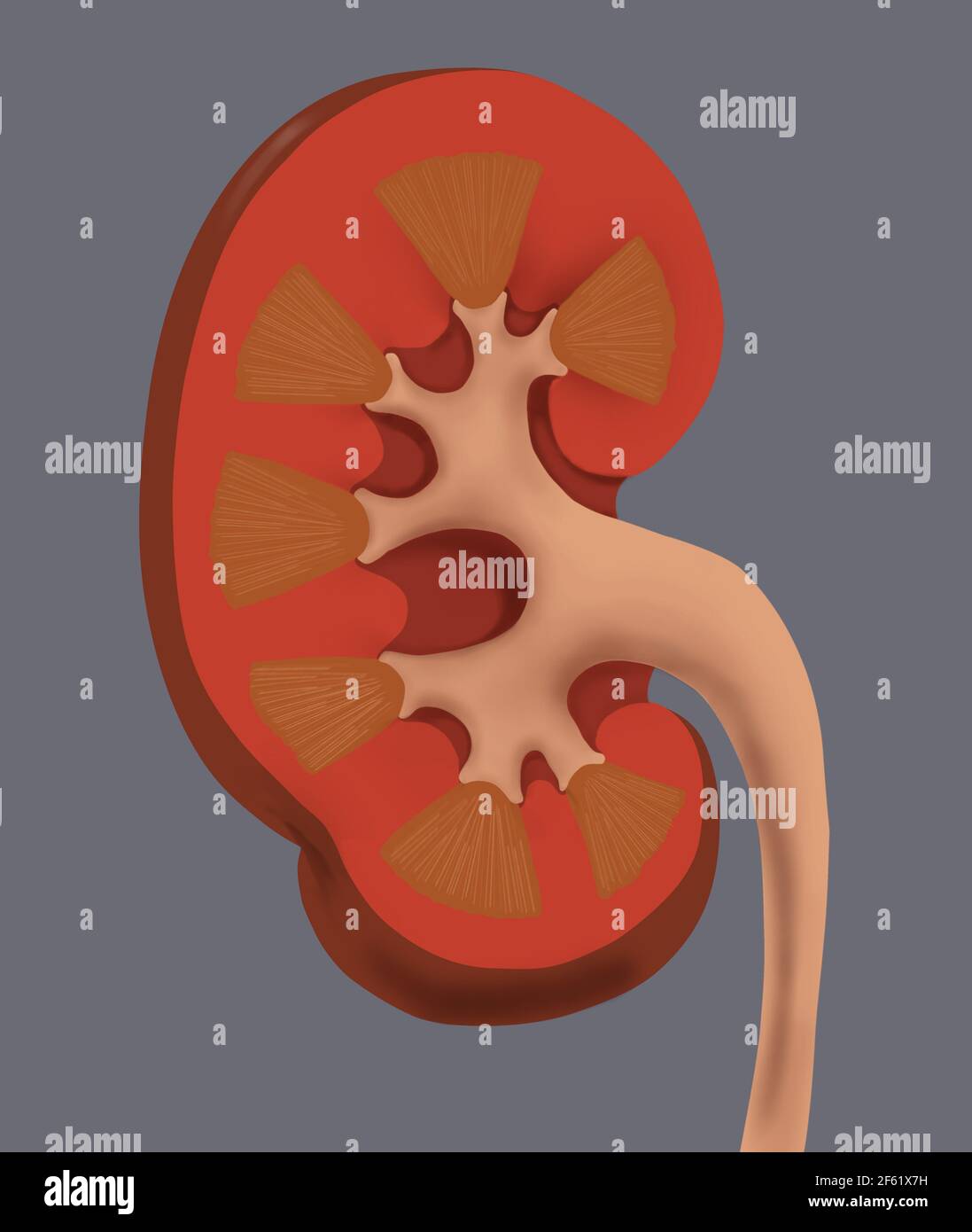 Normal Human Kidney Stock Photohttps://www.alamy.com/image-license-details/?v=1https://www.alamy.com/normal-human-kidney-image416779317.html
RM2F61X7H–Normal Human Kidney
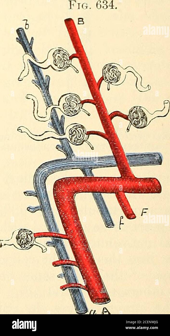 . Anatomy, descriptive and surgical. Diagrammatical Sketch of Kidney.. a P^ A Portion of Fig. 633, enlarged (the references arethe same). a, a, proper renal artery and vein, the former giving off the renal afferents, the latter receiving the renal effer-ents; b, b, interlobular artery and vein, the latter commencing from the stellate veins, and receivingbranches from the plexus around the tubuli eontorti, the former giving off renal afferents; c, straight tube,surrounded by tubuli eontorti, with which it communicates, forming a pyramid of Perrein, as more fullyshown in Fig.-626; d, margin of m Stock Photohttps://www.alamy.com/image-license-details/?v=1https://www.alamy.com/anatomy-descriptive-and-surgical-diagrammatical-sketch-of-kidney-a-p-a-portion-of-fig-633-enlarged-the-references-arethe-same-a-a-proper-renal-artery-and-vein-the-former-giving-off-the-renal-afferents-the-latter-receiving-the-renal-effer-ents-b-b-interlobular-artery-and-vein-the-latter-commencing-from-the-stellate-veins-and-receivingbranches-from-the-plexus-around-the-tubuli-eontorti-the-former-giving-off-renal-afferents-c-straight-tubesurrounded-by-tubuli-eontorti-with-which-it-communicates-forming-a-pyramid-of-perrein-as-more-fullyshown-in-fig-626-d-margin-of-m-image370504024.html
RM2CENWJG–. Anatomy, descriptive and surgical. Diagrammatical Sketch of Kidney.. a P^ A Portion of Fig. 633, enlarged (the references arethe same). a, a, proper renal artery and vein, the former giving off the renal afferents, the latter receiving the renal effer-ents; b, b, interlobular artery and vein, the latter commencing from the stellate veins, and receivingbranches from the plexus around the tubuli eontorti, the former giving off renal afferents; c, straight tube,surrounded by tubuli eontorti, with which it communicates, forming a pyramid of Perrein, as more fullyshown in Fig.-626; d, margin of m
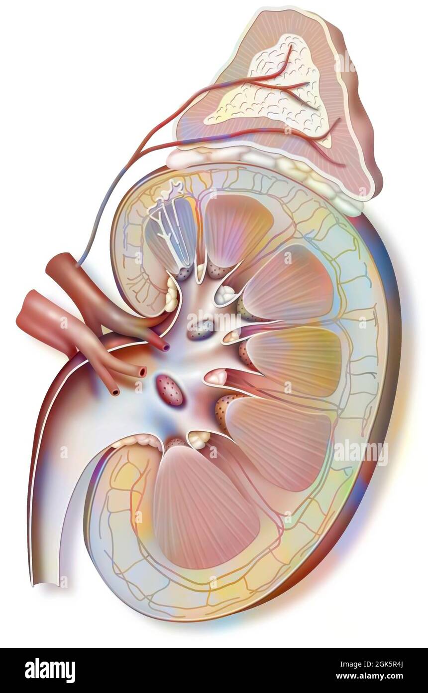 Left kidney (capped with its adrenal gland) and ureter. Stock Photohttps://www.alamy.com/image-license-details/?v=1https://www.alamy.com/left-kidney-capped-with-its-adrenal-gland-and-ureter-image442065586.html
RF2GK5R4J–Left kidney (capped with its adrenal gland) and ureter.
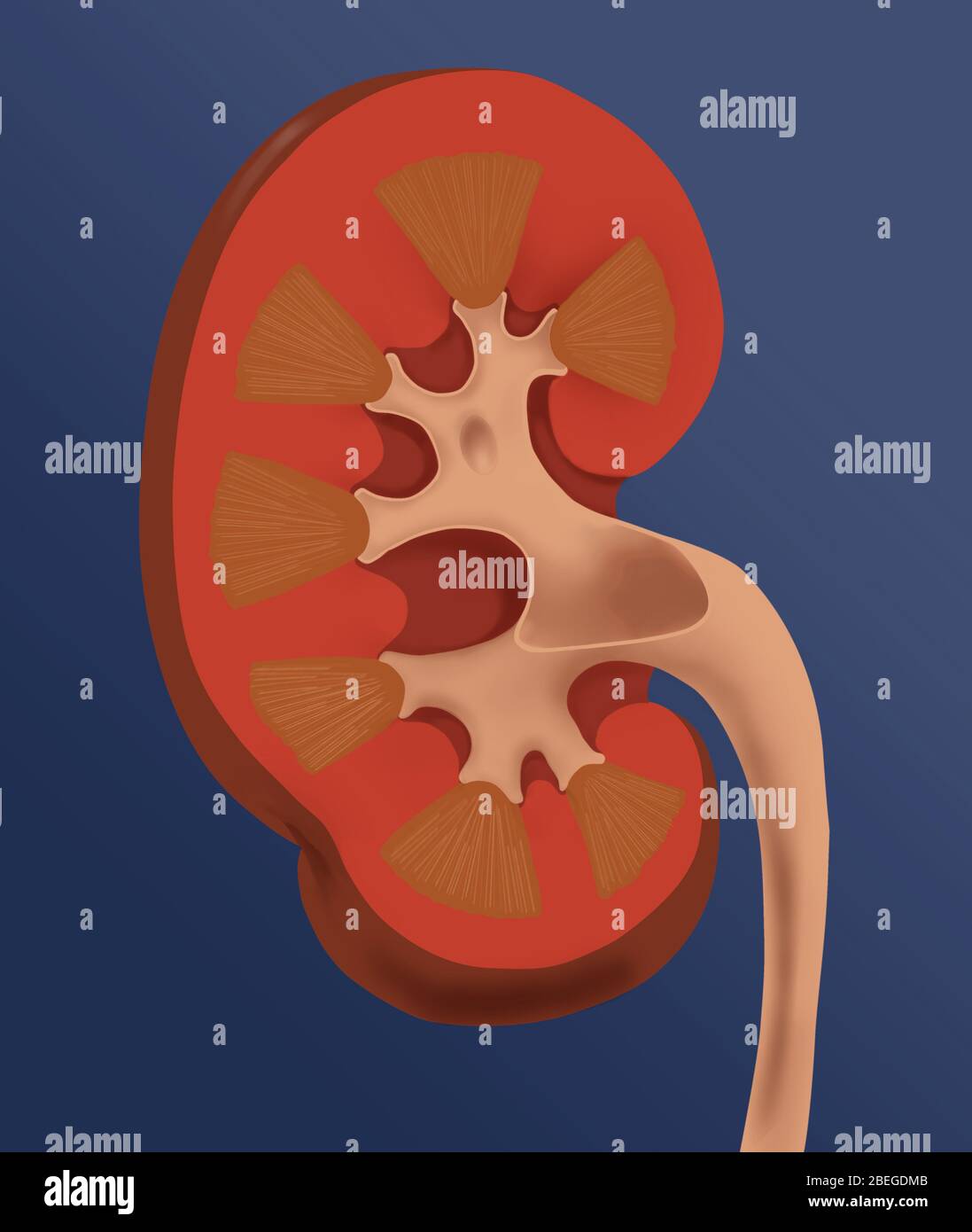 Normal Human Kidney Stock Photohttps://www.alamy.com/image-license-details/?v=1https://www.alamy.com/normal-human-kidney-image353174539.html
RM2BEGDMB–Normal Human Kidney
 . Cunningham's Text-book of anatomy. Anatomy. THE DUCT OF THE KIDNEY. 1269 Cortical substance Basal part of pyramid" Interlobar^ . artery k Pyramid Papilla J-- Renal artery Calyx ureter, in most cases, lies across the external iliac; but this arrangement is by no means constant. The course and position occupied by the abdominal portion of the ureter is well seen in Fig. 983. In X-ray photographs, the shadow cast by the abdominal portion of the ureter when the latter has been rendered opaque, is seen to fall immediately in front of the tips of the transverse processes of the lower lumbar v Stock Photohttps://www.alamy.com/image-license-details/?v=1https://www.alamy.com/cunninghams-text-book-of-anatomy-anatomy-the-duct-of-the-kidney-1269-cortical-substance-basal-part-of-pyramidquot-interlobar-artery-k-pyramid-papilla-j-renal-artery-calyx-ureter-in-most-cases-lies-across-the-external-iliac-but-this-arrangement-is-by-no-means-constant-the-course-and-position-occupied-by-the-abdominal-portion-of-the-ureter-is-well-seen-in-fig-983-in-x-ray-photographs-the-shadow-cast-by-the-abdominal-portion-of-the-ureter-when-the-latter-has-been-rendered-opaque-is-seen-to-fall-immediately-in-front-of-the-tips-of-the-transverse-processes-of-the-lower-lumbar-v-image231848839.html
RMRD5HFK–. Cunningham's Text-book of anatomy. Anatomy. THE DUCT OF THE KIDNEY. 1269 Cortical substance Basal part of pyramid" Interlobar^ . artery k Pyramid Papilla J-- Renal artery Calyx ureter, in most cases, lies across the external iliac; but this arrangement is by no means constant. The course and position occupied by the abdominal portion of the ureter is well seen in Fig. 983. In X-ray photographs, the shadow cast by the abdominal portion of the ureter when the latter has been rendered opaque, is seen to fall immediately in front of the tips of the transverse processes of the lower lumbar v
 Left kidney (capped with its adrenal gland) and ureter. Stock Photohttps://www.alamy.com/image-license-details/?v=1https://www.alamy.com/left-kidney-capped-with-its-adrenal-gland-and-ureter-image441937911.html
RF2GK008R–Left kidney (capped with its adrenal gland) and ureter.
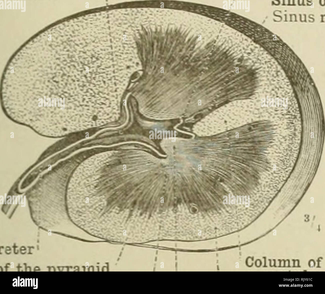 . An atlas of human anatomy for students and physicians. Anatomy. URINARY ORGANS 491. Cut surface of the renal parenchyma Calix' Calyx renalis Sinus of the kidney Internal border binus renalis IMargo medialis Hilum of the kidney iiilus renalis Pelvis of the kidney Pelvis renalis Ureter Ease of the pyramid Basis pyramidis I Pyramid of Malpighi Pvramis renalis '(Malpighii) Column of Berlin, or septulum renum Columna renalis (Bertini) Cortex Substantia corticalis Renal papilla Papilla renalis. Please note that these images are extracted from scanned page images that may have been digitally enhanc Stock Photohttps://www.alamy.com/image-license-details/?v=1https://www.alamy.com/an-atlas-of-human-anatomy-for-students-and-physicians-anatomy-urinary-organs-491-cut-surface-of-the-renal-parenchyma-calix-calyx-renalis-sinus-of-the-kidney-internal-border-binus-renalis-imargo-medialis-hilum-of-the-kidney-iiilus-renalis-pelvis-of-the-kidney-pelvis-renalis-ureter-ease-of-the-pyramid-basis-pyramidis-i-pyramid-of-malpighi-pvramis-renalis-malpighii-column-of-berlin-or-septulum-renum-columna-renalis-bertini-cortex-substantia-corticalis-renal-papilla-papilla-renalis-please-note-that-these-images-are-extracted-from-scanned-page-images-that-may-have-been-digitally-enhanc-image235396040.html
RMRJY61C–. An atlas of human anatomy for students and physicians. Anatomy. URINARY ORGANS 491. Cut surface of the renal parenchyma Calix' Calyx renalis Sinus of the kidney Internal border binus renalis IMargo medialis Hilum of the kidney iiilus renalis Pelvis of the kidney Pelvis renalis Ureter Ease of the pyramid Basis pyramidis I Pyramid of Malpighi Pvramis renalis '(Malpighii) Column of Berlin, or septulum renum Columna renalis (Bertini) Cortex Substantia corticalis Renal papilla Papilla renalis. Please note that these images are extracted from scanned page images that may have been digitally enhanc
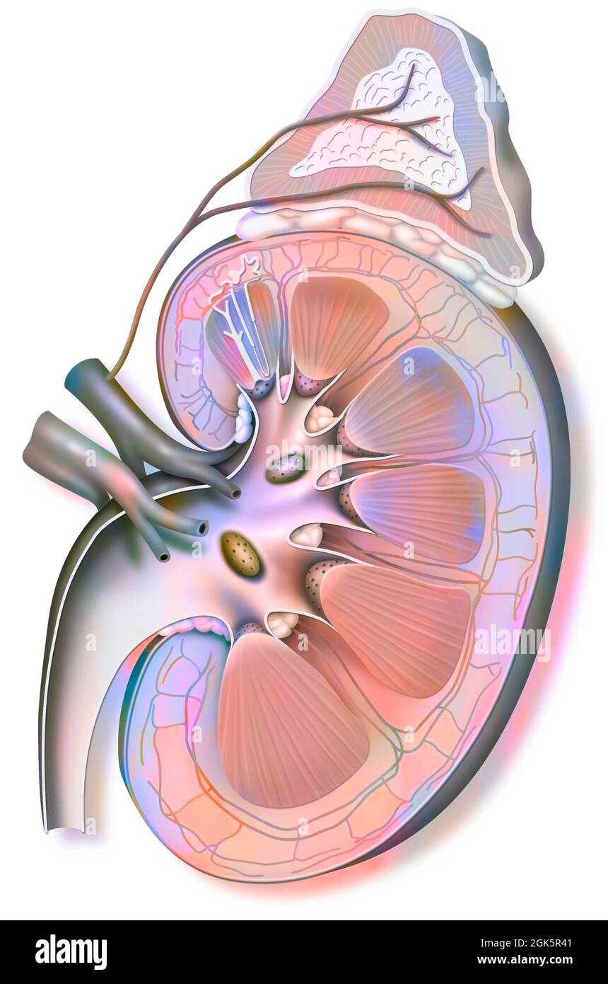 Left kidney (capped with its adrenal gland) and ureter. Stock Photohttps://www.alamy.com/image-license-details/?v=1https://www.alamy.com/left-kidney-capped-with-its-adrenal-gland-and-ureter-image442065569.html
RF2GK5R41–Left kidney (capped with its adrenal gland) and ureter.
 . An atlas of human anatomy for students and physicians. Anatomy. Cut surface of the renal parenchyma Calix' Calyx renalis Sinus of the kidney Internal border binus renalis IMargo medialis Hilum of the kidney iiilus renalis Pelvis of the kidney Pelvis renalis Ureter Ease of the pyramid Basis pyramidis I Pyramid of Malpighi Pvramis renalis '(Malpighii) Column of Berlin, or septulum renum Columna renalis (Bertini) Cortex Substantia corticalis Renal papilla Papilla renalis. Calices Calj ces minores' Superior extremity Extremitas superior Calices' ilyces minores Posterior surface 1 icu i posterior Stock Photohttps://www.alamy.com/image-license-details/?v=1https://www.alamy.com/an-atlas-of-human-anatomy-for-students-and-physicians-anatomy-cut-surface-of-the-renal-parenchyma-calix-calyx-renalis-sinus-of-the-kidney-internal-border-binus-renalis-imargo-medialis-hilum-of-the-kidney-iiilus-renalis-pelvis-of-the-kidney-pelvis-renalis-ureter-ease-of-the-pyramid-basis-pyramidis-i-pyramid-of-malpighi-pvramis-renalis-malpighii-column-of-berlin-or-septulum-renum-columna-renalis-bertini-cortex-substantia-corticalis-renal-papilla-papilla-renalis-calices-calj-ces-minores-superior-extremity-extremitas-superior-calices-ilyces-minores-posterior-surface-1-icu-i-posterior-image235396013.html
RMRJY60D–. An atlas of human anatomy for students and physicians. Anatomy. Cut surface of the renal parenchyma Calix' Calyx renalis Sinus of the kidney Internal border binus renalis IMargo medialis Hilum of the kidney iiilus renalis Pelvis of the kidney Pelvis renalis Ureter Ease of the pyramid Basis pyramidis I Pyramid of Malpighi Pvramis renalis '(Malpighii) Column of Berlin, or septulum renum Columna renalis (Bertini) Cortex Substantia corticalis Renal papilla Papilla renalis. Calices Calj ces minores' Superior extremity Extremitas superior Calices' ilyces minores Posterior surface 1 icu i posterior
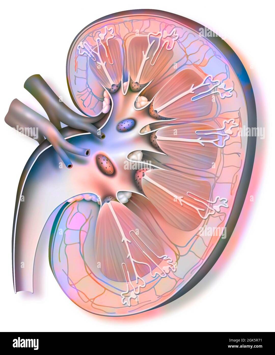 Left kidney (capped with its adrenal gland) and ureter. Stock Photohttps://www.alamy.com/image-license-details/?v=1https://www.alamy.com/left-kidney-capped-with-its-adrenal-gland-and-ureter-image442065653.html
RF2GK5R71–Left kidney (capped with its adrenal gland) and ureter.
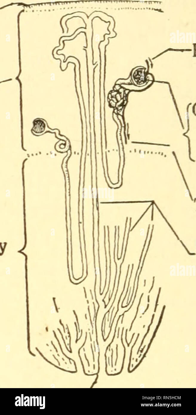 . Animal biology. Biology; Zoology; Physiology. -Tunic -Pelvis Tunic Cortex -"* y Renal artery ?—Renal vein -Ureter Medullary region. From renal artery ("Glomerulus v within I capsule To renal vein Collecting tubules "Cortex Pyramid of medullary region A Tip of pyramid B Fig. 131. — Human kidney. A, longitudinal section; B, diagram of the course of the tubules in the kidney. The cortex is the region in which the tubules come into functional association with the capillaries. Tubules extend through the medullary region to open on the summits of the pyramids. like elements, the tub Stock Photohttps://www.alamy.com/image-license-details/?v=1https://www.alamy.com/animal-biology-biology-zoology-physiology-tunic-pelvis-tunic-cortex-quot-y-renal-artery-renal-vein-ureter-medullary-region-from-renal-artery-quotglomerulus-v-within-i-capsule-to-renal-vein-collecting-tubules-quotcortex-pyramid-of-medullary-region-a-tip-of-pyramid-b-fig-131-human-kidney-a-longitudinal-section-b-diagram-of-the-course-of-the-tubules-in-the-kidney-the-cortex-is-the-region-in-which-the-tubules-come-into-functional-association-with-the-capillaries-tubules-extend-through-the-medullary-region-to-open-on-the-summits-of-the-pyramids-like-elements-the-tub-image236766004.html
RMRN5HCM–. Animal biology. Biology; Zoology; Physiology. -Tunic -Pelvis Tunic Cortex -"* y Renal artery ?—Renal vein -Ureter Medullary region. From renal artery ("Glomerulus v within I capsule To renal vein Collecting tubules "Cortex Pyramid of medullary region A Tip of pyramid B Fig. 131. — Human kidney. A, longitudinal section; B, diagram of the course of the tubules in the kidney. The cortex is the region in which the tubules come into functional association with the capillaries. Tubules extend through the medullary region to open on the summits of the pyramids. like elements, the tub
 Left kidney (capped with its adrenal gland) and ureter. Stock Photohttps://www.alamy.com/image-license-details/?v=1https://www.alamy.com/left-kidney-capped-with-its-adrenal-gland-and-ureter-image441937974.html
RF2GK00B2–Left kidney (capped with its adrenal gland) and ureter.
 . An atlas of human anatomy for students and physicians. Anatomy. Base of the pyramid 1 Basis pyramidis Pyrainid of MalpigM Pyia,.,isren.iiis(Malpigha) Kenal papilla 'lla renalis Arterial arch R enal papiUa Papilla renalis Medulla Substantia medullaris Cortex Stbitantia corticalis Infundibulum' Calyx major Capsule, fibrous coat, or tunica / albuginea of the kidney Tunica fibrosa Calices' Calyces minores. 1;. 825.—Coronal Section through the Right Kidney and the Renal Pelvis. Suhstaniia Cor- iiCALis, the Cortex; sri'.,rMiA Medullaris, ihe Medulla. 1 Base of the pyramid Basis pyramidis Renal pa Stock Photohttps://www.alamy.com/image-license-details/?v=1https://www.alamy.com/an-atlas-of-human-anatomy-for-students-and-physicians-anatomy-base-of-the-pyramid-1-basis-pyramidis-pyrainid-of-malpigm-pyiaisreniiismalpigha-kenal-papilla-lla-renalis-arterial-arch-r-enal-papiua-papilla-renalis-medulla-substantia-medullaris-cortex-stbitantia-corticalis-infundibulum-calyx-major-capsule-fibrous-coat-or-tunica-albuginea-of-the-kidney-tunica-fibrosa-calices-calyces-minores-1-825coronal-section-through-the-right-kidney-and-the-renal-pelvis-suhstaniia-cor-iicalis-the-cortex-srirmia-medullaris-ihe-medulla-1-base-of-the-pyramid-basis-pyramidis-renal-pa-image235395986.html
RMRJY5YE–. An atlas of human anatomy for students and physicians. Anatomy. Base of the pyramid 1 Basis pyramidis Pyrainid of MalpigM Pyia,.,isren.iiis(Malpigha) Kenal papilla 'lla renalis Arterial arch R enal papiUa Papilla renalis Medulla Substantia medullaris Cortex Stbitantia corticalis Infundibulum' Calyx major Capsule, fibrous coat, or tunica / albuginea of the kidney Tunica fibrosa Calices' Calyces minores. 1;. 825.—Coronal Section through the Right Kidney and the Renal Pelvis. Suhstaniia Cor- iiCALis, the Cortex; sri'.,rMiA Medullaris, ihe Medulla. 1 Base of the pyramid Basis pyramidis Renal pa
 Left kidney (capped with its adrenal gland) and ureter. Stock Photohttps://www.alamy.com/image-license-details/?v=1https://www.alamy.com/left-kidney-capped-with-its-adrenal-gland-and-ureter-image441937922.html
RF2GK0096–Left kidney (capped with its adrenal gland) and ureter.
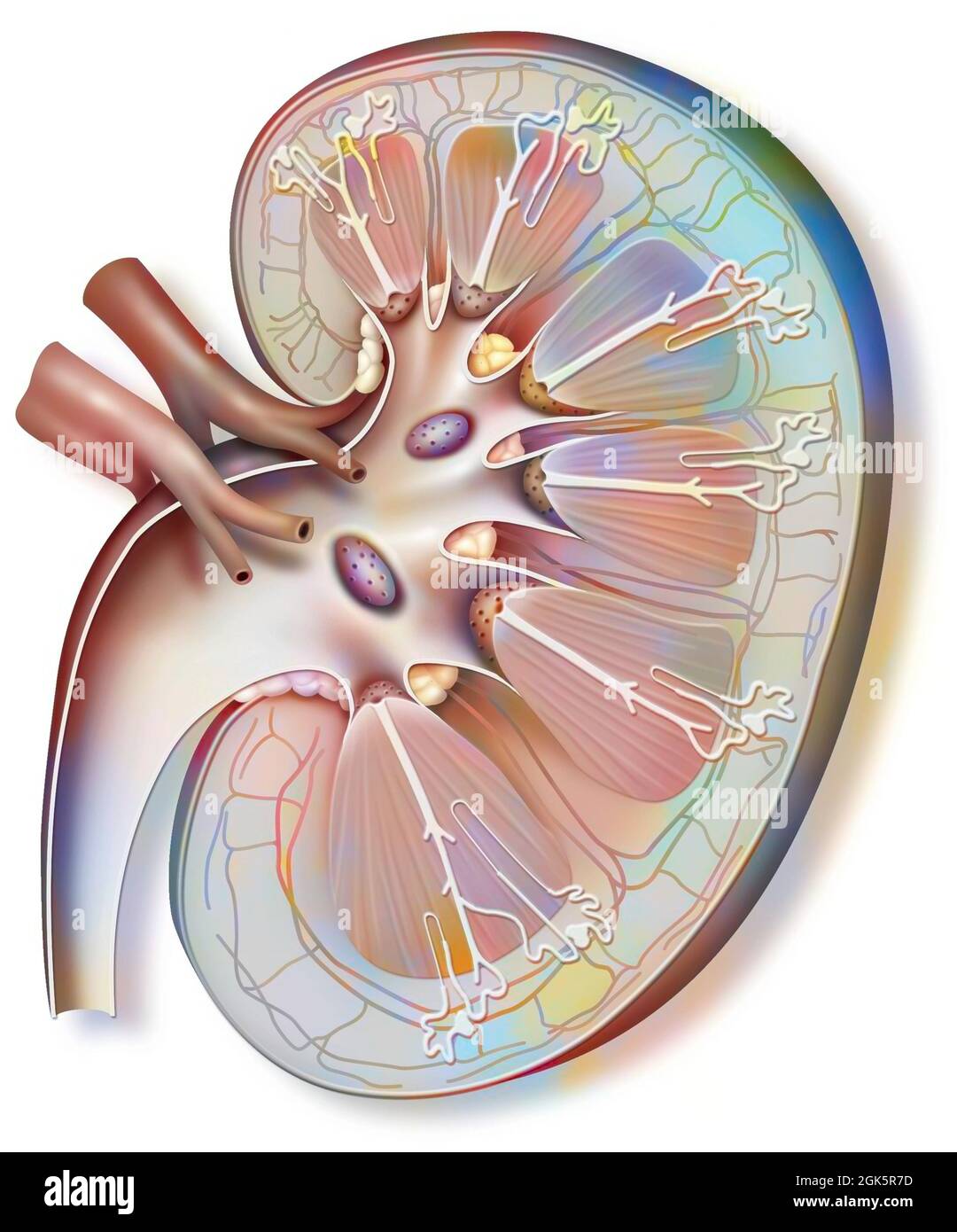 Left kidney (capped with its adrenal gland) and ureter. Stock Photohttps://www.alamy.com/image-license-details/?v=1https://www.alamy.com/left-kidney-capped-with-its-adrenal-gland-and-ureter-image442065665.html
RF2GK5R7D–Left kidney (capped with its adrenal gland) and ureter.
 Human urinary system, anterior view, anatomy. The human urinary system seen anterior with the right kidney seen in section to mount the fibrous capsul Stock Photohttps://www.alamy.com/image-license-details/?v=1https://www.alamy.com/human-urinary-system-anterior-view-anatomy-the-human-urinary-system-seen-anterior-with-the-right-kidney-seen-in-section-to-mount-the-fibrous-capsul-image355210267.html
RM2BHW68Y–Human urinary system, anterior view, anatomy. The human urinary system seen anterior with the right kidney seen in section to mount the fibrous capsul
 Human urinary system, anterior view, anatomy. The human urinary system seen anterior with the right kidney seen in section to mount the fibrous capsul Stock Photohttps://www.alamy.com/image-license-details/?v=1https://www.alamy.com/human-urinary-system-anterior-view-anatomy-the-human-urinary-system-seen-anterior-with-the-right-kidney-seen-in-section-to-mount-the-fibrous-capsul-image355210268.html
RM2BHW690–Human urinary system, anterior view, anatomy. The human urinary system seen anterior with the right kidney seen in section to mount the fibrous capsul
 Human urinary system, anterior view, anatomy. The human urinary system seen anterior with the right kidney seen in section to show the fibrous capsule Stock Photohttps://www.alamy.com/image-license-details/?v=1https://www.alamy.com/human-urinary-system-anterior-view-anatomy-the-human-urinary-system-seen-anterior-with-the-right-kidney-seen-in-section-to-show-the-fibrous-capsule-image355209863.html
RM2BHW5PF–Human urinary system, anterior view, anatomy. The human urinary system seen anterior with the right kidney seen in section to show the fibrous capsule
 Human urinary system, anterior view, anatomy. The human urinary system seen anterior with the right kidney seen in section to show the fibrous capsule Stock Photohttps://www.alamy.com/image-license-details/?v=1https://www.alamy.com/human-urinary-system-anterior-view-anatomy-the-human-urinary-system-seen-anterior-with-the-right-kidney-seen-in-section-to-show-the-fibrous-capsule-image355209826.html
RM2BHW5N6–Human urinary system, anterior view, anatomy. The human urinary system seen anterior with the right kidney seen in section to show the fibrous capsule
 Human urinary system, anterior view, anatomy. The human urinary system seen anterior with the right kidney seen in section to mount the fibrous capsul Stock Photohttps://www.alamy.com/image-license-details/?v=1https://www.alamy.com/human-urinary-system-anterior-view-anatomy-the-human-urinary-system-seen-anterior-with-the-right-kidney-seen-in-section-to-mount-the-fibrous-capsul-image355210260.html
RM2BHW68M–Human urinary system, anterior view, anatomy. The human urinary system seen anterior with the right kidney seen in section to mount the fibrous capsul
 Human urinary system, anterior view, anatomy. The human urinary system seen anterior with the right kidney seen in section to mount the fibrous capsul Stock Photohttps://www.alamy.com/image-license-details/?v=1https://www.alamy.com/human-urinary-system-anterior-view-anatomy-the-human-urinary-system-seen-anterior-with-the-right-kidney-seen-in-section-to-mount-the-fibrous-capsul-image355210259.html
RM2BHW68K–Human urinary system, anterior view, anatomy. The human urinary system seen anterior with the right kidney seen in section to mount the fibrous capsul
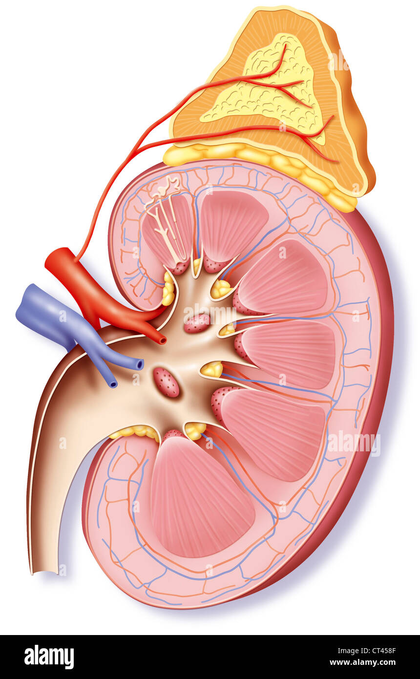 KIDNEY, DRAWING Stock Photohttps://www.alamy.com/image-license-details/?v=1https://www.alamy.com/stock-photo-kidney-drawing-49264447.html
RMCT458F–KIDNEY, DRAWING
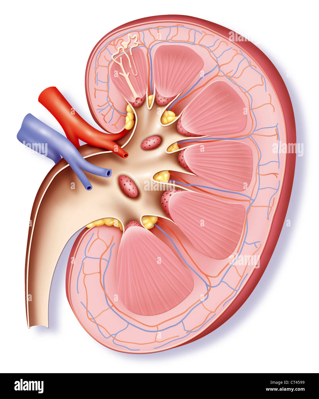 KIDNEY, DRAWING Stock Photohttps://www.alamy.com/image-license-details/?v=1https://www.alamy.com/stock-photo-kidney-drawing-49264469.html
RMCT4599–KIDNEY, DRAWING
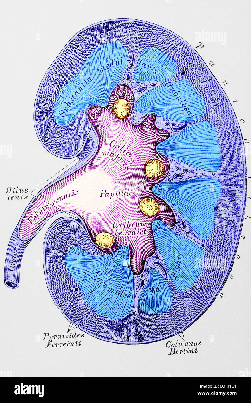 KIDNEY, DRAWING Stock Photohttps://www.alamy.com/image-license-details/?v=1https://www.alamy.com/stock-photo-kidney-drawing-53865169.html
RMD3HNG1–KIDNEY, DRAWING
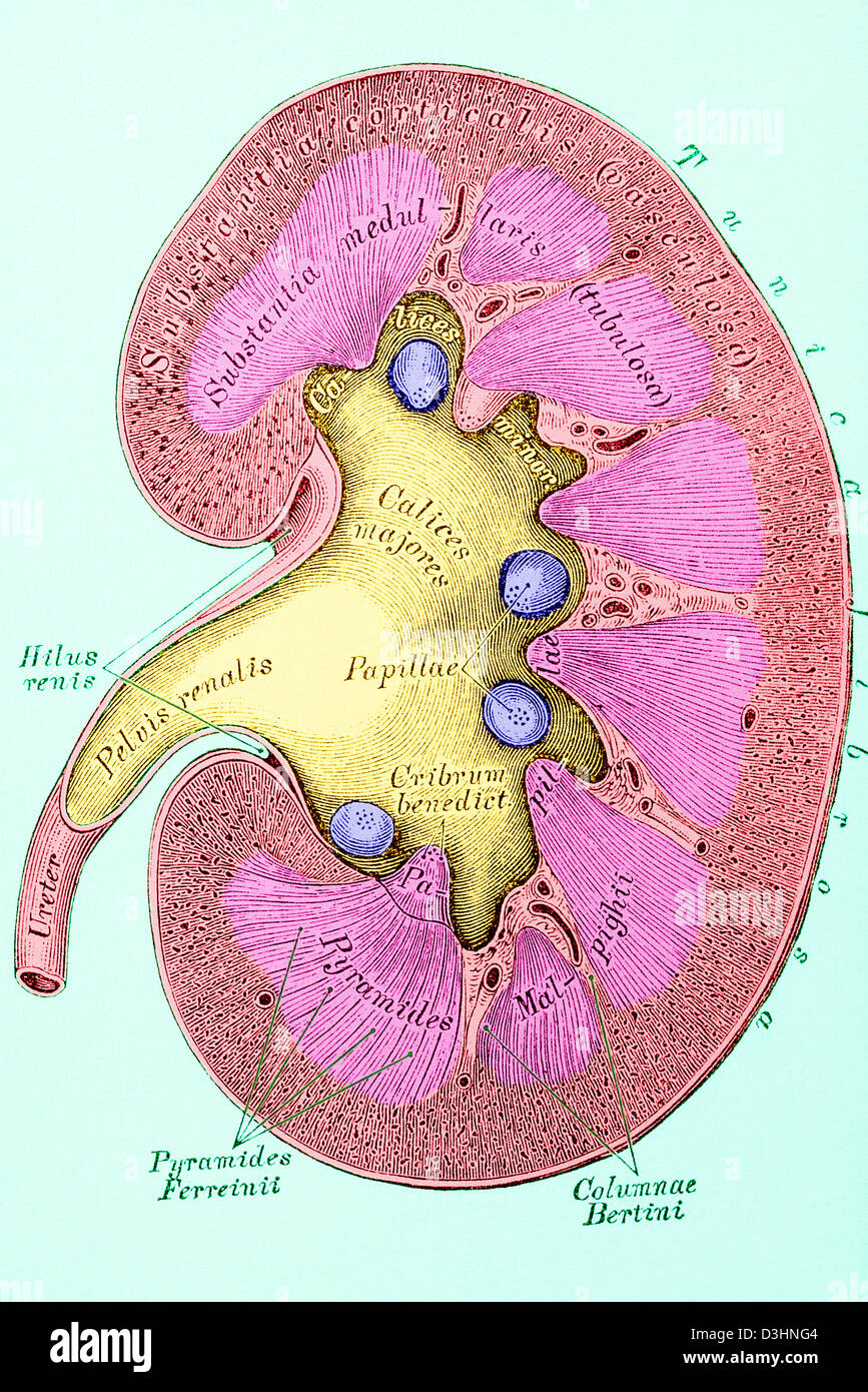 KIDNEY, DRAWING Stock Photohttps://www.alamy.com/image-license-details/?v=1https://www.alamy.com/stock-photo-kidney-drawing-53865172.html
RMD3HNG4–KIDNEY, DRAWING
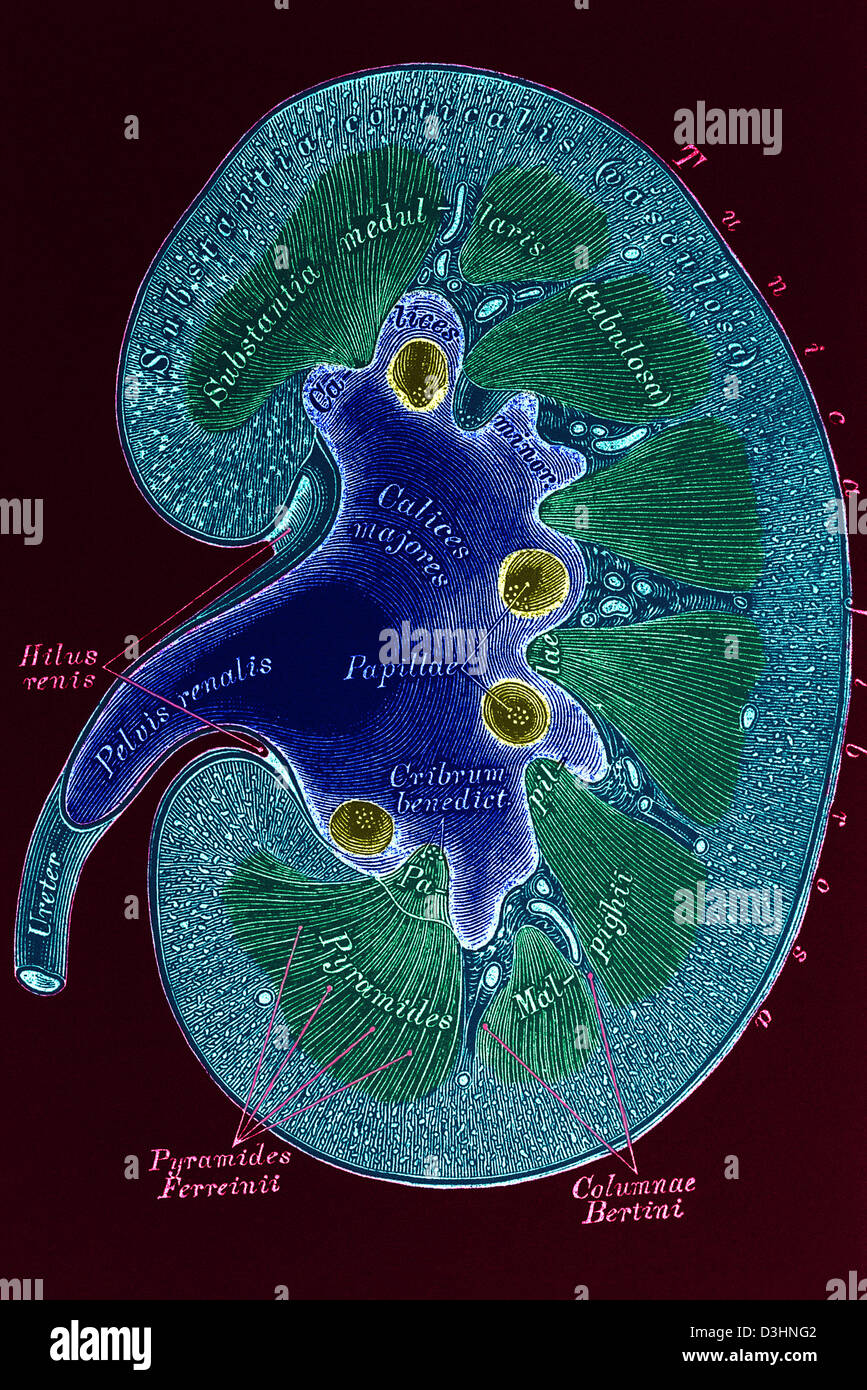 KIDNEY, DRAWING Stock Photohttps://www.alamy.com/image-license-details/?v=1https://www.alamy.com/stock-photo-kidney-drawing-53865170.html
RMD3HNG2–KIDNEY, DRAWING
 KIDNEY, DRAWING Stock Photohttps://www.alamy.com/image-license-details/?v=1https://www.alamy.com/stock-photo-kidney-drawing-49172646.html
RMCT005X–KIDNEY, DRAWING
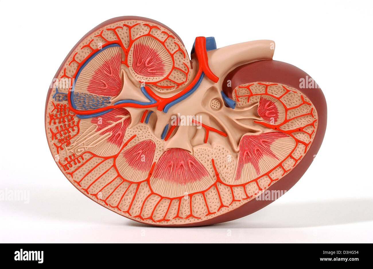 KIDNEY, ANATOMY Stock Photohttps://www.alamy.com/image-license-details/?v=1https://www.alamy.com/stock-photo-kidney-anatomy-53860944.html
RMD3HG54–KIDNEY, ANATOMY
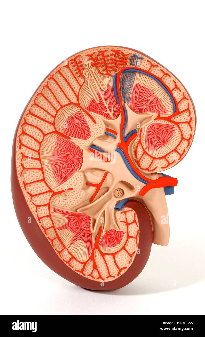 KIDNEY, ANATOMY Stock Photohttps://www.alamy.com/image-license-details/?v=1https://www.alamy.com/stock-photo-kidney-anatomy-53860945.html
RMD3HG55–KIDNEY, ANATOMY
 KIDNEY, ANATOMY Stock Photohttps://www.alamy.com/image-license-details/?v=1https://www.alamy.com/stock-photo-kidney-anatomy-53860941.html
RMD3HG51–KIDNEY, ANATOMY
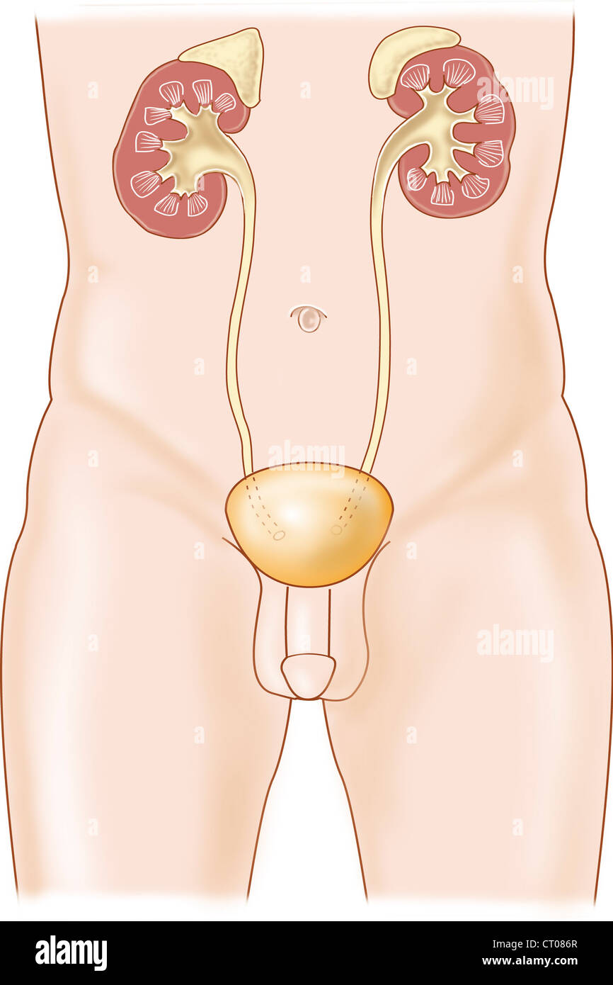 URINARY SYSTEM, DRAWING Stock Photohttps://www.alamy.com/image-license-details/?v=1https://www.alamy.com/stock-photo-urinary-system-drawing-49178943.html
RMCT086R–URINARY SYSTEM, DRAWING
 URINARY SYSTEM, DRAWING Stock Photohttps://www.alamy.com/image-license-details/?v=1https://www.alamy.com/stock-photo-urinary-system-drawing-49170336.html
RMCRYW7C–URINARY SYSTEM, DRAWING
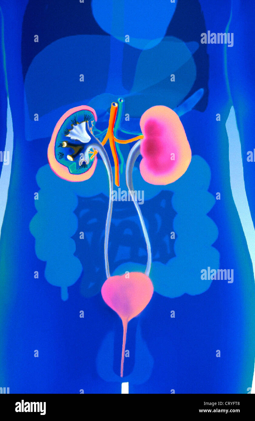 URINARY SYSTEM, DRAWING Stock Photohttps://www.alamy.com/image-license-details/?v=1https://www.alamy.com/stock-photo-urinary-system-drawing-49162968.html
RMCRYFT8–URINARY SYSTEM, DRAWING
 URINARY SYSTEM, DRAWING Stock Photohttps://www.alamy.com/image-license-details/?v=1https://www.alamy.com/stock-photo-urinary-system-drawing-49162971.html
RMCRYFTB–URINARY SYSTEM, DRAWING
 BLADDER, DRAWING Stock Photohttps://www.alamy.com/image-license-details/?v=1https://www.alamy.com/stock-photo-bladder-drawing-49161040.html
RMCRYDBC–BLADDER, DRAWING
 Human urinary system, anterior view, anatomy. The human urinary system seen anterior with the right kidney seen in section to show the fibrous capsule Stock Photohttps://www.alamy.com/image-license-details/?v=1https://www.alamy.com/human-urinary-system-anterior-view-anatomy-the-human-urinary-system-seen-anterior-with-the-right-kidney-seen-in-section-to-show-the-fibrous-capsule-image355209868.html
RM2BHW5PM–Human urinary system, anterior view, anatomy. The human urinary system seen anterior with the right kidney seen in section to show the fibrous capsule
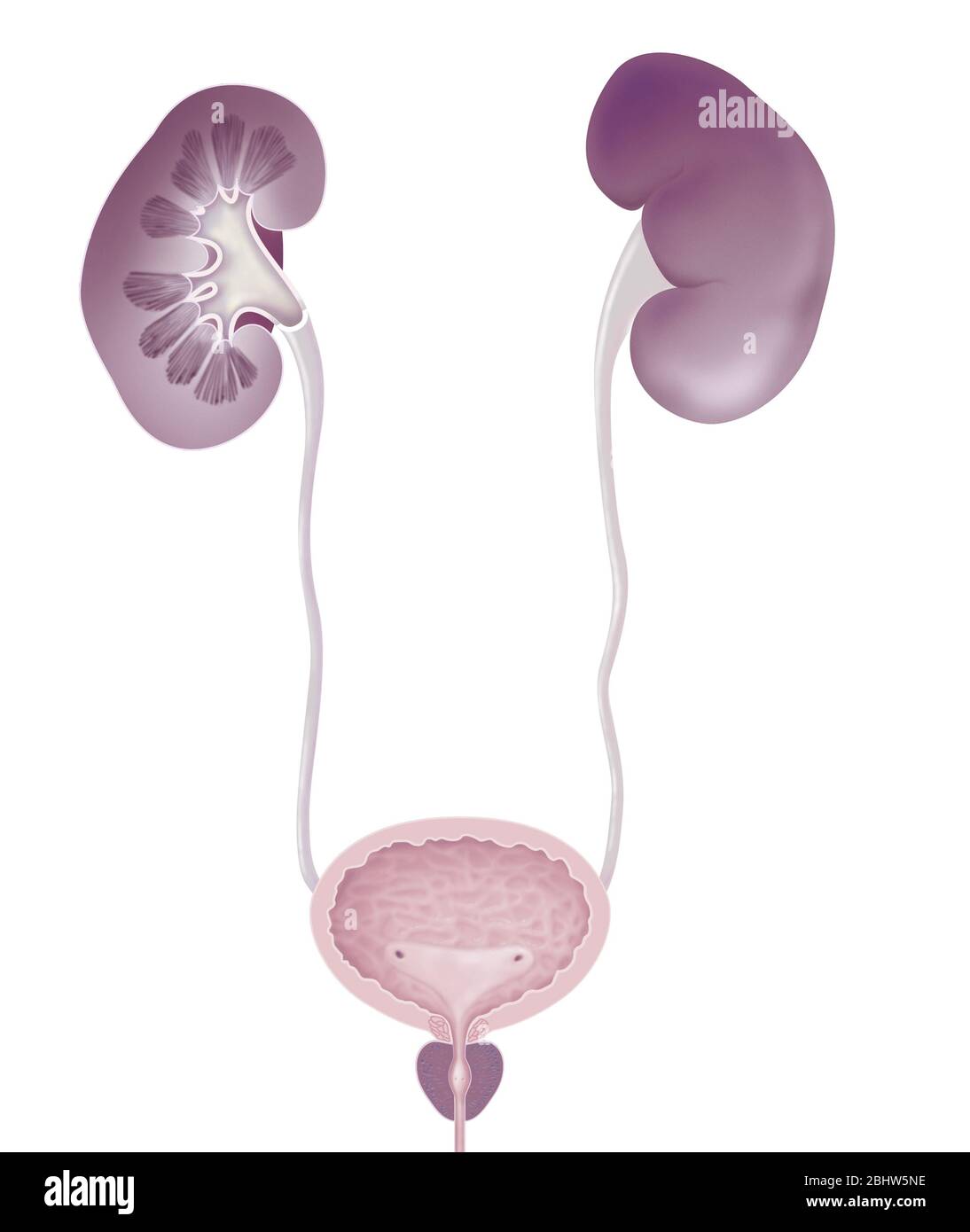 Human urinary system, anterior view, anatomy. The human urinary system seen anterior with the right kidney seen in section to show the fibrous capsule Stock Photohttps://www.alamy.com/image-license-details/?v=1https://www.alamy.com/human-urinary-system-anterior-view-anatomy-the-human-urinary-system-seen-anterior-with-the-right-kidney-seen-in-section-to-show-the-fibrous-capsule-image355209834.html
RM2BHW5NE–Human urinary system, anterior view, anatomy. The human urinary system seen anterior with the right kidney seen in section to show the fibrous capsule
