Quick filters:
Reticulum Stock Photos and Images
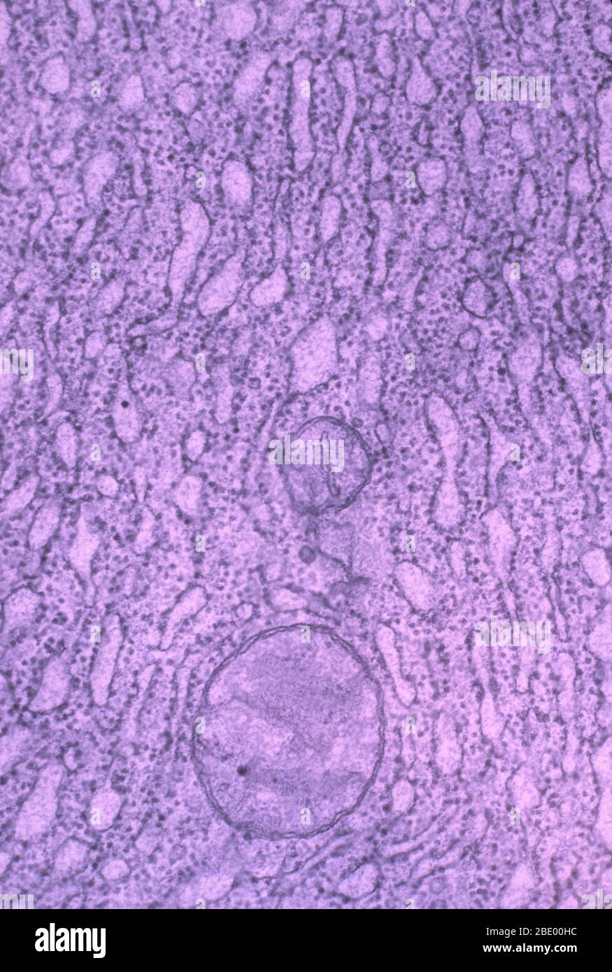 Endoplasmic Reticulum with Ribosomes Stock Photohttps://www.alamy.com/image-license-details/?v=1https://www.alamy.com/endoplasmic-reticulum-with-ribosomes-image352813032.html
RM2BE00HC–Endoplasmic Reticulum with Ribosomes
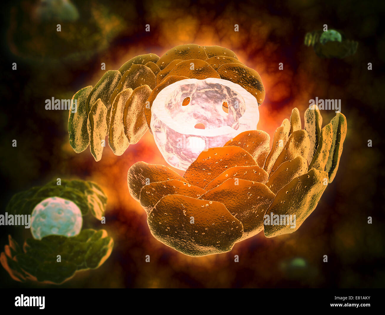 Conceptual image of endoplasmic reticulum around a cell nucleus. Endoplasmic reticulum is an organelle that forms a continuous m Stock Photohttps://www.alamy.com/image-license-details/?v=1https://www.alamy.com/stock-photo-conceptual-image-of-endoplasmic-reticulum-around-a-cell-nucleus-endoplasmic-73789071.html
RFE81AKY–Conceptual image of endoplasmic reticulum around a cell nucleus. Endoplasmic reticulum is an organelle that forms a continuous m
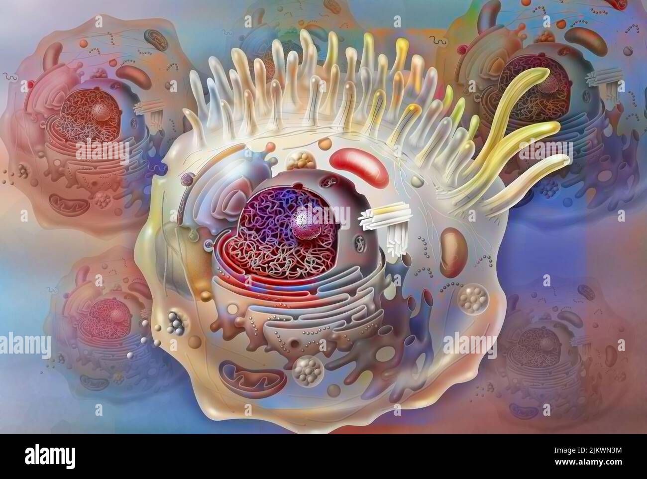 Cell sectional view with all the main organelles: nucleus, reticulum. Stock Photohttps://www.alamy.com/image-license-details/?v=1https://www.alamy.com/cell-sectional-view-with-all-the-main-organelles-nucleus-reticulum-image476923768.html
RF2JKWN3M–Cell sectional view with all the main organelles: nucleus, reticulum.
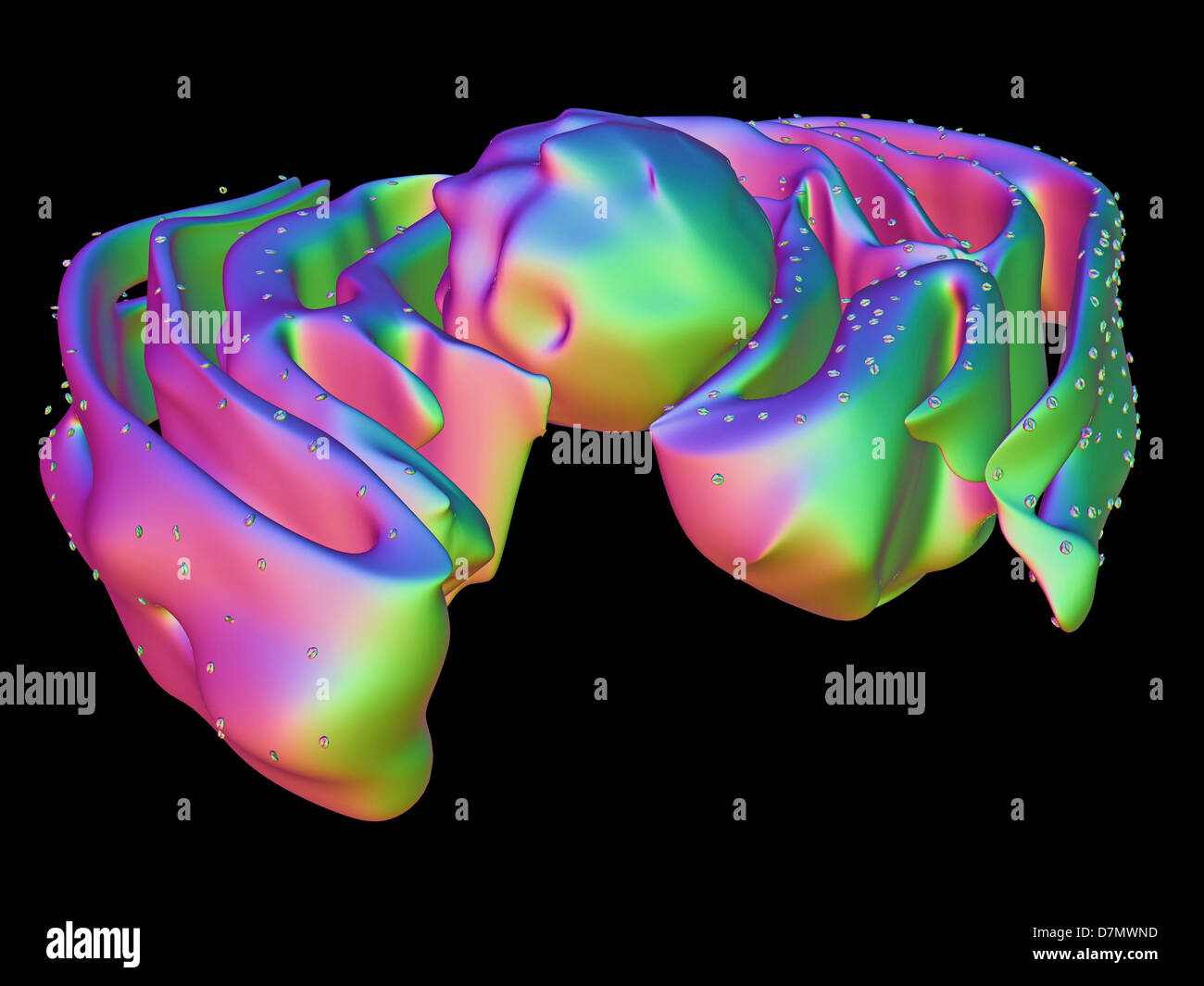 Nucleus and endoplasmic reticulum Stock Photohttps://www.alamy.com/image-license-details/?v=1https://www.alamy.com/stock-photo-nucleus-and-endoplasmic-reticulum-56392937.html
RFD7MWND–Nucleus and endoplasmic reticulum
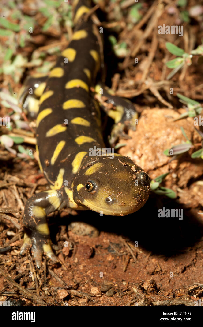 Blotched Tiger Salamander, Ambystoma reticulum, Grand Canyon, Arizona, USA Stock Photohttps://www.alamy.com/image-license-details/?v=1https://www.alamy.com/stock-photo-blotched-tiger-salamander-ambystoma-reticulum-grand-canyon-arizona-70000064.html
RME1TNP8–Blotched Tiger Salamander, Ambystoma reticulum, Grand Canyon, Arizona, USA
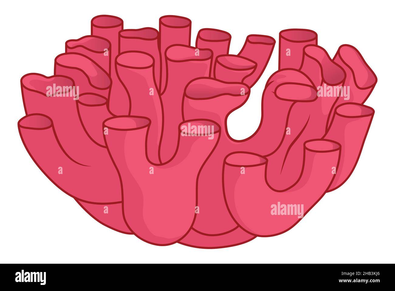 Smooth endoplasmic reticulum simple medical illustration note on tubular structures Stock Photohttps://www.alamy.com/image-license-details/?v=1https://www.alamy.com/smooth-endoplasmic-reticulum-simple-medical-illustration-note-on-tubular-structures-image454312046.html
RF2HB3KJ6–Smooth endoplasmic reticulum simple medical illustration note on tubular structures
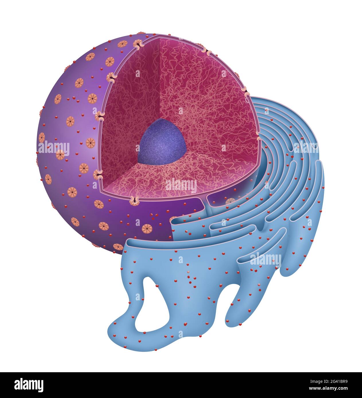 Structure of Nucleus and Rough endoplasmic reticulum Stock Photohttps://www.alamy.com/image-license-details/?v=1https://www.alamy.com/structure-of-nucleus-and-rough-endoplasmic-reticulum-image432749053.html
RF2G41BR9–Structure of Nucleus and Rough endoplasmic reticulum
 The top of an edible mushroom cep (Boletus edulis) lies on a cutting board. A whitish, net-like pattern (reticulum) on a brownish stalk near the cap Stock Photohttps://www.alamy.com/image-license-details/?v=1https://www.alamy.com/the-top-of-an-edible-mushroom-cep-boletus-edulis-lies-on-a-cutting-board-a-whitish-net-like-pattern-reticulum-on-a-brownish-stalk-near-the-cap-image554072599.html
RF2R5C573–The top of an edible mushroom cep (Boletus edulis) lies on a cutting board. A whitish, net-like pattern (reticulum) on a brownish stalk near the cap
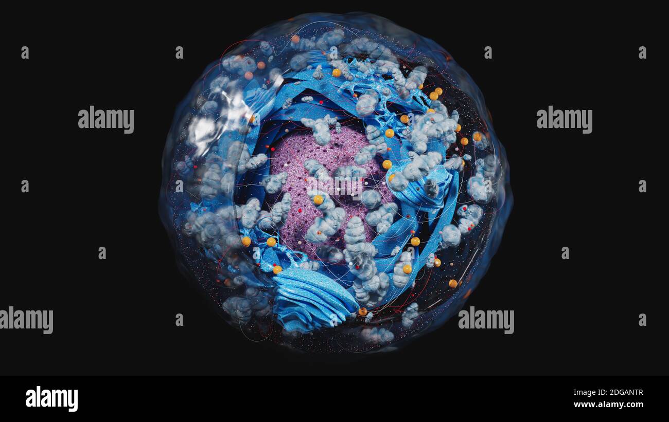 Structure of human cell, anatomy of cell, cellular environment, cellular concept with organelle: nucleus, membrane, mitochondria, Golgi apparatus 3d Stock Photohttps://www.alamy.com/image-license-details/?v=1https://www.alamy.com/structure-of-human-cell-anatomy-of-cell-cellular-environment-cellular-concept-with-organelle-nucleus-membrane-mitochondria-golgi-apparatus-3d-image388699271.html
RF2DGANTR–Structure of human cell, anatomy of cell, cellular environment, cellular concept with organelle: nucleus, membrane, mitochondria, Golgi apparatus 3d
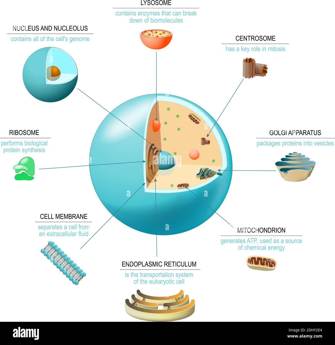 Cell anatomy. Structure and organelles of human's cell. Cross sections of animal cell: nucleus, nucleolus, mitochondria, centresome, golgi apparatus Stock Vectorhttps://www.alamy.com/image-license-details/?v=1https://www.alamy.com/cell-anatomy-structure-and-organelles-of-humans-cell-cross-sections-of-animal-cell-nucleus-nucleolus-mitochondria-centresome-golgi-apparatus-image389671916.html
RF2DHY2E4–Cell anatomy. Structure and organelles of human's cell. Cross sections of animal cell: nucleus, nucleolus, mitochondria, centresome, golgi apparatus
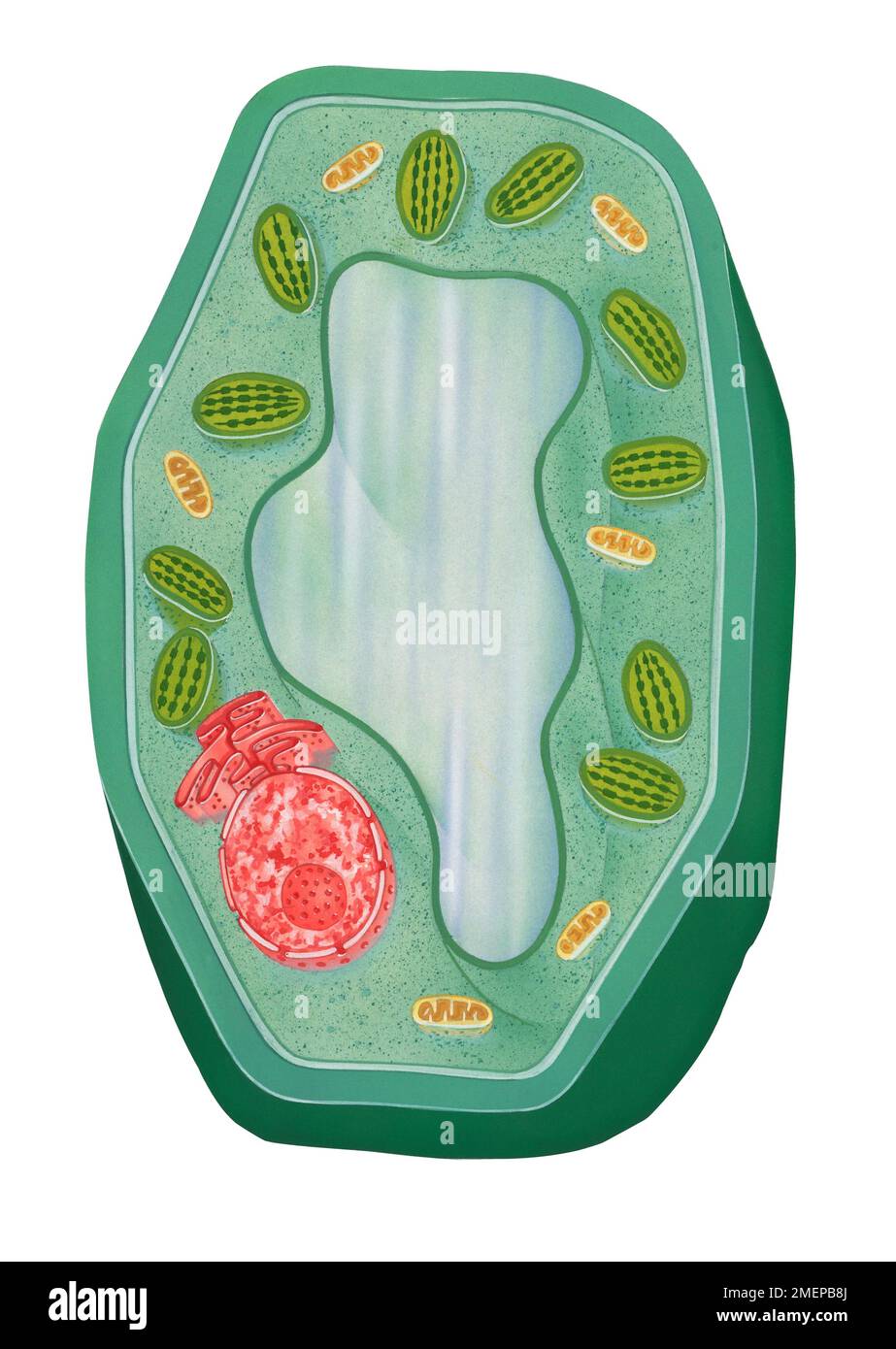 Typical plant cell Stock Photohttps://www.alamy.com/image-license-details/?v=1https://www.alamy.com/typical-plant-cell-image508197666.html
RM2MEPB8J–Typical plant cell
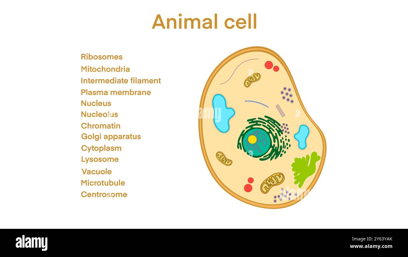 animal cell anatomy, biological animal cell with organelles cross section, Animal cell with placed text annotations to all organelles, Animal cell Stock Photohttps://www.alamy.com/image-license-details/?v=1https://www.alamy.com/animal-cell-anatomy-biological-animal-cell-with-organelles-cross-section-animal-cell-with-placed-text-annotations-to-all-organelles-animal-cell-image623348507.html
RF2Y63YAK–animal cell anatomy, biological animal cell with organelles cross section, Animal cell with placed text annotations to all organelles, Animal cell
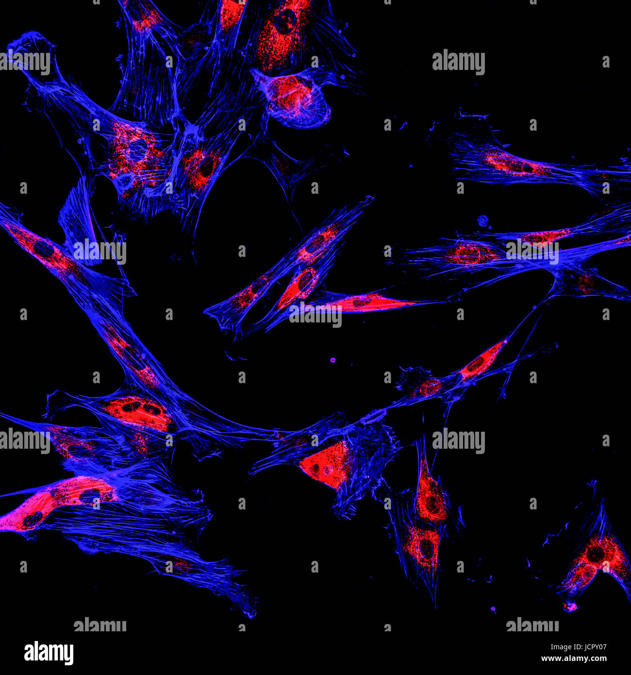 Immunofluorescence confocal imaging of melanoma cancer cells Stock Photohttps://www.alamy.com/image-license-details/?v=1https://www.alamy.com/stock-photo-immunofluorescence-confocal-imaging-of-melanoma-cancer-cells-145562935.html
RFJCPY07–Immunofluorescence confocal imaging of melanoma cancer cells
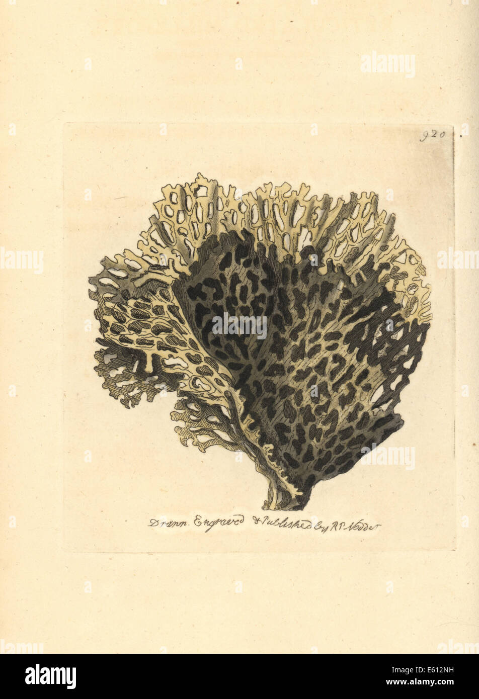 Sea mat, Conopeum reticulum. Stock Photohttps://www.alamy.com/image-license-details/?v=1https://www.alamy.com/stock-photo-sea-mat-conopeum-reticulum-72553533.html
RME612NH–Sea mat, Conopeum reticulum.
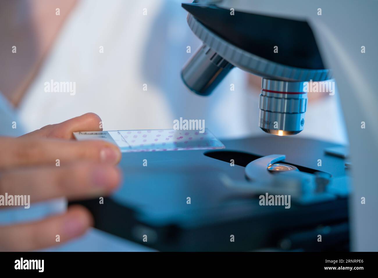 Cell structure investigation: Microscopes allow visualizing microscopic cell structures such as nuclei, mitochondria, and endoplasmic reticulum. Stock Photohttps://www.alamy.com/image-license-details/?v=1https://www.alamy.com/cell-structure-investigation-microscopes-allow-visualizing-microscopic-cell-structures-such-as-nuclei-mitochondria-and-endoplasmic-reticulum-image564162094.html
RF2RNRPE6–Cell structure investigation: Microscopes allow visualizing microscopic cell structures such as nuclei, mitochondria, and endoplasmic reticulum.
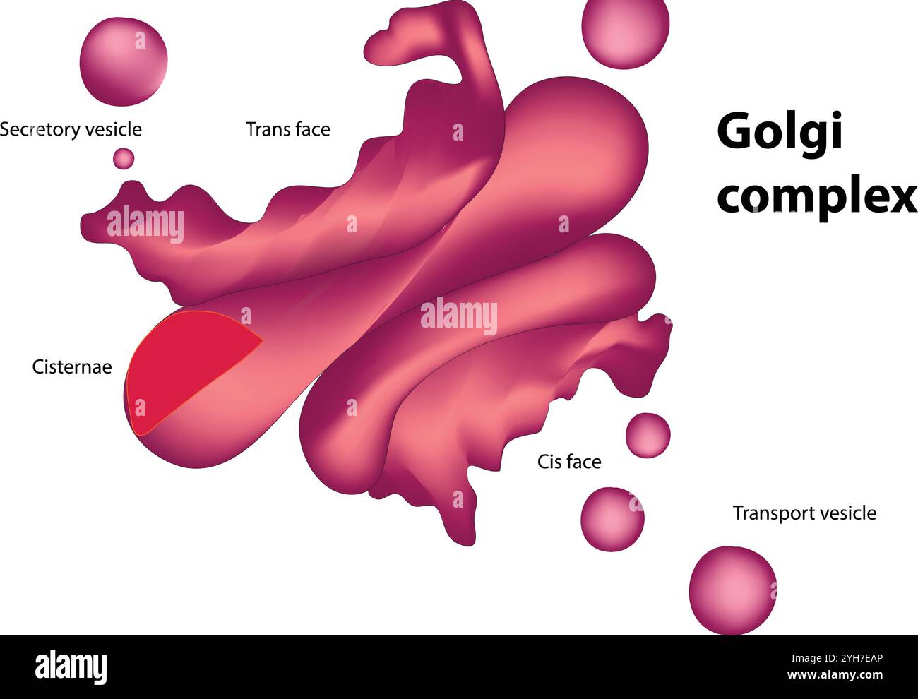 Golgi apparatus Stock Vectorhttps://www.alamy.com/image-license-details/?v=1https://www.alamy.com/golgi-apparatus-image630187342.html
RF2YH7EAP–Golgi apparatus
 Toadstools growing on a tree stump in an English woodland. Individually they do not last long but add their shape and form to the forest floor. Stock Photohttps://www.alamy.com/image-license-details/?v=1https://www.alamy.com/toadstools-growing-on-a-tree-stump-in-an-english-woodland-individually-they-do-not-last-long-but-add-their-shape-and-form-to-the-forest-floor-image395887739.html
RF2E026RR–Toadstools growing on a tree stump in an English woodland. Individually they do not last long but add their shape and form to the forest floor.
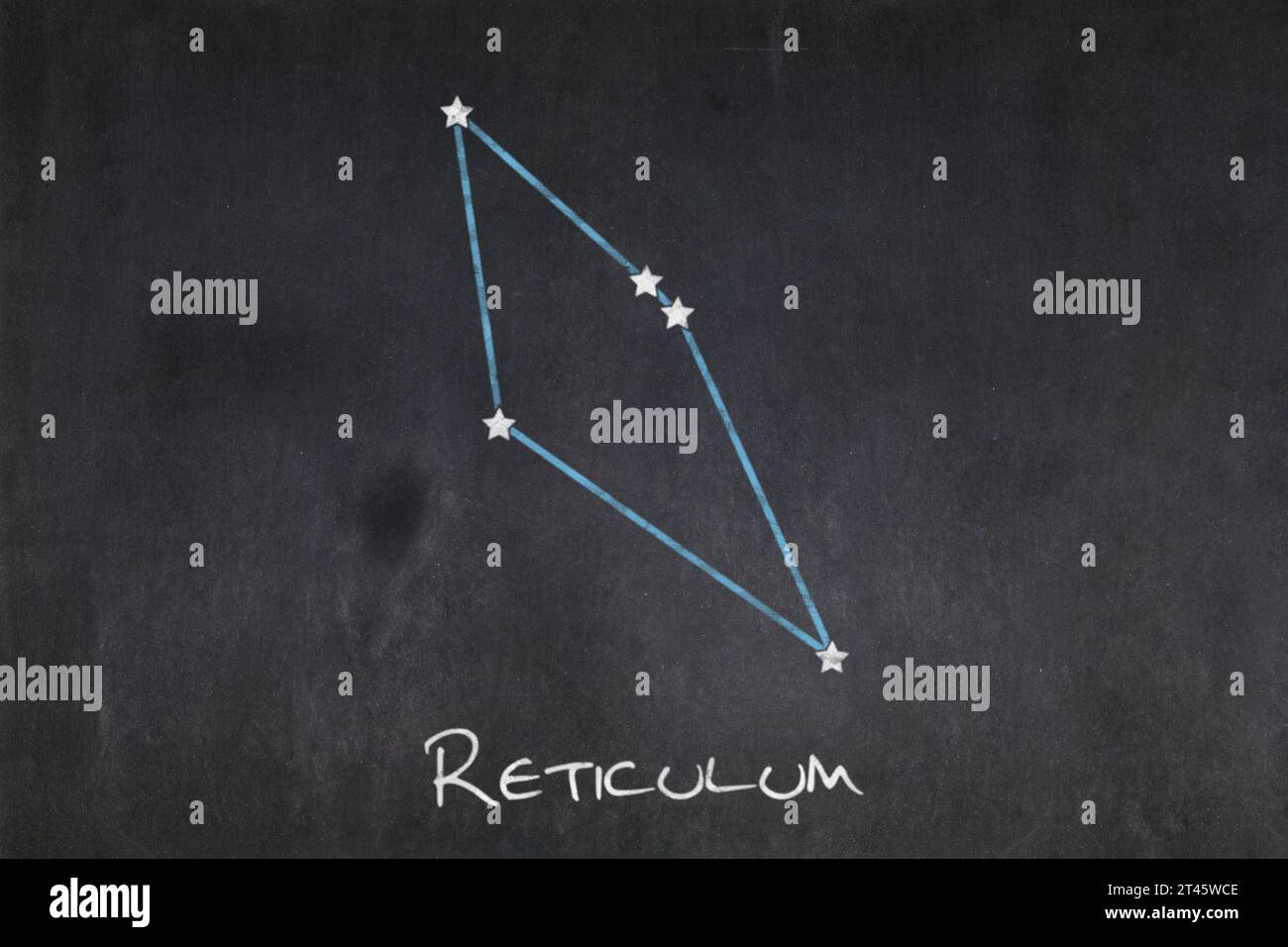 Blackboard with the Reticulum constellation drawn in the middle. Stock Photohttps://www.alamy.com/image-license-details/?v=1https://www.alamy.com/blackboard-with-the-reticulum-constellation-drawn-in-the-middle-image570530478.html
RF2T45WCE–Blackboard with the Reticulum constellation drawn in the middle.
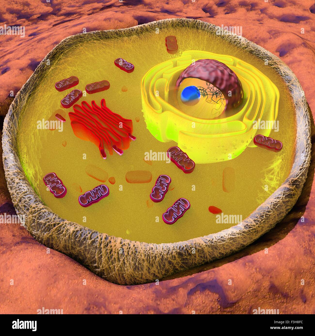 Illustration of a eukaryotic cell showing the main organelles, such as golgi, endoplasmic reticulum, mitochondria, nucleus Stock Photohttps://www.alamy.com/image-license-details/?v=1https://www.alamy.com/stock-photo-illustration-of-a-eukaryotic-cell-showing-the-main-organelles-such-88275696.html
RFF3H8FC–Illustration of a eukaryotic cell showing the main organelles, such as golgi, endoplasmic reticulum, mitochondria, nucleus
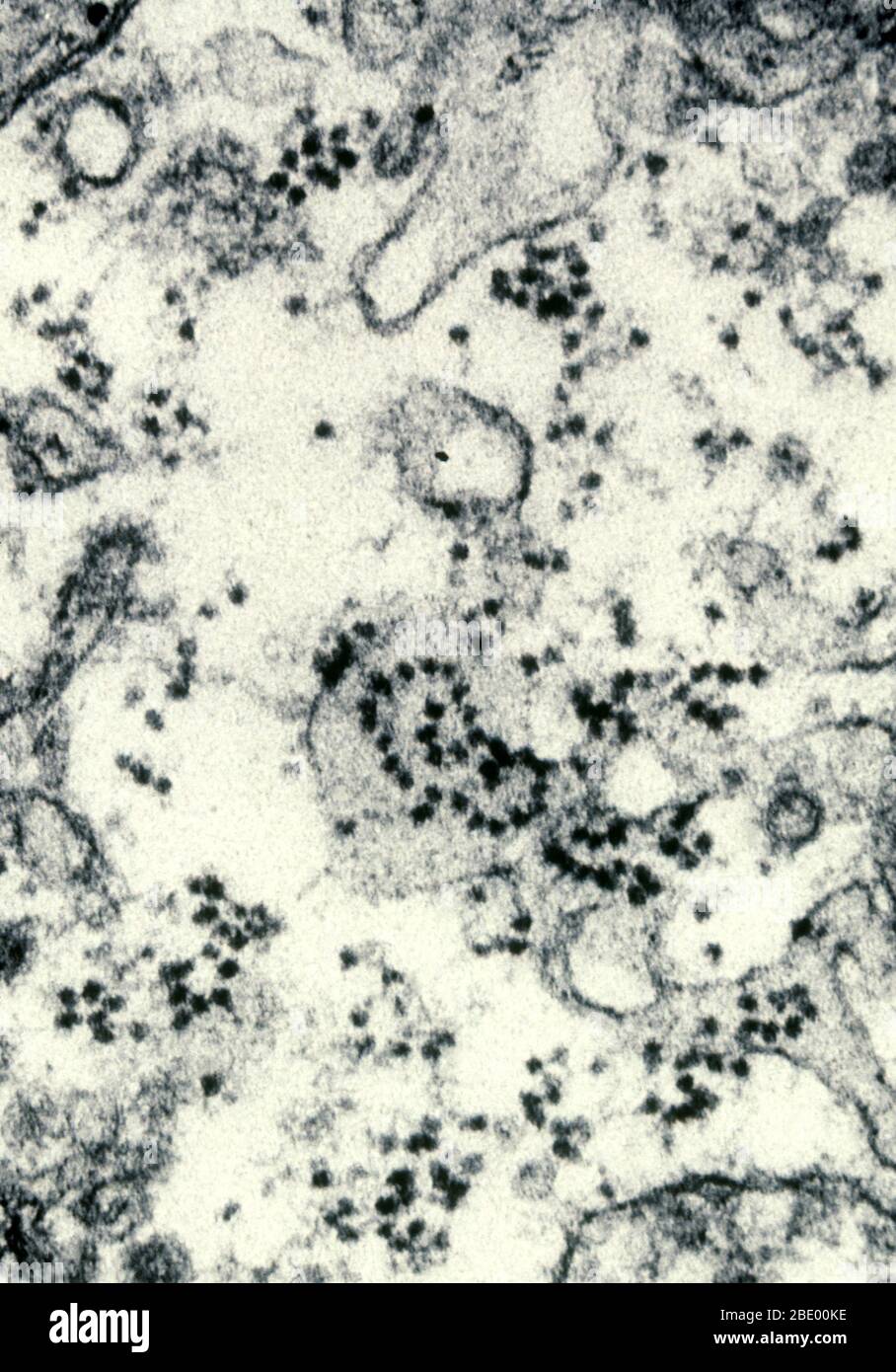 Polysome on Endoplasmic Reticulum Stock Photohttps://www.alamy.com/image-license-details/?v=1https://www.alamy.com/polysome-on-endoplasmic-reticulum-image352813090.html
RM2BE00KE–Polysome on Endoplasmic Reticulum
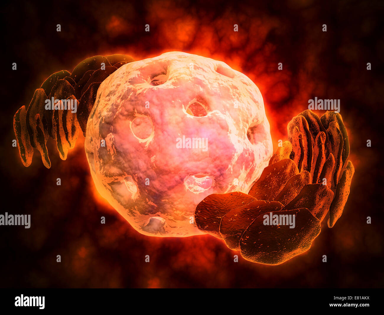 Conceptual image of endoplasmic reticulum around a cell nucleus. Endoplasmic reticulum is an organelle that forms a continuous m Stock Photohttps://www.alamy.com/image-license-details/?v=1https://www.alamy.com/stock-photo-conceptual-image-of-endoplasmic-reticulum-around-a-cell-nucleus-endoplasmic-73789070.html
RFE81AKX–Conceptual image of endoplasmic reticulum around a cell nucleus. Endoplasmic reticulum is an organelle that forms a continuous m
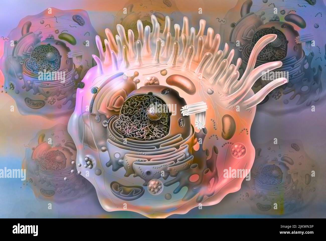 Cell sectional view with all the main organelles: nucleus, reticulum. Stock Photohttps://www.alamy.com/image-license-details/?v=1https://www.alamy.com/cell-sectional-view-with-all-the-main-organelles-nucleus-reticulum-image476923770.html
RF2JKWN3P–Cell sectional view with all the main organelles: nucleus, reticulum.
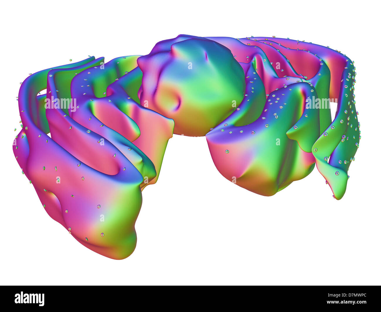 Nucleus and endoplasmic reticulum Stock Photohttps://www.alamy.com/image-license-details/?v=1https://www.alamy.com/stock-photo-nucleus-and-endoplasmic-reticulum-56392964.html
RFD7MWPC–Nucleus and endoplasmic reticulum
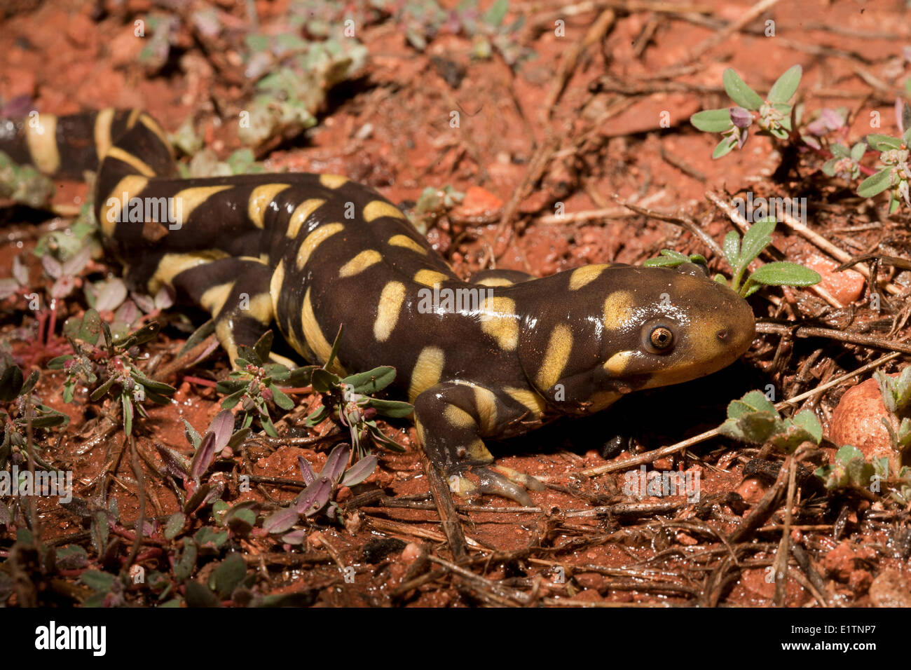 Blotched Tiger Salamander, Ambystoma reticulum, Grand Canyon, Arizona, USA Stock Photohttps://www.alamy.com/image-license-details/?v=1https://www.alamy.com/stock-photo-blotched-tiger-salamander-ambystoma-reticulum-grand-canyon-arizona-70000063.html
RME1TNP7–Blotched Tiger Salamander, Ambystoma reticulum, Grand Canyon, Arizona, USA
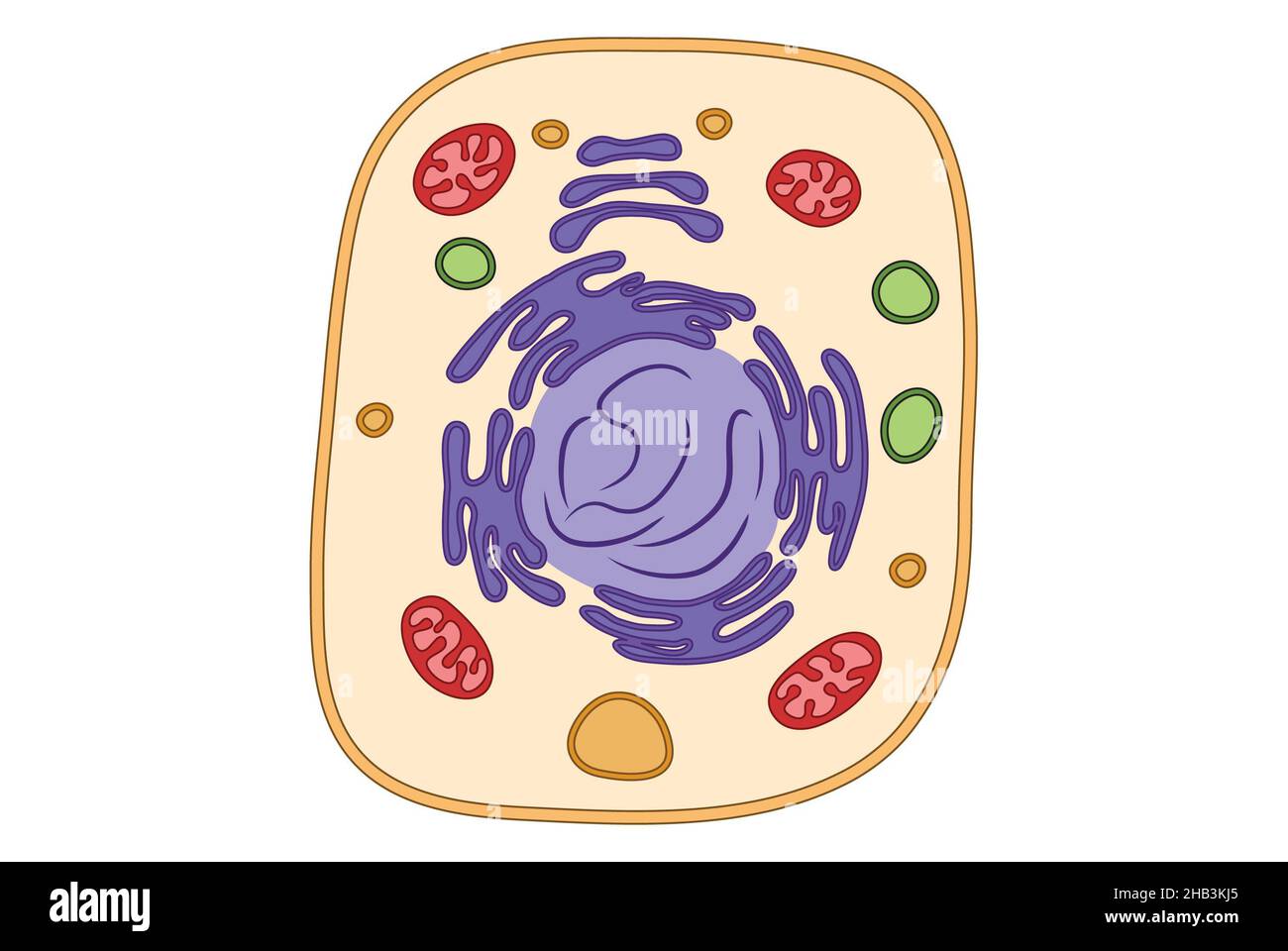 Simple cell structure medical illustration, mitochondria, ger, endoplasmic reticulum, simple illustration Stock Photohttps://www.alamy.com/image-license-details/?v=1https://www.alamy.com/simple-cell-structure-medical-illustration-mitochondria-ger-endoplasmic-reticulum-simple-illustration-image454312045.html
RF2HB3KJ5–Simple cell structure medical illustration, mitochondria, ger, endoplasmic reticulum, simple illustration
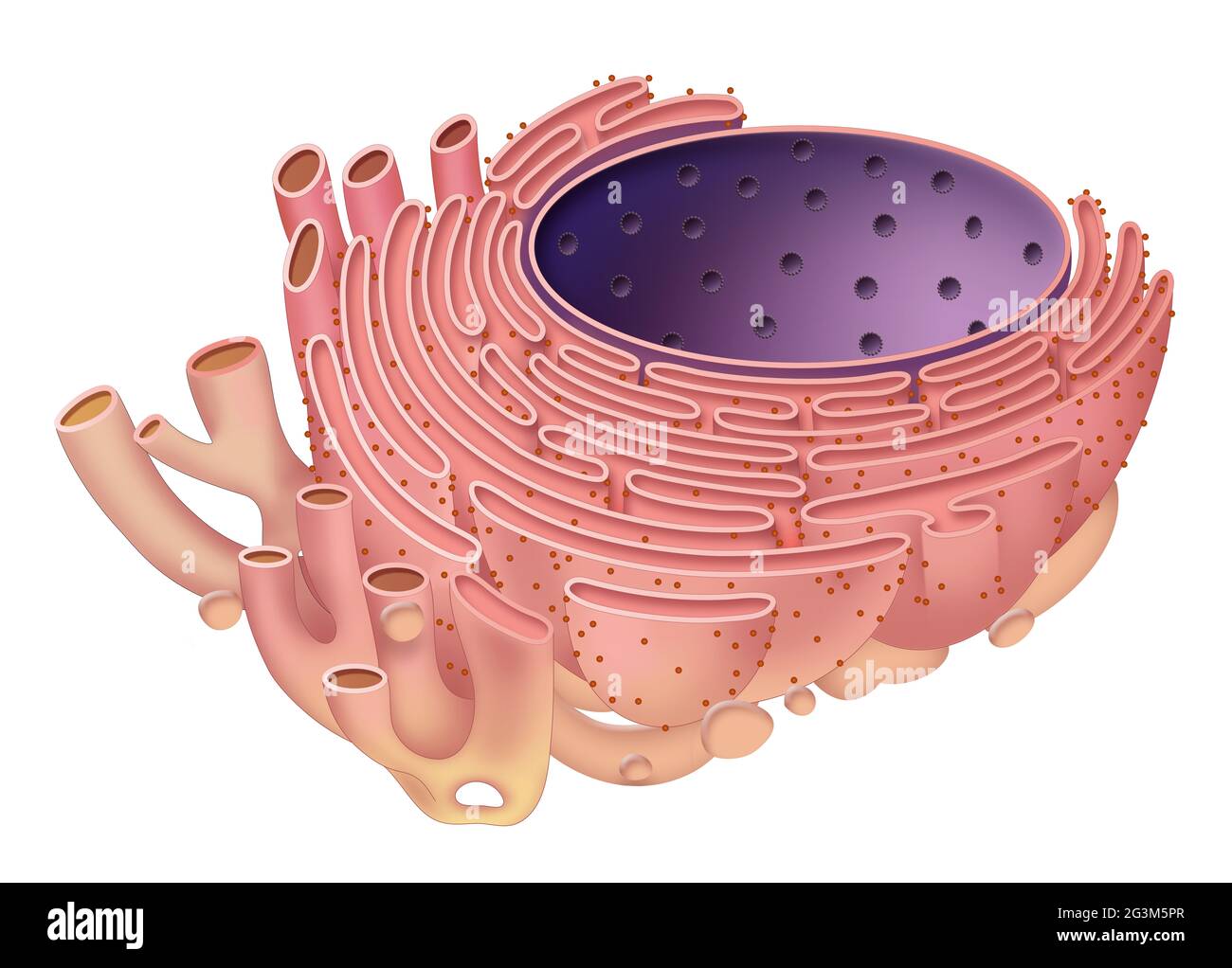 The endoplasmic reticulum is organelle found in eukaryotic cells Stock Photohttps://www.alamy.com/image-license-details/?v=1https://www.alamy.com/the-endoplasmic-reticulum-is-organelle-found-in-eukaryotic-cells-image432546767.html
RF2G3M5PR–The endoplasmic reticulum is organelle found in eukaryotic cells
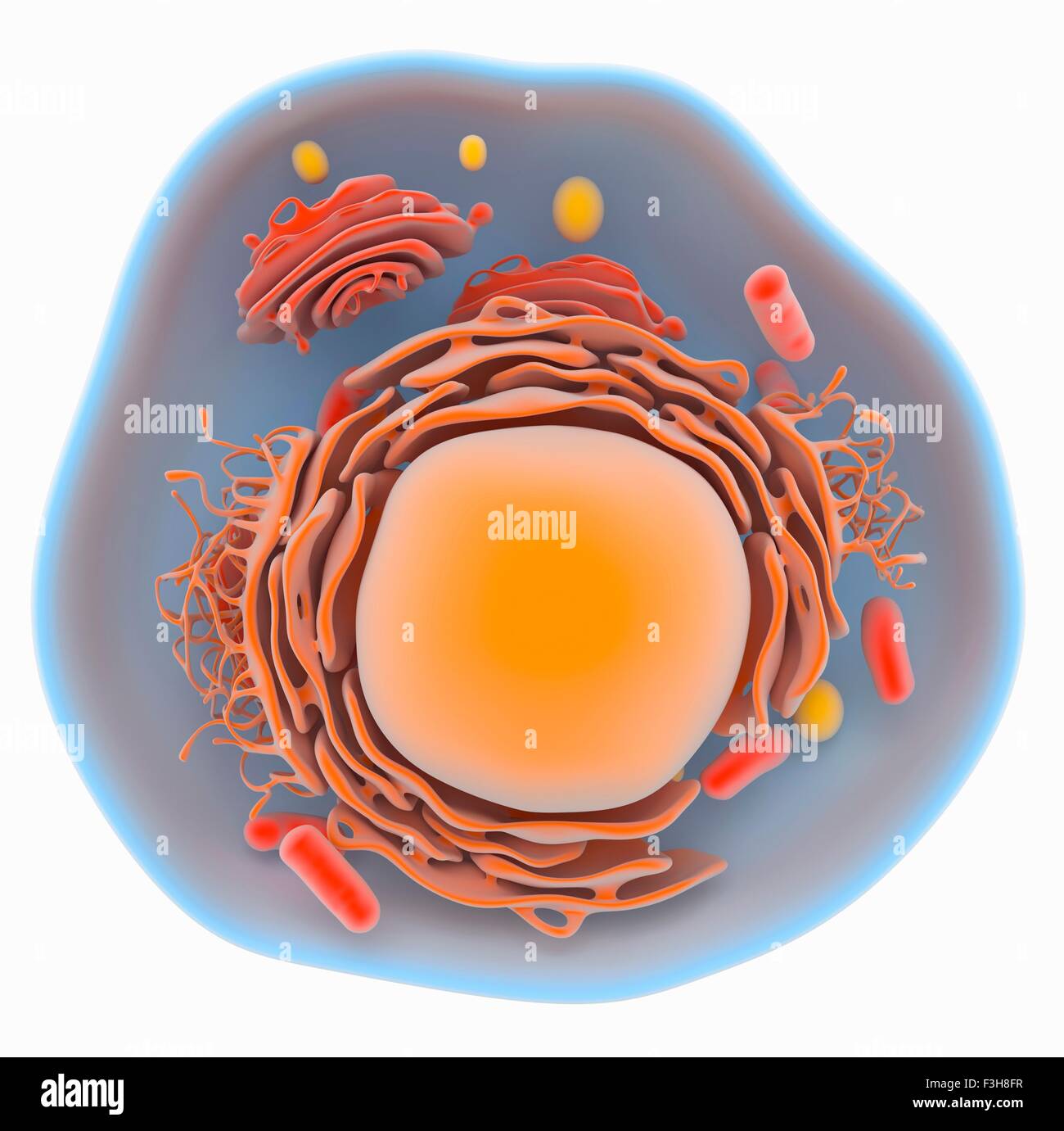 Illustration of a eukaryotic cell showing the main organelles, such as golgi, endoplasmic reticulum, mitochondria, nucleus Stock Photohttps://www.alamy.com/image-license-details/?v=1https://www.alamy.com/stock-photo-illustration-of-a-eukaryotic-cell-showing-the-main-organelles-such-88275707.html
RFF3H8FR–Illustration of a eukaryotic cell showing the main organelles, such as golgi, endoplasmic reticulum, mitochondria, nucleus
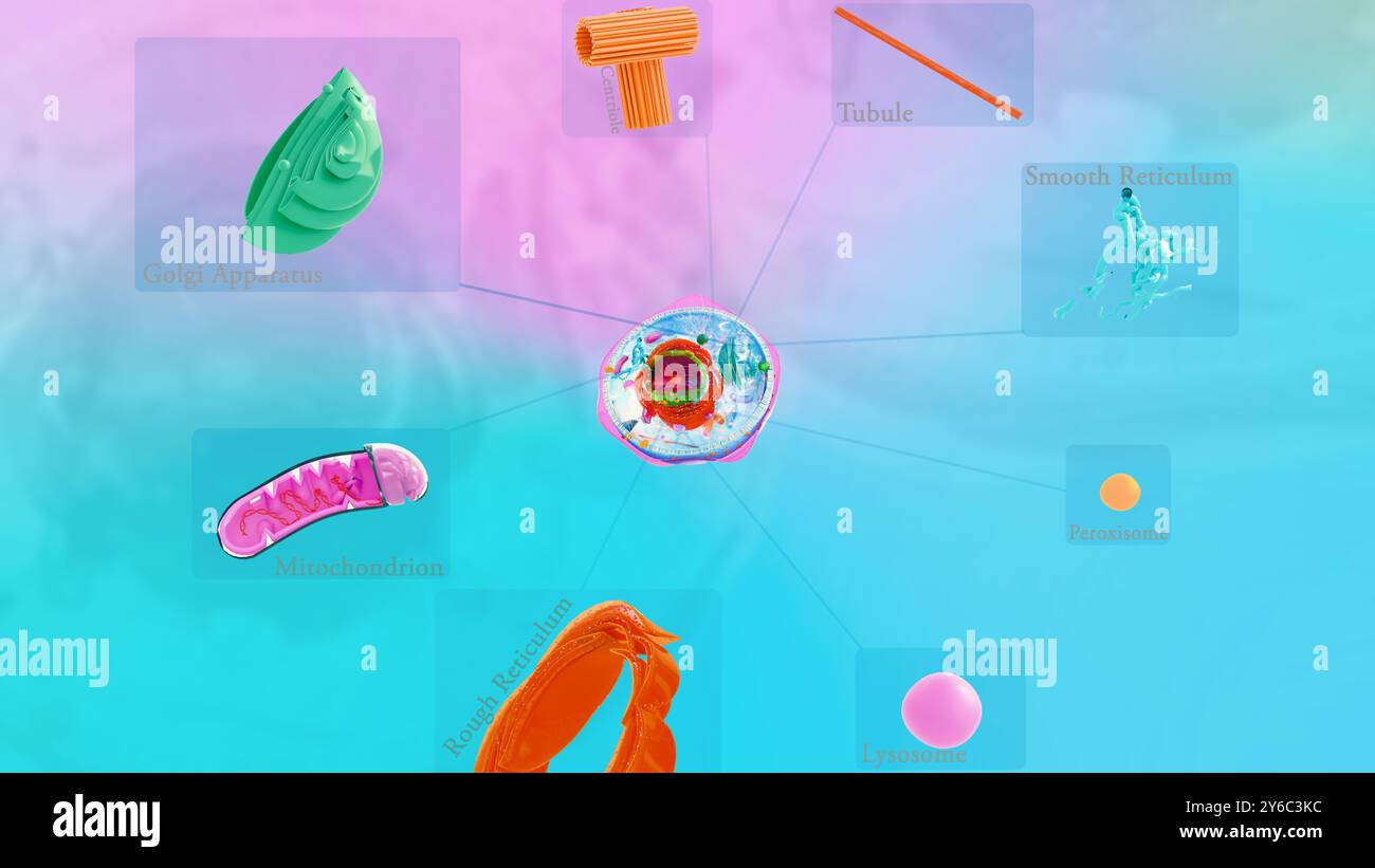 8 organelle, organelle in human cell structure. kinds of human cells Stock Photohttps://www.alamy.com/image-license-details/?v=1https://www.alamy.com/8-organelle-organelle-in-human-cell-structure-kinds-of-human-cells-image623527504.html
RF2Y6C3KC–8 organelle, organelle in human cell structure. kinds of human cells
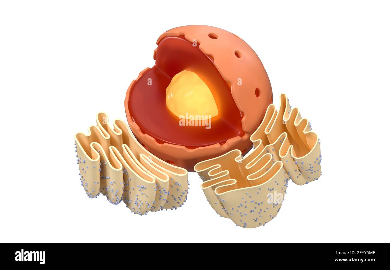 Structure of nuclear and endoplasmic reticulum in an animal cell, 3d rendering. Section view. Computer digital drawing. Stock Photohttps://www.alamy.com/image-license-details/?v=1https://www.alamy.com/structure-of-nuclear-and-endoplasmic-reticulum-in-an-animal-cell-3d-rendering-section-view-computer-digital-drawing-image413031375.html
RF2EYY5MF–Structure of nuclear and endoplasmic reticulum in an animal cell, 3d rendering. Section view. Computer digital drawing.
 Reticulum the reticle constellation map on a starry space background. Stars relative sizes and color shades based on their spectral type. Stock Photohttps://www.alamy.com/image-license-details/?v=1https://www.alamy.com/reticulum-the-reticle-constellation-map-on-a-starry-space-background-stars-relative-sizes-and-color-shades-based-on-their-spectral-type-image246737278.html
RFT9BRWJ–Reticulum the reticle constellation map on a starry space background. Stars relative sizes and color shades based on their spectral type.
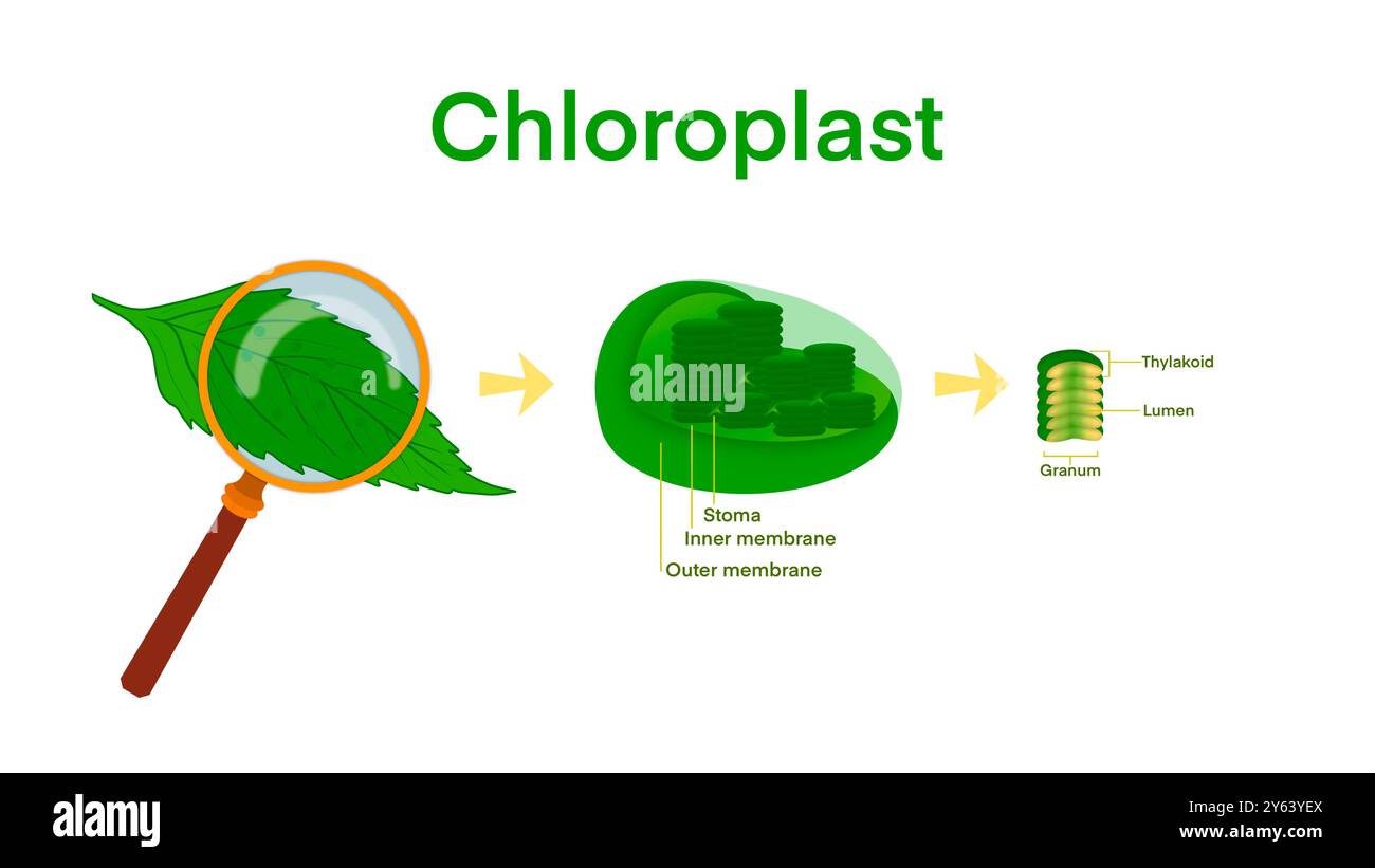 Chloroplast Photosynthesis Infographic Elements, Chloroplast organelles, structure within cells of plants, Cross section of a chloroplast from plant Stock Photohttps://www.alamy.com/image-license-details/?v=1https://www.alamy.com/chloroplast-photosynthesis-infographic-elements-chloroplast-organelles-structure-within-cells-of-plants-cross-section-of-a-chloroplast-from-plant-image623348626.html
RF2Y63YEX–Chloroplast Photosynthesis Infographic Elements, Chloroplast organelles, structure within cells of plants, Cross section of a chloroplast from plant
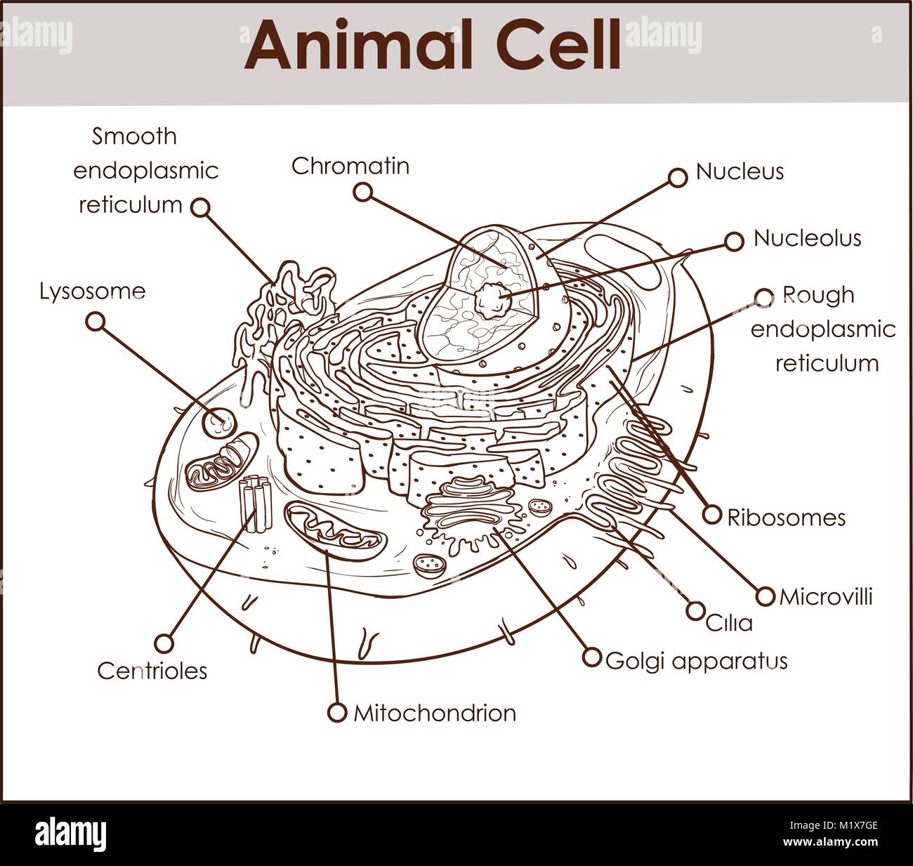 Animal Cell Anatomy Diagram Structure with all parts nucleus smooth rough endoplasmic reticulum cytoplasm golgi apparatus mitochondria membrane centro Stock Vectorhttps://www.alamy.com/image-license-details/?v=1https://www.alamy.com/stock-photo-animal-cell-anatomy-diagram-structure-with-all-parts-nucleus-smooth-173295038.html
RFM1X7GE–Animal Cell Anatomy Diagram Structure with all parts nucleus smooth rough endoplasmic reticulum cytoplasm golgi apparatus mitochondria membrane centro
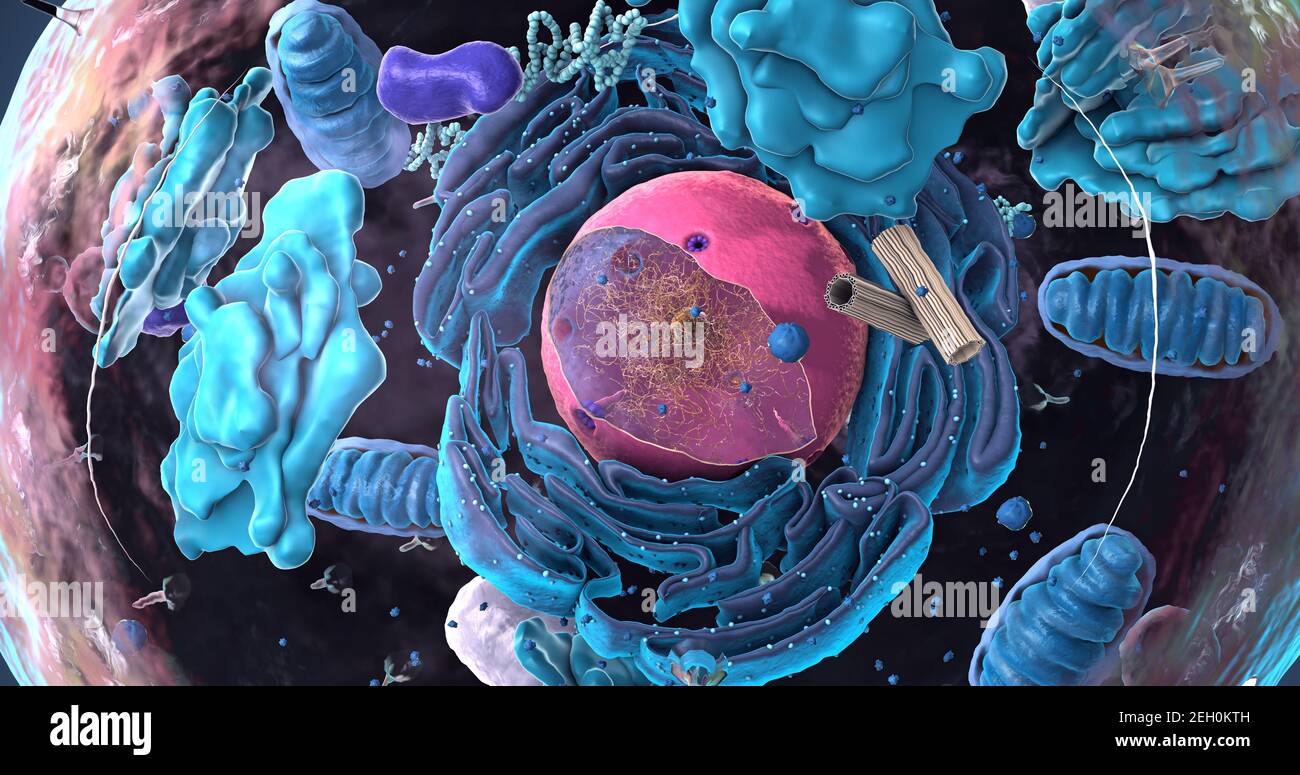 Components of Eukaryotic cell, nucleus and organelles and plasma membrane - 3d illustration Stock Photohttps://www.alamy.com/image-license-details/?v=1https://www.alamy.com/components-of-eukaryotic-cell-nucleus-and-organelles-and-plasma-membrane-3d-illustration-image406303201.html
RF2EH0KTH–Components of Eukaryotic cell, nucleus and organelles and plasma membrane - 3d illustration
 Cell structure investigation: Microscopes allow visualizing microscopic cell structures such as nuclei, mitochondria, and endoplasmic reticulum. Stock Photohttps://www.alamy.com/image-license-details/?v=1https://www.alamy.com/cell-structure-investigation-microscopes-allow-visualizing-microscopic-cell-structures-such-as-nuclei-mitochondria-and-endoplasmic-reticulum-image564105577.html
RF2RNN6BN–Cell structure investigation: Microscopes allow visualizing microscopic cell structures such as nuclei, mitochondria, and endoplasmic reticulum.
RMP24WKW–. Gorgonia reticulum 141 Gorgonia reticulum - - Print - Iconographia Zoologica - Special Collections University of Amsterdam - UBAINV0274 109 02 0040
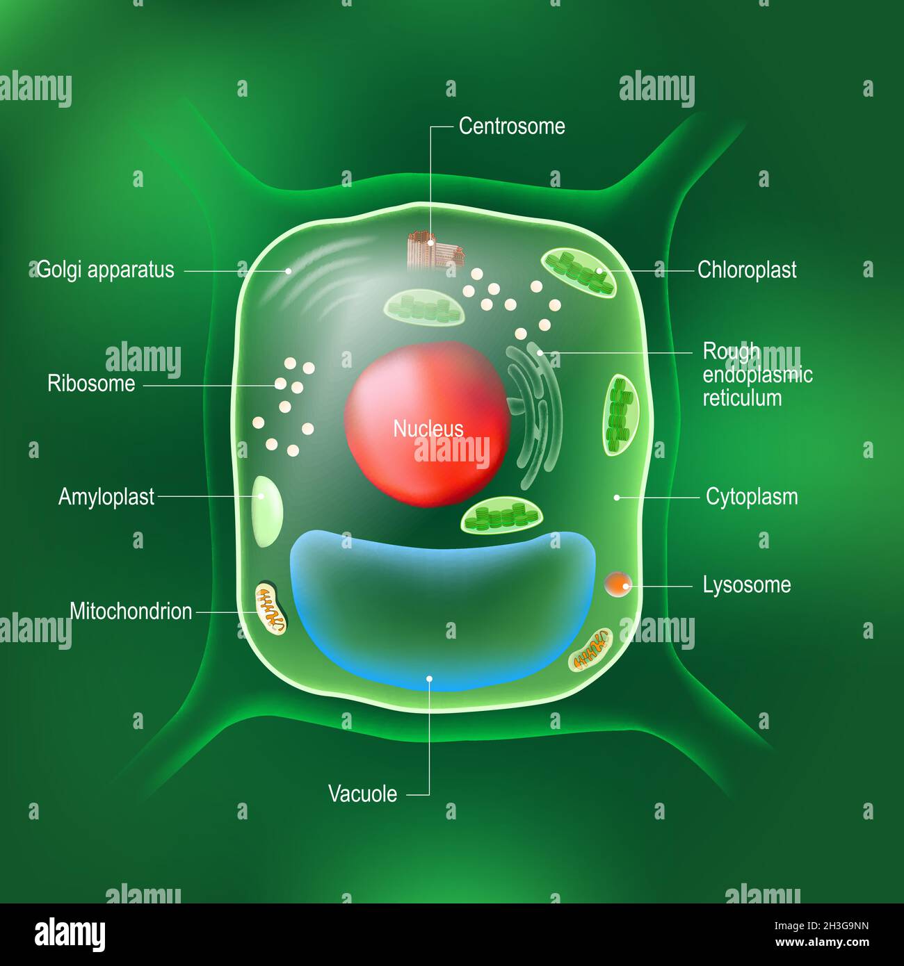 Anatomy of plant cell. All organelles: Nucleus, Ribosome, Rough endoplasmic reticulum, Golgi apparatus, mitochondrion, amyloplast, vacuole Stock Vectorhttps://www.alamy.com/image-license-details/?v=1https://www.alamy.com/anatomy-of-plant-cell-all-organelles-nucleus-ribosome-rough-endoplasmic-reticulum-golgi-apparatus-mitochondrion-amyloplast-vacuole-image449672433.html
RF2H3G9NN–Anatomy of plant cell. All organelles: Nucleus, Ribosome, Rough endoplasmic reticulum, Golgi apparatus, mitochondrion, amyloplast, vacuole
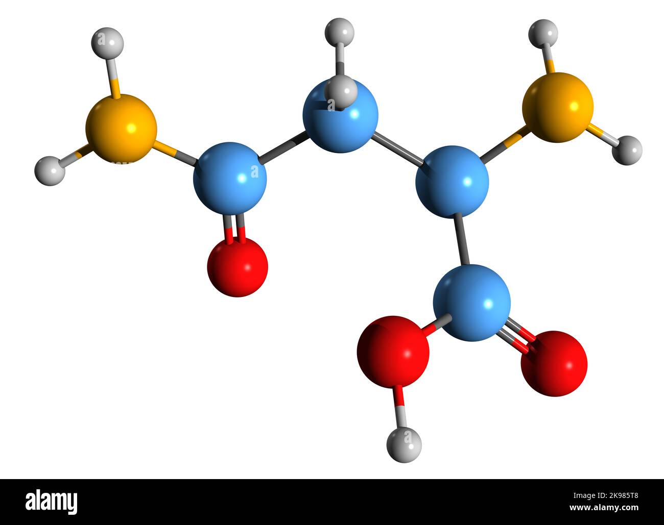 3D image of Asparagine skeletal formula - molecular chemical structure of 2-Amino-3-carbamoylpropanoic acid isolated on white background Stock Photohttps://www.alamy.com/image-license-details/?v=1https://www.alamy.com/3d-image-of-asparagine-skeletal-formula-molecular-chemical-structure-of-2-amino-3-carbamoylpropanoic-acid-isolated-on-white-background-image487602424.html
RF2K985T8–3D image of Asparagine skeletal formula - molecular chemical structure of 2-Amino-3-carbamoylpropanoic acid isolated on white background
 Reticulum is a caul or coif of network for covering the hair vintage line drawing or engraving illustration. Stock Vectorhttps://www.alamy.com/image-license-details/?v=1https://www.alamy.com/reticulum-is-a-caul-or-coif-of-network-for-covering-the-hair-vintage-line-drawing-or-engraving-illustration-image244588434.html
RFT5WY16–Reticulum is a caul or coif of network for covering the hair vintage line drawing or engraving illustration.
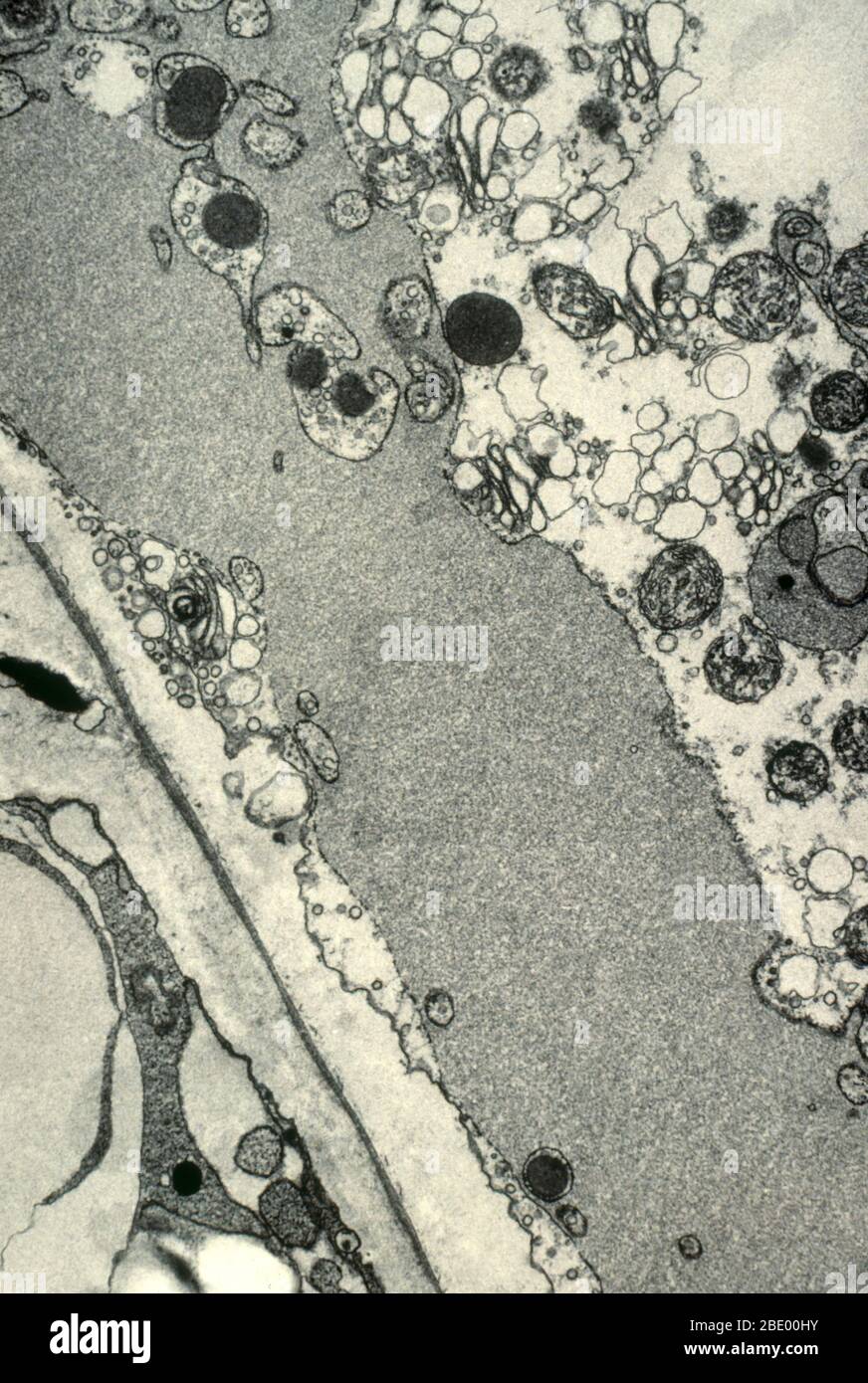 Endoplasmic Reticulum (ER) Filled with Protein Stock Photohttps://www.alamy.com/image-license-details/?v=1https://www.alamy.com/endoplasmic-reticulum-er-filled-with-protein-image352813047.html
RM2BE00HY–Endoplasmic Reticulum (ER) Filled with Protein
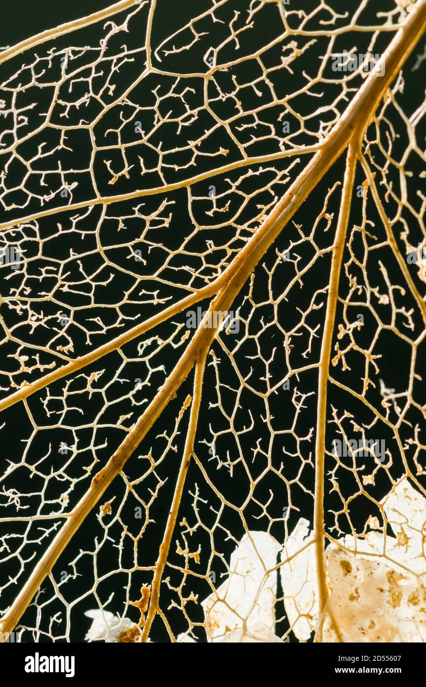 Reticulum of veins in a dead leaf Stock Photohttps://www.alamy.com/image-license-details/?v=1https://www.alamy.com/reticulum-of-veins-in-a-dead-leaf-image381815847.html
RF2D55607–Reticulum of veins in a dead leaf
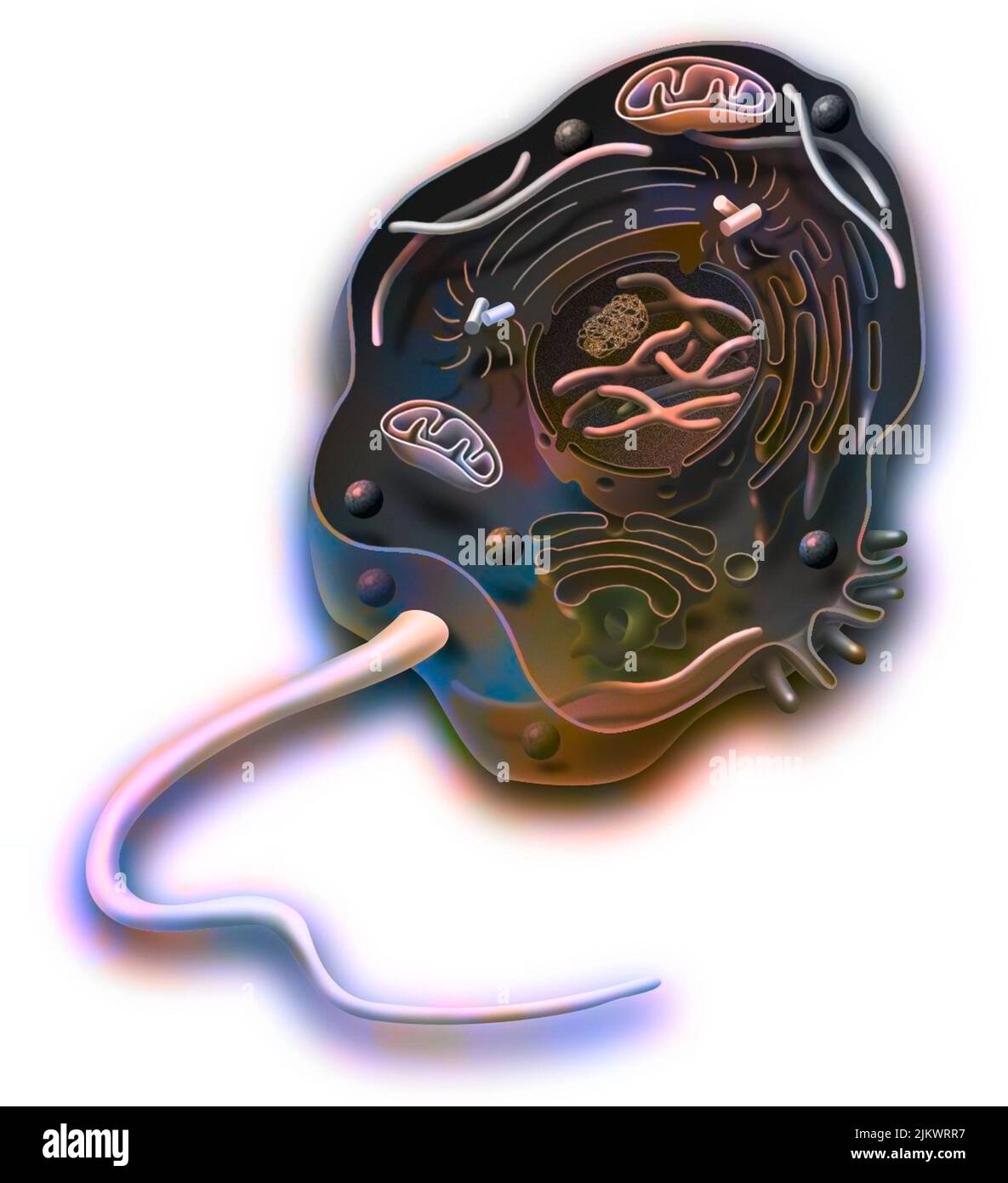 Eukaryote of animal cell type with DNA, endoplasmic reticulum. Stock Photohttps://www.alamy.com/image-license-details/?v=1https://www.alamy.com/eukaryote-of-animal-cell-type-with-dna-endoplasmic-reticulum-image476925883.html
RF2JKWRR7–Eukaryote of animal cell type with DNA, endoplasmic reticulum.
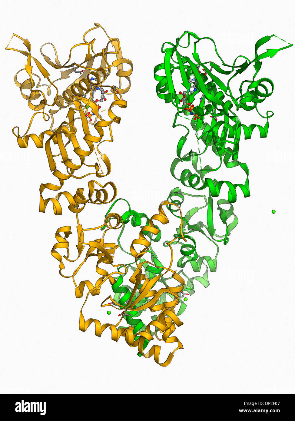 Endoplasmic reticulum chaperone protein Stock Photohttps://www.alamy.com/image-license-details/?v=1https://www.alamy.com/endoplasmic-reticulum-chaperone-protein-image65209207.html
RFDP2F07–Endoplasmic reticulum chaperone protein
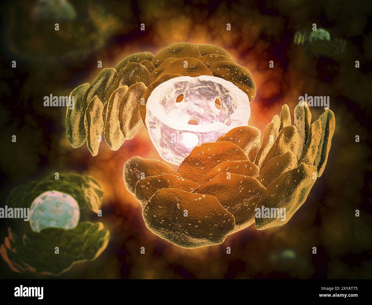 Conceptual image of endoplasmic reticulum around a cell nucleus. Endoplasmic reticulum is an organelle that forms a continuous membrane system of flat Stock Photohttps://www.alamy.com/image-license-details/?v=1https://www.alamy.com/conceptual-image-of-endoplasmic-reticulum-around-a-cell-nucleus-endoplasmic-reticulum-is-an-organelle-that-forms-a-continuous-membrane-system-of-flat-image619197129.html
RM2XYAT75–Conceptual image of endoplasmic reticulum around a cell nucleus. Endoplasmic reticulum is an organelle that forms a continuous membrane system of flat
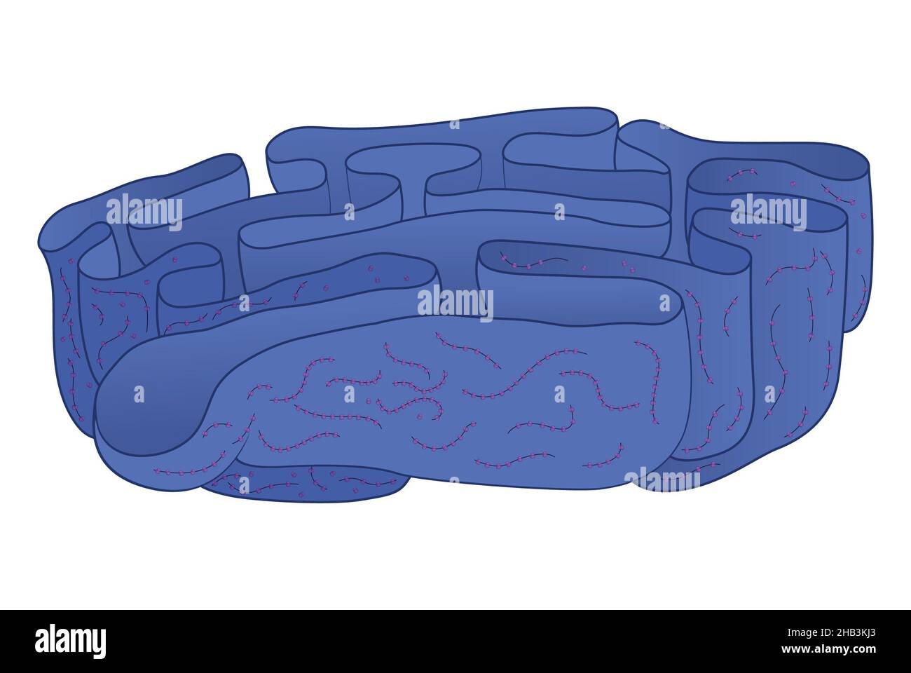 Granular (granulated) endoplasmic reticulum with ribosomes and polyribosomes Stock Photohttps://www.alamy.com/image-license-details/?v=1https://www.alamy.com/granular-granulated-endoplasmic-reticulum-with-ribosomes-and-polyribosomes-image454312043.html
RF2HB3KJ3–Granular (granulated) endoplasmic reticulum with ribosomes and polyribosomes
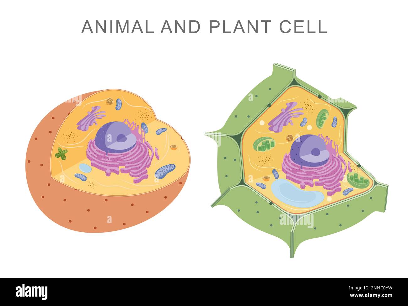 Comparing animal and plant cells Stock Photohttps://www.alamy.com/image-license-details/?v=1https://www.alamy.com/comparing-animal-and-plant-cells-image529483021.html
RF2NNC0YW–Comparing animal and plant cells
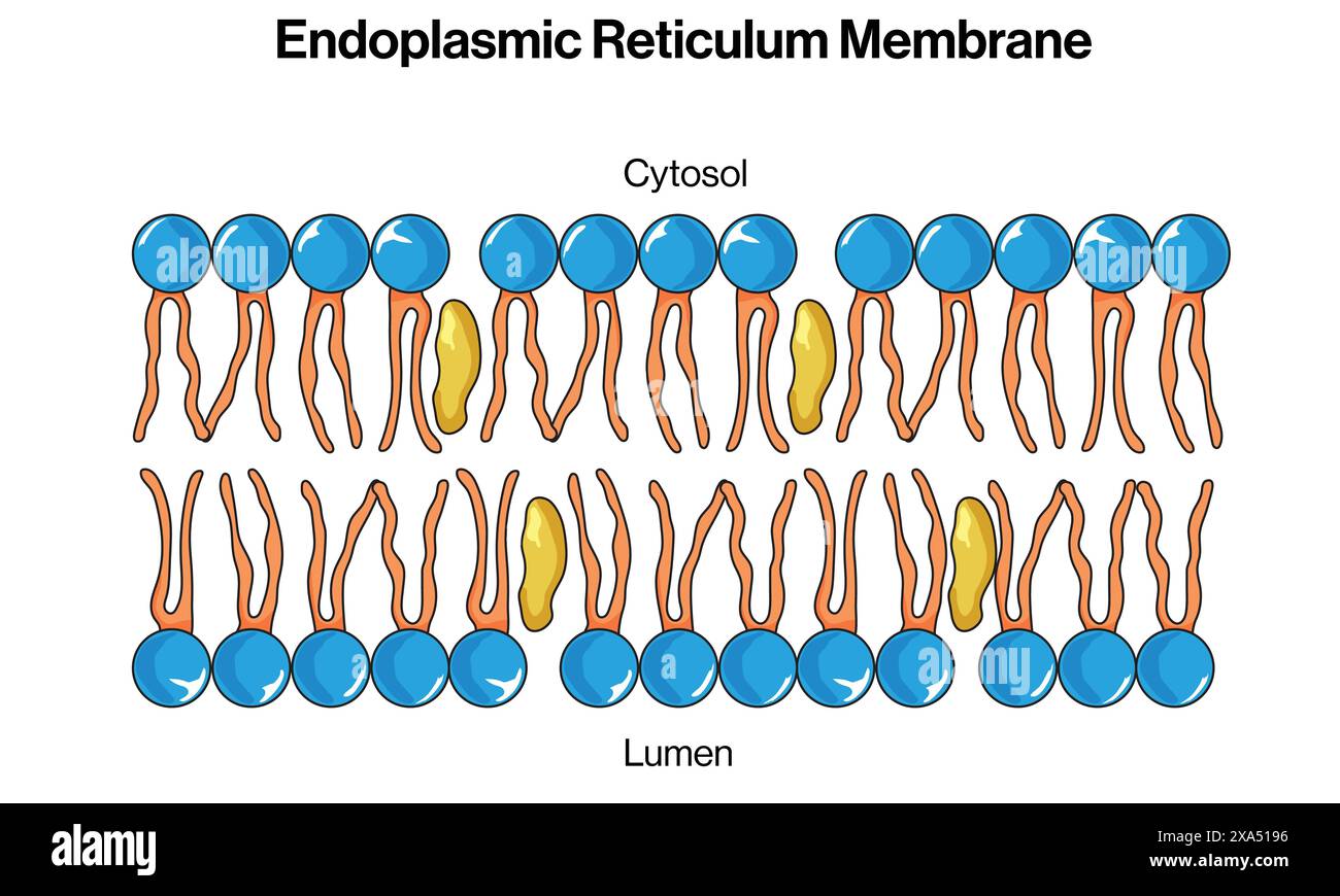 Detailed Vector Illustration of Endoplasmic Reticulum Membrane for Cell Biology, Molecular Biology, and Biochemistry Education on White Background. Stock Vectorhttps://www.alamy.com/image-license-details/?v=1https://www.alamy.com/detailed-vector-illustration-of-endoplasmic-reticulum-membrane-for-cell-biology-molecular-biology-and-biochemistry-education-on-white-background-image608620242.html
RF2XA5196–Detailed Vector Illustration of Endoplasmic Reticulum Membrane for Cell Biology, Molecular Biology, and Biochemistry Education on White Background.
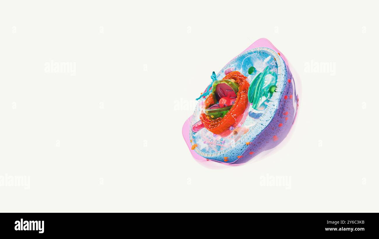 8 organelle, organelle in human cell structure. kinds of human cells Stock Photohttps://www.alamy.com/image-license-details/?v=1https://www.alamy.com/8-organelle-organelle-in-human-cell-structure-kinds-of-human-cells-image623527503.html
RF2Y6C3KB–8 organelle, organelle in human cell structure. kinds of human cells
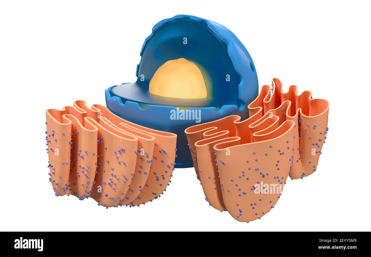 Structure of nuclear and endoplasmic reticulum in an animal cell, 3d rendering. Section view. Computer digital drawing. Stock Photohttps://www.alamy.com/image-license-details/?v=1https://www.alamy.com/structure-of-nuclear-and-endoplasmic-reticulum-in-an-animal-cell-3d-rendering-section-view-computer-digital-drawing-image413031369.html
RF2EYY5M9–Structure of nuclear and endoplasmic reticulum in an animal cell, 3d rendering. Section view. Computer digital drawing.
 Bolete Bokeh, Penny Bun Boletus edulis is a very popular mushroom. It has many common names, Cep in France, Porcini in Italy and Stein Pilz in Germany to name but three. It has a hemispherical brown cap with a white lip. The white stem is spindle shaped with a brown reticulum or net like pattern. The pores are white becoming greenish yellow as the spores mature. Penny Buns are found in woodlands, commonly with oak, birch, beech and pine Stock Photohttps://www.alamy.com/image-license-details/?v=1https://www.alamy.com/bolete-bokeh-penny-bun-boletus-edulis-is-a-very-popular-mushroom-it-has-many-common-names-cep-in-france-porcini-in-italy-and-stein-pilz-in-germany-to-name-but-three-it-has-a-hemispherical-brown-cap-with-a-white-lip-the-white-stem-is-spindle-shaped-with-a-brown-reticulum-or-net-like-pattern-the-pores-are-white-becoming-greenish-yellow-as-the-spores-mature-penny-buns-are-found-in-woodlands-commonly-with-oak-birch-beech-and-pine-image573391293.html
RM2T8T6CD–Bolete Bokeh, Penny Bun Boletus edulis is a very popular mushroom. It has many common names, Cep in France, Porcini in Italy and Stein Pilz in Germany to name but three. It has a hemispherical brown cap with a white lip. The white stem is spindle shaped with a brown reticulum or net like pattern. The pores are white becoming greenish yellow as the spores mature. Penny Buns are found in woodlands, commonly with oak, birch, beech and pine
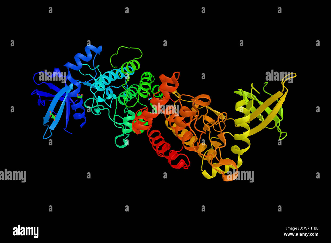 Serine/threonine-protein kinase VRK2, a protein localizes to the endoplasmic reticulum and has been shown to phosphorylate casein and undergo autophos Stock Photohttps://www.alamy.com/image-license-details/?v=1https://www.alamy.com/serinethreonine-protein-kinase-vrk2-a-protein-localizes-to-the-endoplasmic-reticulum-and-has-been-shown-to-phosphorylate-casein-and-undergo-autophos-image262850434.html
RFW7HTBE–Serine/threonine-protein kinase VRK2, a protein localizes to the endoplasmic reticulum and has been shown to phosphorylate casein and undergo autophos
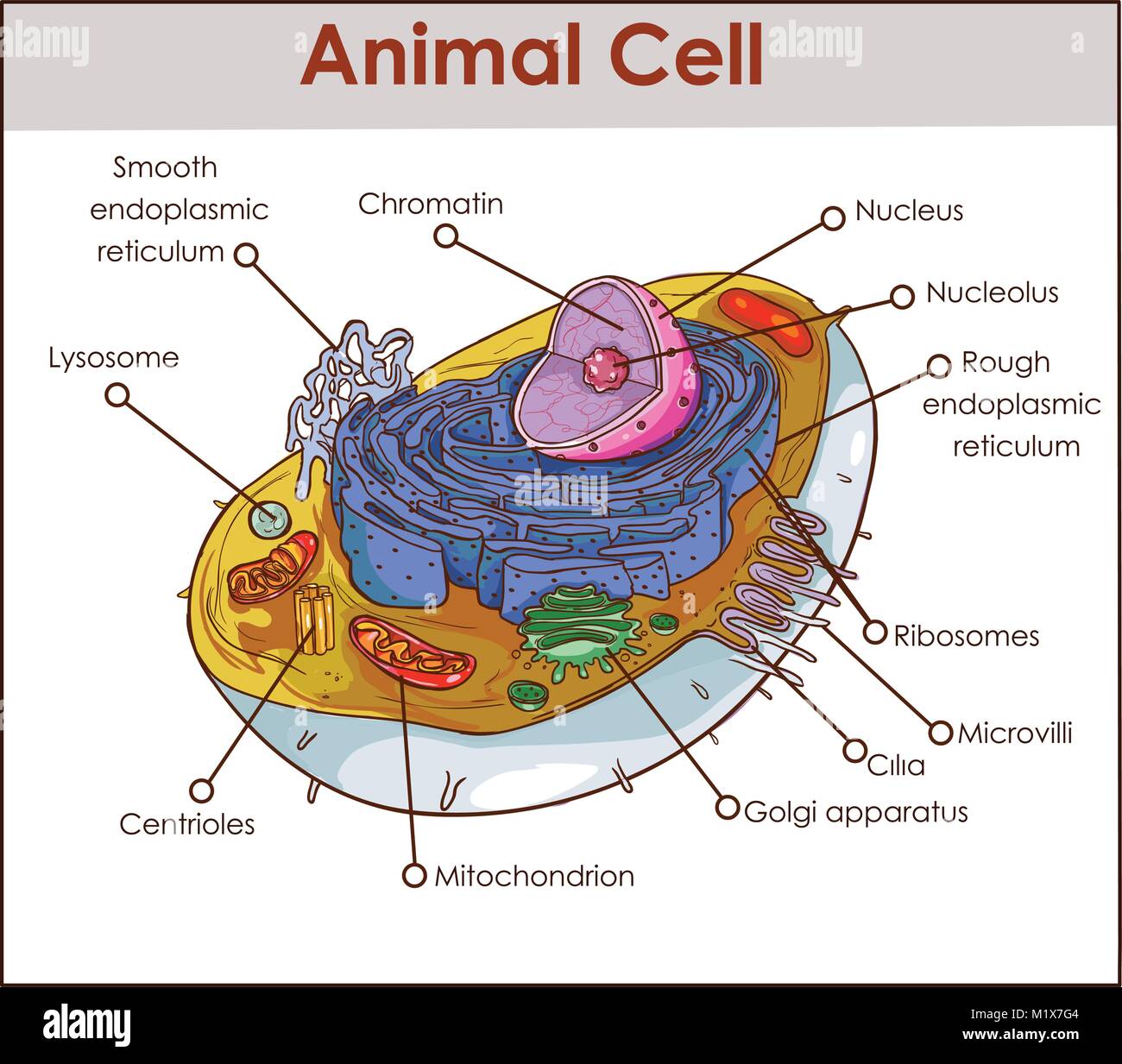 Animal Cell Anatomy Diagram Structure with all parts nucleus smooth rough endoplasmic reticulum cytoplasm golgi apparatus mitochondria membrane centro Stock Vectorhttps://www.alamy.com/image-license-details/?v=1https://www.alamy.com/stock-photo-animal-cell-anatomy-diagram-structure-with-all-parts-nucleus-smooth-173295028.html
RFM1X7G4–Animal Cell Anatomy Diagram Structure with all parts nucleus smooth rough endoplasmic reticulum cytoplasm golgi apparatus mitochondria membrane centro
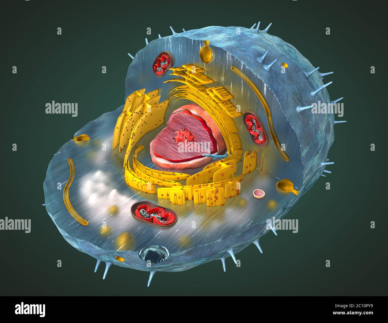 Scientifically correct 3d illustration of the internal structure of a human cell, cut-away Stock Photohttps://www.alamy.com/image-license-details/?v=1https://www.alamy.com/scientifically-correct-3d-illustration-of-the-internal-structure-of-a-human-cell-cut-away-image362050397.html
RF2C10PY9–Scientifically correct 3d illustration of the internal structure of a human cell, cut-away
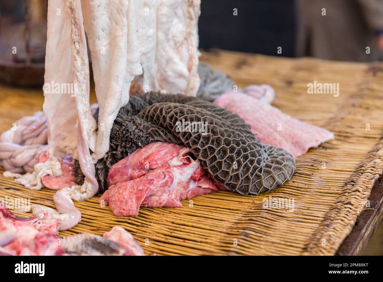 Morocco, High Atlas, Marrakech Safi region, Had-Drâa, the Had-Drâ market is one of the most important in southern Morocco, butchers market, bonnet (also called honeycomb bonnet or network or reticulum) Stock Photohttps://www.alamy.com/image-license-details/?v=1https://www.alamy.com/morocco-high-atlas-marrakech-safi-region-had-dra-the-had-dr-market-is-one-of-the-most-important-in-southern-morocco-butchers-market-bonnet-also-called-honeycomb-bonnet-or-network-or-reticulum-image545996972.html
RM2PM88KT–Morocco, High Atlas, Marrakech Safi region, Had-Drâa, the Had-Drâ market is one of the most important in southern Morocco, butchers market, bonnet (also called honeycomb bonnet or network or reticulum)
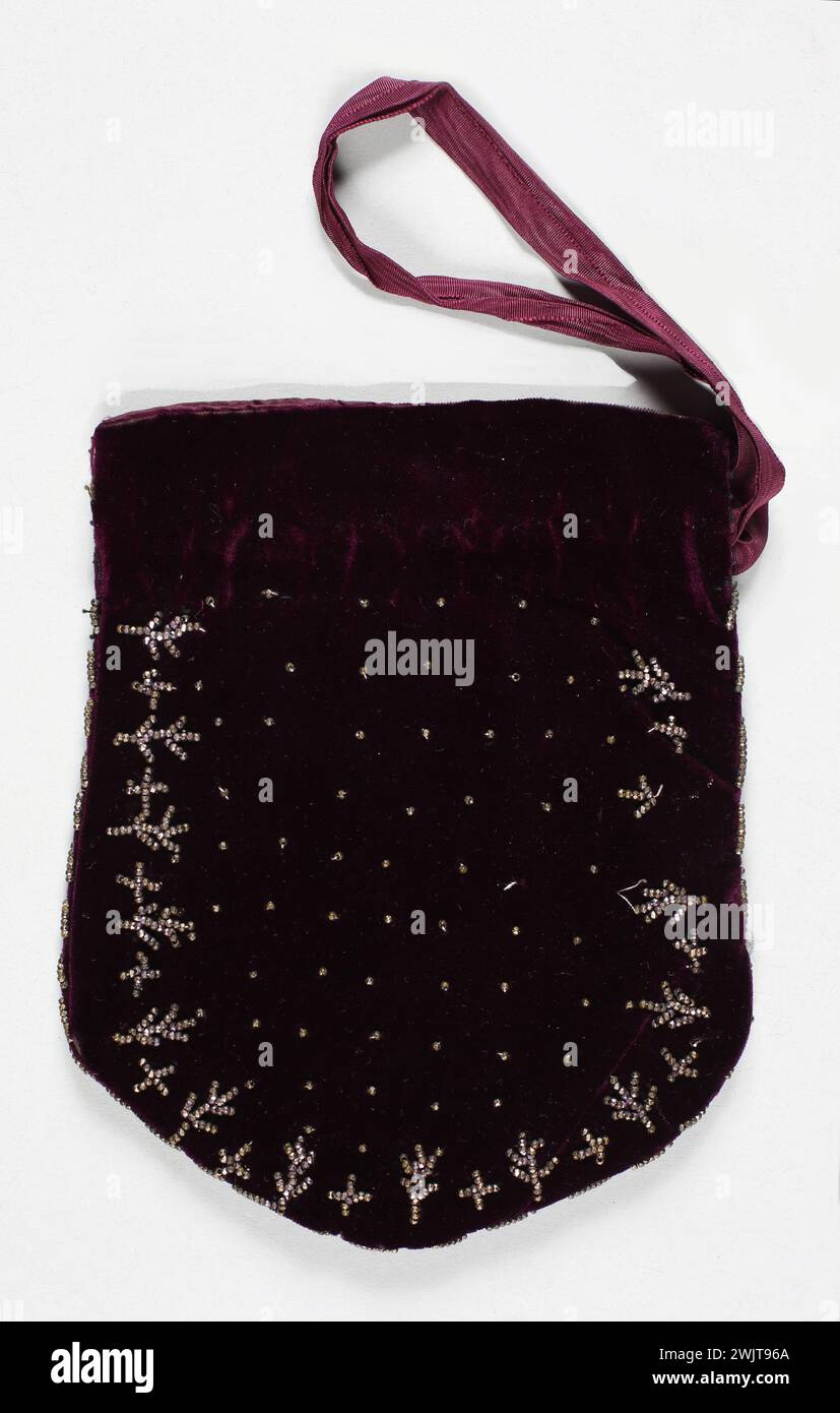 Reticulum bag. XIXth. GAL1989.124.11 Stock Photohttps://www.alamy.com/image-license-details/?v=1https://www.alamy.com/reticulum-bag-xixth-gal198912411-image596750402.html
RM2WJT96A–Reticulum bag. XIXth. GAL1989.124.11
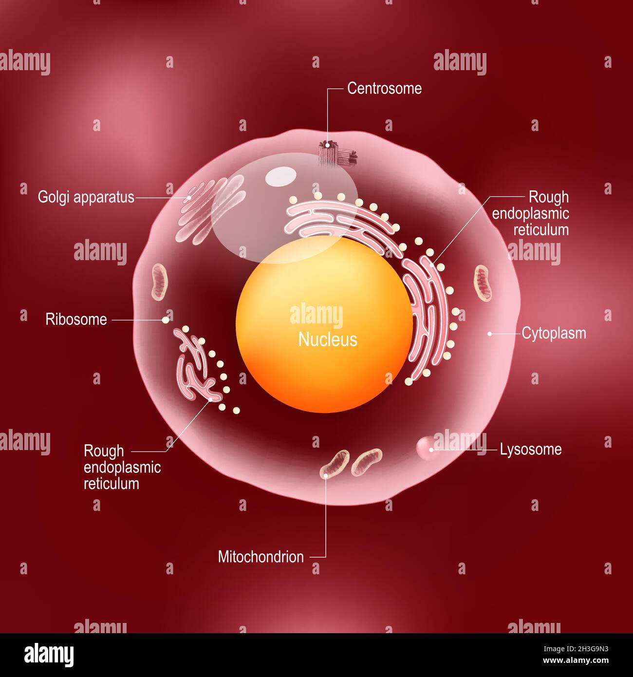 Anatomy of human cell. All organelles: Nucleus, Ribosome, Rough endoplasmic reticulum, Golgi apparatus, mitochondrion, cytoplasm, lysosome, Centrosome Stock Vectorhttps://www.alamy.com/image-license-details/?v=1https://www.alamy.com/anatomy-of-human-cell-all-organelles-nucleus-ribosome-rough-endoplasmic-reticulum-golgi-apparatus-mitochondrion-cytoplasm-lysosome-centrosome-image449672415.html
RF2H3G9N3–Anatomy of human cell. All organelles: Nucleus, Ribosome, Rough endoplasmic reticulum, Golgi apparatus, mitochondrion, cytoplasm, lysosome, Centrosome
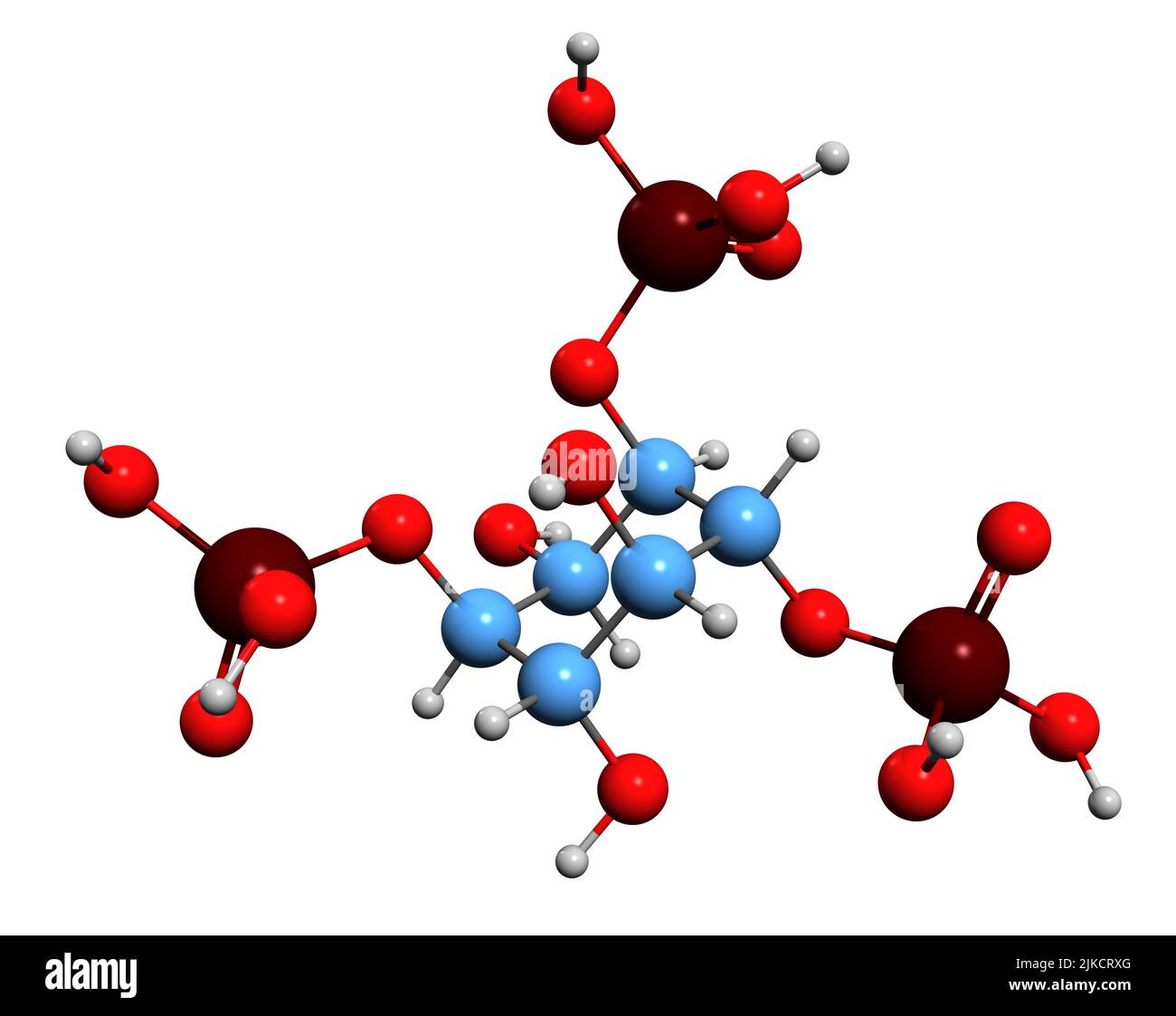 3D image of Inositol trisphosphate skeletal formula - molecular chemical structure of inositol phosphate signaling molecule isolated on white backgro Stock Photohttps://www.alamy.com/image-license-details/?v=1https://www.alamy.com/3d-image-of-inositol-trisphosphate-skeletal-formula-molecular-chemical-structure-of-inositol-phosphate-signaling-molecule-isolated-on-white-backgro-image476640600.html
RF2JKCRXG–3D image of Inositol trisphosphate skeletal formula - molecular chemical structure of inositol phosphate signaling molecule isolated on white backgro
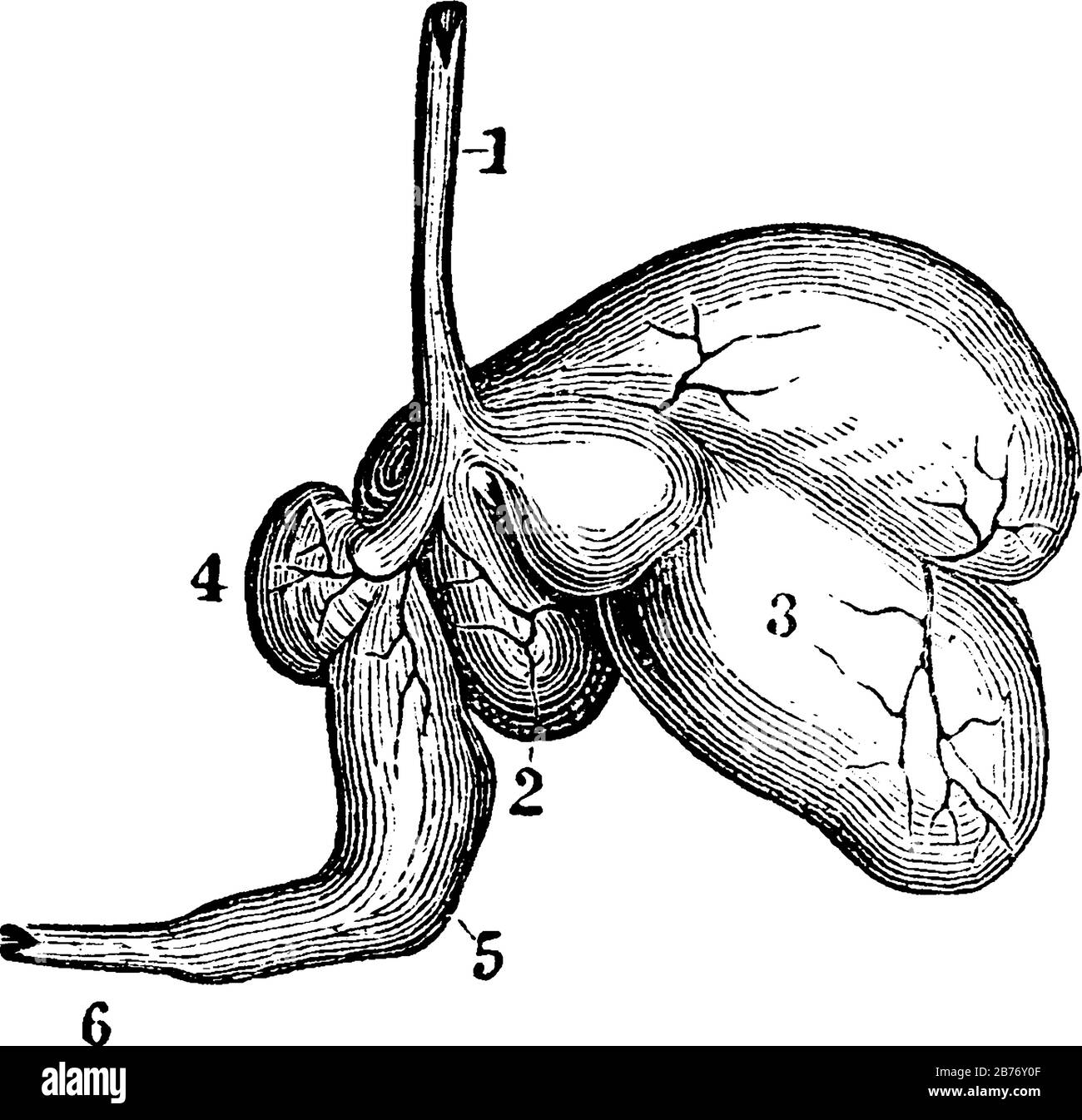 Ruminants, the sheep, have a stomach with four cavities. Labels: 1, esophagus; 2, rumen; 3, reticulum; 4, omasum; 5, abomasum or rennet; 6, intestine, Stock Vectorhttps://www.alamy.com/image-license-details/?v=1https://www.alamy.com/ruminants-the-sheep-have-a-stomach-with-four-cavities-labels-1-esophagus-2-rumen-3-reticulum-4-omasum-5-abomasum-or-rennet-6-intestine-image348662847.html
RF2B76Y0F–Ruminants, the sheep, have a stomach with four cavities. Labels: 1, esophagus; 2, rumen; 3, reticulum; 4, omasum; 5, abomasum or rennet; 6, intestine,
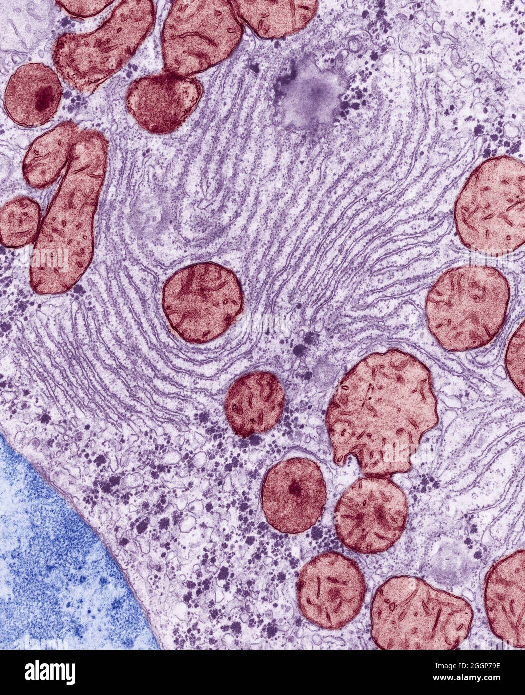 Colorized transmission Electron Micrograph (TEM) of endoplasmic reticulum and mitochondria in the liver of a rat. Stock Photohttps://www.alamy.com/image-license-details/?v=1https://www.alamy.com/colorized-transmission-electron-micrograph-tem-of-endoplasmic-reticulum-and-mitochondria-in-the-liver-of-a-rat-image440582394.html
RM2GGP79E–Colorized transmission Electron Micrograph (TEM) of endoplasmic reticulum and mitochondria in the liver of a rat.
 The common poppy tiny seeds are, as in all species of the genus, kidney-shaped, alveolated with a polygonal reticulum and brown in color. Stock Photohttps://www.alamy.com/image-license-details/?v=1https://www.alamy.com/the-common-poppy-tiny-seeds-are-as-in-all-species-of-the-genus-kidney-shaped-alveolated-with-a-polygonal-reticulum-and-brown-in-color-image606835539.html
RF2X77MWR–The common poppy tiny seeds are, as in all species of the genus, kidney-shaped, alveolated with a polygonal reticulum and brown in color.
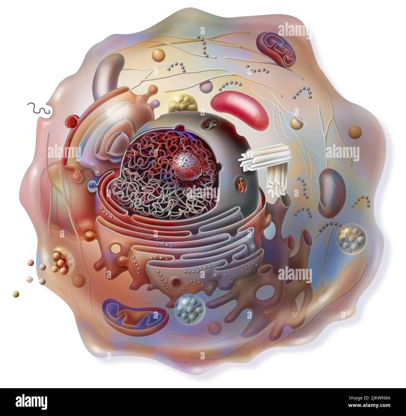 Cell sectional view with all the main organelles: nucleus, reticulum. Stock Photohttps://www.alamy.com/image-license-details/?v=1https://www.alamy.com/cell-sectional-view-with-all-the-main-organelles-nucleus-reticulum-image476923898.html
RF2JKWN8A–Cell sectional view with all the main organelles: nucleus, reticulum.
 Reticulum, illustration Stock Photohttps://www.alamy.com/image-license-details/?v=1https://www.alamy.com/reticulum-illustration-image575291290.html
RF2TBXNWE–Reticulum, illustration
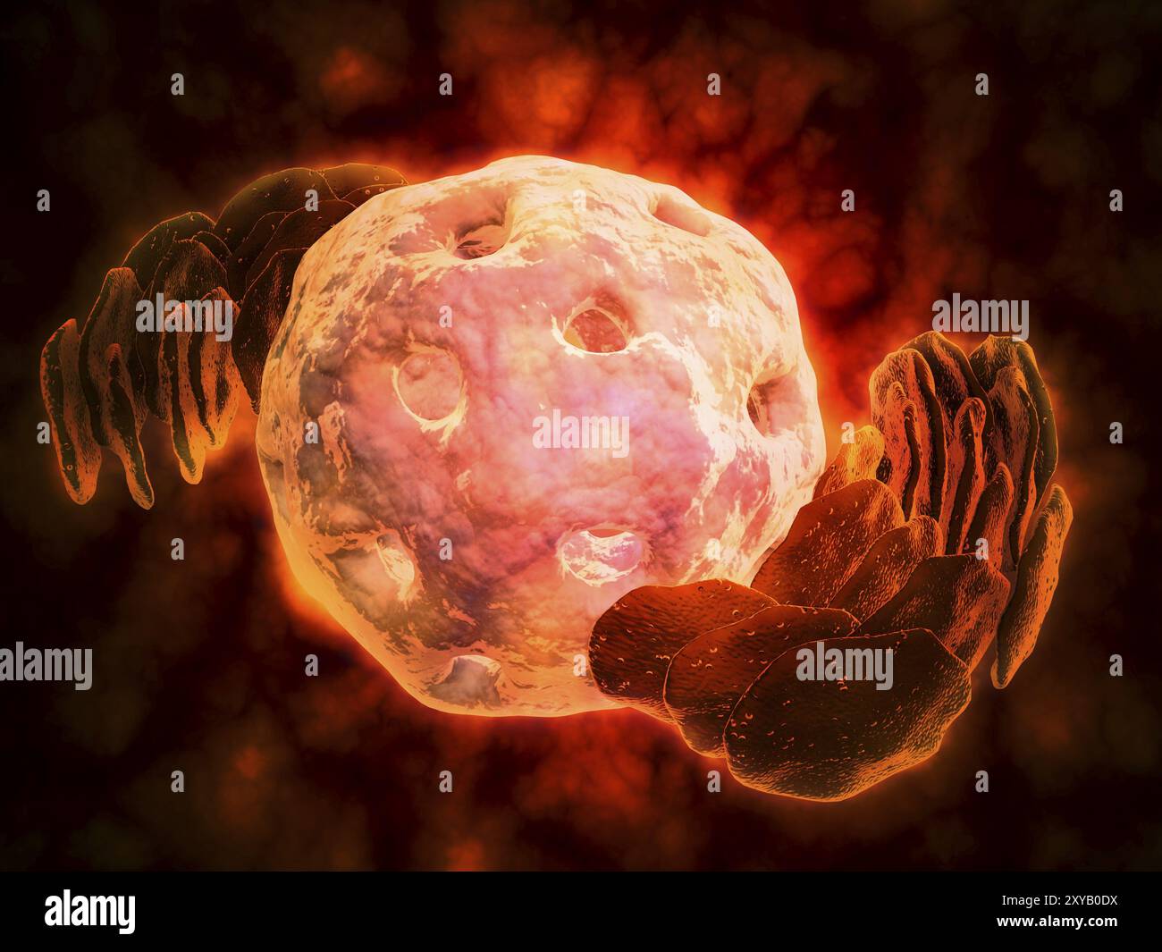 Conceptual image of endoplasmic reticulum around a cell nucleus. Endoplasmic reticulum is an organelle that forms a continuous membrane system of flat Stock Photohttps://www.alamy.com/image-license-details/?v=1https://www.alamy.com/conceptual-image-of-endoplasmic-reticulum-around-a-cell-nucleus-endoplasmic-reticulum-is-an-organelle-that-forms-a-continuous-membrane-system-of-flat-image619200454.html
RM2XYB0DX–Conceptual image of endoplasmic reticulum around a cell nucleus. Endoplasmic reticulum is an organelle that forms a continuous membrane system of flat
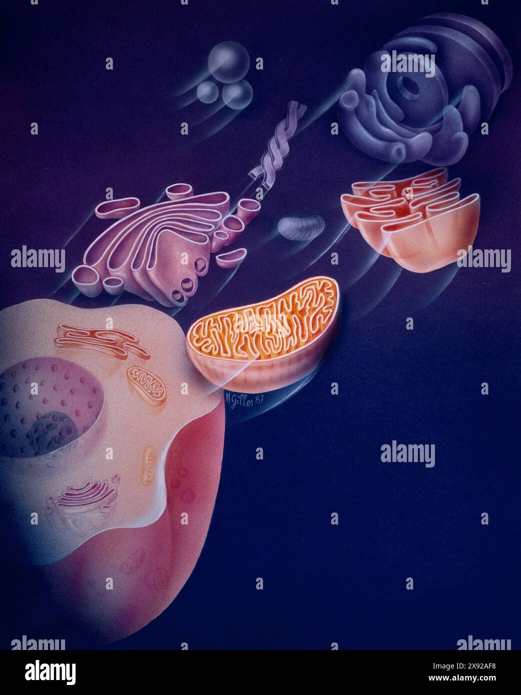 Endoplasmic reticulum at top. Mitochondria is oval shaped organelle at center. Lysosomes are small bubbles to the left in purple. Golgi apparatus at bottom. CELL, DRAWING 001366 002 Stock Photohttps://www.alamy.com/image-license-details/?v=1https://www.alamy.com/endoplasmic-reticulum-at-top-mitochondria-is-oval-shaped-organelle-at-center-lysosomes-are-small-bubbles-to-the-left-in-purple-golgi-apparatus-at-bottom-cell-drawing-001366-002-image607946956.html
RM2X92AF8–Endoplasmic reticulum at top. Mitochondria is oval shaped organelle at center. Lysosomes are small bubbles to the left in purple. Golgi apparatus at bottom. CELL, DRAWING 001366 002
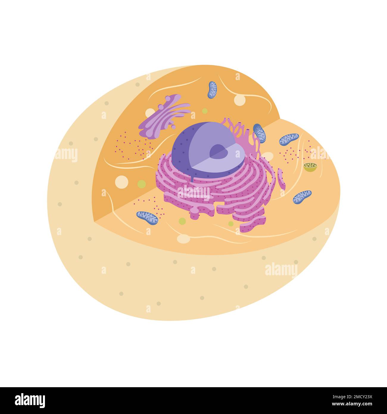 Illustration of animal cell with organelles Stock Photohttps://www.alamy.com/image-license-details/?v=1https://www.alamy.com/illustration-of-animal-cell-with-organelles-image507070926.html
RF2MCY23X–Illustration of animal cell with organelles
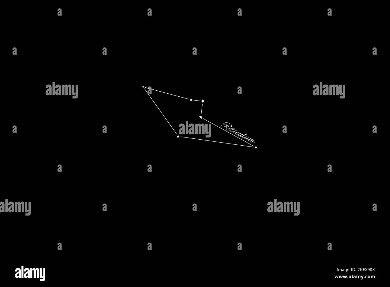 Reticulum constellation, Cluster of stars, Reticle constellation, The Small Net Stock Photohttps://www.alamy.com/image-license-details/?v=1https://www.alamy.com/reticulum-constellation-cluster-of-stars-reticle-constellation-the-small-net-image491073315.html
RF2KEX90K–Reticulum constellation, Cluster of stars, Reticle constellation, The Small Net
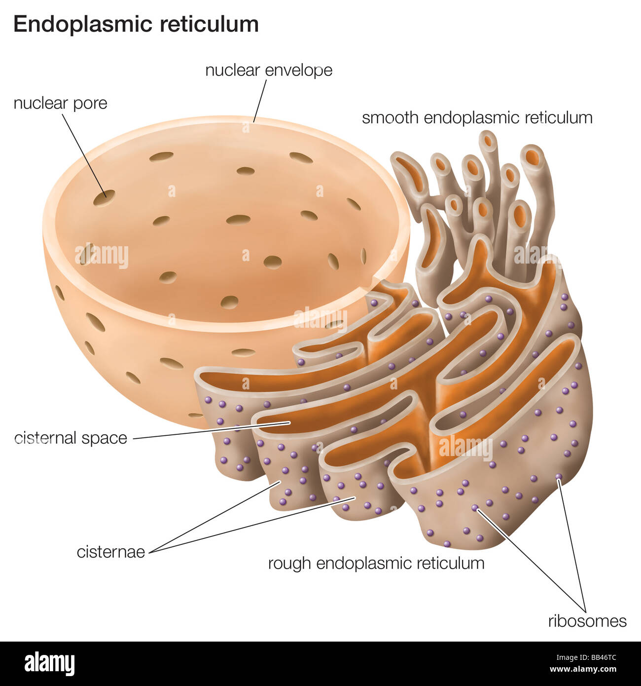 The endoplasmic reticulum plays an important role in the biosynthesis, processing, and transport of proteins and lipids. Stock Photohttps://www.alamy.com/image-license-details/?v=1https://www.alamy.com/stock-photo-the-endoplasmic-reticulum-plays-an-important-role-in-the-biosynthesis-24064780.html
RMBB46TC–The endoplasmic reticulum plays an important role in the biosynthesis, processing, and transport of proteins and lipids.
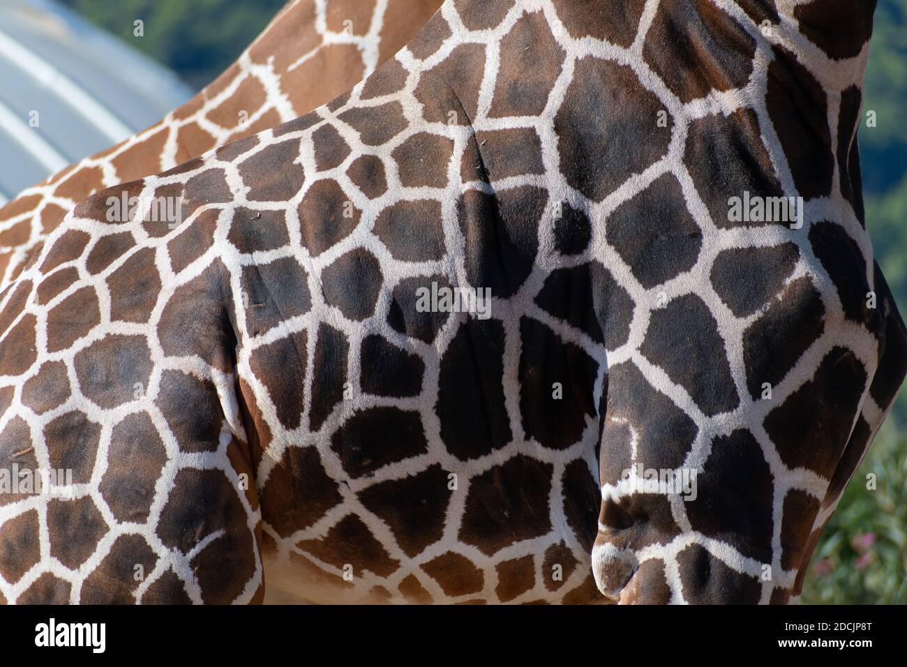 Reticulated giraffe (Giraffa camelopardalis reticulata) coat pattern close up Stock Photohttps://www.alamy.com/image-license-details/?v=1https://www.alamy.com/reticulated-giraffe-giraffa-camelopardalis-reticulata-coat-pattern-close-up-image386416600.html
RF2DCJP8T–Reticulated giraffe (Giraffa camelopardalis reticulata) coat pattern close up
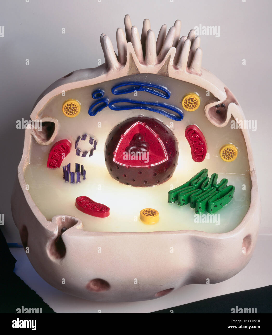 Model of animal cell, including cell nucleus, golgi body, lysosomes, centrioles, mitochondria, endoplasmic reticulum, ribosomes, cytoplasm, vesicles, thin plasma membrane, and microvilli (projections) at top Stock Photohttps://www.alamy.com/image-license-details/?v=1https://www.alamy.com/model-of-animal-cell-including-cell-nucleus-golgi-body-lysosomes-centrioles-mitochondria-endoplasmic-reticulum-ribosomes-cytoplasm-vesicles-thin-plasma-membrane-and-microvilli-projections-at-top-image216033580.html
RMPFD510–Model of animal cell, including cell nucleus, golgi body, lysosomes, centrioles, mitochondria, endoplasmic reticulum, ribosomes, cytoplasm, vesicles, thin plasma membrane, and microvilli (projections) at top
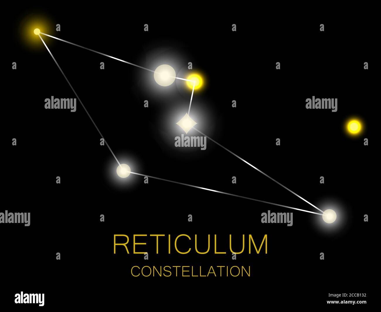 Reticulum constellation. Bright yellow stars in the night sky. A cluster of stars in deep space, the universe. Vector illustration Stock Vectorhttps://www.alamy.com/image-license-details/?v=1https://www.alamy.com/reticulum-constellation-bright-yellow-stars-in-the-night-sky-a-cluster-of-stars-in-deep-space-the-universe-vector-illustration-image369035942.html
RF2CCB132–Reticulum constellation. Bright yellow stars in the night sky. A cluster of stars in deep space, the universe. Vector illustration
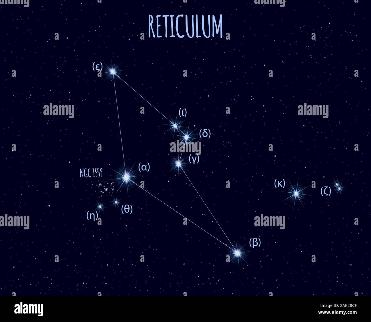 Reticulum (The Net) constellation, vector illustration with basic stars against the starry sky Stock Vectorhttps://www.alamy.com/image-license-details/?v=1https://www.alamy.com/reticulum-the-net-constellation-vector-illustration-with-basic-stars-against-the-starry-sky-image333808735.html
RF2AB28CF–Reticulum (The Net) constellation, vector illustration with basic stars against the starry sky
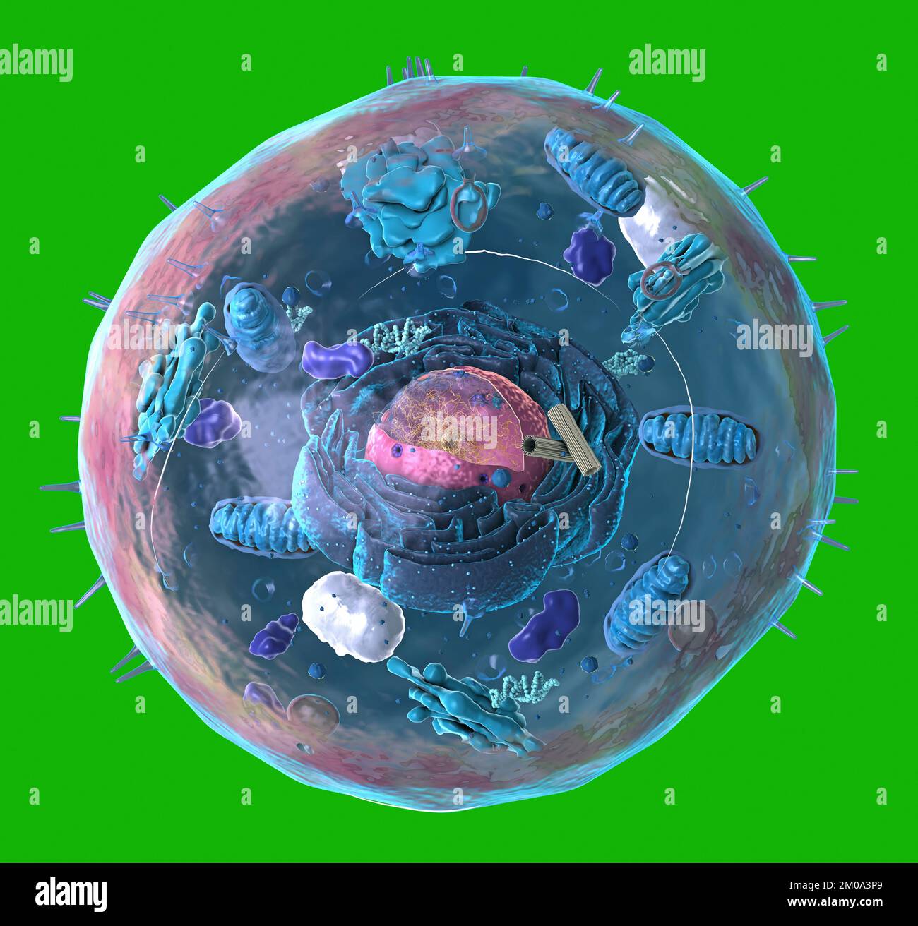 Components of Eukaryotic cell, nucleus and organelles and plasma membrane - 3d illustration Stock Photohttps://www.alamy.com/image-license-details/?v=1https://www.alamy.com/components-of-eukaryotic-cell-nucleus-and-organelles-and-plasma-membrane-3d-illustration-image499323169.html
RF2M0A3P9–Components of Eukaryotic cell, nucleus and organelles and plasma membrane - 3d illustration
 Gorgonia reticulum, Print, Gorgonia is a genus of soft corals, sea fans in the family Gorgoniidae Stock Photohttps://www.alamy.com/image-license-details/?v=1https://www.alamy.com/gorgonia-reticulum-print-gorgonia-is-a-genus-of-soft-corals-sea-fans-in-the-family-gorgoniidae-image328682677.html
RM2A2MP31–Gorgonia reticulum, Print, Gorgonia is a genus of soft corals, sea fans in the family Gorgoniidae
 Reticulum bag. XIXth. GAL1960.6.30 Stock Photohttps://www.alamy.com/image-license-details/?v=1https://www.alamy.com/reticulum-bag-xixth-gal1960630-image596752457.html
RM2WJTBRN–Reticulum bag. XIXth. GAL1960.6.30
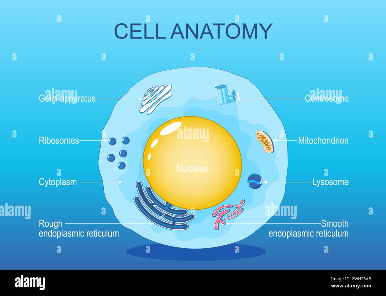 Anatomy of animal cell. Human cell structure. All organelles: Nucleus, Ribosome, Rough endoplasmic reticulum, Golgi apparatus, mitochondrion, cytoplas Stock Vectorhttps://www.alamy.com/image-license-details/?v=1https://www.alamy.com/anatomy-of-animal-cell-human-cell-structure-all-organelles-nucleus-ribosome-rough-endoplasmic-reticulum-golgi-apparatus-mitochondrion-cytoplas-image595652131.html
RF2WH28AB–Anatomy of animal cell. Human cell structure. All organelles: Nucleus, Ribosome, Rough endoplasmic reticulum, Golgi apparatus, mitochondrion, cytoplas
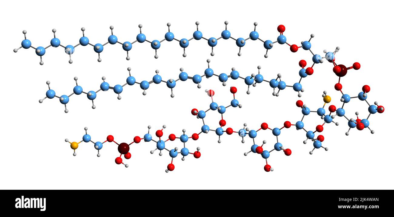 3D image of glycosylphosphatidylinositol skeletal formula - molecular chemical structure of GPI isolated on white background Stock Photohttps://www.alamy.com/image-license-details/?v=1https://www.alamy.com/3d-image-of-glycosylphosphatidylinositol-skeletal-formula-molecular-chemical-structure-of-gpi-isolated-on-white-background-image476466109.html
RF2JK4WAN–3D image of glycosylphosphatidylinositol skeletal formula - molecular chemical structure of GPI isolated on white background
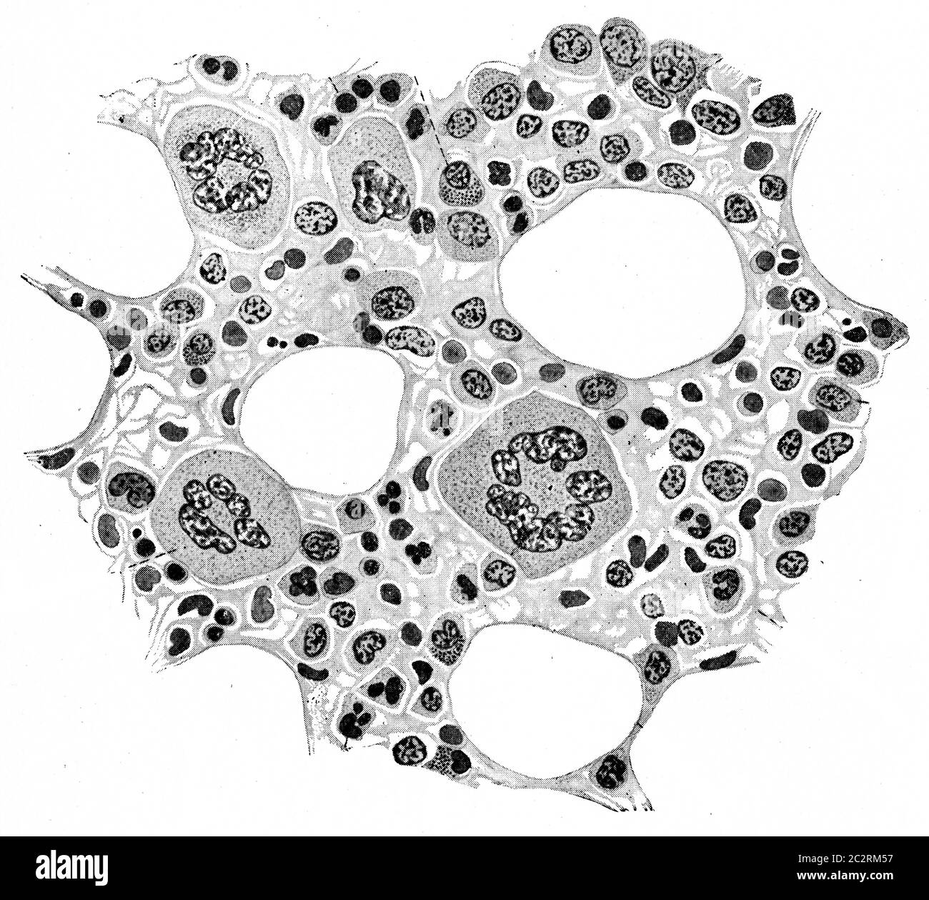 Section of bone marrow of rabbit showing the delicate connective tissue reticulum containing the different element of the marrow, vintage engraved ill Stock Photohttps://www.alamy.com/image-license-details/?v=1https://www.alamy.com/section-of-bone-marrow-of-rabbit-showing-the-delicate-connective-tissue-reticulum-containing-the-different-element-of-the-marrow-vintage-engraved-ill-image363167763.html
RF2C2RM57–Section of bone marrow of rabbit showing the delicate connective tissue reticulum containing the different element of the marrow, vintage engraved ill
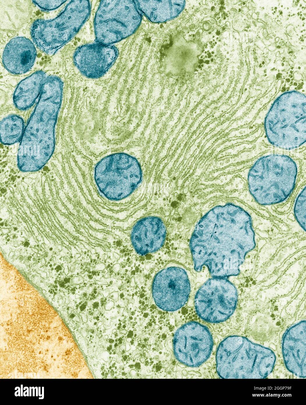 Colorized transmission Electron Micrograph (TEM) of endoplasmic reticulum and mitochondria in the liver of a rat. Stock Photohttps://www.alamy.com/image-license-details/?v=1https://www.alamy.com/colorized-transmission-electron-micrograph-tem-of-endoplasmic-reticulum-and-mitochondria-in-the-liver-of-a-rat-image440582395.html
RM2GGP79F–Colorized transmission Electron Micrograph (TEM) of endoplasmic reticulum and mitochondria in the liver of a rat.
 Representation of the constellation Reticulum (Ret), one of the modern constellations, part of 'Constellations' series Stock Photohttps://www.alamy.com/image-license-details/?v=1https://www.alamy.com/stock-photo-representation-of-the-constellation-reticulum-ret-one-of-the-modern-55947708.html
RMD70HTC–Representation of the constellation Reticulum (Ret), one of the modern constellations, part of 'Constellations' series
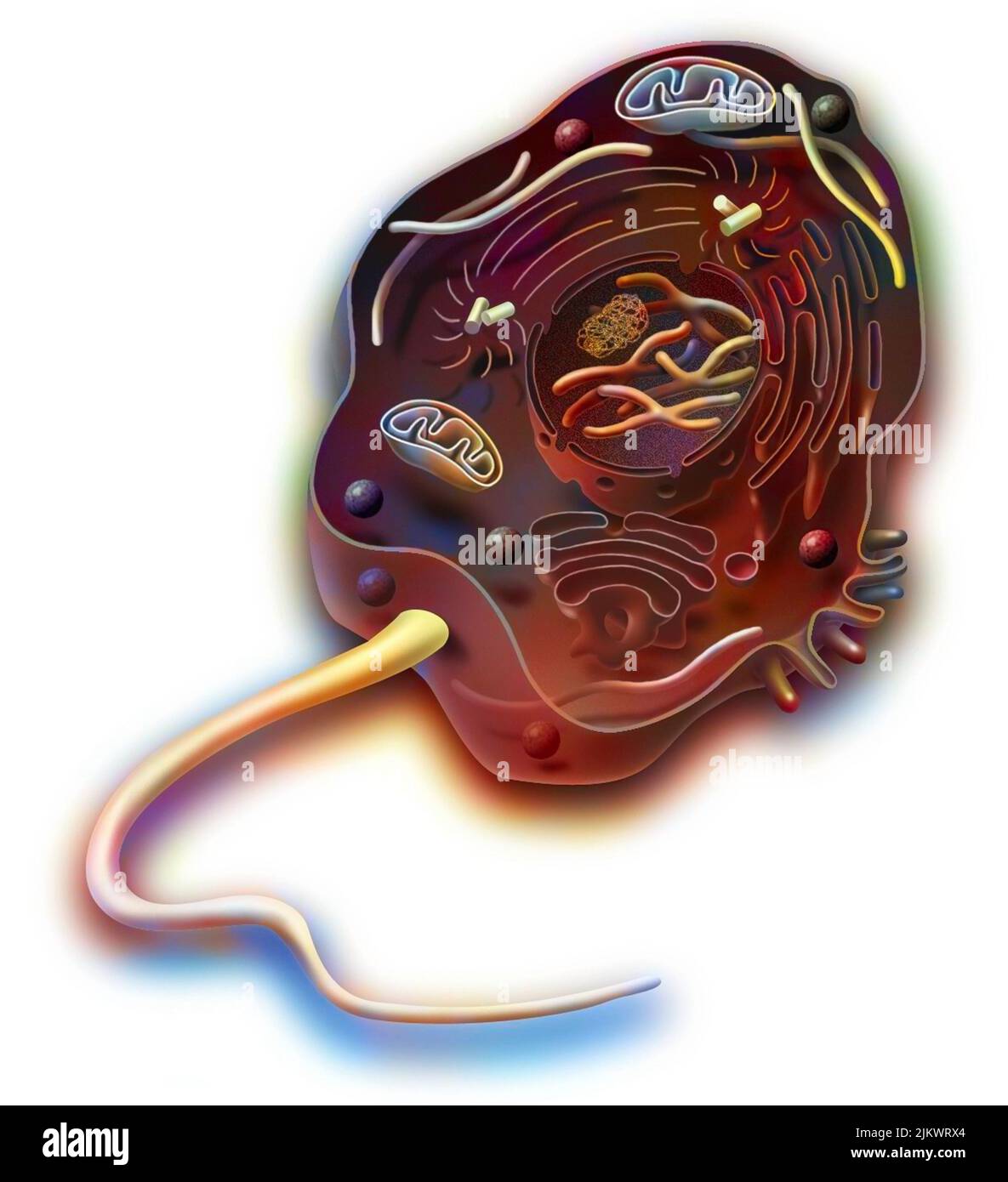 Eukaryote of animal cell type with DNA, endoplasmic reticulum. Stock Photohttps://www.alamy.com/image-license-details/?v=1https://www.alamy.com/eukaryote-of-animal-cell-type-with-dna-endoplasmic-reticulum-image476925964.html
RF2JKWRX4–Eukaryote of animal cell type with DNA, endoplasmic reticulum.
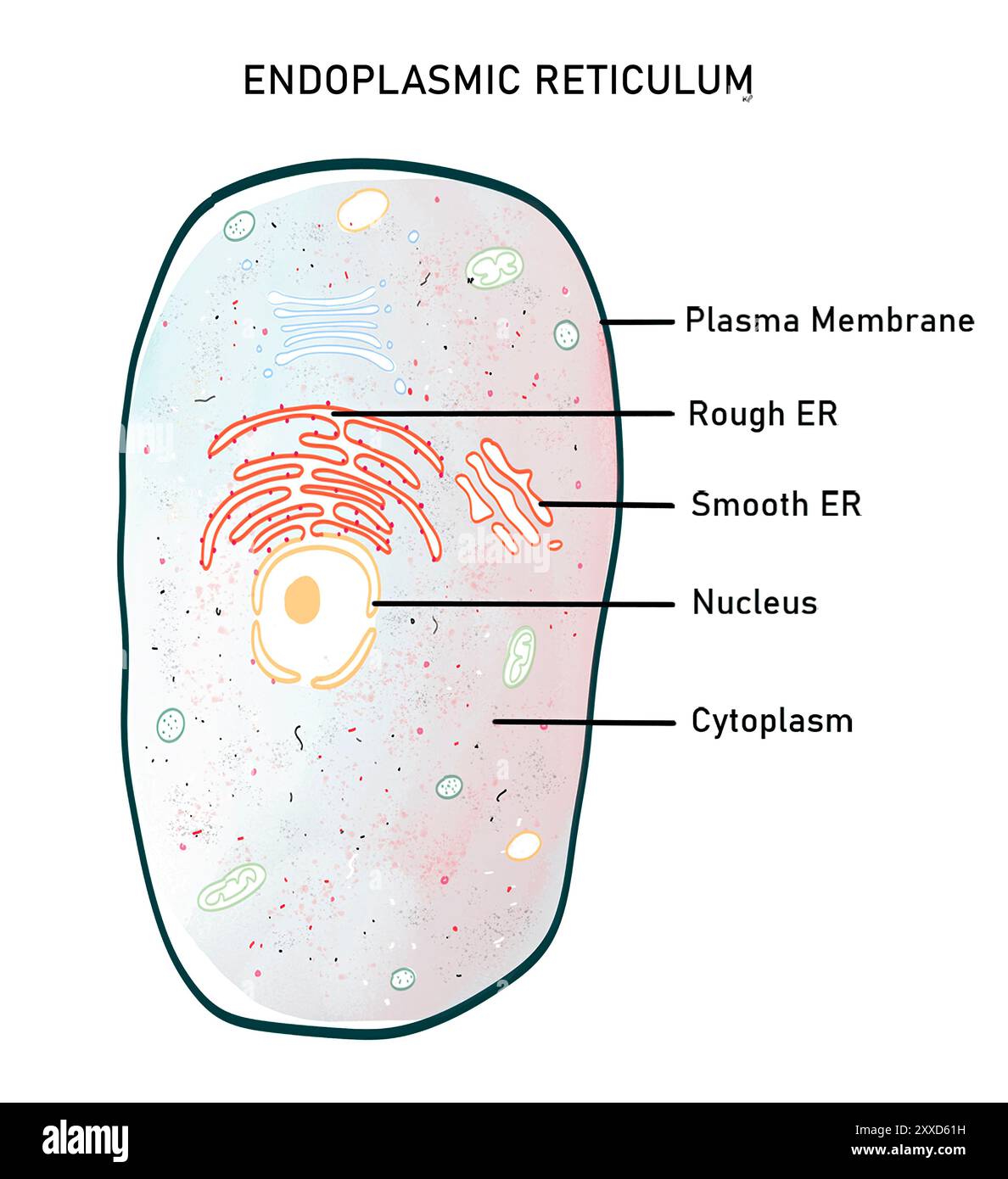 Endoplasmic reticulum, illustration. The endoplasmic reticulum (ER) is a dynamic cell organelle that can be of two types, wither-rough endoplasmic reticulum (RER), or smooth endoplasmic reticulum (SER). They both consist of a continuous system of flattened tubules and sacs bound by a single membrane. Endoplasmic reticulum serves many functions including protein synthesis and folding, lipid synthesis and calcium storage. Stock Photohttps://www.alamy.com/image-license-details/?v=1https://www.alamy.com/endoplasmic-reticulum-illustration-the-endoplasmic-reticulum-er-is-a-dynamic-cell-organelle-that-can-be-of-two-types-wither-rough-endoplasmic-reticulum-rer-or-smooth-endoplasmic-reticulum-ser-they-both-consist-of-a-continuous-system-of-flattened-tubules-and-sacs-bound-by-a-single-membrane-endoplasmic-reticulum-serves-many-functions-including-protein-synthesis-and-folding-lipid-synthesis-and-calcium-storage-image618634061.html
RF2XXD61H–Endoplasmic reticulum, illustration. The endoplasmic reticulum (ER) is a dynamic cell organelle that can be of two types, wither-rough endoplasmic reticulum (RER), or smooth endoplasmic reticulum (SER). They both consist of a continuous system of flattened tubules and sacs bound by a single membrane. Endoplasmic reticulum serves many functions including protein synthesis and folding, lipid synthesis and calcium storage.
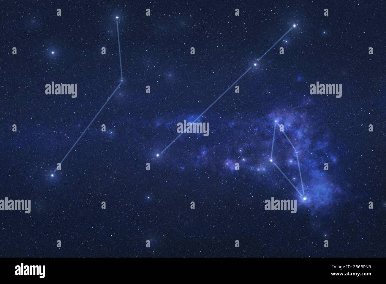 Multiple Constellations in outer space. Dorado, Hydra and Reticulum constellations stars with lines Elements of this image were furnished by NASA Stock Photohttps://www.alamy.com/image-license-details/?v=1https://www.alamy.com/multiple-constellations-in-outer-space-dorado-hydra-and-reticulum-constellations-stars-with-lines-elements-of-this-image-were-furnished-by-nasa-image348154613.html
RF2B6BPN9–Multiple Constellations in outer space. Dorado, Hydra and Reticulum constellations stars with lines Elements of this image were furnished by NASA
 Endoplasmic reticulum at top. Mitochondria is oval shaped organelle at center. Lysosomes are small bubbles to the left in purple. Golgi apparatus at bottom. CELL, DRAWING 001366 002 Stock Photohttps://www.alamy.com/image-license-details/?v=1https://www.alamy.com/endoplasmic-reticulum-at-top-mitochondria-is-oval-shaped-organelle-at-center-lysosomes-are-small-bubbles-to-the-left-in-purple-golgi-apparatus-at-bottom-cell-drawing-001366-002-image607620747.html
RM2X8FECY–Endoplasmic reticulum at top. Mitochondria is oval shaped organelle at center. Lysosomes are small bubbles to the left in purple. Golgi apparatus at bottom. CELL, DRAWING 001366 002
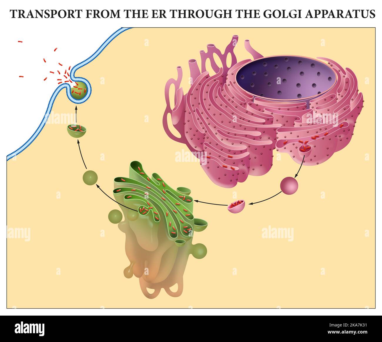 Transport from the ER through the Golgi Apparatus Stock Photohttps://www.alamy.com/image-license-details/?v=1https://www.alamy.com/transport-from-the-er-through-the-golgi-apparatus-image488205509.html
RF2KA7K31–Transport from the ER through the Golgi Apparatus
 Reticulum Star Constellation, On Black Background, Reticle Constellation, The Small Net Stock Photohttps://www.alamy.com/image-license-details/?v=1https://www.alamy.com/reticulum-star-constellation-on-black-background-reticle-constellation-the-small-net-image593764235.html
RF2WE089F–Reticulum Star Constellation, On Black Background, Reticle Constellation, The Small Net
 Detail of the reticulum of a Boletus mushroom, Boletaceae. Stock Photohttps://www.alamy.com/image-license-details/?v=1https://www.alamy.com/stock-photo-detail-of-the-reticulum-of-a-boletus-mushroom-boletaceae-133336159.html
RMHMWYHK–Detail of the reticulum of a Boletus mushroom, Boletaceae.
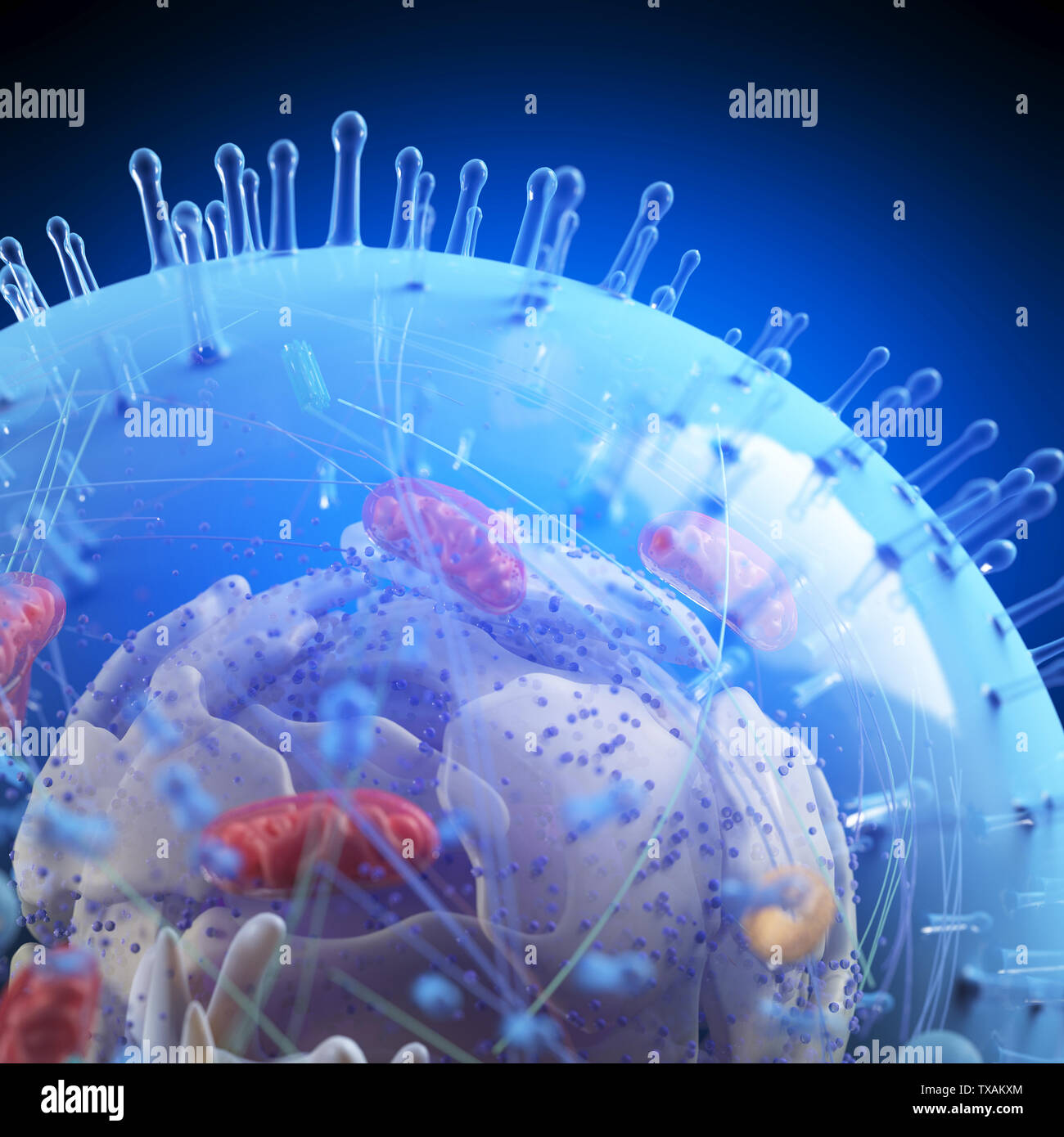 3d rendered medically accurate illustration of a human cell Stock Photohttps://www.alamy.com/image-license-details/?v=1https://www.alamy.com/3d-rendered-medically-accurate-illustration-of-a-human-cell-image257161372.html
RFTXAKXM–3d rendered medically accurate illustration of a human cell
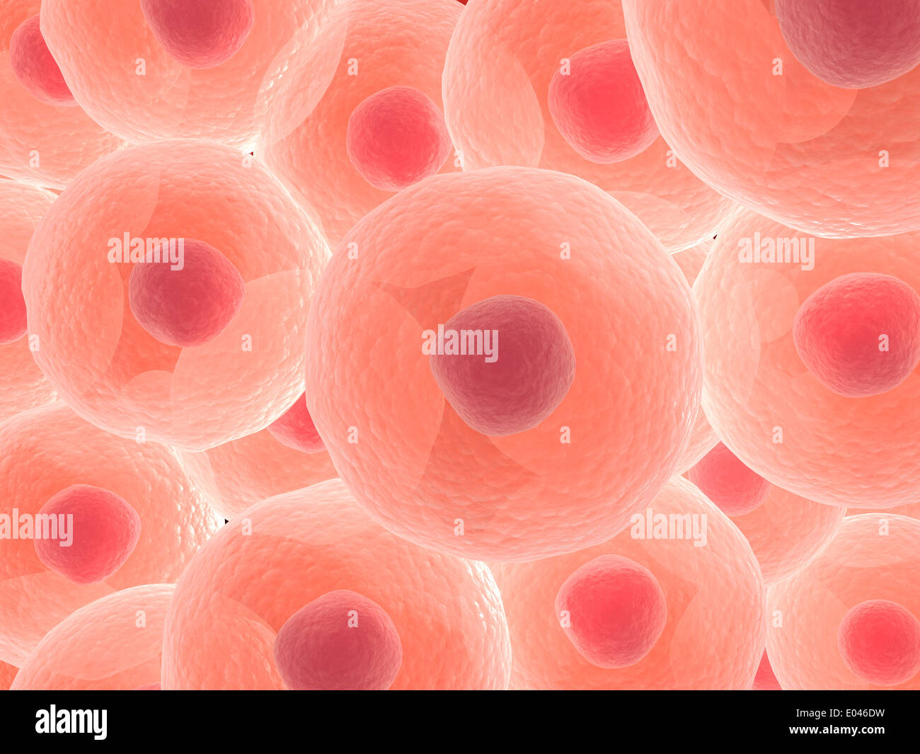 Microscopic view of animal cell. Stock Photohttps://www.alamy.com/image-license-details/?v=1https://www.alamy.com/microscopic-view-of-animal-cell-image68934373.html
RFE046DW–Microscopic view of animal cell.
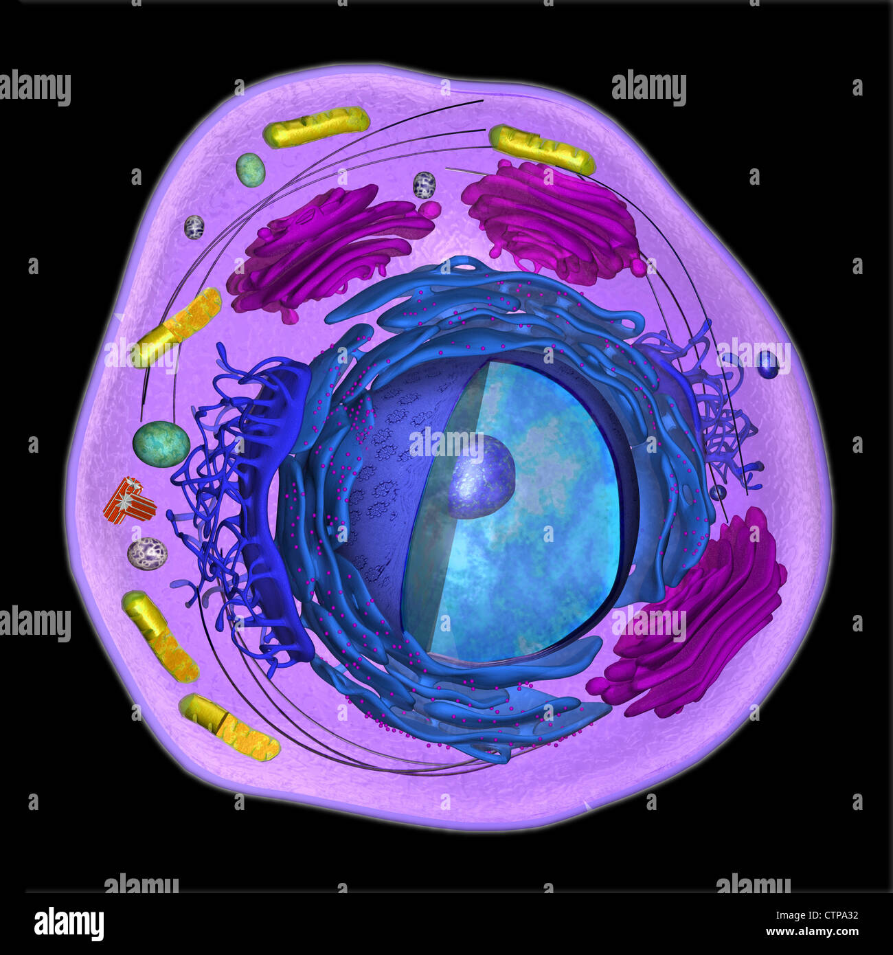 3D model of a eukaryotic cell Stock Photohttps://www.alamy.com/image-license-details/?v=1https://www.alamy.com/stock-photo-3d-model-of-a-eukaryotic-cell-49663350.html
RMCTPA32–3D model of a eukaryotic cell
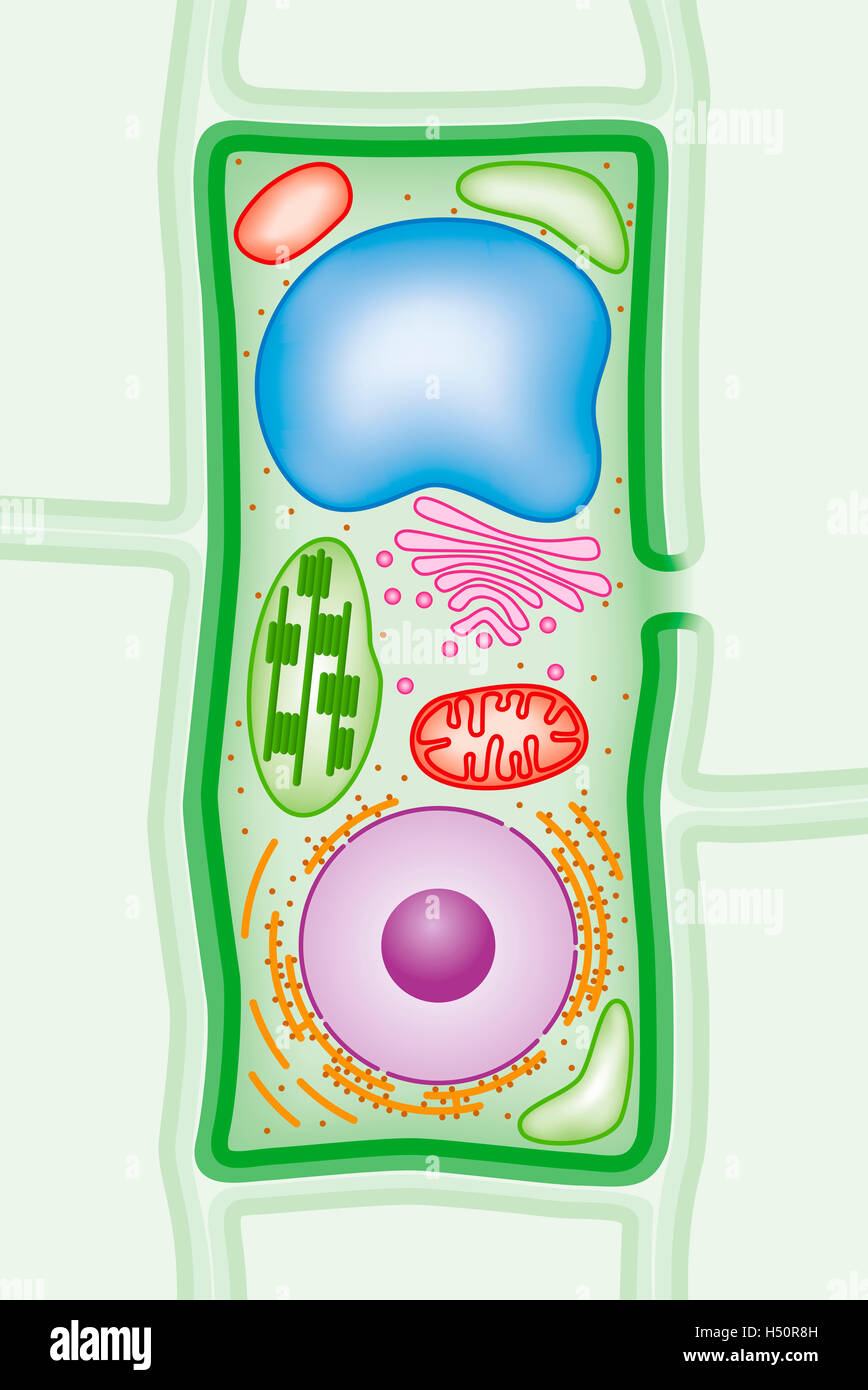 Plant cell structure cross-section. More infos in the Description! Stock Photohttps://www.alamy.com/image-license-details/?v=1https://www.alamy.com/stock-photo-plant-cell-structure-cross-section-more-infos-in-the-description!-123564129.html
RFH50R8H–Plant cell structure cross-section. More infos in the Description!
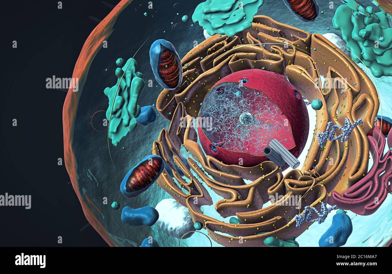 Components of Eukaryotic cell, nucleus and organelles and plasma membrane - 3d illustration Stock Photohttps://www.alamy.com/image-license-details/?v=1https://www.alamy.com/components-of-eukaryotic-cell-nucleus-and-organelles-and-plasma-membrane-3d-illustration-image362180063.html
RF2C16MA7–Components of Eukaryotic cell, nucleus and organelles and plasma membrane - 3d illustration
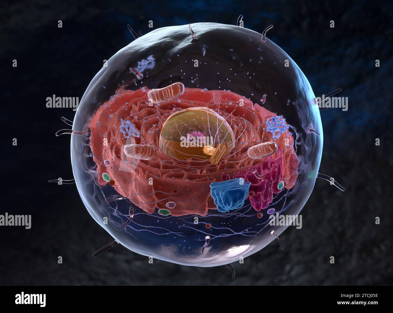 Organelles inside an Eukaryote or eukaryotic cell. 3d illustration Stock Photohttps://www.alamy.com/image-license-details/?v=1https://www.alamy.com/organelles-inside-an-eukaryote-or-eukaryotic-cell-3d-illustration-image575713306.html
RF2TCJ05E–Organelles inside an Eukaryote or eukaryotic cell. 3d illustration
 Reticulum bag. 1880. GAL1971.32.4 Stock Photohttps://www.alamy.com/image-license-details/?v=1https://www.alamy.com/reticulum-bag-1880-gal1971324-image596749492.html
RM2WJT81T–Reticulum bag. 1880. GAL1971.32.4
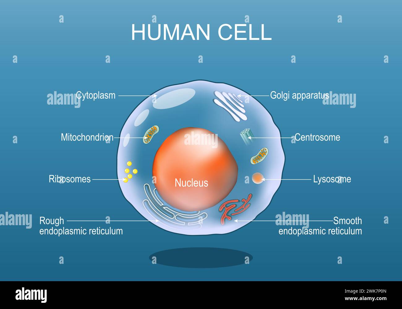 Human cell anatomy. Structure of a eukaryotic cell. All organelles: Nucleus, Ribosome, Rough endoplasmic reticulum, Golgi apparatus, mitochondrion, cy Stock Vectorhttps://www.alamy.com/image-license-details/?v=1https://www.alamy.com/human-cell-anatomy-structure-of-a-eukaryotic-cell-all-organelles-nucleus-ribosome-rough-endoplasmic-reticulum-golgi-apparatus-mitochondrion-cy-image597001909.html
RF2WK7P0N–Human cell anatomy. Structure of a eukaryotic cell. All organelles: Nucleus, Ribosome, Rough endoplasmic reticulum, Golgi apparatus, mitochondrion, cy
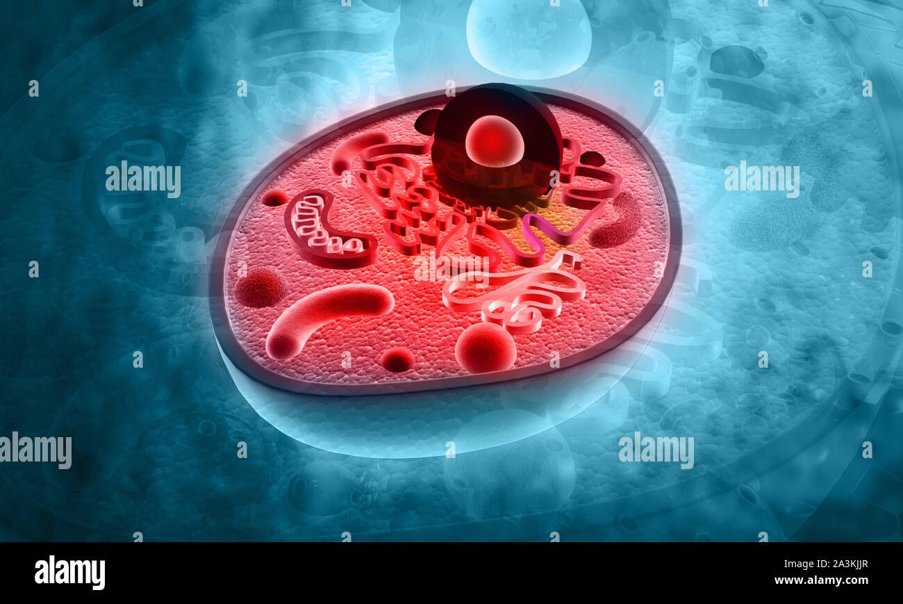 Cell Structure on blue background. 3d illustration Stock Photohttps://www.alamy.com/image-license-details/?v=1https://www.alamy.com/cell-structure-on-blue-background-3d-illustration-image329272687.html
RF2A3KJJR–Cell Structure on blue background. 3d illustration
 Eukaryotic cell structure cut-away - scientifically accurate 3d illustration Stock Photohttps://www.alamy.com/image-license-details/?v=1https://www.alamy.com/stock-photo-eukaryotic-cell-structure-cut-away-scientifically-accurate-3d-illustration-93641937.html
RFFC9N6W–Eukaryotic cell structure cut-away - scientifically accurate 3d illustration
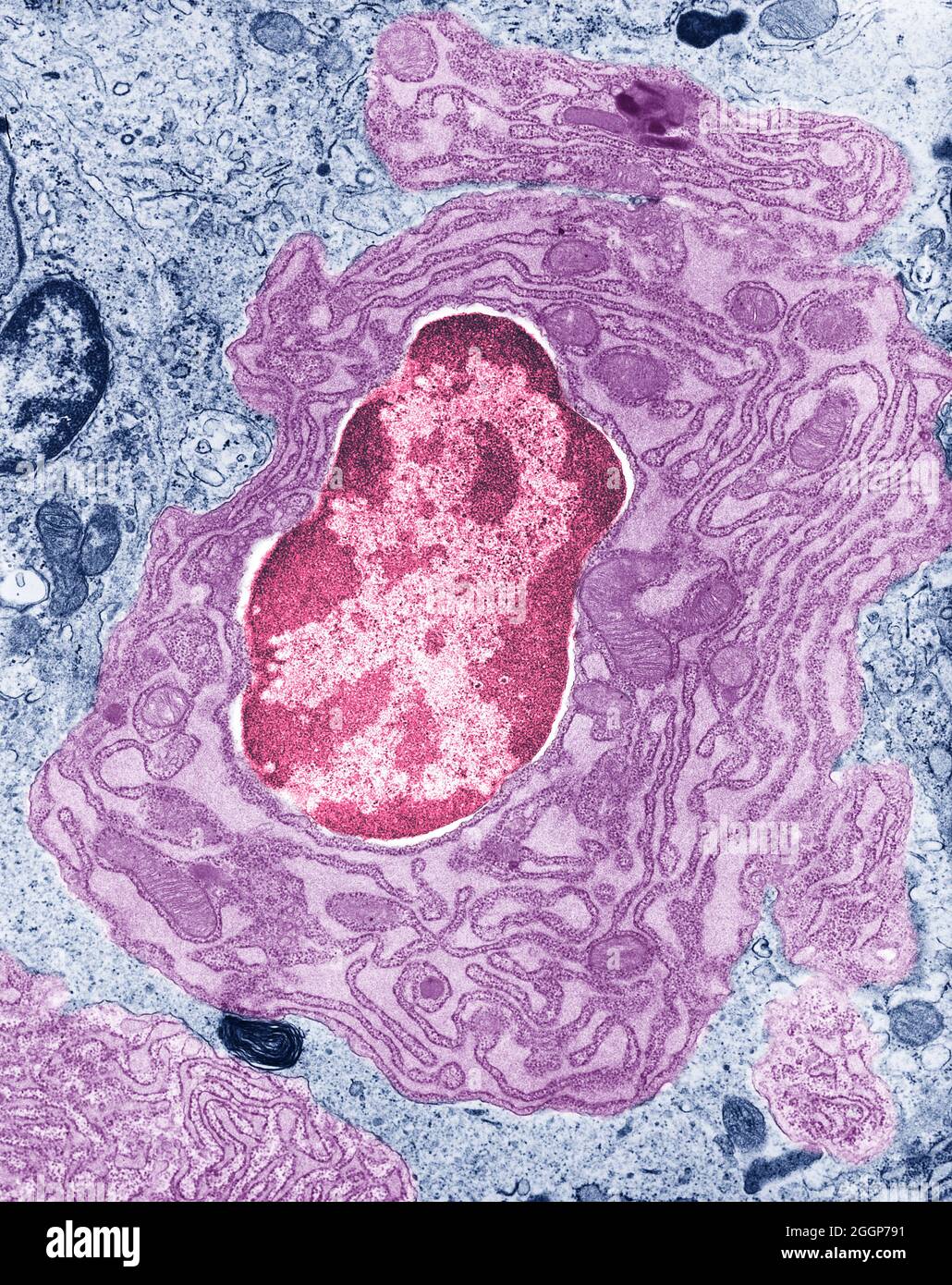 Colorized transmission electron micrograph (TEM) of intestinal cell, showing the nucleus and surrounding endoplasmic reticulum and mitochondria. Stock Photohttps://www.alamy.com/image-license-details/?v=1https://www.alamy.com/colorized-transmission-electron-micrograph-tem-of-intestinal-cell-showing-the-nucleus-and-surrounding-endoplasmic-reticulum-and-mitochondria-image440582381.html
RM2GGP791–Colorized transmission electron micrograph (TEM) of intestinal cell, showing the nucleus and surrounding endoplasmic reticulum and mitochondria.
 Representation of the constellation Reticulum (Ret), one of the modern constellations, part of 'Constellations' series Stock Photohttps://www.alamy.com/image-license-details/?v=1https://www.alamy.com/representation-of-the-constellation-reticulum-ret-one-of-the-modern-image68914686.html
RME039AP–Representation of the constellation Reticulum (Ret), one of the modern constellations, part of 'Constellations' series
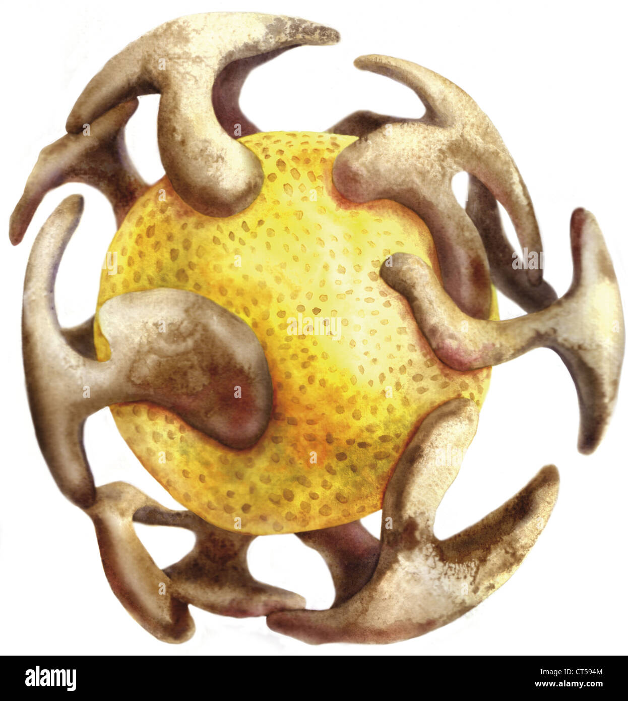 ENDOPLASMIC RETICULUM Stock Photohttps://www.alamy.com/image-license-details/?v=1https://www.alamy.com/stock-photo-endoplasmic-reticulum-49289428.html
RMCT594M–ENDOPLASMIC RETICULUM
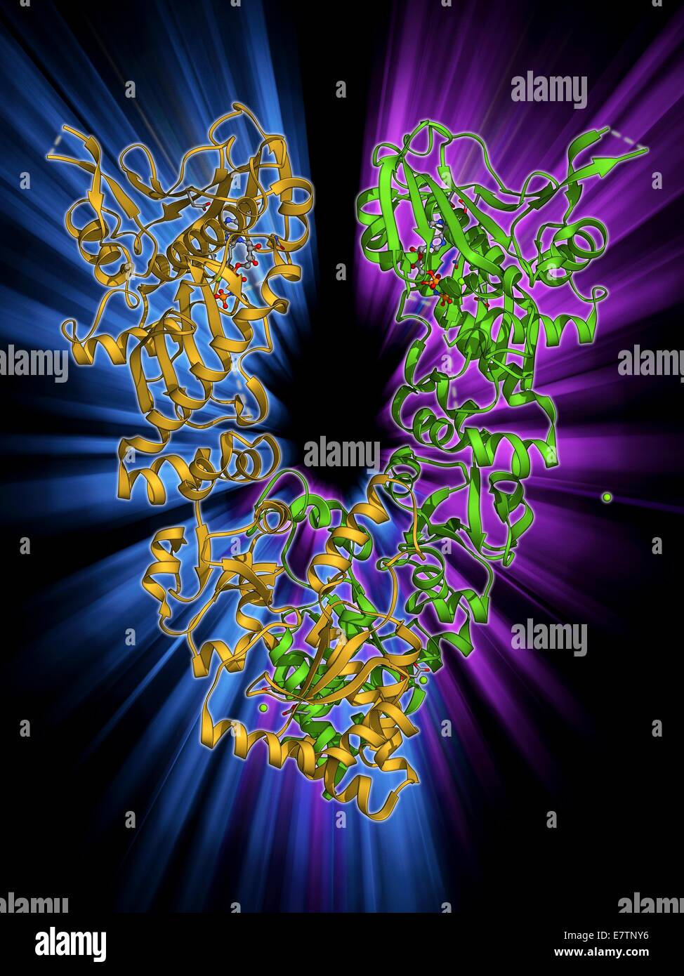 Endoplasmic reticulum chaperone protein. Molecular model of the endoplasmic reticulum chaperone protein GRP94. This protein is essential for the maturation of cell-surface display proteins and proteins that are to be exported. Stock Photohttps://www.alamy.com/image-license-details/?v=1https://www.alamy.com/stock-photo-endoplasmic-reticulum-chaperone-protein-molecular-model-of-the-endoplasmic-73688138.html
RFE7TNY6–Endoplasmic reticulum chaperone protein. Molecular model of the endoplasmic reticulum chaperone protein GRP94. This protein is essential for the maturation of cell-surface display proteins and proteins that are to be exported.
 Engraved portrait of Albrecht Durer, was a painter, printmaker and theorist of the German Renaissance, 1550. From the New York Public Library. Stock Photohttps://www.alamy.com/image-license-details/?v=1https://www.alamy.com/stock-image-engraved-portrait-of-albrecht-durer-was-a-painter-printmaker-and-theorist-166683254.html
RMKK525A–Engraved portrait of Albrecht Durer, was a painter, printmaker and theorist of the German Renaissance, 1550. From the New York Public Library.