Rhabditiform Stock Photos and Images
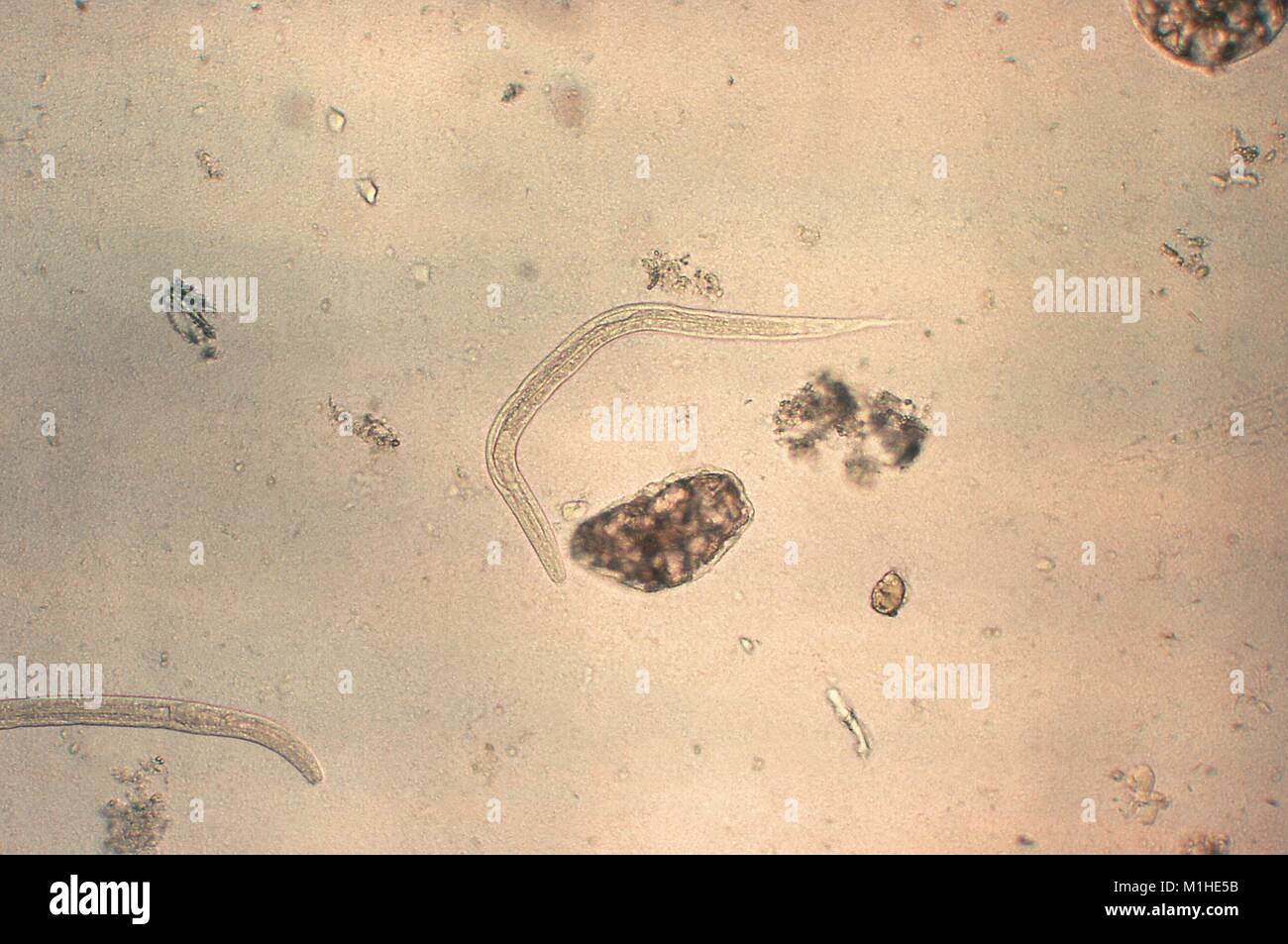 Strongyloides nematode, rhabditiform larval stage revealed in the micrograph film, 1982. Image courtesy Centers for Disease Control (CDC). () Stock Photohttps://www.alamy.com/image-license-details/?v=1https://www.alamy.com/stock-photo-strongyloides-nematode-rhabditiform-larval-stage-revealed-in-the-micrograph-173102647.html
RMM1HE5B–Strongyloides nematode, rhabditiform larval stage revealed in the micrograph film, 1982. Image courtesy Centers for Disease Control (CDC). ()
![. Digest of comments on The pharmacopia of the United States of America and on the National formulary ... 1905-1922. Fig. 16.—Eggs of Old World hookworms(Agchylostoma duodenale) as found in thestools. Greatly enlarged. (After Stiles,1902b, i». 193, fig. 128.1. Figs 17-29.—Embryology of the old World hookworm [Agchylostomaduodenal* i oi man; 17-23, seg-mentation of the egg, 21 -26, the embryo; 27, a rhabditiform embryo escaping from its eggshell; 28 29,empty eggshells. Greatly enlarged. (After Ierroneito. 1882, ]>. 342, tif,. 1 12.) of the adult hookworm. This embryonal esophagus is more or Stock Photo . Digest of comments on The pharmacopia of the United States of America and on the National formulary ... 1905-1922. Fig. 16.—Eggs of Old World hookworms(Agchylostoma duodenale) as found in thestools. Greatly enlarged. (After Stiles,1902b, i». 193, fig. 128.1. Figs 17-29.—Embryology of the old World hookworm [Agchylostomaduodenal* i oi man; 17-23, seg-mentation of the egg, 21 -26, the embryo; 27, a rhabditiform embryo escaping from its eggshell; 28 29,empty eggshells. Greatly enlarged. (After Ierroneito. 1882, ]>. 342, tif,. 1 12.) of the adult hookworm. This embryonal esophagus is more or Stock Photo](https://c8.alamy.com/comp/2CEKDC9/digest-of-comments-on-the-pharmacopia-of-the-united-states-of-america-and-on-the-national-formulary-1905-1922-fig-16eggs-of-old-world-hookwormsagchylostoma-duodenale-as-found-in-thestools-greatly-enlarged-after-stiles1902b-i-193-fig-1281-figs-17-29embryology-of-the-old-world-hookworm-agchylostomaduodenal-i-oi-man-17-23-seg-mentation-of-the-egg-21-26-the-embryo-27-a-rhabditiform-embryo-escaping-from-its-eggshell-28-29empty-eggshells-greatly-enlarged-after-ierroneito-1882-gt-342-tif-1-12-of-the-adult-hookworm-this-embryonal-esophagus-is-more-or-2CEKDC9.jpg) . Digest of comments on The pharmacopia of the United States of America and on the National formulary ... 1905-1922. Fig. 16.—Eggs of Old World hookworms(Agchylostoma duodenale) as found in thestools. Greatly enlarged. (After Stiles,1902b, i». 193, fig. 128.1. Figs 17-29.—Embryology of the old World hookworm [Agchylostomaduodenal* i oi man; 17-23, seg-mentation of the egg, 21 -26, the embryo; 27, a rhabditiform embryo escaping from its eggshell; 28 29,empty eggshells. Greatly enlarged. (After Ierroneito. 1882, ]>. 342, tif,. 1 12.) of the adult hookworm. This embryonal esophagus is more or Stock Photohttps://www.alamy.com/image-license-details/?v=1https://www.alamy.com/digest-of-comments-on-the-pharmacopia-of-the-united-states-of-america-and-on-the-national-formulary-1905-1922-fig-16eggs-of-old-world-hookwormsagchylostoma-duodenale-as-found-in-thestools-greatly-enlarged-after-stiles1902b-i-193-fig-1281-figs-17-29embryology-of-the-old-world-hookworm-agchylostomaduodenal-i-oi-man-17-23-seg-mentation-of-the-egg-21-26-the-embryo-27-a-rhabditiform-embryo-escaping-from-its-eggshell-28-29empty-eggshells-greatly-enlarged-after-ierroneito-1882-gt-342-tif-1-12-of-the-adult-hookworm-this-embryonal-esophagus-is-more-or-image370450537.html
. Digest of comments on The pharmacopia of the United States of America and on the National formulary ... 1905-1922. Fig. 16.—Eggs of Old World hookworms(Agchylostoma duodenale) as found in thestools. Greatly enlarged. (After Stiles,1902b, i». 193, fig. 128.1. Figs 17-29.—Embryology of the old World hookworm [Agchylostomaduodenal* i oi man; 17-23, seg-mentation of the egg, 21 -26, the embryo; 27, a rhabditiform embryo escaping from its eggshell; 28 29,empty eggshells. Greatly enlarged. (After Ierroneito. 1882, ]>. 342, tif,. 1 12.) of the adult hookworm. This embryonal esophagus is more or Stock Photohttps://www.alamy.com/image-license-details/?v=1https://www.alamy.com/digest-of-comments-on-the-pharmacopia-of-the-united-states-of-america-and-on-the-national-formulary-1905-1922-fig-16eggs-of-old-world-hookwormsagchylostoma-duodenale-as-found-in-thestools-greatly-enlarged-after-stiles1902b-i-193-fig-1281-figs-17-29embryology-of-the-old-world-hookworm-agchylostomaduodenal-i-oi-man-17-23-seg-mentation-of-the-egg-21-26-the-embryo-27-a-rhabditiform-embryo-escaping-from-its-eggshell-28-29empty-eggshells-greatly-enlarged-after-ierroneito-1882-gt-342-tif-1-12-of-the-adult-hookworm-this-embryonal-esophagus-is-more-or-image370450537.htmlRM2CEKDC9–. Digest of comments on The pharmacopia of the United States of America and on the National formulary ... 1905-1922. Fig. 16.—Eggs of Old World hookworms(Agchylostoma duodenale) as found in thestools. Greatly enlarged. (After Stiles,1902b, i». 193, fig. 128.1. Figs 17-29.—Embryology of the old World hookworm [Agchylostomaduodenal* i oi man; 17-23, seg-mentation of the egg, 21 -26, the embryo; 27, a rhabditiform embryo escaping from its eggshell; 28 29,empty eggshells. Greatly enlarged. (After Ierroneito. 1882, ]>. 342, tif,. 1 12.) of the adult hookworm. This embryonal esophagus is more or
 Photomicrograph of a parasitic Strongyloides species nematode (roundworm) larva, in its rhabditiform (nonifectious) stage, 1979. Image courtesy CDC. () Stock Photohttps://www.alamy.com/image-license-details/?v=1https://www.alamy.com/stock-photo-photomicrograph-of-a-parasitic-strongyloides-species-nematode-roundworm-173099857.html
RMM1HAHN–Photomicrograph of a parasitic Strongyloides species nematode (roundworm) larva, in its rhabditiform (nonifectious) stage, 1979. Image courtesy CDC. ()
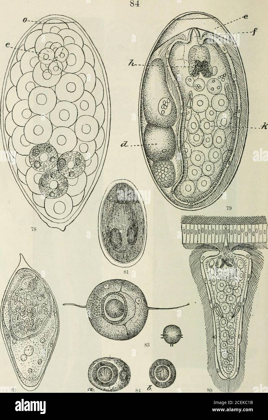 . Digest of comments on The pharmacopia of the United States of America and on the National formulary ... 1905-1922. Fig. 75.—Egg of Cochin-China diarrhea worm (Strongyloides stercoralis) found in stools. (After Thayer, 1901, pi. 9, tig. A.)Fig. 7G.—Rhabditiform embryo of same, from the stools. (After Thayer, 1901, pi. 9, fig. B.)Fig. 77.—Filariform larva of same derived, by direct transformation, from a rhabditiform embryo. (After Thayer, 1901, pi. 9, fig. C.) Figures 75 to 77 were drawn from life, as seen under Leitz, objective 7—ocular 3.. 82 «C.^*±£g2T m $■ SO Fig. 78.—Egg of the common l Stock Photohttps://www.alamy.com/image-license-details/?v=1https://www.alamy.com/digest-of-comments-on-the-pharmacopia-of-the-united-states-of-america-and-on-the-national-formulary-1905-1922-fig-75egg-of-cochin-china-diarrhea-worm-strongyloides-stercoralis-found-in-stools-after-thayer-1901-pi-9-tig-afig-7grhabditiform-embryo-of-same-from-the-stools-after-thayer-1901-pi-9-fig-bfig-77filariform-larva-of-same-derived-by-direct-transformation-from-a-rhabditiform-embryo-after-thayer-1901-pi-9-fig-c-figures-75-to-77-were-drawn-from-life-as-seen-under-leitz-objective-7ocular-3-82-cg2t-m-so-fig-78egg-of-the-common-l-image370449447.html
RM2CEKC1B–. Digest of comments on The pharmacopia of the United States of America and on the National formulary ... 1905-1922. Fig. 75.—Egg of Cochin-China diarrhea worm (Strongyloides stercoralis) found in stools. (After Thayer, 1901, pi. 9, tig. A.)Fig. 7G.—Rhabditiform embryo of same, from the stools. (After Thayer, 1901, pi. 9, fig. B.)Fig. 77.—Filariform larva of same derived, by direct transformation, from a rhabditiform embryo. (After Thayer, 1901, pi. 9, fig. C.) Figures 75 to 77 were drawn from life, as seen under Leitz, objective 7—ocular 3.. 82 «C.^*±£g2T m $■ SO Fig. 78.—Egg of the common l
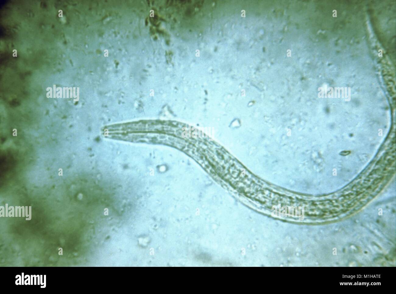 Photomicrograph of human hookworm (Ancylostoma duodenale and Necator americanus) rhabditiform larva which is its early noninfectious stage, 1979. Image courtesy CDC. () Stock Photohttps://www.alamy.com/image-license-details/?v=1https://www.alamy.com/stock-photo-photomicrograph-of-human-hookworm-ancylostoma-duodenale-and-necator-173100046.html
RMM1HATE–Photomicrograph of human hookworm (Ancylostoma duodenale and Necator americanus) rhabditiform larva which is its early noninfectious stage, 1979. Image courtesy CDC. ()
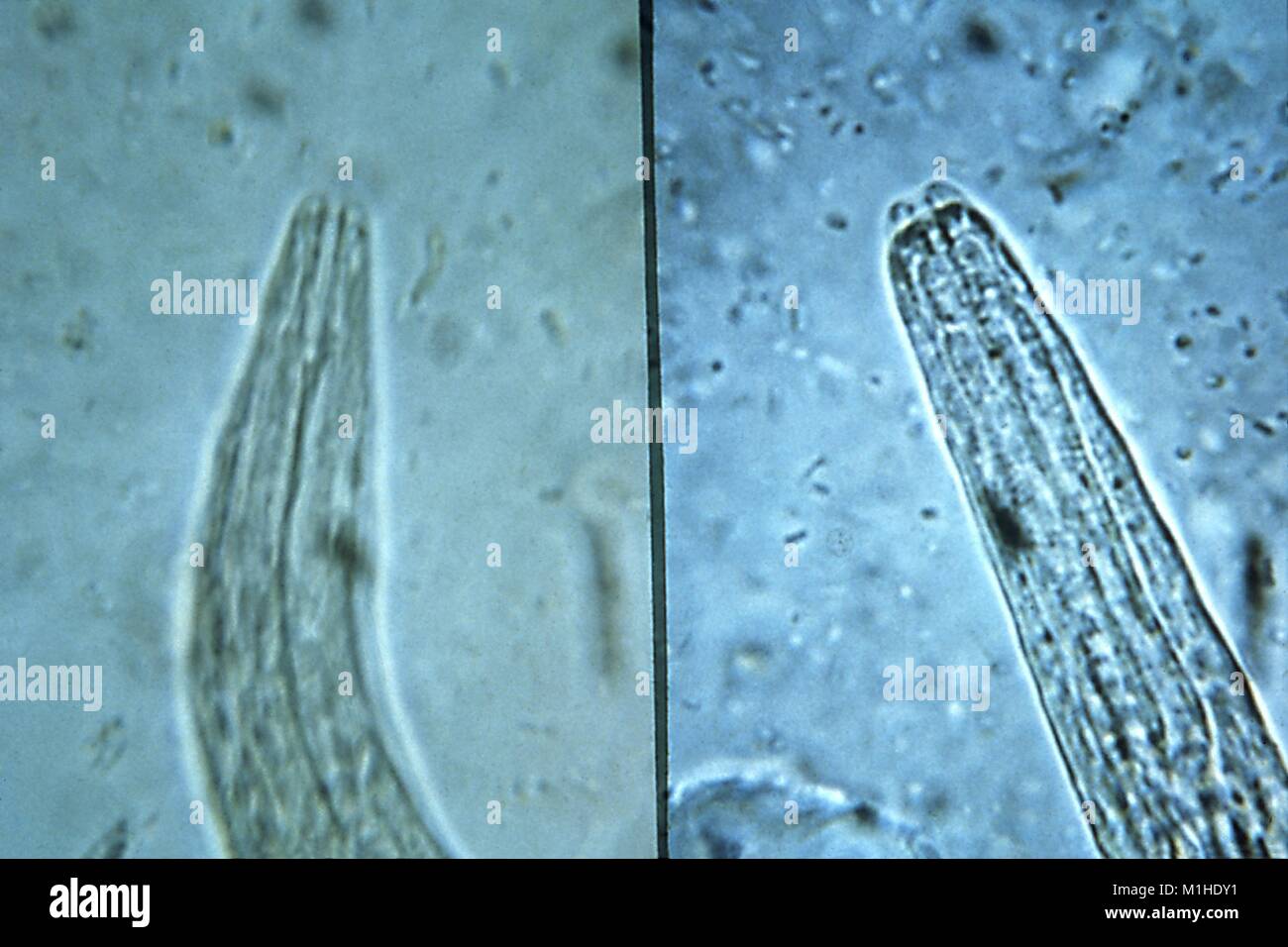 Hookworm and strongyloides, mouthparts of rhabditiform staged larvae revealed in the micrograph film, 1982. Image courtesy Centers for Disease Control (CDC) / Dr Mae Melvin. () Stock Photohttps://www.alamy.com/image-license-details/?v=1https://www.alamy.com/stock-photo-hookworm-and-strongyloides-mouthparts-of-rhabditiform-staged-larvae-173102469.html
RMM1HDY1–Hookworm and strongyloides, mouthparts of rhabditiform staged larvae revealed in the micrograph film, 1982. Image courtesy Centers for Disease Control (CDC) / Dr Mae Melvin. ()
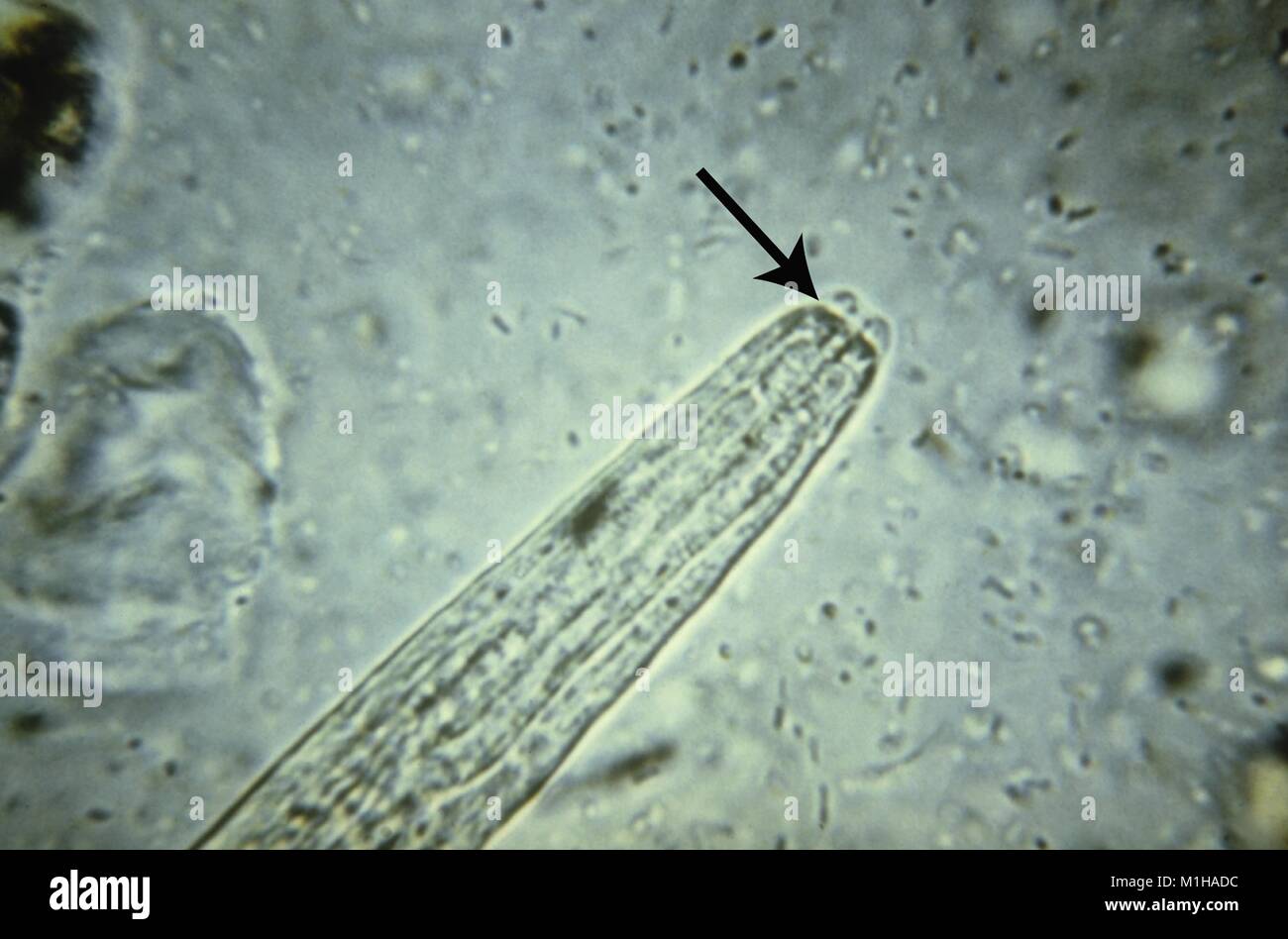 Photomicrograph with a detailed view of the short buccal cavity morphology in the rhabditiform (noninfectious) larval stage of a Strongyloides species nematode (roundworm), 1979. Image courtesy CDC. () Stock Photohttps://www.alamy.com/image-license-details/?v=1https://www.alamy.com/stock-photo-photomicrograph-with-a-detailed-view-of-the-short-buccal-cavity-morphology-173099736.html
RMM1HADC–Photomicrograph with a detailed view of the short buccal cavity morphology in the rhabditiform (noninfectious) larval stage of a Strongyloides species nematode (roundworm), 1979. Image courtesy CDC. ()