Root canal gutta percha Stock Photos and Images
 Doctor hands showing how root canal filling injector is working. Stock Photohttps://www.alamy.com/image-license-details/?v=1https://www.alamy.com/doctor-hands-showing-how-root-canal-filling-injector-is-working-image466385285.html
RF2J2NK59–Doctor hands showing how root canal filling injector is working.
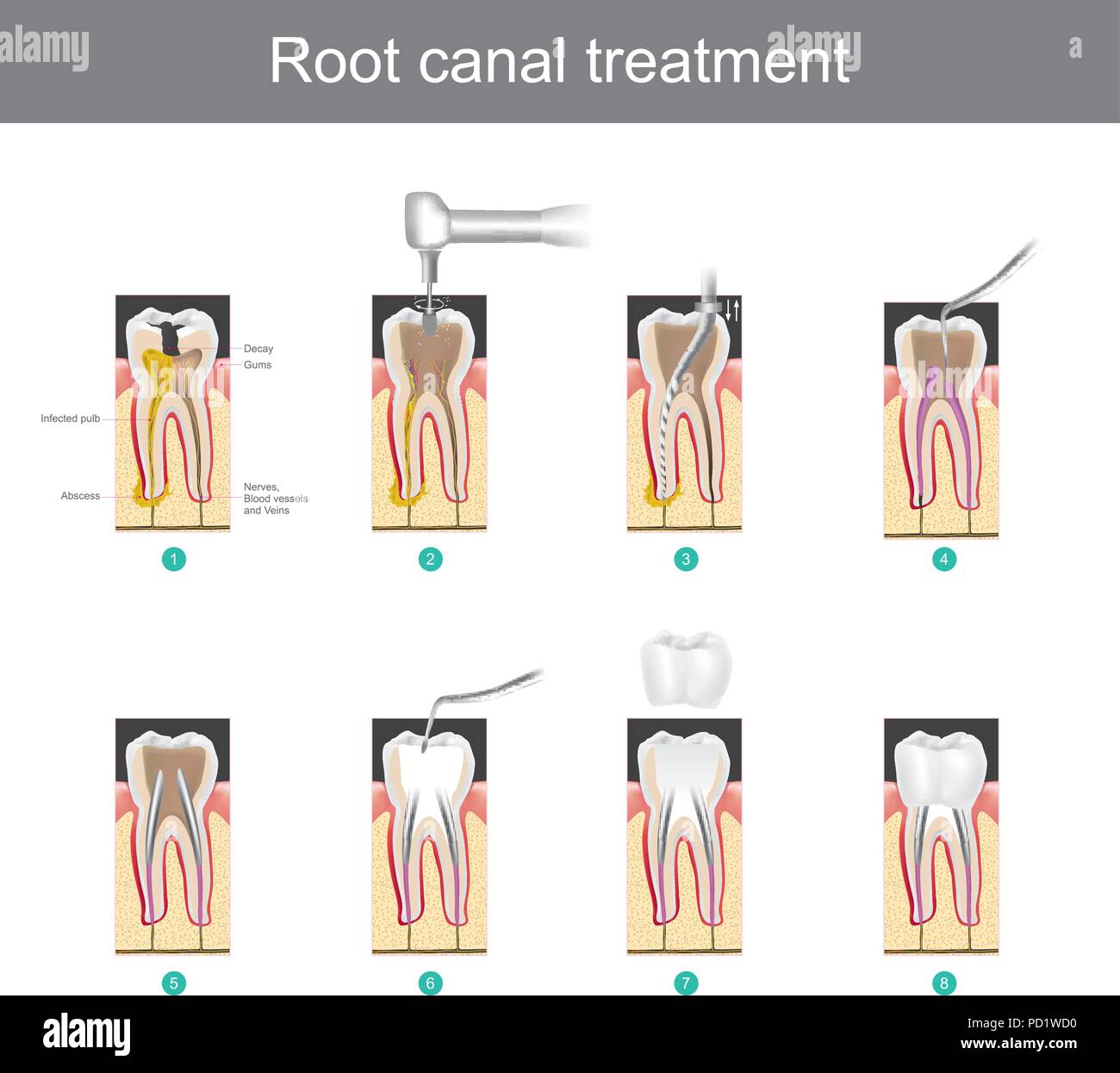 Root canal treatment. How to treat our teeth after the tooth is damaged. or severe caries as causes infection or inflammation Stock Vectorhttps://www.alamy.com/image-license-details/?v=1https://www.alamy.com/root-canal-treatment-how-to-treat-our-teeth-after-the-tooth-is-damaged-or-severe-caries-as-causes-infection-or-inflammation-image214556860.html
RFPD1WD0–Root canal treatment. How to treat our teeth after the tooth is damaged. or severe caries as causes infection or inflammation
 vertical condensation of gutta-percha endomotor system for hot sealing of canals The method of vertical condensation of gutta-percha in modern dentistry Ukraine Vinnitsa 2023 Stock Photohttps://www.alamy.com/image-license-details/?v=1https://www.alamy.com/vertical-condensation-of-gutta-percha-endomotor-system-for-hot-sealing-of-canals-the-method-of-vertical-condensation-of-gutta-percha-in-modern-dentistry-ukraine-vinnitsa-2023-image525533652.html
RF2NF03F0–vertical condensation of gutta-percha endomotor system for hot sealing of canals The method of vertical condensation of gutta-percha in modern dentistry Ukraine Vinnitsa 2023
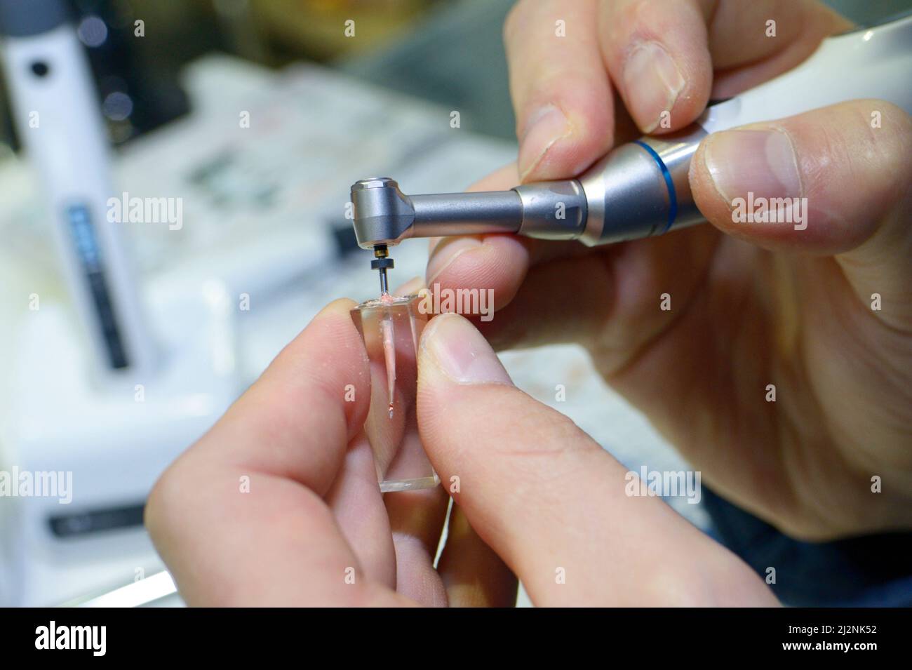 Doctor hands showing how root canal filling injector is working. Stock Photohttps://www.alamy.com/image-license-details/?v=1https://www.alamy.com/doctor-hands-showing-how-root-canal-filling-injector-is-working-image466385278.html
RF2J2NK52–Doctor hands showing how root canal filling injector is working.
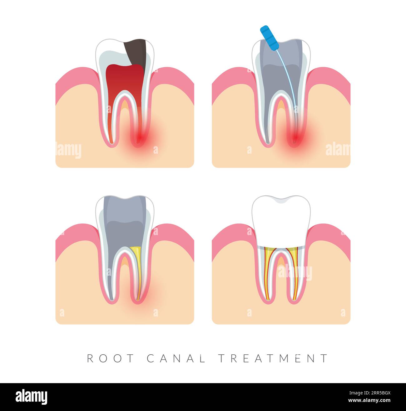 Dental Procedure - Root Canal Treatment - Stock Illustration as EPS 10 File Stock Vectorhttps://www.alamy.com/image-license-details/?v=1https://www.alamy.com/dental-procedure-root-canal-treatment-stock-illustration-as-eps-10-file-image564987722.html
RF2RR5BGX–Dental Procedure - Root Canal Treatment - Stock Illustration as EPS 10 File
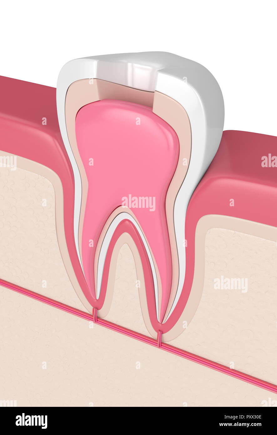 3d render of tooth in gums with root canal treatment procedure Stock Photohttps://www.alamy.com/image-license-details/?v=1https://www.alamy.com/3d-render-of-tooth-in-gums-with-root-canal-treatment-procedure-image223078590.html
RFPXX30E–3d render of tooth in gums with root canal treatment procedure
 . Dental electro-therapeutics. Caries under caps and old stoppings. but to show that where manual skill fails to accomplish theimpossible, nature often takes care of things herself. Thewriter certainly considers it more advisable to treat minuteremnants of nerve filaments in roots found by the radiograph,to be curved, as a negligible quantity, after having sterilizedthem as well as possible, than to run the risk of drillingthrough the side with a Gates-Glidden or Beutelrock root-canal drill. The fact that gutta-percha as a root-canal fillingis most desirable has also been proved, it would seem Stock Photohttps://www.alamy.com/image-license-details/?v=1https://www.alamy.com/dental-electro-therapeutics-caries-under-caps-and-old-stoppings-but-to-show-that-where-manual-skill-fails-to-accomplish-theimpossible-nature-often-takes-care-of-things-herself-thewriter-certainly-considers-it-more-advisable-to-treat-minuteremnants-of-nerve-filaments-in-roots-found-by-the-radiographto-be-curved-as-a-negligible-quantity-after-having-sterilizedthem-as-well-as-possible-than-to-run-the-risk-of-drillingthrough-the-side-with-a-gates-glidden-or-beutelrock-root-canal-drill-the-fact-that-gutta-percha-as-a-root-canal-fillingis-most-desirable-has-also-been-proved-it-would-seem-image336884224.html
RM2AG2B7C–. Dental electro-therapeutics. Caries under caps and old stoppings. but to show that where manual skill fails to accomplish theimpossible, nature often takes care of things herself. Thewriter certainly considers it more advisable to treat minuteremnants of nerve filaments in roots found by the radiograph,to be curved, as a negligible quantity, after having sterilizedthem as well as possible, than to run the risk of drillingthrough the side with a Gates-Glidden or Beutelrock root-canal drill. The fact that gutta-percha as a root-canal fillingis most desirable has also been proved, it would seem
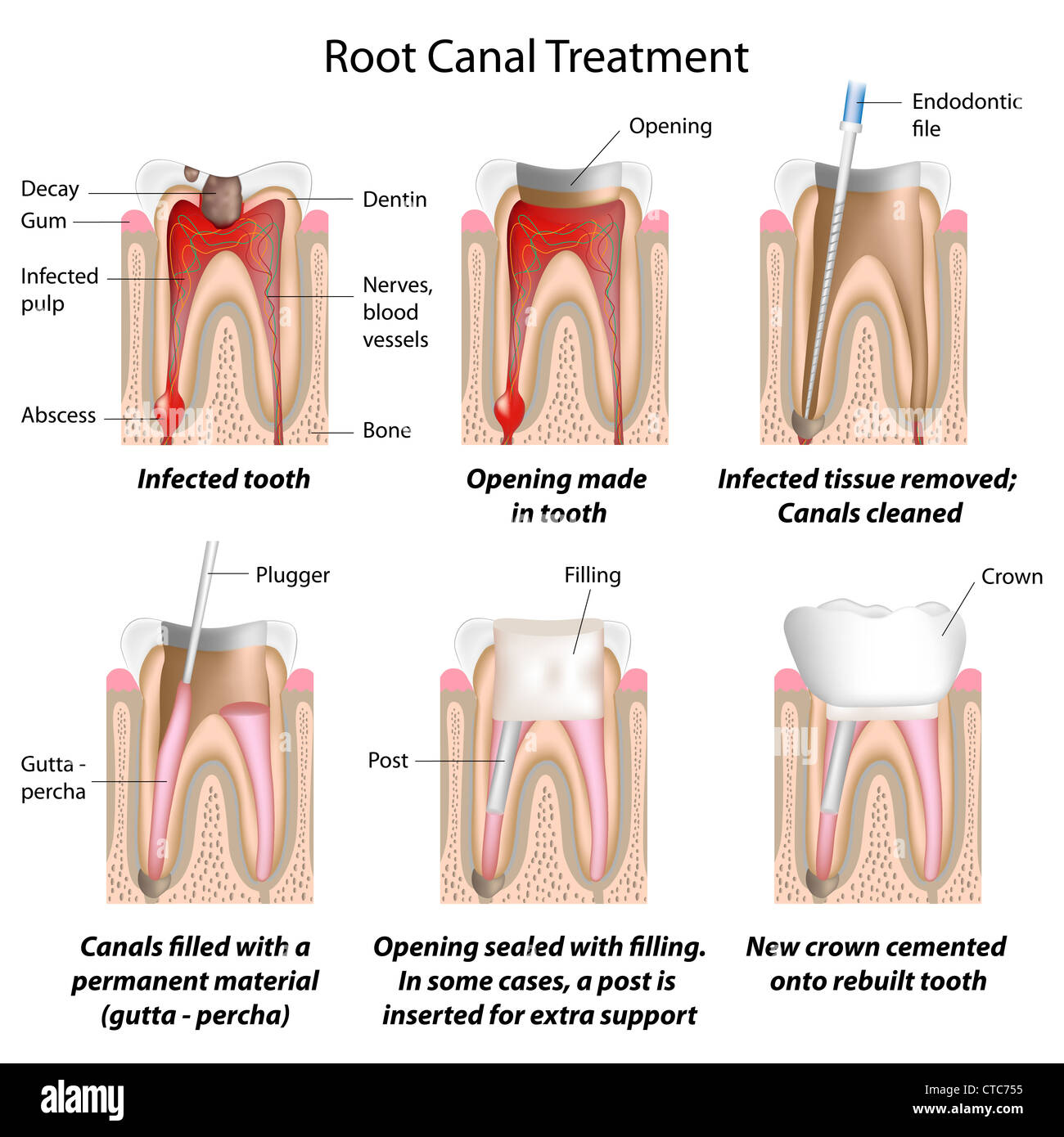 Root canal treatment Stock Photohttps://www.alamy.com/image-license-details/?v=1https://www.alamy.com/stock-photo-root-canal-treatment-49441537.html
RFCTC755–Root canal treatment
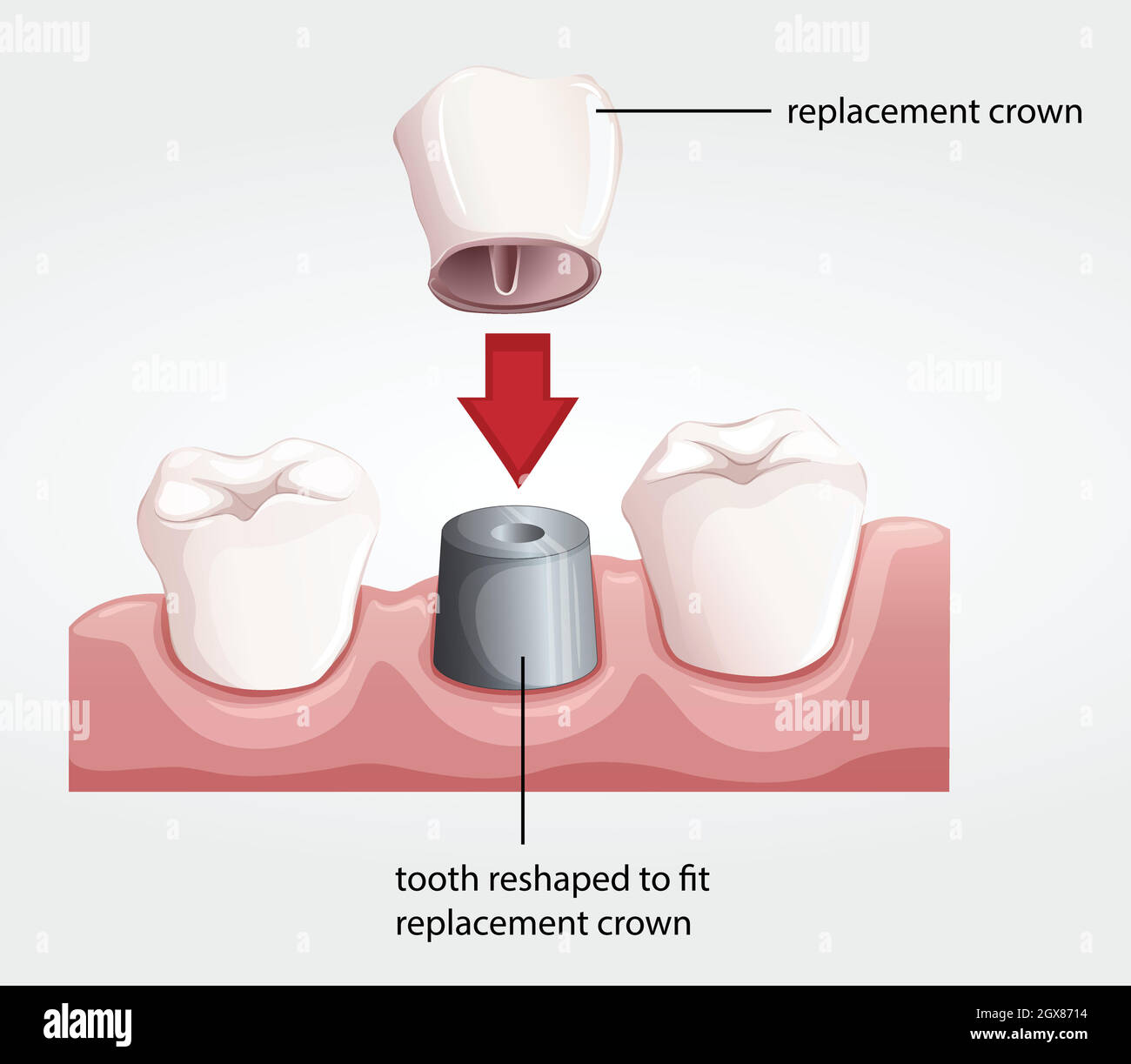 Dental crown procedure Stock Vectorhttps://www.alamy.com/image-license-details/?v=1https://www.alamy.com/dental-crown-procedure-image446421392.html
RF2GX8714–Dental crown procedure
 Gutta percha root canal treatment process. Medically accurate tooth 3D illustration Stock Photohttps://www.alamy.com/image-license-details/?v=1https://www.alamy.com/gutta-percha-root-canal-treatment-process-medically-accurate-tooth-3d-illustration-image488421971.html
RF2KAHF5R–Gutta percha root canal treatment process. Medically accurate tooth 3D illustration
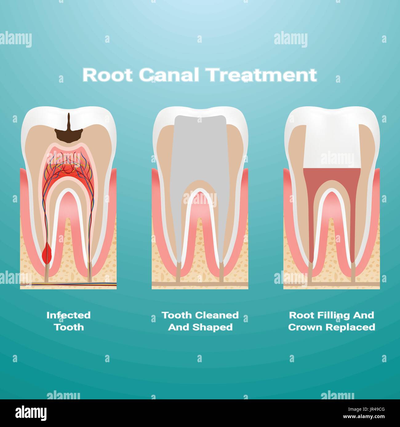 Pulpitis. Root Canal Therapy. Infected Pulp Is Removed From The Tooth And The Space Occupied By It Is Cleaned And Filled With A Gutta Percha Isolated On A Background. Vector Illustration. Stock Vectorhttps://www.alamy.com/image-license-details/?v=1https://www.alamy.com/pulpitis-root-canal-therapy-infected-pulp-is-removed-from-the-tooth-image151915248.html
RFJR49CG–Pulpitis. Root Canal Therapy. Infected Pulp Is Removed From The Tooth And The Space Occupied By It Is Cleaned And Filled With A Gutta Percha Isolated On A Background. Vector Illustration.
 vertical condensation of gutta-percha endomotor system for hot sealing of canals The method of vertical condensation of gutta-percha in modern dentistry Ukraine Vinnitsa 2023 Stock Photohttps://www.alamy.com/image-license-details/?v=1https://www.alamy.com/vertical-condensation-of-gutta-percha-endomotor-system-for-hot-sealing-of-canals-the-method-of-vertical-condensation-of-gutta-percha-in-modern-dentistry-ukraine-vinnitsa-2023-image525533634.html
RF2NF03EA–vertical condensation of gutta-percha endomotor system for hot sealing of canals The method of vertical condensation of gutta-percha in modern dentistry Ukraine Vinnitsa 2023
 Doctor hands working with root canal filling injector. Stock Photohttps://www.alamy.com/image-license-details/?v=1https://www.alamy.com/doctor-hands-working-with-root-canal-filling-injector-image502175284.html
RF2M501KG–Doctor hands working with root canal filling injector.
 3d render of tooth in gums with root canal treatment procedure Stock Photohttps://www.alamy.com/image-license-details/?v=1https://www.alamy.com/3d-render-of-tooth-in-gums-with-root-canal-treatment-procedure-image223078498.html
RFPXX2W6–3d render of tooth in gums with root canal treatment procedure
 . Dental electro-therapeutics. wever, on the age of the patientand the severity of the case. VII. The quest of the dentist who has never had a broachor drill break off in a root-canal would have affordedDiogenes nearly as much exercise as of that variety of manto whose discovery he devoted so much energy and candle-grease; and as there are unfortunately very many casesin which such a broach-end is perfectly innocuous, if sur-rounded by chlora and gutta-percha, it is only necessary,in order to quiet or disturb the operators mind, to determineby an a--ray whether the broken-off piece is entirely Stock Photohttps://www.alamy.com/image-license-details/?v=1https://www.alamy.com/dental-electro-therapeutics-wever-on-the-age-of-the-patientand-the-severity-of-the-case-vii-the-quest-of-the-dentist-who-has-never-had-a-broachor-drill-break-off-in-a-root-canal-would-have-affordeddiogenes-nearly-as-much-exercise-as-of-that-variety-of-manto-whose-discovery-he-devoted-so-much-energy-and-candle-grease-and-as-there-are-unfortunately-very-many-casesin-which-such-a-broach-end-is-perfectly-innocuous-if-sur-rounded-by-chlora-and-gutta-percha-it-is-only-necessaryin-order-to-quiet-or-disturb-the-operators-mind-to-determineby-an-a-ray-whether-the-broken-off-piece-is-entirely-image336884897.html
RM2AG2C3D–. Dental electro-therapeutics. wever, on the age of the patientand the severity of the case. VII. The quest of the dentist who has never had a broachor drill break off in a root-canal would have affordedDiogenes nearly as much exercise as of that variety of manto whose discovery he devoted so much energy and candle-grease; and as there are unfortunately very many casesin which such a broach-end is perfectly innocuous, if sur-rounded by chlora and gutta-percha, it is only necessary,in order to quiet or disturb the operators mind, to determineby an a--ray whether the broken-off piece is entirely
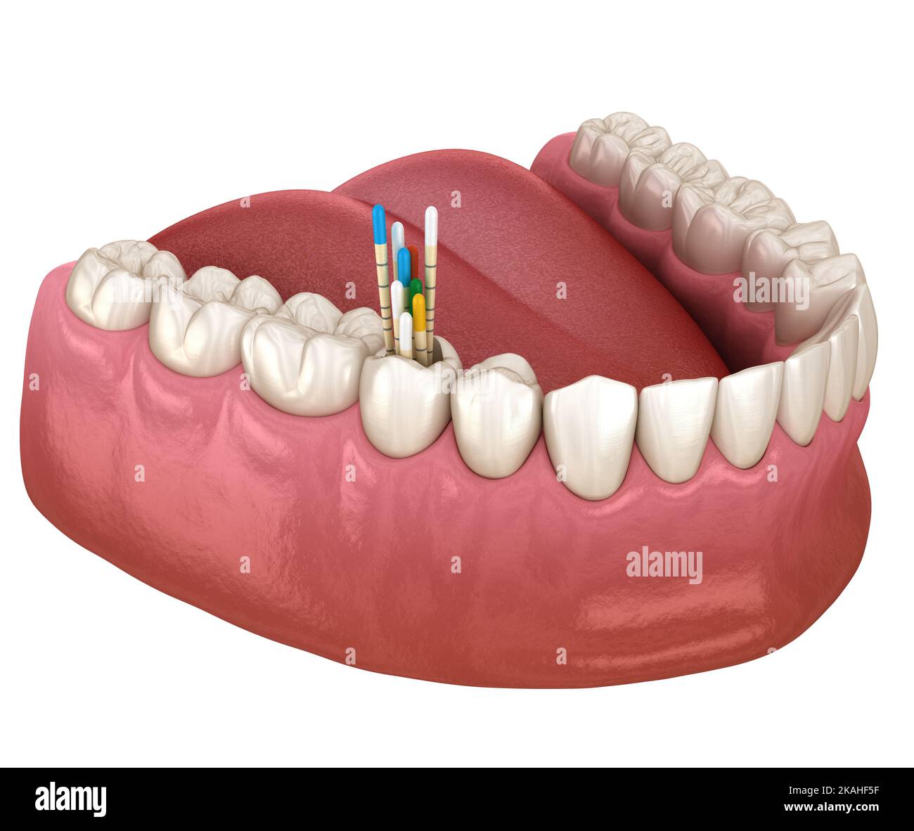 Gutta percha root canal treatment process. Medically accurate tooth 3D illustration Stock Photohttps://www.alamy.com/image-license-details/?v=1https://www.alamy.com/gutta-percha-root-canal-treatment-process-medically-accurate-tooth-3d-illustration-image488421963.html
RF2KAHF5F–Gutta percha root canal treatment process. Medically accurate tooth 3D illustration
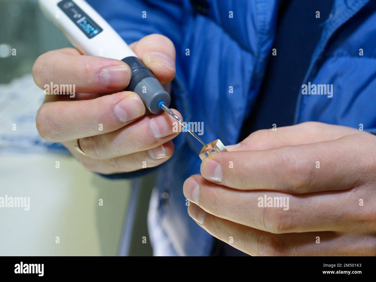 Doctor hands working with root canal filling injector. Stock Photohttps://www.alamy.com/image-license-details/?v=1https://www.alamy.com/doctor-hands-working-with-root-canal-filling-injector-image502175271.html
RF2M501K3–Doctor hands working with root canal filling injector.
 3d render of tooth in gums with root canal treatment procedure Stock Photohttps://www.alamy.com/image-license-details/?v=1https://www.alamy.com/3d-render-of-tooth-in-gums-with-root-canal-treatment-procedure-image223078582.html
RFPXX306–3d render of tooth in gums with root canal treatment procedure
 . Dental electro-therapeutics. nsiders it more advisable to treatminute remnants of nerve filaments in roots found bv the 200 THE X-RAYS OR RONTGEN RAYS radiograph to be curved, as a more or less negligible quantity,after having sterilized them as well as possible, than to runthe risk of drilling through the side with a Gates-Glidden orBeutelroek root-canal drill unless other indications pointto apiectomy or extraction. The fact that gutta-percha as aroot-canal filling is most desirable has also been proved, itwould seem conclusively, by the .r-ray, for the writer has inseveral cases seen how Stock Photohttps://www.alamy.com/image-license-details/?v=1https://www.alamy.com/dental-electro-therapeutics-nsiders-it-more-advisable-to-treatminute-remnants-of-nerve-filaments-in-roots-found-bv-the-200-the-x-rays-or-rontgen-rays-radiograph-to-be-curved-as-a-more-or-less-negligible-quantityafter-having-sterilized-them-as-well-as-possible-than-to-runthe-risk-of-drilling-through-the-side-with-a-gates-glidden-orbeutelroek-root-canal-drill-unless-other-indications-pointto-apiectomy-or-extraction-the-fact-that-gutta-percha-as-aroot-canal-filling-is-most-desirable-has-also-been-proved-itwould-seem-conclusively-by-the-r-ray-for-the-writer-has-inseveral-cases-seen-how-image336685040.html
RM2AFN95M–. Dental electro-therapeutics. nsiders it more advisable to treatminute remnants of nerve filaments in roots found bv the 200 THE X-RAYS OR RONTGEN RAYS radiograph to be curved, as a more or less negligible quantity,after having sterilized them as well as possible, than to runthe risk of drilling through the side with a Gates-Glidden orBeutelroek root-canal drill unless other indications pointto apiectomy or extraction. The fact that gutta-percha as aroot-canal filling is most desirable has also been proved, itwould seem conclusively, by the .r-ray, for the writer has inseveral cases seen how
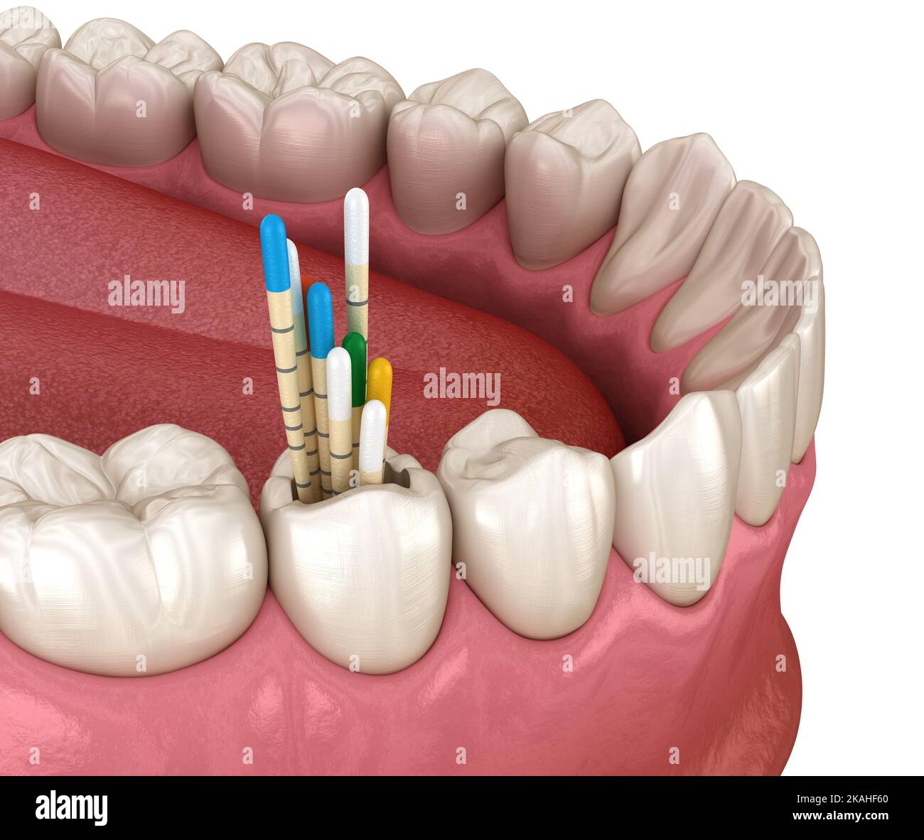 Gutta percha root canal treatment process. Medically accurate tooth 3D illustration Stock Photohttps://www.alamy.com/image-license-details/?v=1https://www.alamy.com/gutta-percha-root-canal-treatment-process-medically-accurate-tooth-3d-illustration-image488421976.html
RF2KAHF60–Gutta percha root canal treatment process. Medically accurate tooth 3D illustration
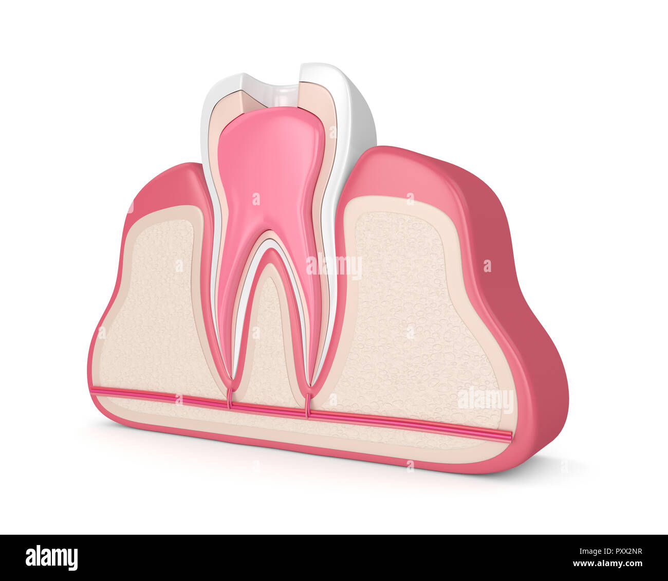 3d render of tooth in gums with root canal treatment procedure Stock Photohttps://www.alamy.com/image-license-details/?v=1https://www.alamy.com/3d-render-of-tooth-in-gums-with-root-canal-treatment-procedure-image223078403.html
RFPXX2NR–3d render of tooth in gums with root canal treatment procedure
 The Dental cosmos . across thebevel of the flowed solder. (This band can be dispensed with alto-gether.) Trim the overhanging edges down to the edge of the solder,leaving that to finish up with (Fig. i). Place the anchorage post in po-sition in the root-canal, and see that when secured the top will be a littlerearward of the center (Fig. 2). For these posts you can use the posts,which I provide for the purpose (Fig. 3), or the How metal screw-postsand the nuts provided with them. Remove the post and fill the root-canal with gutta-percha, heat the post and push it to its place, smoothoff the su Stock Photohttps://www.alamy.com/image-license-details/?v=1https://www.alamy.com/the-dental-cosmos-across-thebevel-of-the-flowed-solder-this-band-can-be-dispensed-with-alto-gether-trim-the-overhanging-edges-down-to-the-edge-of-the-solderleaving-that-to-finish-up-with-fig-i-place-the-anchorage-post-in-po-sition-in-the-root-canal-and-see-that-when-secured-the-top-will-be-a-littlerearward-of-the-center-fig-2-for-these-posts-you-can-use-the-postswhich-i-provide-for-the-purpose-fig-3-or-the-how-metal-screw-postsand-the-nuts-provided-with-them-remove-the-post-and-fill-the-root-canal-with-gutta-percha-heat-the-post-and-push-it-to-its-place-smoothoff-the-su-image340079694.html
RM2AN7Y3A–The Dental cosmos . across thebevel of the flowed solder. (This band can be dispensed with alto-gether.) Trim the overhanging edges down to the edge of the solder,leaving that to finish up with (Fig. i). Place the anchorage post in po-sition in the root-canal, and see that when secured the top will be a littlerearward of the center (Fig. 2). For these posts you can use the posts,which I provide for the purpose (Fig. 3), or the How metal screw-postsand the nuts provided with them. Remove the post and fill the root-canal with gutta-percha, heat the post and push it to its place, smoothoff the su
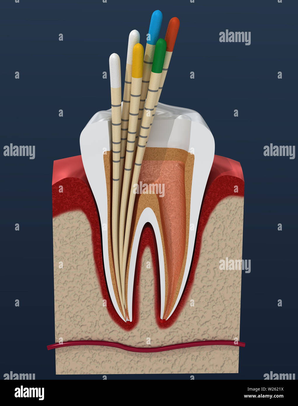 Gutta percha endodontics instrument, dental anatomy. 3D illustration Stock Photohttps://www.alamy.com/image-license-details/?v=1https://www.alamy.com/gutta-percha-endodontics-instrument-dental-anatomy-3d-illustration-image259518166.html
RFW2621X–Gutta percha endodontics instrument, dental anatomy. 3D illustration
 3d render of tooth in gums with root canal treatment procedure Stock Photohttps://www.alamy.com/image-license-details/?v=1https://www.alamy.com/3d-render-of-tooth-in-gums-with-root-canal-treatment-procedure-image223078486.html
RFPXX2TP–3d render of tooth in gums with root canal treatment procedure
 . Dental electro-therapeutics. Figs. 120 to 122.—Caries under caps and old stoppings. but to show that where manual skill fails to accomplish theimpossible, nature often takes care of things herself. Thewriter certainly considers it more advisable to treat minuteremnants of nerve filaments in roots found by the radiograph,to be curved, as a negligible quantity, after having sterilizedthem as well as possible, than to run the risk of drillingthrough the side with a Gates-Glidden or Beutelrock root-canal drill. The fact that gutta-percha as a root-canal fillingis most desirable has also been pro Stock Photohttps://www.alamy.com/image-license-details/?v=1https://www.alamy.com/dental-electro-therapeutics-figs-120-to-122caries-under-caps-and-old-stoppings-but-to-show-that-where-manual-skill-fails-to-accomplish-theimpossible-nature-often-takes-care-of-things-herself-thewriter-certainly-considers-it-more-advisable-to-treat-minuteremnants-of-nerve-filaments-in-roots-found-by-the-radiographto-be-curved-as-a-negligible-quantity-after-having-sterilizedthem-as-well-as-possible-than-to-run-the-risk-of-drillingthrough-the-side-with-a-gates-glidden-or-beutelrock-root-canal-drill-the-fact-that-gutta-percha-as-a-root-canal-fillingis-most-desirable-has-also-been-pro-image336884386.html
RM2AG2BD6–. Dental electro-therapeutics. Figs. 120 to 122.—Caries under caps and old stoppings. but to show that where manual skill fails to accomplish theimpossible, nature often takes care of things herself. Thewriter certainly considers it more advisable to treat minuteremnants of nerve filaments in roots found by the radiograph,to be curved, as a negligible quantity, after having sterilizedthem as well as possible, than to run the risk of drillingthrough the side with a Gates-Glidden or Beutelrock root-canal drill. The fact that gutta-percha as a root-canal fillingis most desirable has also been pro
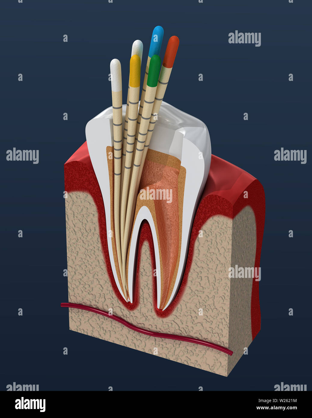 Gutta percha endodontics instrument, dental anatomy. 3D illustration Stock Photohttps://www.alamy.com/image-license-details/?v=1https://www.alamy.com/gutta-percha-endodontics-instrument-dental-anatomy-3d-illustration-image259518160.html
RFW2621M–Gutta percha endodontics instrument, dental anatomy. 3D illustration
 3d render of tooth with root canal treatment procedure over white background Stock Photohttps://www.alamy.com/image-license-details/?v=1https://www.alamy.com/3d-render-of-tooth-with-root-canal-treatment-procedure-over-white-background-image223078372.html
RFPXX2MM–3d render of tooth with root canal treatment procedure over white background
 . Dental electro-therapeutics. Gutta-percha extending throughapex. Absorption of root and gutta-percha protruding. Fig. 124 shows extensive absorption of the end of leftupper canine root. The protruding part of the gutta-percha root-canal stopping has evidently assumed a nearlyhorizontal position. XII. In affections of the antrum the .r-rays often forman important link in the chain of diagnostic factors,and a radiograph should certainly be taken if there remainsany doubt after illuminating the oral cavity and resortingto the usual means of diagnosis. The radiograph should betaken with a large Stock Photohttps://www.alamy.com/image-license-details/?v=1https://www.alamy.com/dental-electro-therapeutics-gutta-percha-extending-throughapex-absorption-of-root-and-gutta-percha-protruding-fig-124-shows-extensive-absorption-of-the-end-of-leftupper-canine-root-the-protruding-part-of-the-gutta-percha-root-canal-stopping-has-evidently-assumed-a-nearlyhorizontal-position-xii-in-affections-of-the-antrum-the-r-rays-often-forman-important-link-in-the-chain-of-diagnostic-factorsand-a-radiograph-should-certainly-be-taken-if-there-remainsany-doubt-after-illuminating-the-oral-cavity-and-resortingto-the-usual-means-of-diagnosis-the-radiograph-should-betaken-with-a-large-image336884053.html
RM2AG2B19–. Dental electro-therapeutics. Gutta-percha extending throughapex. Absorption of root and gutta-percha protruding. Fig. 124 shows extensive absorption of the end of leftupper canine root. The protruding part of the gutta-percha root-canal stopping has evidently assumed a nearlyhorizontal position. XII. In affections of the antrum the .r-rays often forman important link in the chain of diagnostic factors,and a radiograph should certainly be taken if there remainsany doubt after illuminating the oral cavity and resortingto the usual means of diagnosis. The radiograph should betaken with a large
 Gutta percha endodontics instrument, dental anatomy. 3D illustration Stock Photohttps://www.alamy.com/image-license-details/?v=1https://www.alamy.com/gutta-percha-endodontics-instrument-dental-anatomy-3d-illustration-image260639856.html
RFW414P8–Gutta percha endodontics instrument, dental anatomy. 3D illustration
 3d render of tooth with root canal treatment procedure over white background Stock Photohttps://www.alamy.com/image-license-details/?v=1https://www.alamy.com/3d-render-of-tooth-with-root-canal-treatment-procedure-over-white-background-image223078349.html
RFPXX2KW–3d render of tooth with root canal treatment procedure over white background
 . Dental electro-therapeutics. Fig. 129.—Absorption of root and gutta-percha protruding. The conclusions often arrived at upon examination ofradiographs taken to determine whether or not a canal hasbeen filled perfectly, are not always correct, for a perfectlyfilled canal may present the appearance of an imperfectlyfilled one, if the rays are directed a little too much from aboveor below, as the case may be. Either the labial or lingualside of the apex may protrude, on the film, beyond the endof the filling so as to mislead the observer, as the followingdiagram will illustrate.. Fig. 130.—p, p Stock Photohttps://www.alamy.com/image-license-details/?v=1https://www.alamy.com/dental-electro-therapeutics-fig-129absorption-of-root-and-gutta-percha-protruding-the-conclusions-often-arrived-at-upon-examination-ofradiographs-taken-to-determine-whether-or-not-a-canal-hasbeen-filled-perfectly-are-not-always-correct-for-a-perfectlyfilled-canal-may-present-the-appearance-of-an-imperfectlyfilled-one-if-the-rays-are-directed-a-little-too-much-from-aboveor-below-as-the-case-may-be-either-the-labial-or-lingualside-of-the-apex-may-protrude-on-the-film-beyond-the-endof-the-filling-so-as-to-mislead-the-observer-as-the-followingdiagram-will-illustrate-fig-130p-p-image336683526.html
RM2AFN77J–. Dental electro-therapeutics. Fig. 129.—Absorption of root and gutta-percha protruding. The conclusions often arrived at upon examination ofradiographs taken to determine whether or not a canal hasbeen filled perfectly, are not always correct, for a perfectlyfilled canal may present the appearance of an imperfectlyfilled one, if the rays are directed a little too much from aboveor below, as the case may be. Either the labial or lingualside of the apex may protrude, on the film, beyond the endof the filling so as to mislead the observer, as the followingdiagram will illustrate.. Fig. 130.—p, p
 3d render of tooth with root canal treatment procedure over white background Stock Photohttps://www.alamy.com/image-license-details/?v=1https://www.alamy.com/3d-render-of-tooth-with-root-canal-treatment-procedure-over-white-background-image223078375.html
RFPXX2MR–3d render of tooth with root canal treatment procedure over white background
 Diseases of the soft structures of the teeth and their treatment; a text-book for students and practitioners . teethand the adjacent dam is washed with alcohol and dried with bibu-lous paper. A root canal should only be entered into with abso-lutely sterile broaches, absorbent paper or cotton points, gutta-percha cones, root canal pluggers, etc. All long-handled instru-ments should be sterilized by boiling in the usual way in any one 170 SES OF THE DENTAL PULP of the ordinary sterilizers, while the delicate root-canal instru-ments, such as broaches, files, reamers, etc., may be sterilized byd Stock Photohttps://www.alamy.com/image-license-details/?v=1https://www.alamy.com/diseases-of-the-soft-structures-of-the-teeth-and-their-treatment-a-text-book-for-students-and-practitioners-teethand-the-adjacent-dam-is-washed-with-alcohol-and-dried-with-bibu-lous-paper-a-root-canal-should-only-be-entered-into-with-abso-lutely-sterile-broaches-absorbent-paper-or-cotton-points-gutta-percha-cones-root-canal-pluggers-etc-all-long-handled-instru-ments-should-be-sterilized-by-boiling-in-the-usual-way-in-any-one-170-ses-of-the-dental-pulp-of-the-ordinary-sterilizers-while-the-delicate-root-canal-instru-ments-such-as-broaches-files-reamers-etc-may-be-sterilized-byd-image342990617.html
RM2AX0G0W–Diseases of the soft structures of the teeth and their treatment; a text-book for students and practitioners . teethand the adjacent dam is washed with alcohol and dried with bibu-lous paper. A root canal should only be entered into with abso-lutely sterile broaches, absorbent paper or cotton points, gutta-percha cones, root canal pluggers, etc. All long-handled instru-ments should be sterilized by boiling in the usual way in any one 170 SES OF THE DENTAL PULP of the ordinary sterilizers, while the delicate root-canal instru-ments, such as broaches, files, reamers, etc., may be sterilized byd
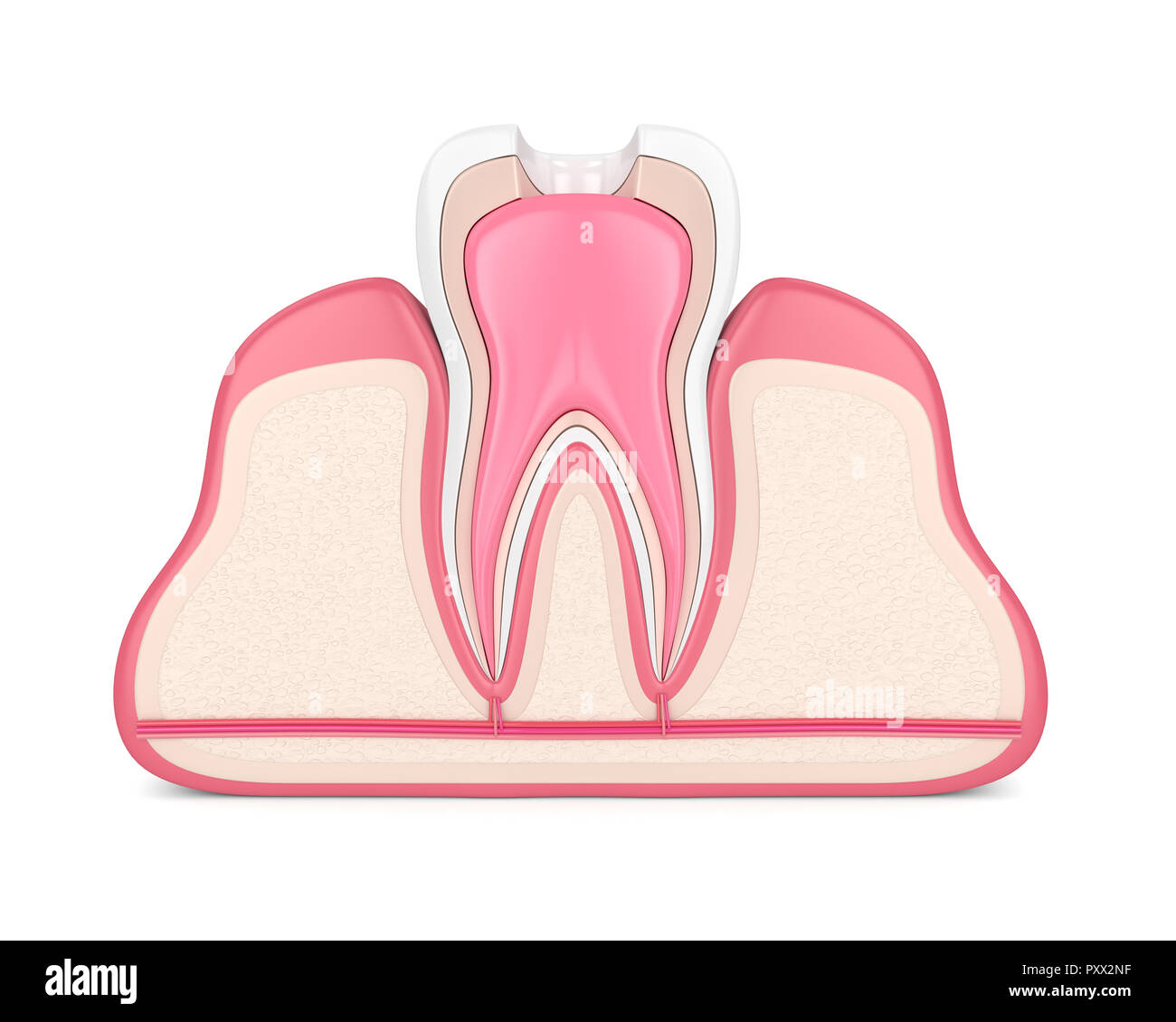 3d render of tooth in gums with root canal treatment procedure over white Stock Photohttps://www.alamy.com/image-license-details/?v=1https://www.alamy.com/3d-render-of-tooth-in-gums-with-root-canal-treatment-procedure-over-white-image223078395.html
RFPXX2NF–3d render of tooth in gums with root canal treatment procedure over white
 . Dental electro-therapeutics. Fig. 127.—Gutta-percha extendingthrough apex. Fig. 128.—Gutta-percha extend-ing through apex. Taken by J. J.Lowe, Boston. Fig. 129 shows extensive absorption of the end of left uppercanine root. The protruding part of the gutta-percha rootcanal stopping has evidently assumed a nearly horizontalposition. The objection to paraffin as a root-canal filling on accountof the inability to distinguish it on the radiograph has beenmet by the addition of bismuth which makes it show on thefilm, although perhaps not quite as distinctly as gutta-percha, THE USES OF THE X-RAYS Stock Photohttps://www.alamy.com/image-license-details/?v=1https://www.alamy.com/dental-electro-therapeutics-fig-127gutta-percha-extendingthrough-apex-fig-128gutta-percha-extend-ing-through-apex-taken-by-j-jlowe-boston-fig-129-shows-extensive-absorption-of-the-end-of-left-uppercanine-root-the-protruding-part-of-the-gutta-percha-rootcanal-stopping-has-evidently-assumed-a-nearly-horizontalposition-the-objection-to-paraffin-as-a-root-canal-filling-on-accountof-the-inability-to-distinguish-it-on-the-radiograph-has-beenmet-by-the-addition-of-bismuth-which-makes-it-show-on-thefilm-although-perhaps-not-quite-as-distinctly-as-gutta-percha-the-uses-of-the-x-rays-image336683679.html
RM2AFN7D3–. Dental electro-therapeutics. Fig. 127.—Gutta-percha extendingthrough apex. Fig. 128.—Gutta-percha extend-ing through apex. Taken by J. J.Lowe, Boston. Fig. 129 shows extensive absorption of the end of left uppercanine root. The protruding part of the gutta-percha rootcanal stopping has evidently assumed a nearly horizontalposition. The objection to paraffin as a root-canal filling on accountof the inability to distinguish it on the radiograph has beenmet by the addition of bismuth which makes it show on thefilm, although perhaps not quite as distinctly as gutta-percha, THE USES OF THE X-RAYS
 3d render of tooth with gutta percha, post and filling in gums. Endodontic treatment concept Stock Photohttps://www.alamy.com/image-license-details/?v=1https://www.alamy.com/3d-render-of-tooth-with-gutta-percha-post-and-filling-in-gums-endodontic-treatment-concept-image225327528.html
RFR2GFFM–3d render of tooth with gutta percha, post and filling in gums. Endodontic treatment concept
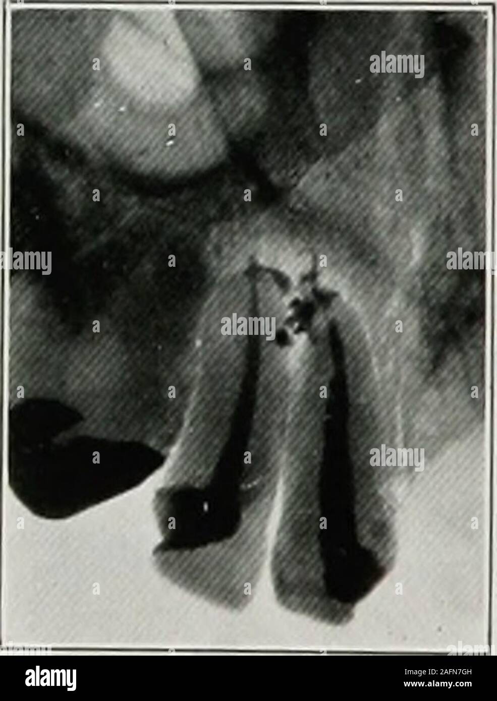 . Dental electro-therapeutics. Fig. 127.—Gutta-percha extendingthrough apex. Fig. 128.—Gutta-percha extend-ing through apex. Taken by J. J.Lowe, Boston. Fig. 129 shows extensive absorption of the end of left uppercanine root. The protruding part of the gutta-percha rootcanal stopping has evidently assumed a nearly horizontalposition. The objection to paraffin as a root-canal filling on accountof the inability to distinguish it on the radiograph has beenmet by the addition of bismuth which makes it show on thefilm, although perhaps not quite as distinctly as gutta-percha, THE USES OF THE X-RAYS Stock Photohttps://www.alamy.com/image-license-details/?v=1https://www.alamy.com/dental-electro-therapeutics-fig-127gutta-percha-extendingthrough-apex-fig-128gutta-percha-extend-ing-through-apex-taken-by-j-jlowe-boston-fig-129-shows-extensive-absorption-of-the-end-of-left-uppercanine-root-the-protruding-part-of-the-gutta-percha-rootcanal-stopping-has-evidently-assumed-a-nearly-horizontalposition-the-objection-to-paraffin-as-a-root-canal-filling-on-accountof-the-inability-to-distinguish-it-on-the-radiograph-has-beenmet-by-the-addition-of-bismuth-which-makes-it-show-on-thefilm-although-perhaps-not-quite-as-distinctly-as-gutta-percha-the-uses-of-the-x-rays-image336683777.html
RM2AFN7GH–. Dental electro-therapeutics. Fig. 127.—Gutta-percha extendingthrough apex. Fig. 128.—Gutta-percha extend-ing through apex. Taken by J. J.Lowe, Boston. Fig. 129 shows extensive absorption of the end of left uppercanine root. The protruding part of the gutta-percha rootcanal stopping has evidently assumed a nearly horizontalposition. The objection to paraffin as a root-canal filling on accountof the inability to distinguish it on the radiograph has beenmet by the addition of bismuth which makes it show on thefilm, although perhaps not quite as distinctly as gutta-percha, THE USES OF THE X-RAYS
 3d render of tooth with gutta percha, post and filling in gums. Endodontic treatment concept Stock Photohttps://www.alamy.com/image-license-details/?v=1https://www.alamy.com/3d-render-of-tooth-with-gutta-percha-post-and-filling-in-gums-endodontic-treatment-concept-image225327394.html
RFR2GFAX–3d render of tooth with gutta percha, post and filling in gums. Endodontic treatment concept
 Dental review; devoted to the advancement of dentistry. . Canal flowed 11U THE DENTAL REVIEW. with eucalyptol, followed by pink chloropercha till reasonably surecanal is full, then gutta percha cone dipped in eucalyptol and forcedin. Small canals sometimes enlarged with hydrochloric acid orGates-Glidden drill. Result: Portions of filling material reachnear to apex, but filling as a whole shows great imperfection as iffrom shrinkage. No. 43. Shown and described previously. Slide 3, No. 17. Distal root of lower molar. Alcohol—C. P.sulphuric acid and hydrogen—dioxide applied in order named. Canal Stock Photohttps://www.alamy.com/image-license-details/?v=1https://www.alamy.com/dental-review-devoted-to-the-advancement-of-dentistry-canal-flowed-11u-the-dental-review-with-eucalyptol-followed-by-pink-chloropercha-till-reasonably-surecanal-is-full-then-gutta-percha-cone-dipped-in-eucalyptol-and-forcedin-small-canals-sometimes-enlarged-with-hydrochloric-acid-orgates-glidden-drill-result-portions-of-filling-material-reachnear-to-apex-but-filling-as-a-whole-shows-great-imperfection-as-iffrom-shrinkage-no-43-shown-and-described-previously-slide-3-no-17-distal-root-of-lower-molar-alcoholc-psulphuric-acid-and-hydrogendioxide-applied-in-order-named-canal-image339300445.html
RM2AM0D51–Dental review; devoted to the advancement of dentistry. . Canal flowed 11U THE DENTAL REVIEW. with eucalyptol, followed by pink chloropercha till reasonably surecanal is full, then gutta percha cone dipped in eucalyptol and forcedin. Small canals sometimes enlarged with hydrochloric acid orGates-Glidden drill. Result: Portions of filling material reachnear to apex, but filling as a whole shows great imperfection as iffrom shrinkage. No. 43. Shown and described previously. Slide 3, No. 17. Distal root of lower molar. Alcohol—C. P.sulphuric acid and hydrogen—dioxide applied in order named. Canal
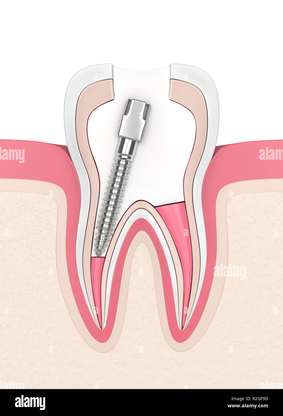 3d render of tooth with gutta percha, post and filling in gums. Endodontic treatment concept Stock Photohttps://www.alamy.com/image-license-details/?v=1https://www.alamy.com/3d-render-of-tooth-with-gutta-percha-post-and-filling-in-gums-endodontic-treatment-concept-image225327353.html
RFR2GF9D–3d render of tooth with gutta percha, post and filling in gums. Endodontic treatment concept
 Diseases of the soft structures of the teeth and their treatment; a text-book for students and practitioners . i:»l 152 I I.s. 151 and 152.—Interradicular perforations.. ilIi the foramen im 11* a gentle pumping motion the air i-expelled and the semiliquid paraffin is coaxed into the canal andthe perforation. Care should be exercised not to overheat theparaffin, although it i- essential to keep the root dryer fairly warmchill the compound. A. jet of warm air applied duringthe filling procedure materially assists in keeping the paraffin -ft.?• rilt- gutta-percha cone with it- tip cut off ina no Stock Photohttps://www.alamy.com/image-license-details/?v=1https://www.alamy.com/diseases-of-the-soft-structures-of-the-teeth-and-their-treatment-a-text-book-for-students-and-practitioners-il-152-i-is-151-and-152interradicular-perforations-ilii-the-foramen-im-11-a-gentle-pumping-motion-the-air-i-expelled-and-the-semiliquid-paraffin-is-coaxed-into-the-canal-andthe-perforation-care-should-be-exercised-not-to-overheat-theparaffin-although-it-i-essential-to-keep-the-root-dryer-fairly-warmchill-the-compound-a-jet-of-warm-air-applied-duringthe-filling-procedure-materially-assists-in-keeping-the-paraffin-ft-rilt-gutta-percha-cone-with-it-tip-cut-off-ina-no-image342965129.html
RM2AWYBEH–Diseases of the soft structures of the teeth and their treatment; a text-book for students and practitioners . i:»l 152 I I.s. 151 and 152.—Interradicular perforations.. ilIi the foramen im 11* a gentle pumping motion the air i-expelled and the semiliquid paraffin is coaxed into the canal andthe perforation. Care should be exercised not to overheat theparaffin, although it i- essential to keep the root dryer fairly warmchill the compound. A. jet of warm air applied duringthe filling procedure materially assists in keeping the paraffin -ft.?• rilt- gutta-percha cone with it- tip cut off ina no
 3d render of tooth with gutta percha, post and filling in gums. Endodontic treatment concept Stock Photohttps://www.alamy.com/image-license-details/?v=1https://www.alamy.com/3d-render-of-tooth-with-gutta-percha-post-and-filling-in-gums-endodontic-treatment-concept-image225327440.html
RFR2GFCG–3d render of tooth with gutta percha, post and filling in gums. Endodontic treatment concept
 The Dental cosmos . material is nevernecessary. Put up in ounce boxes of one dozen sticks, of which one-third arepink and two-thirds white. Price per box 50 cents. Dr. S. Eldred Gilberts Temporary Stopping, For the retention of medicaments used in the treatment of teeth, and thefilling of cavities temporarily ; also for non-conducting layers in deep cavities,and for filling pulp-canals. Put up in ounce boxes. Price per box 50 cents. 19 ROOT-CANAL POINTS. WOOD, METAL, AND GUTTA-PERCHA. Three of the most popular and most widely used materials for filling root-canals are Wood, Metal, and Gutta-pe Stock Photohttps://www.alamy.com/image-license-details/?v=1https://www.alamy.com/the-dental-cosmos-material-is-nevernecessary-put-up-in-ounce-boxes-of-one-dozen-sticks-of-which-one-third-arepink-and-two-thirds-white-price-per-box-50-cents-dr-s-eldred-gilberts-temporary-stopping-for-the-retention-of-medicaments-used-in-the-treatment-of-teeth-and-thefilling-of-cavities-temporarily-also-for-non-conducting-layers-in-deep-cavitiesand-for-filling-pulp-canals-put-up-in-ounce-boxes-price-per-box-50-cents-19-root-canal-points-wood-metal-and-gutta-percha-three-of-the-most-popular-and-most-widely-used-materials-for-filling-root-canals-are-wood-metal-and-gutta-pe-image340000475.html
RM2AN4A23–The Dental cosmos . material is nevernecessary. Put up in ounce boxes of one dozen sticks, of which one-third arepink and two-thirds white. Price per box 50 cents. Dr. S. Eldred Gilberts Temporary Stopping, For the retention of medicaments used in the treatment of teeth, and thefilling of cavities temporarily ; also for non-conducting layers in deep cavities,and for filling pulp-canals. Put up in ounce boxes. Price per box 50 cents. 19 ROOT-CANAL POINTS. WOOD, METAL, AND GUTTA-PERCHA. Three of the most popular and most widely used materials for filling root-canals are Wood, Metal, and Gutta-pe
 3d render of tooth with gutta percha, post and filling in gums. Endodontic treatment concept Stock Photohttps://www.alamy.com/image-license-details/?v=1https://www.alamy.com/3d-render-of-tooth-with-gutta-percha-post-and-filling-in-gums-endodontic-treatment-concept-image225327422.html
RFR2GFBX–3d render of tooth with gutta percha, post and filling in gums. Endodontic treatment concept
 . Elementary and dental radiography / by Howard Riley Raper . Figs. 509 and 510. Tooth substance havingbeen dissolved away from root canal filling.Shows the long minute canals that therosin and gutta-percha solution enters andseals. Fig. 511. Palatine root of upper molar en-larged with drill, making a false pocket.The rosin-gutta-percha solution not onlyfilled the false pocket, but entered the truecanal and filled it to the end. Stock Photohttps://www.alamy.com/image-license-details/?v=1https://www.alamy.com/elementary-and-dental-radiography-by-howard-riley-raper-figs-509-and-510-tooth-substance-havingbeen-dissolved-away-from-root-canal-fillingshows-the-long-minute-canals-that-therosin-and-gutta-percha-solution-enters-andseals-fig-511-palatine-root-of-upper-molar-en-larged-with-drill-making-a-false-pocketthe-rosin-gutta-percha-solution-not-onlyfilled-the-false-pocket-but-entered-the-truecanal-and-filled-it-to-the-end-image375910170.html
RM2CRG576–. Elementary and dental radiography / by Howard Riley Raper . Figs. 509 and 510. Tooth substance havingbeen dissolved away from root canal filling.Shows the long minute canals that therosin and gutta-percha solution enters andseals. Fig. 511. Palatine root of upper molar en-larged with drill, making a false pocket.The rosin-gutta-percha solution not onlyfilled the false pocket, but entered the truecanal and filled it to the end.
 3d render of tooth with gutta percha, post and filling in gums. Endodontic treatment concept Stock Photohttps://www.alamy.com/image-license-details/?v=1https://www.alamy.com/3d-render-of-tooth-with-gutta-percha-post-and-filling-in-gums-endodontic-treatment-concept-image225327524.html
RFR2GFFG–3d render of tooth with gutta percha, post and filling in gums. Endodontic treatment concept
 . Elementary and dental radiography . Fig. 185 Fig. 186 Fig. 185. Gutta-percha canal filling in the upper lateral passing through the side of the rootto the distal. The canal filling also penetrates the apical foramen. The light area to themesial along the apical third of the root indicates an abscess. The mesial surface of the root is roughened in the region of the abscess. (Radiograph by Lewis of Chicago.) Fig. 186. Canal filling penetrating the tissues between the roots of the lower first molar. (Radiograph by Kells, Jr. of New Orleans.) made to learn whether the canals are properly filled, Stock Photohttps://www.alamy.com/image-license-details/?v=1https://www.alamy.com/elementary-and-dental-radiography-fig-185-fig-186-fig-185-gutta-percha-canal-filling-in-the-upper-lateral-passing-through-the-side-of-the-rootto-the-distal-the-canal-filling-also-penetrates-the-apical-foramen-the-light-area-to-themesial-along-the-apical-third-of-the-root-indicates-an-abscess-the-mesial-surface-of-the-root-is-roughened-in-the-region-of-the-abscess-radiograph-by-lewis-of-chicago-fig-186-canal-filling-penetrating-the-tissues-between-the-roots-of-the-lower-first-molar-radiograph-by-kells-jr-of-new-orleans-made-to-learn-whether-the-canals-are-properly-filled-image376067475.html
RM2CRR9W7–. Elementary and dental radiography . Fig. 185 Fig. 186 Fig. 185. Gutta-percha canal filling in the upper lateral passing through the side of the rootto the distal. The canal filling also penetrates the apical foramen. The light area to themesial along the apical third of the root indicates an abscess. The mesial surface of the root is roughened in the region of the abscess. (Radiograph by Lewis of Chicago.) Fig. 186. Canal filling penetrating the tissues between the roots of the lower first molar. (Radiograph by Kells, Jr. of New Orleans.) made to learn whether the canals are properly filled,
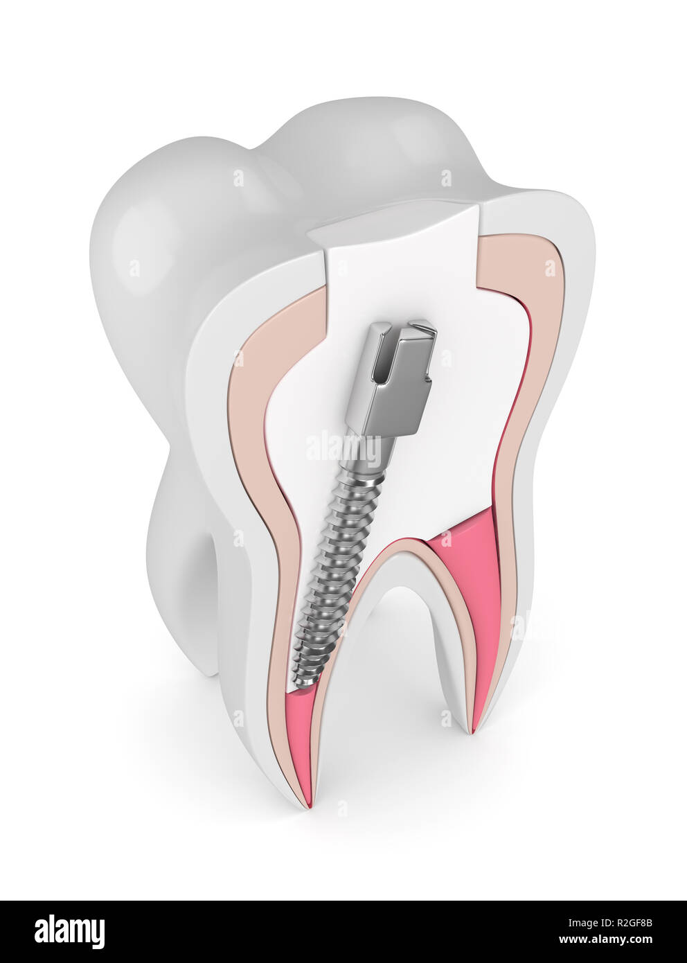 3d render of tooth with gutta percha, post and filling over white. Endodontic treatment concept Stock Photohttps://www.alamy.com/image-license-details/?v=1https://www.alamy.com/3d-render-of-tooth-with-gutta-percha-post-and-filling-over-white-endodontic-treatment-concept-image225327323.html
RFR2GF8B–3d render of tooth with gutta percha, post and filling over white. Endodontic treatment concept
 . Elementary and dental radiography . Fig. 185 Fig. 186 Fig. 185. Gutta-percha canal filling in the upper lateral passing through the side of the rootto the distal. The canal filling also penetrates the apical foramen. The light area to themesial along the apical third of the root indicates an abscess. The mesial surface of the root is roughened in the region of the abscess. (Radiograph by Lewis of Chicago.) Fig. 186. Canal filling penetrating the tissues between the roots of the lower first molar. (Radiograph by Kclls, Jr. of New Orleans.) made to learn whether the canals are properly filled, Stock Photohttps://www.alamy.com/image-license-details/?v=1https://www.alamy.com/elementary-and-dental-radiography-fig-185-fig-186-fig-185-gutta-percha-canal-filling-in-the-upper-lateral-passing-through-the-side-of-the-rootto-the-distal-the-canal-filling-also-penetrates-the-apical-foramen-the-light-area-to-themesial-along-the-apical-third-of-the-root-indicates-an-abscess-the-mesial-surface-of-the-root-is-roughened-in-the-region-of-the-abscess-radiograph-by-lewis-of-chicago-fig-186-canal-filling-penetrating-the-tissues-between-the-roots-of-the-lower-first-molar-radiograph-by-kclls-jr-of-new-orleans-made-to-learn-whether-the-canals-are-properly-filled-image376073580.html
RM2CRRHK8–. Elementary and dental radiography . Fig. 185 Fig. 186 Fig. 185. Gutta-percha canal filling in the upper lateral passing through the side of the rootto the distal. The canal filling also penetrates the apical foramen. The light area to themesial along the apical third of the root indicates an abscess. The mesial surface of the root is roughened in the region of the abscess. (Radiograph by Lewis of Chicago.) Fig. 186. Canal filling penetrating the tissues between the roots of the lower first molar. (Radiograph by Kclls, Jr. of New Orleans.) made to learn whether the canals are properly filled,
 3d render of tooth with gutta percha, post and filling over white. Endodontic treatment concept Stock Photohttps://www.alamy.com/image-license-details/?v=1https://www.alamy.com/3d-render-of-tooth-with-gutta-percha-post-and-filling-over-white-endodontic-treatment-concept-image225327322.html
RFR2GF8A–3d render of tooth with gutta percha, post and filling over white. Endodontic treatment concept
 . The principles and practice of dental surgery. ompressed pivot wood,is somewhat more than can be made with the thumb and finger,applied by means of a small pine stick notched at the end to re-ceive the cutting edge of the tooth. It is important that the pivot should exactly equal the depth of the canal. If too long, the crown will not go up to its place; if too short, there will be either an unnecessary weakening of the root or the crown will be insecure. II IJL JL A small piece of smooth wire or knitting needle with W Bj a sliding collar of wood or gutta percha forms a sim-ple instrument fo Stock Photohttps://www.alamy.com/image-license-details/?v=1https://www.alamy.com/the-principles-and-practice-of-dental-surgery-ompressed-pivot-woodis-somewhat-more-than-can-be-made-with-the-thumb-and-fingerapplied-by-means-of-a-small-pine-stick-notched-at-the-end-to-re-ceive-the-cutting-edge-of-the-tooth-it-is-important-that-the-pivot-should-exactly-equal-the-depth-of-the-canal-if-too-long-the-crown-will-not-go-up-to-its-place-if-too-short-there-will-be-either-an-unnecessary-weakening-of-the-root-or-the-crown-will-be-insecure-ii-ijl-jl-a-small-piece-of-smooth-wire-or-knitting-needle-with-w-bj-a-sliding-collar-of-wood-or-gutta-percha-forms-a-sim-ple-instrument-fo-image370439313.html
RM2CEJY3D–. The principles and practice of dental surgery. ompressed pivot wood,is somewhat more than can be made with the thumb and finger,applied by means of a small pine stick notched at the end to re-ceive the cutting edge of the tooth. It is important that the pivot should exactly equal the depth of the canal. If too long, the crown will not go up to its place; if too short, there will be either an unnecessary weakening of the root or the crown will be insecure. II IJL JL A small piece of smooth wire or knitting needle with W Bj a sliding collar of wood or gutta percha forms a sim-ple instrument fo
 3d render of tooth with gutta percha, post and filling over white. Endodontic treatment concept Stock Photohttps://www.alamy.com/image-license-details/?v=1https://www.alamy.com/3d-render-of-tooth-with-gutta-percha-post-and-filling-over-white-endodontic-treatment-concept-image225327267.html
RFR2GF6B–3d render of tooth with gutta percha, post and filling over white. Endodontic treatment concept
 . Elementary and dental radiography / by Howard Riley Raper . Fig. 185 Fig. 188 Fig. 185. Gutta-percha canal filling in the upper lateral passing through the side of the rootto the distal. The canal filling also penetrates the apical foramen. The light area to themesial along the apical third of the root indicates an abscess. The mesial surface of the root is roughened in the region of the abscess. (Radiograph by Lewis of Chicago.) Kig. 186. Canal filling penetrating the tissues between the roots of the lower first molar (Radiograph by Kells, Jr. of New Orleans.) made to learn whether the cana Stock Photohttps://www.alamy.com/image-license-details/?v=1https://www.alamy.com/elementary-and-dental-radiography-by-howard-riley-raper-fig-185-fig-188-fig-185-gutta-percha-canal-filling-in-the-upper-lateral-passing-through-the-side-of-the-rootto-the-distal-the-canal-filling-also-penetrates-the-apical-foramen-the-light-area-to-themesial-along-the-apical-third-of-the-root-indicates-an-abscess-the-mesial-surface-of-the-root-is-roughened-in-the-region-of-the-abscess-radiograph-by-lewis-of-chicago-kig-186-canal-filling-penetrating-the-tissues-between-the-roots-of-the-lower-first-molar-radiograph-by-kells-jr-of-new-orleans-made-to-learn-whether-the-cana-image375961008.html
RM2CRJE2T–. Elementary and dental radiography / by Howard Riley Raper . Fig. 185 Fig. 188 Fig. 185. Gutta-percha canal filling in the upper lateral passing through the side of the rootto the distal. The canal filling also penetrates the apical foramen. The light area to themesial along the apical third of the root indicates an abscess. The mesial surface of the root is roughened in the region of the abscess. (Radiograph by Lewis of Chicago.) Kig. 186. Canal filling penetrating the tissues between the roots of the lower first molar (Radiograph by Kells, Jr. of New Orleans.) made to learn whether the cana
 3d render of tooth with gutta percha, fiber post and filling in gums. Endodontic treatment concept Stock Photohttps://www.alamy.com/image-license-details/?v=1https://www.alamy.com/3d-render-of-tooth-with-gutta-percha-fiber-post-and-filling-in-gums-endodontic-treatment-concept-image226144746.html
RFR3WNX2–3d render of tooth with gutta percha, fiber post and filling in gums. Endodontic treatment concept
 . Elementary and dental radiography . wire as a guide to theTig. 170. length of the root, the proper distance was measured on a canal plugger, and the distance marked on theplugger by passing it through a little piece of base-plate gutta-percha,stopping the gutta-percha on the plugger at a distance from its end equiv-alent to the length of the wire. The end of the canal plugger was then 176 DEXTAL RADIOGRAPHY warmed slightly, and brought in contact with a small piece of gutta-percha canal point. With the piece of gutta-percha so fastened on thecanal plugger, it was carried into the canal a suf Stock Photohttps://www.alamy.com/image-license-details/?v=1https://www.alamy.com/elementary-and-dental-radiography-wire-as-a-guide-to-thetig-170-length-of-the-root-the-proper-distance-was-measured-on-a-canal-plugger-and-the-distance-marked-on-theplugger-by-passing-it-through-a-little-piece-of-base-plate-gutta-perchastopping-the-gutta-percha-on-the-plugger-at-a-distance-from-its-end-equiv-alent-to-the-length-of-the-wire-the-end-of-the-canal-plugger-was-then-176-dextal-radiography-warmed-slightly-and-brought-in-contact-with-a-small-piece-of-gutta-percha-canal-point-with-the-piece-of-gutta-percha-so-fastened-on-thecanal-plugger-it-was-carried-into-the-canal-a-suf-image376067920.html
RM2CRRAD4–. Elementary and dental radiography . wire as a guide to theTig. 170. length of the root, the proper distance was measured on a canal plugger, and the distance marked on theplugger by passing it through a little piece of base-plate gutta-percha,stopping the gutta-percha on the plugger at a distance from its end equiv-alent to the length of the wire. The end of the canal plugger was then 176 DEXTAL RADIOGRAPHY warmed slightly, and brought in contact with a small piece of gutta-percha canal point. With the piece of gutta-percha so fastened on thecanal plugger, it was carried into the canal a suf
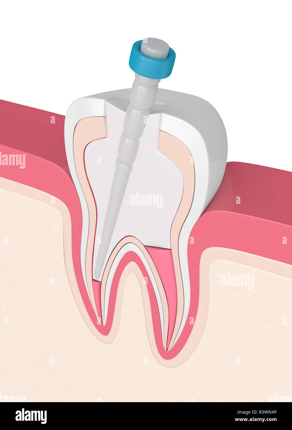 3d render of tooth with gutta percha, fiber post and filling in gums. Endodontic treatment concept Stock Photohttps://www.alamy.com/image-license-details/?v=1https://www.alamy.com/3d-render-of-tooth-with-gutta-percha-fiber-post-and-filling-in-gums-endodontic-treatment-concept-image226144766.html
RFR3WNXP–3d render of tooth with gutta percha, fiber post and filling in gums. Endodontic treatment concept
 . Elementary and dental radiography / by Howard Riley Raper . Figs. 509 and 510. Tooth substance havingbeen dissolved away from root canal filling.Shows the long minute canals that therosin and gutta-percha solution enters andseals. Fig. 511. Palatine root of upper molar en-larged with drill, making a false pocket.The rosin-gutta-percha solution not onlyfilled the false pocket, but entered the truecanal and filled it to the end.. Fig. 512. The shaded area about the root canal filling shows the distance that the rosin haspenetrated the dentinal tubuli in this tooth root. CANAL SURGERY AND ORAL Stock Photohttps://www.alamy.com/image-license-details/?v=1https://www.alamy.com/elementary-and-dental-radiography-by-howard-riley-raper-figs-509-and-510-tooth-substance-havingbeen-dissolved-away-from-root-canal-fillingshows-the-long-minute-canals-that-therosin-and-gutta-percha-solution-enters-andseals-fig-511-palatine-root-of-upper-molar-en-larged-with-drill-making-a-false-pocketthe-rosin-gutta-percha-solution-not-onlyfilled-the-false-pocket-but-entered-the-truecanal-and-filled-it-to-the-end-fig-512-the-shaded-area-about-the-root-canal-filling-shows-the-distance-that-the-rosin-haspenetrated-the-dentinal-tubuli-in-this-tooth-root-canal-surgery-and-oral-image375910128.html
RM2CRG55M–. Elementary and dental radiography / by Howard Riley Raper . Figs. 509 and 510. Tooth substance havingbeen dissolved away from root canal filling.Shows the long minute canals that therosin and gutta-percha solution enters andseals. Fig. 511. Palatine root of upper molar en-larged with drill, making a false pocket.The rosin-gutta-percha solution not onlyfilled the false pocket, but entered the truecanal and filled it to the end.. Fig. 512. The shaded area about the root canal filling shows the distance that the rosin haspenetrated the dentinal tubuli in this tooth root. CANAL SURGERY AND ORAL
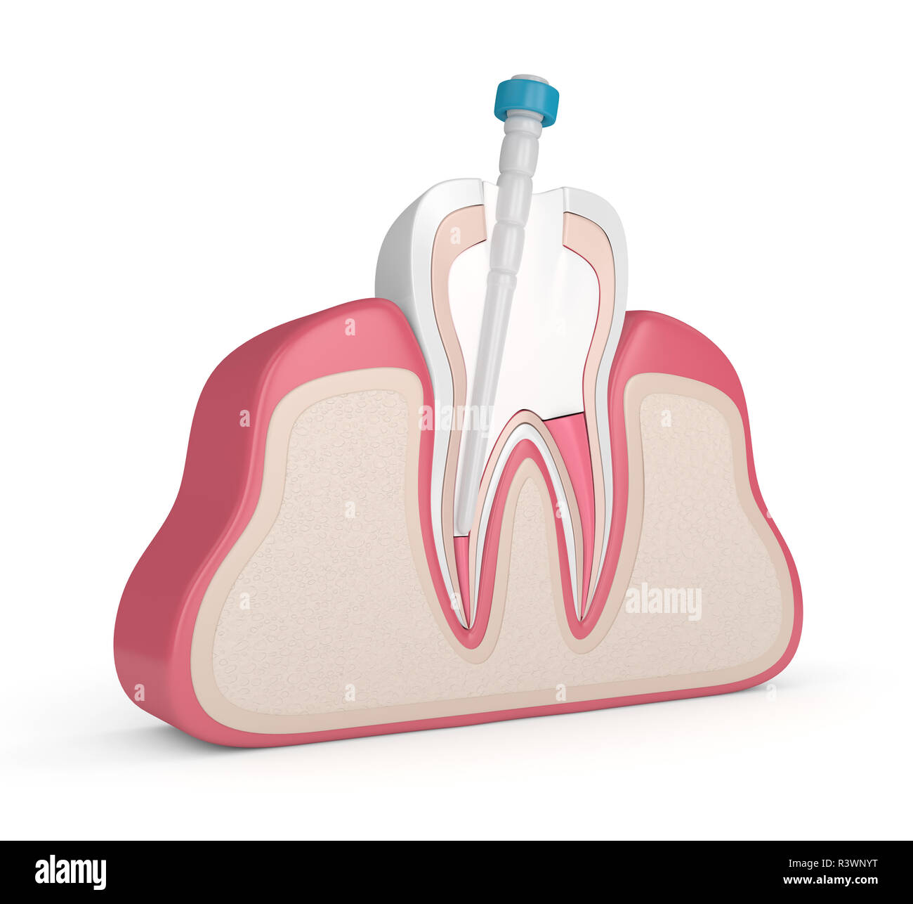 3d render of tooth with gutta percha, fiber post and filling in gums. Endodontic treatment concept Stock Photohttps://www.alamy.com/image-license-details/?v=1https://www.alamy.com/3d-render-of-tooth-with-gutta-percha-fiber-post-and-filling-in-gums-endodontic-treatment-concept-image226144796.html
RFR3WNYT–3d render of tooth with gutta percha, fiber post and filling in gums. Endodontic treatment concept
 . Elementary and dental radiography / by Howard Riley Raper . Kg. 171 Fig. 172 Fig. 171. The dark streaks in the lower molar are wires in the canals. (Operator, Dr. Moag,Indianapolis.) Fig. 172. The same as Fig. 171 after canal filling. canal filling, and the tissues above the apex are not infected, then thepassage of a little gutta-percha into the apical tissues will probably notresult in suppuration or even inflammation, so well do tissues tolerategutta-percha. But the fact remains: The ideal canal filling is one whichfills the canals, neither falling short of the end of the root nor passing Stock Photohttps://www.alamy.com/image-license-details/?v=1https://www.alamy.com/elementary-and-dental-radiography-by-howard-riley-raper-kg-171-fig-172-fig-171-the-dark-streaks-in-the-lower-molar-are-wires-in-the-canals-operator-dr-moagindianapolis-fig-172-the-same-as-fig-171-after-canal-filling-canal-filling-and-the-tissues-above-the-apex-are-not-infected-then-thepassage-of-a-little-gutta-percha-into-the-apical-tissues-will-probably-notresult-in-suppuration-or-even-inflammation-so-well-do-tissues-tolerategutta-percha-but-the-fact-remains-the-ideal-canal-filling-is-one-whichfills-the-canals-neither-falling-short-of-the-end-of-the-root-nor-passing-image375962910.html
RM2CRJGEP–. Elementary and dental radiography / by Howard Riley Raper . Kg. 171 Fig. 172 Fig. 171. The dark streaks in the lower molar are wires in the canals. (Operator, Dr. Moag,Indianapolis.) Fig. 172. The same as Fig. 171 after canal filling. canal filling, and the tissues above the apex are not infected, then thepassage of a little gutta-percha into the apical tissues will probably notresult in suppuration or even inflammation, so well do tissues tolerategutta-percha. But the fact remains: The ideal canal filling is one whichfills the canals, neither falling short of the end of the root nor passing
 3d render of tooth with gutta percha, fiber post and filling in gums. Endodontic treatment concept Stock Photohttps://www.alamy.com/image-license-details/?v=1https://www.alamy.com/3d-render-of-tooth-with-gutta-percha-fiber-post-and-filling-in-gums-endodontic-treatment-concept-image226144736.html
RFR3WNWM–3d render of tooth with gutta percha, fiber post and filling in gums. Endodontic treatment concept
 . Elementary and dental radiography . wire as a guide to thefig. 170. length of the root, the proper distance was measured on a canal plugger, and the distance marked on theplugger by passing it through a little piece of base-plate gutta-percha,stopping the gutta-percha on the plugger at a distance from its end equiv-alent to the length of the wire. The end of the canal plugger was then 176 DENTAL RADIOGRAPHY warmed slightly, and brought in contact with a small piece of gutta-percha canal point. With the piece of gutta-percha so fastened on thecanal plugger, it was carried into the canal a suf Stock Photohttps://www.alamy.com/image-license-details/?v=1https://www.alamy.com/elementary-and-dental-radiography-wire-as-a-guide-to-thefig-170-length-of-the-root-the-proper-distance-was-measured-on-a-canal-plugger-and-the-distance-marked-on-theplugger-by-passing-it-through-a-little-piece-of-base-plate-gutta-perchastopping-the-gutta-percha-on-the-plugger-at-a-distance-from-its-end-equiv-alent-to-the-length-of-the-wire-the-end-of-the-canal-plugger-was-then-176-dental-radiography-warmed-slightly-and-brought-in-contact-with-a-small-piece-of-gutta-percha-canal-point-with-the-piece-of-gutta-percha-so-fastened-on-thecanal-plugger-it-was-carried-into-the-canal-a-suf-image376074035.html
RM2CRRJ7F–. Elementary and dental radiography . wire as a guide to thefig. 170. length of the root, the proper distance was measured on a canal plugger, and the distance marked on theplugger by passing it through a little piece of base-plate gutta-percha,stopping the gutta-percha on the plugger at a distance from its end equiv-alent to the length of the wire. The end of the canal plugger was then 176 DENTAL RADIOGRAPHY warmed slightly, and brought in contact with a small piece of gutta-percha canal point. With the piece of gutta-percha so fastened on thecanal plugger, it was carried into the canal a suf
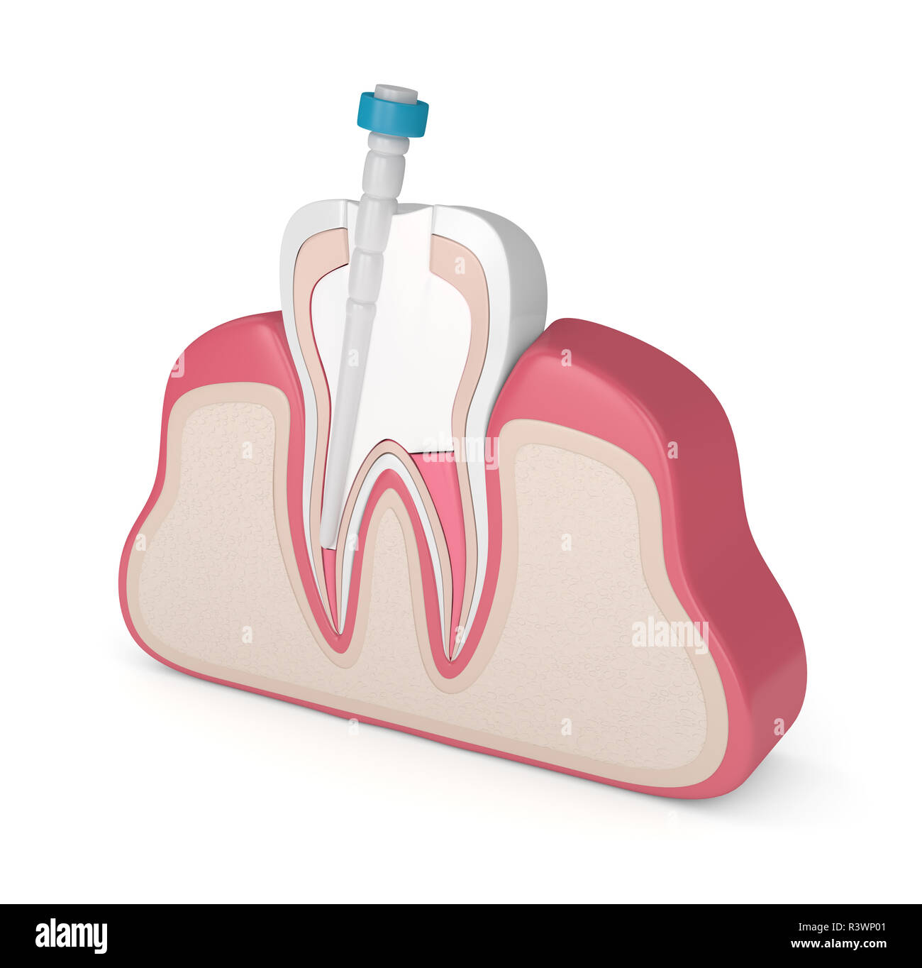 3d render of tooth with gutta percha, fiber post and filling in gums. Endodontic treatment concept Stock Photohttps://www.alamy.com/image-license-details/?v=1https://www.alamy.com/3d-render-of-tooth-with-gutta-percha-fiber-post-and-filling-in-gums-endodontic-treatment-concept-image226144801.html
RFR3WP01–3d render of tooth with gutta percha, fiber post and filling in gums. Endodontic treatment concept
 . Elementary and dental radiography . Fig. 168. Fig. 169. Fig. 170. Fig. 168. A wire passing through a large apical foramen in an upper, central incisor, extendingseveral millimeters into the tissues above the apex of the root. Fig. 169. The same case as Fig. 168, after the wire has been removed, a part of it cut offand reinserted into the canal. The wire reaches just to the apical foramen. Fig. 170. The same case as Figs. 168 and 169, showing a canal filling of gutta-percha closingthe apical foramen, not penetratirg through it. and not leaving a little of the canal unfilledat the apex of the Stock Photohttps://www.alamy.com/image-license-details/?v=1https://www.alamy.com/elementary-and-dental-radiography-fig-168-fig-169-fig-170-fig-168-a-wire-passing-through-a-large-apical-foramen-in-an-upper-central-incisor-extendingseveral-millimeters-into-the-tissues-above-the-apex-of-the-root-fig-169-the-same-case-as-fig-168-after-the-wire-has-been-removed-a-part-of-it-cut-offand-reinserted-into-the-canal-the-wire-reaches-just-to-the-apical-foramen-fig-170-the-same-case-as-figs-168-and-169-showing-a-canal-filling-of-gutta-percha-closingthe-apical-foramen-not-penetratirg-through-it-and-not-leaving-a-little-of-the-canal-unfilledat-the-apex-of-the-image376074044.html
RM2CRRJ7T–. Elementary and dental radiography . Fig. 168. Fig. 169. Fig. 170. Fig. 168. A wire passing through a large apical foramen in an upper, central incisor, extendingseveral millimeters into the tissues above the apex of the root. Fig. 169. The same case as Fig. 168, after the wire has been removed, a part of it cut offand reinserted into the canal. The wire reaches just to the apical foramen. Fig. 170. The same case as Figs. 168 and 169, showing a canal filling of gutta-percha closingthe apical foramen, not penetratirg through it. and not leaving a little of the canal unfilledat the apex of the
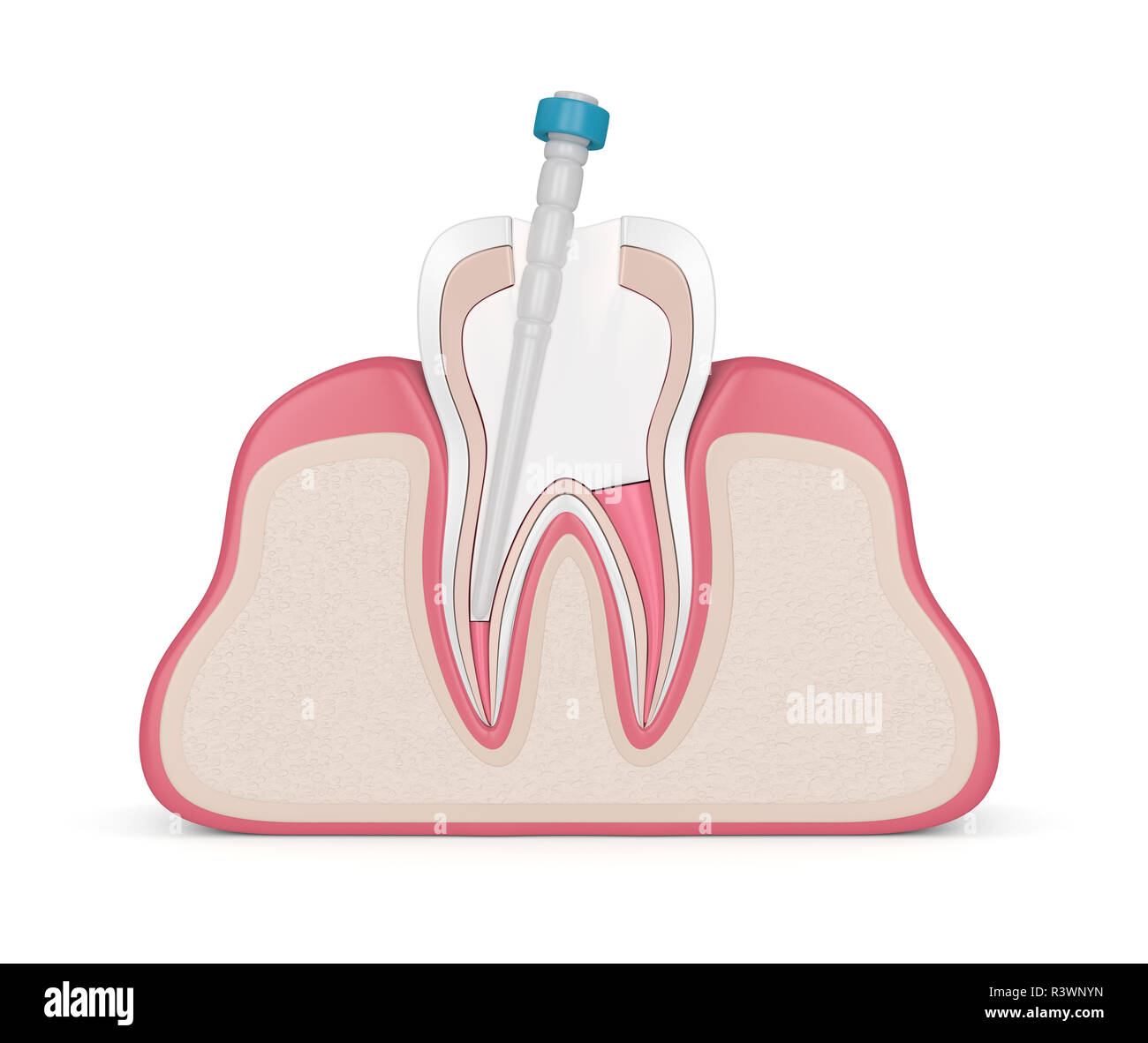 3d render of tooth with gutta percha, fiber post and filling in gums. Endodontic treatment concept Stock Photohttps://www.alamy.com/image-license-details/?v=1https://www.alamy.com/3d-render-of-tooth-with-gutta-percha-fiber-post-and-filling-in-gums-endodontic-treatment-concept-image226144793.html
RFR3WNYN–3d render of tooth with gutta percha, fiber post and filling in gums. Endodontic treatment concept
 . Elementary and dental radiography / by Howard Riley Raper . Fig. 168. Fig. 169. Fig. 170. Fig. 168. A wire passing through a large apical foramen in an upper, central incisor, extendingseveral millimeters into the tissues above the apex of the root. Fig. 169. The same case as Fig. 168, after the wire has been removed, a part of it cut offand reinserted into the canal. The wire reaches just to the apical foramen. Fig. 170. The same case as Figs. 168 and 169, showing a cana! filling of gutta-percha closingthe apical foramen, not penetrati: g through it. and not leaving a little of the canal un Stock Photohttps://www.alamy.com/image-license-details/?v=1https://www.alamy.com/elementary-and-dental-radiography-by-howard-riley-raper-fig-168-fig-169-fig-170-fig-168-a-wire-passing-through-a-large-apical-foramen-in-an-upper-central-incisor-extendingseveral-millimeters-into-the-tissues-above-the-apex-of-the-root-fig-169-the-same-case-as-fig-168-after-the-wire-has-been-removed-a-part-of-it-cut-offand-reinserted-into-the-canal-the-wire-reaches-just-to-the-apical-foramen-fig-170-the-same-case-as-figs-168-and-169-showing-a-cana!-filling-of-gutta-percha-closingthe-apical-foramen-not-penetrati-g-through-it-and-not-leaving-a-little-of-the-canal-un-image375963449.html
RM2CRJH61–. Elementary and dental radiography / by Howard Riley Raper . Fig. 168. Fig. 169. Fig. 170. Fig. 168. A wire passing through a large apical foramen in an upper, central incisor, extendingseveral millimeters into the tissues above the apex of the root. Fig. 169. The same case as Fig. 168, after the wire has been removed, a part of it cut offand reinserted into the canal. The wire reaches just to the apical foramen. Fig. 170. The same case as Figs. 168 and 169, showing a cana! filling of gutta-percha closingthe apical foramen, not penetrati: g through it. and not leaving a little of the canal un
 3d render of tooth with gutta percha, fiber post and filling over white. Endodontic treatment concept Stock Photohttps://www.alamy.com/image-license-details/?v=1https://www.alamy.com/3d-render-of-tooth-with-gutta-percha-fiber-post-and-filling-over-white-endodontic-treatment-concept-image226144777.html
RFR3WNY5–3d render of tooth with gutta percha, fiber post and filling over white. Endodontic treatment concept
 . Elementary and dental radiography . Fig. 168. Fig. 169. Fig. 170. Fig. 1G8. A wire passing through a large apical foramen in an upper, central incisor, extendingseveral millimeters into the tissues above the apex of the root. Fig. 1G9. The same case as Fig. 168, after the wire has been removed, a part of it cut offand reinserted into the canal. The wire reaches just to the apical foramen. Fig. 170. The same case as Figs. 168 and 169, showing a canal filling of gutta-percha closingthe apical foramen, not penetratir.g through it, and not leaving a little of the canal unfilledat the apex of the Stock Photohttps://www.alamy.com/image-license-details/?v=1https://www.alamy.com/elementary-and-dental-radiography-fig-168-fig-169-fig-170-fig-1g8-a-wire-passing-through-a-large-apical-foramen-in-an-upper-central-incisor-extendingseveral-millimeters-into-the-tissues-above-the-apex-of-the-root-fig-1g9-the-same-case-as-fig-168-after-the-wire-has-been-removed-a-part-of-it-cut-offand-reinserted-into-the-canal-the-wire-reaches-just-to-the-apical-foramen-fig-170-the-same-case-as-figs-168-and-169-showing-a-canal-filling-of-gutta-percha-closingthe-apical-foramen-not-penetratirg-through-it-and-not-leaving-a-little-of-the-canal-unfilledat-the-apex-of-the-image376067994.html
RM2CRRAFP–. Elementary and dental radiography . Fig. 168. Fig. 169. Fig. 170. Fig. 1G8. A wire passing through a large apical foramen in an upper, central incisor, extendingseveral millimeters into the tissues above the apex of the root. Fig. 1G9. The same case as Fig. 168, after the wire has been removed, a part of it cut offand reinserted into the canal. The wire reaches just to the apical foramen. Fig. 170. The same case as Figs. 168 and 169, showing a canal filling of gutta-percha closingthe apical foramen, not penetratir.g through it, and not leaving a little of the canal unfilledat the apex of the
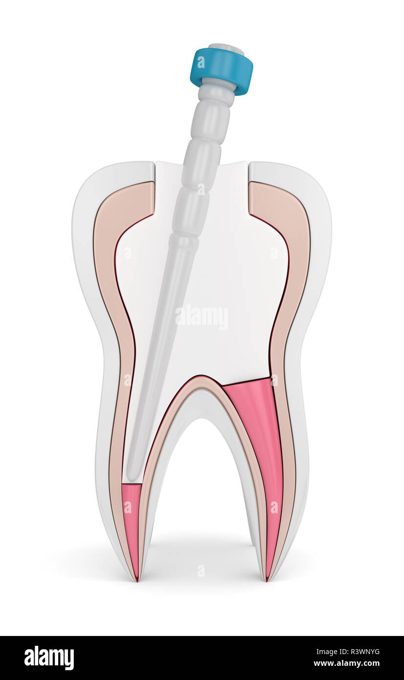 3d render of tooth with gutta percha, fiber post and filling over white. Endodontic treatment concept Stock Photohttps://www.alamy.com/image-license-details/?v=1https://www.alamy.com/3d-render-of-tooth-with-gutta-percha-fiber-post-and-filling-over-white-endodontic-treatment-concept-image226144788.html
RFR3WNYG–3d render of tooth with gutta percha, fiber post and filling over white. Endodontic treatment concept
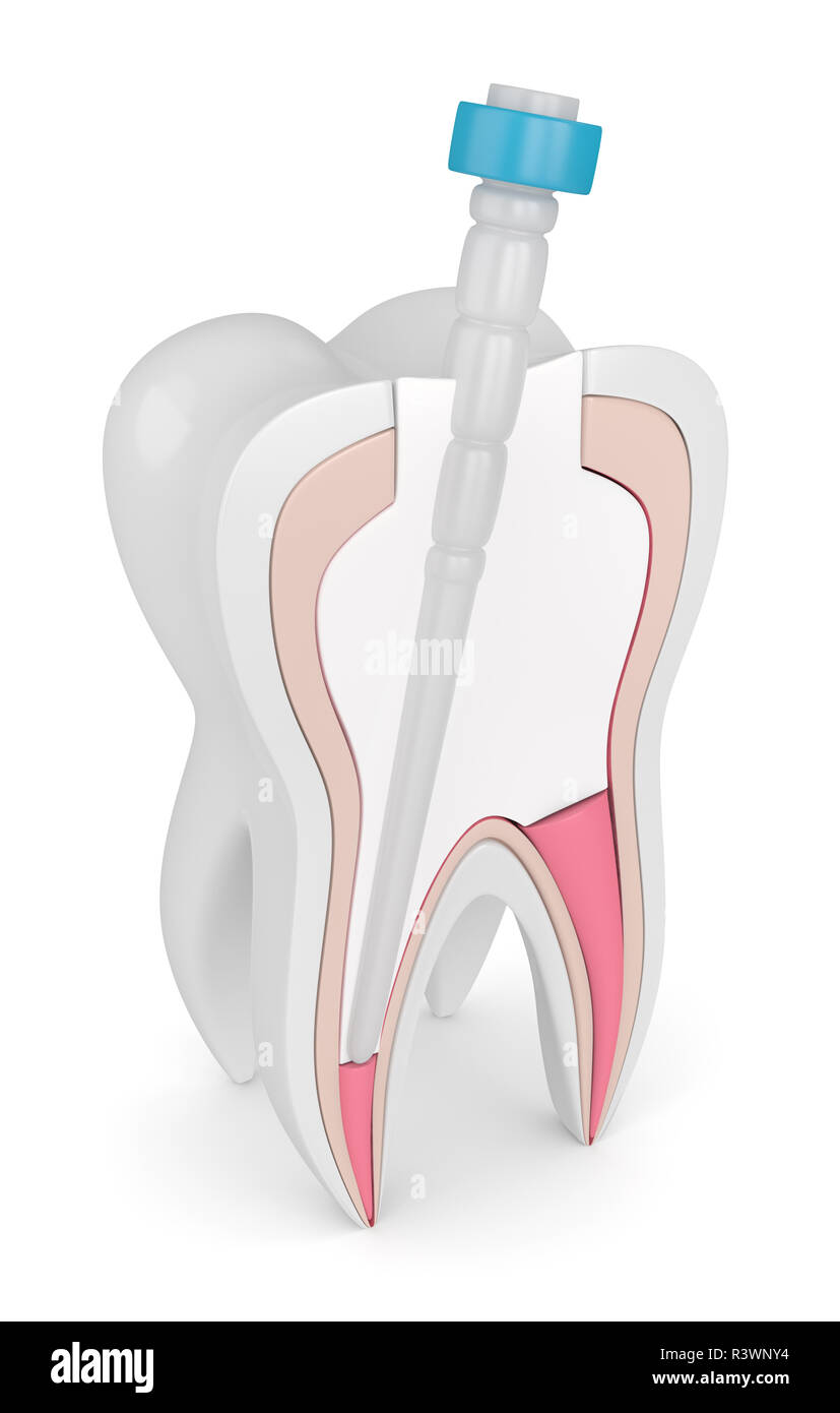 3d render of tooth with gutta percha, fiber post and filling over white. Endodontic treatment concept Stock Photohttps://www.alamy.com/image-license-details/?v=1https://www.alamy.com/3d-render-of-tooth-with-gutta-percha-fiber-post-and-filling-over-white-endodontic-treatment-concept-image226144776.html
RFR3WNY4–3d render of tooth with gutta percha, fiber post and filling over white. Endodontic treatment concept
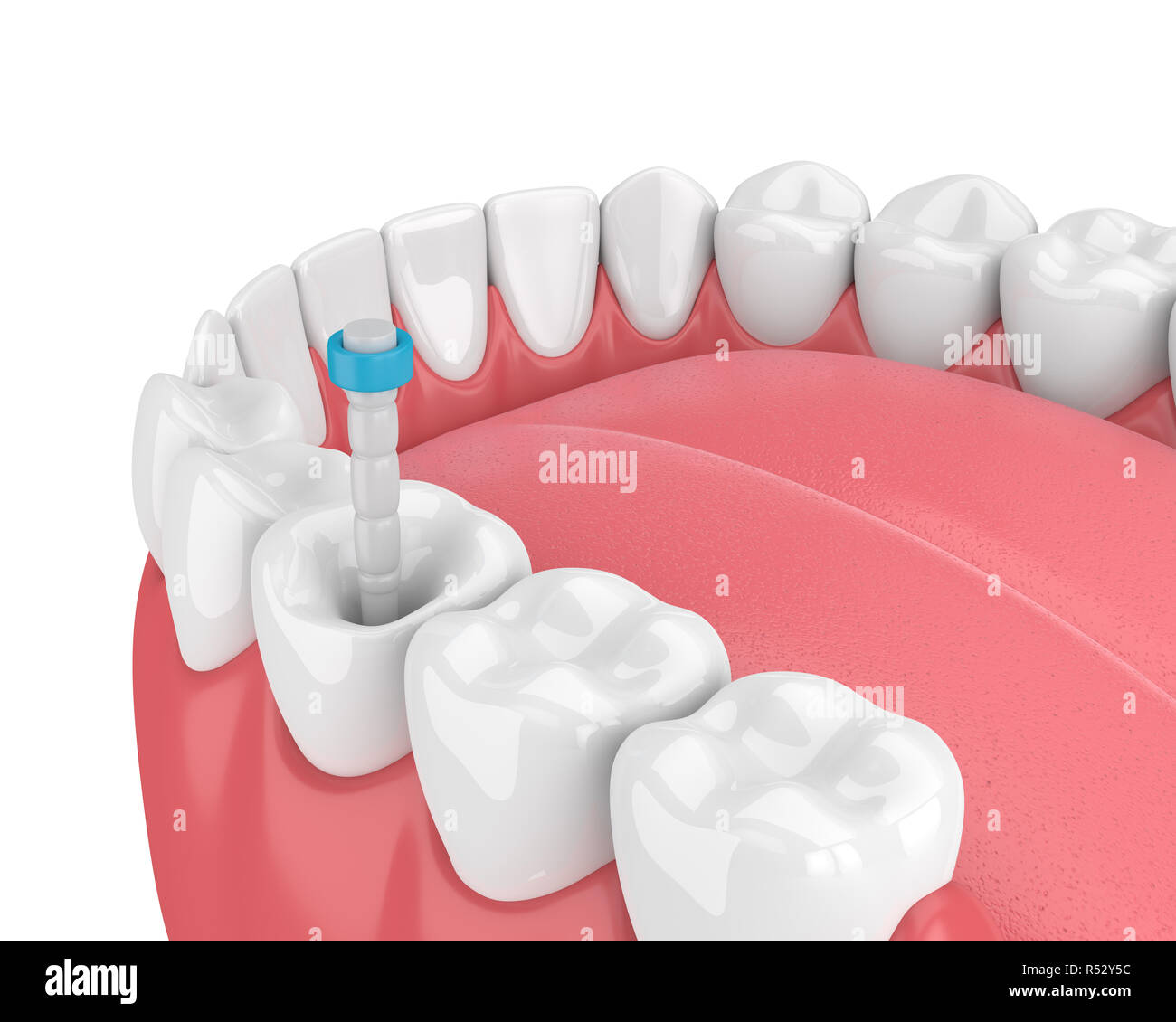 3d render of jaw with teeth and fiber post over white. Endodontic treatment concept Stock Photohttps://www.alamy.com/image-license-details/?v=1https://www.alamy.com/3d-render-of-jaw-with-teeth-and-fiber-post-over-white-endodontic-treatment-concept-image226873288.html
RFR52Y5C–3d render of jaw with teeth and fiber post over white. Endodontic treatment concept
 3d render of tooth with stainless steel dental post and filling in gums. Endodontic treatment concept Stock Photohttps://www.alamy.com/image-license-details/?v=1https://www.alamy.com/3d-render-of-tooth-with-stainless-steel-dental-post-and-filling-in-gums-endodontic-treatment-concept-image226144832.html
RFR3WP14–3d render of tooth with stainless steel dental post and filling in gums. Endodontic treatment concept
 3d render of jaw with teeth and fiber post over white. Endodontic treatment concept Stock Photohttps://www.alamy.com/image-license-details/?v=1https://www.alamy.com/3d-render-of-jaw-with-teeth-and-fiber-post-over-white-endodontic-treatment-concept-image226873332.html
RFR52Y70–3d render of jaw with teeth and fiber post over white. Endodontic treatment concept
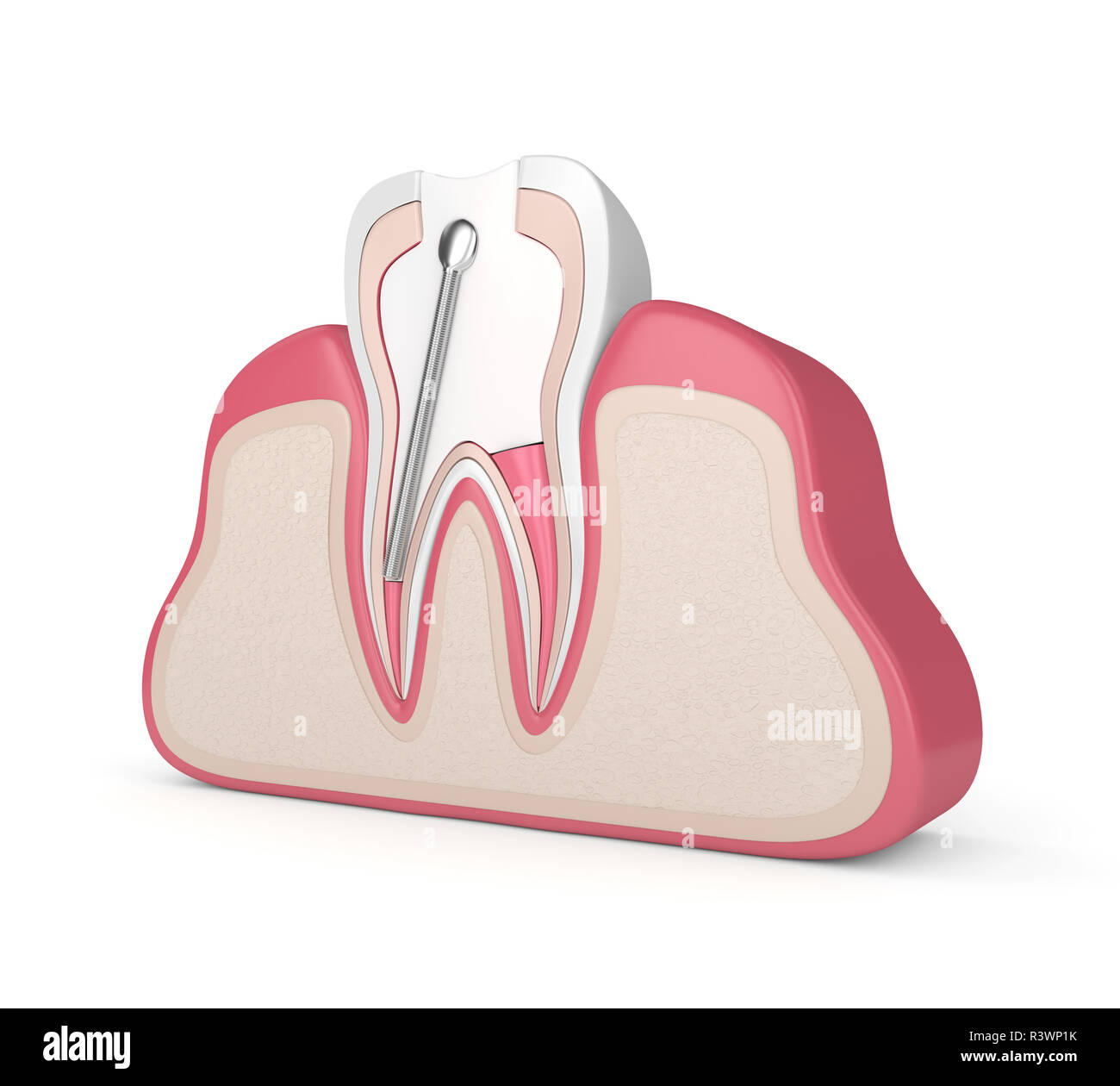 3d render of tooth with stainless steel dental post and filling in gums. Endodontic treatment concept Stock Photohttps://www.alamy.com/image-license-details/?v=1https://www.alamy.com/3d-render-of-tooth-with-stainless-steel-dental-post-and-filling-in-gums-endodontic-treatment-concept-image226144847.html
RFR3WP1K–3d render of tooth with stainless steel dental post and filling in gums. Endodontic treatment concept
 3d render of jaw with teeth and fiber post over white. Endodontic treatment concept Stock Photohttps://www.alamy.com/image-license-details/?v=1https://www.alamy.com/3d-render-of-jaw-with-teeth-and-fiber-post-over-white-endodontic-treatment-concept-image226873289.html
RFR52Y5D–3d render of jaw with teeth and fiber post over white. Endodontic treatment concept
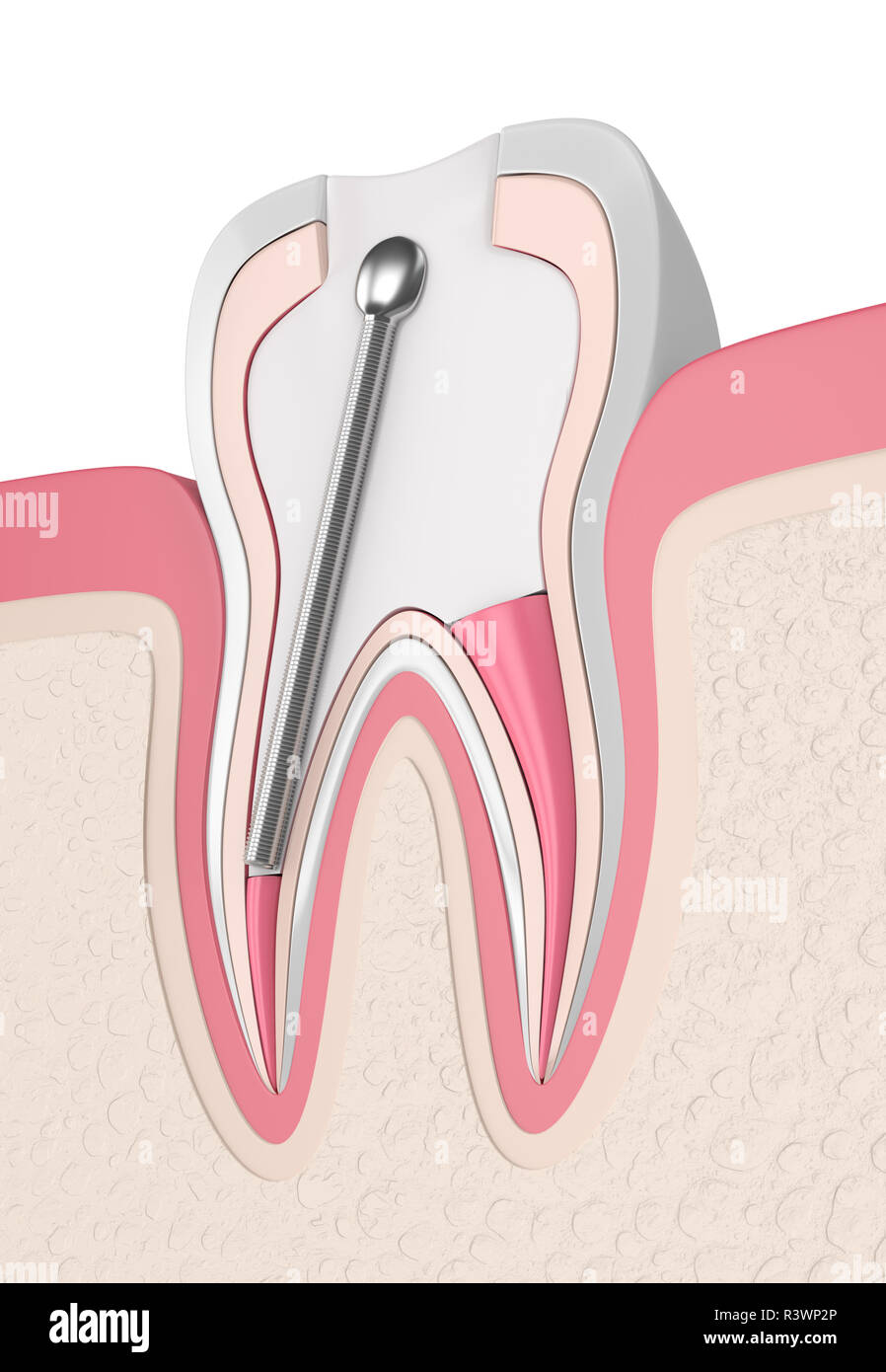 3d render of tooth with stainless steel dental post and filling in gums. Endodontic treatment concept Stock Photohttps://www.alamy.com/image-license-details/?v=1https://www.alamy.com/3d-render-of-tooth-with-stainless-steel-dental-post-and-filling-in-gums-endodontic-treatment-concept-image226144878.html
RFR3WP2P–3d render of tooth with stainless steel dental post and filling in gums. Endodontic treatment concept
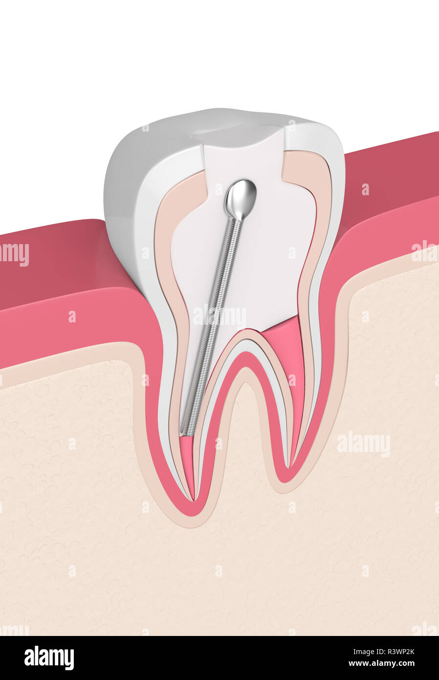 3d render of tooth with stainless steel dental post and filling in gums. Endodontic treatment concept Stock Photohttps://www.alamy.com/image-license-details/?v=1https://www.alamy.com/3d-render-of-tooth-with-stainless-steel-dental-post-and-filling-in-gums-endodontic-treatment-concept-image226144875.html
RFR3WP2K–3d render of tooth with stainless steel dental post and filling in gums. Endodontic treatment concept
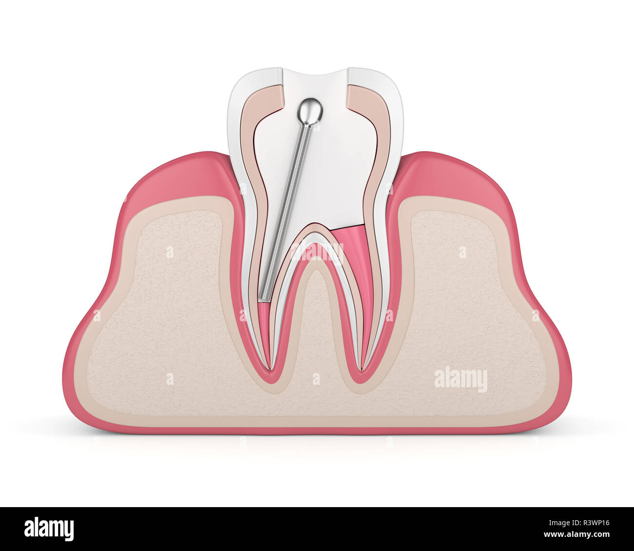 3d render of tooth with stainless steel dental post and filling in gums. Endodontic treatment concept Stock Photohttps://www.alamy.com/image-license-details/?v=1https://www.alamy.com/3d-render-of-tooth-with-stainless-steel-dental-post-and-filling-in-gums-endodontic-treatment-concept-image226144834.html
RFR3WP16–3d render of tooth with stainless steel dental post and filling in gums. Endodontic treatment concept
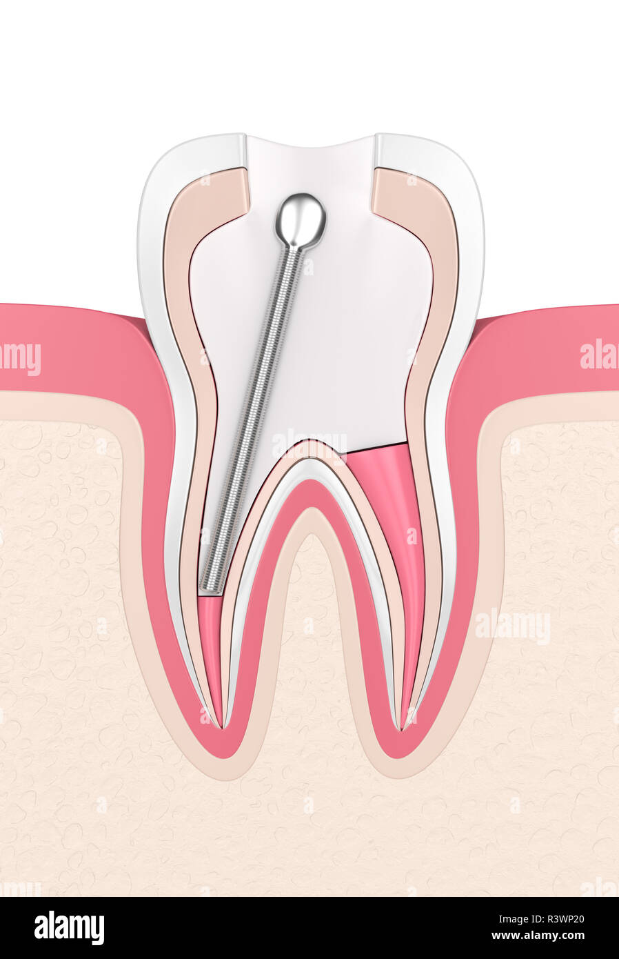 3d render of tooth with stainless steel dental post and filling in gums. Endodontic treatment concept Stock Photohttps://www.alamy.com/image-license-details/?v=1https://www.alamy.com/3d-render-of-tooth-with-stainless-steel-dental-post-and-filling-in-gums-endodontic-treatment-concept-image226144856.html
RFR3WP20–3d render of tooth with stainless steel dental post and filling in gums. Endodontic treatment concept
 3d render of tooth with dental stainless steel post in gums over white. Endodontic treatment concept Stock Photohttps://www.alamy.com/image-license-details/?v=1https://www.alamy.com/3d-render-of-tooth-with-dental-stainless-steel-post-in-gums-over-white-endodontic-treatment-concept-image227079032.html
RFR5C9HC–3d render of tooth with dental stainless steel post in gums over white. Endodontic treatment concept
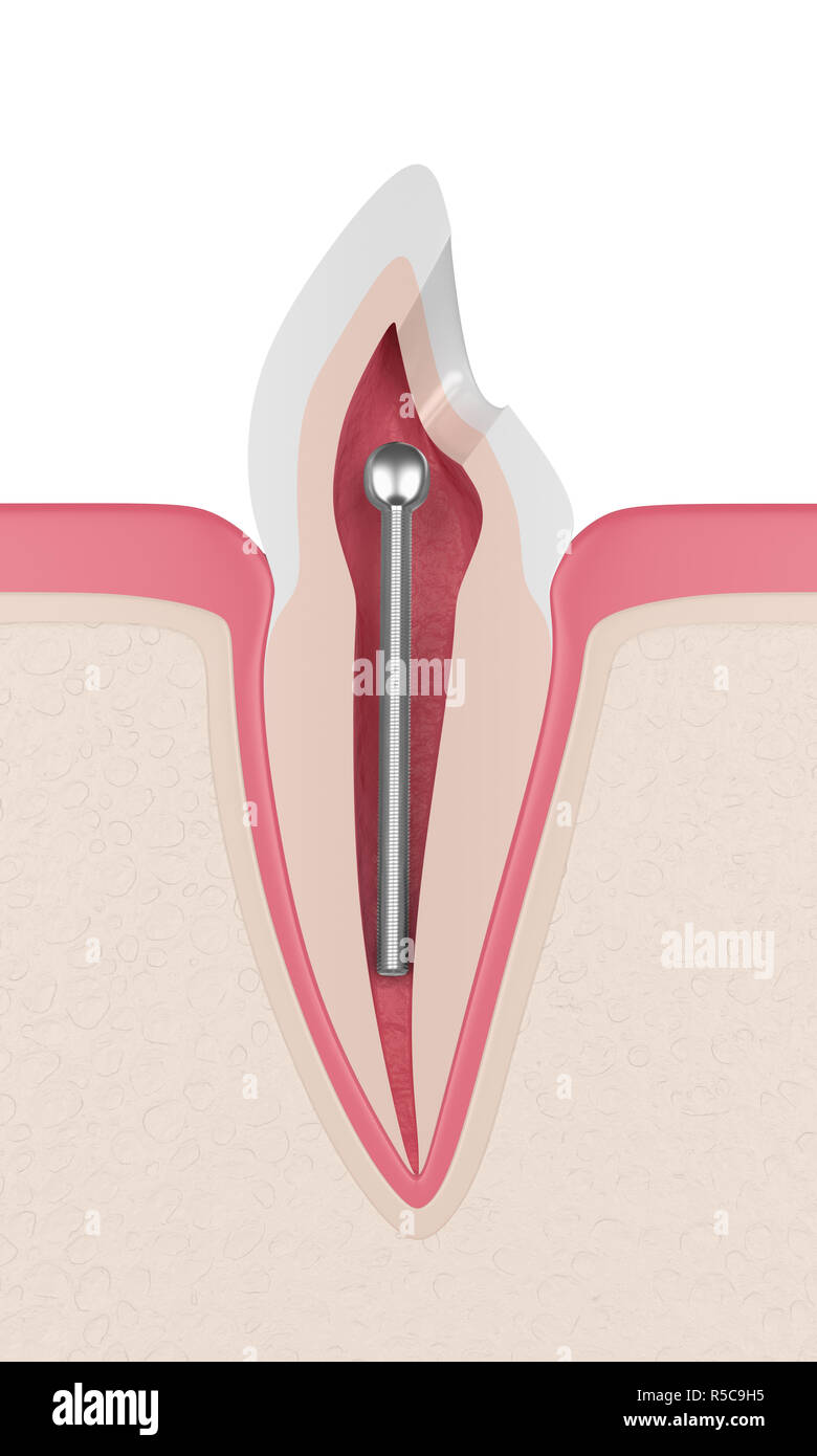 3d render of tooth with dental stainless steel post in gums. Endodontic treatment concept Stock Photohttps://www.alamy.com/image-license-details/?v=1https://www.alamy.com/3d-render-of-tooth-with-dental-stainless-steel-post-in-gums-endodontic-treatment-concept-image227079025.html
RFR5C9H5–3d render of tooth with dental stainless steel post in gums. Endodontic treatment concept
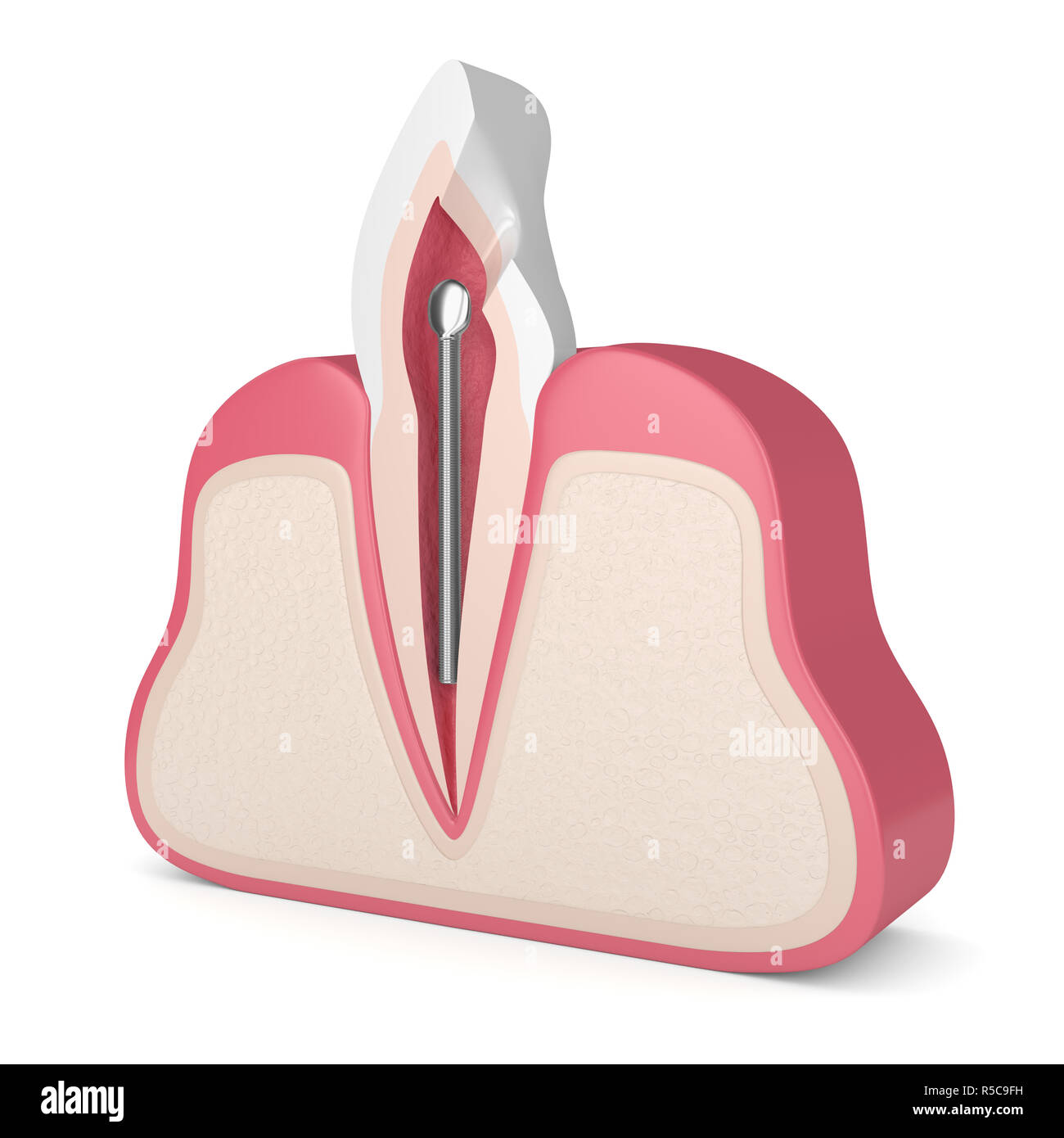 3d render of tooth with dental stainless steel post in gums over white. Endodontic treatment concept Stock Photohttps://www.alamy.com/image-license-details/?v=1https://www.alamy.com/3d-render-of-tooth-with-dental-stainless-steel-post-in-gums-over-white-endodontic-treatment-concept-image227078981.html
RFR5C9FH–3d render of tooth with dental stainless steel post in gums over white. Endodontic treatment concept
 3d render of tooth with dental stainless steel post in gums. Endodontic treatment concept Stock Photohttps://www.alamy.com/image-license-details/?v=1https://www.alamy.com/3d-render-of-tooth-with-dental-stainless-steel-post-in-gums-endodontic-treatment-concept-image227079024.html
RFR5C9H4–3d render of tooth with dental stainless steel post in gums. Endodontic treatment concept
 3d render of tooth with dental stainless steel post in gums over white. Endodontic treatment concept Stock Photohttps://www.alamy.com/image-license-details/?v=1https://www.alamy.com/3d-render-of-tooth-with-dental-stainless-steel-post-in-gums-over-white-endodontic-treatment-concept-image227078978.html
RFR5C9FE–3d render of tooth with dental stainless steel post in gums over white. Endodontic treatment concept
 3d render of tooth with dental stainless steel post in gums over white. Endodontic treatment concept Stock Photohttps://www.alamy.com/image-license-details/?v=1https://www.alamy.com/3d-render-of-tooth-with-dental-stainless-steel-post-in-gums-over-white-endodontic-treatment-concept-image227078984.html
RFR5C9FM–3d render of tooth with dental stainless steel post in gums over white. Endodontic treatment concept
 3d render of jaw with teeth and fiber post over white background Stock Photohttps://www.alamy.com/image-license-details/?v=1https://www.alamy.com/3d-render-of-jaw-with-teeth-and-fiber-post-over-white-background-image256622649.html
RFTWE4PH–3d render of jaw with teeth and fiber post over white background
 3d render of tooth with fiber post over white background Stock Photohttps://www.alamy.com/image-license-details/?v=1https://www.alamy.com/3d-render-of-tooth-with-fiber-post-over-white-background-image256622512.html
RFTWE4HM–3d render of tooth with fiber post over white background
 3d render of teeth with fiber post over white background Stock Photohttps://www.alamy.com/image-license-details/?v=1https://www.alamy.com/3d-render-of-teeth-with-fiber-post-over-white-background-image256622946.html
RFTWE556–3d render of teeth with fiber post over white background
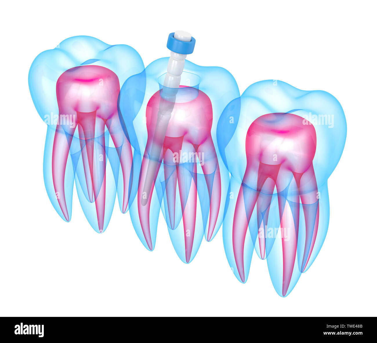 3d render of teeth with fiber post over white background Stock Photohttps://www.alamy.com/image-license-details/?v=1https://www.alamy.com/3d-render-of-teeth-with-fiber-post-over-white-background-image256622251.html
RFTWE48B–3d render of teeth with fiber post over white background
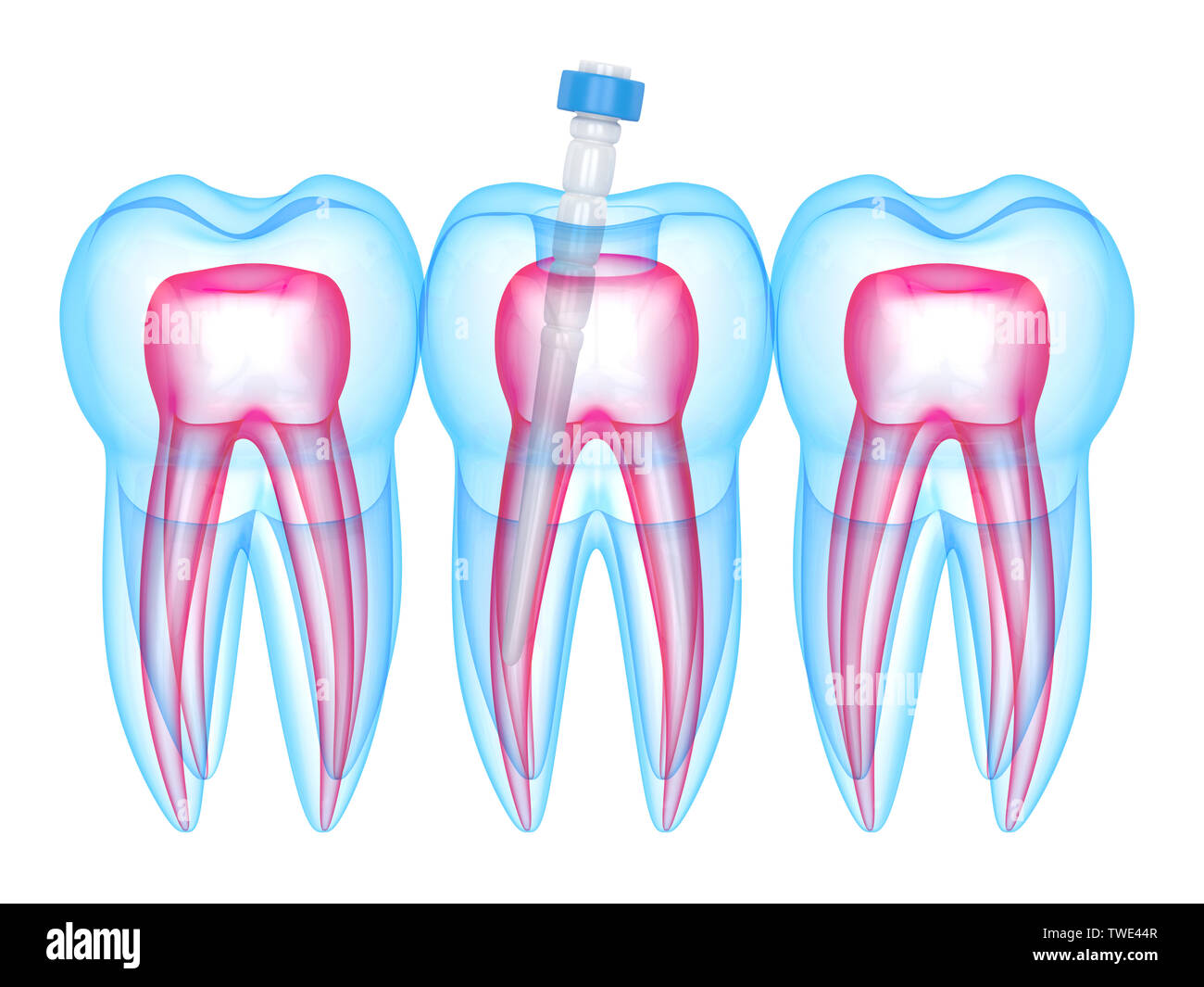 3d render of teeth with fiber post over white background Stock Photohttps://www.alamy.com/image-license-details/?v=1https://www.alamy.com/3d-render-of-teeth-with-fiber-post-over-white-background-image256622151.html
RFTWE44R–3d render of teeth with fiber post over white background
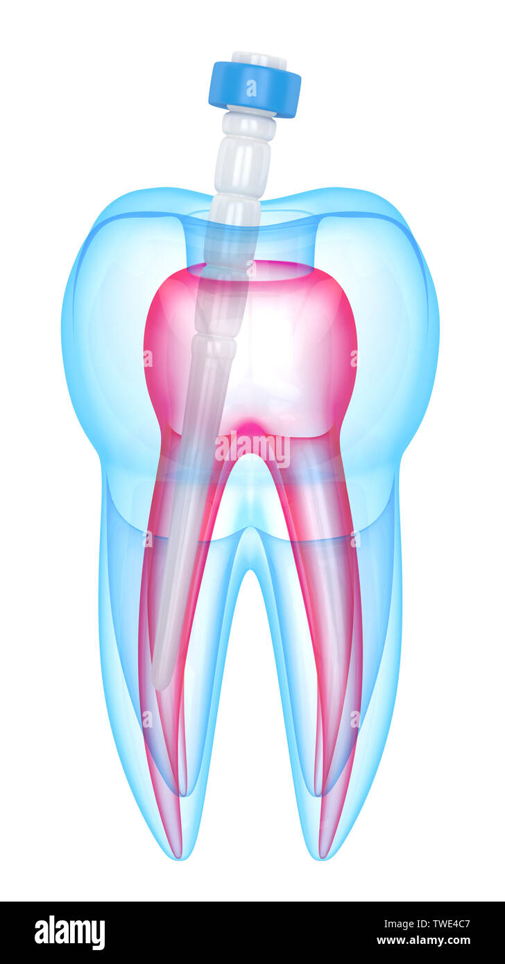 3d render of tooth with fiber post over white background Stock Photohttps://www.alamy.com/image-license-details/?v=1https://www.alamy.com/3d-render-of-tooth-with-fiber-post-over-white-background-image256622359.html
RFTWE4C7–3d render of tooth with fiber post over white background
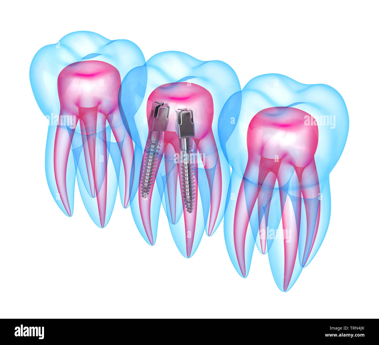 3d render of x-ray teeth with stainless steel dental post over white background. Endodontic treatment concept Stock Photohttps://www.alamy.com/image-license-details/?v=1https://www.alamy.com/3d-render-of-x-ray-teeth-with-stainless-steel-dental-post-over-white-background-endodontic-treatment-concept-image255546891.html
RFTRN4JK–3d render of x-ray teeth with stainless steel dental post over white background. Endodontic treatment concept
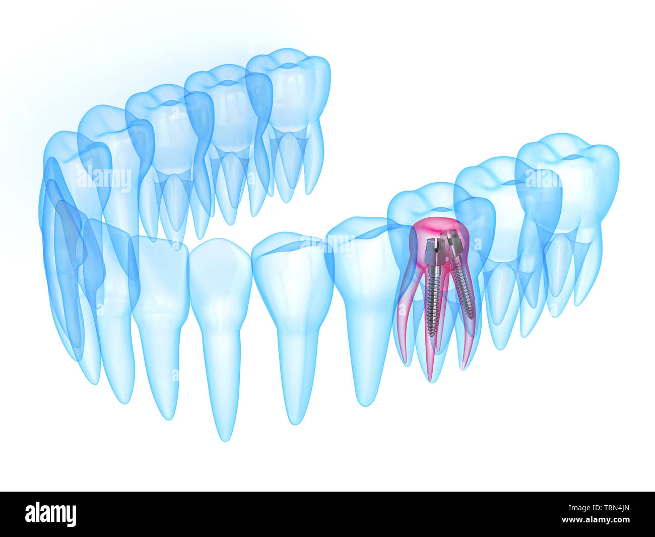 3d render of x-ray teeth with stainless steel dental post over white background. Endodontic treatment concept Stock Photohttps://www.alamy.com/image-license-details/?v=1https://www.alamy.com/3d-render-of-x-ray-teeth-with-stainless-steel-dental-post-over-white-background-endodontic-treatment-concept-image255546893.html
RFTRN4JN–3d render of x-ray teeth with stainless steel dental post over white background. Endodontic treatment concept
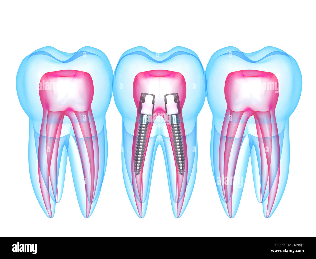 3d render of x-ray teeth with stainless steel dental post over white background. Endodontic treatment concept Stock Photohttps://www.alamy.com/image-license-details/?v=1https://www.alamy.com/3d-render-of-x-ray-teeth-with-stainless-steel-dental-post-over-white-background-endodontic-treatment-concept-image255546879.html
RFTRN4J7–3d render of x-ray teeth with stainless steel dental post over white background. Endodontic treatment concept
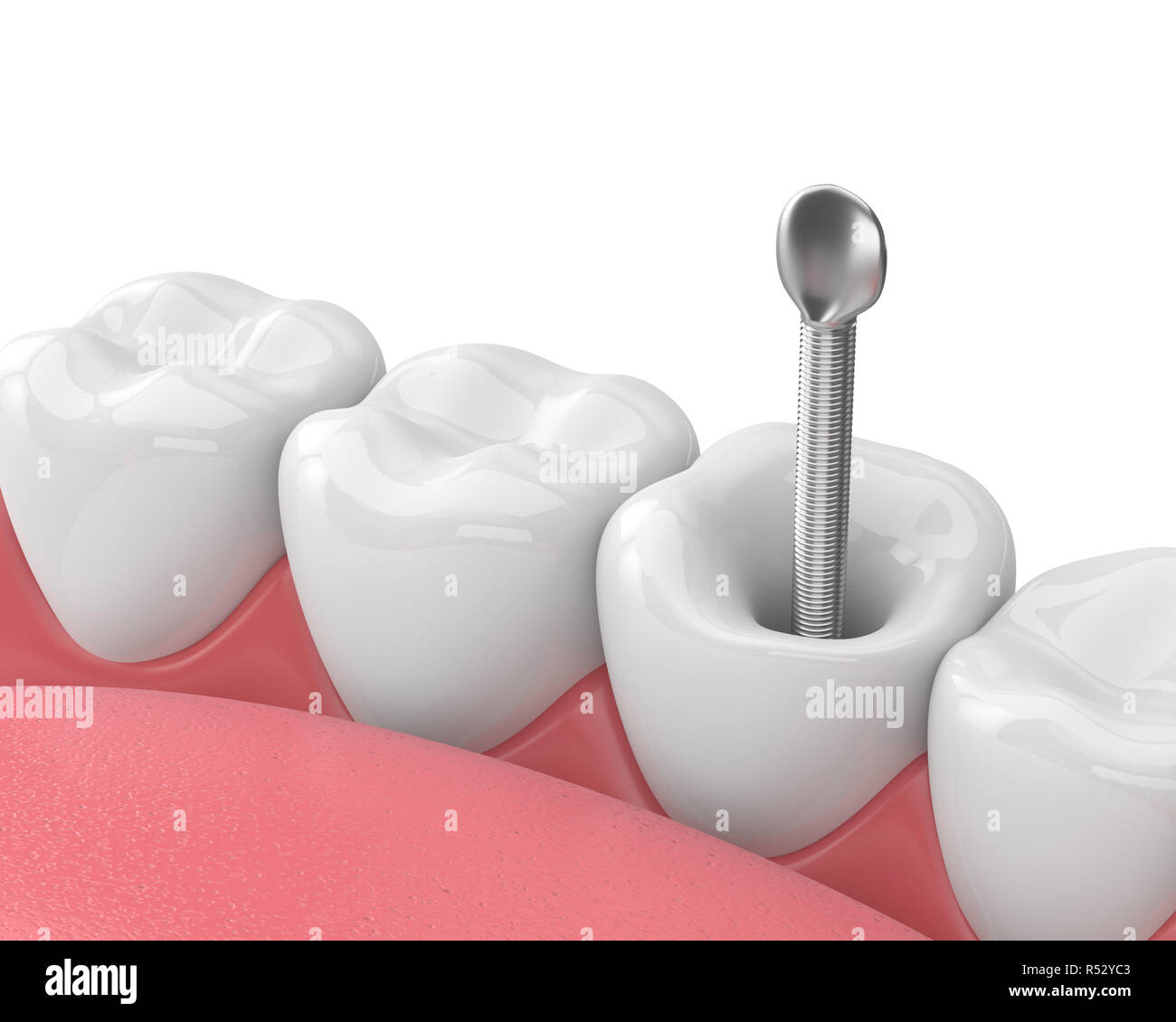 3d render of jaw with teeth and dental metal post over white. Endodontic treatment concept Stock Photohttps://www.alamy.com/image-license-details/?v=1https://www.alamy.com/3d-render-of-jaw-with-teeth-and-dental-metal-post-over-white-endodontic-treatment-concept-image226873475.html
RFR52YC3–3d render of jaw with teeth and dental metal post over white. Endodontic treatment concept
 3d render of incisor tooth with dental metal post in gums. Endodontic treatment concept Stock Photohttps://www.alamy.com/image-license-details/?v=1https://www.alamy.com/3d-render-of-incisor-tooth-with-dental-metal-post-in-gums-endodontic-treatment-concept-image226873716.html
RFR52YMM–3d render of incisor tooth with dental metal post in gums. Endodontic treatment concept
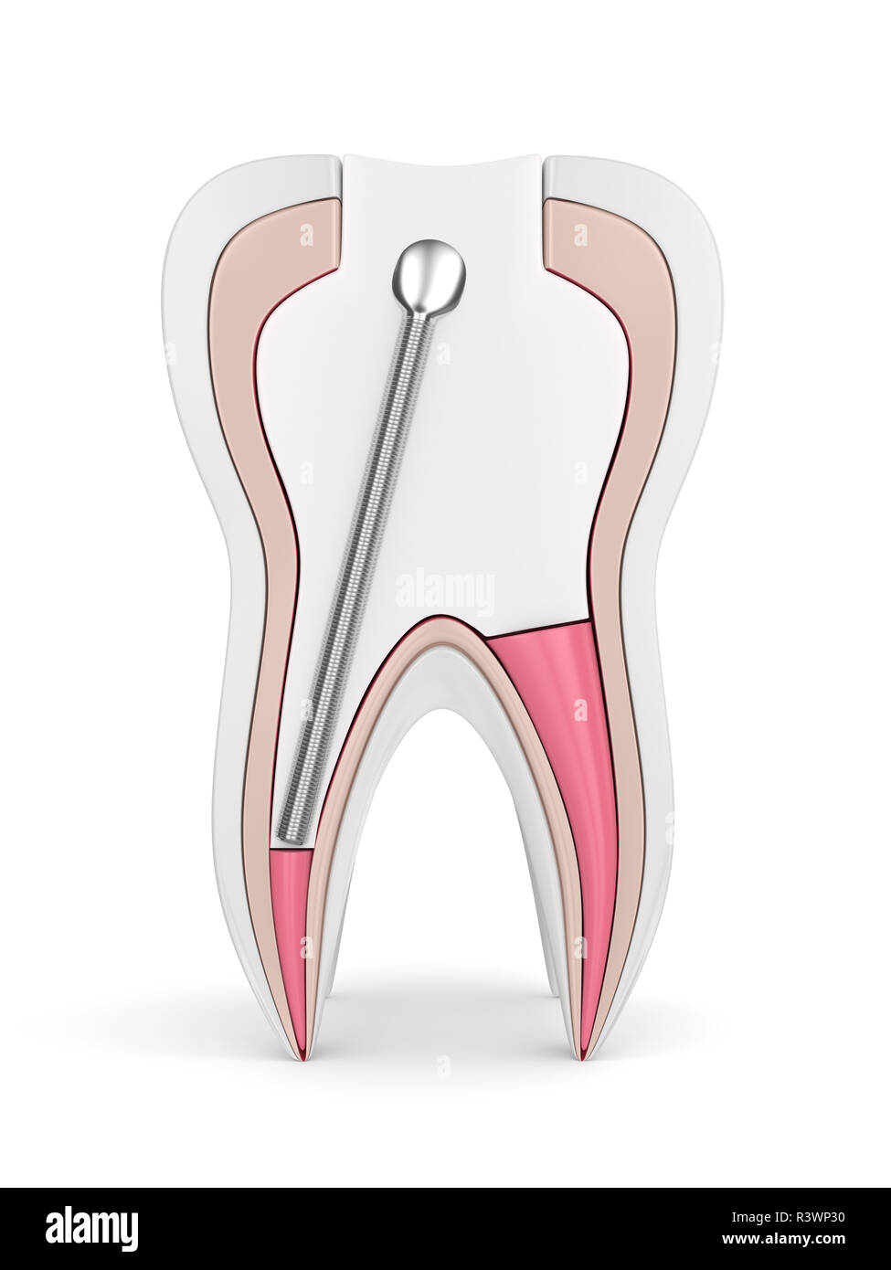 3d render of tooth with stainless steel dental post and filling over white background. Endodontic treatment concept Stock Photohttps://www.alamy.com/image-license-details/?v=1https://www.alamy.com/3d-render-of-tooth-with-stainless-steel-dental-post-and-filling-over-white-background-endodontic-treatment-concept-image226144884.html
RFR3WP30–3d render of tooth with stainless steel dental post and filling over white background. Endodontic treatment concept
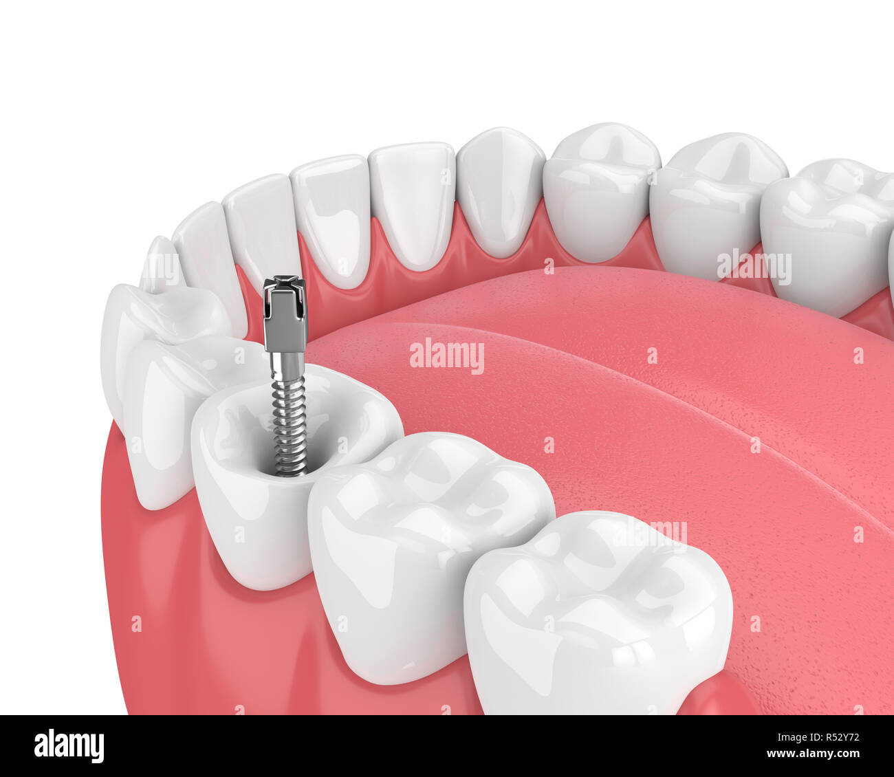 3d render of jaw with teeth and dental metal post over white. Endodontic treatment concept Stock Photohttps://www.alamy.com/image-license-details/?v=1https://www.alamy.com/3d-render-of-jaw-with-teeth-and-dental-metal-post-over-white-endodontic-treatment-concept-image226873334.html
RFR52Y72–3d render of jaw with teeth and dental metal post over white. Endodontic treatment concept
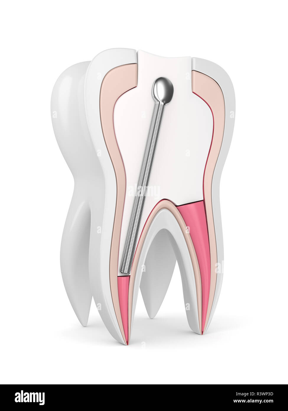 3d render of tooth with stainless steel dental post and filling over white background. Endodontic treatment concept Stock Photohttps://www.alamy.com/image-license-details/?v=1https://www.alamy.com/3d-render-of-tooth-with-stainless-steel-dental-post-and-filling-over-white-background-endodontic-treatment-concept-image226144897.html
RFR3WP3D–3d render of tooth with stainless steel dental post and filling over white background. Endodontic treatment concept
 3d render of jaw with teeth and dental metal post over white. Endodontic treatment concept Stock Photohttps://www.alamy.com/image-license-details/?v=1https://www.alamy.com/3d-render-of-jaw-with-teeth-and-dental-metal-post-over-white-endodontic-treatment-concept-image226873385.html
RFR52Y8W–3d render of jaw with teeth and dental metal post over white. Endodontic treatment concept
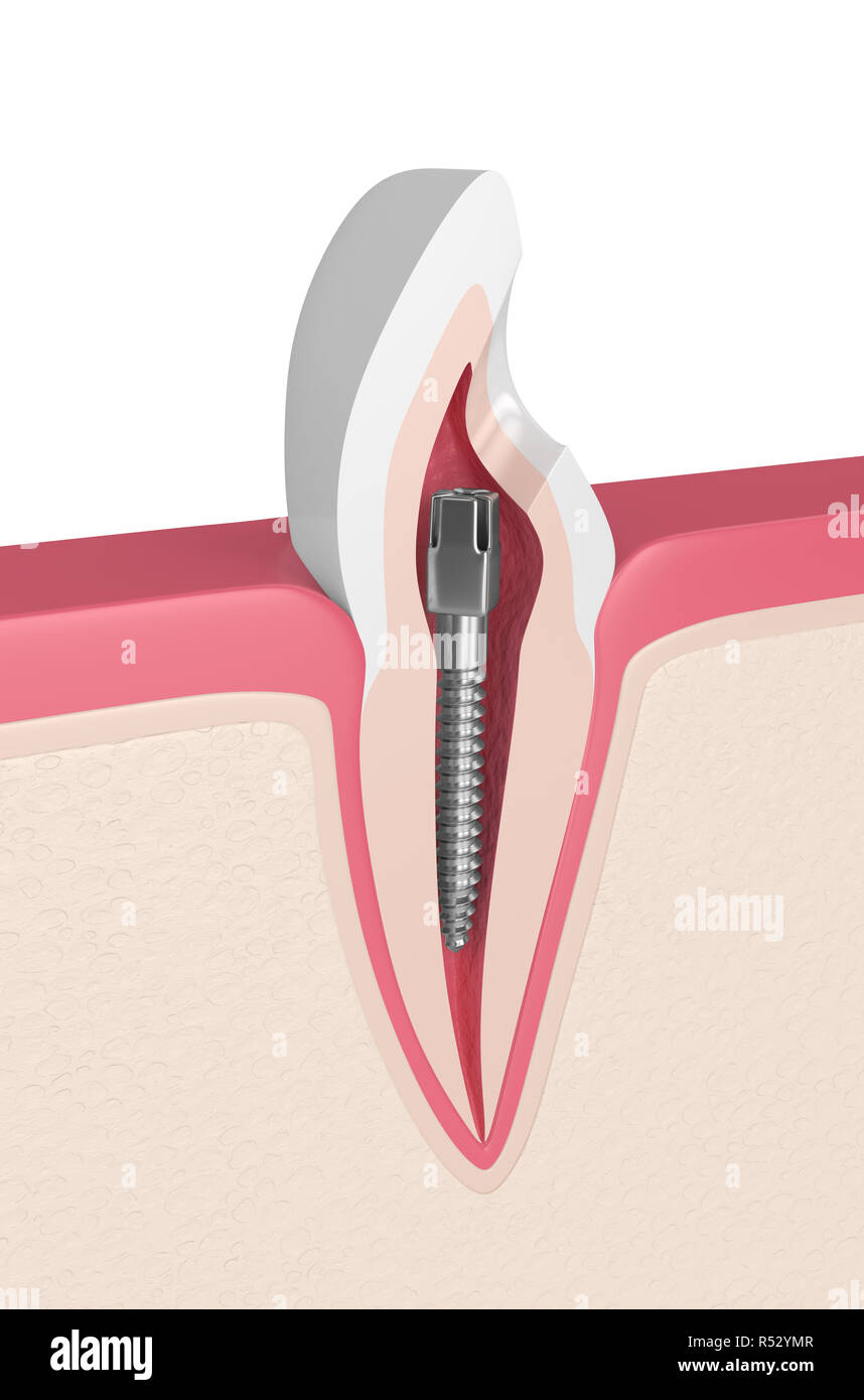 3d render of incisor tooth with dental metal post in gums. Endodontic treatment concept Stock Photohttps://www.alamy.com/image-license-details/?v=1https://www.alamy.com/3d-render-of-incisor-tooth-with-dental-metal-post-in-gums-endodontic-treatment-concept-image226873719.html
RFR52YMR–3d render of incisor tooth with dental metal post in gums. Endodontic treatment concept