Rough endoplasmic reticulum cell Cut Out Stock Images
(55)See rough endoplasmic reticulum cell stock video clipsRough endoplasmic reticulum cell Cut Out Stock Images
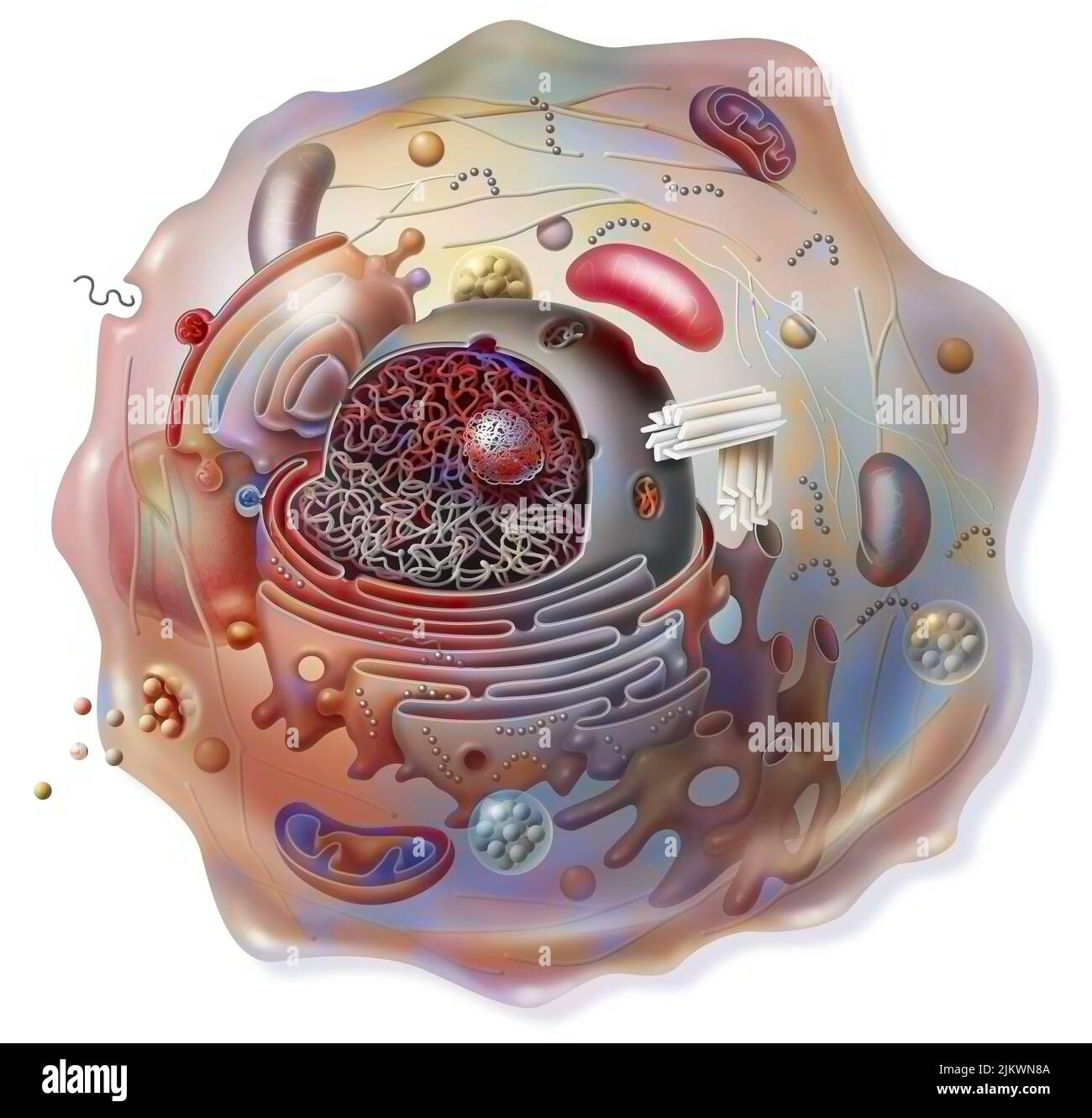 Cell sectional view with all the main organelles: nucleus, reticulum. Stock Photohttps://www.alamy.com/image-license-details/?v=1https://www.alamy.com/cell-sectional-view-with-all-the-main-organelles-nucleus-reticulum-image476923898.html
RF2JKWN8A–Cell sectional view with all the main organelles: nucleus, reticulum.
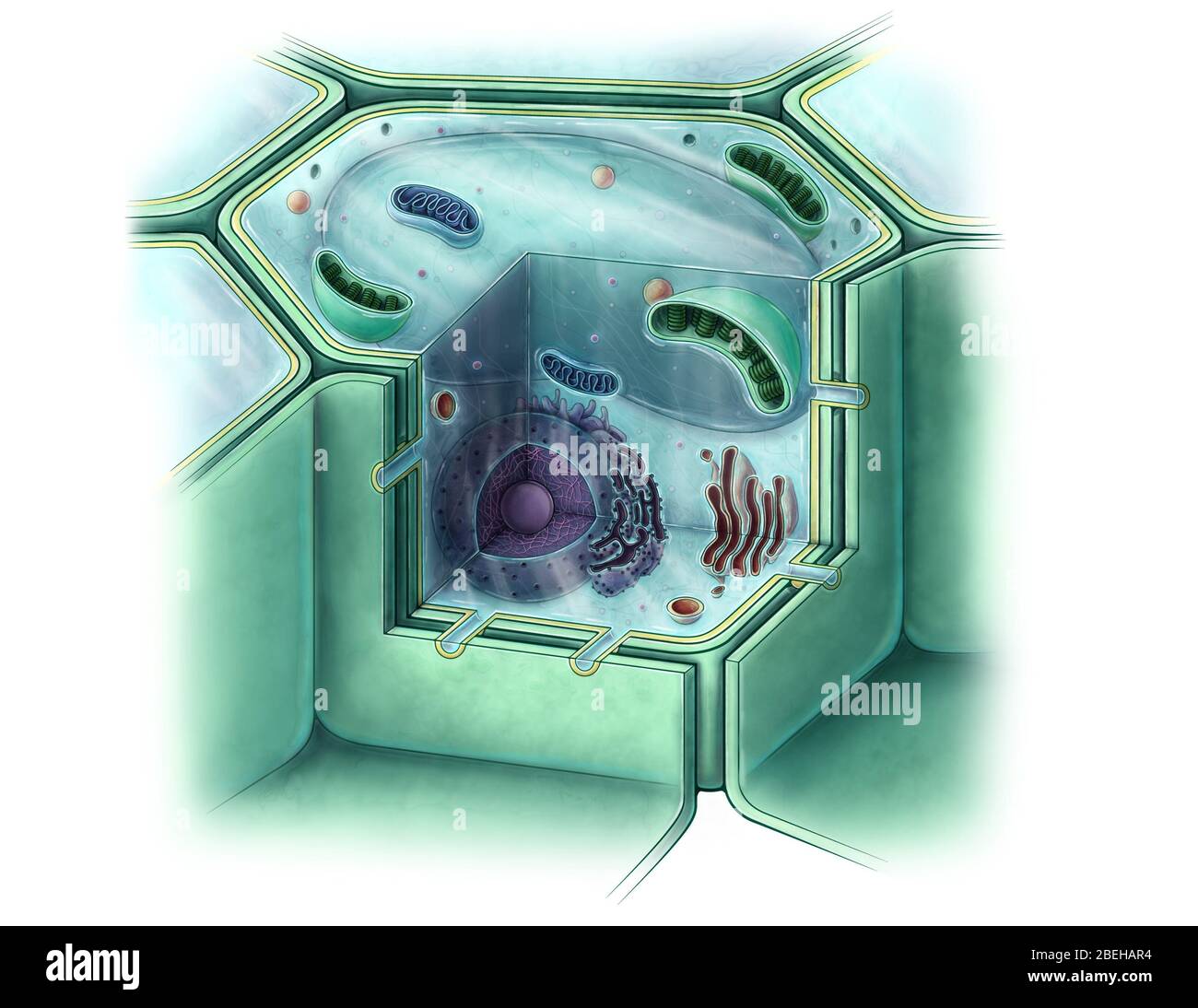 Plant Cell, Illustration Stock Photohttps://www.alamy.com/image-license-details/?v=1https://www.alamy.com/plant-cell-illustration-image353194216.html
RM2BEHAR4–Plant Cell, Illustration
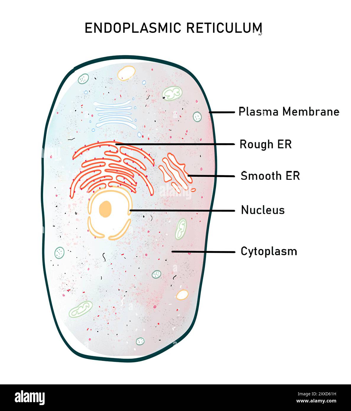 Endoplasmic reticulum, illustration. The endoplasmic reticulum (ER) is a dynamic cell organelle that can be of two types, wither-rough endoplasmic reticulum (RER), or smooth endoplasmic reticulum (SER). They both consist of a continuous system of flattened tubules and sacs bound by a single membrane. Endoplasmic reticulum serves many functions including protein synthesis and folding, lipid synthesis and calcium storage. Stock Photohttps://www.alamy.com/image-license-details/?v=1https://www.alamy.com/endoplasmic-reticulum-illustration-the-endoplasmic-reticulum-er-is-a-dynamic-cell-organelle-that-can-be-of-two-types-wither-rough-endoplasmic-reticulum-rer-or-smooth-endoplasmic-reticulum-ser-they-both-consist-of-a-continuous-system-of-flattened-tubules-and-sacs-bound-by-a-single-membrane-endoplasmic-reticulum-serves-many-functions-including-protein-synthesis-and-folding-lipid-synthesis-and-calcium-storage-image618634061.html
RF2XXD61H–Endoplasmic reticulum, illustration. The endoplasmic reticulum (ER) is a dynamic cell organelle that can be of two types, wither-rough endoplasmic reticulum (RER), or smooth endoplasmic reticulum (SER). They both consist of a continuous system of flattened tubules and sacs bound by a single membrane. Endoplasmic reticulum serves many functions including protein synthesis and folding, lipid synthesis and calcium storage.
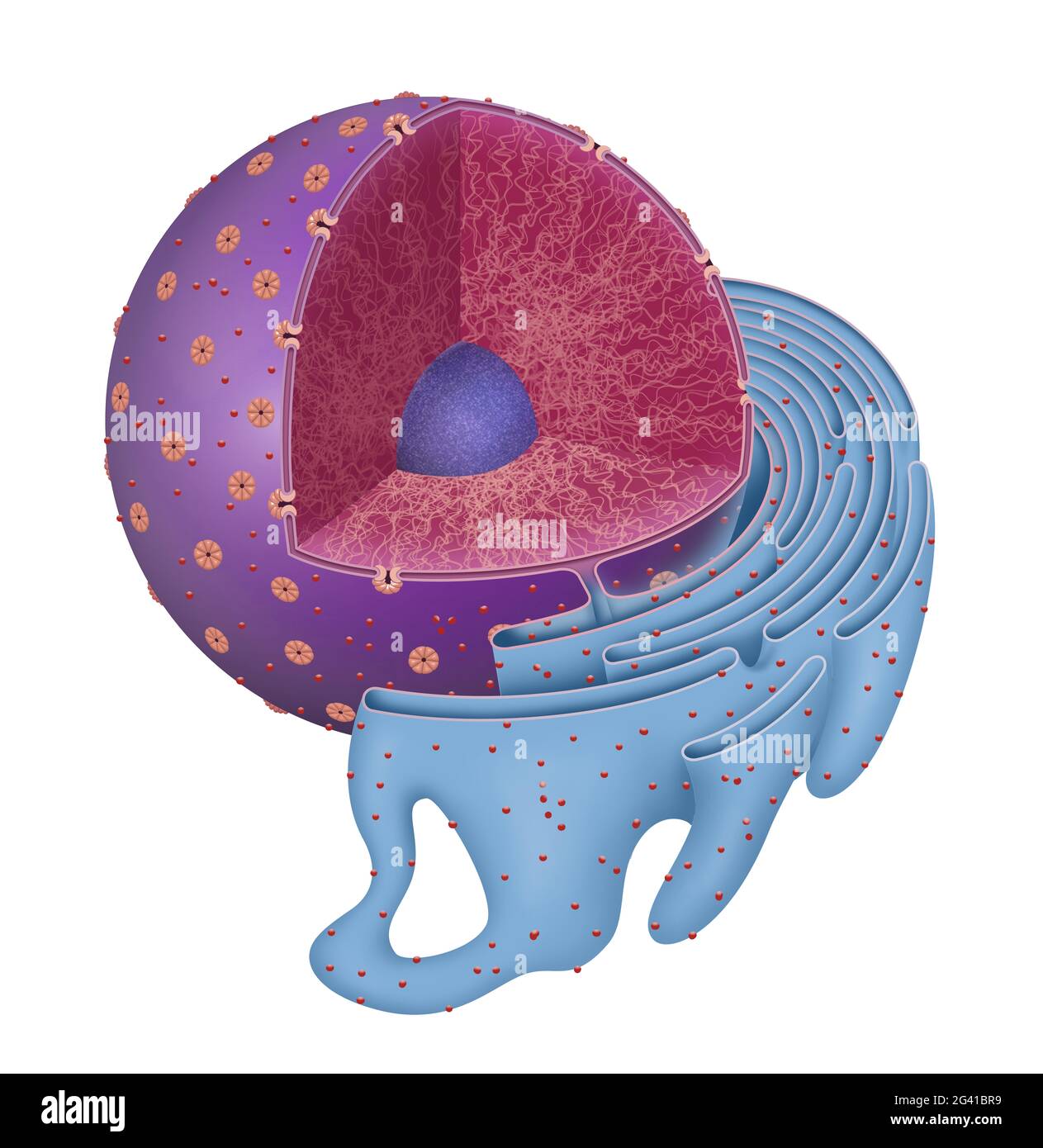 Structure of Nucleus and Rough endoplasmic reticulum Stock Photohttps://www.alamy.com/image-license-details/?v=1https://www.alamy.com/structure-of-nucleus-and-rough-endoplasmic-reticulum-image432749053.html
RF2G41BR9–Structure of Nucleus and Rough endoplasmic reticulum
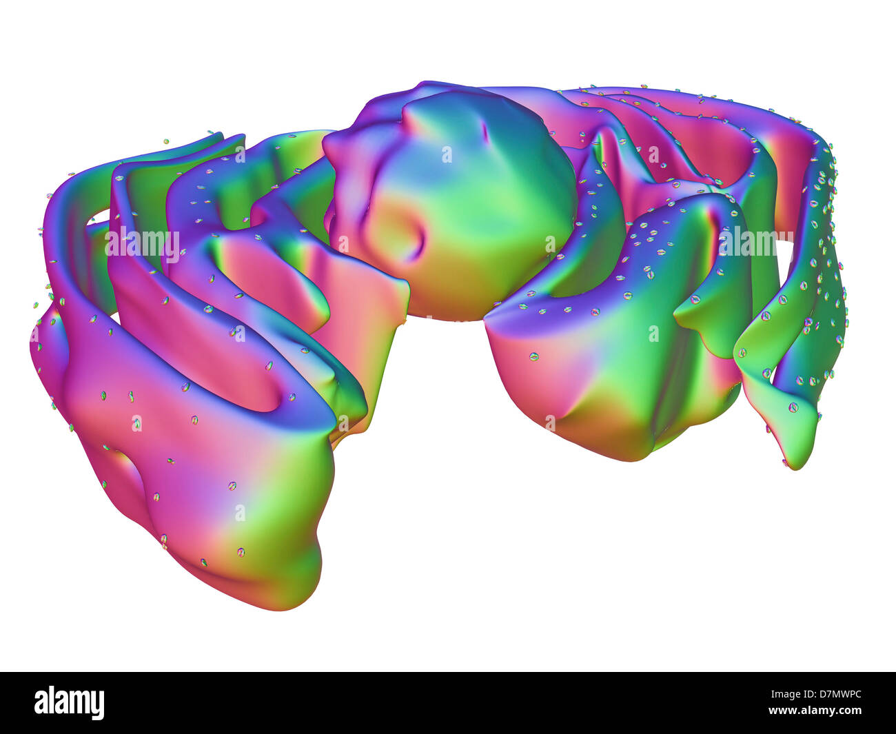 Nucleus and endoplasmic reticulum Stock Photohttps://www.alamy.com/image-license-details/?v=1https://www.alamy.com/stock-photo-nucleus-and-endoplasmic-reticulum-56392964.html
RFD7MWPC–Nucleus and endoplasmic reticulum
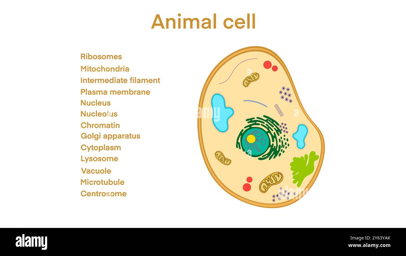 animal cell anatomy, biological animal cell with organelles cross section, Animal cell with placed text annotations to all organelles, Animal cell Stock Photohttps://www.alamy.com/image-license-details/?v=1https://www.alamy.com/animal-cell-anatomy-biological-animal-cell-with-organelles-cross-section-animal-cell-with-placed-text-annotations-to-all-organelles-animal-cell-image623348507.html
RF2Y63YAK–animal cell anatomy, biological animal cell with organelles cross section, Animal cell with placed text annotations to all organelles, Animal cell
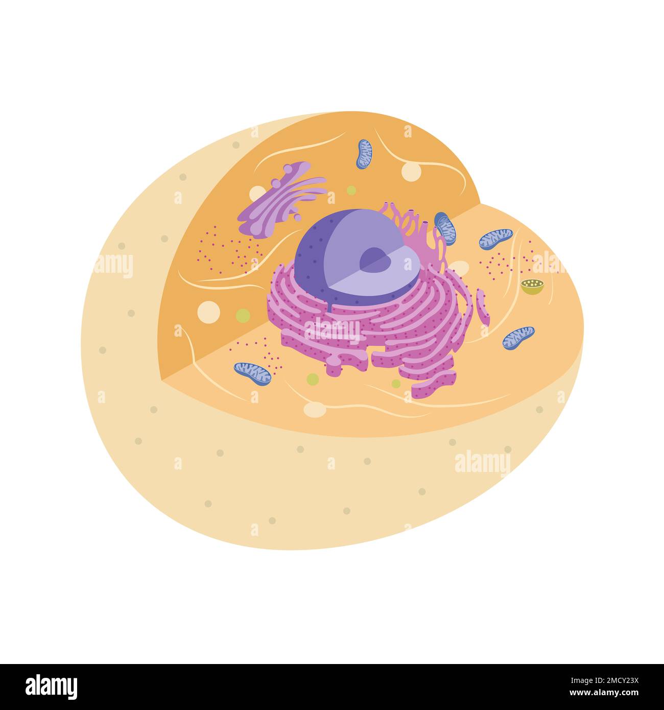 Illustration of animal cell with organelles Stock Photohttps://www.alamy.com/image-license-details/?v=1https://www.alamy.com/illustration-of-animal-cell-with-organelles-image507070926.html
RF2MCY23X–Illustration of animal cell with organelles
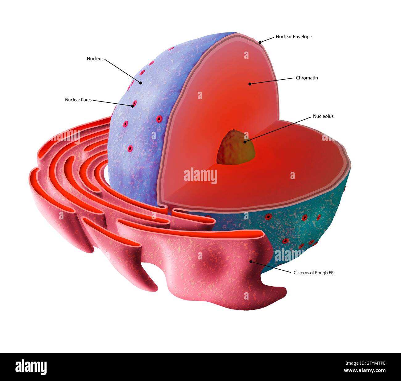 Cell nucleus structure, illustration Stock Photohttps://www.alamy.com/image-license-details/?v=1https://www.alamy.com/cell-nucleus-structure-illustration-image430103030.html
RF2FYMTPE–Cell nucleus structure, illustration
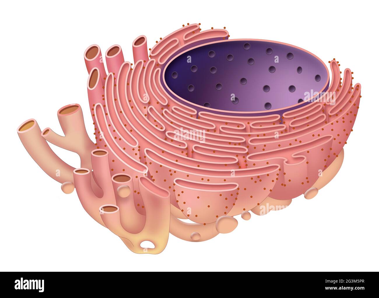 The endoplasmic reticulum is organelle found in eukaryotic cells Stock Photohttps://www.alamy.com/image-license-details/?v=1https://www.alamy.com/the-endoplasmic-reticulum-is-organelle-found-in-eukaryotic-cells-image432546767.html
RF2G3M5PR–The endoplasmic reticulum is organelle found in eukaryotic cells
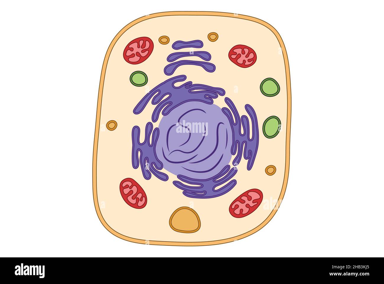 Simple cell structure medical illustration, mitochondria, ger, endoplasmic reticulum, simple illustration Stock Photohttps://www.alamy.com/image-license-details/?v=1https://www.alamy.com/simple-cell-structure-medical-illustration-mitochondria-ger-endoplasmic-reticulum-simple-illustration-image454312045.html
RF2HB3KJ5–Simple cell structure medical illustration, mitochondria, ger, endoplasmic reticulum, simple illustration
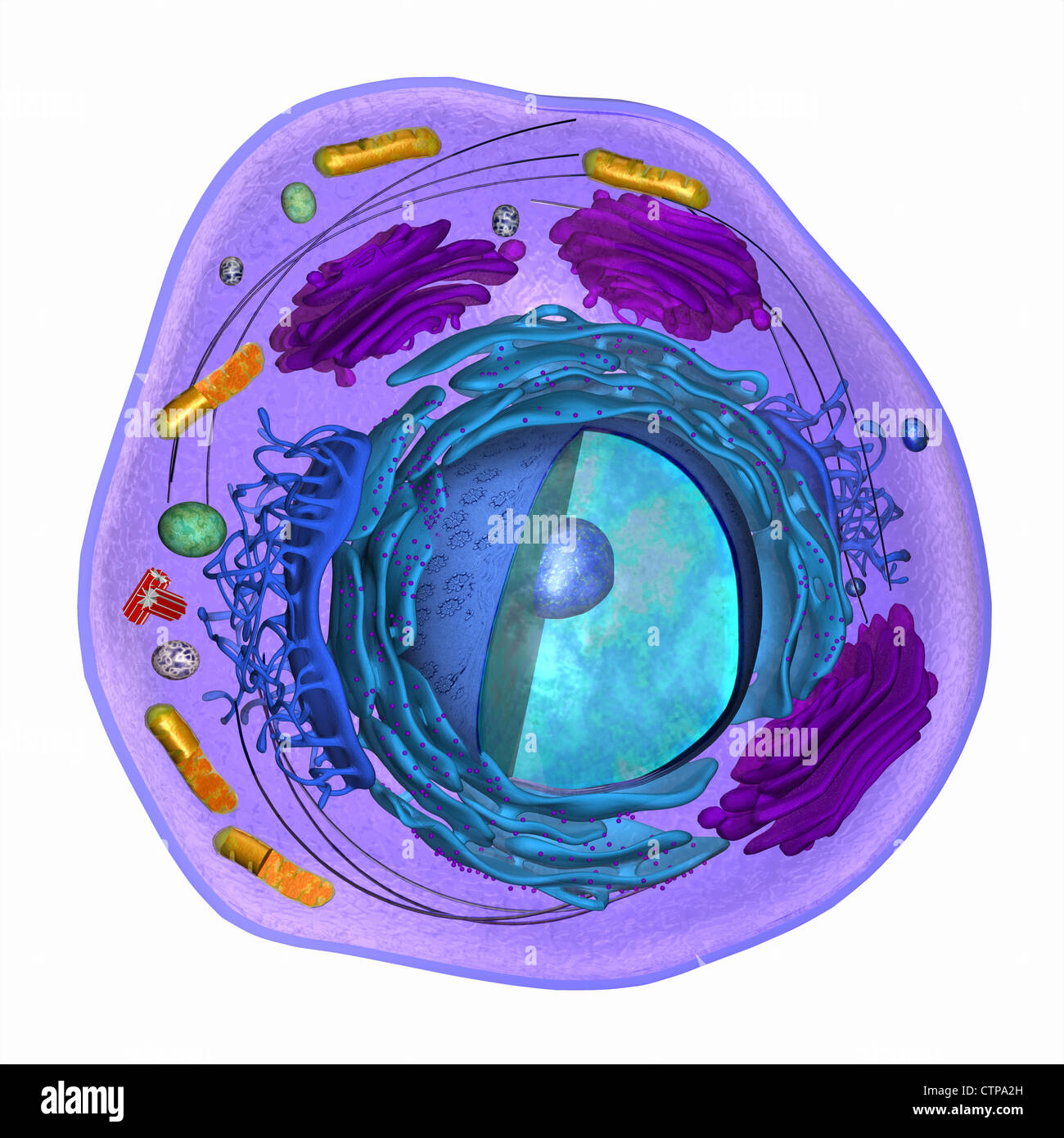 3D model of a eukaryotic cell Stock Photohttps://www.alamy.com/image-license-details/?v=1https://www.alamy.com/stock-photo-3d-model-of-a-eukaryotic-cell-49663337.html
RMCTPA2H–3D model of a eukaryotic cell
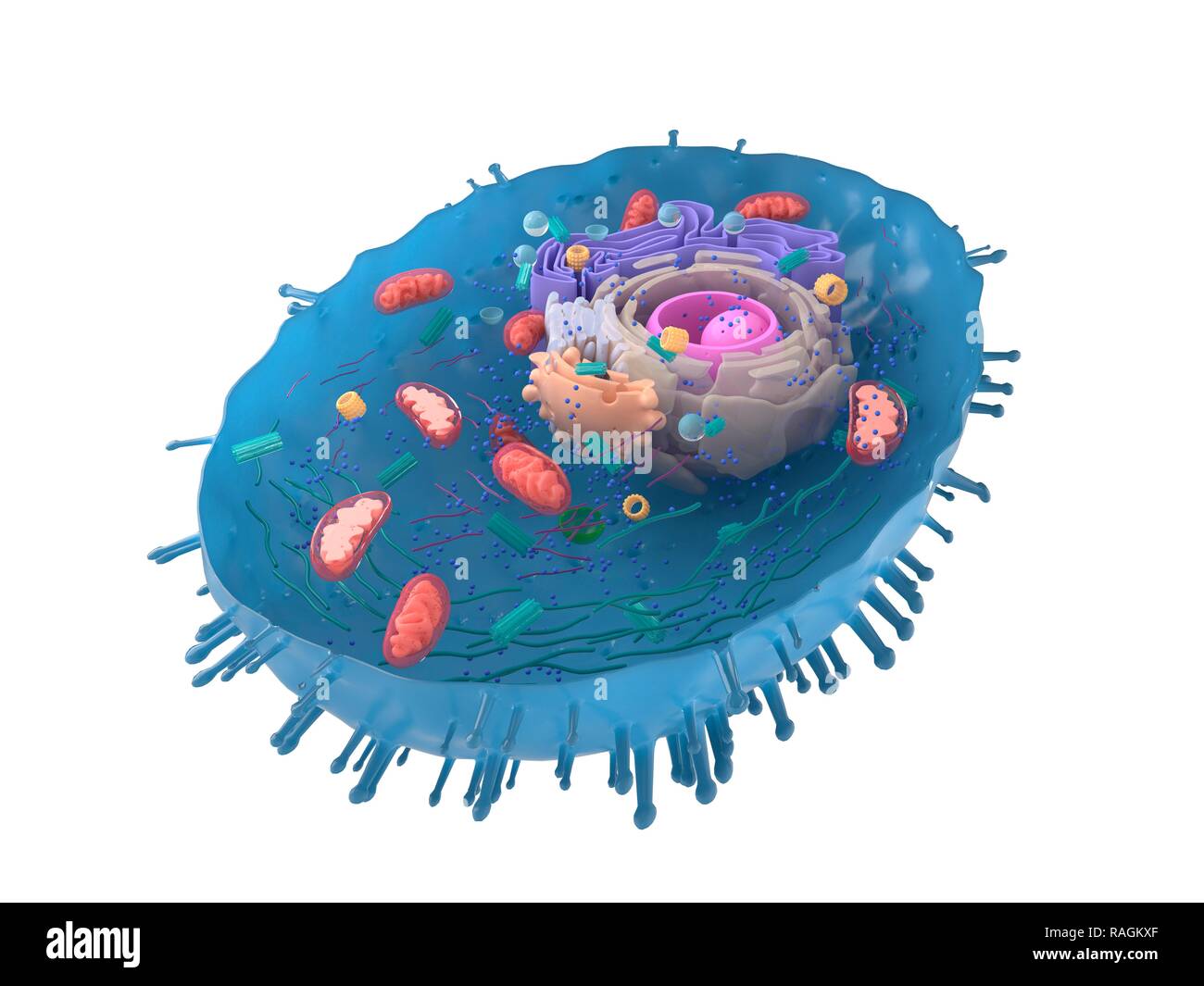 Illustration of a human cell cross-section. Stock Photohttps://www.alamy.com/image-license-details/?v=1https://www.alamy.com/illustration-of-a-human-cell-cross-section-image230248215.html
RFRAGKXF–Illustration of a human cell cross-section.
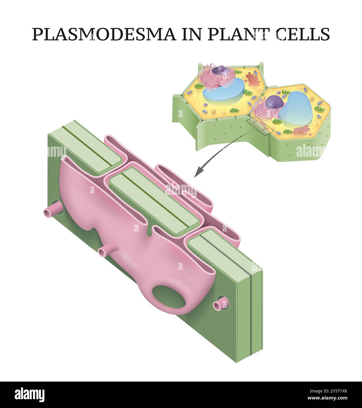 Plasmodesma in plant cells illustration Stock Photohttps://www.alamy.com/image-license-details/?v=1https://www.alamy.com/plasmodesma-in-plant-cells-illustration-image622801723.html
RF2Y571XK–Plasmodesma in plant cells illustration
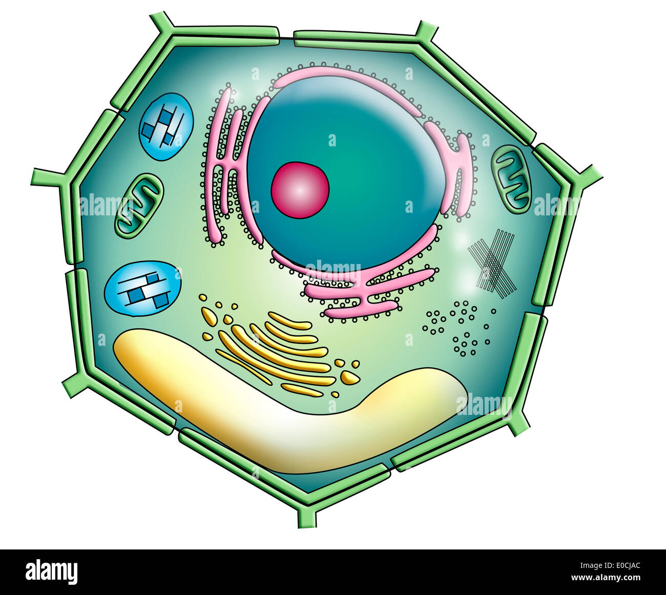 Plant cell, drawing Stock Photohttps://www.alamy.com/image-license-details/?v=1https://www.alamy.com/plant-cell-drawing-image69119300.html
RME0CJAC–Plant cell, drawing
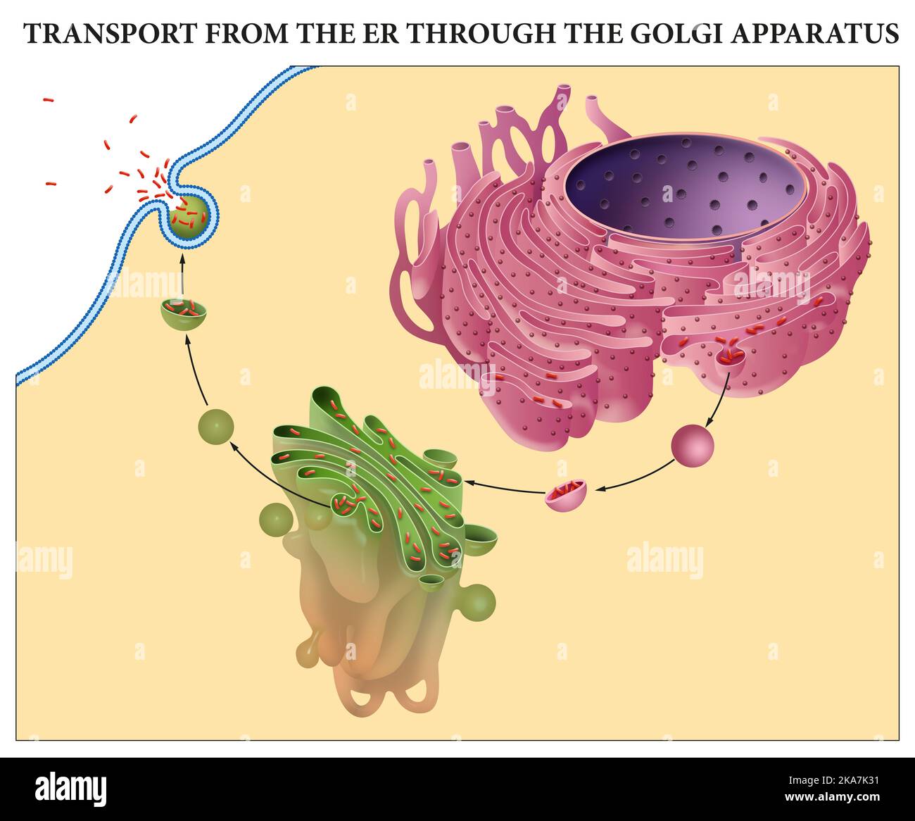 Transport from the ER through the Golgi Apparatus Stock Photohttps://www.alamy.com/image-license-details/?v=1https://www.alamy.com/transport-from-the-er-through-the-golgi-apparatus-image488205509.html
RF2KA7K31–Transport from the ER through the Golgi Apparatus
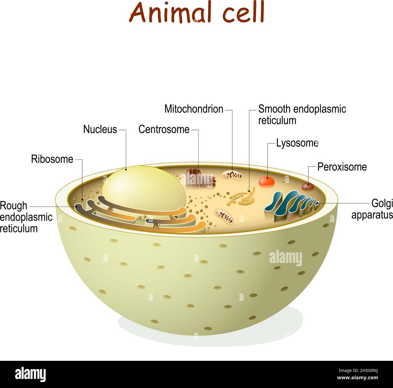 Animal cell anatomy. Organelles and structure of eukaryotic cell. Vector diagram. color can be changed easily Stock Vectorhttps://www.alamy.com/image-license-details/?v=1https://www.alamy.com/animal-cell-anatomy-organelles-and-structure-of-eukaryotic-cell-vector-diagram-color-can-be-changed-easily-image449672430.html
RF2H3G9NJ–Animal cell anatomy. Organelles and structure of eukaryotic cell. Vector diagram. color can be changed easily
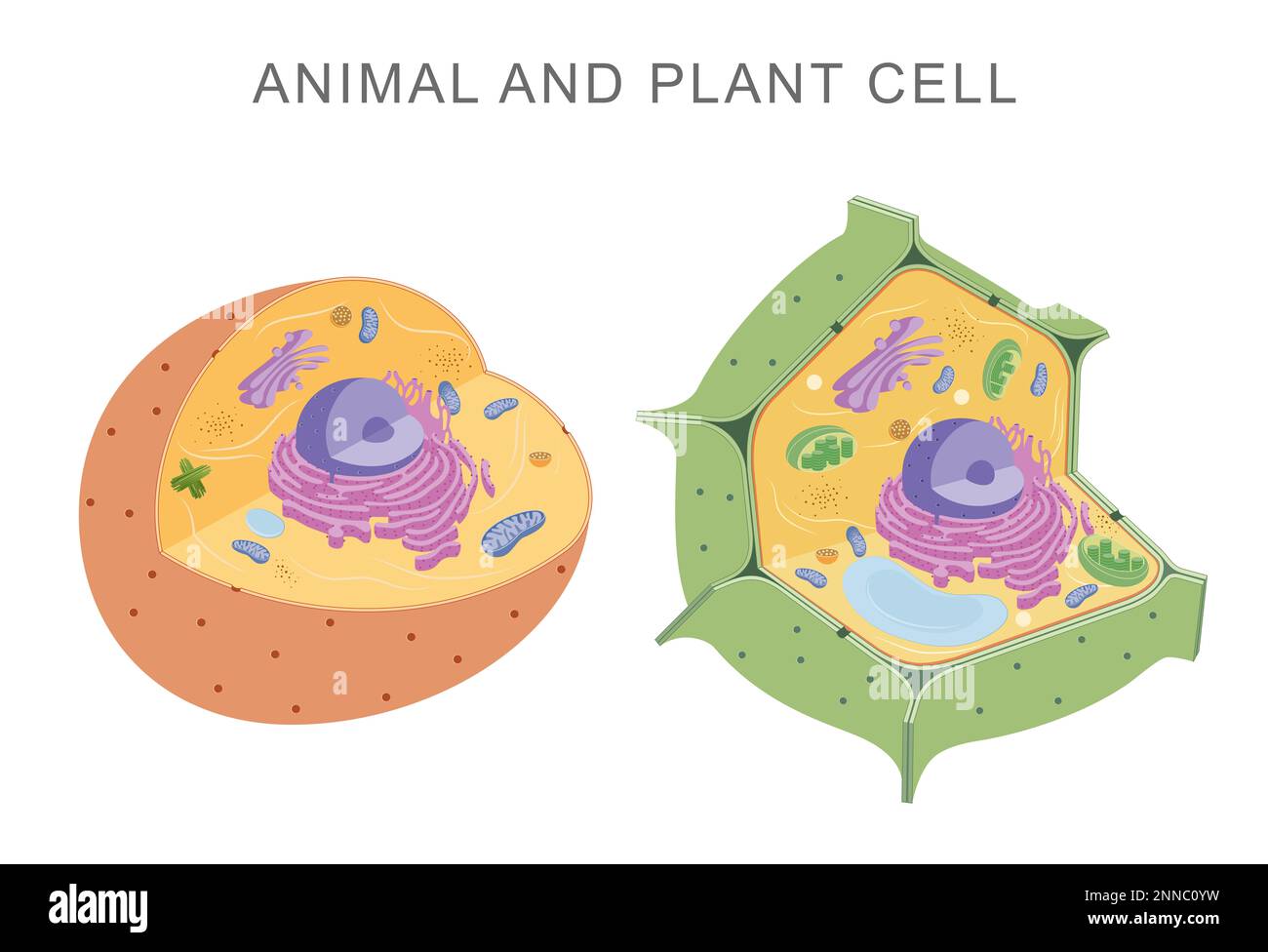 Comparing animal and plant cells Stock Photohttps://www.alamy.com/image-license-details/?v=1https://www.alamy.com/comparing-animal-and-plant-cells-image529483021.html
RF2NNC0YW–Comparing animal and plant cells
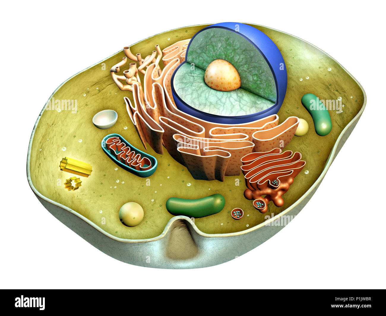 Internal structure of an animal cell. Digital illustration. Clipping path included. Stock Photohttps://www.alamy.com/image-license-details/?v=1https://www.alamy.com/internal-structure-of-an-animal-cell-digital-illustration-clipping-path-included-image207554139.html
RFP1JWBR–Internal structure of an animal cell. Digital illustration. Clipping path included.
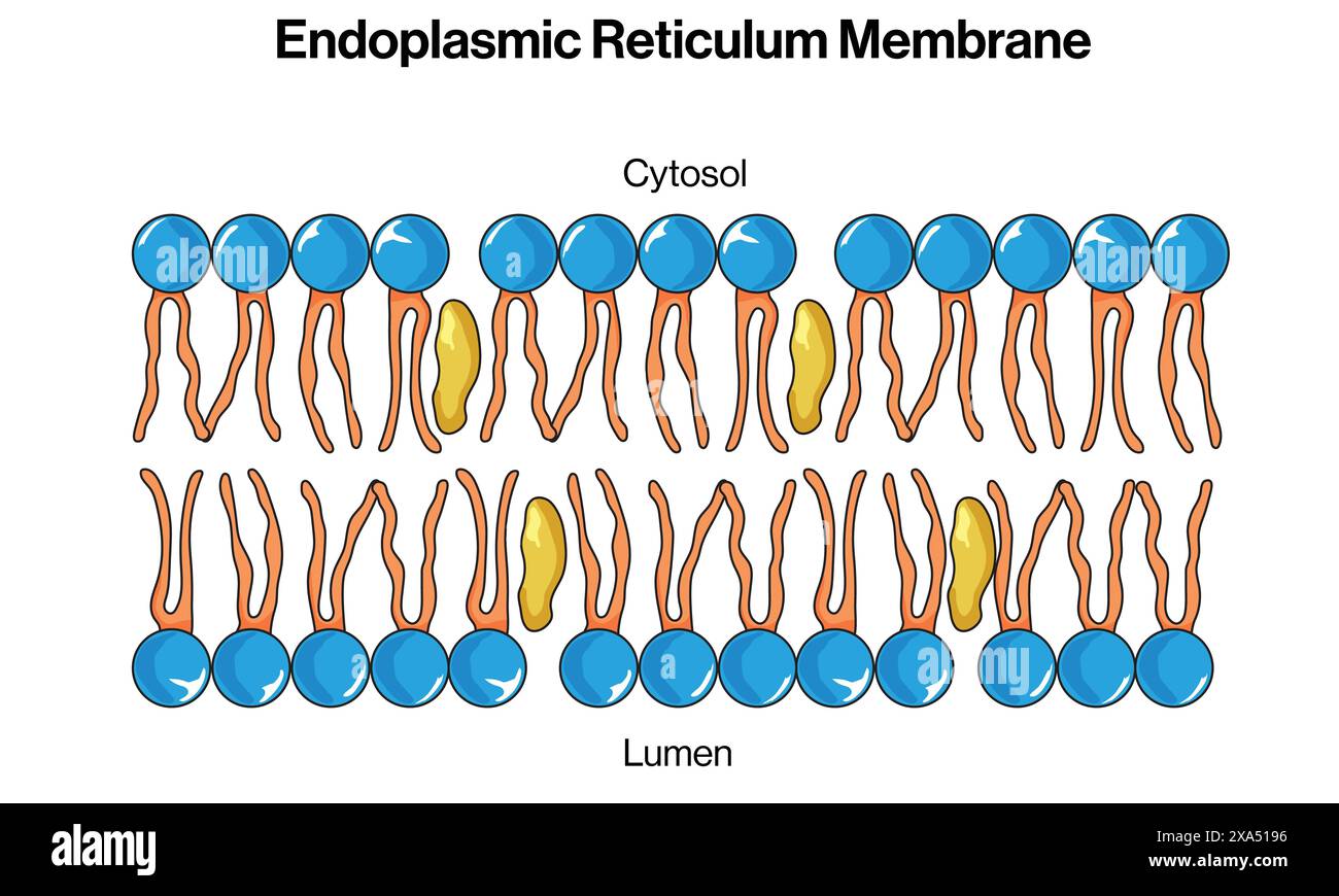 Detailed Vector Illustration of Endoplasmic Reticulum Membrane for Cell Biology, Molecular Biology, and Biochemistry Education on White Background. Stock Vectorhttps://www.alamy.com/image-license-details/?v=1https://www.alamy.com/detailed-vector-illustration-of-endoplasmic-reticulum-membrane-for-cell-biology-molecular-biology-and-biochemistry-education-on-white-background-image608620242.html
RF2XA5196–Detailed Vector Illustration of Endoplasmic Reticulum Membrane for Cell Biology, Molecular Biology, and Biochemistry Education on White Background.
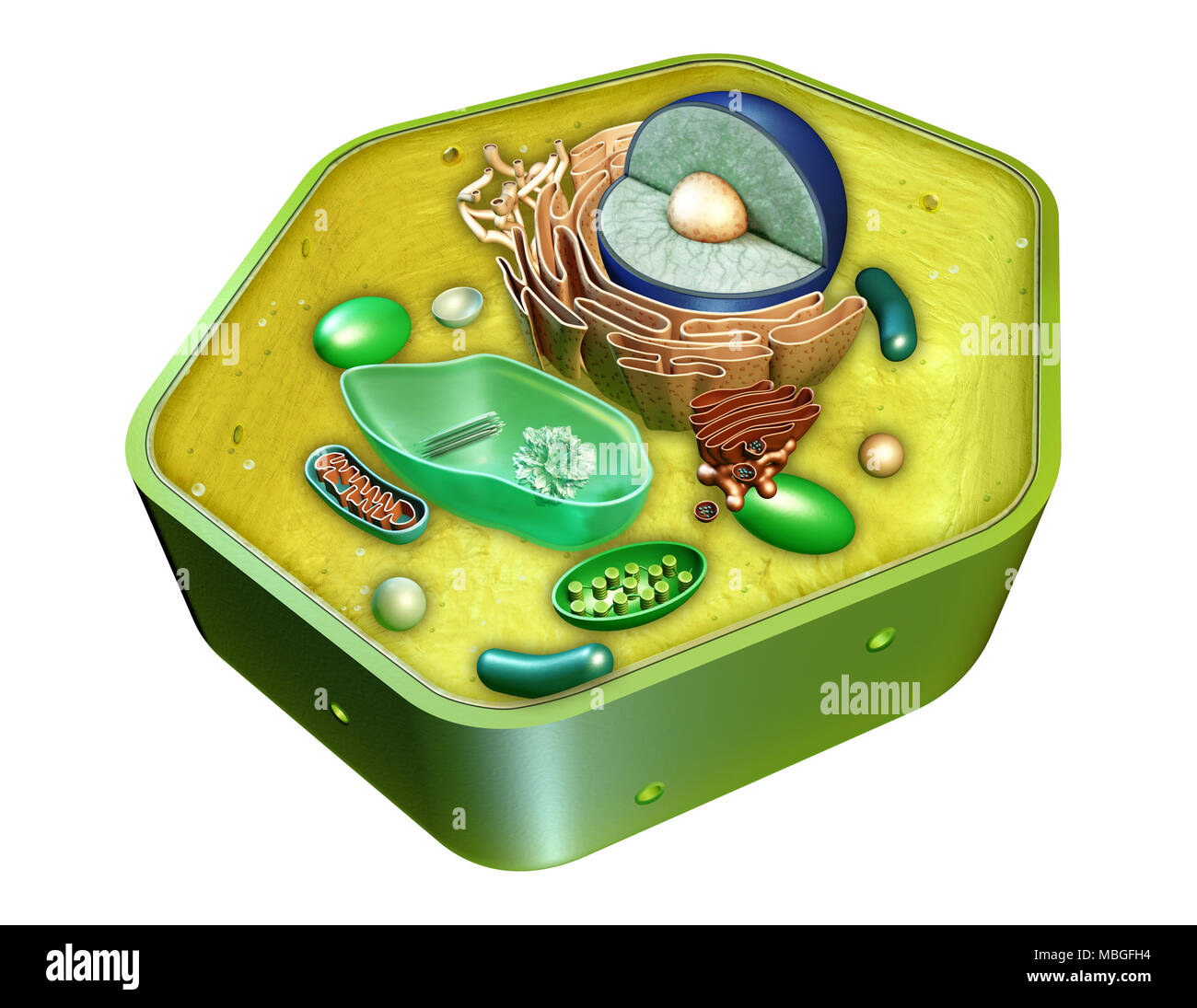 Internal structure of a plant cell. Digital illustration. Clipping path included. Stock Photohttps://www.alamy.com/image-license-details/?v=1https://www.alamy.com/internal-structure-of-a-plant-cell-digital-illustration-clipping-path-included-image179228368.html
RFMBGFH4–Internal structure of a plant cell. Digital illustration. Clipping path included.
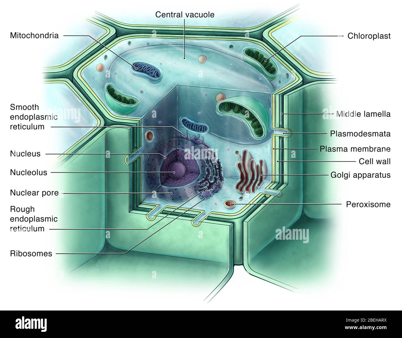 Plant Cell, Illustration Stock Photohttps://www.alamy.com/image-license-details/?v=1https://www.alamy.com/plant-cell-illustration-image353194238.html
RM2BEHARX–Plant Cell, Illustration
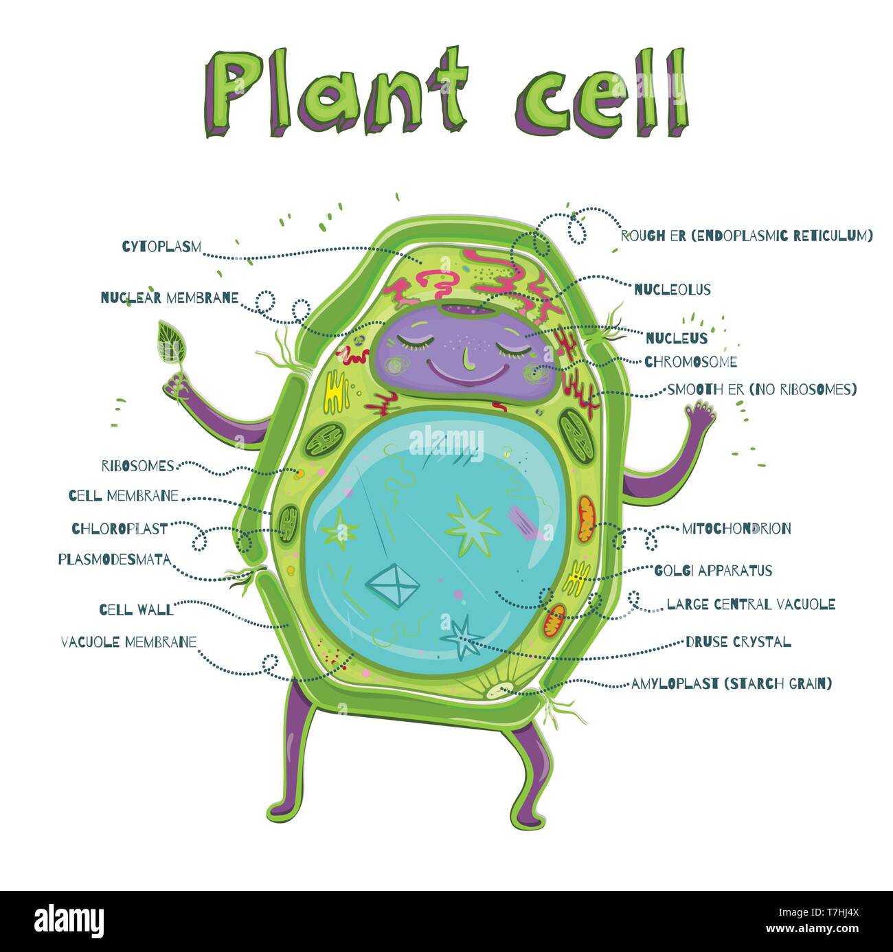 Cartoon vector illustration of structure of plant cell. Illustration showing the plant cell anatomy Stock Vectorhttps://www.alamy.com/image-license-details/?v=1https://www.alamy.com/cartoon-vector-illustration-of-structure-of-plant-cell-illustration-showing-the-plant-cell-anatomy-image245635178.html
RFT7HJ4X–Cartoon vector illustration of structure of plant cell. Illustration showing the plant cell anatomy
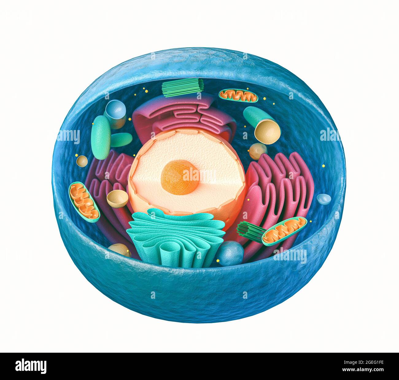 3d rendering of biological animal cell with organelles cross section isolated on white Stock Photohttps://www.alamy.com/image-license-details/?v=1https://www.alamy.com/3d-rendering-of-biological-animal-cell-with-organelles-cross-section-isolated-on-white-image439216834.html
RF2GEG1FE–3d rendering of biological animal cell with organelles cross section isolated on white
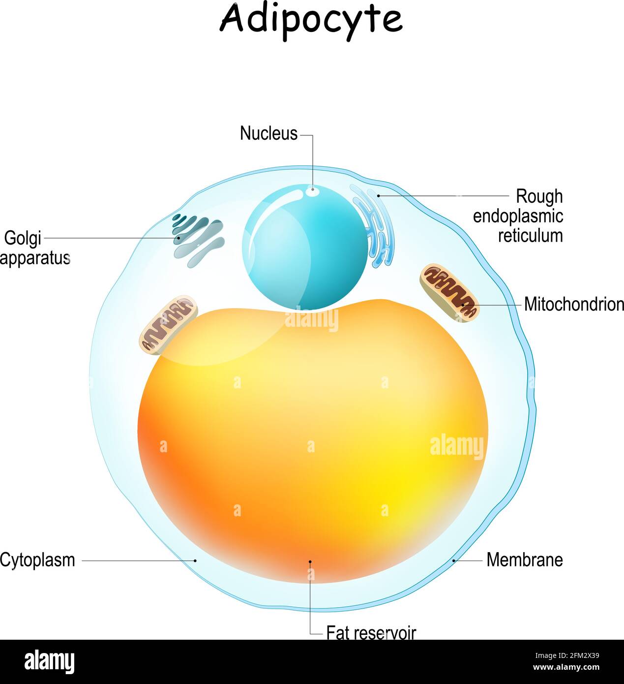 Adipocytes anatomy. Structure of fat cell. adipose tissue. Vector illustration Stock Vectorhttps://www.alamy.com/image-license-details/?v=1https://www.alamy.com/adipocytes-anatomy-structure-of-fat-cell-adipose-tissue-vector-illustration-image425406333.html
RF2FM2X39–Adipocytes anatomy. Structure of fat cell. adipose tissue. Vector illustration
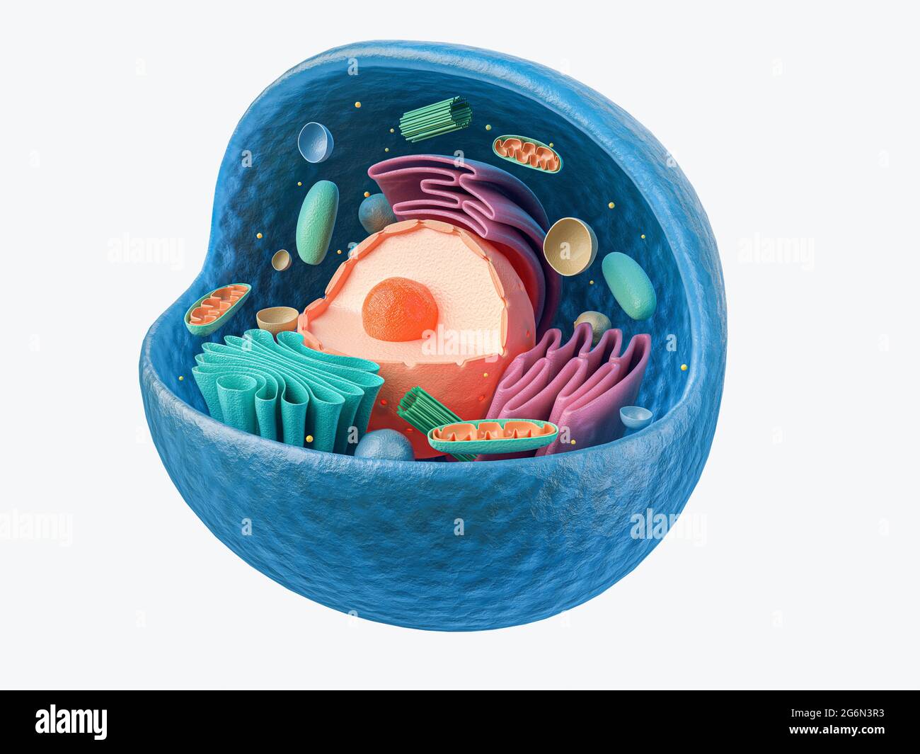 3d rendering of biological animal cell with organelles cross section isolated on white Stock Photohttps://www.alamy.com/image-license-details/?v=1https://www.alamy.com/3d-rendering-of-biological-animal-cell-with-organelles-cross-section-isolated-on-white-image434411127.html
RF2G6N3R3–3d rendering of biological animal cell with organelles cross section isolated on white
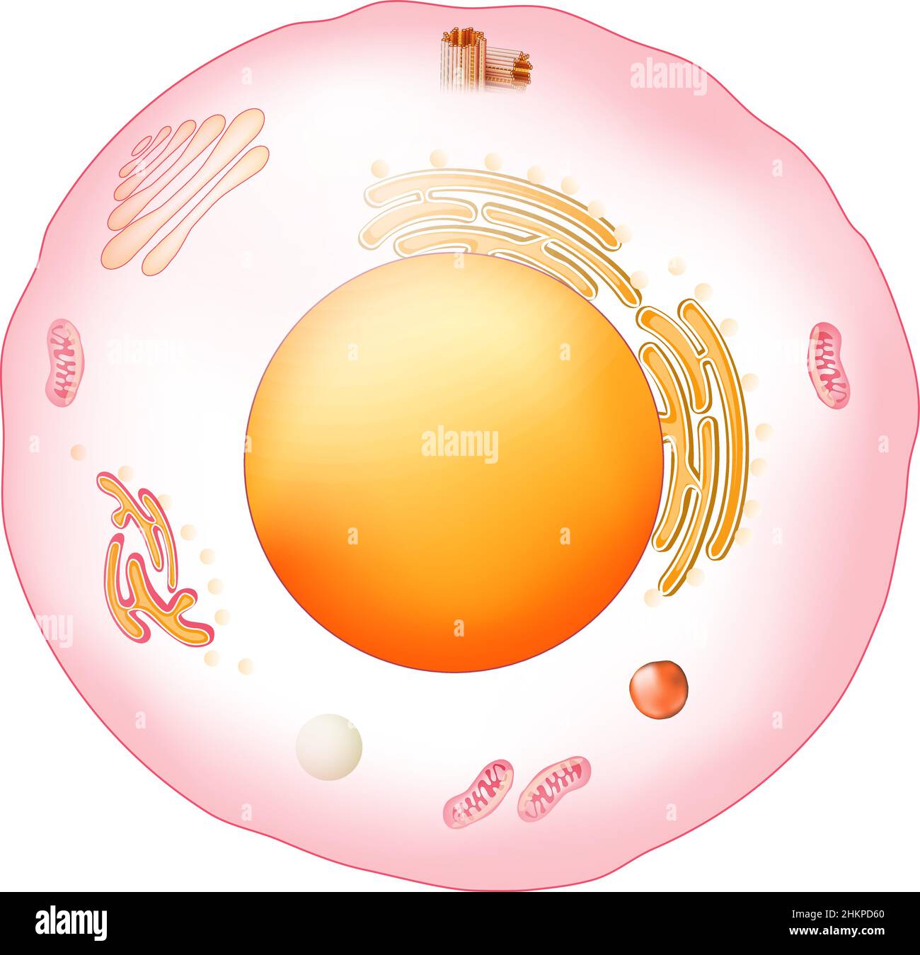 Animal cell anatomy. Structure and organelles of Eukaryotic cell. Vector poster for education. illustration Stock Vectorhttps://www.alamy.com/image-license-details/?v=1https://www.alamy.com/animal-cell-anatomy-structure-and-organelles-of-eukaryotic-cell-vector-poster-for-education-illustration-image459641336.html
RF2HKPD60–Animal cell anatomy. Structure and organelles of Eukaryotic cell. Vector poster for education. illustration
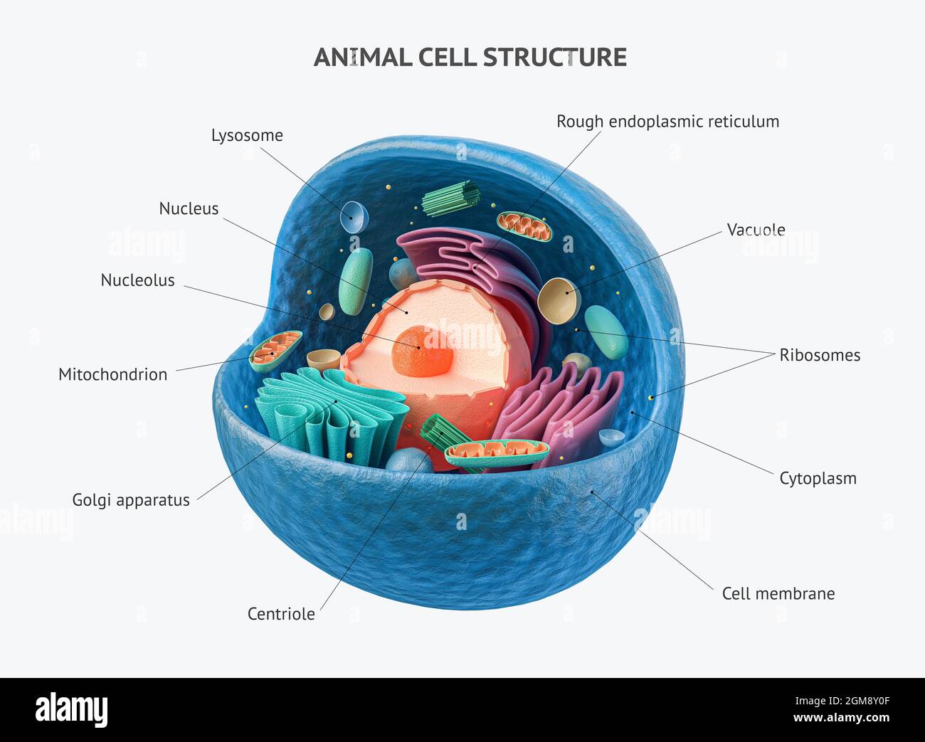 3d rendering of biological animal cell with organelles cross section isolated on white. Animal cell with placed text annotations to all organelles Stock Photohttps://www.alamy.com/image-license-details/?v=1https://www.alamy.com/3d-rendering-of-biological-animal-cell-with-organelles-cross-section-isolated-on-white-animal-cell-with-placed-text-annotations-to-all-organelles-image442749119.html
RF2GM8Y0F–3d rendering of biological animal cell with organelles cross section isolated on white. Animal cell with placed text annotations to all organelles
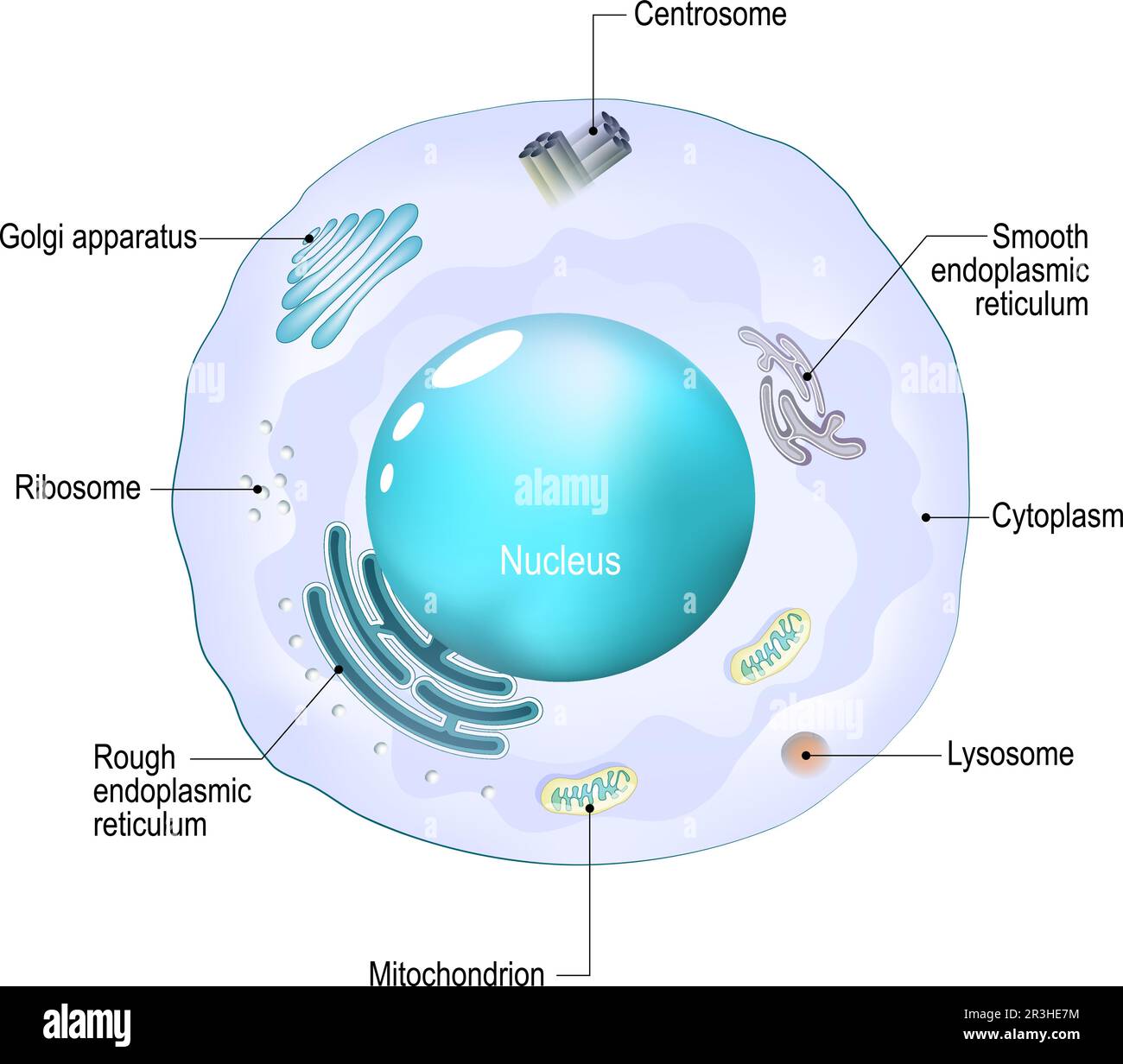 Cell organelles. Structure and anatomy of a animal cell. realistic cell on a white background. Vector illustration. Poster for education Stock Vectorhttps://www.alamy.com/image-license-details/?v=1https://www.alamy.com/cell-organelles-structure-and-anatomy-of-a-animal-cell-realistic-cell-on-a-white-background-vector-illustration-poster-for-education-image552960120.html
RF2R3HE7M–Cell organelles. Structure and anatomy of a animal cell. realistic cell on a white background. Vector illustration. Poster for education
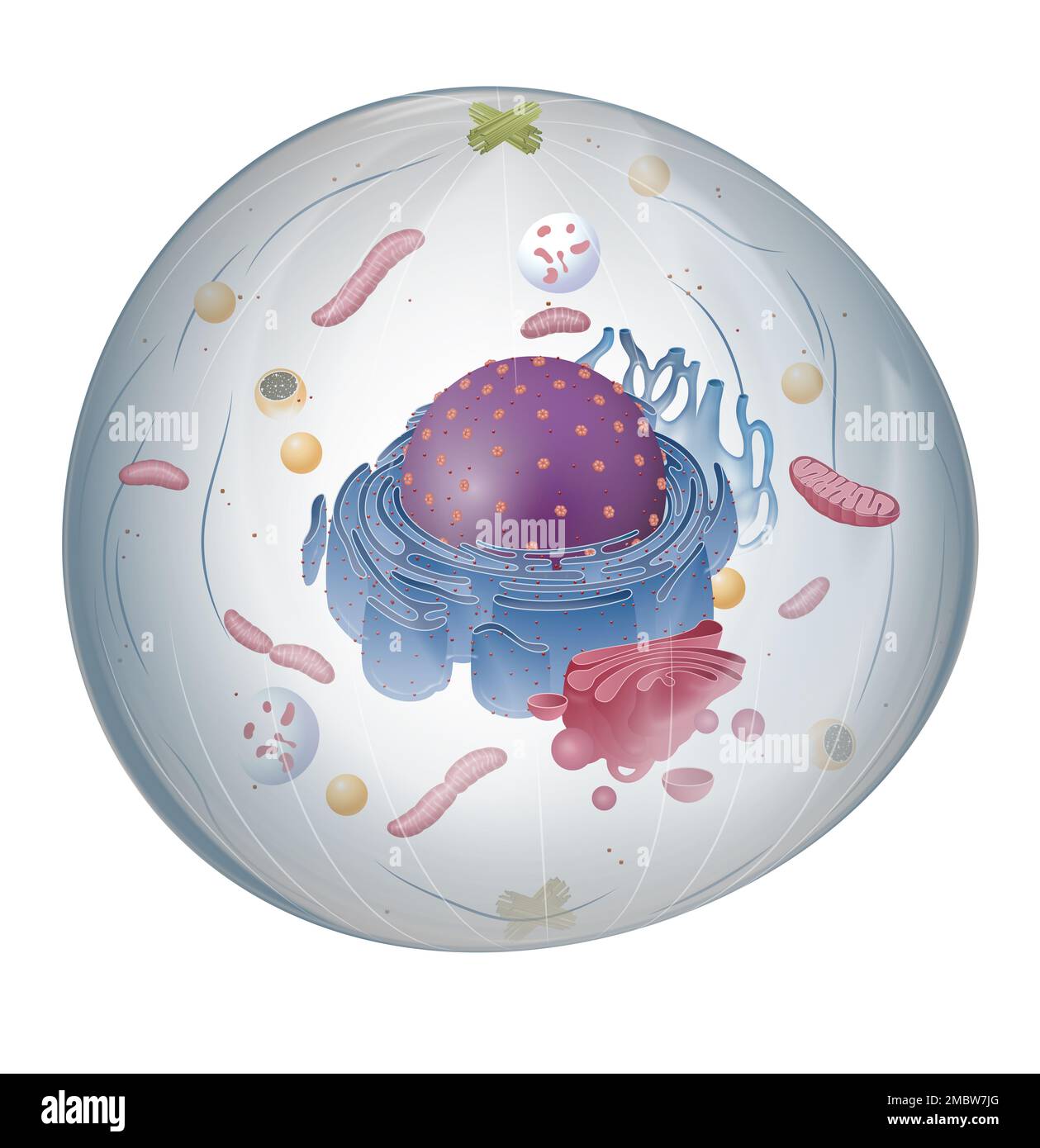 The structure of Animal cell Stock Photohttps://www.alamy.com/image-license-details/?v=1https://www.alamy.com/the-structure-of-animal-cell-image506416696.html
RF2MBW7JG–The structure of Animal cell
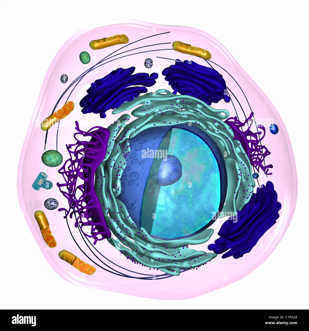 3D model of a eukaryotic cell Stock Photohttps://www.alamy.com/image-license-details/?v=1https://www.alamy.com/stock-photo-3d-model-of-a-eukaryotic-cell-49663328.html
RMCTPA28–3D model of a eukaryotic cell
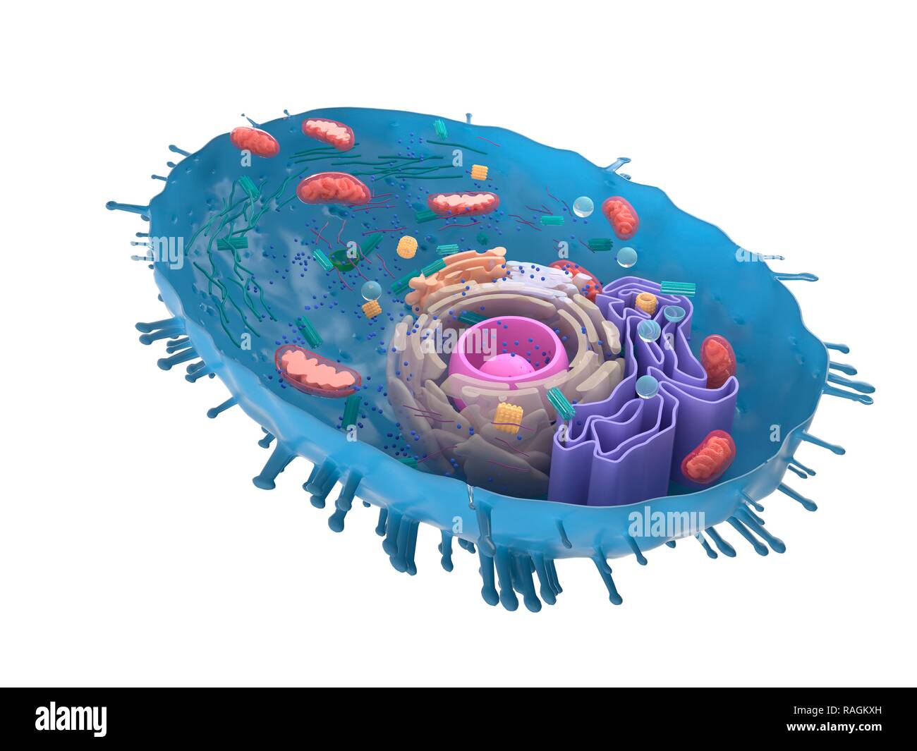 Illustration of a human cell cross-section. Stock Photohttps://www.alamy.com/image-license-details/?v=1https://www.alamy.com/illustration-of-a-human-cell-cross-section-image230248217.html
RFRAGKXH–Illustration of a human cell cross-section.
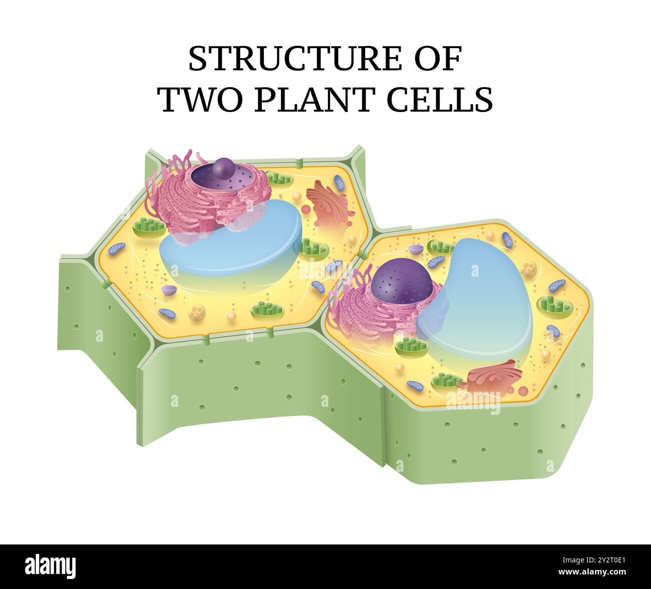 Structure of two plant cells Stock Photohttps://www.alamy.com/image-license-details/?v=1https://www.alamy.com/structure-of-two-plant-cells-image621329801.html
RF2Y2T0E1–Structure of two plant cells
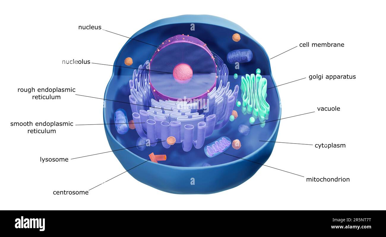 Animal cell structure, illustration Stock Photohttps://www.alamy.com/image-license-details/?v=1https://www.alamy.com/animal-cell-structure-illustration-image554285084.html
RF2R5NT7T–Animal cell structure, illustration
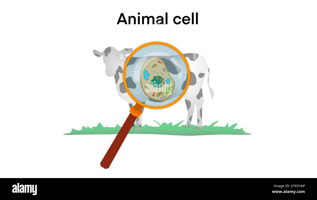 animal cell anatomy, biological animal cell with organelles cross section, Animal cell structure. Educational material, Anatomy of animal cell Stock Photohttps://www.alamy.com/image-license-details/?v=1https://www.alamy.com/animal-cell-anatomy-biological-animal-cell-with-organelles-cross-section-animal-cell-structure-educational-material-anatomy-of-animal-cell-image623348510.html
RF2Y63YAP–animal cell anatomy, biological animal cell with organelles cross section, Animal cell structure. Educational material, Anatomy of animal cell
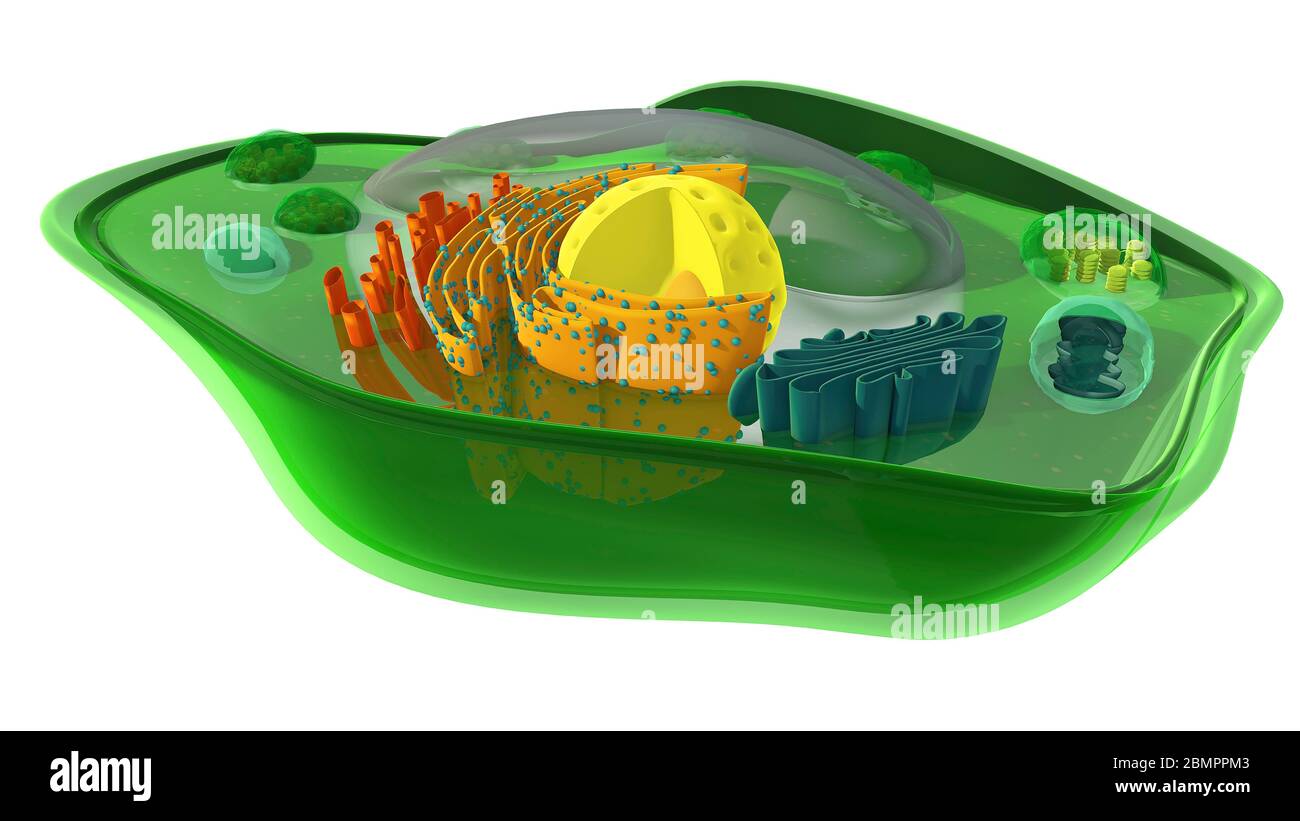 Computer illustration showing the internal structure of a plant cell. In addition to the chloroplast, the mitochondrion and the nucleus are cut through revealing their internal structures. Stock Photohttps://www.alamy.com/image-license-details/?v=1https://www.alamy.com/computer-illustration-showing-the-internal-structure-of-a-plant-cell-in-addition-to-the-chloroplast-the-mitochondrion-and-the-nucleus-are-cut-through-revealing-their-internal-structures-image357001235.html
RF2BMPPM3–Computer illustration showing the internal structure of a plant cell. In addition to the chloroplast, the mitochondrion and the nucleus are cut through revealing their internal structures.
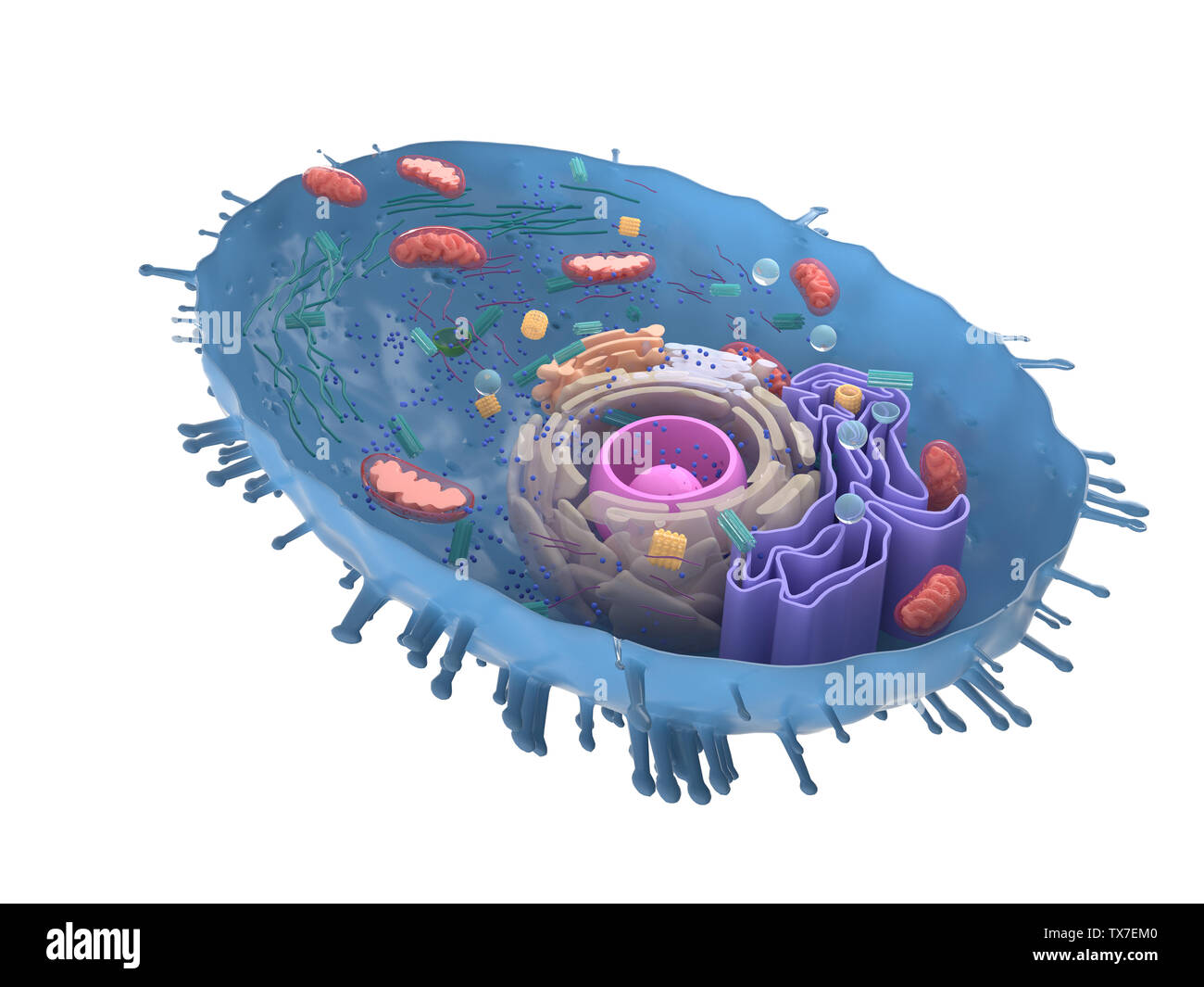 3d rendered illustration of a human cell cross-section Stock Photohttps://www.alamy.com/image-license-details/?v=1https://www.alamy.com/3d-rendered-illustration-of-a-human-cell-cross-section-image257091408.html
RFTX7EM0–3d rendered illustration of a human cell cross-section
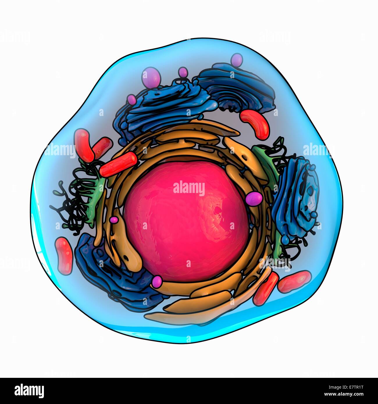 Animal cell structure, computer artwork. Stock Photohttps://www.alamy.com/image-license-details/?v=1https://www.alamy.com/stock-photo-animal-cell-structure-computer-artwork-73688996.html
RFE7TR1T–Animal cell structure, computer artwork.
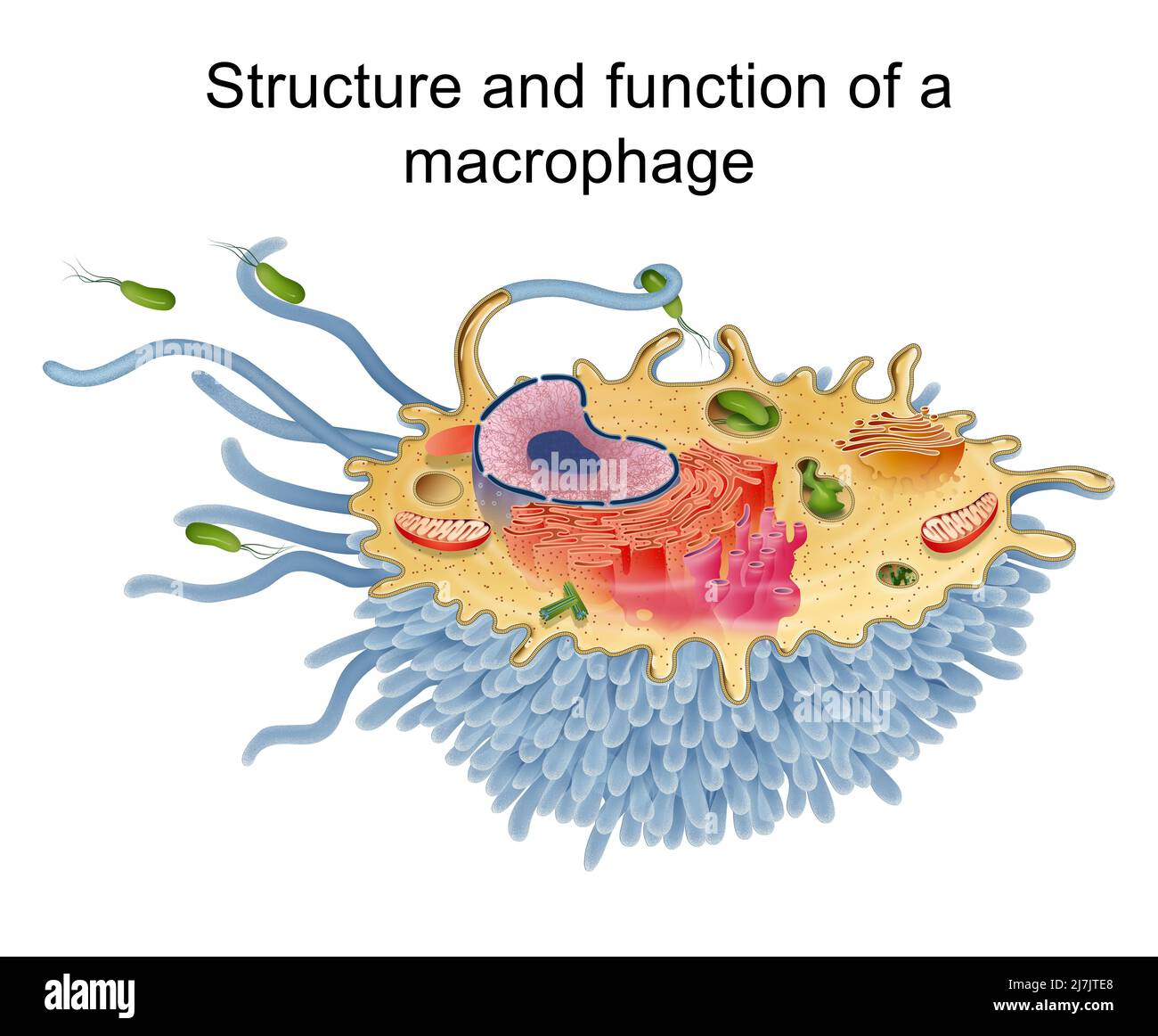 Structure and function of a macrophage Category Science Stock Photohttps://www.alamy.com/image-license-details/?v=1https://www.alamy.com/structure-and-function-of-a-macrophage-category-science-image469396880.html
RF2J7JTE8–Structure and function of a macrophage Category Science
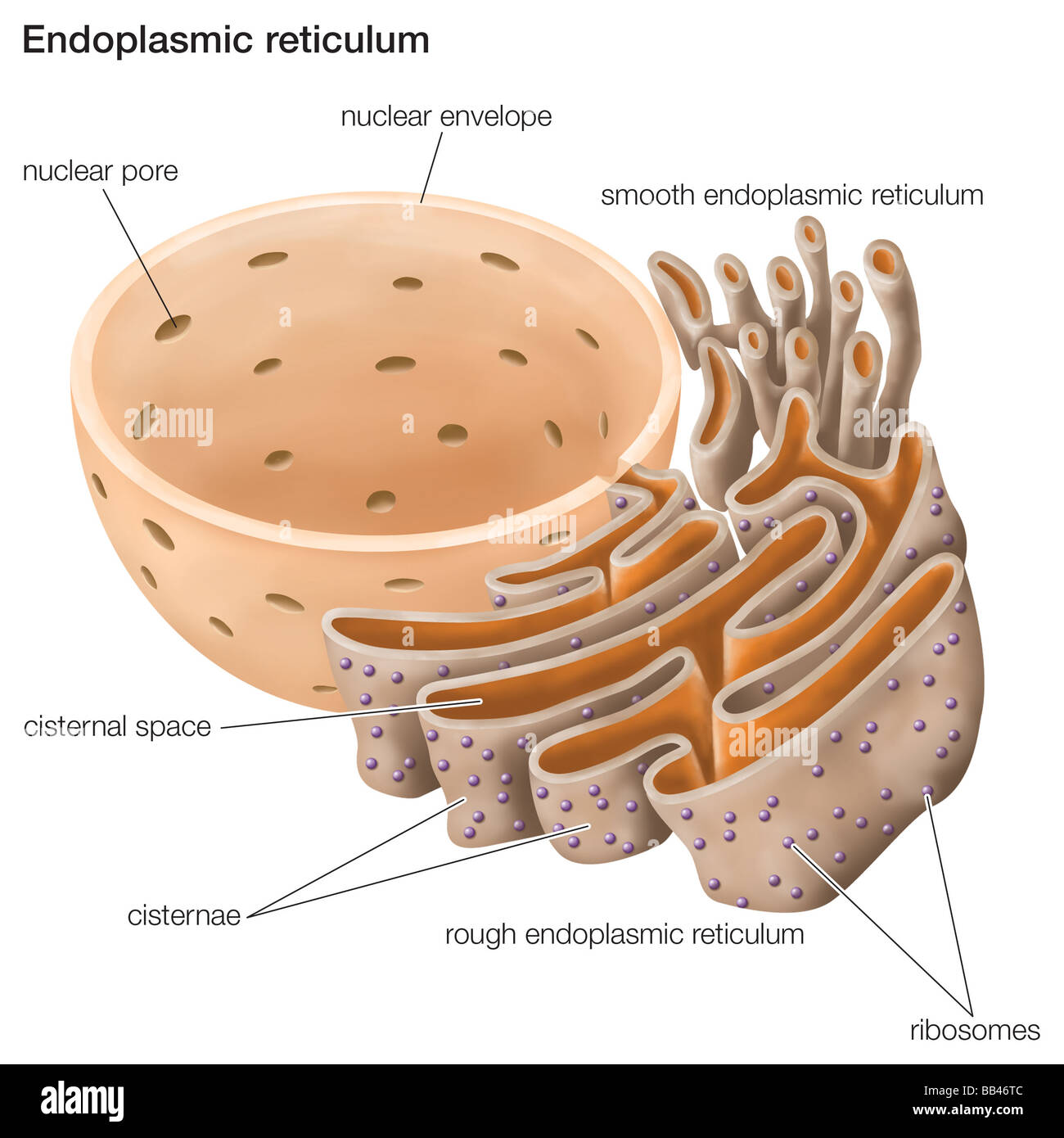 The endoplasmic reticulum plays an important role in the biosynthesis, processing, and transport of proteins and lipids. Stock Photohttps://www.alamy.com/image-license-details/?v=1https://www.alamy.com/stock-photo-the-endoplasmic-reticulum-plays-an-important-role-in-the-biosynthesis-24064780.html
RMBB46TC–The endoplasmic reticulum plays an important role in the biosynthesis, processing, and transport of proteins and lipids.
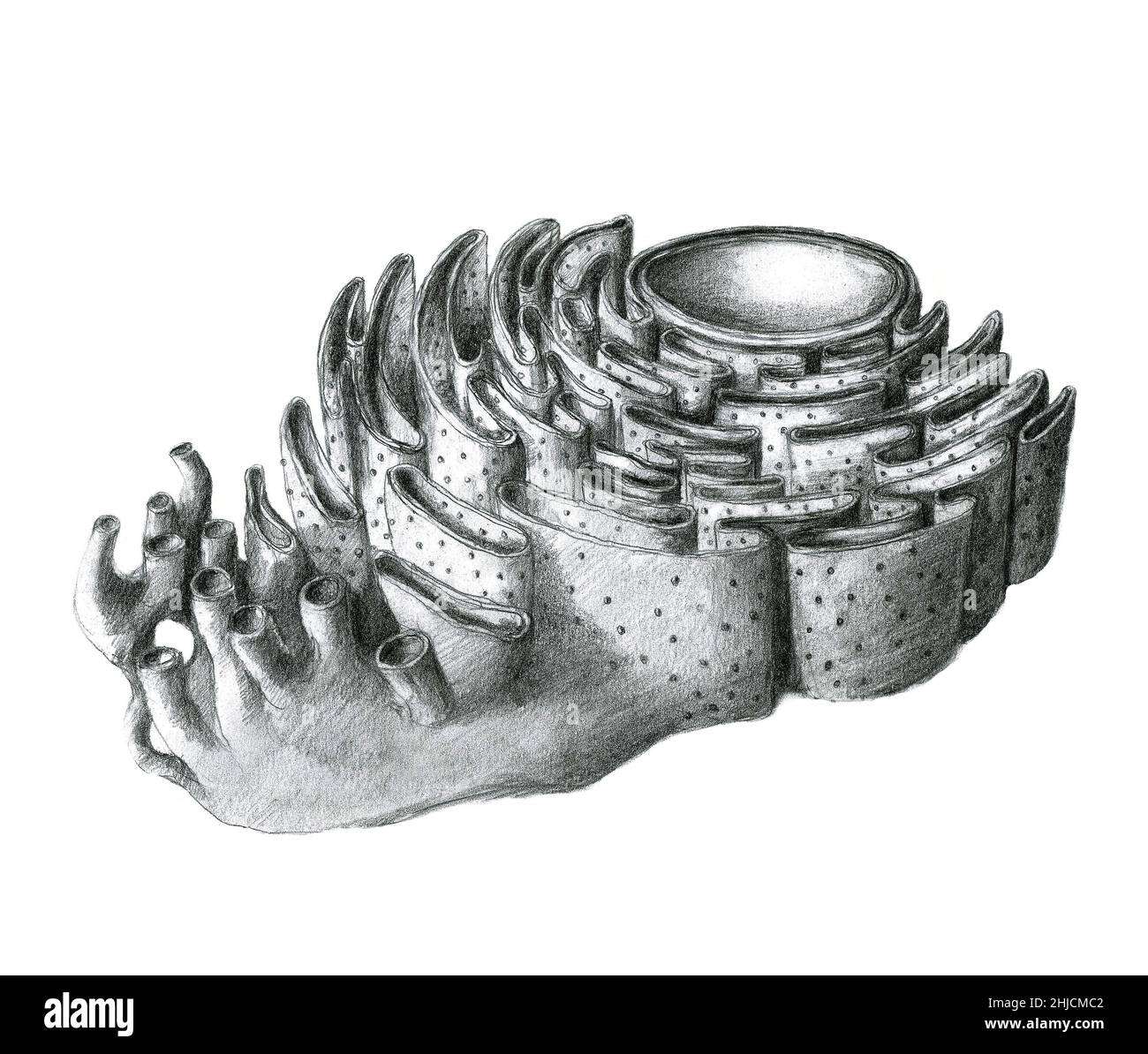 The endoplasmic reticulum (ER) is a eukaryotic organelle that forms a network of tubules, vesicles, and cisternae inside cells. Rough endoplasmic reticula synthesize proteins, while smooth endoplasmic reticula synthesize lipids and steroids, metabolize carbohydrates and steroids, and regulate calcium concentration, drug detoxification, and attachment of receptors on cell membrane proteins. Stock Photohttps://www.alamy.com/image-license-details/?v=1https://www.alamy.com/the-endoplasmic-reticulum-er-is-a-eukaryotic-organelle-that-forms-a-network-of-tubules-vesicles-and-cisternae-inside-cells-rough-endoplasmic-reticula-synthesize-proteins-while-smooth-endoplasmic-reticula-synthesize-lipids-and-steroids-metabolize-carbohydrates-and-steroids-and-regulate-calcium-concentration-drug-detoxification-and-attachment-of-receptors-on-cell-membrane-proteins-image458812818.html
RM2HJCMC2–The endoplasmic reticulum (ER) is a eukaryotic organelle that forms a network of tubules, vesicles, and cisternae inside cells. Rough endoplasmic reticula synthesize proteins, while smooth endoplasmic reticula synthesize lipids and steroids, metabolize carbohydrates and steroids, and regulate calcium concentration, drug detoxification, and attachment of receptors on cell membrane proteins.
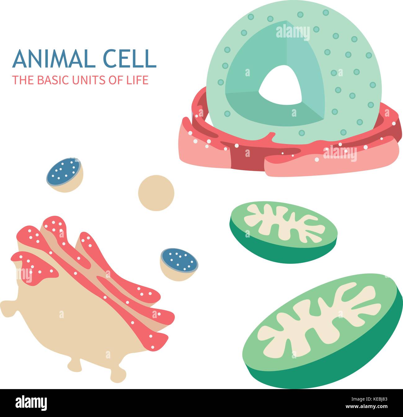 Animal Cell Anatomy structure vector illustration Stock Vectorhttps://www.alamy.com/image-license-details/?v=1https://www.alamy.com/stock-image-animal-cell-anatomy-structure-vector-illustration-163754307.html
RFKEBJ83–Animal Cell Anatomy structure vector illustration
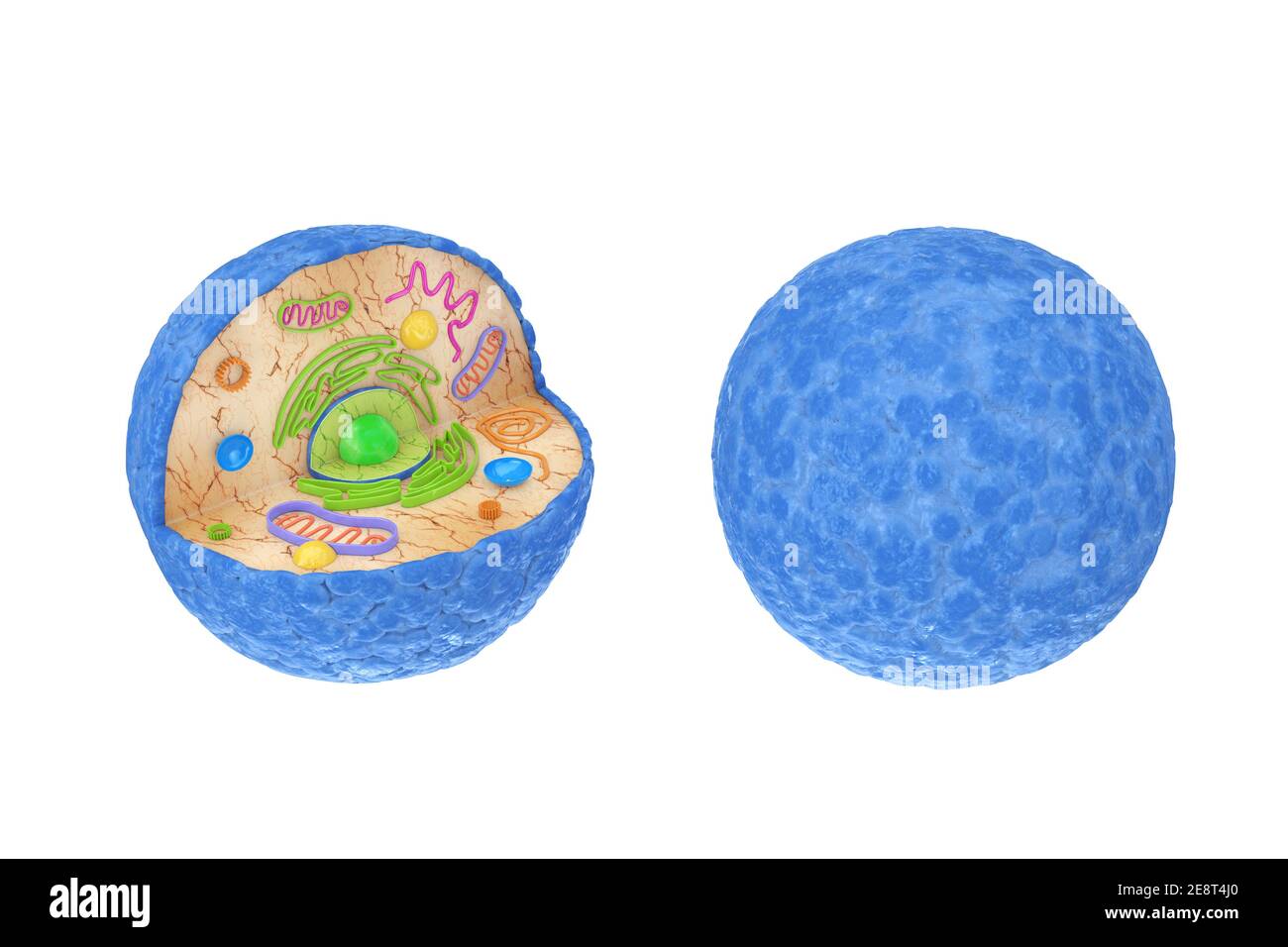 Internal Diagram Structure of Human Cell on a white background. 3d Rendering Stock Photohttps://www.alamy.com/image-license-details/?v=1https://www.alamy.com/internal-diagram-structure-of-human-cell-on-a-white-background-3d-rendering-image401286200.html
RF2E8T4J0–Internal Diagram Structure of Human Cell on a white background. 3d Rendering
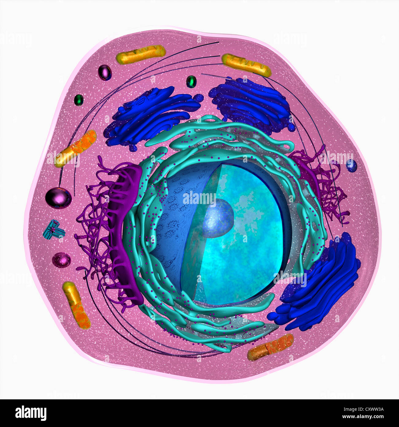 3D model of a eukaryotic cell Stock Photohttps://www.alamy.com/image-license-details/?v=1https://www.alamy.com/stock-photo-3d-model-of-a-eukaryotic-cell-50970286.html
RFCXWW3A–3D model of a eukaryotic cell
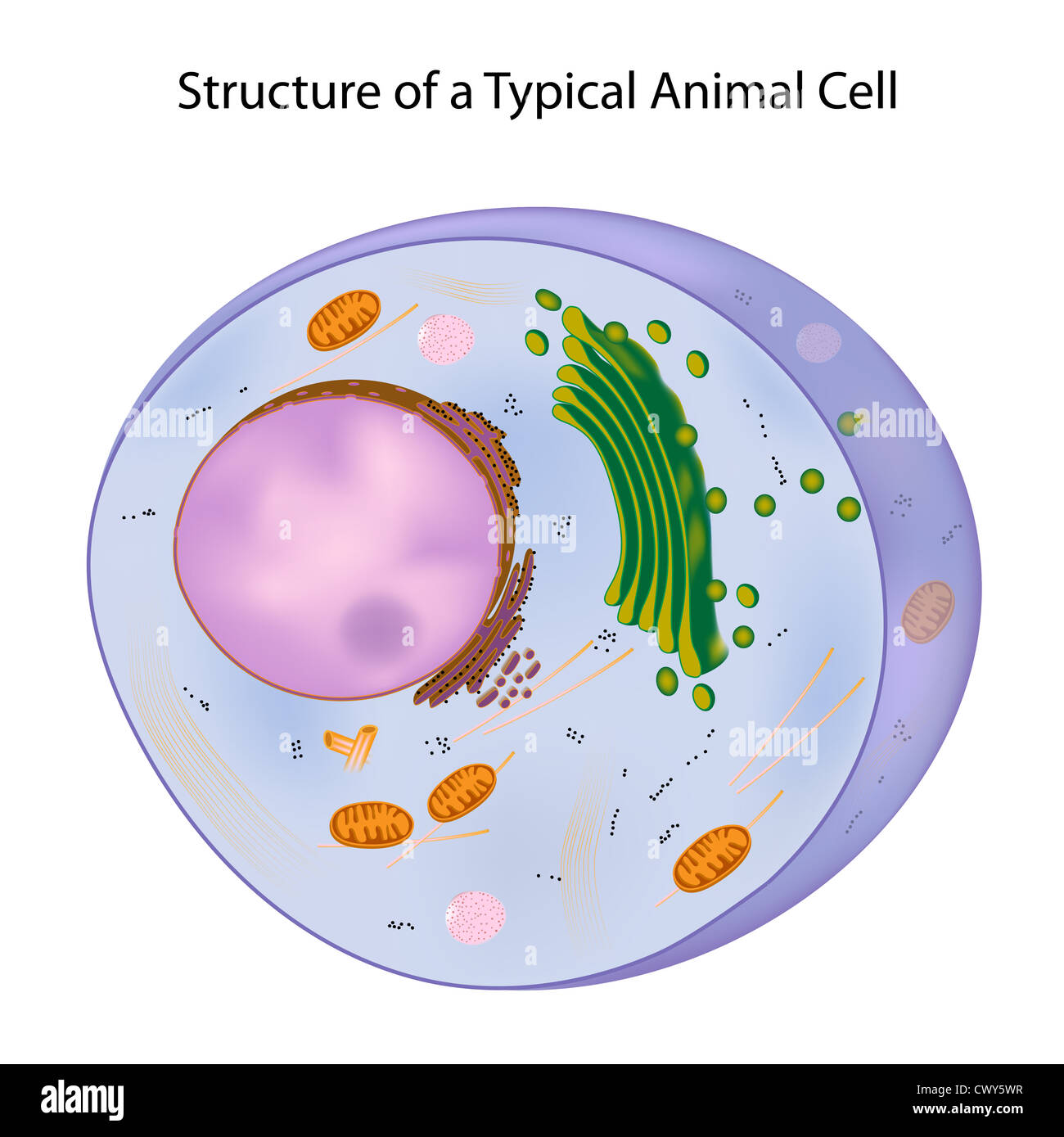 A typical cell Stock Photohttps://www.alamy.com/image-license-details/?v=1https://www.alamy.com/stock-photo-a-typical-cell-50384483.html
RFCWY5WR–A typical cell
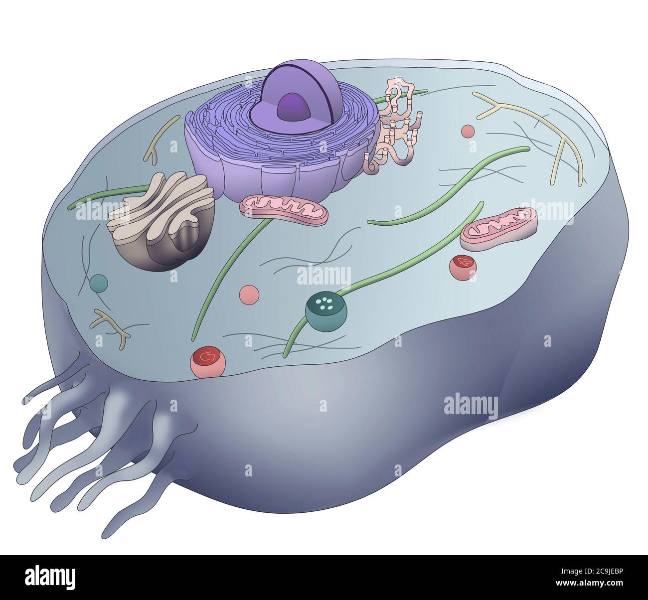 Computer illustration showing the normal human cell anatomy. Shown is the nucleus (purple, consisting of nucleolus (inner dark purple sphere) and nucl Stock Photohttps://www.alamy.com/image-license-details/?v=1https://www.alamy.com/computer-illustration-showing-the-normal-human-cell-anatomy-shown-is-the-nucleus-purple-consisting-of-nucleolus-inner-dark-purple-sphere-and-nucl-image367356074.html
RF2C9JEBP–Computer illustration showing the normal human cell anatomy. Shown is the nucleus (purple, consisting of nucleolus (inner dark purple sphere) and nucl
 Structure of osteoclasts. Osteoclast is a type of bone cell that breaks down bone tissue. Stock Vectorhttps://www.alamy.com/image-license-details/?v=1https://www.alamy.com/structure-of-osteoclasts-osteoclast-is-a-type-of-bone-cell-that-breaks-down-bone-tissue-image608808489.html
RF2XADHC9–Structure of osteoclasts. Osteoclast is a type of bone cell that breaks down bone tissue.
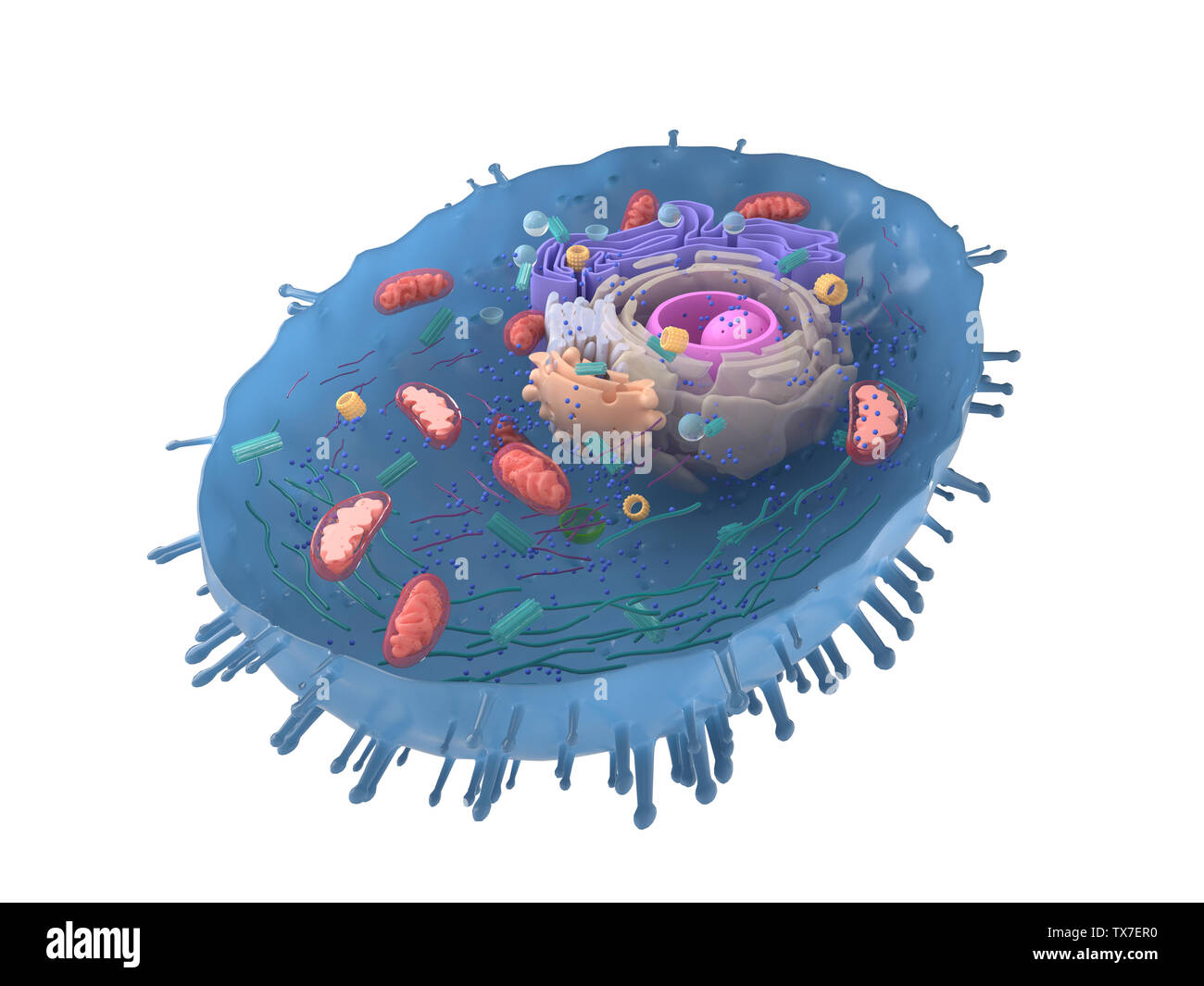 3d rendered illustration of a human cell cross-section Stock Photohttps://www.alamy.com/image-license-details/?v=1https://www.alamy.com/3d-rendered-illustration-of-a-human-cell-cross-section-image257091492.html
RFTX7ER0–3d rendered illustration of a human cell cross-section
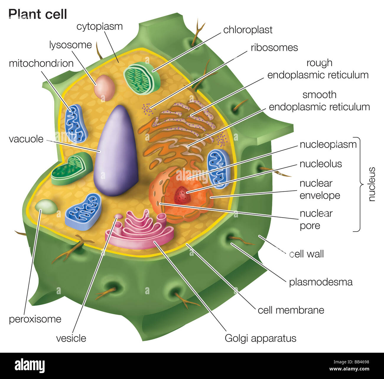 Cutaway drawing of a eukaryotic plant cell. Stock Photohttps://www.alamy.com/image-license-details/?v=1https://www.alamy.com/stock-photo-cutaway-drawing-of-a-eukaryotic-plant-cell-24064356.html
RMBB4698–Cutaway drawing of a eukaryotic plant cell.
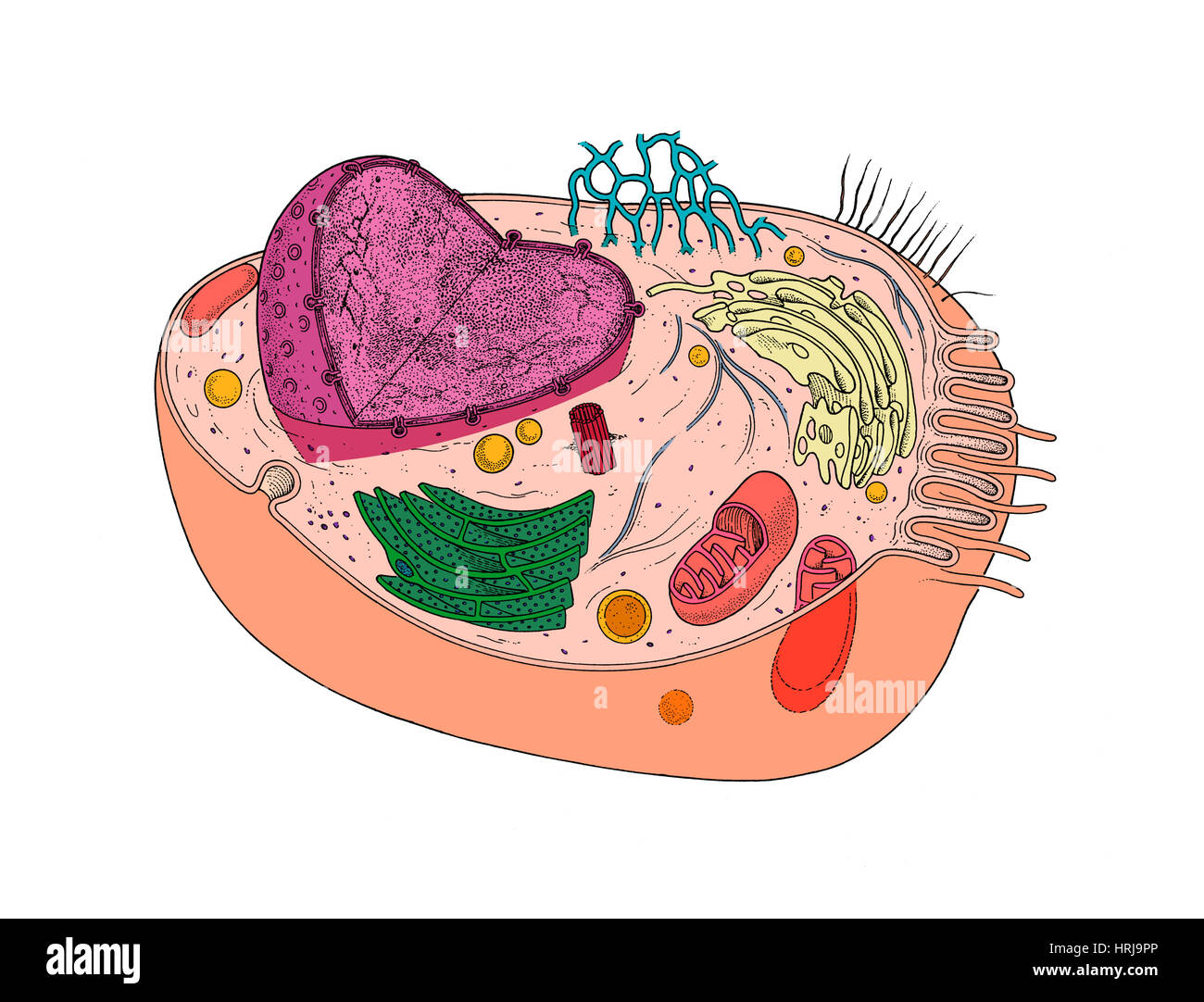 Animal Cell Diagram Stock Photohttps://www.alamy.com/image-license-details/?v=1https://www.alamy.com/stock-photo-animal-cell-diagram-135012494.html
RMHRJ9PP–Animal Cell Diagram
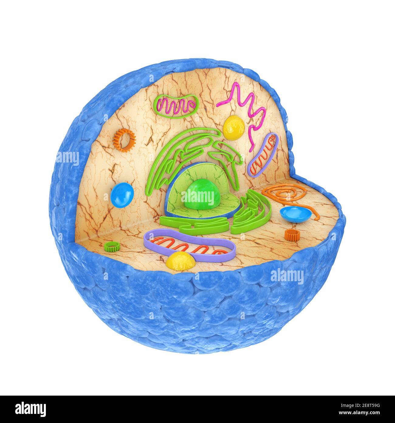 Internal Diagram Structure of Human Cell on a white background. 3d Rendering Stock Photohttps://www.alamy.com/image-license-details/?v=1https://www.alamy.com/internal-diagram-structure-of-human-cell-on-a-white-background-3d-rendering-image401286748.html
RF2E8T59G–Internal Diagram Structure of Human Cell on a white background. 3d Rendering
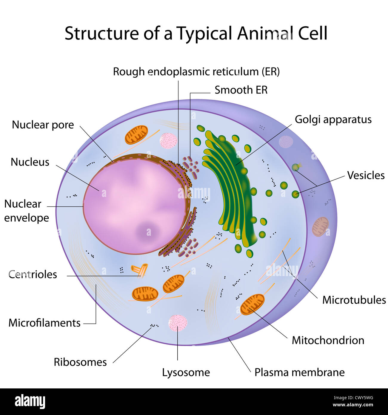 A typical cell, labeled Stock Photohttps://www.alamy.com/image-license-details/?v=1https://www.alamy.com/stock-photo-a-typical-cell-labeled-50384476.html
RFCWY5WG–A typical cell, labeled
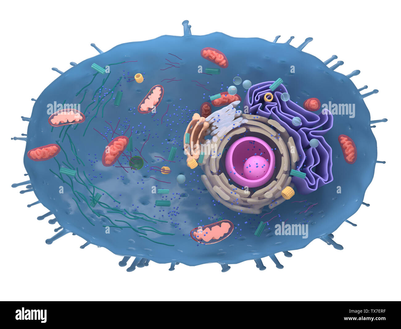 3d rendered illustration of a human cell cross-section Stock Photohttps://www.alamy.com/image-license-details/?v=1https://www.alamy.com/3d-rendered-illustration-of-a-human-cell-cross-section-image257091507.html
RFTX7ERF–3d rendered illustration of a human cell cross-section
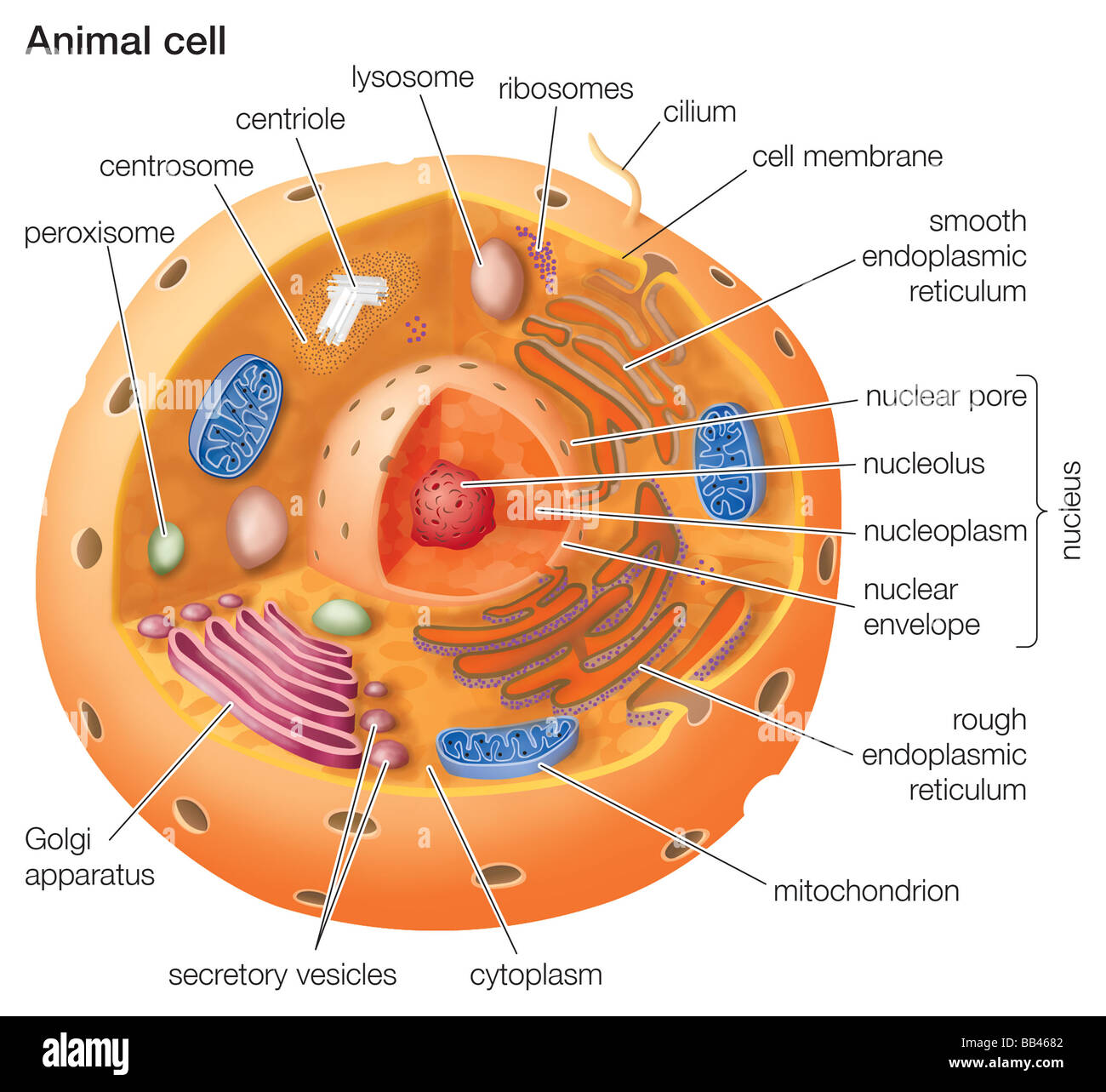 Cutaway drawing of a eukaryotic animal cell. Stock Photohttps://www.alamy.com/image-license-details/?v=1https://www.alamy.com/stock-photo-cutaway-drawing-of-a-eukaryotic-animal-cell-24064322.html
RMBB4682–Cutaway drawing of a eukaryotic animal cell.
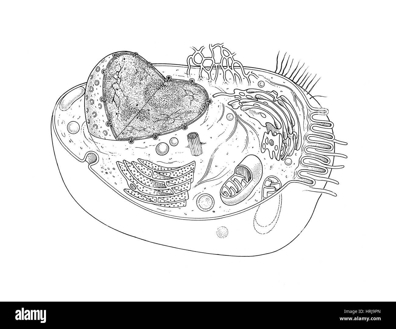 Animal Cell Diagram Stock Photohttps://www.alamy.com/image-license-details/?v=1https://www.alamy.com/stock-photo-animal-cell-diagram-135012493.html
RMHRJ9PN–Animal Cell Diagram
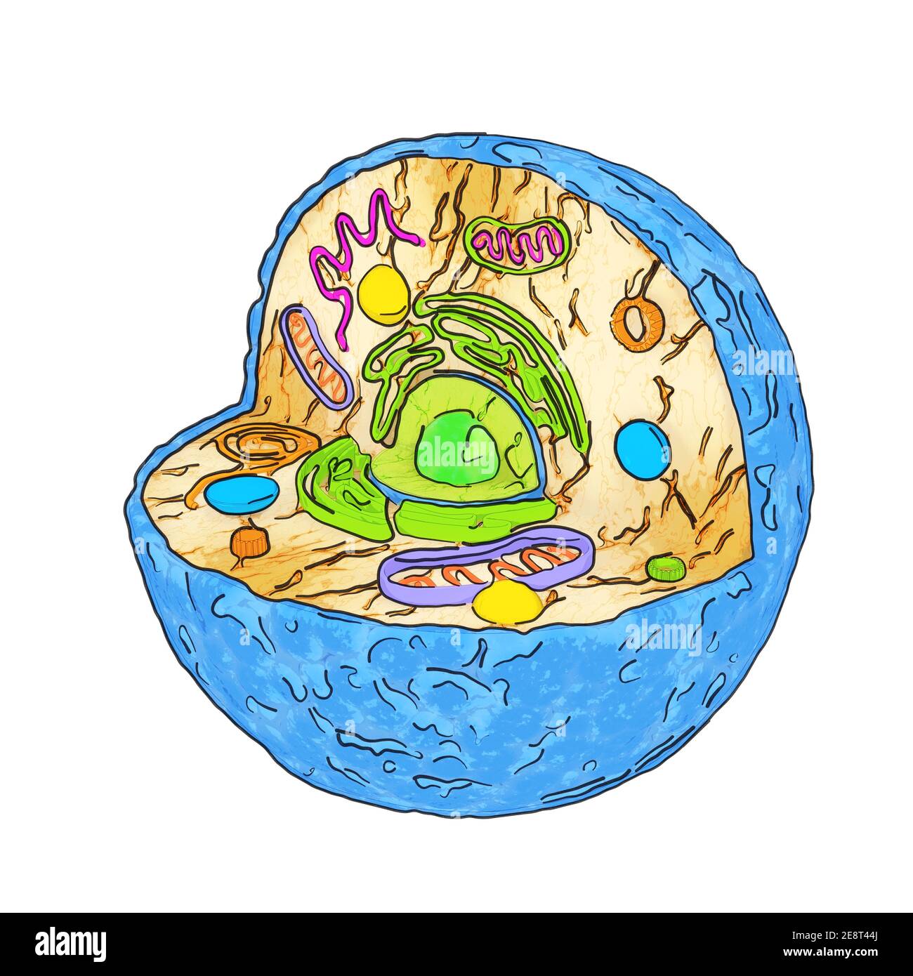 Internal Diagram Structure of Human Cell in Cartoon Sketch Style on a white background. 3d Rendering Stock Photohttps://www.alamy.com/image-license-details/?v=1https://www.alamy.com/internal-diagram-structure-of-human-cell-in-cartoon-sketch-style-on-a-white-background-3d-rendering-image401285826.html
RF2E8T44J–Internal Diagram Structure of Human Cell in Cartoon Sketch Style on a white background. 3d Rendering