Quick filters:
Sacral cornua Stock Photos and Images
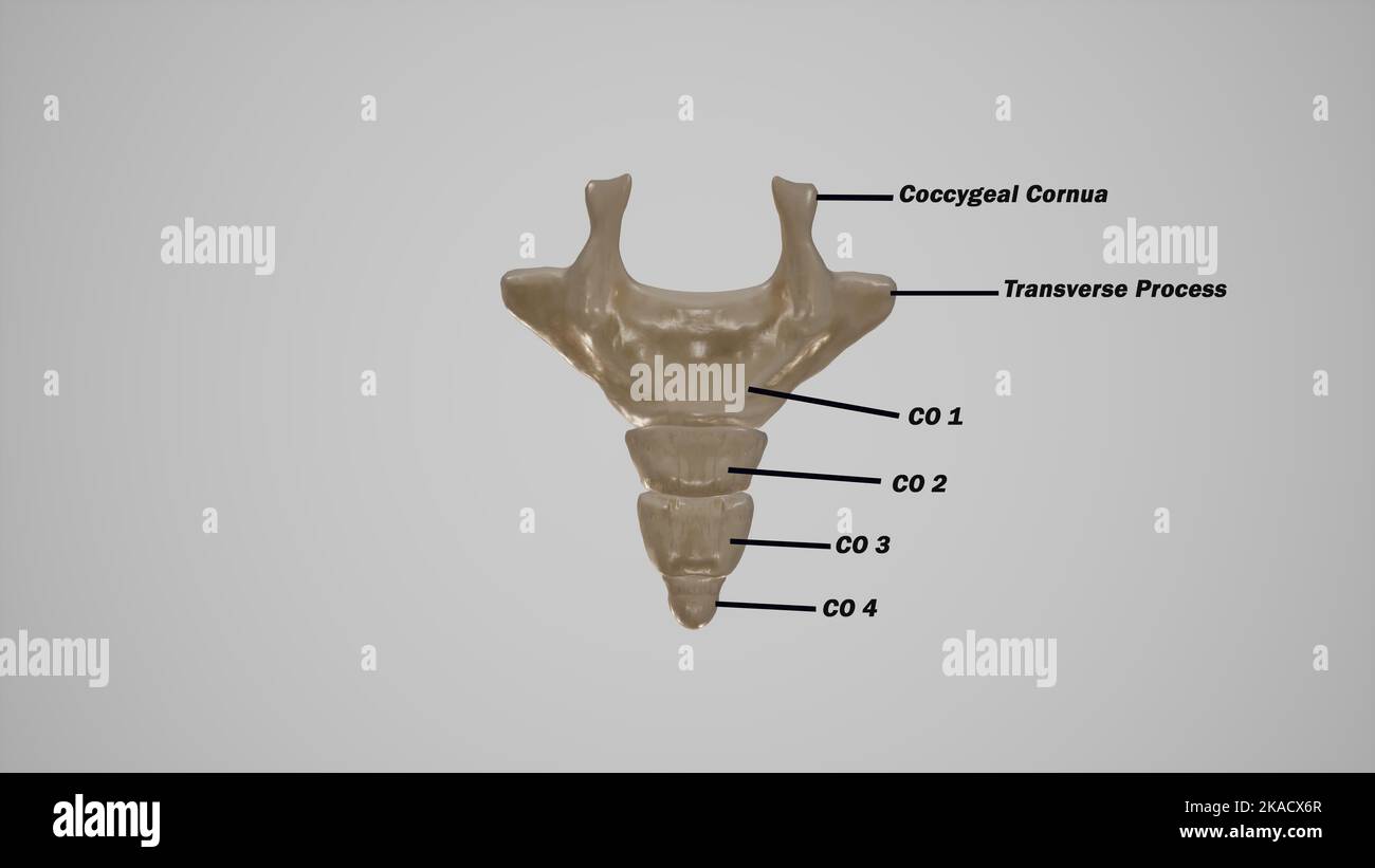 Coccyx bone anatomy labeled Stock Photohttps://www.alamy.com/image-license-details/?v=1https://www.alamy.com/coccyx-bone-anatomy-labeled-image488320863.html
RF2KACX6R–Coccyx bone anatomy labeled
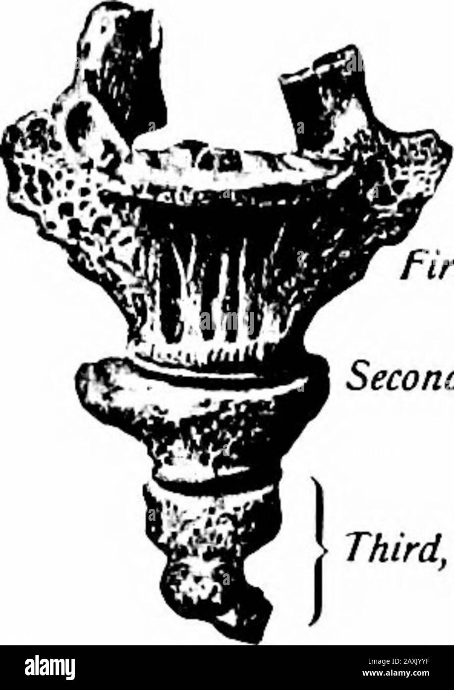 A manual of anatomy . tta and McMurricti.) that articulates with the sacrum. The dorsal surface presents twoupward-projecting processes (cornua coccygea) representing articularprocesses for articulation with the cornua sacrahs. These assist informing two foramina for the fifth pair of sacral nerves. Laterallythis segment presents a rudimentary transverse process on each side.The second may also present such processes but the remaining seg-ments are rudimentary nodules of bone. Ossification.—The coccyx is developed from four centers, one for each segment.That for the first segment appears durin Stock Photohttps://www.alamy.com/image-license-details/?v=1https://www.alamy.com/a-manual-of-anatomy-tta-and-mcmurricti-that-articulates-with-the-sacrum-the-dorsal-surface-presents-twoupward-projecting-processes-cornua-coccygea-representing-articularprocesses-for-articulation-with-the-cornua-sacrahs-these-assist-informing-two-foramina-for-the-fifth-pair-of-sacral-nerves-laterallythis-segment-presents-a-rudimentary-transverse-process-on-each-sidethe-second-may-also-present-such-processes-but-the-remaining-seg-ments-are-rudimentary-nodules-of-bone-ossificationthe-coccyx-is-developed-from-four-centers-one-for-each-segmentthat-for-the-first-segment-appears-durin-image343395123.html
RM2AXJYYF–A manual of anatomy . tta and McMurricti.) that articulates with the sacrum. The dorsal surface presents twoupward-projecting processes (cornua coccygea) representing articularprocesses for articulation with the cornua sacrahs. These assist informing two foramina for the fifth pair of sacral nerves. Laterallythis segment presents a rudimentary transverse process on each side.The second may also present such processes but the remaining seg-ments are rudimentary nodules of bone. Ossification.—The coccyx is developed from four centers, one for each segment.That for the first segment appears durin
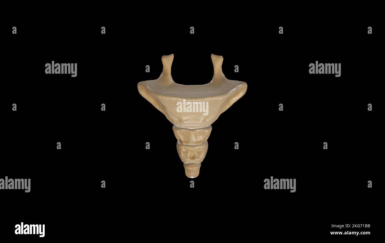 Anterior view of Coccyx Stock Photohttps://www.alamy.com/image-license-details/?v=1https://www.alamy.com/anterior-view-of-coccyx-image491879567.html
RF2KG71BB–Anterior view of Coccyx
 A manual of anatomy . Superior articular process Sacral canal. Median sacralcrest Articularsacral crest Posterior sacralforamina Apex of sacrum . Sacral Sacralhiatus cornu ViG. 12.—The sacrum ieen from behind (dorsal surface). (Sobotta and McMurrich.j THE COCCYX 33 vertebrae. The sacral cornua are the downward-projecting inferiorarticular processes of the last sacral vertebra. The apex of thesacrum represents the oval body of the fifth sacral vertebra. The sacral canal is flattened dorsoventrally and curves with thebone. It communicates with the dorsal and ventral sacral foramina. The sacrum o Stock Photohttps://www.alamy.com/image-license-details/?v=1https://www.alamy.com/a-manual-of-anatomy-superior-articular-process-sacral-canal-median-sacralcrest-articularsacral-crest-posterior-sacralforamina-apex-of-sacrum-sacral-sacralhiatus-cornu-vig-12the-sacrum-ieen-from-behind-dorsal-surface-sobotta-and-mcmurrichj-the-coccyx-33-vertebrae-the-sacral-cornua-are-the-downward-projecting-inferiorarticular-processes-of-the-last-sacral-vertebra-the-apex-of-thesacrum-represents-the-oval-body-of-the-fifth-sacral-vertebra-the-sacral-canal-is-flattened-dorsoventrally-and-curves-with-thebone-it-communicates-with-the-dorsal-and-ventral-sacral-foramina-the-sacrum-o-image343395596.html
RM2AXK0GC–A manual of anatomy . Superior articular process Sacral canal. Median sacralcrest Articularsacral crest Posterior sacralforamina Apex of sacrum . Sacral Sacralhiatus cornu ViG. 12.—The sacrum ieen from behind (dorsal surface). (Sobotta and McMurrich.j THE COCCYX 33 vertebrae. The sacral cornua are the downward-projecting inferiorarticular processes of the last sacral vertebra. The apex of thesacrum represents the oval body of the fifth sacral vertebra. The sacral canal is flattened dorsoventrally and curves with thebone. It communicates with the dorsal and ventral sacral foramina. The sacrum o
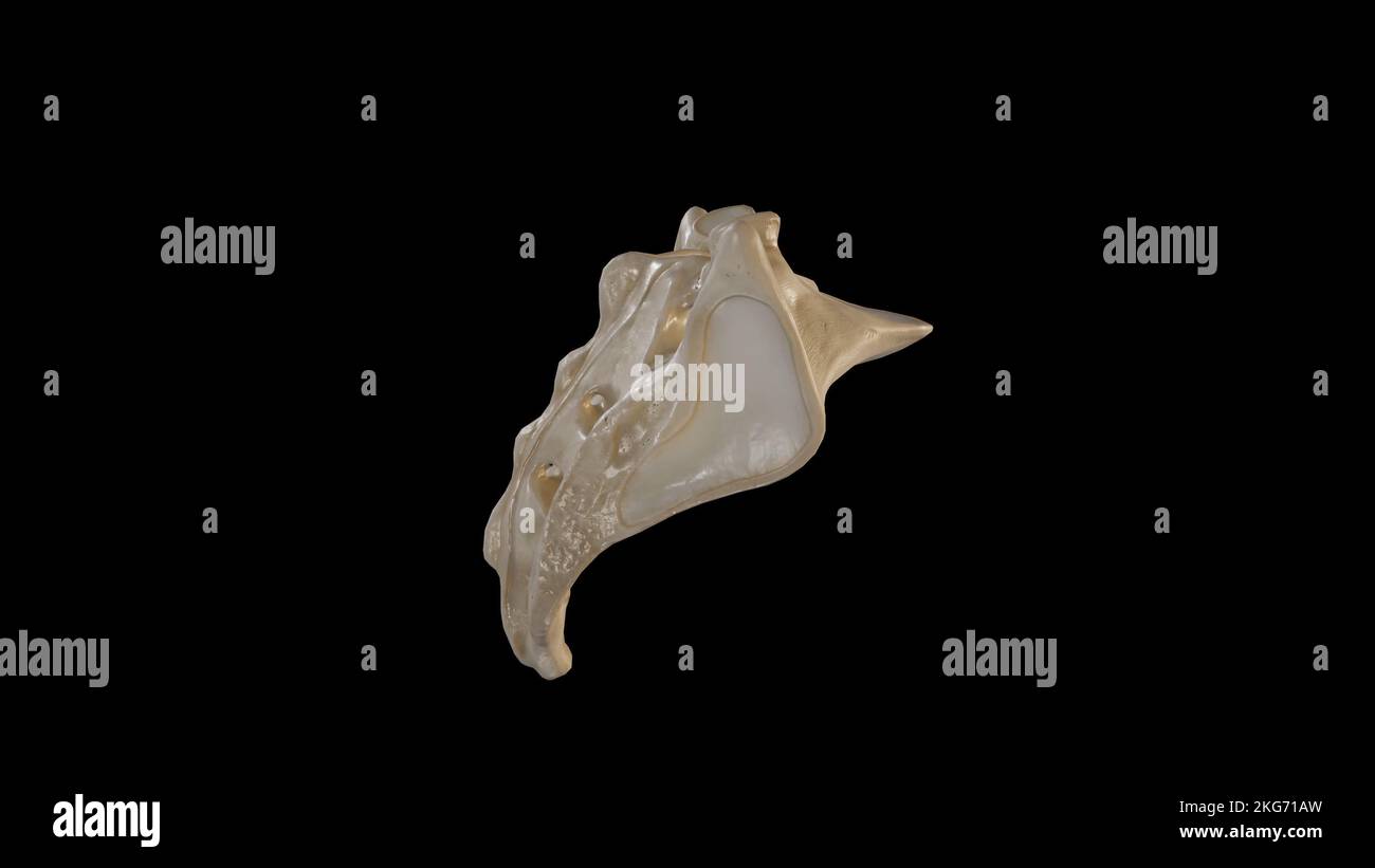 Lateral view of Sacrum Stock Photohttps://www.alamy.com/image-license-details/?v=1https://www.alamy.com/lateral-view-of-sacrum-image491879553.html
RF2KG71AW–Lateral view of Sacrum
 . Regional anesthesia : its technic and clinical application . joining the sacral cornua and the fourth sacral spinous process. the jimcture of the coccyx with the sacrum, and bounded by the sacralcornua on each side and the fourth sacral spinous process on the mid-line a little higher up. These three prominences, palpable in themajority of cases, form the angles of a triangular surface at the middlepoint of which the needle is introduced with ease and success (Fig. 210).The spinal puncture needle, with its stylet in and its bevel turnedupward, is introduced through a wheal raised at this poin Stock Photohttps://www.alamy.com/image-license-details/?v=1https://www.alamy.com/regional-anesthesia-its-technic-and-clinical-application-joining-the-sacral-cornua-and-the-fourth-sacral-spinous-process-the-jimcture-of-the-coccyx-with-the-sacrum-and-bounded-by-the-sacralcornua-on-each-side-and-the-fourth-sacral-spinous-process-on-the-mid-line-a-little-higher-up-these-three-prominences-palpable-in-themajority-of-cases-form-the-angles-of-a-triangular-surface-at-the-middlepoint-of-which-the-needle-is-introduced-with-ease-and-success-fig-210the-spinal-puncture-needle-with-its-stylet-in-and-its-bevel-turnedupward-is-introduced-through-a-wheal-raised-at-this-poin-image370060634.html
RM2CE1M36–. Regional anesthesia : its technic and clinical application . joining the sacral cornua and the fourth sacral spinous process. the jimcture of the coccyx with the sacrum, and bounded by the sacralcornua on each side and the fourth sacral spinous process on the mid-line a little higher up. These three prominences, palpable in themajority of cases, form the angles of a triangular surface at the middlepoint of which the needle is introduced with ease and success (Fig. 210).The spinal puncture needle, with its stylet in and its bevel turnedupward, is introduced through a wheal raised at this poin
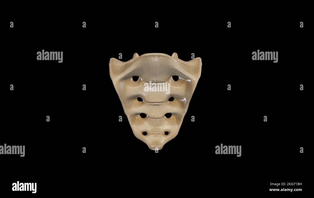
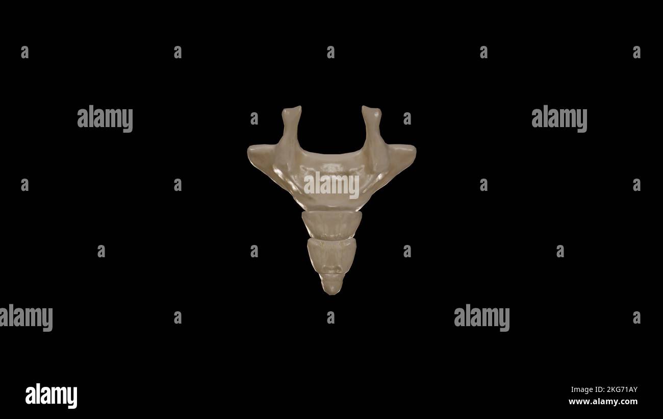 Posterior view of Coccyx Stock Photohttps://www.alamy.com/image-license-details/?v=1https://www.alamy.com/posterior-view-of-coccyx-image491879555.html
RF2KG71AY–Posterior view of Coccyx
 . Quain's Elements of anatomy. c fossaof the hip-bone, and known as the ala of the sacrum. The lower end or apex, formed by the small inferiorsurface of the body of the fifth sacral vertebra, is trans-versely oval, and articulates with the coccyx. The sacral canal is curved with the bone, and gra-dually narrows as it descends ; in transverse sectionit is three-sided above, but flattened and rather semi-lunar below. It terminates on the posterior surface ofthe bone between the sacral cornua, where the laminasof the last two sacral vertebrae areimperfect. From this canal therepass outwards in th Stock Photohttps://www.alamy.com/image-license-details/?v=1https://www.alamy.com/quains-elements-of-anatomy-c-fossaof-the-hip-bone-and-known-as-the-ala-of-the-sacrum-the-lower-end-or-apex-formed-by-the-small-inferiorsurface-of-the-body-of-the-fifth-sacral-vertebra-is-trans-versely-oval-and-articulates-with-the-coccyx-the-sacral-canal-is-curved-with-the-bone-and-gra-dually-narrows-as-it-descends-in-transverse-sectionit-is-three-sided-above-but-flattened-and-rather-semi-lunar-below-it-terminates-on-the-posterior-surface-ofthe-bone-between-the-sacral-cornua-where-the-laminasof-the-last-two-sacral-vertebrae-areimperfect-from-this-canal-therepass-outwards-in-th-image370417192.html
RM2CEHXWC–. Quain's Elements of anatomy. c fossaof the hip-bone, and known as the ala of the sacrum. The lower end or apex, formed by the small inferiorsurface of the body of the fifth sacral vertebra, is trans-versely oval, and articulates with the coccyx. The sacral canal is curved with the bone, and gra-dually narrows as it descends ; in transverse sectionit is three-sided above, but flattened and rather semi-lunar below. It terminates on the posterior surface ofthe bone between the sacral cornua, where the laminasof the last two sacral vertebrae areimperfect. From this canal therepass outwards in th
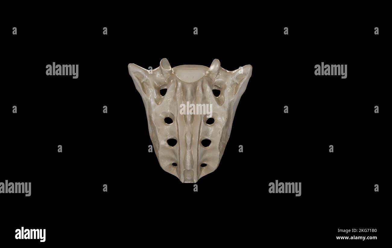 Posterior view of Sacrum Stock Photohttps://www.alamy.com/image-license-details/?v=1https://www.alamy.com/posterior-view-of-sacrum-image491879556.html
RF2KG71B0–Posterior view of Sacrum
 . Regional anesthesia : its technic and clinical application . the sacral cornua oneach side and the spinous process of the fourth sacral vertebra on themidline a little higher up. It has the shape of an inverted V or U.The arms of the inverted V or U are generally salient edges and their18 274 REGIONAL ANESTHESIA extremities prominent tubercles; but in a certain number of cases theseanatomic features are thin and fiat and cannot be defined by palpation,even in lean patients. The sacral canal is a prismatic space occupying the whole height ofthe sacrum. Its upper extremity is connected with th Stock Photohttps://www.alamy.com/image-license-details/?v=1https://www.alamy.com/regional-anesthesia-its-technic-and-clinical-application-the-sacral-cornua-oneach-side-and-the-spinous-process-of-the-fourth-sacral-vertebra-on-themidline-a-little-higher-up-it-has-the-shape-of-an-inverted-v-or-uthe-arms-of-the-inverted-v-or-u-are-generally-salient-edges-and-their18-274-regional-anesthesia-extremities-prominent-tubercles-but-in-a-certain-number-of-cases-theseanatomic-features-are-thin-and-fiat-and-cannot-be-defined-by-palpationeven-in-lean-patients-the-sacral-canal-is-a-prismatic-space-occupying-the-whole-height-ofthe-sacrum-its-upper-extremity-is-connected-with-th-image370060764.html
RM2CE1M7T–. Regional anesthesia : its technic and clinical application . the sacral cornua oneach side and the spinous process of the fourth sacral vertebra on themidline a little higher up. It has the shape of an inverted V or U.The arms of the inverted V or U are generally salient edges and their18 274 REGIONAL ANESTHESIA extremities prominent tubercles; but in a certain number of cases theseanatomic features are thin and fiat and cannot be defined by palpation,even in lean patients. The sacral canal is a prismatic space occupying the whole height ofthe sacrum. Its upper extremity is connected with th
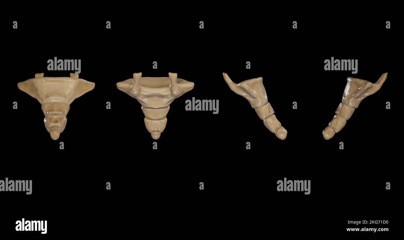 Coccyx-Multiple Views Stock Photohttps://www.alamy.com/image-license-details/?v=1https://www.alamy.com/coccyx-multiple-views-image491879612.html
RF2KG71D0–Coccyx-Multiple Views
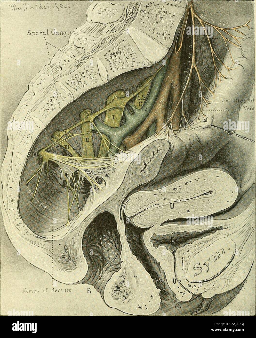 Operative gynecology : . Fig. 37.—The Topography of the Fixed Part of the Bladder. The Vesical Cornua lie to theRight and Left of the Ureteral Orifices, Just in Front of the Broad Ligaments. sacral and lateral pelvic regions. The distribution of the superior hemor-rhoidal vessels is also seen. The sacral plexus of nerves is seen to emergefrom the sacral foramina, forming the lumbo-sacral cord, and the first, second, 72 TOPOGRAPHICAL ANATOMY. third, fourth, and fifth sacral cords, which converge toward the great sacro-sciatic foramen, to unite in the sciatic nerve. The sacral ganglia of the. Fi Stock Photohttps://www.alamy.com/image-license-details/?v=1https://www.alamy.com/operative-gynecology-fig-37the-topography-of-the-fixed-part-of-the-bladder-the-vesical-cornua-lie-to-theright-and-left-of-the-ureteral-orifices-just-in-front-of-the-broad-ligaments-sacral-and-lateral-pelvic-regions-the-distribution-of-the-superior-hemor-rhoidal-vessels-is-also-seen-the-sacral-plexus-of-nerves-is-seen-to-emergefrom-the-sacral-foramina-forming-the-lumbo-sacral-cord-and-the-first-second-72-topographical-anatomy-third-fourth-and-fifth-sacral-cords-which-converge-toward-the-great-sacro-sciatic-foramen-to-unite-in-the-sciatic-nerve-the-sacral-ganglia-of-the-fi-image338298034.html
RM2AJAPGJ–Operative gynecology : . Fig. 37.—The Topography of the Fixed Part of the Bladder. The Vesical Cornua lie to theRight and Left of the Ureteral Orifices, Just in Front of the Broad Ligaments. sacral and lateral pelvic regions. The distribution of the superior hemor-rhoidal vessels is also seen. The sacral plexus of nerves is seen to emergefrom the sacral foramina, forming the lumbo-sacral cord, and the first, second, 72 TOPOGRAPHICAL ANATOMY. third, fourth, and fifth sacral cords, which converge toward the great sacro-sciatic foramen, to unite in the sciatic nerve. The sacral ganglia of the. Fi
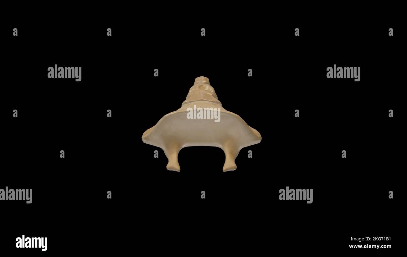 Superior view of Coccyx Stock Photohttps://www.alamy.com/image-license-details/?v=1https://www.alamy.com/superior-view-of-coccyx-image491879557.html
RF2KG71B1–Superior view of Coccyx
 . An American text-book of obstetrics. For practitioners and students. Fig. 9.—Variation in sacral curves (Hirst): P, promontory of sacrum; C, coccyx. the last sacral segment to the coccygeal cornua constitute the supracornual orlateral ligaments. The intertransverse ligament is represented by fibrous bandswhich pass from the lower lateral angle of the sacrum to the transverse pro-cess of the first piece of the coccyx. The intervertebral disk is a rudimentary member of the series of fibro-car-tilaginous plates interposed between the vertebrae ; a distinct cavity sometimesexists within this dis Stock Photohttps://www.alamy.com/image-license-details/?v=1https://www.alamy.com/an-american-text-book-of-obstetrics-for-practitioners-and-students-fig-9variation-in-sacral-curves-hirst-p-promontory-of-sacrum-c-coccyx-the-last-sacral-segment-to-the-coccygeal-cornua-constitute-the-supracornual-orlateral-ligaments-the-intertransverse-ligament-is-represented-by-fibrous-bandswhich-pass-from-the-lower-lateral-angle-of-the-sacrum-to-the-transverse-pro-cess-of-the-first-piece-of-the-coccyx-the-intervertebral-disk-is-a-rudimentary-member-of-the-series-of-fibro-car-tilaginous-plates-interposed-between-the-vertebrae-a-distinct-cavity-sometimesexists-within-this-dis-image370607992.html
RM2CEXJ7M–. An American text-book of obstetrics. For practitioners and students. Fig. 9.—Variation in sacral curves (Hirst): P, promontory of sacrum; C, coccyx. the last sacral segment to the coccygeal cornua constitute the supracornual orlateral ligaments. The intertransverse ligament is represented by fibrous bandswhich pass from the lower lateral angle of the sacrum to the transverse pro-cess of the first piece of the coccyx. The intervertebral disk is a rudimentary member of the series of fibro-car-tilaginous plates interposed between the vertebrae ; a distinct cavity sometimesexists within this dis
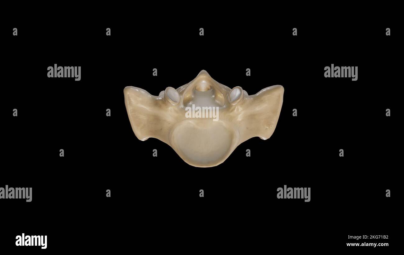 Superior view of Sacrum Stock Photohttps://www.alamy.com/image-license-details/?v=1https://www.alamy.com/superior-view-of-sacrum-image491879558.html
RF2KG71B2–Superior view of Sacrum
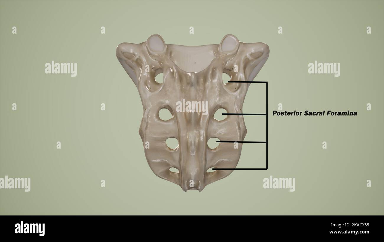 Posterior view of human sacrum showing the posterior sacral foramina-Labeled Stock Photohttps://www.alamy.com/image-license-details/?v=1https://www.alamy.com/posterior-view-of-human-sacrum-showing-the-posterior-sacral-foramina-labeled-image488320817.html
RF2KACX55–Posterior view of human sacrum showing the posterior sacral foramina-Labeled
 . A text-book of medicine for students and practitioners . Fig. 149.—Lesion at the level of the fifthlumbar segment. Fig. 150.—Lesion of the first sacral segment. toucli and position, wc may be quite certain that there is a central lesionin the posterior gray cornua, such as is found especially in syringomyelia,injury of the cord, and central glioma. 292 DISEASES OF THE NERVOUS SYSTEM [Brissaud and other recent French writers lay much stress upon tlie dif-ference between the sensory areas supplied hy the spinal roots (radicularnictanierisni) and the sensory areas supplied by the ditlerent spin Stock Photohttps://www.alamy.com/image-license-details/?v=1https://www.alamy.com/a-text-book-of-medicine-for-students-and-practitioners-fig-149lesion-at-the-level-of-the-fifthlumbar-segment-fig-150lesion-of-the-first-sacral-segment-toucli-and-position-wc-may-be-quite-certain-that-there-is-a-central-lesionin-the-posterior-gray-cornua-such-as-is-found-especially-in-syringomyeliainjury-of-the-cord-and-central-glioma-292-diseases-of-the-nervous-system-brissaud-and-other-recent-french-writers-lay-much-stress-upon-tlie-dif-ference-between-the-sensory-areas-supplied-hy-the-spinal-roots-radicularnictanierisni-and-the-sensory-areas-supplied-by-the-ditlerent-spin-image369636207.html
RM2CDAAN3–. A text-book of medicine for students and practitioners . Fig. 149.—Lesion at the level of the fifthlumbar segment. Fig. 150.—Lesion of the first sacral segment. toucli and position, wc may be quite certain that there is a central lesionin the posterior gray cornua, such as is found especially in syringomyelia,injury of the cord, and central glioma. 292 DISEASES OF THE NERVOUS SYSTEM [Brissaud and other recent French writers lay much stress upon tlie dif-ference between the sensory areas supplied hy the spinal roots (radicularnictanierisni) and the sensory areas supplied by the ditlerent spin
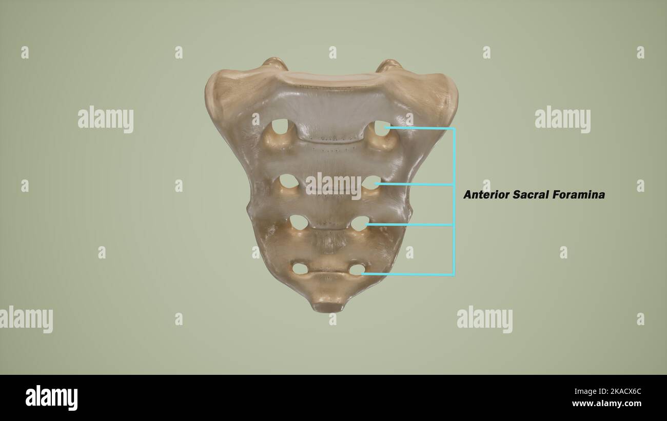 Anterior view of human sacrum showing the anterior sacral foramina-Labeled Stock Photohttps://www.alamy.com/image-license-details/?v=1https://www.alamy.com/anterior-view-of-human-sacrum-showing-the-anterior-sacral-foramina-labeled-image488320852.html
RF2KACX6C–Anterior view of human sacrum showing the anterior sacral foramina-Labeled
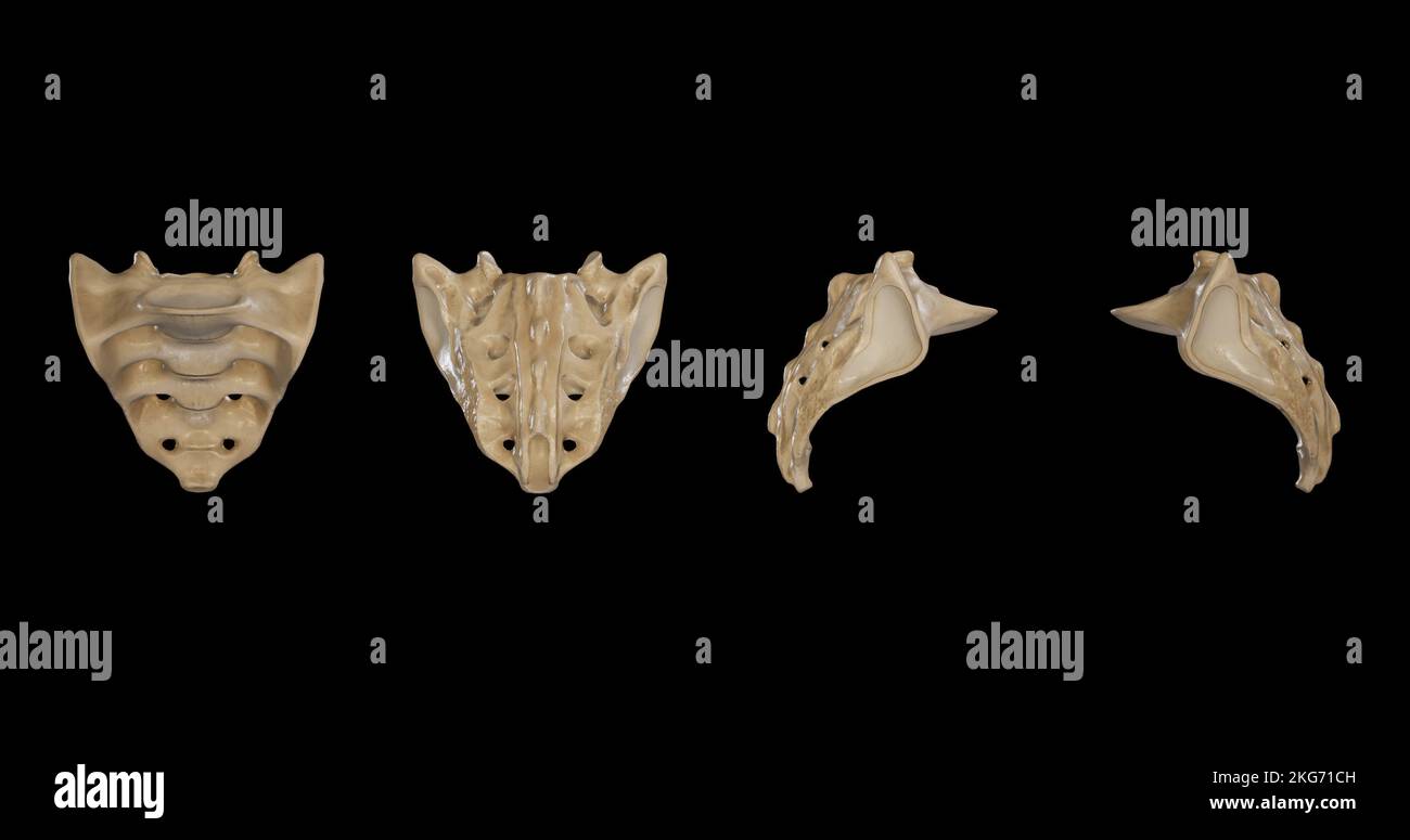 Sacrum from multiple sides Stock Photohttps://www.alamy.com/image-license-details/?v=1https://www.alamy.com/sacrum-from-multiple-sides-image491879601.html
RF2KG71CH–Sacrum from multiple sides
 . A text-book of medicine for students and practitioners . Fig. 149.—Lesion at the level of the fifthlumbar segment. Fig. 150.—Lesion of the first sacral segment. toucli and position, wc may be quite certain that there is a central lesionin the posterior gray cornua, such as is found especially in syringomyelia,injury of the cord, and central glioma. 292 DISEASES OF THE NERVOUS SYSTEM [Brissaud and other recent French writers lay much stress upon tlie dif-ference between the sensory areas supplied hy the spinal roots (radicularnictanierisni) and the sensory areas supplied by the ditlerent spin Stock Photohttps://www.alamy.com/image-license-details/?v=1https://www.alamy.com/a-text-book-of-medicine-for-students-and-practitioners-fig-149lesion-at-the-level-of-the-fifthlumbar-segment-fig-150lesion-of-the-first-sacral-segment-toucli-and-position-wc-may-be-quite-certain-that-there-is-a-central-lesionin-the-posterior-gray-cornua-such-as-is-found-especially-in-syringomyeliainjury-of-the-cord-and-central-glioma-292-diseases-of-the-nervous-system-brissaud-and-other-recent-french-writers-lay-much-stress-upon-tlie-dif-ference-between-the-sensory-areas-supplied-hy-the-spinal-roots-radicularnictanierisni-and-the-sensory-areas-supplied-by-the-ditlerent-spin-image369636300.html
RM2CDAATC–. A text-book of medicine for students and practitioners . Fig. 149.—Lesion at the level of the fifthlumbar segment. Fig. 150.—Lesion of the first sacral segment. toucli and position, wc may be quite certain that there is a central lesionin the posterior gray cornua, such as is found especially in syringomyelia,injury of the cord, and central glioma. 292 DISEASES OF THE NERVOUS SYSTEM [Brissaud and other recent French writers lay much stress upon tlie dif-ference between the sensory areas supplied hy the spinal roots (radicularnictanierisni) and the sensory areas supplied by the ditlerent spin
