Quick filters:
Sacral spinal nerves Stock Photos and Images
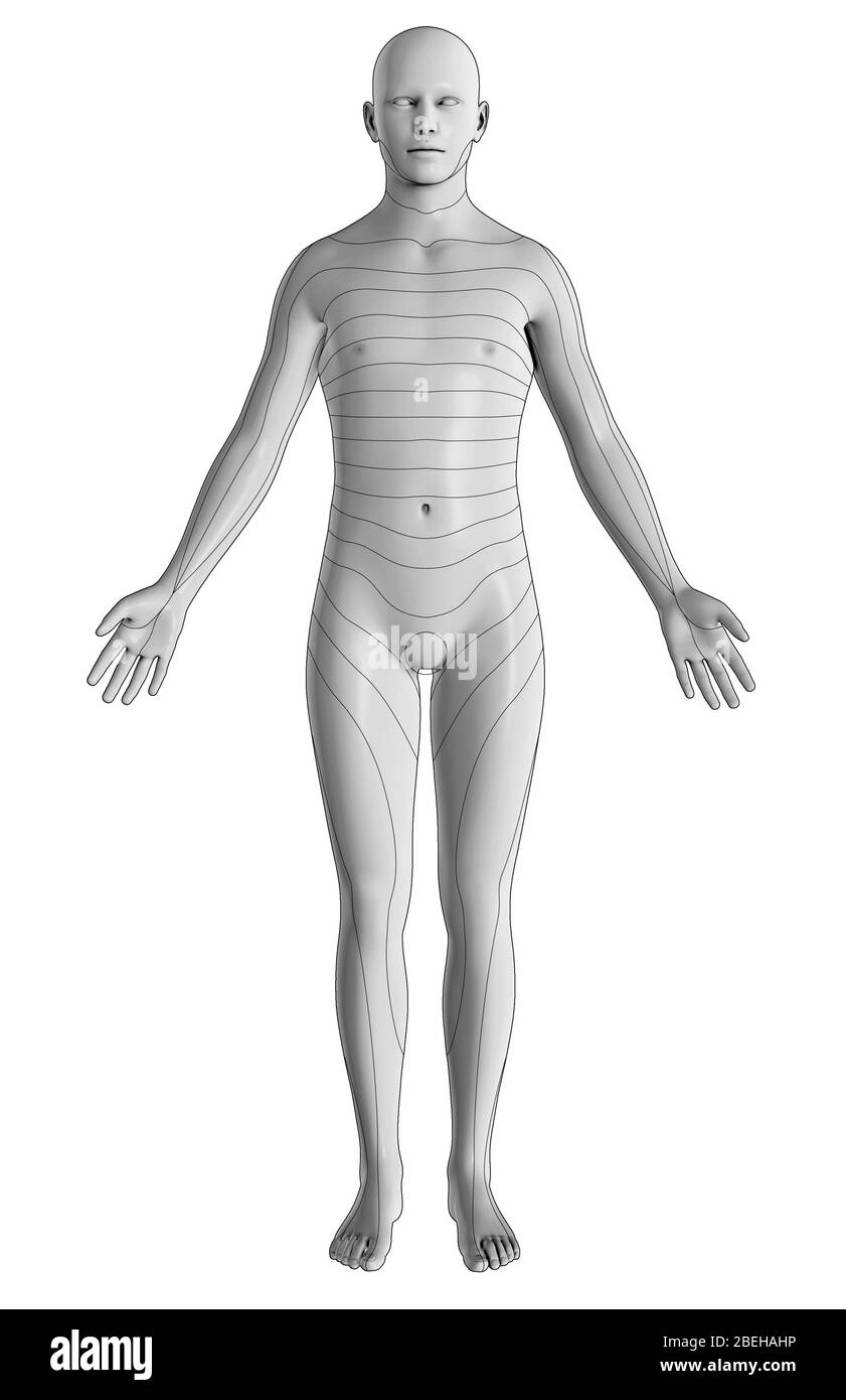 An illustration of the dermatomes of the body from an anterior view. Dermatomes are regions of skin supplied by specific spinal nerves, which relay sensory information to the brain. This includes eight cervical nerves (excludes C1), twelve thoracic nerves, five lumbar nerves, and five sacral nerves. Stock Photohttps://www.alamy.com/image-license-details/?v=1https://www.alamy.com/an-illustration-of-the-dermatomes-of-the-body-from-an-anterior-view-dermatomes-are-regions-of-skin-supplied-by-specific-spinal-nerves-which-relay-sensory-information-to-the-brain-this-includes-eight-cervical-nerves-excludes-c1-twelve-thoracic-nerves-five-lumbar-nerves-and-five-sacral-nerves-image353194066.html
RM2BEHAHP–An illustration of the dermatomes of the body from an anterior view. Dermatomes are regions of skin supplied by specific spinal nerves, which relay sensory information to the brain. This includes eight cervical nerves (excludes C1), twelve thoracic nerves, five lumbar nerves, and five sacral nerves.
 Sciatic Nerve on Black Background.3d rendering Stock Photohttps://www.alamy.com/image-license-details/?v=1https://www.alamy.com/sciatic-nerve-on-black-background3d-rendering-image501580952.html
RF2M40YHC–Sciatic Nerve on Black Background.3d rendering
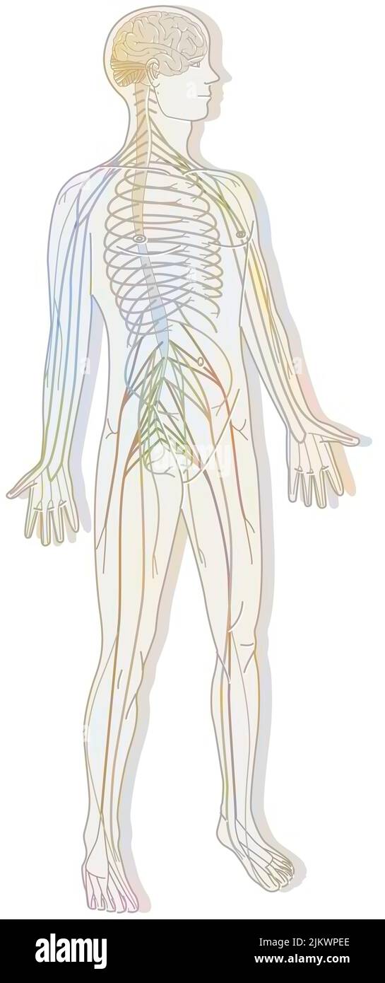 Silhouette with the nervous system consisting of the central and peripheral system. Stock Photohttps://www.alamy.com/image-license-details/?v=1https://www.alamy.com/silhouette-with-the-nervous-system-consisting-of-the-central-and-peripheral-system-image476924854.html
RF2JKWPEE–Silhouette with the nervous system consisting of the central and peripheral system.
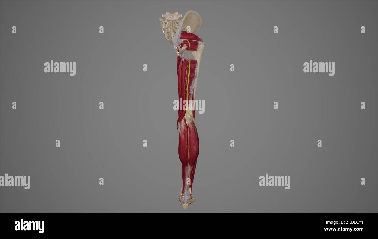 Anatomical Illustration of Sciatic Nerve Stock Photohttps://www.alamy.com/image-license-details/?v=1https://www.alamy.com/anatomical-illustration-of-sciatic-nerve-image490198325.html
RF2KDECY1–Anatomical Illustration of Sciatic Nerve
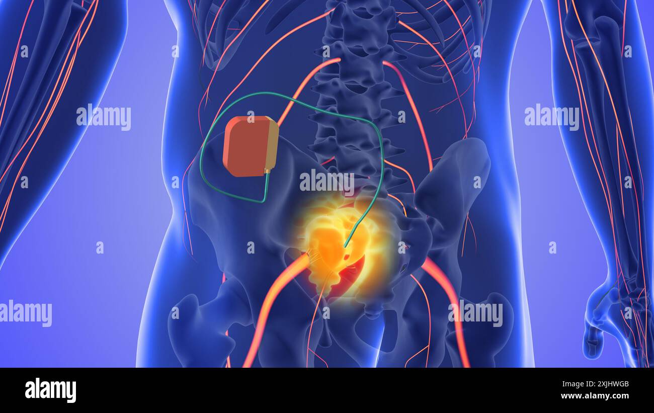 Sacral nerve stimulation medical concept Stock Photohttps://www.alamy.com/image-license-details/?v=1https://www.alamy.com/sacral-nerve-stimulation-medical-concept-image613819931.html
RF2XJHWGB–Sacral nerve stimulation medical concept
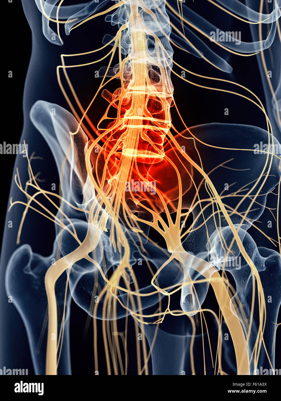 medically accurate illustration - painful sacral nerves Stock Photohttps://www.alamy.com/image-license-details/?v=1https://www.alamy.com/stock-photo-medically-accurate-illustration-painful-sacral-nerves-89769678.html
RFF61A3X–medically accurate illustration - painful sacral nerves
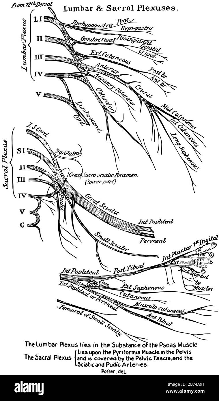 This illustration represents The Lumbar and Sacral Plexuses of the Spinal Nerves, vintage line drawing or engraving illustration. Stock Vectorhttps://www.alamy.com/image-license-details/?v=1https://www.alamy.com/this-illustration-represents-the-lumbar-and-sacral-plexuses-of-the-spinal-nerves-vintage-line-drawing-or-engraving-illustration-image348605876.html
RF2B74A9T–This illustration represents The Lumbar and Sacral Plexuses of the Spinal Nerves, vintage line drawing or engraving illustration.
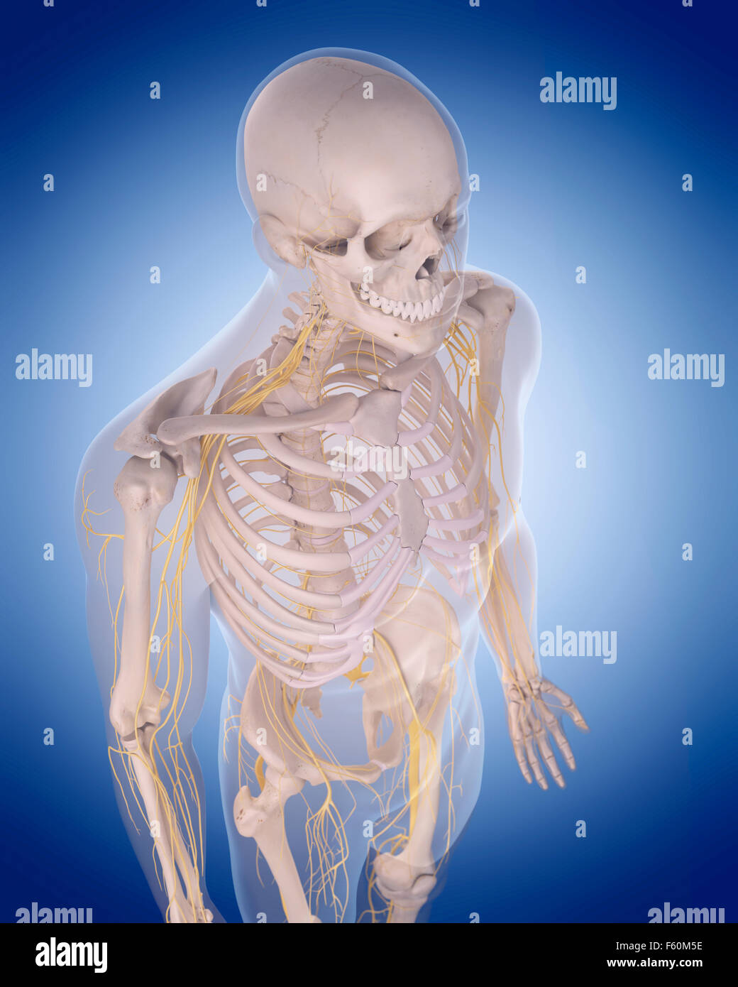 medically accurate illustration - nerves of the upper body Stock Photohttps://www.alamy.com/image-license-details/?v=1https://www.alamy.com/stock-photo-medically-accurate-illustration-nerves-of-the-upper-body-89755610.html
RFF60M5E–medically accurate illustration - nerves of the upper body
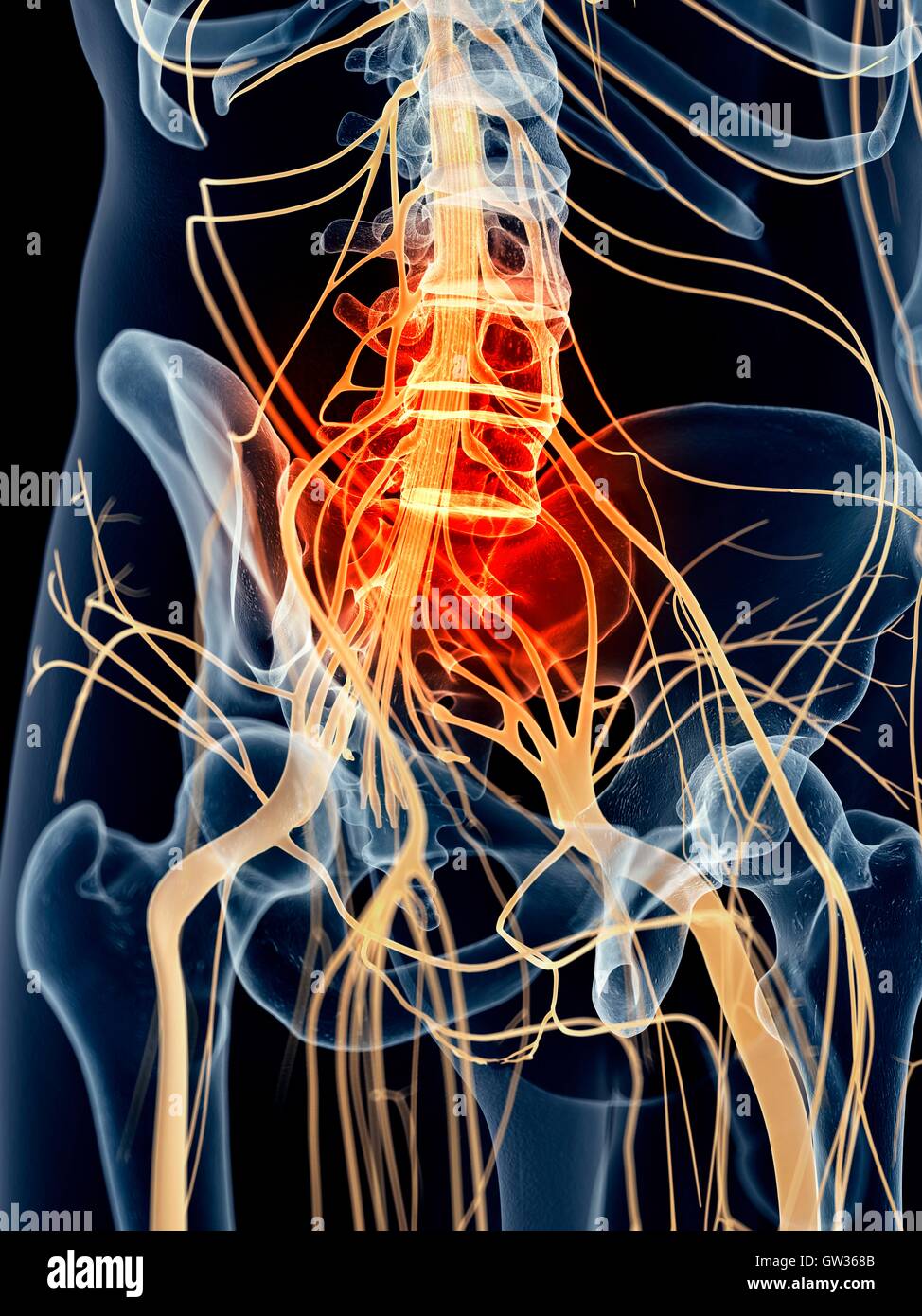 Human sacral nerve pain, illustration. Stock Photohttps://www.alamy.com/image-license-details/?v=1https://www.alamy.com/stock-photo-human-sacral-nerve-pain-illustration-118699403.html
RFGW368B–Human sacral nerve pain, illustration.
 A segment of the spinal cord with nerves and a vertebra. 3D illustration. Stock Photohttps://www.alamy.com/image-license-details/?v=1https://www.alamy.com/a-segment-of-the-spinal-cord-with-nerves-and-a-vertebra-3d-illustration-image591886519.html
RF2WAXN87–A segment of the spinal cord with nerves and a vertebra. 3D illustration.
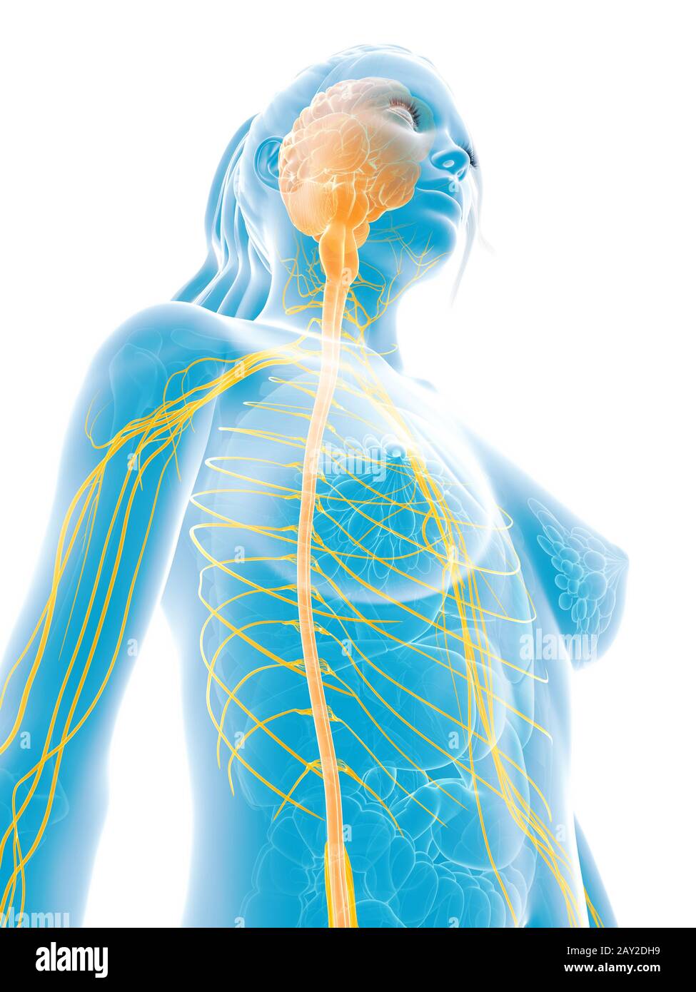 3d rendered medical illustration - female nerves Stock Photohttps://www.alamy.com/image-license-details/?v=1https://www.alamy.com/3d-rendered-medical-illustration-female-nerves-image343647285.html
RM2AY2DH9–3d rendered medical illustration - female nerves
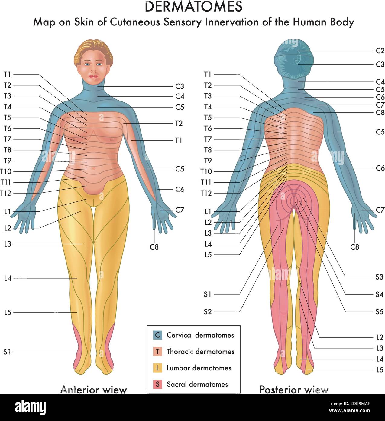 Map on Skin of Cutaneous Sensory Innervation of the Human Body Stock Vectorhttps://www.alamy.com/image-license-details/?v=1https://www.alamy.com/map-on-skin-of-cutaneous-sensory-innervation-of-the-human-body-image385602855.html
RF2DB9MAF–Map on Skin of Cutaneous Sensory Innervation of the Human Body
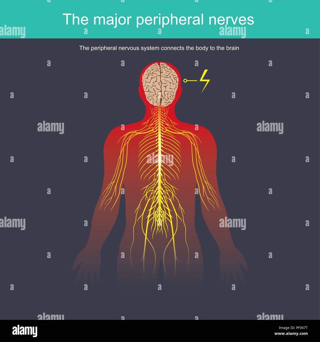 The peripheral nervous system connects the body to the brain Stock Vectorhttps://www.alamy.com/image-license-details/?v=1https://www.alamy.com/the-peripheral-nervous-system-connects-the-body-to-the-brain-image215815036.html
RFPF367T–The peripheral nervous system connects the body to the brain
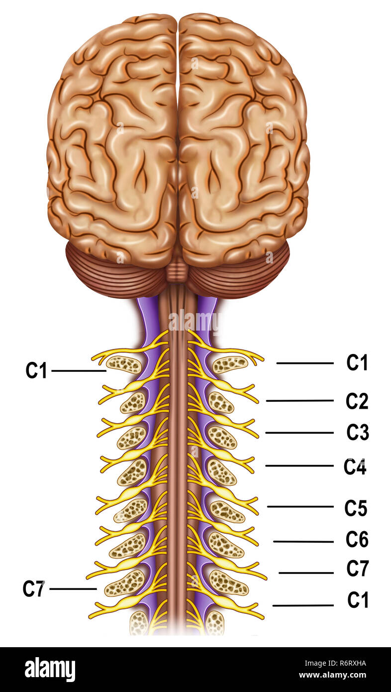 The cervical plexus. It controls the motor functions of the neck, it is the superior nervous plexus of the peripheral nervous system. Stock Photohttps://www.alamy.com/image-license-details/?v=1https://www.alamy.com/the-cervical-plexus-it-controls-the-motor-functions-of-the-neck-it-is-the-superior-nervous-plexus-of-the-peripheral-nervous-system-image227948486.html
RFR6RXHA–The cervical plexus. It controls the motor functions of the neck, it is the superior nervous plexus of the peripheral nervous system.
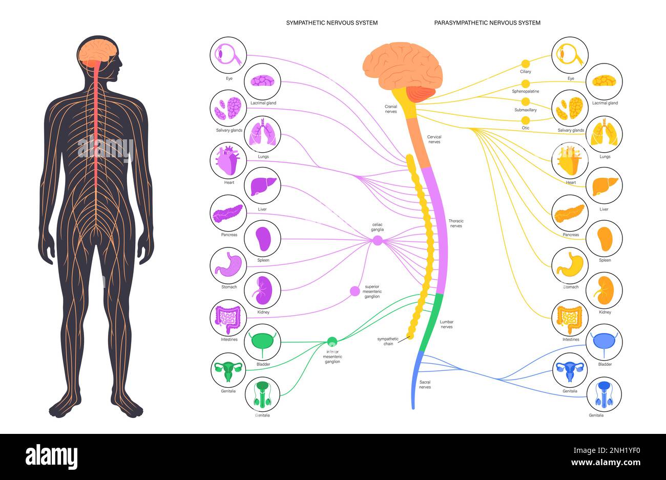 Autonomic nervous system, illustration Stock Photohttps://www.alamy.com/image-license-details/?v=1https://www.alamy.com/autonomic-nervous-system-illustration-image526803732.html
RF2NH1YF0–Autonomic nervous system, illustration
 Vintage diagrams of Nerves of the Human Body 1900s. Stock Photohttps://www.alamy.com/image-license-details/?v=1https://www.alamy.com/vintage-diagrams-of-nerves-of-the-human-body-1900s-image350211968.html
RF2B9NEX8–Vintage diagrams of Nerves of the Human Body 1900s.
 The nerves of the trunk Stock Photohttps://www.alamy.com/image-license-details/?v=1https://www.alamy.com/stock-photo-the-nerves-of-the-trunk-13169149.html
RFACJCFX–The nerves of the trunk
 3D illustration of Sacral Spine - Part of Human Skeleton. Stock Photohttps://www.alamy.com/image-license-details/?v=1https://www.alamy.com/stock-photo-3d-illustration-of-sacral-spine-part-of-human-skeleton-104311896.html
RFG1KPTT–3D illustration of Sacral Spine - Part of Human Skeleton.
 The lumbosacral plexus is a network of nerve fibers, derived from the roots of lumbar and sacral spinal nerves that branch out to form the nerves supp Stock Photohttps://www.alamy.com/image-license-details/?v=1https://www.alamy.com/the-lumbosacral-plexus-is-a-network-of-nerve-fibers-derived-from-the-roots-of-lumbar-and-sacral-spinal-nerves-that-branch-out-to-form-the-nerves-supp-image596595777.html
RF2WJH801–The lumbosacral plexus is a network of nerve fibers, derived from the roots of lumbar and sacral spinal nerves that branch out to form the nerves supp
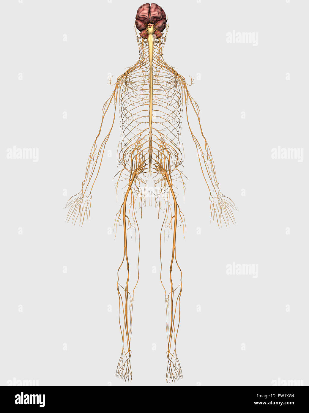 Medical illustration of peripheral nervous system with brain. Stock Photohttps://www.alamy.com/image-license-details/?v=1https://www.alamy.com/stock-photo-medical-illustration-of-peripheral-nervous-system-with-brain-84250660.html
RFEW1XG4–Medical illustration of peripheral nervous system with brain.
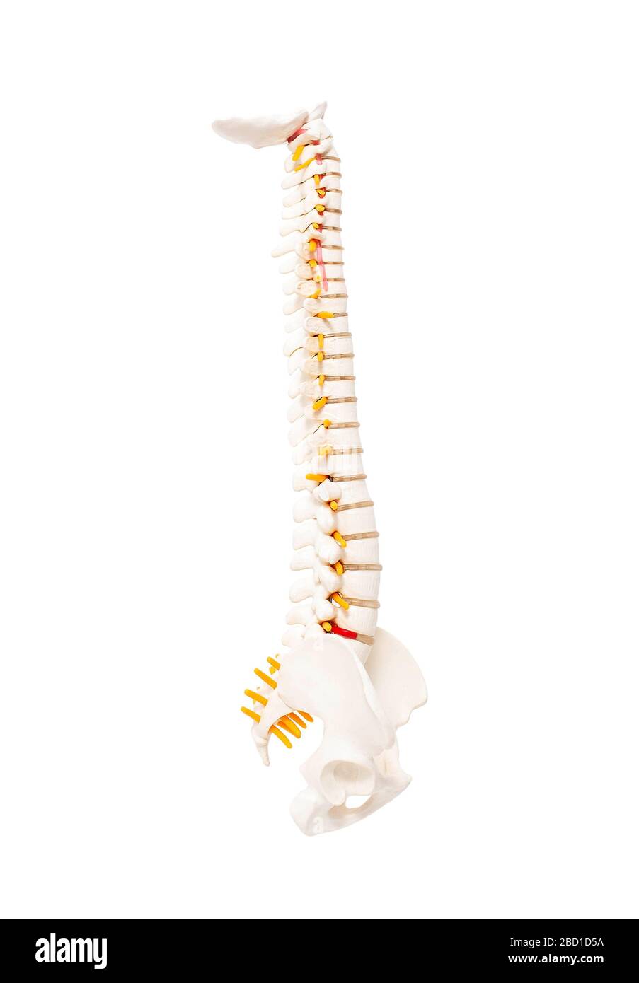 Mock up human spine on a white background. The concept of segments and divisions of the spine, the structure and anatomy of the bone marrow, nerves an Stock Photohttps://www.alamy.com/image-license-details/?v=1https://www.alamy.com/mock-up-human-spine-on-a-white-background-the-concept-of-segments-and-divisions-of-the-spine-the-structure-and-anatomy-of-the-bone-marrow-nerves-an-image352230182.html
RF2BD1D5A–Mock up human spine on a white background. The concept of segments and divisions of the spine, the structure and anatomy of the bone marrow, nerves an
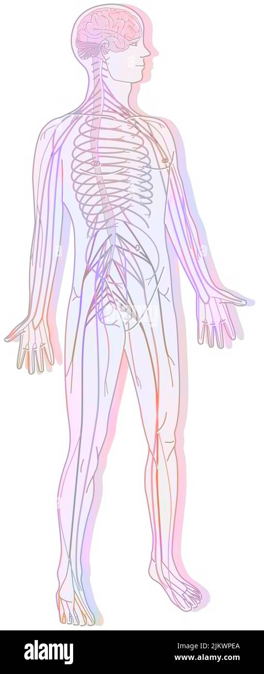 Silhouette with the nervous system consisting of the central and peripheral system. Stock Photohttps://www.alamy.com/image-license-details/?v=1https://www.alamy.com/silhouette-with-the-nervous-system-consisting-of-the-central-and-peripheral-system-image476924850.html
RF2JKWPEA–Silhouette with the nervous system consisting of the central and peripheral system.
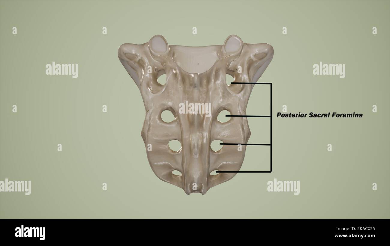 Posterior view of human sacrum showing the posterior sacral foramina-Labeled Stock Photohttps://www.alamy.com/image-license-details/?v=1https://www.alamy.com/posterior-view-of-human-sacrum-showing-the-posterior-sacral-foramina-labeled-image488320817.html
RF2KACX55–Posterior view of human sacrum showing the posterior sacral foramina-Labeled
 Sacral nerve stimulation medical concept Stock Photohttps://www.alamy.com/image-license-details/?v=1https://www.alamy.com/sacral-nerve-stimulation-medical-concept-image613819930.html
RF2XJHWGA–Sacral nerve stimulation medical concept
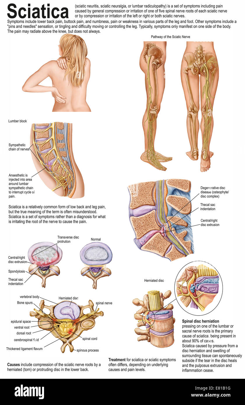 Medical chart showing the signs and symptoms of sciatica. Stock Photohttps://www.alamy.com/image-license-details/?v=1https://www.alamy.com/stock-photo-medical-chart-showing-the-signs-and-symptoms-of-sciatica-73789340.html
RFE81B1G–Medical chart showing the signs and symptoms of sciatica.
 Pinched human sciatic nerve, anatomical vision. 3d illustration. Stock Photohttps://www.alamy.com/image-license-details/?v=1https://www.alamy.com/pinched-human-sciatic-nerve-anatomical-vision-3d-illustration-image364068055.html
RF2C48MEF–Pinched human sciatic nerve, anatomical vision. 3d illustration.
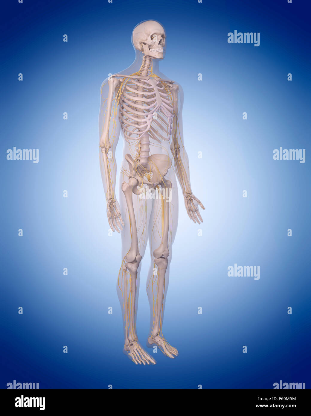 medically accurate illustration of the human nervous system Stock Photohttps://www.alamy.com/image-license-details/?v=1https://www.alamy.com/stock-photo-medically-accurate-illustration-of-the-human-nervous-system-89755616.html
RFF60M5M–medically accurate illustration of the human nervous system
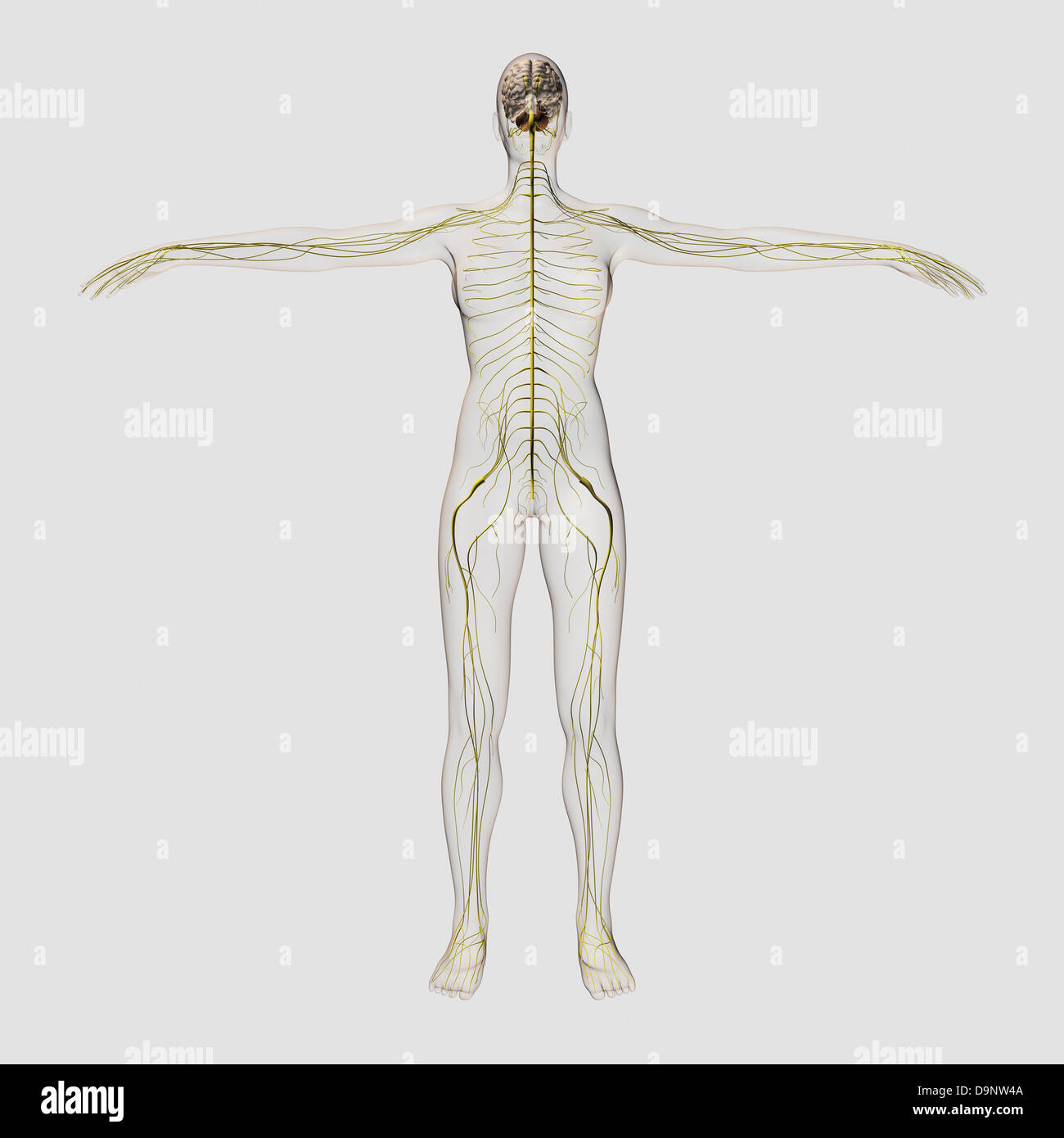 Medical illustration of the human nervous system and brain, full frontal view. Stock Photohttps://www.alamy.com/image-license-details/?v=1https://www.alamy.com/stock-photo-medical-illustration-of-the-human-nervous-system-and-brain-full-frontal-57643722.html
RFD9NW4A–Medical illustration of the human nervous system and brain, full frontal view.
 Model of a segment of the spinal cord with nerves and a vertebra. 3D illustration. Stock Photohttps://www.alamy.com/image-license-details/?v=1https://www.alamy.com/model-of-a-segment-of-the-spinal-cord-with-nerves-and-a-vertebra-3d-illustration-image591886522.html
RF2WAXN8A–Model of a segment of the spinal cord with nerves and a vertebra. 3D illustration.
 3d rendered medical illustration - female nerves Stock Photohttps://www.alamy.com/image-license-details/?v=1https://www.alamy.com/3d-rendered-medical-illustration-female-nerves-image343647187.html
RM2AY2DDR–3d rendered medical illustration - female nerves
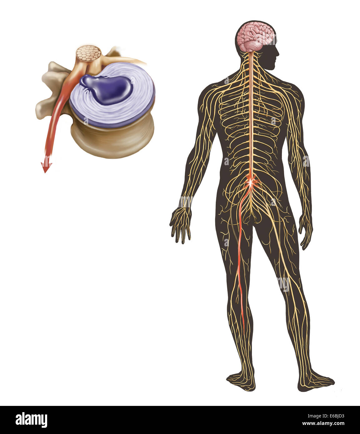 Sciatica caused from herniated disc. Stock Photohttps://www.alamy.com/image-license-details/?v=1https://www.alamy.com/stock-photo-sciatica-caused-from-herniated-disc-72785359.html
RME6BJD3–Sciatica caused from herniated disc.
 Pinched human sciatic nerve, anatomical vision. 3d illustration. Stock Photohttps://www.alamy.com/image-license-details/?v=1https://www.alamy.com/pinched-human-sciatic-nerve-anatomical-vision-3d-illustration-image364067992.html
RF2C48MC8–Pinched human sciatic nerve, anatomical vision. 3d illustration.
 Dermatome Stock Photohttps://www.alamy.com/image-license-details/?v=1https://www.alamy.com/stock-photo-dermatome-49485586.html
RFCTE7AA–Dermatome
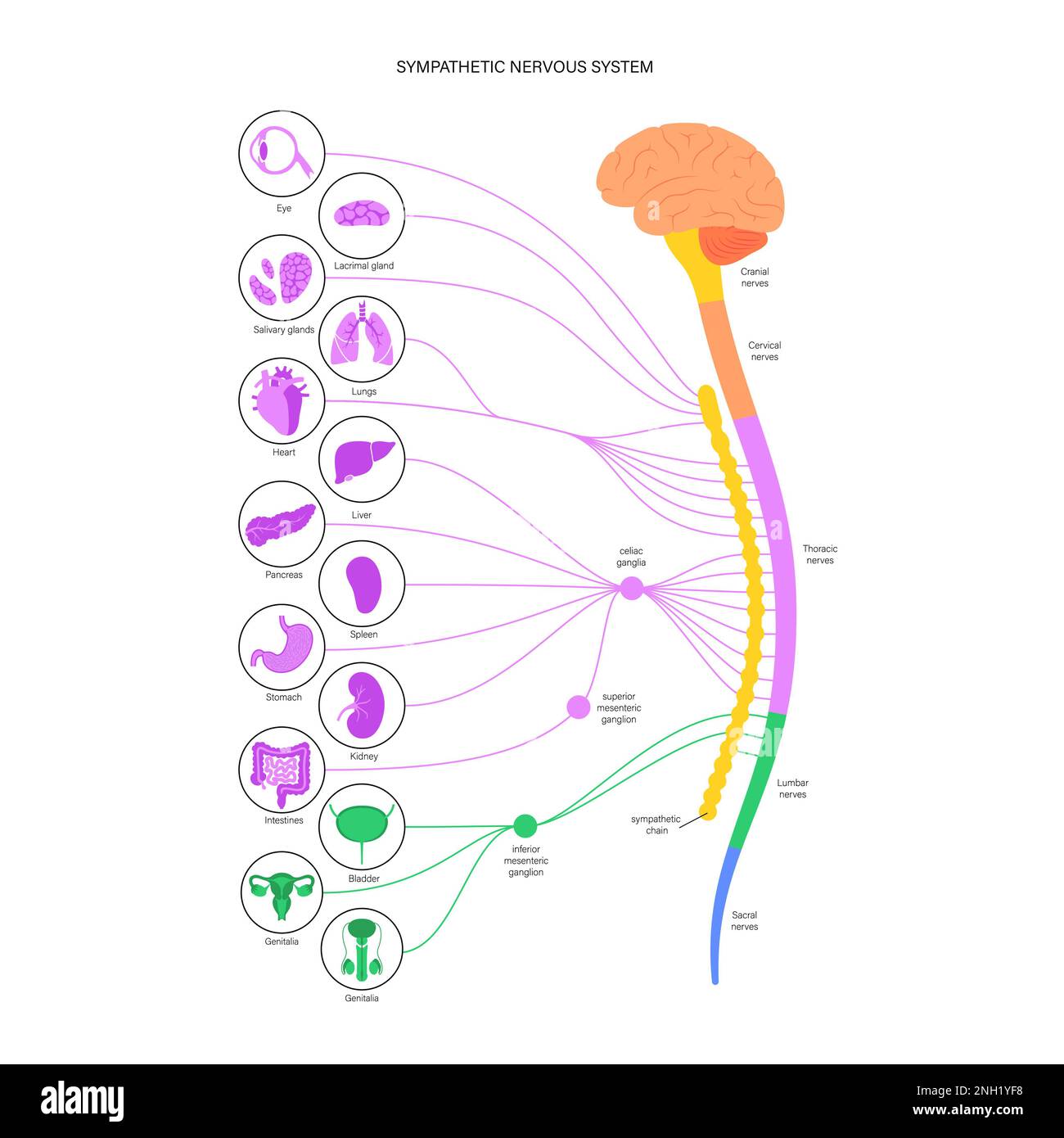 Sympathetic nervous system, illustration Stock Photohttps://www.alamy.com/image-license-details/?v=1https://www.alamy.com/sympathetic-nervous-system-illustration-image526803740.html
RF2NH1YF8–Sympathetic nervous system, illustration
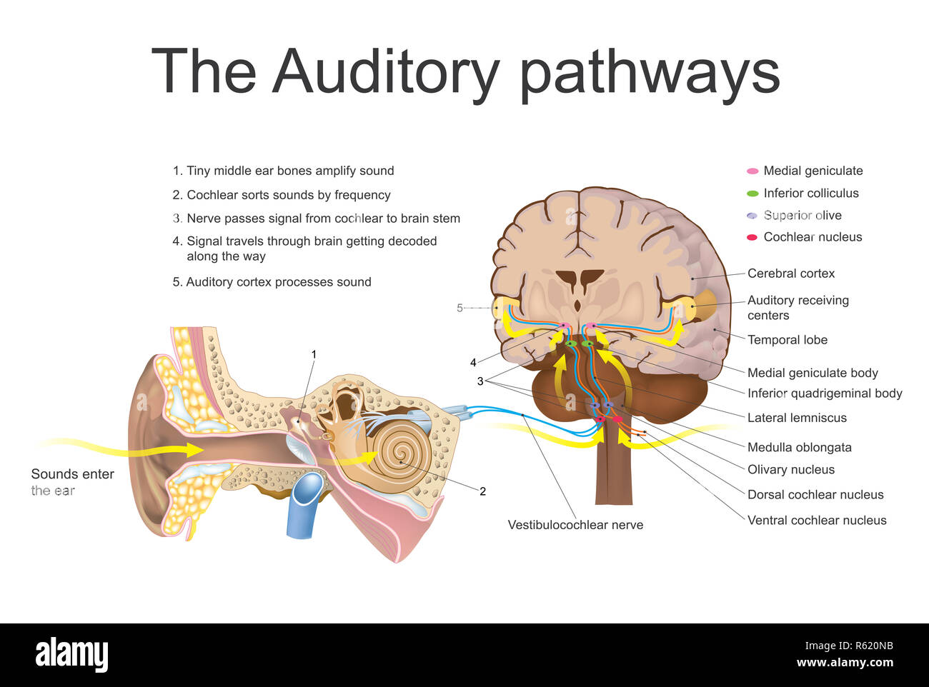 The auditory system. Education info graphic vector. Stock Photohttps://www.alamy.com/image-license-details/?v=1https://www.alamy.com/the-auditory-system-education-info-graphic-vector-image227467223.html
RFR620NB–The auditory system. Education info graphic vector.
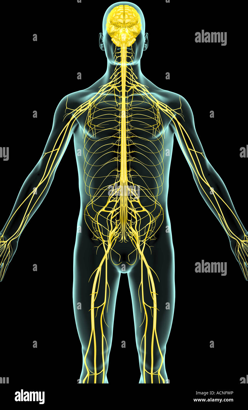 The nerves of the upper body Stock Photohttps://www.alamy.com/image-license-details/?v=1https://www.alamy.com/stock-photo-the-nerves-of-the-upper-body-13198497.html
RFACNFWP–The nerves of the upper body
 3D illustration of Sacral Spine - Part of Human Skeleton. Stock Photohttps://www.alamy.com/image-license-details/?v=1https://www.alamy.com/stock-photo-3d-illustration-of-sacral-spine-part-of-human-skeleton-104309958.html
RFG1KMBJ–3D illustration of Sacral Spine - Part of Human Skeleton.
 The lumbosacral plexus is a network of nerve fibers, derived from the roots of lumbar and sacral spinal nerves that branch out to form the nerves supp Stock Photohttps://www.alamy.com/image-license-details/?v=1https://www.alamy.com/the-lumbosacral-plexus-is-a-network-of-nerve-fibers-derived-from-the-roots-of-lumbar-and-sacral-spinal-nerves-that-branch-out-to-form-the-nerves-supp-image596677131.html
RF2WJMYNF–The lumbosacral plexus is a network of nerve fibers, derived from the roots of lumbar and sacral spinal nerves that branch out to form the nerves supp
 3D illustration of Sacral Spine - Part of Human Skeleton. Stock Photohttps://www.alamy.com/image-license-details/?v=1https://www.alamy.com/stock-image-3d-illustration-of-sacral-spine-part-of-human-skeleton-162613946.html
RFKCFKMX–3D illustration of Sacral Spine - Part of Human Skeleton.
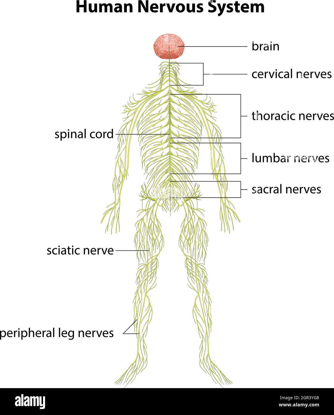 Human nervous system Stock Vectorhttps://www.alamy.com/image-license-details/?v=1https://www.alamy.com/human-nervous-system-image444483768.html
RF2GR3YG8–Human nervous system
 Anatomy of the nerves of the lower limb (leg). Stock Photohttps://www.alamy.com/image-license-details/?v=1https://www.alamy.com/anatomy-of-the-nerves-of-the-lower-limb-leg-image476924332.html
RF2JKWNRT–Anatomy of the nerves of the lower limb (leg).
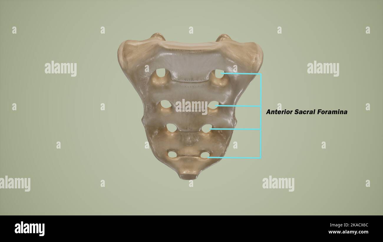 Anterior view of human sacrum showing the anterior sacral foramina-Labeled Stock Photohttps://www.alamy.com/image-license-details/?v=1https://www.alamy.com/anterior-view-of-human-sacrum-showing-the-anterior-sacral-foramina-labeled-image488320852.html
RF2KACX6C–Anterior view of human sacrum showing the anterior sacral foramina-Labeled
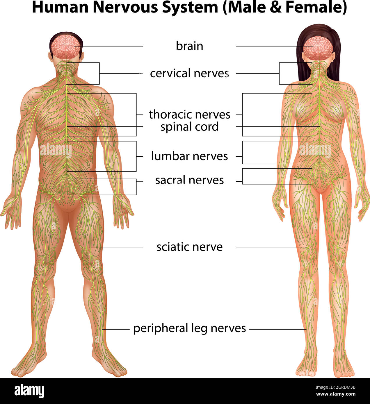 Human nervous system Stock Vectorhttps://www.alamy.com/image-license-details/?v=1https://www.alamy.com/human-nervous-system-image444697439.html
RF2GRDM3B–Human nervous system
 A reference handbook of the medical sciences, embracing the entire range of scientific and practical medicine and allied science . in thefacial and glossopharyngeal nerves for the blood-vessels of the salivary glands, tongue, tonsils, softpalate, pharynx, and adjacent structures and possiblyin the vagus for the coronary arteries. A specialgroup of vasodilators also arises from the sacral regionof the cord in the anterior roots of thefirst, second, andthird sacral spinal nerves. These are distributedthrough the nervi erigentes and hyi^ogastric plexusto the vessels of the external genital organs Stock Photohttps://www.alamy.com/image-license-details/?v=1https://www.alamy.com/a-reference-handbook-of-the-medical-sciences-embracing-the-entire-range-of-scientific-and-practical-medicine-and-allied-science-in-thefacial-and-glossopharyngeal-nerves-for-the-blood-vessels-of-the-salivary-glands-tongue-tonsils-softpalate-pharynx-and-adjacent-structures-and-possiblyin-the-vagus-for-the-coronary-arteries-a-specialgroup-of-vasodilators-also-arises-from-the-sacral-regionof-the-cord-in-the-anterior-roots-of-thefirst-second-andthird-sacral-spinal-nerves-these-are-distributedthrough-the-nervi-erigentes-and-hyiogastric-plexusto-the-vessels-of-the-external-genital-organs-image339035390.html
RM2AKGB2P–A reference handbook of the medical sciences, embracing the entire range of scientific and practical medicine and allied science . in thefacial and glossopharyngeal nerves for the blood-vessels of the salivary glands, tongue, tonsils, softpalate, pharynx, and adjacent structures and possiblyin the vagus for the coronary arteries. A specialgroup of vasodilators also arises from the sacral regionof the cord in the anterior roots of thefirst, second, andthird sacral spinal nerves. These are distributedthrough the nervi erigentes and hyi^ogastric plexusto the vessels of the external genital organs
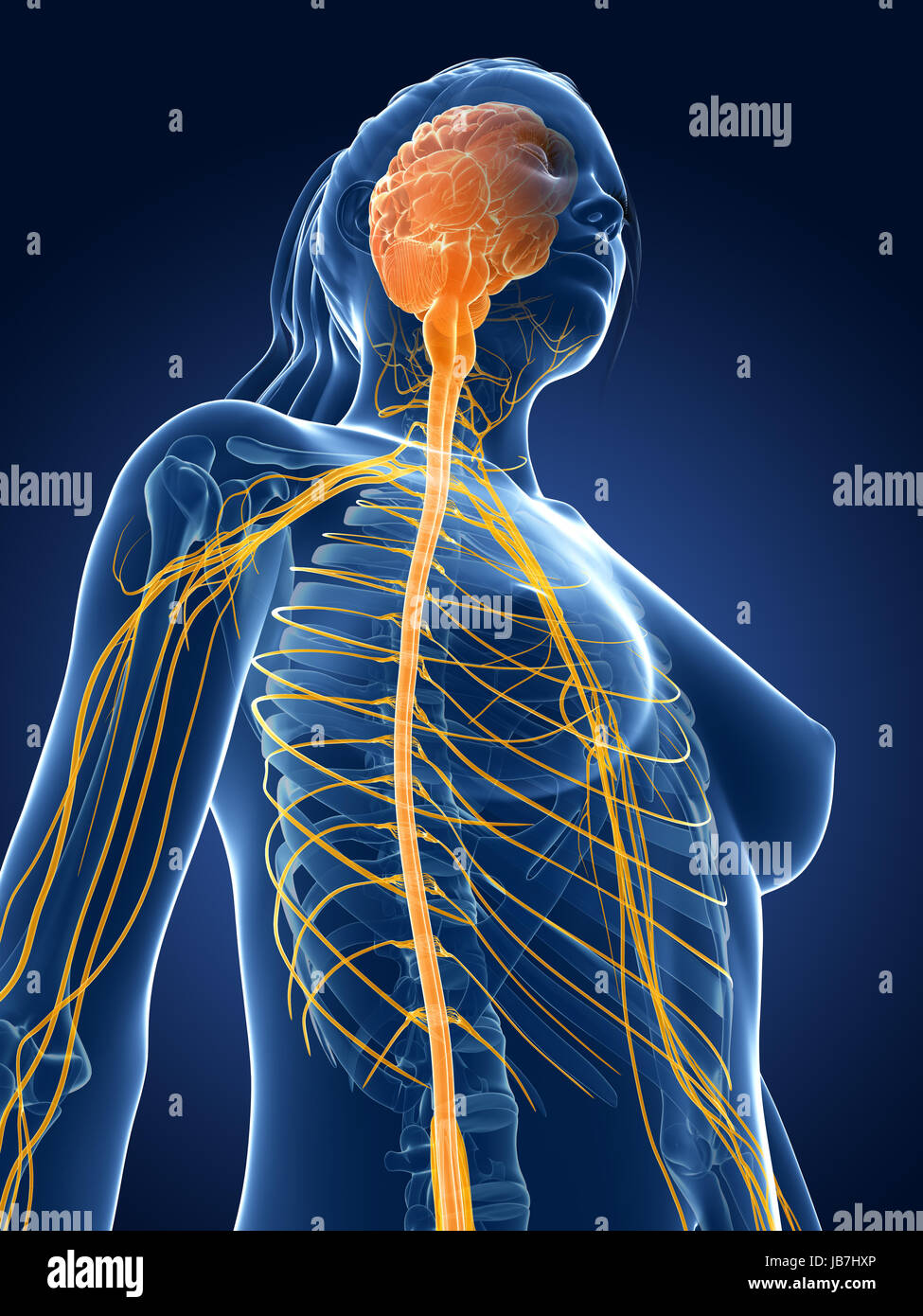 3d rendered medical illustration - female nerves Stock Photohttps://www.alamy.com/image-license-details/?v=1https://www.alamy.com/stock-photo-3d-rendered-medical-illustration-female-nerves-144611902.html
RFJB7HXP–3d rendered medical illustration - female nerves
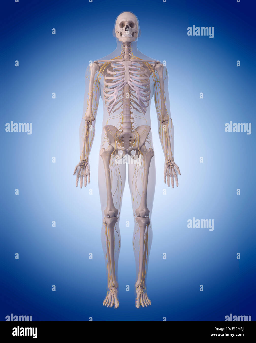 medically accurate illustration of the human nervous system Stock Photohttps://www.alamy.com/image-license-details/?v=1https://www.alamy.com/stock-photo-medically-accurate-illustration-of-the-human-nervous-system-89755614.html
RFF60M5J–medically accurate illustration of the human nervous system
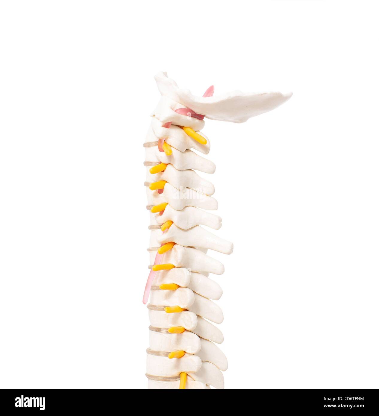 cervical and thoracic spine on a white background, isolate. Osteochondrosis and degenerative changes in the spine Stock Photohttps://www.alamy.com/image-license-details/?v=1https://www.alamy.com/cervical-and-thoracic-spine-on-a-white-background-isolate-osteochondrosis-and-degenerative-changes-in-the-spine-image382855248.html
RF2D6TFNM–cervical and thoracic spine on a white background, isolate. Osteochondrosis and degenerative changes in the spine
 A modell of a segment of the spinal cord with nerves and a vertebra. 3D illustration. Stock Photohttps://www.alamy.com/image-license-details/?v=1https://www.alamy.com/a-modell-of-a-segment-of-the-spinal-cord-with-nerves-and-a-vertebra-3d-illustration-image591886523.html
RF2WAXN8B–A modell of a segment of the spinal cord with nerves and a vertebra. 3D illustration.
 3d rendered medical illustration - female nerves Stock Photohttps://www.alamy.com/image-license-details/?v=1https://www.alamy.com/3d-rendered-medical-illustration-female-nerves-image343647217.html
RM2AY2DEW–3d rendered medical illustration - female nerves
 A stylized female figure with a wire frame appearance with nerves and brain present. Stock Photohttps://www.alamy.com/image-license-details/?v=1https://www.alamy.com/stock-photo-a-stylized-female-figure-with-a-wire-frame-appearance-with-nerves-52090698.html
RMD0MX62–A stylized female figure with a wire frame appearance with nerves and brain present.
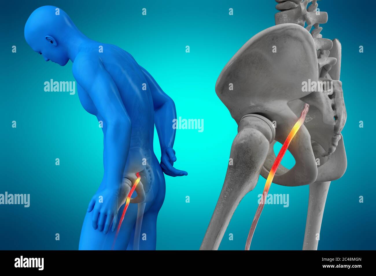 Pinched human sciatic nerve, anatomical vision. 3d illustration. Stock Photohttps://www.alamy.com/image-license-details/?v=1https://www.alamy.com/pinched-human-sciatic-nerve-anatomical-vision-3d-illustration-image364068117.html
RF2C48MGN–Pinched human sciatic nerve, anatomical vision. 3d illustration.
 Chordoma word cloud conceptual design isolated on white background. Stock Vectorhttps://www.alamy.com/image-license-details/?v=1https://www.alamy.com/chordoma-word-cloud-conceptual-design-isolated-on-white-background-image636935084.html
RF2S06W5G–Chordoma word cloud conceptual design isolated on white background.
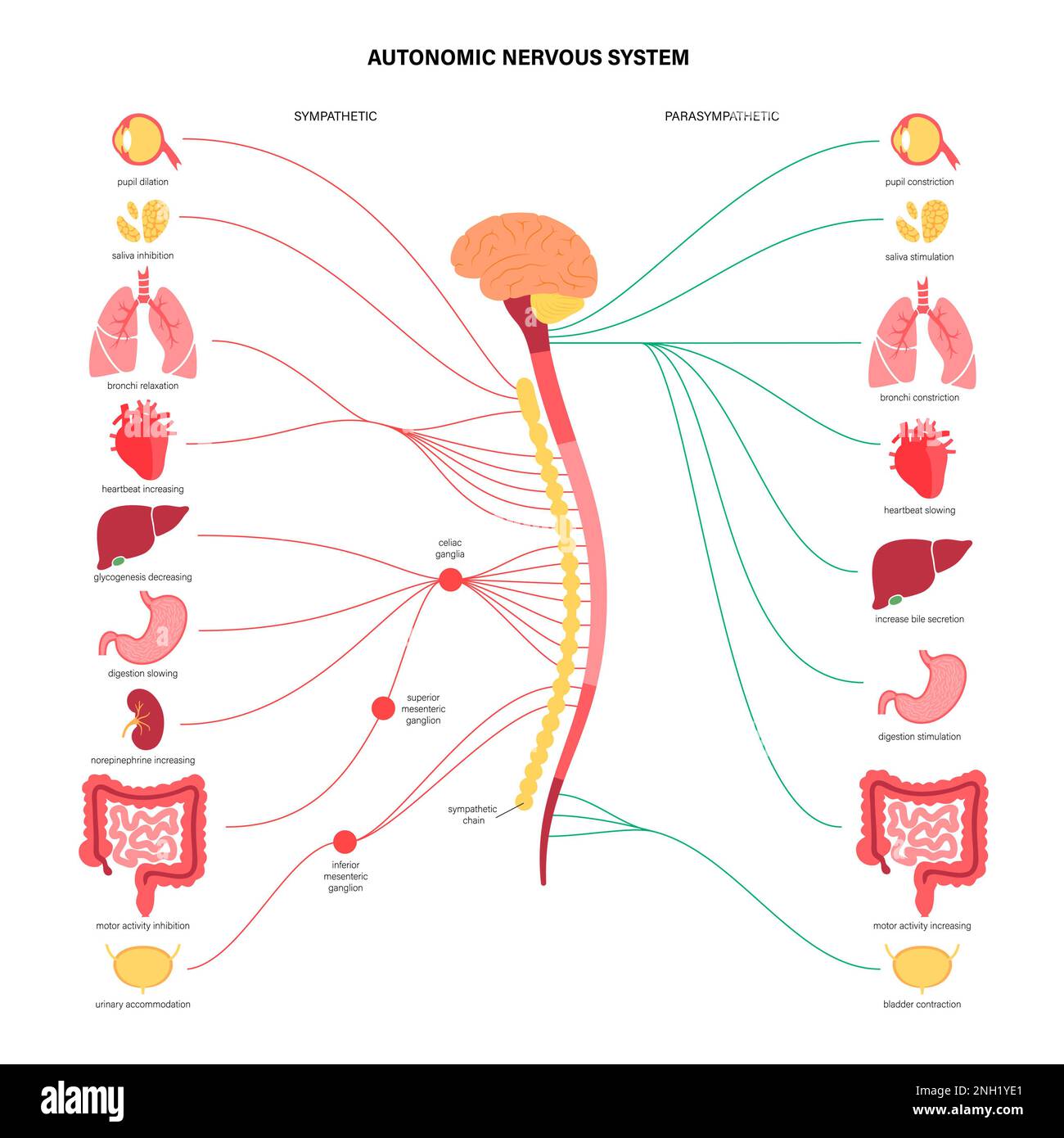 Autonomic nervous system, illustration Stock Photohttps://www.alamy.com/image-license-details/?v=1https://www.alamy.com/autonomic-nervous-system-illustration-image526803705.html
RF2NH1YE1–Autonomic nervous system, illustration
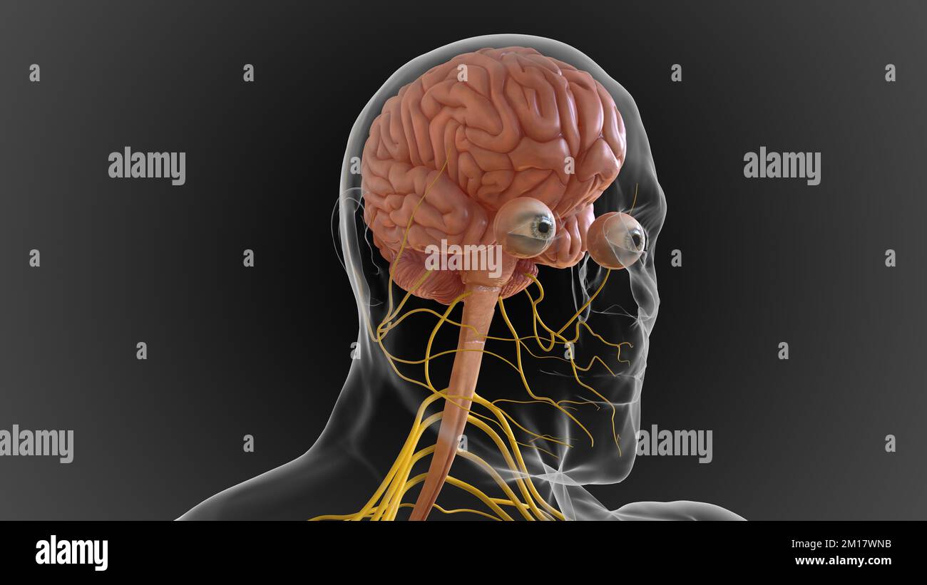 The central nervous system is made up of the brain and spinal cord 3D illustration Stock Photohttps://www.alamy.com/image-license-details/?v=1https://www.alamy.com/the-central-nervous-system-is-made-up-of-the-brain-and-spinal-cord-3d-illustration-image499889191.html
RF2M17WNB–The central nervous system is made up of the brain and spinal cord 3D illustration
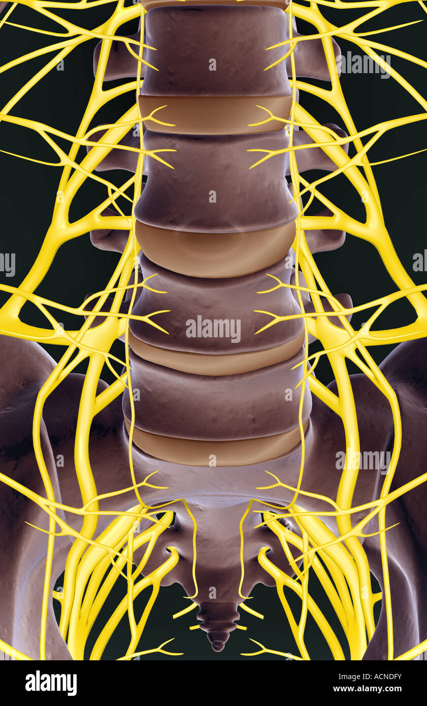 The lumbar and sacral plexus Stock Photohttps://www.alamy.com/image-license-details/?v=1https://www.alamy.com/stock-photo-the-lumbar-and-sacral-plexus-13197710.html
RFACNDFY–The lumbar and sacral plexus
 3D illustration of SCIATIC title on a medical document Stock Photohttps://www.alamy.com/image-license-details/?v=1https://www.alamy.com/3d-illustration-of-sciatic-title-on-a-medical-document-image241192461.html
RFT0B7CD–3D illustration of SCIATIC title on a medical document
 The lumbosacral plexus is a network of nerve fibers, derived from the roots of lumbar and sacral spinal nerves that branch out to form the nerves supp Stock Photohttps://www.alamy.com/image-license-details/?v=1https://www.alamy.com/the-lumbosacral-plexus-is-a-network-of-nerve-fibers-derived-from-the-roots-of-lumbar-and-sacral-spinal-nerves-that-branch-out-to-form-the-nerves-supp-image596594821.html
RF2WJH6NW–The lumbosacral plexus is a network of nerve fibers, derived from the roots of lumbar and sacral spinal nerves that branch out to form the nerves supp
 3D illustration of Sacral Spine - Part of Human Skeleton. Stock Photohttps://www.alamy.com/image-license-details/?v=1https://www.alamy.com/stock-image-3d-illustration-of-sacral-spine-part-of-human-skeleton-161980648.html
RFKBERY4–3D illustration of Sacral Spine - Part of Human Skeleton.
 3D illustration of SCIATIC NERVE PAIN title on a medical document Stock Photohttps://www.alamy.com/image-license-details/?v=1https://www.alamy.com/3d-illustration-of-sciatic-nerve-pain-title-on-a-medical-document-image248527238.html
RFTC9B0P–3D illustration of SCIATIC NERVE PAIN title on a medical document
 Anatomy of the nerves of the lower limb (leg). Stock Photohttps://www.alamy.com/image-license-details/?v=1https://www.alamy.com/anatomy-of-the-nerves-of-the-lower-limb-leg-image476924461.html
RF2JKWP0D–Anatomy of the nerves of the lower limb (leg).
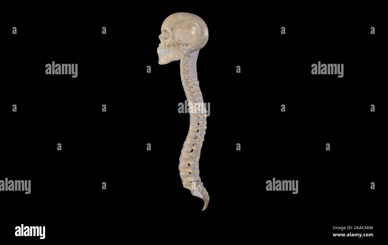 Spine Anatomy Lateral view showing the Curvatures of Vertebral Column Stock Photohttps://www.alamy.com/image-license-details/?v=1https://www.alamy.com/spine-anatomy-lateral-view-showing-the-curvatures-of-vertebral-column-image488320865.html
RF2KACX6W–Spine Anatomy Lateral view showing the Curvatures of Vertebral Column
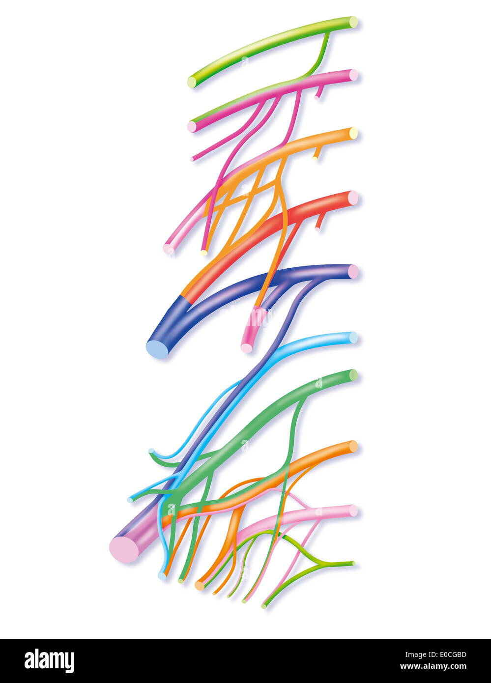 Lumbar plexus Stock Photohttps://www.alamy.com/image-license-details/?v=1https://www.alamy.com/lumbar-plexus-image69117761.html
RME0CGBD–Lumbar plexus
 3D illustration of Sacral Spine - Part of Human Skeleton. Stock Photohttps://www.alamy.com/image-license-details/?v=1https://www.alamy.com/stock-photo-3d-illustration-of-sacral-spine-part-of-human-skeleton-118971979.html
RMGWFHY7–3D illustration of Sacral Spine - Part of Human Skeleton.
 3d rendered medical illustration - female nerves Stock Photohttps://www.alamy.com/image-license-details/?v=1https://www.alamy.com/stock-photo-3d-rendered-medical-illustration-female-nerves-144611906.html
RFJB7HXX–3d rendered medical illustration - female nerves
 3D illustration of Sacral Spine - Part of Human Skeleton. Stock Photohttps://www.alamy.com/image-license-details/?v=1https://www.alamy.com/stock-photo-3d-illustration-of-sacral-spine-part-of-human-skeleton-177563743.html
RFM8TMA7–3D illustration of Sacral Spine - Part of Human Skeleton.
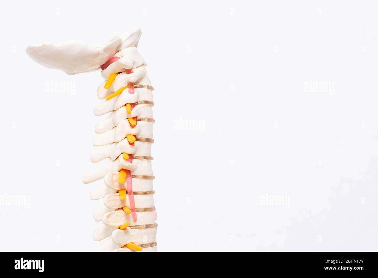 Model of the cervical spine with blood vessels on a white background. The concept of lordosis and spondylosis of the cervical spine Stock Photohttps://www.alamy.com/image-license-details/?v=1https://www.alamy.com/model-of-the-cervical-spine-with-blood-vessels-on-a-white-background-the-concept-of-lordosis-and-spondylosis-of-the-cervical-spine-image355129487.html
RF2BHNF7Y–Model of the cervical spine with blood vessels on a white background. The concept of lordosis and spondylosis of the cervical spine
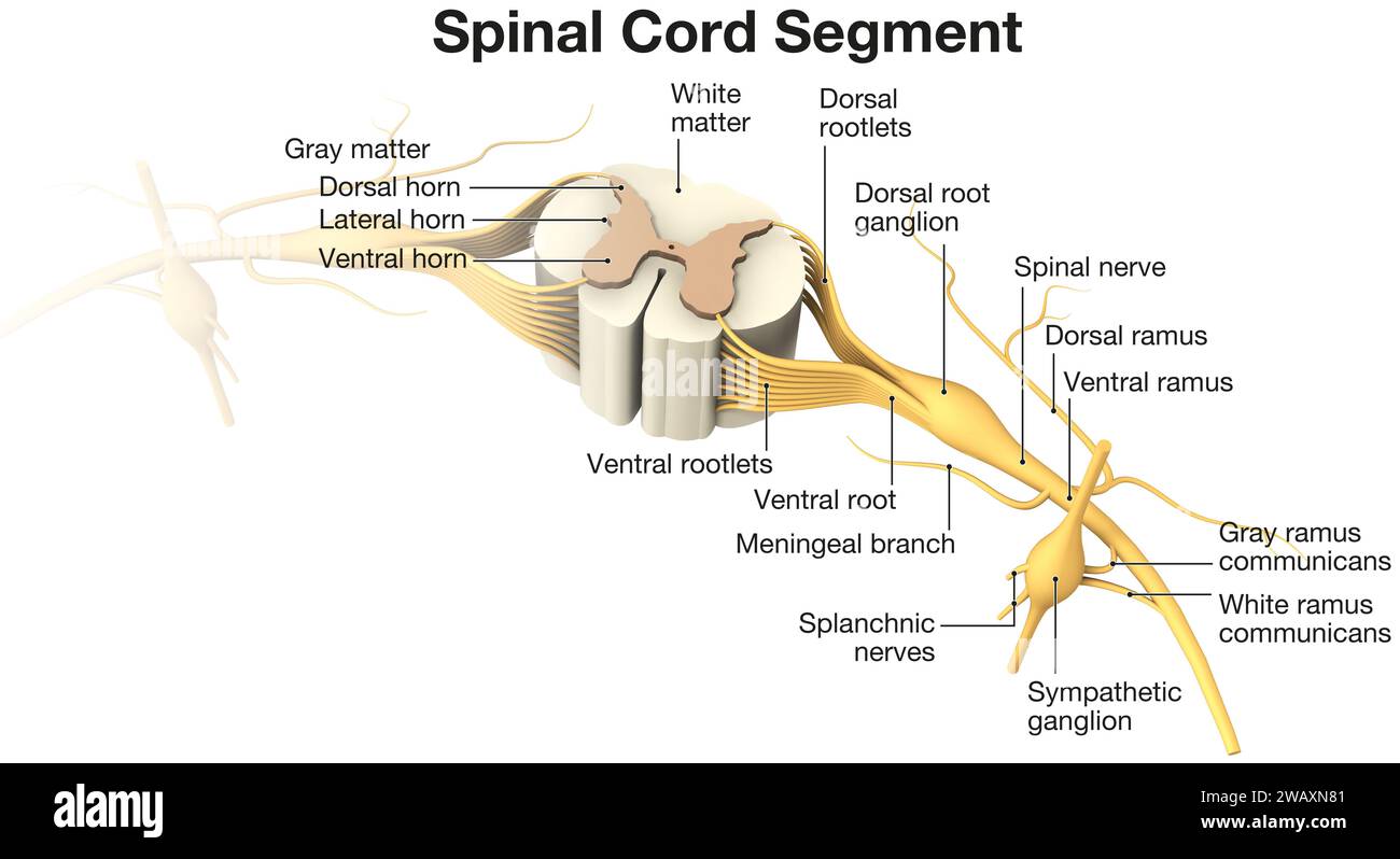 Spinal Cord Segment. Labeled 3D illustration Stock Photohttps://www.alamy.com/image-license-details/?v=1https://www.alamy.com/spinal-cord-segment-labeled-3d-illustration-image591886513.html
RF2WAXN81–Spinal Cord Segment. Labeled 3D illustration
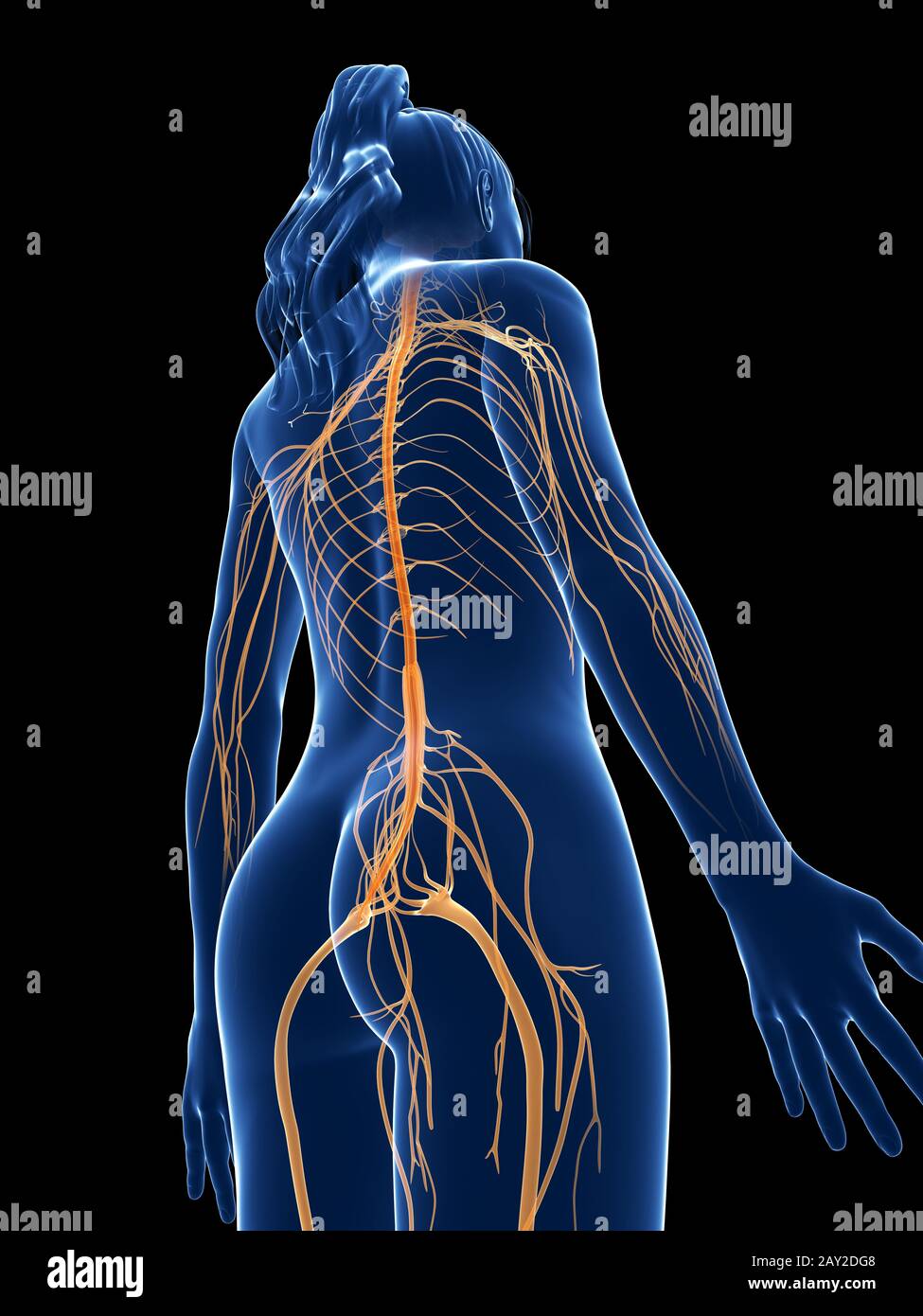 3d rendered medical illustration - female nerves Stock Photohttps://www.alamy.com/image-license-details/?v=1https://www.alamy.com/3d-rendered-medical-illustration-female-nerves-image343647256.html
RM2AY2DG8–3d rendered medical illustration - female nerves
 3D illustration of Sacral Spine - Part of Human Skeleton. Stock Photohttps://www.alamy.com/image-license-details/?v=1https://www.alamy.com/stock-photo-3d-illustration-of-sacral-spine-part-of-human-skeleton-118971992.html
RMGWFHYM–3D illustration of Sacral Spine - Part of Human Skeleton.
 Pinched human sciatic nerve, anatomical vision. 3d illustration. Stock Photohttps://www.alamy.com/image-license-details/?v=1https://www.alamy.com/pinched-human-sciatic-nerve-anatomical-vision-3d-illustration-image364068101.html
RF2C48MG5–Pinched human sciatic nerve, anatomical vision. 3d illustration.
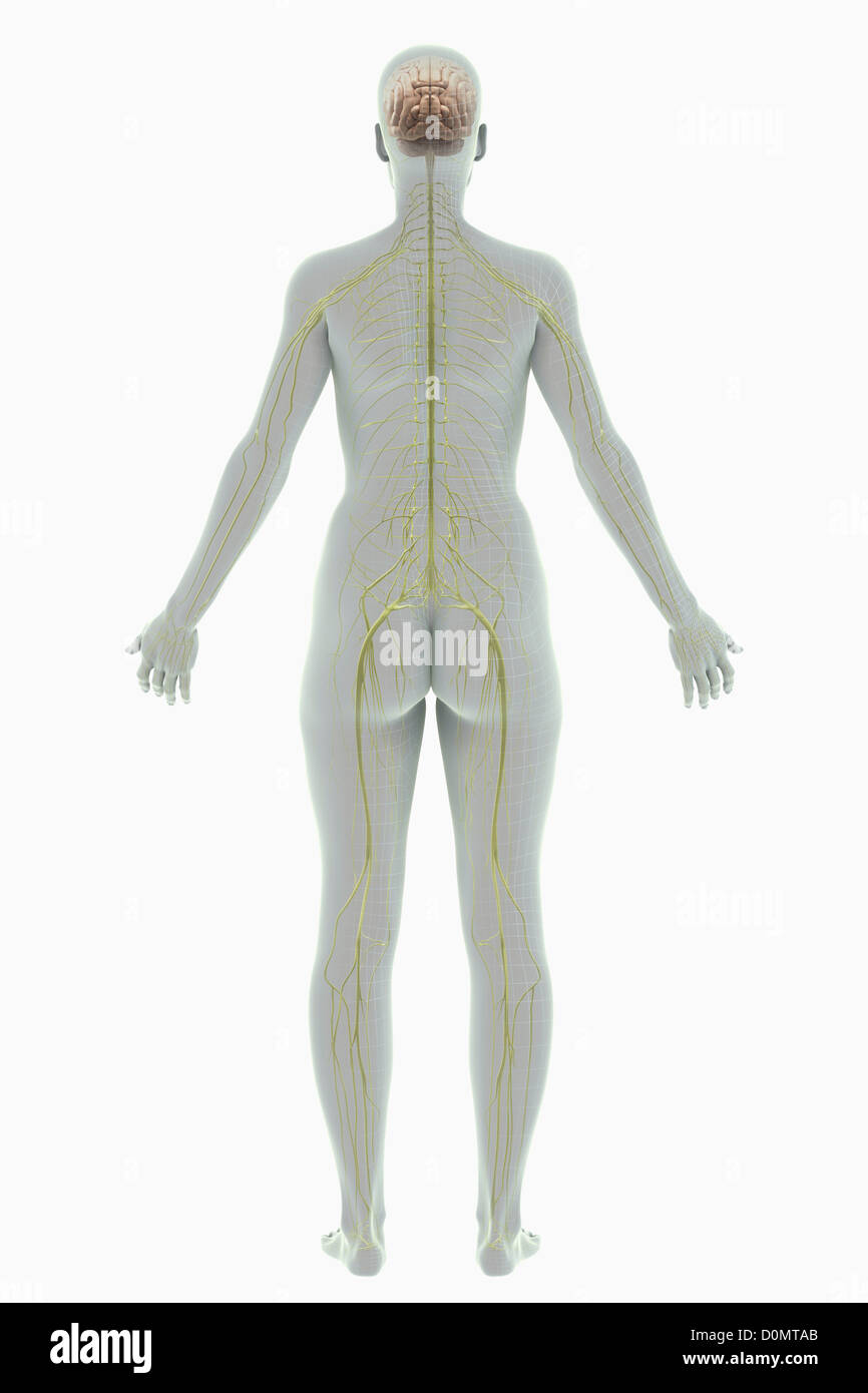 A rear view of a stylized female figure with a wire frame appearance with nerves and brain present. Stock Photohttps://www.alamy.com/image-license-details/?v=1https://www.alamy.com/stock-photo-a-rear-view-of-a-stylized-female-figure-with-a-wire-frame-appearance-52089251.html
RMD0MTAB–A rear view of a stylized female figure with a wire frame appearance with nerves and brain present.
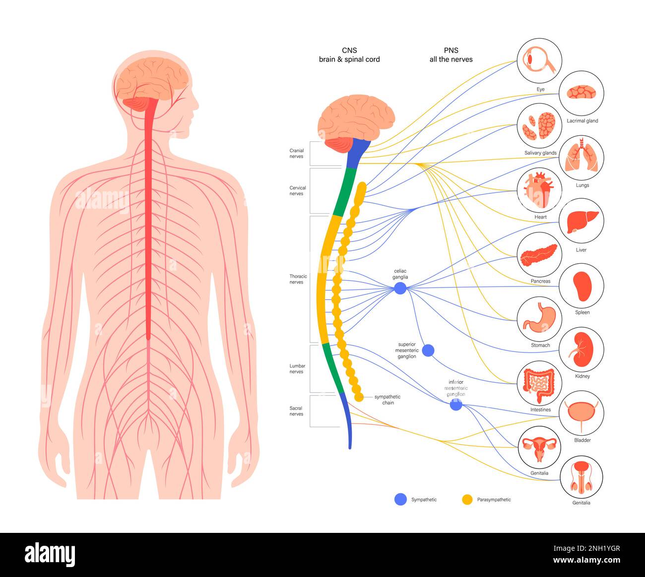 Autonomic nervous system, illustration Stock Photohttps://www.alamy.com/image-license-details/?v=1https://www.alamy.com/autonomic-nervous-system-illustration-image526803783.html
RF2NH1YGR–Autonomic nervous system, illustration
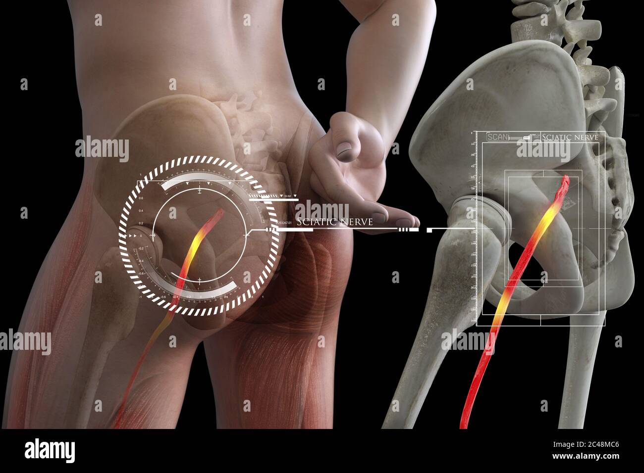 Pinched human sciatic nerve, anatomical vision. 3d illustration. Stock Photohttps://www.alamy.com/image-license-details/?v=1https://www.alamy.com/pinched-human-sciatic-nerve-anatomical-vision-3d-illustration-image364067990.html
RF2C48MC6–Pinched human sciatic nerve, anatomical vision. 3d illustration.
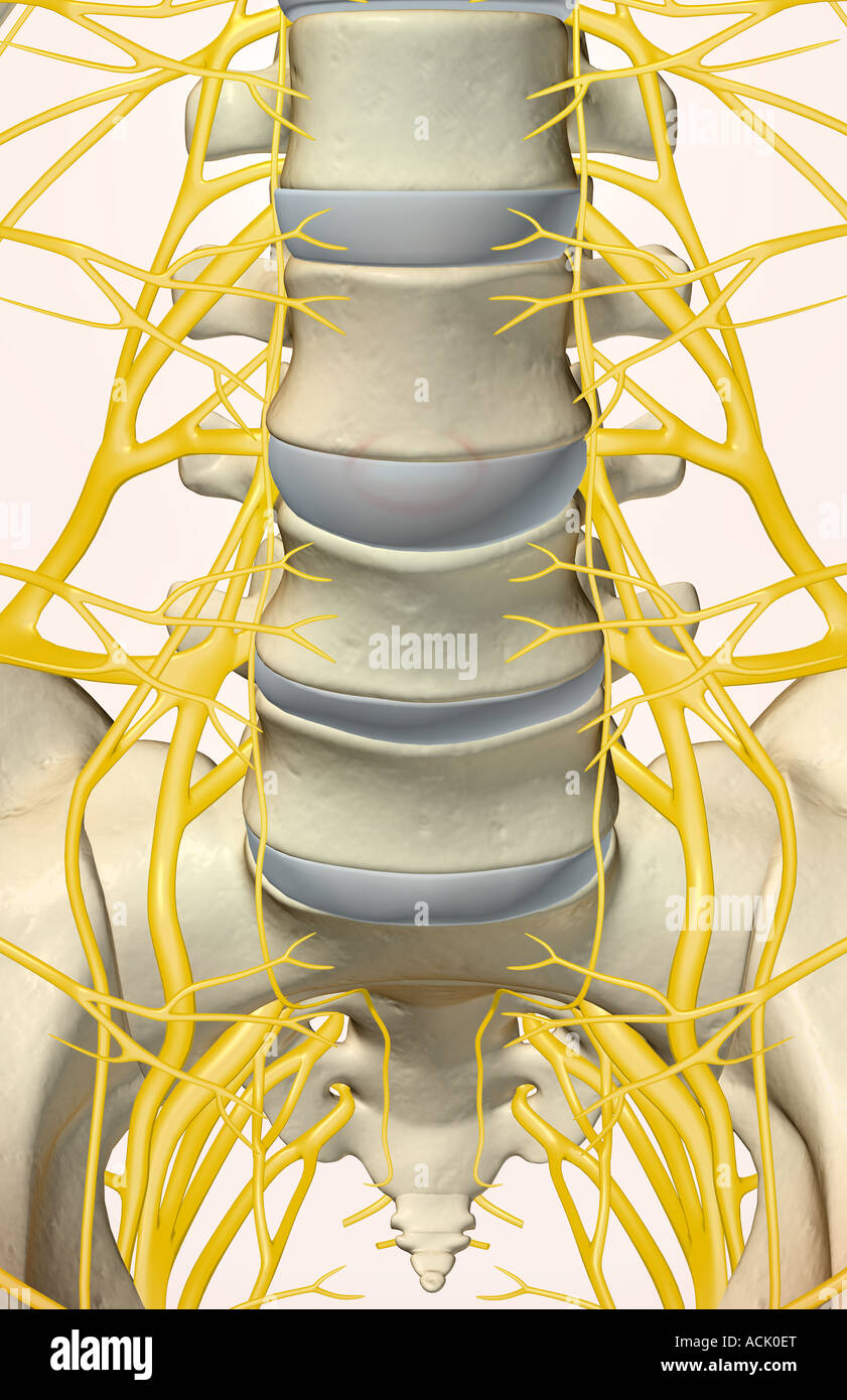 The lumbar and sacral plexus Stock Photohttps://www.alamy.com/image-license-details/?v=1https://www.alamy.com/stock-photo-the-lumbar-and-sacral-plexus-13174511.html
RFACK0ET–The lumbar and sacral plexus
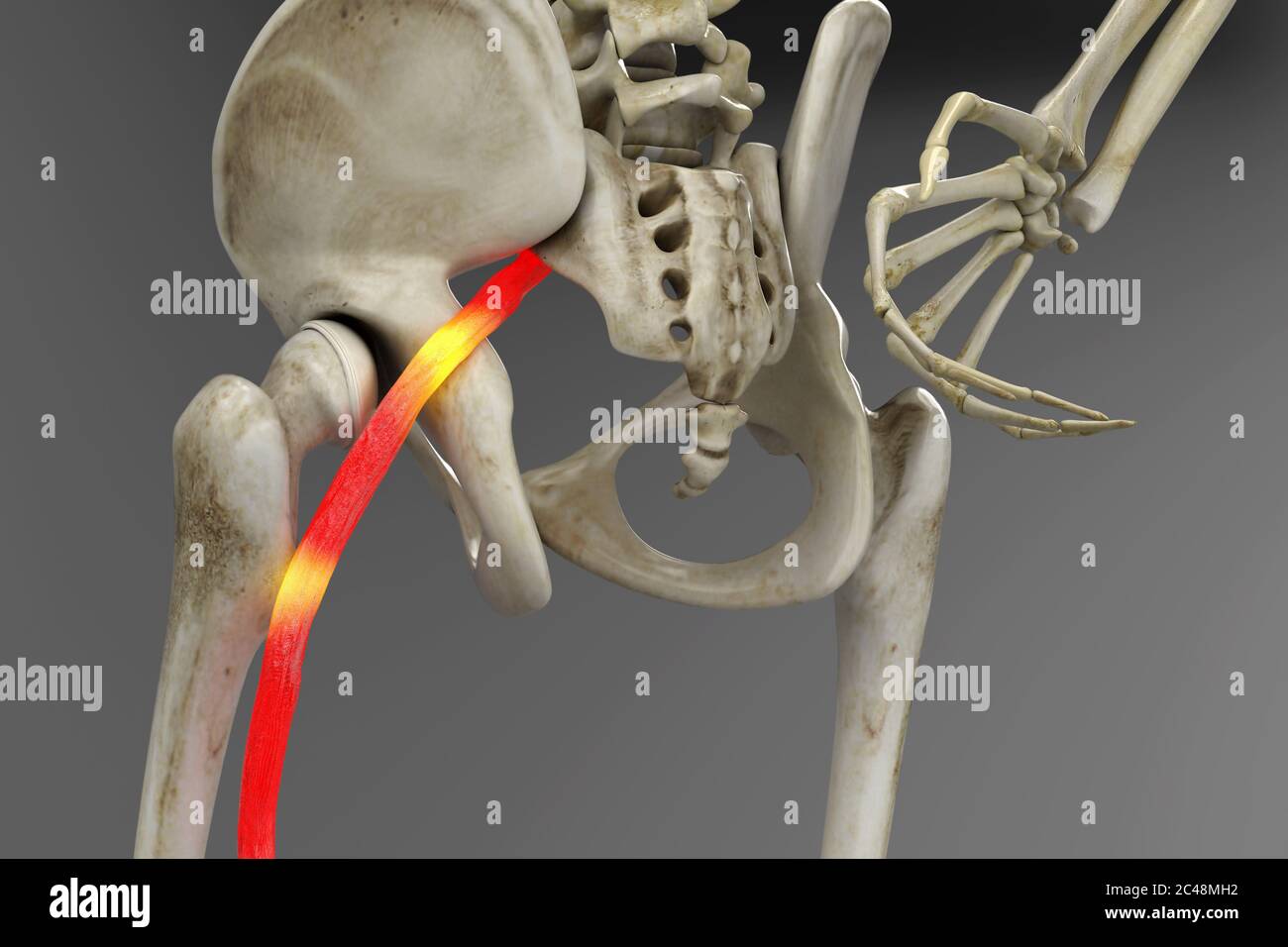 Pinched human sciatic nerve, anatomical vision. 3d illustration. Stock Photohttps://www.alamy.com/image-license-details/?v=1https://www.alamy.com/pinched-human-sciatic-nerve-anatomical-vision-3d-illustration-image364068126.html
RF2C48MH2–Pinched human sciatic nerve, anatomical vision. 3d illustration.
 The lumbosacral plexus is a network of nerve fibers, derived from the roots of lumbar and sacral spinal nerves that branch out to form the nerves supp Stock Photohttps://www.alamy.com/image-license-details/?v=1https://www.alamy.com/the-lumbosacral-plexus-is-a-network-of-nerve-fibers-derived-from-the-roots-of-lumbar-and-sacral-spinal-nerves-that-branch-out-to-form-the-nerves-supp-image596599945.html
RF2WJHD8W–The lumbosacral plexus is a network of nerve fibers, derived from the roots of lumbar and sacral spinal nerves that branch out to form the nerves supp
 Chordoma word cloud conceptual design isolated on white background. Stock Vectorhttps://www.alamy.com/image-license-details/?v=1https://www.alamy.com/chordoma-word-cloud-conceptual-design-isolated-on-white-background-image636934937.html
RF2S06W09–Chordoma word cloud conceptual design isolated on white background.
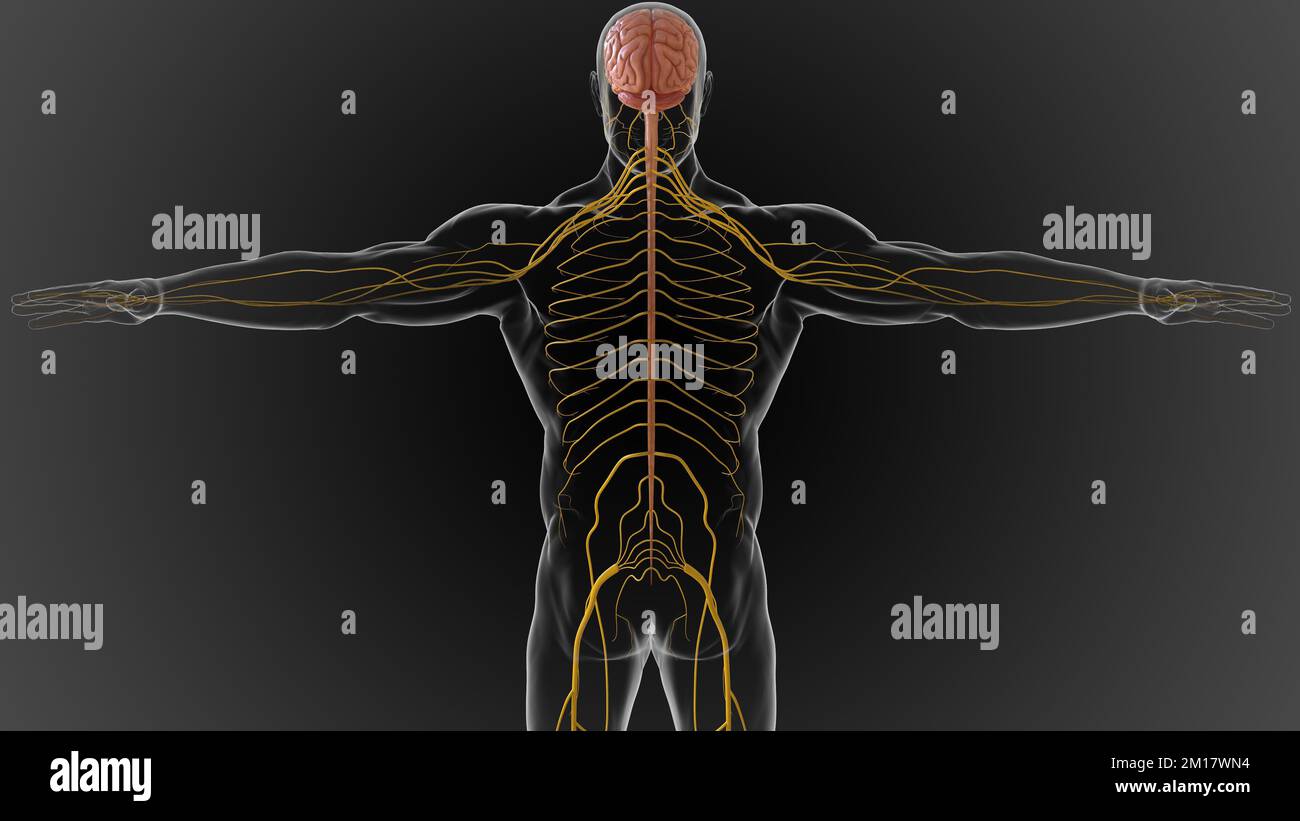 The central nervous system is made up of the brain and spinal cord 3D illustration Stock Photohttps://www.alamy.com/image-license-details/?v=1https://www.alamy.com/the-central-nervous-system-is-made-up-of-the-brain-and-spinal-cord-3d-illustration-image499889184.html
RF2M17WN4–The central nervous system is made up of the brain and spinal cord 3D illustration
![. Practical anatomy of the rabbit [microform] : an elementary laboratory textbook in mammalian anatomy. Lapins; Anatomy, Comparative; Rabbits; Rabbits; Lapins; Anatomie comparée. The Ribs. 73 P.IT.C .urf,c. (facies pelviâa)^he line, onuncH^- °''u"" ^â¢'"'. »' between the bodies, or between the ranfj,il°" '"^^^ "â¢Â»Â«'' """er apertures on this surfacâ¢The â,.â" "'>^ââ¢'^'*'' ^""^ P^'" »' anteriora), lead into thiZ"'n!brThlI^^i'°'^'°^^^'^^i>^ the sacral spinal nerves. On the dol â¢"rf-^/f''" ^&qu Stock Photo . Practical anatomy of the rabbit [microform] : an elementary laboratory textbook in mammalian anatomy. Lapins; Anatomy, Comparative; Rabbits; Rabbits; Lapins; Anatomie comparée. The Ribs. 73 P.IT.C .urf,c. (facies pelviâa)^he line, onuncH^- °''u"" ^â¢'"'. »' between the bodies, or between the ranfj,il°" '"^^^ "â¢Â»Â«'' """er apertures on this surfacâ¢The â,.â" "'>^ââ¢'^'*'' ^""^ P^'" »' anteriora), lead into thiZ"'n!brThlI^^i'°'^'°^^^'^^i>^ the sacral spinal nerves. On the dol â¢"rf-^/f''" ^&qu Stock Photo](https://c8.alamy.com/comp/RENRB8/practical-anatomy-of-the-rabbit-microform-an-elementary-laboratory-textbook-in-mammalian-anatomy-lapins-anatomy-comparative-rabbits-rabbits-lapins-anatomie-compare-the-ribs-73-pitc-urfc-facies-pelviahe-line-onunch-uquotquot-quot-between-the-bodies-or-between-the-ranfjilquot-quot-quot-quotquotquoter-apertures-on-this-surfacthe-quot-quotgt-quotquot-pquot-anteriora-lead-into-thizquotn!brthliiigt-the-sacral-spinal-nerves-on-the-dol-quotrf-fquot-qu-RENRB8.jpg) . Practical anatomy of the rabbit [microform] : an elementary laboratory textbook in mammalian anatomy. Lapins; Anatomy, Comparative; Rabbits; Rabbits; Lapins; Anatomie comparée. The Ribs. 73 P.IT.C .urf,c. (facies pelviâa)^he line, onuncH^- °''u"" ^â¢'"'. »' between the bodies, or between the ranfj,il°" '"^^^ "â¢Â»Â«'' """er apertures on this surfacâ¢The â,.â" "'>^ââ¢'^'*'' ^""^ P^'" »' anteriora), lead into thiZ"'n!brThlI^^i'°'^'°^^^'^^i>^ the sacral spinal nerves. On the dol â¢"rf-^/f''" ^&qu Stock Photohttps://www.alamy.com/image-license-details/?v=1https://www.alamy.com/practical-anatomy-of-the-rabbit-microform-an-elementary-laboratory-textbook-in-mammalian-anatomy-lapins-anatomy-comparative-rabbits-rabbits-lapins-anatomie-compare-the-ribs-73-pitc-urfc-facies-pelviahe-line-onunch-uquotquot-quot-between-the-bodies-or-between-the-ranfjilquot-quot-quot-quotquotquoter-apertures-on-this-surfacthe-quot-quotgt-quotquot-pquot-anteriora-lead-into-thizquotn!brthliiigt-the-sacral-spinal-nerves-on-the-dol-quotrf-fquot-qu-image232819308.html
. Practical anatomy of the rabbit [microform] : an elementary laboratory textbook in mammalian anatomy. Lapins; Anatomy, Comparative; Rabbits; Rabbits; Lapins; Anatomie comparée. The Ribs. 73 P.IT.C .urf,c. (facies pelviâa)^he line, onuncH^- °''u"" ^â¢'"'. »' between the bodies, or between the ranfj,il°" '"^^^ "â¢Â»Â«'' """er apertures on this surfacâ¢The â,.â" "'>^ââ¢'^'*'' ^""^ P^'" »' anteriora), lead into thiZ"'n!brThlI^^i'°'^'°^^^'^^i>^ the sacral spinal nerves. On the dol â¢"rf-^/f''" ^&qu Stock Photohttps://www.alamy.com/image-license-details/?v=1https://www.alamy.com/practical-anatomy-of-the-rabbit-microform-an-elementary-laboratory-textbook-in-mammalian-anatomy-lapins-anatomy-comparative-rabbits-rabbits-lapins-anatomie-compare-the-ribs-73-pitc-urfc-facies-pelviahe-line-onunch-uquotquot-quot-between-the-bodies-or-between-the-ranfjilquot-quot-quot-quotquotquoter-apertures-on-this-surfacthe-quot-quotgt-quotquot-pquot-anteriora-lead-into-thizquotn!brthliiigt-the-sacral-spinal-nerves-on-the-dol-quotrf-fquot-qu-image232819308.htmlRMRENRB8–. Practical anatomy of the rabbit [microform] : an elementary laboratory textbook in mammalian anatomy. Lapins; Anatomy, Comparative; Rabbits; Rabbits; Lapins; Anatomie comparée. The Ribs. 73 P.IT.C .urf,c. (facies pelviâa)^he line, onuncH^- °''u"" ^â¢'"'. »' between the bodies, or between the ranfj,il°" '"^^^ "â¢Â»Â«'' """er apertures on this surfacâ¢The â,.â" "'>^ââ¢'^'*'' ^""^ P^'" »' anteriora), lead into thiZ"'n!brThlI^^i'°'^'°^^^'^^i>^ the sacral spinal nerves. On the dol â¢"rf-^/f''" ^&qu
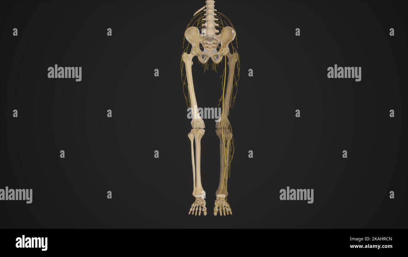 Nerves of Lower Limb Stock Photohttps://www.alamy.com/image-license-details/?v=1https://www.alamy.com/nerves-of-lower-limb-image488428437.html
RF2KAHRCN–Nerves of Lower Limb
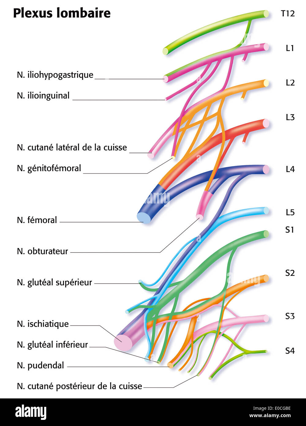 Lumbar plexus Stock Photohttps://www.alamy.com/image-license-details/?v=1https://www.alamy.com/lumbar-plexus-image69117762.html
RME0CGBE–Lumbar plexus
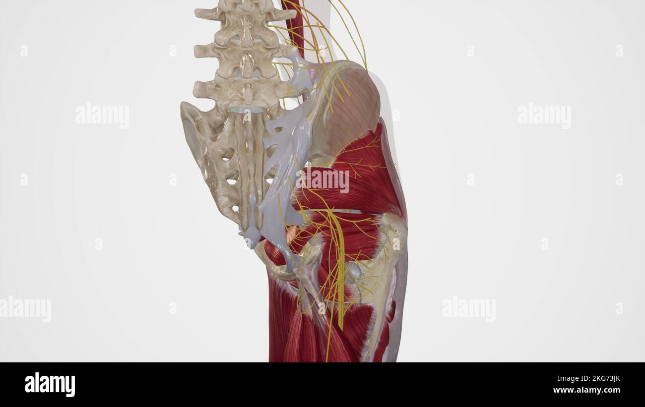 Nerves of Gluteal Region Stock Photohttps://www.alamy.com/image-license-details/?v=1https://www.alamy.com/nerves-of-gluteal-region-image491881339.html
RF2KG73JK–Nerves of Gluteal Region
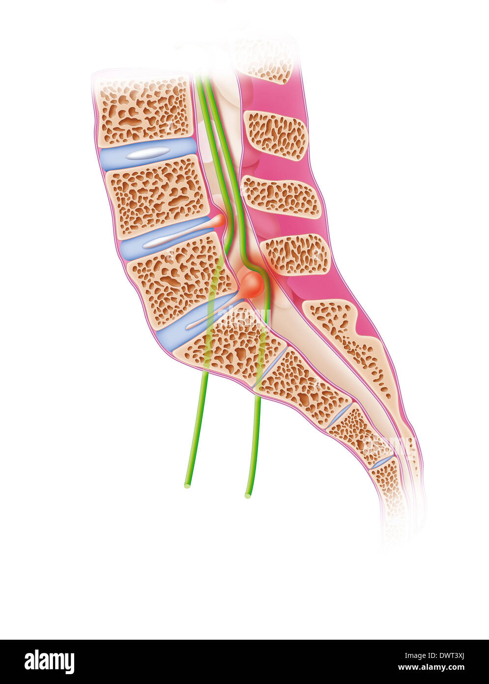 Sciatica, drawing Stock Photohttps://www.alamy.com/image-license-details/?v=1https://www.alamy.com/sciatica-drawing-image67527450.html
RMDWT3XJ–Sciatica, drawing
 Lateral Femoral Cutaneous Nerve Stock Photohttps://www.alamy.com/image-license-details/?v=1https://www.alamy.com/lateral-femoral-cutaneous-nerve-image490198315.html
RF2KDECXK–Lateral Femoral Cutaneous Nerve
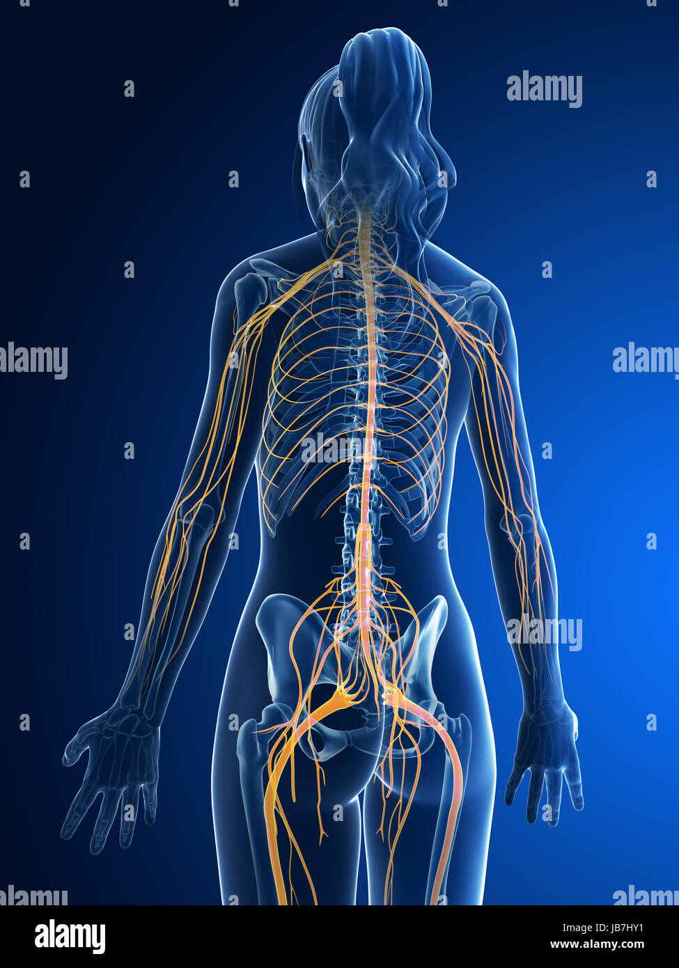 3d rendered medical illustration - female nerves Stock Photohttps://www.alamy.com/image-license-details/?v=1https://www.alamy.com/stock-photo-3d-rendered-medical-illustration-female-nerves-144611909.html
RFJB7HY1–3d rendered medical illustration - female nerves
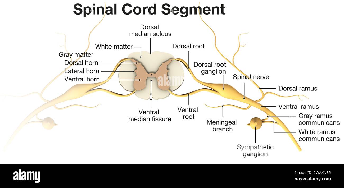 Spinal Cord Segment. Top View. 3D illustration Stock Photohttps://www.alamy.com/image-license-details/?v=1https://www.alamy.com/spinal-cord-segment-top-view-3d-illustration-image591886517.html
RF2WAXN85–Spinal Cord Segment. Top View. 3D illustration
 3d rendered medical illustration - female nerves Stock Photohttps://www.alamy.com/image-license-details/?v=1https://www.alamy.com/3d-rendered-medical-illustration-female-nerves-image343647236.html
RM2AY2DFG–3d rendered medical illustration - female nerves
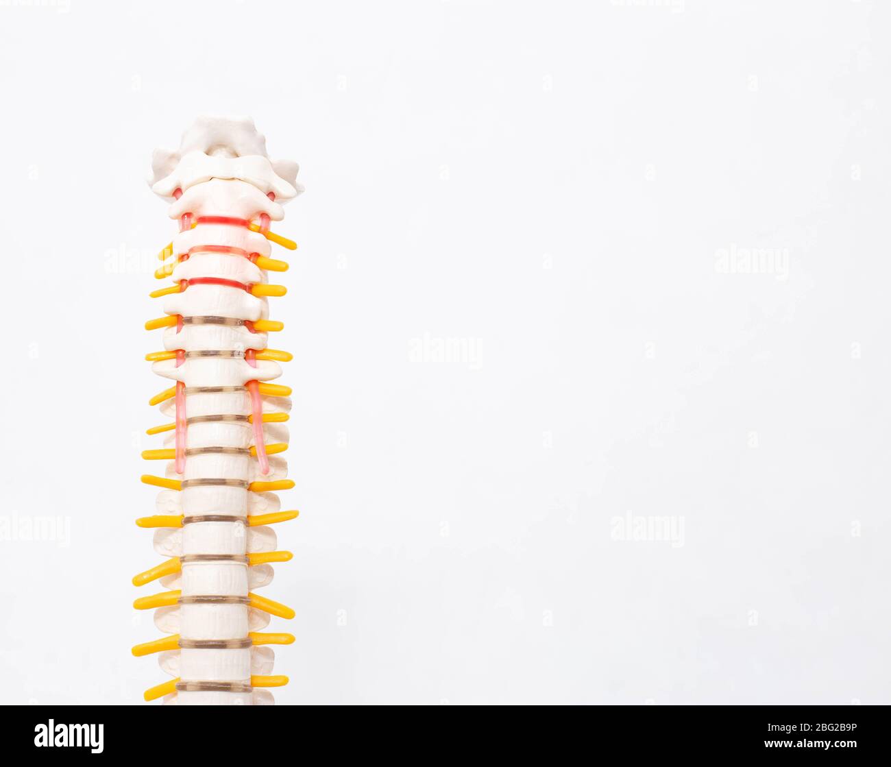 Cervical and thoracic spine on a white background. Intervertebral hernia of the cervical spine, rupture of the fibrous ring. Osteochondrosis Stock Photohttps://www.alamy.com/image-license-details/?v=1https://www.alamy.com/cervical-and-thoracic-spine-on-a-white-background-intervertebral-hernia-of-the-cervical-spine-rupture-of-the-fibrous-ring-osteochondrosis-image354094658.html
RF2BG2B9P–Cervical and thoracic spine on a white background. Intervertebral hernia of the cervical spine, rupture of the fibrous ring. Osteochondrosis
 Nervous system nerve body system anatomical internal organ graphic illustration Stock Vectorhttps://www.alamy.com/image-license-details/?v=1https://www.alamy.com/nervous-system-nerve-body-system-anatomical-internal-organ-graphic-illustration-image482395821.html
RF2K0R0P5–Nervous system nerve body system anatomical internal organ graphic illustration
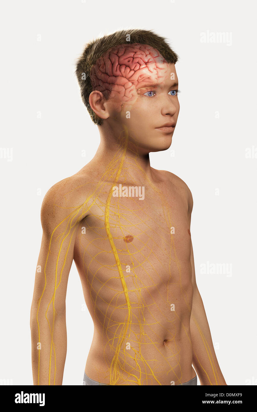 Digital illustration of a pre-adolescent male child with the brain and nerves of the nervous system visible within the body. Stock Photohttps://www.alamy.com/image-license-details/?v=1https://www.alamy.com/stock-photo-digital-illustration-of-a-pre-adolescent-male-child-with-the-brain-52090957.html
RMD0MXF9–Digital illustration of a pre-adolescent male child with the brain and nerves of the nervous system visible within the body.
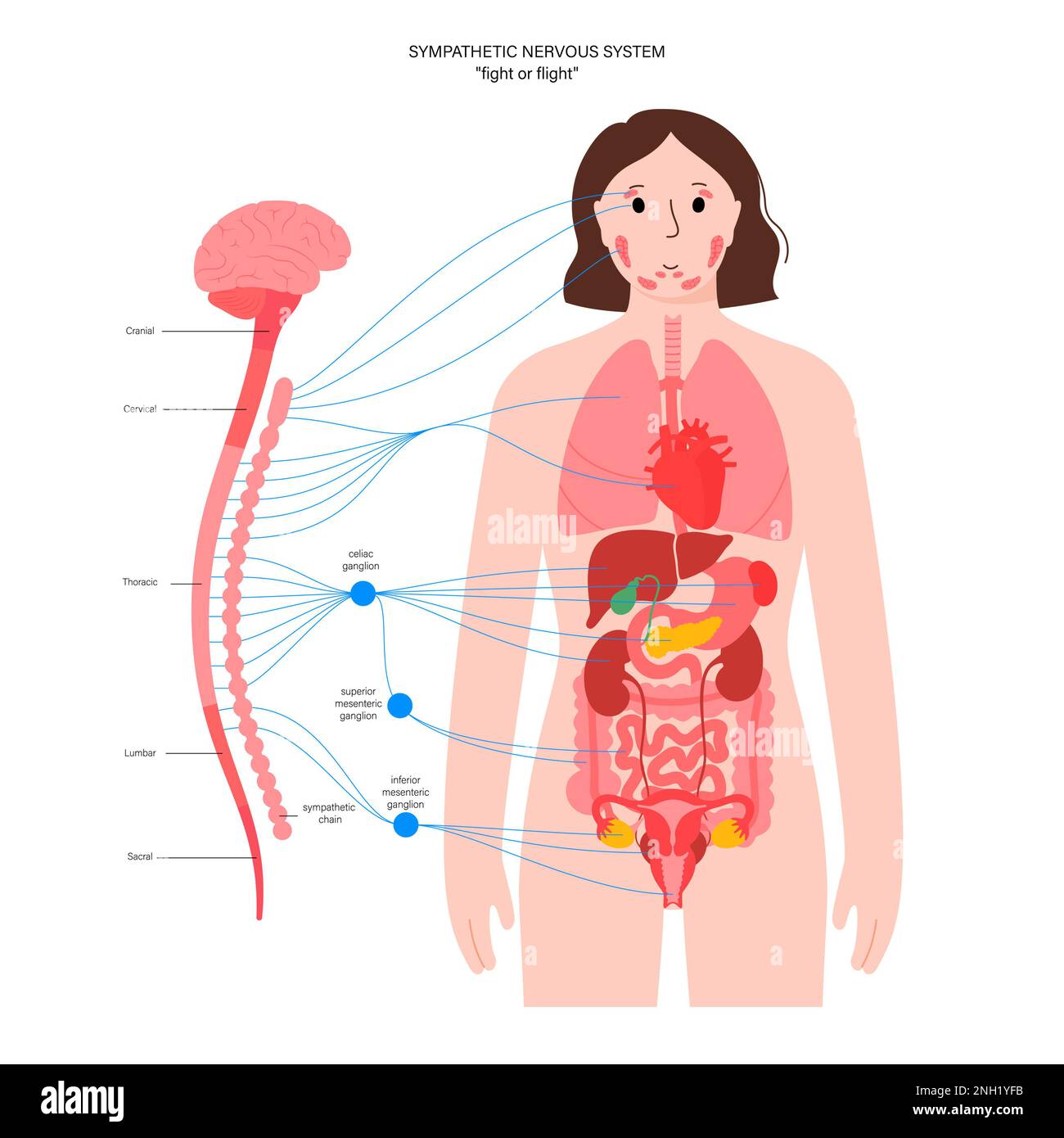 Sympathetic nervous system, illustration Stock Photohttps://www.alamy.com/image-license-details/?v=1https://www.alamy.com/sympathetic-nervous-system-illustration-image526803743.html
RF2NH1YFB–Sympathetic nervous system, illustration
 Skull with nervous system Stock Photohttps://www.alamy.com/image-license-details/?v=1https://www.alamy.com/stock-photo-skull-with-nervous-system-77386273.html
RFEDW6YD–Skull with nervous system
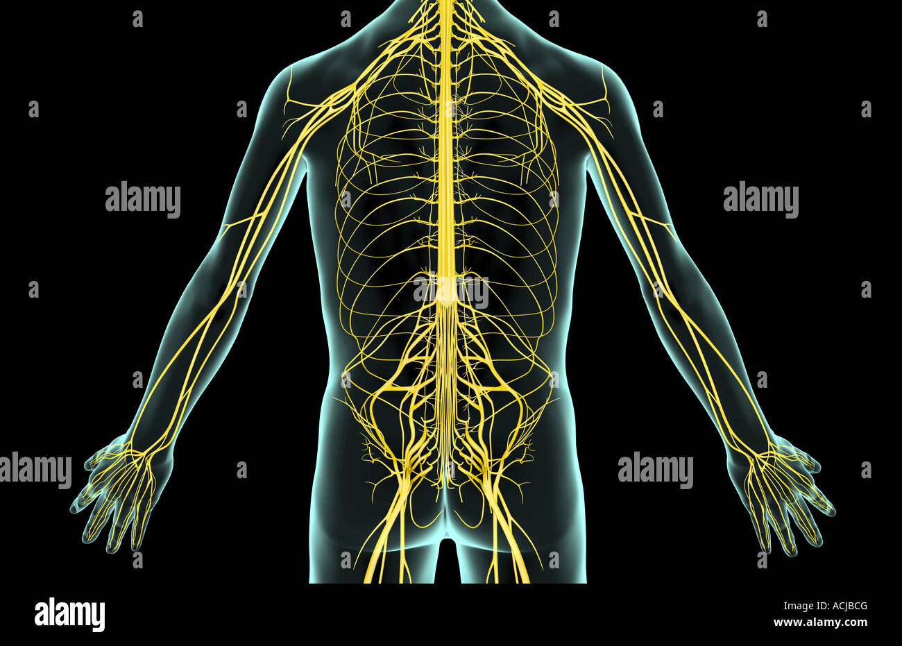 The nerves of the trunk Stock Photohttps://www.alamy.com/image-license-details/?v=1https://www.alamy.com/stock-photo-the-nerves-of-the-trunk-13168767.html
RFACJBCG–The nerves of the trunk
 Illustration of female central nervous system. Stock Photohttps://www.alamy.com/image-license-details/?v=1https://www.alamy.com/stock-photo-illustration-of-female-central-nervous-system-112683033.html
RFGF94A1–Illustration of female central nervous system.
 The lumbosacral plexus is a network of nerve fibers, derived from the roots of lumbar and sacral spinal nerves that branch out to form the nerves supp Stock Photohttps://www.alamy.com/image-license-details/?v=1https://www.alamy.com/the-lumbosacral-plexus-is-a-network-of-nerve-fibers-derived-from-the-roots-of-lumbar-and-sacral-spinal-nerves-that-branch-out-to-form-the-nerves-supp-image596601118.html
RF2WJHEPP–The lumbosacral plexus is a network of nerve fibers, derived from the roots of lumbar and sacral spinal nerves that branch out to form the nerves supp
 Spina bifida, illustration Stock Photohttps://www.alamy.com/image-license-details/?v=1https://www.alamy.com/spina-bifida-illustration-image415744163.html
RF2F4ANWR–Spina bifida, illustration
 Chordoma word cloud conceptual design isolated on white background. Stock Vectorhttps://www.alamy.com/image-license-details/?v=1https://www.alamy.com/chordoma-word-cloud-conceptual-design-isolated-on-white-background-image636935254.html
RF2S06WBJ–Chordoma word cloud conceptual design isolated on white background.
 Nervous system of the abdomen and pelvis, illustration Stock Photohttps://www.alamy.com/image-license-details/?v=1https://www.alamy.com/nervous-system-of-the-abdomen-and-pelvis-illustration-image561718771.html
RF2RHTE0K–Nervous system of the abdomen and pelvis, illustration
 The central nervous system is made up of the brain and spinal cord 3D illustration Stock Photohttps://www.alamy.com/image-license-details/?v=1https://www.alamy.com/the-central-nervous-system-is-made-up-of-the-brain-and-spinal-cord-3d-illustration-image499889200.html
RF2M17WNM–The central nervous system is made up of the brain and spinal cord 3D illustration
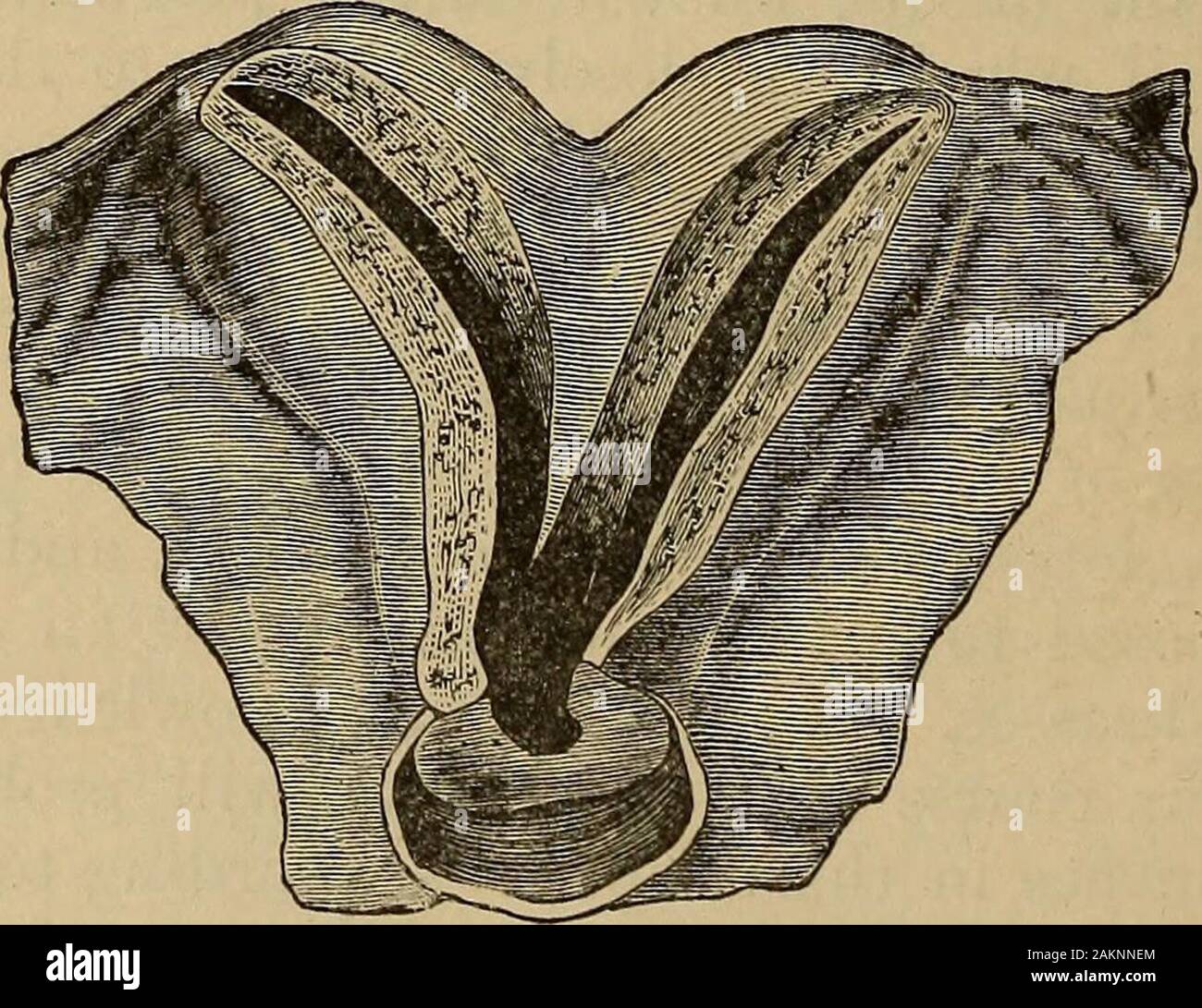 A treatise on the science and practice of midwifery . from the sympathetic, but, as the hypogastric plexus is connectedwith the sacral nerves, it is probable that some fibres from the cerebro-spinal system are distributed to the cervix. It is now generally admit-ted that nervous filaments are distributed to the cervix, even as far asthe external os, although their existence in this situation has been deniedby Jobert and other writers. The ultimate distribution of the nervesis not yet made out. Polle describes a nerve-filament as entering thepapillae of the cervical mucous membrane along with t Stock Photohttps://www.alamy.com/image-license-details/?v=1https://www.alamy.com/a-treatise-on-the-science-and-practice-of-midwifery-from-the-sympathetic-but-as-the-hypogastric-plexus-is-connectedwith-the-sacral-nerves-it-is-probable-that-some-fibres-from-the-cerebro-spinal-system-are-distributed-to-the-cervix-it-is-now-generally-admit-ted-that-nervous-filaments-are-distributed-to-the-cervix-even-as-far-asthe-external-os-although-their-existence-in-this-situation-has-been-deniedby-jobert-and-other-writers-the-ultimate-distribution-of-the-nervesis-not-yet-made-out-polle-describes-a-nerve-filament-as-entering-thepapillae-of-the-cervical-mucous-membrane-along-with-t-image339153324.html
RM2AKNNEM–A treatise on the science and practice of midwifery . from the sympathetic, but, as the hypogastric plexus is connectedwith the sacral nerves, it is probable that some fibres from the cerebro-spinal system are distributed to the cervix. It is now generally admit-ted that nervous filaments are distributed to the cervix, even as far asthe external os, although their existence in this situation has been deniedby Jobert and other writers. The ultimate distribution of the nervesis not yet made out. Polle describes a nerve-filament as entering thepapillae of the cervical mucous membrane along with t