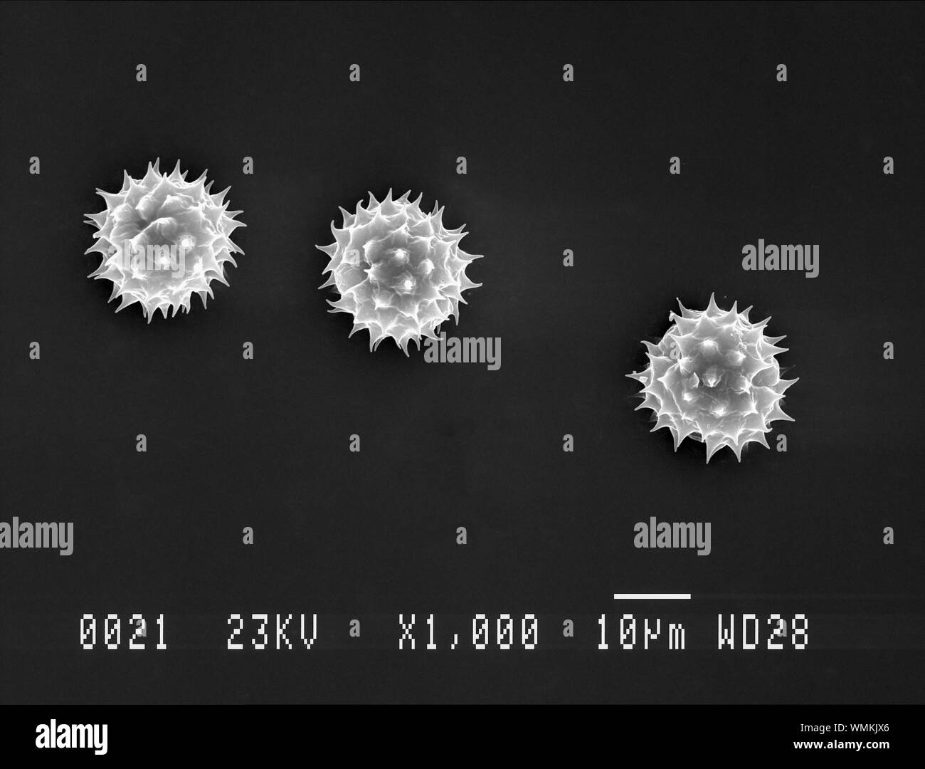
Scanning electron microscope sem Stock Photos and Images
(3,268)See scanning electron microscope sem stock video clipsQuick filters:
Scanning electron microscope sem Stock Photos and Images
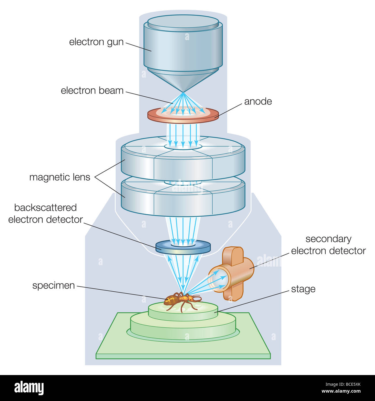 The components of a scanning electron microscope (SEM). Stock Photohttps://www.alamy.com/image-license-details/?v=1https://www.alamy.com/stock-photo-the-components-of-a-scanning-electron-microscope-sem-24898235.html
RMBCE5XK–The components of a scanning electron microscope (SEM).
 Scanning electron microscope (SEM) image of a human macrophage Stock Photohttps://www.alamy.com/image-license-details/?v=1https://www.alamy.com/stock-photo-scanning-electron-microscope-sem-image-of-a-human-macrophage-26900632.html
RMBFNC0T–Scanning electron microscope (SEM) image of a human macrophage
 Surgical needle, SEM Stock Photohttps://www.alamy.com/image-license-details/?v=1https://www.alamy.com/surgical-needle-sem-image341959471.html
RF2AT9GP7–Surgical needle, SEM
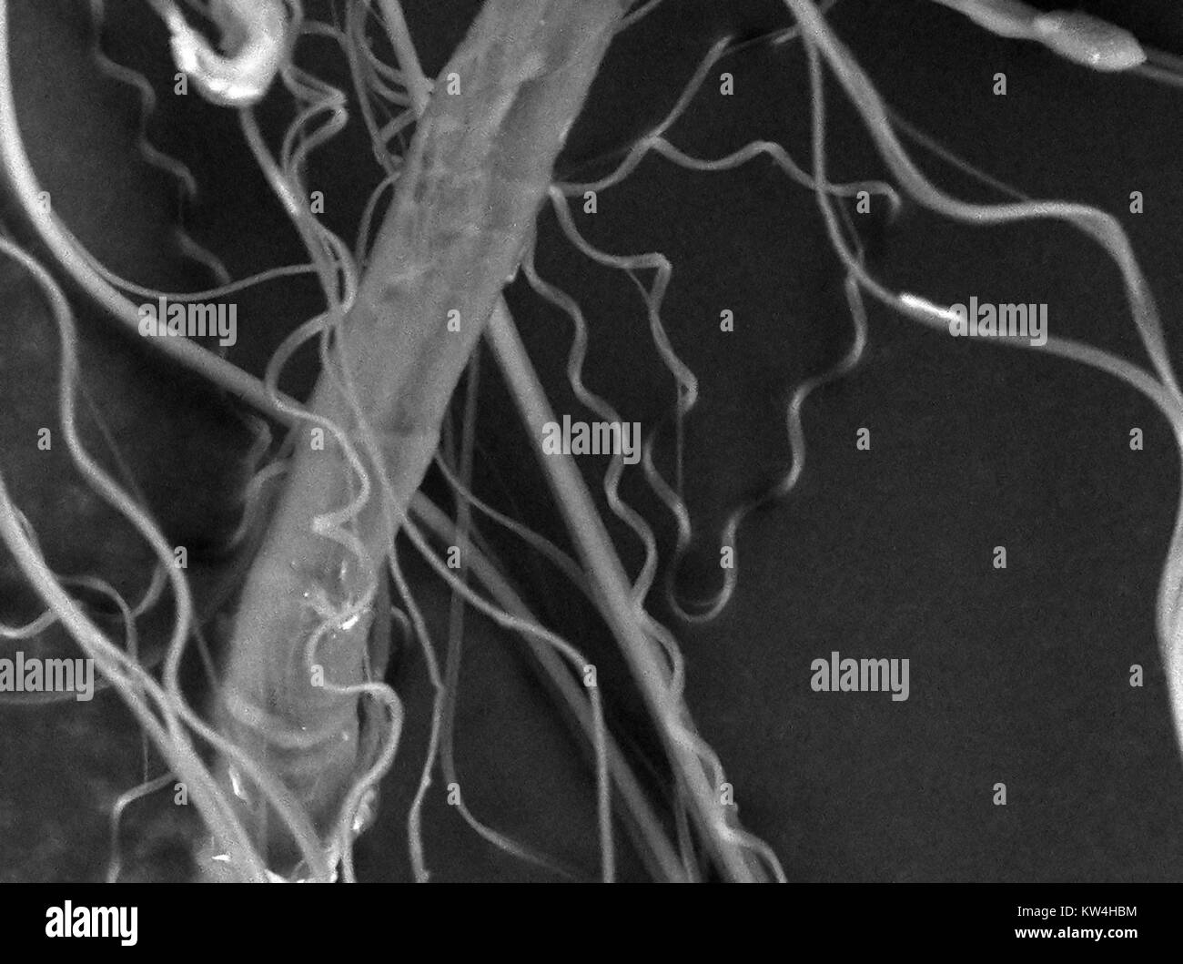 Scanning electron microscope (SEM) micrograph showing spider's silk, at a magnification of 2500x, 2016. Stock Photohttps://www.alamy.com/image-license-details/?v=1https://www.alamy.com/stock-photo-scanning-electron-microscope-sem-micrograph-showing-spiders-silk-at-170361176.html
RMKW4HBM–Scanning electron microscope (SEM) micrograph showing spider's silk, at a magnification of 2500x, 2016.
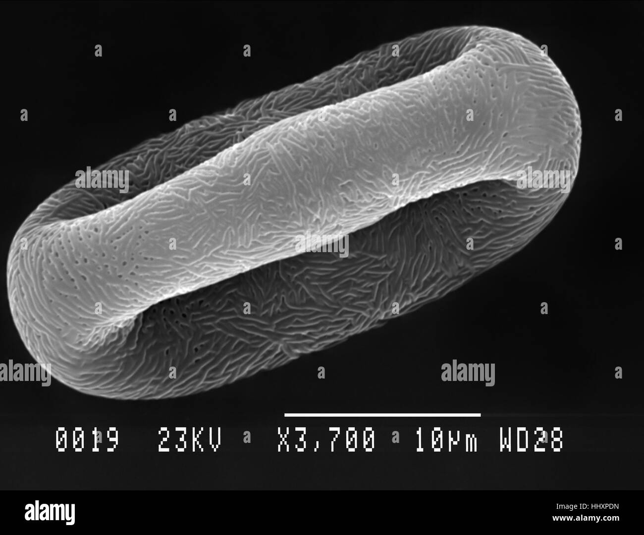 Cow parsley (Anthriscus sylvestris) pollen particle magnified under scanning electron microscope (SEM) Stock Photohttps://www.alamy.com/image-license-details/?v=1https://www.alamy.com/stock-photo-cow-parsley-anthriscus-sylvestris-pollen-particle-magnified-under-131510113.html
RFHHXPDN–Cow parsley (Anthriscus sylvestris) pollen particle magnified under scanning electron microscope (SEM)
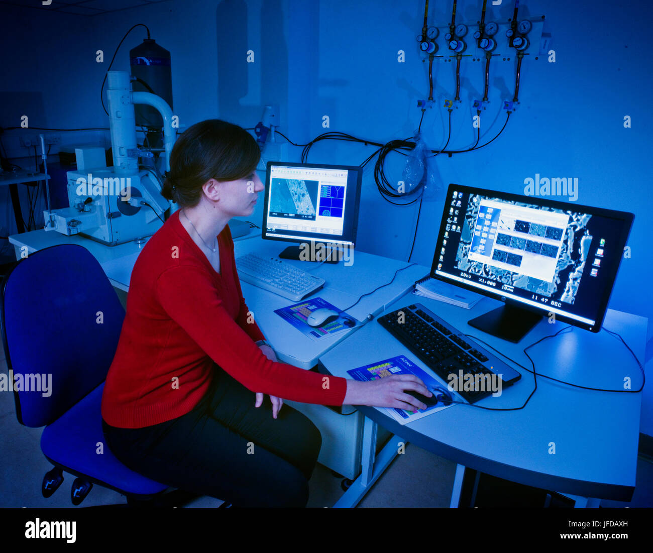 Scanning electron microscope in use at the Research Laboratory for Archaeology & the History of Art at the University of Oxford. Stock Photohttps://www.alamy.com/image-license-details/?v=1https://www.alamy.com/stock-photo-scanning-electron-microscope-in-use-at-the-research-laboratory-for-147196745.html
RMJFDAXH–Scanning electron microscope in use at the Research Laboratory for Archaeology & the History of Art at the University of Oxford.
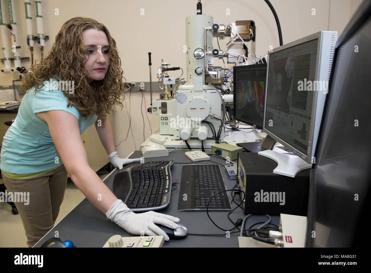 Female researcher uses a Scanning Electron Microscope (SEM) to perform high-resolution, electron microscopy studies of structural materials, in a lab located at Pacific Northwest National Laboratory (PNNL) located in Richland, Washington, image courtesy of the US Department of Energy, November 15, 2016. () Stock Photohttps://www.alamy.com/image-license-details/?v=1https://www.alamy.com/female-researcher-uses-a-scanning-electron-microscope-sem-to-perform-high-resolution-electron-microscopy-studies-of-structural-materials-in-a-lab-located-at-pacific-northwest-national-laboratory-pnnl-located-in-richland-washington-image-courtesy-of-the-us-department-of-energy-november-15-2016-image178438485.html
RMMA8G31–Female researcher uses a Scanning Electron Microscope (SEM) to perform high-resolution, electron microscopy studies of structural materials, in a lab located at Pacific Northwest National Laboratory (PNNL) located in Richland, Washington, image courtesy of the US Department of Energy, November 15, 2016. ()
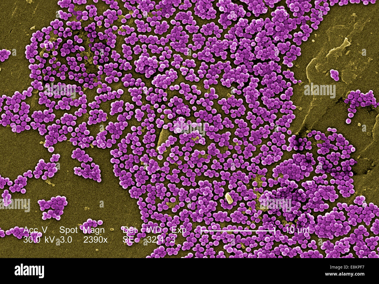 This colorized scanning electron micrograph (SEM) depicted numerous clumps of methicillin-resistant Staphylococcus aureus Stock Photohttps://www.alamy.com/image-license-details/?v=1https://www.alamy.com/stock-photo-this-colorized-scanning-electron-micrograph-sem-depicted-numerous-74193483.html
RME8KPF7–This colorized scanning electron micrograph (SEM) depicted numerous clumps of methicillin-resistant Staphylococcus aureus
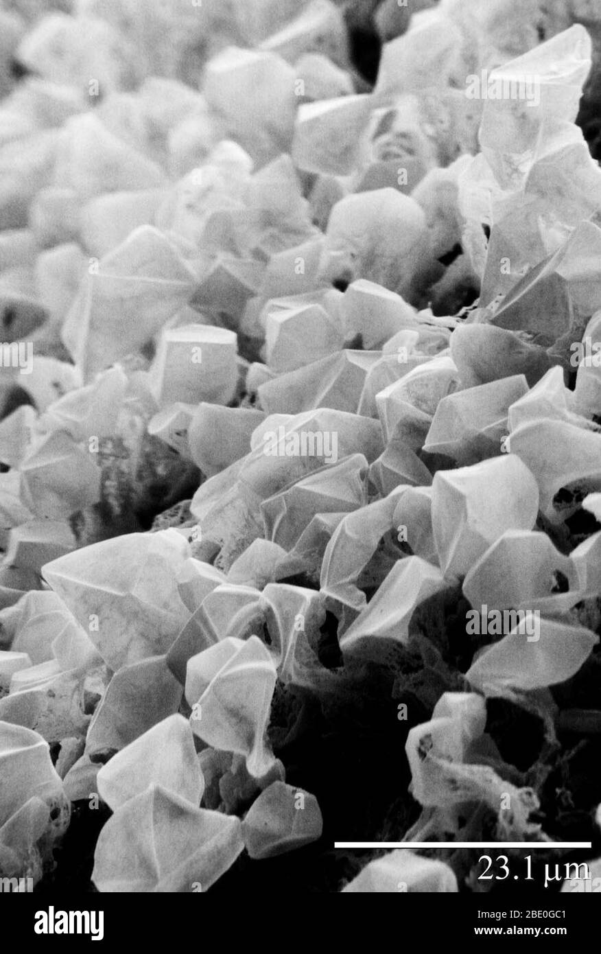 Carbon dioxide (CO2) ice/frost on Mars. Image was obtained using a Low Temperature Scanning Electron Microscope (LT-SEM). Stock Photohttps://www.alamy.com/image-license-details/?v=1https://www.alamy.com/carbon-dioxide-co2-icefrost-on-mars-image-was-obtained-using-a-low-temperature-scanning-electron-microscope-lt-sem-image352825425.html
RM2BE0GC1–Carbon dioxide (CO2) ice/frost on Mars. Image was obtained using a Low Temperature Scanning Electron Microscope (LT-SEM).
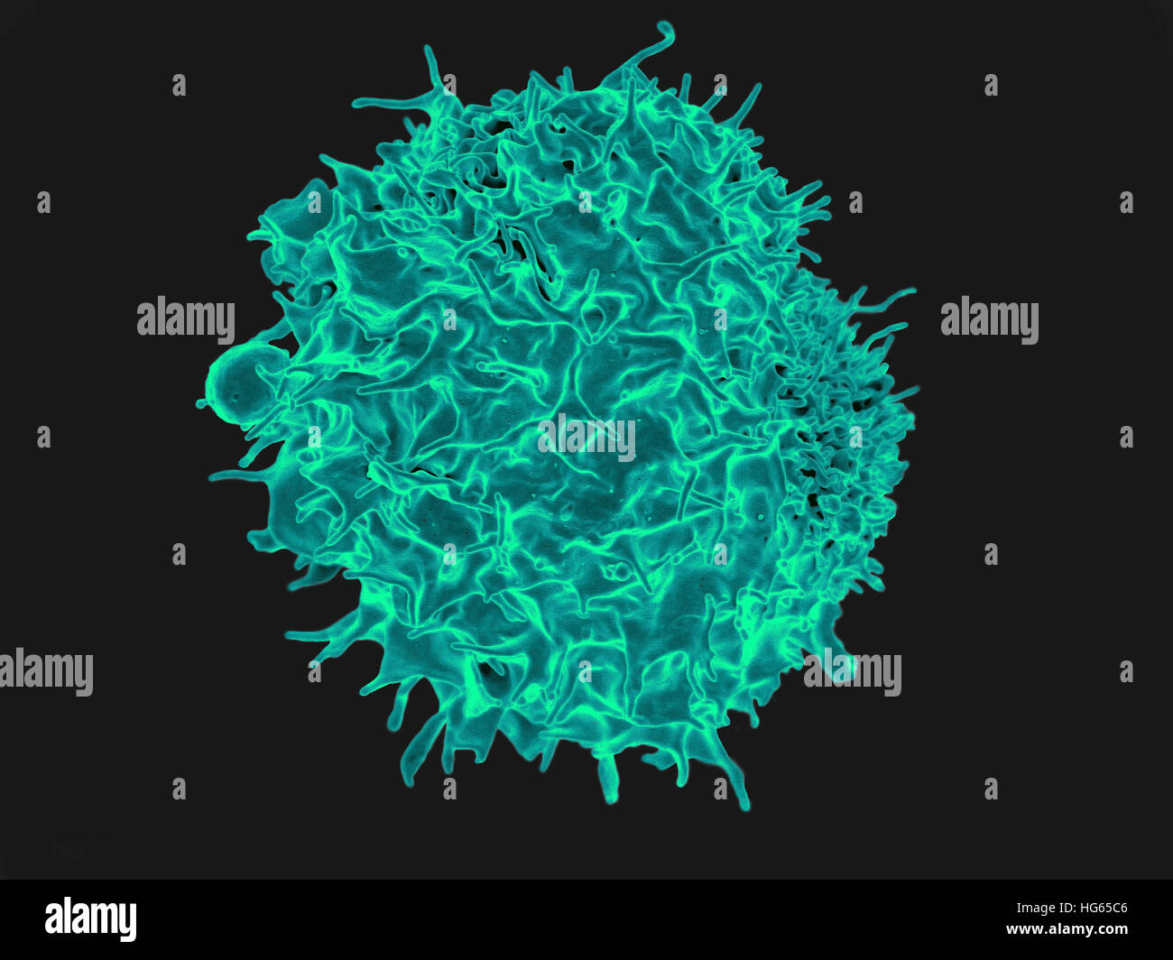 Colorized scanning electron micrograph of a T lymphocyte. Stock Photohttps://www.alamy.com/image-license-details/?v=1https://www.alamy.com/stock-photo-colorized-scanning-electron-micrograph-of-a-t-lymphocyte-130443046.html
RFHG65C6–Colorized scanning electron micrograph of a T lymphocyte.
 Erlangen, Germany. 19th Dec, 2022. A scanning electron microscope (SEM) stands in the research and laboratory building on the new Siemens Campus Erlangen during the official opening of the campus. The technology corporation Siemens has unveiled one of the largest construction projects in its company history at its Erlangen location. The new Siemens Campus, an open urban district with 540,000 square meters of space, will soon be home to 17,000 employees of Siemens and Siemens Energy. Credit: Daniel Karmann/dpa/Alamy Live News Stock Photohttps://www.alamy.com/image-license-details/?v=1https://www.alamy.com/erlangen-germany-19th-dec-2022-a-scanning-electron-microscope-sem-stands-in-the-research-and-laboratory-building-on-the-new-siemens-campus-erlangen-during-the-official-opening-of-the-campus-the-technology-corporation-siemens-has-unveiled-one-of-the-largest-construction-projects-in-its-company-history-at-its-erlangen-location-the-new-siemens-campus-an-open-urban-district-with-540000-square-meters-of-space-will-soon-be-home-to-17000-employees-of-siemens-and-siemens-energy-credit-daniel-karmanndpaalamy-live-news-image501716347.html
RM2M4748Y–Erlangen, Germany. 19th Dec, 2022. A scanning electron microscope (SEM) stands in the research and laboratory building on the new Siemens Campus Erlangen during the official opening of the campus. The technology corporation Siemens has unveiled one of the largest construction projects in its company history at its Erlangen location. The new Siemens Campus, an open urban district with 540,000 square meters of space, will soon be home to 17,000 employees of Siemens and Siemens Energy. Credit: Daniel Karmann/dpa/Alamy Live News
 NASA astronaut Kayla Barron sets up the Mochii microscope. Mochii is a miniature scanning electron microscope (SEM) with spectroscopy to conduct real-time, on-site imaging and compositional measurements of particles on the International Space Station (ISS). Stock Photohttps://www.alamy.com/image-license-details/?v=1https://www.alamy.com/nasa-astronaut-kayla-barron-sets-up-the-mochii-microscope-mochii-is-a-miniature-scanning-electron-microscope-sem-with-spectroscopy-to-conduct-real-time-on-site-imaging-and-compositional-measurements-of-particles-on-the-international-space-station-iss-image485254043.html
RM2K5D6DF–NASA astronaut Kayla Barron sets up the Mochii microscope. Mochii is a miniature scanning electron microscope (SEM) with spectroscopy to conduct real-time, on-site imaging and compositional measurements of particles on the International Space Station (ISS).
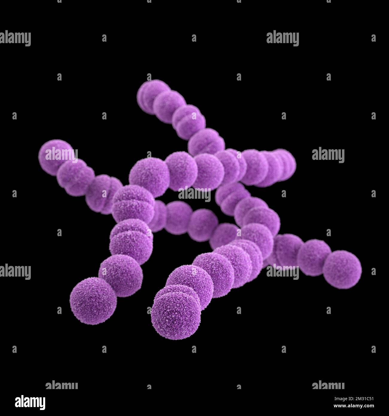 Group A streptococcus bacteria. STREP A Streptococcus pyogenes is a species of Gram-positive, aerotolerant bacteria in the genus Streptococcus. These bacteria are extracellular, and made up of non-motile and non-sporing cocci that tend to link in chains. This illustration depicted a 3D, computer-generated image, of a group of Gram-positive, Streptococcus pyogenes (group A Streptococcus) bacteria. The visualisation was based upon scanning electron microscopic (SEM) imagery. Optimised version of an image produced by the US Centers for Disease Control and Prevention / credit CDC /J.Oosthuizen Stock Photohttps://www.alamy.com/image-license-details/?v=1https://www.alamy.com/group-a-streptococcus-bacteria-strep-a-streptococcus-pyogenes-is-a-species-of-gram-positive-aerotolerant-bacteria-in-the-genus-streptococcus-these-bacteria-are-extracellular-and-made-up-of-non-motile-and-non-sporing-cocci-that-tend-to-link-in-chains-this-illustration-depicted-a-3d-computer-generated-image-of-a-group-of-gram-positive-streptococcus-pyogenes-group-a-streptococcus-bacteria-the-visualisation-was-based-upon-scanning-electron-microscopic-sem-imagery-optimised-version-of-an-image-produced-by-the-us-centers-for-disease-control-and-prevention-credit-cdc-joosthuizen-image500976141.html
RM2M31C51–Group A streptococcus bacteria. STREP A Streptococcus pyogenes is a species of Gram-positive, aerotolerant bacteria in the genus Streptococcus. These bacteria are extracellular, and made up of non-motile and non-sporing cocci that tend to link in chains. This illustration depicted a 3D, computer-generated image, of a group of Gram-positive, Streptococcus pyogenes (group A Streptococcus) bacteria. The visualisation was based upon scanning electron microscopic (SEM) imagery. Optimised version of an image produced by the US Centers for Disease Control and Prevention / credit CDC /J.Oosthuizen
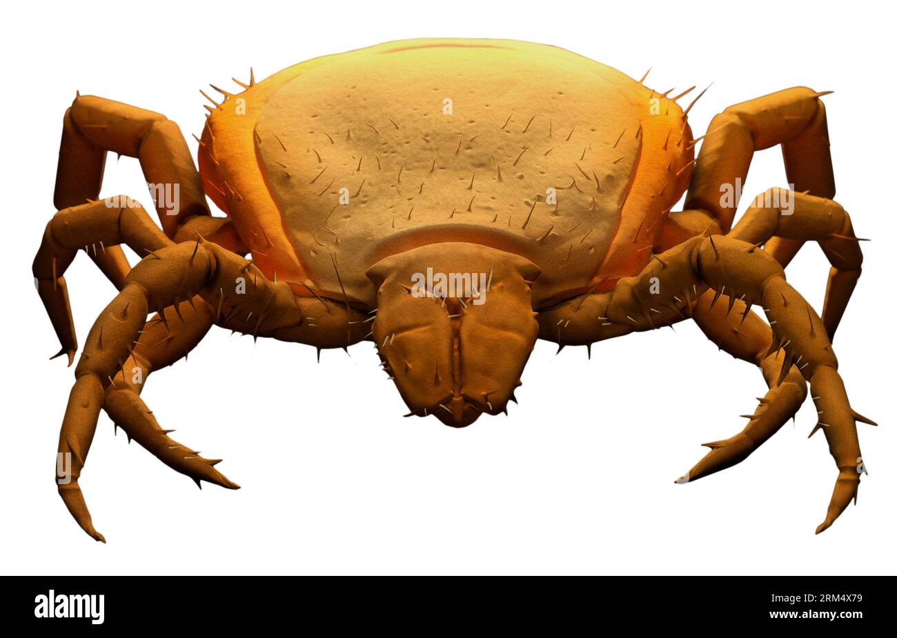 3D illustration of a detailed tick: SEM Electron Microscope CGI Replica in digital orange colour Stock Photohttps://www.alamy.com/image-license-details/?v=1https://www.alamy.com/3d-illustration-of-a-detailed-tick-sem-electron-microscope-cgi-replica-in-digital-orange-colour-image563133293.html
RF2RM4X79–3D illustration of a detailed tick: SEM Electron Microscope CGI Replica in digital orange colour
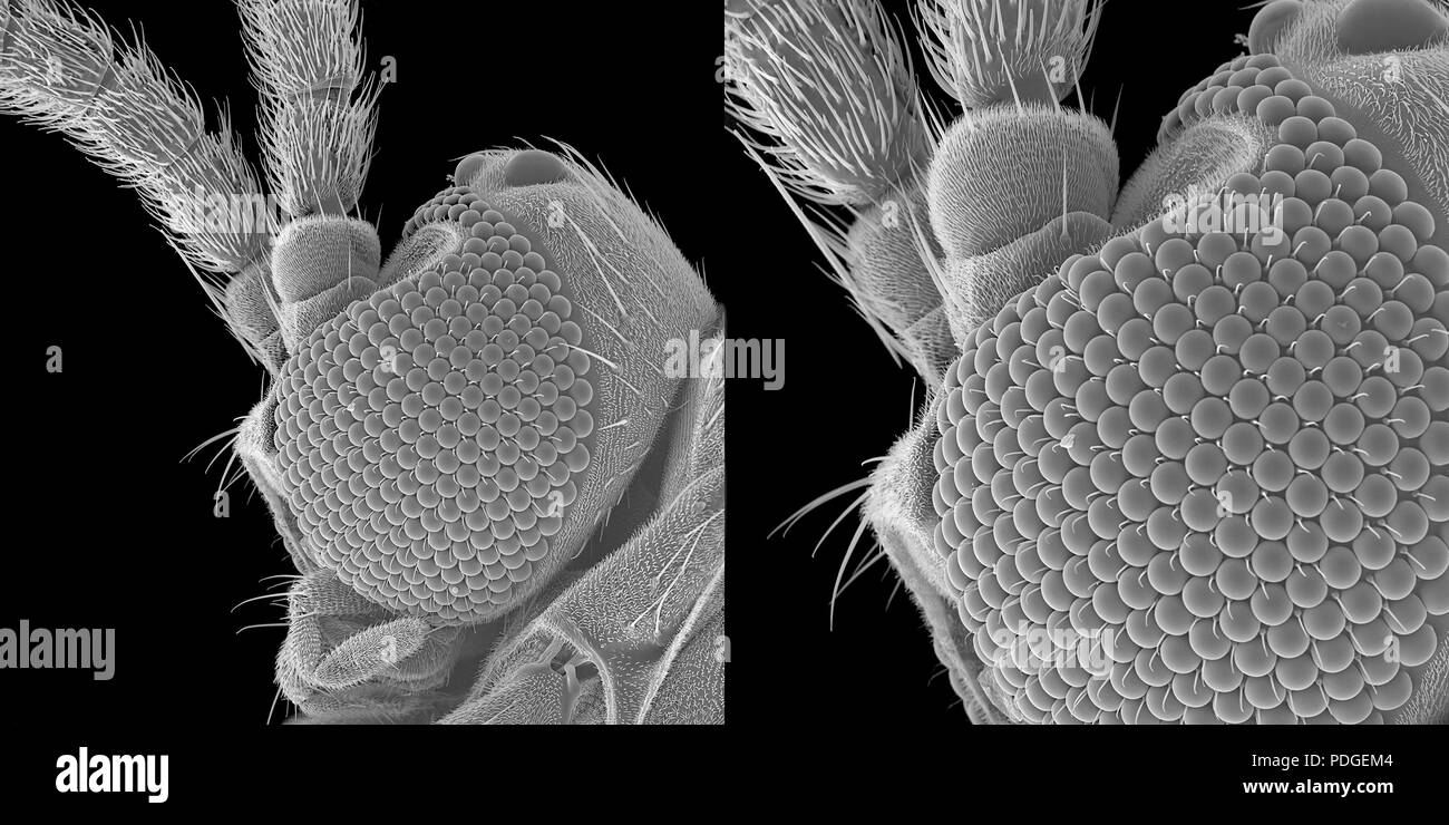 Tiny fungus gnat under scanning electron microscope Stock Photohttps://www.alamy.com/image-license-details/?v=1https://www.alamy.com/tiny-fungus-gnat-under-scanning-electron-microscope-image214877716.html
RMPDGEM4–Tiny fungus gnat under scanning electron microscope
 Red Blood Cells.Scanning electron microscope Stock Photohttps://www.alamy.com/image-license-details/?v=1https://www.alamy.com/red-blood-cellsscanning-electron-microscope-image209285722.html
RFP4DP22–Red Blood Cells.Scanning electron microscope
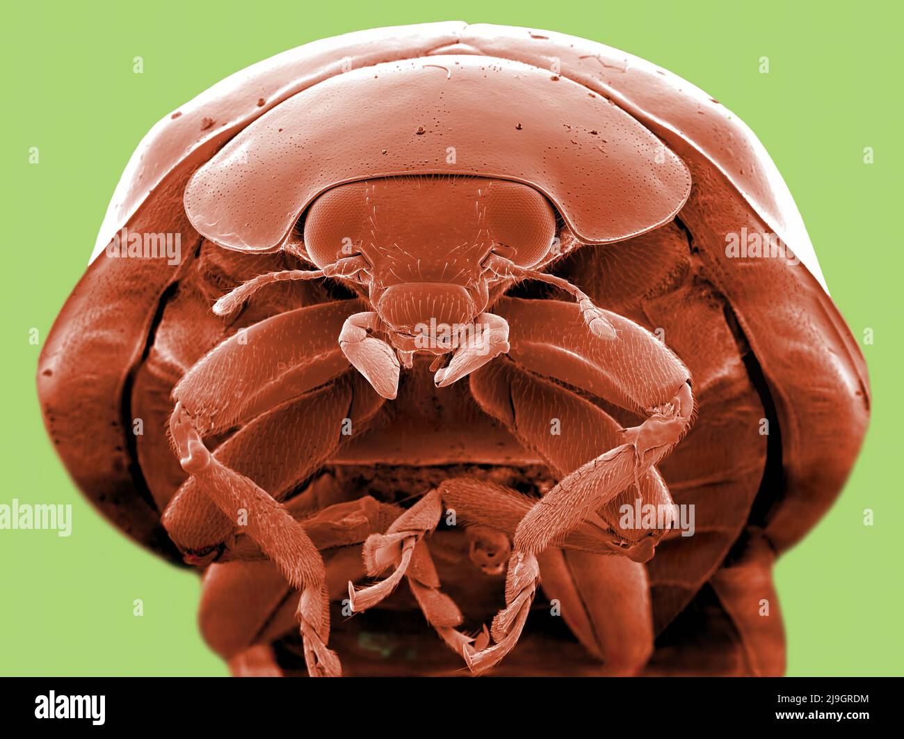 SEM Scanning Electron Microscope image of a Ladybug Ladybird microscopic Stock Photohttps://www.alamy.com/image-license-details/?v=1https://www.alamy.com/sem-scanning-electron-microscope-image-of-a-ladybug-ladybird-microscopic-image470581488.html
RM2J9GRDM–SEM Scanning Electron Microscope image of a Ladybug Ladybird microscopic
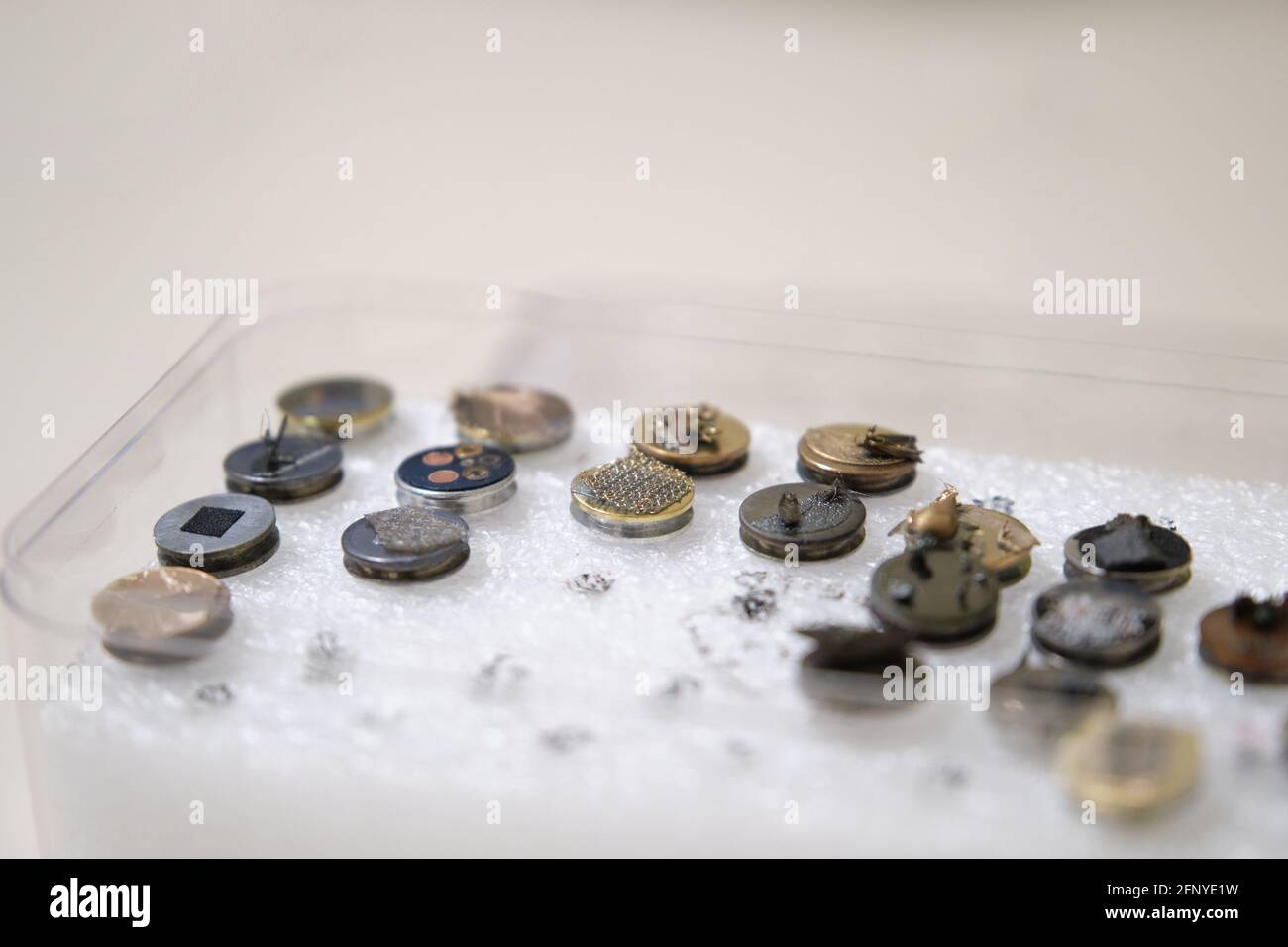 Scanning electron microscope samples on specimen mounts. SEM pins to analyze. Stock Photohttps://www.alamy.com/image-license-details/?v=1https://www.alamy.com/scanning-electron-microscope-samples-on-specimen-mounts-sem-pins-to-analyze-image426560341.html
RF2FNYE1W–Scanning electron microscope samples on specimen mounts. SEM pins to analyze.
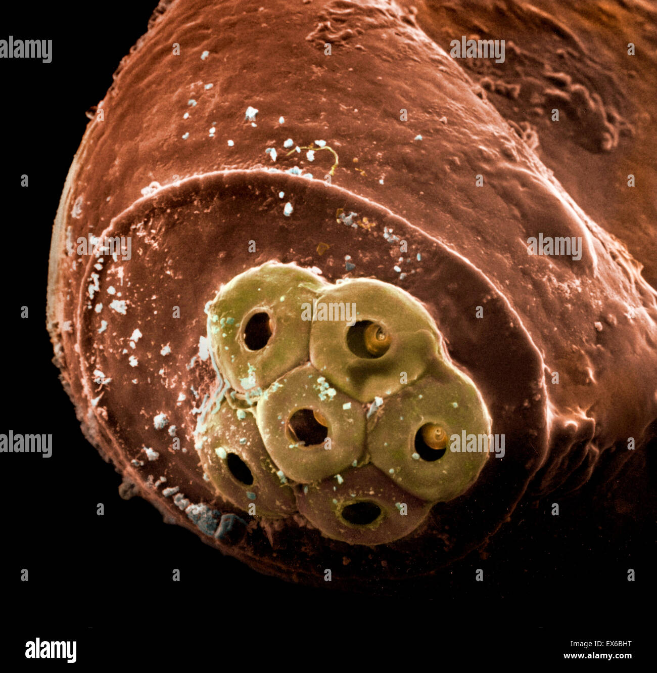 Pediculus humanus capitis, head louse egg detail, SEM Stock Photohttps://www.alamy.com/image-license-details/?v=1https://www.alamy.com/stock-photo-pediculus-humanus-capitis-head-louse-egg-detail-sem-84963364.html
RMEX6BHT–Pediculus humanus capitis, head louse egg detail, SEM
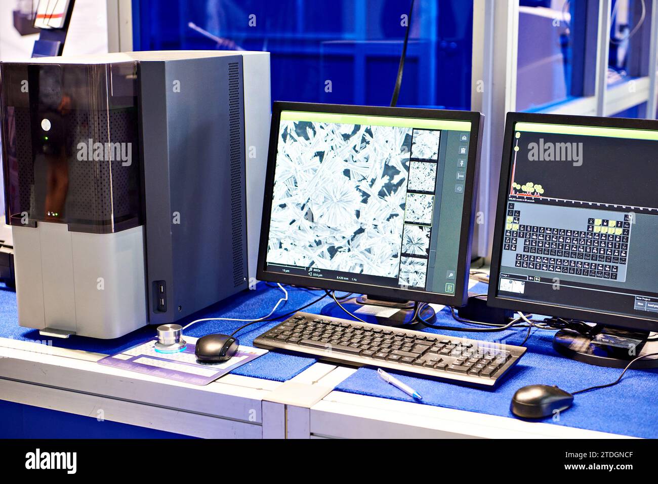 Scanning electron microscope with EMF microanalysis Stock Photohttps://www.alamy.com/image-license-details/?v=1https://www.alamy.com/scanning-electron-microscope-with-emf-microanalysis-image576300719.html
RF2TDGNCF–Scanning electron microscope with EMF microanalysis
 Scanning electron microscope (SEM) image of a human macrophage Stock Photohttps://www.alamy.com/image-license-details/?v=1https://www.alamy.com/stock-photo-scanning-electron-microscope-sem-image-of-a-human-macrophage-26900649.html
RMBFNC1D–Scanning electron microscope (SEM) image of a human macrophage
 Surgical needle, SEM Stock Photohttps://www.alamy.com/image-license-details/?v=1https://www.alamy.com/surgical-needle-sem-image341959514.html
RF2AT9GRP–Surgical needle, SEM
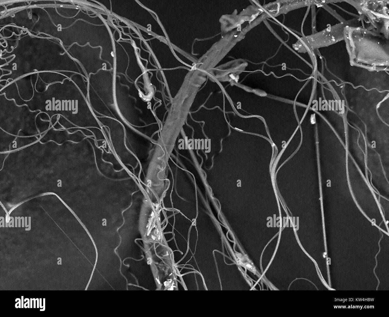 Scanning electron microscope (SEM) micrograph showing spider's silk, at a magnification of 1500x, 2016. Stock Photohttps://www.alamy.com/image-license-details/?v=1https://www.alamy.com/stock-photo-scanning-electron-microscope-sem-micrograph-showing-spiders-silk-at-170361181.html
RMKW4HBW–Scanning electron microscope (SEM) micrograph showing spider's silk, at a magnification of 1500x, 2016.
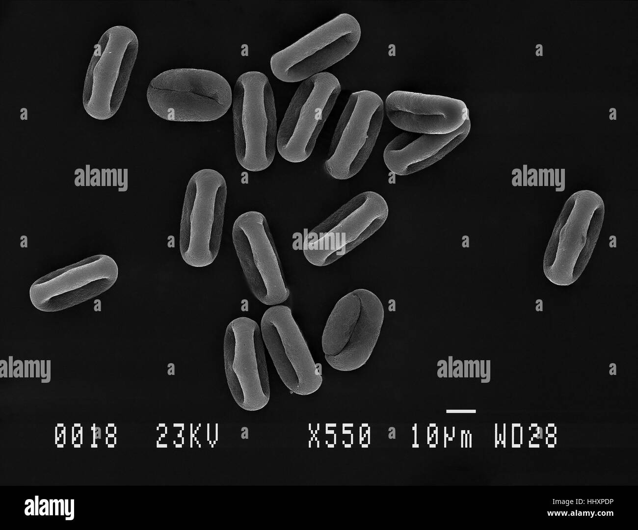 Many cow parsley (Anthriscus sylvestris) pollen grains magnified under scanning electron microscope (SEM), demonstrating shape at different angles. Stock Photohttps://www.alamy.com/image-license-details/?v=1https://www.alamy.com/stock-photo-many-cow-parsley-anthriscus-sylvestris-pollen-grains-magnified-under-131510114.html
RFHHXPDP–Many cow parsley (Anthriscus sylvestris) pollen grains magnified under scanning electron microscope (SEM), demonstrating shape at different angles.
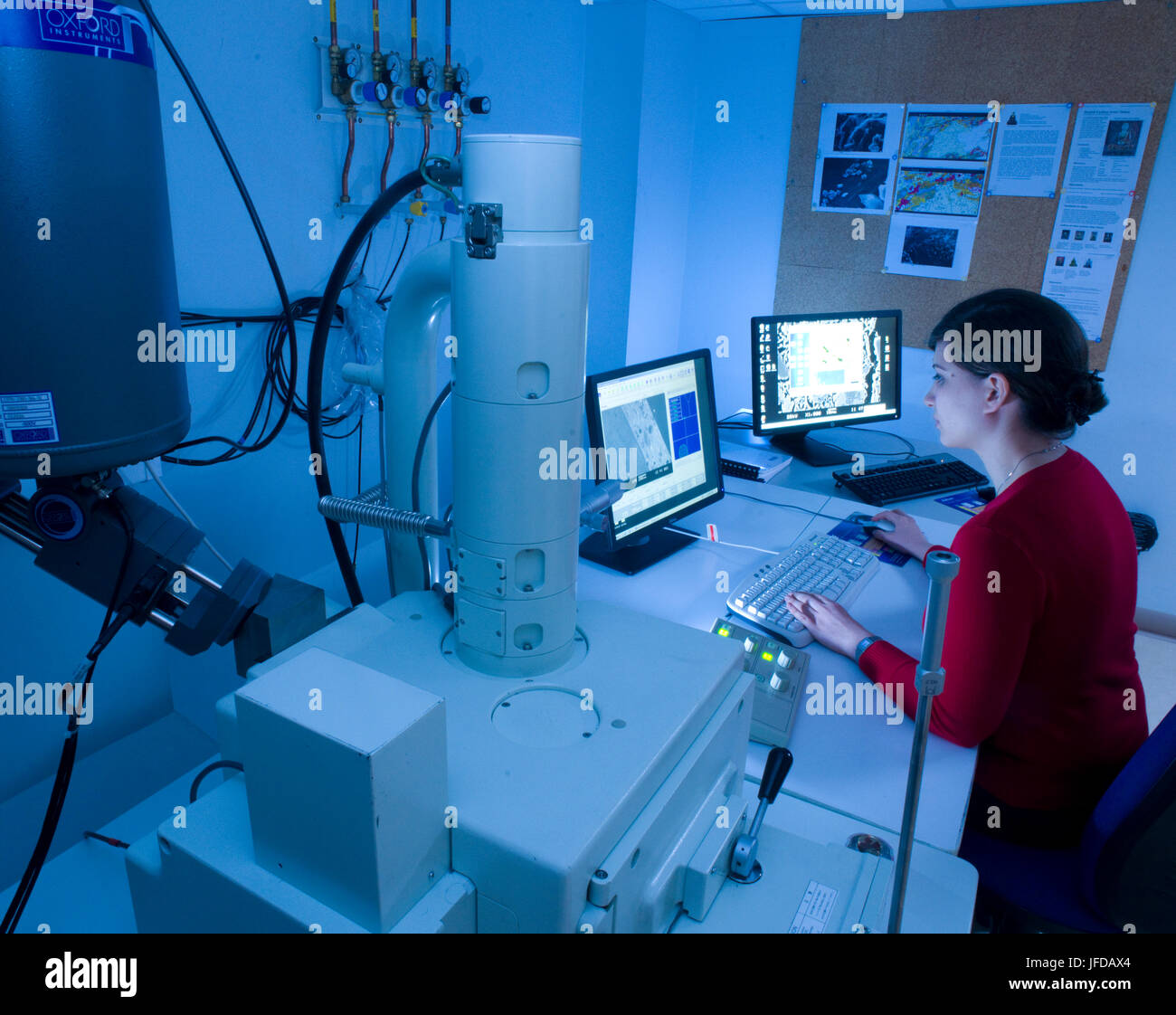 Scanning electron microscope in use at the Research Laboratory for Archaeology & the History of Art at the University of Oxford. Stock Photohttps://www.alamy.com/image-license-details/?v=1https://www.alamy.com/stock-photo-scanning-electron-microscope-in-use-at-the-research-laboratory-for-147196732.html
RMJFDAX4–Scanning electron microscope in use at the Research Laboratory for Archaeology & the History of Art at the University of Oxford.
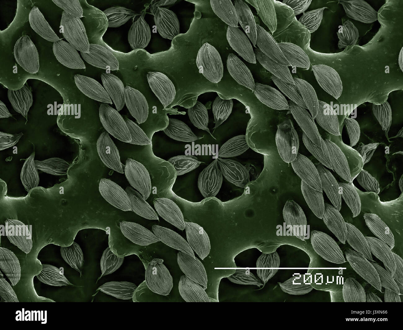 Detail of elytra of a recticulated beetle (Coleoptera: Cupedidae: Cupes sp.) imaged in a scanning electron microscope Stock Photohttps://www.alamy.com/image-license-details/?v=1https://www.alamy.com/stock-photo-detail-of-elytra-of-a-recticulated-beetle-coleoptera-cupedidae-cupes-140114302.html
RFJ3XN66–Detail of elytra of a recticulated beetle (Coleoptera: Cupedidae: Cupes sp.) imaged in a scanning electron microscope
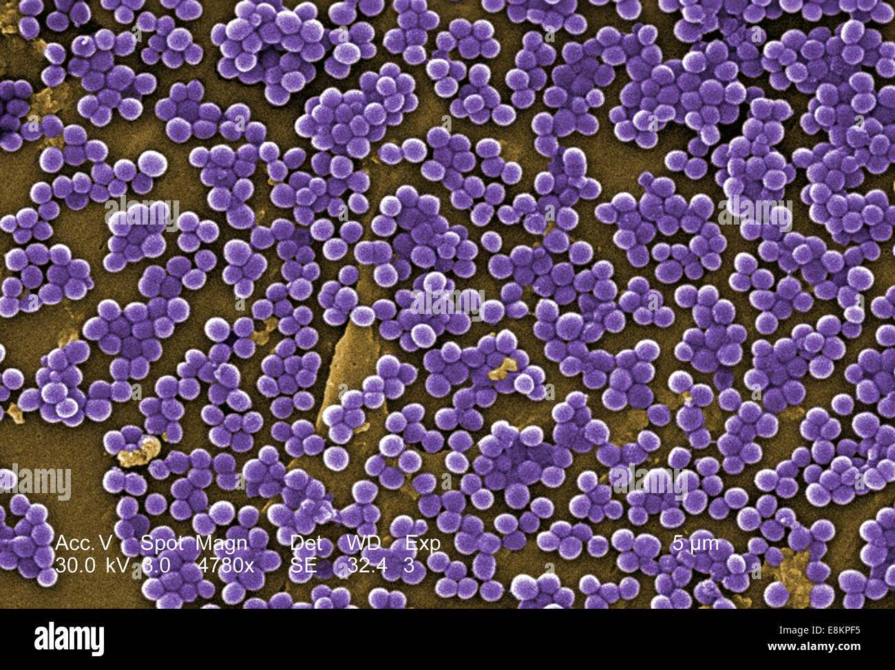 This colorized scanning electron micrograph (SEM) depicted numerous clumps of methicillin-resistant Staphylococcus aureus Stock Photohttps://www.alamy.com/image-license-details/?v=1https://www.alamy.com/stock-photo-this-colorized-scanning-electron-micrograph-sem-depicted-numerous-74193481.html
RME8KPF5–This colorized scanning electron micrograph (SEM) depicted numerous clumps of methicillin-resistant Staphylococcus aureus
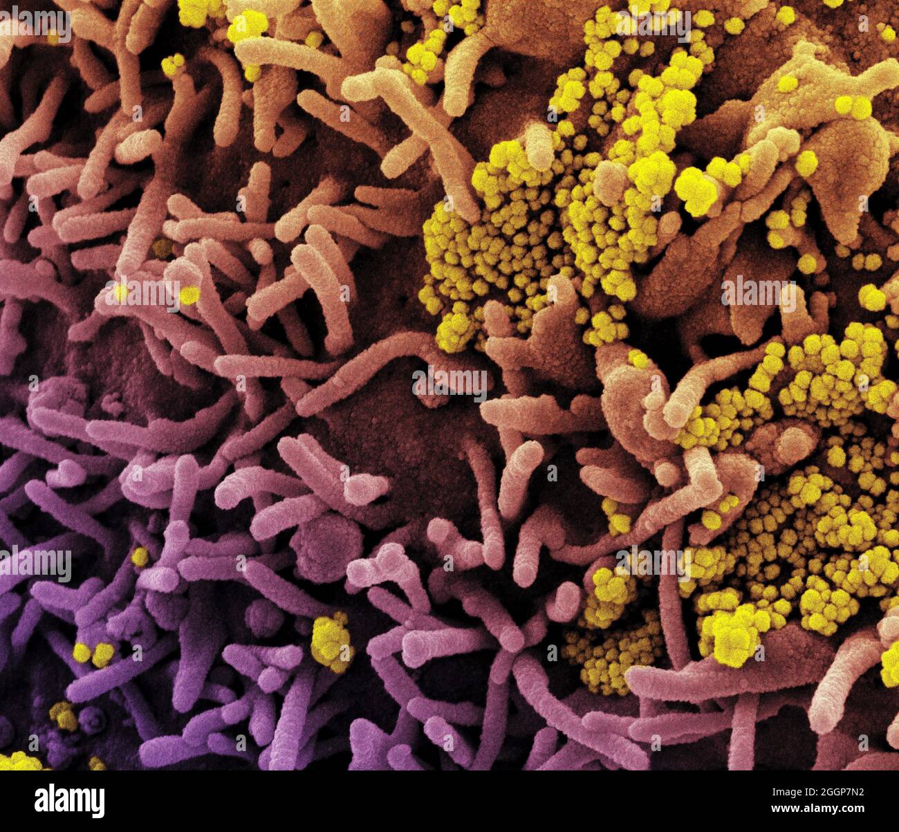 Colorized scanning electron micrograph of a cell (tan) infected with SARS-CoV-2 virus particles (yellow), isolated from a patient sample. Stock Photohttps://www.alamy.com/image-license-details/?v=1https://www.alamy.com/colorized-scanning-electron-micrograph-of-a-cell-tan-infected-with-sars-cov-2-virus-particles-yellow-isolated-from-a-patient-sample-image440582718.html
RM2GGP7N2–Colorized scanning electron micrograph of a cell (tan) infected with SARS-CoV-2 virus particles (yellow), isolated from a patient sample.
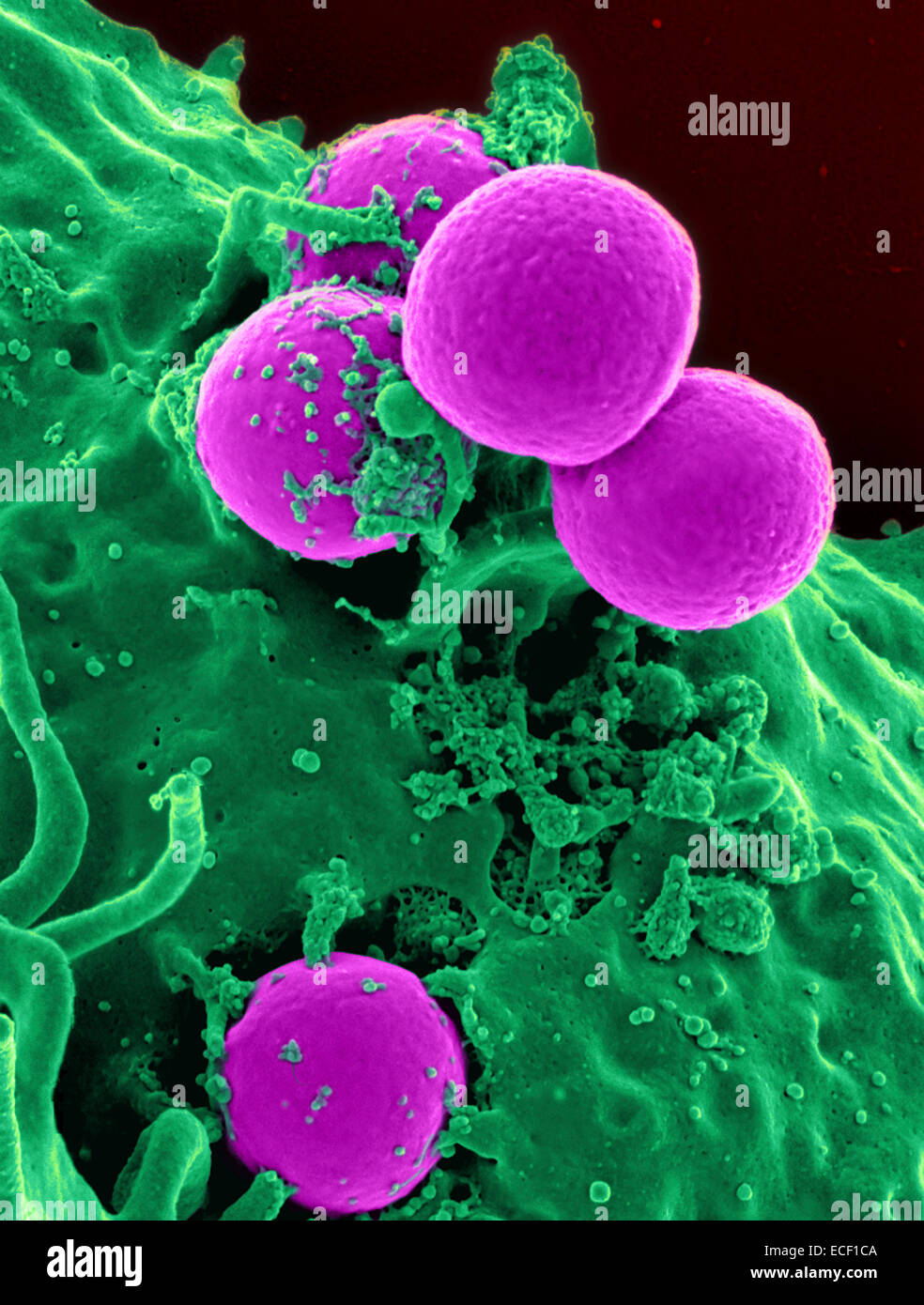 Scanning electron micrograph of a human neutrophil ingesting MRSA. Stock Photohttps://www.alamy.com/image-license-details/?v=1https://www.alamy.com/stock-photo-scanning-electron-micrograph-of-a-human-neutrophil-ingesting-mrsa-76547754.html
RFECF1CA–Scanning electron micrograph of a human neutrophil ingesting MRSA.
 Jakarta Selatan, Indonesia. 26th Apr, 2016. A researcher tightens rock-holder to avoid shaking during the process inside Scanning Electron Microscope 'SEM' machine. SEM is used to analyze rock minerals up to 1µm. It can zoom 300.000x than usual polarize microscope. © Anton Raharjo/Pacific Press/Alamy Live News Stock Photohttps://www.alamy.com/image-license-details/?v=1https://www.alamy.com/stock-photo-jakarta-selatan-indonesia-26th-apr-2016-a-researcher-tightens-rock-102955185.html
RMFYE0AW–Jakarta Selatan, Indonesia. 26th Apr, 2016. A researcher tightens rock-holder to avoid shaking during the process inside Scanning Electron Microscope 'SEM' machine. SEM is used to analyze rock minerals up to 1µm. It can zoom 300.000x than usual polarize microscope. © Anton Raharjo/Pacific Press/Alamy Live News
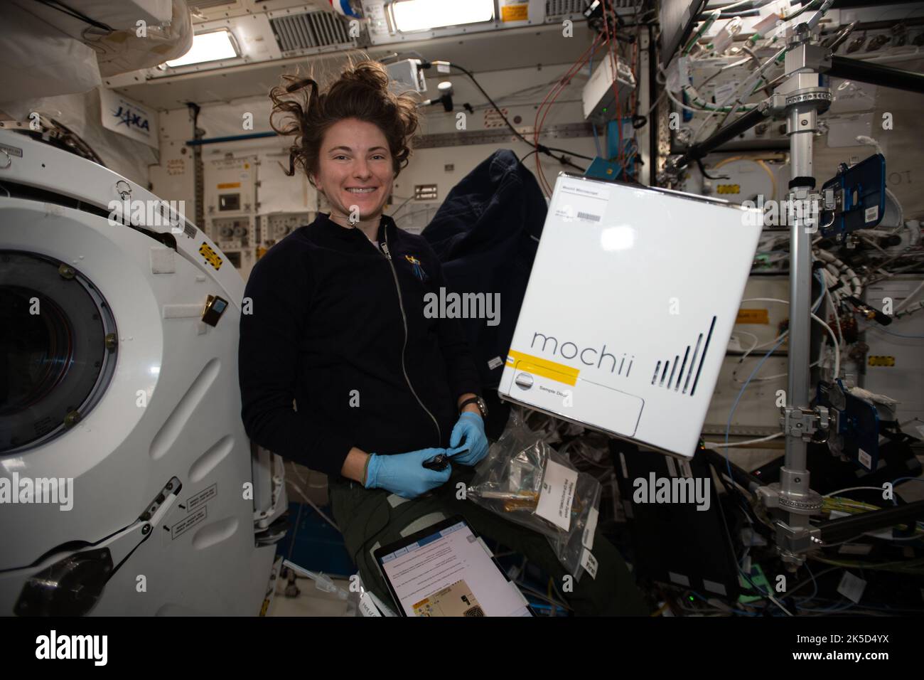 NASA astronaut Kayla Barron sets up the Mochii microscope. Mochii is a miniature scanning electron microscope (SEM) with spectroscopy to conduct real-time, on-site imaging and compositional measurements of particles on the International Space Station (ISS). Stock Photohttps://www.alamy.com/image-license-details/?v=1https://www.alamy.com/nasa-astronaut-kayla-barron-sets-up-the-mochii-microscope-mochii-is-a-miniature-scanning-electron-microscope-sem-with-spectroscopy-to-conduct-real-time-on-site-imaging-and-compositional-measurements-of-particles-on-the-international-space-station-iss-image485252878.html
RM2K5D4YX–NASA astronaut Kayla Barron sets up the Mochii microscope. Mochii is a miniature scanning electron microscope (SEM) with spectroscopy to conduct real-time, on-site imaging and compositional measurements of particles on the International Space Station (ISS).
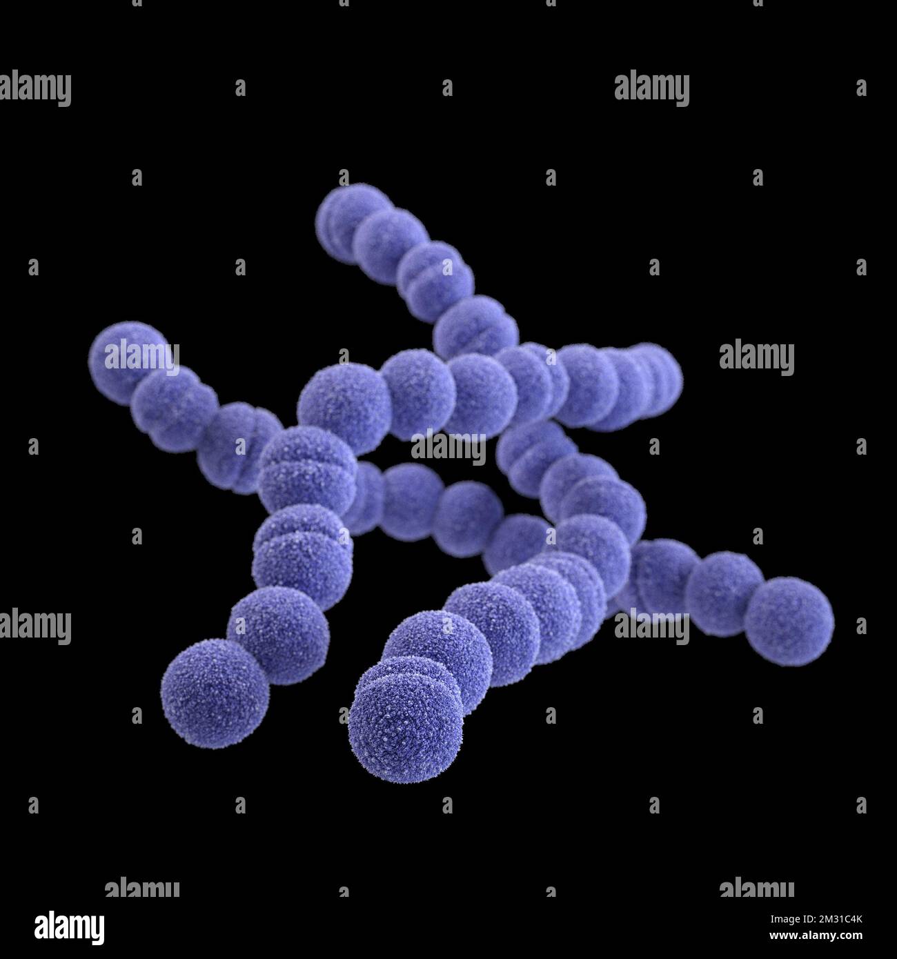 Group A streptococcus bacteria. STREP A Streptococcus pyogenes is a species of Gram-positive, aerotolerant bacteria in the genus Streptococcus. These bacteria are extracellular, and made up of non-motile and non-sporing cocci that tend to link in chains. This illustration depicted a 3D, computer-generated image, of a group of Gram-positive, Streptococcus pyogenes (group A Streptococcus) bacteria. The visualisation was based upon scanning electron microscopic (SEM) imagery. Optimised version of an image produced by the US Centers for Disease Control and Prevention / credit CDC /J.Oosthuizen Stock Photohttps://www.alamy.com/image-license-details/?v=1https://www.alamy.com/group-a-streptococcus-bacteria-strep-a-streptococcus-pyogenes-is-a-species-of-gram-positive-aerotolerant-bacteria-in-the-genus-streptococcus-these-bacteria-are-extracellular-and-made-up-of-non-motile-and-non-sporing-cocci-that-tend-to-link-in-chains-this-illustration-depicted-a-3d-computer-generated-image-of-a-group-of-gram-positive-streptococcus-pyogenes-group-a-streptococcus-bacteria-the-visualisation-was-based-upon-scanning-electron-microscopic-sem-imagery-optimised-version-of-an-image-produced-by-the-us-centers-for-disease-control-and-prevention-credit-cdc-joosthuizen-image500976131.html
RM2M31C4K–Group A streptococcus bacteria. STREP A Streptococcus pyogenes is a species of Gram-positive, aerotolerant bacteria in the genus Streptococcus. These bacteria are extracellular, and made up of non-motile and non-sporing cocci that tend to link in chains. This illustration depicted a 3D, computer-generated image, of a group of Gram-positive, Streptococcus pyogenes (group A Streptococcus) bacteria. The visualisation was based upon scanning electron microscopic (SEM) imagery. Optimised version of an image produced by the US Centers for Disease Control and Prevention / credit CDC /J.Oosthuizen
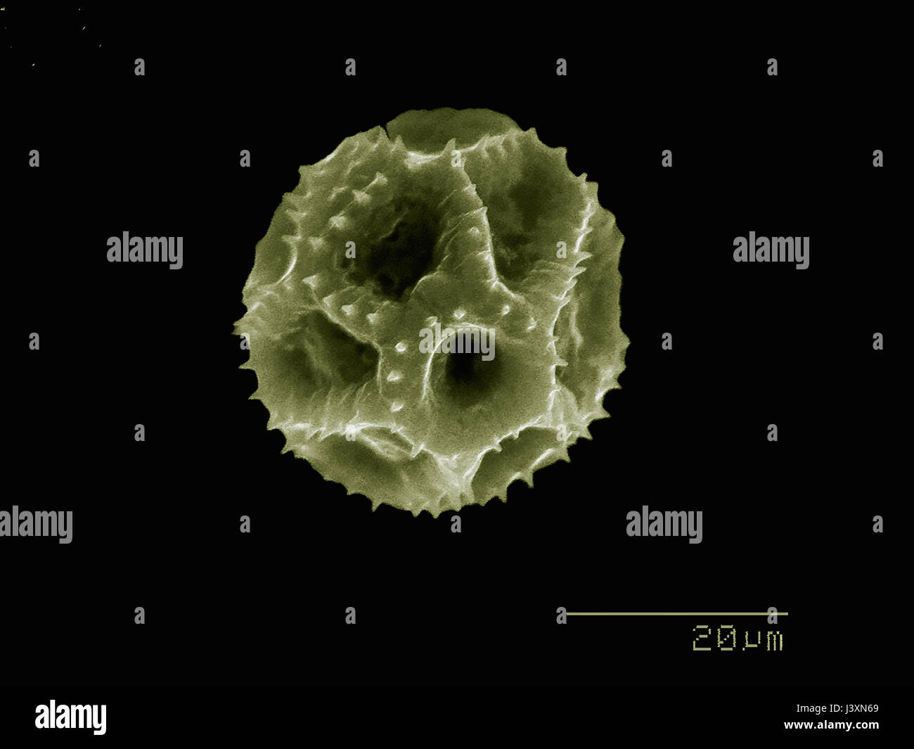 Dandelion pollen imaged in a scanning electron microscope Stock Photohttps://www.alamy.com/image-license-details/?v=1https://www.alamy.com/stock-photo-dandelion-pollen-imaged-in-a-scanning-electron-microscope-140114305.html
RFJ3XN69–Dandelion pollen imaged in a scanning electron microscope
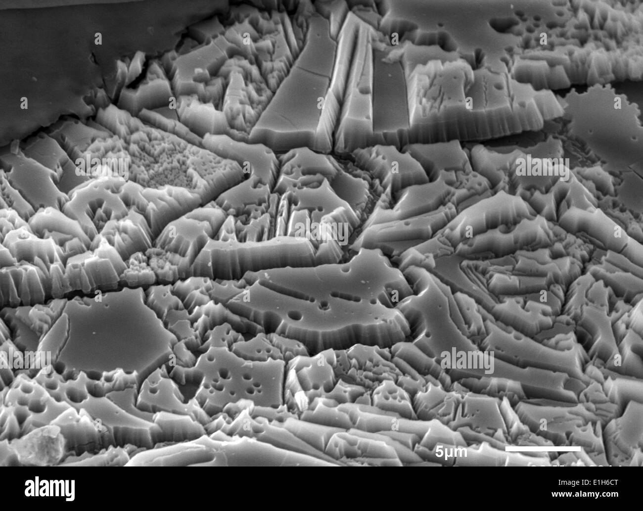 Scanning electron micrograph image of iron oxide formations with sulphur and chlorine present Stock Photohttps://www.alamy.com/image-license-details/?v=1https://www.alamy.com/scanning-electron-micrograph-image-of-iron-oxide-formations-with-sulphur-image69834376.html
RFE1H6CT–Scanning electron micrograph image of iron oxide formations with sulphur and chlorine present
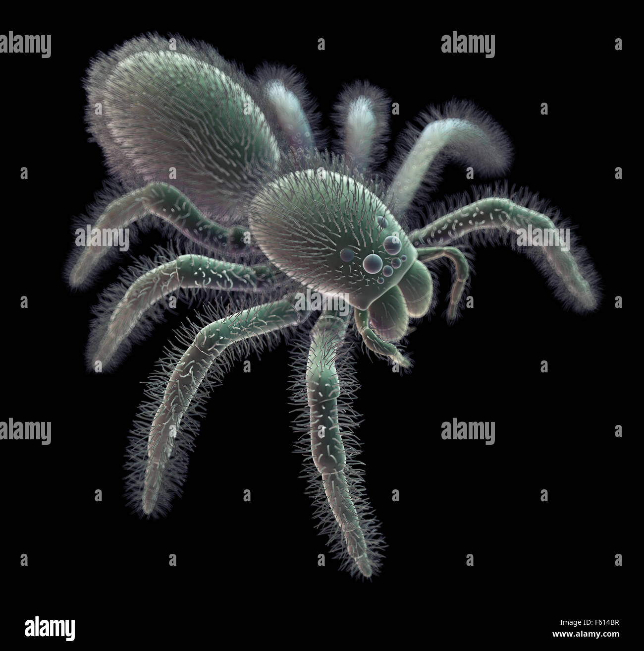 SEM style illustration of a spider Stock Photohttps://www.alamy.com/image-license-details/?v=1https://www.alamy.com/stock-photo-sem-style-illustration-of-a-spider-89765195.html
RFF614BR–SEM style illustration of a spider
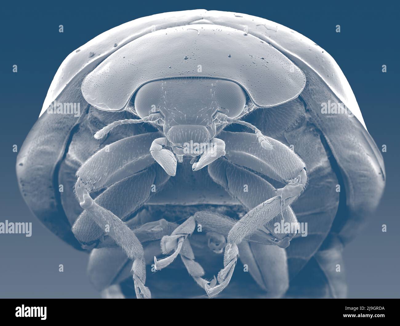 SEM Scanning Electron Microscope image of a Ladybug Ladybird microscopic Stock Photohttps://www.alamy.com/image-license-details/?v=1https://www.alamy.com/sem-scanning-electron-microscope-image-of-a-ladybug-ladybird-microscopic-image470581478.html
RM2J9GRDA–SEM Scanning Electron Microscope image of a Ladybug Ladybird microscopic
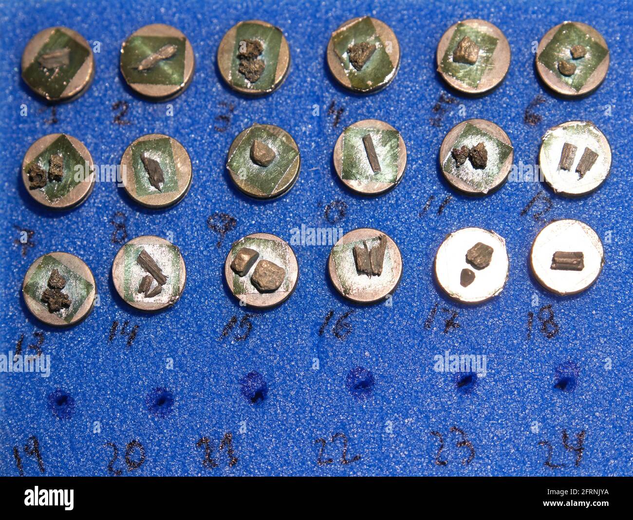 Scanning electron microscope samples on specimen mounts. SEM pins to analyze. Stock Photohttps://www.alamy.com/image-license-details/?v=1https://www.alamy.com/scanning-electron-microscope-samples-on-specimen-mounts-sem-pins-to-analyze-image427661790.html
RF2FRNJYA–Scanning electron microscope samples on specimen mounts. SEM pins to analyze.
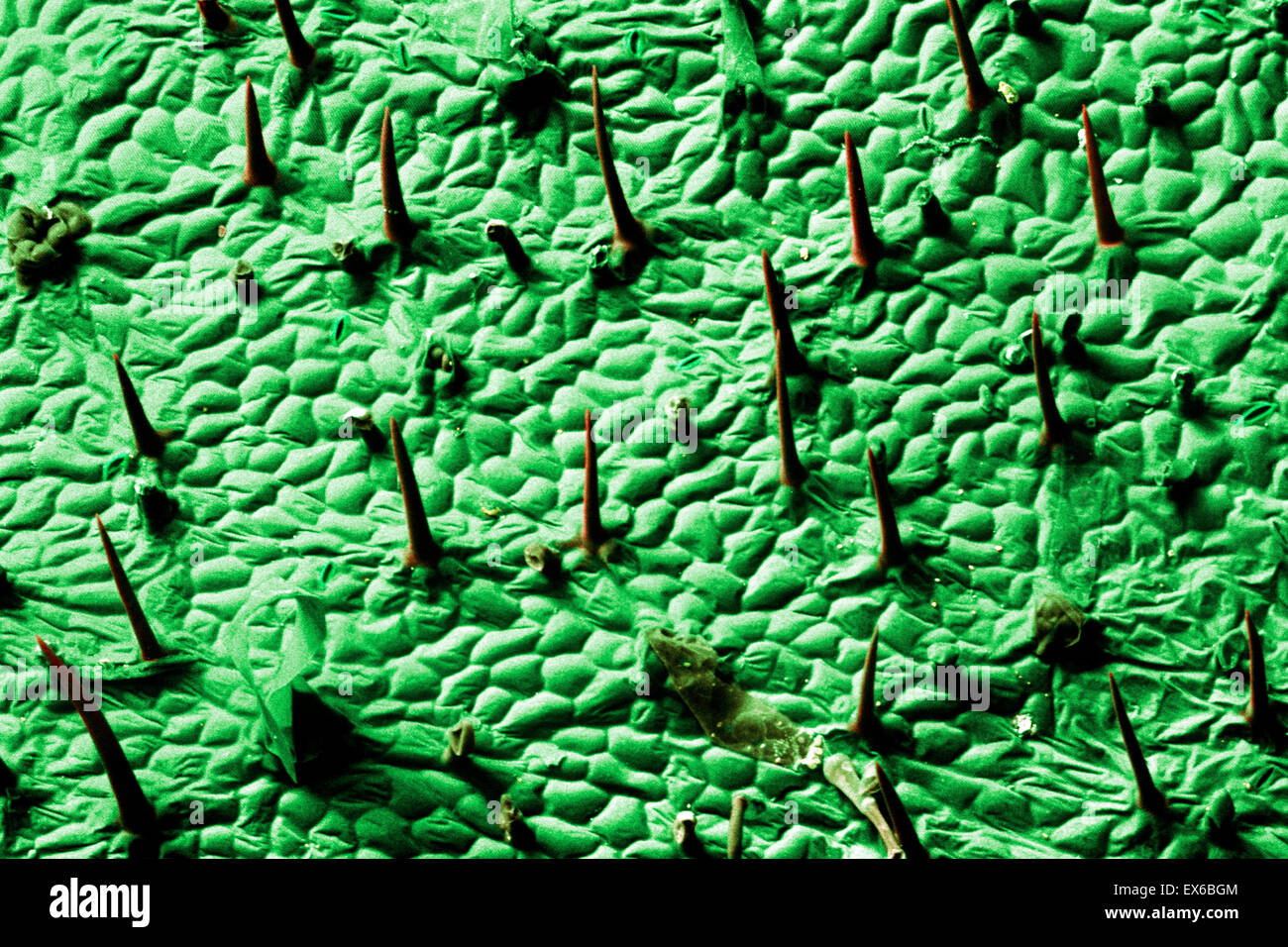 Plant trichomes, SEM Stock Photohttps://www.alamy.com/image-license-details/?v=1https://www.alamy.com/stock-photo-plant-trichomes-sem-84963332.html
RMEX6BGM–Plant trichomes, SEM
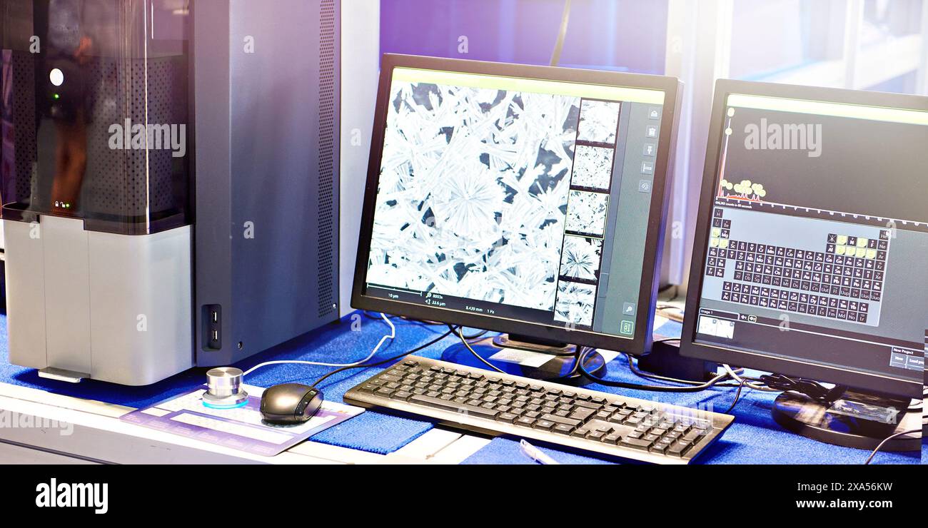 Scanning electron microscope with EMF microanalysis Stock Photohttps://www.alamy.com/image-license-details/?v=1https://www.alamy.com/scanning-electron-microscope-with-emf-microanalysis-image608624461.html
RF2XA56KW–Scanning electron microscope with EMF microanalysis
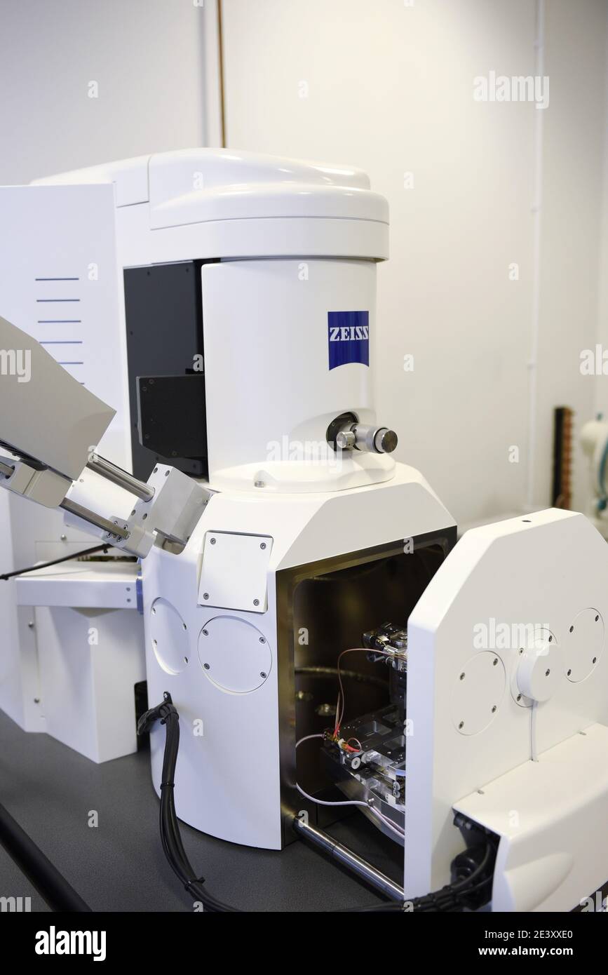 Zeiss EVO 15 Scanning Electron Microscope scanning station in a science lab. It is flexible variable pressure scanning electron microscope or SEM with Stock Photohttps://www.alamy.com/image-license-details/?v=1https://www.alamy.com/zeiss-evo-15-scanning-electron-microscope-scanning-station-in-a-science-lab-it-is-flexible-variable-pressure-scanning-electron-microscope-or-sem-with-image398273960.html
RM2E3XXE0–Zeiss EVO 15 Scanning Electron Microscope scanning station in a science lab. It is flexible variable pressure scanning electron microscope or SEM with
 Surgical needle, SEM Stock Photohttps://www.alamy.com/image-license-details/?v=1https://www.alamy.com/surgical-needle-sem-image341959483.html
RF2AT9GPK–Surgical needle, SEM
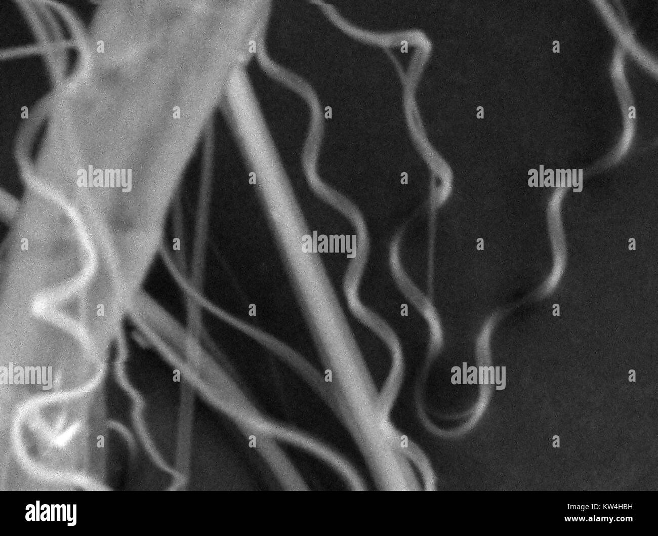 Scanning electron microscope (SEM) micrograph showing spider's silk, at a magnification of 5000x, 2016. Stock Photohttps://www.alamy.com/image-license-details/?v=1https://www.alamy.com/stock-photo-scanning-electron-microscope-sem-micrograph-showing-spiders-silk-at-170361173.html
RMKW4HBH–Scanning electron microscope (SEM) micrograph showing spider's silk, at a magnification of 5000x, 2016.
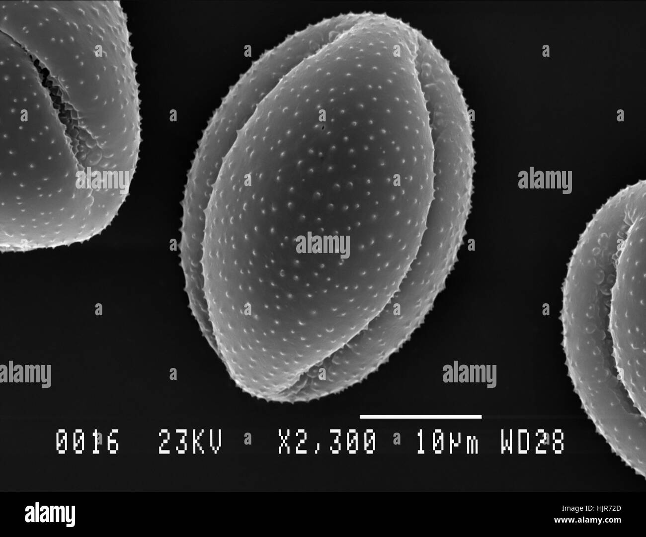 Scanning electron micrograph of one pollen particle from Lesser Celandine flower. Nottingham, UK. April. Stock Photohttps://www.alamy.com/image-license-details/?v=1https://www.alamy.com/stock-photo-scanning-electron-micrograph-of-one-pollen-particle-from-lesser-celandine-132046837.html
RFHJR72D–Scanning electron micrograph of one pollen particle from Lesser Celandine flower. Nottingham, UK. April.
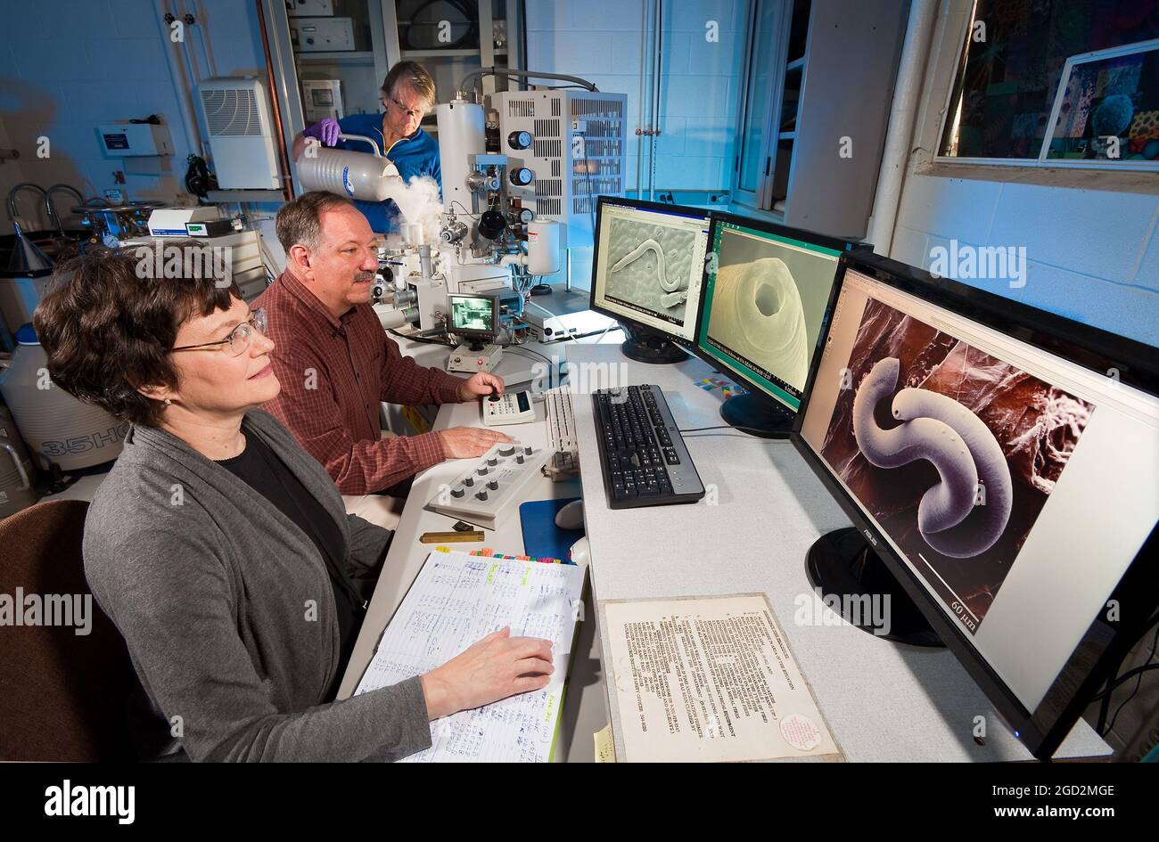 Workers use a low temperature scanning electron microscope (LT-SEM) to view anatomical structures needed to identify nematodes like the Parasitorhabditis nematode associated with pine trees and bark beetles on April 19, 2012. Stock Photohttps://www.alamy.com/image-license-details/?v=1https://www.alamy.com/workers-use-a-low-temperature-scanning-electron-microscope-lt-sem-to-view-anatomical-structures-needed-to-identify-nematodes-like-the-parasitorhabditis-nematode-associated-with-pine-trees-and-bark-beetles-on-april-19-2012-image438309774.html
RM2GD2MGE–Workers use a low temperature scanning electron microscope (LT-SEM) to view anatomical structures needed to identify nematodes like the Parasitorhabditis nematode associated with pine trees and bark beetles on April 19, 2012.
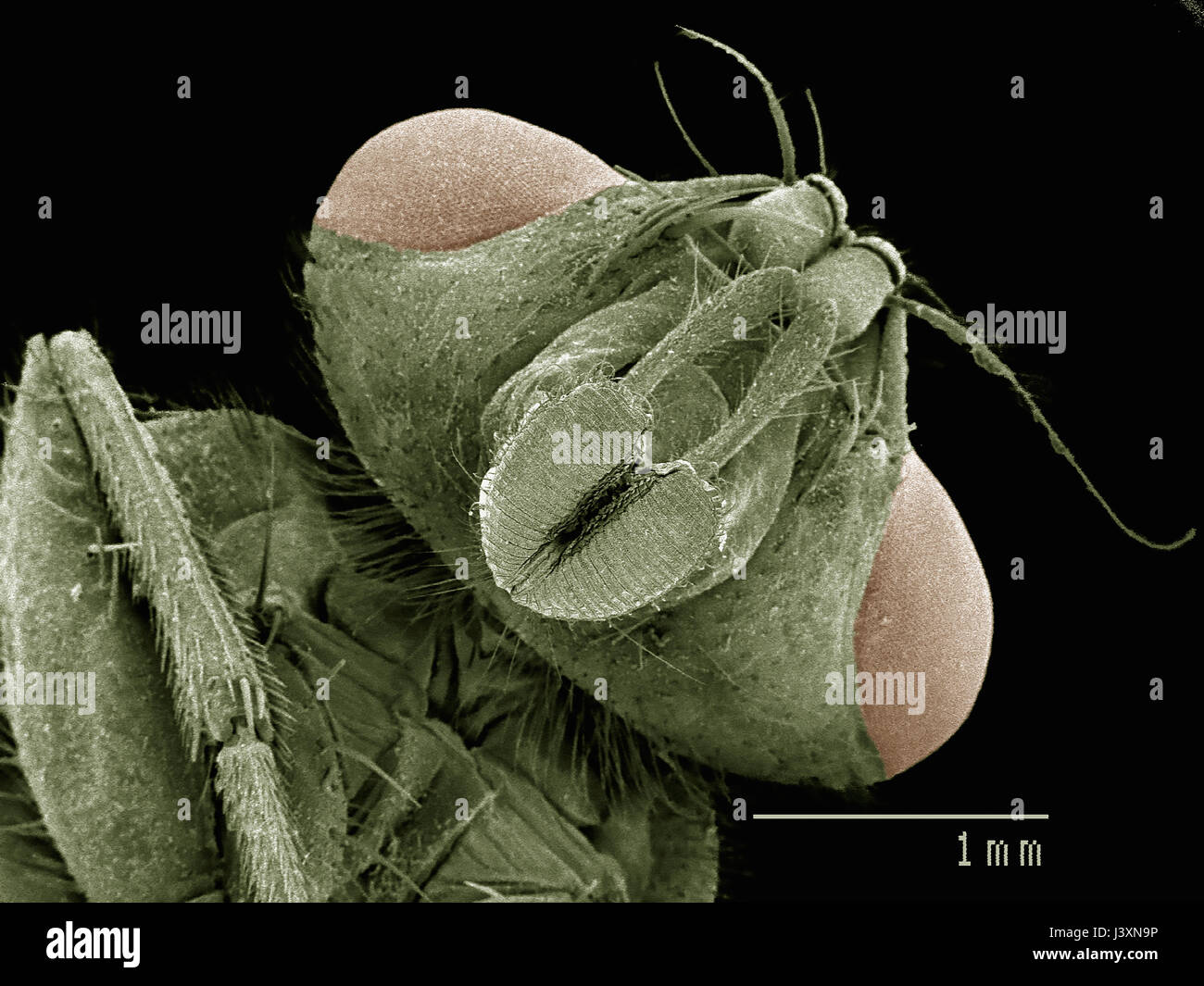 Head of house fly (Muscidae) imaged in a scanning electron microscope Stock Photohttps://www.alamy.com/image-license-details/?v=1https://www.alamy.com/stock-photo-head-of-house-fly-muscidae-imaged-in-a-scanning-electron-microscope-140114402.html
RFJ3XN9P–Head of house fly (Muscidae) imaged in a scanning electron microscope
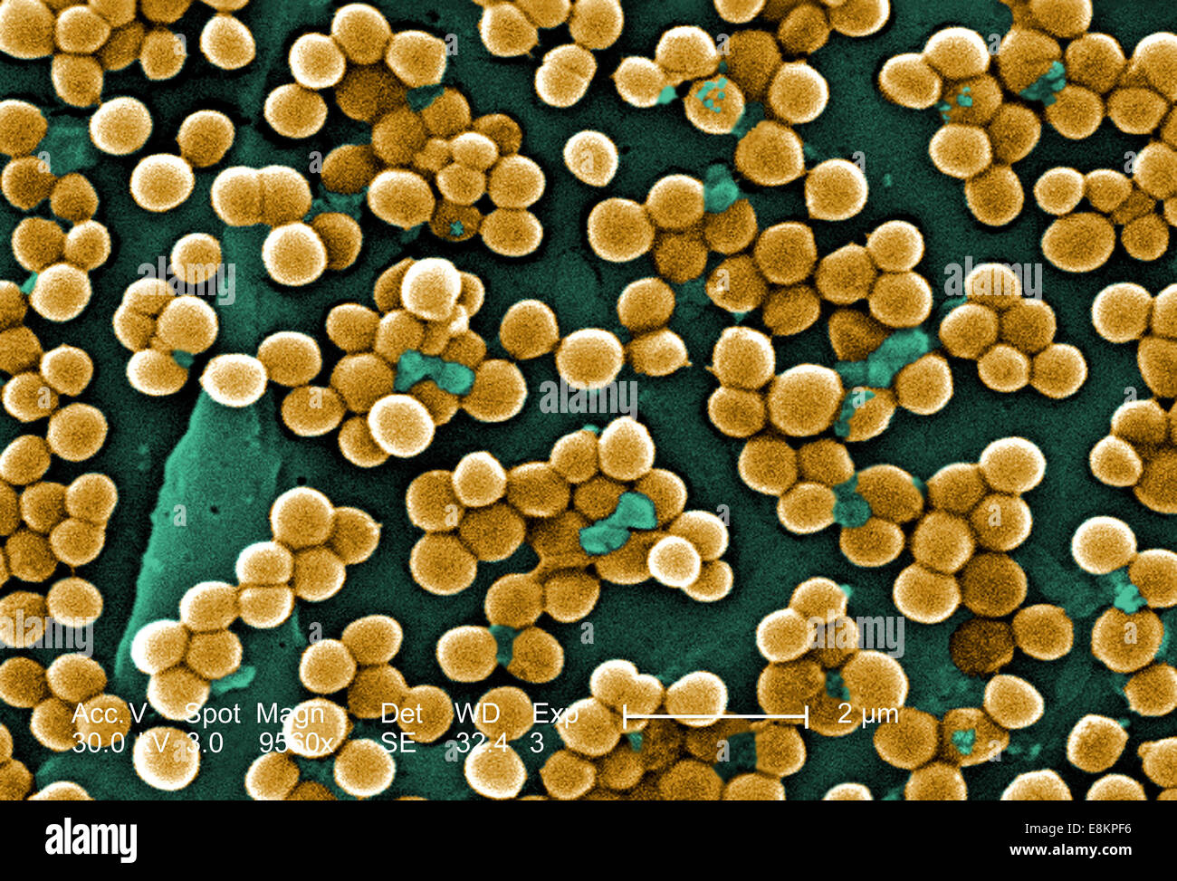 This scanning electron micrograph (SEM) depicted numerous clumps of methicillin-resistant Staphylococcus aureus bacteria, Stock Photohttps://www.alamy.com/image-license-details/?v=1https://www.alamy.com/stock-photo-this-scanning-electron-micrograph-sem-depicted-numerous-clumps-of-74193482.html
RME8KPF6–This scanning electron micrograph (SEM) depicted numerous clumps of methicillin-resistant Staphylococcus aureus bacteria,
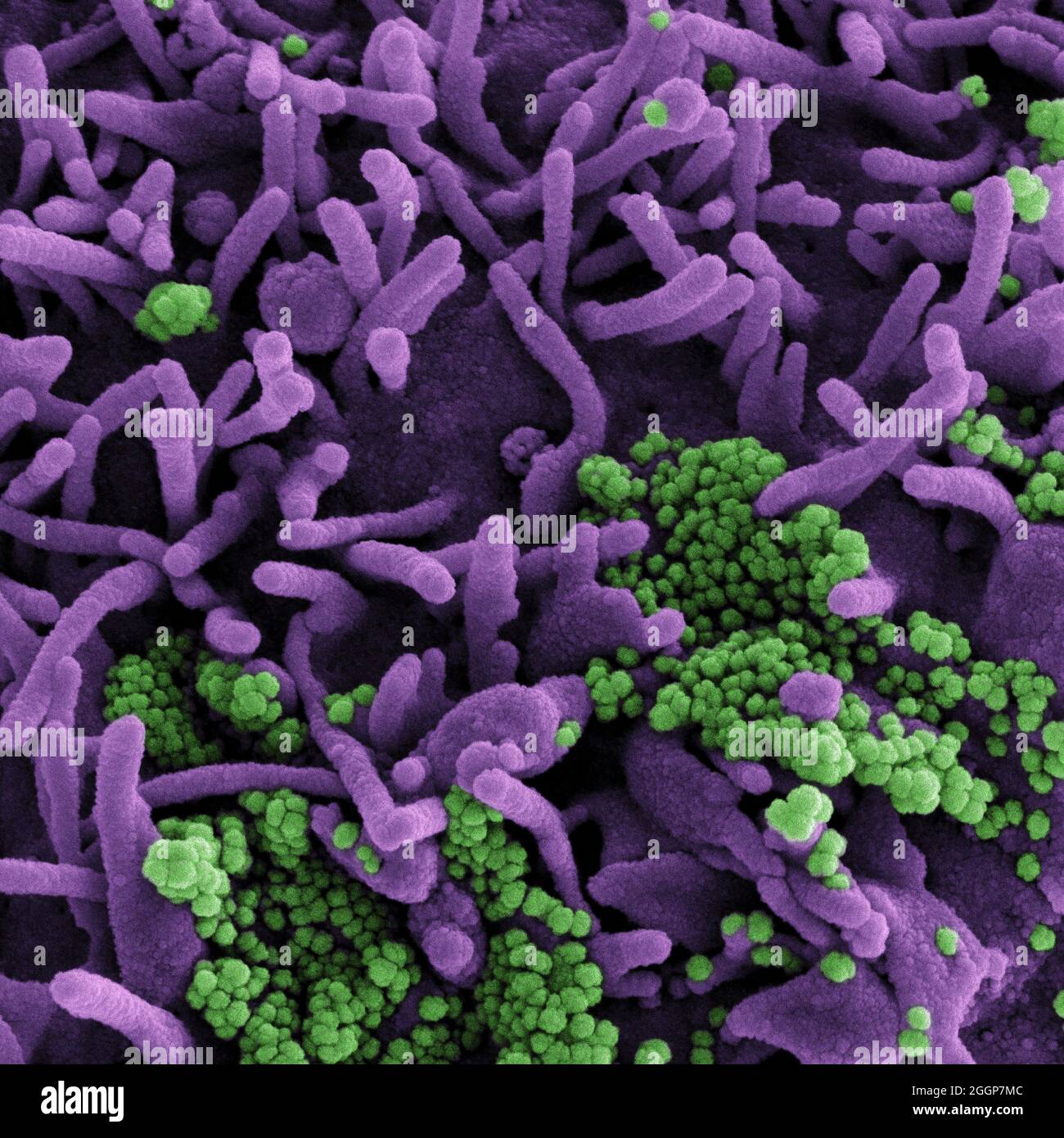 Colorized scanning electron micrograph of a cell (purple) infected with SARS-CoV-2 virus particles (green), isolated from a patient sample. Stock Photohttps://www.alamy.com/image-license-details/?v=1https://www.alamy.com/colorized-scanning-electron-micrograph-of-a-cell-purple-infected-with-sars-cov-2-virus-particles-green-isolated-from-a-patient-sample-image440582700.html
RM2GGP7MC–Colorized scanning electron micrograph of a cell (purple) infected with SARS-CoV-2 virus particles (green), isolated from a patient sample.
 3D structure of a melanoma cell derived by ion abrasion scanning electron microscopy. Stock Photohttps://www.alamy.com/image-license-details/?v=1https://www.alamy.com/stock-photo-3d-structure-of-a-melanoma-cell-derived-by-ion-abrasion-scanning-electron-76547748.html
RFECF1C4–3D structure of a melanoma cell derived by ion abrasion scanning electron microscopy.
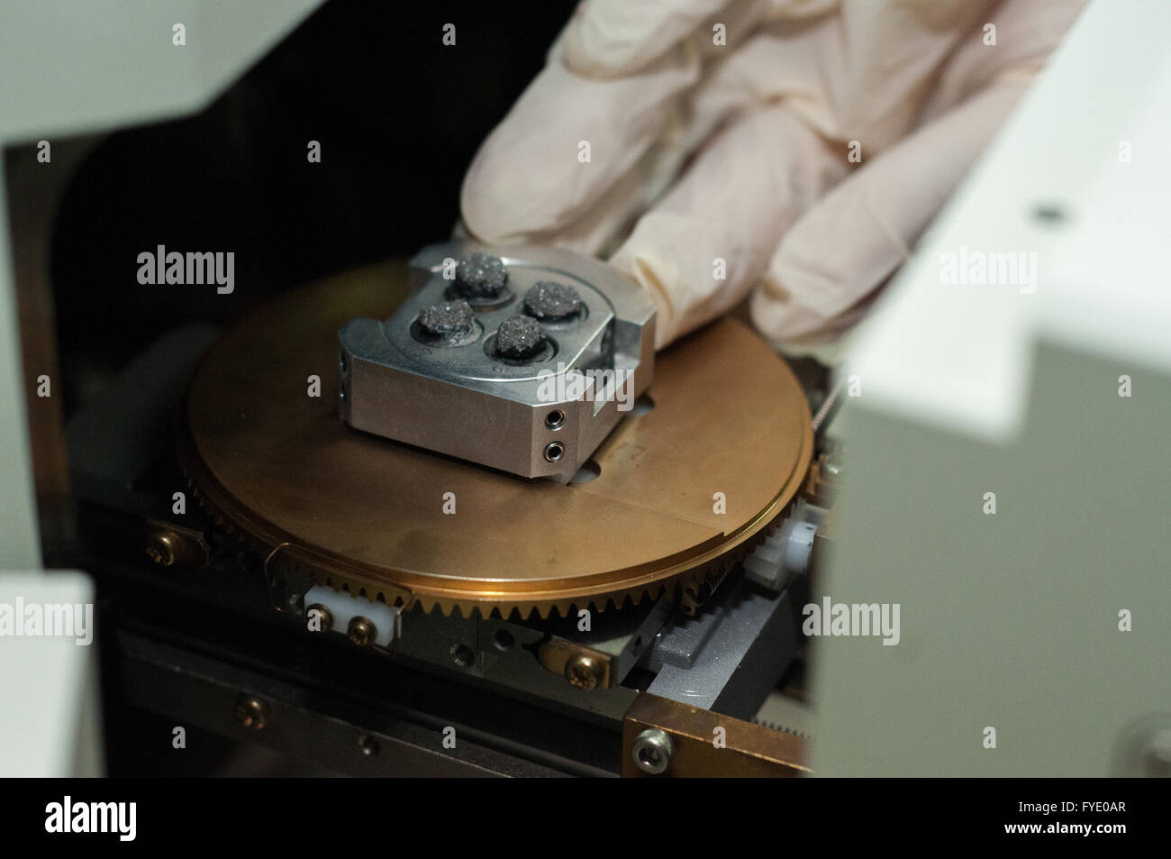 Jakarta Selatan, Indonesia. 26th Apr, 2016. The researcher inserts inside the Scanning Electron Microscope 'SEM' machine the rock holder. SEM is used to analyze rock minerals up to 1µm. It can zoom 300.000x than usual polarize microscope. © Anton Raharjo/Pacific Press/Alamy Live News Stock Photohttps://www.alamy.com/image-license-details/?v=1https://www.alamy.com/stock-photo-jakarta-selatan-indonesia-26th-apr-2016-the-researcher-inserts-inside-102955183.html
RMFYE0AR–Jakarta Selatan, Indonesia. 26th Apr, 2016. The researcher inserts inside the Scanning Electron Microscope 'SEM' machine the rock holder. SEM is used to analyze rock minerals up to 1µm. It can zoom 300.000x than usual polarize microscope. © Anton Raharjo/Pacific Press/Alamy Live News
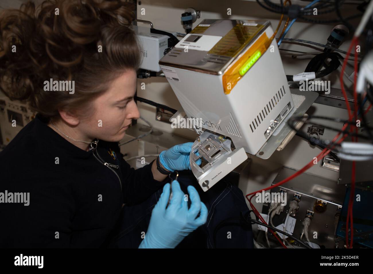 NASA astronaut Kayla Barron sets up the Mochii microscope. Mochii is a miniature scanning electron microscope (SEM) with spectroscopy to conduct real-time, on-site imaging and compositional measurements of particles on the International Space Station (ISS). Stock Photohttps://www.alamy.com/image-license-details/?v=1https://www.alamy.com/nasa-astronaut-kayla-barron-sets-up-the-mochii-microscope-mochii-is-a-miniature-scanning-electron-microscope-sem-with-spectroscopy-to-conduct-real-time-on-site-imaging-and-compositional-measurements-of-particles-on-the-international-space-station-iss-image485252511.html
RM2K5D4ER–NASA astronaut Kayla Barron sets up the Mochii microscope. Mochii is a miniature scanning electron microscope (SEM) with spectroscopy to conduct real-time, on-site imaging and compositional measurements of particles on the International Space Station (ISS).
 A cluster of barrel-shaped, Clostridium perfringens bacteria. False-colour interpreted artistic rendition based on scanning electron microscopic (SEM) imagery. Stock Photohttps://www.alamy.com/image-license-details/?v=1https://www.alamy.com/a-cluster-of-barrel-shaped-clostridium-perfringens-bacteria-false-colour-interpreted-artistic-rendition-based-on-scanning-electron-microscopic-sem-imagery-image471319964.html
RM2JAPDBT–A cluster of barrel-shaped, Clostridium perfringens bacteria. False-colour interpreted artistic rendition based on scanning electron microscopic (SEM) imagery.
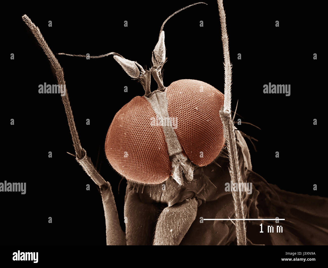 Head of long legged fly (dolichopodiae) imaged in a scanning electron microscope Stock Photohttps://www.alamy.com/image-license-details/?v=1https://www.alamy.com/stock-photo-head-of-long-legged-fly-dolichopodiae-imaged-in-a-scanning-electron-140114390.html
RFJ3XN9A–Head of long legged fly (dolichopodiae) imaged in a scanning electron microscope
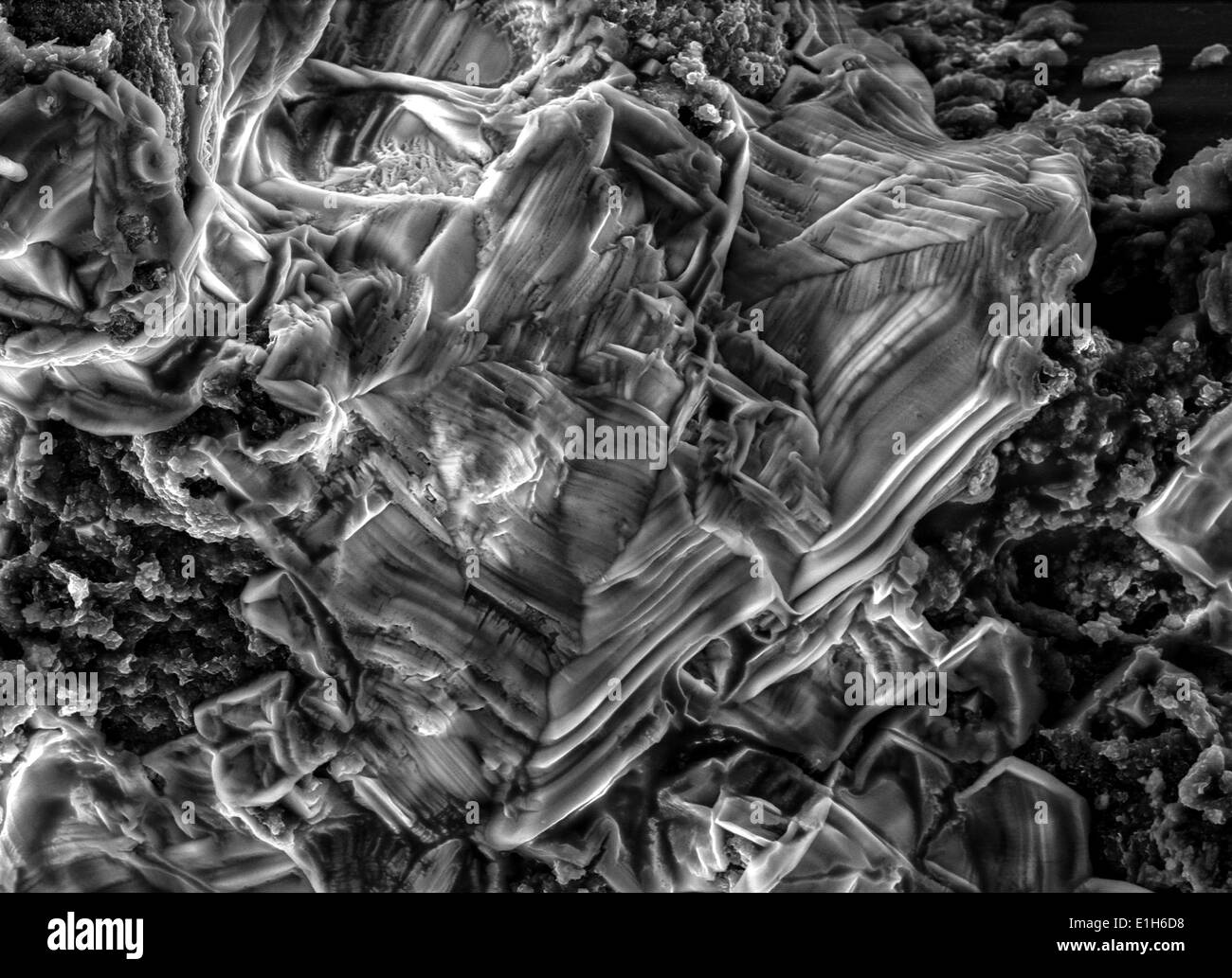 Scanning electron micrograph image of iron oxide formations with chlorine and sulphur present Stock Photohttps://www.alamy.com/image-license-details/?v=1https://www.alamy.com/scanning-electron-micrograph-image-of-iron-oxide-formations-with-chlorine-image69834388.html
RFE1H6D8–Scanning electron micrograph image of iron oxide formations with chlorine and sulphur present
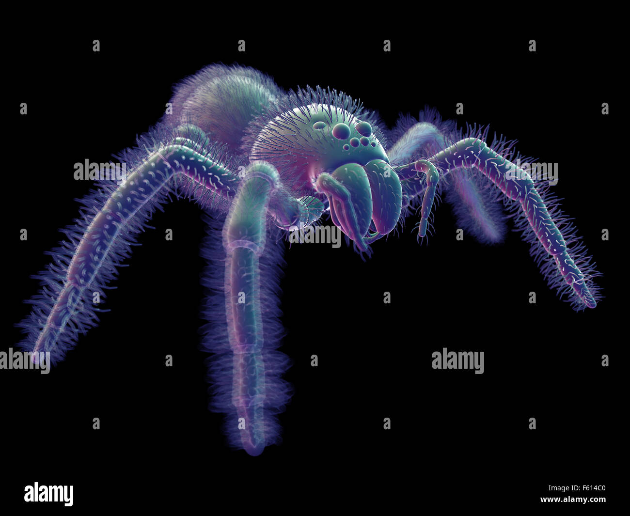 SEM style illustration of a spider Stock Photohttps://www.alamy.com/image-license-details/?v=1https://www.alamy.com/stock-photo-sem-style-illustration-of-a-spider-89765200.html
RFF614C0–SEM style illustration of a spider
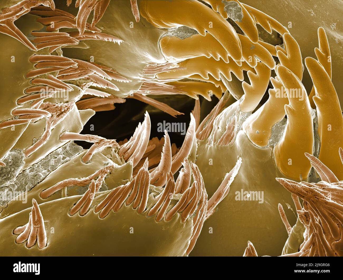 SEM Scanning Electron Microscope image of a Sandhopper, Sand Flea, amphipod Stock Photohttps://www.alamy.com/image-license-details/?v=1https://www.alamy.com/sem-scanning-electron-microscope-image-of-a-sandhopper-sand-flea-amphipod-image470581558.html
RM2J9GRG6–SEM Scanning Electron Microscope image of a Sandhopper, Sand Flea, amphipod
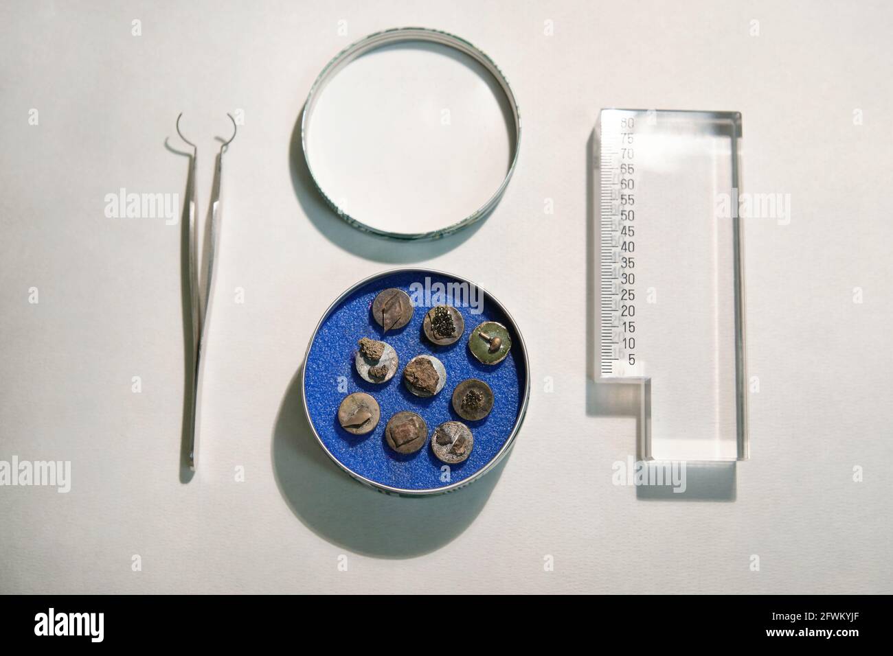 Scanning electron microscope samples on specimen mounts, tweezers and a ruler. SEM pins to analyze. Stock Photohttps://www.alamy.com/image-license-details/?v=1https://www.alamy.com/scanning-electron-microscope-samples-on-specimen-mounts-tweezers-and-a-ruler-sem-pins-to-analyze-image428854007.html
RF2FWKYJF–Scanning electron microscope samples on specimen mounts, tweezers and a ruler. SEM pins to analyze.
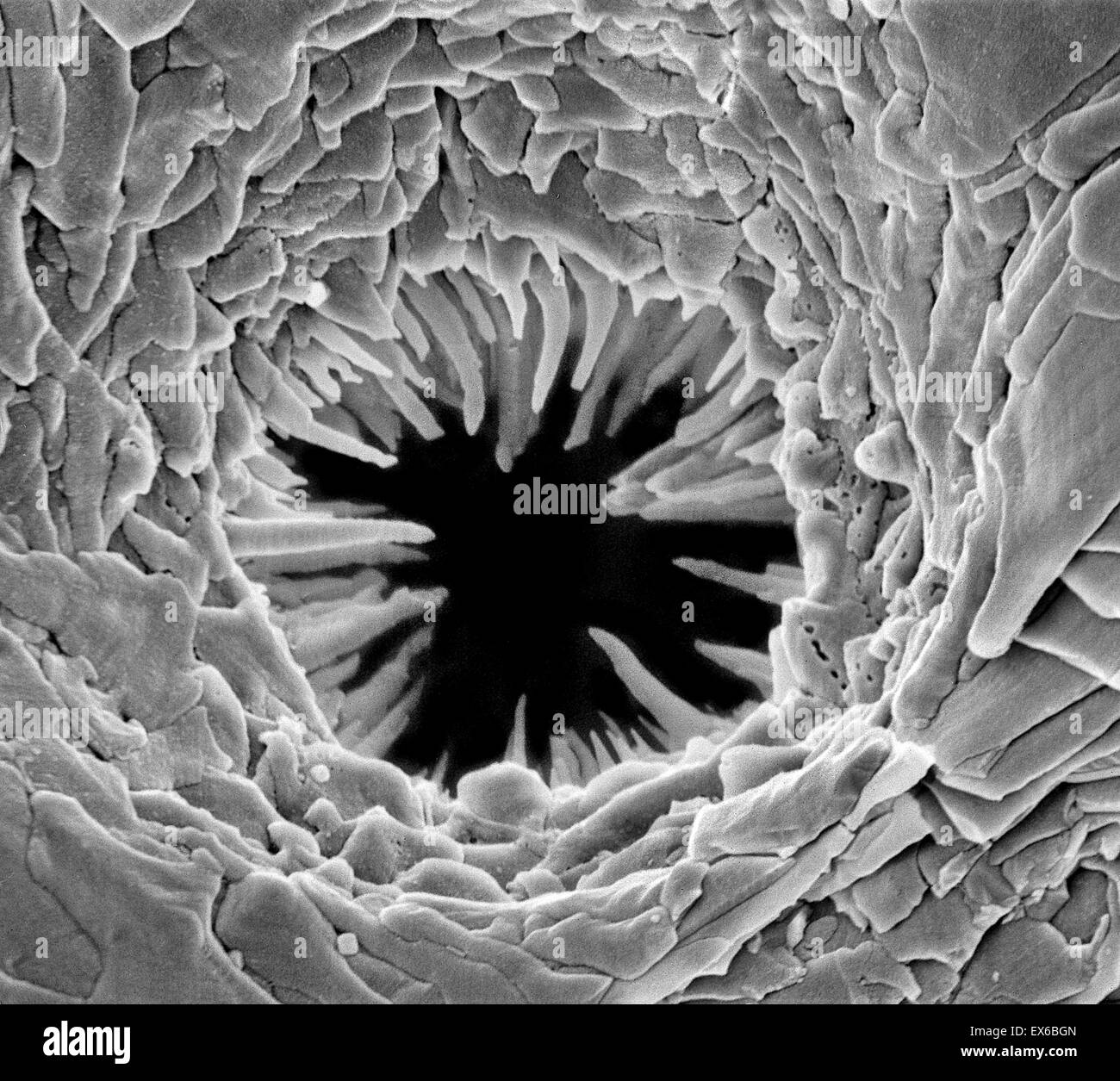 Bryozoan pore, SEM Stock Photohttps://www.alamy.com/image-license-details/?v=1https://www.alamy.com/stock-photo-bryozoan-pore-sem-84963333.html
RMEX6BGN–Bryozoan pore, SEM
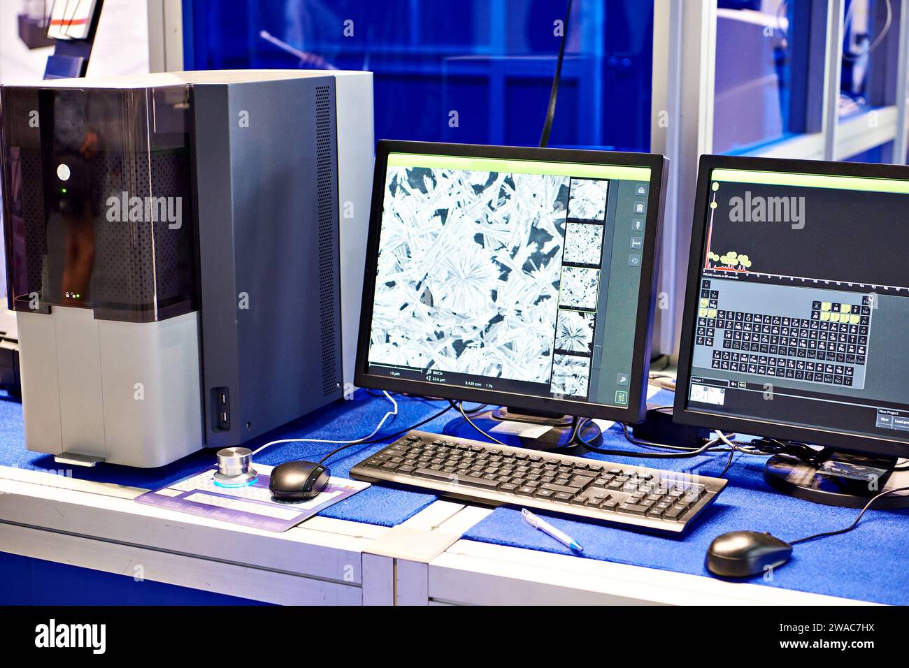 Scanning electron microscope with EMF microanalysis Stock Photohttps://www.alamy.com/image-license-details/?v=1https://www.alamy.com/scanning-electron-microscope-with-emf-microanalysis-image591568486.html
RF2WAC7HX–Scanning electron microscope with EMF microanalysis
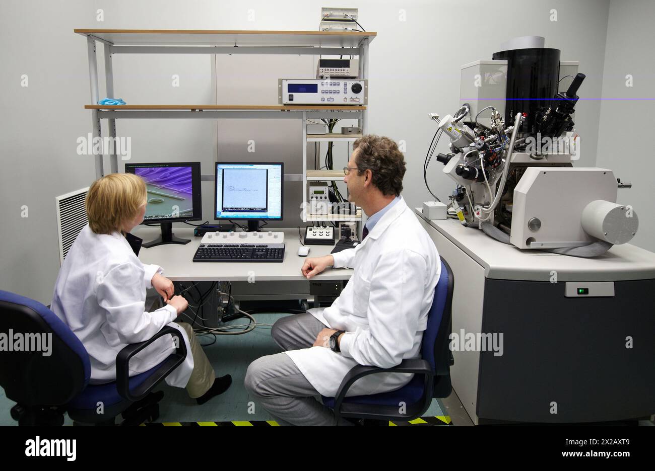 Discussing on SEM images, Analysis of nanostructures and nanodevices, Environmental scanning electron microscopy Laboratory, ESEM, Microscope Quanta T Stock Photohttps://www.alamy.com/image-license-details/?v=1https://www.alamy.com/discussing-on-sem-images-analysis-of-nanostructures-and-nanodevices-environmental-scanning-electron-microscopy-laboratory-esem-microscope-quanta-t-image603832777.html
RM2X2AXT9–Discussing on SEM images, Analysis of nanostructures and nanodevices, Environmental scanning electron microscopy Laboratory, ESEM, Microscope Quanta T
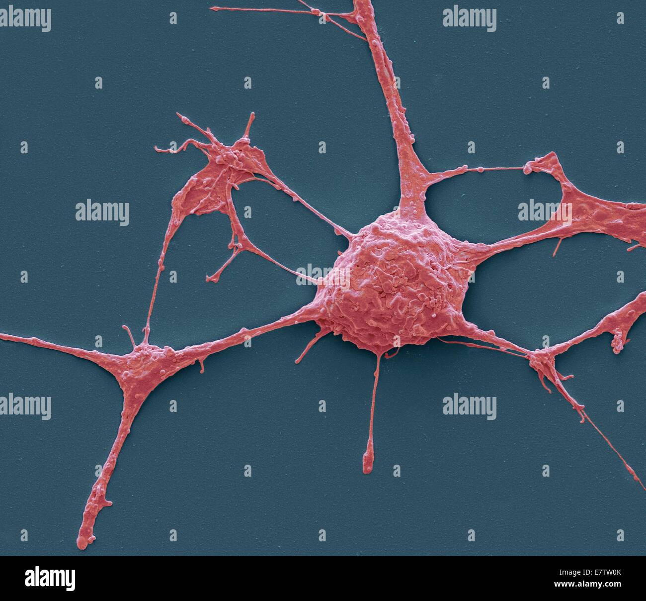 Neurone. Scanning electron micrograph (SEM) of a PC12 neurone in culture.The PC12 cell line, developed from a pheochromocytoma tumor of the rat adrenal medulla, has become a premiere model for the study of neuronal differentiation. When treated in culture Stock Photohttps://www.alamy.com/image-license-details/?v=1https://www.alamy.com/stock-photo-neurone-scanning-electron-micrograph-sem-of-a-pc12-neurone-in-culturethe-73690531.html
RFE7TW0K–Neurone. Scanning electron micrograph (SEM) of a PC12 neurone in culture.The PC12 cell line, developed from a pheochromocytoma tumor of the rat adrenal medulla, has become a premiere model for the study of neuronal differentiation. When treated in culture
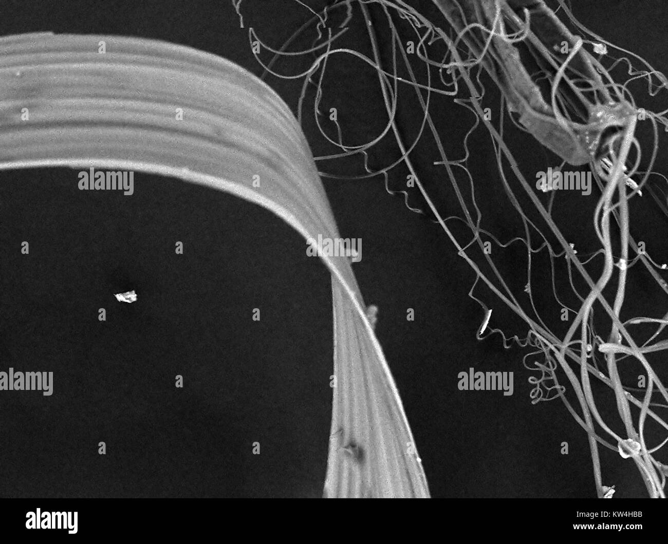 Scanning electron microscope (SEM) micrograph showing spider's silk, including thread, hydrogel and nano-fibril silk types, at a magnification of 800x, 2016. Stock Photohttps://www.alamy.com/image-license-details/?v=1https://www.alamy.com/stock-photo-scanning-electron-microscope-sem-micrograph-showing-spiders-silk-including-170361167.html
RMKW4HBB–Scanning electron microscope (SEM) micrograph showing spider's silk, including thread, hydrogel and nano-fibril silk types, at a magnification of 800x, 2016.
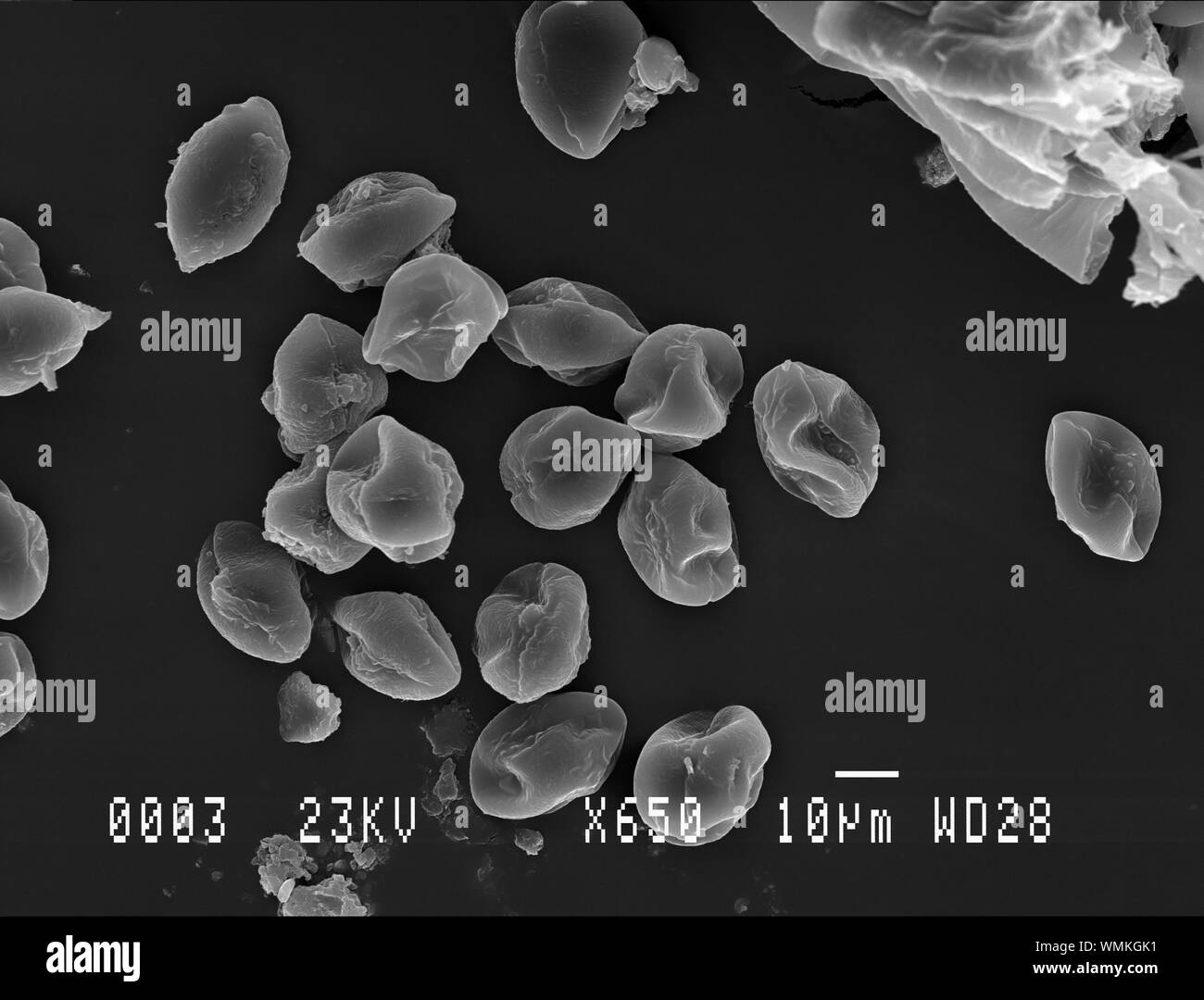 Horse chestnut pollen under electron microscope Stock Photohttps://www.alamy.com/image-license-details/?v=1https://www.alamy.com/horse-chestnut-pollen-under-electron-microscope-image270878805.html
RFWMKGK1–Horse chestnut pollen under electron microscope
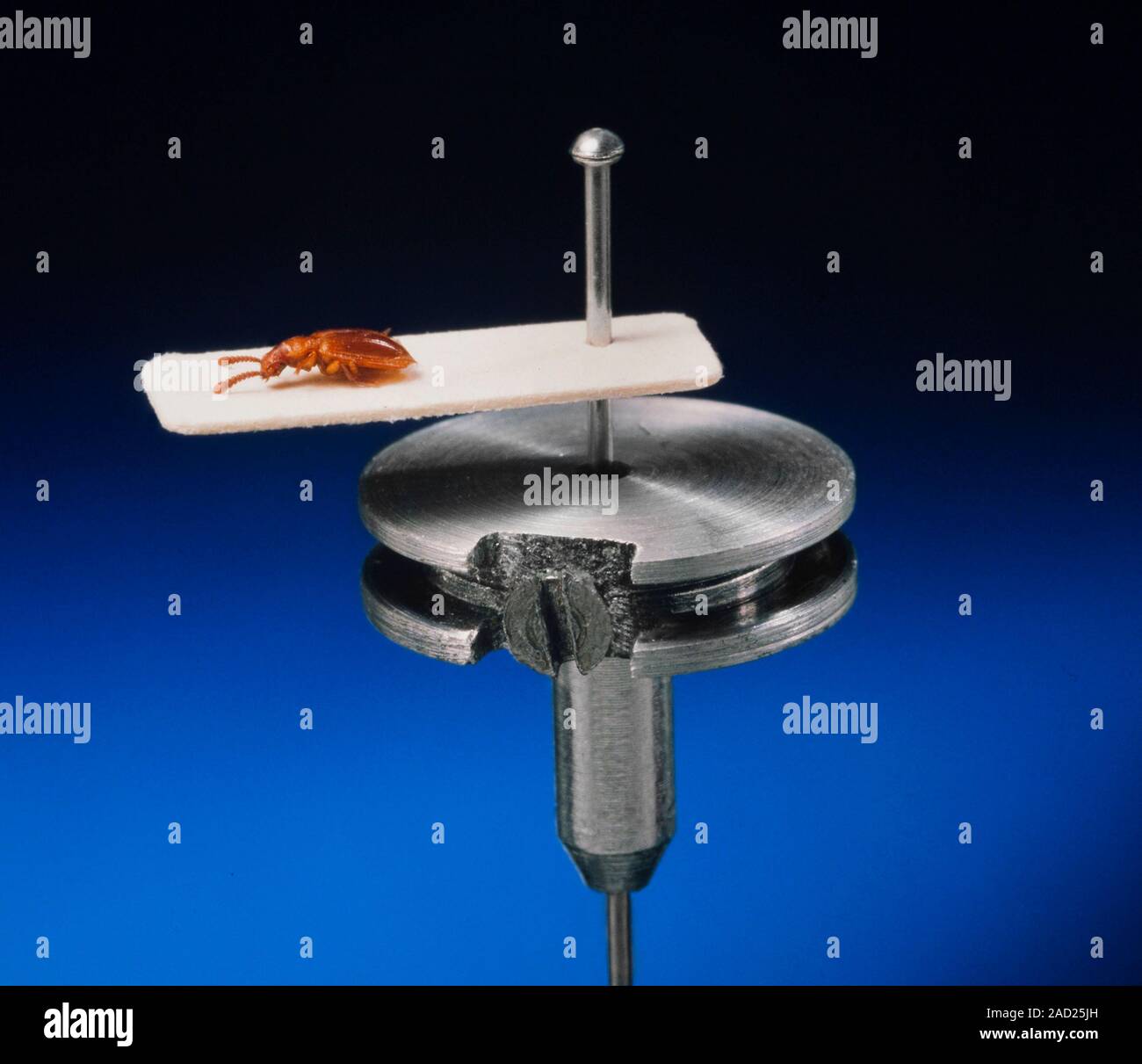 Entomological pinned specimen mounted on a SEM specimen stub Stock Photohttps://www.alamy.com/image-license-details/?v=1https://www.alamy.com/entomological-pinned-specimen-mounted-on-a-sem-specimen-stub-image335035865.html
RM2AD25JH–Entomological pinned specimen mounted on a SEM specimen stub
RFE0AC9M–SEM image of SiO2 Silicon Dioxide empty balls, coated with gold and imaged in scanning electron microscope
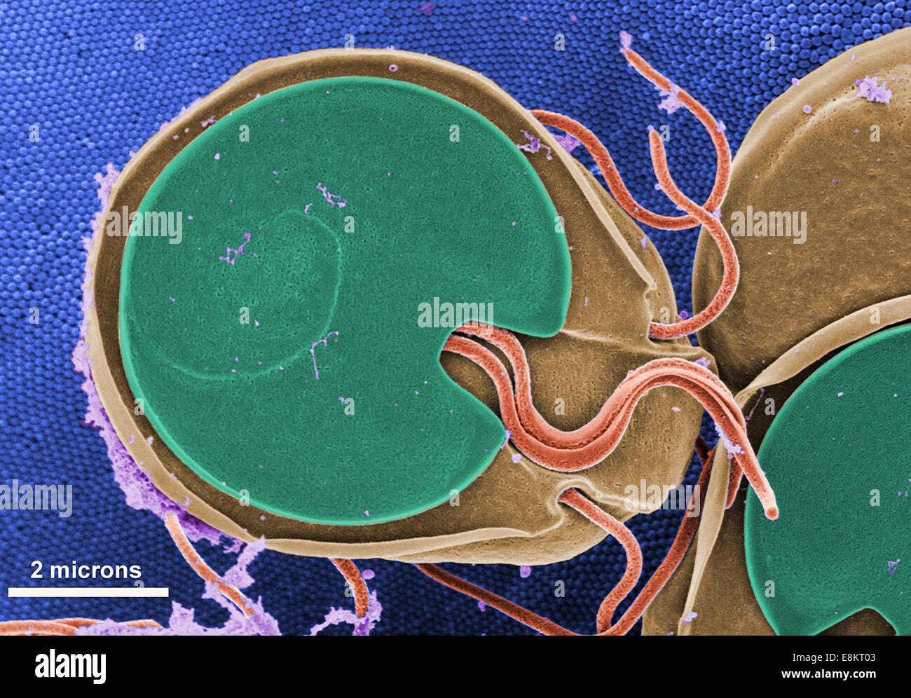 This scanning electron micrograph (SEM) revealed ventral surface of Giardia muris trophozoite that had settled atop mucosal Stock Photohttps://www.alamy.com/image-license-details/?v=1https://www.alamy.com/stock-photo-this-scanning-electron-micrograph-sem-revealed-ventral-surface-of-74194627.html
RME8KT03–This scanning electron micrograph (SEM) revealed ventral surface of Giardia muris trophozoite that had settled atop mucosal
 Colorized scanning electron micrograph of a cell (green) heavily infected with SARS-CoV-2 virus particles, isolated from a patient sample. Stock Photohttps://www.alamy.com/image-license-details/?v=1https://www.alamy.com/colorized-scanning-electron-micrograph-of-a-cell-green-heavily-infected-with-sars-cov-2-virus-particles-isolated-from-a-patient-sample-image440582705.html
RM2GGP7MH–Colorized scanning electron micrograph of a cell (green) heavily infected with SARS-CoV-2 virus particles, isolated from a patient sample.
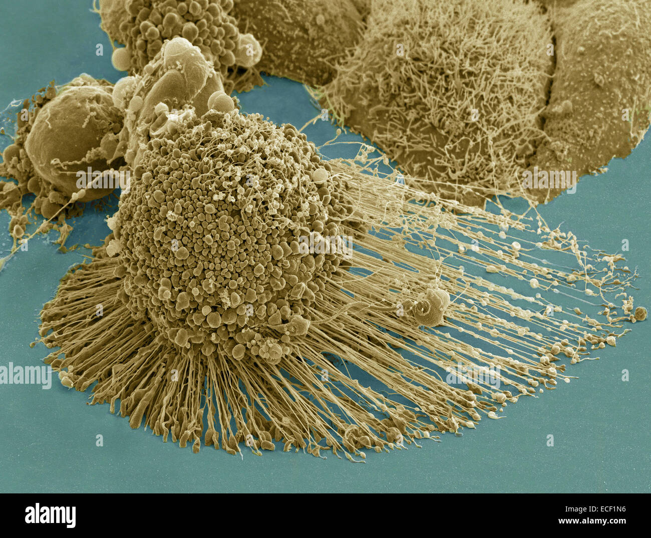 Scanning electron micrograph of an apoptotic HeLa cell. Zeiss Merlin HR-SEM. Stock Photohttps://www.alamy.com/image-license-details/?v=1https://www.alamy.com/stock-photo-scanning-electron-micrograph-of-an-apoptotic-hela-cell-zeiss-merlin-76548002.html
RFECF1N6–Scanning electron micrograph of an apoptotic HeLa cell. Zeiss Merlin HR-SEM.
 Jakarta Selatan, Indonesia. 26th Apr, 2016. Researcher operates Scanning Electron Microscope 'SEM' machine to capture rock mineral photos. SEM used to analyze rock minerals up to 1µm. It can zoom 300.000x than usual polarize microscope. © Anton Raharjo/Pacific Press/Alamy Live News Stock Photohttps://www.alamy.com/image-license-details/?v=1https://www.alamy.com/stock-photo-jakarta-selatan-indonesia-26th-apr-2016-researcher-operates-scanning-102955189.html
RMFYE0B1–Jakarta Selatan, Indonesia. 26th Apr, 2016. Researcher operates Scanning Electron Microscope 'SEM' machine to capture rock mineral photos. SEM used to analyze rock minerals up to 1µm. It can zoom 300.000x than usual polarize microscope. © Anton Raharjo/Pacific Press/Alamy Live News
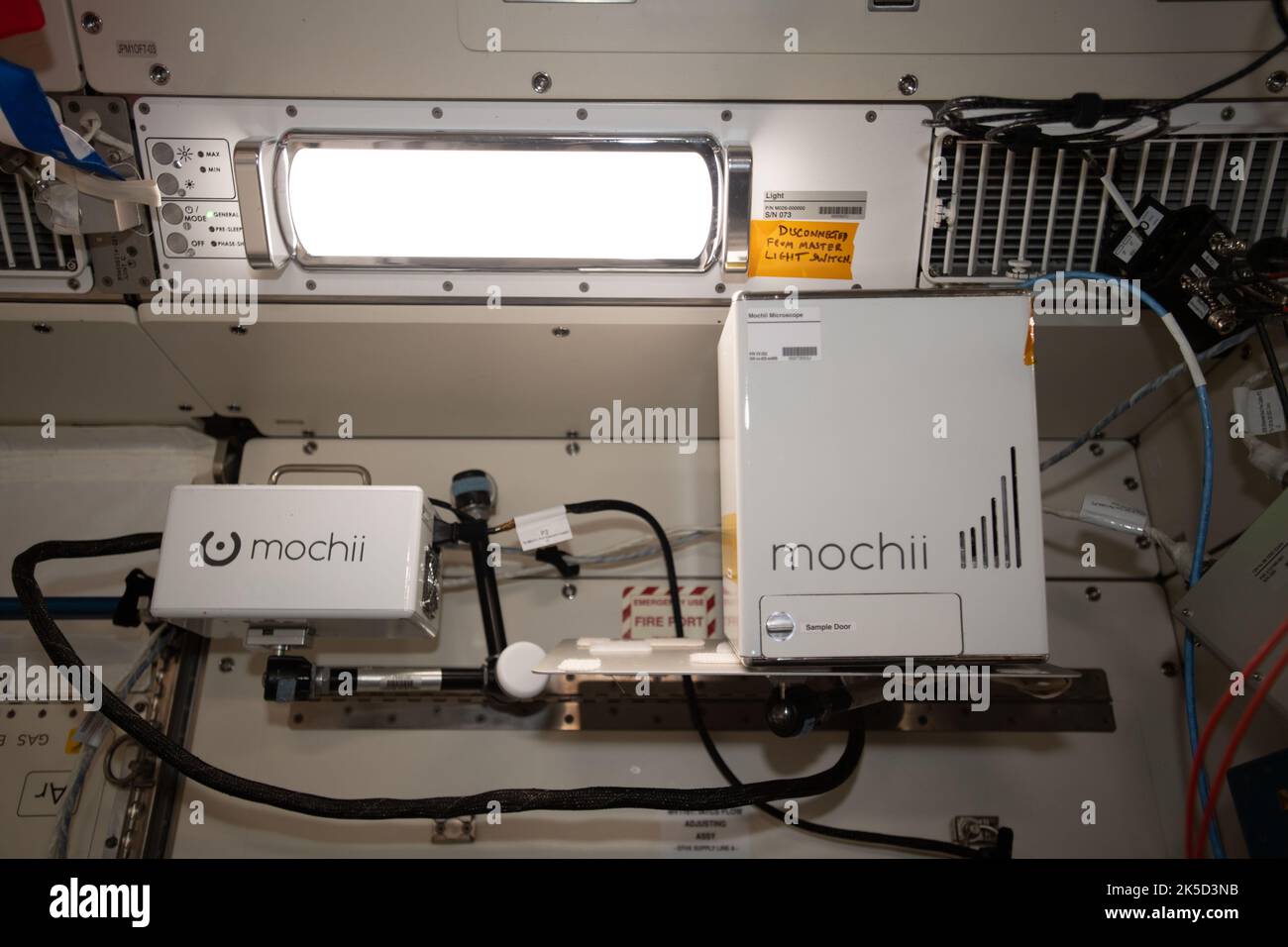 A view of the Mochii microscope aboard the International Space Station (ISS. Mochii is a miniature scanning electron microscope (SEM) with spectroscopy to conduct real-time, on-site imaging and compositional measurements of particles on the International Space Station (ISS). Stock Photohttps://www.alamy.com/image-license-details/?v=1https://www.alamy.com/a-view-of-the-mochii-microscope-aboard-the-international-space-station-iss-mochii-is-a-miniature-scanning-electron-microscope-sem-with-spectroscopy-to-conduct-real-time-on-site-imaging-and-compositional-measurements-of-particles-on-the-international-space-station-iss-image485251911.html
RM2K5D3NB–A view of the Mochii microscope aboard the International Space Station (ISS. Mochii is a miniature scanning electron microscope (SEM) with spectroscopy to conduct real-time, on-site imaging and compositional measurements of particles on the International Space Station (ISS).
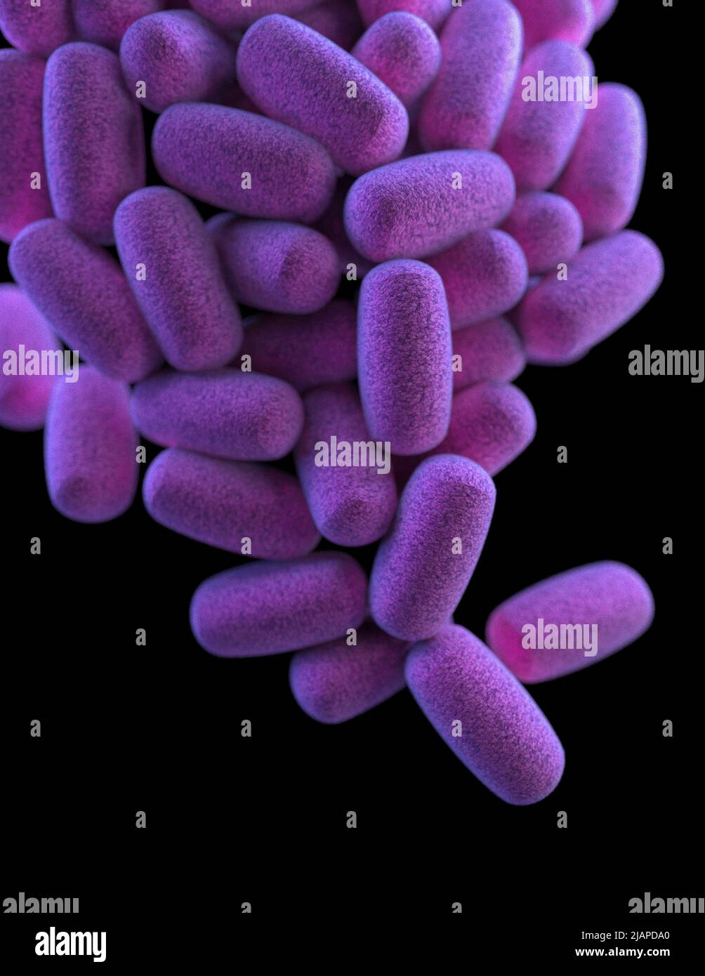 A cluster of barrel-shaped, Clostridium perfringens bacteria. False-colour interpreted artistic rendition based on scanning electron microscopic (SEM) imagery. Stock Photohttps://www.alamy.com/image-license-details/?v=1https://www.alamy.com/a-cluster-of-barrel-shaped-clostridium-perfringens-bacteria-false-colour-interpreted-artistic-rendition-based-on-scanning-electron-microscopic-sem-imagery-image471319912.html
RM2JAPDA0–A cluster of barrel-shaped, Clostridium perfringens bacteria. False-colour interpreted artistic rendition based on scanning electron microscopic (SEM) imagery.
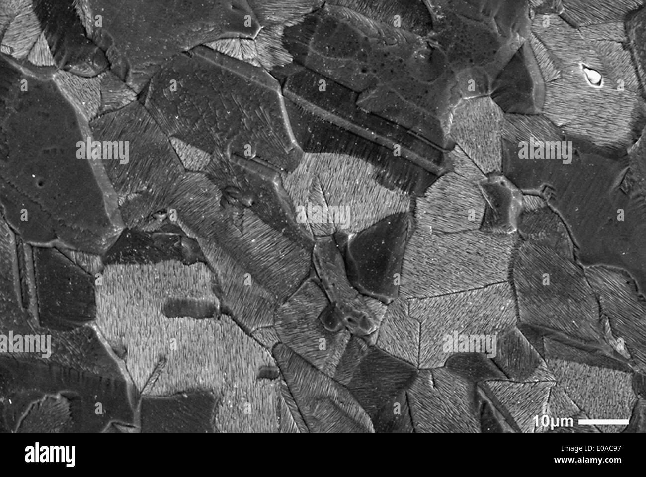 Scanning Electron Micrograph of fracture surface of stainless steel, rouged and etched sample Stock Photohttps://www.alamy.com/image-license-details/?v=1https://www.alamy.com/scanning-electron-micrograph-of-fracture-surface-of-stainless-steel-image69070659.html
RFE0AC97–Scanning Electron Micrograph of fracture surface of stainless steel, rouged and etched sample
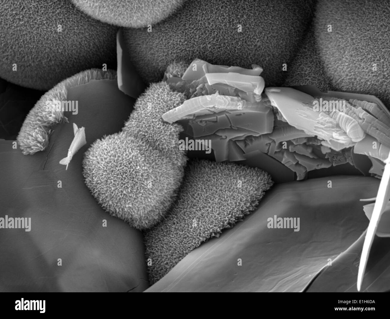 Iron oxide formations with chlorine and sulphur present, imaged in a scanning electron microscope Stock Photohttps://www.alamy.com/image-license-details/?v=1https://www.alamy.com/iron-oxide-formations-with-chlorine-and-sulphur-present-imaged-in-image69834390.html
RFE1H6DA–Iron oxide formations with chlorine and sulphur present, imaged in a scanning electron microscope
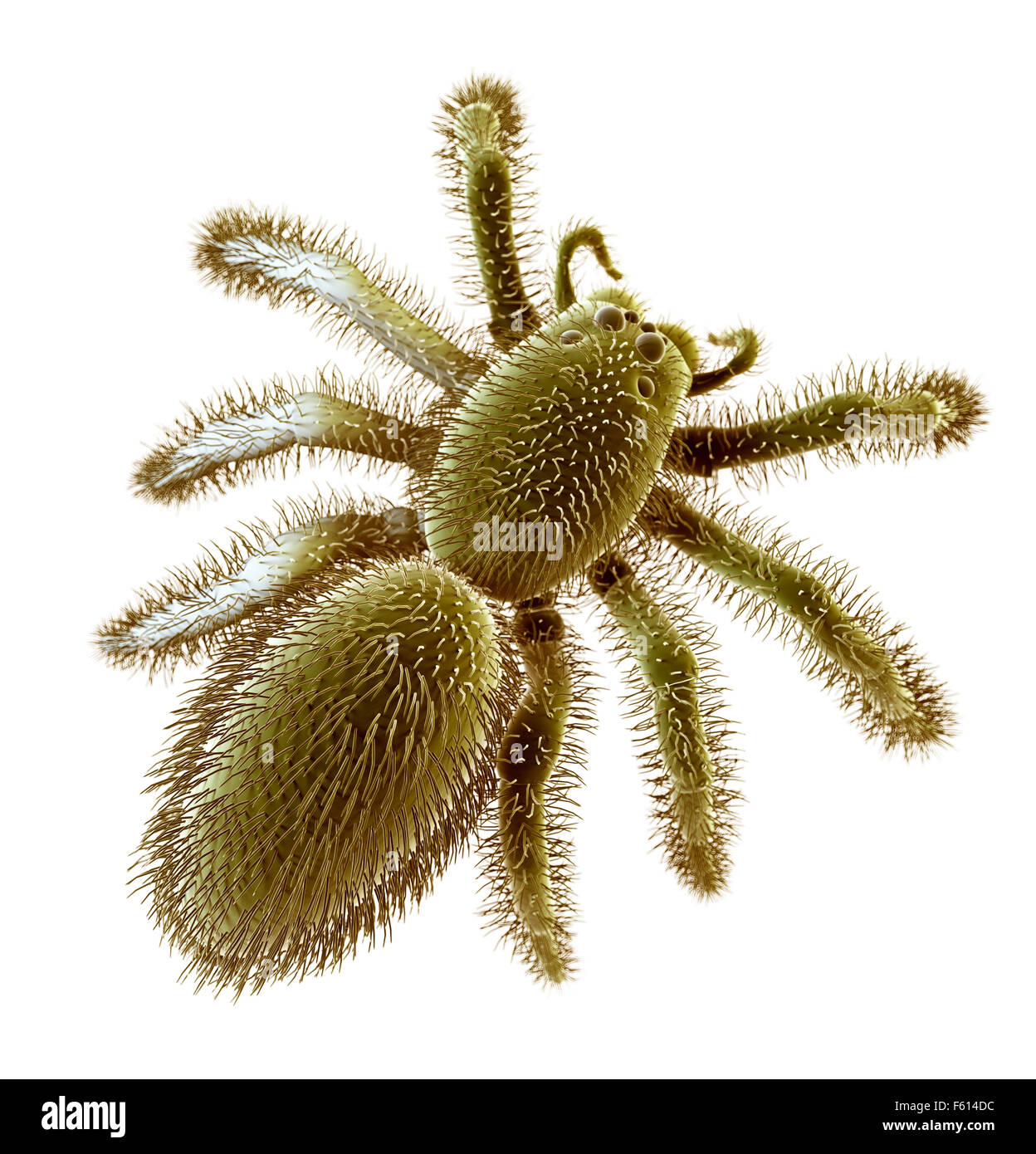 SEM style illustration of a spider Stock Photohttps://www.alamy.com/image-license-details/?v=1https://www.alamy.com/stock-photo-sem-style-illustration-of-a-spider-89765240.html
RFF614DC–SEM style illustration of a spider
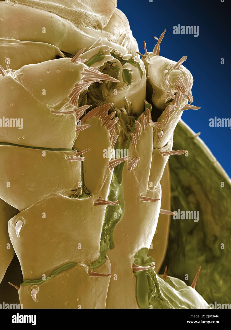 SEM Scanning Electron Microscope image of a Sandhopper, Sand Flea, amphipod Stock Photohttps://www.alamy.com/image-license-details/?v=1https://www.alamy.com/sem-scanning-electron-microscope-image-of-a-sandhopper-sand-flea-amphipod-image470581222.html
RM2J9GR46–SEM Scanning Electron Microscope image of a Sandhopper, Sand Flea, amphipod
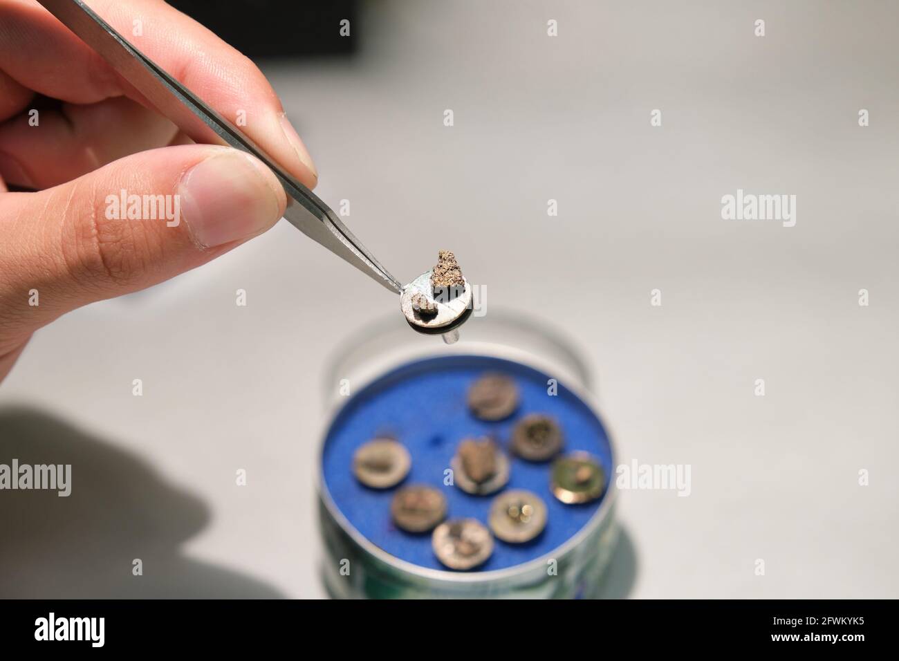 Scientific hand holding a tweezers with a scanning electron microscope sample on a specimen mount. SEM pins to analyze. Stock Photohttps://www.alamy.com/image-license-details/?v=1https://www.alamy.com/scientific-hand-holding-a-tweezers-with-a-scanning-electron-microscope-sample-on-a-specimen-mount-sem-pins-to-analyze-image428854025.html
RF2FWKYK5–Scientific hand holding a tweezers with a scanning electron microscope sample on a specimen mount. SEM pins to analyze.
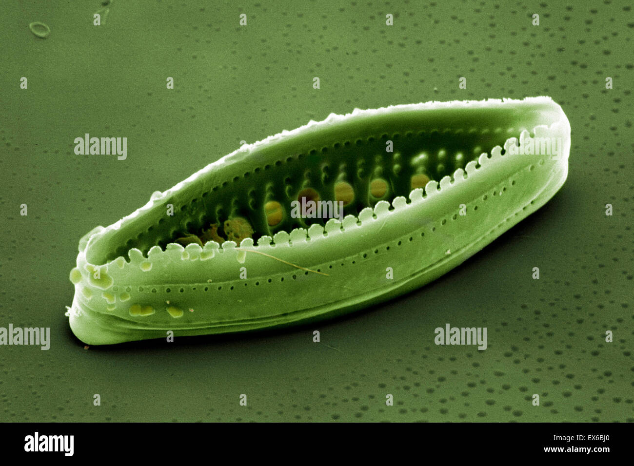 Diatom, SEM Stock Photohttps://www.alamy.com/image-license-details/?v=1https://www.alamy.com/stock-photo-diatom-sem-84963368.html
RMEX6BJ0–Diatom, SEM
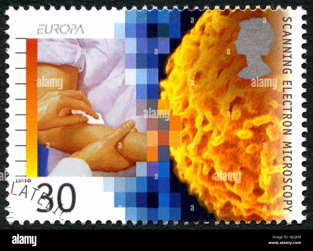 GREAT BRITAIN - CIRCA 1994: A used postage stamp from the UK, commemorating the invention of the Scanning Electron Microscope, circa 1994. Stock Photohttps://www.alamy.com/image-license-details/?v=1https://www.alamy.com/stock-photo-great-britain-circa-1994-a-used-postage-stamp-from-the-uk-commemorating-131814332.html
RMHJCJEM–GREAT BRITAIN - CIRCA 1994: A used postage stamp from the UK, commemorating the invention of the Scanning Electron Microscope, circa 1994.
 Douglass Bryant, a materials engineer of Naval Surface Warfare Center Philadelphia Division (NSWCPD), carefully places a sheered screw into a scanning electron microscope (SEM) at the Navy Yard in Philadelphia Aug. 30, 2017.SEMs provide engineers like Bryant with a high-resolution, high-magnification image of materials that have failed in the fleet, to discover if the part failed from normal wear and tear or environmental causes such as exposure to extreme temperatures or corrosive chemicals. (U.S. Navy photo by Petty Officer 1st Class Richard Hoffner/Released) Stock Photohttps://www.alamy.com/image-license-details/?v=1https://www.alamy.com/douglass-bryant-a-materials-engineer-of-naval-surface-warfare-center-philadelphia-division-nswcpd-carefully-places-a-sheered-screw-into-a-scanning-electron-microscope-sem-at-the-navy-yard-in-philadelphia-aug-30-2017sems-provide-engineers-like-bryant-with-a-high-resolution-high-magnification-image-of-materials-that-have-failed-in-the-fleet-to-discover-if-the-part-failed-from-normal-wear-and-tear-or-environmental-causes-such-as-exposure-to-extreme-temperatures-or-corrosive-chemicals-us-navy-photo-by-petty-officer-1st-class-richard-hoffnerreleased-image185120348.html
RMMN4XW0–Douglass Bryant, a materials engineer of Naval Surface Warfare Center Philadelphia Division (NSWCPD), carefully places a sheered screw into a scanning electron microscope (SEM) at the Navy Yard in Philadelphia Aug. 30, 2017.SEMs provide engineers like Bryant with a high-resolution, high-magnification image of materials that have failed in the fleet, to discover if the part failed from normal wear and tear or environmental causes such as exposure to extreme temperatures or corrosive chemicals. (U.S. Navy photo by Petty Officer 1st Class Richard Hoffner/Released)
 Neurone. Scanning electron micrograph (SEM) of a PC12 neurone in culture.The PC12 cell line, developed from a pheochromocytoma tumor of the rat adrenal medulla, has become a premiere model for the study of neuronal differentiation. When treated in culture Stock Photohttps://www.alamy.com/image-license-details/?v=1https://www.alamy.com/stock-photo-neurone-scanning-electron-micrograph-sem-of-a-pc12-neurone-in-culturethe-73690532.html
RFE7TW0M–Neurone. Scanning electron micrograph (SEM) of a PC12 neurone in culture.The PC12 cell line, developed from a pheochromocytoma tumor of the rat adrenal medulla, has become a premiere model for the study of neuronal differentiation. When treated in culture
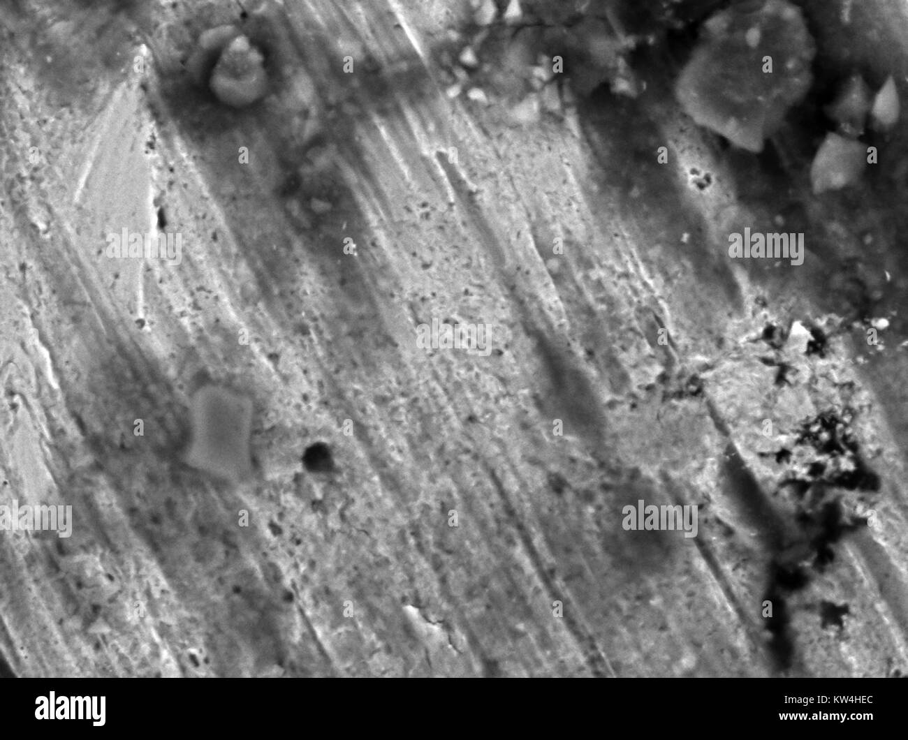 Scanning electron microscope (SEM) micrograph of the surface of a United States penny coin, showing copper oxide particles, at a magnification of 5000x, 2016. Stock Photohttps://www.alamy.com/image-license-details/?v=1https://www.alamy.com/stock-photo-scanning-electron-microscope-sem-micrograph-of-the-surface-of-a-united-170361252.html
RMKW4HEC–Scanning electron microscope (SEM) micrograph of the surface of a United States penny coin, showing copper oxide particles, at a magnification of 5000x, 2016.
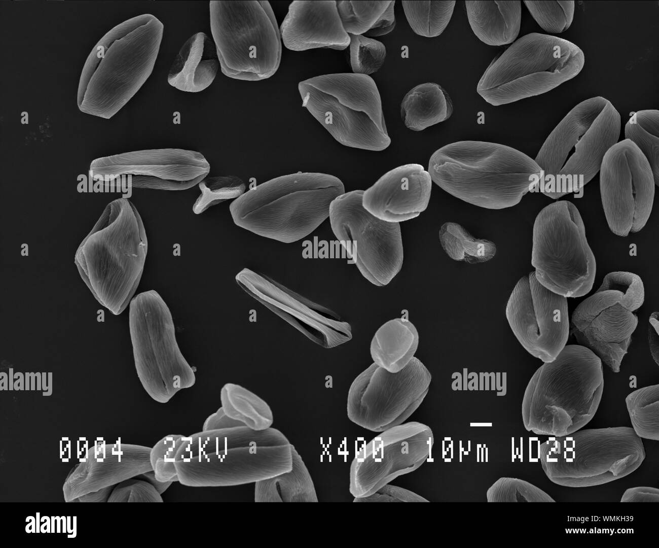 Cherry tree pollen under electron microscope Stock Photohttps://www.alamy.com/image-license-details/?v=1https://www.alamy.com/cherry-tree-pollen-under-electron-microscope-image270879149.html
RFWMKH39–Cherry tree pollen under electron microscope
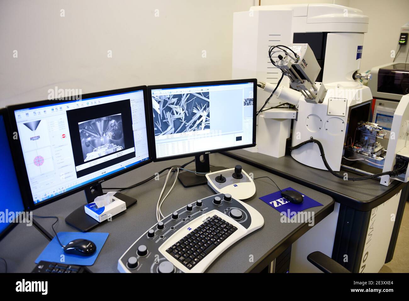 Zeiss EVO 15 Scanning Electron Microscope scanning station in a science lab. It is flexible variable pressure scanning electron microscope or SEM with Stock Photohttps://www.alamy.com/image-license-details/?v=1https://www.alamy.com/zeiss-evo-15-scanning-electron-microscope-scanning-station-in-a-science-lab-it-is-flexible-variable-pressure-scanning-electron-microscope-or-sem-with-image398273964.html
RM2E3XXE4–Zeiss EVO 15 Scanning Electron Microscope scanning station in a science lab. It is flexible variable pressure scanning electron microscope or SEM with
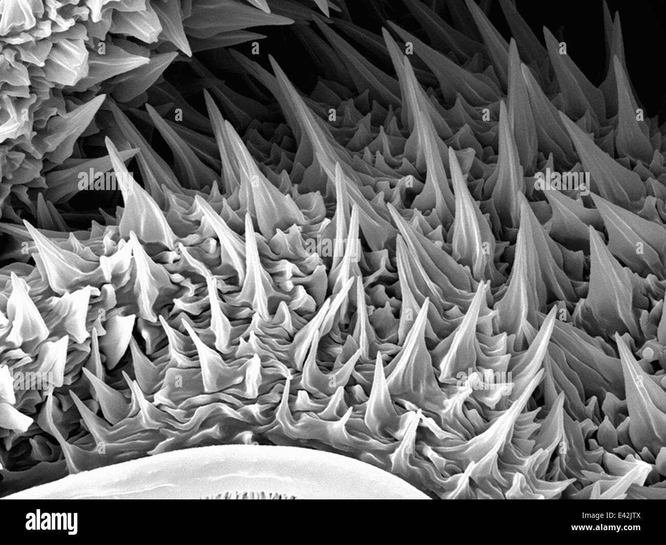 Large Caterpillar: Gold coated and imaged in Scanning electron microscope Stock Photohttps://www.alamy.com/image-license-details/?v=1https://www.alamy.com/stock-photo-large-caterpillar-gold-coated-and-imaged-in-scanning-electron-microscope-71358810.html
RFE42JTX–Large Caterpillar: Gold coated and imaged in Scanning electron microscope
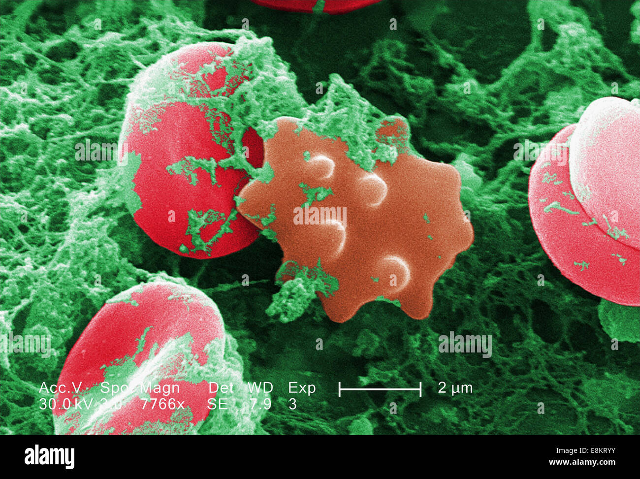 This scanning electron micrograph (SEM) depicted number of red blood cells found enmeshed in fibrinous matrix on luminal Stock Photohttps://www.alamy.com/image-license-details/?v=1https://www.alamy.com/stock-photo-this-scanning-electron-micrograph-sem-depicted-number-of-red-blood-74194623.html
RME8KRYY–This scanning electron micrograph (SEM) depicted number of red blood cells found enmeshed in fibrinous matrix on luminal
 Colorized scanning electron micrograph of an apoptotic cell (blue) infected with SARS-COV-2 virus particles (orange), isolated from a patient sample. Stock Photohttps://www.alamy.com/image-license-details/?v=1https://www.alamy.com/colorized-scanning-electron-micrograph-of-an-apoptotic-cell-blue-infected-with-sars-cov-2-virus-particles-orange-isolated-from-a-patient-sample-image440582710.html
RM2GGP7MP–Colorized scanning electron micrograph of an apoptotic cell (blue) infected with SARS-COV-2 virus particles (orange), isolated from a patient sample.
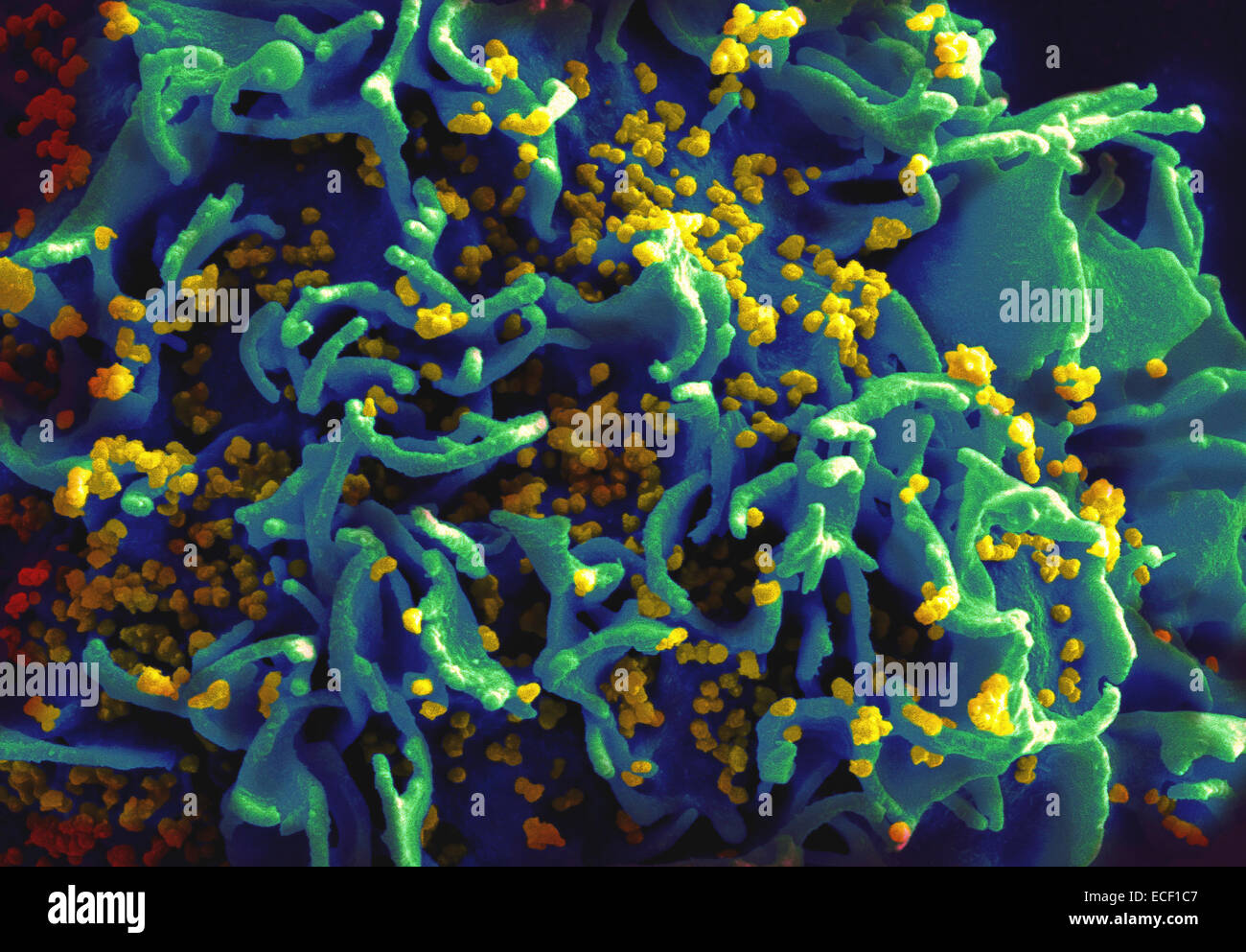 Scanning electron micrograph of HIV particles infecting a human T cell. Stock Photohttps://www.alamy.com/image-license-details/?v=1https://www.alamy.com/stock-photo-scanning-electron-micrograph-of-hiv-particles-infecting-a-human-t-76547751.html
RFECF1C7–Scanning electron micrograph of HIV particles infecting a human T cell.
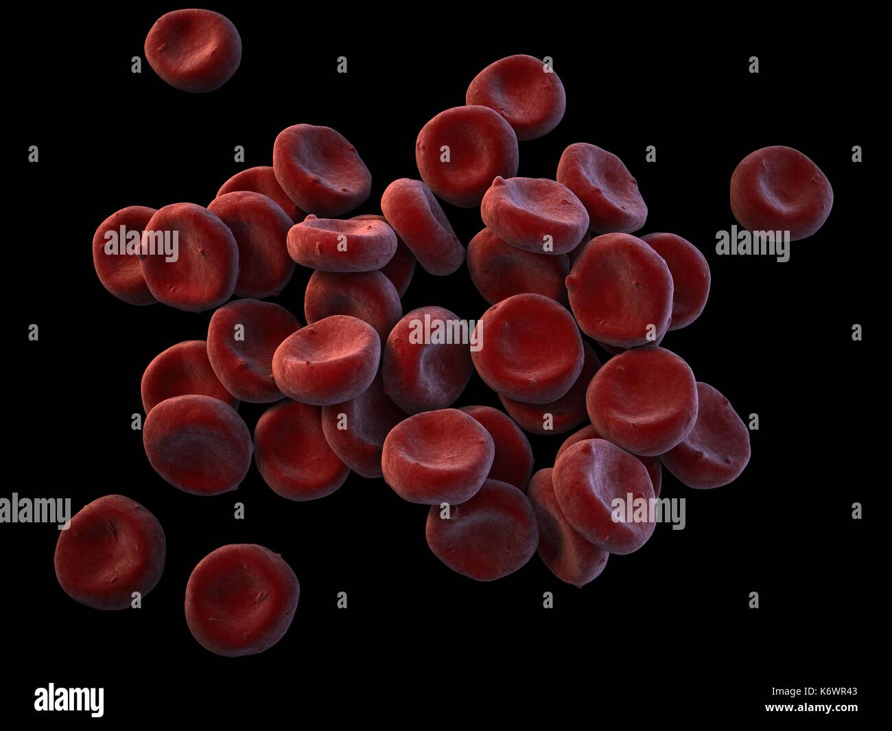 Topographical SEM (scanning Electron Microscope) close-up of oxygenated Red Blood Cells (Erythrocytes) piled up on dark grey surface background. Stock Photohttps://www.alamy.com/image-license-details/?v=1https://www.alamy.com/topographical-sem-scanning-electron-microscope-close-up-of-oxygenated-image159148195.html
RMK6WR43–Topographical SEM (scanning Electron Microscope) close-up of oxygenated Red Blood Cells (Erythrocytes) piled up on dark grey surface background.
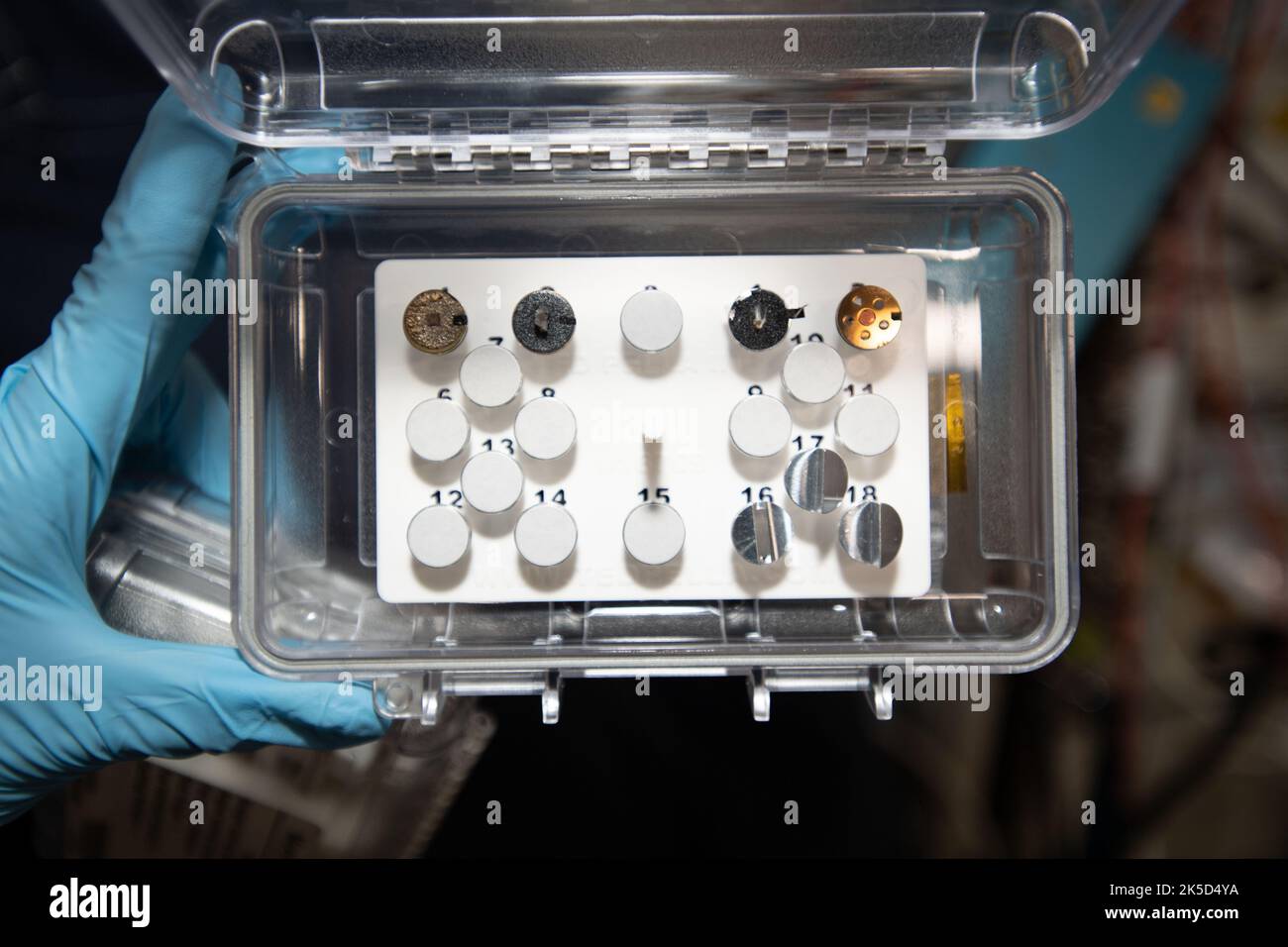 A view of the Mochii microscope sample load aboard the International Space Station (ISS). Mochii is a miniature scanning electron microscope (SEM) with spectroscopy to conduct real-time, on-site imaging and compositional measurements of particles on the International Space Station (ISS) Stock Photohttps://www.alamy.com/image-license-details/?v=1https://www.alamy.com/a-view-of-the-mochii-microscope-sample-load-aboard-the-international-space-station-iss-mochii-is-a-miniature-scanning-electron-microscope-sem-with-spectroscopy-to-conduct-real-time-on-site-imaging-and-compositional-measurements-of-particles-on-the-international-space-station-iss-image485252862.html
RM2K5D4YA–A view of the Mochii microscope sample load aboard the International Space Station (ISS). Mochii is a miniature scanning electron microscope (SEM) with spectroscopy to conduct real-time, on-site imaging and compositional measurements of particles on the International Space Station (ISS)
 Digitally colourised, scanning electron microscopic (SEM) image of a dry fractured Vero cell, revealing its contents, and the ultrastructural details at the site of an opened vacuole, inside of which you can see numerous, Coxiella burnetii bacteria, undergoing rapid replication. Q fever is a disease caused by the bacteria Coxiella burnetii. This bacteria naturally infects some animals, such as goats, sheep, and cattle. An optimised and enhanced version of an image produced by the US National Institute of Allergy and Infectious Diseases (NIAID) / CDC. Stock Photohttps://www.alamy.com/image-license-details/?v=1https://www.alamy.com/digitally-colourised-scanning-electron-microscopic-sem-image-of-a-dry-fractured-vero-cell-revealing-its-contents-and-the-ultrastructural-details-at-the-site-of-an-opened-vacuole-inside-of-which-you-can-see-numerous-coxiella-burnetii-bacteria-undergoing-rapid-replication-q-fever-is-a-disease-caused-by-the-bacteria-coxiella-burnetii-this-bacteria-naturally-infects-some-animals-such-as-goats-sheep-and-cattle-an-optimised-and-enhanced-version-of-an-image-produced-by-the-us-national-institute-of-allergy-and-infectious-diseases-niaid-cdc-image471319954.html
RM2JAPDBE–Digitally colourised, scanning electron microscopic (SEM) image of a dry fractured Vero cell, revealing its contents, and the ultrastructural details at the site of an opened vacuole, inside of which you can see numerous, Coxiella burnetii bacteria, undergoing rapid replication. Q fever is a disease caused by the bacteria Coxiella burnetii. This bacteria naturally infects some animals, such as goats, sheep, and cattle. An optimised and enhanced version of an image produced by the US National Institute of Allergy and Infectious Diseases (NIAID) / CDC.
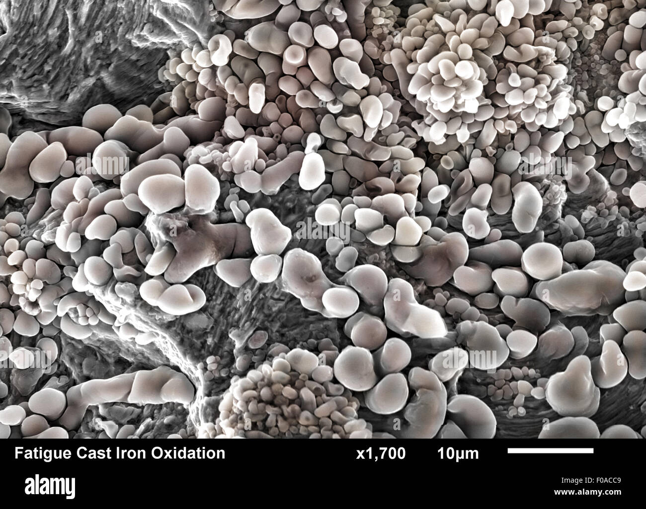 Cast Iron fractured surface. Fatigue striations under iron oxide corrosion imaged in a scanning electron microscope Stock Photohttps://www.alamy.com/image-license-details/?v=1https://www.alamy.com/stock-photo-cast-iron-fractured-surface-fatigue-striations-under-iron-oxide-corrosion-86281113.html
RFF0ACC9–Cast Iron fractured surface. Fatigue striations under iron oxide corrosion imaged in a scanning electron microscope
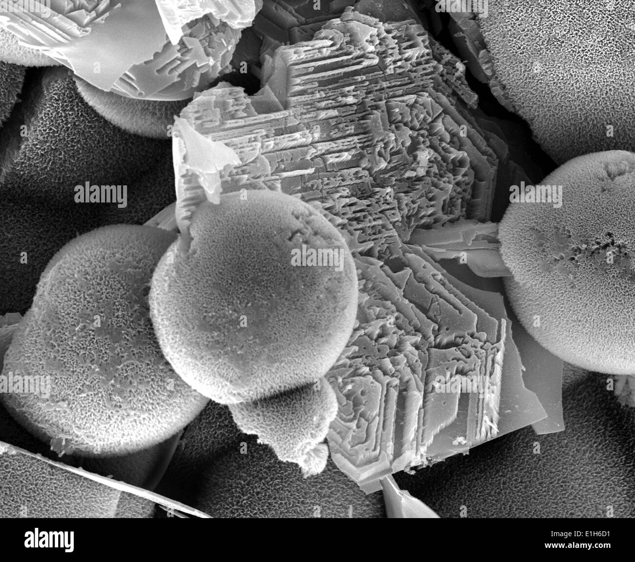 Iron oxide formations with sulphur and chlorine present, imaged with a scanning electron microscope Stock Photohttps://www.alamy.com/image-license-details/?v=1https://www.alamy.com/iron-oxide-formations-with-sulphur-and-chlorine-present-imaged-with-image69834381.html
RFE1H6D1–Iron oxide formations with sulphur and chlorine present, imaged with a scanning electron microscope
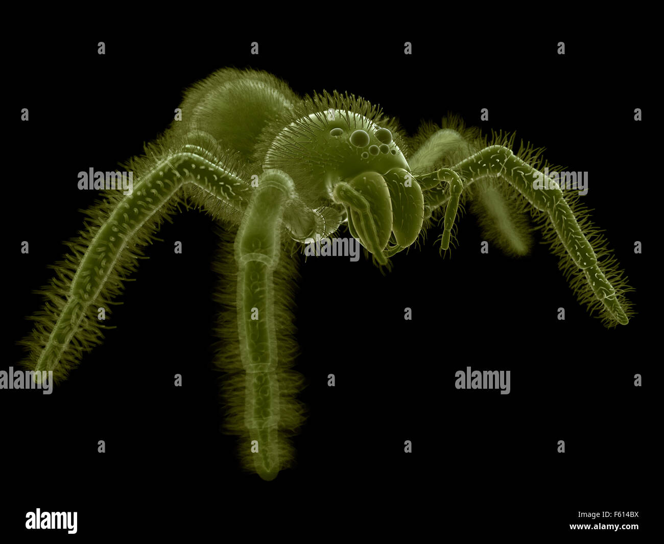 SEM style illustration of a spider Stock Photohttps://www.alamy.com/image-license-details/?v=1https://www.alamy.com/stock-photo-sem-style-illustration-of-a-spider-89765198.html
RFF614BX–SEM style illustration of a spider
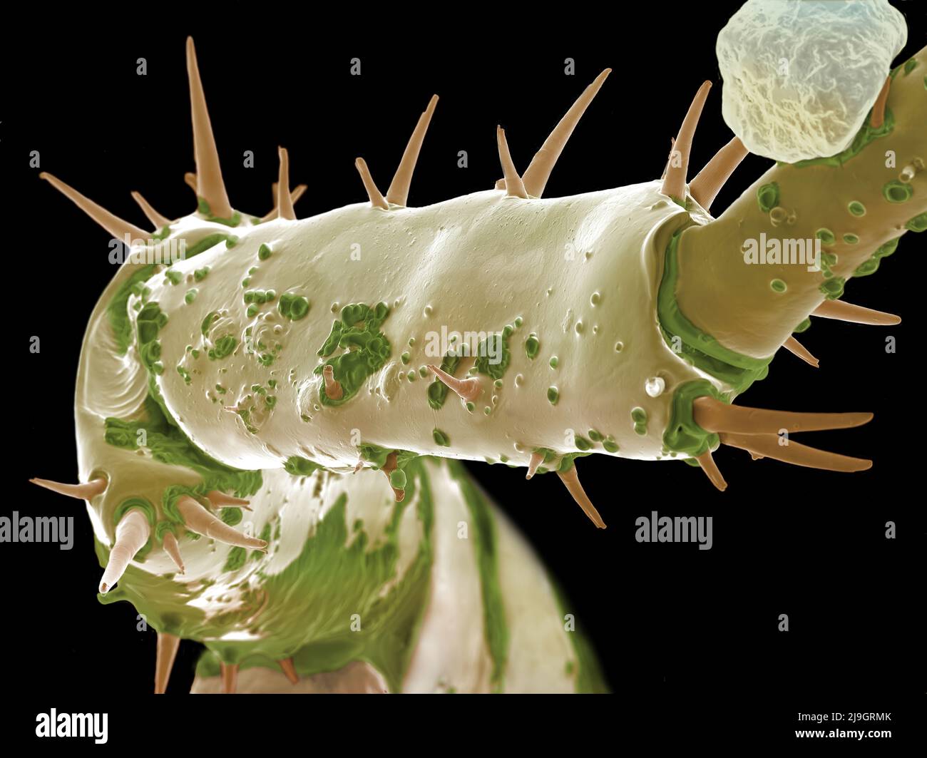 SEM Scanning Electron Microscope image of a Sandhopper, Sand Flea, amphipod Stock Photohttps://www.alamy.com/image-license-details/?v=1https://www.alamy.com/sem-scanning-electron-microscope-image-of-a-sandhopper-sand-flea-amphipod-image470581683.html
RM2J9GRMK–SEM Scanning Electron Microscope image of a Sandhopper, Sand Flea, amphipod
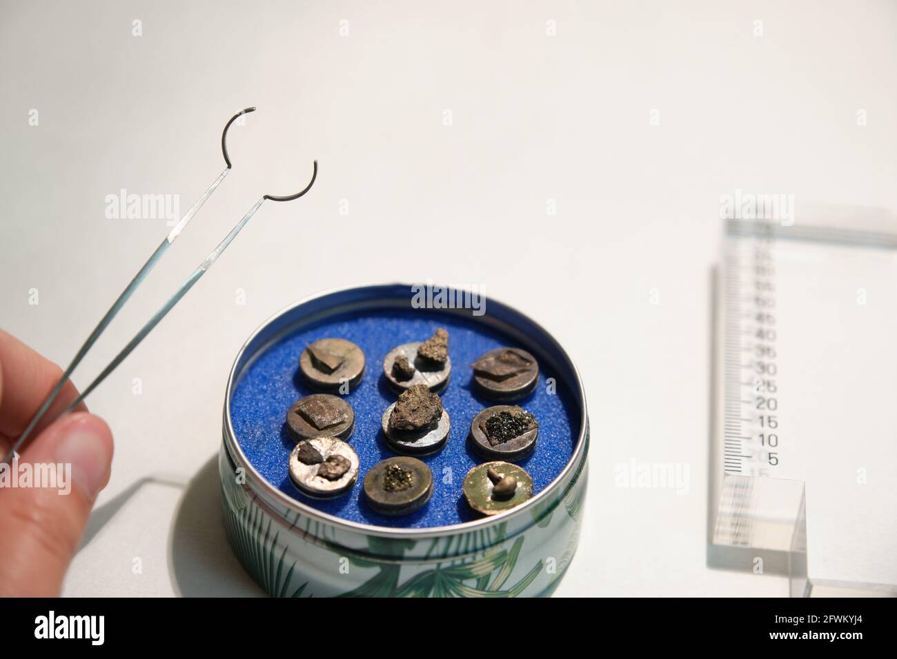 Scanning electron microscope samples on specimen mounts, tweezers and a ruler. SEM pins to analyze. Stock Photohttps://www.alamy.com/image-license-details/?v=1https://www.alamy.com/scanning-electron-microscope-samples-on-specimen-mounts-tweezers-and-a-ruler-sem-pins-to-analyze-image428853996.html
RF2FWKYJ4–Scanning electron microscope samples on specimen mounts, tweezers and a ruler. SEM pins to analyze.
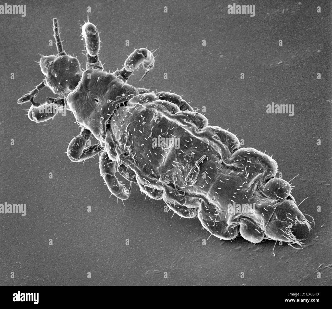 Pediculus humanus capitis, head louse, SEM Stock Photohttps://www.alamy.com/image-license-details/?v=1https://www.alamy.com/stock-photo-pediculus-humanus-capitis-head-louse-sem-84963366.html
RMEX6BHX–Pediculus humanus capitis, head louse, SEM
 Ground samples of soil from the crime scene are flattened under high pressure,mounted on metal discs in preparation for SEM Stock Photohttps://www.alamy.com/image-license-details/?v=1https://www.alamy.com/ground-samples-of-soil-from-the-crime-scene-are-flattened-under-high-image9140372.html
RMARATW5–Ground samples of soil from the crime scene are flattened under high pressure,mounted on metal discs in preparation for SEM
 Close-up of a Mosquito imaged with a Scanning Electron Microscope (SEM) (color enhanced) Stock Photohttps://www.alamy.com/image-license-details/?v=1https://www.alamy.com/stock-photo-close-up-of-a-mosquito-imaged-with-a-scanning-electron-microscope-38157566.html
RMC626A6–Close-up of a Mosquito imaged with a Scanning Electron Microscope (SEM) (color enhanced)
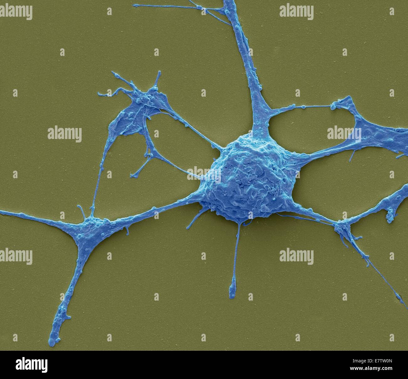 Neurone. Scanning electron micrograph (SEM) of a PC12 neurone in culture.The PC12 cell line, developed from a pheochromocytoma tumor of the rat adrenal medulla, has become a premiere model for the study of neuronal differentiation. When treated in culture Stock Photohttps://www.alamy.com/image-license-details/?v=1https://www.alamy.com/stock-photo-neurone-scanning-electron-micrograph-sem-of-a-pc12-neurone-in-culturethe-73690533.html
RFE7TW0N–Neurone. Scanning electron micrograph (SEM) of a PC12 neurone in culture.The PC12 cell line, developed from a pheochromocytoma tumor of the rat adrenal medulla, has become a premiere model for the study of neuronal differentiation. When treated in culture
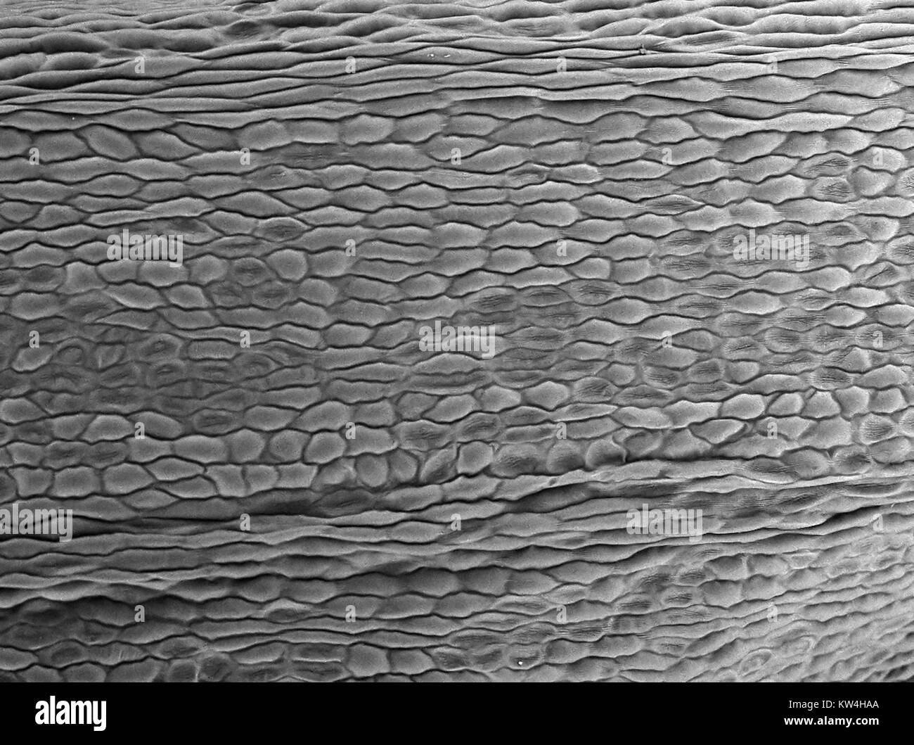 Scanning electron microscope (SEM) micrograph depicting the petal of a carnation flower (Dianthus caryophyllus), showing cellular structure, at a magnification of 200x, 2016. Stock Photohttps://www.alamy.com/image-license-details/?v=1https://www.alamy.com/stock-photo-scanning-electron-microscope-sem-micrograph-depicting-the-petal-of-170361138.html
RMKW4HAA–Scanning electron microscope (SEM) micrograph depicting the petal of a carnation flower (Dianthus caryophyllus), showing cellular structure, at a magnification of 200x, 2016.
