Secondary protein structure Stock Photos and Images
(721)See secondary protein structure stock video clipsQuick filters:
Secondary protein structure Stock Photos and Images
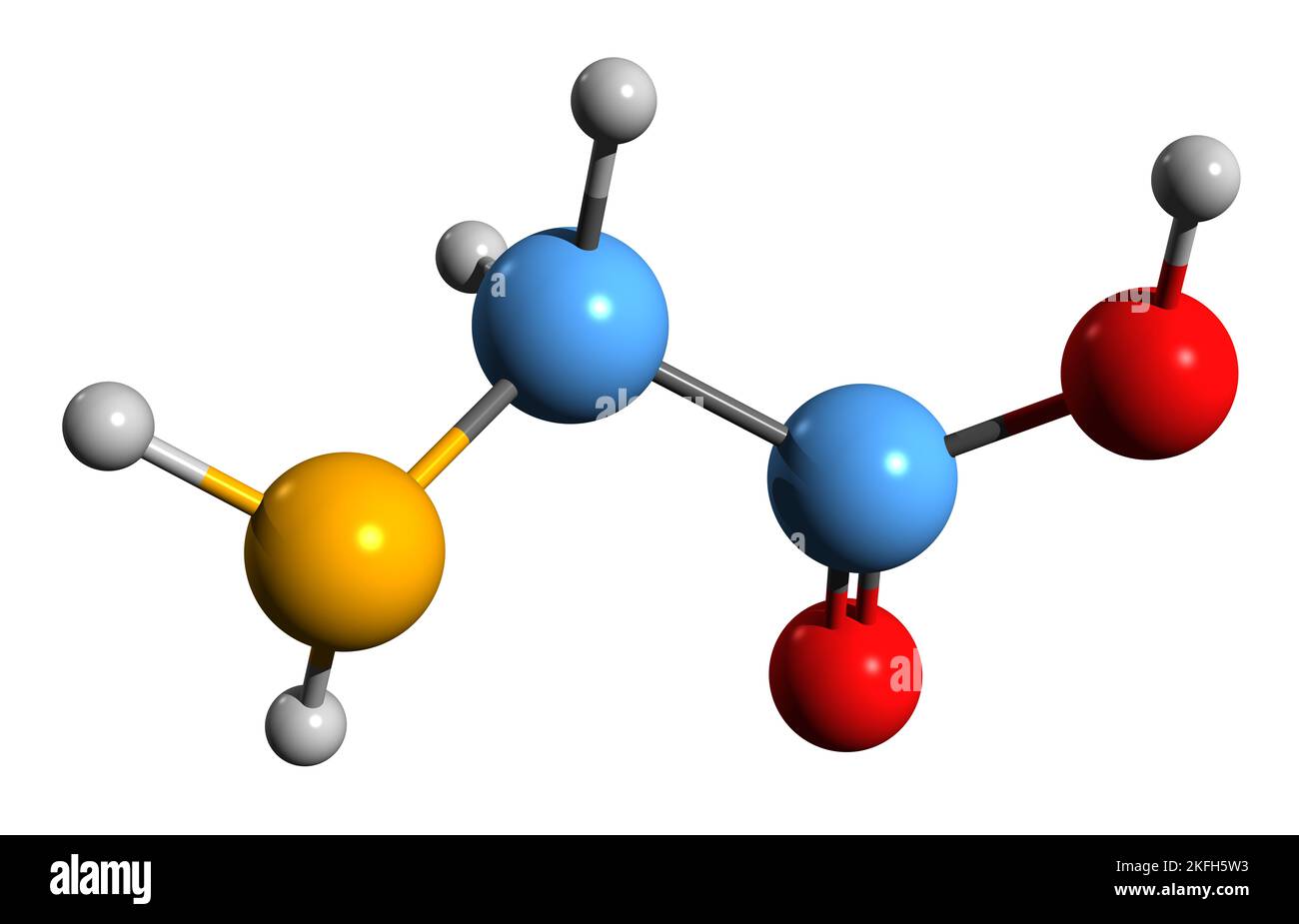 3D image of Glycine skeletal formula - molecular chemical structure of amino acid isolated on white background Stock Photohttps://www.alamy.com/image-license-details/?v=1https://www.alamy.com/3d-image-of-glycine-skeletal-formula-molecular-chemical-structure-of-amino-acid-isolated-on-white-background-image491487951.html
RF2KFH5W3–3D image of Glycine skeletal formula - molecular chemical structure of amino acid isolated on white background
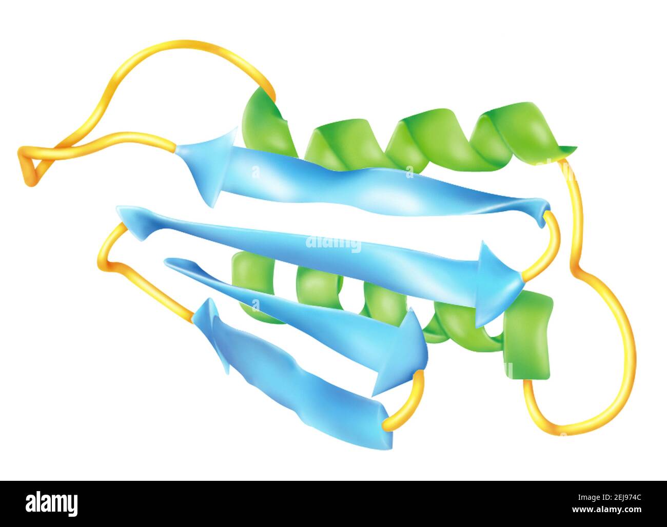 Prion Stock Photohttps://www.alamy.com/image-license-details/?v=1https://www.alamy.com/prion-image407105452.html
RM2EJ974C–Prion
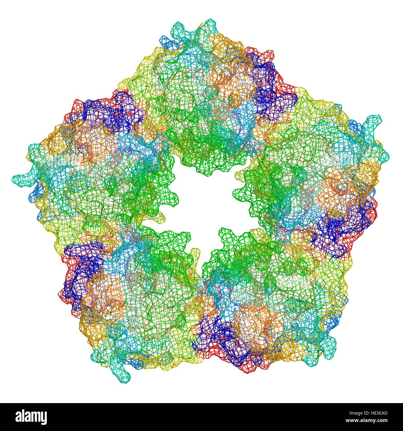 C-reactive protein,molecular model.The protein is made up of five sub-units (monomers) arranged in ring.The secondary structure of protein is shown,with beta sheets (arrows) alpha helices (spirals) connected by linking regions.C-reactive protein (CRP) is blood plasma protein produced by liver.It is acute phase protein,one whose levels rise in response to inflammation.It assists binding of complement proteins to foreign or damaged cells,an immunological response that destroys target cells.High blood levels of CRP are associated increased risk of heart disease diabetes. Stock Photohttps://www.alamy.com/image-license-details/?v=1https://www.alamy.com/stock-photo-c-reactive-proteinmolecular-modelthe-protein-is-made-up-of-five-sub-129659781.html
RFHEXEAD–C-reactive protein,molecular model.The protein is made up of five sub-units (monomers) arranged in ring.The secondary structure of protein is shown,with beta sheets (arrows) alpha helices (spirals) connected by linking regions.C-reactive protein (CRP) is blood plasma protein produced by liver.It is acute phase protein,one whose levels rise in response to inflammation.It assists binding of complement proteins to foreign or damaged cells,an immunological response that destroys target cells.High blood levels of CRP are associated increased risk of heart disease diabetes.
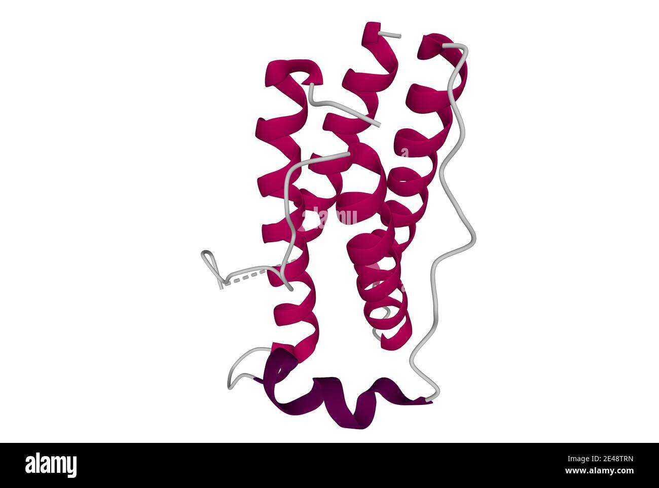 Structure of the human obesity protein, leptin. 3D cartoon model isolated, white background Stock Photohttps://www.alamy.com/image-license-details/?v=1https://www.alamy.com/structure-of-the-human-obesity-protein-leptin-3d-cartoon-model-isolated-white-background-image398492185.html
RF2E48TRN–Structure of the human obesity protein, leptin. 3D cartoon model isolated, white background
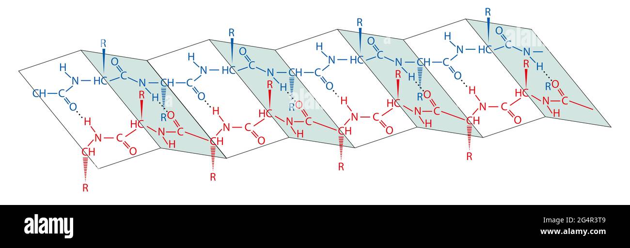 Proteins are large biomolecules, or macromolecules, consisting of one or more long chains of amino acid residues. Protein structure Stock Photohttps://www.alamy.com/image-license-details/?v=1https://www.alamy.com/proteins-are-large-biomolecules-or-macromolecules-consisting-of-one-or-more-long-chains-of-amino-acid-residues-protein-structure-image433225753.html
RF2G4R3T9–Proteins are large biomolecules, or macromolecules, consisting of one or more long chains of amino acid residues. Protein structure
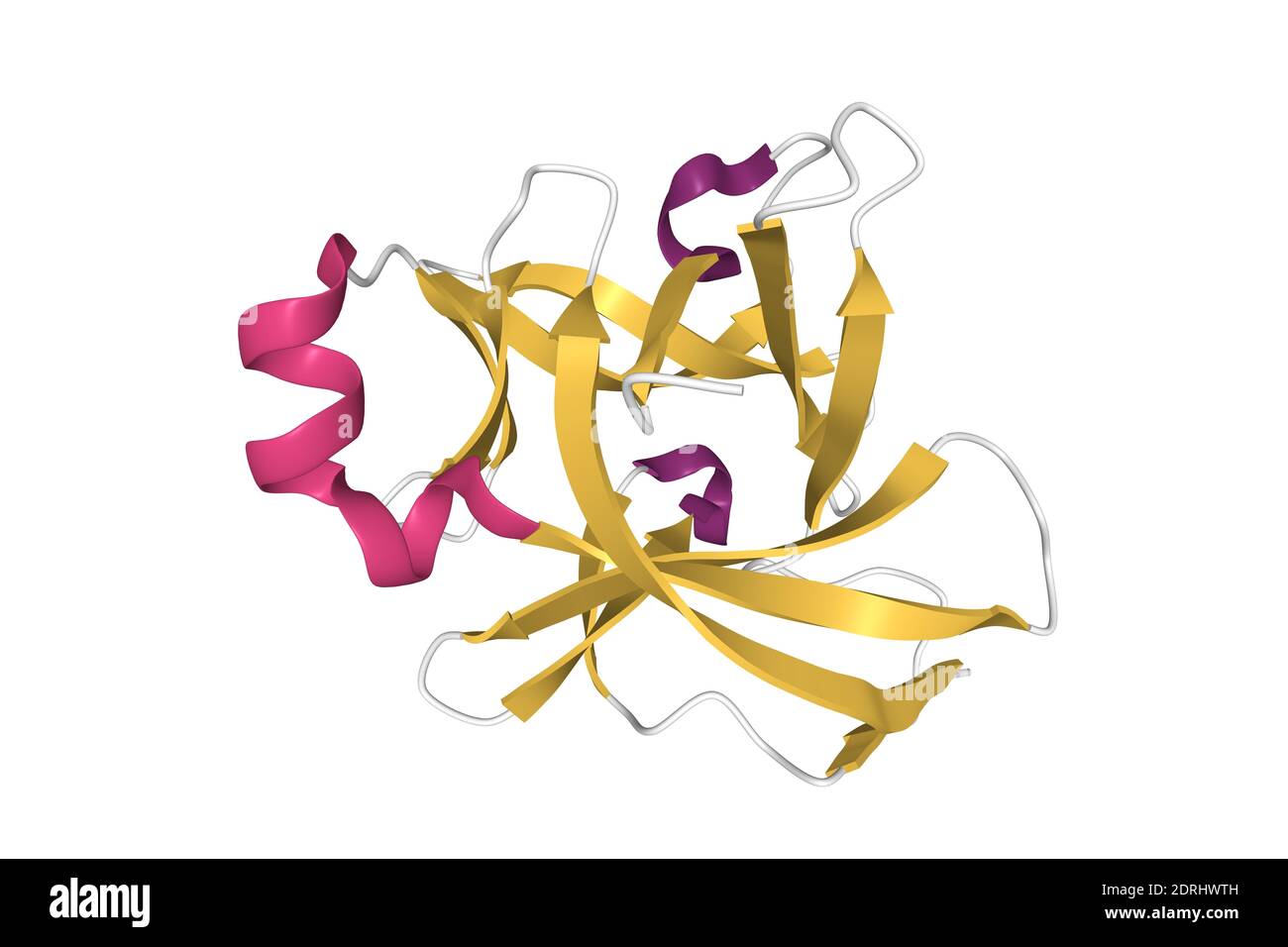 Structure of human interleukin-18, 3D cartoon model isolated with differently colored secondary structure elements, white background Stock Photohttps://www.alamy.com/image-license-details/?v=1https://www.alamy.com/structure-of-human-interleukin-18-3d-cartoon-model-isolated-with-differently-colored-secondary-structure-elements-white-background-image393158657.html
RF2DRHWTH–Structure of human interleukin-18, 3D cartoon model isolated with differently colored secondary structure elements, white background
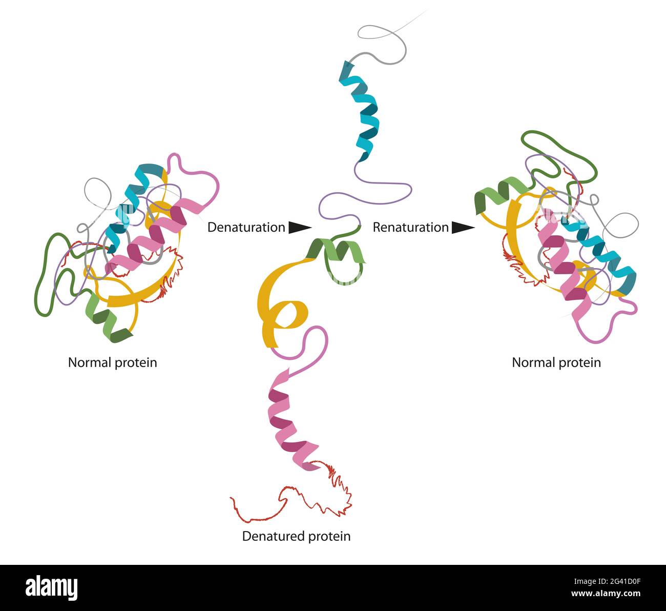 Structure of normal and disassembled protein Stock Photohttps://www.alamy.com/image-license-details/?v=1https://www.alamy.com/structure-of-normal-and-disassembled-protein-image432749983.html
RF2G41D0F–Structure of normal and disassembled protein
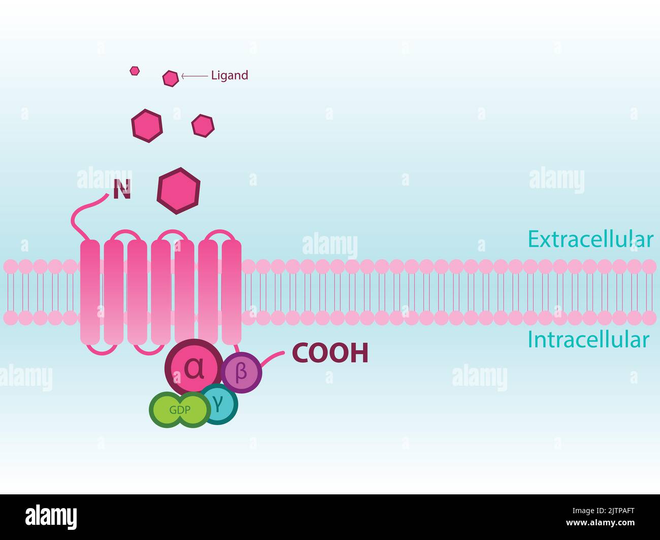 Simplified structure of G protein coupled receptor (GPCR) - including subunits alpha, beta, gamma. Stock Vectorhttps://www.alamy.com/image-license-details/?v=1https://www.alamy.com/simplified-structure-of-g-protein-coupled-receptor-gpcr-including-subunits-alpha-beta-gamma-image479922908.html
RF2JTPAFT–Simplified structure of G protein coupled receptor (GPCR) - including subunits alpha, beta, gamma.
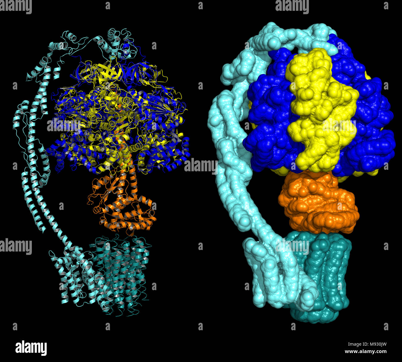 Bovine mitochondrial ATP synthase in state 1b - biological assembly, representation as cartoon and surface Stock Photohttps://www.alamy.com/image-license-details/?v=1https://www.alamy.com/bovine-mitochondrial-atp-synthase-in-state-1b-biological-assembly-representation-as-cartoon-and-surface-image177701969.html
RFM930JW–Bovine mitochondrial ATP synthase in state 1b - biological assembly, representation as cartoon and surface
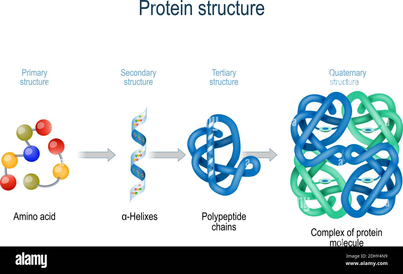 Levels of protein structure from amino acids to Complex of protein molecule. Protein is a polymer (polypeptide) Stock Vectorhttps://www.alamy.com/image-license-details/?v=1https://www.alamy.com/levels-of-protein-structure-from-amino-acids-to-complex-of-protein-molecule-protein-is-a-polymerpolypeptide-image389673685.html
RF2DHY4N9–Levels of protein structure from amino acids to Complex of protein molecule. Protein is a polymer (polypeptide)
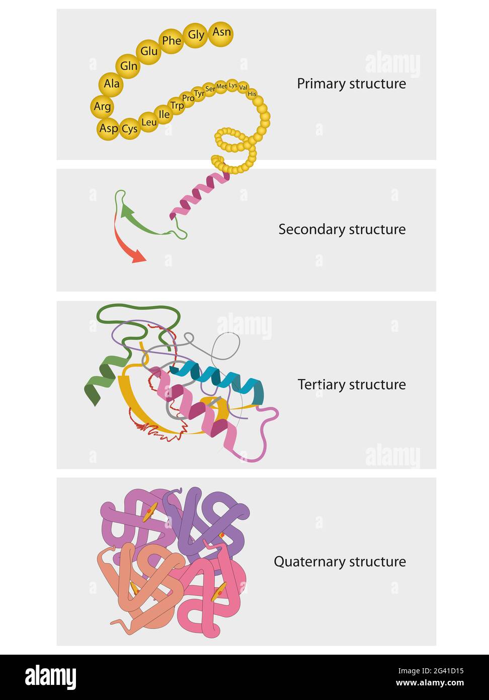 Types of Protein Structure. Proteins are biological polymers composed of amino acids Stock Photohttps://www.alamy.com/image-license-details/?v=1https://www.alamy.com/types-of-protein-structure-proteins-are-biological-polymers-composed-of-amino-acids-image432750001.html
RF2G41D15–Types of Protein Structure. Proteins are biological polymers composed of amino acids
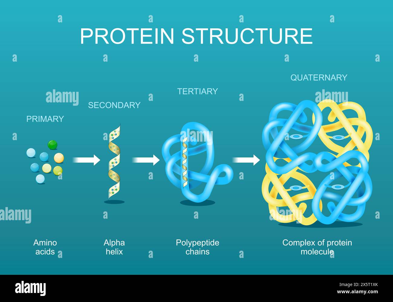 Protein structure. Amino acids, Alpha helix, Polypeptide chains, and Complex of protein molecule. Protein is a polymer (polypeptide) that formed from Stock Vectorhttps://www.alamy.com/image-license-details/?v=1https://www.alamy.com/protein-structure-amino-acids-alpha-helix-polypeptide-chains-and-complex-of-protein-molecule-protein-is-a-polymer-polypeptide-that-formed-from-image605964539.html
RF2X5T1XK–Protein structure. Amino acids, Alpha helix, Polypeptide chains, and Complex of protein molecule. Protein is a polymer (polypeptide) that formed from
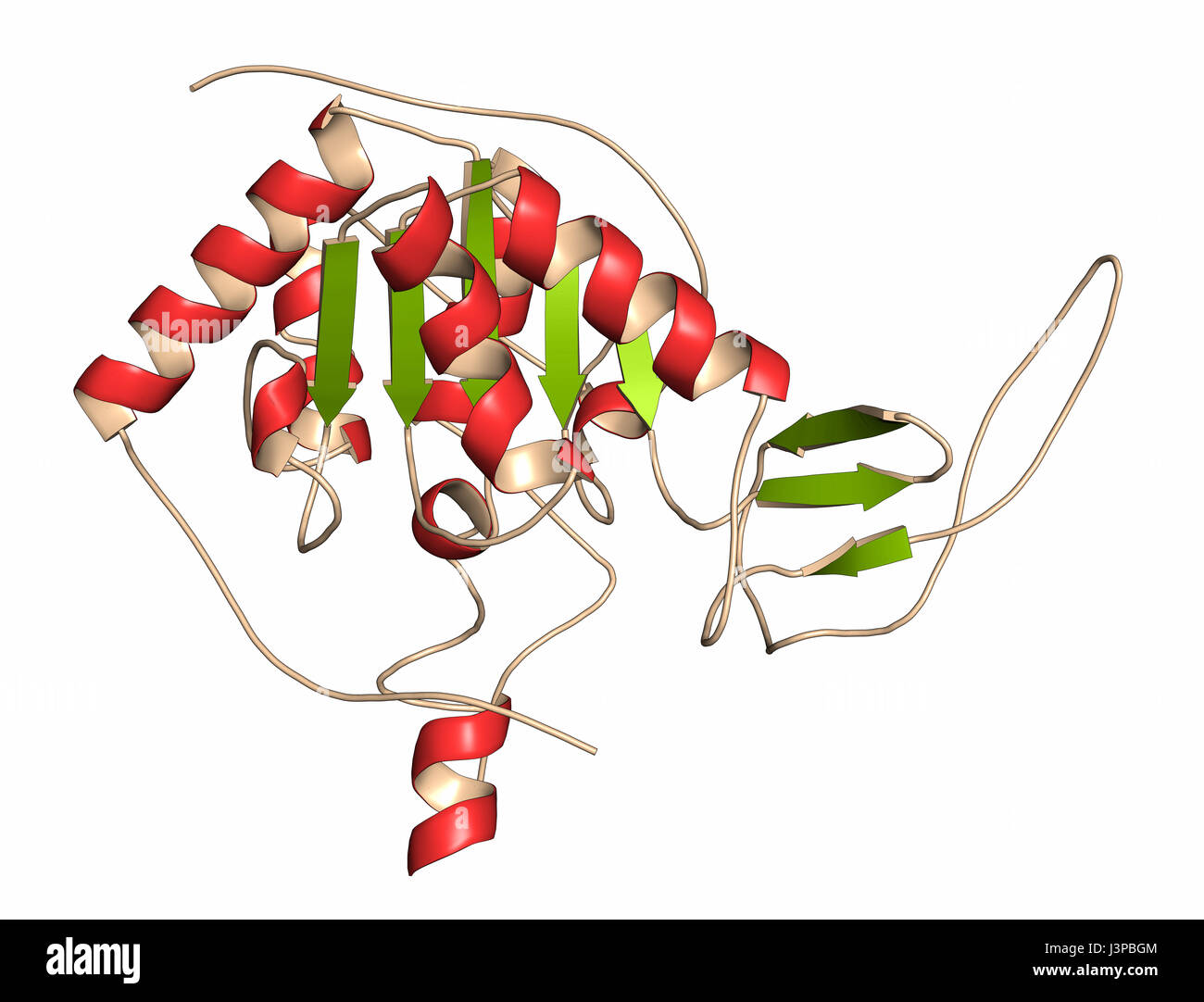 Sirtuin 6 (SIRT6) protein. Linked to longevity in mammals. Cartoon representation. Secondary structure coloring. Stock Photohttps://www.alamy.com/image-license-details/?v=1https://www.alamy.com/stock-photo-sirtuin-6-sirt6-protein-linked-to-longevity-in-mammals-cartoon-representation-140018948.html
RFJ3PBGM–Sirtuin 6 (SIRT6) protein. Linked to longevity in mammals. Cartoon representation. Secondary structure coloring.
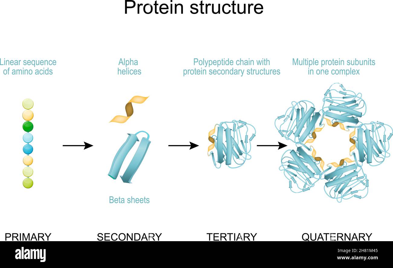 Protein structure. From Linear sequence of amino acids, alpha helices and Linear sequence to Polypeptide chain and Multiple protein subunits in one co Stock Vectorhttps://www.alamy.com/image-license-details/?v=1https://www.alamy.com/protein-structure-from-linear-sequence-of-amino-acids-alpha-helices-and-linear-sequence-to-polypeptide-chain-and-multiple-protein-subunits-in-one-co-image452424565.html
RF2H81M45–Protein structure. From Linear sequence of amino acids, alpha helices and Linear sequence to Polypeptide chain and Multiple protein subunits in one co
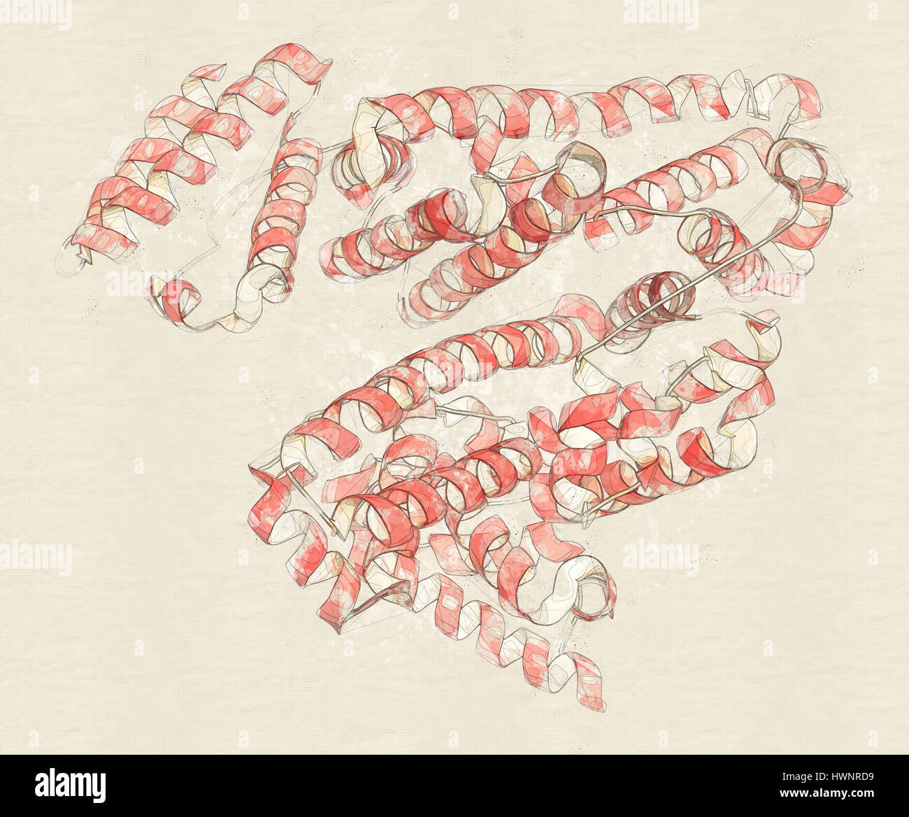 Human serum albumin protein, 3D rendering. Cartoon representation with secondary structure coloring. Stock Photohttps://www.alamy.com/image-license-details/?v=1https://www.alamy.com/stock-photo-human-serum-albumin-protein-3d-rendering-cartoon-representation-with-136318373.html
RFHWNRD9–Human serum albumin protein, 3D rendering. Cartoon representation with secondary structure coloring.
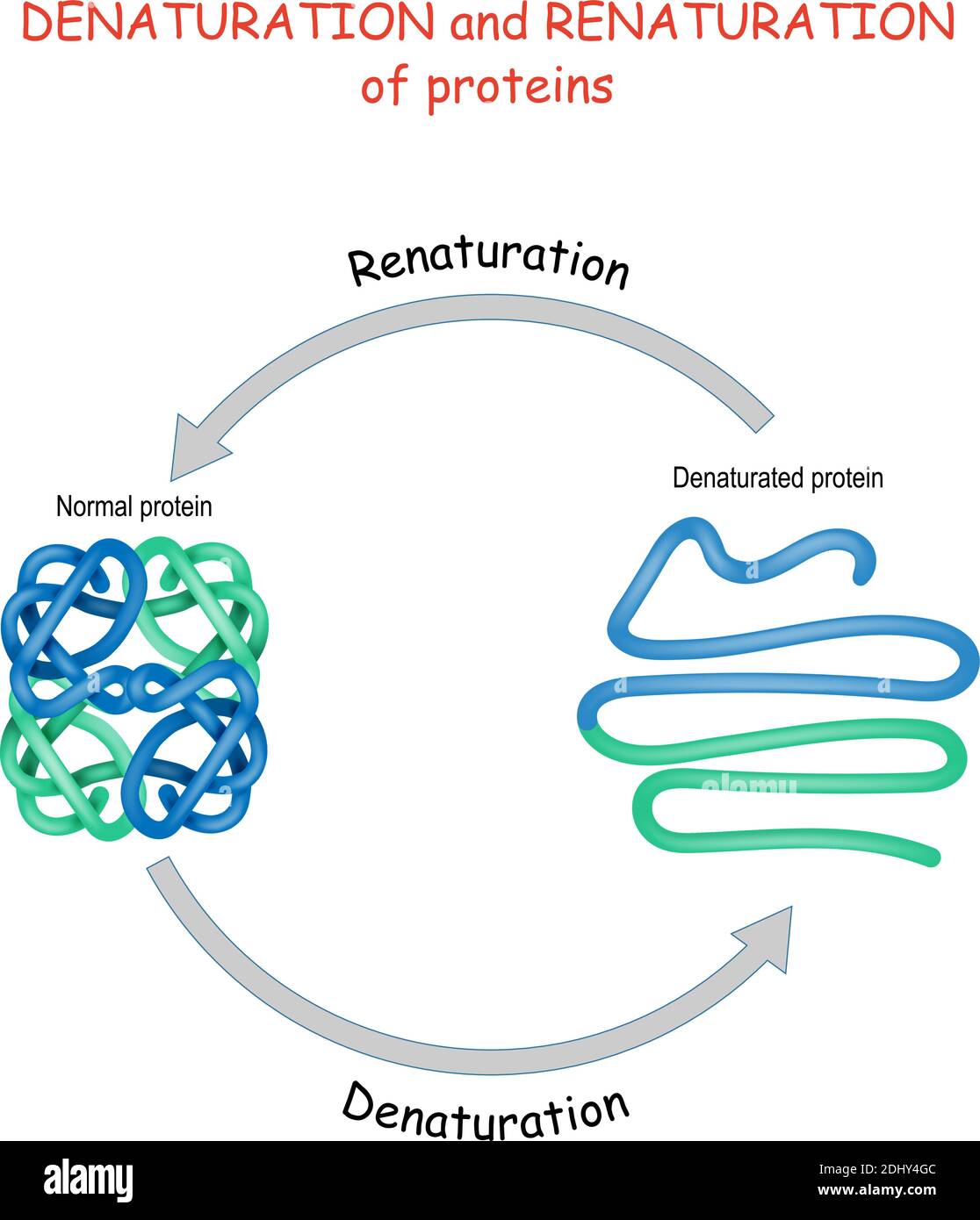 Process of Denaturation and renaturation of proteins. Vector diagram for science, education, and medical use. Stock Vectorhttps://www.alamy.com/image-license-details/?v=1https://www.alamy.com/process-of-denaturation-and-renaturation-of-proteins-vector-diagram-for-science-education-and-medical-use-image389673548.html
RF2DHY4GC–Process of Denaturation and renaturation of proteins. Vector diagram for science, education, and medical use.
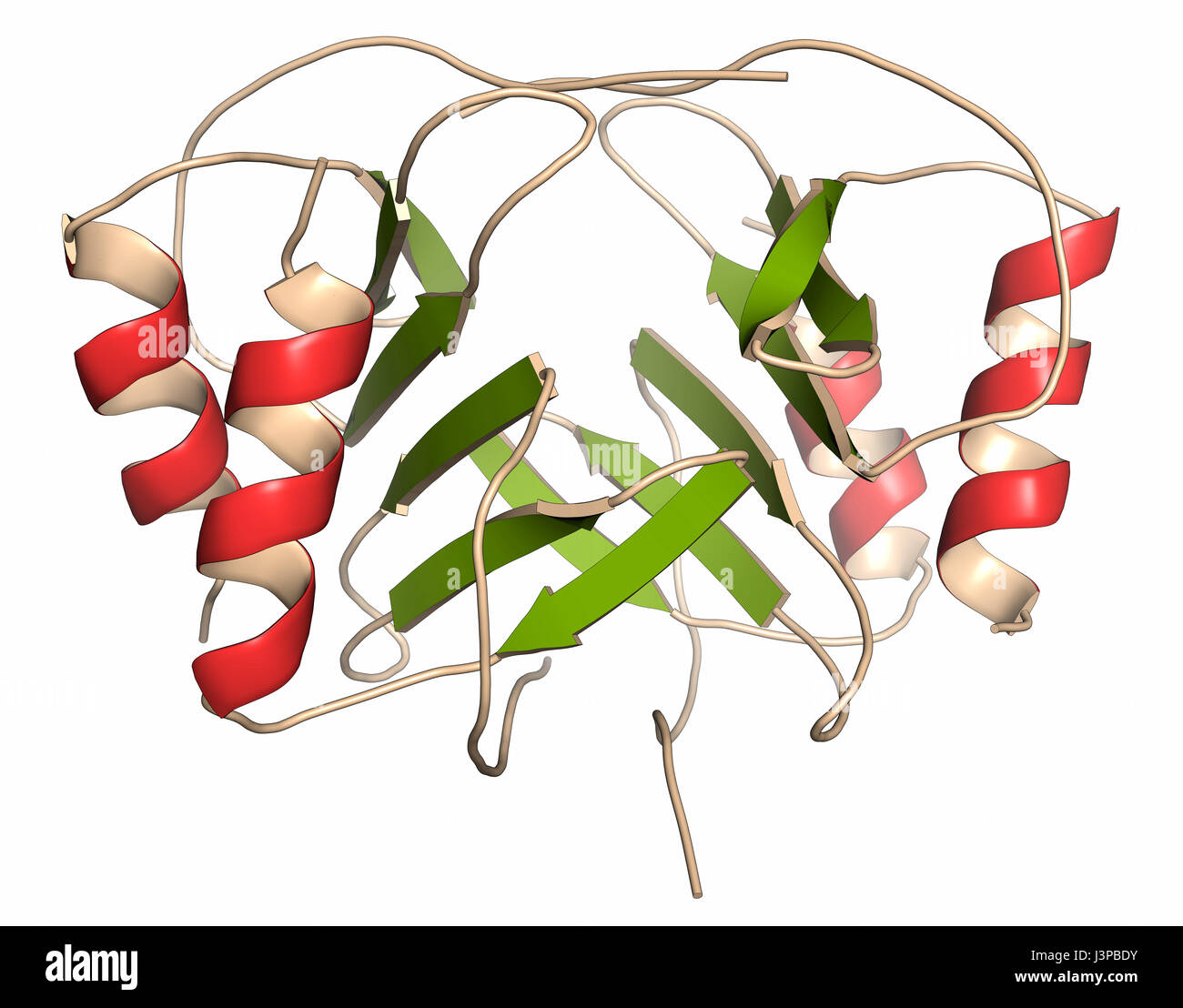 Platelet factor 4 (PF-4) chemokine protein. Cartoon representation. Secondary structure coloring. Stock Photohttps://www.alamy.com/image-license-details/?v=1https://www.alamy.com/stock-photo-platelet-factor-4-pf-4-chemokine-protein-cartoon-representation-secondary-140018871.html
RFJ3PBDY–Platelet factor 4 (PF-4) chemokine protein. Cartoon representation. Secondary structure coloring.
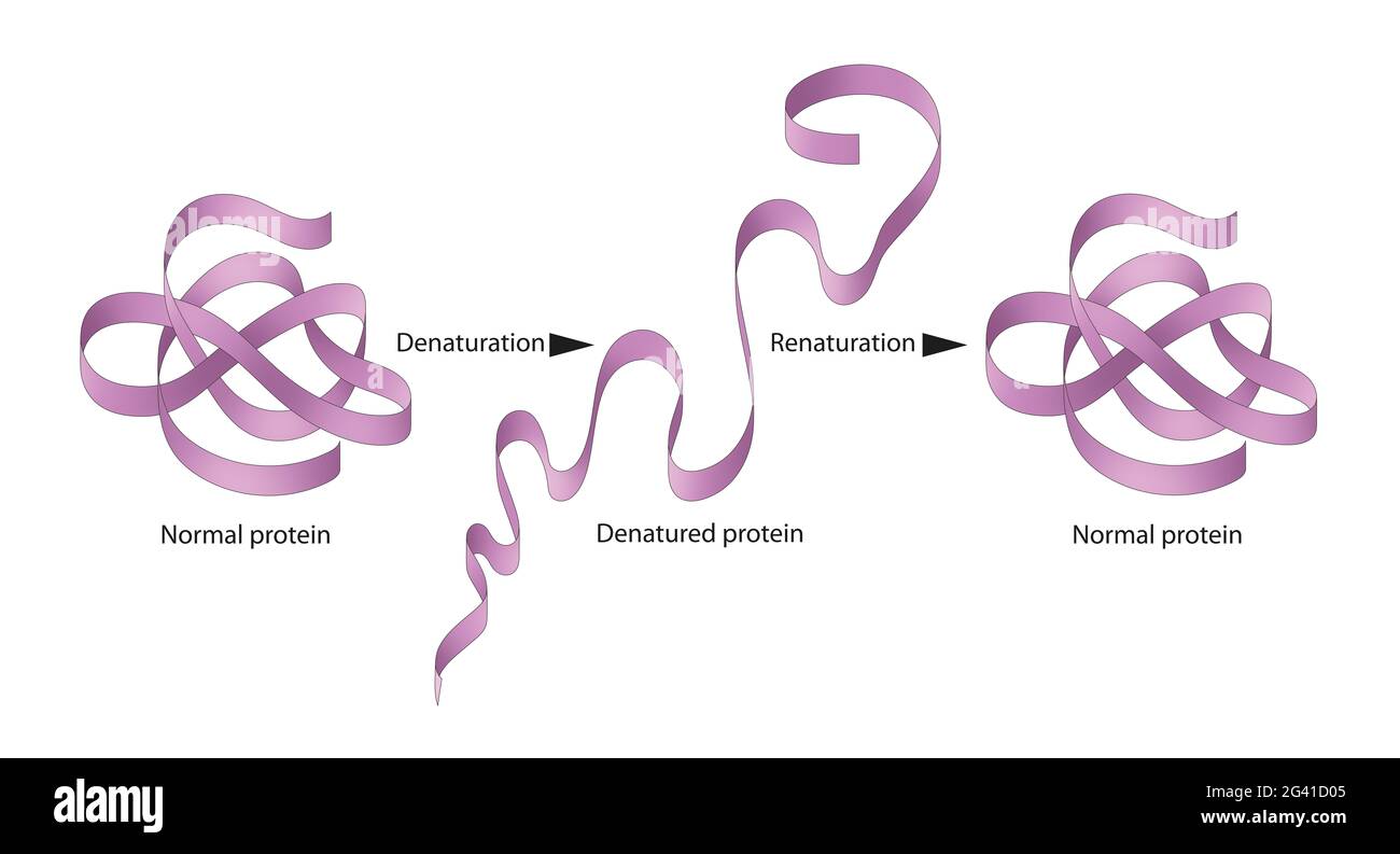 Denaturation and renaturation of Proteins Stock Photohttps://www.alamy.com/image-license-details/?v=1https://www.alamy.com/denaturation-and-renaturation-of-proteins-image432749973.html
RF2G41D05–Denaturation and renaturation of Proteins
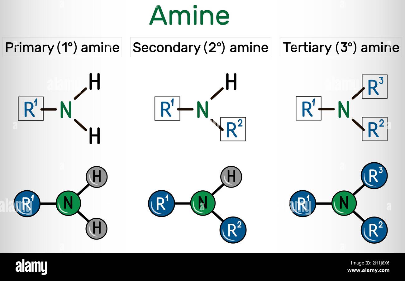 Amino group (primary, secondary, tertiary). It is functional group comprised of nitrogen atom linked with a lone pair. Amino group attached to an Stock Vectorhttps://www.alamy.com/image-license-details/?v=1https://www.alamy.com/amino-group-primary-secondary-tertiary-it-is-functional-group-comprised-of-nitrogen-atom-linked-with-a-lone-pair-amino-group-attached-to-an-image448486366.html
RF2H1J8X6–Amino group (primary, secondary, tertiary). It is functional group comprised of nitrogen atom linked with a lone pair. Amino group attached to an
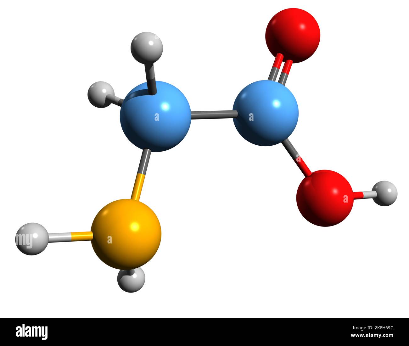 3D image of Aminoethanoic acid skeletal formula - molecular chemical structure of amino acid isolated on white background Stock Photohttps://www.alamy.com/image-license-details/?v=1https://www.alamy.com/3d-image-of-aminoethanoic-acid-skeletal-formula-molecular-chemical-structure-of-amino-acid-isolated-on-white-background-image491488296.html
RF2KFH69C–3D image of Aminoethanoic acid skeletal formula - molecular chemical structure of amino acid isolated on white background
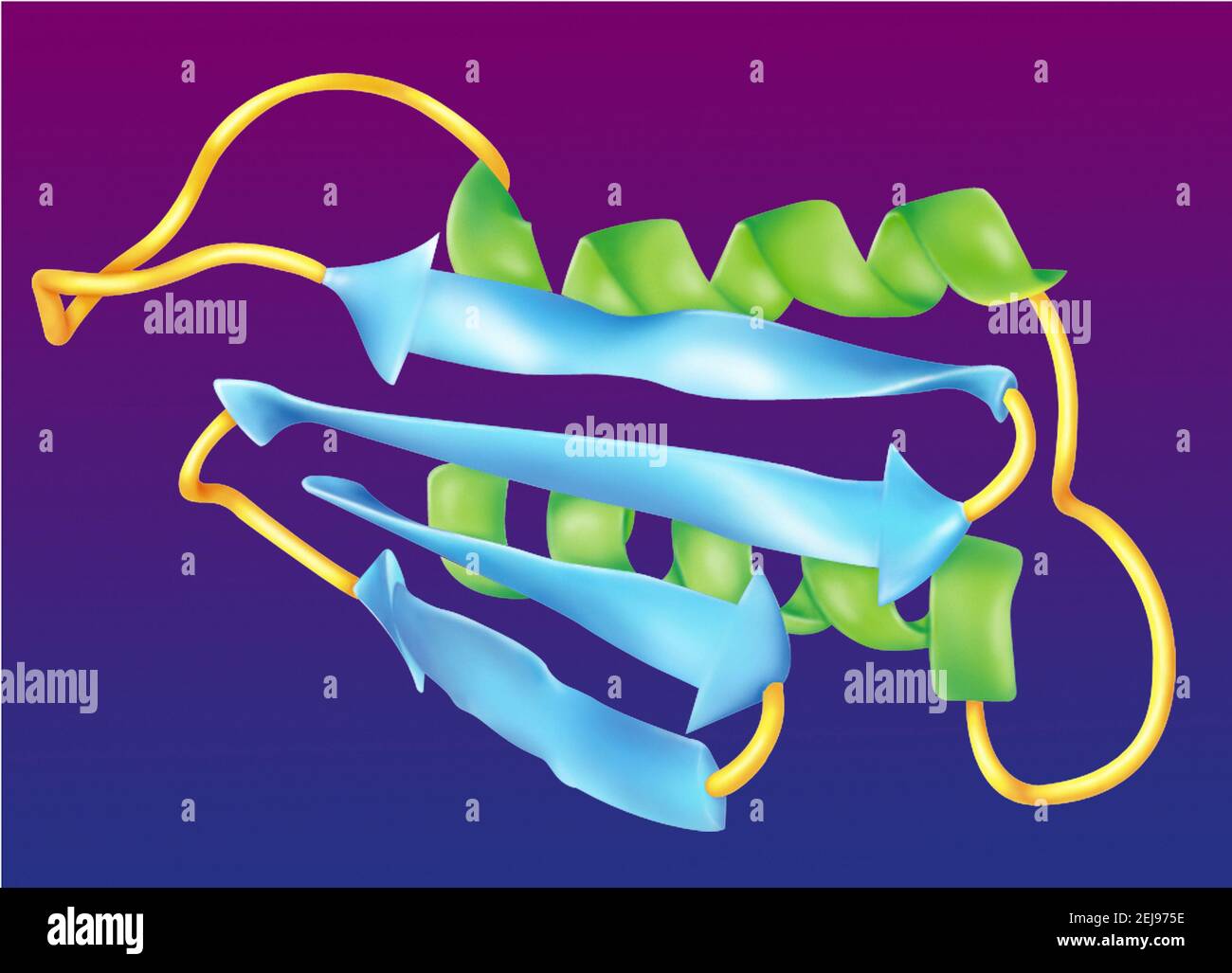 Prion Stock Photohttps://www.alamy.com/image-license-details/?v=1https://www.alamy.com/prion-image407105482.html
RM2EJ975E–Prion
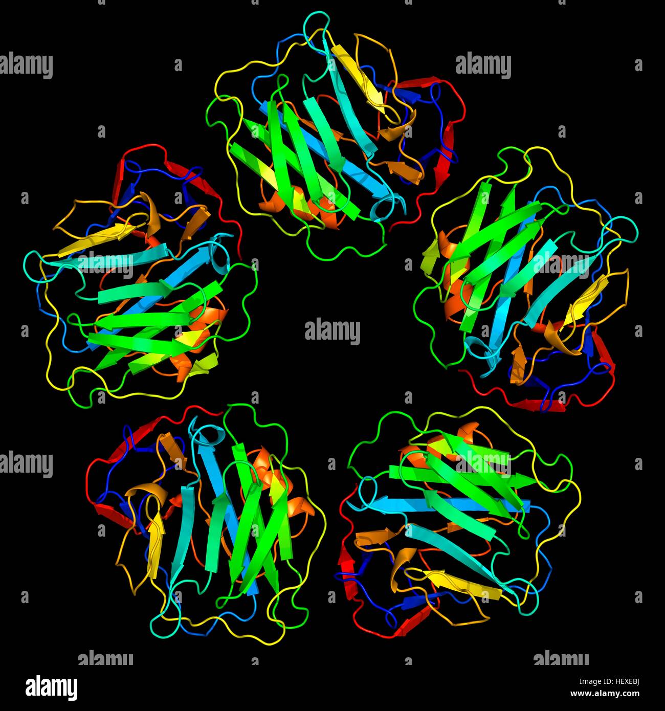 C-reactive protein,molecular model.The protein is made up of five sub-units (monomers) arranged in ring.The secondary structure of protein is shown,with beta sheets (arrows) alpha helices (spirals) connected by linking regions.C-reactive protein (CRP) is blood plasma protein produced by liver.It is acute phase protein,one whose levels rise in response to inflammation.It assists binding of complement proteins to foreign or damaged cells,an immunological response that destroys target cells.High blood levels of CRP are associated increased risk of heart disease diabetes. Stock Photohttps://www.alamy.com/image-license-details/?v=1https://www.alamy.com/stock-photo-c-reactive-proteinmolecular-modelthe-protein-is-made-up-of-five-sub-129659814.html
RFHEXEBJ–C-reactive protein,molecular model.The protein is made up of five sub-units (monomers) arranged in ring.The secondary structure of protein is shown,with beta sheets (arrows) alpha helices (spirals) connected by linking regions.C-reactive protein (CRP) is blood plasma protein produced by liver.It is acute phase protein,one whose levels rise in response to inflammation.It assists binding of complement proteins to foreign or damaged cells,an immunological response that destroys target cells.High blood levels of CRP are associated increased risk of heart disease diabetes.
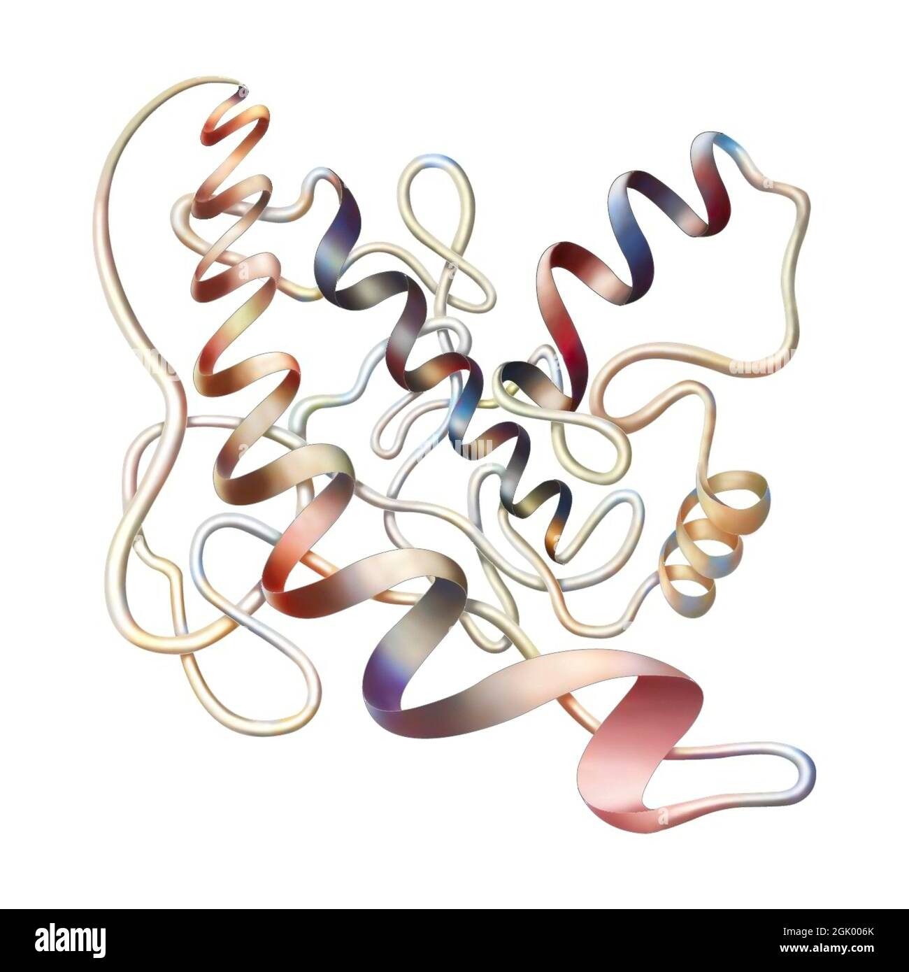 Representation of the secondary structure of a protein. Stock Photohttps://www.alamy.com/image-license-details/?v=1https://www.alamy.com/representation-of-the-secondary-structure-of-a-protein-image441937851.html
RF2GK006K–Representation of the secondary structure of a protein.
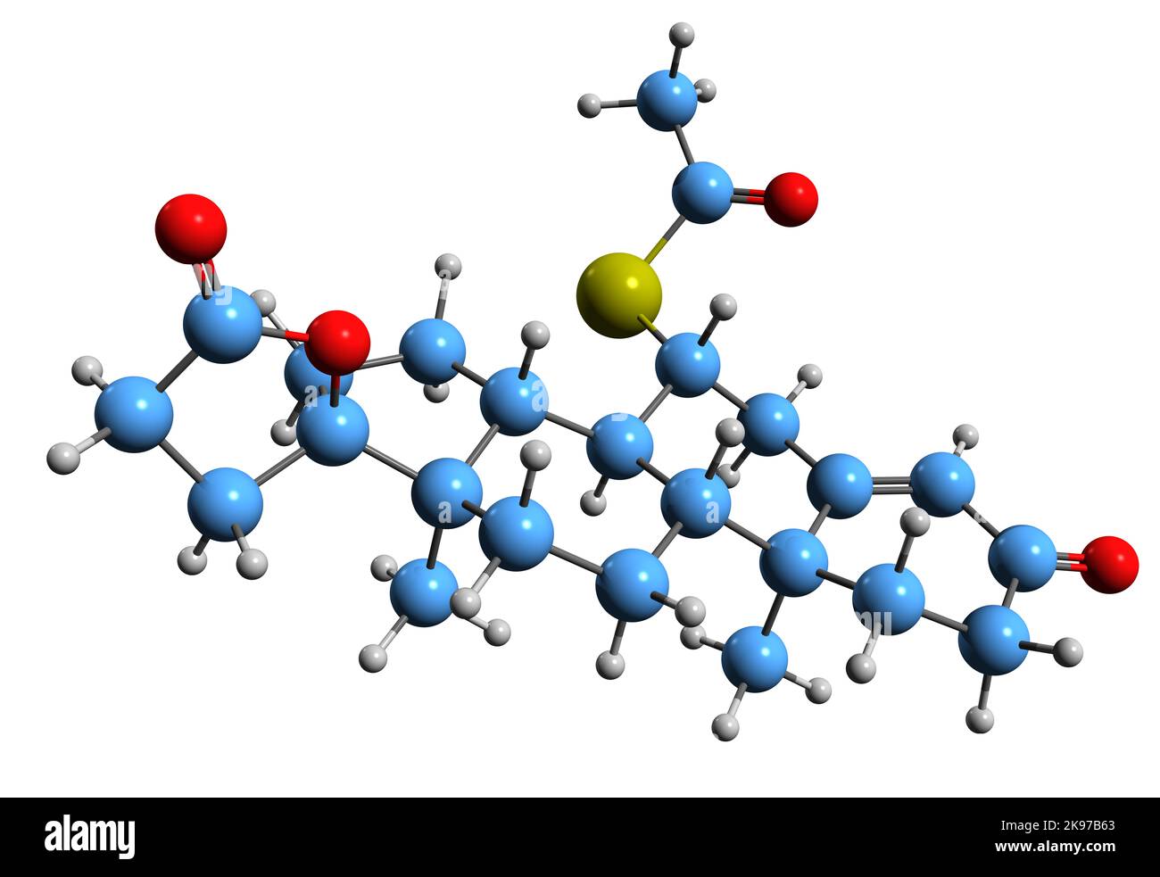 3D image of Spironolactone skeletal formula - molecular chemical structure of Antimineralocorticoid isolated on white background Stock Photohttps://www.alamy.com/image-license-details/?v=1https://www.alamy.com/3d-image-of-spironolactone-skeletal-formula-molecular-chemical-structure-of-antimineralocorticoid-isolated-on-white-background-image487584667.html
RF2K97B63–3D image of Spironolactone skeletal formula - molecular chemical structure of Antimineralocorticoid isolated on white background
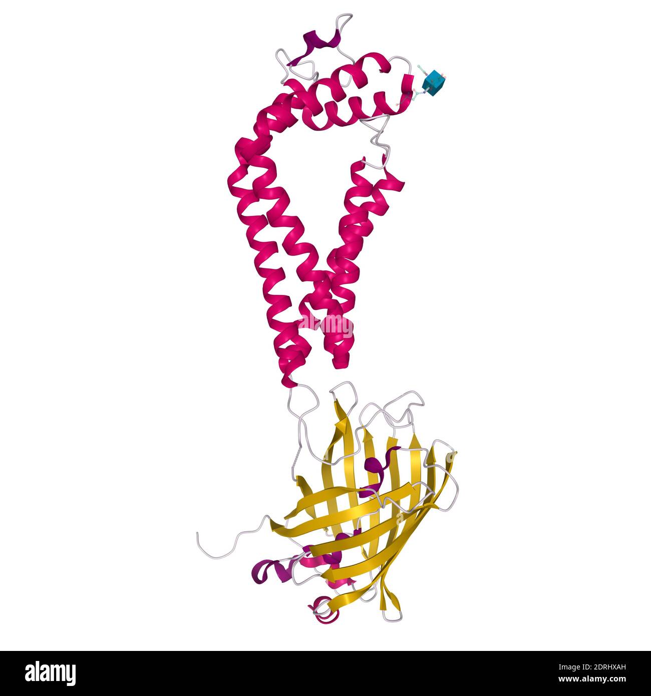 Crystal structure of human CD53, 3D cartoon model with differently colored elements of the secondary structure, white background Stock Photohttps://www.alamy.com/image-license-details/?v=1https://www.alamy.com/crystal-structure-of-human-cd53-3d-cartoon-model-with-differently-colored-elements-of-the-secondary-structure-white-background-image393159049.html
RF2DRHXAH–Crystal structure of human CD53, 3D cartoon model with differently colored elements of the secondary structure, white background
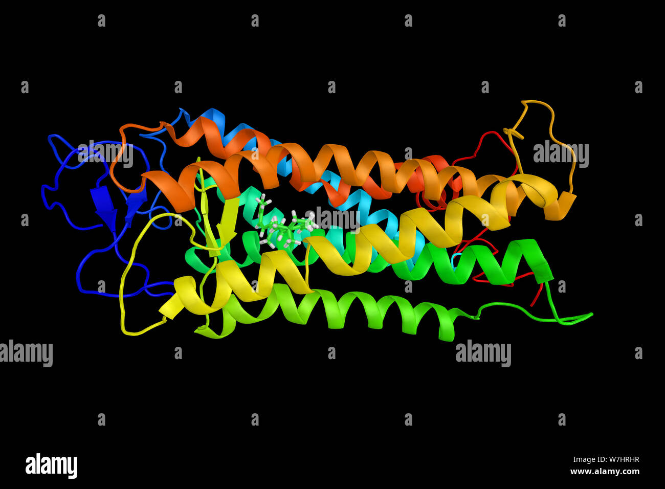 Red-sensitive opsin, a protein. Its name as the 'red' opsin reflects the fact that it is more sensitive to red than the other two human opsins. 3d ren Stock Photohttps://www.alamy.com/image-license-details/?v=1https://www.alamy.com/red-sensitive-opsin-a-protein-its-name-as-the-red-opsin-reflects-the-fact-that-it-is-more-sensitive-to-red-than-the-other-two-human-opsins-3d-ren-image262849827.html
RFW7HRHR–Red-sensitive opsin, a protein. Its name as the 'red' opsin reflects the fact that it is more sensitive to red than the other two human opsins. 3d ren
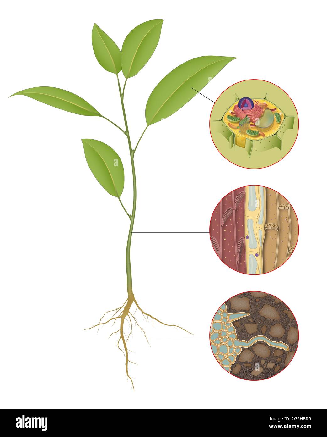 Three types of plant cells Stock Photohttps://www.alamy.com/image-license-details/?v=1https://www.alamy.com/three-types-of-plant-cells-image434329611.html
RF2G6HBRR–Three types of plant cells
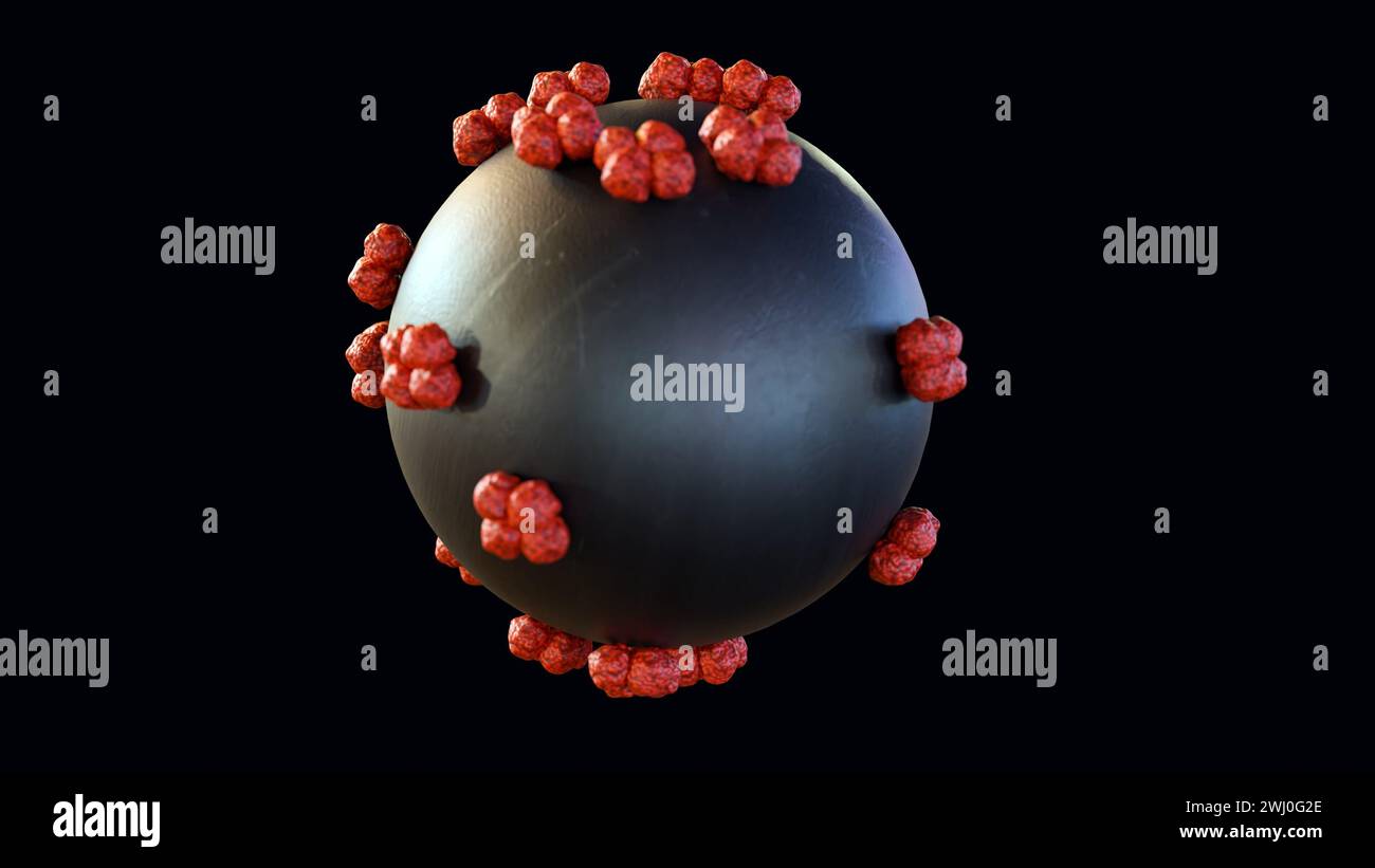 3d rendering of nanoparticles conjugated haemogoblin molecules Stock Photohttps://www.alamy.com/image-license-details/?v=1https://www.alamy.com/3d-rendering-of-nanoparticles-conjugated-haemogoblin-molecules-image596228934.html
RF2WJ0G2E–3d rendering of nanoparticles conjugated haemogoblin molecules
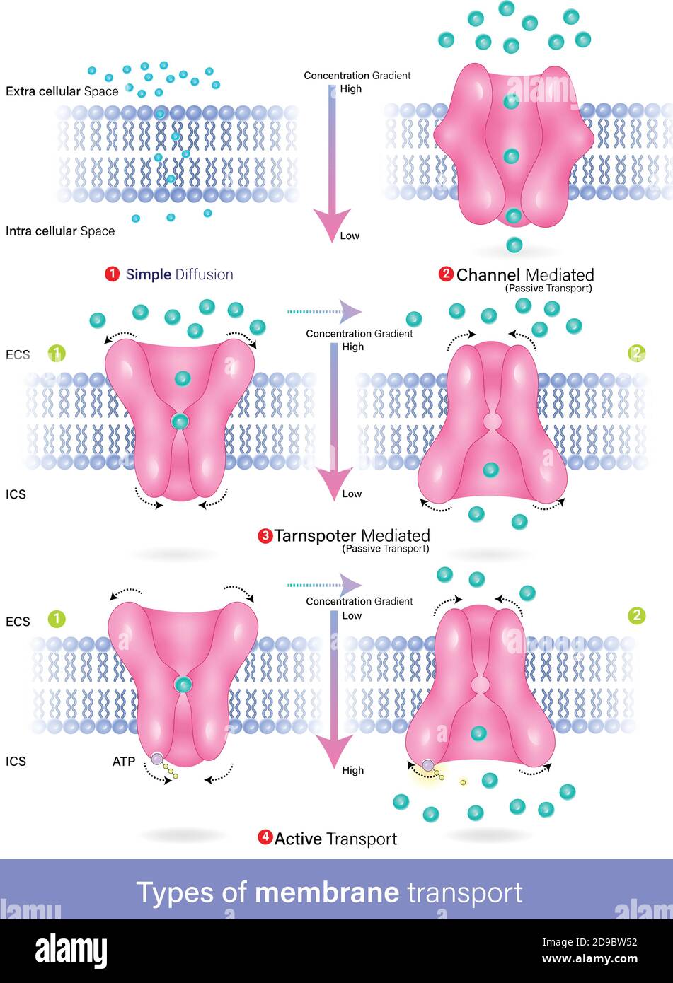 Vector of types of membrane transport diffusion, active and passive transport, facilitated diffusion Stock Vectorhttps://www.alamy.com/image-license-details/?v=1https://www.alamy.com/vector-of-types-of-membrane-transport-diffusion-active-and-passive-transport-facilitated-diffusion-image384421214.html
RF2D9BW52–Vector of types of membrane transport diffusion, active and passive transport, facilitated diffusion
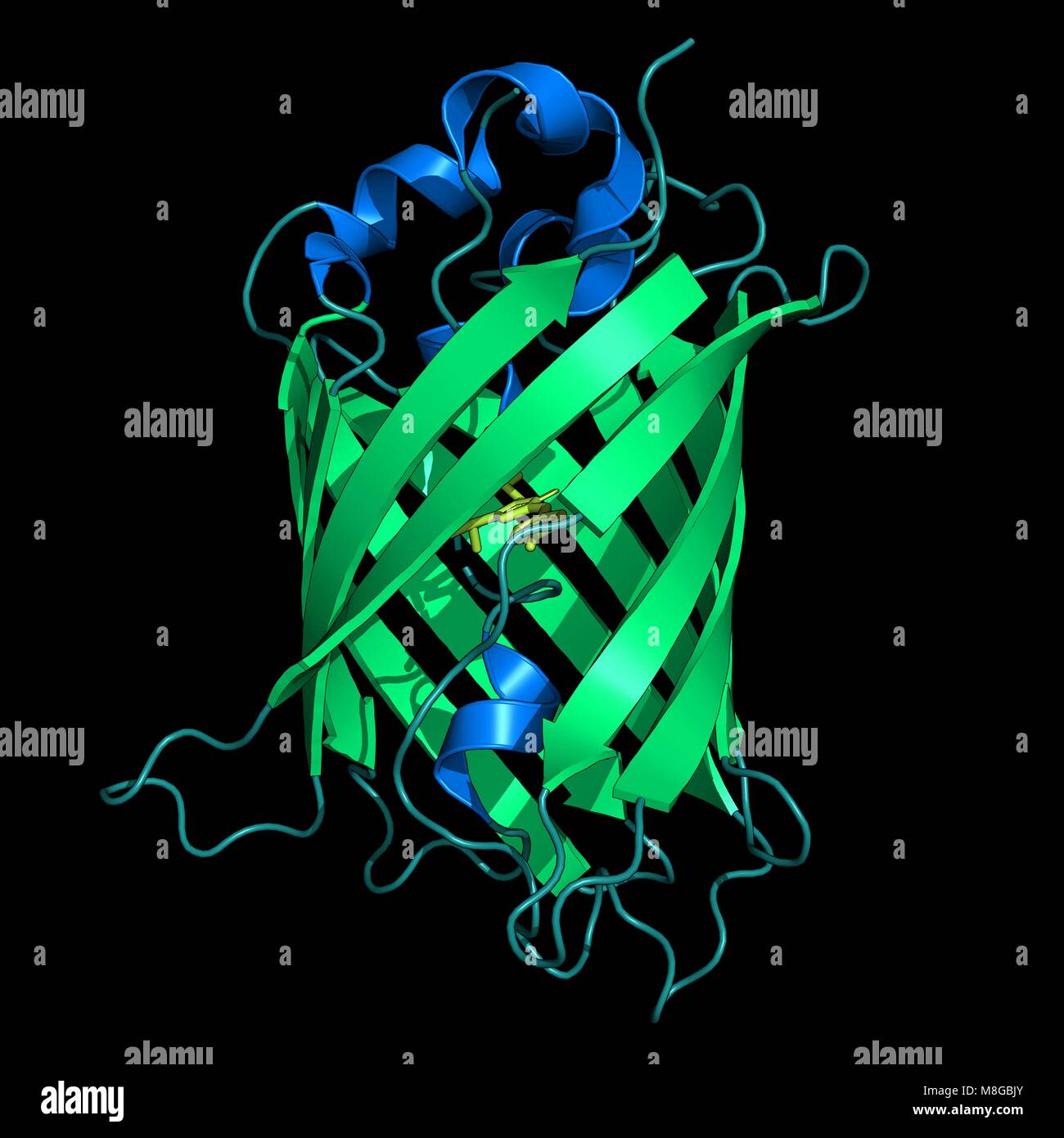 Representation of the green fluorescent protein from Aequorea victoria Stock Photohttps://www.alamy.com/image-license-details/?v=1https://www.alamy.com/stock-photo-representation-of-the-green-fluorescent-protein-from-aequorea-victoria-177381315.html
RFM8GBJY–Representation of the green fluorescent protein from Aequorea victoria
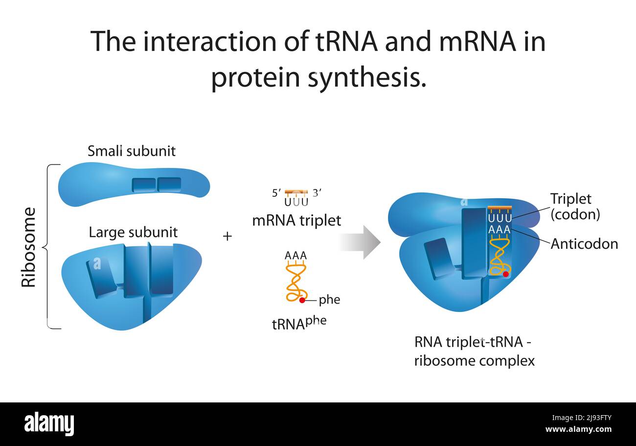 The interaction of tRNA and mRNA in protein synthesis Stock Photohttps://www.alamy.com/image-license-details/?v=1https://www.alamy.com/the-interaction-of-trna-and-mrna-in-protein-synthesis-image470290155.html
RF2J93FTY–The interaction of tRNA and mRNA in protein synthesis
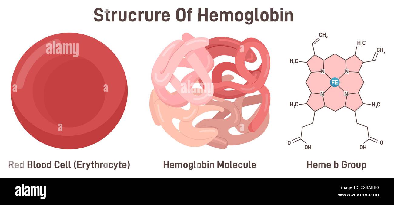 Hemoglobin molecule structure. Iron-containing oxygen-transport metalloprotein in the red blood cell, combination of iron and the protein globin. Heme b group formula. Flat vector illustration Stock Vectorhttps://www.alamy.com/image-license-details/?v=1https://www.alamy.com/hemoglobin-molecule-structure-iron-containing-oxygen-transport-metalloprotein-in-the-red-blood-cell-combination-of-iron-and-the-protein-globin-heme-b-group-formula-flat-vector-illustration-image609352548.html
RF2XBABB0–Hemoglobin molecule structure. Iron-containing oxygen-transport metalloprotein in the red blood cell, combination of iron and the protein globin. Heme b group formula. Flat vector illustration
 Scientific Designing of Biochemial Structure of Amino acids, Peptides And Proteins Molecular Model. Vector Illustration. Stock Vectorhttps://www.alamy.com/image-license-details/?v=1https://www.alamy.com/scientific-designing-of-biochemial-structure-of-amino-acids-peptides-and-proteins-molecular-model-vector-illustration-image483744557.html
RF2K30D39–Scientific Designing of Biochemial Structure of Amino acids, Peptides And Proteins Molecular Model. Vector Illustration.
RF2XCPNJB–Amino acid chemical molecule of Leucine, molecular formula and structure chain, vector icon. Leucine essential amino acid of biosynthesis of protein for pharmacy medicine and health nutrition study
 Chromosome structure. Biochemistry genetics, biology dna, chromosome genes vector illustration Stock Vectorhttps://www.alamy.com/image-license-details/?v=1https://www.alamy.com/chromosome-structure-biochemistry-genetics-biology-dna-chromosome-genes-vector-illustration-image565582682.html
RF2RT4EDE–Chromosome structure. Biochemistry genetics, biology dna, chromosome genes vector illustration
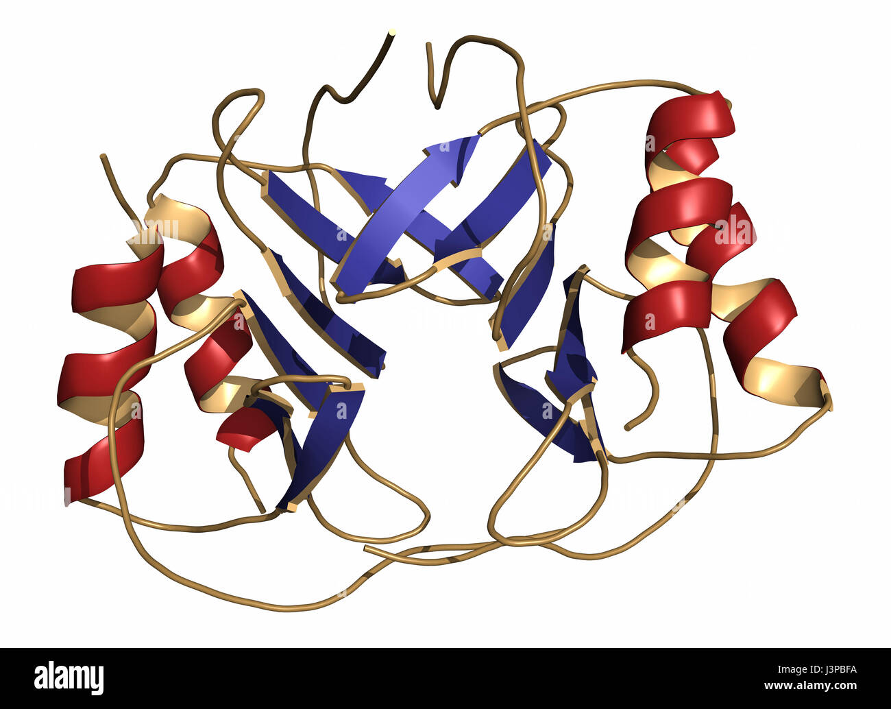 Platelet factor 4 (PF-4) chemokine protein. Cartoon representation. Secondary structure coloring. Stock Photohttps://www.alamy.com/image-license-details/?v=1https://www.alamy.com/stock-photo-platelet-factor-4-pf-4-chemokine-protein-cartoon-representation-secondary-140018910.html
RFJ3PBFA–Platelet factor 4 (PF-4) chemokine protein. Cartoon representation. Secondary structure coloring.
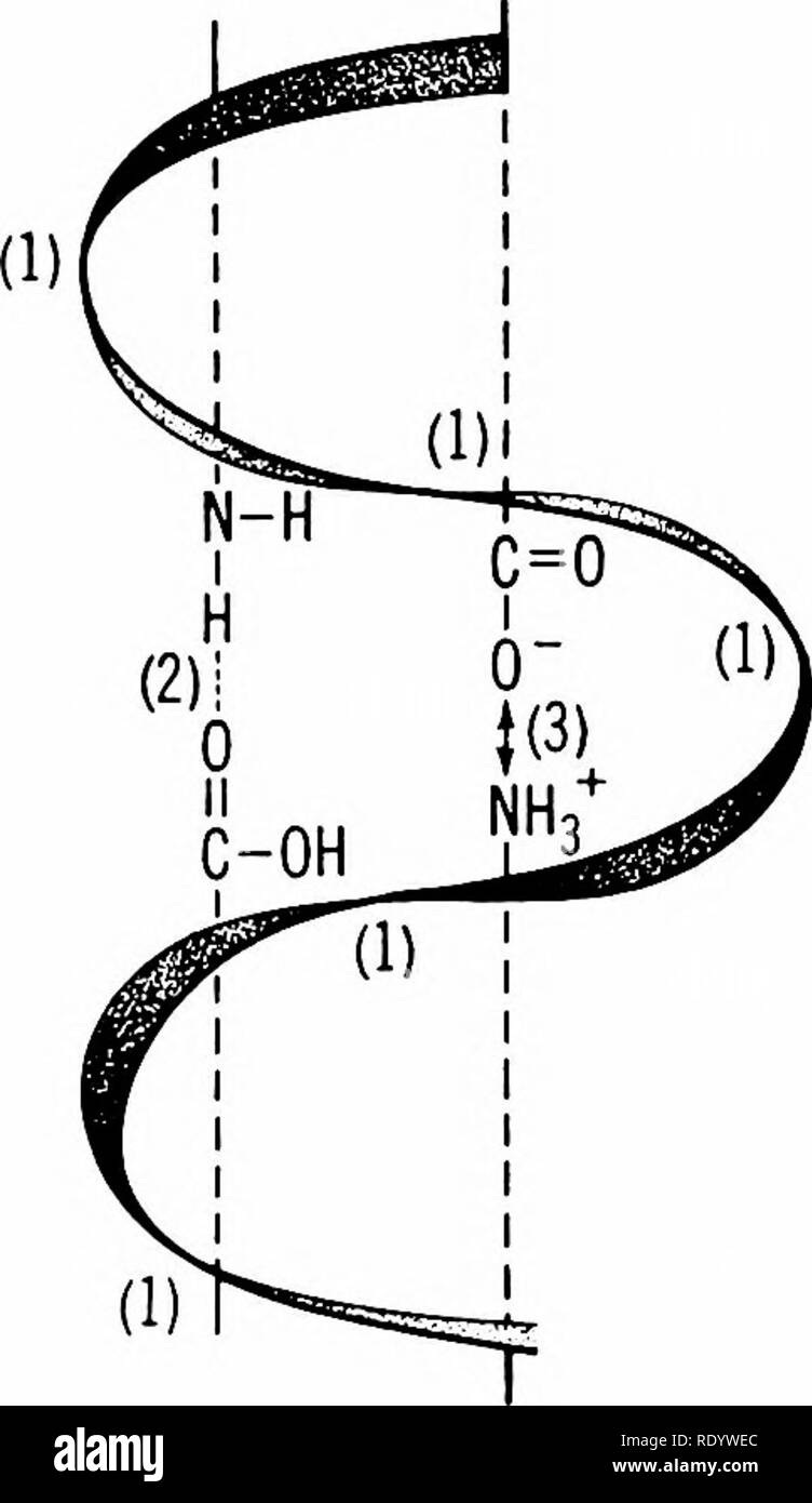 . Principles of modern biology. Biology. The Chemical the RNA components ol the cytoplasm, as will be considered in Chapters 7 and 27. Three-Dimensional Configuration of Protein Molecules: Primary, Secondary, and Tertiary Structure. The primary struc- ture of a protein molecule is constituted by one or more peptide chains. Within each chain the amino acid units are held together by peptide linkages; the peptide chains, if. -S-S-Kl) Fig. 4-17. Helical configuration of a protein mole- cule. Three types of bonds determine the primary, sec- ondary, and tertiary aspects of protein structure-. (1) s Stock Photohttps://www.alamy.com/image-license-details/?v=1https://www.alamy.com/principles-of-modern-biology-biology-the-chemical-the-rna-components-ol-the-cytoplasm-as-will-be-considered-in-chapters-7-and-27-three-dimensional-configuration-of-protein-molecules-primary-secondary-and-tertiary-structure-the-primary-struc-ture-of-a-protein-molecule-is-constituted-by-one-or-more-peptide-chains-within-each-chain-the-amino-acid-units-are-held-together-by-peptide-linkages-the-peptide-chains-if-s-s-kl-fig-4-17-helical-configuration-of-a-protein-mole-cule-three-types-of-bonds-determine-the-primary-sec-ondary-and-tertiary-aspects-of-protein-structure-1-s-image232338020.html
RMRDYWEC–. Principles of modern biology. Biology. The Chemical the RNA components ol the cytoplasm, as will be considered in Chapters 7 and 27. Three-Dimensional Configuration of Protein Molecules: Primary, Secondary, and Tertiary Structure. The primary struc- ture of a protein molecule is constituted by one or more peptide chains. Within each chain the amino acid units are held together by peptide linkages; the peptide chains, if. -S-S-Kl) Fig. 4-17. Helical configuration of a protein mole- cule. Three types of bonds determine the primary, sec- ondary, and tertiary aspects of protein structure-. (1) s
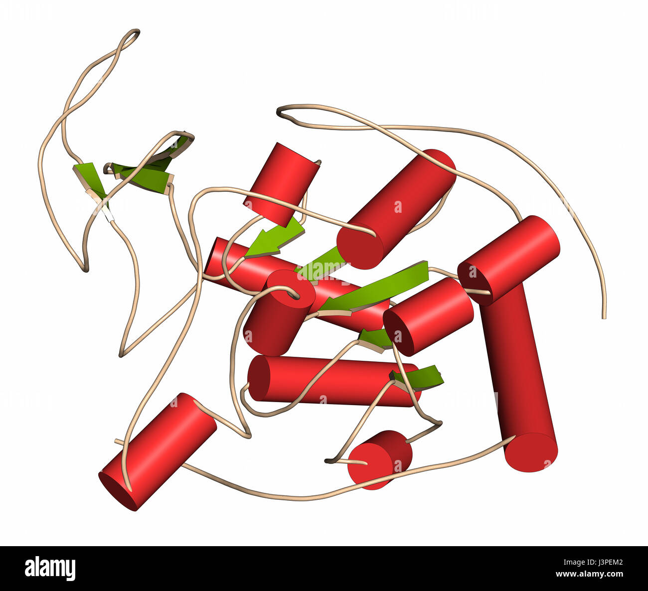 Sirtuin 6 (SIRT6) protein. Linked to longevity in mammals. Cartoon representation. Secondary structure coloring. Stock Photohttps://www.alamy.com/image-license-details/?v=1https://www.alamy.com/stock-photo-sirtuin-6-sirt6-protein-linked-to-longevity-in-mammals-cartoon-representation-140021394.html
RFJ3PEM2–Sirtuin 6 (SIRT6) protein. Linked to longevity in mammals. Cartoon representation. Secondary structure coloring.
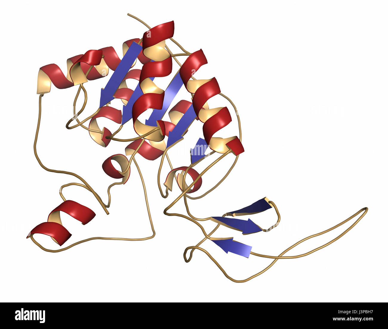 Sirtuin 6 (SIRT6) protein. Linked to longevity in mammals. Cartoon representation. Secondary structure coloring. Stock Photohttps://www.alamy.com/image-license-details/?v=1https://www.alamy.com/stock-photo-sirtuin-6-sirt6-protein-linked-to-longevity-in-mammals-cartoon-representation-140018963.html
RFJ3PBH7–Sirtuin 6 (SIRT6) protein. Linked to longevity in mammals. Cartoon representation. Secondary structure coloring.
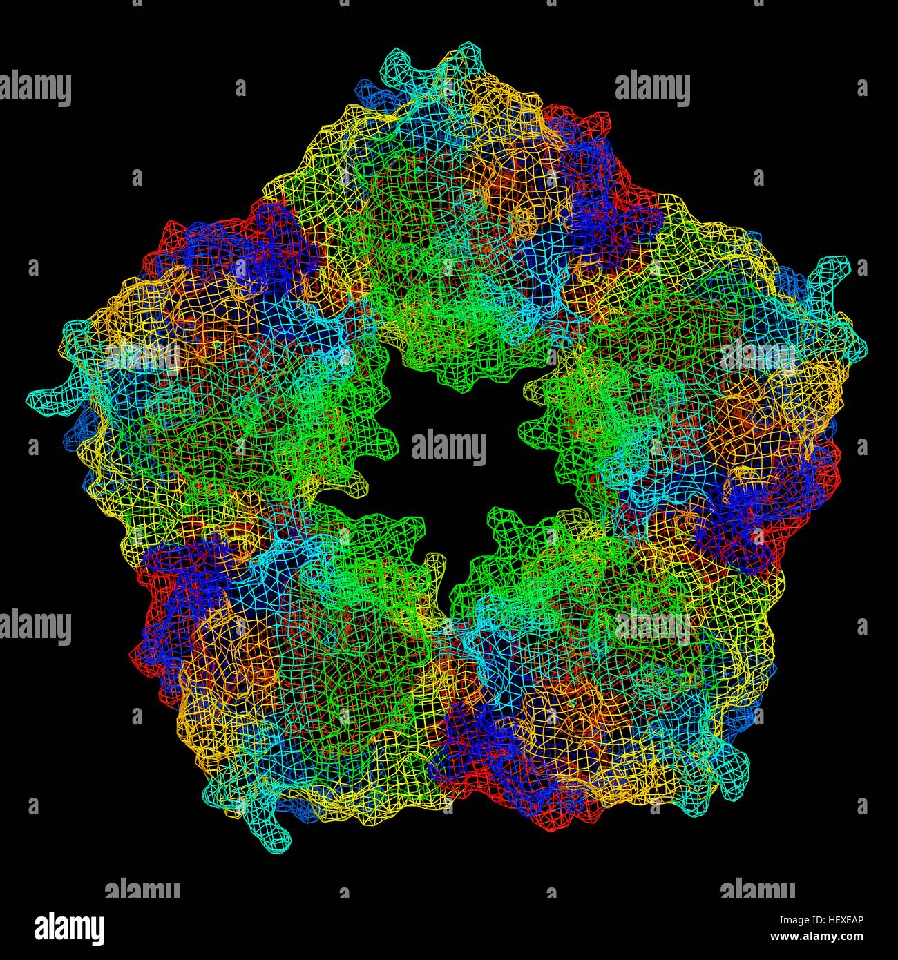 C-reactive protein,molecular model.The protein is made up of five sub-units (monomers) arranged in ring.The secondary structure of protein is shown,with beta sheets (arrows) alpha helices (spirals) connected by linking regions.C-reactive protein (CRP) is blood plasma protein produced by liver.It is acute phase protein,one whose levels rise in response to inflammation.It assists binding of complement proteins to foreign or damaged cells,an immunological response that destroys target cells.High blood levels of CRP are associated increased risk of heart disease diabetes. Stock Photohttps://www.alamy.com/image-license-details/?v=1https://www.alamy.com/stock-photo-c-reactive-proteinmolecular-modelthe-protein-is-made-up-of-five-sub-129659790.html
RFHEXEAP–C-reactive protein,molecular model.The protein is made up of five sub-units (monomers) arranged in ring.The secondary structure of protein is shown,with beta sheets (arrows) alpha helices (spirals) connected by linking regions.C-reactive protein (CRP) is blood plasma protein produced by liver.It is acute phase protein,one whose levels rise in response to inflammation.It assists binding of complement proteins to foreign or damaged cells,an immunological response that destroys target cells.High blood levels of CRP are associated increased risk of heart disease diabetes.
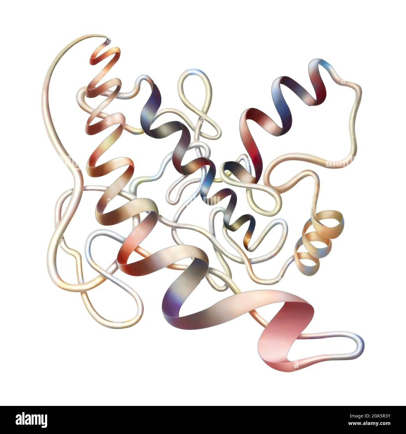 Representation of the secondary structure of a protein. Stock Photohttps://www.alamy.com/image-license-details/?v=1https://www.alamy.com/representation-of-the-secondary-structure-of-a-protein-image442065567.html
RF2GK5R3Y–Representation of the secondary structure of a protein.
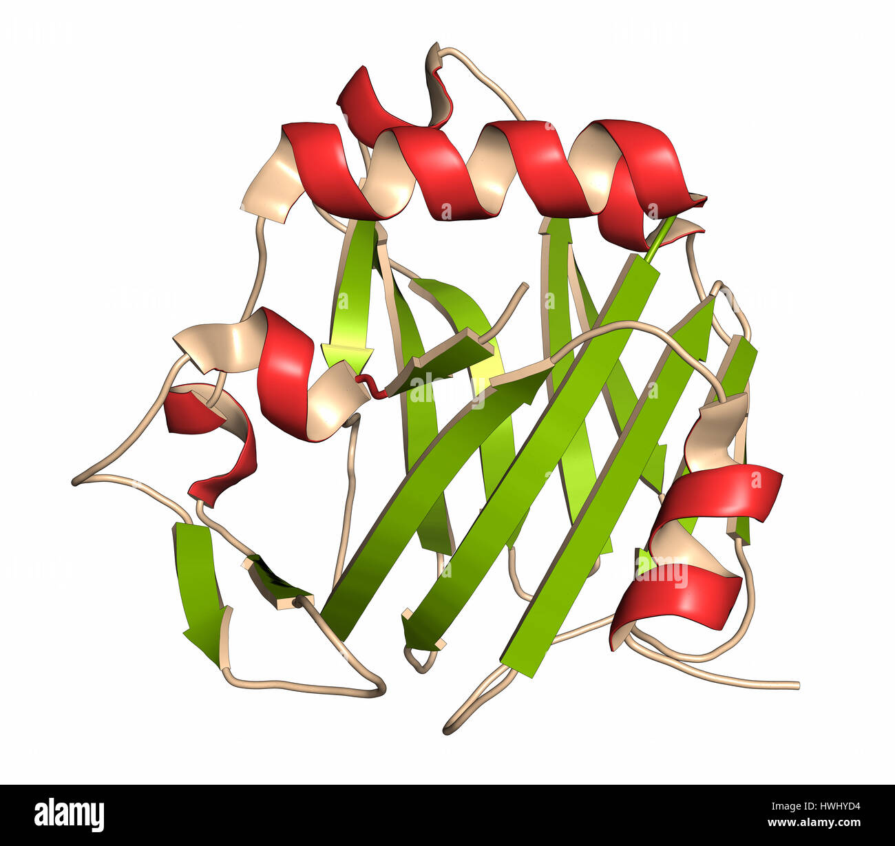 Thrombospondin-1 protein (N-terminal domain). Cartoon representation with secondary structure coloring (green sheets, red helices). Stock Photohttps://www.alamy.com/image-license-details/?v=1https://www.alamy.com/stock-photo-thrombospondin-1-protein-n-terminal-domain-cartoon-representation-136233696.html
RFHWHYD4–Thrombospondin-1 protein (N-terminal domain). Cartoon representation with secondary structure coloring (green sheets, red helices).
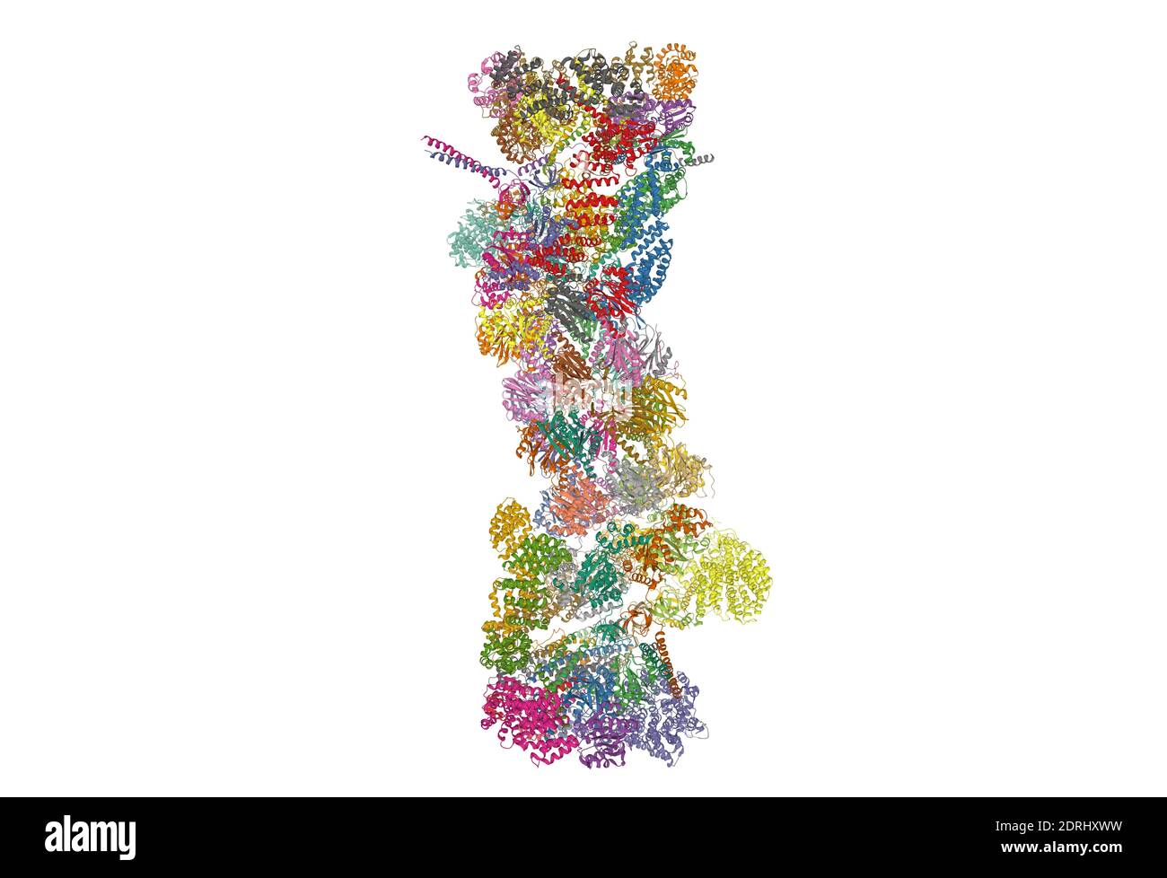 Structure of 26S proteasome, 3D cartoon model isolated, white background Stock Photohttps://www.alamy.com/image-license-details/?v=1https://www.alamy.com/structure-of-26s-proteasome-3d-cartoon-model-isolated-white-background-image393159477.html
RF2DRHXWW–Structure of 26S proteasome, 3D cartoon model isolated, white background
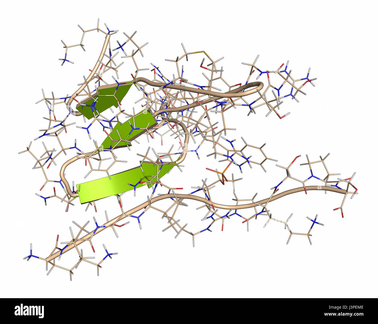 Tau protein fragment. May play a role in Alzheimer's disease. Cartoon + line representation. Secondary structure coloring. Stock Photohttps://www.alamy.com/image-license-details/?v=1https://www.alamy.com/stock-photo-tau-protein-fragment-may-play-a-role-in-alzheimers-disease-cartoon-140021406.html
RFJ3PEME–Tau protein fragment. May play a role in Alzheimer's disease. Cartoon + line representation. Secondary structure coloring.
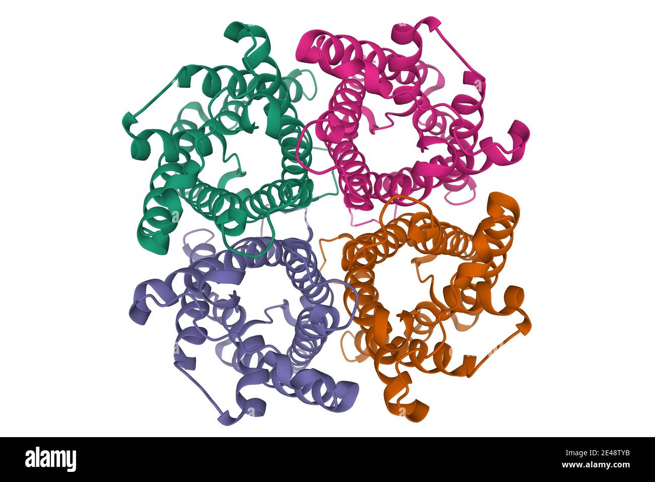 Quaternary structure of rat AQUAPORIN-4, 3D cartoon model isolated, white background Stock Photohttps://www.alamy.com/image-license-details/?v=1https://www.alamy.com/quaternary-structure-of-rat-aquaporin-4-3d-cartoon-model-isolated-white-background-image398492287.html
RF2E48TYB–Quaternary structure of rat AQUAPORIN-4, 3D cartoon model isolated, white background
 3d rendering of nanoparticles conjugated haemogoblin molecules Stock Photohttps://www.alamy.com/image-license-details/?v=1https://www.alamy.com/3d-rendering-of-nanoparticles-conjugated-haemogoblin-molecules-image596228936.html
RF2WJ0G2G–3d rendering of nanoparticles conjugated haemogoblin molecules
 Representation of the blue light receptor YtvA of Bacillus subtilis Stock Photohttps://www.alamy.com/image-license-details/?v=1https://www.alamy.com/stock-photo-representation-of-the-blue-light-receptor-ytva-of-bacillus-subtilis-177355115.html
RFM8F677–Representation of the blue light receptor YtvA of Bacillus subtilis
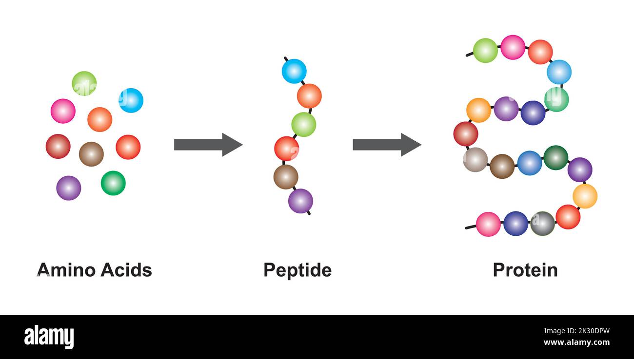 Scientific Designing of Biochemial Structure of Amino acids, Peptides And Proteins Molecular Model. Vector Illustration. Stock Vectorhttps://www.alamy.com/image-license-details/?v=1https://www.alamy.com/scientific-designing-of-biochemial-structure-of-amino-acids-peptides-and-proteins-molecular-model-vector-illustration-image483745105.html
RF2K30DPW–Scientific Designing of Biochemial Structure of Amino acids, Peptides And Proteins Molecular Model. Vector Illustration.
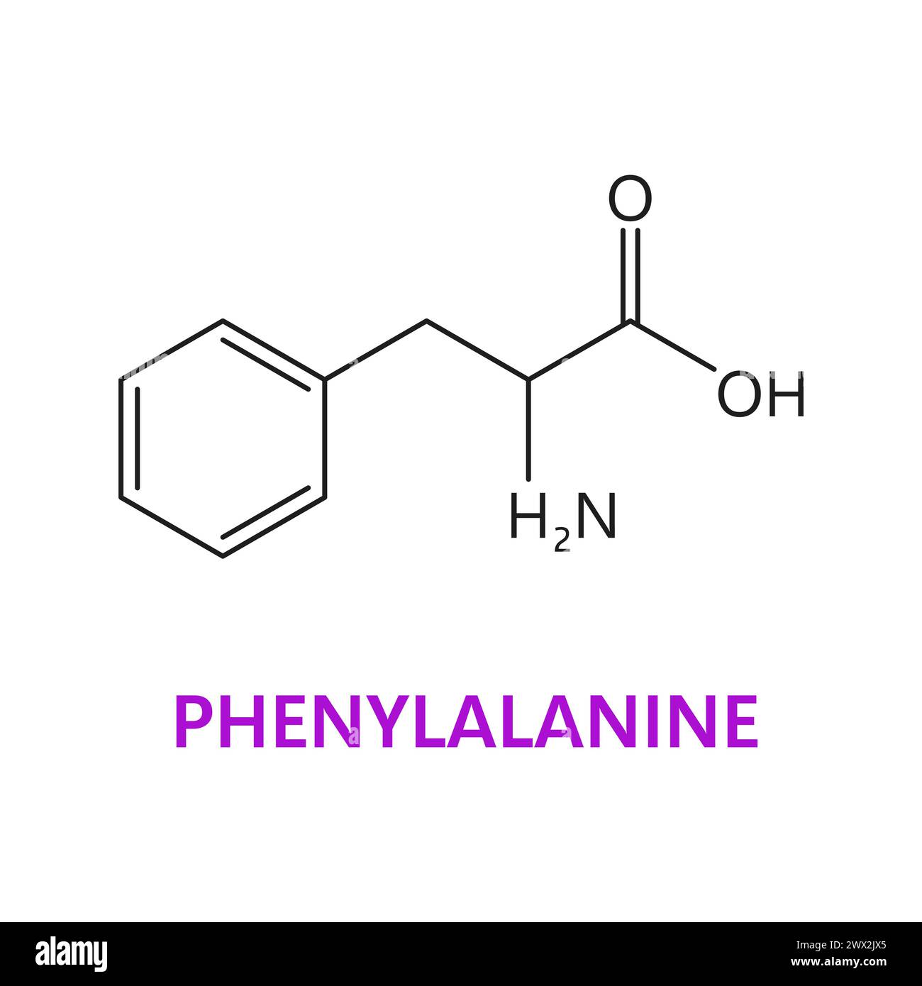 Amino acid, Phenylalanine, chemical formula and essential chain vector structure. Phenylalanine proteinogenic amino acid molecular bond structure and atom connection, medicine and biosynthesis study Stock Vectorhttps://www.alamy.com/image-license-details/?v=1https://www.alamy.com/amino-acid-phenylalanine-chemical-formula-and-essential-chain-vector-structure-phenylalanine-proteinogenic-amino-acid-molecular-bond-structure-and-atom-connection-medicine-and-biosynthesis-study-image601192317.html
RF2WX2JX5–Amino acid, Phenylalanine, chemical formula and essential chain vector structure. Phenylalanine proteinogenic amino acid molecular bond structure and atom connection, medicine and biosynthesis study
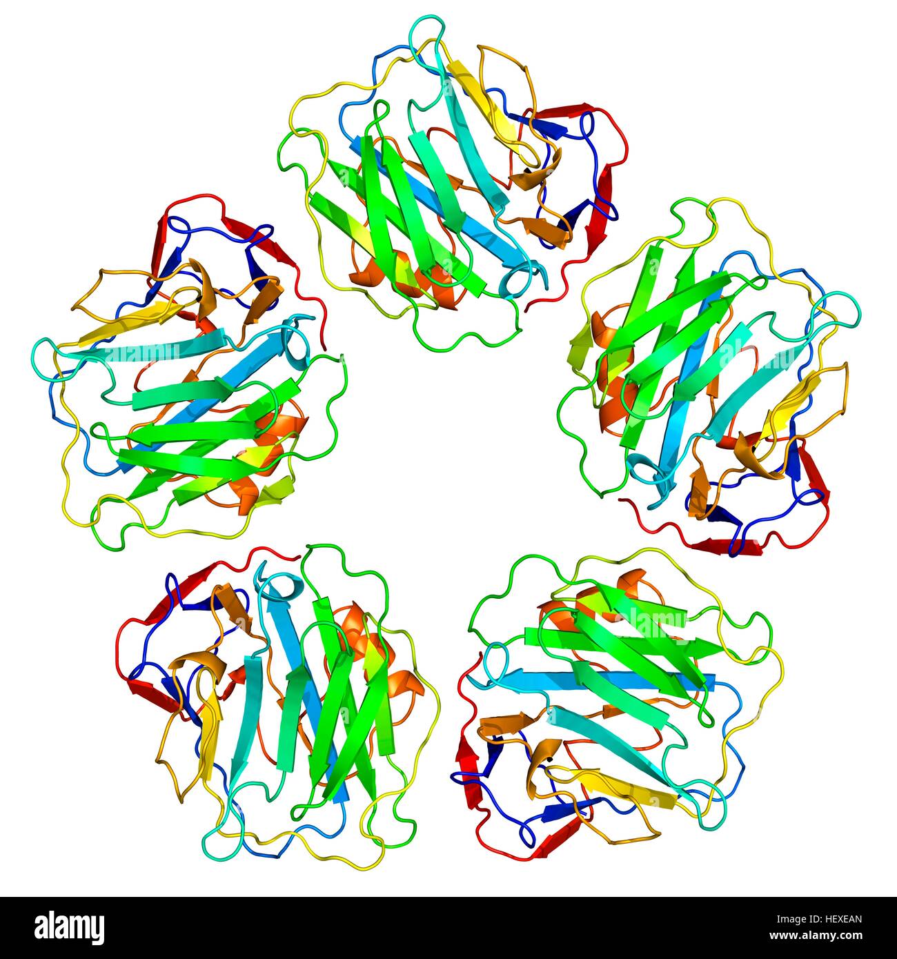 C-reactive protein,molecular model.The protein is made up of five sub-units (monomers) arranged in ring.The secondary structure of protein is shown,with beta sheets (arrows) alpha helices (spirals) connected by linking regions.C-reactive protein (CRP) is blood plasma protein produced by liver.It is acute phase protein,one whose levels rise in response to inflammation.It assists binding of complement proteins to foreign or damaged cells,an immunological response that destroys target cells.High blood levels of CRP are associated increased risk of heart disease diabetes. Stock Photohttps://www.alamy.com/image-license-details/?v=1https://www.alamy.com/stock-photo-c-reactive-proteinmolecular-modelthe-protein-is-made-up-of-five-sub-129659789.html
RFHEXEAN–C-reactive protein,molecular model.The protein is made up of five sub-units (monomers) arranged in ring.The secondary structure of protein is shown,with beta sheets (arrows) alpha helices (spirals) connected by linking regions.C-reactive protein (CRP) is blood plasma protein produced by liver.It is acute phase protein,one whose levels rise in response to inflammation.It assists binding of complement proteins to foreign or damaged cells,an immunological response that destroys target cells.High blood levels of CRP are associated increased risk of heart disease diabetes.
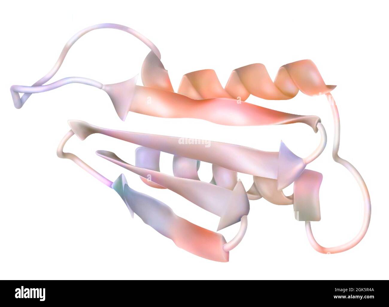 Drawing of a prion: infectious protein (encephalopathy agent). Stock Photohttps://www.alamy.com/image-license-details/?v=1https://www.alamy.com/drawing-of-a-prion-infectious-protein-encephalopathy-agent-image442065578.html
RF2GK5R4A–Drawing of a prion: infectious protein (encephalopathy agent).
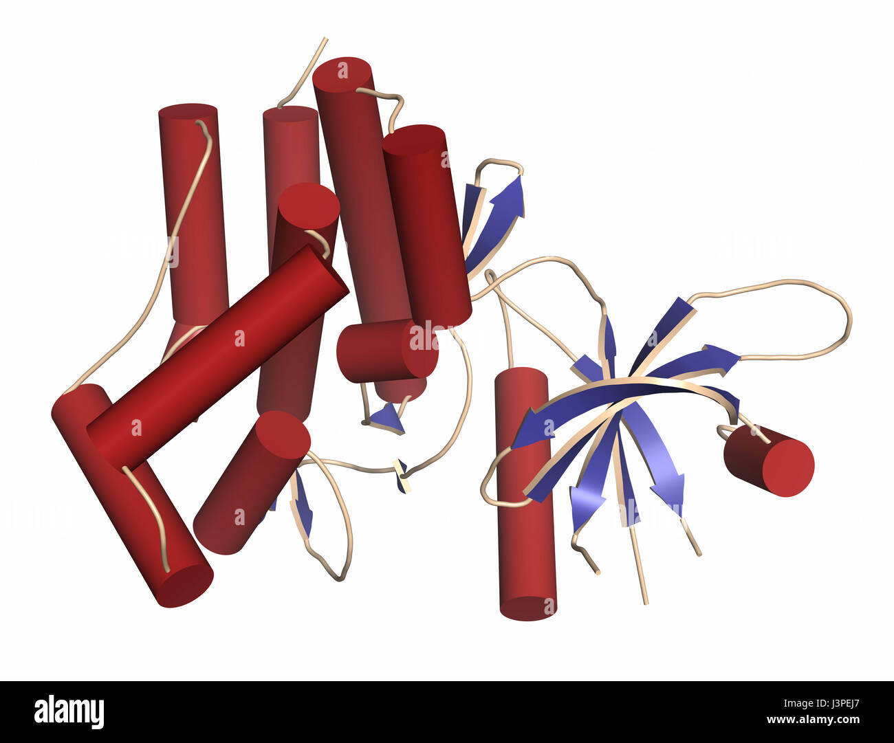 Janus kinase 1 protein. Part of JAK-STAT signalling pathway and drug target. Cartoon representation. Secondary structure coloring. Stock Photohttps://www.alamy.com/image-license-details/?v=1https://www.alamy.com/stock-photo-janus-kinase-1-protein-part-of-jak-stat-signalling-pathway-and-drug-140021343.html
RFJ3PEJ7–Janus kinase 1 protein. Part of JAK-STAT signalling pathway and drug target. Cartoon representation. Secondary structure coloring.
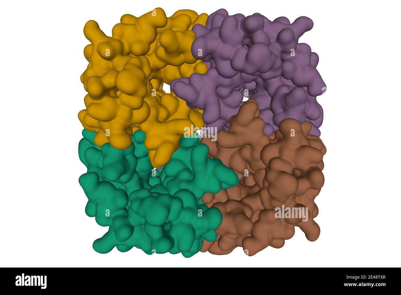 Crystal structure of human Aquaporin 7, 3D surface model isolated, white background Stock Photohttps://www.alamy.com/image-license-details/?v=1https://www.alamy.com/crystal-structure-of-human-aquaporin-7-3d-surface-model-isolated-white-background-image398492271.html
RF2E48TXR–Crystal structure of human Aquaporin 7, 3D surface model isolated, white background
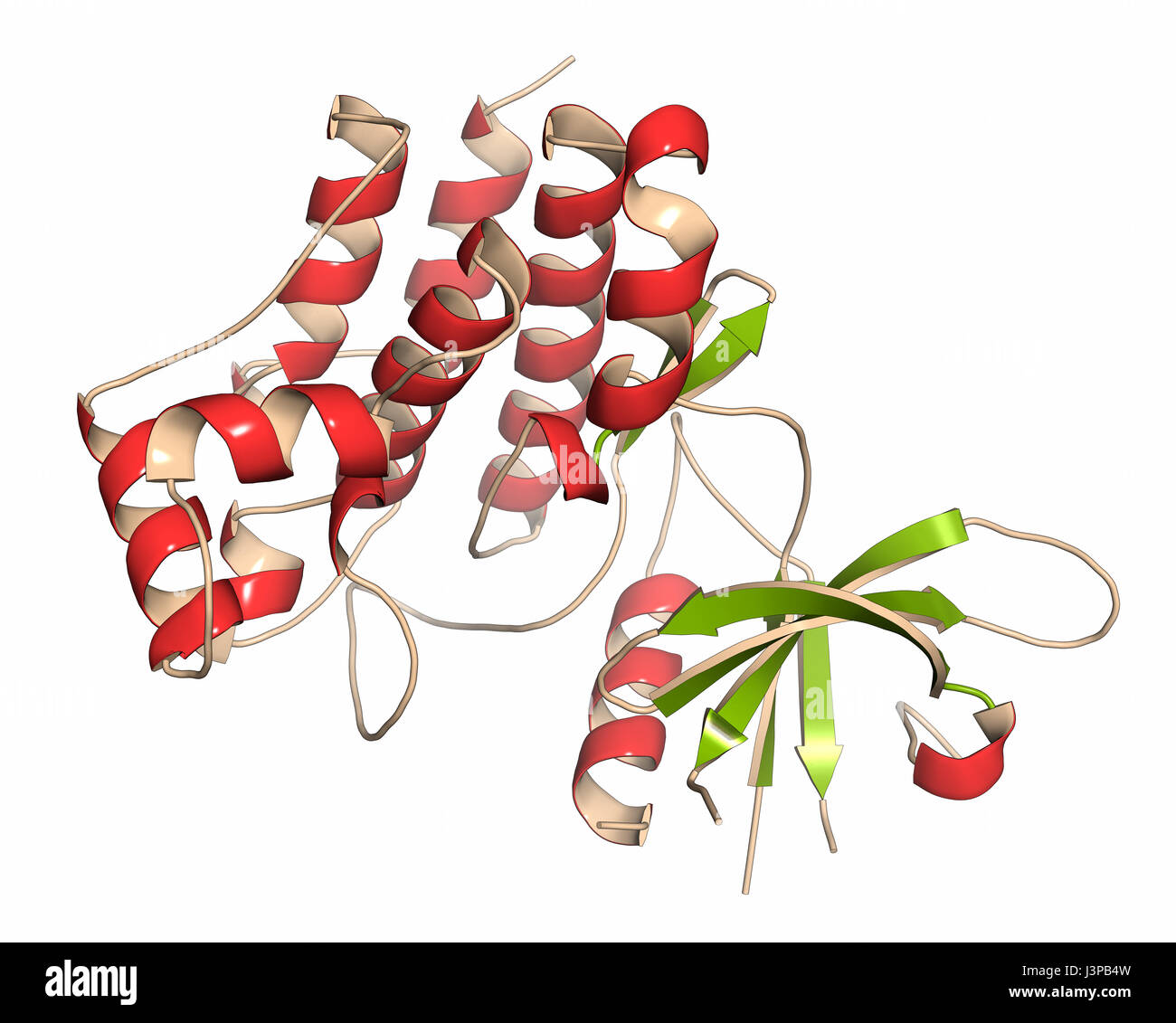 Janus kinase 1 protein. Part of JAK-STAT signalling pathway and drug target. Cartoon representation. Secondary structure coloring. Stock Photohttps://www.alamy.com/image-license-details/?v=1https://www.alamy.com/stock-photo-janus-kinase-1-protein-part-of-jak-stat-signalling-pathway-and-drug-140018617.html
RFJ3PB4W–Janus kinase 1 protein. Part of JAK-STAT signalling pathway and drug target. Cartoon representation. Secondary structure coloring.
 3d rendering of nanoparticles conjugated haemogoblin molecules Stock Photohttps://www.alamy.com/image-license-details/?v=1https://www.alamy.com/3d-rendering-of-nanoparticles-conjugated-haemogoblin-molecules-image596228933.html
RF2WJ0G2D–3d rendering of nanoparticles conjugated haemogoblin molecules
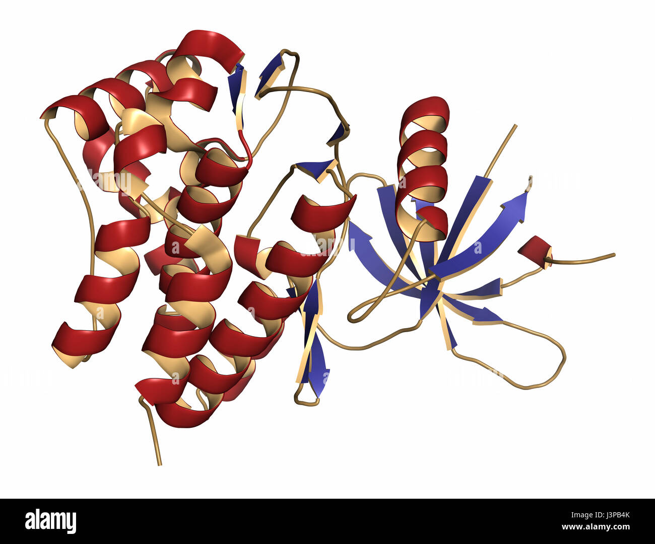 Janus kinase 1 protein. Part of JAK-STAT signalling pathway and drug target. Cartoon representation. Secondary structure coloring. Stock Photohttps://www.alamy.com/image-license-details/?v=1https://www.alamy.com/stock-photo-janus-kinase-1-protein-part-of-jak-stat-signalling-pathway-and-drug-140018611.html
RFJ3PB4K–Janus kinase 1 protein. Part of JAK-STAT signalling pathway and drug target. Cartoon representation. Secondary structure coloring.
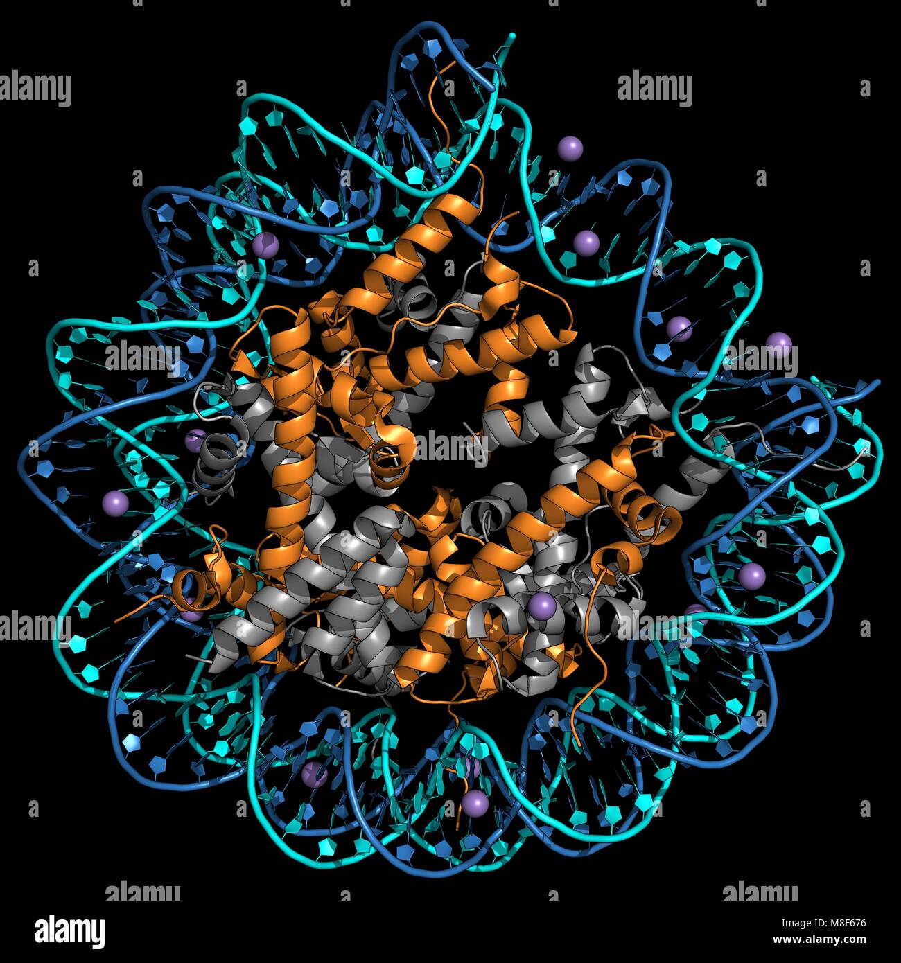 Representation of a human nucleosome - Packing of DNA with histones Stock Photohttps://www.alamy.com/image-license-details/?v=1https://www.alamy.com/stock-photo-representation-of-a-human-nucleosome-packing-of-dna-with-histones-177355114.html
RFM8F676–Representation of a human nucleosome - Packing of DNA with histones
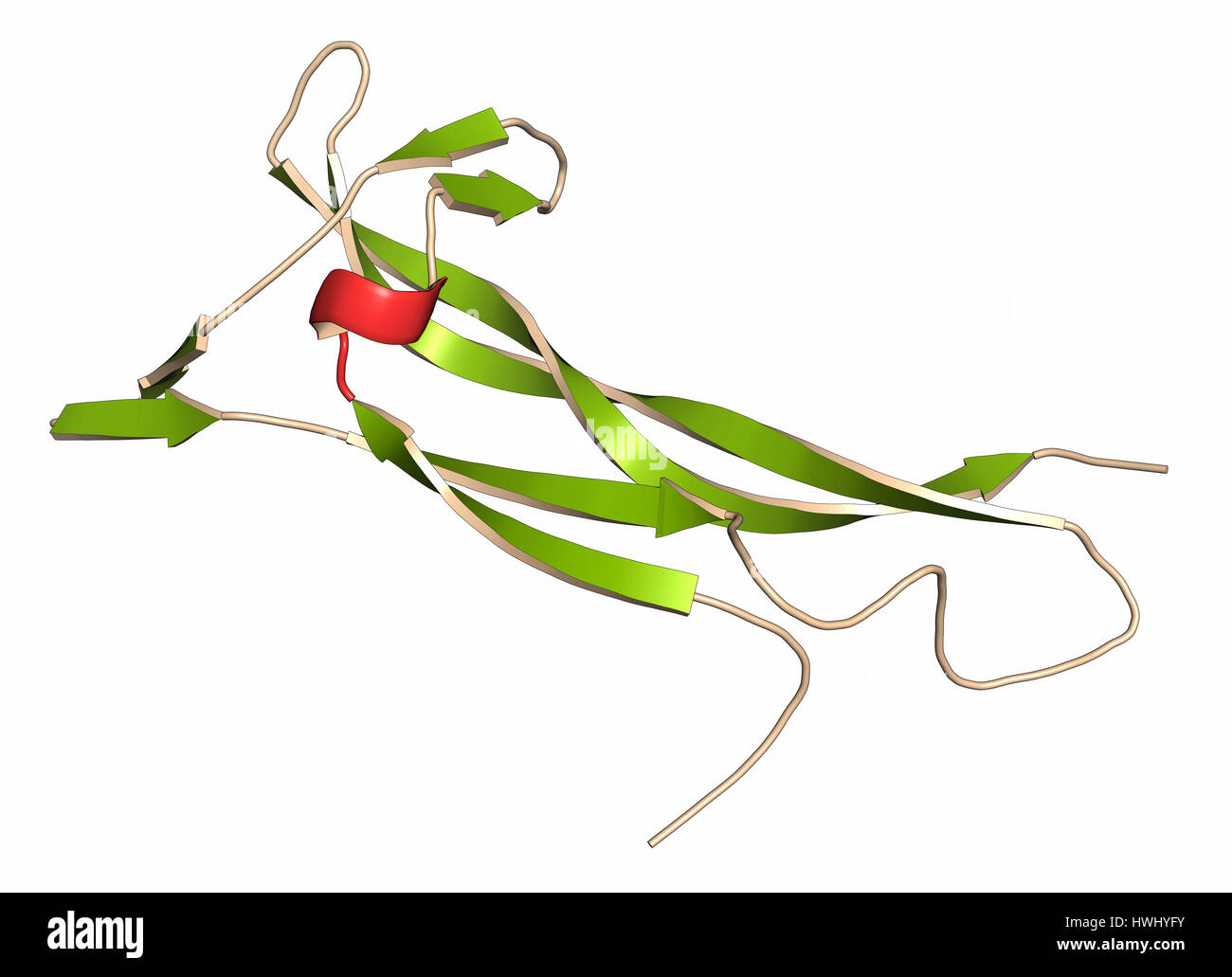 Brain-derived neurotrophic factor (BDNF) protein molecule. Cartoon representation with secondary structure coloring (green sheets, red helices). Stock Photohttps://www.alamy.com/image-license-details/?v=1https://www.alamy.com/stock-photo-brain-derived-neurotrophic-factor-bdnf-protein-molecule-cartoon-representation-136233775.html
RFHWHYFY–Brain-derived neurotrophic factor (BDNF) protein molecule. Cartoon representation with secondary structure coloring (green sheets, red helices).
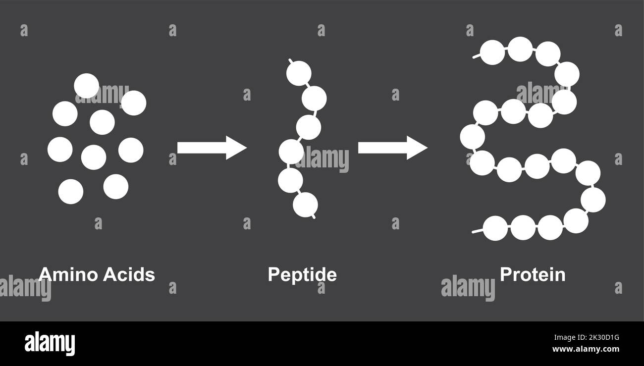 Scientific Designing of Biochemial Structure of Amino acids, Peptides And Proteins Molecular Model. Vector Illustration. Stock Vectorhttps://www.alamy.com/image-license-details/?v=1https://www.alamy.com/scientific-designing-of-biochemial-structure-of-amino-acids-peptides-and-proteins-molecular-model-vector-illustration-image483744508.html
RF2K30D1G–Scientific Designing of Biochemial Structure of Amino acids, Peptides And Proteins Molecular Model. Vector Illustration.
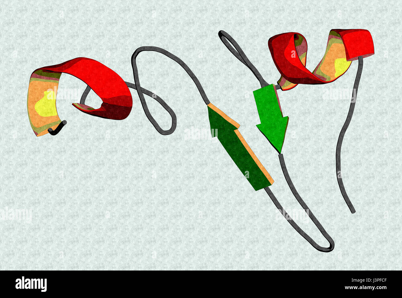 Epidermal growth factor (EGF) signaling protein molecule. Cartoon representation with secondary structure coloring (green sheets, red helices). Stock Photohttps://www.alamy.com/image-license-details/?v=1https://www.alamy.com/stock-photo-epidermal-growth-factor-egf-signaling-protein-molecule-cartoon-representation-140021967.html
RFJ3PFCF–Epidermal growth factor (EGF) signaling protein molecule. Cartoon representation with secondary structure coloring (green sheets, red helices).
RF2WF8GHF–Amino acid chemical molecule of Proline, molecular formula and chain structure, vector icon. Proline proteinogenic amino acid molecular structure and chain formula for medicine and pharmacy
 Interleukin 4 (IL-4) cytokine protein. 3D Illustration. Cartoon representation with secondary structure coloring (green sheets, red helices). Stock Photohttps://www.alamy.com/image-license-details/?v=1https://www.alamy.com/stock-photo-interleukin-4-il-4-cytokine-protein-3d-illustration-cartoon-representation-136235385.html
RFHWJ1HD–Interleukin 4 (IL-4) cytokine protein. 3D Illustration. Cartoon representation with secondary structure coloring (green sheets, red helices).
 Birch pollen allergen protein Bet V1, 3D rendering. Cartoon representation with secondary structure coloring (green sheets, red helices). Stock Photohttps://www.alamy.com/image-license-details/?v=1https://www.alamy.com/stock-photo-birch-pollen-allergen-protein-bet-v1-3d-rendering-cartoon-representation-136318267.html
RFHWNR9F–Birch pollen allergen protein Bet V1, 3D rendering. Cartoon representation with secondary structure coloring (green sheets, red helices).
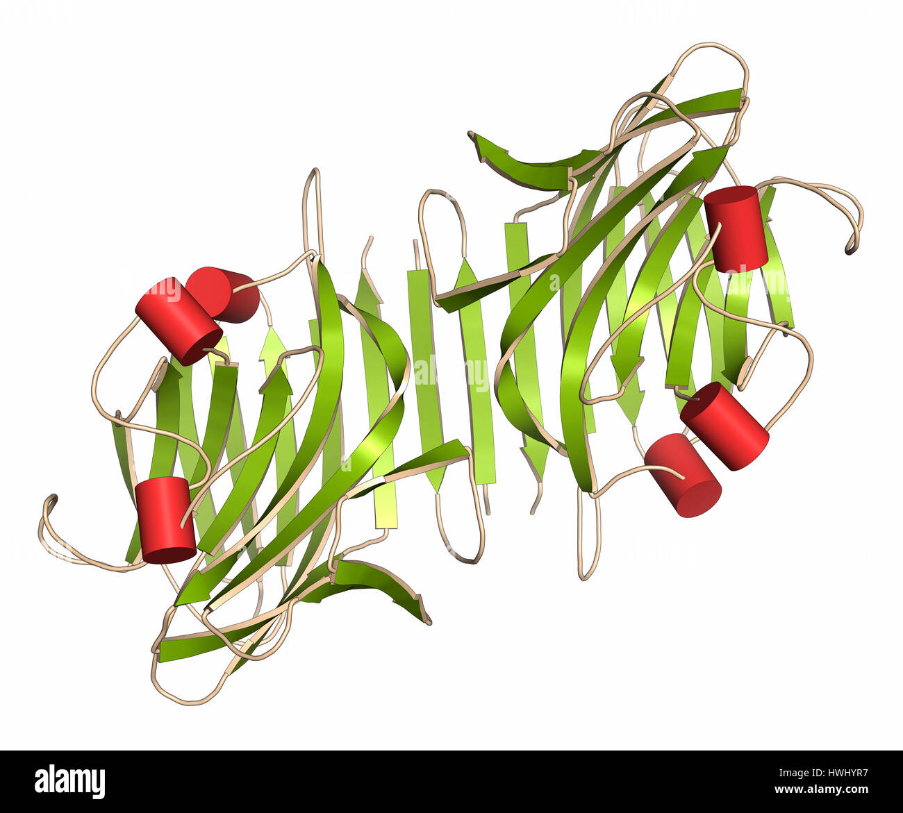 Pea lectin protein. Carbohydrate binding protein isolated from Pisum sativum. Cartoon representation with secondary structure coloring (green sheets, Stock Photohttps://www.alamy.com/image-license-details/?v=1https://www.alamy.com/stock-photo-pea-lectin-protein-carbohydrate-binding-protein-isolated-from-pisum-136233979.html
RFHWHYR7–Pea lectin protein. Carbohydrate binding protein isolated from Pisum sativum. Cartoon representation with secondary structure coloring (green sheets,
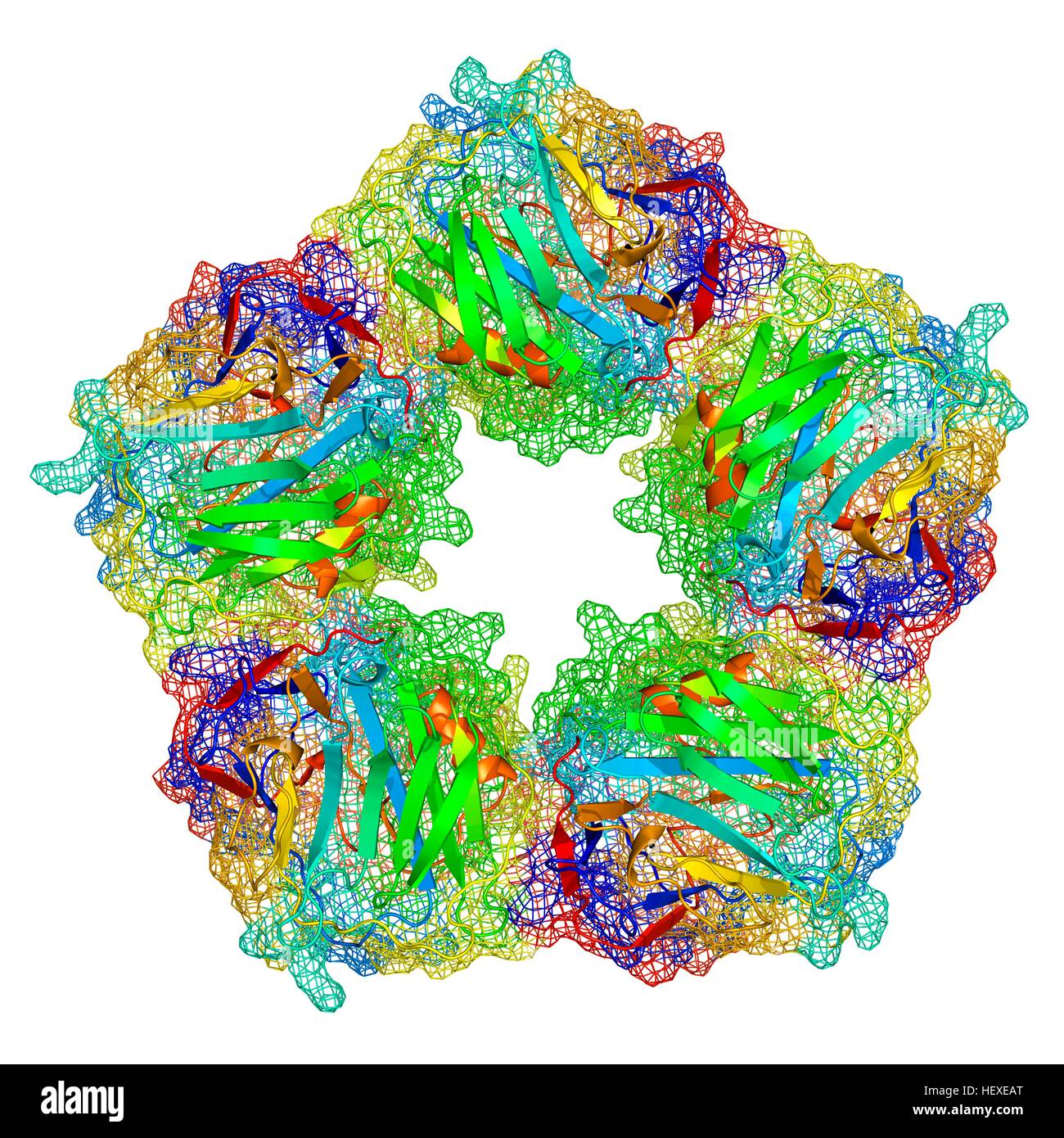 C-reactive protein,molecular model.The protein is made up of five sub-units (monomers) arranged in ring.The secondary structure of protein is shown,with beta sheets (arrows) alpha helices (spirals) connected by linking regions.C-reactive protein (CRP) is blood plasma protein produced by liver.It is acute phase protein,one whose levels rise in response to inflammation.It assists binding of complement proteins to foreign or damaged cells,an immunological response that destroys target cells.High blood levels of CRP are associated increased risk of heart disease diabetes. Stock Photohttps://www.alamy.com/image-license-details/?v=1https://www.alamy.com/stock-photo-c-reactive-proteinmolecular-modelthe-protein-is-made-up-of-five-sub-129659792.html
RFHEXEAT–C-reactive protein,molecular model.The protein is made up of five sub-units (monomers) arranged in ring.The secondary structure of protein is shown,with beta sheets (arrows) alpha helices (spirals) connected by linking regions.C-reactive protein (CRP) is blood plasma protein produced by liver.It is acute phase protein,one whose levels rise in response to inflammation.It assists binding of complement proteins to foreign or damaged cells,an immunological response that destroys target cells.High blood levels of CRP are associated increased risk of heart disease diabetes.
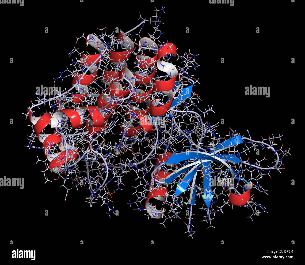 Janus kinase 1 protein. Part of JAK-STAT signalling pathway and drug target. Cartoon + line representation. Secondary structure coloring. Stock Photohttps://www.alamy.com/image-license-details/?v=1https://www.alamy.com/stock-photo-janus-kinase-1-protein-part-of-jak-stat-signalling-pathway-and-drug-140021340.html
RFJ3PEJ4–Janus kinase 1 protein. Part of JAK-STAT signalling pathway and drug target. Cartoon + line representation. Secondary structure coloring.
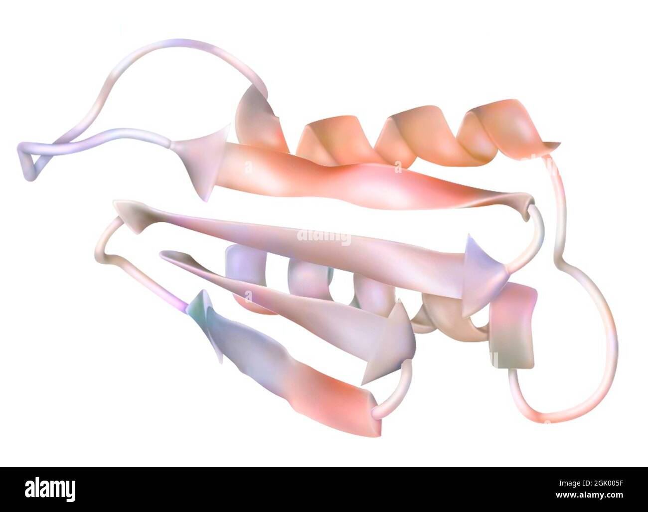 Drawing of a prion: infectious protein (encephalopathy agent). Stock Photohttps://www.alamy.com/image-license-details/?v=1https://www.alamy.com/drawing-of-a-prion-infectious-protein-encephalopathy-agent-image441937819.html
RF2GK005F–Drawing of a prion: infectious protein (encephalopathy agent).
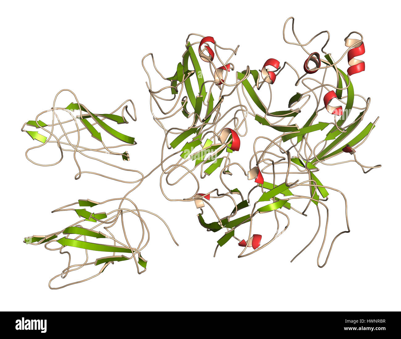 Coagulation factor VIII (fVIII) protein, 3D rendering. Deficiency causes hemophilia A. Cartoon representation with secondary structure coloring (green Stock Photohttps://www.alamy.com/image-license-details/?v=1https://www.alamy.com/stock-photo-coagulation-factor-viii-fviii-protein-3d-rendering-deficiency-causes-136318331.html
RFHWNRBR–Coagulation factor VIII (fVIII) protein, 3D rendering. Deficiency causes hemophilia A. Cartoon representation with secondary structure coloring (green
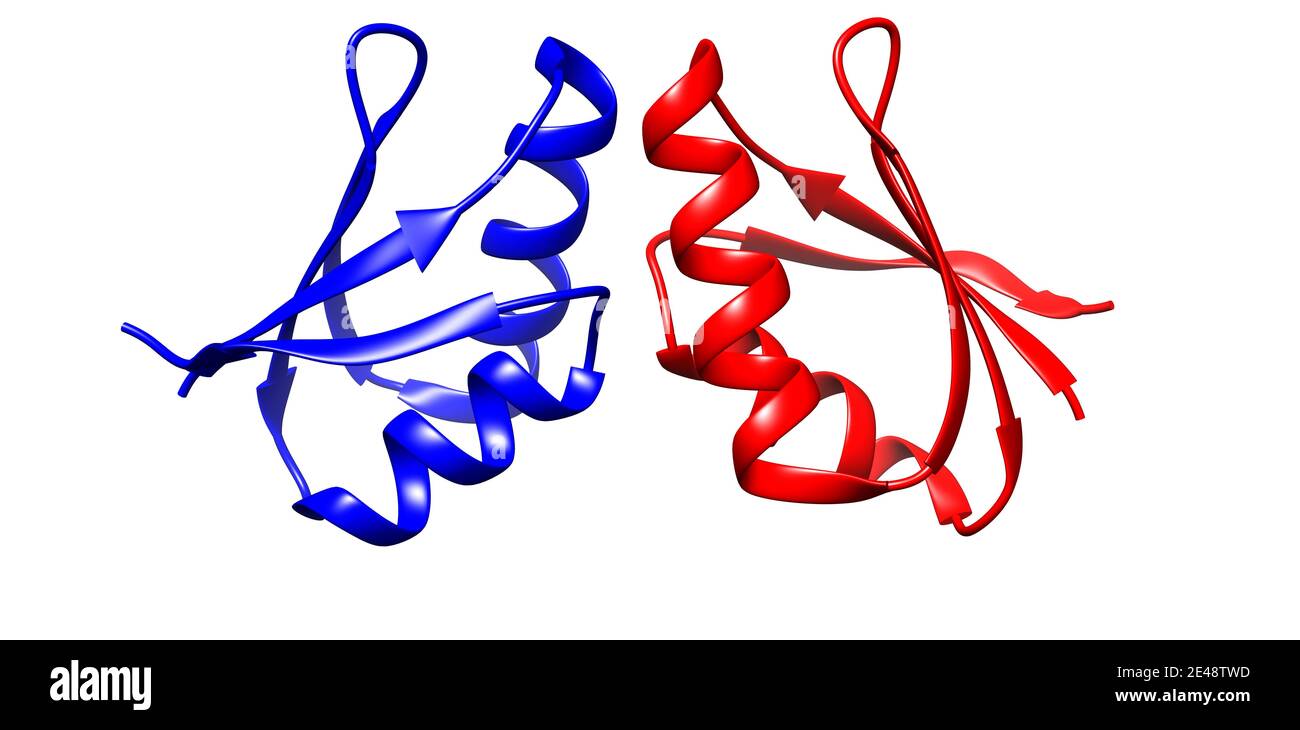 Crystal structure of human copper chaperone homodimer, 3D cartoon model, white background Stock Photohttps://www.alamy.com/image-license-details/?v=1https://www.alamy.com/crystal-structure-of-human-copper-chaperone-homodimer-3d-cartoon-model-white-background-image398492233.html
RF2E48TWD–Crystal structure of human copper chaperone homodimer, 3D cartoon model, white background
 Alpha-synuclein protein. May play role in Parkinson's and Alzheimer's disease. Cartoon representation with secondary structure coloring (red helices). Stock Photohttps://www.alamy.com/image-license-details/?v=1https://www.alamy.com/stock-photo-alpha-synuclein-protein-may-play-role-in-parkinsons-and-alzheimers-136234018.html
RFHWHYTJ–Alpha-synuclein protein. May play role in Parkinson's and Alzheimer's disease. Cartoon representation with secondary structure coloring (red helices).
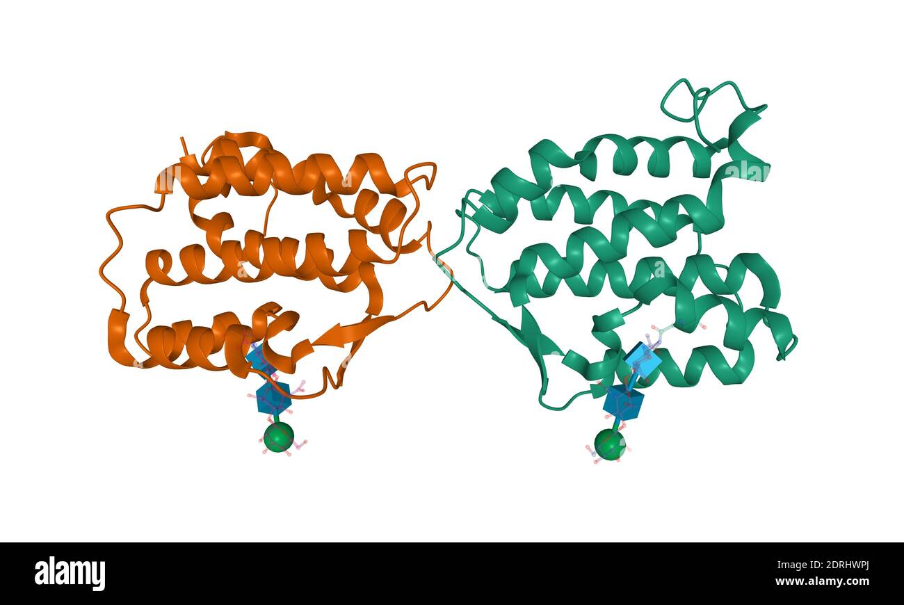 Structure of human interleukin-34 homodimer, 3D cartoon model isolated, white background Stock Photohttps://www.alamy.com/image-license-details/?v=1https://www.alamy.com/structure-of-human-interleukin-34-homodimer-3d-cartoon-model-isolated-white-background-image393158602.html
RF2DRHWPJ–Structure of human interleukin-34 homodimer, 3D cartoon model isolated, white background
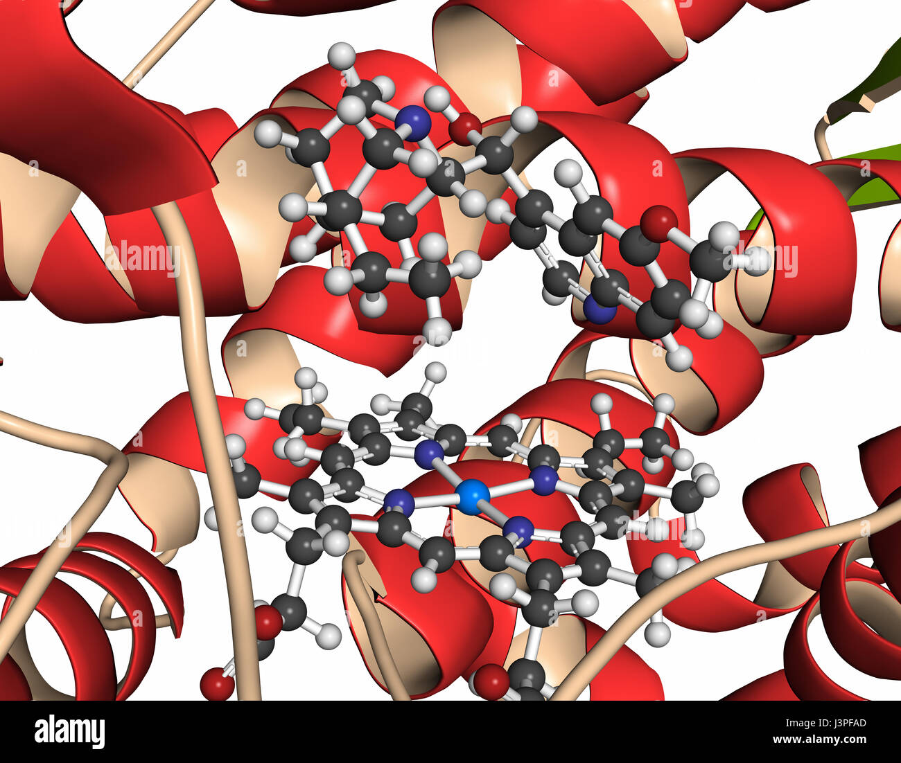 Cytochrome P450 (CYP2D6) liver enzyme in complex with the drug quinine. Protein: cartoon representation with secondary structure coloring (green sheet Stock Photohttps://www.alamy.com/image-license-details/?v=1https://www.alamy.com/stock-photo-cytochrome-p450-cyp2d6-liver-enzyme-in-complex-with-the-drug-quinine-140021909.html
RFJ3PFAD–Cytochrome P450 (CYP2D6) liver enzyme in complex with the drug quinine. Protein: cartoon representation with secondary structure coloring (green sheet
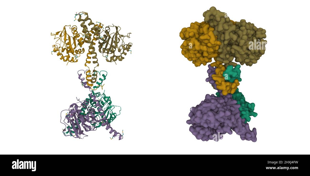 Structure of ubiquitin carboxyl-terminal hydrolase isozyme L5 (Uch37) tetramer. 3D cartoon and Gaussian surface models, PDB 3ihr Stock Photohttps://www.alamy.com/image-license-details/?v=1https://www.alamy.com/structure-of-ubiquitin-carboxyl-terminal-hydrolase-isozyme-l5-uch37-tetramer-3d-cartoon-and-gaussian-surface-models-pdb-3ihr-image463849341.html
RF2HXJ4FW–Structure of ubiquitin carboxyl-terminal hydrolase isozyme L5 (Uch37) tetramer. 3D cartoon and Gaussian surface models, PDB 3ihr
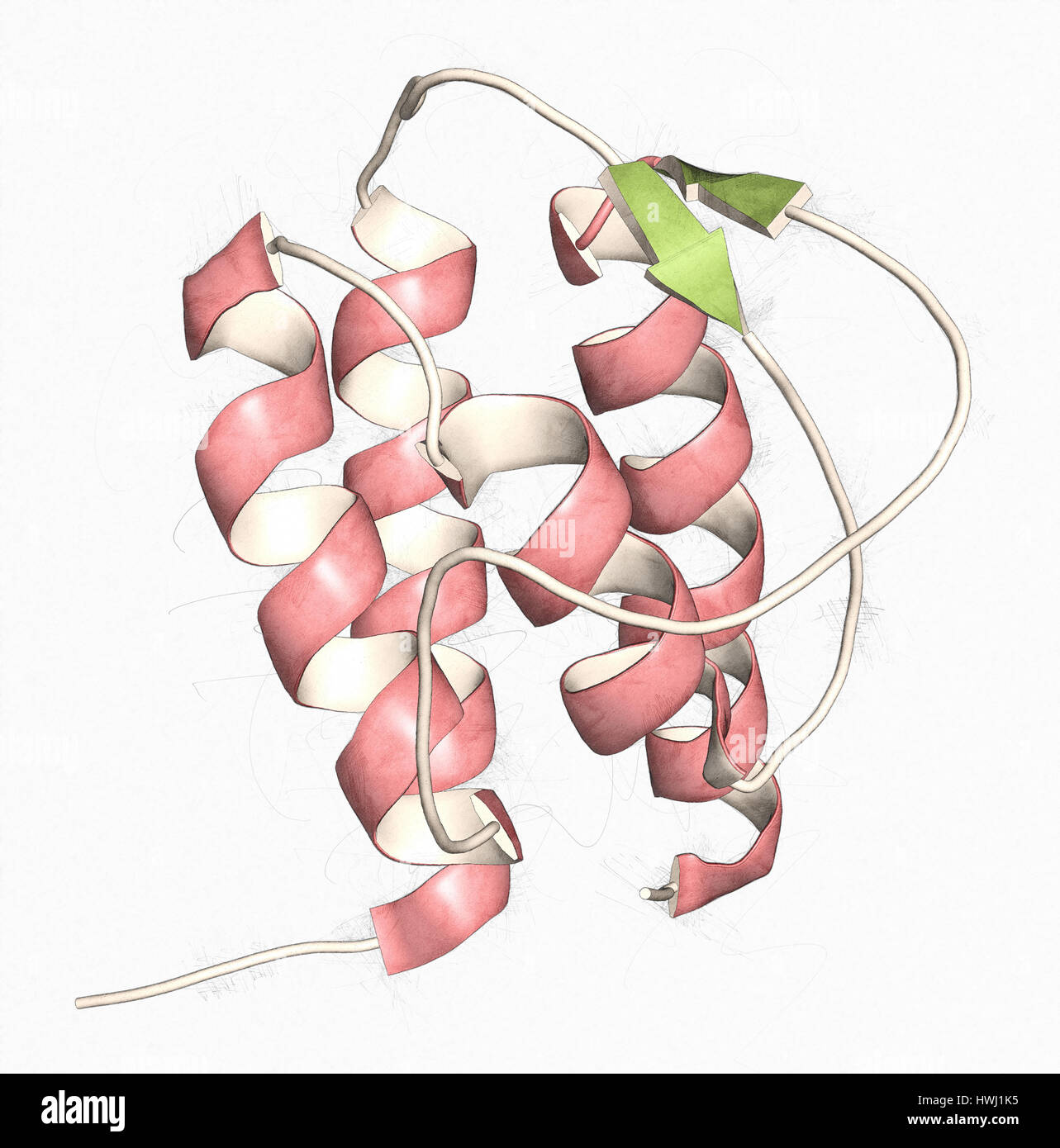 Interleukin 13 (IL-13) cytokine protein. 3D illustration. Cartoon representation with secondary structure coloring (green sheets, red helices). Stock Photohttps://www.alamy.com/image-license-details/?v=1https://www.alamy.com/stock-photo-interleukin-13-il-13-cytokine-protein-3d-illustration-cartoon-representation-136235433.html
RFHWJ1K5–Interleukin 13 (IL-13) cytokine protein. 3D illustration. Cartoon representation with secondary structure coloring (green sheets, red helices).
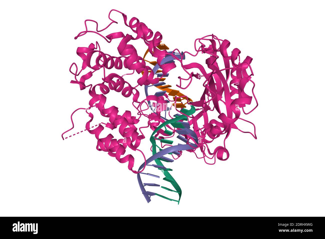 Crystal structure of human DNA ligase I bound to 5'-adenylated, nicked DNA, 3D cartoon model, white background Stock Photohttps://www.alamy.com/image-license-details/?v=1https://www.alamy.com/crystal-structure-of-human-dna-ligase-i-bound-to-5-adenylated-nicked-dna-3d-cartoon-model-white-background-image393159468.html
RF2DRHXWG–Crystal structure of human DNA ligase I bound to 5'-adenylated, nicked DNA, 3D cartoon model, white background
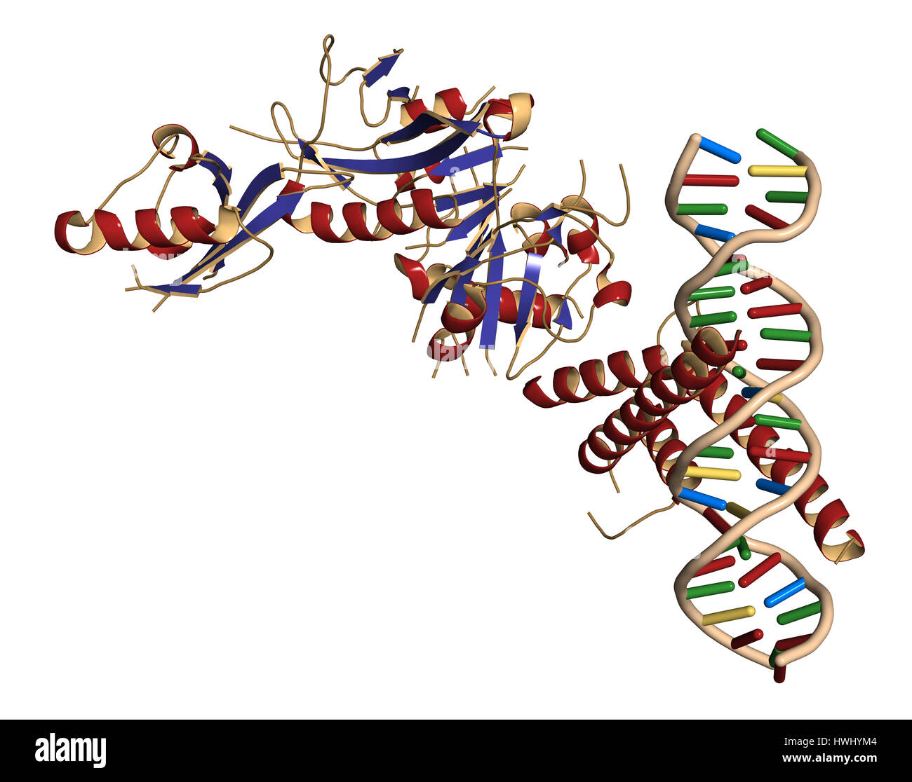 Hypoxia-inducible factor 1 (HIF-1) transcription factor, bound to DNA. Protein: cartoon representation with secondary structure coloring (blue sheets, Stock Photohttps://www.alamy.com/image-license-details/?v=1https://www.alamy.com/stock-photo-hypoxia-inducible-factor-1-hif-1-transcription-factor-bound-to-dna-136233892.html
RFHWHYM4–Hypoxia-inducible factor 1 (HIF-1) transcription factor, bound to DNA. Protein: cartoon representation with secondary structure coloring (blue sheets,
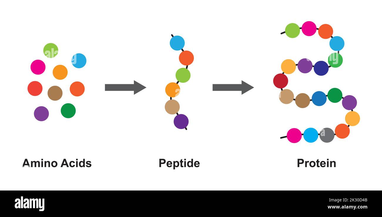 Scientific Designing of Biochemial Structure of Amino acids, Peptides And Proteins Molecular Model. Vector Illustration. Stock Vectorhttps://www.alamy.com/image-license-details/?v=1https://www.alamy.com/scientific-designing-of-biochemial-structure-of-amino-acids-peptides-and-proteins-molecular-model-vector-illustration-image483744587.html
RF2K30D4B–Scientific Designing of Biochemial Structure of Amino acids, Peptides And Proteins Molecular Model. Vector Illustration.
RF2XB6YD3–Amino acid Serine, chemical molecule structure and molecular chain formula, vector icon. Serine proteinogenic amino acid molecular structure and chain formula for medicine and biosynthesis study
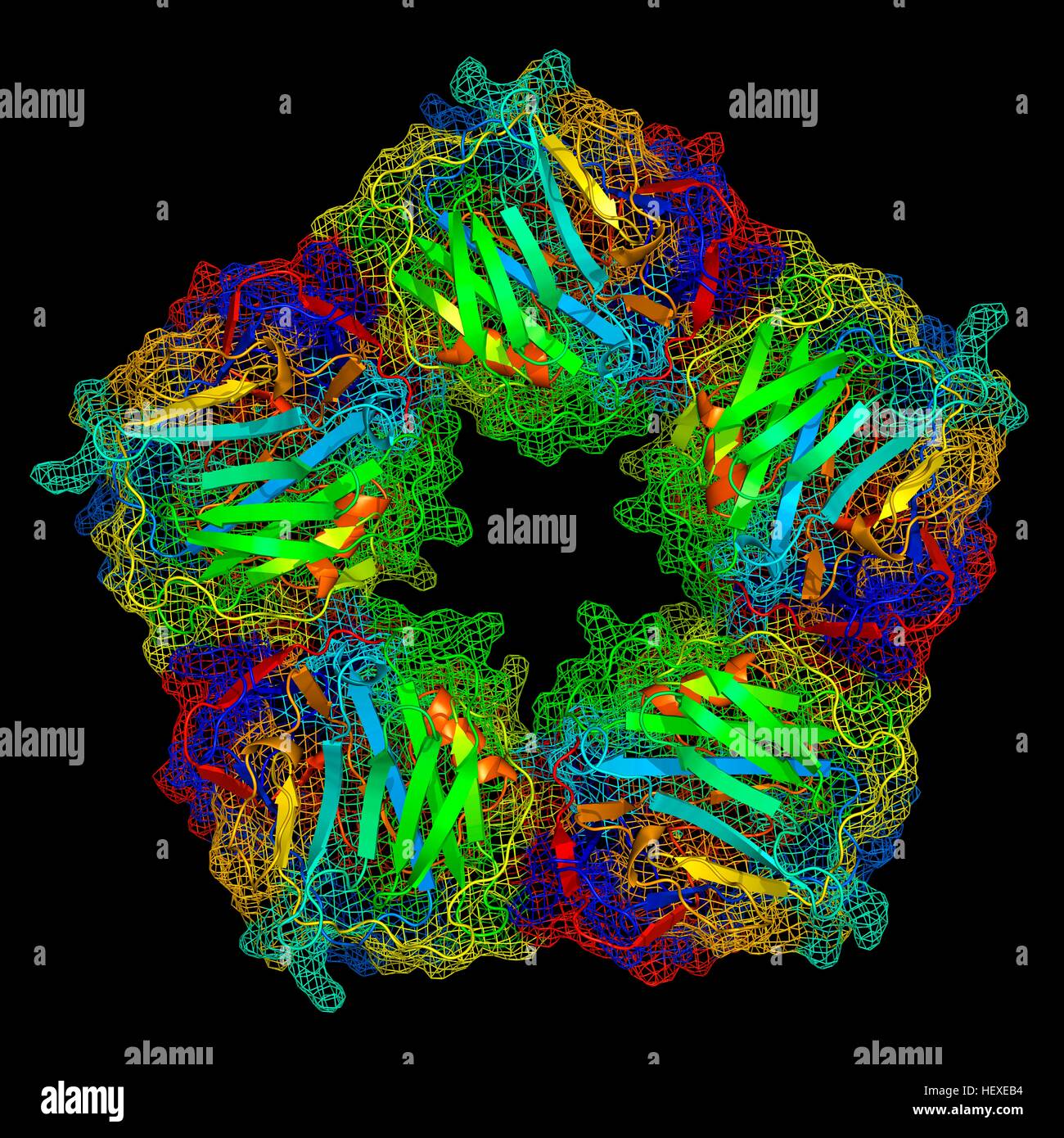 C-reactive protein,molecular model.The protein is made up of five sub-units (monomers) arranged in ring.The secondary structure of protein is shown,with beta sheets (arrows) alpha helices (spirals) connected by linking regions.C-reactive protein (CRP) is blood plasma protein produced by liver.It is acute phase protein,one whose levels rise in response to inflammation.It assists binding of complement proteins to foreign or damaged cells,an immunological response that destroys target cells.High blood levels of CRP are associated increased risk of heart disease diabetes. Stock Photohttps://www.alamy.com/image-license-details/?v=1https://www.alamy.com/stock-photo-c-reactive-proteinmolecular-modelthe-protein-is-made-up-of-five-sub-129659800.html
RFHEXEB4–C-reactive protein,molecular model.The protein is made up of five sub-units (monomers) arranged in ring.The secondary structure of protein is shown,with beta sheets (arrows) alpha helices (spirals) connected by linking regions.C-reactive protein (CRP) is blood plasma protein produced by liver.It is acute phase protein,one whose levels rise in response to inflammation.It assists binding of complement proteins to foreign or damaged cells,an immunological response that destroys target cells.High blood levels of CRP are associated increased risk of heart disease diabetes.
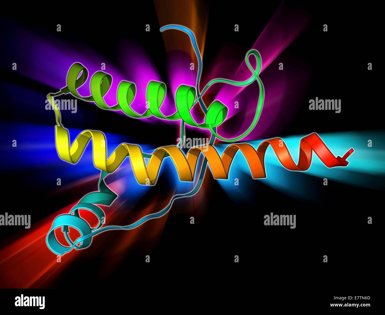 Human prion precursor protein, molecular model showing secondary structure. The human prion protein (hPrP) is a prion precursor. Prions are abnormal proteins that are the cause of transmissible spongiform encephalopathies (TSEs) such as BSE in cows, scrap Stock Photohttps://www.alamy.com/image-license-details/?v=1https://www.alamy.com/stock-photo-human-prion-precursor-protein-molecular-model-showing-secondary-structure-73687557.html
RFE7TN6D–Human prion precursor protein, molecular model showing secondary structure. The human prion protein (hPrP) is a prion precursor. Prions are abnormal proteins that are the cause of transmissible spongiform encephalopathies (TSEs) such as BSE in cows, scrap
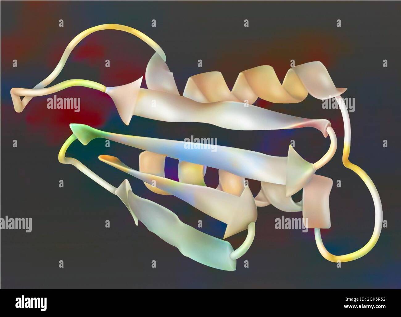 Drawing of a prion: infectious protein (encephalopathy agent). Stock Photohttps://www.alamy.com/image-license-details/?v=1https://www.alamy.com/drawing-of-a-prion-infectious-protein-encephalopathy-agent-image442065598.html
RF2GK5R52–Drawing of a prion: infectious protein (encephalopathy agent).
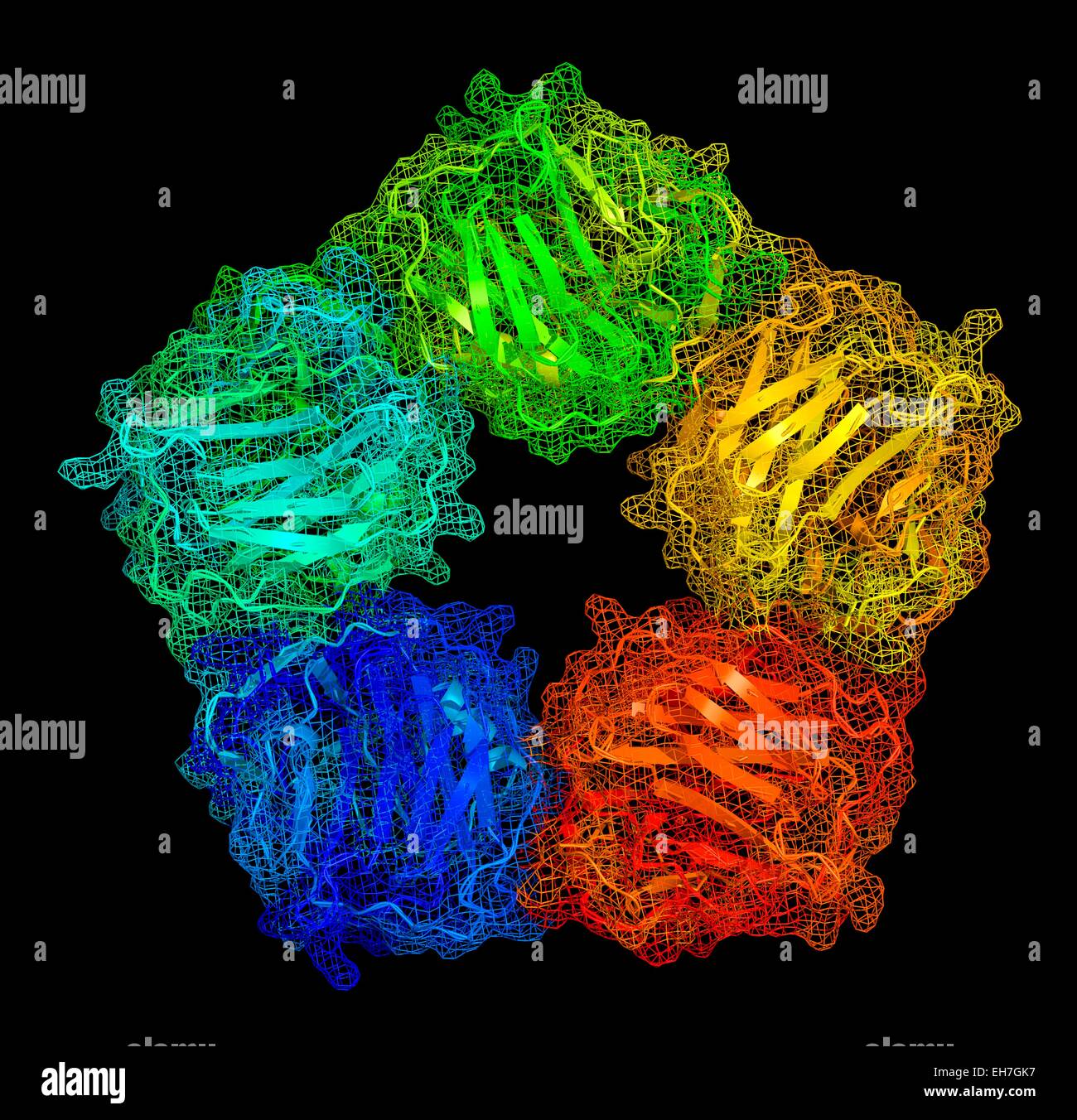 C-reactive protein, molecular model Stock Photohttps://www.alamy.com/image-license-details/?v=1https://www.alamy.com/stock-photo-c-reactive-protein-molecular-model-79457371.html
RFEH7GK7–C-reactive protein, molecular model
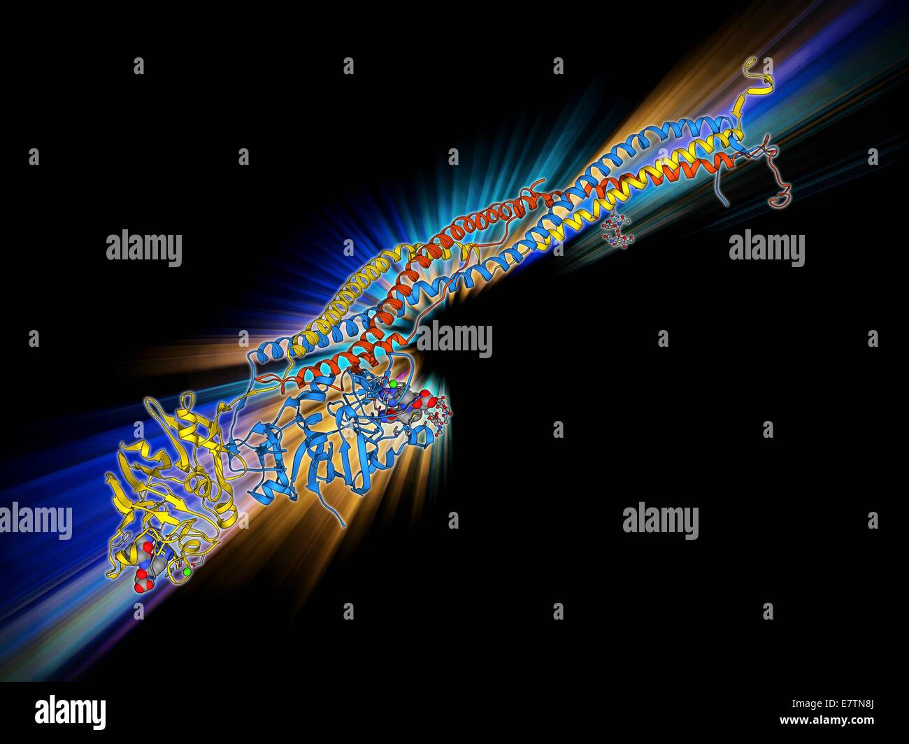 Fibrinogen. Molecular model showing the secondary structure of the blood clotting glycoprotein fibrinogen (factor I). Fibrinogen, which is synthesised by the liver, is converted by thrombin into fibrin during a cascade of complex reactions initiated by ch Stock Photohttps://www.alamy.com/image-license-details/?v=1https://www.alamy.com/stock-photo-fibrinogen-molecular-model-showing-the-secondary-structure-of-the-73687618.html
RFE7TN8J–Fibrinogen. Molecular model showing the secondary structure of the blood clotting glycoprotein fibrinogen (factor I). Fibrinogen, which is synthesised by the liver, is converted by thrombin into fibrin during a cascade of complex reactions initiated by ch
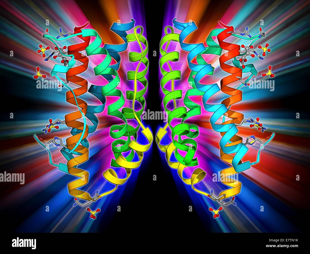 Interleukin-6. Molecular model of the cytokine protein human interleukin-6. This protein is produced in the body and has a wide variety of functions in the immune system. Stock Photohttps://www.alamy.com/image-license-details/?v=1https://www.alamy.com/stock-photo-interleukin-6-molecular-model-of-the-cytokine-protein-human-interleukin-73687423.html
RFE7TN1K–Interleukin-6. Molecular model of the cytokine protein human interleukin-6. This protein is produced in the body and has a wide variety of functions in the immune system.
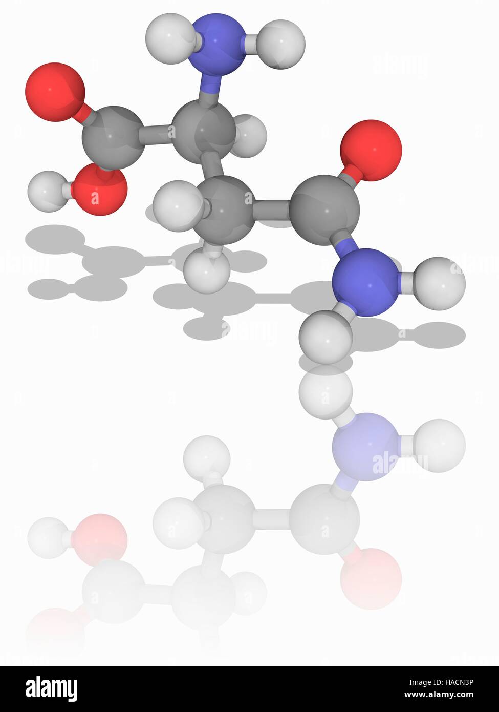 Asparagine. Molecular model of the amino acid asparagine (C4.H8.N2.O3). It is a non-essential amino acid. It can be synthesised by the body and so does not need to come from the diet. Asparagine plays an important role in the folding of protein molecules into their secondary structures. Atoms are represented as spheres and are colour-coded: carbon (grey), hydrogen (white), nitrogen (blue) and oxygen (red). Illustration. Stock Photohttps://www.alamy.com/image-license-details/?v=1https://www.alamy.com/stock-photo-asparagine-molecular-model-of-the-amino-acid-asparagine-c4h8n2o3-it-126899130.html
RFHACN3P–Asparagine. Molecular model of the amino acid asparagine (C4.H8.N2.O3). It is a non-essential amino acid. It can be synthesised by the body and so does not need to come from the diet. Asparagine plays an important role in the folding of protein molecules into their secondary structures. Atoms are represented as spheres and are colour-coded: carbon (grey), hydrogen (white), nitrogen (blue) and oxygen (red). Illustration.
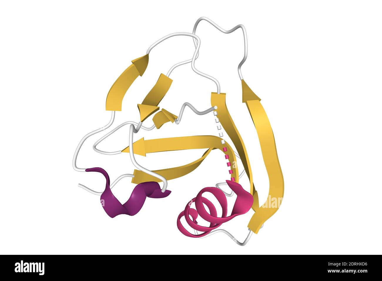 Crystal structure of CD5 DIII with the differently colored secondary structure elements, 3D cartoon model, white background Stock Photohttps://www.alamy.com/image-license-details/?v=1https://www.alamy.com/crystal-structure-of-cd5-diii-with-the-differently-colored-secondary-structure-elements-3d-cartoon-model-white-background-image393159122.html
RF2DRHXD6–Crystal structure of CD5 DIII with the differently colored secondary structure elements, 3D cartoon model, white background
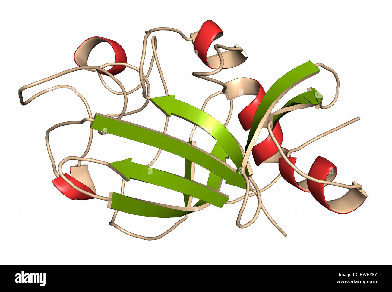 Angiopoietin 2 vascular growth factor protein (receptor binding domain). Cartoon representation with secondary structure coloring (green sheets, red h Stock Photohttps://www.alamy.com/image-license-details/?v=1https://www.alamy.com/stock-photo-angiopoietin-2-vascular-growth-factor-protein-receptor-binding-domain-136233747.html
RFHWHYEY–Angiopoietin 2 vascular growth factor protein (receptor binding domain). Cartoon representation with secondary structure coloring (green sheets, red h
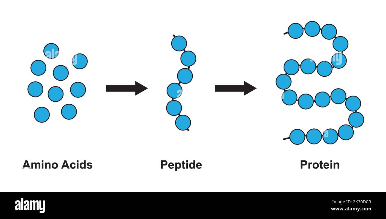 Scientific Designing of Biochemial Structure of Amino acids, Peptides And Proteins Molecular Model. Vector Illustration. Stock Vectorhttps://www.alamy.com/image-license-details/?v=1https://www.alamy.com/scientific-designing-of-biochemial-structure-of-amino-acids-peptides-and-proteins-molecular-model-vector-illustration-image483744823.html
RF2K30DCR–Scientific Designing of Biochemial Structure of Amino acids, Peptides And Proteins Molecular Model. Vector Illustration.
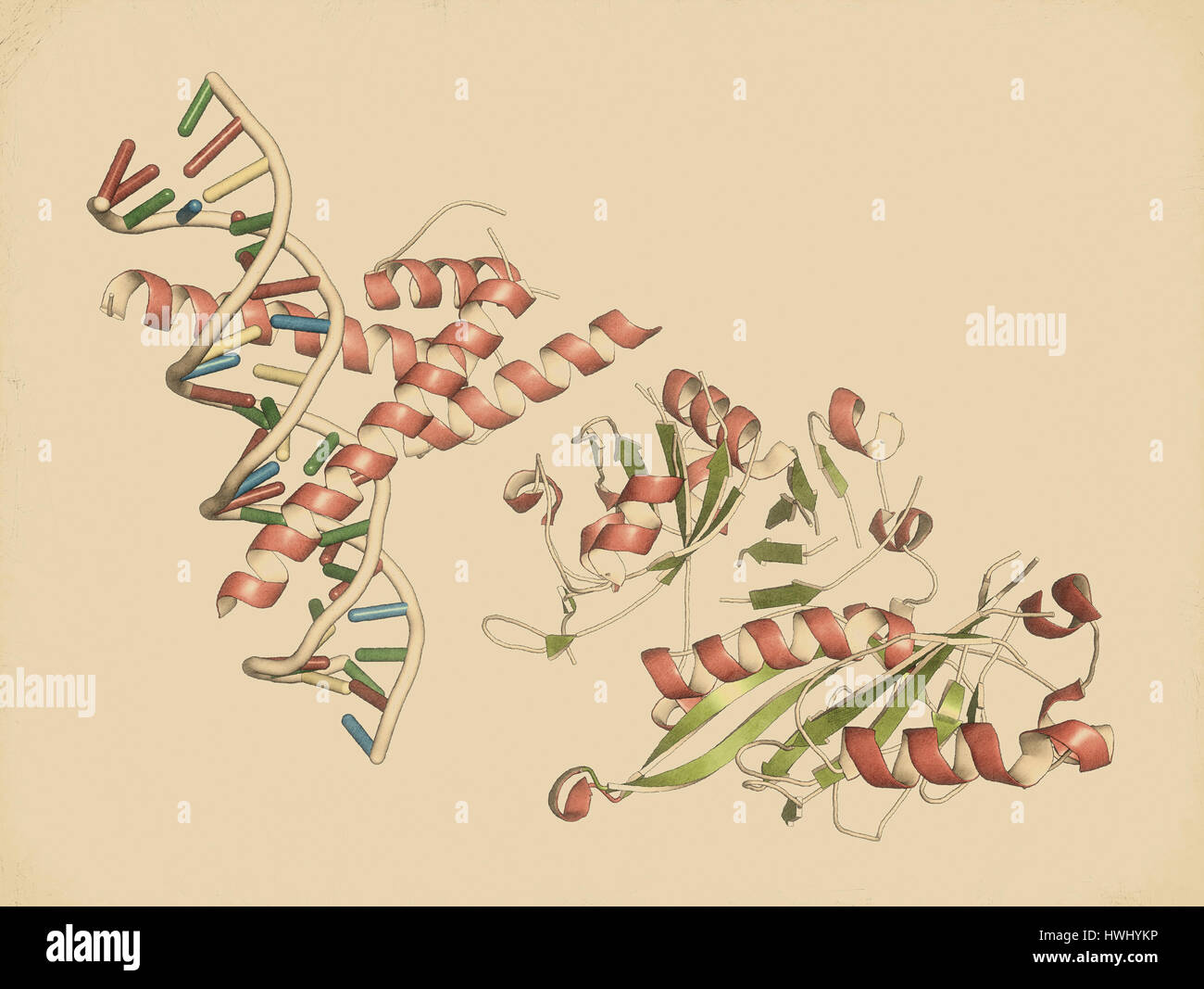 Hypoxia-inducible factor 1 (HIF-1) transcription factor, bound to DNA. Protein: cartoon representation with secondary structure coloring (green sheets Stock Photohttps://www.alamy.com/image-license-details/?v=1https://www.alamy.com/stock-photo-hypoxia-inducible-factor-1-hif-1-transcription-factor-bound-to-dna-136233882.html
RFHWHYKP–Hypoxia-inducible factor 1 (HIF-1) transcription factor, bound to DNA. Protein: cartoon representation with secondary structure coloring (green sheets
RF2X2JCE0–Amino acid chemical molecule of Histidine, molecular formula and chain structure, vector icon. Histidine essential amino acid molecular structure and chain formula for medicine and health pharmacy
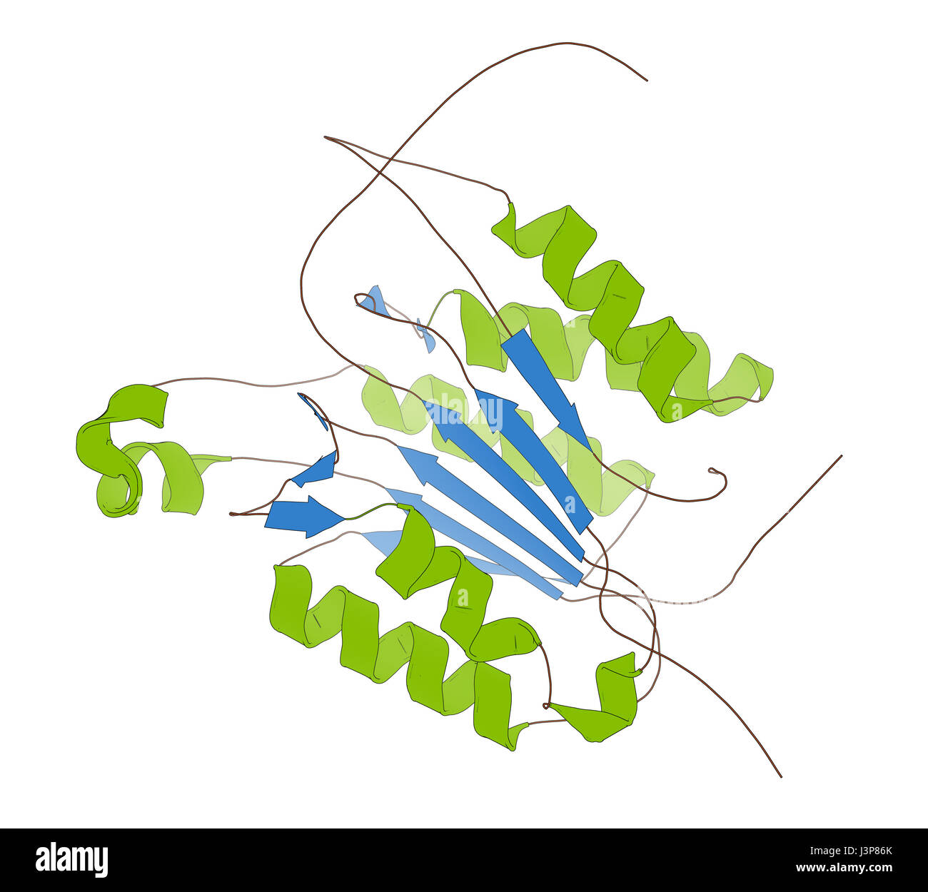 Caspase 3 apoptosis protein. Enzyme that plays important role in programmed cell death. Cartoon model, secondary structure coloring: alpha-helices gre Stock Photohttps://www.alamy.com/image-license-details/?v=1https://www.alamy.com/stock-photo-caspase-3-apoptosis-protein-enzyme-that-plays-important-role-in-programmed-140016315.html
RFJ3P86K–Caspase 3 apoptosis protein. Enzyme that plays important role in programmed cell death. Cartoon model, secondary structure coloring: alpha-helices gre
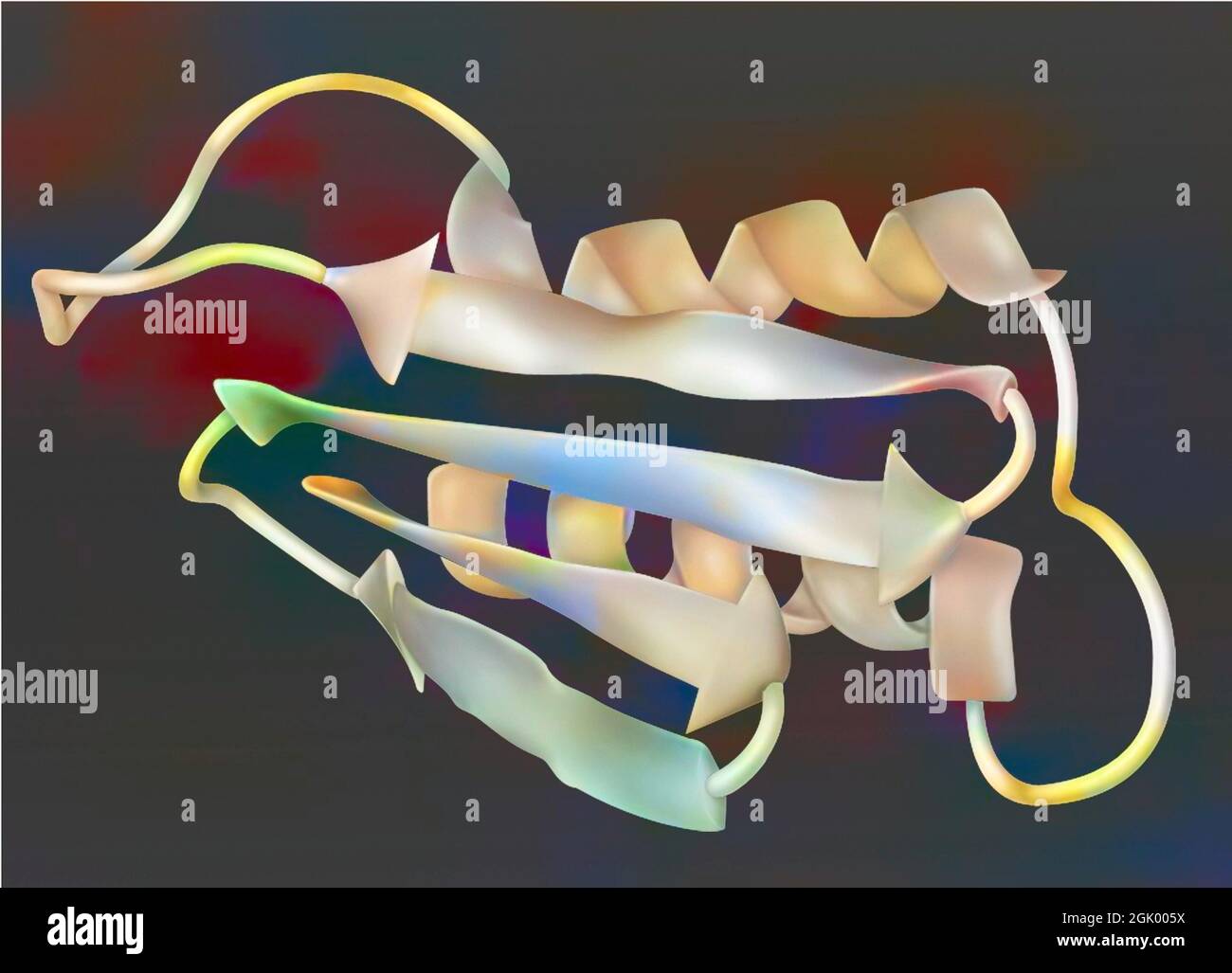 Drawing of a prion: infectious protein (encephalopathy agent). Stock Photohttps://www.alamy.com/image-license-details/?v=1https://www.alamy.com/drawing-of-a-prion-infectious-protein-encephalopathy-agent-image441937830.html
RF2GK005X–Drawing of a prion: infectious protein (encephalopathy agent).
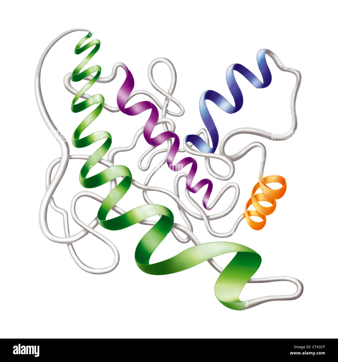 Protein Stock Photohttps://www.alamy.com/image-license-details/?v=1https://www.alamy.com/stock-photo-protein-49262991.html
RMCT43CF–Protein
 Asparagine. Molecular model of the amino acid asparagine (C4.H8.N2.O3). It is a non-essential amino acid. It can be synthesised by the body and so does not need to come from the diet. Asparagine plays an important role in the folding of protein molecules into their secondary structures. Atoms are represented as spheres and are colour-coded: carbon (grey), hydrogen (white), nitrogen (blue) and oxygen (red). Illustration. Stock Photohttps://www.alamy.com/image-license-details/?v=1https://www.alamy.com/stock-photo-asparagine-molecular-model-of-the-amino-acid-asparagine-c4h8n2o3-it-126899781.html
RFHACNY1–Asparagine. Molecular model of the amino acid asparagine (C4.H8.N2.O3). It is a non-essential amino acid. It can be synthesised by the body and so does not need to come from the diet. Asparagine plays an important role in the folding of protein molecules into their secondary structures. Atoms are represented as spheres and are colour-coded: carbon (grey), hydrogen (white), nitrogen (blue) and oxygen (red). Illustration.
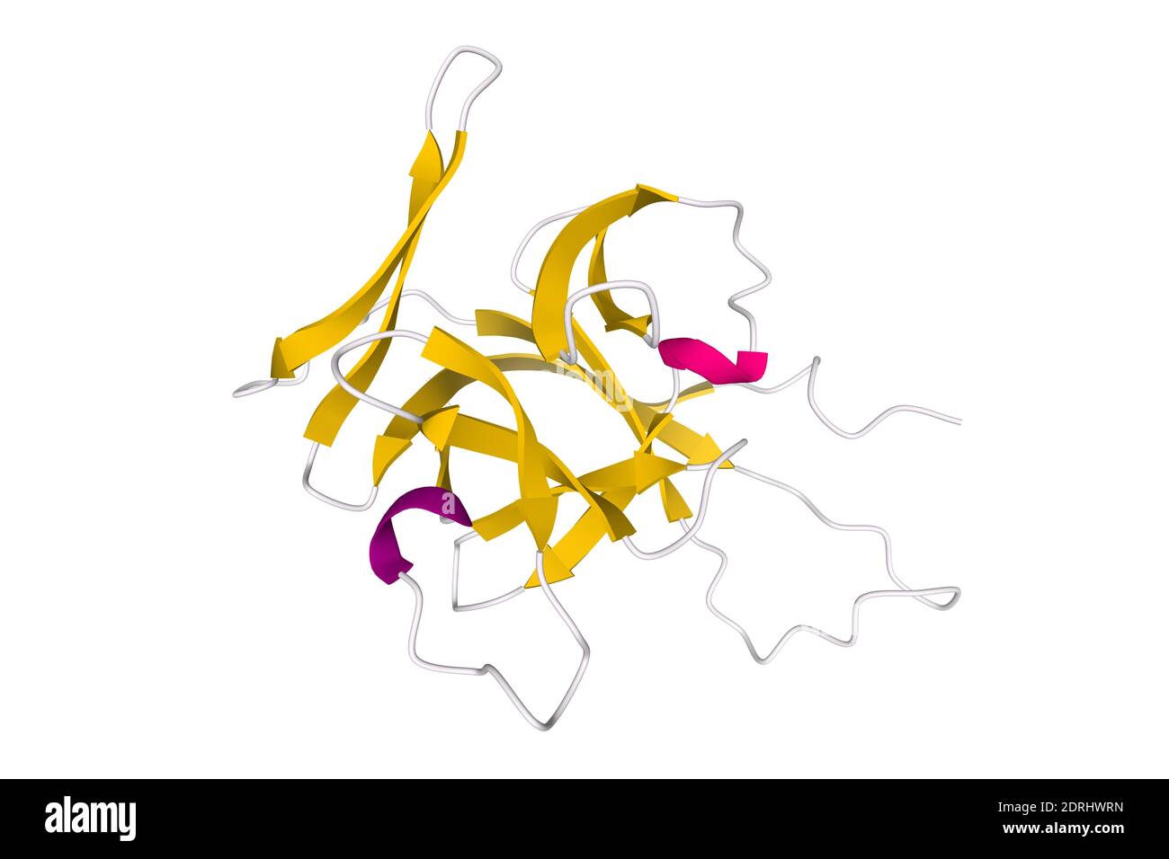 Structure of human interleukin-33, 3D cartoon model isolated with differently colored elements of the secondary structure, white background Stock Photohttps://www.alamy.com/image-license-details/?v=1https://www.alamy.com/structure-of-human-interleukin-33-3d-cartoon-model-isolated-with-differently-colored-elements-of-the-secondary-structure-white-background-image393158633.html
RF2DRHWRN–Structure of human interleukin-33, 3D cartoon model isolated with differently colored elements of the secondary structure, white background
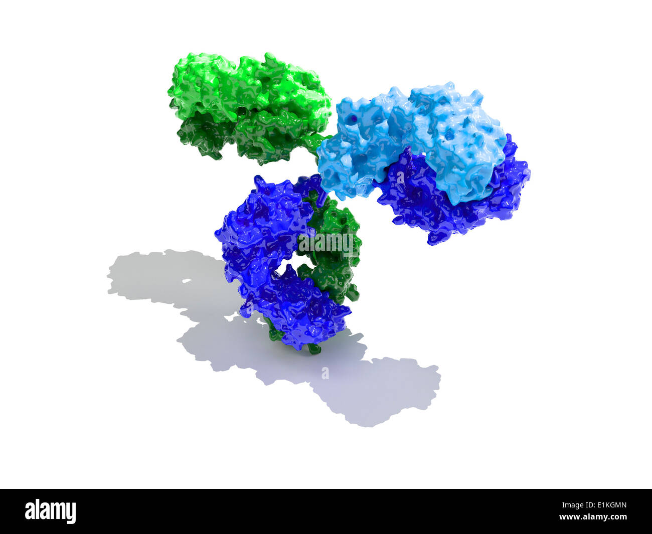 Immunoglobulin G antibody molecule Computer model showing the secondary structure of an immunoglobulin G (IgG) molecule This is Stock Photohttps://www.alamy.com/image-license-details/?v=1https://www.alamy.com/immunoglobulin-g-antibody-molecule-computer-model-showing-the-secondary-image69886341.html
RFE1KGMN–Immunoglobulin G antibody molecule Computer model showing the secondary structure of an immunoglobulin G (IgG) molecule This is
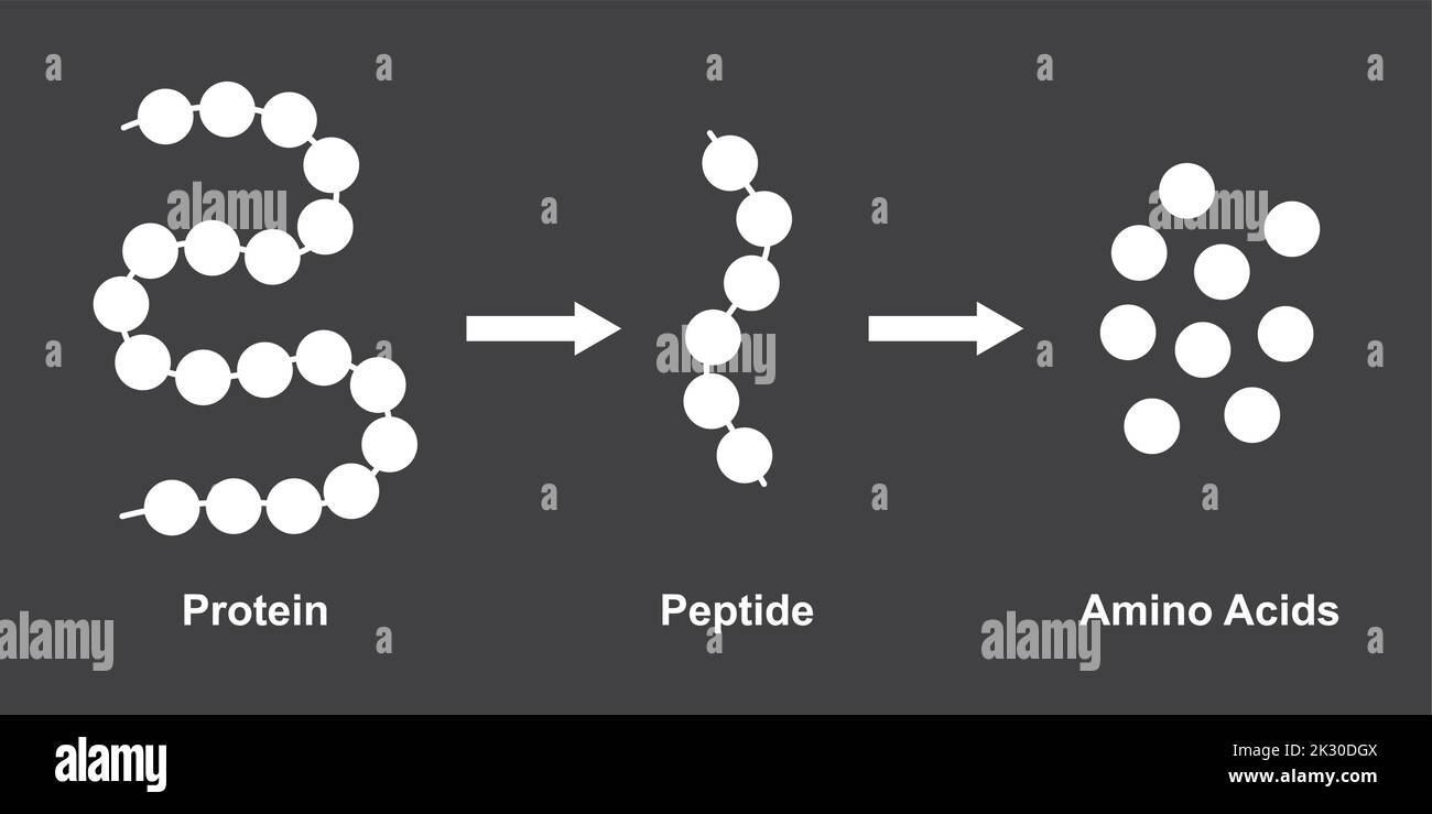 Scientific Designing of Biochemial Structure of Amino acids, Peptides And Proteins Molecular Model. Vector Illustration. Stock Vectorhttps://www.alamy.com/image-license-details/?v=1https://www.alamy.com/scientific-designing-of-biochemial-structure-of-amino-acids-peptides-and-proteins-molecular-model-vector-illustration-image483744938.html
RF2K30DGX–Scientific Designing of Biochemial Structure of Amino acids, Peptides And Proteins Molecular Model. Vector Illustration.
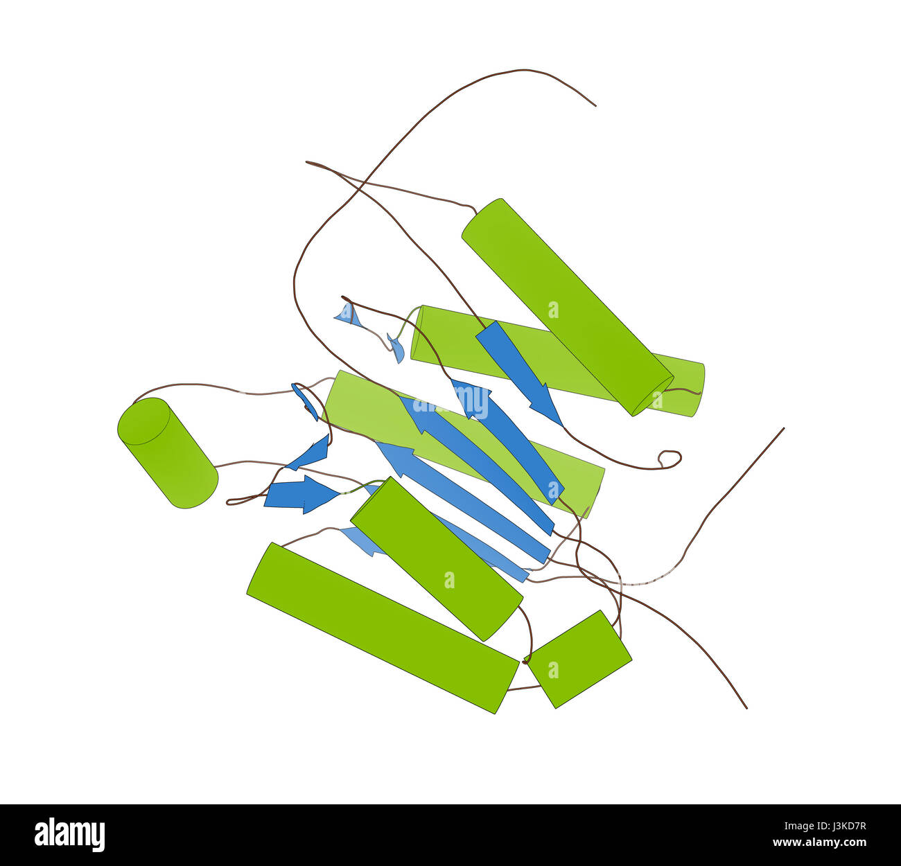 Caspase 3 apoptosis protein. Enzyme that plays important role in programmed cell death. Cartoon model, secondary structure coloring: alpha-helices gre Stock Photohttps://www.alamy.com/image-license-details/?v=1https://www.alamy.com/stock-photo-caspase-3-apoptosis-protein-enzyme-that-plays-important-role-in-programmed-139954411.html
RFJ3KD7R–Caspase 3 apoptosis protein. Enzyme that plays important role in programmed cell death. Cartoon model, secondary structure coloring: alpha-helices gre
 Structure of human interleukin-21, 3D cartoon model isolated with the colored elements of the secondary structure, white background Stock Photohttps://www.alamy.com/image-license-details/?v=1https://www.alamy.com/structure-of-human-interleukin-21-3d-cartoon-model-isolated-with-the-colored-elements-of-the-secondary-structure-white-background-image393158615.html
RF2DRHWR3–Structure of human interleukin-21, 3D cartoon model isolated with the colored elements of the secondary structure, white background
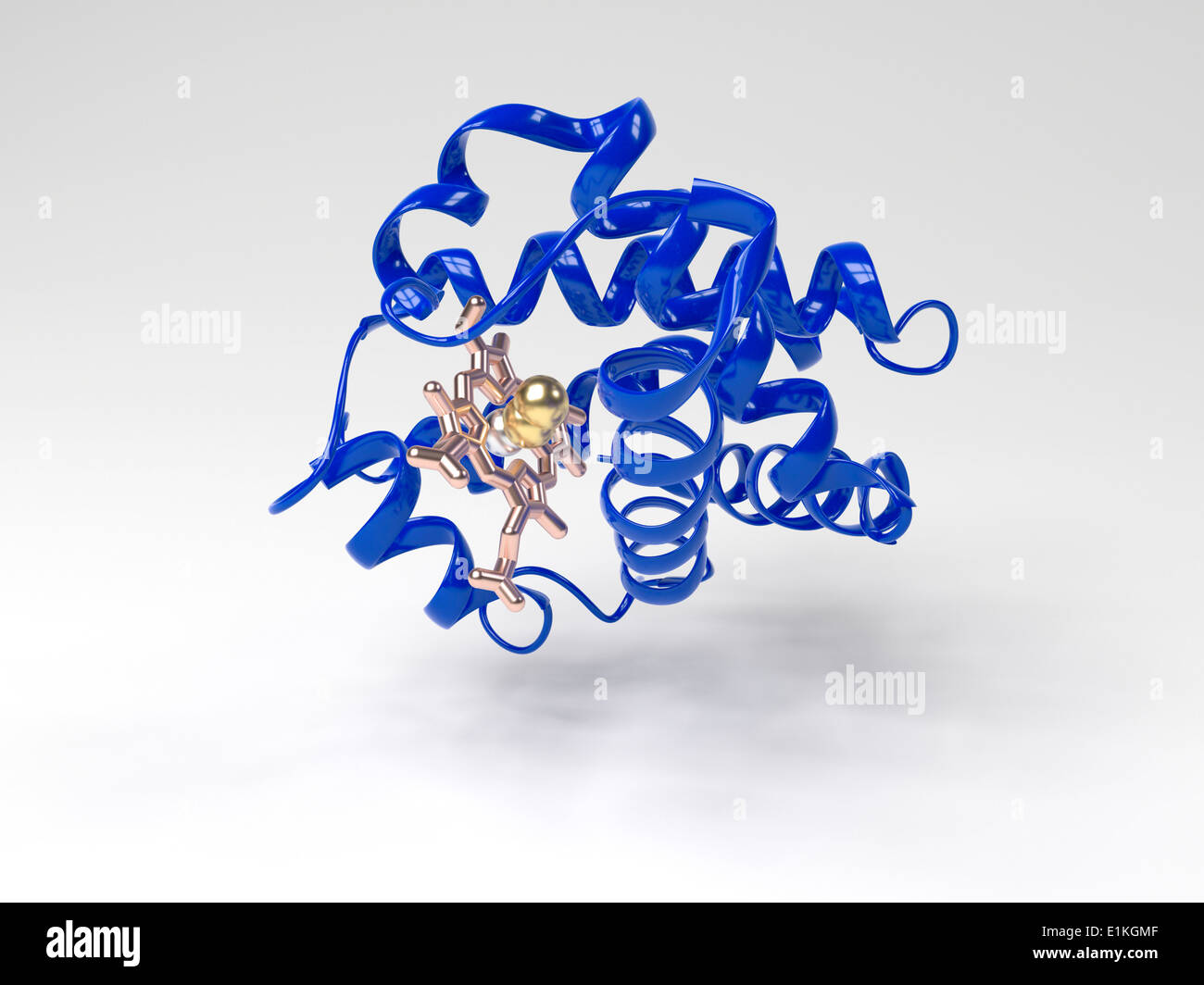 Haemoglobin molecule Computer artwork showing the tertiary structure of a haemoglobin monomer subunit Haemoglobin is a Stock Photohttps://www.alamy.com/image-license-details/?v=1https://www.alamy.com/haemoglobin-molecule-computer-artwork-showing-the-tertiary-structure-image69886335.html
RFE1KGMF–Haemoglobin molecule Computer artwork showing the tertiary structure of a haemoglobin monomer subunit Haemoglobin is a