Quick filters:
Serosa Stock Photos and Images
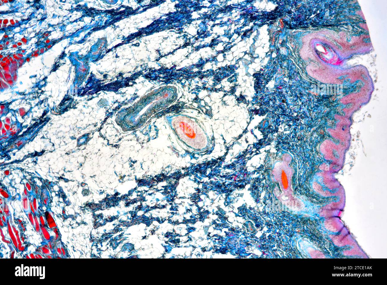 Rectum (large intestine) showing serosa, connective tissue, muscle, adipose tissue and vessels. Optical microscope X40. Stock Photohttps://www.alamy.com/image-license-details/?v=1https://www.alamy.com/rectum-large-intestine-showing-serosa-connective-tissue-muscle-adipose-tissue-and-vessels-optical-microscope-x40-image575626427.html
RF2TCE1AK–Rectum (large intestine) showing serosa, connective tissue, muscle, adipose tissue and vessels. Optical microscope X40.
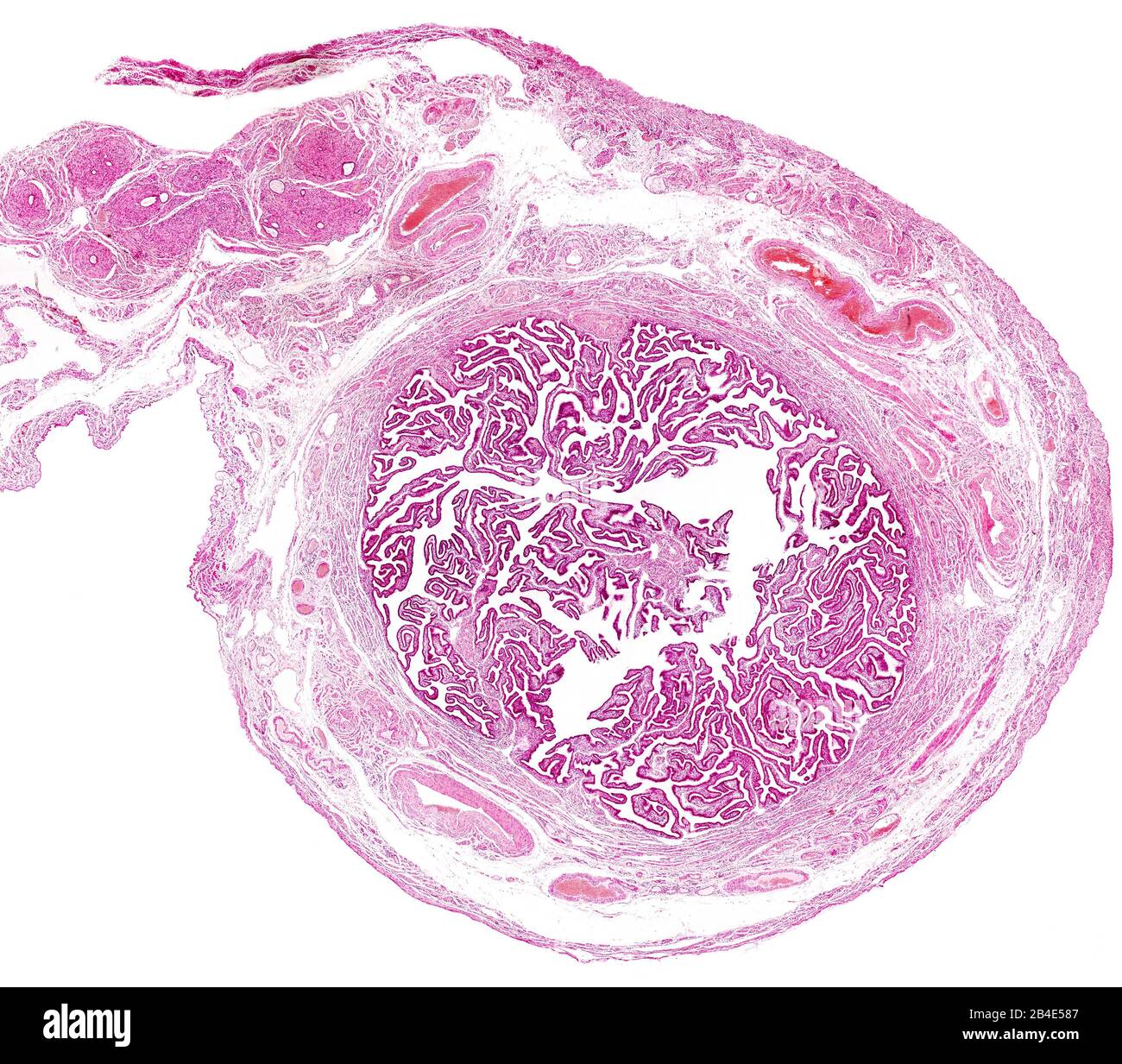 Cross section of a human fallopian tube. The innermost layer is a very folded mucosa. Outside are located the muscular layer and the serosa layer, sho Stock Photohttps://www.alamy.com/image-license-details/?v=1https://www.alamy.com/cross-section-of-a-human-fallopian-tube-the-innermost-layer-is-a-very-folded-mucosa-outside-are-located-the-muscular-layer-and-the-serosa-layer-sho-image346977463.html
RF2B4E587–Cross section of a human fallopian tube. The innermost layer is a very folded mucosa. Outside are located the muscular layer and the serosa layer, sho
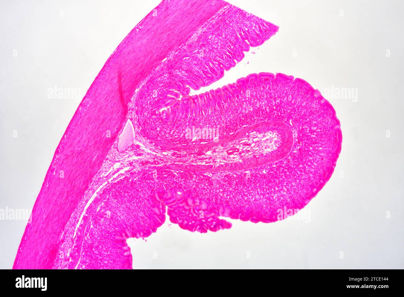 Stomach cross section showing mucosa, submucosa, muscular layer, serosa and gastric glands. Optical microscope X40. Stock Photohttps://www.alamy.com/image-license-details/?v=1https://www.alamy.com/stomach-cross-section-showing-mucosa-submucosa-muscular-layer-serosa-and-gastric-glands-optical-microscope-x40-image575626244.html
RF2TCE144–Stomach cross section showing mucosa, submucosa, muscular layer, serosa and gastric glands. Optical microscope X40.
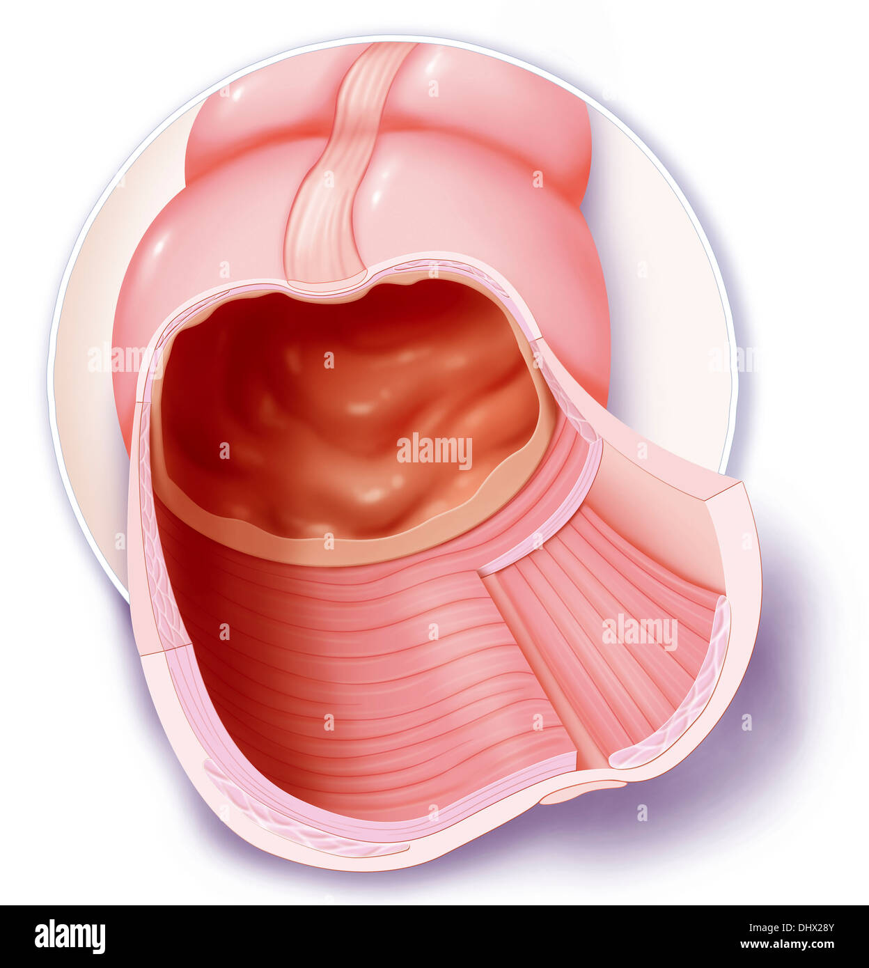 COLON, DRAWING Stock Photohttps://www.alamy.com/image-license-details/?v=1https://www.alamy.com/colon-drawing-image62652827.html
RMDHX28Y–COLON, DRAWING
 Human gastrointestinal wall showing from left to right: serosa, muscle fibers (longitudinal and circular), submucosa and mucosa with villi, glands and Stock Photohttps://www.alamy.com/image-license-details/?v=1https://www.alamy.com/human-gastrointestinal-wall-showing-from-left-to-right-serosa-muscle-fibers-longitudinal-and-circular-submucosa-and-mucosa-with-villi-glands-and-image592002173.html
RF2WB40PN–Human gastrointestinal wall showing from left to right: serosa, muscle fibers (longitudinal and circular), submucosa and mucosa with villi, glands and
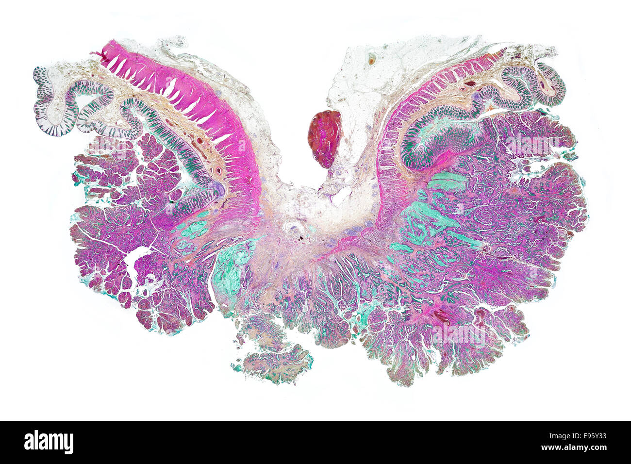 Human gut with carcanoma. brightfield photomicrograph, stained section Stock Photohttps://www.alamy.com/image-license-details/?v=1https://www.alamy.com/stock-photo-human-gut-with-carcanoma-brightfield-photomicrograph-stained-section-74504391.html
RME95Y33–Human gut with carcanoma. brightfield photomicrograph, stained section
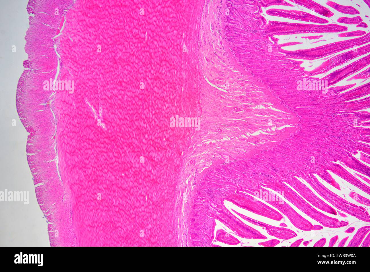 Human small intestine showing from left to right: serosa, muscular fibers longitudinal and circular, submucosa and mucosa with Lieberkhun crypts. X25 Stock Photohttps://www.alamy.com/image-license-details/?v=1https://www.alamy.com/human-small-intestine-showing-from-left-to-right-serosa-muscular-fibers-longitudinal-and-circular-submucosa-and-mucosa-with-lieberkhun-crypts-x25-image591999194.html
RF2WB3W0A–Human small intestine showing from left to right: serosa, muscular fibers longitudinal and circular, submucosa and mucosa with Lieberkhun crypts. X25
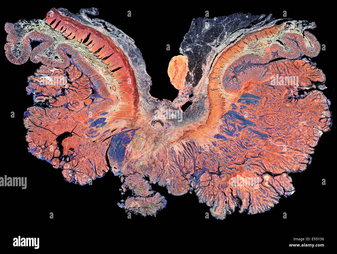 Human gut with carcanoma. darkfield photomicrograph, stained section Stock Photohttps://www.alamy.com/image-license-details/?v=1https://www.alamy.com/stock-photo-human-gut-with-carcanoma-darkfield-photomicrograph-stained-section-74504394.html
RME95Y36–Human gut with carcanoma. darkfield photomicrograph, stained section
 Human small intestine showing from left to right: serosa, muscular fibers (longitudinal and circular), submucosa and mucosa with Lieberkhun crypts. X7 Stock Photohttps://www.alamy.com/image-license-details/?v=1https://www.alamy.com/human-small-intestine-showing-from-left-to-right-serosa-muscular-fibers-longitudinal-and-circular-submucosa-and-mucosa-with-lieberkhun-crypts-x7-image591999265.html
RF2WB3W2W–Human small intestine showing from left to right: serosa, muscular fibers (longitudinal and circular), submucosa and mucosa with Lieberkhun crypts. X7
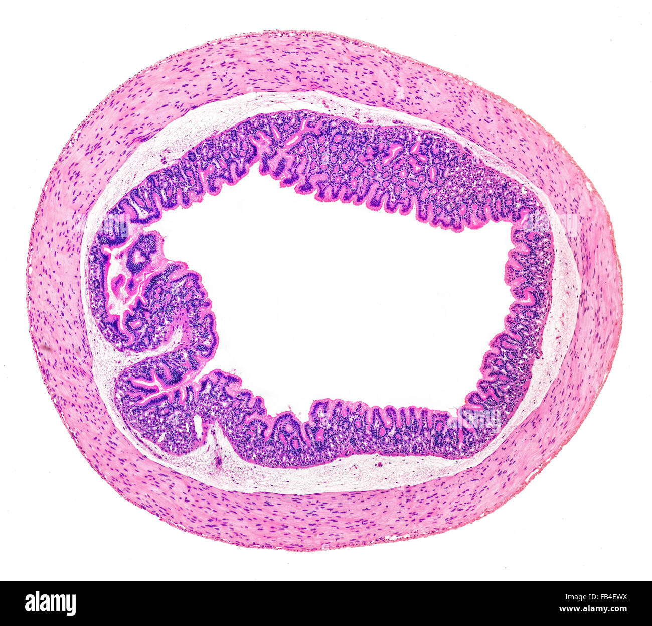 Salamander lizard stomach section stained, TS. Brightfield photomicrograph Stock Photohttps://www.alamy.com/image-license-details/?v=1https://www.alamy.com/stock-photo-salamander-lizard-stomach-section-stained-ts-brightfield-photomicrograph-92912566.html
RMFB4EWX–Salamander lizard stomach section stained, TS. Brightfield photomicrograph
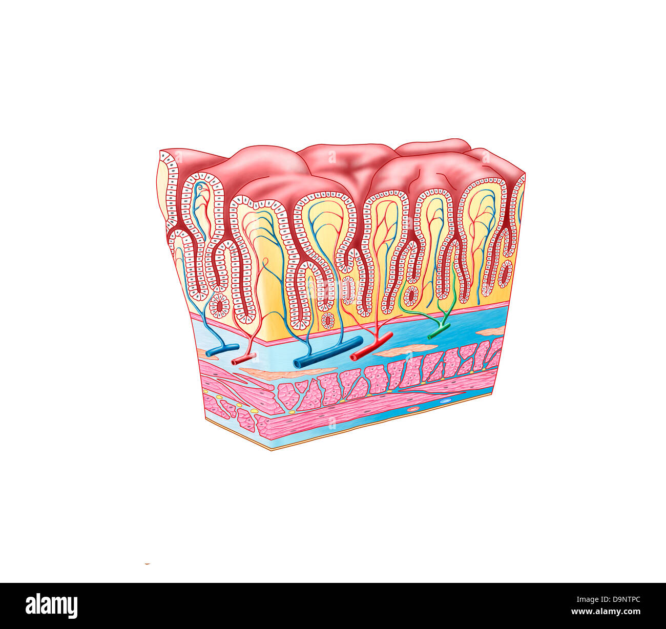 Anatomy of the structure and layers of the stomach wall. Stock Photohttps://www.alamy.com/image-license-details/?v=1https://www.alamy.com/stock-photo-anatomy-of-the-structure-and-layers-of-the-stomach-wall-57643444.html
RFD9NTPC–Anatomy of the structure and layers of the stomach wall.
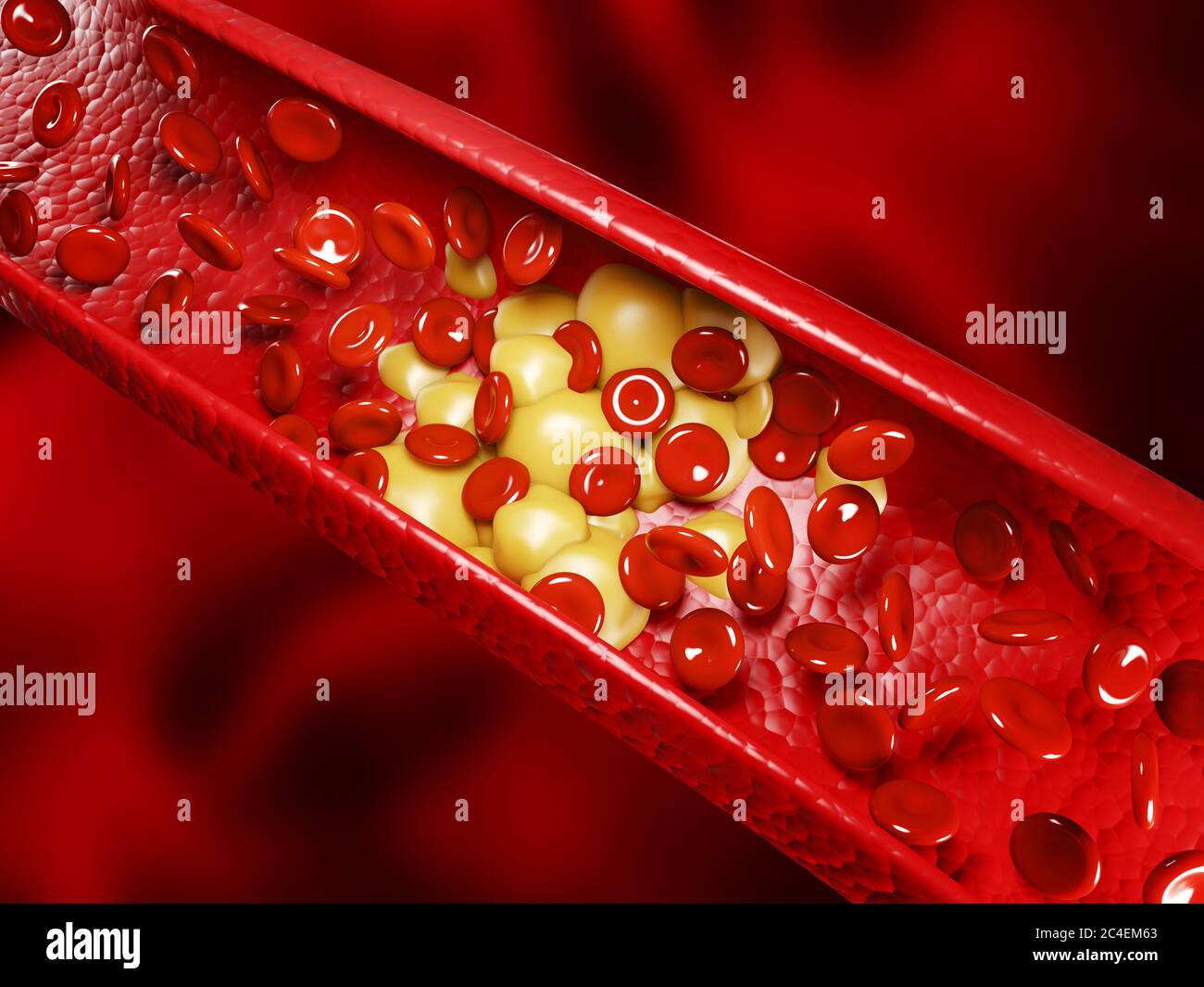 Healthy human red bloodcells and cholesterol plaques. 3d rendering concept Stock Photohttps://www.alamy.com/image-license-details/?v=1https://www.alamy.com/healthy-human-red-bloodcells-and-cholesterol-plaques-3d-rendering-concept-image364199531.html
RF2C4EM63–Healthy human red bloodcells and cholesterol plaques. 3d rendering concept
 wild boar Sus scrofa or colloquially simply called the boar is an omnivorous gregarious mammal of the biological family Suidae Stock Photohttps://www.alamy.com/image-license-details/?v=1https://www.alamy.com/stock-photo-wild-boar-sus-scrofa-or-colloquially-simply-called-the-boar-is-an-20944419.html
RMB622PY–wild boar Sus scrofa or colloquially simply called the boar is an omnivorous gregarious mammal of the biological family Suidae
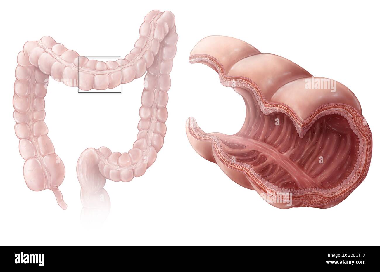 Large Intestine Mucosae Stock Photohttps://www.alamy.com/image-license-details/?v=1https://www.alamy.com/large-intestine-mucosae-image353183290.html
RM2BEGTTX–Large Intestine Mucosae
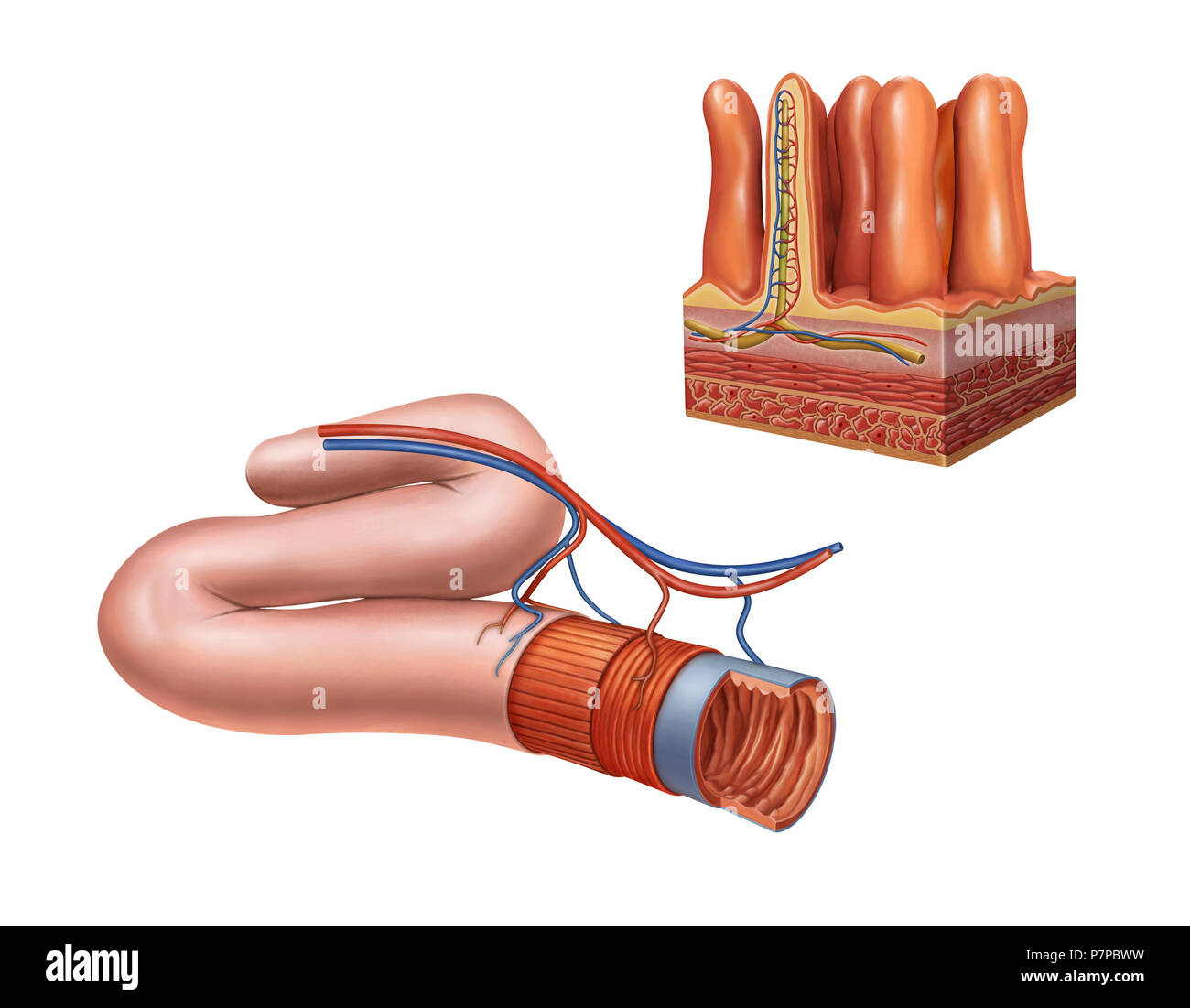 Small intestine anatomy. Digital illustration. Stock Photohttps://www.alamy.com/image-license-details/?v=1https://www.alamy.com/small-intestine-anatomy-digital-illustration-image211319301.html
RFP7PBWW–Small intestine anatomy. Digital illustration.
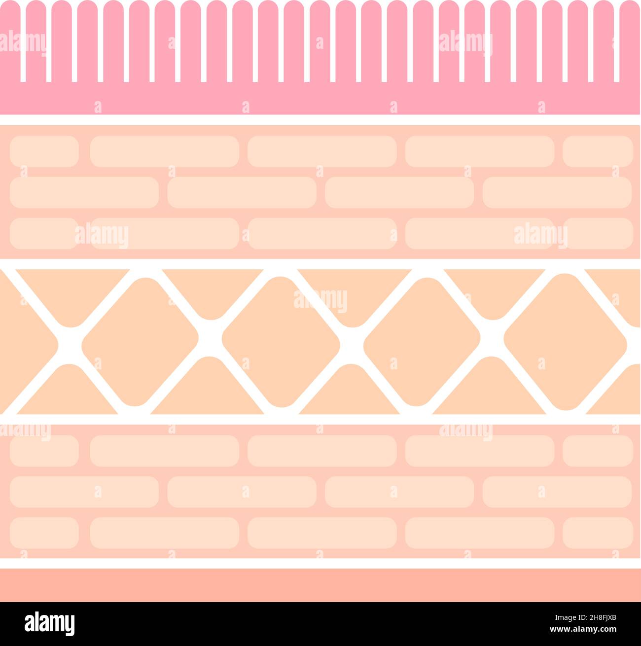 Sectional view illustration of gastric wall (stomach lining) Stock Vectorhttps://www.alamy.com/image-license-details/?v=1https://www.alamy.com/sectional-view-illustration-of-gastric-wall-stomach-lining-image452730947.html
RF2H8FJXB–Sectional view illustration of gastric wall (stomach lining)
 Chronic stomach ulcer Stock Photohttps://www.alamy.com/image-license-details/?v=1https://www.alamy.com/stock-photo-chronic-stomach-ulcer-13236661.html
RFACWHDX–Chronic stomach ulcer
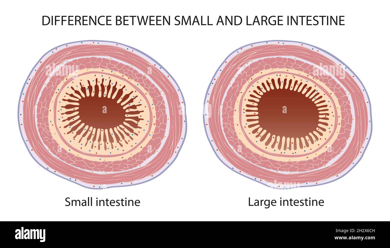 Difference Between Small and Large Intestine Stock Photohttps://www.alamy.com/image-license-details/?v=1https://www.alamy.com/difference-between-small-and-large-intestine-image449274689.html
RF2H2X6CH–Difference Between Small and Large Intestine
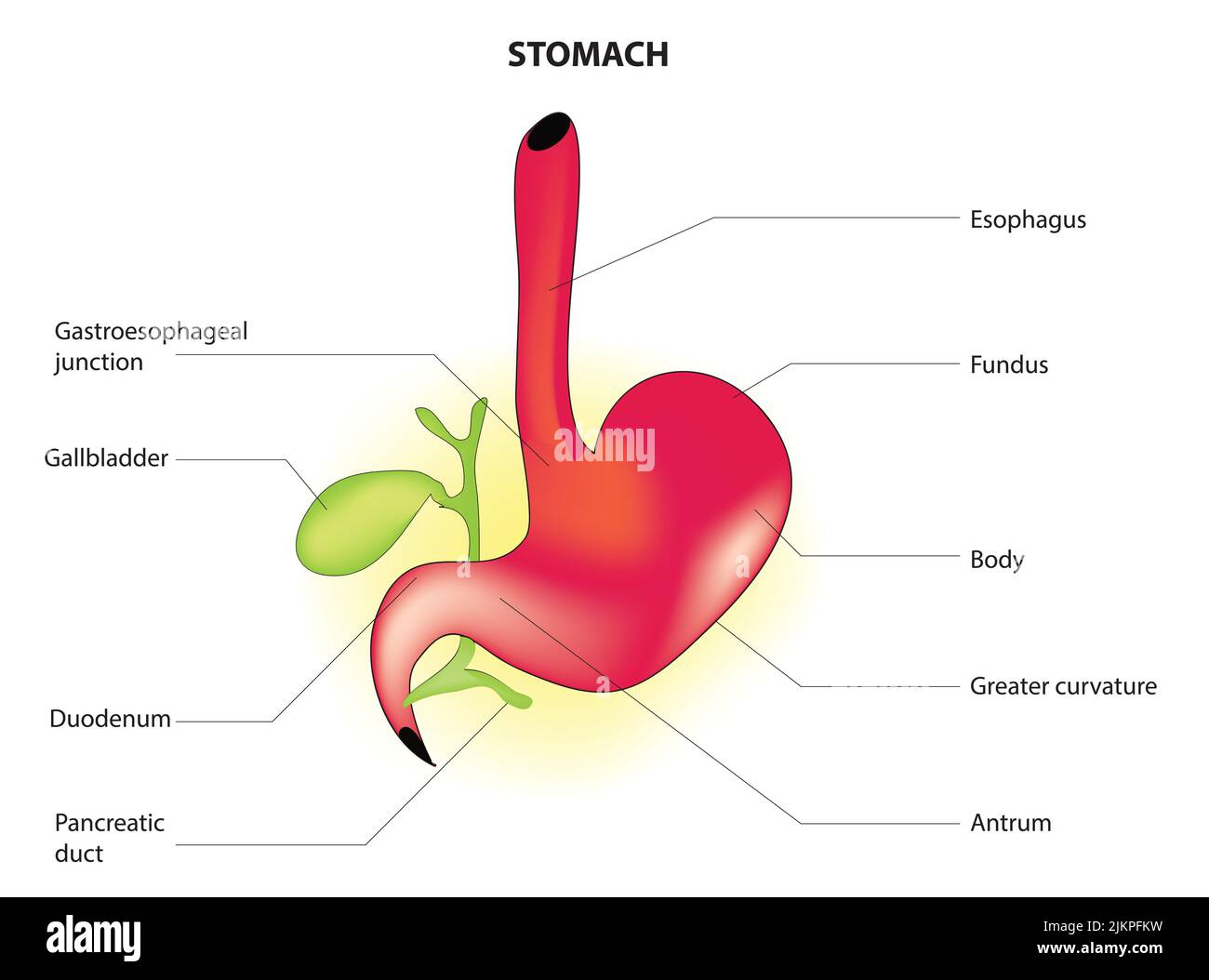 Stomach anatomy Stock Photohttps://www.alamy.com/image-license-details/?v=1https://www.alamy.com/stomach-anatomy-image476853661.html
RF2JKPFKW–Stomach anatomy
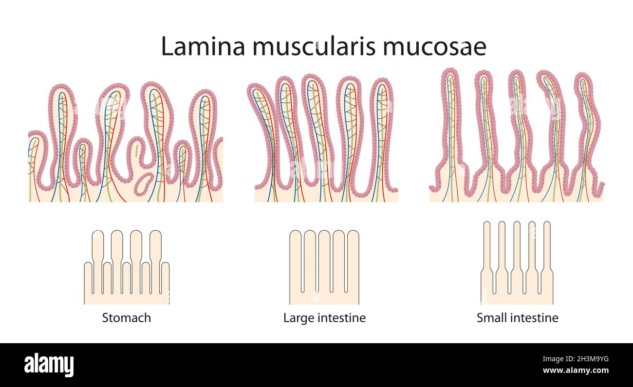 Difference of mucosae of stomach, small and large intestines Difference of mucosae of stomach, small and large intestines Stock Photohttps://www.alamy.com/image-license-details/?v=1https://www.alamy.com/difference-of-mucosae-of-stomach-small-and-large-intestines-difference-of-mucosae-of-stomach-small-and-large-intestines-image449760404.html
RF2H3M9YG–Difference of mucosae of stomach, small and large intestines Difference of mucosae of stomach, small and large intestines
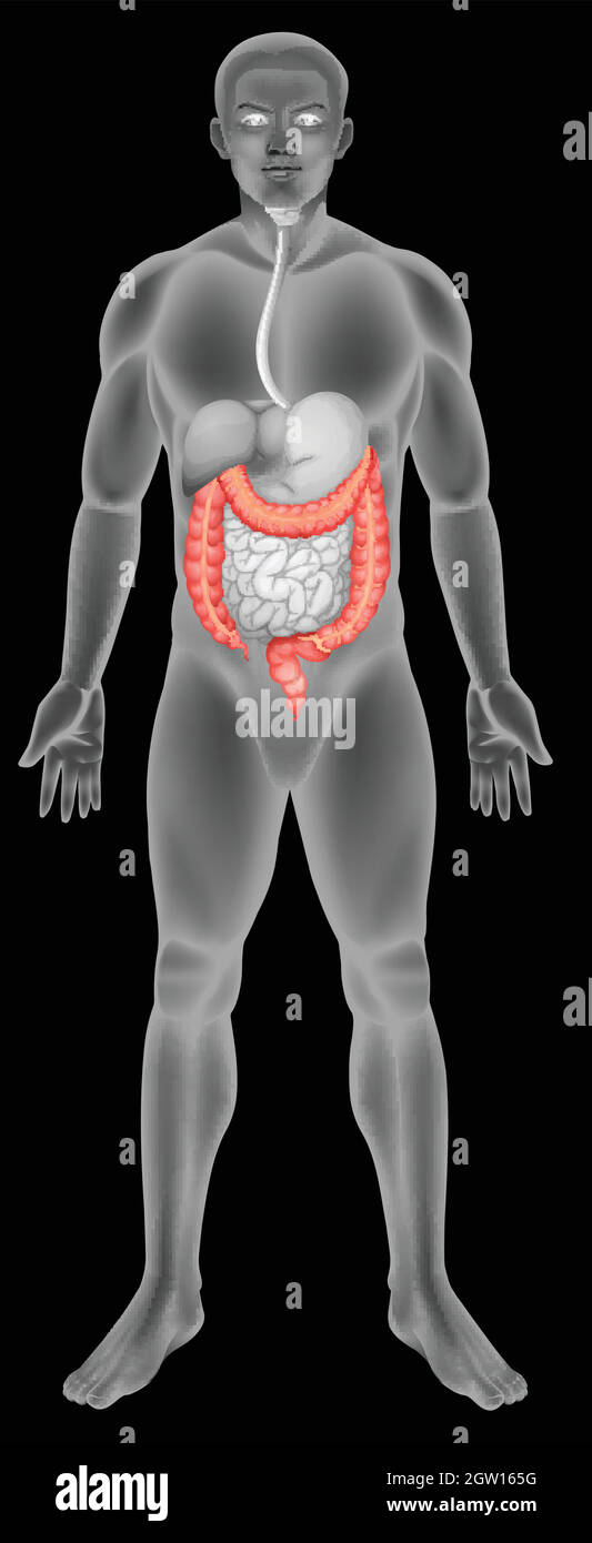 The large intestine Stock Vectorhttps://www.alamy.com/image-license-details/?v=1https://www.alamy.com/the-large-intestine-image445652412.html
RF2GW165G–The large intestine
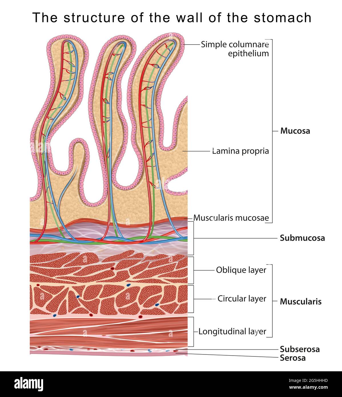 The structure of the wall of the stomach Stock Photohttps://www.alamy.com/image-license-details/?v=1https://www.alamy.com/the-structure-of-the-wall-of-the-stomach-image433719481.html
RF2G5HHHD–The structure of the wall of the stomach
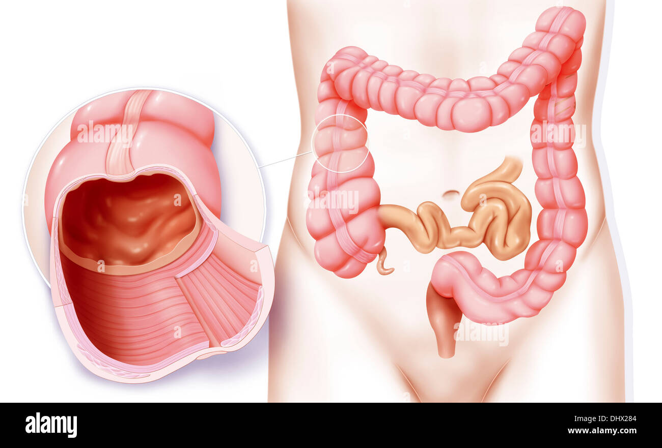 COLON, DRAWING Stock Photohttps://www.alamy.com/image-license-details/?v=1https://www.alamy.com/colon-drawing-image62652804.html
RMDHX284–COLON, DRAWING
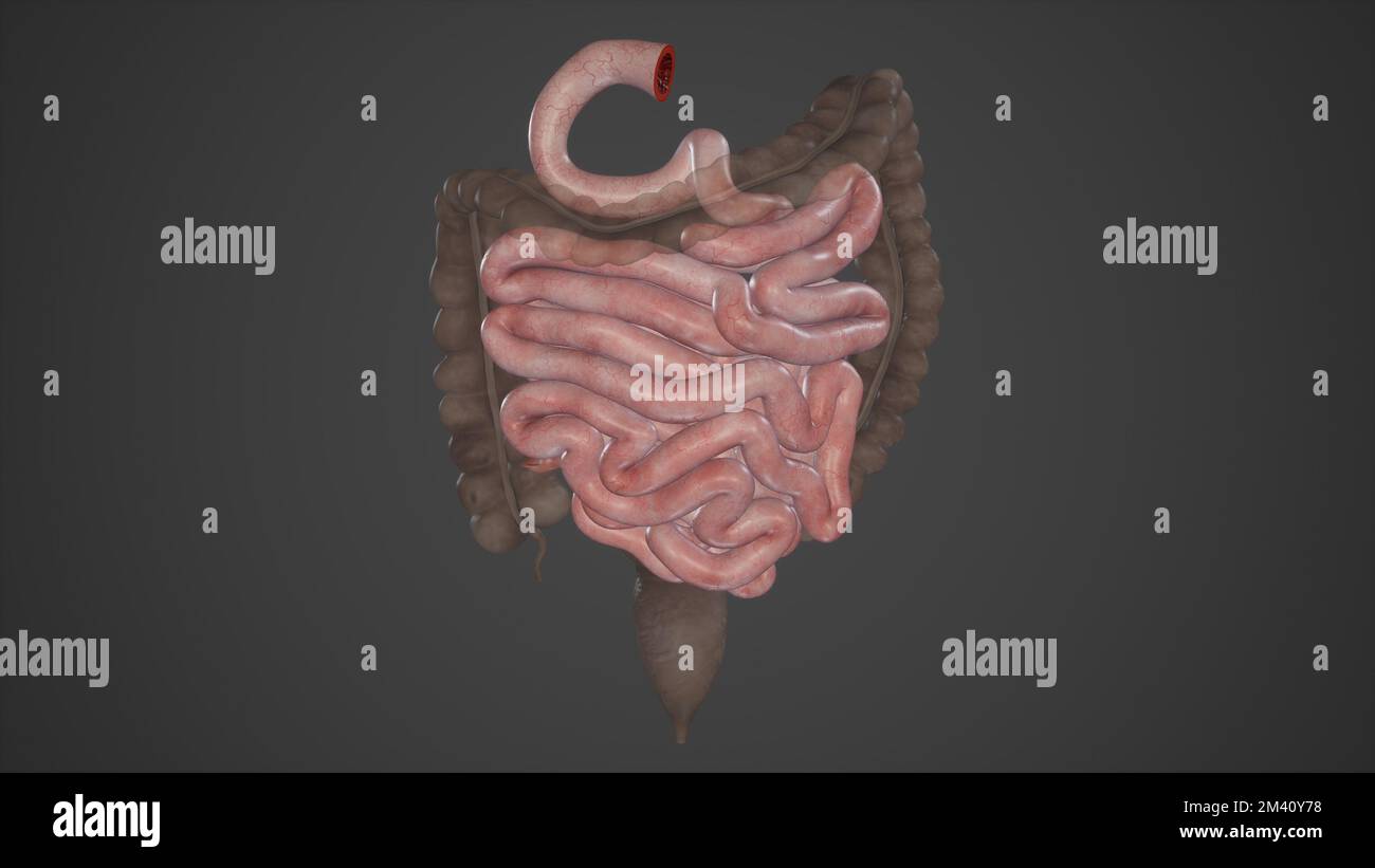 Small Intestine Parts Stock Photohttps://www.alamy.com/image-license-details/?v=1https://www.alamy.com/small-intestine-parts-image501580668.html
RF2M40Y78–Small Intestine Parts
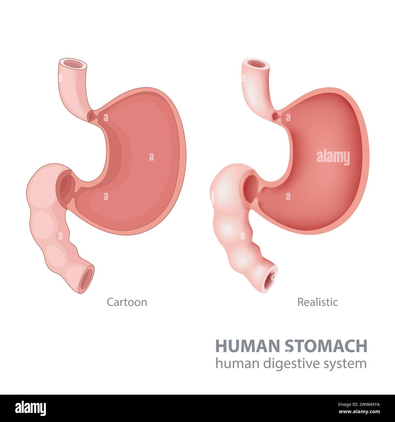 Human Stomach In Cartoon and Realistic, Vector Illustration Stock Vectorhttps://www.alamy.com/image-license-details/?v=1https://www.alamy.com/human-stomach-in-cartoon-and-realistic-vector-illustration-image600627870.html
RF2WW4XYA–Human Stomach In Cartoon and Realistic, Vector Illustration
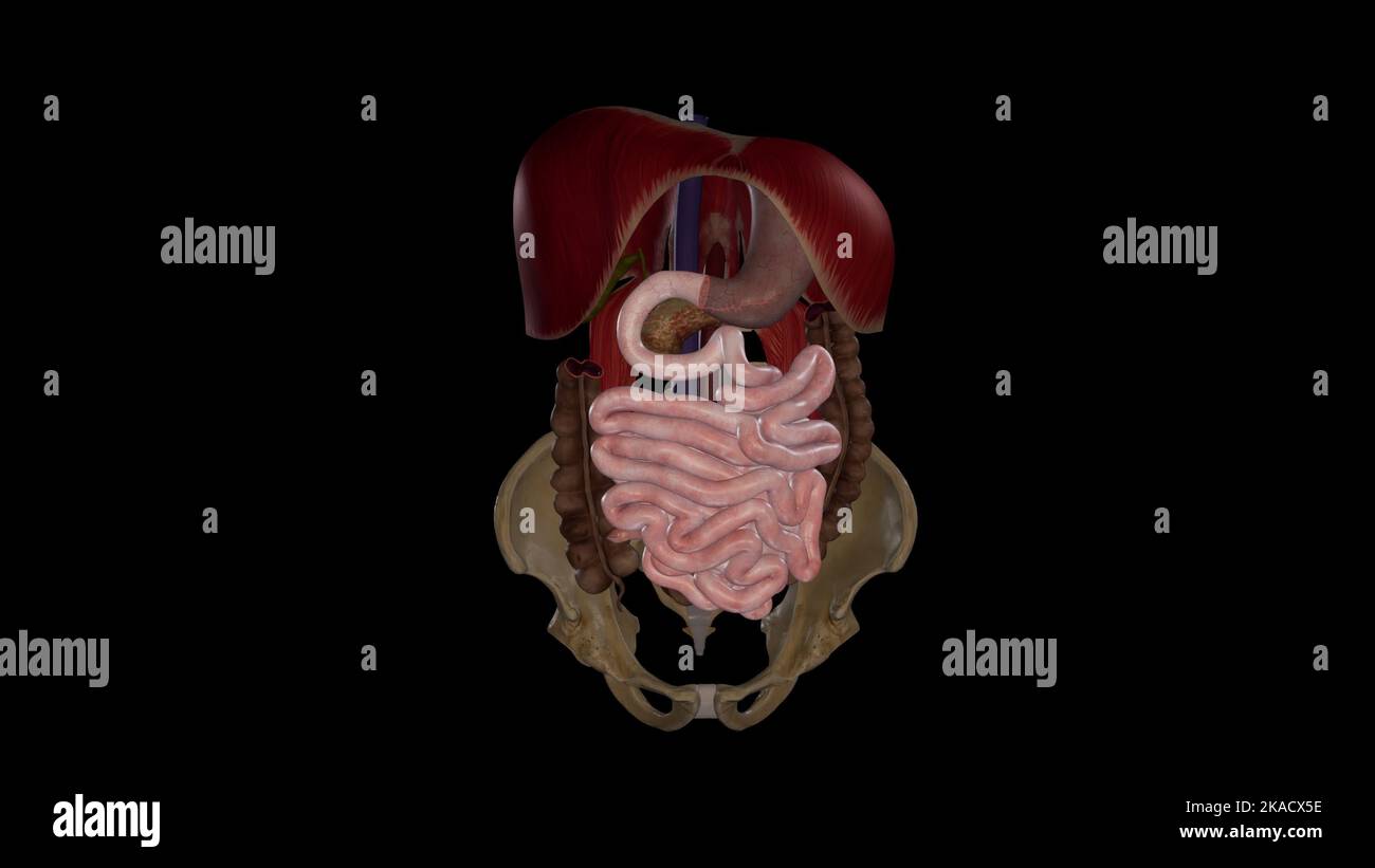 Medical Illustration of Small Intestine - Anterior View Stock Photohttps://www.alamy.com/image-license-details/?v=1https://www.alamy.com/medical-illustration-of-small-intestine-anterior-view-image488320826.html
RF2KACX5E–Medical Illustration of Small Intestine - Anterior View
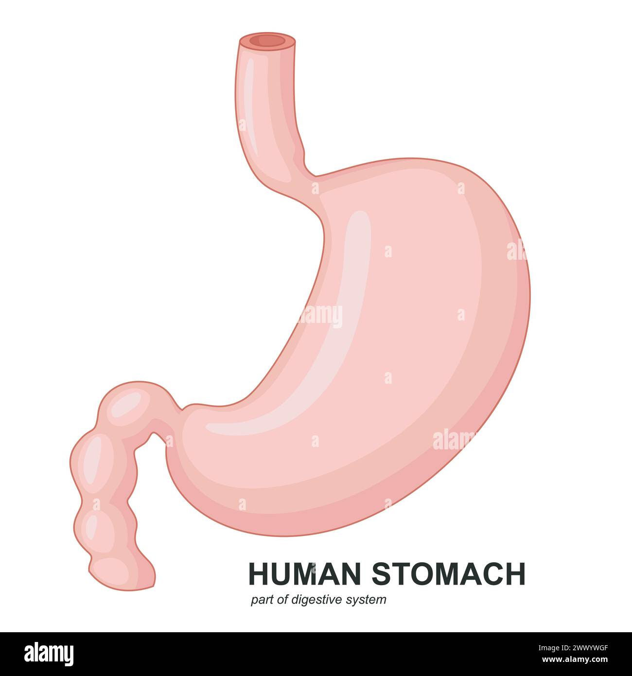 Human Stomach Cartoon, Vector Illustration Stock Vectorhttps://www.alamy.com/image-license-details/?v=1https://www.alamy.com/human-stomach-cartoon-vector-illustration-image601131679.html
RF2WWYWGF–Human Stomach Cartoon, Vector Illustration
 Human small intestine (ileum) showing from left to right: serosa, muscular fibers (longitudinal and circular), submucosa and mucosa with Lieberkhun cr Stock Photohttps://www.alamy.com/image-license-details/?v=1https://www.alamy.com/human-small-intestine-ileum-showing-from-left-to-right-serosa-muscular-fibers-longitudinal-and-circular-submucosa-and-mucosa-with-lieberkhun-cr-image591999272.html
RF2WB3W34–Human small intestine (ileum) showing from left to right: serosa, muscular fibers (longitudinal and circular), submucosa and mucosa with Lieberkhun cr
 micrograph of medical science simple squamous epithelium cell Stock Photohttps://www.alamy.com/image-license-details/?v=1https://www.alamy.com/stock-photo-micrograph-of-medical-science-simple-squamous-epithelium-cell-52335794.html
RFD142RE–micrograph of medical science simple squamous epithelium cell
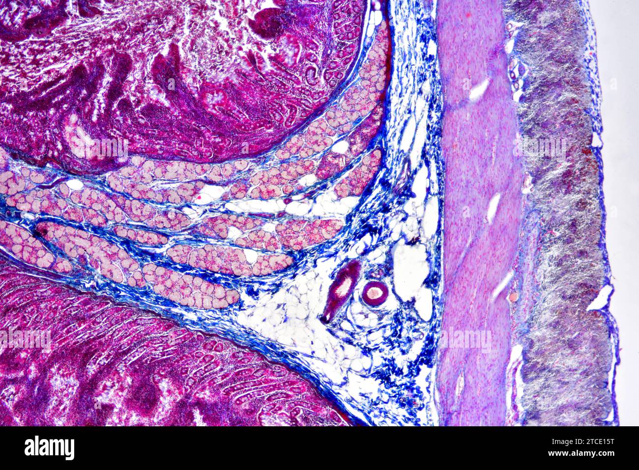 Human duodenum (small intestine) showing villous mucosa, submucosa, muscular layer, serosa, vessel, Brunner glands and duodenal glands. Optical micros Stock Photohttps://www.alamy.com/image-license-details/?v=1https://www.alamy.com/human-duodenum-small-intestine-showing-villous-mucosa-submucosa-muscular-layer-serosa-vessel-brunner-glands-and-duodenal-glands-optical-micros-image575626292.html
RF2TCE15T–Human duodenum (small intestine) showing villous mucosa, submucosa, muscular layer, serosa, vessel, Brunner glands and duodenal glands. Optical micros
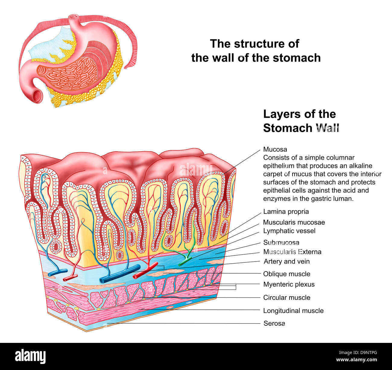 Anatomy of the structure and layers of the stomach wall. Stock Photohttps://www.alamy.com/image-license-details/?v=1https://www.alamy.com/stock-photo-anatomy-of-the-structure-and-layers-of-the-stomach-wall-57643448.html
RFD9NTPG–Anatomy of the structure and layers of the stomach wall.
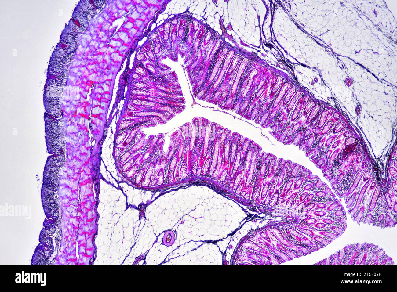 Human colon showing epithelium, mucosa, submucosa, muscular layer, adipose tissue, serosa, intestinal glands, villi and vessels. Optical microscope X4 Stock Photohttps://www.alamy.com/image-license-details/?v=1https://www.alamy.com/human-colon-showing-epithelium-mucosa-submucosa-muscular-layer-adipose-tissue-serosa-intestinal-glands-villi-and-vessels-optical-microscope-x4-image575626117.html
RF2TCE0YH–Human colon showing epithelium, mucosa, submucosa, muscular layer, adipose tissue, serosa, intestinal glands, villi and vessels. Optical microscope X4
 Healthy human red bloodcells and cholesterol plaques. 3d rendering concept Stock Photohttps://www.alamy.com/image-license-details/?v=1https://www.alamy.com/healthy-human-red-bloodcells-and-cholesterol-plaques-3d-rendering-concept-image364281157.html
RF2C4JC99–Healthy human red bloodcells and cholesterol plaques. 3d rendering concept
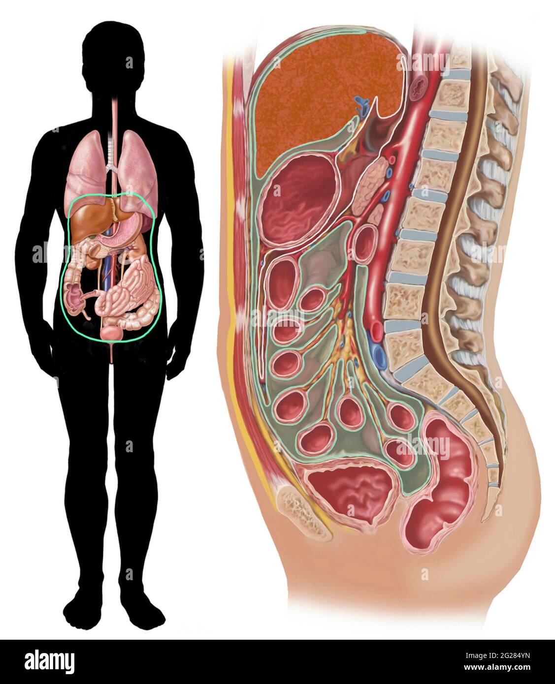 Location of peritoneum inside body cavity. Stock Photohttps://www.alamy.com/image-license-details/?v=1https://www.alamy.com/location-of-peritoneum-inside-body-cavity-image431668041.html
RM2G284YN–Location of peritoneum inside body cavity.
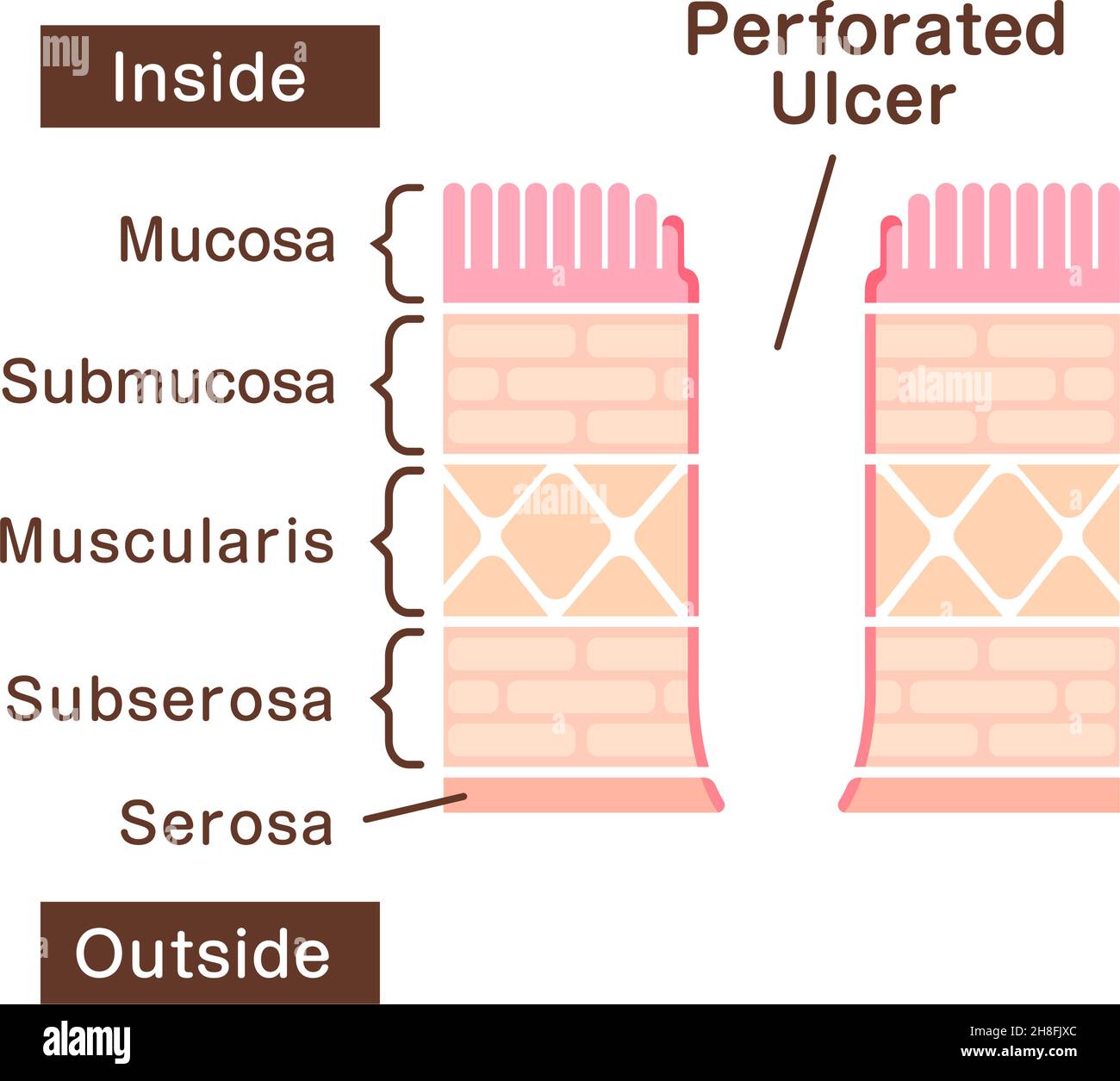 Sectional view illustration of perforated ulcer Stock Vectorhttps://www.alamy.com/image-license-details/?v=1https://www.alamy.com/sectional-view-illustration-of-perforated-ulcer-image452730948.html
RF2H8FJXC–Sectional view illustration of perforated ulcer
 Small Intestine Diagram. Cross-section of a typical segment of the intestinal wall showing the layers: mucosa, submucosa, muscularis, and serosa. Stock Vectorhttps://www.alamy.com/image-license-details/?v=1https://www.alamy.com/small-intestine-diagram-cross-section-of-a-typical-segment-of-the-intestinal-wall-showing-the-layers-mucosa-submucosa-muscularis-and-serosa-image496267300.html
RF2KRAX04–Small Intestine Diagram. Cross-section of a typical segment of the intestinal wall showing the layers: mucosa, submucosa, muscularis, and serosa.
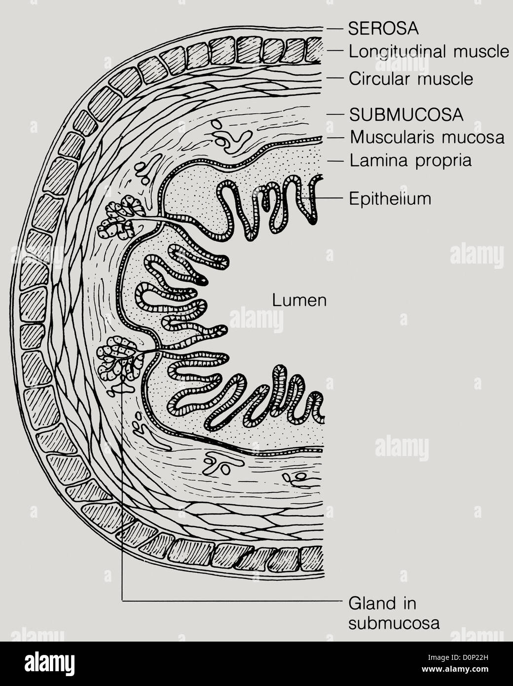 The walls digestive tract have four layers tissue: mucosa submucosa muscularis externa serosa. inner-most layer is mucosa Stock Photohttps://www.alamy.com/image-license-details/?v=1https://www.alamy.com/stock-photo-the-walls-digestive-tract-have-four-layers-tissue-mucosa-submucosa-52115689.html
RMD0P22H–The walls digestive tract have four layers tissue: mucosa submucosa muscularis externa serosa. inner-most layer is mucosa
 The serosa is the outer layer of the colon 3d illustration Stock Photohttps://www.alamy.com/image-license-details/?v=1https://www.alamy.com/the-serosa-is-the-outer-layer-of-the-colon-3d-illustration-image596911148.html
RF2WK3J78–The serosa is the outer layer of the colon 3d illustration
 Alcea setosa or bristly hollyhock ornamental plant on green blurred background. Stock Photohttps://www.alamy.com/image-license-details/?v=1https://www.alamy.com/alcea-setosa-or-bristly-hollyhock-ornamental-plant-on-green-blurred-background-image364957056.html
RF2C5N6CG–Alcea setosa or bristly hollyhock ornamental plant on green blurred background.
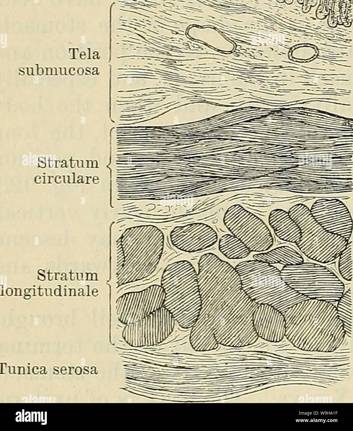 Archive image from page 1207 of Cunningham's Text-book of anatomy (1914). Cunningham's Text-book of anatomy cunninghamstextb00cunn Year: 1914 ( 1174 THE DIGESTIVE SYSTEM. ilrffli Tunica mucosa Tela submucosa Structure of the Stomach. The stomach wall is composed of four coats—namely, from without inwards: (1) Tunica serosa, (2) tunica muscularis, (3) tela submucosa, and (4) tunica mucosa (Fig. 924). Tunica Serosa.—This coat is formed of the peritoneum, the relations of which to the stomach have already been described. It is closely attached to the subjacent muscular coat, except near the curv Stock Photohttps://www.alamy.com/image-license-details/?v=1https://www.alamy.com/archive-image-from-page-1207-of-cunninghams-text-book-of-anatomy-1914-cunninghams-text-book-of-anatomy-cunninghamstextb00cunn-year-1914-1174-the-digestive-system-ilrffli-tunica-mucosa-tela-submucosa-structure-of-the-stomach-the-stomach-wall-is-composed-of-four-coatsnamely-from-without-inwards-1-tunica-serosa-2-tunica-muscularis-3-tela-submucosa-and-4-tunica-mucosa-fig-924-tunica-serosathis-coat-is-formed-of-the-peritoneum-the-relations-of-which-to-the-stomach-have-already-been-described-it-is-closely-attached-to-the-subjacent-muscular-coat-except-near-the-curv-image264068491.html
RMW9HA1F–Archive image from page 1207 of Cunningham's Text-book of anatomy (1914). Cunningham's Text-book of anatomy cunninghamstextb00cunn Year: 1914 ( 1174 THE DIGESTIVE SYSTEM. ilrffli Tunica mucosa Tela submucosa Structure of the Stomach. The stomach wall is composed of four coats—namely, from without inwards: (1) Tunica serosa, (2) tunica muscularis, (3) tela submucosa, and (4) tunica mucosa (Fig. 924). Tunica Serosa.—This coat is formed of the peritoneum, the relations of which to the stomach have already been described. It is closely attached to the subjacent muscular coat, except near the curv
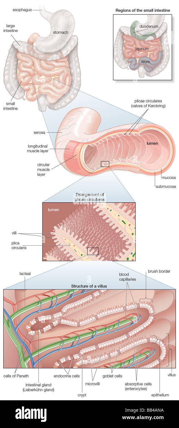 Diagram of the human small intestine, with insets of musculature, mucosa histology and its regions in situ Stock Photohttps://www.alamy.com/image-license-details/?v=1https://www.alamy.com/stock-photo-diagram-of-the-human-small-intestine-with-insets-of-musculature-mucosa-24067830.html
RMBB4ANA–Diagram of the human small intestine, with insets of musculature, mucosa histology and its regions in situ
 Surgical treatment; a practical treatise on the therapy of surgical diseases for the use of practitioners and students of surgery . Pig. 1323.—Anastomosis by the Circular Occlusion Method.The bowel is to be tied tightly on either side of the disease with strong ligatures andthe ends left long. Clamps are applied on the segment to be removed between the twoligatures. The bowel is cut between the ligature and the clamp. is reached, it will be apparent how much excess of the larger segment is to bedisposed of. This should be removed as a triangle. The V-shaped gapshould be sutured serosa-to-seros Stock Photohttps://www.alamy.com/image-license-details/?v=1https://www.alamy.com/surgical-treatment-a-practical-treatise-on-the-therapy-of-surgical-diseases-for-the-use-of-practitioners-and-students-of-surgery-pig-1323anastomosis-by-the-circular-occlusion-methodthe-bowel-is-to-be-tied-tightly-on-either-side-of-the-disease-with-strong-ligatures-andthe-ends-left-long-clamps-are-applied-on-the-segment-to-be-removed-between-the-twoligatures-the-bowel-is-cut-between-the-ligature-and-the-clamp-is-reached-it-will-be-apparent-how-much-excess-of-the-larger-segment-is-to-bedisposed-of-this-should-be-removed-as-a-triangle-the-v-shaped-gapshould-be-sutured-serosa-to-seros-image338221650.html
RM2AJ794J–Surgical treatment; a practical treatise on the therapy of surgical diseases for the use of practitioners and students of surgery . Pig. 1323.—Anastomosis by the Circular Occlusion Method.The bowel is to be tied tightly on either side of the disease with strong ligatures andthe ends left long. Clamps are applied on the segment to be removed between the twoligatures. The bowel is cut between the ligature and the clamp. is reached, it will be apparent how much excess of the larger segment is to bedisposed of. This should be removed as a triangle. The V-shaped gapshould be sutured serosa-to-seros
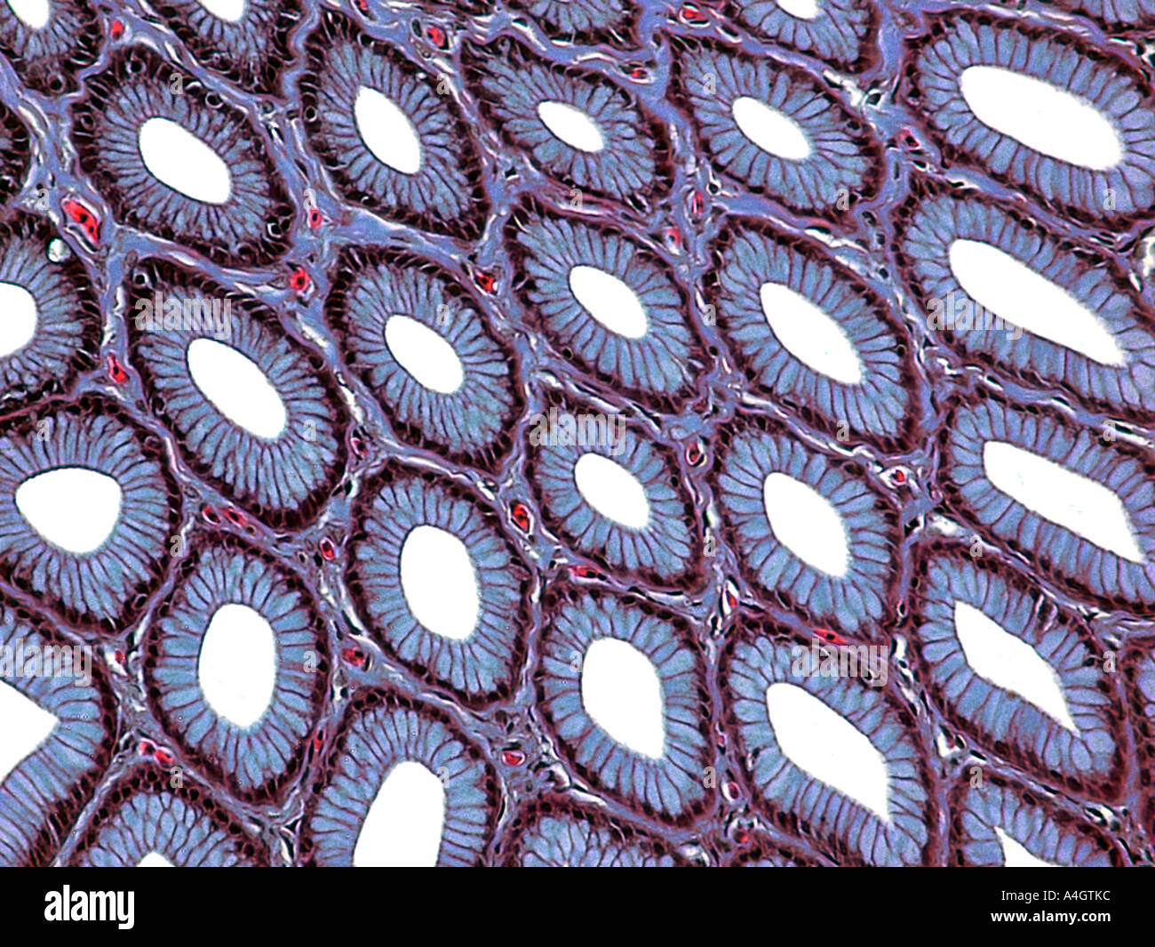 Stomach mucosa (lining) of a fish, stained thin section examined on a microscope slide Stock Photohttps://www.alamy.com/image-license-details/?v=1https://www.alamy.com/stomach-mucosa-lining-of-a-fish-stained-thin-section-examined-on-a-image6312507.html
RFA4GTKC–Stomach mucosa (lining) of a fish, stained thin section examined on a microscope slide
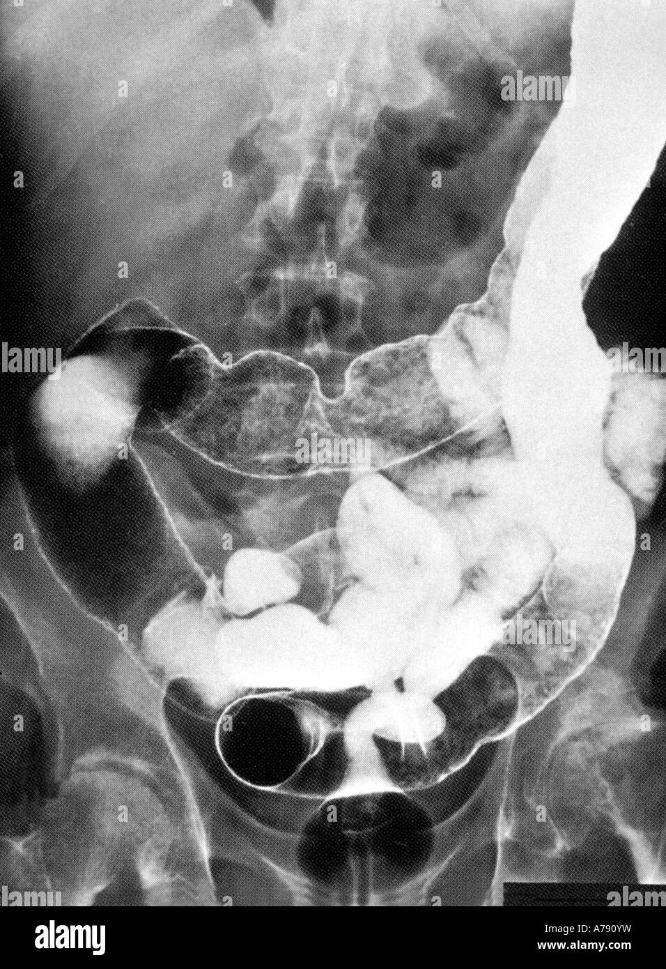 This is an abdominal x-ray of a patient with Crohn's disease Stock Photohttps://www.alamy.com/image-license-details/?v=1https://www.alamy.com/stock-photo-this-is-an-abdominal-x-ray-of-a-patient-with-crohns-disease-11763468.html
RMA790YW–This is an abdominal x-ray of a patient with Crohn's disease
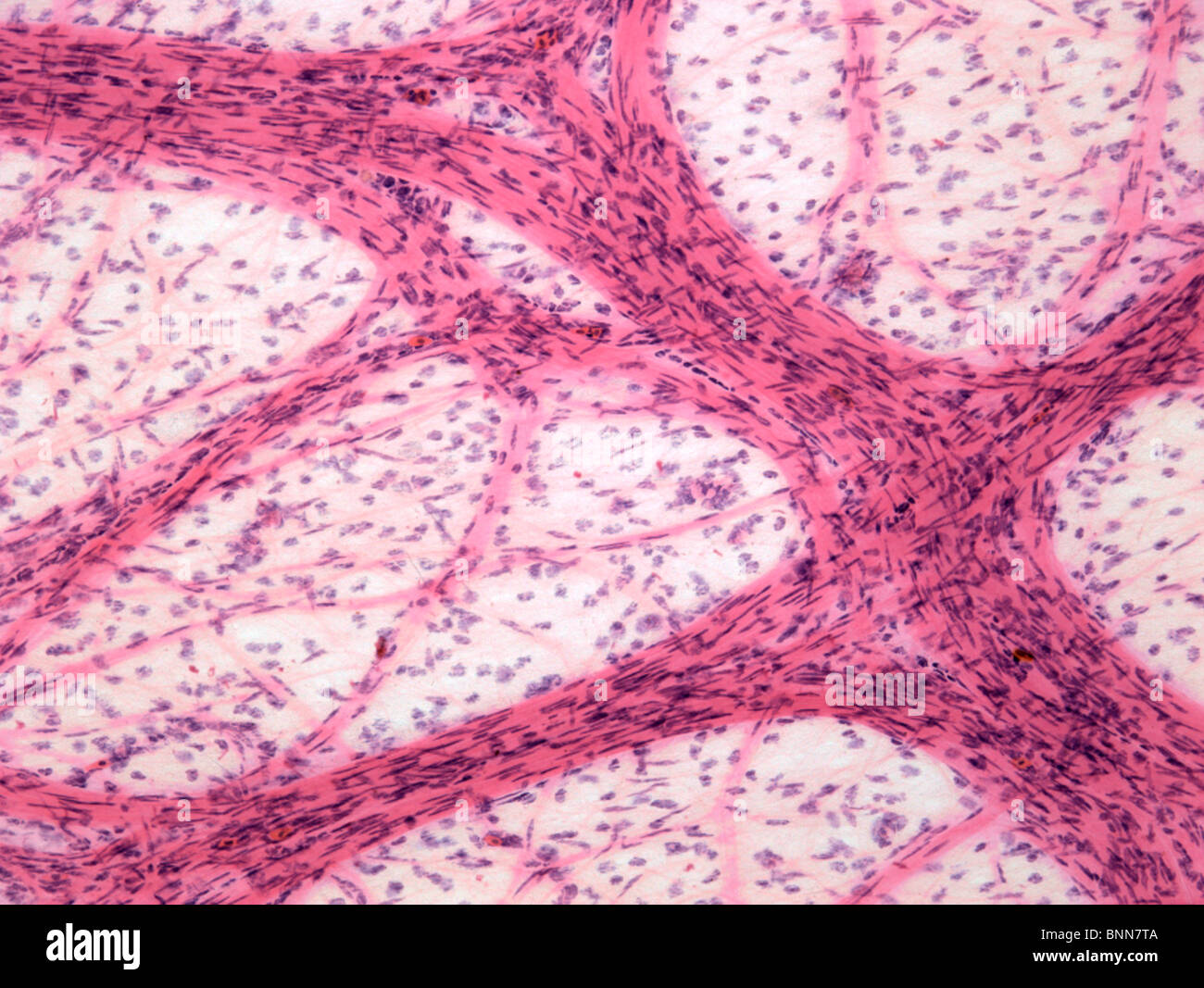 Omentum, light micrograph Stock Photohttps://www.alamy.com/image-license-details/?v=1https://www.alamy.com/stock-photo-omentum-light-micrograph-30585306.html
RFBNN7TA–Omentum, light micrograph
 High cholesterol levels text on paper hole Stock Photohttps://www.alamy.com/image-license-details/?v=1https://www.alamy.com/high-cholesterol-levels-text-on-paper-hole-image68585412.html
RFDYG9B0–High cholesterol levels text on paper hole
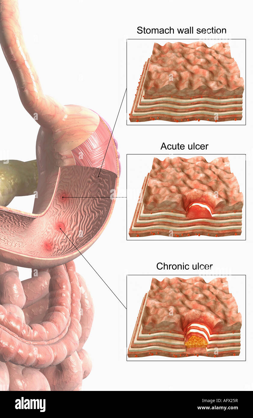 Stomach ulcer Stock Photohttps://www.alamy.com/image-license-details/?v=1https://www.alamy.com/stock-photo-stomach-ulcer-14031202.html
RFAFX25R–Stomach ulcer
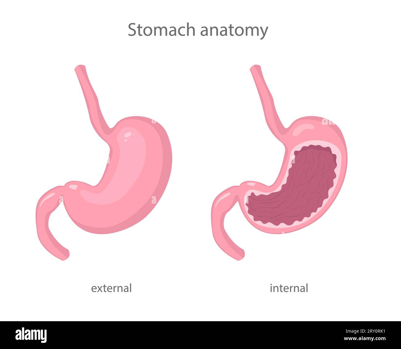 Scientific illustration of human healthy stomach external and internal view in realistic style with shadows and highlights. Stock Vectorhttps://www.alamy.com/image-license-details/?v=1https://www.alamy.com/scientific-illustration-of-human-healthy-stomach-external-and-internal-view-in-realistic-style-with-shadows-and-highlights-image567346053.html
RF2RY0RK1–Scientific illustration of human healthy stomach external and internal view in realistic style with shadows and highlights.
 Structure of stomach medical educational vector Stock Vectorhttps://www.alamy.com/image-license-details/?v=1https://www.alamy.com/structure-of-stomach-medical-educational-vector-image636866846.html
RF2S03P4E–Structure of stomach medical educational vector
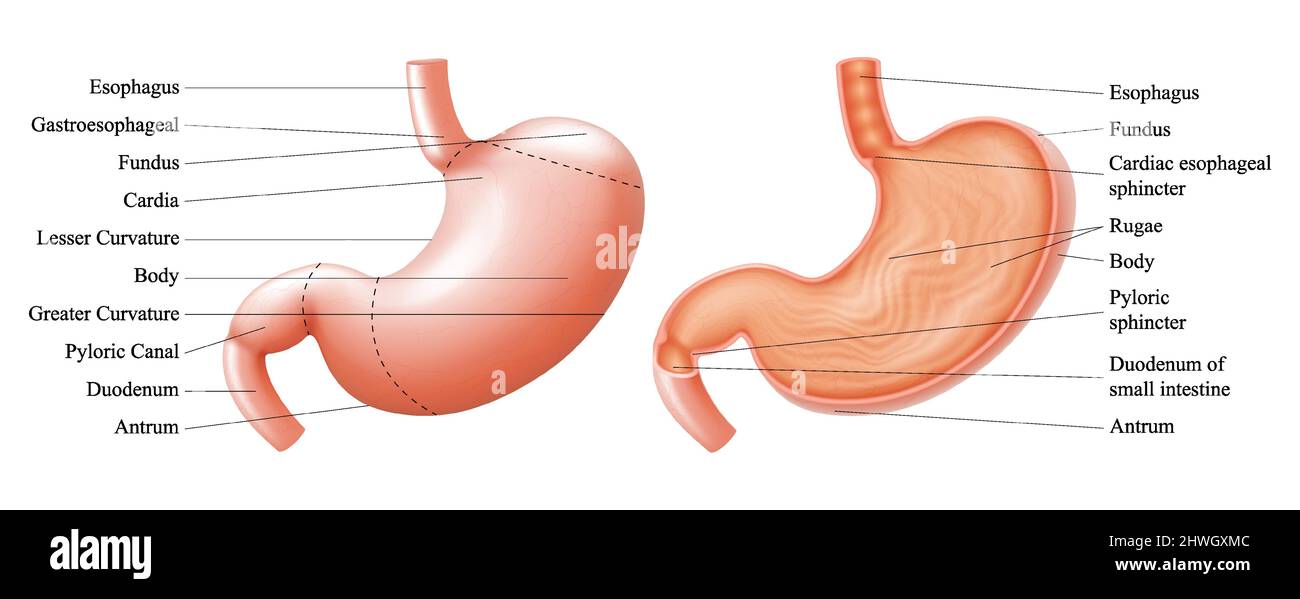 Anatomy of the human stomach and his shell structure with description of the corresponding internal parts. Sagittal section. Anatomical vector illustr Stock Vectorhttps://www.alamy.com/image-license-details/?v=1https://www.alamy.com/anatomy-of-the-human-stomach-and-his-shell-structure-with-description-of-the-corresponding-internal-parts-sagittal-section-anatomical-vector-illustr-image463208156.html
RF2HWGXMC–Anatomy of the human stomach and his shell structure with description of the corresponding internal parts. Sagittal section. Anatomical vector illustr
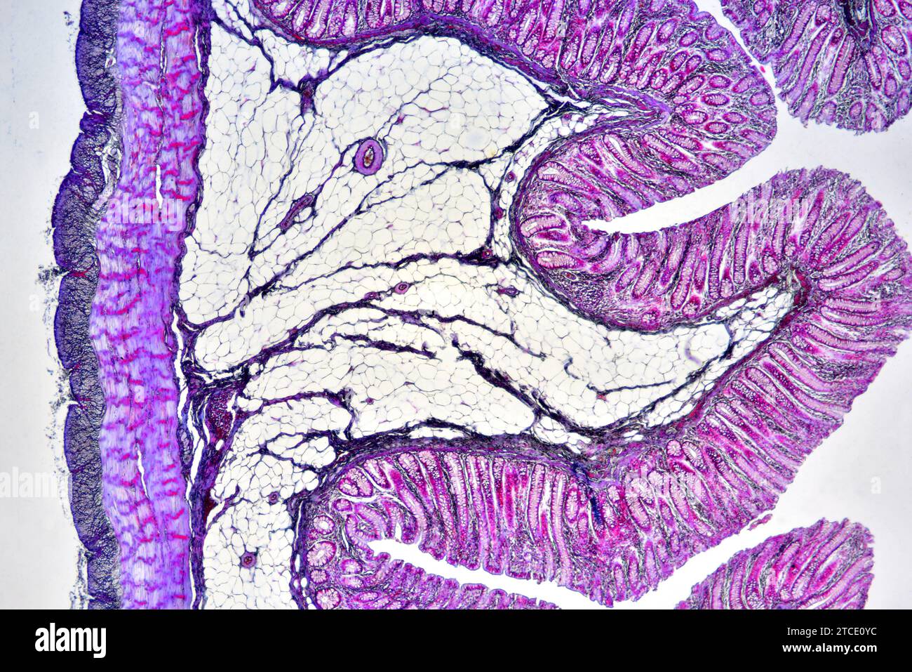 Human colon showing epithelium, mucosa, submucosa, muscular layer, adipose tissue, serosa, intestinal glands, villi and vessels. Optical microscope X4 Stock Photohttps://www.alamy.com/image-license-details/?v=1https://www.alamy.com/human-colon-showing-epithelium-mucosa-submucosa-muscular-layer-adipose-tissue-serosa-intestinal-glands-villi-and-vessels-optical-microscope-x4-image575626112.html
RF2TCE0YC–Human colon showing epithelium, mucosa, submucosa, muscular layer, adipose tissue, serosa, intestinal glands, villi and vessels. Optical microscope X4
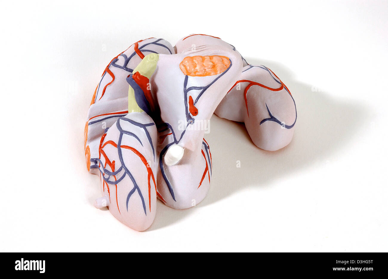 ANATOMY, FEMALE GENITALIA Stock Photohttps://www.alamy.com/image-license-details/?v=1https://www.alamy.com/stock-photo-anatomy-female-genitalia-53860964.html
RMD3HG5T–ANATOMY, FEMALE GENITALIA
 Falopian tube showing from outer to inner: serosa, subserosa, blood vessels, muscularis layer, submucosa, mucosa lamina propria and ciliated epitheliu Stock Photohttps://www.alamy.com/image-license-details/?v=1https://www.alamy.com/falopian-tube-showing-from-outer-to-inner-serosa-subserosa-blood-vessels-muscularis-layer-submucosa-mucosa-lamina-propria-and-ciliated-epitheliu-image592003205.html
RF2WB423H–Falopian tube showing from outer to inner: serosa, subserosa, blood vessels, muscularis layer, submucosa, mucosa lamina propria and ciliated epitheliu
 Sectional view illustration of perforated ulcer Stock Vectorhttps://www.alamy.com/image-license-details/?v=1https://www.alamy.com/sectional-view-illustration-of-perforated-ulcer-image452730953.html
RF2H8FJXH–Sectional view illustration of perforated ulcer
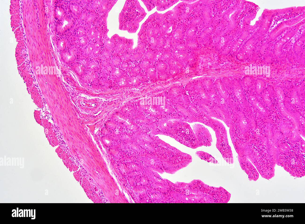 Human small intestine (jejunum) showing from left to right: serosa, muscular fibers, submucosa and mucosa with Lieberkhun crypts. X75 at 10 cm wide. Stock Photohttps://www.alamy.com/image-license-details/?v=1https://www.alamy.com/human-small-intestine-jejunum-showing-from-left-to-right-serosa-muscular-fibers-submucosa-and-mucosa-with-lieberkhun-crypts-x75-at-10-cm-wide-image591999276.html
RF2WB3W38–Human small intestine (jejunum) showing from left to right: serosa, muscular fibers, submucosa and mucosa with Lieberkhun crypts. X75 at 10 cm wide.
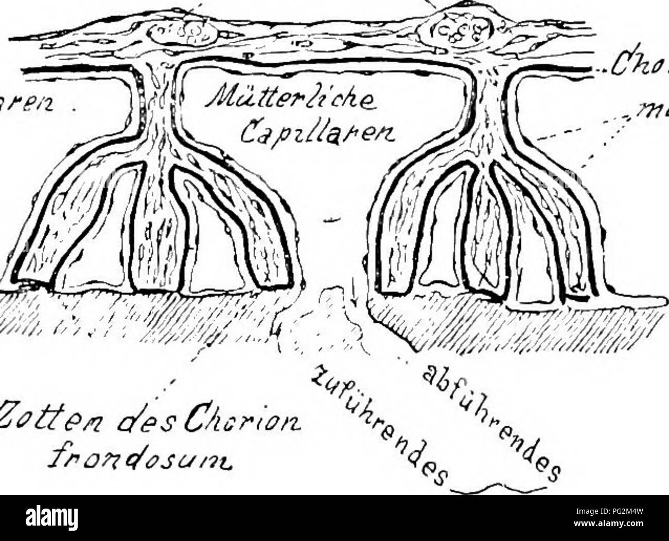 . Elements of the comparative anatomy of vertebrates. Anatomy, Comparative. FCETAL MEMBRANES 339 allantoic placenta arises, consisting of maternal and foetal parts (Fig. 9). Thus the embryo is supplied with the necessities for existence during its comparatively long intra-uterine life. Various forms of placenta are met with amongst the Placentalia. The most primitive type is apparently that in which the allantois becomes attached around the whole serosa, so that the resulting chorion, from which the comparatively simple villi arise, are equally distributed over the whole surface (Fig. 271). Th Stock Photohttps://www.alamy.com/image-license-details/?v=1https://www.alamy.com/elements-of-the-comparative-anatomy-of-vertebrates-anatomy-comparative-fcetal-membranes-339-allantoic-placenta-arises-consisting-of-maternal-and-foetal-parts-fig-9-thus-the-embryo-is-supplied-with-the-necessities-for-existence-during-its-comparatively-long-intra-uterine-life-various-forms-of-placenta-are-met-with-amongst-the-placentalia-the-most-primitive-type-is-apparently-that-in-which-the-allantois-becomes-attached-around-the-whole-serosa-so-that-the-resulting-chorion-from-which-the-comparatively-simple-villi-arise-are-equally-distributed-over-the-whole-surface-fig-271-th-image216418633.html
RMPG2M4W–. Elements of the comparative anatomy of vertebrates. Anatomy, Comparative. FCETAL MEMBRANES 339 allantoic placenta arises, consisting of maternal and foetal parts (Fig. 9). Thus the embryo is supplied with the necessities for existence during its comparatively long intra-uterine life. Various forms of placenta are met with amongst the Placentalia. The most primitive type is apparently that in which the allantois becomes attached around the whole serosa, so that the resulting chorion, from which the comparatively simple villi arise, are equally distributed over the whole surface (Fig. 271). Th
 The serosa is the outer layer of the colon 3d illustration Stock Photohttps://www.alamy.com/image-license-details/?v=1https://www.alamy.com/the-serosa-is-the-outer-layer-of-the-colon-3d-illustration-image596912958.html
RF2WK3MFX–The serosa is the outer layer of the colon 3d illustration
 . Abb. 3. Hinterer Eipol mit Eistiel von FsylUi inali in Apfelborke befestigt. — Ch — Chorion; Dh = Dotterhaut; E = Embryo; Kk = Kork- kambium; Myc— Mycetom; Ser = Serosa. — Oritrinal. Stock Photohttps://www.alamy.com/image-license-details/?v=1https://www.alamy.com/abb-3-hinterer-eipol-mit-eistiel-von-fsylui-inali-in-apfelborke-befestigt-ch-chorion-dh-=-dotterhaut-e-=-embryo-kk-=-kork-kambium-myc-mycetom-ser-=-serosa-oritrinal-image179899667.html
RMMCK3T3–. Abb. 3. Hinterer Eipol mit Eistiel von FsylUi inali in Apfelborke befestigt. — Ch — Chorion; Dh = Dotterhaut; E = Embryo; Kk = Kork- kambium; Myc— Mycetom; Ser = Serosa. — Oritrinal.
 Archive image from page 499 of Denkschriften der Medicinisch-Naturwissenschaftlichen Gesellschaft zu. Denkschriften der Medicinisch-Naturwissenschaftlichen Gesellschaft zu Jena denkschriftender47medi Year: 1879 Kcard.p.s. Mfsogatf. B£C.p.tj. Fig. 568. Fig. 569. portae. Die Arteria coeliacomesenterica verläuft dicht unter der Serosa an der rechten Seite des Pancreas- wulstes. Central verlaufen zwischen der Pfortader und dem Ductus choledochus (40 [i äusserer Durch- messer) die Sammelgänge des Pancreas, welche ein enges Lumen und einen äusseren Durchmesser von 20 fi aufweisen. Sie gehen peri Stock Photohttps://www.alamy.com/image-license-details/?v=1https://www.alamy.com/archive-image-from-page-499-of-denkschriften-der-medicinisch-naturwissenschaftlichen-gesellschaft-zu-denkschriften-der-medicinisch-naturwissenschaftlichen-gesellschaft-zu-jena-denkschriftender47medi-year-1879-kcardps-mfsogatf-bcptj-fig-568-fig-569-portae-die-arteria-coeliacomesenterica-verluft-dicht-unter-der-serosa-an-der-rechten-seite-des-pancreas-wulstes-central-verlaufen-zwischen-der-pfortader-und-dem-ductus-choledochus-40-i-usserer-durch-messer-die-sammelgnge-des-pancreas-welche-ein-enges-lumen-und-einen-usseren-durchmesser-von-20-fi-aufweisen-sie-gehen-peri-image259314551.html
RMW1TP9Y–Archive image from page 499 of Denkschriften der Medicinisch-Naturwissenschaftlichen Gesellschaft zu. Denkschriften der Medicinisch-Naturwissenschaftlichen Gesellschaft zu Jena denkschriftender47medi Year: 1879 Kcard.p.s. Mfsogatf. B£C.p.tj. Fig. 568. Fig. 569. portae. Die Arteria coeliacomesenterica verläuft dicht unter der Serosa an der rechten Seite des Pancreas- wulstes. Central verlaufen zwischen der Pfortader und dem Ductus choledochus (40 [i äusserer Durch- messer) die Sammelgänge des Pancreas, welche ein enges Lumen und einen äusseren Durchmesser von 20 fi aufweisen. Sie gehen peri
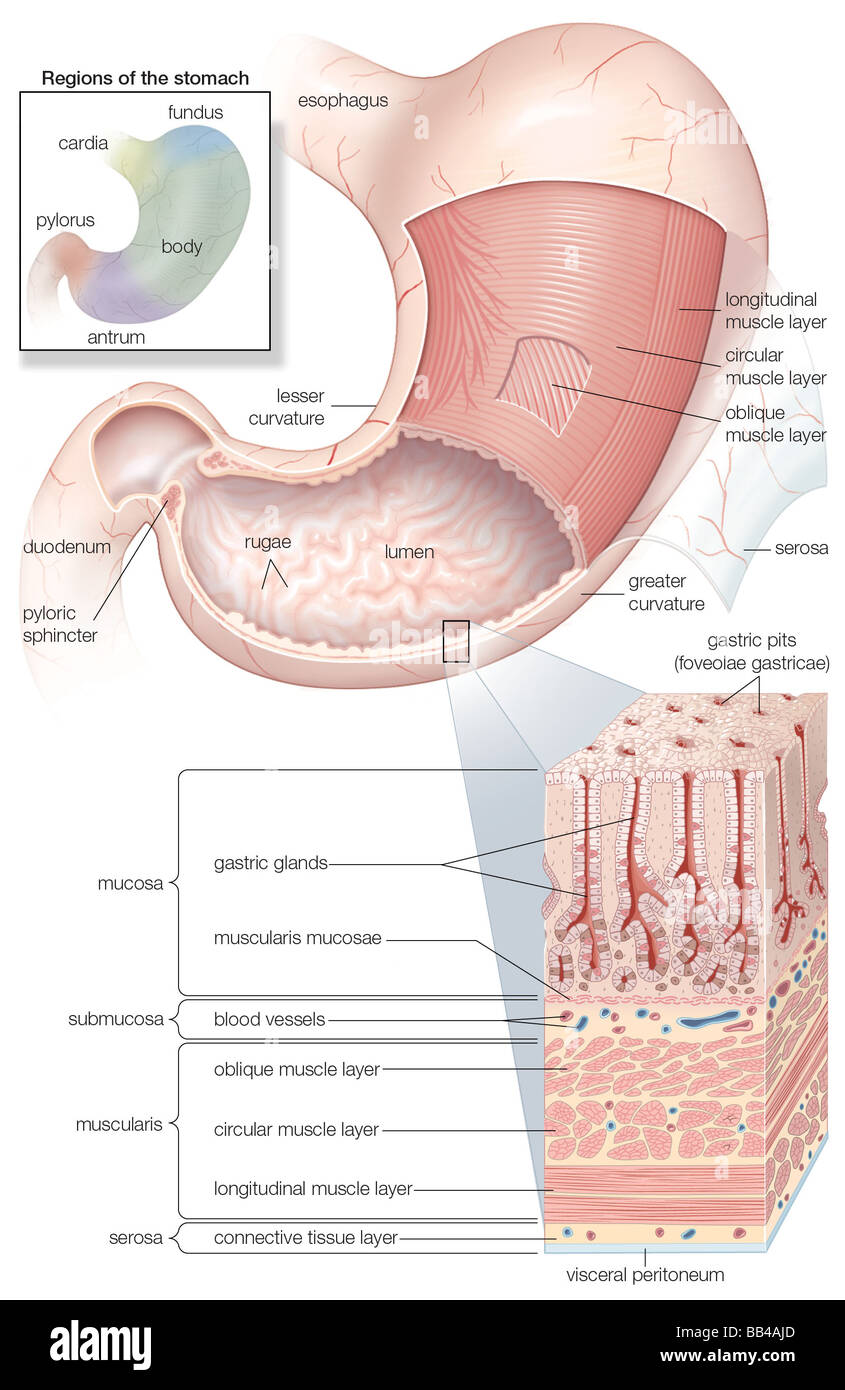 Diagram showing the Mucosa and musculature of the human stomach plus insets of histology and regions Stock Photohttps://www.alamy.com/image-license-details/?v=1https://www.alamy.com/stock-photo-diagram-showing-the-mucosa-and-musculature-of-the-human-stomach-plus-24067749.html
RMBB4AJD–Diagram showing the Mucosa and musculature of the human stomach plus insets of histology and regions
 . The practice of pediatrics. l of thefluid, owing to the fact that what remains of the brain tissue fallstogether in a broken mass. The ependyma may be normal or thickened and infiltrated. Chronic external hydrocephalus is of rare occurrence. When present,it will be found associated in nearly all cases with a pachymeningitis.* Nervous Diseases of Children. HYDROCEPHALUS 493 The congenital form of external hydrocephalus is exceedingly rare.Very few authentic cases have been reported. Internal hydrocephalus (acute) (meningitis serosa) is of infectiousorigin. Any of the pathogenic bacteria may b Stock Photohttps://www.alamy.com/image-license-details/?v=1https://www.alamy.com/the-practice-of-pediatrics-l-of-thefluid-owing-to-the-fact-that-what-remains-of-the-brain-tissue-fallstogether-in-a-broken-mass-the-ependyma-may-be-normal-or-thickened-and-infiltrated-chronic-external-hydrocephalus-is-of-rare-occurrence-when-presentit-will-be-found-associated-in-nearly-all-cases-with-a-pachymeningitis-nervous-diseases-of-children-hydrocephalus-493-the-congenital-form-of-external-hydrocephalus-is-exceedingly-rarevery-few-authentic-cases-have-been-reported-internal-hydrocephalus-acute-meningitis-serosa-is-of-infectiousorigin-any-of-the-pathogenic-bacteria-may-b-image336929252.html
RM2AG4CKG–. The practice of pediatrics. l of thefluid, owing to the fact that what remains of the brain tissue fallstogether in a broken mass. The ependyma may be normal or thickened and infiltrated. Chronic external hydrocephalus is of rare occurrence. When present,it will be found associated in nearly all cases with a pachymeningitis.* Nervous Diseases of Children. HYDROCEPHALUS 493 The congenital form of external hydrocephalus is exceedingly rare.Very few authentic cases have been reported. Internal hydrocephalus (acute) (meningitis serosa) is of infectiousorigin. Any of the pathogenic bacteria may b
 Stomach mucosa (lining) of a fish, stained thin section examined on a microscope slide Stock Photohttps://www.alamy.com/image-license-details/?v=1https://www.alamy.com/stomach-mucosa-lining-of-a-fish-stained-thin-section-examined-on-a-image6312502.html
RFA4GTK7–Stomach mucosa (lining) of a fish, stained thin section examined on a microscope slide
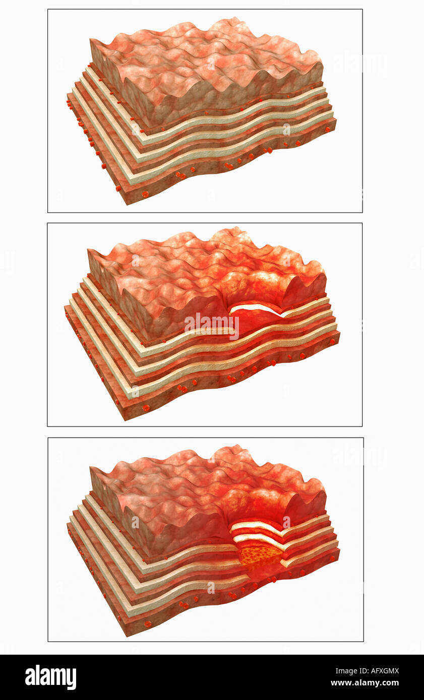 Stomach ulcer Stock Photohttps://www.alamy.com/image-license-details/?v=1https://www.alamy.com/stock-photo-stomach-ulcer-14036089.html
RFAFXGMX–Stomach ulcer
 Structure of stomach medical educational vector Stock Vectorhttps://www.alamy.com/image-license-details/?v=1https://www.alamy.com/structure-of-stomach-medical-educational-vector-image636866793.html
RF2S03P2H–Structure of stomach medical educational vector
 Sectional view illustration of gastric erosion Stock Vectorhttps://www.alamy.com/image-license-details/?v=1https://www.alamy.com/sectional-view-illustration-of-gastric-erosion-image452730917.html
RF2H8FJW9–Sectional view illustration of gastric erosion
 Human small intestine (jejunum) showing from left to right: serosa, muscular fibers, submucosa and mucosa with Lieberkhun crypts. X25 at 10 cm wide. Stock Photohttps://www.alamy.com/image-license-details/?v=1https://www.alamy.com/human-small-intestine-jejunum-showing-from-left-to-right-serosa-muscular-fibers-submucosa-and-mucosa-with-lieberkhun-crypts-x25-at-10-cm-wide-image591999274.html
RF2WB3W36–Human small intestine (jejunum) showing from left to right: serosa, muscular fibers, submucosa and mucosa with Lieberkhun crypts. X25 at 10 cm wide.
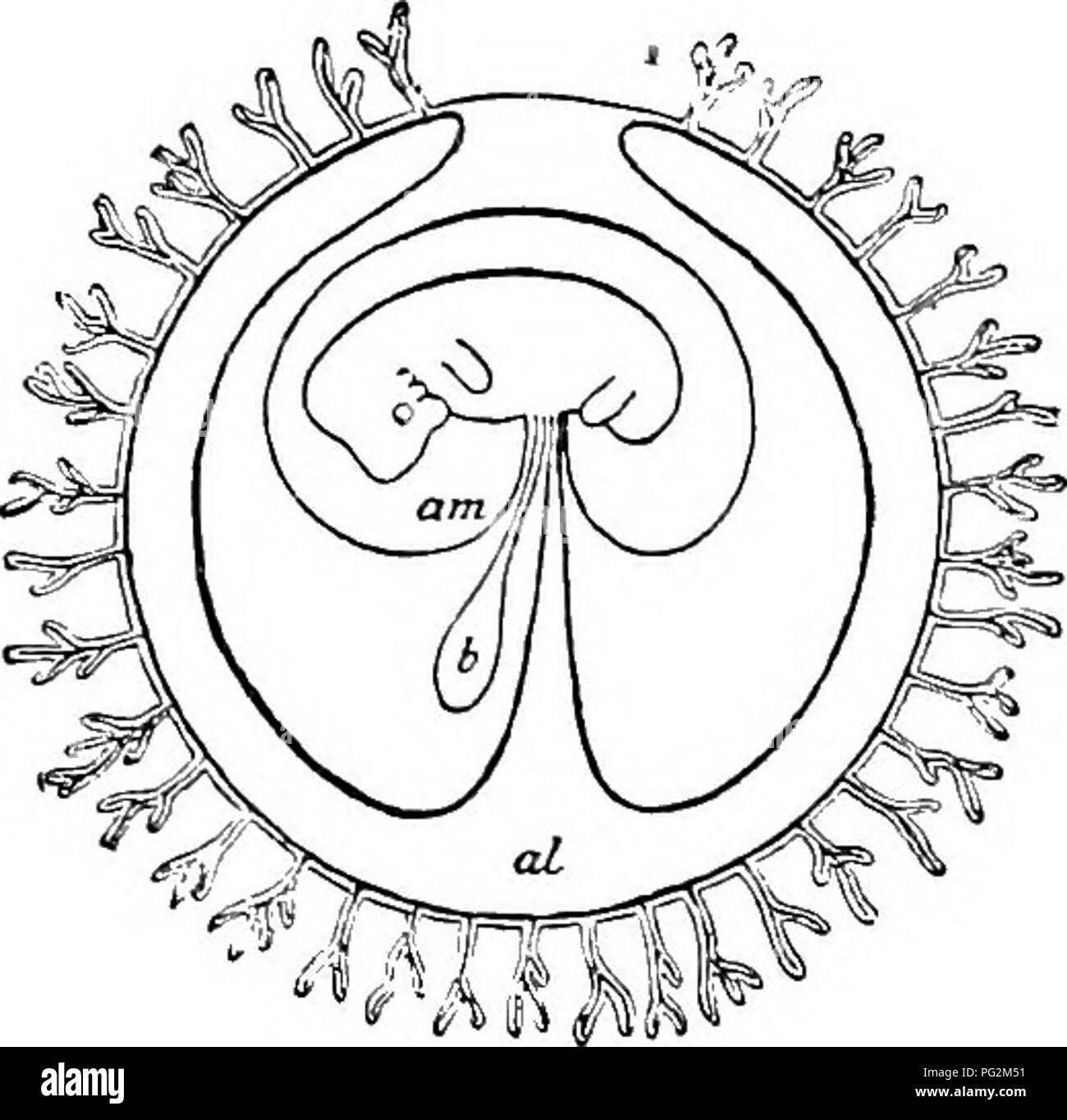 . Elements of the comparative anatomy of vertebrates. Anatomy, Comparative. 338 COMPARATIVE ANATOMY moreover retained in the Monotremes, and even in Marsupials the ova are relatively large as compared with those of the higher Mammalia. As the amount of yolk gradually became reduced in the course of phylogenetic development, close relations were set up between the foetal (allantoic) and maternal blood-vessels, the allantois becoming closely applied to the serosa to form a chorion (Fig. 271); but that this condition was only very slowly evolved is shown by the fact that, even at the present day, Stock Photohttps://www.alamy.com/image-license-details/?v=1https://www.alamy.com/elements-of-the-comparative-anatomy-of-vertebrates-anatomy-comparative-338-comparative-anatomy-moreover-retained-in-the-monotremes-and-even-in-marsupials-the-ova-are-relatively-large-as-compared-with-those-of-the-higher-mammalia-as-the-amount-of-yolk-gradually-became-reduced-in-the-course-of-phylogenetic-development-close-relations-were-set-up-between-the-foetal-allantoic-and-maternal-blood-vessels-the-allantois-becoming-closely-applied-to-the-serosa-to-form-a-chorion-fig-271-but-that-this-condition-was-only-very-slowly-evolved-is-shown-by-the-fact-that-even-at-the-present-day-image216418637.html
RMPG2M51–. Elements of the comparative anatomy of vertebrates. Anatomy, Comparative. 338 COMPARATIVE ANATOMY moreover retained in the Monotremes, and even in Marsupials the ova are relatively large as compared with those of the higher Mammalia. As the amount of yolk gradually became reduced in the course of phylogenetic development, close relations were set up between the foetal (allantoic) and maternal blood-vessels, the allantois becoming closely applied to the serosa to form a chorion (Fig. 271); but that this condition was only very slowly evolved is shown by the fact that, even at the present day,
 Falopian tube showing from outer (left) to inner: serosa, subserosa, blood vessels, muscularis layer, submucosa, mucosa, lamina propria and ciliated e Stock Photohttps://www.alamy.com/image-license-details/?v=1https://www.alamy.com/falopian-tube-showing-from-outer-left-to-inner-serosa-subserosa-blood-vessels-muscularis-layer-submucosa-mucosa-lamina-propria-and-ciliated-e-image592003209.html
RF2WB423N–Falopian tube showing from outer (left) to inner: serosa, subserosa, blood vessels, muscularis layer, submucosa, mucosa, lamina propria and ciliated e
 The serosa is the outer layer of the colon 3d illustration Stock Photohttps://www.alamy.com/image-license-details/?v=1https://www.alamy.com/the-serosa-is-the-outer-layer-of-the-colon-3d-illustration-image596913355.html
RF2WK3N23–The serosa is the outer layer of the colon 3d illustration
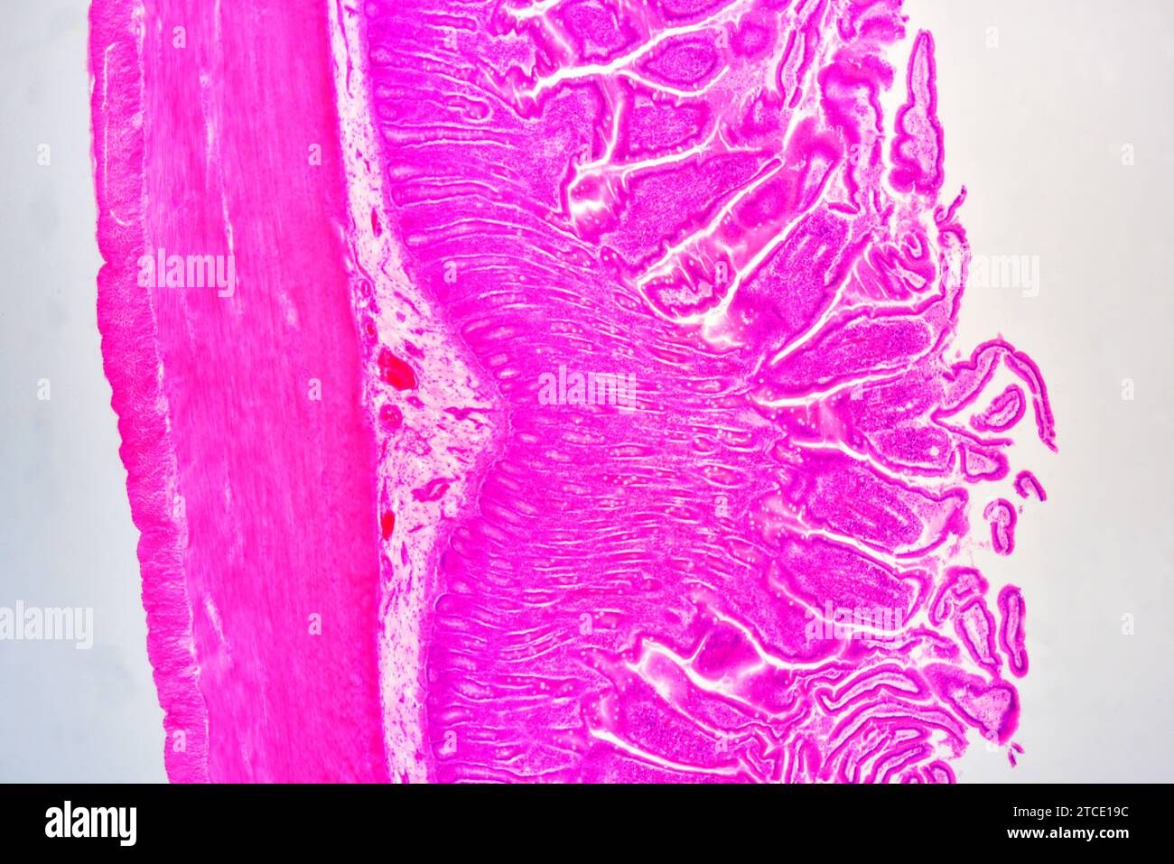 Small intestine cross section showing mucosa, submucosa, lamina propria, muscular layer, serosa and Brunner glands. Optical microscope X40. Stock Photohttps://www.alamy.com/image-license-details/?v=1https://www.alamy.com/small-intestine-cross-section-showing-mucosa-submucosa-lamina-propria-muscular-layer-serosa-and-brunner-glands-optical-microscope-x40-image575626392.html
RF2TCE19C–Small intestine cross section showing mucosa, submucosa, lamina propria, muscular layer, serosa and Brunner glands. Optical microscope X40.
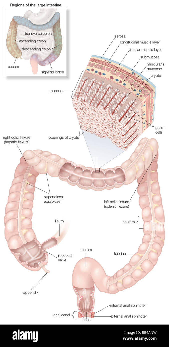 Diagram of the human large intestines, including and inset of its musculature and mucosa histology and an inset of its regions Stock Photohttps://www.alamy.com/image-license-details/?v=1https://www.alamy.com/stock-photo-diagram-of-the-human-large-intestines-including-and-inset-of-its-musculature-24067845.html
RMBB4ANW–Diagram of the human large intestines, including and inset of its musculature and mucosa histology and an inset of its regions
 Surgical treatment; a practical treatise on the therapy of surgical diseases for the use of practitioners and students of surgery . ressed together and the union reinforced with interrupted sutures care being taken that they do not slip back into the bowel. If the surgeonconsiders applying a serosa suture after the buttons have been pressedtogether, this should not be attempted in bowel wall which is so tightlydrawn over the button that the suture threatens to tear out or perforate THE ABDOMEN 671 the lumen. The button is liberated by pressure necrosis sometime usuallyduring the second or thir Stock Photohttps://www.alamy.com/image-license-details/?v=1https://www.alamy.com/surgical-treatment-a-practical-treatise-on-the-therapy-of-surgical-diseases-for-the-use-of-practitioners-and-students-of-surgery-ressed-together-and-the-union-reinforced-with-interrupted-sutures-care-being-taken-that-they-do-not-slip-back-into-the-bowel-if-the-surgeonconsiders-applying-a-serosa-suture-after-the-buttons-have-been-pressedtogether-this-should-not-be-attempted-in-bowel-wall-which-is-so-tightlydrawn-over-the-button-that-the-suture-threatens-to-tear-out-or-perforate-the-abdomen-671-the-lumen-the-button-is-liberated-by-pressure-necrosis-sometime-usuallyduring-the-second-or-thir-image338215627.html
RM2AJ71DF–Surgical treatment; a practical treatise on the therapy of surgical diseases for the use of practitioners and students of surgery . ressed together and the union reinforced with interrupted sutures care being taken that they do not slip back into the bowel. If the surgeonconsiders applying a serosa suture after the buttons have been pressedtogether, this should not be attempted in bowel wall which is so tightlydrawn over the button that the suture threatens to tear out or perforate THE ABDOMEN 671 the lumen. The button is liberated by pressure necrosis sometime usuallyduring the second or thir
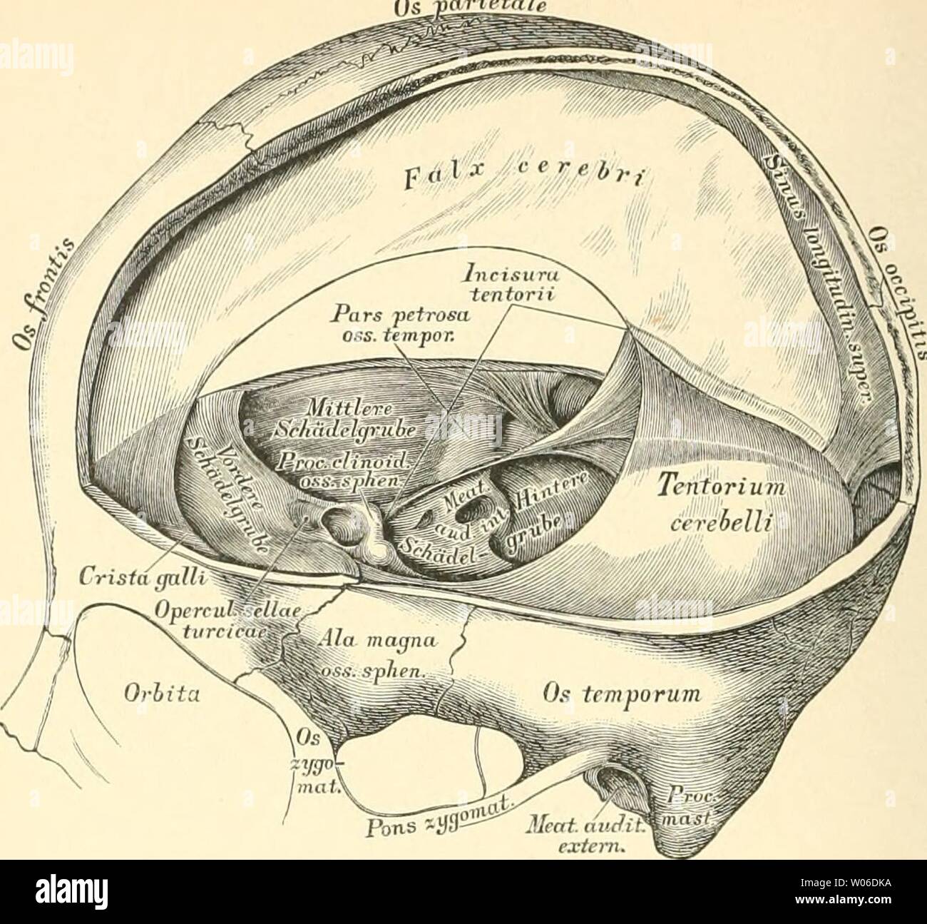 Archive image from page 415 of Die descriptive und topographische Anatomie. Die descriptive und topographische Anatomie des Menschen diedescriptiveun00heit Year: 1896 370 Hüllen des Gehirns und Rückenmarkes. 553. Die Hüllen des Gehirns. Fortsätze der harten Hirnhaut. Gehirn und Rückenmark sind innerhalb der Knochenräume von drei häutigen Hüllen umschlossen: der harten (fibrösen) Hirnhaut, Dura mittt-r (Ifaiiii.r fihnisa), der Spinnwebenkaut, Arachnoidca (Meninx serosa), und der weichen Hirnhaut, Pia mater (Meninx vasculosa). Die Dum mater bildet die äussere Hülle, eine derbe, bindegewebig Stock Photohttps://www.alamy.com/image-license-details/?v=1https://www.alamy.com/archive-image-from-page-415-of-die-descriptive-und-topographische-anatomie-die-descriptive-und-topographische-anatomie-des-menschen-diedescriptiveun00heit-year-1896-370-hllen-des-gehirns-und-rckenmarkes-553-die-hllen-des-gehirns-fortstze-der-harten-hirnhaut-gehirn-und-rckenmark-sind-innerhalb-der-knochenrume-von-drei-hutigen-hllen-umschlossen-der-harten-fibrsen-hirnhaut-dura-mittt-r-ifaiiiir-fihnisa-der-spinnwebenkaut-arachnoidca-meninx-serosa-und-der-weichen-hirnhaut-pia-mater-meninx-vasculosa-die-dum-mater-bildet-die-ussere-hlle-eine-derbe-bindegewebig-image258297966.html
RMW06DKA–Archive image from page 415 of Die descriptive und topographische Anatomie. Die descriptive und topographische Anatomie des Menschen diedescriptiveun00heit Year: 1896 370 Hüllen des Gehirns und Rückenmarkes. 553. Die Hüllen des Gehirns. Fortsätze der harten Hirnhaut. Gehirn und Rückenmark sind innerhalb der Knochenräume von drei häutigen Hüllen umschlossen: der harten (fibrösen) Hirnhaut, Dura mittt-r (Ifaiiii.r fihnisa), der Spinnwebenkaut, Arachnoidca (Meninx serosa), und der weichen Hirnhaut, Pia mater (Meninx vasculosa). Die Dum mater bildet die äussere Hülle, eine derbe, bindegewebig
 . Die forstinsekten Mitteleuropas. Ein lehr- und handbuch . Mcs. M. Fig. 140. Einstülpung des Vorder- (Stom) und Hinterdarms {Trost); Am Amnion; Ser Serosa; Mes Mesoderm; Ek Ektoderm; HEnt hinterer Entodermkeim; VEni vorderer Entodermkeim. Stock Photohttps://www.alamy.com/image-license-details/?v=1https://www.alamy.com/die-forstinsekten-mitteleuropas-ein-lehr-und-handbuch-mcs-m-fig-140-einstlpung-des-vorder-stom-und-hinterdarms-trost-am-amnion-ser-serosa-mes-mesoderm-ek-ektoderm-hent-hinterer-entodermkeim-veni-vorderer-entodermkeim-image181049894.html
RMMEFEYJ–. Die forstinsekten Mitteleuropas. Ein lehr- und handbuch . Mcs. M. Fig. 140. Einstülpung des Vorder- (Stom) und Hinterdarms {Trost); Am Amnion; Ser Serosa; Mes Mesoderm; Ek Ektoderm; HEnt hinterer Entodermkeim; VEni vorderer Entodermkeim.
 Chronic stomach ulcer Stock Photohttps://www.alamy.com/image-license-details/?v=1https://www.alamy.com/stock-photo-chronic-stomach-ulcer-14034290.html
RFAFXBAY–Chronic stomach ulcer
 Structure of stomach medical educational vector Stock Vectorhttps://www.alamy.com/image-license-details/?v=1https://www.alamy.com/structure-of-stomach-medical-educational-vector-image636866918.html
RF2S03P72–Structure of stomach medical educational vector
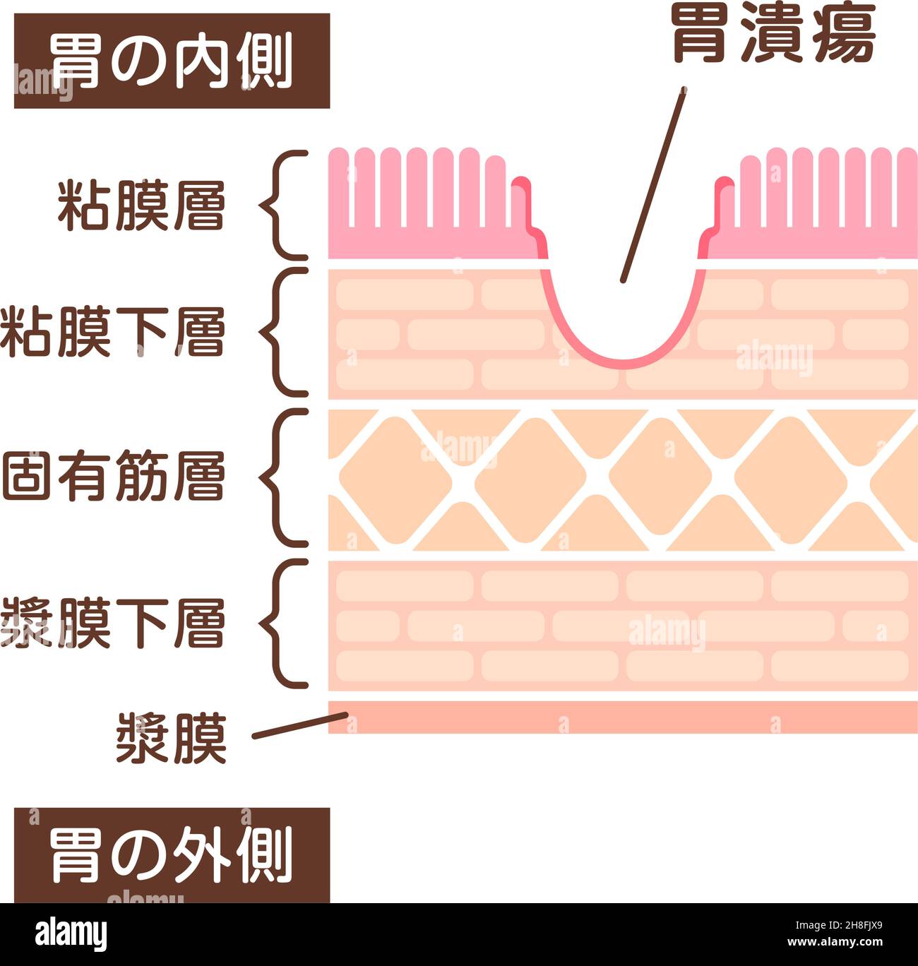 Sectional view illustration of gastric ulser Stock Vectorhttps://www.alamy.com/image-license-details/?v=1https://www.alamy.com/sectional-view-illustration-of-gastric-ulser-image452730945.html
RF2H8FJX9–Sectional view illustration of gastric ulser
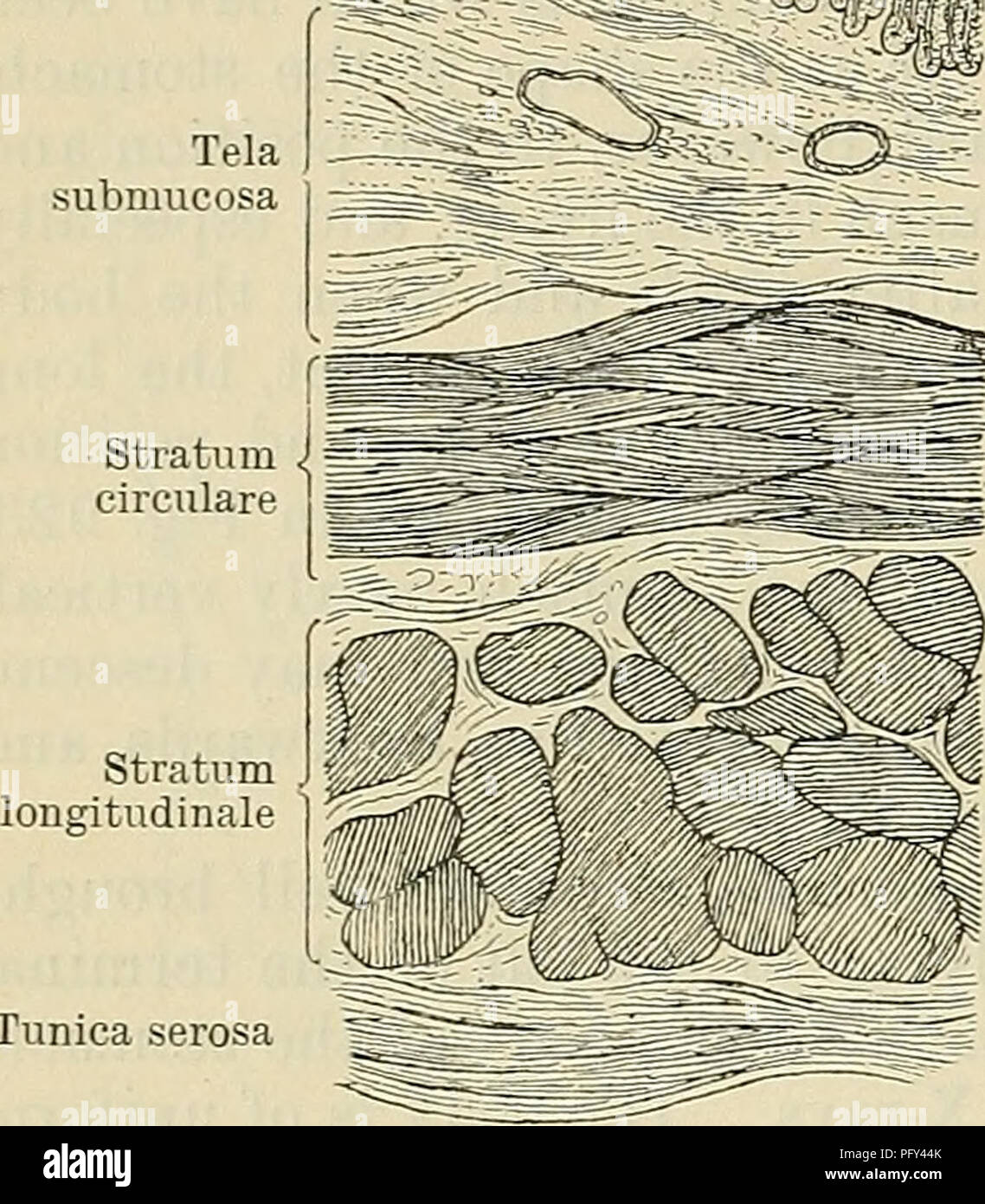 . Cunningham's Text-book of anatomy. Anatomy. 1174 THE DIGESTIVE SYSTEM. il^r^ffli^ Tunica mucosa Tela submucosa Structure of the Stomach. The stomach wall is composed of four coats—namely, from without inwards: (1) Tunica serosa, (2) tunica muscularis, (3) tela submucosa, and (4) tunica mucosa (Fig. 924). Tunica Serosa.—This coat is formed of the peritoneum, the relations of which to the stomach have already been described. It is closely attached to the subjacent muscular coat, except near the curvatures, where the connexion is more lax; and it confers on the stomach its smooth and glistening Stock Photohttps://www.alamy.com/image-license-details/?v=1https://www.alamy.com/cunninghams-text-book-of-anatomy-anatomy-1174-the-digestive-system-ilrffli-tunica-mucosa-tela-submucosa-structure-of-the-stomach-the-stomach-wall-is-composed-of-four-coatsnamely-from-without-inwards-1-tunica-serosa-2-tunica-muscularis-3-tela-submucosa-and-4-tunica-mucosa-fig-924-tunica-serosathis-coat-is-formed-of-the-peritoneum-the-relations-of-which-to-the-stomach-have-already-been-described-it-is-closely-attached-to-the-subjacent-muscular-coat-except-near-the-curvatures-where-the-connexion-is-more-lax-and-it-confers-on-the-stomach-its-smooth-and-glistening-image216340227.html
RMPFY44K–. Cunningham's Text-book of anatomy. Anatomy. 1174 THE DIGESTIVE SYSTEM. il^r^ffli^ Tunica mucosa Tela submucosa Structure of the Stomach. The stomach wall is composed of four coats—namely, from without inwards: (1) Tunica serosa, (2) tunica muscularis, (3) tela submucosa, and (4) tunica mucosa (Fig. 924). Tunica Serosa.—This coat is formed of the peritoneum, the relations of which to the stomach have already been described. It is closely attached to the subjacent muscular coat, except near the curvatures, where the connexion is more lax; and it confers on the stomach its smooth and glistening
 The serosa is the outer layer of the colon 3d illustration Stock Photohttps://www.alamy.com/image-license-details/?v=1https://www.alamy.com/the-serosa-is-the-outer-layer-of-the-colon-3d-illustration-image596913052.html
RF2WK3MK8–The serosa is the outer layer of the colon 3d illustration
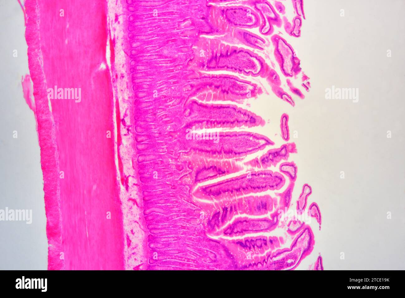 Small intestine cross section showing mucosa, submucosa, lamina propria, muscular layer, serosa and Brunner glands. Optical microscope X40. Stock Photohttps://www.alamy.com/image-license-details/?v=1https://www.alamy.com/small-intestine-cross-section-showing-mucosa-submucosa-lamina-propria-muscular-layer-serosa-and-brunner-glands-optical-microscope-x40-image575626399.html
RF2TCE19K–Small intestine cross section showing mucosa, submucosa, lamina propria, muscular layer, serosa and Brunner glands. Optical microscope X40.
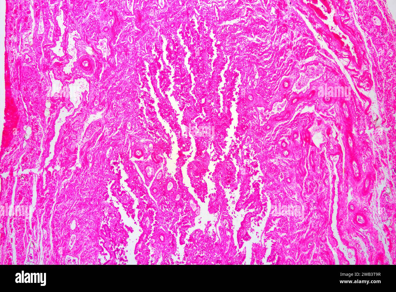 Human Fallopian tube section. X30 at 10 cm wide. Stock Photohttps://www.alamy.com/image-license-details/?v=1https://www.alamy.com/human-fallopian-tube-section-x30-at-10-cm-wide-image591998675.html
RF2WB3T9R–Human Fallopian tube section. X30 at 10 cm wide.
 The practice of pediatrics . cing extension of the skull, separation of the bones, and enlarge-ment of the head when it occurs in infancy. Internal hydrocephalusmay be either a general internal hydrocephalus, with distention of allthe ventricles, or a partial hydrocephalus in which the fourth ventricle DISEASES OF THE BRAIN 965 is not involved. Hydrocephalus may be congenital or acquired. Of theacquired form we have to deal with the chronic internal hydrocephalusand an acute internal hydrocephalus—the meningitis serosa of Quincke.Acute Internal Hydrocephalus. Etiology.—While an acute internalh Stock Photohttps://www.alamy.com/image-license-details/?v=1https://www.alamy.com/the-practice-of-pediatrics-cing-extension-of-the-skull-separation-of-the-bones-and-enlarge-ment-of-the-head-when-it-occurs-in-infancy-internal-hydrocephalusmay-be-either-a-general-internal-hydrocephalus-with-distention-of-allthe-ventricles-or-a-partial-hydrocephalus-in-which-the-fourth-ventricle-diseases-of-the-brain-965-is-not-involved-hydrocephalus-may-be-congenital-or-acquired-of-theacquired-form-we-have-to-deal-with-the-chronic-internal-hydrocephalusand-an-acute-internal-hydrocephalusthe-meningitis-serosa-of-quinckeacute-internal-hydrocephalus-etiologywhile-an-acute-internalh-image339025854.html
RM2AKFXX6–The practice of pediatrics . cing extension of the skull, separation of the bones, and enlarge-ment of the head when it occurs in infancy. Internal hydrocephalusmay be either a general internal hydrocephalus, with distention of allthe ventricles, or a partial hydrocephalus in which the fourth ventricle DISEASES OF THE BRAIN 965 is not involved. Hydrocephalus may be congenital or acquired. Of theacquired form we have to deal with the chronic internal hydrocephalusand an acute internal hydrocephalus—the meningitis serosa of Quincke.Acute Internal Hydrocephalus. Etiology.—While an acute internalh
![Embryology of insects and myriapods; Embryology of insects and myriapods; the developmental history of insects, centipedes, and millepedes from egg desposition [!] to hatching embryologyofinse00joha Year: 1941 Fig 200 —Pyrrhocoris A.ftei blastolviiicsis Retracted serosa {ser) covering the head. stem Stock Photo Embryology of insects and myriapods; Embryology of insects and myriapods; the developmental history of insects, centipedes, and millepedes from egg desposition [!] to hatching embryologyofinse00joha Year: 1941 Fig 200 —Pyrrhocoris A.ftei blastolviiicsis Retracted serosa {ser) covering the head. stem Stock Photo](https://c8.alamy.com/comp/RWW4X1/embryology-of-insects-and-myriapods-embryology-of-insects-and-myriapods-the-developmental-history-of-insects-centipedes-and-millepedes-from-egg-desposition-!-to-hatching-embryologyofinse00joha-year-1941-fig-200-pyrrhocoris-aftei-blastolviiicsis-retracted-serosa-ser-covering-the-head-stem-RWW4X1.jpg) Embryology of insects and myriapods; Embryology of insects and myriapods; the developmental history of insects, centipedes, and millepedes from egg desposition [!] to hatching embryologyofinse00joha Year: 1941 Fig 200 —Pyrrhocoris A.ftei blastolviiicsis Retracted serosa {ser) covering the head. stem Stock Photohttps://www.alamy.com/image-license-details/?v=1https://www.alamy.com/embryology-of-insects-and-myriapods-embryology-of-insects-and-myriapods-the-developmental-history-of-insects-centipedes-and-millepedes-from-egg-desposition-!-to-hatching-embryologyofinse00joha-year-1941-fig-200-pyrrhocoris-aftei-blastolviiicsis-retracted-serosa-ser-covering-the-head-stem-image239653849.html
Embryology of insects and myriapods; Embryology of insects and myriapods; the developmental history of insects, centipedes, and millepedes from egg desposition [!] to hatching embryologyofinse00joha Year: 1941 Fig 200 —Pyrrhocoris A.ftei blastolviiicsis Retracted serosa {ser) covering the head. stem Stock Photohttps://www.alamy.com/image-license-details/?v=1https://www.alamy.com/embryology-of-insects-and-myriapods-embryology-of-insects-and-myriapods-the-developmental-history-of-insects-centipedes-and-millepedes-from-egg-desposition-!-to-hatching-embryologyofinse00joha-year-1941-fig-200-pyrrhocoris-aftei-blastolviiicsis-retracted-serosa-ser-covering-the-head-stem-image239653849.htmlRMRWW4X1–Embryology of insects and myriapods; Embryology of insects and myriapods; the developmental history of insects, centipedes, and millepedes from egg desposition [!] to hatching embryologyofinse00joha Year: 1941 Fig 200 —Pyrrhocoris A.ftei blastolviiicsis Retracted serosa {ser) covering the head. stem
 . Cysto-adenoma on the serosa of the intestine. (Alt removed or they may be left alone. They will not ordinarily aflfect the health of the bird. Even malignant tumors are apparently not contagious. The ultimate nature of the causative agents has not been determined. With our present knowledge it is impossible to explain either the spontaneous origin of these agents within an individual or to account for their transfer from one individ- Stock Photohttps://www.alamy.com/image-license-details/?v=1https://www.alamy.com/cysto-adenoma-on-the-serosa-of-the-intestine-alt-removed-or-they-may-be-left-alone-they-will-not-ordinarily-aflfect-the-health-of-the-bird-even-malignant-tumors-are-apparently-not-contagious-the-ultimate-nature-of-the-causative-agents-has-not-been-determined-with-our-present-knowledge-it-is-impossible-to-explain-either-the-spontaneous-origin-of-these-agents-within-an-individual-or-to-account-for-their-transfer-from-one-individ-image179900395.html
RMMCK4P3–. Cysto-adenoma on the serosa of the intestine. (Alt removed or they may be left alone. They will not ordinarily aflfect the health of the bird. Even malignant tumors are apparently not contagious. The ultimate nature of the causative agents has not been determined. With our present knowledge it is impossible to explain either the spontaneous origin of these agents within an individual or to account for their transfer from one individ-
 Stomach ulcer Stock Photohttps://www.alamy.com/image-license-details/?v=1https://www.alamy.com/stock-photo-stomach-ulcer-14033376.html
RFAFX8JW–Stomach ulcer
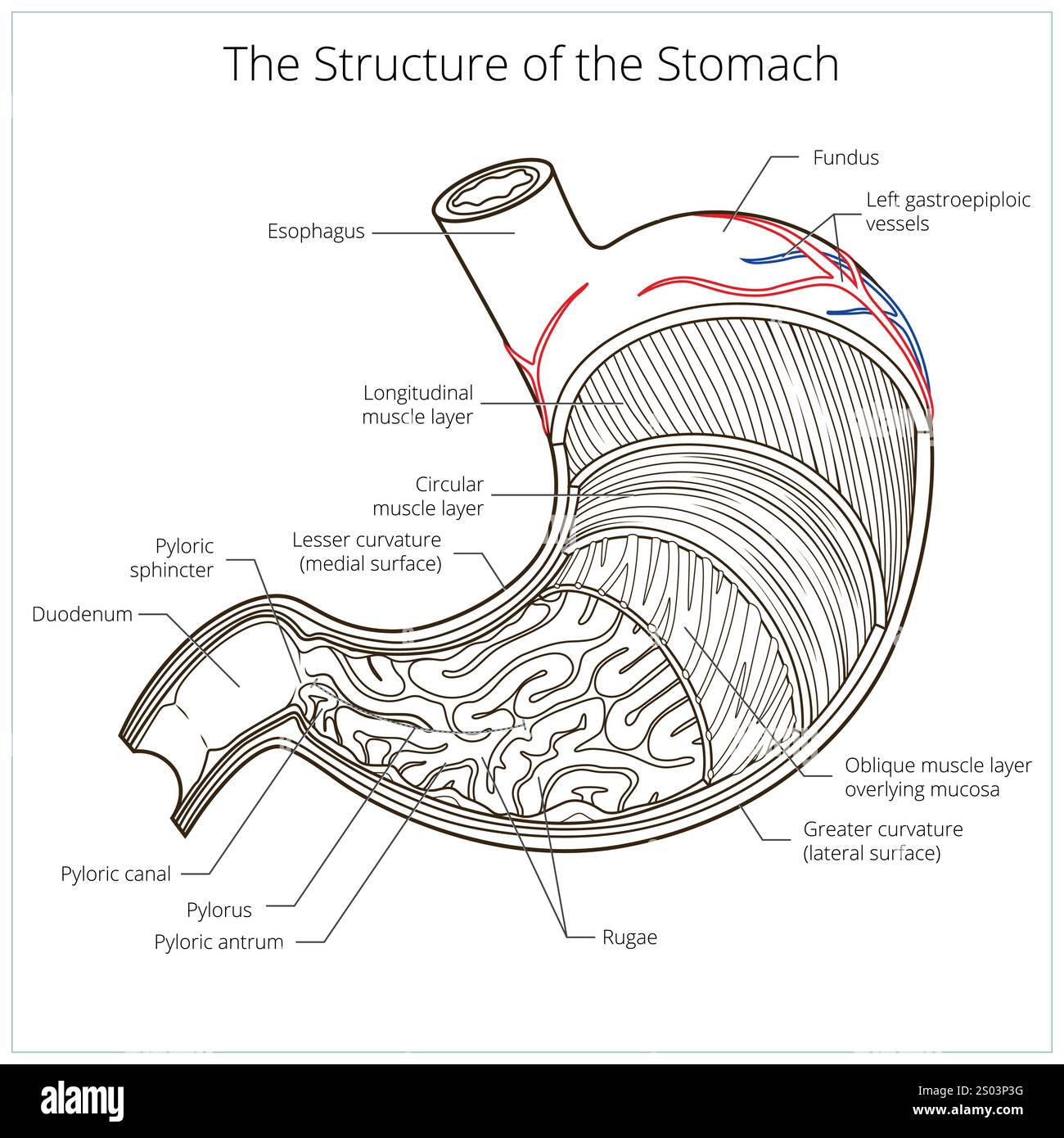 Structure of stomach medical educational vector Stock Vectorhttps://www.alamy.com/image-license-details/?v=1https://www.alamy.com/structure-of-stomach-medical-educational-vector-image636866820.html
RF2S03P3G–Structure of stomach medical educational vector
 Sectional view illustration of perforated ulcer Stock Vectorhttps://www.alamy.com/image-license-details/?v=1https://www.alamy.com/sectional-view-illustration-of-perforated-ulcer-image452731117.html
RF2H8FK4D–Sectional view illustration of perforated ulcer
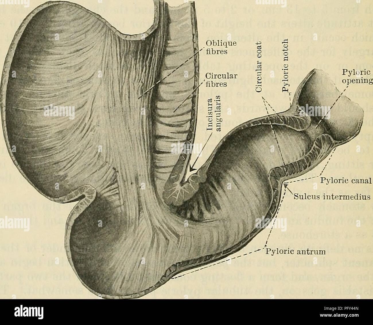 . Cunningham's Text-book of anatomy. Anatomy. Tunica serosa tlBBS^. Pyloric canal Sulcus intermedius Fig. Pyloric antrum 925.—Muscular Coat of the Stomach, seen from within after removal of the mucous and submucous layers. The anterior half of the stomach is shown, viewed from behind (Cunningham). The stratum circulare is composed mainly of circular fibres, continuous with the more superficial of the circular fibres at the lower end of the oesophagus (Fig. 925). They begin as a set of U-shaped bundles which loop over the lesser curvature at the right of. Please note that these images are extra Stock Photohttps://www.alamy.com/image-license-details/?v=1https://www.alamy.com/cunninghams-text-book-of-anatomy-anatomy-tunica-serosa-tlbbs-pyloric-canal-sulcus-intermedius-fig-pyloric-antrum-925muscular-coat-of-the-stomach-seen-from-within-after-removal-of-the-mucous-and-submucous-layers-the-anterior-half-of-the-stomach-is-shown-viewed-from-behind-cunningham-the-stratum-circulare-is-composed-mainly-of-circular-fibres-continuous-with-the-more-superficial-of-the-circular-fibres-at-the-lower-end-of-the-oesophagus-fig-925-they-begin-as-a-set-of-u-shaped-bundles-which-loop-over-the-lesser-curvature-at-the-right-of-please-note-that-these-images-are-extra-image216340229.html
RMPFY44N–. Cunningham's Text-book of anatomy. Anatomy. Tunica serosa tlBBS^. Pyloric canal Sulcus intermedius Fig. Pyloric antrum 925.—Muscular Coat of the Stomach, seen from within after removal of the mucous and submucous layers. The anterior half of the stomach is shown, viewed from behind (Cunningham). The stratum circulare is composed mainly of circular fibres, continuous with the more superficial of the circular fibres at the lower end of the oesophagus (Fig. 925). They begin as a set of U-shaped bundles which loop over the lesser curvature at the right of. Please note that these images are extra
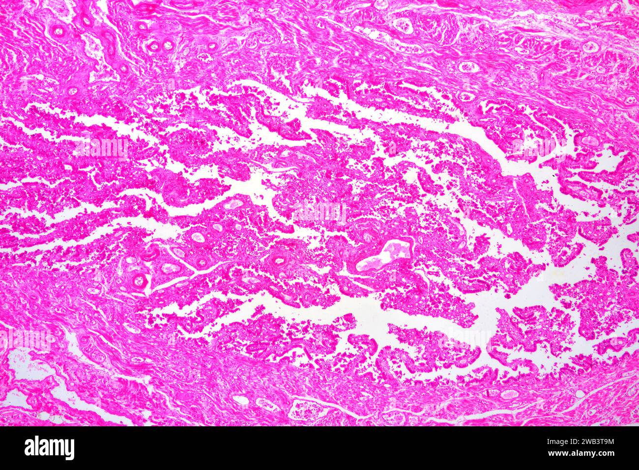 Human Fallopian tube section. X30 at 10 cm wide. Stock Photohttps://www.alamy.com/image-license-details/?v=1https://www.alamy.com/human-fallopian-tube-section-x30-at-10-cm-wide-image591998672.html
RF2WB3T9M–Human Fallopian tube section. X30 at 10 cm wide.
 Surgical treatment; a practical treatise on the therapy of surgical diseases for the use of practitioners and students of surgery . e raw surface without exposing it. This sutureshould be applied tightly in such a direction as to embrace the blood-vessels, THE ABDOMEN 729 and after being tied should be covered by a second layer of serosa sutures.This is the operation of emergency, not of choice. If no sign of ulcer is to be seen through the peritoneum, and direct treat-ment of the hemorrhage is to be undertaken, the stomach should be drawnforward, surrounded by protecting pads and opened. An i Stock Photohttps://www.alamy.com/image-license-details/?v=1https://www.alamy.com/surgical-treatment-a-practical-treatise-on-the-therapy-of-surgical-diseases-for-the-use-of-practitioners-and-students-of-surgery-e-raw-surface-without-exposing-it-this-sutureshould-be-applied-tightly-in-such-a-direction-as-to-embrace-the-blood-vessels-the-abdomen-729-and-after-being-tied-should-be-covered-by-a-second-layer-of-serosa-suturesthis-is-the-operation-of-emergency-not-of-choice-if-no-sign-of-ulcer-is-to-be-seen-through-the-peritoneum-and-direct-treat-ment-of-the-hemorrhage-is-to-be-undertaken-the-stomach-should-be-drawnforward-surrounded-by-protecting-pads-and-opened-an-i-image338204666.html
RM2AJ6FE2–Surgical treatment; a practical treatise on the therapy of surgical diseases for the use of practitioners and students of surgery . e raw surface without exposing it. This sutureshould be applied tightly in such a direction as to embrace the blood-vessels, THE ABDOMEN 729 and after being tied should be covered by a second layer of serosa sutures.This is the operation of emergency, not of choice. If no sign of ulcer is to be seen through the peritoneum, and direct treat-ment of the hemorrhage is to be undertaken, the stomach should be drawnforward, surrounded by protecting pads and opened. An i
 Human rectum showing from top to bottom: mucosa with intestinal glands, lamina propria, submucosa, muscular layers (circular and longitudinal) and se Stock Photohttps://www.alamy.com/image-license-details/?v=1https://www.alamy.com/human-rectum-showing-from-top-to-bottom-mucosa-with-intestinal-glands-lamina-propria-submucosa-muscular-layers-circular-and-longitudinal-and-se-image591999863.html
RF2WB3WT7–Human rectum showing from top to bottom: mucosa with intestinal glands, lamina propria, submucosa, muscular layers (circular and longitudinal) and se
![Embryology of insects and myriapods; Embryology of insects and myriapods; the developmental history of insects, centipedes, and millepedes from egg desposition [!] to hatching embryologyofinse00joha Year: 1941 do4r. Fig. 10.—Germ-band formation. A, sagittal section. B, cross sec- tion, (do) Dorsal organ, {gb) Germ band, {ser) Future serosa, {yc} Yolk cells. ser Stock Photo Embryology of insects and myriapods; Embryology of insects and myriapods; the developmental history of insects, centipedes, and millepedes from egg desposition [!] to hatching embryologyofinse00joha Year: 1941 do4r. Fig. 10.—Germ-band formation. A, sagittal section. B, cross sec- tion, (do) Dorsal organ, {gb) Germ band, {ser) Future serosa, {yc} Yolk cells. ser Stock Photo](https://c8.alamy.com/comp/RWMRR4/embryology-of-insects-and-myriapods-embryology-of-insects-and-myriapods-the-developmental-history-of-insects-centipedes-and-millepedes-from-egg-desposition-!-to-hatching-embryologyofinse00joha-year-1941-do4r-fig-10germ-band-formation-a-sagittal-section-b-cross-sec-tion-do-dorsal-organ-gb-germ-band-ser-future-serosa-yc-yolk-cells-ser-RWMRR4.jpg) Embryology of insects and myriapods; Embryology of insects and myriapods; the developmental history of insects, centipedes, and millepedes from egg desposition [!] to hatching embryologyofinse00joha Year: 1941 do4r. Fig. 10.—Germ-band formation. A, sagittal section. B, cross sec- tion, (do) Dorsal organ, {gb) Germ band, {ser) Future serosa, {yc} Yolk cells. ser Stock Photohttps://www.alamy.com/image-license-details/?v=1https://www.alamy.com/embryology-of-insects-and-myriapods-embryology-of-insects-and-myriapods-the-developmental-history-of-insects-centipedes-and-millepedes-from-egg-desposition-!-to-hatching-embryologyofinse00joha-year-1941-do4r-fig-10germ-band-formation-a-sagittal-section-b-cross-sec-tion-do-dorsal-organ-gb-germ-band-ser-future-serosa-yc-yolk-cells-ser-image239558904.html
Embryology of insects and myriapods; Embryology of insects and myriapods; the developmental history of insects, centipedes, and millepedes from egg desposition [!] to hatching embryologyofinse00joha Year: 1941 do4r. Fig. 10.—Germ-band formation. A, sagittal section. B, cross sec- tion, (do) Dorsal organ, {gb) Germ band, {ser) Future serosa, {yc} Yolk cells. ser Stock Photohttps://www.alamy.com/image-license-details/?v=1https://www.alamy.com/embryology-of-insects-and-myriapods-embryology-of-insects-and-myriapods-the-developmental-history-of-insects-centipedes-and-millepedes-from-egg-desposition-!-to-hatching-embryologyofinse00joha-year-1941-do4r-fig-10germ-band-formation-a-sagittal-section-b-cross-sec-tion-do-dorsal-organ-gb-germ-band-ser-future-serosa-yc-yolk-cells-ser-image239558904.htmlRMRWMRR4–Embryology of insects and myriapods; Embryology of insects and myriapods; the developmental history of insects, centipedes, and millepedes from egg desposition [!] to hatching embryologyofinse00joha Year: 1941 do4r. Fig. 10.—Germ-band formation. A, sagittal section. B, cross sec- tion, (do) Dorsal organ, {gb) Germ band, {ser) Future serosa, {yc} Yolk cells. ser
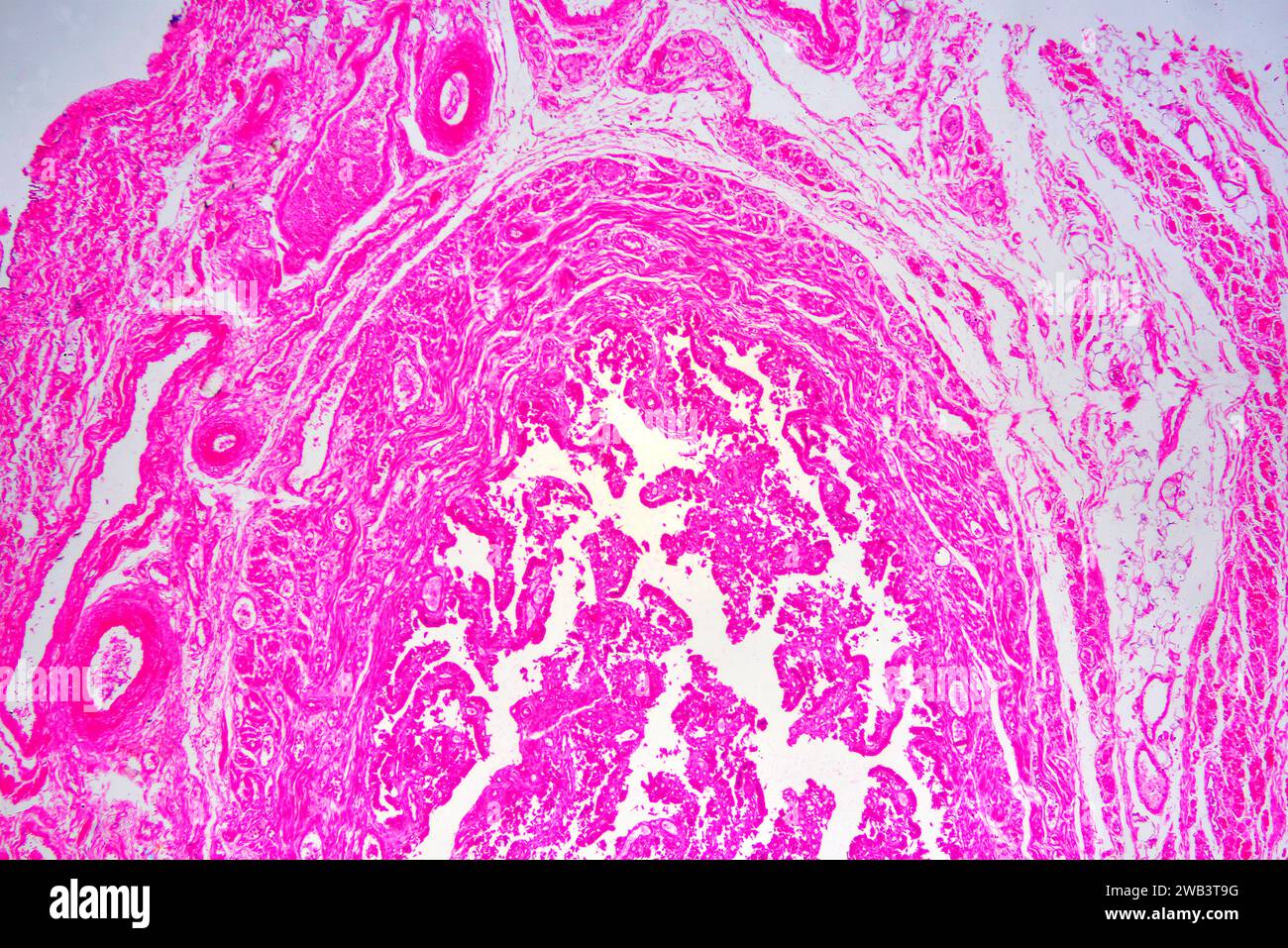 Human Fallopian tube section. X30 at 10 cm wide. Stock Photohttps://www.alamy.com/image-license-details/?v=1https://www.alamy.com/human-fallopian-tube-section-x30-at-10-cm-wide-image591998668.html
RF2WB3T9G–Human Fallopian tube section. X30 at 10 cm wide.
 Stomach ulcer Stock Photohttps://www.alamy.com/image-license-details/?v=1https://www.alamy.com/stock-photo-stomach-ulcer-14034740.html
RFAFXCMN–Stomach ulcer
 . Fig. 72. — Cysto-adenoma on the serosa of the intestine. (After Pickens.) removed or they may be left alone. They will not ordinarily affect the health of the bird. Even malignant tumors are apparently not contagious. The ultimate nature of the causative agents has not been determined. With our present knowledge it is impossible to explain either the spontaneous origin of these agents within an individual or to account for their transfer from one individ- Stock Photohttps://www.alamy.com/image-license-details/?v=1https://www.alamy.com/fig-72-cysto-adenoma-on-the-serosa-of-the-intestine-after-pickens-removed-or-they-may-be-left-alone-they-will-not-ordinarily-affect-the-health-of-the-bird-even-malignant-tumors-are-apparently-not-contagious-the-ultimate-nature-of-the-causative-agents-has-not-been-determined-with-our-present-knowledge-it-is-impossible-to-explain-either-the-spontaneous-origin-of-these-agents-within-an-individual-or-to-account-for-their-transfer-from-one-individ-image179916980.html
RMMCKWXC–. Fig. 72. — Cysto-adenoma on the serosa of the intestine. (After Pickens.) removed or they may be left alone. They will not ordinarily affect the health of the bird. Even malignant tumors are apparently not contagious. The ultimate nature of the causative agents has not been determined. With our present knowledge it is impossible to explain either the spontaneous origin of these agents within an individual or to account for their transfer from one individ-
 Structure of stomach medical educational vector Stock Vectorhttps://www.alamy.com/image-license-details/?v=1https://www.alamy.com/structure-of-stomach-medical-educational-vector-image636866830.html
RF2S03P3X–Structure of stomach medical educational vector
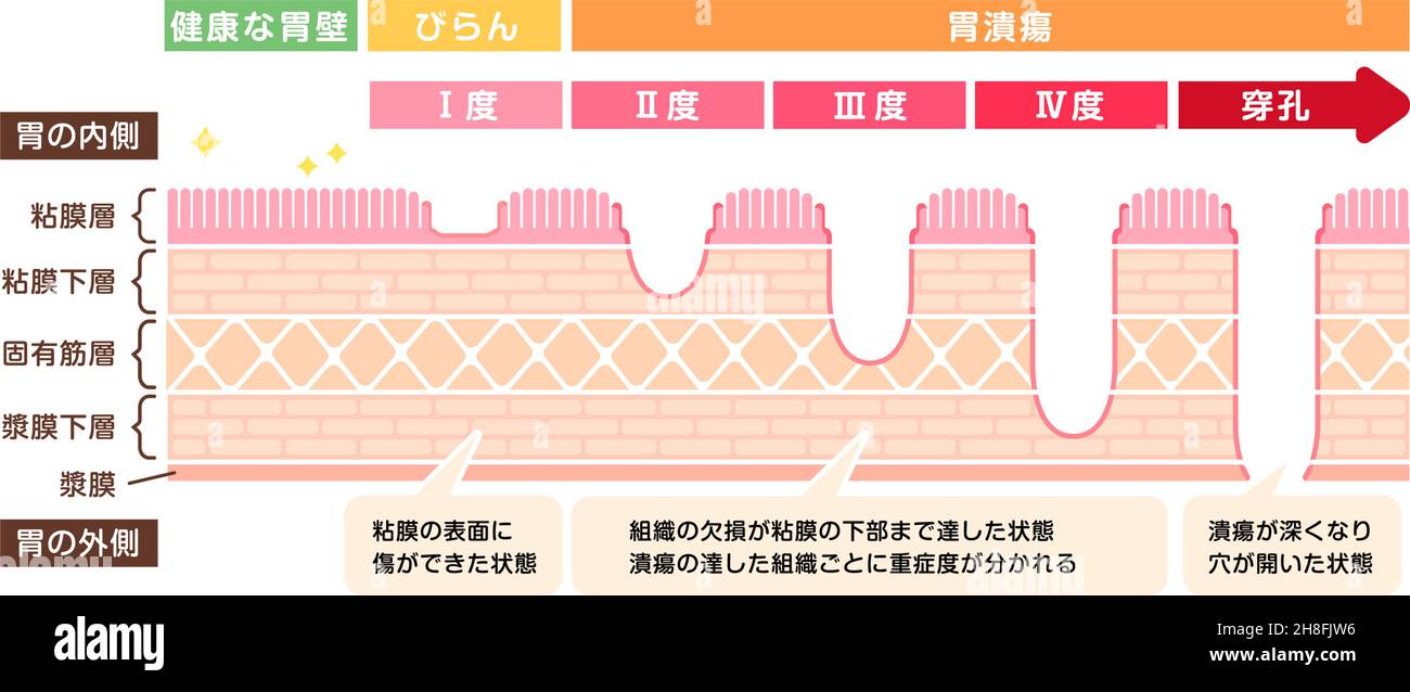 Stages of gastric ulcer ( stomach ulser ) vector illustration Stock Vectorhttps://www.alamy.com/image-license-details/?v=1https://www.alamy.com/stages-of-gastric-ulcer-stomach-ulser-vector-illustration-image452730914.html
RF2H8FJW6–Stages of gastric ulcer ( stomach ulser ) vector illustration
![. Cunningham's Text-book of anatomy. Anatomy. 1198 THE DIGESTIVE SYSTEM. Stkucture of the Liver. The liver is invested by an outer tunica serosa described in connexion with the peritoneum. Within this is a thin capsula fibrosa [Glissonii] (O.T. G-lisson's capsule) Intralobular capillary plexus Intralobular capillary plexus. Central vein Central vein Sublobular vein Fig 940.—Liver of a Pig injected from the Hepatic Vein by T. A. Carter. (From a specimen left in the Anatomical Department of Edinburgh University by Sir William Turner.) Liver lobules of delicate fibrous tissue, which is most evi Stock Photo . Cunningham's Text-book of anatomy. Anatomy. 1198 THE DIGESTIVE SYSTEM. Stkucture of the Liver. The liver is invested by an outer tunica serosa described in connexion with the peritoneum. Within this is a thin capsula fibrosa [Glissonii] (O.T. G-lisson's capsule) Intralobular capillary plexus Intralobular capillary plexus. Central vein Central vein Sublobular vein Fig 940.—Liver of a Pig injected from the Hepatic Vein by T. A. Carter. (From a specimen left in the Anatomical Department of Edinburgh University by Sir William Turner.) Liver lobules of delicate fibrous tissue, which is most evi Stock Photo](https://c8.alamy.com/comp/PFY427/cunninghams-text-book-of-anatomy-anatomy-1198-the-digestive-system-stkucture-of-the-liver-the-liver-is-invested-by-an-outer-tunica-serosa-described-in-connexion-with-the-peritoneum-within-this-is-a-thin-capsula-fibrosa-glissonii-ot-g-lissons-capsule-intralobular-capillary-plexus-intralobular-capillary-plexus-central-vein-central-vein-sublobular-vein-fig-940liver-of-a-pig-injected-from-the-hepatic-vein-by-t-a-carter-from-a-specimen-left-in-the-anatomical-department-of-edinburgh-university-by-sir-william-turner-liver-lobules-of-delicate-fibrous-tissue-which-is-most-evi-PFY427.jpg) . Cunningham's Text-book of anatomy. Anatomy. 1198 THE DIGESTIVE SYSTEM. Stkucture of the Liver. The liver is invested by an outer tunica serosa described in connexion with the peritoneum. Within this is a thin capsula fibrosa [Glissonii] (O.T. G-lisson's capsule) Intralobular capillary plexus Intralobular capillary plexus. Central vein Central vein Sublobular vein Fig 940.—Liver of a Pig injected from the Hepatic Vein by T. A. Carter. (From a specimen left in the Anatomical Department of Edinburgh University by Sir William Turner.) Liver lobules of delicate fibrous tissue, which is most evi Stock Photohttps://www.alamy.com/image-license-details/?v=1https://www.alamy.com/cunninghams-text-book-of-anatomy-anatomy-1198-the-digestive-system-stkucture-of-the-liver-the-liver-is-invested-by-an-outer-tunica-serosa-described-in-connexion-with-the-peritoneum-within-this-is-a-thin-capsula-fibrosa-glissonii-ot-g-lissons-capsule-intralobular-capillary-plexus-intralobular-capillary-plexus-central-vein-central-vein-sublobular-vein-fig-940liver-of-a-pig-injected-from-the-hepatic-vein-by-t-a-carter-from-a-specimen-left-in-the-anatomical-department-of-edinburgh-university-by-sir-william-turner-liver-lobules-of-delicate-fibrous-tissue-which-is-most-evi-image216340159.html
. Cunningham's Text-book of anatomy. Anatomy. 1198 THE DIGESTIVE SYSTEM. Stkucture of the Liver. The liver is invested by an outer tunica serosa described in connexion with the peritoneum. Within this is a thin capsula fibrosa [Glissonii] (O.T. G-lisson's capsule) Intralobular capillary plexus Intralobular capillary plexus. Central vein Central vein Sublobular vein Fig 940.—Liver of a Pig injected from the Hepatic Vein by T. A. Carter. (From a specimen left in the Anatomical Department of Edinburgh University by Sir William Turner.) Liver lobules of delicate fibrous tissue, which is most evi Stock Photohttps://www.alamy.com/image-license-details/?v=1https://www.alamy.com/cunninghams-text-book-of-anatomy-anatomy-1198-the-digestive-system-stkucture-of-the-liver-the-liver-is-invested-by-an-outer-tunica-serosa-described-in-connexion-with-the-peritoneum-within-this-is-a-thin-capsula-fibrosa-glissonii-ot-g-lissons-capsule-intralobular-capillary-plexus-intralobular-capillary-plexus-central-vein-central-vein-sublobular-vein-fig-940liver-of-a-pig-injected-from-the-hepatic-vein-by-t-a-carter-from-a-specimen-left-in-the-anatomical-department-of-edinburgh-university-by-sir-william-turner-liver-lobules-of-delicate-fibrous-tissue-which-is-most-evi-image216340159.htmlRMPFY427–. Cunningham's Text-book of anatomy. Anatomy. 1198 THE DIGESTIVE SYSTEM. Stkucture of the Liver. The liver is invested by an outer tunica serosa described in connexion with the peritoneum. Within this is a thin capsula fibrosa [Glissonii] (O.T. G-lisson's capsule) Intralobular capillary plexus Intralobular capillary plexus. Central vein Central vein Sublobular vein Fig 940.—Liver of a Pig injected from the Hepatic Vein by T. A. Carter. (From a specimen left in the Anatomical Department of Edinburgh University by Sir William Turner.) Liver lobules of delicate fibrous tissue, which is most evi
 Surgical treatment; a practical treatise on the therapy of surgical diseases for the use of practitioners and students of surgery . owel must first be done. The formation of a blind end may be accomplishedby any one of several different methods. The most useful are the following: i. The bowel is divided between two clamps. The end to be closed is thensutured with an over-and-over suture through all of its coats bringing thewound edges together or serosa to serosa. This suture begins opposite themesenteric attachment and ends at the mesentery. Its object is to compressthe blood-vessels. A secon Stock Photohttps://www.alamy.com/image-license-details/?v=1https://www.alamy.com/surgical-treatment-a-practical-treatise-on-the-therapy-of-surgical-diseases-for-the-use-of-practitioners-and-students-of-surgery-owel-must-first-be-done-the-formation-of-a-blind-end-may-be-accomplishedby-any-one-of-several-different-methods-the-most-useful-are-the-following-i-the-bowel-is-divided-between-two-clamps-the-end-to-be-closed-is-thensutured-with-an-over-and-over-suture-through-all-of-its-coats-bringing-thewound-edges-together-or-serosa-to-serosa-this-suture-begins-opposite-themesenteric-attachment-and-ends-at-the-mesentery-its-object-is-to-compressthe-blood-vessels-a-secon-image338218728.html
RM2AJ75C8–Surgical treatment; a practical treatise on the therapy of surgical diseases for the use of practitioners and students of surgery . owel must first be done. The formation of a blind end may be accomplishedby any one of several different methods. The most useful are the following: i. The bowel is divided between two clamps. The end to be closed is thensutured with an over-and-over suture through all of its coats bringing thewound edges together or serosa to serosa. This suture begins opposite themesenteric attachment and ends at the mesentery. Its object is to compressthe blood-vessels. A secon
![Embryology of insects and myriapods; Embryology of insects and myriapods; the developmental history of insects, centipedes, and millepedes from egg desposition [!] to hatching embryologyofinse00joha Year: 1941 274 EMBRYOLOGY OF INSECTS AND MYRIAPODS am.ser A Fig. 205.—Oncopeltus. Cross sections at different levels, {am. cav) Amniotic cavity. {am-, ser) Amnioserosa. {gb) Germ band. {M) Head lobe. {U) Inner layer, {ser) Serosa. Stock Photo Embryology of insects and myriapods; Embryology of insects and myriapods; the developmental history of insects, centipedes, and millepedes from egg desposition [!] to hatching embryologyofinse00joha Year: 1941 274 EMBRYOLOGY OF INSECTS AND MYRIAPODS am.ser A Fig. 205.—Oncopeltus. Cross sections at different levels, {am. cav) Amniotic cavity. {am-, ser) Amnioserosa. {gb) Germ band. {M) Head lobe. {U) Inner layer, {ser) Serosa. Stock Photo](https://c8.alamy.com/comp/RWW4D1/embryology-of-insects-and-myriapods-embryology-of-insects-and-myriapods-the-developmental-history-of-insects-centipedes-and-millepedes-from-egg-desposition-!-to-hatching-embryologyofinse00joha-year-1941-274-embryology-of-insects-and-myriapods-amser-a-fig-205oncopeltus-cross-sections-at-different-levels-am-cav-amniotic-cavity-am-ser-amnioserosa-gb-germ-band-m-head-lobe-u-inner-layer-ser-serosa-RWW4D1.jpg) Embryology of insects and myriapods; Embryology of insects and myriapods; the developmental history of insects, centipedes, and millepedes from egg desposition [!] to hatching embryologyofinse00joha Year: 1941 274 EMBRYOLOGY OF INSECTS AND MYRIAPODS am.ser A Fig. 205.—Oncopeltus. Cross sections at different levels, {am. cav) Amniotic cavity. {am-, ser) Amnioserosa. {gb) Germ band. {M) Head lobe. {U) Inner layer, {ser) Serosa. Stock Photohttps://www.alamy.com/image-license-details/?v=1https://www.alamy.com/embryology-of-insects-and-myriapods-embryology-of-insects-and-myriapods-the-developmental-history-of-insects-centipedes-and-millepedes-from-egg-desposition-!-to-hatching-embryologyofinse00joha-year-1941-274-embryology-of-insects-and-myriapods-amser-a-fig-205oncopeltus-cross-sections-at-different-levels-am-cav-amniotic-cavity-am-ser-amnioserosa-gb-germ-band-m-head-lobe-u-inner-layer-ser-serosa-image239653485.html
Embryology of insects and myriapods; Embryology of insects and myriapods; the developmental history of insects, centipedes, and millepedes from egg desposition [!] to hatching embryologyofinse00joha Year: 1941 274 EMBRYOLOGY OF INSECTS AND MYRIAPODS am.ser A Fig. 205.—Oncopeltus. Cross sections at different levels, {am. cav) Amniotic cavity. {am-, ser) Amnioserosa. {gb) Germ band. {M) Head lobe. {U) Inner layer, {ser) Serosa. Stock Photohttps://www.alamy.com/image-license-details/?v=1https://www.alamy.com/embryology-of-insects-and-myriapods-embryology-of-insects-and-myriapods-the-developmental-history-of-insects-centipedes-and-millepedes-from-egg-desposition-!-to-hatching-embryologyofinse00joha-year-1941-274-embryology-of-insects-and-myriapods-amser-a-fig-205oncopeltus-cross-sections-at-different-levels-am-cav-amniotic-cavity-am-ser-amnioserosa-gb-germ-band-m-head-lobe-u-inner-layer-ser-serosa-image239653485.htmlRMRWW4D1–Embryology of insects and myriapods; Embryology of insects and myriapods; the developmental history of insects, centipedes, and millepedes from egg desposition [!] to hatching embryologyofinse00joha Year: 1941 274 EMBRYOLOGY OF INSECTS AND MYRIAPODS am.ser A Fig. 205.—Oncopeltus. Cross sections at different levels, {am. cav) Amniotic cavity. {am-, ser) Amnioserosa. {gb) Germ band. {M) Head lobe. {U) Inner layer, {ser) Serosa.