Smooth muscle photomicrograph Stock Photos and Images
(69)See smooth muscle photomicrograph stock video clipsSmooth muscle photomicrograph Stock Photos and Images
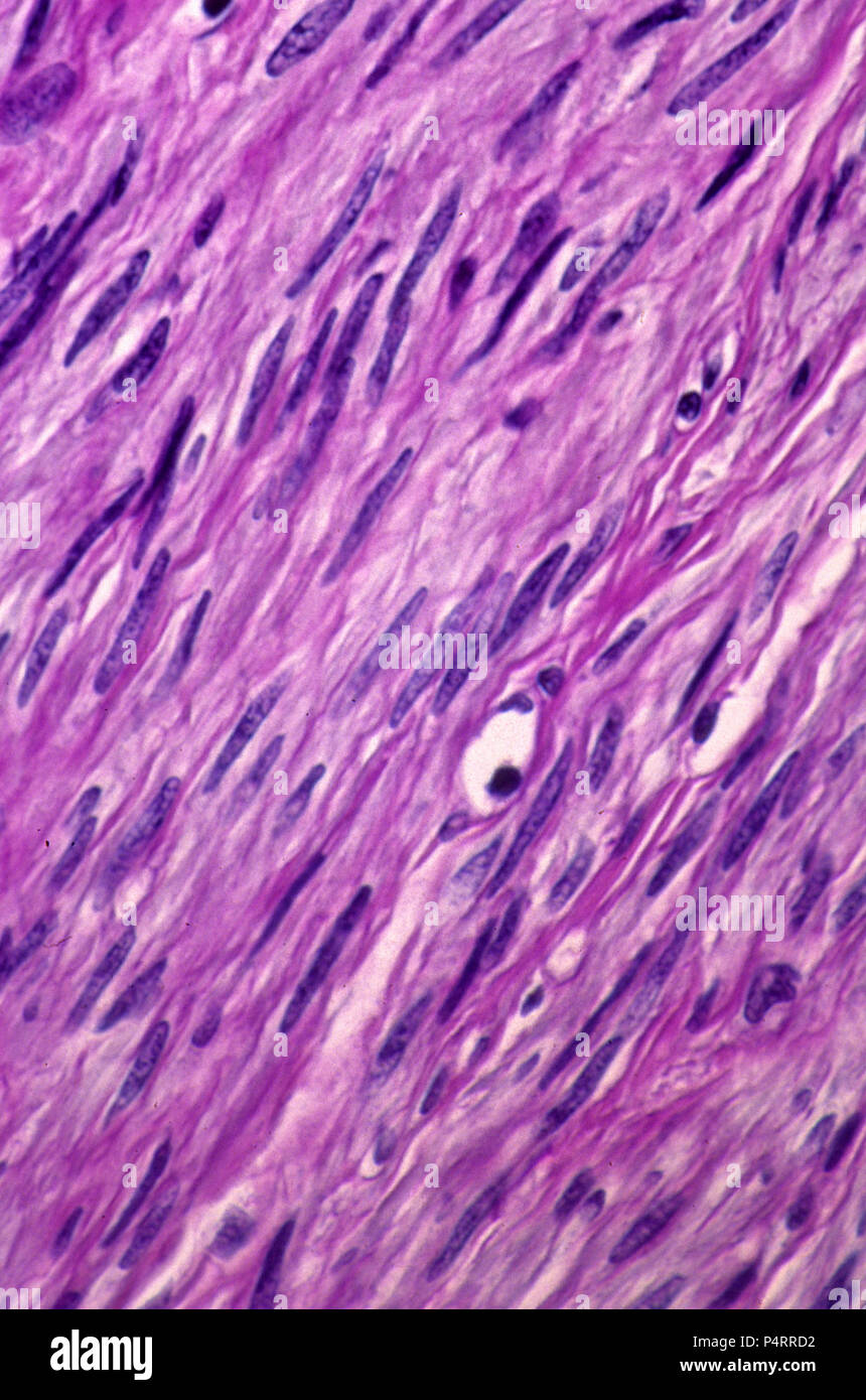 Smooth muscle fibres Stock Photohttps://www.alamy.com/image-license-details/?v=1https://www.alamy.com/smooth-muscle-fibres-image209506334.html
RFP4RRD2–Smooth muscle fibres
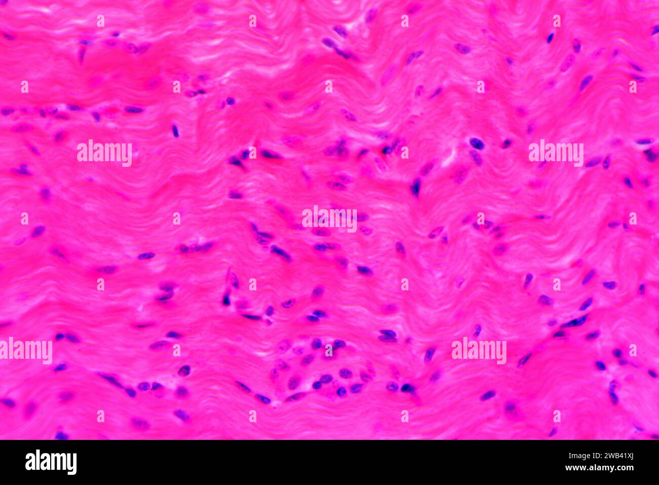 Smooth muscle longitudinal section. Photomicrograph X300 at 10 cm wide. Stock Photohttps://www.alamy.com/image-license-details/?v=1https://www.alamy.com/smooth-muscle-longitudinal-section-photomicrograph-x300-at-10-cm-wide-image592003066.html
RF2WB41XJ–Smooth muscle longitudinal section. Photomicrograph X300 at 10 cm wide.
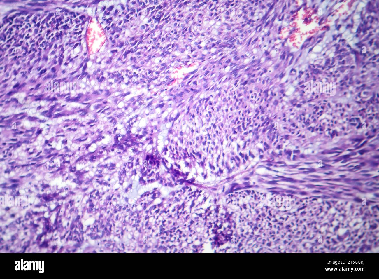 Photomicrograph of leiomyoma, illustrating benign smooth muscle tumor cells within the uterine tissue. Stock Photohttps://www.alamy.com/image-license-details/?v=1https://www.alamy.com/photomicrograph-of-leiomyoma-illustrating-benign-smooth-muscle-tumor-cells-within-the-uterine-tissue-image571994518.html
RF2T6GGRJ–Photomicrograph of leiomyoma, illustrating benign smooth muscle tumor cells within the uterine tissue.
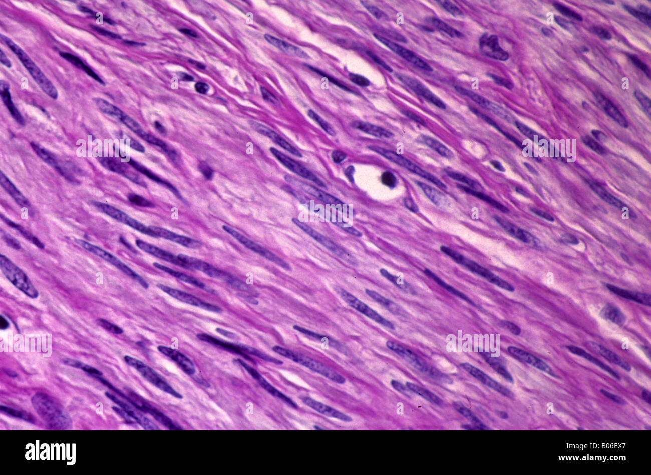 Smooth muscle fibres Stock Photohttps://www.alamy.com/image-license-details/?v=1https://www.alamy.com/stock-photo-smooth-muscle-fibres-17353791.html
RFB06EX7–Smooth muscle fibres
 . American journal of obstetrics and gynecology. T/;./? •-? - 111.?-??? • ? ? «•*. ,.4- Fig. 5.—Photomicrograph of a section through the cystic growth at a in Fig. 1.Practically no smooth muscle is present, the reaction about the adenoma. beingmore in the nature of very fibrous granulation tissue. Some blood is present in manyof the cystic spaces. velopment of this transplanted endometrium is of interest althoughreally not surprising. The requisite ovarian hormone may have circu-lated in the blood for a period of time of longer or shorter durationfollowing the extirpation of the ovaries and du Stock Photohttps://www.alamy.com/image-license-details/?v=1https://www.alamy.com/american-journal-of-obstetrics-and-gynecology-t-111-4-fig-5photomicrograph-of-a-section-through-the-cystic-growth-at-a-in-fig-1practically-no-smooth-muscle-is-present-the-reaction-about-the-adenoma-beingmore-in-the-nature-of-very-fibrous-granulation-tissue-some-blood-is-present-in-manyof-the-cystic-spaces-velopment-of-this-transplanted-endometrium-is-of-interest-althoughreally-not-surprising-the-requisite-ovarian-hormone-may-have-circu-lated-in-the-blood-for-a-period-of-time-of-longer-or-shorter-durationfollowing-the-extirpation-of-the-ovaries-and-du-image370563563.html
RM2CETHGY–. American journal of obstetrics and gynecology. T/;./? •-? - 111.?-??? • ? ? «•*. ,.4- Fig. 5.—Photomicrograph of a section through the cystic growth at a in Fig. 1.Practically no smooth muscle is present, the reaction about the adenoma. beingmore in the nature of very fibrous granulation tissue. Some blood is present in manyof the cystic spaces. velopment of this transplanted endometrium is of interest althoughreally not surprising. The requisite ovarian hormone may have circu-lated in the blood for a period of time of longer or shorter durationfollowing the extirpation of the ovaries and du
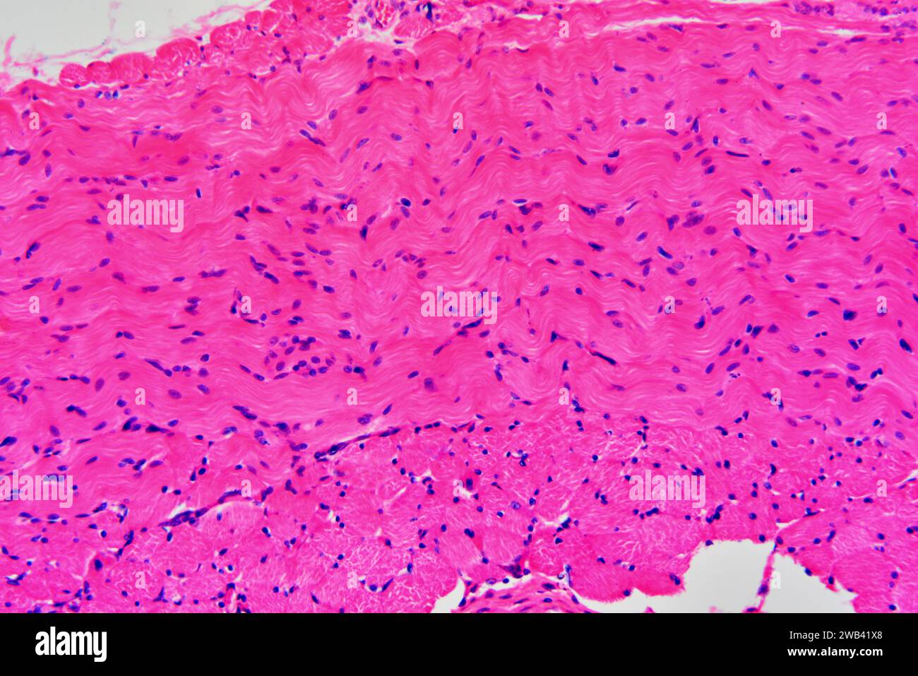 Smooth muscle longitudinal and cross sections. Photomicrograph X150 at 10 cm wide. Stock Photohttps://www.alamy.com/image-license-details/?v=1https://www.alamy.com/smooth-muscle-longitudinal-and-cross-sections-photomicrograph-x150-at-10-cm-wide-image592003056.html
RF2WB41X8–Smooth muscle longitudinal and cross sections. Photomicrograph X150 at 10 cm wide.
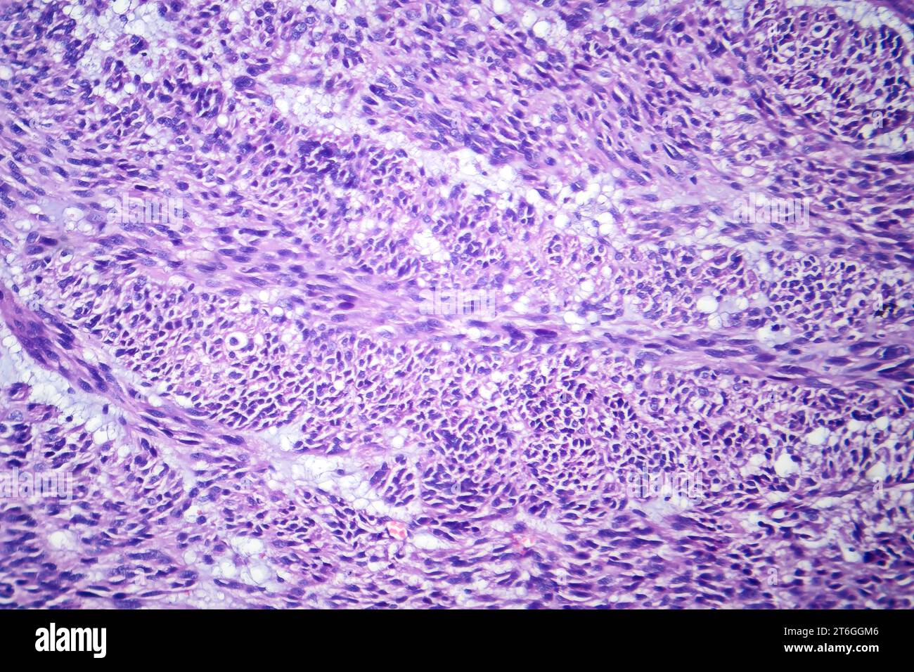 Photomicrograph of leiomyoma, illustrating benign smooth muscle tumor cells within the uterine tissue. Stock Photohttps://www.alamy.com/image-license-details/?v=1https://www.alamy.com/photomicrograph-of-leiomyoma-illustrating-benign-smooth-muscle-tumor-cells-within-the-uterine-tissue-image571994422.html
RF2T6GGM6–Photomicrograph of leiomyoma, illustrating benign smooth muscle tumor cells within the uterine tissue.
 . The anatomical record. Anatomy; Anatomy. Fig. 3 Photomicrograph of the largest island found after a search through many sections of the accessory pancreas of dog D 93. It is normal, but small. X2.50. Fig. I I'hotoinicrograph of one of the average-sized islands of the major I)ancreas of dog I) 93. Compare with figure 3. X"2oO. Fig. 5 Photomicrograph of section of accessory i)ancreas of dog D .ilVi. Note the ahsence of island tissue and the intimate relation of pancreatic and smooth muscle tissue. X75. 267. Please note that these images are extracted from scanned page images that may have Stock Photohttps://www.alamy.com/image-license-details/?v=1https://www.alamy.com/the-anatomical-record-anatomy-anatomy-fig-3-photomicrograph-of-the-largest-island-found-after-a-search-through-many-sections-of-the-accessory-pancreas-of-dog-d-93-it-is-normal-but-small-x250-fig-i-ihotoinicrograph-of-one-of-the-average-sized-islands-of-the-major-iancreas-of-dog-i-93-compare-with-figure-3-xquot2oo-fig-5-photomicrograph-of-section-of-accessory-iancreas-of-dog-d-ilvi-note-the-ahsence-of-island-tissue-and-the-intimate-relation-of-pancreatic-and-smooth-muscle-tissue-x75-267-please-note-that-these-images-are-extracted-from-scanned-page-images-that-may-have-image236873777.html
RMRNAEWN–. The anatomical record. Anatomy; Anatomy. Fig. 3 Photomicrograph of the largest island found after a search through many sections of the accessory pancreas of dog D 93. It is normal, but small. X2.50. Fig. I I'hotoinicrograph of one of the average-sized islands of the major I)ancreas of dog I) 93. Compare with figure 3. X"2oO. Fig. 5 Photomicrograph of section of accessory i)ancreas of dog D .ilVi. Note the ahsence of island tissue and the intimate relation of pancreatic and smooth muscle tissue. X75. 267. Please note that these images are extracted from scanned page images that may have
 Smooth muscle longitudinal and cross sections. Photomicrograph X150 at 10 cm wide. Stock Photohttps://www.alamy.com/image-license-details/?v=1https://www.alamy.com/smooth-muscle-longitudinal-and-cross-sections-photomicrograph-x150-at-10-cm-wide-image592003061.html
RF2WB41XD–Smooth muscle longitudinal and cross sections. Photomicrograph X150 at 10 cm wide.
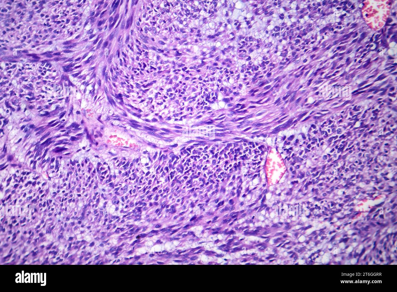 Photomicrograph of leiomyoma, illustrating benign smooth muscle tumor cells within the uterine tissue. Stock Photohttps://www.alamy.com/image-license-details/?v=1https://www.alamy.com/photomicrograph-of-leiomyoma-illustrating-benign-smooth-muscle-tumor-cells-within-the-uterine-tissue-image571994523.html
RF2T6GGRR–Photomicrograph of leiomyoma, illustrating benign smooth muscle tumor cells within the uterine tissue.
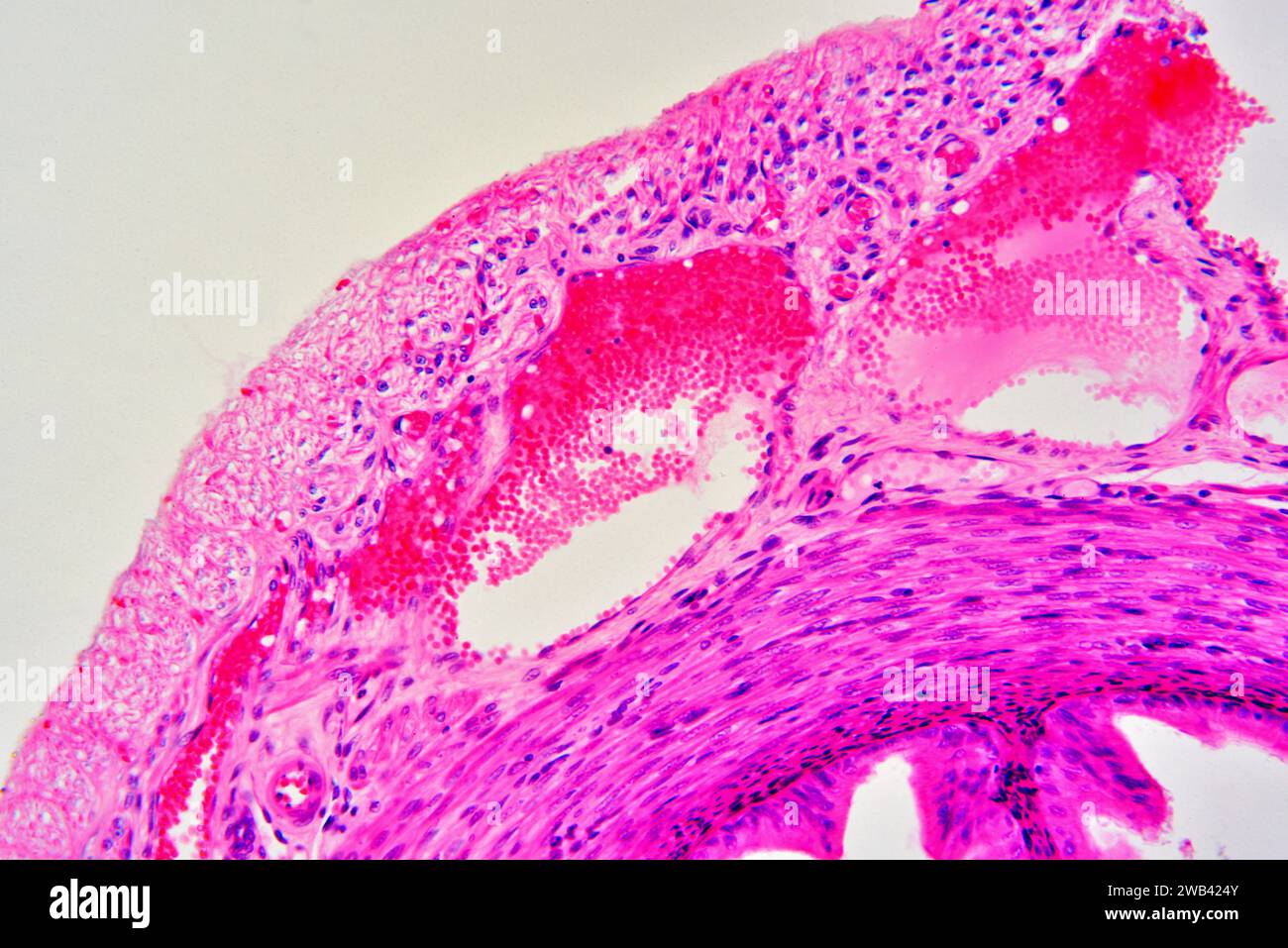 Veins with red blood cells surrounded by connective tissue and smooth muscle fibers. Photomicrograph X150 at 10 cm wide. Stock Photohttps://www.alamy.com/image-license-details/?v=1https://www.alamy.com/veins-with-red-blood-cells-surrounded-by-connective-tissue-and-smooth-muscle-fibers-photomicrograph-x150-at-10-cm-wide-image592003243.html
RF2WB424Y–Veins with red blood cells surrounded by connective tissue and smooth muscle fibers. Photomicrograph X150 at 10 cm wide.
 Photomicrograph of leiomyoma, illustrating benign smooth muscle tumor cells within the uterine tissue. Stock Photohttps://www.alamy.com/image-license-details/?v=1https://www.alamy.com/photomicrograph-of-leiomyoma-illustrating-benign-smooth-muscle-tumor-cells-within-the-uterine-tissue-image571994526.html
RF2T6GGRX–Photomicrograph of leiomyoma, illustrating benign smooth muscle tumor cells within the uterine tissue.
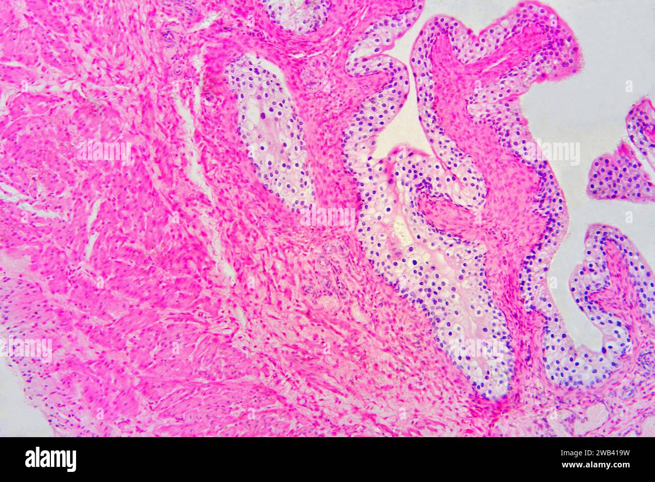 Urinary bladder with urothelium (transitional epithelium), connective tissue and smooth muscle fibers. Photomicrograph X75 at 10 cm wide. Stock Photohttps://www.alamy.com/image-license-details/?v=1https://www.alamy.com/urinary-bladder-with-urothelium-transitional-epithelium-connective-tissue-and-smooth-muscle-fibers-photomicrograph-x75-at-10-cm-wide-image592002597.html
RF2WB419W–Urinary bladder with urothelium (transitional epithelium), connective tissue and smooth muscle fibers. Photomicrograph X75 at 10 cm wide.
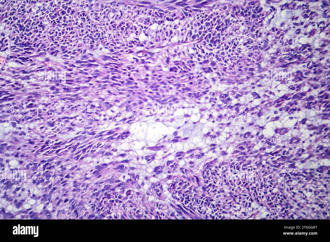 Photomicrograph of leiomyoma, illustrating benign smooth muscle tumor cells within the uterine tissue. Stock Photohttps://www.alamy.com/image-license-details/?v=1https://www.alamy.com/photomicrograph-of-leiomyoma-illustrating-benign-smooth-muscle-tumor-cells-within-the-uterine-tissue-image571994524.html
RF2T6GGRT–Photomicrograph of leiomyoma, illustrating benign smooth muscle tumor cells within the uterine tissue.
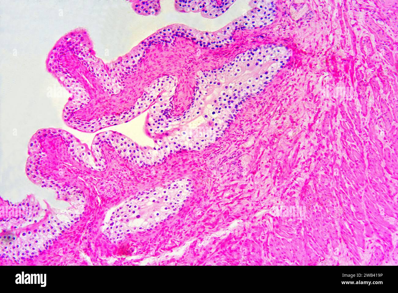 Urinary bladder with urothelium (transitional epithelium), connective tissue and smooth muscle fibers. Photomicrograph X75 at 10 cm wide. Stock Photohttps://www.alamy.com/image-license-details/?v=1https://www.alamy.com/urinary-bladder-with-urothelium-transitional-epithelium-connective-tissue-and-smooth-muscle-fibers-photomicrograph-x75-at-10-cm-wide-image592002594.html
RF2WB419P–Urinary bladder with urothelium (transitional epithelium), connective tissue and smooth muscle fibers. Photomicrograph X75 at 10 cm wide.
 Photomicrograph of leiomyoma, illustrating benign smooth muscle tumor cells within the uterine tissue. Stock Photohttps://www.alamy.com/image-license-details/?v=1https://www.alamy.com/photomicrograph-of-leiomyoma-illustrating-benign-smooth-muscle-tumor-cells-within-the-uterine-tissue-image571994435.html
RF2T6GGMK–Photomicrograph of leiomyoma, illustrating benign smooth muscle tumor cells within the uterine tissue.
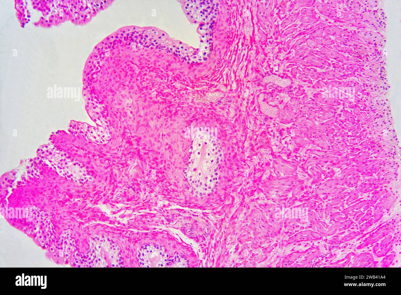 Urinary bladder with urothelium (transitional epithelium), connective tissue and smooth muscle fibers. Photomicrograph X75 at 10 cm wide. Stock Photohttps://www.alamy.com/image-license-details/?v=1https://www.alamy.com/urinary-bladder-with-urothelium-transitional-epithelium-connective-tissue-and-smooth-muscle-fibers-photomicrograph-x75-at-10-cm-wide-image592002604.html
RF2WB41A4–Urinary bladder with urothelium (transitional epithelium), connective tissue and smooth muscle fibers. Photomicrograph X75 at 10 cm wide.
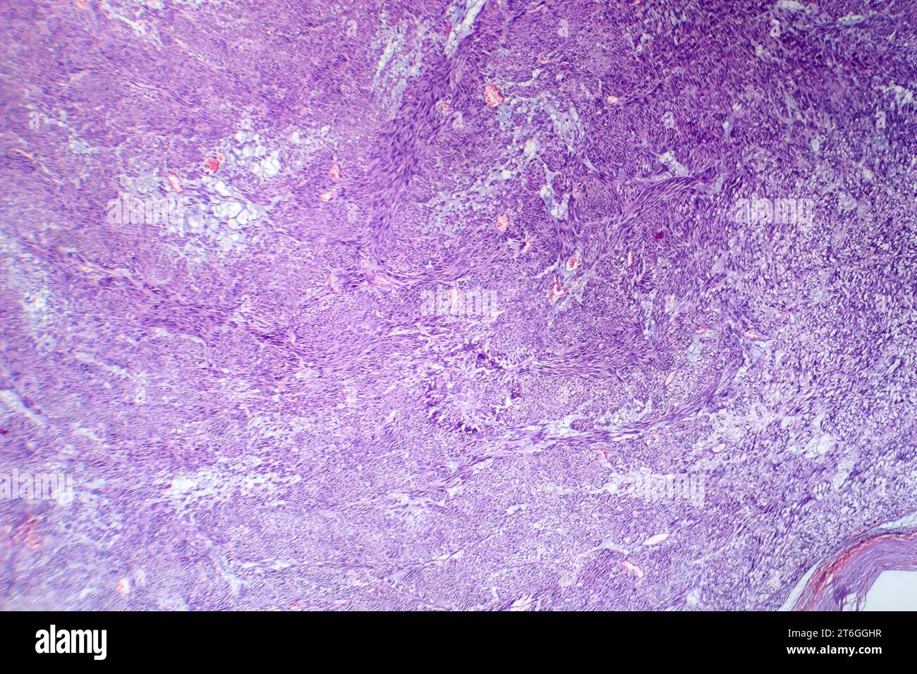 Photomicrograph of leiomyoma, illustrating benign smooth muscle tumor cells within the uterine tissue. Stock Photohttps://www.alamy.com/image-license-details/?v=1https://www.alamy.com/photomicrograph-of-leiomyoma-illustrating-benign-smooth-muscle-tumor-cells-within-the-uterine-tissue-image571994355.html
RF2T6GGHR–Photomicrograph of leiomyoma, illustrating benign smooth muscle tumor cells within the uterine tissue.
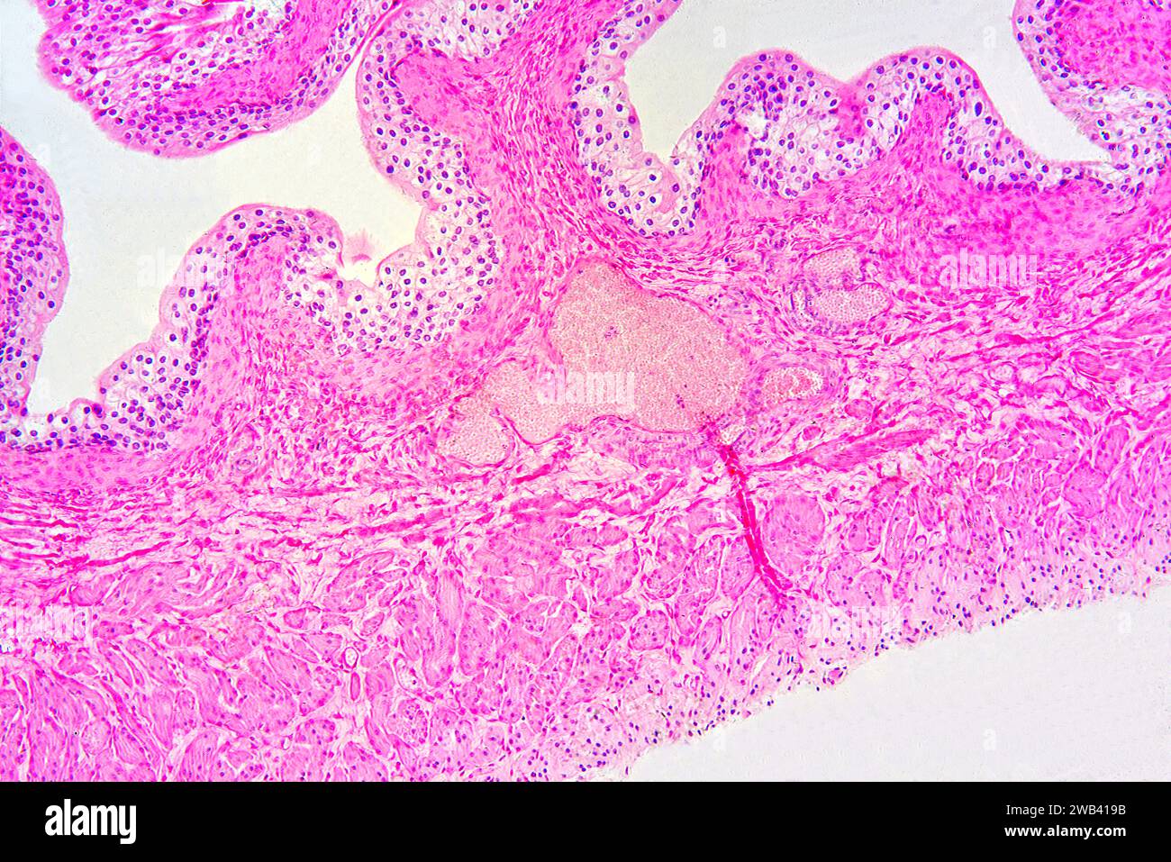 Urinary bladder with urothelium (transitional epithelium), connective tissue and smooth muscle fibers. Photomicrograph X75 at 10 cm wide. Stock Photohttps://www.alamy.com/image-license-details/?v=1https://www.alamy.com/urinary-bladder-with-urothelium-transitional-epithelium-connective-tissue-and-smooth-muscle-fibers-photomicrograph-x75-at-10-cm-wide-image592002583.html
RF2WB419B–Urinary bladder with urothelium (transitional epithelium), connective tissue and smooth muscle fibers. Photomicrograph X75 at 10 cm wide.
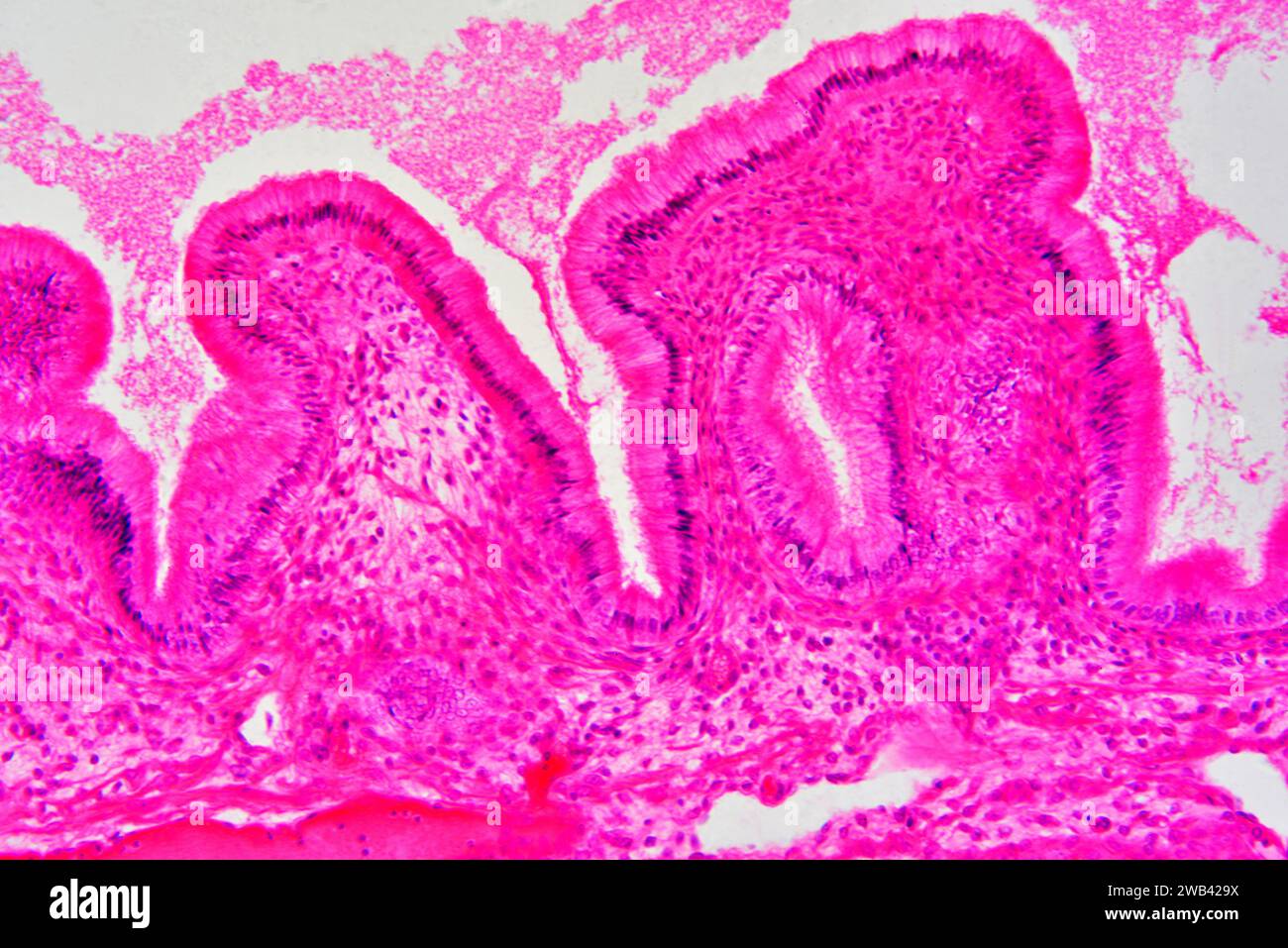 Gallbladder wall showing columnar epithelium with mucosal folds, connective tissue and smooth muscle fibers. Photomicrograph X150 at 10cm wide. Stock Photohttps://www.alamy.com/image-license-details/?v=1https://www.alamy.com/gallbladder-wall-showing-columnar-epithelium-with-mucosal-folds-connective-tissue-and-smooth-muscle-fibers-photomicrograph-x150-at-10cm-wide-image592003382.html
RF2WB429X–Gallbladder wall showing columnar epithelium with mucosal folds, connective tissue and smooth muscle fibers. Photomicrograph X150 at 10cm wide.
 Photomicrograph of leiomyoma, illustrating benign smooth muscle tumor cells within the uterine tissue. Stock Photohttps://www.alamy.com/image-license-details/?v=1https://www.alamy.com/photomicrograph-of-leiomyoma-illustrating-benign-smooth-muscle-tumor-cells-within-the-uterine-tissue-image571994437.html
RF2T6GGMN–Photomicrograph of leiomyoma, illustrating benign smooth muscle tumor cells within the uterine tissue.
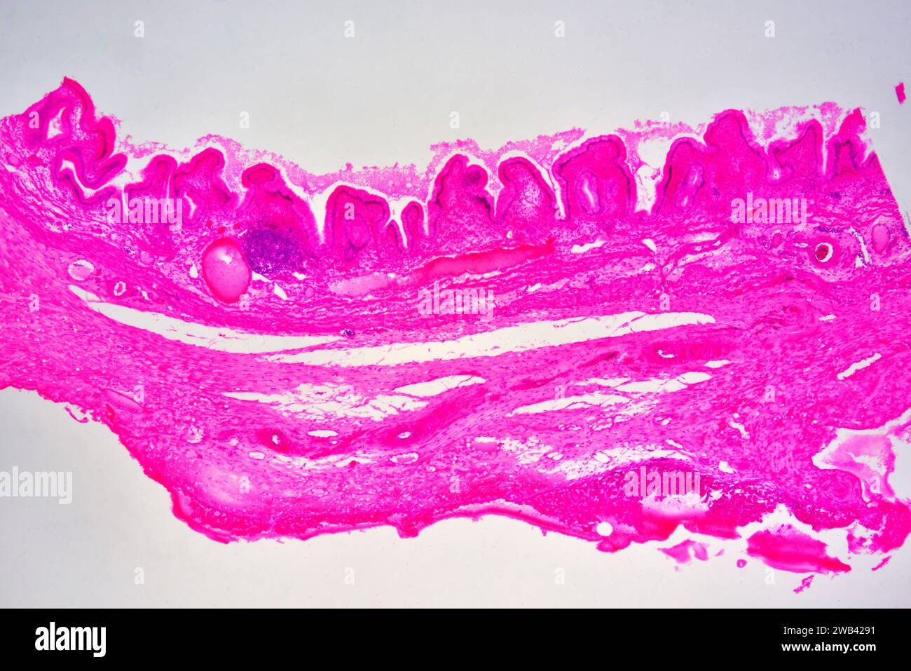 Gallbladder wall showing columnar epithelium with mucosal folds, connective tissue and smooth muscle fibers. Photomicrograph X30 at 10cm wide. Stock Photohttps://www.alamy.com/image-license-details/?v=1https://www.alamy.com/gallbladder-wall-showing-columnar-epithelium-with-mucosal-folds-connective-tissue-and-smooth-muscle-fibers-photomicrograph-x30-at-10cm-wide-image592003357.html
RF2WB4291–Gallbladder wall showing columnar epithelium with mucosal folds, connective tissue and smooth muscle fibers. Photomicrograph X30 at 10cm wide.
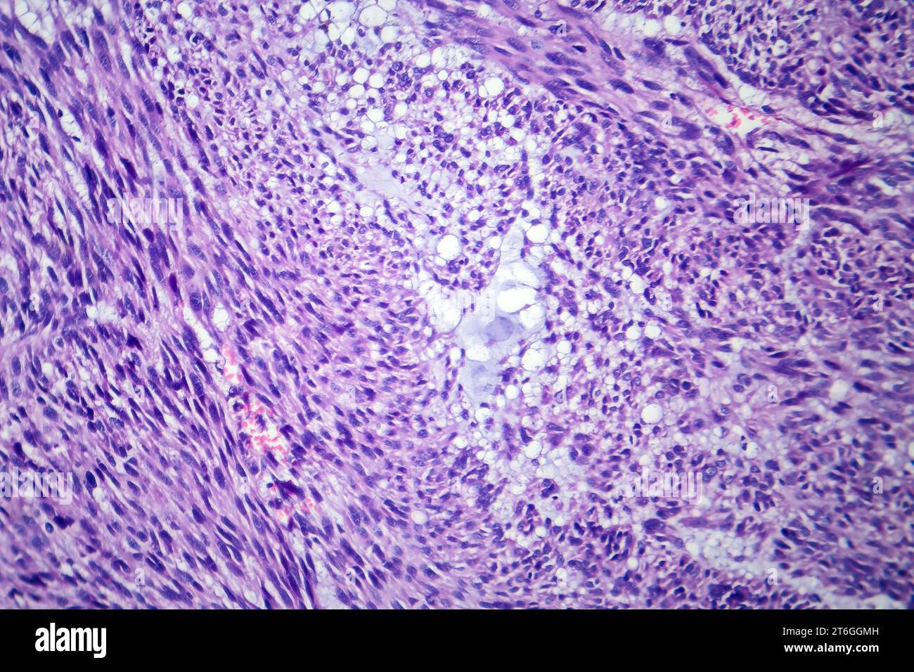 Photomicrograph of leiomyoma, illustrating benign smooth muscle tumor cells within the uterine tissue. Stock Photohttps://www.alamy.com/image-license-details/?v=1https://www.alamy.com/photomicrograph-of-leiomyoma-illustrating-benign-smooth-muscle-tumor-cells-within-the-uterine-tissue-image571994433.html
RF2T6GGMH–Photomicrograph of leiomyoma, illustrating benign smooth muscle tumor cells within the uterine tissue.
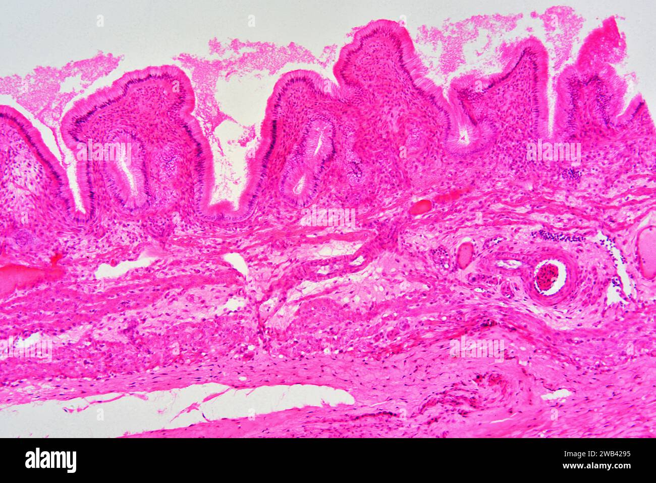 Gallbladder wall showing columnar epithelium with mucosal foldsi, connective tissue and smooth muscle fibers. Photomicrograph X30 at 10cm wide. Stock Photohttps://www.alamy.com/image-license-details/?v=1https://www.alamy.com/gallbladder-wall-showing-columnar-epithelium-with-mucosal-foldsi-connective-tissue-and-smooth-muscle-fibers-photomicrograph-x30-at-10cm-wide-image592003361.html
RF2WB4295–Gallbladder wall showing columnar epithelium with mucosal foldsi, connective tissue and smooth muscle fibers. Photomicrograph X30 at 10cm wide.
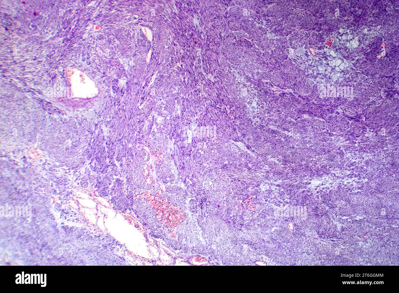 Photomicrograph of leiomyoma, illustrating benign smooth muscle tumor cells within the uterine tissue. Stock Photohttps://www.alamy.com/image-license-details/?v=1https://www.alamy.com/photomicrograph-of-leiomyoma-illustrating-benign-smooth-muscle-tumor-cells-within-the-uterine-tissue-image571994436.html
RF2T6GGMM–Photomicrograph of leiomyoma, illustrating benign smooth muscle tumor cells within the uterine tissue.
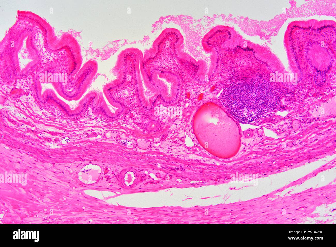 Gallbladder wall showing columnar epithelium with mucosal folds, connective tissue, blood vessels and smooth muscle fibers. Photomicrograph X75 at 10 Stock Photohttps://www.alamy.com/image-license-details/?v=1https://www.alamy.com/gallbladder-wall-showing-columnar-epithelium-with-mucosal-folds-connective-tissue-blood-vessels-and-smooth-muscle-fibers-photomicrograph-x75-at-10-image592003370.html
RF2WB429E–Gallbladder wall showing columnar epithelium with mucosal folds, connective tissue, blood vessels and smooth muscle fibers. Photomicrograph X75 at 10
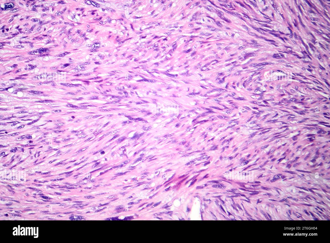 Photomicrograph of leiomyosarcoma, depicting malignant smooth muscle tumor cells, indicative of aggressive soft tissue cancer. Stock Photohttps://www.alamy.com/image-license-details/?v=1https://www.alamy.com/photomicrograph-of-leiomyosarcoma-depicting-malignant-smooth-muscle-tumor-cells-indicative-of-aggressive-soft-tissue-cancer-image571994644.html
RF2T6GH04–Photomicrograph of leiomyosarcoma, depicting malignant smooth muscle tumor cells, indicative of aggressive soft tissue cancer.
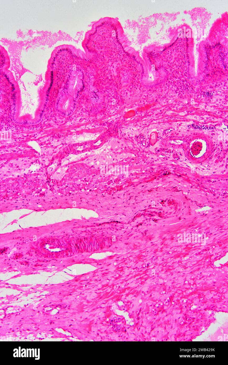 Gallbladder wall showing columnar epithelium with mucosal folds, connective tissue, blood vessels and smooth muscle fibers. Photomicrograph X75 at 10 Stock Photohttps://www.alamy.com/image-license-details/?v=1https://www.alamy.com/gallbladder-wall-showing-columnar-epithelium-with-mucosal-folds-connective-tissue-blood-vessels-and-smooth-muscle-fibers-photomicrograph-x75-at-10-image592003375.html
RF2WB429K–Gallbladder wall showing columnar epithelium with mucosal folds, connective tissue, blood vessels and smooth muscle fibers. Photomicrograph X75 at 10
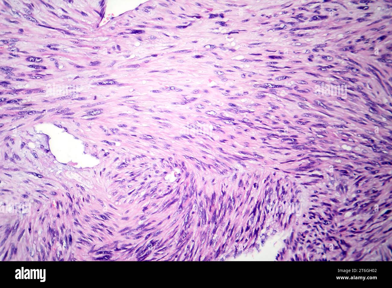 Photomicrograph of leiomyosarcoma, depicting malignant smooth muscle tumor cells, indicative of aggressive soft tissue cancer. Stock Photohttps://www.alamy.com/image-license-details/?v=1https://www.alamy.com/photomicrograph-of-leiomyosarcoma-depicting-malignant-smooth-muscle-tumor-cells-indicative-of-aggressive-soft-tissue-cancer-image571994642.html
RF2T6GH02–Photomicrograph of leiomyosarcoma, depicting malignant smooth muscle tumor cells, indicative of aggressive soft tissue cancer.
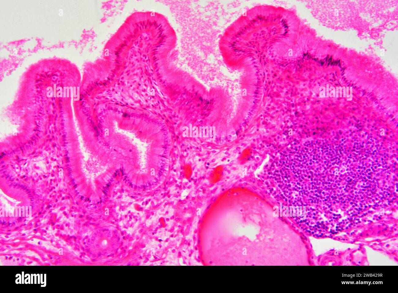 Gallbladder wall showing columnar epithelium with mucosal folds, connective tissue, blood vessels and smooth muscle fibers. Photomicrograph X150 at 10 Stock Photohttps://www.alamy.com/image-license-details/?v=1https://www.alamy.com/gallbladder-wall-showing-columnar-epithelium-with-mucosal-folds-connective-tissue-blood-vessels-and-smooth-muscle-fibers-photomicrograph-x150-at-10-image592003379.html
RF2WB429R–Gallbladder wall showing columnar epithelium with mucosal folds, connective tissue, blood vessels and smooth muscle fibers. Photomicrograph X150 at 10
 Human glandular epithelium and smooth muscle fibers. X75 at 10 cm wide. Stock Photohttps://www.alamy.com/image-license-details/?v=1https://www.alamy.com/human-glandular-epithelium-and-smooth-muscle-fibers-x75-at-10-cm-wide-image592001858.html
RF2WB40BE–Human glandular epithelium and smooth muscle fibers. X75 at 10 cm wide.
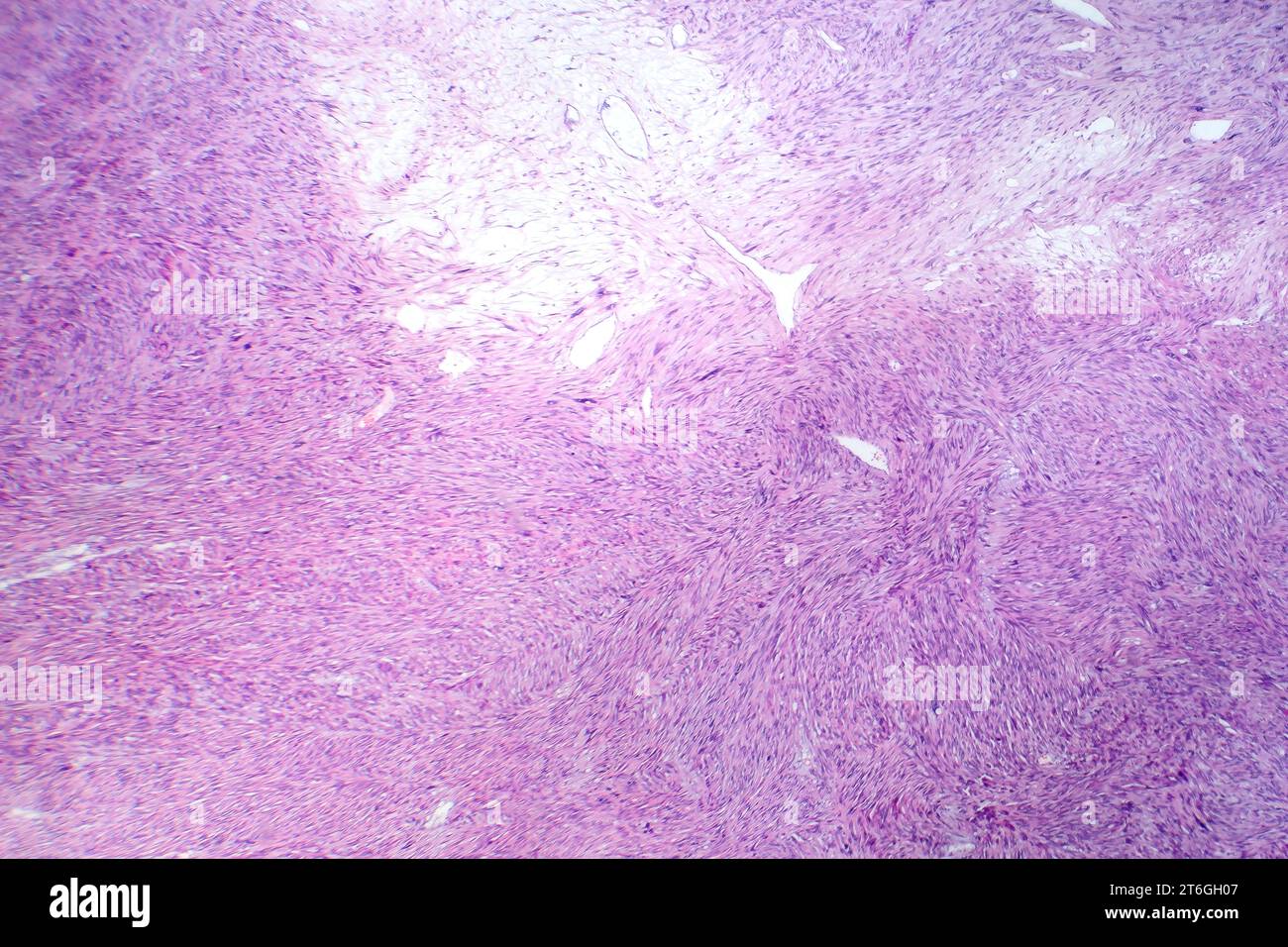 Photomicrograph of leiomyosarcoma, depicting malignant smooth muscle tumor cells, indicative of aggressive soft tissue cancer. Stock Photohttps://www.alamy.com/image-license-details/?v=1https://www.alamy.com/photomicrograph-of-leiomyosarcoma-depicting-malignant-smooth-muscle-tumor-cells-indicative-of-aggressive-soft-tissue-cancer-image571994647.html
RF2T6GH07–Photomicrograph of leiomyosarcoma, depicting malignant smooth muscle tumor cells, indicative of aggressive soft tissue cancer.
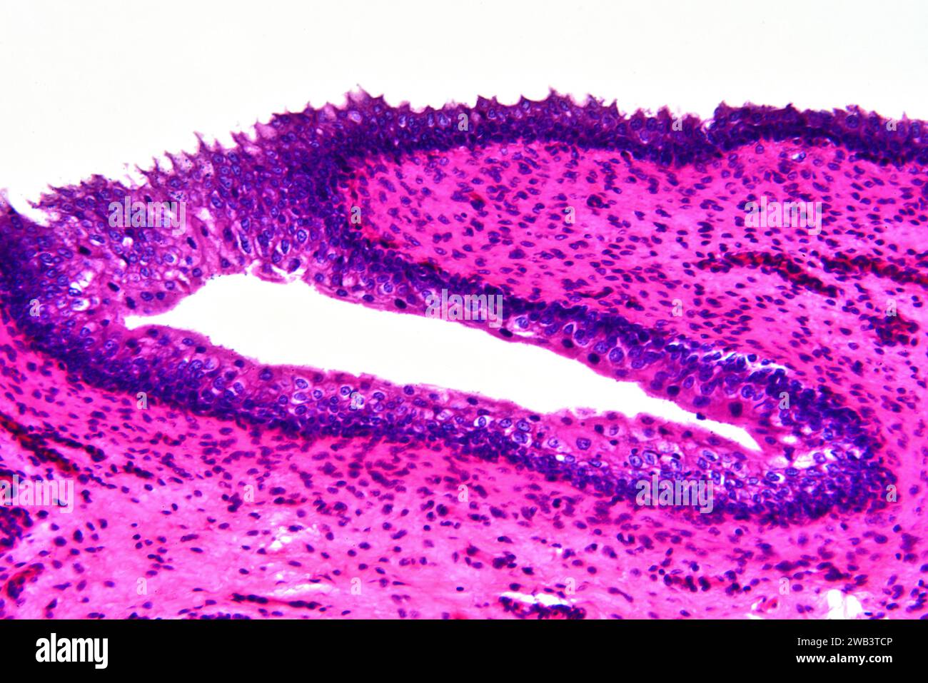 Human transitional epithelium and smooth muscle tissue. X125 at 10 cm wide. Stock Photohttps://www.alamy.com/image-license-details/?v=1https://www.alamy.com/human-transitional-epithelium-and-smooth-muscle-tissue-x125-at-10-cm-wide-image591998758.html
RF2WB3TCP–Human transitional epithelium and smooth muscle tissue. X125 at 10 cm wide.
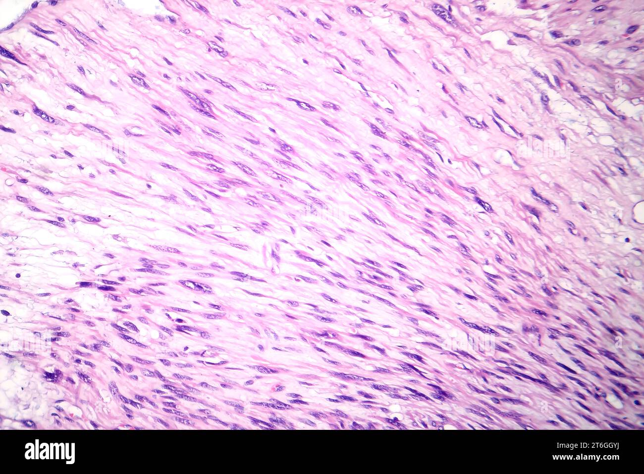 Photomicrograph of leiomyosarcoma, depicting malignant smooth muscle tumor cells, indicative of aggressive soft tissue cancer. Stock Photohttps://www.alamy.com/image-license-details/?v=1https://www.alamy.com/photomicrograph-of-leiomyosarcoma-depicting-malignant-smooth-muscle-tumor-cells-indicative-of-aggressive-soft-tissue-cancer-image571994630.html
RF2T6GGYJ–Photomicrograph of leiomyosarcoma, depicting malignant smooth muscle tumor cells, indicative of aggressive soft tissue cancer.
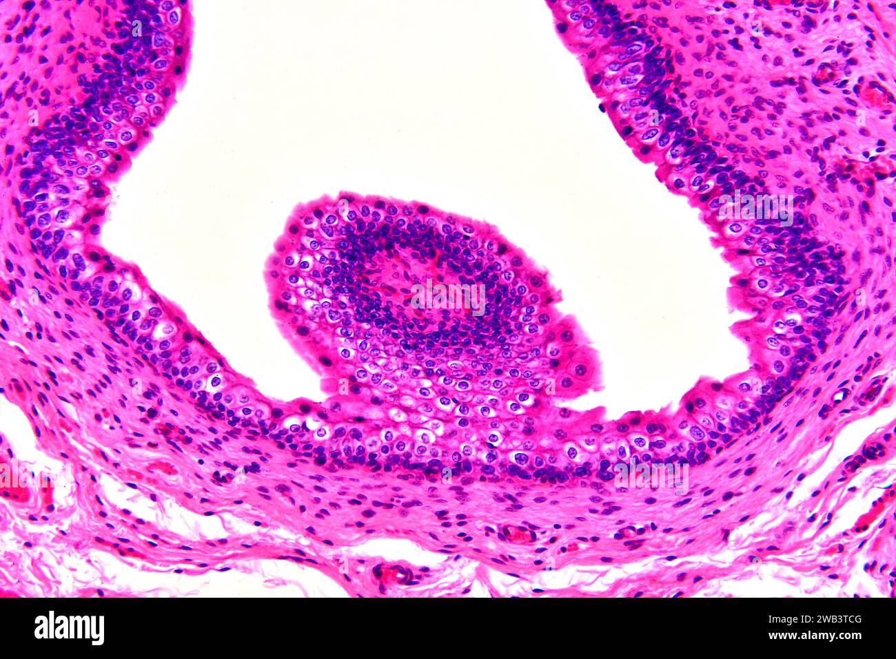 Human transitional epithelium and smooth muscle tissue. X125 at 10 cm wide. Stock Photohttps://www.alamy.com/image-license-details/?v=1https://www.alamy.com/human-transitional-epithelium-and-smooth-muscle-tissue-x125-at-10-cm-wide-image591998752.html
RF2WB3TCG–Human transitional epithelium and smooth muscle tissue. X125 at 10 cm wide.
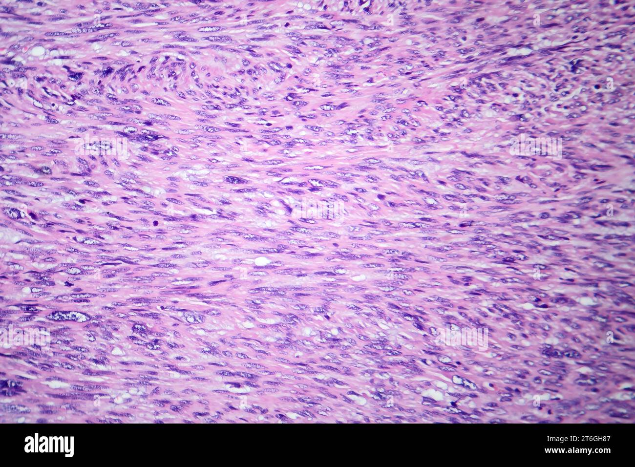 Photomicrograph of leiomyosarcoma, depicting malignant smooth muscle tumor cells, indicative of aggressive soft tissue cancer. Stock Photohttps://www.alamy.com/image-license-details/?v=1https://www.alamy.com/photomicrograph-of-leiomyosarcoma-depicting-malignant-smooth-muscle-tumor-cells-indicative-of-aggressive-soft-tissue-cancer-image571994871.html
RF2T6GH87–Photomicrograph of leiomyosarcoma, depicting malignant smooth muscle tumor cells, indicative of aggressive soft tissue cancer.
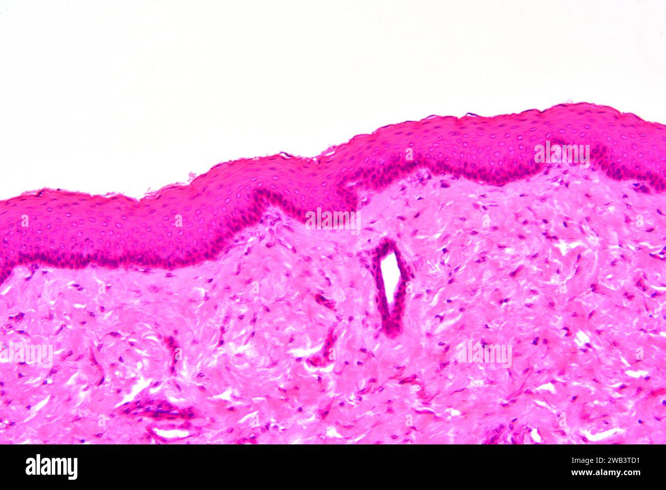 Human transitional epithelium and smooth muscle tissue. X125 at 10 cm wide. Stock Photohttps://www.alamy.com/image-license-details/?v=1https://www.alamy.com/human-transitional-epithelium-and-smooth-muscle-tissue-x125-at-10-cm-wide-image591998765.html
RF2WB3TD1–Human transitional epithelium and smooth muscle tissue. X125 at 10 cm wide.
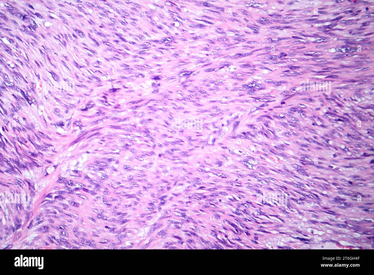 Photomicrograph of leiomyosarcoma, depicting malignant smooth muscle tumor cells, indicative of aggressive soft tissue cancer. Stock Photohttps://www.alamy.com/image-license-details/?v=1https://www.alamy.com/photomicrograph-of-leiomyosarcoma-depicting-malignant-smooth-muscle-tumor-cells-indicative-of-aggressive-soft-tissue-cancer-image571994767.html
RF2T6GH4F–Photomicrograph of leiomyosarcoma, depicting malignant smooth muscle tumor cells, indicative of aggressive soft tissue cancer.
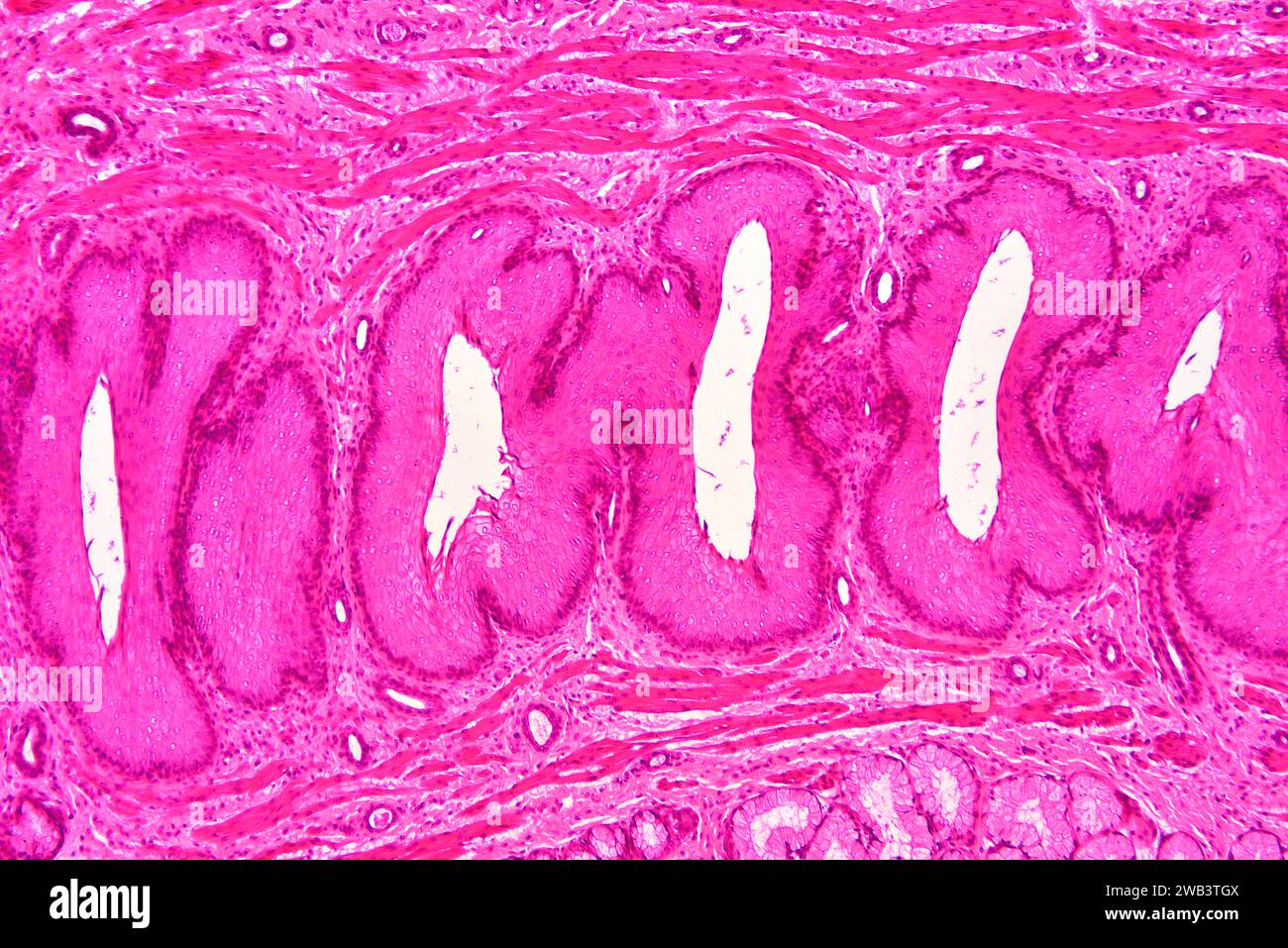
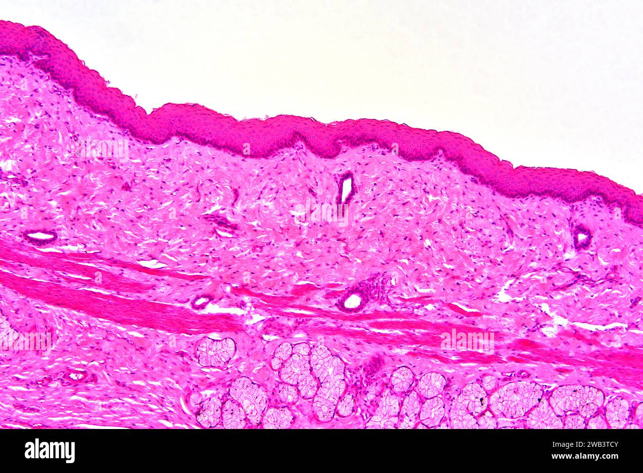 Human stratified squamous epithelium, smooth muscle and secretory gland. X75 at 10 cm wide. Stock Photohttps://www.alamy.com/image-license-details/?v=1https://www.alamy.com/human-stratified-squamous-epithelium-smooth-muscle-and-secretory-gland-x75-at-10-cm-wide-image591998763.html
RF2WB3TCY–Human stratified squamous epithelium, smooth muscle and secretory gland. X75 at 10 cm wide.
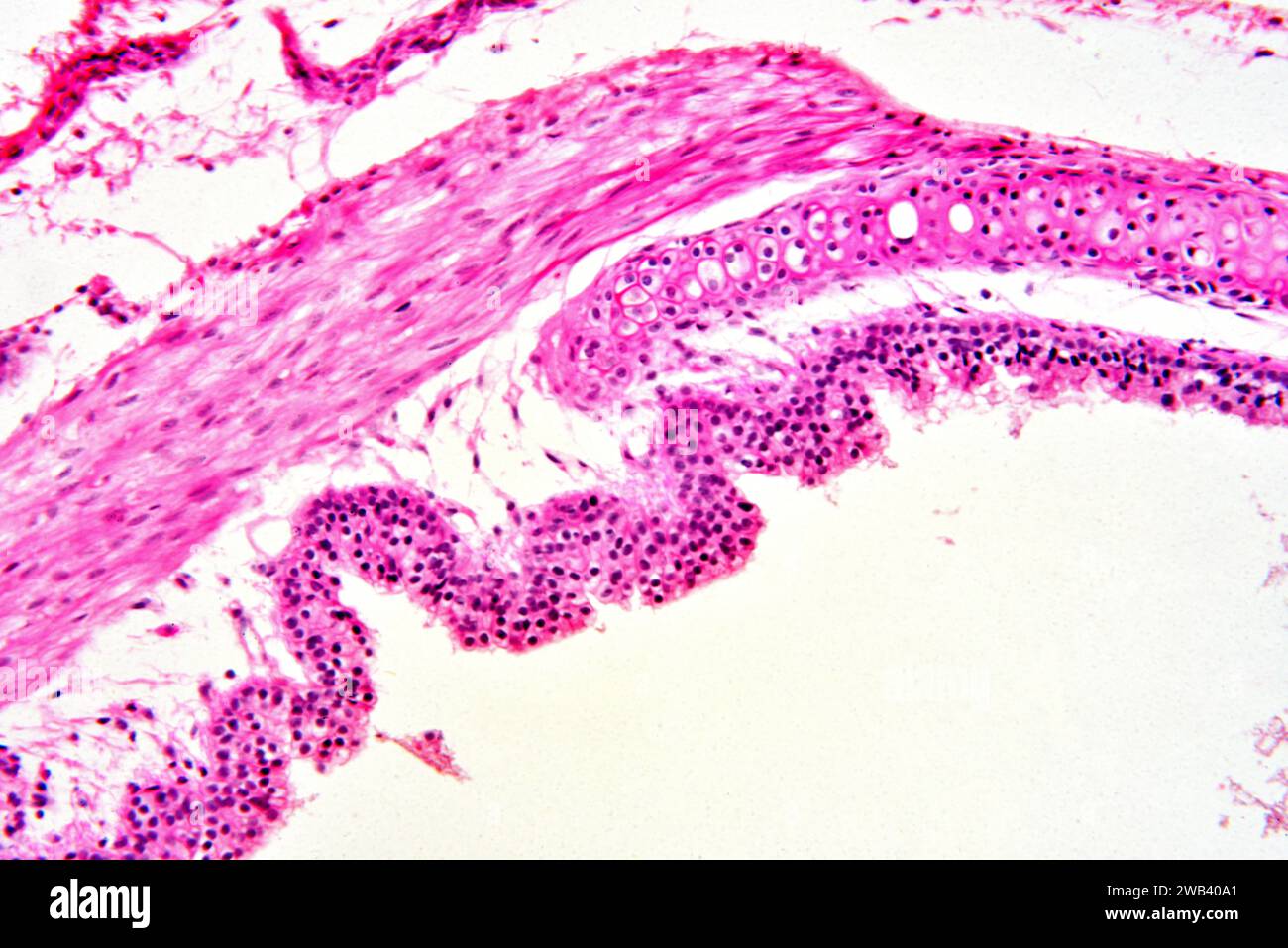 Human trachea showing hyaline cartilage, smooth muscle fibers and mucosa with epithelial tissue. X75 at 10 cm wide. Stock Photohttps://www.alamy.com/image-license-details/?v=1https://www.alamy.com/human-trachea-showing-hyaline-cartilage-smooth-muscle-fibers-and-mucosa-with-epithelial-tissue-x75-at-10-cm-wide-image592001817.html
RF2WB40A1–Human trachea showing hyaline cartilage, smooth muscle fibers and mucosa with epithelial tissue. X75 at 10 cm wide.
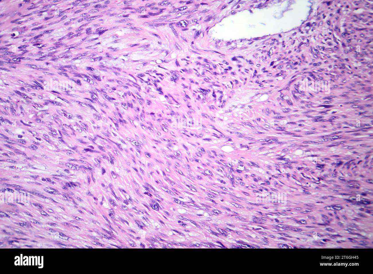 Photomicrograph of leiomyosarcoma, depicting malignant smooth muscle tumor cells, indicative of aggressive soft tissue cancer. Stock Photohttps://www.alamy.com/image-license-details/?v=1https://www.alamy.com/photomicrograph-of-leiomyosarcoma-depicting-malignant-smooth-muscle-tumor-cells-indicative-of-aggressive-soft-tissue-cancer-image571994757.html
RF2T6GH45–Photomicrograph of leiomyosarcoma, depicting malignant smooth muscle tumor cells, indicative of aggressive soft tissue cancer.
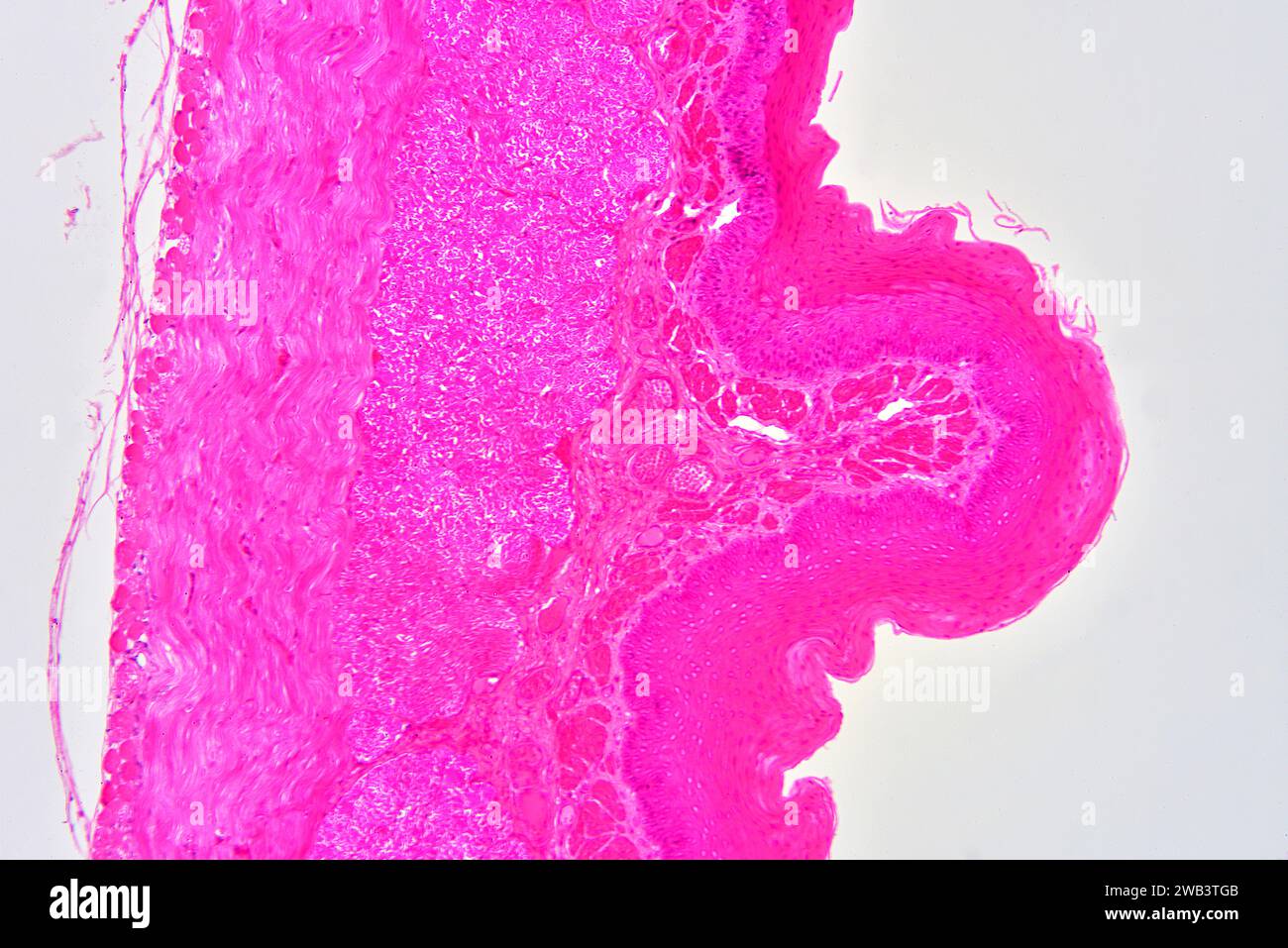 Human esophagus showing non-keratinized stratified squamous epithelium connective tissue and smooth muscle. X75 at 10 cm wide. Stock Photohttps://www.alamy.com/image-license-details/?v=1https://www.alamy.com/human-esophagus-showing-non-keratinized-stratified-squamous-epithelium-connective-tissue-and-smooth-muscle-x75-at-10-cm-wide-image591998859.html
RF2WB3TGB–Human esophagus showing non-keratinized stratified squamous epithelium connective tissue and smooth muscle. X75 at 10 cm wide.
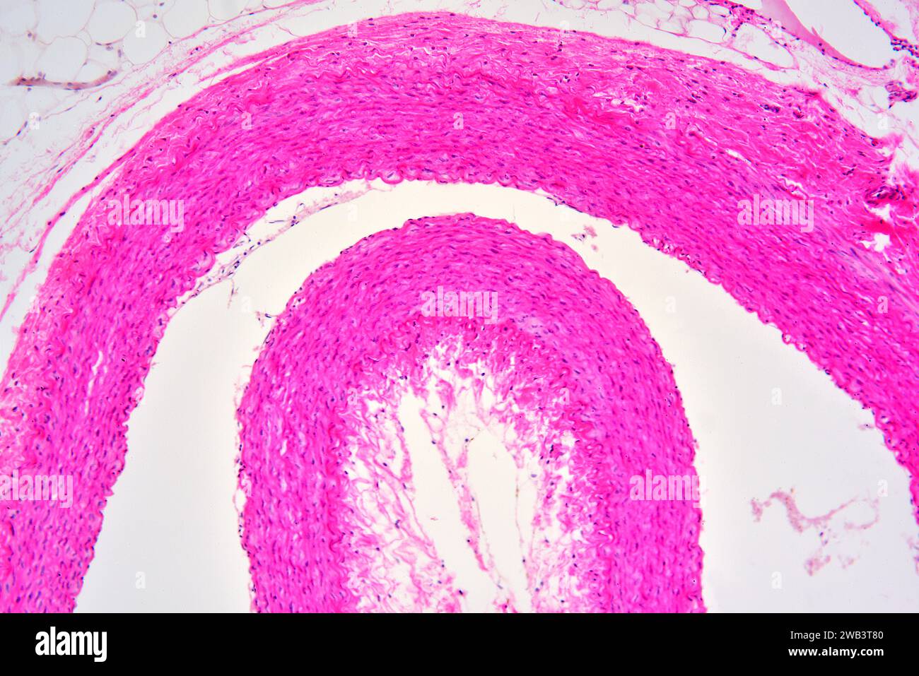 Human artery cross section showing tunica externa, tunica media with smooth muscle fibers and tunica intima. X75 at 10 cm wide. Stock Photohttps://www.alamy.com/image-license-details/?v=1https://www.alamy.com/human-artery-cross-section-showing-tunica-externa-tunica-media-with-smooth-muscle-fibers-and-tunica-intima-x75-at-10-cm-wide-image591998624.html
RF2WB3T80–Human artery cross section showing tunica externa, tunica media with smooth muscle fibers and tunica intima. X75 at 10 cm wide.
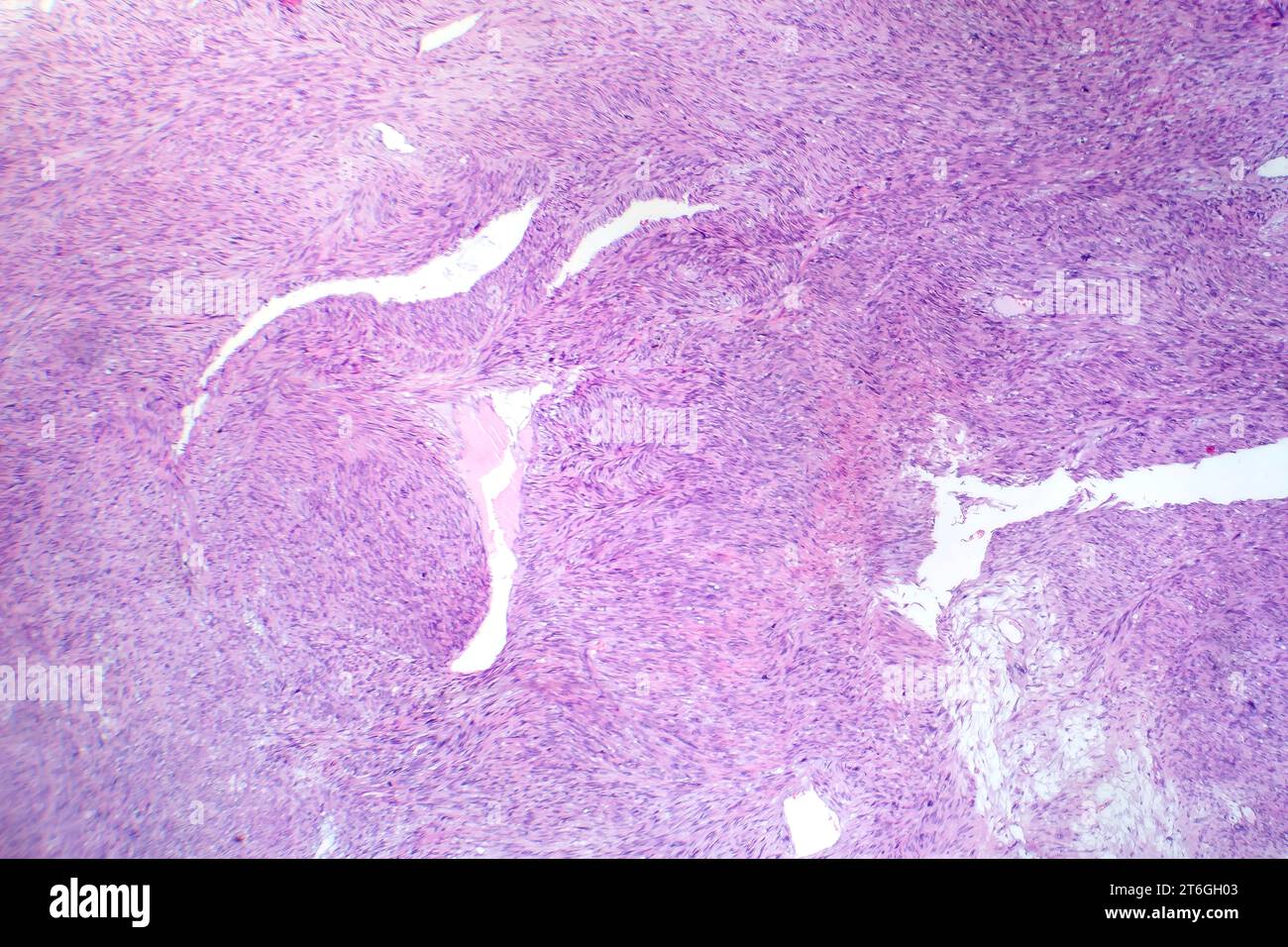 Photomicrograph of leiomyosarcoma, depicting malignant smooth muscle tumor cells, indicative of aggressive soft tissue cancer. Stock Photohttps://www.alamy.com/image-license-details/?v=1https://www.alamy.com/photomicrograph-of-leiomyosarcoma-depicting-malignant-smooth-muscle-tumor-cells-indicative-of-aggressive-soft-tissue-cancer-image571994643.html
RF2T6GH03–Photomicrograph of leiomyosarcoma, depicting malignant smooth muscle tumor cells, indicative of aggressive soft tissue cancer.
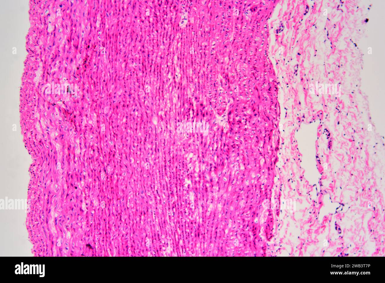 Human artery cross section showing tunica externa, tunica media with smooth muscle fibers and tunica intima. X75 at 10 cm wide. Stock Photohttps://www.alamy.com/image-license-details/?v=1https://www.alamy.com/human-artery-cross-section-showing-tunica-externa-tunica-media-with-smooth-muscle-fibers-and-tunica-intima-x75-at-10-cm-wide-image591998618.html
RF2WB3T7P–Human artery cross section showing tunica externa, tunica media with smooth muscle fibers and tunica intima. X75 at 10 cm wide.
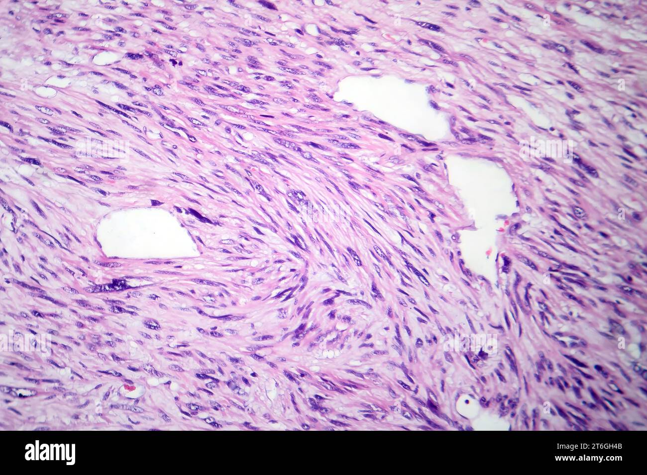 Photomicrograph of leiomyosarcoma, depicting malignant smooth muscle tumor cells, indicative of aggressive soft tissue cancer. Stock Photohttps://www.alamy.com/image-license-details/?v=1https://www.alamy.com/photomicrograph-of-leiomyosarcoma-depicting-malignant-smooth-muscle-tumor-cells-indicative-of-aggressive-soft-tissue-cancer-image571994763.html
RF2T6GH4B–Photomicrograph of leiomyosarcoma, depicting malignant smooth muscle tumor cells, indicative of aggressive soft tissue cancer.
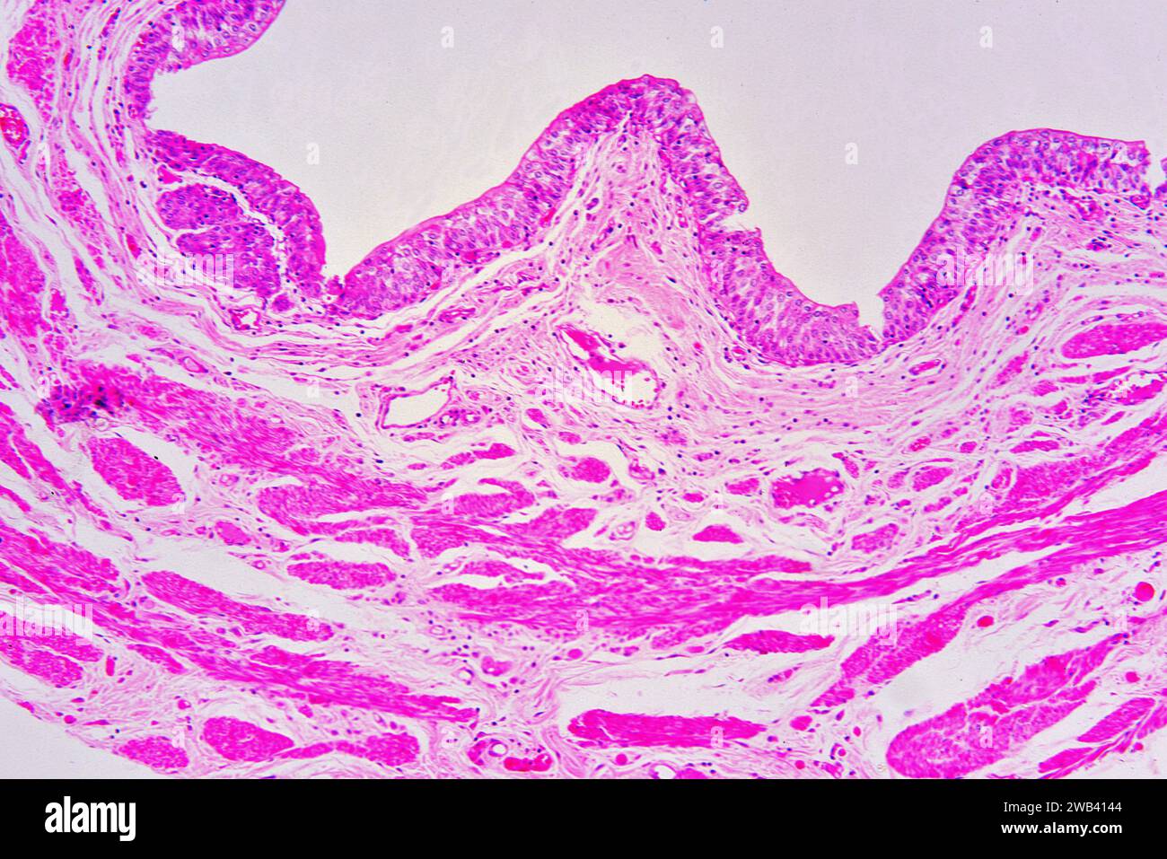 Human ureter section showing from up to down: transitional epithelium or urothelium, smooth muscle fibers and connective tissue. X75 at 10 cm wide. Stock Photohttps://www.alamy.com/image-license-details/?v=1https://www.alamy.com/human-ureter-section-showing-from-up-to-down-transitional-epithelium-or-urothelium-smooth-muscle-fibers-and-connective-tissue-x75-at-10-cm-wide-image592002436.html
RF2WB4144–Human ureter section showing from up to down: transitional epithelium or urothelium, smooth muscle fibers and connective tissue. X75 at 10 cm wide.
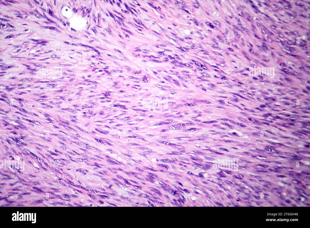 Photomicrograph of leiomyosarcoma, depicting malignant smooth muscle tumor cells, indicative of aggressive soft tissue cancer. Stock Photohttps://www.alamy.com/image-license-details/?v=1https://www.alamy.com/photomicrograph-of-leiomyosarcoma-depicting-malignant-smooth-muscle-tumor-cells-indicative-of-aggressive-soft-tissue-cancer-image571994760.html
RF2T6GH48–Photomicrograph of leiomyosarcoma, depicting malignant smooth muscle tumor cells, indicative of aggressive soft tissue cancer.
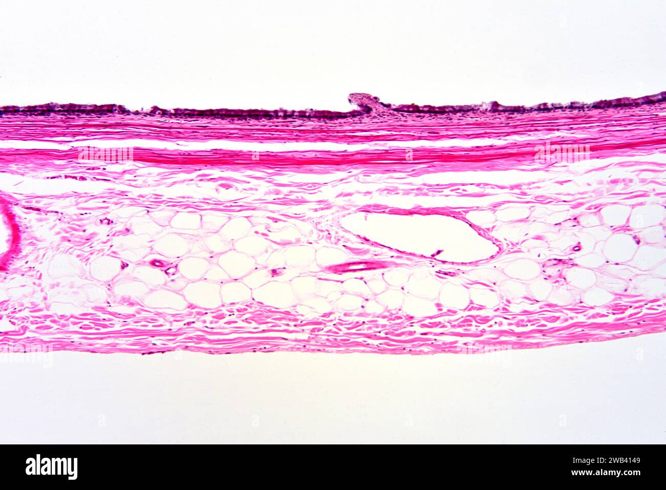 Human gallbladder wall section showing from up to down: simple columnar epithelium, lamina propria, smooth muscle fibers and connective tissue. X75 at Stock Photohttps://www.alamy.com/image-license-details/?v=1https://www.alamy.com/human-gallbladder-wall-section-showing-from-up-to-down-simple-columnar-epithelium-lamina-propria-smooth-muscle-fibers-and-connective-tissue-x75-at-image592002441.html
RF2WB4149–Human gallbladder wall section showing from up to down: simple columnar epithelium, lamina propria, smooth muscle fibers and connective tissue. X75 at
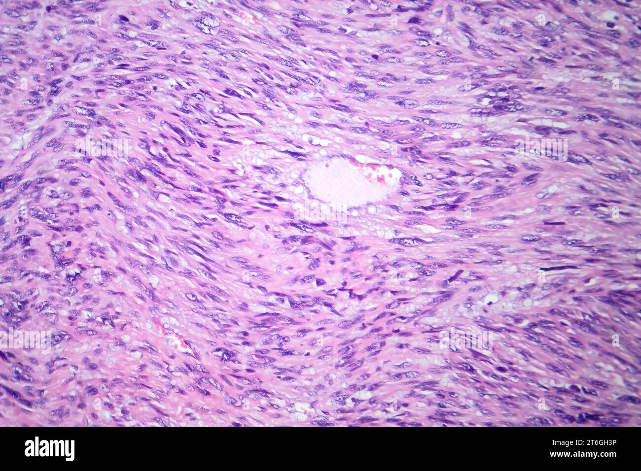 Photomicrograph of leiomyosarcoma, depicting malignant smooth muscle tumor cells, indicative of aggressive soft tissue cancer. Stock Photohttps://www.alamy.com/image-license-details/?v=1https://www.alamy.com/photomicrograph-of-leiomyosarcoma-depicting-malignant-smooth-muscle-tumor-cells-indicative-of-aggressive-soft-tissue-cancer-image571994746.html
RF2T6GH3P–Photomicrograph of leiomyosarcoma, depicting malignant smooth muscle tumor cells, indicative of aggressive soft tissue cancer.
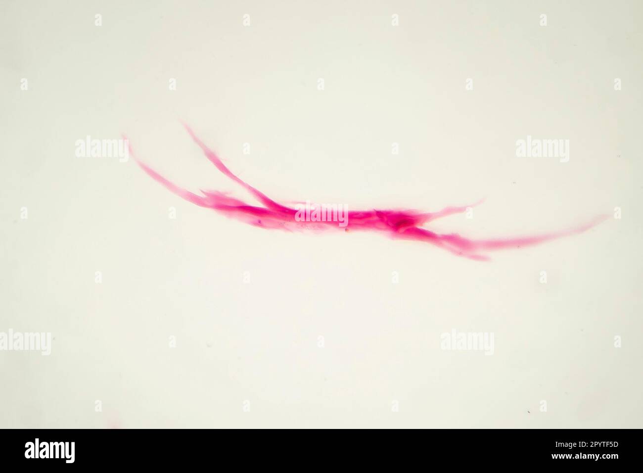 Teased human smooth muscle fiber, light micrograph Stock Photohttps://www.alamy.com/image-license-details/?v=1https://www.alamy.com/teased-human-smooth-muscle-fiber-light-micrograph-image550655881.html
RF2PYTF5D–Teased human smooth muscle fiber, light micrograph
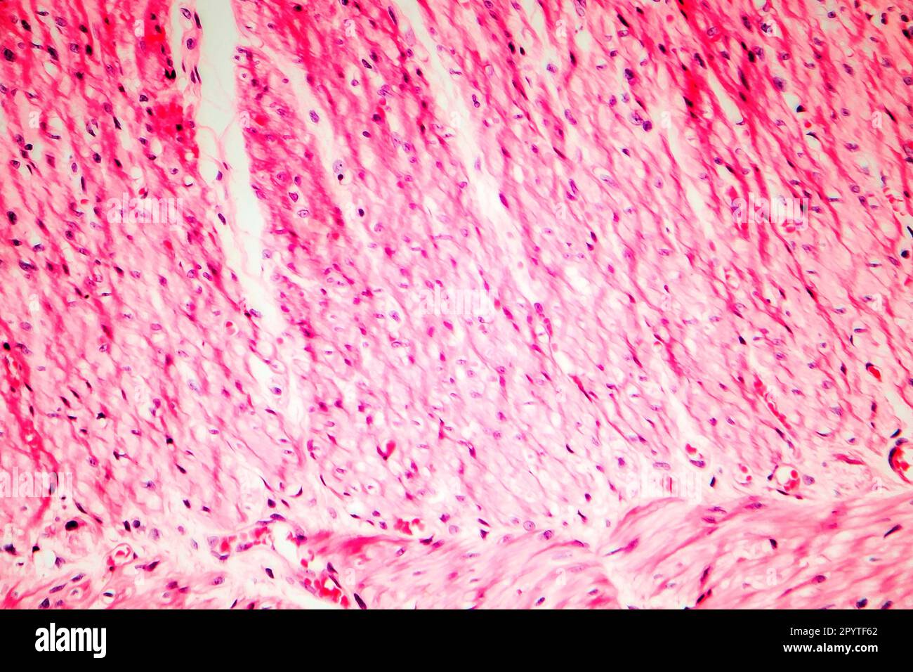 Human smooth muscle, cross section, light micrograph Stock Photohttps://www.alamy.com/image-license-details/?v=1https://www.alamy.com/human-smooth-muscle-cross-section-light-micrograph-image550655898.html
RF2PYTF62–Human smooth muscle, cross section, light micrograph
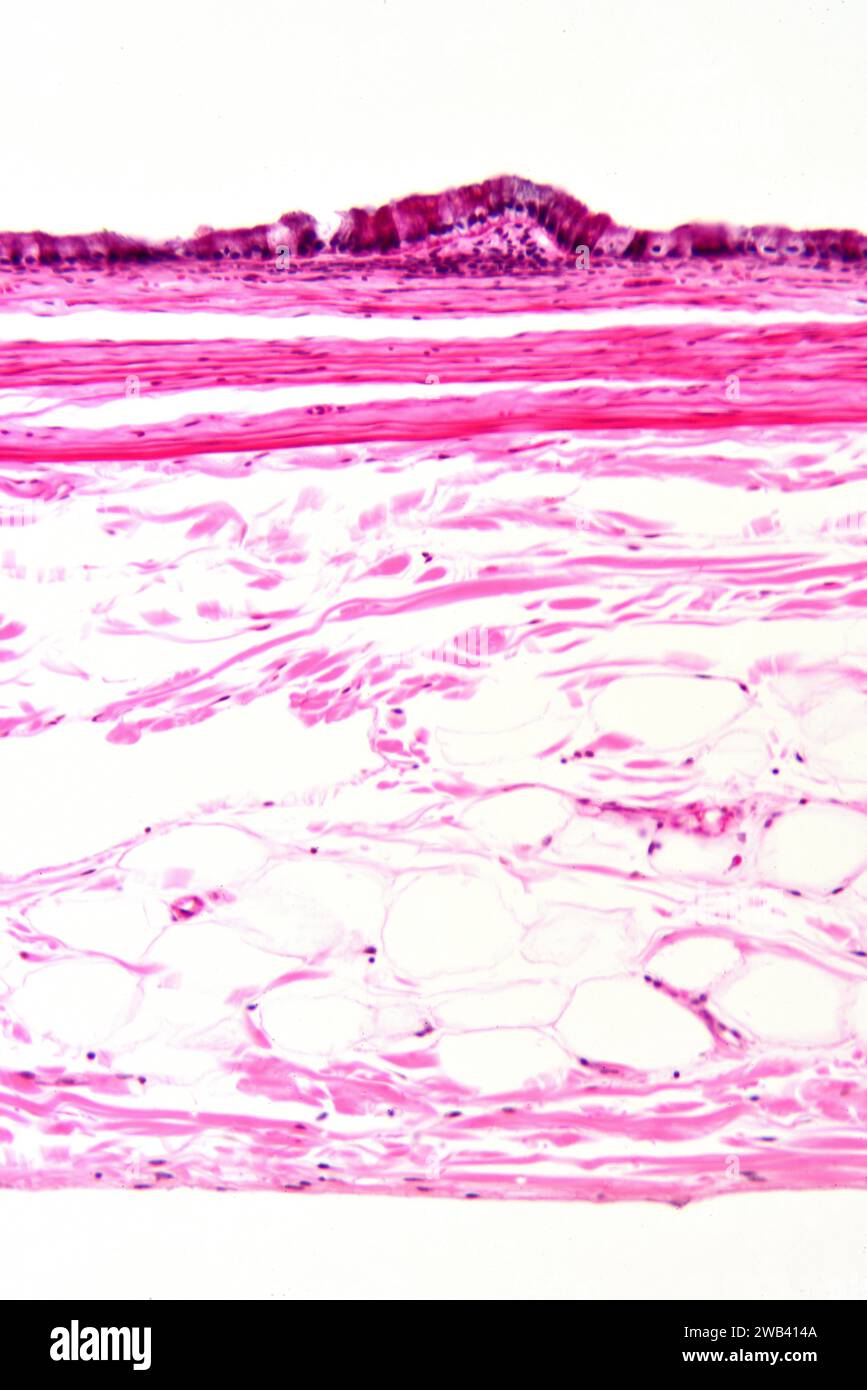 Human gallbladder wall section showing from up to down: simple columnar epithelium, lamina propria, smooth muscle fibers and connective tissue. X125 a Stock Photohttps://www.alamy.com/image-license-details/?v=1https://www.alamy.com/human-gallbladder-wall-section-showing-from-up-to-down-simple-columnar-epithelium-lamina-propria-smooth-muscle-fibers-and-connective-tissue-x125-a-image592002442.html
RF2WB414A–Human gallbladder wall section showing from up to down: simple columnar epithelium, lamina propria, smooth muscle fibers and connective tissue. X125 a
 Human umbilical cord mucosa showing: mucous glands, loose conective tissue, adipocytes, smooth muscle fibers and blood vessels. X25 at 10 cm wide. Stock Photohttps://www.alamy.com/image-license-details/?v=1https://www.alamy.com/human-umbilical-cord-mucosa-showing-mucous-glands-loose-conective-tissue-adipocytes-smooth-muscle-fibers-and-blood-vessels-x25-at-10-cm-wide-image591999377.html
RF2WB3W6W–Human umbilical cord mucosa showing: mucous glands, loose conective tissue, adipocytes, smooth muscle fibers and blood vessels. X25 at 10 cm wide.
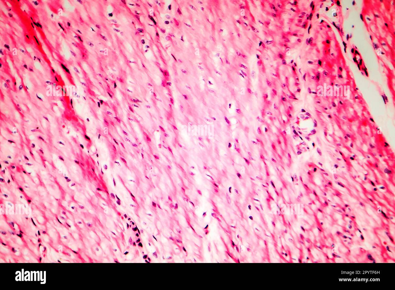 Human smooth muscle, cross section, light micrograph Stock Photohttps://www.alamy.com/image-license-details/?v=1https://www.alamy.com/human-smooth-muscle-cross-section-light-micrograph-image550655913.html
RF2PYTF6H–Human smooth muscle, cross section, light micrograph
 Human umbilical cord mucosa showing: mucous glands, loose conective tissue, adipocytes, smooth muscle fibers and blood vessels. X25 at 10 cm wide. Stock Photohttps://www.alamy.com/image-license-details/?v=1https://www.alamy.com/human-umbilical-cord-mucosa-showing-mucous-glands-loose-conective-tissue-adipocytes-smooth-muscle-fibers-and-blood-vessels-x25-at-10-cm-wide-image591999388.html
RF2WB3W78–Human umbilical cord mucosa showing: mucous glands, loose conective tissue, adipocytes, smooth muscle fibers and blood vessels. X25 at 10 cm wide.
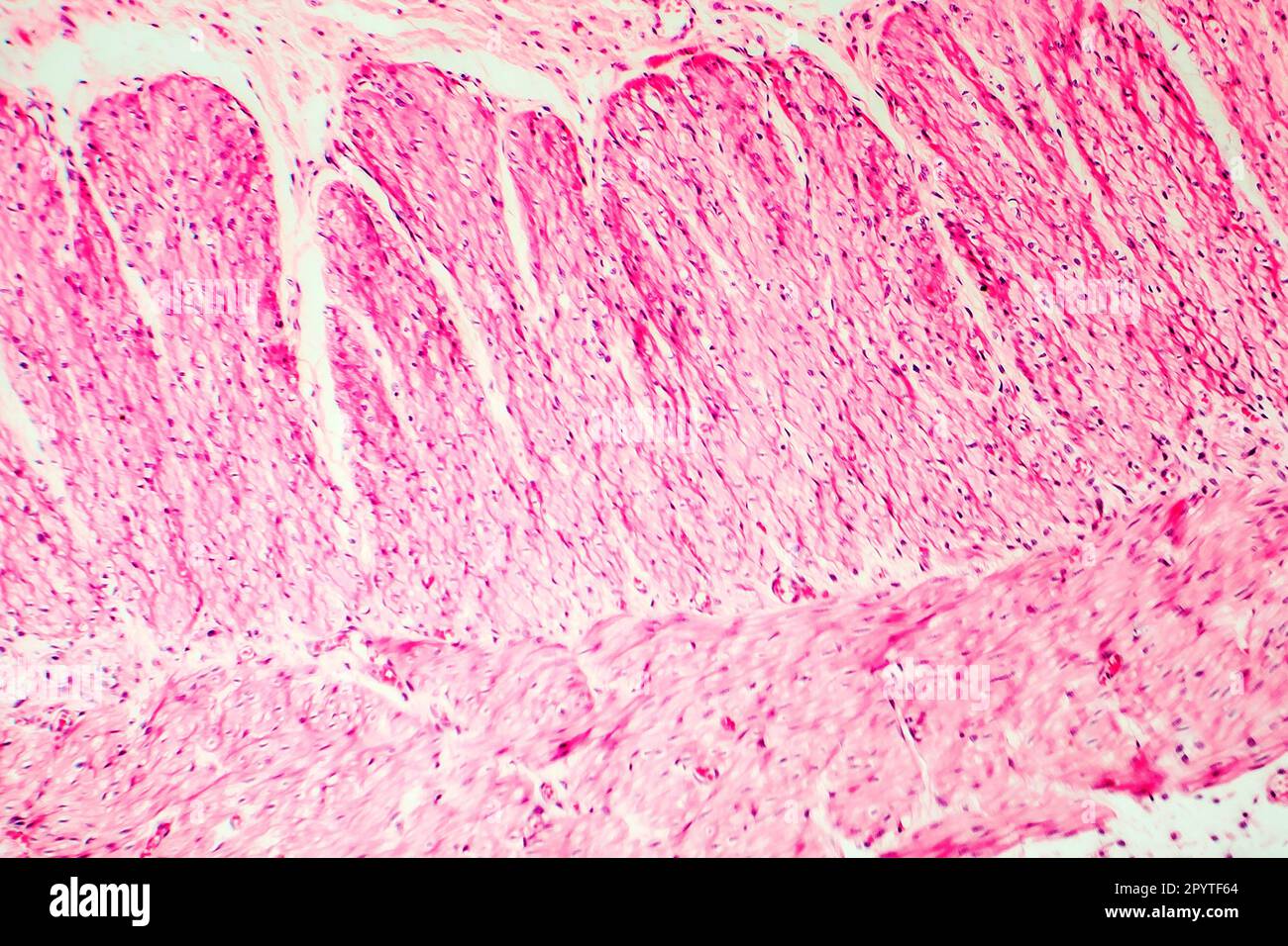 Human smooth muscle, cross section, light micrograph Stock Photohttps://www.alamy.com/image-license-details/?v=1https://www.alamy.com/human-smooth-muscle-cross-section-light-micrograph-image550655900.html
RF2PYTF64–Human smooth muscle, cross section, light micrograph
 Human umbilical cord mucosa showing: mucous glands, loose conective tissue, adipocytes, smooth muscle fibers and blood vessels. X25 at 10 cm wide. Stock Photohttps://www.alamy.com/image-license-details/?v=1https://www.alamy.com/human-umbilical-cord-mucosa-showing-mucous-glands-loose-conective-tissue-adipocytes-smooth-muscle-fibers-and-blood-vessels-x25-at-10-cm-wide-image591999382.html
RF2WB3W72–Human umbilical cord mucosa showing: mucous glands, loose conective tissue, adipocytes, smooth muscle fibers and blood vessels. X25 at 10 cm wide.
 Human umbilical cord mucosa showing: mucous glands, loose conective tissue, adipocytes, smooth muscle fibers and blood vessels. X25 at 10 cm wide. Stock Photohttps://www.alamy.com/image-license-details/?v=1https://www.alamy.com/human-umbilical-cord-mucosa-showing-mucous-glands-loose-conective-tissue-adipocytes-smooth-muscle-fibers-and-blood-vessels-x25-at-10-cm-wide-image591999374.html
RF2WB3W6P–Human umbilical cord mucosa showing: mucous glands, loose conective tissue, adipocytes, smooth muscle fibers and blood vessels. X25 at 10 cm wide.
 Human umbilical cord mucosa showing: mucous glands, loose conective tissue, adipocytes, smooth muscle fibers and blood vessels. X75 at 10 cm wide. Stock Photohttps://www.alamy.com/image-license-details/?v=1https://www.alamy.com/human-umbilical-cord-mucosa-showing-mucous-glands-loose-conective-tissue-adipocytes-smooth-muscle-fibers-and-blood-vessels-x75-at-10-cm-wide-image591999396.html
RF2WB3W7G–Human umbilical cord mucosa showing: mucous glands, loose conective tissue, adipocytes, smooth muscle fibers and blood vessels. X75 at 10 cm wide.
 Human umbilical cord mucosa showing: mucous glands, loose conective tissue, adipocytes, smooth muscle fibers and blood vessels. X125 at 10 cm wide. Stock Photohttps://www.alamy.com/image-license-details/?v=1https://www.alamy.com/human-umbilical-cord-mucosa-showing-mucous-glands-loose-conective-tissue-adipocytes-smooth-muscle-fibers-and-blood-vessels-x125-at-10-cm-wide-image591999399.html
RF2WB3W7K–Human umbilical cord mucosa showing: mucous glands, loose conective tissue, adipocytes, smooth muscle fibers and blood vessels. X125 at 10 cm wide.
 Human umbilical cord mucosa showing: mucous glands, loose conective tissue, adipocytes, smooth muscle fibers and blood vessels. X25 at 10 cm wide. Stock Photohttps://www.alamy.com/image-license-details/?v=1https://www.alamy.com/human-umbilical-cord-mucosa-showing-mucous-glands-loose-conective-tissue-adipocytes-smooth-muscle-fibers-and-blood-vessels-x25-at-10-cm-wide-image591999385.html
RF2WB3W75–Human umbilical cord mucosa showing: mucous glands, loose conective tissue, adipocytes, smooth muscle fibers and blood vessels. X25 at 10 cm wide.
 Human umbilical cord mucosa showing: mucous glands, loose conective tissue, adipocytes, smooth muscle fibers and blood vessels. X75 at 10 cm wide. Stock Photohttps://www.alamy.com/image-license-details/?v=1https://www.alamy.com/human-umbilical-cord-mucosa-showing-mucous-glands-loose-conective-tissue-adipocytes-smooth-muscle-fibers-and-blood-vessels-x75-at-10-cm-wide-image591999392.html
RF2WB3W7C–Human umbilical cord mucosa showing: mucous glands, loose conective tissue, adipocytes, smooth muscle fibers and blood vessels. X75 at 10 cm wide.
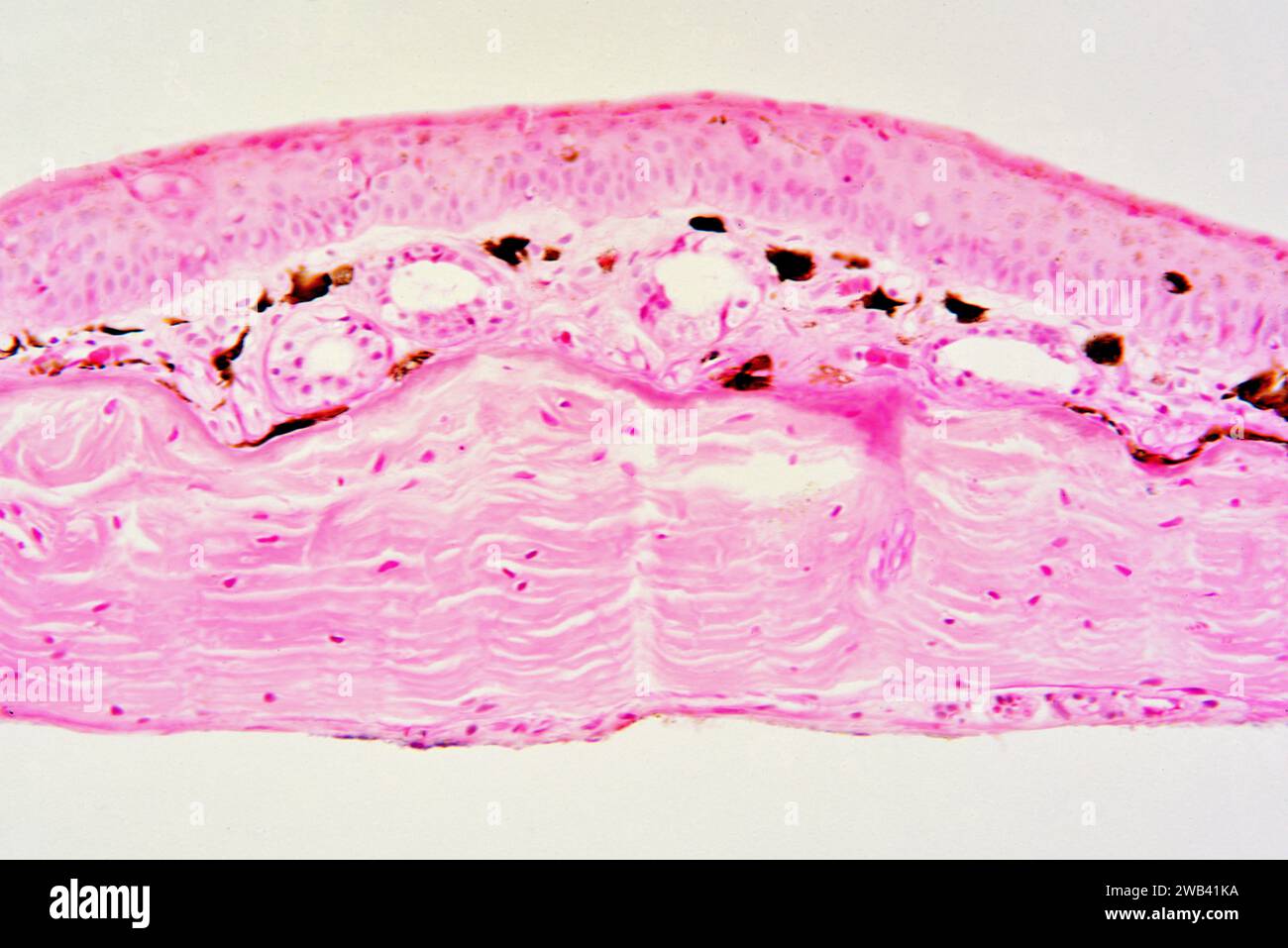 Frog skin showing epidermis, pigmentary layer with melanocytes (brown), blood vessels and dermis. Photomicrograph X150 at 10 cm wide. Stock Photohttps://www.alamy.com/image-license-details/?v=1https://www.alamy.com/frog-skin-showing-epidermis-pigmentary-layer-with-melanocytes-brown-blood-vessels-and-dermis-photomicrograph-x150-at-10-cm-wide-image592002862.html
RF2WB41KA–Frog skin showing epidermis, pigmentary layer with melanocytes (brown), blood vessels and dermis. Photomicrograph X150 at 10 cm wide.
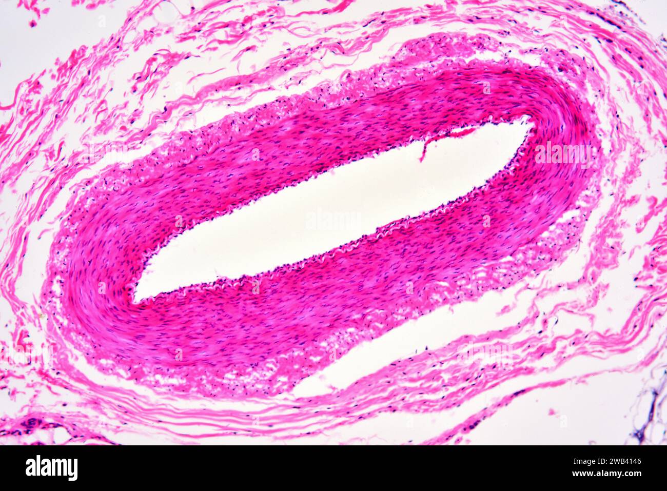 Human arteriole showing from inside to outside: endothelium, smooth mucle fibers and tunica. X75 at 10 cm wide. Stock Photohttps://www.alamy.com/image-license-details/?v=1https://www.alamy.com/human-arteriole-showing-from-inside-to-outside-endothelium-smooth-mucle-fibers-and-tunica-x75-at-10-cm-wide-image592002438.html
RF2WB4146–Human arteriole showing from inside to outside: endothelium, smooth mucle fibers and tunica. X75 at 10 cm wide.
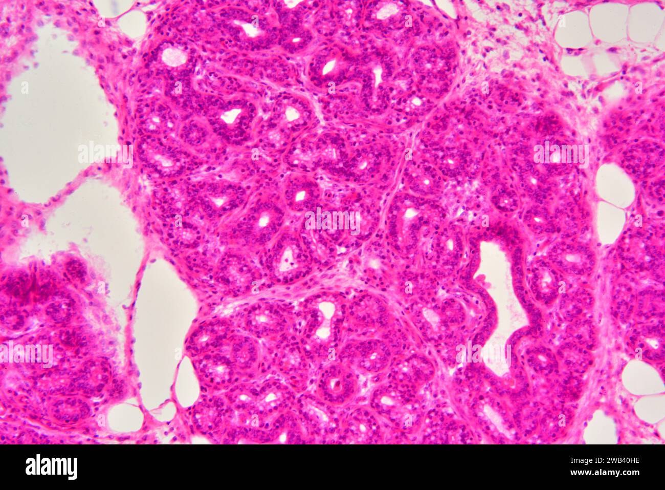 Human glandular epithelium. X125 at 10 cm wide. Stock Photohttps://www.alamy.com/image-license-details/?v=1https://www.alamy.com/human-glandular-epithelium-x125-at-10-cm-wide-image592002026.html
RF2WB40HE–Human glandular epithelium. X125 at 10 cm wide.
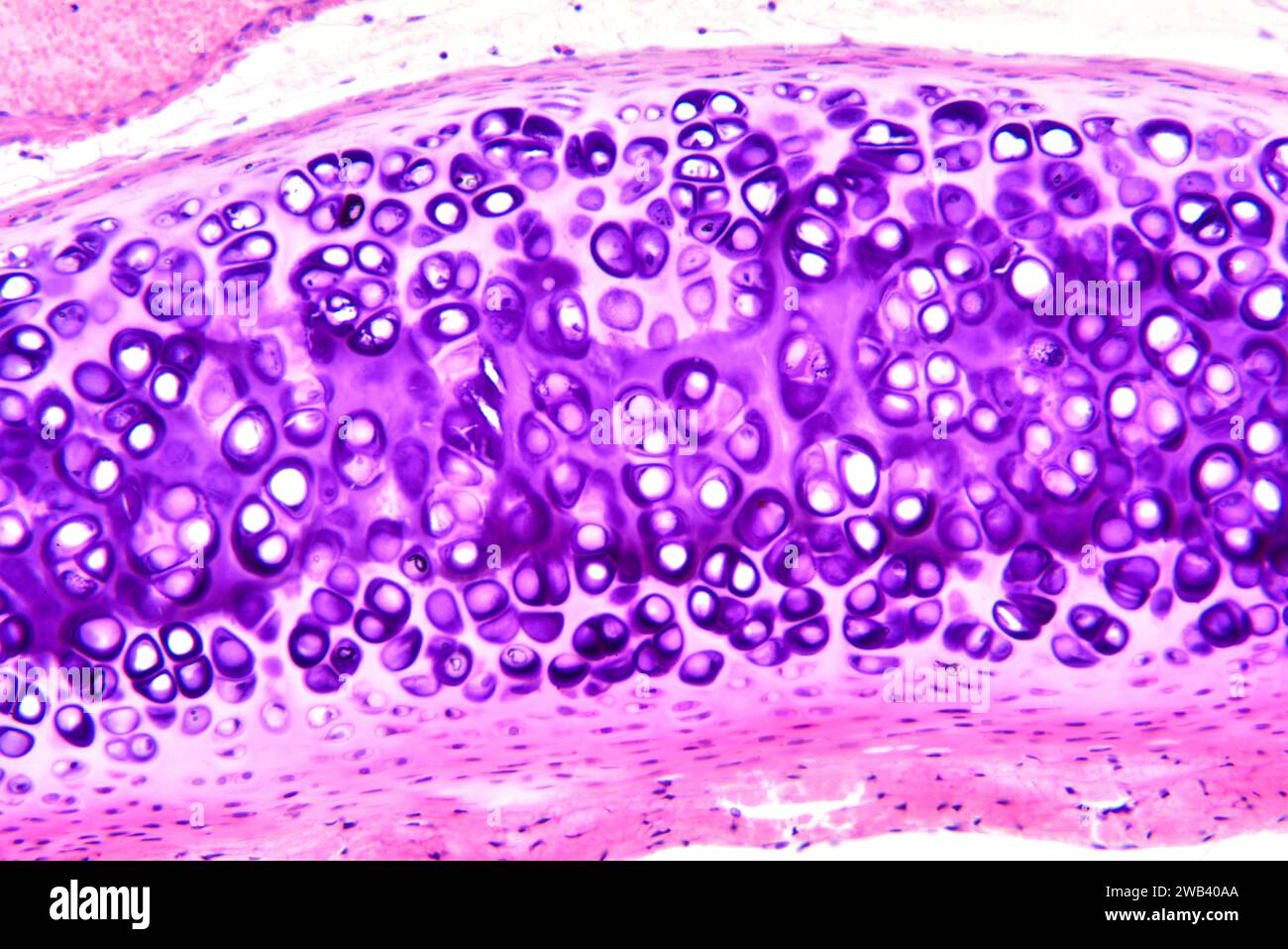 Human hyaline cartilage. X125 at 10 cm wide. Stock Photohttps://www.alamy.com/image-license-details/?v=1https://www.alamy.com/human-hyaline-cartilage-x125-at-10-cm-wide-image592001826.html
RF2WB40AA–Human hyaline cartilage. X125 at 10 cm wide.

