Special pathological anatomy Stock Photos and Images
(13)See special pathological anatomy stock video clipsSpecial pathological anatomy Stock Photos and Images
 A text-book of special pathological anatomy : Ziegler, Ernst, 1849-1905 Stock Photohttps://www.alamy.com/image-license-details/?v=1https://www.alamy.com/a-text-book-of-special-pathological-anatomy-ziegler-ernst-1849-1905-image261426378.html
RMW5900A–A text-book of special pathological anatomy : Ziegler, Ernst, 1849-1905
![Manual of pathological anatomy . bercle in the brain. These changbs have been observed after various lesions of thebrain, as embolism, tubercle, &c., but not after recent apo]3lexy.They also occur in consequence of special lesions of the cord itself(from tumours, &c.) below the part affected. In the degenerated portions there was seen, beside the atrophyand fatty degeneration of the nerve fibres, what appeared to be anmcrease of the connective tissue, which must, of course, have beensecondary to the nervous atroph3^ The ultimate result is probablysimple atrophy, and thus it is only in an inter Stock Photo Manual of pathological anatomy . bercle in the brain. These changbs have been observed after various lesions of thebrain, as embolism, tubercle, &c., but not after recent apo]3lexy.They also occur in consequence of special lesions of the cord itself(from tumours, &c.) below the part affected. In the degenerated portions there was seen, beside the atrophyand fatty degeneration of the nerve fibres, what appeared to be anmcrease of the connective tissue, which must, of course, have beensecondary to the nervous atroph3^ The ultimate result is probablysimple atrophy, and thus it is only in an inter Stock Photo](https://c8.alamy.com/comp/2AMXRM0/manual-of-pathological-anatomy-bercle-in-the-brain-these-changbs-have-been-observed-after-various-lesions-of-thebrain-as-embolism-tubercle-c-but-not-after-recent-apo-3lexythey-also-occur-in-consequence-of-special-lesions-of-the-cord-itselffrom-tumours-c-below-the-part-affected-in-the-degenerated-portions-there-was-seen-beside-the-atrophyand-fatty-degeneration-of-the-nerve-fibres-what-appeared-to-be-anmcrease-of-the-connective-tissue-which-must-of-course-have-beensecondary-to-the-nervous-atroph3-the-ultimate-result-is-probablysimple-atrophy-and-thus-it-is-only-in-an-inter-2AMXRM0.jpg) Manual of pathological anatomy . bercle in the brain. These changbs have been observed after various lesions of thebrain, as embolism, tubercle, &c., but not after recent apo]3lexy.They also occur in consequence of special lesions of the cord itself(from tumours, &c.) below the part affected. In the degenerated portions there was seen, beside the atrophyand fatty degeneration of the nerve fibres, what appeared to be anmcrease of the connective tissue, which must, of course, have beensecondary to the nervous atroph3^ The ultimate result is probablysimple atrophy, and thus it is only in an inter Stock Photohttps://www.alamy.com/image-license-details/?v=1https://www.alamy.com/manual-of-pathological-anatomy-bercle-in-the-brain-these-changbs-have-been-observed-after-various-lesions-of-thebrain-as-embolism-tubercle-c-but-not-after-recent-apo-3lexythey-also-occur-in-consequence-of-special-lesions-of-the-cord-itselffrom-tumours-c-below-the-part-affected-in-the-degenerated-portions-there-was-seen-beside-the-atrophyand-fatty-degeneration-of-the-nerve-fibres-what-appeared-to-be-anmcrease-of-the-connective-tissue-which-must-of-course-have-beensecondary-to-the-nervous-atroph3-the-ultimate-result-is-probablysimple-atrophy-and-thus-it-is-only-in-an-inter-image339879456.html
Manual of pathological anatomy . bercle in the brain. These changbs have been observed after various lesions of thebrain, as embolism, tubercle, &c., but not after recent apo]3lexy.They also occur in consequence of special lesions of the cord itself(from tumours, &c.) below the part affected. In the degenerated portions there was seen, beside the atrophyand fatty degeneration of the nerve fibres, what appeared to be anmcrease of the connective tissue, which must, of course, have beensecondary to the nervous atroph3^ The ultimate result is probablysimple atrophy, and thus it is only in an inter Stock Photohttps://www.alamy.com/image-license-details/?v=1https://www.alamy.com/manual-of-pathological-anatomy-bercle-in-the-brain-these-changbs-have-been-observed-after-various-lesions-of-thebrain-as-embolism-tubercle-c-but-not-after-recent-apo-3lexythey-also-occur-in-consequence-of-special-lesions-of-the-cord-itselffrom-tumours-c-below-the-part-affected-in-the-degenerated-portions-there-was-seen-beside-the-atrophyand-fatty-degeneration-of-the-nerve-fibres-what-appeared-to-be-anmcrease-of-the-connective-tissue-which-must-of-course-have-beensecondary-to-the-nervous-atroph3-the-ultimate-result-is-probablysimple-atrophy-and-thus-it-is-only-in-an-inter-image339879456.htmlRM2AMXRM0–Manual of pathological anatomy . bercle in the brain. These changbs have been observed after various lesions of thebrain, as embolism, tubercle, &c., but not after recent apo]3lexy.They also occur in consequence of special lesions of the cord itself(from tumours, &c.) below the part affected. In the degenerated portions there was seen, beside the atrophyand fatty degeneration of the nerve fibres, what appeared to be anmcrease of the connective tissue, which must, of course, have beensecondary to the nervous atroph3^ The ultimate result is probablysimple atrophy, and thus it is only in an inter
 GERMANY, Berlin - CIRCA 1953: a postage stamp from Germany, Berlin showing Men from the history of Berlin: Rudolf Virchow Stock Photohttps://www.alamy.com/image-license-details/?v=1https://www.alamy.com/germany-berlin-circa-1953-a-postage-stamp-from-germany-berlin-showing-men-from-the-history-of-berlin-rudolf-virchow-image451360808.html
RF2H6978T–GERMANY, Berlin - CIRCA 1953: a postage stamp from Germany, Berlin showing Men from the history of Berlin: Rudolf Virchow
 April 1, 2023 Exhibition of anatomical exhibits. Rabbit with pathological development of cyclopia. Exhibits of the Museum of Anthropology and Ethnogra Stock Photohttps://www.alamy.com/image-license-details/?v=1https://www.alamy.com/april-1-2023-exhibition-of-anatomical-exhibits-rabbit-with-pathological-development-of-cyclopia-exhibits-of-the-museum-of-anthropology-and-ethnogra-image559985813.html
RM2RF1FH9–April 1, 2023 Exhibition of anatomical exhibits. Rabbit with pathological development of cyclopia. Exhibits of the Museum of Anthropology and Ethnogra
 A text-book of special pathological anatomy : Ziegler, Ernst, 1849-1905 Stock Photohttps://www.alamy.com/image-license-details/?v=1https://www.alamy.com/a-text-book-of-special-pathological-anatomy-ziegler-ernst-1849-1905-image261424587.html
RMW58WMB–A text-book of special pathological anatomy : Ziegler, Ernst, 1849-1905
 Manual of pathological anatomy . or special changein the medullary cells; the generalappearance being rather that of passivethan of active Haversian canals theoutwards, and from the cancelli andfrom the medullary cavity also out-wards, till only a thin shell of bone isleft, which perhaps escapes becausenourished by the periosteum. It seems,then, that the process is clearly oneconnected with the medullaprecisely in what way is not known. As the bones of the trunk are espe-cially liable to be attacked, the indi-vidual affected becomes reduced in sizefrom the collapse of the vertebralcolumn. A Stock Photohttps://www.alamy.com/image-license-details/?v=1https://www.alamy.com/manual-of-pathological-anatomy-or-special-changein-the-medullary-cells-the-generalappearance-being-rather-that-of-passivethan-of-active-haversian-canals-theoutwards-and-from-the-cancelli-andfrom-the-medullary-cavity-also-out-wards-till-only-a-thin-shell-of-bone-isleft-which-perhaps-escapes-becausenourished-by-the-periosteum-it-seemsthen-that-the-process-is-clearly-oneconnected-with-the-medullaprecisely-in-what-way-is-not-known-as-the-bones-of-the-trunk-are-espe-cially-liable-to-be-attacked-the-indi-vidual-affected-becomes-reduced-in-sizefrom-the-collapse-of-the-vertebralcolumn-a-image339440571.html
RM2AM6RWF–Manual of pathological anatomy . or special changein the medullary cells; the generalappearance being rather that of passivethan of active Haversian canals theoutwards, and from the cancelli andfrom the medullary cavity also out-wards, till only a thin shell of bone isleft, which perhaps escapes becausenourished by the periosteum. It seems,then, that the process is clearly oneconnected with the medullaprecisely in what way is not known. As the bones of the trunk are espe-cially liable to be attacked, the indi-vidual affected becomes reduced in sizefrom the collapse of the vertebralcolumn. A
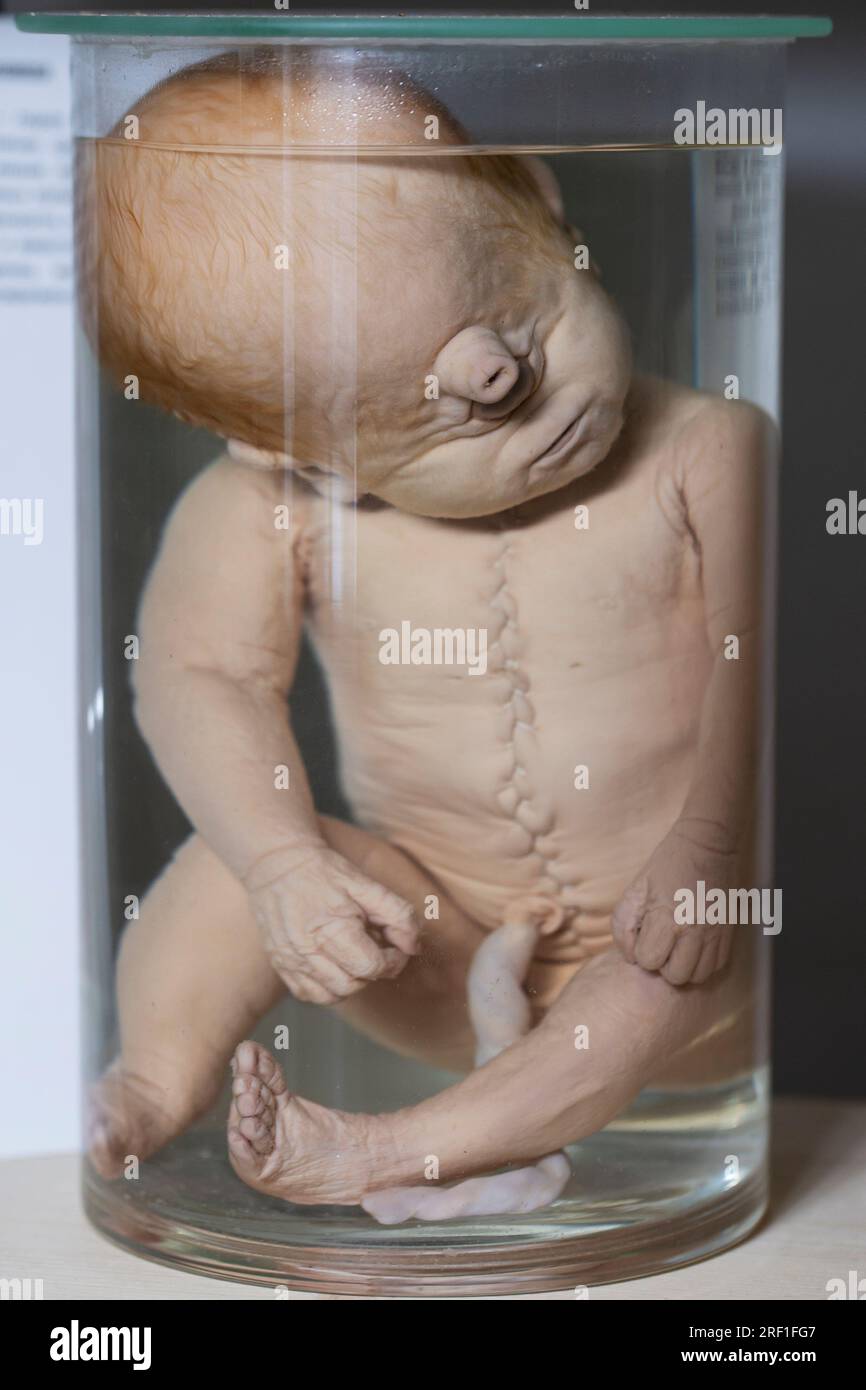
 April 1, 2023 Exhibition of anatomical exhibits. Human infant with pathological development of cyclopia. Exhibits of the Museum of Anthropology and Et Stock Photohttps://www.alamy.com/image-license-details/?v=1https://www.alamy.com/april-1-2023-exhibition-of-anatomical-exhibits-human-infant-with-pathological-development-of-cyclopia-exhibits-of-the-museum-of-anthropology-and-et-image559985783.html
RM2RF1FG7–April 1, 2023 Exhibition of anatomical exhibits. Human infant with pathological development of cyclopia. Exhibits of the Museum of Anthropology and Et
 Manual of pathological anatomy . istinct affection such as that which has beentermed glandular or follicular enteritis. Dr. Copland speaks of itas occurring almost always consecutively to other diseases, asfevers continued or remittent, dysentery, and even tuberculosis.Eokitansky does not seeoa to recognize its special character, but toconsider that the follicles may be more particularly affected inmorbid processes of different kinds. In this opinion we entirelycoincide, but wish to notice here certain points in the anatomical 586 INFLAMMATION OF THE INTESTINE. structure of these parts, wliicl Stock Photohttps://www.alamy.com/image-license-details/?v=1https://www.alamy.com/manual-of-pathological-anatomy-istinct-affection-such-as-that-which-has-beentermed-glandular-or-follicular-enteritis-dr-copland-speaks-of-itas-occurring-almost-always-consecutively-to-other-diseases-asfevers-continued-or-remittent-dysentery-and-even-tuberculosiseokitansky-does-not-seeoa-to-recognize-its-special-character-but-toconsider-that-the-follicles-may-be-more-particularly-affected-inmorbid-processes-of-different-kinds-in-this-opinion-we-entirelycoincide-but-wish-to-notice-here-certain-points-in-the-anatomical-586-inflammation-of-the-intestine-structure-of-these-parts-wliicl-image339463530.html
RM2AM7W5E–Manual of pathological anatomy . istinct affection such as that which has beentermed glandular or follicular enteritis. Dr. Copland speaks of itas occurring almost always consecutively to other diseases, asfevers continued or remittent, dysentery, and even tuberculosis.Eokitansky does not seeoa to recognize its special character, but toconsider that the follicles may be more particularly affected inmorbid processes of different kinds. In this opinion we entirelycoincide, but wish to notice here certain points in the anatomical 586 INFLAMMATION OF THE INTESTINE. structure of these parts, wliicl
 . The American journal of anatomy. osed specificity of the epi-thelium, which is in all respects similar to that of the oesophagus.In view of the great plasticity of epithelium of a low grade of special-ization, little importance can be attached to the argument because,both pathological histology and normal histology afford many in-stances of the transformation of a cylindrical or a ciliated epitheliuminto a stratified epithelium, as a result of the operation of simplemechanical causes, such as friction. Such an instance has been notedby Haycraft and Carlier, 89-90, in the trachea of the cat. Stock Photohttps://www.alamy.com/image-license-details/?v=1https://www.alamy.com/the-american-journal-of-anatomy-osed-specificity-of-the-epi-thelium-which-is-in-all-respects-similar-to-that-of-the-oesophagusin-view-of-the-great-plasticity-of-epithelium-of-a-low-grade-of-special-ization-little-importance-can-be-attached-to-the-argument-becauseboth-pathological-histology-and-normal-histology-afford-many-in-stances-of-the-transformation-of-a-cylindrical-or-a-ciliated-epitheliuminto-a-stratified-epithelium-as-a-result-of-the-operation-of-simplemechanical-causes-such-as-friction-such-an-instance-has-been-notedby-haycraft-and-carlier-89-90-in-the-trachea-of-the-cat-image336920548.html
RM2AG41GM–. The American journal of anatomy. osed specificity of the epi-thelium, which is in all respects similar to that of the oesophagus.In view of the great plasticity of epithelium of a low grade of special-ization, little importance can be attached to the argument because,both pathological histology and normal histology afford many in-stances of the transformation of a cylindrical or a ciliated epitheliuminto a stratified epithelium, as a result of the operation of simplemechanical causes, such as friction. Such an instance has been notedby Haycraft and Carlier, 89-90, in the trachea of the cat.
 Manual of pathological anatomy . n accompanies general gout; some-times when no symptoms of gout have occurred, similar depositswill be found in the articular cartilages of the great toe. Some-times it is alleged to be the solitary gouty deposit in the body. Tomake a special form of kidnej^ disease—arthritic nephritis—onthese appearances, as has been done by some French and Germanpathologists, appears to us quite unnecessary; since in London,at least, many of the most pronounced granular kidneys are gouty.A certain proportion of the gouty cases are associated with leadpoisoning. CYSTS IN THE K Stock Photohttps://www.alamy.com/image-license-details/?v=1https://www.alamy.com/manual-of-pathological-anatomy-n-accompanies-general-gout-some-times-when-no-symptoms-of-gout-have-occurred-similar-depositswill-be-found-in-the-articular-cartilages-of-the-great-toe-some-times-it-is-alleged-to-be-the-solitary-gouty-deposit-in-the-body-tomake-a-special-form-of-kidnej-diseasearthritic-nephritisonthese-appearances-as-has-been-done-by-some-french-and-germanpathologists-appears-to-us-quite-unnecessary-since-in-londonat-least-many-of-the-most-pronounced-granular-kidneys-are-goutya-certain-proportion-of-the-gouty-cases-are-associated-with-leadpoisoning-cysts-in-the-k-image339451045.html
RM2AM797H–Manual of pathological anatomy . n accompanies general gout; some-times when no symptoms of gout have occurred, similar depositswill be found in the articular cartilages of the great toe. Some-times it is alleged to be the solitary gouty deposit in the body. Tomake a special form of kidnej^ disease—arthritic nephritis—onthese appearances, as has been done by some French and Germanpathologists, appears to us quite unnecessary; since in London,at least, many of the most pronounced granular kidneys are gouty.A certain proportion of the gouty cases are associated with leadpoisoning. CYSTS IN THE K
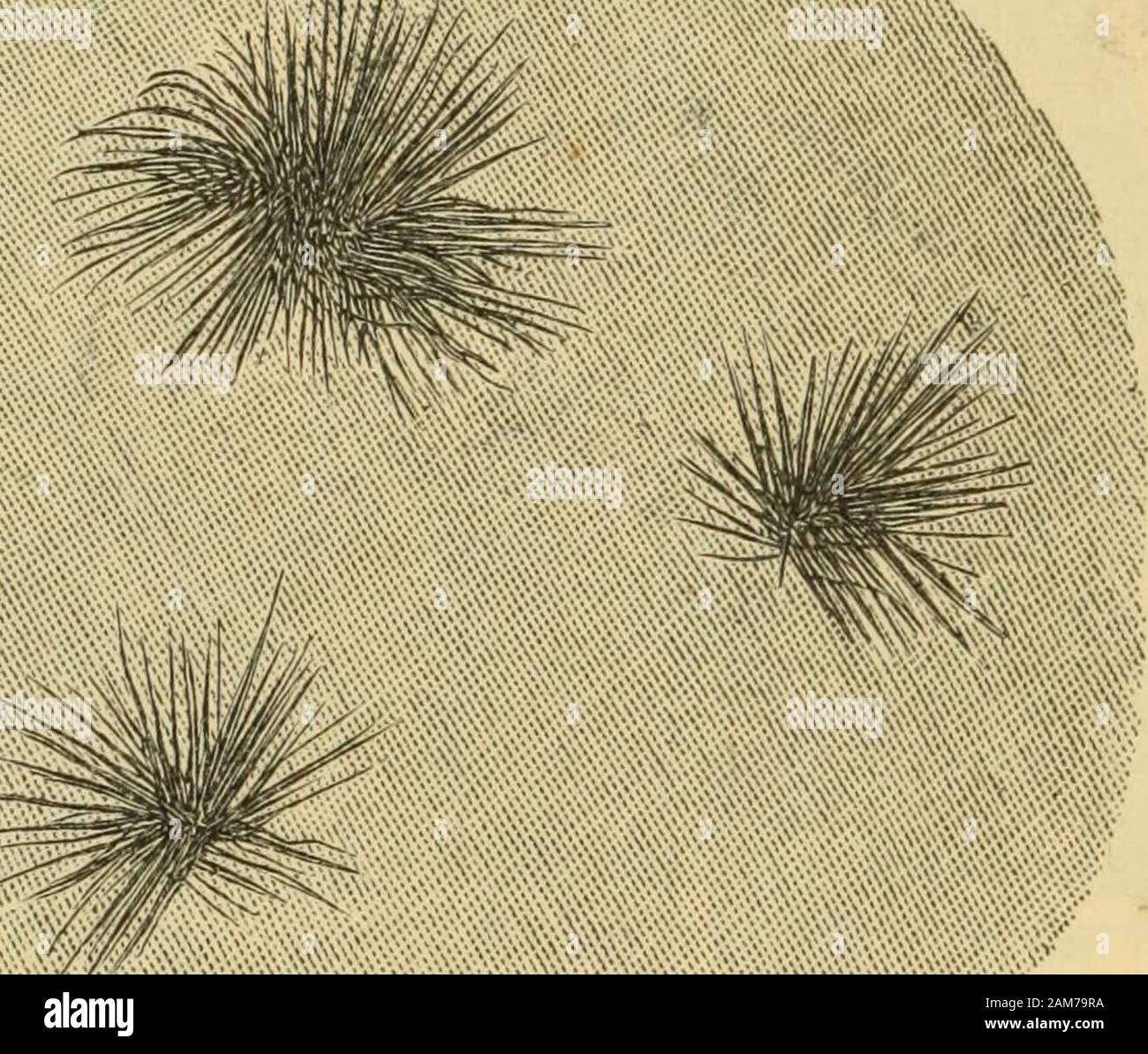
 Manual of pathological anatomy . •^. Tufts of acicular crystals, composed of urate of soda from the matrix ofa granular kidney. the form of stellate bunches of acicular crystals, as shown inPig. 163. This condition often accompanies general gout; some-times when no symptoms of gout have occurred, similar depositswill be found in the articular cartilages of the great toe. Some-times it is alleged to be the solitary gouty deposit in the body. Tomake a special form of kidnej^ disease—arthritic nephritis—onthese appearances, as has been done by some French and Germanpathologists, appears to us qu Stock Photohttps://www.alamy.com/image-license-details/?v=1https://www.alamy.com/manual-of-pathological-anatomy-tufts-of-acicular-crystals-composed-of-urate-of-soda-from-the-matrix-ofa-granular-kidney-the-form-of-stellate-bunches-of-acicular-crystals-as-shown-inpig-163-this-condition-often-accompanies-general-gout-some-times-when-no-symptoms-of-gout-have-occurred-similar-depositswill-be-found-in-the-articular-cartilages-of-the-great-toe-some-times-it-is-alleged-to-be-the-solitary-gouty-deposit-in-the-body-tomake-a-special-form-of-kidnej-diseasearthritic-nephritisonthese-appearances-as-has-been-done-by-some-french-and-germanpathologists-appears-to-us-qu-image339451486.html
RM2AM79RA–Manual of pathological anatomy . •^. Tufts of acicular crystals, composed of urate of soda from the matrix ofa granular kidney. the form of stellate bunches of acicular crystals, as shown inPig. 163. This condition often accompanies general gout; some-times when no symptoms of gout have occurred, similar depositswill be found in the articular cartilages of the great toe. Some-times it is alleged to be the solitary gouty deposit in the body. Tomake a special form of kidnej^ disease—arthritic nephritis—onthese appearances, as has been done by some French and Germanpathologists, appears to us qu
 A manual of human physiology, including histology and microscopical anatomy, with special reference to the requirements of practical medicine . lls being 2 to 4times the breadth of a colourless blood-corpuscle. These cells occur chiefly in themorning sputum in individuals over 30 years of age. In younger persons theirpresence indicates a pathological condition of the pulmonary parenchyma(Guttman, H. Schmidt, and Bizzozero). They often undergo fatty degeneration,and theyjnay contain pigment granules (3); or, they may present the appearanceof what Buhl has called mydin degenerated cells; i.e., c Stock Photohttps://www.alamy.com/image-license-details/?v=1https://www.alamy.com/a-manual-of-human-physiology-including-histology-and-microscopical-anatomy-with-special-reference-to-the-requirements-of-practical-medicine-lls-being-2-to-4times-the-breadth-of-a-colourless-blood-corpuscle-these-cells-occur-chiefly-in-themorning-sputum-in-individuals-over-30-years-of-age-in-younger-persons-theirpresence-indicates-a-pathological-condition-of-the-pulmonary-parenchymaguttman-h-schmidt-and-bizzozero-they-often-undergo-fatty-degenerationand-theyjnay-contain-pigment-granules-3-or-they-may-present-the-appearanceof-what-buhl-has-called-mydin-degenerated-cells-ie-c-image340031923.html
RM2AN5P57–A manual of human physiology, including histology and microscopical anatomy, with special reference to the requirements of practical medicine . lls being 2 to 4times the breadth of a colourless blood-corpuscle. These cells occur chiefly in themorning sputum in individuals over 30 years of age. In younger persons theirpresence indicates a pathological condition of the pulmonary parenchyma(Guttman, H. Schmidt, and Bizzozero). They often undergo fatty degeneration,and theyjnay contain pigment granules (3); or, they may present the appearanceof what Buhl has called mydin degenerated cells; i.e., c
 . The elements of pathological histology with special reference to practical methods . , a), and theadjacent fissures in the conne<;tive tissue of those parts of the skinwhere the process is still actually advancing; whereas in a phlegmoncaused by the Streptococcus pyogenes the cocci may be found everywherein the tissue, as well as in the interior of blood-vessels and in differentsituations in the inflamed parts. Lastly, a further difference may be brought out in connection withthe pathological anatomy, in the fact that human erysipelas, in contrastto a phlegmon caused by streptococci, runs Stock Photohttps://www.alamy.com/image-license-details/?v=1https://www.alamy.com/the-elements-of-pathological-histology-with-special-reference-to-practical-methods-a-and-theadjacent-fissures-in-the-connelttive-tissue-of-those-parts-of-the-skinwhere-the-process-is-still-actually-advancing-whereas-in-a-phlegmoncaused-by-the-streptococcus-pyogenes-the-cocci-may-be-found-everywherein-the-tissue-as-well-as-in-the-interior-of-blood-vessels-and-in-differentsituations-in-the-inflamed-parts-lastly-a-further-difference-may-be-brought-out-in-connection-withthe-pathological-anatomy-in-the-fact-that-human-erysipelas-in-contrastto-a-phlegmon-caused-by-streptococci-runs-image369708230.html
RM2CDDJHA–. The elements of pathological histology with special reference to practical methods . , a), and theadjacent fissures in the conne<;tive tissue of those parts of the skinwhere the process is still actually advancing; whereas in a phlegmoncaused by the Streptococcus pyogenes the cocci may be found everywherein the tissue, as well as in the interior of blood-vessels and in differentsituations in the inflamed parts. Lastly, a further difference may be brought out in connection withthe pathological anatomy, in the fact that human erysipelas, in contrastto a phlegmon caused by streptococci, runs