Spina occulta Stock Photos and Images
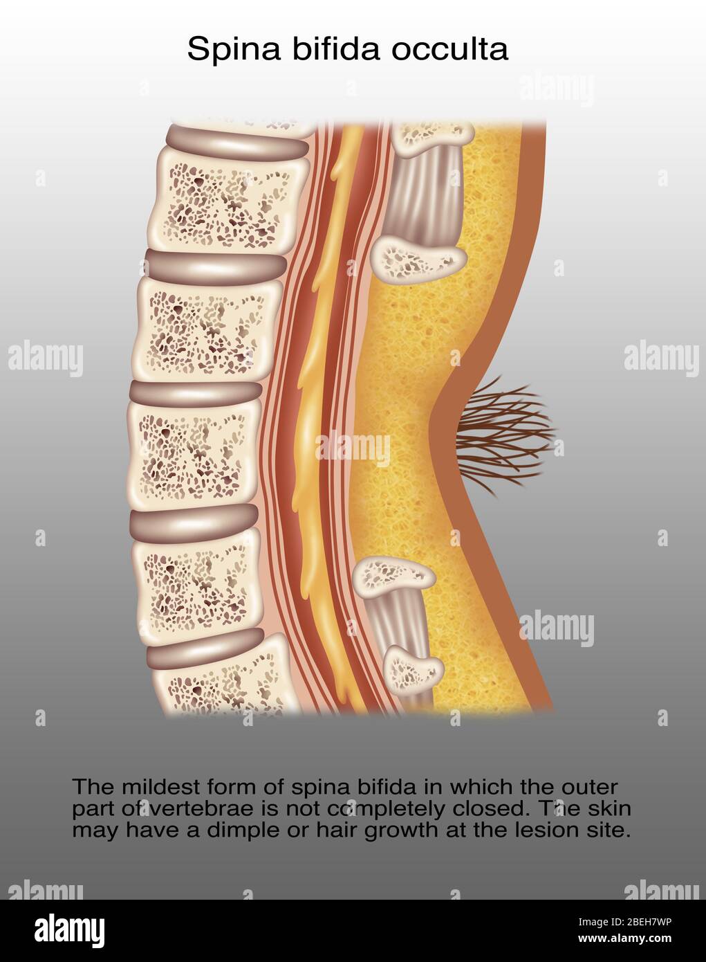 Spina Bifida Occulta, Illustration Stock Photohttps://www.alamy.com/image-license-details/?v=1https://www.alamy.com/spina-bifida-occulta-illustration-image353191938.html
RF2BEH7WP–Spina Bifida Occulta, Illustration
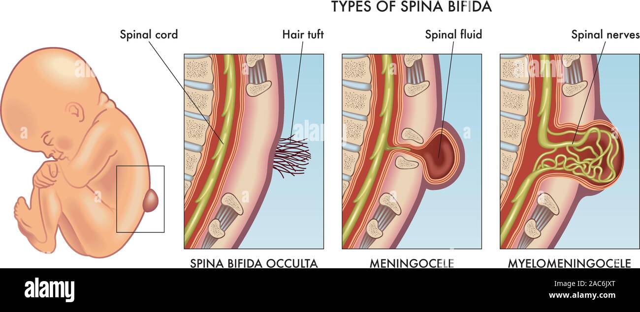 Medical illustration of infant spina bifida with annotation. Stock Vectorhttps://www.alamy.com/image-license-details/?v=1https://www.alamy.com/medical-illustration-of-infant-spina-bifida-with-annotation-image334519440.html
RF2AC6JXT–Medical illustration of infant spina bifida with annotation.
 Pencil with eraser Stock Photohttps://www.alamy.com/image-license-details/?v=1https://www.alamy.com/stock-photo-pencil-with-eraser-88431830.html
RFF3TBKJ–Pencil with eraser
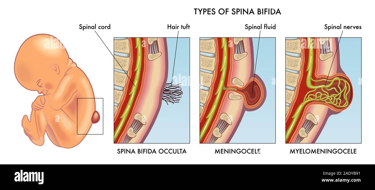 Medical illustration of infant spina bifida with annotation. Stock Photohttps://www.alamy.com/image-license-details/?v=1https://www.alamy.com/medical-illustration-of-infant-spina-bifida-with-annotation-image335589101.html
RF2ADYB91–Medical illustration of infant spina bifida with annotation.
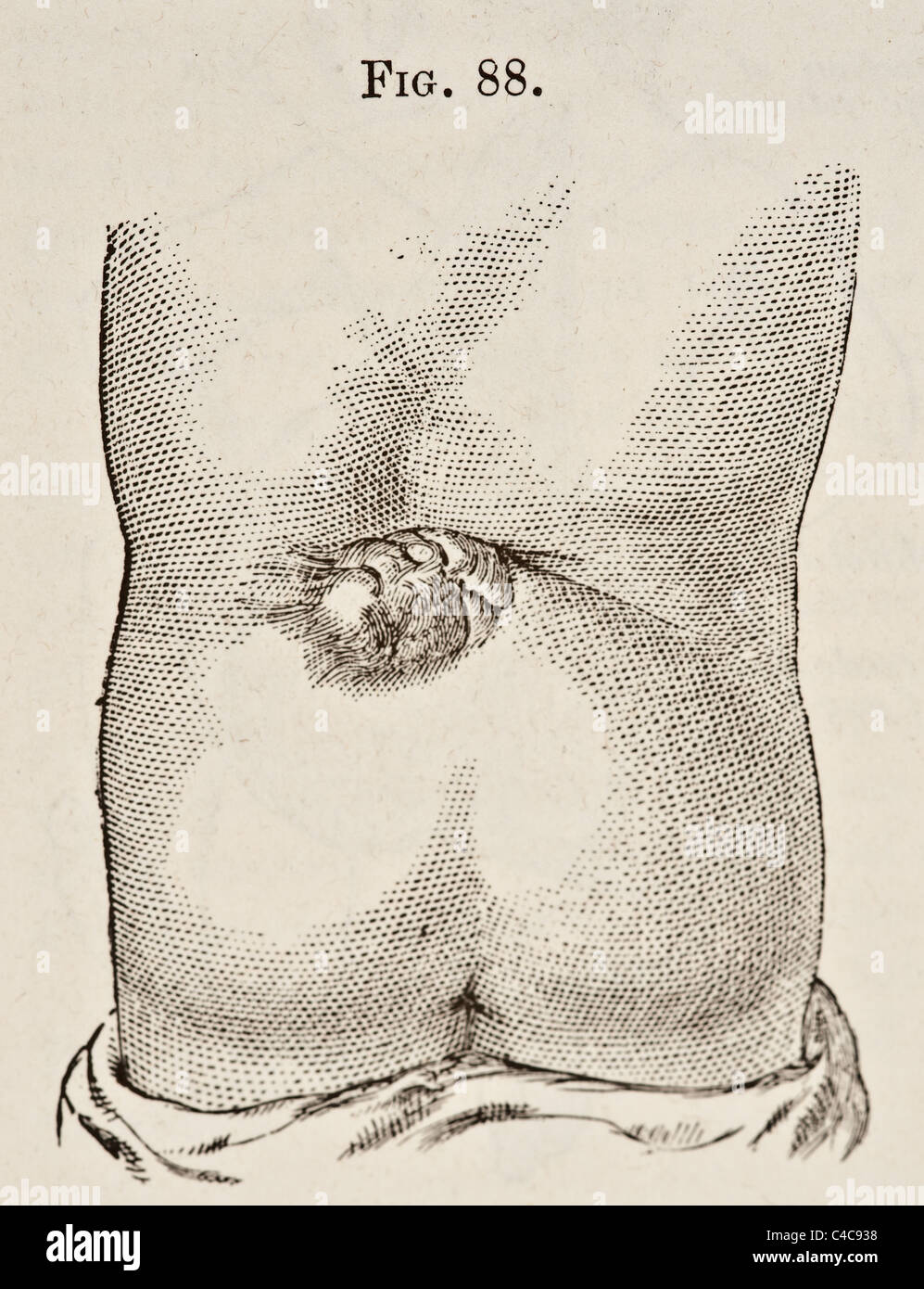 Antique Medical Illustration Depicting Spina Bifida circa 1881 Stock Photohttps://www.alamy.com/image-license-details/?v=1https://www.alamy.com/stock-photo-antique-medical-illustration-depicting-spina-bifida-circa-1881-37149932.html
RFC4C938–Antique Medical Illustration Depicting Spina Bifida circa 1881
 Spina bifida, illustration Stock Photohttps://www.alamy.com/image-license-details/?v=1https://www.alamy.com/spina-bifida-illustration-image415744163.html
RF2F4ANWR–Spina bifida, illustration
 . Tumours, innocent and malignant; their clinical characters and appropriate treatment. cation of the bones of the cranialvault. In contrast, it should bementioned that the bone beneaththe exquisite crest of the crownedcrane is abnormally thick. Animportant condition often associ-ated with spina bifida occulta isperforating ulcer of the foot. In-deed, this association is now sowell recognized that in every caseof perforating ulcer of the foot,occurring in young patients, it isthe duty of the surgeon, as amatter of routine, to examine theloins. In addition to non-union of thearches in the vicin Stock Photohttps://www.alamy.com/image-license-details/?v=1https://www.alamy.com/tumours-innocent-and-malignant-their-clinical-characters-and-appropriate-treatment-cation-of-the-bones-of-the-cranialvault-in-contrast-it-should-bementioned-that-the-bone-beneaththe-exquisite-crest-of-the-crownedcrane-is-abnormally-thick-animportant-condition-often-associ-ated-with-spina-bifida-occulta-isperforating-ulcer-of-the-foot-in-deed-this-association-is-now-sowell-recognized-that-in-every-caseof-perforating-ulcer-of-the-footoccurring-in-young-patients-it-isthe-duty-of-the-surgeon-as-amatter-of-routine-to-examine-theloins-in-addition-to-non-union-of-thearches-in-the-vicin-image336894411.html
RM2AG2T77–. Tumours, innocent and malignant; their clinical characters and appropriate treatment. cation of the bones of the cranialvault. In contrast, it should bementioned that the bone beneaththe exquisite crest of the crownedcrane is abnormally thick. Animportant condition often associ-ated with spina bifida occulta isperforating ulcer of the foot. In-deed, this association is now sowell recognized that in every caseof perforating ulcer of the foot,occurring in young patients, it isthe duty of the surgeon, as amatter of routine, to examine theloins. In addition to non-union of thearches in the vicin
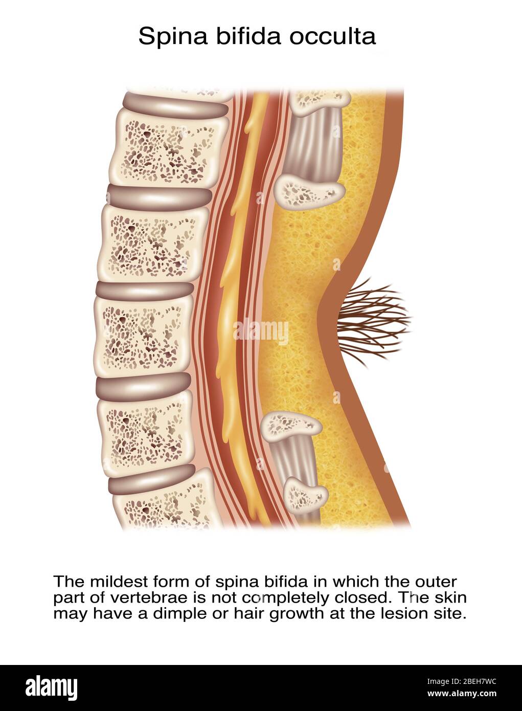 Spina Bifida Occulta, Illustration Stock Photohttps://www.alamy.com/image-license-details/?v=1https://www.alamy.com/spina-bifida-occulta-illustration-image353191928.html
RF2BEH7WC–Spina Bifida Occulta, Illustration
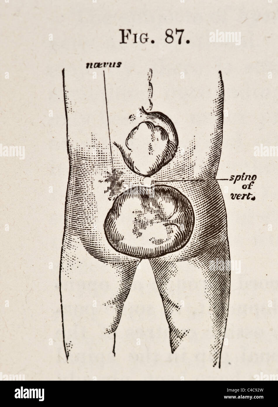 Antique Medical Illustration Depicting Spina Bifida circa 1881 Stock Photohttps://www.alamy.com/image-license-details/?v=1https://www.alamy.com/stock-photo-antique-medical-illustration-depicting-spina-bifida-circa-1881-37149921.html
RFC4C92W–Antique Medical Illustration Depicting Spina Bifida circa 1881
 . Tumours, innocent and malignant; their clinical characters and appropriate treatment. Fig. 345.— Hair-field overlying a spina bifida occulta ; there is also a long tuft in the cervical region. (FiscJier.) SPINA BIFIDA 647 several children seen liydroceplialus supervene when the sacin the loin has been made to shrink by artificial means. We have now to consider the various modes by whichspina bifida destroys life. Of all the varieties of this malfor-mation, myelocele is the most fatal. A very large proportionof fcetuses in which this condition is present are stillborn;the few that survive the Stock Photohttps://www.alamy.com/image-license-details/?v=1https://www.alamy.com/tumours-innocent-and-malignant-their-clinical-characters-and-appropriate-treatment-fig-345-hair-field-overlying-a-spina-bifida-occulta-there-is-also-a-long-tuft-in-the-cervical-region-fiscjier-spina-bifida-647-several-children-seen-liydroceplialus-supervene-when-the-sacin-the-loin-has-been-made-to-shrink-by-artificial-means-we-have-now-to-consider-the-various-modes-by-whichspina-bifida-destroys-life-of-all-the-varieties-of-this-malfor-mation-myelocele-is-the-most-fatal-a-very-large-proportionof-fcetuses-in-which-this-condition-is-present-are-stillbornthe-few-that-survive-the-image336894013.html
RM2AG2RN1–. Tumours, innocent and malignant; their clinical characters and appropriate treatment. Fig. 345.— Hair-field overlying a spina bifida occulta ; there is also a long tuft in the cervical region. (FiscJier.) SPINA BIFIDA 647 several children seen liydroceplialus supervene when the sacin the loin has been made to shrink by artificial means. We have now to consider the various modes by whichspina bifida destroys life. Of all the varieties of this malfor-mation, myelocele is the most fatal. A very large proportionof fcetuses in which this condition is present are stillborn;the few that survive the
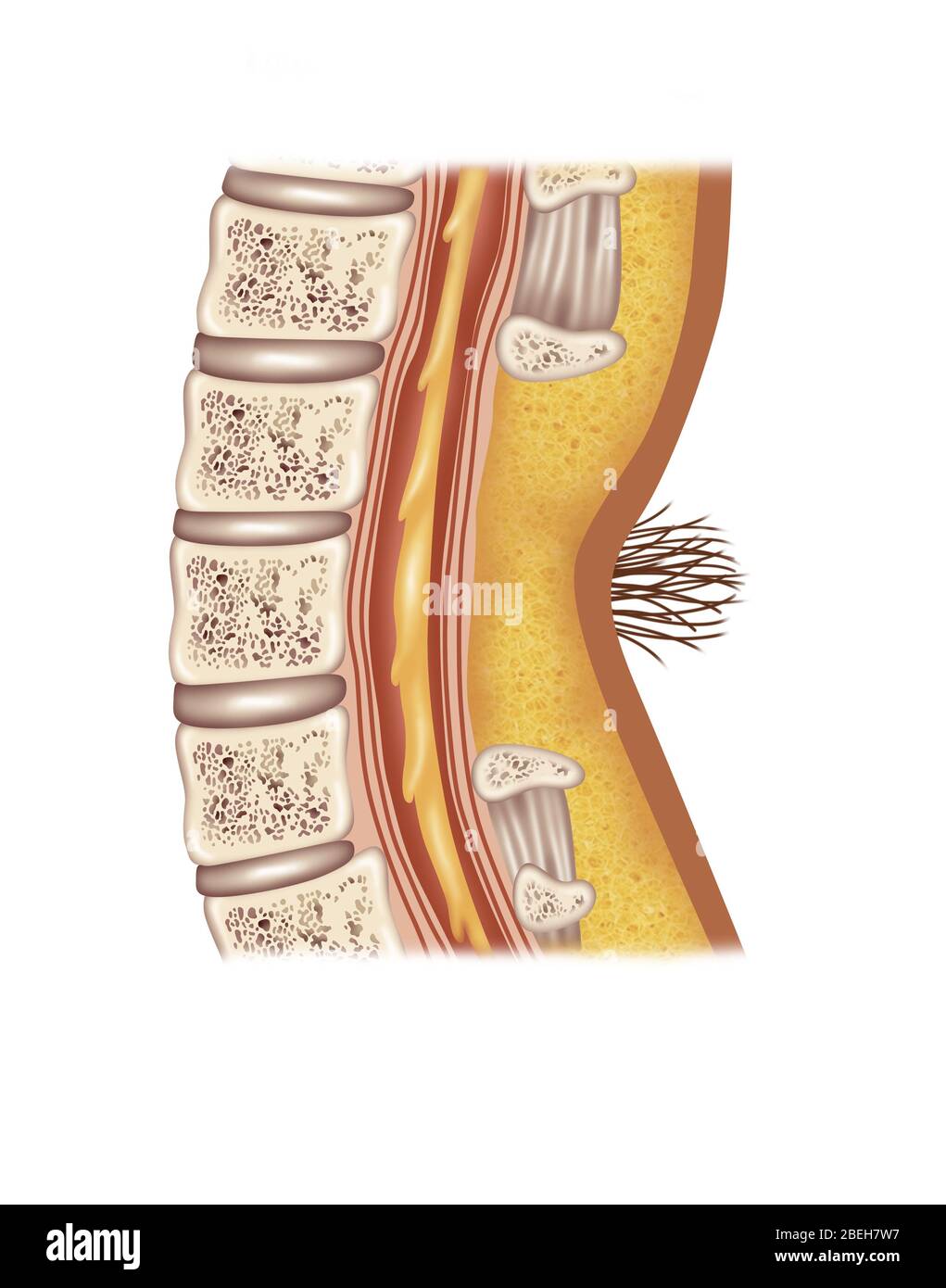 Spina Bifida Occulta, Illustration Stock Photohttps://www.alamy.com/image-license-details/?v=1https://www.alamy.com/spina-bifida-occulta-illustration-image353191923.html
RF2BEH7W7–Spina Bifida Occulta, Illustration
 . Tumours, innocent and malignant; their clinical characters and appropriate treatment. spina bifida occulta; the skin covering the de-fective spines presented the hair-field usual in these cases.In the tissues immediately over the stunted spinous pro-cesses a dermoid was found containing sebaceous materialand hair (Fig. 222). It is very rare to find dermoids within the spinal canal. 444 DERMOIDS An interesting instance of this has been recorded by HaleWhite. It grew in the thoracic region of the spine, andproduced paraplegia. Laminectomy was performed on thepatient, a man 26 years of age, but Stock Photohttps://www.alamy.com/image-license-details/?v=1https://www.alamy.com/tumours-innocent-and-malignant-their-clinical-characters-and-appropriate-treatment-spina-bifida-occulta-the-skin-covering-the-de-fective-spines-presented-the-hair-field-usual-in-these-casesin-the-tissues-immediately-over-the-stunted-spinous-pro-cesses-a-dermoid-was-found-containing-sebaceous-materialand-hair-fig-222-it-is-very-rare-to-find-dermoids-within-the-spinal-canal-444-dermoids-an-interesting-instance-of-this-has-been-recorded-by-halewhite-it-grew-in-the-thoracic-region-of-the-spine-andproduced-paraplegia-laminectomy-was-performed-on-thepatient-a-man-26-years-of-age-but-image336921544.html
RM2AG42T8–. Tumours, innocent and malignant; their clinical characters and appropriate treatment. spina bifida occulta; the skin covering the de-fective spines presented the hair-field usual in these cases.In the tissues immediately over the stunted spinous pro-cesses a dermoid was found containing sebaceous materialand hair (Fig. 222). It is very rare to find dermoids within the spinal canal. 444 DERMOIDS An interesting instance of this has been recorded by HaleWhite. It grew in the thoracic region of the spine, andproduced paraplegia. Laminectomy was performed on thepatient, a man 26 years of age, but
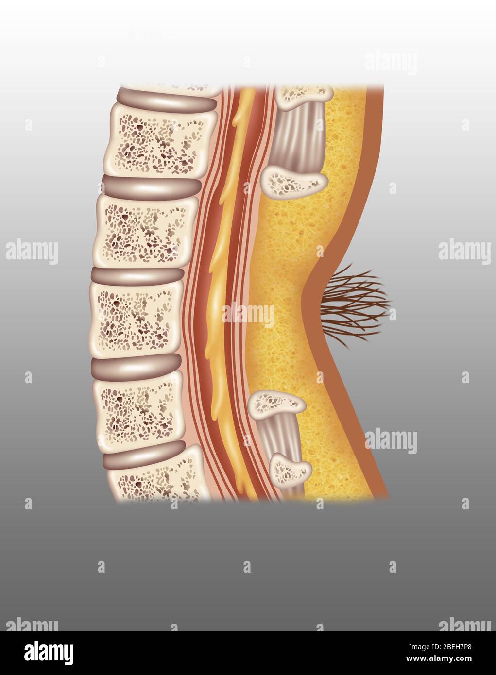 Spina Bifida Occulta, Illustration Stock Photohttps://www.alamy.com/image-license-details/?v=1https://www.alamy.com/spina-bifida-occulta-illustration-image353191840.html
RF2BEH7P8–Spina Bifida Occulta, Illustration
 AMAarchives of neurology & psychiatry . vitiligo 305 Weiss, H. B.: Organotherapy in dyspituitarism 206 Weskott, H.: Spina bifida occulta and sciatica 303 Weston, P. G.: Magnesium as a sedative 209 and Howard. M. Q.: Determination of sodium, potassium, calcium and magnesium in blood and spinal fluid in manic-depressive insanity. .*179 1^ IXPEX TO VOLUME 8 IAGE Whitehorn, J. C.: Chemistry oi cpinephrin 32.^ Wilson, G.: Manganese poisoning 332 Trismus developing in course of a tumor of pons 335 Wilson, S. A. K.: Decercl)rate rigidity in man and occurrencq of tonic fits 553 Winkclman. N. W.: Thala Stock Photohttps://www.alamy.com/image-license-details/?v=1https://www.alamy.com/amaarchives-of-neurology-psychiatry-vitiligo-305-weiss-h-b-organotherapy-in-dyspituitarism-206-weskott-h-spina-bifida-occulta-and-sciatica-303-weston-p-g-magnesium-as-a-sedative-209-and-howard-m-q-determination-of-sodium-potassium-calcium-and-magnesium-in-blood-and-spinal-fluid-in-manic-depressive-insanity-179-1-ixpex-to-volume-8-iage-whitehorn-j-c-chemistry-oi-cpinephrin-32-wilson-g-manganese-poisoning-332-trismus-developing-in-course-of-a-tumor-of-pons-335-wilson-s-a-k-decerclrate-rigidity-in-man-and-occurrencq-of-tonic-fits-553-winkclman-n-w-thala-image340189414.html
RM2ANCY1X–AMAarchives of neurology & psychiatry . vitiligo 305 Weiss, H. B.: Organotherapy in dyspituitarism 206 Weskott, H.: Spina bifida occulta and sciatica 303 Weston, P. G.: Magnesium as a sedative 209 and Howard. M. Q.: Determination of sodium, potassium, calcium and magnesium in blood and spinal fluid in manic-depressive insanity. .*179 1^ IXPEX TO VOLUME 8 IAGE Whitehorn, J. C.: Chemistry oi cpinephrin 32.^ Wilson, G.: Manganese poisoning 332 Trismus developing in course of a tumor of pons 335 Wilson, S. A. K.: Decercl)rate rigidity in man and occurrencq of tonic fits 553 Winkclman. N. W.: Thala
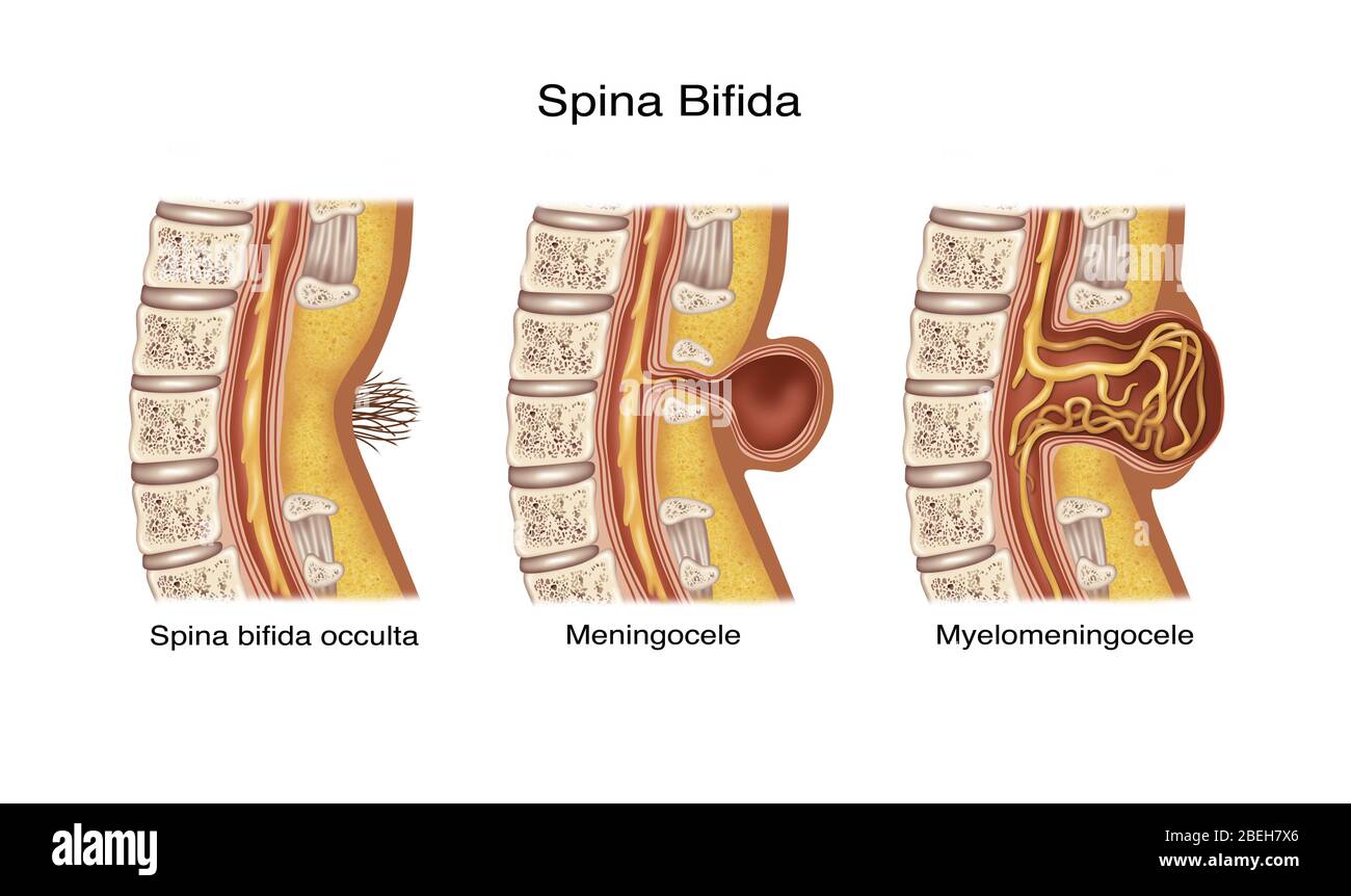 Spina Bifida, Illustration Stock Photohttps://www.alamy.com/image-license-details/?v=1https://www.alamy.com/spina-bifida-illustration-image353191950.html
RF2BEH7X6–Spina Bifida, Illustration
 . Living anatomy and pathology; . PLATE 44.SPINA BIFIDA OCCULTA. Girl, age 3^ years. (Same subject as Plates 42 and 43.) The arrow points to the right hip after reduction. The deformities of the vertebrae and sacrum are the sameas in Plate 42. Note the retarded development of the upper epiphysis ofthe right femur in comparison with that of the left. Plate 44. Stock Photohttps://www.alamy.com/image-license-details/?v=1https://www.alamy.com/living-anatomy-and-pathology-plate-44spina-bifida-occulta-girl-age-3-years-same-subject-as-plates-42-and-43-the-arrow-points-to-the-right-hip-after-reduction-the-deformities-of-the-vertebrae-and-sacrum-are-the-sameas-in-plate-42-note-the-retarded-development-of-the-upper-epiphysis-ofthe-right-femur-in-comparison-with-that-of-the-left-plate-44-image375986339.html
RM2CRKJBF–. Living anatomy and pathology; . PLATE 44.SPINA BIFIDA OCCULTA. Girl, age 3^ years. (Same subject as Plates 42 and 43.) The arrow points to the right hip after reduction. The deformities of the vertebrae and sacrum are the sameas in Plate 42. Note the retarded development of the upper epiphysis ofthe right femur in comparison with that of the left. Plate 44.
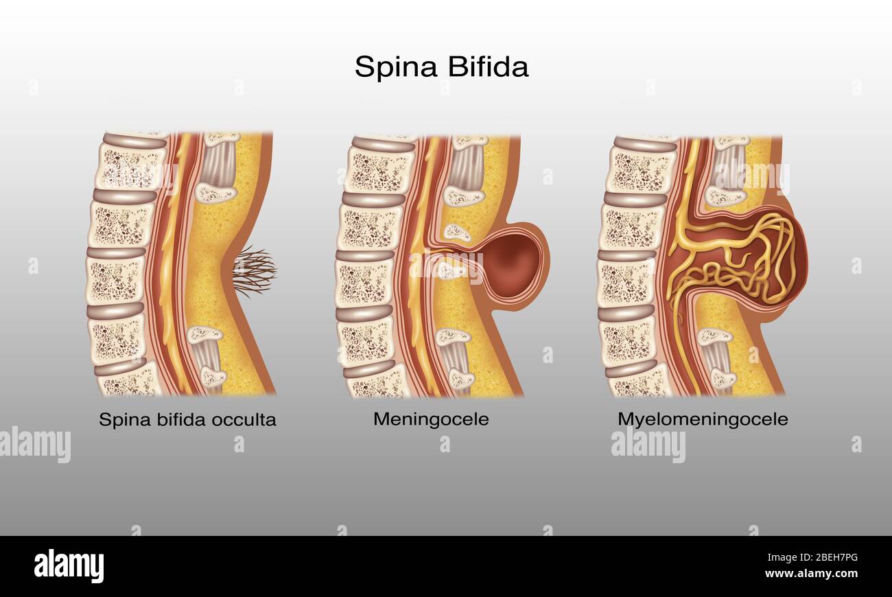 Spina Bifida, Illustration Stock Photohttps://www.alamy.com/image-license-details/?v=1https://www.alamy.com/spina-bifida-illustration-image353191848.html
RF2BEH7PG–Spina Bifida, Illustration
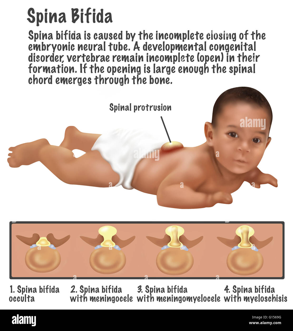 An illustration of a baby with Spina Bifida. Spina bifida is caused by the incomplete closing of the embryonic neutral tube. A development congenital disorder, vertebrae remain incomplete (open) in their formation. If the opening is large enough the spina Stock Photohttps://www.alamy.com/image-license-details/?v=1https://www.alamy.com/stock-photo-an-illustration-of-a-baby-with-spina-bifida-spina-bifida-is-caused-103991596.html
RMG1569G–An illustration of a baby with Spina Bifida. Spina bifida is caused by the incomplete closing of the embryonic neutral tube. A development congenital disorder, vertebrae remain incomplete (open) in their formation. If the opening is large enough the spina
![. Living anatomy and pathology; . PLATE 43.SPINA BIFIDA OCCULTA. Girl, age 3J years. (Same subject as Plates 42 and 44.) A. The narrowed third lumbar vertebra. B. The narrowed third intervertebral disk. C. Sacralization of the left fifth lumbar vertebra. D. Just to the right and below the point of the arrow is fissure of the first body of the sacrum. Plat*] 43. <$? ■fM Stock Photo . Living anatomy and pathology; . PLATE 43.SPINA BIFIDA OCCULTA. Girl, age 3J years. (Same subject as Plates 42 and 44.) A. The narrowed third lumbar vertebra. B. The narrowed third intervertebral disk. C. Sacralization of the left fifth lumbar vertebra. D. Just to the right and below the point of the arrow is fissure of the first body of the sacrum. Plat*] 43. <$? ■fM Stock Photo](https://c8.alamy.com/comp/2CRKJC1/living-anatomy-and-pathology-plate-43spina-bifida-occulta-girl-age-3j-years-same-subject-as-plates-42-and-44-a-the-narrowed-third-lumbar-vertebra-b-the-narrowed-third-intervertebral-disk-c-sacralization-of-the-left-fifth-lumbar-vertebra-d-just-to-the-right-and-below-the-point-of-the-arrow-is-fissure-of-the-first-body-of-the-sacrum-plat-43-lt-fm-2CRKJC1.jpg) . Living anatomy and pathology; . PLATE 43.SPINA BIFIDA OCCULTA. Girl, age 3J years. (Same subject as Plates 42 and 44.) A. The narrowed third lumbar vertebra. B. The narrowed third intervertebral disk. C. Sacralization of the left fifth lumbar vertebra. D. Just to the right and below the point of the arrow is fissure of the first body of the sacrum. Plat*] 43. <$? ■fM Stock Photohttps://www.alamy.com/image-license-details/?v=1https://www.alamy.com/living-anatomy-and-pathology-plate-43spina-bifida-occulta-girl-age-3j-years-same-subject-as-plates-42-and-44-a-the-narrowed-third-lumbar-vertebra-b-the-narrowed-third-intervertebral-disk-c-sacralization-of-the-left-fifth-lumbar-vertebra-d-just-to-the-right-and-below-the-point-of-the-arrow-is-fissure-of-the-first-body-of-the-sacrum-plat-43-lt-fm-image375986353.html
. Living anatomy and pathology; . PLATE 43.SPINA BIFIDA OCCULTA. Girl, age 3J years. (Same subject as Plates 42 and 44.) A. The narrowed third lumbar vertebra. B. The narrowed third intervertebral disk. C. Sacralization of the left fifth lumbar vertebra. D. Just to the right and below the point of the arrow is fissure of the first body of the sacrum. Plat*] 43. <$? ■fM Stock Photohttps://www.alamy.com/image-license-details/?v=1https://www.alamy.com/living-anatomy-and-pathology-plate-43spina-bifida-occulta-girl-age-3j-years-same-subject-as-plates-42-and-44-a-the-narrowed-third-lumbar-vertebra-b-the-narrowed-third-intervertebral-disk-c-sacralization-of-the-left-fifth-lumbar-vertebra-d-just-to-the-right-and-below-the-point-of-the-arrow-is-fissure-of-the-first-body-of-the-sacrum-plat-43-lt-fm-image375986353.htmlRM2CRKJC1–. Living anatomy and pathology; . PLATE 43.SPINA BIFIDA OCCULTA. Girl, age 3J years. (Same subject as Plates 42 and 44.) A. The narrowed third lumbar vertebra. B. The narrowed third intervertebral disk. C. Sacralization of the left fifth lumbar vertebra. D. Just to the right and below the point of the arrow is fissure of the first body of the sacrum. Plat*] 43. <$? ■fM
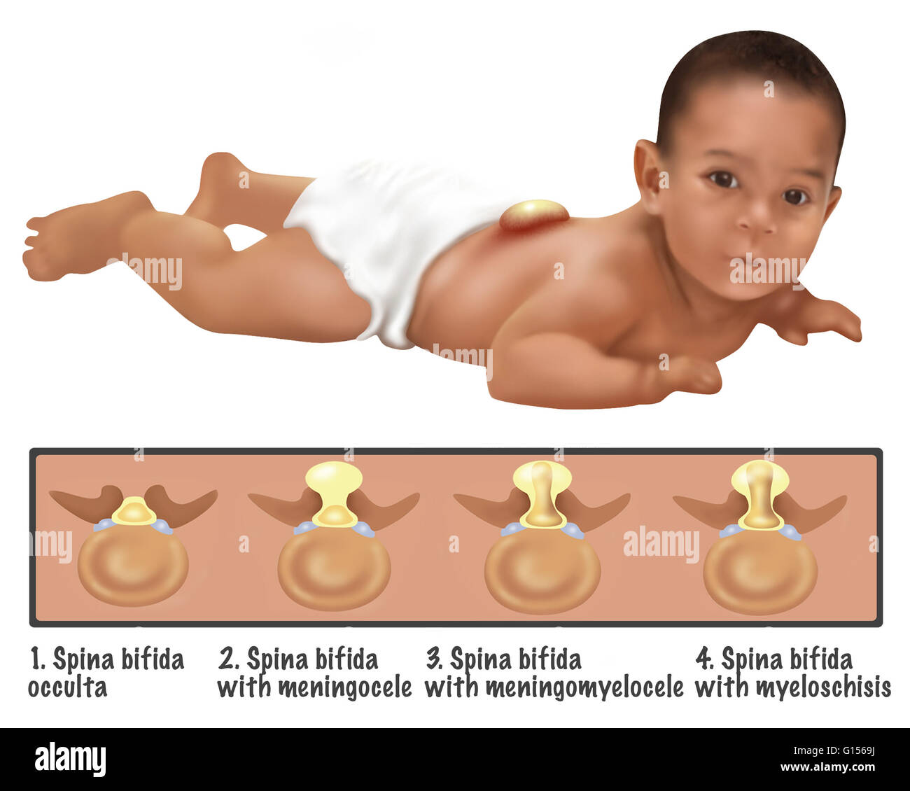 An illustration of a baby with Spina Bifida. Spina bifida is caused by the incomplete closing of the embryonic neutral tube. A development congenital disorder, vertebrae remain incomplete (open) in their formation. If the opening is large enough the spina Stock Photohttps://www.alamy.com/image-license-details/?v=1https://www.alamy.com/stock-photo-an-illustration-of-a-baby-with-spina-bifida-spina-bifida-is-caused-103991598.html
RMG1569J–An illustration of a baby with Spina Bifida. Spina bifida is caused by the incomplete closing of the embryonic neutral tube. A development congenital disorder, vertebrae remain incomplete (open) in their formation. If the opening is large enough the spina
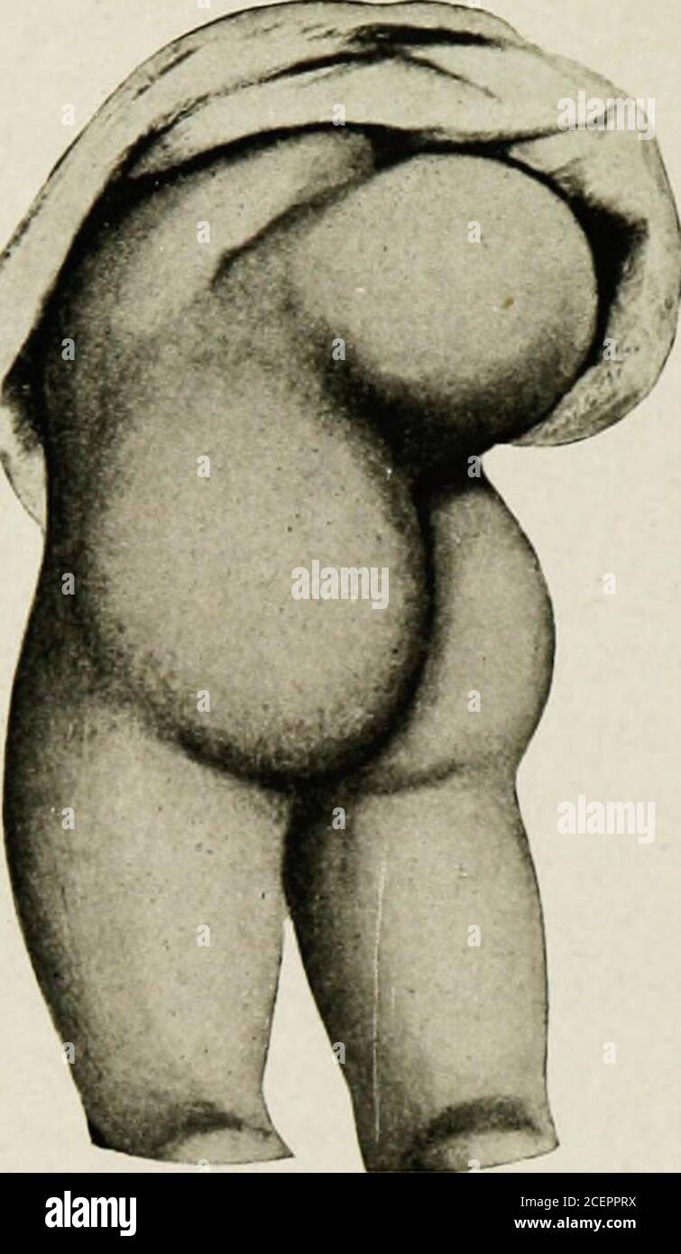 . The diseases of infancy and childhood. his prevents the union of the parts of the vertebralarches. Although the tumor is generally associated with a bifid spine, thisis not necessarily the case. The protrusion may take place through theintervertebral notch or foramen, or there may be a fissure of the bodiesof the vertebrae, and an anterior tumor projecting into the cavity of thethorax, abdomen, or pelvis; the tumor may be so small as not to be recog-nized externally—spina bifida occulta. The principal anatomical varie-ties are meningocele, meningomyelocele, and syringomyelocele. Meningocele. Stock Photohttps://www.alamy.com/image-license-details/?v=1https://www.alamy.com/the-diseases-of-infancy-and-childhood-his-prevents-the-union-of-the-parts-of-the-vertebralarches-although-the-tumor-is-generally-associated-with-a-bifid-spine-thisis-not-necessarily-the-case-the-protrusion-may-take-place-through-theintervertebral-notch-or-foramen-or-there-may-be-a-fissure-of-the-bodiesof-the-vertebrae-and-an-anterior-tumor-projecting-into-the-cavity-of-thethorax-abdomen-or-pelvis-the-tumor-may-be-so-small-as-not-to-be-recog-nized-externallyspina-bifida-occulta-the-principal-anatomical-varie-ties-are-meningocele-meningomyelocele-and-syringomyelocele-meningocele-image370523774.html
RM2CEPPRX–. The diseases of infancy and childhood. his prevents the union of the parts of the vertebralarches. Although the tumor is generally associated with a bifid spine, thisis not necessarily the case. The protrusion may take place through theintervertebral notch or foramen, or there may be a fissure of the bodiesof the vertebrae, and an anterior tumor projecting into the cavity of thethorax, abdomen, or pelvis; the tumor may be so small as not to be recog-nized externally—spina bifida occulta. The principal anatomical varie-ties are meningocele, meningomyelocele, and syringomyelocele. Meningocele.
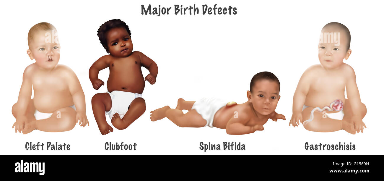 An illustration of 4 common birth defects found in babies. Cleft palate, clubfoot, spina bifida and gastroschisis. 1 baby in 30 is born with one or more major birth defects. Most are correctable with modern surgery. Stock Photohttps://www.alamy.com/image-license-details/?v=1https://www.alamy.com/stock-photo-an-illustration-of-4-common-birth-defects-found-in-babies-cleft-palate-103991601.html
RMG1569N–An illustration of 4 common birth defects found in babies. Cleft palate, clubfoot, spina bifida and gastroschisis. 1 baby in 30 is born with one or more major birth defects. Most are correctable with modern surgery.
 . Evolution and disease . 24 EVOLUTION AND DISEASE. same way that the hairy tuft may be accounted for inthe back of those with spina bifida occulta. Thesefowls are extremely uncertain in their gait, given toperforming circular movements, and walking sideways ifexcited, as though they possessed an unstable nervoussystem. Darwin was assured that we had here to dealwith a character first acquired and transmitted by thehen.1. Fig. 13.—The head of a Polish fowl to show the feathery tuft.(After Darwin.) A somewhat similar condition is seen in ducks. Pre-served in the museum of the Royal College of S Stock Photohttps://www.alamy.com/image-license-details/?v=1https://www.alamy.com/evolution-and-disease-24-evolution-and-disease-same-way-that-the-hairy-tuft-may-be-accounted-for-inthe-back-of-those-with-spina-bifida-occulta-thesefowls-are-extremely-uncertain-in-their-gait-given-toperforming-circular-movements-and-walking-sideways-ifexcited-as-though-they-possessed-an-unstable-nervoussystem-darwin-was-assured-that-we-had-here-to-dealwith-a-character-first-acquired-and-transmitted-by-thehen1-fig-13the-head-of-a-polish-fowl-to-show-the-feathery-tuftafter-darwin-a-somewhat-similar-condition-is-seen-in-ducks-pre-served-in-the-museum-of-the-royal-college-of-s-image372025736.html
RM2CH76HC–. Evolution and disease . 24 EVOLUTION AND DISEASE. same way that the hairy tuft may be accounted for inthe back of those with spina bifida occulta. Thesefowls are extremely uncertain in their gait, given toperforming circular movements, and walking sideways ifexcited, as though they possessed an unstable nervoussystem. Darwin was assured that we had here to dealwith a character first acquired and transmitted by thehen.1. Fig. 13.—The head of a Polish fowl to show the feathery tuft.(After Darwin.) A somewhat similar condition is seen in ducks. Pre-served in the museum of the Royal College of S
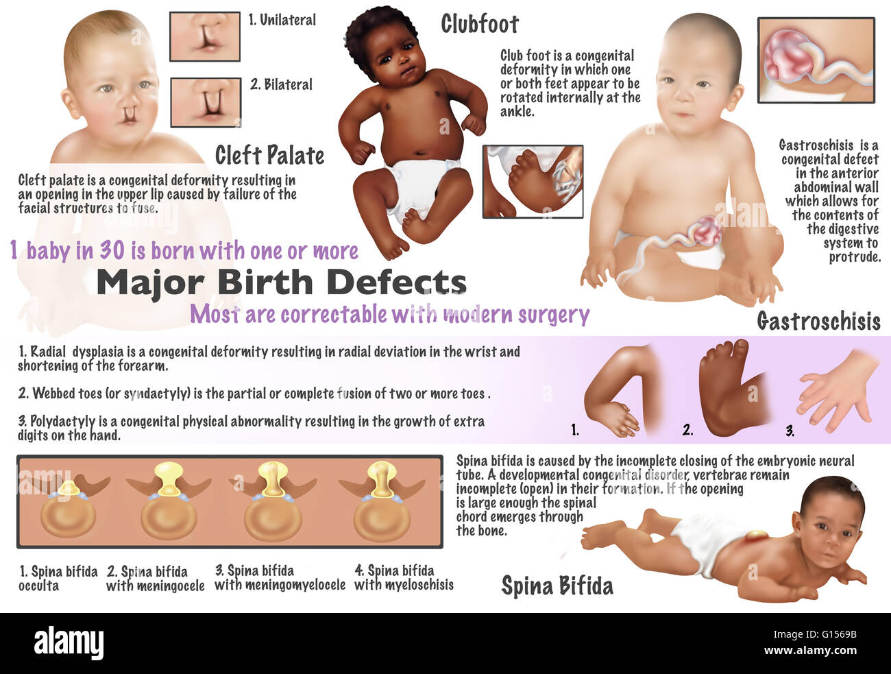 Illustration of birth defects found in babies. Cleft palate, clubfoot, radial dysplasia, webbed toes, polydactyl, gastroschisis, and spina bifida. 1 baby in 30 is born with one or more major birth defects. Most are correctable with modern surgery. Stock Photohttps://www.alamy.com/image-license-details/?v=1https://www.alamy.com/stock-photo-illustration-of-birth-defects-found-in-babies-cleft-palate-clubfoot-103991591.html
RMG1569B–Illustration of birth defects found in babies. Cleft palate, clubfoot, radial dysplasia, webbed toes, polydactyl, gastroschisis, and spina bifida. 1 baby in 30 is born with one or more major birth defects. Most are correctable with modern surgery.
 . Evolution and disease . 24 EVOLUTION AND DISEASE. same way that the hairy tuft may be accounted for inthe back of those with spina bifida occulta. Thesefowls arc extremely uncertain in their gait, given toperforming circular movements, and walking sideways ifexcited, as though they possessed an unstable nervoussystem. Darwin was assured that we had here to dealwith a character first acquired and transmitted by th^hen. I. Fig. 13.—The head of a Polish fowl to show the feathery tuft.(After Darwin.) A somewhat similar condition is seen in ducks. Pre-served in the museum of the Royal College of Stock Photohttps://www.alamy.com/image-license-details/?v=1https://www.alamy.com/evolution-and-disease-24-evolution-and-disease-same-way-that-the-hairy-tuft-may-be-accounted-for-inthe-back-of-those-with-spina-bifida-occulta-thesefowls-arc-extremely-uncertain-in-their-gait-given-toperforming-circular-movements-and-walking-sideways-ifexcited-as-though-they-possessed-an-unstable-nervoussystem-darwin-was-assured-that-we-had-here-to-dealwith-a-character-first-acquired-and-transmitted-by-thhen-i-fig-13the-head-of-a-polish-fowl-to-show-the-feathery-tuftafter-darwin-a-somewhat-similar-condition-is-seen-in-ducks-pre-served-in-the-museum-of-the-royal-college-of-image372055975.html
RM2CH8H5B–. Evolution and disease . 24 EVOLUTION AND DISEASE. same way that the hairy tuft may be accounted for inthe back of those with spina bifida occulta. Thesefowls arc extremely uncertain in their gait, given toperforming circular movements, and walking sideways ifexcited, as though they possessed an unstable nervoussystem. Darwin was assured that we had here to dealwith a character first acquired and transmitted by th^hen. I. Fig. 13.—The head of a Polish fowl to show the feathery tuft.(After Darwin.) A somewhat similar condition is seen in ducks. Pre-served in the museum of the Royal College of
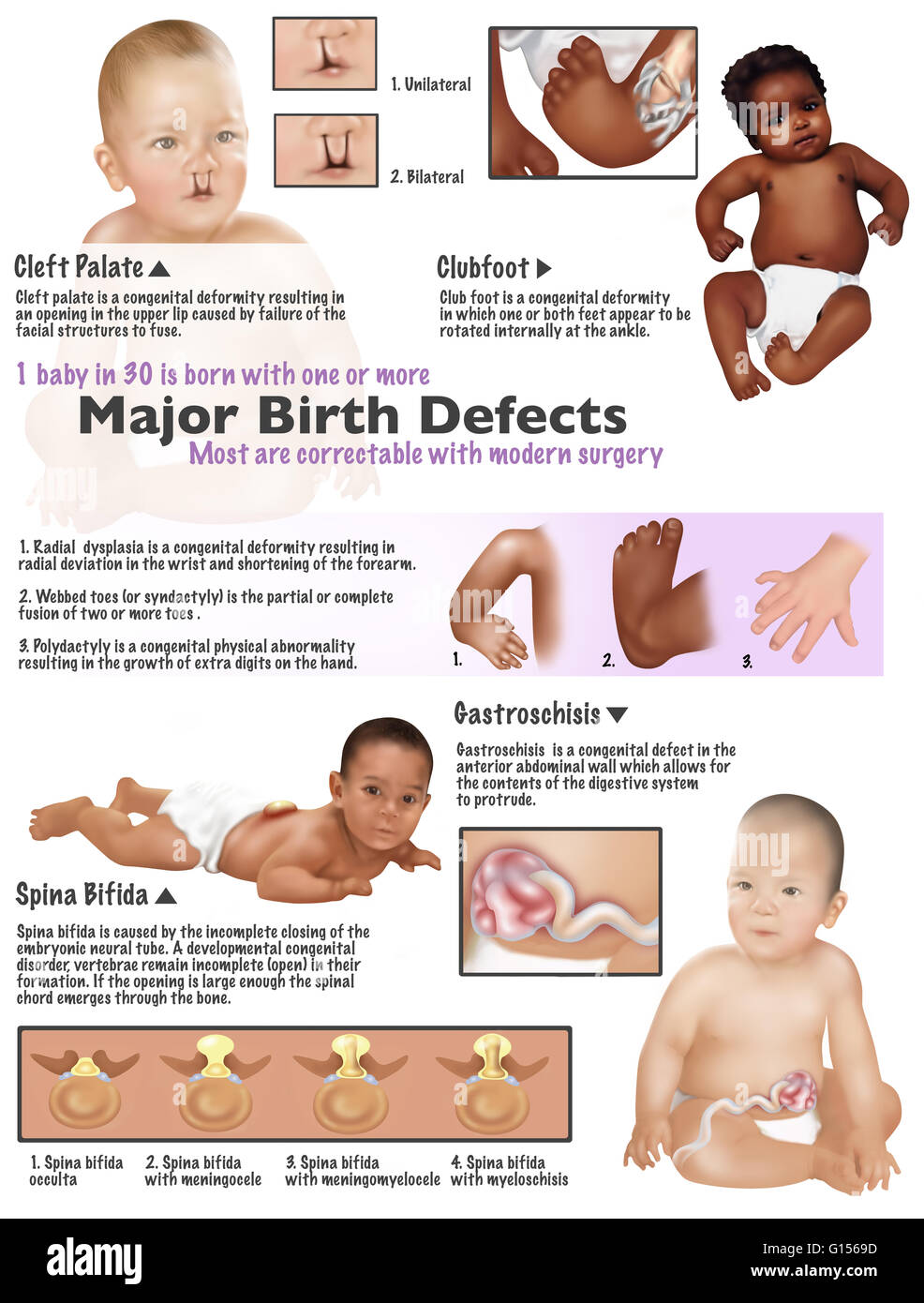 Illustration of birth defects found in babies. Cleft palate, clubfoot, radial dysplasia, webbed toes, polydactyl, gastroschisis, and spina bifida. 1 baby in 30 is born with one or more major birth defects. Most are correctable with modern surgery. Stock Photohttps://www.alamy.com/image-license-details/?v=1https://www.alamy.com/stock-photo-illustration-of-birth-defects-found-in-babies-cleft-palate-clubfoot-103991593.html
RMG1569D–Illustration of birth defects found in babies. Cleft palate, clubfoot, radial dysplasia, webbed toes, polydactyl, gastroschisis, and spina bifida. 1 baby in 30 is born with one or more major birth defects. Most are correctable with modern surgery.
![. Living anatomy and pathology; . PLATE 42.SPINA BIFIDA OCCULTA. Girl, age 3£ years. (Same subject as Plates 43 and 44.) The arrow points to hair and skin which cover the defect inthe vertebrae. Congenital dislocation of right hip. Platje 42. PLATE 43.SPINA BIFIDA OCCULTA. Girl, age 3J years. (Same subject as Plates 42 and 44.) A. The narrowed third lumbar vertebra. B. The narrowed third intervertebral disk. C. Sacralization of the left fifth lumbar vertebra. D. Just to the right and below the point of the arrow is fissure of the first body of the sacrum. Plat*] 43 Stock Photo . Living anatomy and pathology; . PLATE 42.SPINA BIFIDA OCCULTA. Girl, age 3£ years. (Same subject as Plates 43 and 44.) The arrow points to hair and skin which cover the defect inthe vertebrae. Congenital dislocation of right hip. Platje 42. PLATE 43.SPINA BIFIDA OCCULTA. Girl, age 3J years. (Same subject as Plates 42 and 44.) A. The narrowed third lumbar vertebra. B. The narrowed third intervertebral disk. C. Sacralization of the left fifth lumbar vertebra. D. Just to the right and below the point of the arrow is fissure of the first body of the sacrum. Plat*] 43 Stock Photo](https://c8.alamy.com/comp/2CRKK1A/living-anatomy-and-pathology-plate-42spina-bifida-occulta-girl-age-3-years-same-subject-as-plates-43-and-44-the-arrow-points-to-hair-and-skin-which-cover-the-defect-inthe-vertebrae-congenital-dislocation-of-right-hip-platje-42-plate-43spina-bifida-occulta-girl-age-3j-years-same-subject-as-plates-42-and-44-a-the-narrowed-third-lumbar-vertebra-b-the-narrowed-third-intervertebral-disk-c-sacralization-of-the-left-fifth-lumbar-vertebra-d-just-to-the-right-and-below-the-point-of-the-arrow-is-fissure-of-the-first-body-of-the-sacrum-plat-43-2CRKK1A.jpg) . Living anatomy and pathology; . PLATE 42.SPINA BIFIDA OCCULTA. Girl, age 3£ years. (Same subject as Plates 43 and 44.) The arrow points to hair and skin which cover the defect inthe vertebrae. Congenital dislocation of right hip. Platje 42. PLATE 43.SPINA BIFIDA OCCULTA. Girl, age 3J years. (Same subject as Plates 42 and 44.) A. The narrowed third lumbar vertebra. B. The narrowed third intervertebral disk. C. Sacralization of the left fifth lumbar vertebra. D. Just to the right and below the point of the arrow is fissure of the first body of the sacrum. Plat*] 43 Stock Photohttps://www.alamy.com/image-license-details/?v=1https://www.alamy.com/living-anatomy-and-pathology-plate-42spina-bifida-occulta-girl-age-3-years-same-subject-as-plates-43-and-44-the-arrow-points-to-hair-and-skin-which-cover-the-defect-inthe-vertebrae-congenital-dislocation-of-right-hip-platje-42-plate-43spina-bifida-occulta-girl-age-3j-years-same-subject-as-plates-42-and-44-a-the-narrowed-third-lumbar-vertebra-b-the-narrowed-third-intervertebral-disk-c-sacralization-of-the-left-fifth-lumbar-vertebra-d-just-to-the-right-and-below-the-point-of-the-arrow-is-fissure-of-the-first-body-of-the-sacrum-plat-43-image375986838.html
. Living anatomy and pathology; . PLATE 42.SPINA BIFIDA OCCULTA. Girl, age 3£ years. (Same subject as Plates 43 and 44.) The arrow points to hair and skin which cover the defect inthe vertebrae. Congenital dislocation of right hip. Platje 42. PLATE 43.SPINA BIFIDA OCCULTA. Girl, age 3J years. (Same subject as Plates 42 and 44.) A. The narrowed third lumbar vertebra. B. The narrowed third intervertebral disk. C. Sacralization of the left fifth lumbar vertebra. D. Just to the right and below the point of the arrow is fissure of the first body of the sacrum. Plat*] 43 Stock Photohttps://www.alamy.com/image-license-details/?v=1https://www.alamy.com/living-anatomy-and-pathology-plate-42spina-bifida-occulta-girl-age-3-years-same-subject-as-plates-43-and-44-the-arrow-points-to-hair-and-skin-which-cover-the-defect-inthe-vertebrae-congenital-dislocation-of-right-hip-platje-42-plate-43spina-bifida-occulta-girl-age-3j-years-same-subject-as-plates-42-and-44-a-the-narrowed-third-lumbar-vertebra-b-the-narrowed-third-intervertebral-disk-c-sacralization-of-the-left-fifth-lumbar-vertebra-d-just-to-the-right-and-below-the-point-of-the-arrow-is-fissure-of-the-first-body-of-the-sacrum-plat-43-image375986838.htmlRM2CRKK1A–. Living anatomy and pathology; . PLATE 42.SPINA BIFIDA OCCULTA. Girl, age 3£ years. (Same subject as Plates 43 and 44.) The arrow points to hair and skin which cover the defect inthe vertebrae. Congenital dislocation of right hip. Platje 42. PLATE 43.SPINA BIFIDA OCCULTA. Girl, age 3J years. (Same subject as Plates 42 and 44.) A. The narrowed third lumbar vertebra. B. The narrowed third intervertebral disk. C. Sacralization of the left fifth lumbar vertebra. D. Just to the right and below the point of the arrow is fissure of the first body of the sacrum. Plat*] 43
 . Annals of surgery . iam Randolph : Cardior- raphy in Acute Injuries, 696.Sodium Chloride, Therapeutic Value of theAdministration of, in Intestinal Obstruc-tion, 755.Speese, John: Spina Bifida Occulta, 534; Subastragaloid Dislocation, 536.Spina Bifida Occulta, 534.Spine, Osteomyelitis of the, 119.Splenectomy for Advanced Splenic Anae-mia, 408; for Bantis Disease, 557; inHemorrhagic Purpura, 186.Splenomegaly, 553, 669.Stetten, De Witt: A Modified InguinalHernioplasty, 48; Repair of tlie Musculo-spiral Nerve, 407; Technic of InguinalHernioplasty, 410; The Relation of Sur-gery to Certain Disturb Stock Photohttps://www.alamy.com/image-license-details/?v=1https://www.alamy.com/annals-of-surgery-iam-randolph-cardior-raphy-in-acute-injuries-696sodium-chloride-therapeutic-value-of-theadministration-of-in-intestinal-obstruc-tion-755speese-john-spina-bifida-occulta-534-subastragaloid-dislocation-536spina-bifida-occulta-534spine-osteomyelitis-of-the-119splenectomy-for-advanced-splenic-anae-mia-408-for-bantis-disease-557-inhemorrhagic-purpura-186splenomegaly-553-669stetten-de-witt-a-modified-inguinalhernioplasty-48-repair-of-tlie-musculo-spiral-nerve-407-technic-of-inguinalhernioplasty-410-the-relation-of-sur-gery-to-certain-disturb-image369993658.html
RM2CDXJK6–. Annals of surgery . iam Randolph : Cardior- raphy in Acute Injuries, 696.Sodium Chloride, Therapeutic Value of theAdministration of, in Intestinal Obstruc-tion, 755.Speese, John: Spina Bifida Occulta, 534; Subastragaloid Dislocation, 536.Spina Bifida Occulta, 534.Spine, Osteomyelitis of the, 119.Splenectomy for Advanced Splenic Anae-mia, 408; for Bantis Disease, 557; inHemorrhagic Purpura, 186.Splenomegaly, 553, 669.Stetten, De Witt: A Modified InguinalHernioplasty, 48; Repair of tlie Musculo-spiral Nerve, 407; Technic of InguinalHernioplasty, 410; The Relation of Sur-gery to Certain Disturb
 . Journal of radiology . nation ofthe lumber spine and sacrum has beenmade in approximately 12,000 of aconsecutive series of 70,000 patients inthe Mayo Clinic. Six hundred andtwenty-one of the 12,000 had spinabifida occulta, showing an incidence ofslightly more than 5 per cent. Thecondition occurred about twice as oftenin the male as in the female. The de-fect was in the first one or two sacralsegments in 70 per cent, in the fifthlumbar segment in 24.5 per cent, andin the lower sacral segment in 3 percent. Associated spina bifida occultaand bifurcation with sacralization of thetransverse proce Stock Photohttps://www.alamy.com/image-license-details/?v=1https://www.alamy.com/journal-of-radiology-nation-ofthe-lumber-spine-and-sacrum-has-beenmade-in-approximately-12000-of-aconsecutive-series-of-70000-patients-inthe-mayo-clinic-six-hundred-andtwenty-one-of-the-12000-had-spinabifida-occulta-showing-an-incidence-ofslightly-more-than-5-per-cent-thecondition-occurred-about-twice-as-oftenin-the-male-as-in-the-female-the-de-fect-was-in-the-first-one-or-two-sacralsegments-in-70-per-cent-in-the-fifthlumbar-segment-in-245-per-cent-andin-the-lower-sacral-segment-in-3-percent-associated-spina-bifida-occultaand-bifurcation-with-sacralization-of-thetransverse-proce-image375951318.html
RM2CRJ1MP–. Journal of radiology . nation ofthe lumber spine and sacrum has beenmade in approximately 12,000 of aconsecutive series of 70,000 patients inthe Mayo Clinic. Six hundred andtwenty-one of the 12,000 had spinabifida occulta, showing an incidence ofslightly more than 5 per cent. Thecondition occurred about twice as oftenin the male as in the female. The de-fect was in the first one or two sacralsegments in 70 per cent, in the fifthlumbar segment in 24.5 per cent, andin the lower sacral segment in 3 percent. Associated spina bifida occultaand bifurcation with sacralization of thetransverse proce
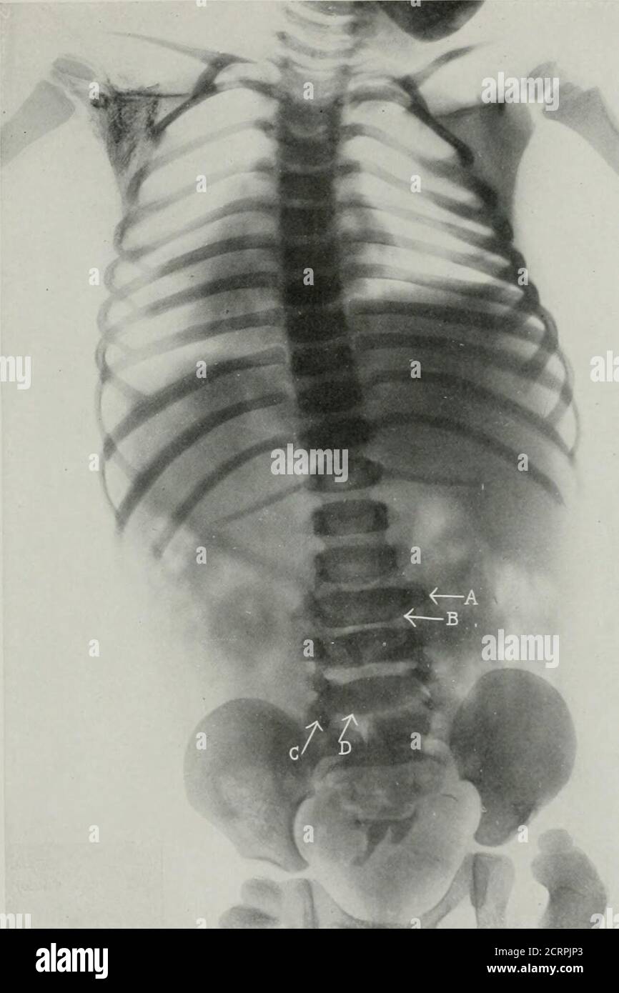 . Living anatomy and pathology; the diagnosis of diseases in early life by the Roentgen method . PLAT1-. 43. PPIXA BIFIDA OCTTLTA. Girl, age 3J years. (Same subject as Plates 42 and 44.) A. The narrowed third lumbar vertebra. B. The narrowed third intcivertebral disk. C. Sacralization of the left fifth lumbar vertebra. D. Just to the risiht and below the point of the arrow i.-; fissure of the first bodj of the sacrum. Plate 43. PLATE 44. SPINA BIFIDA OCCULTA. Girl, age 3i years. (Same Mibject as Plates 42 ami 4^.) The arrow points to thp right hip after reduetion. The deformities of tlio verte Stock Photohttps://www.alamy.com/image-license-details/?v=1https://www.alamy.com/living-anatomy-and-pathology-the-diagnosis-of-diseases-in-early-life-by-the-roentgen-method-plat1-43-ppixa-bifida-octtlta-girl-age-3j-years-same-subject-as-plates-42-and-44-a-the-narrowed-third-lumbar-vertebra-b-the-narrowed-third-intcivertebral-disk-c-sacralization-of-the-left-fifth-lumbar-vertebra-d-just-to-the-risiht-and-below-the-point-of-the-arrow-i-fissure-of-the-first-bodj-of-the-sacrum-plate-43-plate-44-spina-bifida-occulta-girl-age-3i-years-same-mibject-as-plates-42-ami-4-the-arrow-points-to-thp-right-hip-after-reduetion-the-deformities-of-tlio-verte-image376052491.html
RM2CRPJP3–. Living anatomy and pathology; the diagnosis of diseases in early life by the Roentgen method . PLAT1-. 43. PPIXA BIFIDA OCTTLTA. Girl, age 3J years. (Same subject as Plates 42 and 44.) A. The narrowed third lumbar vertebra. B. The narrowed third intcivertebral disk. C. Sacralization of the left fifth lumbar vertebra. D. Just to the risiht and below the point of the arrow i.-; fissure of the first bodj of the sacrum. Plate 43. PLATE 44. SPINA BIFIDA OCCULTA. Girl, age 3i years. (Same Mibject as Plates 42 ami 4^.) The arrow points to thp right hip after reduetion. The deformities of tlio verte
![. Living anatomy and pathology; the diagnosis of diseases in early life by the Roentgen method . PLATE 44. SPINA BIFIDA OCCULTA. Girl, age 3i years. (Same Mibject as Plates 42 ami 4^.) The arrow points to thp right hip after reduetion. The deformities of tlio vertebric and sacrum are the sameas in Phite 42. Xote the retardcil development of the upper epi]5hysis ofthe right femur in comparison with that of the left.. Pl^ATK 44 PLATE 45.FUSION- OF RIBS. MARKED SCOLIOSIS OF COXGENITAI- ORK.IX. Child, age about 4 years. (Ueiluced 27%.) -4. Hyoid bone. B. Wedged dor-sal vertebrae. C. Fusion of ribs Stock Photo . Living anatomy and pathology; the diagnosis of diseases in early life by the Roentgen method . PLATE 44. SPINA BIFIDA OCCULTA. Girl, age 3i years. (Same Mibject as Plates 42 ami 4^.) The arrow points to thp right hip after reduetion. The deformities of tlio vertebric and sacrum are the sameas in Phite 42. Xote the retardcil development of the upper epi]5hysis ofthe right femur in comparison with that of the left.. Pl^ATK 44 PLATE 45.FUSION- OF RIBS. MARKED SCOLIOSIS OF COXGENITAI- ORK.IX. Child, age about 4 years. (Ueiluced 27%.) -4. Hyoid bone. B. Wedged dor-sal vertebrae. C. Fusion of ribs Stock Photo](https://c8.alamy.com/comp/2CRPJK0/living-anatomy-and-pathology-the-diagnosis-of-diseases-in-early-life-by-the-roentgen-method-plate-44-spina-bifida-occulta-girl-age-3i-years-same-mibject-as-plates-42-ami-4-the-arrow-points-to-thp-right-hip-after-reduetion-the-deformities-of-tlio-vertebric-and-sacrum-are-the-sameas-in-phite-42-xote-the-retardcil-development-of-the-upper-epi-5hysis-ofthe-right-femur-in-comparison-with-that-of-the-left-platk-44-plate-45fusion-of-ribs-marked-scoliosis-of-coxgenitai-orkix-child-age-about-4-years-ueiluced-27-4-hyoid-bone-b-wedged-dor-sal-vertebrae-c-fusion-of-ribs-2CRPJK0.jpg) . Living anatomy and pathology; the diagnosis of diseases in early life by the Roentgen method . PLATE 44. SPINA BIFIDA OCCULTA. Girl, age 3i years. (Same Mibject as Plates 42 ami 4^.) The arrow points to thp right hip after reduetion. The deformities of tlio vertebric and sacrum are the sameas in Phite 42. Xote the retardcil development of the upper epi]5hysis ofthe right femur in comparison with that of the left.. Pl^ATK 44 PLATE 45.FUSION- OF RIBS. MARKED SCOLIOSIS OF COXGENITAI- ORK.IX. Child, age about 4 years. (Ueiluced 27%.) -4. Hyoid bone. B. Wedged dor-sal vertebrae. C. Fusion of ribs Stock Photohttps://www.alamy.com/image-license-details/?v=1https://www.alamy.com/living-anatomy-and-pathology-the-diagnosis-of-diseases-in-early-life-by-the-roentgen-method-plate-44-spina-bifida-occulta-girl-age-3i-years-same-mibject-as-plates-42-ami-4-the-arrow-points-to-thp-right-hip-after-reduetion-the-deformities-of-tlio-vertebric-and-sacrum-are-the-sameas-in-phite-42-xote-the-retardcil-development-of-the-upper-epi-5hysis-ofthe-right-femur-in-comparison-with-that-of-the-left-platk-44-plate-45fusion-of-ribs-marked-scoliosis-of-coxgenitai-orkix-child-age-about-4-years-ueiluced-27-4-hyoid-bone-b-wedged-dor-sal-vertebrae-c-fusion-of-ribs-image376052404.html
. Living anatomy and pathology; the diagnosis of diseases in early life by the Roentgen method . PLATE 44. SPINA BIFIDA OCCULTA. Girl, age 3i years. (Same Mibject as Plates 42 ami 4^.) The arrow points to thp right hip after reduetion. The deformities of tlio vertebric and sacrum are the sameas in Phite 42. Xote the retardcil development of the upper epi]5hysis ofthe right femur in comparison with that of the left.. Pl^ATK 44 PLATE 45.FUSION- OF RIBS. MARKED SCOLIOSIS OF COXGENITAI- ORK.IX. Child, age about 4 years. (Ueiluced 27%.) -4. Hyoid bone. B. Wedged dor-sal vertebrae. C. Fusion of ribs Stock Photohttps://www.alamy.com/image-license-details/?v=1https://www.alamy.com/living-anatomy-and-pathology-the-diagnosis-of-diseases-in-early-life-by-the-roentgen-method-plate-44-spina-bifida-occulta-girl-age-3i-years-same-mibject-as-plates-42-ami-4-the-arrow-points-to-thp-right-hip-after-reduetion-the-deformities-of-tlio-vertebric-and-sacrum-are-the-sameas-in-phite-42-xote-the-retardcil-development-of-the-upper-epi-5hysis-ofthe-right-femur-in-comparison-with-that-of-the-left-platk-44-plate-45fusion-of-ribs-marked-scoliosis-of-coxgenitai-orkix-child-age-about-4-years-ueiluced-27-4-hyoid-bone-b-wedged-dor-sal-vertebrae-c-fusion-of-ribs-image376052404.htmlRM2CRPJK0–. Living anatomy and pathology; the diagnosis of diseases in early life by the Roentgen method . PLATE 44. SPINA BIFIDA OCCULTA. Girl, age 3i years. (Same Mibject as Plates 42 ami 4^.) The arrow points to thp right hip after reduetion. The deformities of tlio vertebric and sacrum are the sameas in Phite 42. Xote the retardcil development of the upper epi]5hysis ofthe right femur in comparison with that of the left.. Pl^ATK 44 PLATE 45.FUSION- OF RIBS. MARKED SCOLIOSIS OF COXGENITAI- ORK.IX. Child, age about 4 years. (Ueiluced 27%.) -4. Hyoid bone. B. Wedged dor-sal vertebrae. C. Fusion of ribs
 . Lehrbuch der allgemeinen und speciellen pathologischen Anatomie und Pathogenese. auch Geschwülste ent-stehen. Es hat auch denAnschein, als ob eine Ver-lagerung der Gewebe anOrte, wo sie nicht hin-gehören, eine gewisse Dis-position zu atypischer Wu-cherung derselben schafft.Neuestens hat v. Reck-LiNGHAüSEN die Aufmerk-samkeit namentlich aufdie Entwickelungsstörun-gen des Wirbelkanales hin-gelenkt und den Nachweiserbracht, dass auch hierdurch örtliche Missbildun-gen verursachte Verlage- Fig. 178. Spina bifida occulta mit Myolipom innerhalb des VPirbel-k anales. Sagittaler Durchschnitt fast 1 c Stock Photohttps://www.alamy.com/image-license-details/?v=1https://www.alamy.com/lehrbuch-der-allgemeinen-und-speciellen-pathologischen-anatomie-und-pathogenese-auch-geschwlste-ent-stehen-es-hat-auch-denanschein-als-ob-eine-ver-lagerung-der-gewebe-anorte-wo-sie-nicht-hin-gehren-eine-gewisse-dis-position-zu-atypischer-wu-cherung-derselben-schafftneuestens-hat-v-reck-linghasen-die-aufmerk-samkeit-namentlich-aufdie-entwickelungsstrun-gen-des-wirbelkanales-hin-gelenkt-und-den-nachweiserbracht-dass-auch-hierdurch-rtliche-missbildun-gen-verursachte-verlage-fig-178-spina-bifida-occulta-mit-myolipom-innerhalb-des-vpirbel-k-anales-sagittaler-durchschnitt-fast-1-c-image370319498.html
RM2CEDE8A–. Lehrbuch der allgemeinen und speciellen pathologischen Anatomie und Pathogenese. auch Geschwülste ent-stehen. Es hat auch denAnschein, als ob eine Ver-lagerung der Gewebe anOrte, wo sie nicht hin-gehören, eine gewisse Dis-position zu atypischer Wu-cherung derselben schafft.Neuestens hat v. Reck-LiNGHAüSEN die Aufmerk-samkeit namentlich aufdie Entwickelungsstörun-gen des Wirbelkanales hin-gelenkt und den Nachweiserbracht, dass auch hierdurch örtliche Missbildun-gen verursachte Verlage- Fig. 178. Spina bifida occulta mit Myolipom innerhalb des VPirbel-k anales. Sagittaler Durchschnitt fast 1 c
 . Anatomischer Anzeiger. Anatomy, Comparative; Anatomy, Comparative. 433 Nachdruck verboten. Barissimo caso di atresia ed anomale disposizioiii congenite deir intcstino; coiicomitante spina bifida occulta. Pel Dott. Gaetano Cutore, Aiuto e Professore incaricato di Anatomia topografica. (Istituto Anatomico dell'üniversitä, di Catania, diretto dal Prof. R. Sta- DERINI.) Con 9 figure. (Schluß.) Esame microscopico dell'iiitestino. Degli organi che ho passato fin qui in speciale rassegna perche difettosi o irregolari nel loro sviluppo, varii segraenti del canale ali- nientare mi parve meritassero u Stock Photohttps://www.alamy.com/image-license-details/?v=1https://www.alamy.com/anatomischer-anzeiger-anatomy-comparative-anatomy-comparative-433-nachdruck-verboten-barissimo-caso-di-atresia-ed-anomale-disposizioiii-congenite-deir-intcstino-coiicomitante-spina-bifida-occulta-pel-dott-gaetano-cutore-aiuto-e-professore-incaricato-di-anatomia-topografica-istituto-anatomico-dellniversit-di-catania-diretto-dal-prof-r-sta-derini-con-9-figure-schlu-esame-microscopico-delliiitestino-degli-organi-che-ho-passato-fin-qui-in-speciale-rassegna-perche-difettosi-o-irregolari-nel-loro-sviluppo-varii-segraenti-del-canale-ali-nientare-mi-parve-meritassero-u-image236806765.html
RMRN7DCD–. Anatomischer Anzeiger. Anatomy, Comparative; Anatomy, Comparative. 433 Nachdruck verboten. Barissimo caso di atresia ed anomale disposizioiii congenite deir intcstino; coiicomitante spina bifida occulta. Pel Dott. Gaetano Cutore, Aiuto e Professore incaricato di Anatomia topografica. (Istituto Anatomico dell'üniversitä, di Catania, diretto dal Prof. R. Sta- DERINI.) Con 9 figure. (Schluß.) Esame microscopico dell'iiitestino. Degli organi che ho passato fin qui in speciale rassegna perche difettosi o irregolari nel loro sviluppo, varii segraenti del canale ali- nientare mi parve meritassero u